Note: Descriptions are shown in the official language in which they were submitted.
CA 03071295 2020-01-27
WO 2019/023508 PCT/US2018/043973
- 1 -
METHODS AND DEVICES FOR DETECTION OF
ANTICOAGULANTS IN PLASMA AND WHOLE BLOOD
RELATED APPLICATIONS
[0001] This application claims the benefit of U.S. Provisional Application
No.
62/538,618, filed on July 28, 2017, and U.S. Provisional Application No.
62/699,665, filed on
July 17, 2018, the entire contents of which are hereby incorporated by
reference.
GOVERNMENT SUPPORT
[0002] This invention was made with Government support under Grant Nos. P41
EB002503, P30 ES002109, and P50 GM021700 awarded by the National Institutes of
Health.
The Government has certain rights in the invention.
BACKGROUND
[0003] The coagulation system is a delicate balance between hemorrhage and
thrombosis.
There are many disease states, including cancer, auto-immune disease,
infection, trauma,
surgery, heart disease, and drugs, that can cause a disruption of this balance
and result in a
patient having severe, even life-threatening, bleeding or clotting events.
Anticoagulant
medications are commonly prescribed for thrombotic disorders. Conventional
anticoagulant
medications, such as Heparin, will indirectly inhibit multiple factors of the
clotting cascade.
The more recent introduction of direct oral anticoagulants (DOACs) allows for
targeted
inhibition of the coagulation pathway.
[0004] The biggest risk of anticoagulation therapy is the increased risk of
bleeding, and
thus, traditionally, patients taking anticoagulant medications are carefully
monitored to
ensure that they are receiving an appropriate dose. Current clinical tests
available to evaluate
a patient's bleeding and clotting are either rudimentary and provide very
vague information,
such as prothrombin time (PT) and activated thromboplastin time (aPTT), or are
more
detailed but require expensive machines, lengthy training, and careful
handling. Included in
the latter category are thromboelastography (TEG), thromboelastometry (TEM),
rotational
thromboelastometry (ROTEM), platelet aggregometry and flow cytometry.
Currently,
specific tests for the DOACs are not available. Most of the DOAC assays that
have been
proposed are pharmacokinetic assays that measure the absolute concentration of
the drug
CA 03071295 2020-01-27
WO 2019/023508 PCT/US2018/043973
- 2 -
itself and, therefore, provide limited functional information to support
clinical decision-
making.
[0005] Coagulation tests are needed that can detect, characterize, and/or
quantify
impairments in coagulation, including detection of DOACs in patient samples,
to better
manage patients at high risk of severe bleeding or clotting, including, but
not limited to, the
urgent care setting.
SUMMARY
[0006] Methods and devices for evaluating coagulation are described,
including methods
and devices for detecting an anticoagulation agent or a coagulation
abnormality. Coagulation
abnormality includes abnormality of clot formation (e.g., thrombosis) and
abnormality of clot
degradation (e.g., fibrinolysis). In various embodiments, the methods and
devices of the
invention measure coagulation of a sample in response to a gradient of one or
more
coagulation factors. These responses can be evaluated to accurately profile
coagulation
impairments of the sample, including the presence of a DOAC or traditional
anticoagulant
medication. In various embodiments, the invention provides point-of-care or
bedside testing
with a convenient, microfluidic device that can be used by minimally trained
personnel.
[0007] In some aspects, the invention provides methods for assessing
coagulation in a
blood sample. The method comprises adding a coagulation factor to plural
portions (e.g.,
aliquots) of the blood sample, each portion receiving the coagulation factor
at a different
concentration, and measuring clot formation or clot formation times in
response to the
different concentrations. By assessing coagulation in response to the
different concentrations
of one or more coagulation factors, blood clotting function can be accurately
profiled,
including the impact of DOACs or other drugs on coagulation. In some
embodiments, the
presence or absence of a genetic clotting abnormality is determined. The
methods as
described herein may be performed using a microfluidic device as described,
where one or
more of the channels can be configured to trigger formation and localization
of a clot.
[0008] As used herein, unless described otherwise, a "blood sample" refers
to a whole
blood sample or a plasma sample. The term plasma includes both platelet-rich-
plasma (PRP)
and platelet-poor-plasma (PPP).
[0009] The term "coagulation factor" as used herein means any factor
implicated in the
coagulation cascade (intrinsic, extrinsic and common pathways), including
Factors Ito XIII,
CA 03071295 2020-01-27
WO 2019/023508 PCT/US2018/043973
- 3 -
von Willebrand factor, prekallikrein (Fletcher factor), high-molecular-weight
kininogen
(1-IMWK) (Fitzgerald factor), fibronectin, antithrombin III, heparin cofactor
II, protein C,
protein S, protein Z, Protein Z-related protease inhibitor (ZPI), plasminogen,
alpha 2-
antiplasmin, tissue plasminogen activator (tPA), urokinase, plasminogen
activator inhibitor-1
(PAI1), plasminogen activator inhibitor-2 (PAI2), Tissue Factor Pathway
Inhibitor (TFPI),
and cancer procoagulant. The coagulation factor(s) can be in activated form or
inactivated
(e.g., precursor) form. For example, for detecting the presence of a
coagulation factor
inhibitor in a sample, the coagulation factor should be in activated form
(e.g., Factor Xa or
Factor Ha). In other embodiments, for detection of a genetic clotting
abnormality, the
coagulation factor may be in inactivated form (e.g., Factor X or Factor II).
Further, the
coagulation factor(s) can be from a human, an animal (such as bovine, porcine
or other), or
can be a synthesized or recombinant protein.
[0010] In some embodiments, the invention provides a method of detecting an
anticoagulation agent. Anticoagulation agents are substances that prevent or
reduce
coagulation of blood, prolonging clotting time. Anticoagulation agents
include, but are not
limited to, Factor-specific inhibitors (such as FXa inhibitors, FIIa
inhibitors, FXIa inhibitors,
FXIIa inhibitors), heparins, and vitamin K antagonists (e.g., warfarin). In
some embodiment,
they include Direct Oral Anticoagulants (DOACs), also known as Novel Oral
Anticoagulants
(NOACs), such as XARELTO (Rivaroxaban) by Janssen Pharmaceuticals, Inc.,
ELIQUIS
(Apixaban) by Bristol-Myers Squibb and Pfizer Inc., SAVAYSA (Edoxaban) by
Daiichi
Sankyo, Inc., PRADAXA (Dabigatran) by Boehringer Ingelheim, and BEVYXXA
(Betrixaban) by Portola Pharmaceuticals, Inc.
[0011] By measuring clot formation (e.g., clot formation times) in response
to increasing
concentrations of exogenously added coagulation factors, the presence and/or
point of
inhibition by a therapeutic agent can be determined. For example, a sample
that is positive for
a coagulation inhibitor will show a concentration-dependent decrease in
clotting time as the
coagulation factor that is targeted by the inhibitor is added to the sample.
Meanwhile, when a
coagulation factor upstream from the point of inhibition is added (in
increasing amounts), the
clotting time will remain prolonged, as compared to the clotting time upon the
addition of a
coagulation factor downstream of the point of inhibition. See FIGS. 9-13.
[0012] In some embodiments, results for a patient sample can be compared to
reference
standards, including standards for normal and/or abnormal clotting, or
reference standards
CA 03071295 2020-01-27
WO 2019/023508 PCT/US2018/043973
- 4 -
corresponding to anticoagulant therapy with particular agents. In some
embodiments,
reference standards are personalized for the patient.
[0013] In various embodiments, clotting curves can be constructed to
characterize the
response of clot formation to the addition of various coagulation factors in
increasing
concentrations or amounts. These clotting curves allow for the identity and
amount of
coagulation inhibitors to be determined, to thereby guide patient care. In
some embodiments,
the appropriate coagulation inhibitor reversal agent is then administered to
the patient to
reverse the therapeutic intervention as needed.
[0014] In some aspects, the invention provides a microfluidic device for
evaluating
coagulation in a sample. The device includes a series of channels in a
substrate, each channel
having an area with a geometry to trigger and/or localize formation of a clot,
to allow for
evaluation of clot formation in response to one or more reagents, such as the
amount or
concentration of an exogenously added coagulation factor. The channels in the
series each
have the same geometry, so as to trigger identical clot formation properties
(when exposed to
the same sample and reagents). By evaluating clot formation in the presence of
a gradient of
one or more coagulation factors, the invention allows for sensitive and
specific detection of
coagulation abnormalities or impairments, including the presence or activity
of a DOAC in
the sample.
[0015] In one embodiment, the microfluidic device for detecting coagulation
includes
plural channels formed in a substrate, each channel including a clot forming
area having a
geometry configured to trigger and/or localize formation of a clot. The clot
forming areas of
the plural channels may be arranged in a central region of the substrate in
some embodiments,
such that the clotting properties can be simultaneously imaged or analyzed
across the
channels. See FIGS. 1A-1B, 2B. The device may further include plural sample
input ports to
receive a sample (e.g., whole blood or plasma), each sample input port
connected to a first
end of one of the plural channels. See FIGS. 1A-1D. In other embodiments, the
device has a
single sample input port in fluid communication with the plural channels, or a
series of
channels. See FIG. 5A. In some embodiments, each channel has an independent
output port,
each output port connected to a second end of one of the plural channels. In
embodiments
employing independent sample input ports, the input and output ports can be
arranged in an
alternating pattern at a periphery of the substrate. See FIGS. 1A-1B, 2A. In
some
CA 03071295 2020-01-27
WO 2019/023508 PCT/US2018/043973
- 5 -
embodiments, the input and output ports are arranged in a pattern other than
an alternating
pattern.
[0016] The term "central region" as used herein means a region that is
located in the
center of a substrate relative to a periphery of the substrate and can include
a region that is
positioned off-center. For example, depending upon the configuration, the
central region
might be off-center and the areas in the microfluidic channels in which clots
begin can be
controlled by the flow patterns in the channels.
[0017] In some embodiments, the clot forming areas of the plural channels
are arranged
in a region of the substrate which is not central, such as, but not limited
to, the periphery. See
FIGS. 5A-5B.
[0018] Each channel may further comprise one or more additional input ports
to receive
reagents, such as coagulation factor(s) and/or calcium. In some embodiments,
there is more
than one input port (e.g., for introducing sample and one or more reagents)
per output port.
For example, in one embodiment, there can be one input port for the sample and
1 to 2 input
ports for the reagents (e.g., coagulation factor and, optionally, calcium).
See FIG. 1B. In some
embodiments, there is one common input port for the sample, and each channel
further
comprises further input ports (e.g., 1 or 2) for reagents.
[0019] In the microfluidic device, each clot forming area can be configured
to create an
area of stasis or disruption in fluid flow to trigger and/or localize
formation of a clot. In some
embodiments, each clot forming area can be configured to create an area of
flow disturbance
to trigger and/or localize clot formation. Exemplary geometries for triggering
formation of
and localizing a clot are illustrated in FIGS. 2B, 3A, 5A and 5B.
[0020] Channels of the microfluidic device can be coated with, contain or
otherwise
include a coagulation factor at a different amount or concentration. For
example, a first
group or series of the plural channels can be coated with, contain or
otherwise include a first
coagulation factor, and a second group or series of the plural channels can be
coated with,
contain or otherwise include a second coagulation factor. Further, in some
embodiments, one
of the plural channels is a negative control channel, e.g., may not be coated
with and may not
include a coagulation factor. In other embodiments, the device does not
comprise such a
negative control channel.
[0021] In the case where one or more channels include the coagulation
factor(s), the
coagulation factor(s) may be in suspension or solution, or lyophilized and not
surface-bound.
CA 03071295 2020-01-27
WO 2019/023508 PCT/US2018/043973
- 6 -
The coagulation factor(s) can be pre-included in the channel(s) (e.g., at the
time of
manufacturing the device), can be added prior to placing the sample into the
device, or can be
entered into the device through an input port (or multiple input ports)
simultaneously with the
sample or after the sample.
[0022] In embodiments of the microfluidic device that include first and
second groups of
channels (whether or not such embodiments may also include a negative control
channel in
addition to the first and second groups of channels), each channel in the
first group of the
plural channels can be coated with, contain or otherwise include a first
coagulation factor at a
different amount or concentration, and each channel in the second group of the
plural
channels can be coated with, contain or otherwise include a second coagulation
factor at a
different amount or concentration. In some embodiments, the microfluidic
device may
contain more than two groups or series of plural channels, such as three,
four, five or more
groups, wherein each group or series of plural channels is coated with,
contains or otherwise
includes a different coagulation factor at an increasing amount across the
group or series
(e.g., a microfluidic device containing four groups of channels, each group of
the plural
channels can be coated with, contain or otherwise include a different
coagulation factor
selected from Factors Ha, Xa, XI, XIa, XII, and XIIa). By measuring clot
formation or
clotting time as a function of coagulation factor gradients, the sample's
clotting properties
can be profiled at several specific points of the coagulation pathway(s)
(illustrated in FIG. 8),
providing a clinician with detailed and specific information concerning the
patient's clotting
physiology and/or the status of any therapeutic intervention.
[0023] The second coagulation factor can be upstream in the coagulation
cascade from
the first coagulation factor. For example, the first coagulation factor can
be, e.g.,
prothrombin (Factor II), thrombin (Factor IIa), or both. The second
coagulation factor can
be, e.g., Factor X, Factor Xa, or both.
[0024] The microfluidic device can further include a detection device
configured to
measure clot formation times in each of the channels to assess coagulation
based on the clot
formation times measured. For example, the detection device can be configured
to image the
clot forming areas simultaneously to measure clot formation times. In some
embodiments,
the degree of clot formation in each of the channels is quantified at a fixed
time or times. For
example, the detection device in connection with the methods and devices
described herein
can include a microscope and an image sensor. Imaging the clot forming areas
can include
CA 03071295 2020-01-27
WO 2019/023508 PCT/US2018/043973
- 7 -
bright-field imaging. For the devices and assays described herein, clotting
times can also be
measured with other methodologies such as detection based on light absorbance,
fluorescence
measurements, ultrasound, etc., and the detection device can be configured to
employ one or
more of these other methodologies. Ways to detect clotting also include, but
are not limited
to, detection based on electrical impedance, the addition of beads and
quantifying bead flow
rate/number, measurement of flow velocity and/or pressure before and/or after
the site of clot
formation, thromboelastography, fluorescence detection (such as with
fluorescent
fibrinogen), turbidity, magnetic, flow dynamics (pressure or flow velocity),
infrared light
detection, infrared spectroscopy, detection using acoustic and/or photonic
sensors, flow
cytometry, and visual clotting detection.
[0025] In some embodiments, the method described herein does not employ a
microfluidic device, but uses wells or containers suitable for inducing and
measuring
formation of a clot.
[0026] In addition to clot formation times, other characteristics of clot
formation can be
considered. It is contemplated that a qualitative measure of clot formation,
in addition to clot
formation times, can be useful, e.g., to determine the most sensitive
detection mode for
coagulation. For example, properties of the clot such as size, strength,
density and
composition can be assessed in addition to time to form a clot. Such
properties may be
assessed using the same or a different detection modality than is used to
detect clot formation
times.
[0027] In some embodiments, clot lysis can be assessed in addition to clot
formation. For
example, if a patient is on a fibrinolytic or thrombolytic agent, one can
evaluate the clot when
it is being formed as well as its breakdown over time. In one embodiment, the
same methods
described herein and known in the art to detect clot formation can be used to
assess clot lysis
over time.
[0028] As described herein regarding the use of thromboelastography (TEG),
one can
evaluate both clot formation and fibrinolysis. This would be useful for
detecting clotting
abnormalities in patients that are hypocoagulable due to problems with
fibrinolysis or
iatrogenic administration of fibrinolytic and thrombolytic drugs. See, for
example, C.
Mauffrey, et al., "Strategies for the management of haemorrhage following
pelvic fractures
and associated trauma-induced coagulopathy," Bone Joint J. 2014; 96-B:1143-54,
the
relevant teachings of which are incorporated herein by reference.
CA 03071295 2020-01-27
WO 2019/023508 PCT/US2018/043973
-8-
100291 In any of the devices and methods described herein, the blood sample
can be a
whole blood sample or a plasma sample. Using whole blood can be particularly
useful for
certain applications, such as those implemented at the bedside of a patient.
[0030] The disclosed devices and methods can be applied to all individuals,
including
mammals (e.g., humans, such as human patients, as well as non-human mammals),
reptiles,
birds, and fish, among others, and can be useful for research and veterinary
medicine. An
individual can be, for example, mature (e.g., adult) or immature (e.g., child,
infant, neonate,
or pre-term infant).
[0031] The disclosed devices and methods can be used not just for
diagnostic purposes
but also for research and discovery to explore the coagulation cascade in a
research setting.
For example, this can be useful for basic drug discovery, understanding
disease or disorder
pathophysiology, for example, in the context of hemorrhagic diseases (Dengue
virus, Zika
virus, Ebola virus, etc.), and also to monitor for adverse events of
experimental treatments.
[0032] The disclosed devices and methods can be used to guide therapy of a
patient. For
example, physicians can use the results to determine subsequent treatments
with both drugs
and procedural interventions (both invasive and non-invasive). For example, if
a patient tests
positive for Factor Ha inhibition due to dabigatran administration, then the
healthcare
provider may choose to administer the reversal agent (idarucizumab) for this
inhibitor prior to
surgery or other invasive procedures. Likewise, if the patient tests positive
for Factor Xa
inhibition, then the healthcare provider may choose to administer the
appropriate reversal
agent (coagulation factor Xa (recombinant), inactivated-zhzo) for this
inhibitor. The
healthcare provider may choose to administer other agents that overcome the
effects of these
inhibitors as well, such as 4-factor prothrombin complex concentrates or
activated
prothrombin complex concentrates.
[0033] Other aspects and embodiments of the invention will be apparent from
the
following Drawings and Detailed Description.
BRIEF DESCRIPTION OF THE DRAWINGS
[0034] The patent or application file contains at least one drawing
executed in color.
Copies of this patent or patent application publication with color drawings
will be provided
by the Office upon request and payment of the necessary fee.
CA 03071295 2020-01-27
WO 2019/023508 PCT/US2018/043973
-9-
100351 The foregoing will be apparent from the following more particular
description of
example embodiments, as illustrated in the accompanying drawings in which like
reference
characters refer to the same parts throughout the different views. The
drawings are not
necessarily to scale, emphasis instead being placed upon illustrating
embodiments.
[0036] FIGS. 1A-1D are schematic illustrations of microfluidic device
layouts employing
multiple sample ports according to example embodiments of the invention.
[0037] FIG. 2A is a top view of the circular microfluidic clotting device
according to an
example embodiment.
[0038] FIG. 2B is a magnified view of the central portion of the device of
FIG. 2A. FIG.
2B illustrates exemplary geometries of clot formation areas.
[0039] FIGS. 3A-3C illustrate clot detection using plasma and fluorescent-
labeled
fibrinogen within a microfluidic device having four channels according to an
example
embodiment. FIG. 3A is a top view bright-field image of a central portion of
the example
microfluidic device. FIG. 3B is a fluorescent image of clot formation using
the device of
FIG. 3A. FIG. 3C is a fluorescent image showing a magnified view of a clot
forming area.
[0040] FIGS. 4A and 4B are bright-field images illustrating clot detection
using whole
blood in a parallel microfluidic channel device employing a FXa gradient,
according to an
example embodiment. FIG. 4A contains no anticoagulant. FIG. 4B contains
unfractionated
heparin.
[0041] FIGS. 5A and 5B are schematic illustrations of microfluidic device
configurations
employing a single port for sample input according to example embodiments of
the invention.
[0042] FIG. 6 is a flow diagram of an assay or a method according to
example
embodiments of the invention.
[0043] FIG. 7A is a graph of example data illustrating detection of
Rivaroxaban, using a
FXa gradient.
[0044] FIG. 7B is a graph of example data illustrating detection of
Apixaban, using a FXa
gradient.
[0045] FIG. 7C is a graph of example data illustrating detection of
Edoxaban, using a
FXa gradient.
[0046] FIG. 7D is a graph of example data illustrating detection of
Dabigatran, using a
FIIa gradient.
[0047] FIG. 8 is a diagram illustrating a basic clotting cascade.
CA 03071295 2020-01-27
WO 2019/023508
PCT/US2018/043973
- 10 -
[0048] FIG. 9 is a diagram illustrating how to detect FXa
inhibition/deficiency/abnormality of function by employing coagulation factor
gradients.
[0049] FIG. 10 is a diagram illustrating how to detect FIIa
inhibition/deficiency/abnormality of function by employing coagulation factor
gradients.
[0050] FIG. 11 is a diagram illustrating how to detect and differentiate
between FIIa and
FXa inhibition in a sample by employing coagulation factor gradients.
[0051] FIG. 12 is a diagram illustrating how to detect indirect FXa
inhibition/deficiency/abnormality of function by employing coagulation factor
gradients.
[0052] FIG. 13 is a diagram illustrating how to detect and differentiate
between FXIIa
and FXIa inhibition in a sample by employing coagulation factor gradients.
[0053] FIG. 14 is a diagram illustrating how to detect and differentiate
between various
types of hemophilia by employing coagulation factor gradients.
[0054] FIG. 15 is a diagram illustrating how to detect problems with
fibrinogen or FXIII
(e.g., FXIII deficiency) by employing coagulation factor gradients.
[0055] FIGS. 16A-16C illustrate Clotting Curve Scores (CCS) for FXa and
FIIa
inhibitors at various concentrations.
[0056] FIG. 17 shows Table 1 of patient descriptive statistics (Example
17).
[0057] FIGS. 18A-18C illustrate measurements of sensitivity and specificity
of
prothrombin time (PT) (FIG. 18A) and international normalized ratio (INR)
(FIG. 18B) for
FXa inhibitor (FXa-I) anticoagulation.
[0058] FIGS. 19A-19G illustrate example clotting time data and comparative
clotting
curves.
[0059] FIGS. 20A-20E illustrate Clotting Curve Score (CCS) analysis and
evaluation of
CCS utilization for the detection of FXa-I in patient samples.
[0060] FIGS. 21A and 21B illustrate example functional drug concentration
calculation.
[0061] FIG. 22 illustrates a current decision-making paradigm for a patient
that is
bleeding or at high risk.
[0062] FIG. 23 illustrates an improved decision-making paradigm using
embodiment(s)
of the present invention for a patient that is bleeding or at high risk.
[0063] FIGS. 24A and 24B illustrate detection of decrease in FXa inhibition
by a FXa-I
after the addition of activated prothrombin complex concentrate (aPCC).
DETAILED DESCRIPTION
CA 03071295 2020-01-27
WO 2019/023508 PCT/US2018/043973
-11-
100641 The invention generally relates to methods and devices for the
detection of
coagulation, including detection of coagulation abnormalities and detection of
anticoagulants
and platelet inhibitors in plasma and/or whole blood.
[0065] Acquired coagulopathies are a major component of morbidity and
mortality in a
number of medical settings. Individuals may have increased risk of internal
bleeding
secondary to drugs (e.g., clopidogrel, heparin, warfarin or other vitamin K
antagonists,
dabigatran or other Direct Oral Anticoagulants, etc.), trauma, surgery,
sepsis, cancer, organ
dysfunction (e.g., liver), or congenital abnormality (e.g., hemophilia). On
the other end of the
spectrum, increased propensity for clotting can be due to autoimmune disease,
cancer,
atherosclerosis, early trauma and sepsis, organ dysfunction (e.g., kidney),
immobility,
inflammation, foreign body (e.g., stent or prosthesis), or congenital
abnormality (e.g., Factor
V Leidin thrombophilia). With recent innovations in drug development (e.g.,
anticoagulants,
including Direct Oral Anticoagulants, or DOACs), innovation is now needed for
hemostasis/coagulation analyzers, to fully realize benefits for patients,
including in the urgent
care setting. Specifically, current clinical tests available to evaluate a
patient's bleeding and
clotting are either rudimentary and provide very vague information, such as
prothrombin time
(PT) and activated thromboplastin time (aPTT), or are more detailed but
require expensive
machines, lengthy training, and careful handling, such as thromboelastography
(TEG),
thromboelastometry (TEM), rotational thromboelastometry (ROTEM), platelet
aggregometry
and flow cytometry. Currently, specific tests for DOACs are not available.
Most of the
DOAC assays that have been proposed are pharmacokinetic assays that measure
the absolute
concentration of the drug itself and, therefore, provide limited functional
information for
clinical decision-making.
[0066] With the increased use of DOACs, studies and reviews are finding
that, although
these new drugs pose less risk for acute, life-threatening bleeding events,
they are potentially
linked to higher rates of gastrointestinal (GI) bleeding. Additionally, these
new drugs are
found to have different pharmacokinetic properties in patients with decreased
liver and/or
kidney function or in patients that are on multiple drugs at the same time, as
is common in the
geriatric population. In these cases, providing functional clinical
information to the doctor to
help personalize the anticoagulant combination and dosage would be of great
benefit to the
patient and possibly decrease subsequent, related adverse events. Embodiments
of the
invention can be used in clotting panels that evaluate the coagulation,
fibrinolysis, and
CA 03071295 2020-01-27
WO 2019/023508 PCT/US2018/043973
- 12 -
platelet function within an individual. The microfluidic technology and
advanced assays
described herein in some embodiments provide for custom clotting panels,
whereby clinicians
can determine a patient's coagulation function bedside. These embodiments
provide for vast
improvements in patient care, including in the urgent care setting.
[0067] In addition to these assays being rapid and easy-to-interpret, they
can also be
customizable, allowing for the selection of clinically-relevant coagulation
and platelet
function testing for each customer and/or end-user segment. Because
embodiments of the
assay can be applied in a bedside platform, it can also be utilized for trend-
monitoring in
patients on various treatments (including at the hospital, at anticoagulation
clinics and at
home). In an aspect of the invention, a gradient of the factor(s) is added to
the sample after it
is subdivided into and/or distributed among the multiple groups of plural
channels, wells, or
containers, which method permits evaluation of coagulation function/inhibition
and
identification and differentiation between various coagulation abnormalities
within a sample.
This means that embodiments of the invention (e.g., clotting panels, assays,
etc.) are
potentially useful for assessing coagulation in patients that have poor
medical compliance,
where dosage/time taken is unknown, or in patients that are unconscious, where
the doctor,
surgeon or other healthcare provider needs to know whether the patient has any
of these
drugs in their system. Further, embodiments can help in monitoring
anticoagulation and
guiding the administration of reversal reagents that are now becoming
available.
[0068] Examples of potential users for product or services based on
embodiments of the
invention can range from healthcare workers, e.g., clinicians and
veterinarians, to researchers
in pharmaceutical research and development.
[0069] The invention can be applied to patient care in various settings. In
some
embodiments, the patient is scheduled for surgery or is in need of an invasive
procedure, and
the methods and devices of the invention can be used for clinical decision-
making, including
preparing the patient for the procedure to minimize bleeding risks. In some
embodiments, the
patient is administered a drug that impacts coagulation, and the methods and
devices of the
invention can be used for early evaluation of drug action and for selection of
the appropriate
therapy and dose. In some embodiments, the patient receives a drug or blood
product, and
methods and devices of the invention can be used to guide administration and
dose. In some
embodiments, the patient has or is suspected of having, or is at risk of
acquiring, a
hemorrhagic virus. In some embodiments, the patient is a neonate, where only
small volumes
CA 03071295 2020-01-27
WO 2019/023508 PCT/US2018/043973
- 13 -
of blood are available for evaluating coagulation (including for administering
anticoagulant
therapy or for detecting a congenital coagulation abnormality). In some
embodiments, the
patient is a pregnant mother, and the methods and devices allow for detecting
a congenital
coagulation abnormality, or for early diagnosis of a condition that results in
a coagulation
abnormality such as pre-eclampsia and eclampsia.
[0070] In some embodiments, the patient or subject is a veterinary or
animal patient (e.g.,
such as a dog, cat, or horse). In some embodiments, the patient is a non-human
mammal. The
cost-restrictions and limited blood volume of veterinary patients and
laboratory animal
research result in a large need for coagulation diagnostics that are easy-to-
use, require only
microliters of blood, and have lower overhead costs.
[0071] Due to the immense interest in novel coagulation testing platforms,
the blood-
testing platform (e.g., assay, microfluidic device, and/or combination
thereof) described
herein offers tremendous potential for research and product development.
[0072] In some embodiments, the patient is receiving an anticoagulant
therapy, such as a
heparin or vitamin K antagonist (e.g., warfarin). In some embodiment, the
patient is
undergoing therapy with a Direct Oral Anticoagulant (DOAC), such as XARELTO
(Rivaroxaban), ELIQUIS (Apixaban), SAVAYSA (Edoxaban), PRADAXA (Dabigatran),
or
BEVYXXA (Betrixaban). In some embodiments, the patient is undergoing therapy
with an
antibody against TFPI. Anticoagulant drugs are used commonly in many medical
settings,
including emergency and critical care, surgery, cardiology, and cancer.
Several new
anticoagulants have been introduced, but there are no current tests that can
reliably determine
if a patient is on the right dose. Too much anticoagulation can cause life-
threatening bleeding
and too little can lead to an increased risk of stroke and heart attacks.
Embodiments of the
invention can be used as or incorporated into a bedside test that can
accurately monitor these
new anticoagulants and improve the safety for these patients. This test can be
performed with
minimal training and in an easy to interpret format. In an embodiment, these
assays can be
performed in the lab in a device requiring less than about 1 mL, or less than
about 500 L, or
less than about 100 L, or less than about 50 [IL (one drop) of fresh or
citrated, whole blood,
with the results being read within 10 minutes.
[0073] The Direct Oral Anticoagulant (DOAC) market currently consists of
drugs that
selectively target specific factors within the coagulation pathways, e.g.,
Factor Ha or Factor
Xa. While these drugs are very potent, because of the dearth of reliable or
easy-to-use
CA 03071295 2020-01-27
WO 2019/023508 PCT/US2018/043973
- 14 -
diagnostic and monitoring tests, there is an increased risk associated with
the use and
administration of these drugs, especially in the critical care setting. One of
the primary risks
of DOAC use is gastrointestinal bleeding. These adverse events not only lead
to morbidity
and mortality but also result in increased medical costs and longer
hospitalization times.
[0074] In some embodiments, the method involves detecting a coagulation
abnormality in
a blood sample, and pinpointing where it occurs within the coagulation
cascade, by
comparing the clot formation times determined to coagulation factor-specific
clot formation
reference ranges, e.g., from individual(s) who do not suffer from a
coagulation cascade
abnormality. In some embodiments, the reference ranges can be established
using the
detection method on a normal subject or subjects, e.g., individuals who do not
suffer from a
coagulation abnormality. In some embodiments, the reference range can be
established based
on the same individual from whom the test blood sample(s) is obtained. For
example, the
reference range can be established prior to commencement of a medical
treatment of an
individual, and the test sample can be obtained from the same individual after
the
commencement of a treatment. The sample can also be obtained from a relative
(e.g., parent,
sibling or offspring) of the individual from whom the test sample is obtained.
The reference
ranges may be tailored to or dependent on a particular assay configuration,
including
microfluidic device configuration. In some embodiments, each subject's
clotting can be
compared to a "normal" control at the testing time or to previously-determined
"normal"
reference ranges for the specific coagulation factor or combination of
factors. In some
embodiments, the assay approach requires the establishment and/or verification
of reference
ranges.
[0075] In some embodiments, reference ranges are from controls or standards
of a
specific coagulation cascade abnormality, such as from individuals who do not
suffer from a
coagulation cascade abnormality. In some embodiments, the reference ranges are
from
spiked or depleted samples/controls, which can be commercially available.
[0076] It should be understood that one can also compare clot formation
times to
reference ranges from someone who does suffer from a coagulation abnormality.
For
example, it is common with reference intervals to have a "normal" interval
range for people
who do not suffer from an abnormality and an "abnormal" interval range for
people
confirmed to have that abnormality. Sometimes, there is a gray zone in-between
the normal
CA 03071295 2020-01-27
WO 2019/023508 PCT/US2018/043973
- 15 -
and abnormal zones, that is indicative that further in-depth testing needs to
be done on that
patient sample for a definitive diagnosis.
[0077] In some embodiments, the invention does not require comparison to a
reference
range or standard and, instead, provides internal controls by evaluating
coagulation factors
upstream and downstream of a suspected point of inhibition in the coagulation
pathway(s).
[0078] A description of example embodiments follows.
[0079] Embodiments described herein include rapid assays (e.g., <30
minutes, <20
minutes, <15 minutes, or <10 minutes in some embodiments) for the detection of
anticoagulants and platelet inhibitors in whole blood or plasma and the
assessment of patient
coagulation status. The availability of these customizable coagulation panels
fills an unmet
need within various coagulation testing environments by providing rapid,
bedside diagnostics
and drug monitoring capabilities.
[0080] In an embodiment, the method includes an assay wherein a specific
coagulation
factor suspected of being inhibited is added into a blood sample (e.g., a
whole blood or
plasma sample), in various concentrations or amounts. For example, the
coagulation factor
can be added to divided portions of the sample in amounts that vary by a
factor of 2 to a
factor of 100. In some embodiments, coagulation factor is added to divided
portions of the
sample at concentrations increasing by a factor of 5 to a factor of 20 (e.g.,
about a factor of
10) across the divided portions. In some embodiments, the concentration of the
coagulation
factor added to the divided portions of the sample can be in the range of 0.1
ng/mL to 10
g/mL. The addition of the coagulation factor at specific concentrations or
amounts (e.g., a
gradient or multiple samples with different concentrations) enables
determination of:
a) The presence of a specific abnormality at this specific point of the
coagulation
cascade (e.g., drug-induced via an anticoagulant, auto-immune, or genetic,
such as in hemophilia); and
b) The inhibition of coagulation function at this specific point of the
coagulation
cascade.
[0081] Examples of the utility of this assay include:
a) Detection of Factor Ha (thrombin) inhibitors and assessment of
Factor Ha
inhibition via the addition of Factor Ha at various concentrations (e.g.,
ranging
from 10 [tg/mL to 10 pg/mL; see, e.g., FIGS. 7D, 10, 11, 16).
CA 03071295 2020-01-27
WO 2019/023508 PCT/US2018/043973
- 16 -
b) Detection of Factor Xa inhibitors and assessment of Factor Xa inhibition
via
the addition of Factor Xa at various concentrations (e.g., ranging from 10
ug/mL to 10 pg/mL; see, e.g., FIGS. 3, 4A, 7A-7C, 9, 11, 16, 19-21).
c) Detection of Factor XI or XIa inhibitors and assessment of Factor XI or
XIa
inhibition via the addition of Factor XI or XIa and/or X or Xa at various
concentrations (e.g., ranging from 10 ug/mL to 10 pg/mL; see, e.g., FIG. 13).
d) Detection of Factor XII or XIIa inhibitors and assessment of Factor XII
or
XIIa inhibition via the addition of Factor XII or XIIa and/or XI or XIa and/or
X or Xa at various concentrations (e.g., ranging from 10 ug/mL to 10 pg/mL;
see, e.g., FIG. 13).
e) Detection of all types of anticoagulant agents, including Heparin
(fractionated,
low molecular weight, or other) via the addition of Factor IIa, Xa, or a
combination of the factors at various concentrations (e.g., ranging from 10
ug/mL to 10 pg/mL; see, e.g., FIGS. 4B and 12).
Detection and assessment of fibrinolytics (including, but not limited to,
tissue
plasminogen activator (tPA)) by the addition of various coagulation factors at
various concentrations (e.g., ranging from 10 ug/mL to 1 pg/mL).
g) Detection of other coagulation abnormalities via the addition of an
inhibited/abnormal/absent factor, including:
i. Afibrinogenemia/dysfibrinogenemia via the addition of Fibrin
Factor V deficiency via the addition of Factor V and/or Va
Haemophilia A or B via the addition of Factor VIII and/or VIIIa,
Factor IX and/or IXa
iv. Von Willebrand factor disease via the addition of von Willebrand
Factor
v. Vitamin K-dependent abnormalities (warfarin, vitamin K deficiency,
liver failure) via the addition of Factor II/VII/IX/X and/or
IIaNIIa/IXa/Xa
vi. Antithrombin deficiency (kidney disease) via the addition of ATIII.
See, e.g., FIGS. 9-13.
[0082] Embodiments of methods and devices described herein can be used to
evaluate
coagulation abnormalities (e.g., pro- or anti-thrombotic) using various
coagulation detection
CA 03071295 2020-01-27
WO 2019/023508 PCT/US2018/043973
- 17 -
technologies, such as those described herein, including: electrical impedance,
the addition of
beads and quantifying bead flow rate/number, measurement of flow velocity
and/or pressure
before and/or after the site of clot formation, thromboelastography,
fluorescence detection
(such as with fluorescent fibrinogen), turbidity, magnetic, flow dynamics
(pressure or flow
velocity), infrared light detection, infrared spectroscopy, detection using
acoustic and/or
photonic sensors, flow cytometry, and visual clotting detection.
[0083] Whole blood and plasma can be used in various embodiments.
[0084] Embodiments of the assays can be combined with ATP-luciferase assays
in order
to measure platelet and coagulation system function at the same time. This can
provide
evaluation of the coagulation cascade, as well as platelet function, via the
degranulation of
the platelet upon sufficient activation. Activation of the platelet can occur
via the addition of
the coagulation factors listed herein, or by the addition of specific platelet
agonists, such as,
e.g., adenosine diphosphate (ADP), adenosine triphosphate (ATP), epinephrine,
collagen,
thrombin, and ristocetin. This combined technique can be used to assess
platelet function
when patients are taking platelet inhibitors, such as aspirin or clopidogrel.
These agonists can
be added as a concentration gradient in combination with the coagulation
factors. Luciferase
is typically measured by light absorbance.
[0085] Coagulation abnormalities that can be detected or analyzed include,
but are not
limited to, congenital or hereditary coagulopathies and acquired
coagulopathies.
[0086] Congenital or hereditary coagulopathies include acquired mutations
and hereditary
coagulopathies, i.e., inherited from a parent.
[0087] Congenital coagulopathies are present at birth and are likely due to
a
developmental abnormality that occurred in utero. Congenital coagulopathies
may or may
not be genetic. In some embodiments, the patient may have or be suspected to
have a
coagulation factor deficiency, which may be caused by the production of a
deficient amount
of the clotting factor, or the clotting factor is encoded by a gene with a
mutation that
decreases the function of the clotting factor.
[0088] Examples of congenital and hereditary coagulopathies include, but
are not limited
to:
a) Hemophilia A (Factor VIII deficiency)
b) Hemophilia B (Factor IX deficiency)
c) Hemophilia C (Factor XI deficiency)
CA 03071295 2020-01-27
WO 2019/023508 PCT/US2018/043973
- 18 -
d) Factor I (fibrinogen) deficiency
e) Factor V deficiency
f) Factor VII deficiency
g) Factor X deficiency
h) Factor XIII deficiency
i) Alpha2-antitrypsin deficiency
Alphal-antitrypsin Pittsburgh (Anthithrombin III Pittsburgh) deficiency
k) Combined factor deficiencies (e.g., Factor V and VIII, Factor II,
VII, IX, and
X)
1) Platelet abnormalities (e.g., Gray platelet syndrome, B ernard- S
oul i er
syndrome, von Willebrand disease, Glanzmann thrombasthenia, Hermansky-
Pudlak syndrome, clopidogrel or aspirin resistance).
[0089] Causes of acquired coagulopathies include, but are not limited to:
organ (e.g.,
liver) dysfunction or failure, bone marrow dysfunction or failure, trauma
(e.g., automobile
accident), surgery, infection (e.g., flavivirus, hemolytic uremic syndrome,
sepsis, etc.),
cancer, immobility, drugs (e.g., antibiotics, anticoagulation, fibrinolytics,
thrombolytics,
chemotherapy, fluids, etc.), neutraceuticals/pharmaceuticals, toxicities,
envenomation (e.g.,
snake, spider, etc.), foods, auto-immune diseases (whether primary, acquired
or idiopathic),
implants (e.g., surgical), cardiovascular event(s) (e.g., a clot of blood
anywhere in the body,
including stroke, heart attack, etc.), vasculitis, transfusions (e.g., whole
blood, packed red
blood cells, plasma, platelets, etc.), transplants (e.g., bone marrow, kidney,
liver, etc.),
pregnancy (e.g., pre-eclampsia, eclampsia, diabetes, etc.), endocrine disease
(e.g.,
pheochomocytoma, cushings, diabetes, etc.), chronic inflammatory disease
(e.g., irritable
bowel syndrome, irritable, bowel disease, colitis, etc.), disseminated
intravascular
coagulation, and infection.
[0090] Coagulopathies may also be iatrogenic (e.g., caused by medical
treatment) or have
idiopathic causes (e.g., cancer treatment, such as chemotherapy, or bone
marrow transplant).
[0091] In some embodiments, the invention employs a microfluidic approach.
The
microfluidic device includes a series of channels in a substrate, each channel
having an area
with a geometry to trigger and/or localize formation of a clot, to allow for
evaluation of clot
formation in response to one or more reagents, such as the amount or
concentration of an
exogenously added coagulation factor. Each of the channels in the series has
the same
CA 03071295 2020-01-27
WO 2019/023508 PCT/US2018/043973
- 19 -
geometry, so as to trigger identical clot formation properties (when exposed
to the same
sample and reagents). By evaluating clot formation in the presence of a
gradient of one or
more coagulation factors, the invention allows for sensitive and specific
detection of
coagulation abnormalities or impairments, as described above.
[0092] Embodiments employing a microfluidic device, may involve the
following
procedures:
a) A sample is acquired from a patient;
b) One or more agonists (specific factor(s)) is/are added to the patient
sample as
described herein (either before entry into the microfluidic device or within
the
microfluidic device), each agonist at an increasing concentration across a
series of channels in the microfluidic device;
c) +/- calcium is added if the sample is collected in an anticoagulant,
such as
sodium citrate or acid citrate dextrose;
d) The sample then flows through the microfluidic device where formation of
a
clot is triggered at a location within the channels;
e) The time to clot is measured and/or quantified at the location, and then
recorded;
f) Multiple concentrations of the same agonist may be added to the
aliquoted
sample (in separate channels) to determine the presence and concentration of a
coagulation cascade abnormality; concentrations can (but need not
necessarily) range, for example, from about 0.75 ng/mL to about 750 ng/mL;
g) Multiple factors may be added to the aliquoted sample (in separate
channels)
to identify the part of the coagulation cascade that is functioning
abnormally.
By utilizing upstream and downstream factors, such as the use of Factor Ha
and Xa in the identification of DOACs, one can identify the point at which
normal clotting is recovered. Another example embodiment is identification
of dysfibrinogenemia or afibrinogenemia: With a whole blood sample, one
may have prolonged clotting times in the negative control lane (no agonist
added); while addition of coagulation factors (such as Factors Ha and Xa) will
not recover normal clotting times, the addition of fibrinogen to the sample
recovers the clotting time since this missing/abnormal factor is being
replaced
in the device.
CA 03071295 2020-01-27
WO 2019/023508 PCT/US2018/043973
- 20 -
[0093] A microfluidic device for detecting coagulation can include plural
channels
formed in a substrate, each channel including a clot forming area having a
geometry
configured to trigger and/or localize formation of a clot. In some
embodiments, the clot
forming areas of the plural channels are arranged in a central region of the
substrate. In some
embodiments, the device further includes plural sample input ports, each
sample input port
connected to a first end of one of the plural channels. In some embodiments,
the device
comprises plural output ports, each output port connected to a second end of
one of the plural
channels. The input and output ports may be arranged in an alternating pattern
at a periphery
of the substrate. In some embodiments, the device comprises a common sample
input port, in
fluid connection with all channels or a series of channels.
[0094] A substrate can be, for example, any type of plastic,
polydimethylsiloxane
(PDMS), silicon, glass, or other material or combination of materials. In an
embodiment, the
device includes a substrate bound to glass, but other substrates can be used,
such as glass on
glass, PDMS on PDMS, silicon, any type of plastic, or combinations thereof. In
one
embodiment, the substrate is plastic. The substrate can be (but need not be)
transparent to
facilitate the detection of clot formation (vis-à-vis, e.g., imaging).
[0095] The device can include microfluidic channels with a diameter of
about 501.tm, a
height of about 111.tm, and a length of 100+1,tm. Other channel dimensions can
be employed.
[0096] One entry and one exit port for the sample input can be provided for
each channel.
Alternatively, devices can provide a single sample port for all channels or
for one or more
groups (or series) of channels.
[0097] In various embodiments, an agonist (e.g., a coagulation factor) is
added to the
sample prior to input into the device or the agonist is coated to, or
otherwise pre-loaded
within, the device prior to sample loading. In the case where one or more
channels include
the coagulation factor(s), the coagulation factor(s) may be in suspension,
solution, or
lyophilized, and may be surface-bound or not surface-bound. The coagulation
factor(s) can
be pre-included in the channel(s) (e.g., at the time of manufacturing the
device), can be added
prior to placing the sample into the device, or can be entered into the device
through an input
port (or multiple input ports) simultaneously with the sample or after the
sample.
[0098] In an embodiment, calcium is added to the sample prior to input into
the device.
Calcium can be added within the device, through an additional port, or pre-
loaded within the
channel.
CA 03071295 2020-01-27
WO 2019/023508 PCT/US2018/043973
-21 -
[0099] In an embodiment, 488-conjugated fibrinogen is added to the sample
to detect the
time it takes for a clot to form via the detection of cross-linking of the
fibrinogen.
[00100] In bright-field, clot formation can also be detected by visualizing
the cross-linking
of fibrin and by the stopping of the flow of the sample through the
microfluidic channel,
which can be performed with or without an additional flushing step to flush
out material not
associated with a clot.
[00101] In an embodiment, the sample is loaded into the device or microfluidic
cartridge
via capillary action. The sample can also be forced to flow through the
channel, e.g., through
the use of a vacuum, syringe-pump, or other suitable means, including, in some
embodiments, gravity. The sample can also be encouraged to load by capillary
action or flow
by using coating that alters the surface properties of the microfluidic device
(e.g., substrate),
such as by making it hydrophilic.
[00102] In an embodiment, the design of the microfluidic channel(s) includes
one area of
an altered geometry (including different angled bends and/or diameters) in
order to create one
area of flow separation and stasis to trigger and/or localize formation of the
blood or fibrin
clot. The time that it takes for the clot to form can be quantified and
recorded.
[00103] In an embodiment, the device is used to detect the presence and assess
the effect
of anticoagulation agents, e.g., FXa inhibitors, FIIa inhibitors, heparin, and
vitamin K
antagonists (e.g., warfarin) by assessing the time is takes to form a clot.
[00104] The measured clot formation time is correlated to the amount of
clotting inhibition
that is resultant from an anticoagulant in the sample. This process can also
be applied to a
fibrinolytic drug. This process can also be applied to other pathologies,
including acquired or
congenital causes of abnormal clotting times, as described herein.
[00105] In an embodiment, the device provides a read-out in a relatively short
period of
time, for example, in about 3-10 minutes, and, in a particular example, in
about 5 minutes.
[00106] Example microfluidic devices and assays are described below and
illustrated in
the figures.
EXAMPLES
[00107] EXAMPLE 1
CA 03071295 2020-01-27
WO 2019/023508 PCT/US2018/043973
- 22 -
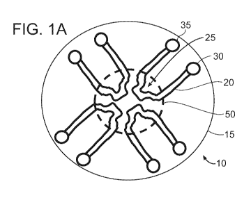
[00108] FIGS. 1A-1D are schematic illustrations of microfluidic device layouts
according
to example embodiments of the invention.
[00109] FIG. 1A is a top view of a circular layout (it can also be any
symmetrical polygon
with a center point) of microfluidic device 10 having one or more continuous
microfluidic
channels (e.g., microchannels) 20 formed in a substrate 15, each channel
connected to one
inlet (input port) 30 and one outlet (output port) 35. A portion of the
channel, e.g., the center
of the channel, can have a unique shape, e.g., a clot forming/localizing area
25, in order to
result in flow separation or disruption, or stasis of sample flow to promote
clot formation.
There may be two or more of these microfluidic channels in this single device,
dependent on
the specific assay being used. This design can allow for multiple samples,
such as three or
more samples, e.g., up to 10 samples, or more than 10 samples, to be evaluated
simultaneously. Typically, each sample (or each aliquot of a sample) requires
a separate
channel. In FIG. 1A, four channels are illustrated, each having a clot
forming/localizing area
25 located proximally on the microfluidic device, e.g., locate in a central
region of the
microfluidic device. The sample can enter the device through the inlet
manually or by an
electronic dispenser and will go through the microfluidic channel by an
applied
pressure/vacuum, capillary action, or via chemical interactions, such as if
the microfluidic
channel is coated with or made of a hydrophilic material. In this example set-
up, the agonists
+/- calcium +/- clot detection reagents must be added to the main inlet, pre-
mixed into the
sample, or must be coated on to the inlet or the microfluidic channel. (The
term "+/-", as
used herein means "with or without.") All of the clot forming/localizing areas
may be
viewed in one single imaging field (dashed circle 50 encompassing clot forming
areas 25) at
magnification that may range from, for example, 2X-10X.
[00110] FIG. 1B is a top view of a similar layout as in FIG. 1A but with
examples of
multiple inlet ports 30, 40, 42 for each channel 20. This allows for the
agonist +/- calcium
+/- clot detection reagents to be added to the sample within the microfluidic
channel. There
can be one or more additional inputs 40, 42 and they can individually connect
directly to the
main channel 20 or the main input area, or some may connect indirectly to each
other with at
least one connecting to the main channel or the primary input port.
[00111] FIG. 1C is a side view of a microfluidic device layout illustrating
input 30 and
output 35 ports of a channel 20 in substrate 15. Only one channel is shown,
but one or more
channels may be provided as illustrated in FIG. 1A. In addition, one or more
input ports may
CA 03071295 2020-01-27
WO 2019/023508 PCT/US2018/043973
- 23 -
be provided for each channel, as illustrated in FIG. 1B. As schematically
illustrated in FIG.
1C, a detection device 55 can be provided to measure clot formation in each of
the channels.
The detection device 55 can include an imaging sensor to detect clot
formation, e.g., clot
formation times. Imaging can be bright-field imaging as described herein. The
detection
device may use any of the other measuring/detection methodologies described
herein.
[00112] FIG. 1D is a top view of a microfluidic device 110 having an alternate
layout that
may be utilized for various assays. There can be one or more inlets (input
ports) 130 with one
outlet (output port) 135 per sample input and channel 120. An area of shape
change 125 to
stimulate clot formation is included in each channel 120. The channels are
arranged in a
parallel fashion in order to allow for visualization of the clot
formation/localization areas 125
within one field of view (dashed rectangle 150) at magnification that may
range, for example,
from 2X-10X. Each channel can include one or more areas 140 for agonist and/or
calcium
addition and a region 145 for mixing. In the example shown, the channels 120
have identical
geometries.
[00113] FIGS. 2A and 2B illustrate a circular microfluidic clotting device 210
according to
an example embodiment. As shown, the device includes four channels 220, each
channel
including a clot forming/localizing area 225 having a geometry to trigger
and/or localize clot
formation. The clot forming areas 225 are arranged in a central region. Each
channel 220 is
connected to an input port 230 and an output port 235. The input and output
ports of all the
channels are arranged in an alternating pattern at a periphery of the device
210. The dashed
circle 250 in the center indicates a general field of view encompassing
'clotting areas' 225 of
all input channels. The configuration of channels shown in FIG. 2A is a
configuration in
which wicking capillary flow occurs, but many other configurations are
possible. A
particular configuration may be selected based on one or more criteria, such
as whether the
configuration is particular advantageous for manufacturing the device.
[00114] FIG. 2B is a magnified view of the central portion of the device 210
of FIG. 2A
illustrating examples of clot forming/localizing areas 225 within the field of
view. The clot
forming areas can have configurations conducive to formation of a clot that
can be quantified.
The clot forming areas can have shapes designed to cause flow separation,
stasis, flow
disturbances, or combinations thereof, for clot formation, and may have shapes
designed to
cause flow disturbance for clot formation. In the example, the clot forming
areas have
different shapes to illustrate various shapes that can be used. Typically, the
shapes will be the
CA 03071295 2020-01-27
WO 2019/023508 PCT/US2018/043973
- 24 -
same for each channel so as to ensure the same flow conditions in each
channel. The shapes
of clot forming areas illustrated in FIG. 2B are examples and not all-
inclusive of the shape
variations that can be used.
[00115] As illustrated in FIG. 2B, each clot forming area can be configured
(e.g., shaped)
such that a sample flowing through a clot forming area is forced to change
direction at least
once, preferably multiple times. Each change in direction can be in the range
of, for example,
about 45 degrees to about 135 degrees, of about 60 degrees to about 120
degrees, of about 75
degrees to about 105 degrees, or of about 90 degrees. In addition, one or more
flow
disruptors, such as protrusions or islands, can be provided to disrupt flow.
As a sample
passes through the clot forming area, it encounters flow disruptor(s) and is
forced to flow
around the disruptor(s). A disruptor may include corners or pointed edges, and
can be
triangular, rectangular, or otherwise shaped as illustrated in FIG. 2B. A
combination of
disruptors and other structural features, or just other structural features,
may form a
circulatory region, where sample flow in a circular pattern interacts with new
sample entering
the region as other sample departs. Eddy currents behind disruptors, from a
fluid flow point
of view, may also encourage coagulation as sample interacts with other sample
at
intersections (e.g., turbulence intersections) of fluid flow and sample in an
eddy region.
[00116] In some embodiments, the disruptor can include a concavity (e.g., FIG.
3A). A
clot forming/localizing area may include a narrowing of the channel. By
changing the
direction of sample flow and/or changes in diameter, angle, and/or shape of
the channel,
and/or forcing the sample to flow around one or more disruptors, the clot
forming areas
introduce flow separation and stasis of sample flow to promote clot formation.
Typically, the
channels and clot forming areas are arranged in a symmetrical pattern in order
to provide the
same flow characteristic for each of the channels.
[00117] EXAMPLE 2
[00118] A general protocol for performing the assay according to an embodiment
of the
invention is as follows:
a) Add together sample, agonist, +/- calcium, +/- clot detection
agent
i. Calcium to a final concentration of 0.2 mM (This
concentration is
particularly suitable for use with 3.2% buffered sodium citrate. If
another anticoagulant is used, the concentration of calcium may not be
0.2 mM.)
CA 03071295 2020-01-27
WO 2019/023508 PCT/US2018/043973
- 25 -
ii. Clot detection agents can include fluorescent labeled
fibrinogen,
magnets, beads (may be fluorescent or colored)
b) Load into microfluidic device
i. See, e.g., FIGS. 1A-1D, 2A and 2B for examples of input
loading
configuration and order
c) Temperature control
i. Room temperature
May increase up to 37 C (body temperature)
(Body temperature is typically 37 C but the temperature of the assay
run can be changed according to the patient's actual temperature. For
example, if a patient has a fever, the temperature of the assay run can
be increased.)
d) Perform clot detection and measure time of clot formation (e.g., 4-12
minutes)
e) Log time when each sample starts to form a clot
[00119] EXAMPLE 3
[00120] FIGS. 3A-3C illustrates clot detection using plasma and fluorescent-
labeled
fibrinogen with a microfluidic device 310 having four channels 320 with clot
forming/localizing areas 225 according to an example embodiment. The
microfluidic device
is similar to the device show in FIGS. 2A and 2B except that all clot forming
areas 325 have
the same shape. Each clot forming/localizing area 325 includes a protrusion to
disrupt
sample flow. In this example, as shown in FIG. 3A, the protrusion generally is
triangular in
shape. Two sides of the protrusion are straight and one side is concave. Each
clot forming
area 325 causes the flow to change direction four times, including two 90
degree changes in
direction.
[00121] In an example, the process of clot detection can include the following
procedural
steps:
a) A plasma sample is pre-mixed to include: 6 [EL plasma + 0.6 [EL
agonist (10%
volume to sample) + 0.6 [EL Calcium (stock 2 mM, 10% volume to sample) +
0.6 [EL Fibrinogen (this can vary in concentration, in general <10% volume of
sample). The foregoing values can be adjusted and changed and similar results
obtained.
CA 03071295 2020-01-27
WO 2019/023508 PCT/US2018/043973
- 26 -
b) For each channel, an aliquot of the pre-mixed sample is placed into the
input
port of the channel.
c) The sample aliquot is drawn into the channel by capillary action.
d) The channels are imaged for 10 minutes at 37 C, and the time to detect
a clot
is recorded.
[00122] The example in FIG. 3B shows a fluorescent image taken of the
microfluidic
channels at one time point (5 minutes). The plasma sample used contains 250
ng/mL of
Apixaban. An agonist, Factor Xa (FXa) at various concentrations (0.75 ng/mL
FXa, 7.5
ng/mL FXa, and 75 ng/mL FXa) or buffer alone (negative control) was added to
the plasma
sample, along with calcium and 488-conjugated fibrinogen. Crosslinking of the
fluorescent
fibrinogen is indicative of the formation and presence of a cross-linked
fibrin clot. Higher
concentrations of the FXa (7.5 ng/mL FXa, and 75 ng/mL FXa), visible in the
channels on
the right in FIG. 3B, result in clot formation earlier than the lower
concentration (0.75 ng/mL
FXa) or the negative control, visible in the channels on the left in FIG. 3B.
FIG. 3C is a
magnified view of a clot forming area of one channel illustrating a cross-
linked fibrin clot.
[00123] EXAMPLE 4
[00124] FIGS. 4A and 4B are fluorescent images illustrating clot detection
using whole
blood in a parallel microfluidic channel device 410 according to an example
embodiment.
Microfluidic channels 420 were pre-coated with agonist, Factor Xa, at various
concentrations
(7.5 ng/mL, 75 ng/mL, 750 ng/mL) or with buffer alone (negative control). The
fluorescent
images are taken at one time point (10 minutes). Microfluidic channels were
washed with
buffer prior to use to leave only bound FXa within the microfluidic channel.
Fresh whole
blood was placed into each input port and the blood was drawn in through
capillary action.
The blood was left to flow for 10 minutes and then the channel was gently
washed with
buffer. Depicted is a brightfield image of two samples evaluated. The sample
in FIG. 4A
contained no anticoagulant (finger prick of blood), which resulted in clots in
all 4 channels,
including the negative control. The sample in FIG. 4B contained unfractionated
heparin
(which was added to the finger prick of blood), which resulted in a gradient
of clot formation
dependent on the concentration of FXa in the channel. Almost no cells were
adhered in the
negative control, indicating minimal clot formation. Unfractionated heparin
inhibits Factors
IIa and Xa in an antithrombin III-dependent fashion, which is why the addition
of these
factors at appropriate concentrations can help recover the clotting capability
of the sample.
CA 03071295 2020-01-27
WO 2019/023508 PCT/US2018/043973
- 27 -
[00125] EXAMPLE 5
[00126] FIGS. 5A and 5B illustrate additional embodiments of microfluidic
device designs
that include the features of: (1) each channel subjects the blood/plasma to
equal conditions
and (2) there is a clot-promoting geometry within each channel where clotting
detection is
optimized and performed. FIG. 5A illustrates a device 510 including circular
array of
symmetrical channels 520 surrounding and connected to a single sample input
530, where
each channel has a clot-promoting and/or localizing area 525. The channels 520
may or may
not also include one or more areas for agonist and/or calcium addition 540
and/or mixing
545. FIG. 5B illustrates an alternative embodiment of a device 512 utilizing a
cylindrical
design with a single sample input port 530 that divides into multiple
symmetrical channels
520 with a clot-forming area 525 with or without an area for agonist/calcium
540 addition
and/or mixing 545. Both devices 510, 512 may also include a sample collection
reservoir 560
with or without an absorbent filter.
[00127] EXAMPLE 6
[00128] FIG. 6 is a flow diagram of a method of assessing coagulation in a
blood sample
according to example embodiments of the invention. The blood sample can be a
whole blood
sample or a plasma sample. According to the method, a coagulation factor is
added to plural
aliquots of the blood sample. Each aliquot can receive the coagulation factor
at a different
concentration. The plural aliquots can be applied to plural channels of a
microfluidic device.
Alternatively, or in addition, the coagulation factor(s) can be pre-coated on
or into the device
to which the blood sample is applied. Clot formation times are measured in
each of the
channels and coagulation is assessed based on the clot formation times
measured.
Alternatively, or in addition, degree of clot formation (optionally, degree of
clot dissolution)
in each of the channels is measured at a fixed time or times, and coagulation
is assessed based
on the degree of clot formation (optionally, degree of clot dissolution)
measured.
[00129] Optionally, as illustrated in FIG. 6, the clot formation times can be
compared to a
reference value or reference ranges. In one example, the clot formation times
are compared
to coagulation factor specific clot formation reference ranges from
individuals who do not
suffer from a coagulation cascade abnormality. This is useful, e.g., to detect
a coagulation
cascade abnormality in the blood sample. In another example, the clot
formation times are
compared to clot formation times measured for a sample from an individual who
does not
suffer from a coagulation cascade abnormality. This is also useful, e.g., to
detect a
CA 03071295 2020-01-27
WO 2019/023508 PCT/US2018/043973
- 28 -
coagulation cascade abnormality in the blood sample. In yet another example,
the clot
formation times are compared to clot formation times measured for a sample
containing a
known amount of an anticoagulation agent. This is useful, e.g., to detect the
anticoagulation
agent in the blood sample.
[00130] The microfluidic device for use in the method of FIG. 6 can be any
microfluidic
device described herein having plural channels, such as the devices
illustrated in FIGS. 1A-
1D, 2A-2B, 3A-3C, 4A-4B and 5A-5B. In an embodiment, the device includes
plural
channels formed in a substrate, each channel including a clot forming area
having a geometry
configured to trigger and/or localize formation of a clot, the clot forming
areas of the plural
channels being arranged in a central region of the substrate; plural input
ports, each input port
connected to a first end of one of the plural channels; and plural output
ports, each output port
connected to a second end of one of the plural channels, the input and output
ports being
arranged in an alternating pattern at a periphery of the substrate.
[00131] EXAMPLE 7
[00132] FIGS. 7A-7D illustrate example clotting curves for various FXa and
FIIa
inhibitors at various concentrations. The time it takes for each of the
combinations to form a
clot is then plotted. The clotting curve for each concentration of inhibitor
is dependent on the
presence and concentration of the anticoagulant in the sample. The figures
illustrate the time-
to-clot for four (4) different DOACs when exposed to agonists at various
concentrations. The
time-to-clot increases as the concentration of the inhibitor increases,
demonstrating an
increase in functional anticoagulation. Concentration of the agonist (FXa for
FIGS. 7A-7C,
and FIIa for FIG. 7D) is plotted on the X-axis for each of the figures.
[00133] FIG. 7A is a graph of example data illustrating detection of
Rivaroxaban. The
graph shows clotting curves for different concentrations of the inhibitor
Rivaroxaban (0
ng/mL, 250 ng/mL, and 500 ng/mL). Each curve shows average clot detection time
(minutes;
y-axis) as a function of agonist (FXa) concentration (ng/mL; x-axis). The data
shown in the
graph can be summarized as follows:
[00134] At a concentration of 0 ng/mL Rivaroxaban, clot formation detected
in < 2.5
minutes with agonist concentration down to 7.5 ng/mL.
[00135] At a concentration of 250 ng/mL Rivaroxaban, clot formation time is
significantly
longer than the negative control but lower than 500 ng/mL with agonist
concentration down
to 375 ng/mL.
CA 03071295 2020-01-27
WO 2019/023508 PCT/US2018/043973
- 29 -
[00136] At a concentration of 500 ng/mL Rivaroxaban, clot formation
detected < 2.5
minutes down to 750 ng/mL.
[00137] FIG. 7B is a graph of example data illustrating detection of Apixaban.
The graph
shows clotting curves for different concentrations of Apixaban (0 ng/mL, 250
ng/mL, and
500 ng/mL). As in FIG. 7A, each curve shows average clot detection time
(minutes; y-axis)
as a function of agonist (FXa) concentration (ng/mL; x-axis). The data shown
in the graph
can be summarized as follows:
[00138] At a concentration of 0 ng/mL Apixaban, clot formation detected in <
2.5 minutes
with agonist concentration down to 7.5 ng/mL.
[00139] At a concentration of 250 ng/mL Apixaban, clot formation detected in
<2.5
minutes with agonist concentration down to 75 ng/mL.
[00140] At a concentration of 500 ng/mL Apixaban, clot formation detected in
<2.5
minutes with agonist concentration down to 938 ng/mL.
[00141] FIG. 7C is a graph of example data illustrating detection of Edoxaban.
The graph
shows clotting curves for different concentrations of Edoxaban (0 ng/mL, 250
ng/mL, and
500 ng/mL). As in FIG. 7A, each curve shows average clot detection time
(minutes; y-axis)
as a function of agonist (FXa) concentration (ng/mL; x-axis).
[00142] FIG. 7D is a graph of example data illustrating detection of
Dabigatran. As in
FIGs. 7A and 7B, the graph of FIG. 7D shows clotting curves for different
concentrations of
the inhibitor, here Dabigatran (0 ng/mL, 25 ng/mL, 250 ng/mL, and 500 ng/mL).
Each curve
shows average clot detection time (minutes; y-axis) as a function of agonist
(FIIa)
concentration (ng/mL; x-axis). The data shown in the graph of FIG. 7D can be
summarized
as follows:
[00143] At a concentration of < 25 ng/mL Dabigatran, clot formation detected
in < 2.5
minutes with agonist concentration down to 71 ng/mL.
[00144] At a concentration of 250 ng/mL Dabigatran, get clot formation
detected in <2.5
minutes with agonist concentration down to 710 ng/mL.
[00145] At a concentration of 500 ng/mL Dabigatran, clot formation detected in
< 2.5
minutes down to 710 ng/mL.
[00146] Automation can be employed to reduce variation between samples and
assays.
[00147] EXAMPLE 8
CA 03071295 2020-01-27
WO 2019/023508 PCT/US2018/043973
- 30 -
[00148] In addition to the detection of the presence of FXa inhibitors and
estimation of
their relative concentrations, the assay described here can differentiate FXa
inhibitors from
FIIa inhibitors by selecting appropriate upstream and downstream clotting
factors to add to
the samples.
[00149] FIG. 8 illustrates a basic clotting cascade that can guide the
selection of
appropriate clotting factors, as further described in the following examples.
As shown in
FIG. 8, the cascade includes an intrinsic pathway and an extrinsic pathway,
both of which can
lead, via a common pathway of the cascade, to a cross-linked Fibrin clot. The
intrinsic
pathway can, for example, be activated by surface contact. The extrinsic
pathway can be
activated, for example, by tissue trauma.
[00150] EXAMPLE 9
[00151] FIG. 9 is a schematic diagram providing a demonstration of how to
detect Factor
Xa (FXa) inhibition/deficiency/abnormality of function. The addition of
upstream (not active
or activated) coagulation factors, including but not limited to FXII, FXI,
FIX, FVIII, will
demonstrate prolongation of clotting time, e.g., as compared to addition of a
downstream
factor. Alternatively, prolongation of clotting time can be determined with
reference to a
control clotting time. However, the addition of downstream (not active or
activated)
coagulation factors, including but not limited to FII, Fl, will demonstrate
unaffected (e.g.,
normal) clotting time, which can serve as a control. The addition of FXa will
demonstrate
prolongation of clotting time in a concentration-dependent manner, and even at
high
concentration of the upstream factor, the clotting team will likely not reach
the control. As
illustrated, example direct FXa inhibitors include Rivaroxaban, Apixaban,
Edoxaban, and
Betrixaban.
[00152] EXAMPLE 10
[00153] FIG. 10 is a schematic diagram providing a demonstration of how to
detect Factor
IIa (FIIa) inhibition/deficiency/abnormality of function. The addition of
upstream (not active
or activated) coagulation factors, including but not limited to FXII, FXI,
FIX, FX, FV, FVIII,
will demonstrate prolongation of clotting time. The addition of downstream
(not active or
activated) coagulation factors, including but not limited to Fl, will
demonstrate unaffected
clotting time. The addition of FIIa will demonstrate prolongation of clotting
time in a
CA 03071295 2020-01-27
WO 2019/023508 PCT/US2018/043973
- 31 -
concentration-dependent manner. As illustrated, example direct FIIa inhibitors
include
Dabigatran, Bivalirudin, and Argotraban.
[00154] EXAMPLE 11
[00155] FIG. 11 is a schematic diagram providing a demonstration of how to
detect and
differentiate between FIIa and FXa inhibition in a sample. In the presence of
a FXa and FIIa
inhibitor, addition of upstream (not active or activated) coagulation factors,
including but not
limited to FXII, FXI, FIX, FVIII, will demonstrate prolongation of clotting
time. The
addition of FXa to the sample will demonstrate prolongation of clotting time,
in a
concentration-dependent manner for FXa and FIIa inhibition. The addition of
FIIa to the
sample will demonstrate prolongation of clotting time, in a concentration-
dependent manner
in the presence of FIIa inhibition but will demonstrate unaffected clotting
time in the
presence of FXa inhibition.
[00156] EXAMPLE 12
[00157] FIG. 12 is a schematic diagram providing a demonstration of how to
detect
indirect FXa inhibition/deficiency/abnormality of function. The addition of
upstream (not
active or activated) coagulation factors, including but not limited to FXII,
FXI, FIX, FVIII,
will demonstrate prolongation of clotting time. The addition of downstream
(not active or
activated) coagulation factors, including but not limited to FII, Fl, will
demonstrate normal
clotting time or slight prolongation of clotting time in a concentration-
dependent manner
dependent on the type of inhibitor present. The addition of FXa will
demonstrate
prolongation of clotting time in a concentration-dependent manner. This is due
to secondary
FXa inhibition via the presence of a drug that increases the affinity/binding
of Antithrombin
III (ATIII) to FXa, thereby inhibiting it. Embodiments can include detection
of ATIII,
thereby detecting indirect inhibition of FXa, FIIa, or both. Drugs that
increase
binding/affinity of ATIII for FXa include Heparin, e.g., Low Molecular Weight
Heparin
(LMWH) and Unfractionated Heparin (UFH), Enoxaparin, and Fondaparinux.
[00158] EXAMPLE 13
[00159] FIG. 13 is a schematic diagram providing a demonstration of how to
detect and
differentiate between FXIIa and FXIa inhibition in a sample. In the presence
of a FXIIa
inhibitor, addition of FXIIa will result in concentration-dependent
prolongation of clotting
time. The addition of factors added downstream, including but not limited to,
FXI, FIX,
CA 03071295 2020-01-27
WO 2019/023508 PCT/US2018/043973
- 32 -
FVIII, FX, FIT, FV, would result unaffected clotting times. In the presence of
a FXIa
inhibitor, addition of FXIIa will result in prolongation of the clotting time.
Addition of FXIa
would result in a concentration-dependent prolongation of clotting time. The
addition of
factors added downstream, including but not limited to, FIX, FVIII, FX, FIT,
FV, would result
unaffected clotting times. This approach can also be used in various
combinations to perform
a comprehensive panel for the detection and differentiation of FXIIa
inhibitors, FXIa
inhibitors, FXa inhibitor, and FIIa inhibitors.
[00160] EXAMPLE 14
[00161] FIG. 14 is a schematic diagram providing a demonstration of how to
detect and
differentiate between various types of hemophilia. Hemophilia C would result
in
prolongation of clotting time with the addition of FXIIa, concentration-
dependent
prolongation with the addition of FXI, and unaffected clotting times with the
addition of
FXIa or any other downstream factor. Hemophilia B would result in prolongation
of clotting
time with the addition of FXIIa and FXIa, concentration-dependent prolongation
with the
addition of FIX, and unaffected clotting times with the addition of FIXa or
any other
downstream factor. Hemophilia A would result in prolongation of clotting time
with the
addition of FXIIa, FXIa, concentration-dependent prolongation with the
addition of FVIII,
and unaffected clotting times with the addition of FXa or any other downstream
factor.
[00162] For congenital disorders, embodiments can add non-activated factor(s)
for
detection, whereas non-activated factor(s) can serve as control.
[00163] EXAMPLE 15
[00164] FIG. 15 is a schematic diagram providing a demonstration of how to
detect
problems with fibrinogen, i.e., Factor I (Fl), or FXIII. Afibrinogenemia or
dysfibrinogenemia
would result in prolongation of clotting time with the addition of all factors
upstream of Fl,
concentration-dependent prolongation with the addition of FT. FXIII
deficiency/abnormality
would result in changes in clot strength and clot stability over time with the
addition of all
factors upstream of FXIII, concentration-dependent changes in clot strength
and stability of
time with the addition of FXIII.
[00165] EXAMPLE 16
[00166] FIGS. 16A-16C illustrate Clotting Curve Scores (CCS) for FXa and FIIa
inhibitors at various concentrations. Raw data of the clotting times of each
of the agonists at
CA 03071295 2020-01-27
WO 2019/023508 PCT/US2018/043973
- 33 -
various concentrations are used to calculate a single Clotting Curve Score
(CCS) based on
multivariate statistical modeling. This CCS can then be used as a single whole
number to bin
patients into positive or negative for specific inhibitors. This CCS can also
be used to
extrapolate the functional concentration of the drug in the patient sample.
Functional
concentration represents the amount of anticoagulation secondary to the drug
in the blood
sample. FIG. 16A shows how the CCS of two FXa inhibitors (Apixaban,
Rivaroxaban) and
one FIIa inhibitor (Dabigatran) vary dependent on concentration using FXa as
the agonist.
FIG. 16B shows how the CCS of the two FXa inhibitors and the one FIIa
inhibitor vary
dependent on concentration using FIIa as the agonist. FIG. 16C demonstrates
how the CCS
for each agonist can be used to identify the type of inhibitor in the sample.
[00167] EXAMPLE 17
[00168] FIG. 17 shows Table 1 that provides patient descriptive statistics.
Citrated plasma
samples were collected from patients admitted into the Massachusetts General
Hospital
Emergency Department. All plasma samples had clinician-ordered coagulation
tests
(PT/INR, aPTT, DTT, or other). Patient samples were evaluated using an
embodiment of the
assay described herein. Patient medical records were reviewed for the
administration of
history of anticoagulants. All patient samples were collected following
Institutional Review
Board (IRB) approval and regulations at both the Massachusetts General
Hospital and the
Massachusetts Institute of Technology.
[00169] EXAMPLE 18
[00170] FIGS. 18A-18C illustrate prothrombin time (PT) and international
normalized
ratio (INR) is sensitive but not specific for FXa-I anticoagulation. Both PT
and INR were
compared between control patients and patients documented to be on FXa-I.
Abnormal PT
was defined as >14 seconds and abnormal INR was defined as >1.2. FIGS. 18A and
18B
show ROC curves comparing PT and INR of total controls to patients on FXa-I.
FIG. 18C
shows a table of descriptive statistics of patients with PT and INR results
evaluated. One-
way ANOVA was used to compare normal and abnormal controls with both
rivaroxaban and
apixaban. Significance was defined as p < 0.05. Results show that, when
compared to
abnormal controls, there is no significance compared to the FXa-I patients.
[00171] EXAMPLE 19
CA 03071295 2020-01-27
WO 2019/023508 PCT/US2018/043973
- 34 -
[00172] FIGS. 19A-19G illustrate example clotting time data and comparative
clotting
curves. Clotting times were compared at various agonist concentrations for all
the patient
groups to construct clotting curves. FIGS. 19A-19D show scatter plots
demonstrating the
mean and standard error bars of the clotting times at various agonist
concentrations with
respect to patients in different groups. FIG. 19E shows the mean clotting time
with standard
error bars of all patient groups, which are demonstrated on a single graph for
comparison.
All three FXa-I groups (Apixaban, Rivaroxaban, FXa-I) appear subjectively very
different
from the control group, with there being multiple concentrations where there
are significant
statistical differences between the controls and the total FXa-I, Rivaroxaban,
and Apixaban
groups. FIGS. 19F and 19G shows mean time to clot with standard error bars of
the control
group divided into patients with normal versus abnormal PT or INR,
demonstrating no large
difference in these tests between the different control groups.
[00173] EXAMPLE 20
[00174] FIGS. 20A-20E illustrate Clotting Curve Score (CCS) analysis and
evaluation of
CCS utilization for the detection of FXa-I in patient samples. FIG. 20A shows
a scatter plot
with mean and standard error bars for CCS comparison between patient groups.
Dotted line
at CCS of 0 represents the chosen cut-off for the determination of whether
there is FXa
inhibition in the patient sample. FIG. 20B shows an ROC curve of utilizing the
CCS scores
to determine whether a patient has an FXa-I in their system. FIG. 20C provides
descriptive
statistics of the CCS for the different patient groups. FIGS. 20D and 20E
illustrate evaluation
of using the CCS for the determination of the accuracy of FXa-I detection.
[00175] EXAMPLE 21
[00176] FIGS. 21A and 21B illustrate functional drug concentration
calculation. Utilizing
the CCS score calculated for each of the controlled spiked Rivaroxaban
samples, a best-fit
line was plotted for an equation that converted CCS into drug concentration,
as shown in
FIG. 21A. This equation was then applied to each of the CCS values for the
patient samples
evaluated in order to derive a functional concentration for each patient
sample. These
concentrations were directly compared to anti-Xa chromogenic assay-derived
Rivaroxaban
concentrations in each sample. Plotting these two values against each other
demonstrated a
good correlation between the anti-Xa concentration and the DOAC test
concentration (RA2 =
0.827), as shown in FIG. 21B. Note that hemolyzed samples were not included in
this direct
CA 03071295 2020-01-27
WO 2019/023508
PCT/US2018/043973
- 35 -
comparison because it is known that hemolyzed, icteric, and lipemic plasma
samples
negatively affect the anti-Xa chromogenic assay concentrations.
[00177] In
addition to identifying inhibition, as illustrated in the examples of FIGS.
21A
and 21B, embodiments can be used to quantify the amount of inhibition.
[00178] EXAMPLE 22
[00179] FIG. 22 illustrates a current decision-making paradigm if a patient is
on a Direct
Oral Anticoagulant (DOAC).
[00180] When a patient is at high-risk for a bleeding event or has an active
bleed
coagulation tests are ordered. These tests can include PT, INR, aPTT, ACT,
TEG, or other
currently available point-of-care tests. Abnormal clotting results on
currently-available tests
are non-specific for the presence of DOACs and leaves the healthcare worker
guessing as to
which treatment is the most appropriate for the patient. If the coagulation
times are normal,
due to the lack of sensitivity of these tests, the healthcare worker may miss
the presence of a
DOAC in the patient sample and proceed with treatment, putting the patient at
an increased
risk of bleeding.
[00181] EXAMPLE 23
[00182] FIG. 23 illustrates an improved decision-making paradigm using
embodiment(s)
of the present invention if a patient is on a DOAC. Double arrows indicate
possible iterative
procedures. For example, if traditional coagulation tests show a patient has
normal clotting
times and the DOAC test according to an embodiment of the invention shows
abnormal
results, a DOAC reversal agent can be selected, based on test results, and
administered to the
patient. The patient can then re-tested, and, if still abnormal according to
the DOAC, re-
tested again, optionally after administrating a modified or different DOAC
reversal agent. If
the traditional coagulation tests are abnormal and the DOAC test is also
abnormal, then the
healthcare worker may choose the DOAC reversal agent or another treatment and
re-test after
administration of the reagent. If the traditional coagulation tests are
abnormal and the DOAC
test is negative for the presence of a DOAC then the healthcare worker has the
information
necessary to determine that another hemostatic treatment may be necessary.
[00183] EXAMPLE 24
CA 03071295 2020-01-27
WO 2019/023508 PCT/US2018/043973
- 36 -
[00184] FIGS. 24A and 24B illustrate detection of the reversal of FXa
inhibition following
the addition of activated prothrombin complex concentrate (aPCC; FEIBA). FEIBA
is a
combination of activated factors administered to overcome FXa inhibitors in
patients.
Another example is Kcentra, which is inactive prothrombin complex concentrate.
There are
also specific FXa inhibitor reversal agents, such as coagulation factor Xa
(recombinant),
inactivated-zhzo. FIG 24A demonstrates the expected clotting times for
Edoxaban upon the
addition of 7.5 ng/mL of FXa. FIG 24B shows the change in clotting time with a
plasma
sample with 500 ng/mL of Edoxaban is treated with aPCC. This data demonstrates
that the
test according to an embodiment of the invention has utility to monitor the
reversal or
overcoming of the anticoagulant effect of these DOACs.
[00185] The teachings of all patents, published applications and references
cited herein are
incorporated by reference in their entirety.
[00186] While example embodiments have been particularly shown and described,
it will
be understood by those skilled in the art that various changes in form and
details may be
made therein without departing from the scope of the embodiments encompassed
by the
appended claims.