Note: Descriptions are shown in the official language in which they were submitted.
CA 03071328 2020-01-27
WO 2019/023597
PCT/US2018/044123
SEQUENTIAL LATERAL FLOW DEVICE
Related Applications
[0001] This application claims the benefit of U.S. Provisional Application
Serial No.
62/537,701 filed on July 27, 2017, the entire teachings of which are
incorporated herein by
reference.
Back2round
[0002] Lateral flow immunoassays (LFIs) are methods for the rapid detection of
chemical
and biochemical markers for toxicity and disease. LFIs can be classified into
three types:
(1) competitive assays such as those used to detect small molecule drugs of
abuse and
therapeutic drugs; (2) immunometric assays, also referred to as "sandwich"
assays, for
macromolecules such as proteins, glycoproteins, and lipopolysaccharides; and
(3)
serological assays, which detect antibodies to a specific antigen in a
biological sample.
This disclosure is focused on immunometric assays, for which the term
"sandwich" assay
will be used interchangeably for the sake of reference.
[0003] Sandwich LFIs (or immunometric LFIs) generally involve a bibulous flow
path on
which is immobilized a capture antibody and a mobilizable detector antibody
that is
attached to a signal-producing agent. The signal-producing agent is typically
chosen from
detectable colored and/or fluorescent particles, dyes, and/or enzymes. When
the signal-
producing agent is conjugated directly to the detector antibody, the term
"direct assay"
will be used.
[0004] Several patents and applications have described so-called sequential
LFIs. A multi-
port device is typically described in these applications as necessary to
accommodate an
extra reagent. In several patents and applications (e.g., US 4,981,786, EP 1
044 372 B1,
US 2013/0164193), a primary use for the additional port is to separately and
sequentially
introduce a chromogenic and/or fluorogenic substrate reagent for an enzyme
attached to a
detector antibody. Other applications describe using a second port to enable
the
introduction of a wash buffer (e.g., US 4,981,786). These assays, although
sequential in
the order of manipulations, are still direct assays in that the signal
producing agent (e.g.,
enzyme) is directly conjugated to the detector antibody.
[0005] In examining the history of LFIs from its early concepts in the 1980s
to the present
day, the use of enzymes as signal-producing moieties on detector antibodies
has
1
CA 03071328 2020-01-27
WO 2019/023597
PCT/US2018/044123
diminished and has been supplanted by detector antibodies directly conjugated
to signaling
particles. Gold colloid and colored and/or fluorescent latex have found
widespread and
easier to use. The addition of a substrate is avoided when such signaling
particles
conjugated directly to detector antibodies are used.
[0006] In the course of developing and optimizing multi-analyte immunometric
LFIs, we
have found that immunometric assays that use detector antibodies directly
conjugated to
signaling particles suffer from decreasing sensitivity when more target
analytes are present
for detection via measurement on a single test strip. For example, if a
sandwich LFI test
strip were to detect three different target analytes, specific limits of
detection (LoDs) for
each analyte would be obtained. However, the LoDs would be degraded if an
additional
three target analytes were present in the sample mix (for a total of six
target analytes). If
another three target analytes were present in addition to the six target
analytes for a total of
nine target analytes for detection, the LoD for each target analyte would
become even
worse, to the point of eventual inutility.
[0007] These limitations are due, in part, to the surface area limit of the
signaling particles
and the limited conjugation capacity on the particle surface. These and other
features of
current methods and devices limit their applications. The present disclosure
provides
improved methods and devices that, among other things, address limitations of
current
approaches.
Summary
[0008] In one aspect, the present disclosure provides improved methods and
devices for
detecting a plurality of target analytes, such as bacterial, viral, and/or
fungal antigens, in a
rapid, simple, sensitive, and multi-analyte manner. The disclosed methods
and/or devices
overcome some of the challenges and limitations of the conventional lateral
flow devices
and/or methods by allowing for running a binding assay using well-controlled
sequential
steps and innovative device features. Moreover, the methods and devices of
this disclosure
are particularly well-suited to detection of multiple target analytes, such as
bacterial,
fungal, and/or viral antigens, in a single assay using a plurality of binding
agents, such as
one or more than one types or idiotypes of antibodies (e.g., polyclonal
antibodies) against
numerous target-binding sites. In such settings where multiple antigens are
being detected
in complex samples using polyclonal antibodies as the binding agents, assay
sensitivity is
particularly crucial.
2
CA 03071328 2020-01-27
WO 2019/023597
PCT/US2018/044123
[0009] In one aspect, this disclosure provides a multi-analyte detection
method, for
example, an assay using a sandwich LFI test strip according to some
implementations
and/or a lateral flow device according to some implementations, that is able
to detect as
many as thirty, forty, or more possible target analytes in a liquid sample.
For example, this
disclosure describes a multi-analyte detection assay for detecting multiple
species and
strains of target analytes, such as bacterial antigens that potentially could
infect platelet
preparations intended for therapeutic and/or protective transfusion into
patients. This
disclosure also provides a rapid assay and/or test, such as an LFI (e.g., a
sandwich LFI),
which is considered an improvement as a safety measure over established
culture-based
tests as the disclosed assays or tests can be performed right before
transfusion, while a
culture test requires initiation of a test one to three days prior to
transfusion.
[0010] A single LFI test strip used in conventional methods and/or devices is
severely
challenged in its ability to detect the variety of contaminating bacterial
species and strains
at the clinically relevant LoDs due to the limitations of particle conjugates,
(or detector
antibody-signaling particle conjugates), capacity for multi-analyte detection,
especially for
forty or more target analytes.
[0011] In one aspect, by separating the binding agent (e.g., a plurality of
antibodies, such
as one or more than one type of polyclonal antibodies) from the signaling
agent (e.g.,
signaling particles) at the initiation of the LFI test procedure, and enabling
the plurality of
target analytes in the liquid sample to first form sandwich complexes at the
capture zone
before the release of the signaling agent, the constraint to sensitivity
imposed by the
limited number of binding agents on the surface of the detecting particle is
significantly
overcome. In this sequential manner, LoDs can be improved by two-fold to as
much as
two log-fold in a multiplexed assay, as compared to a similar multiplexed non-
sequential
direct assay.
[0012] In one approach, as long as the formation of the sandwich complexes
(e.g., a
complex between the binding agent, target analyte, and immobilized capture
agent) at the
capture zones occurs before the binding agent or analyte contacts the
signaling agent. In
some implementations, the liquid sample is optionally mixed with detector
antibodies
before being introduced into a lateral flow device. This procedure allows some
of the
detector antibodies to bind to some of the targt analytes before entering the
flow path. In
addition, the number of required signaling particles can be reduced to only
sufficient
3
CA 03071328 2020-01-27
WO 2019/023597
PCT/US2018/044123
numbers to label the actual sandwich complexes. As the target analytes get
captured by the
capture agents immobilized on the flow path, the efficiency of labeling the
target analyte
with detector antibodies greatly increases as the reaction between the target
analytes and
the detector antibodies is no longer limited by diffusion. Excess of the
detector antibodies
may flow past the captured target analytes on the flow path for binding. The
sensitivity of
the assay is, therefore, driven by the number of effective binding events
between the
binding agent (e.g., detector antibodies) and the captured target analytes,
rather than the
number of binding agent-signaling agent conjugates or signaling agents bound
to the target
analytes and captured in the capture zone. Since the binding agents are a lot
smaller in size
than the binding agent-signaling agent conjugates or conjugates that are bound
to analytes,
higher local concentration of binding agents can be achieved in a given
processed sample
volume; in some cases this is in log-fold excess compared to the maximum
number of
signaling particles that the strip typically accomodates. Moreover, in a non-
sequential
assay, a large percentage of binding agent-signaling agent conjugates become
captured at
the capture zone without even binding to the target analytes, causing
interference and
lowering detection sensitivity.
[0013] In some implementations, by separating the binding agent (e.g.,
detector
antibodies) from the signaling agent (e.g., signaling particles), the binding
agent and the
signaling agent in excess amount can be applied or flow sequentially across
the capture
zones in a controlled manner. In this way, a significant improvement in
sensitivity can be
achieved, allowing a greater multiplicity of target analytes to be detected,
e.g., using a
single LFI strip at the desired LoDs. A greater number of sandwich complexes
can
therefore be formed with the increased local concentrations of binding agents,
which
subsequently are "labeled" with signaling agents.
[0014] In addition, the disclosed methods and devices can detect a plurality
of target
analytes at clinically significant levels with improved sensitivity in a time-
sensitive
manner, for example, testing bacteria contamination in less than a few hours
(e.g., less
than four hours, less than three hours, less than two hours, and/or less than
one hour). This
facilitates, for example, the testing of blood and/or blood products prior to
transfusion
without the need of accelerated growth of bacteria and/or virus in nutrient
media, which
may take one to a few days.
4
CA 03071328 2020-01-27
WO 2019/023597
PCT/US2018/044123
[0015] In some aspects, this disclosure provides methods and devices for
conducting an
assay in a sequential multi-step manner, such as an immunoassay with
sequential steps, for
detection/determination of a plurality of target analytes in a liquid sample,
such as blood
or blood products. In exemplary implementations, the disclosed method detects
multiple
analytes using binding agents that are themselves complex, such as one or more
than one
type of polyclonal antibodies.
[0016] In one aspect, the disclosure provides a method for detecting a
plurality of target
analytes in a liquid sample using a lateral flow device. The method includes
first
contacting the liquid sample with a plurality of capture agents disposed on a
solid support
in the lateral flow device and a plurality of binding agents under conditions
that permit
formation of at least one sandwich complex comprising one or more of the
plurality of
binding agents, one or more of the target analytes, and one or more of the
capture agents.
Each of the binding agents is tagged with one member of a conjugate pair. In
some
implementations, the plurality of binding agents and the plurality of capture
agents are
antibodies. In some implementations, the plurality of binding agents and the
plurality of
capture agents are monoclonal antibodies, polyclonal antibodies, and/or
mixtures thereof
In some implementations, the plurality of binding agents and the plurality of
capture
agents are polyclonal antibodies. In some implementations, the plurality of
binding agents
and/or the plurality of capture agents can bind to at least one antigen common
to at least a
subset of the target analytes. In some implementations, the plurality of
target analytes may
include multiple subsets or types of target analytes, such as bacteria (e.g.,
Gram-positive
and/or Gram-negative bacteria), viruses, and/or fungi. Each type or subset of
the target
analytes may further include sub-types or different genera. Within each subset
or type (or
sub-types or genera) of target analytes, there may be a common antigen that
the binding
agents or capture agents can bind to. For example, the plurality of target
analytes may
include at least a subset of Gram-positive bacteria and/or a subset of Gram-
negative
bacteria and/or subtypes thereof Each of the binding agents and/or capture
agents can
bind to an antigen common to at least a subset of Gram-positive bacteria or
subtypes
thereof and/or bind to an antigen common to at least a subset of Gram-negative
bacteria or
subtypes thereof Due to the antigenic diversity among species, multiple types
of binding
agents may be used. Following the formation of the sandwich complex, a
signaling agent
(e.g., non-enzymatic signaling agent) is introduced to the device and brought
into contact
CA 03071328 2020-01-27
WO 2019/023597
PCT/US2018/044123
with the sandwich complex under conditions that permit the signaling agent to
bind to a
binding agent of the sandwich complex to form a detection complex (e.g.,
through the
interactions of the conjugate pair). Formation of the detection complex
indicates the
presence of one or more of the plurality of the target analytes as the
signaling agent
produces a detectable signal where the detection complex is formed in the
device. The
lateral flow device is adapted to inhibit the signaling agent from contacting
the plurality of
binding agents prior to the formation of the sandwich complex. A signal
generated by the
formation of the detection complex is detected to determine the presence of
the target
analytes. In one aspect, the signaling agent is a non-enzymatic agent, which
allows direct
detection of the signal without resorting to amplification techniques, such as
PCR
amplification or amplification based on products of catalytic enzymatic
reactions. In this
way, the disclosed methods or devices include a simplified detection step,
reducing the
cost for operating and/or manufacturing. While prior LFIs may have
incorporated some
degree of sequential treatment of the reagents, this sequential treatment was
forced by the
use of enzymatic assays to generate signals in situ (e.g., an enzymatic
signaling agent). In
addition, enzymatic assays are affected by factors such as temperature and
enzyme/substrate concentrations and may be interfered by the presence of any
inhibitors
or activators, which are not directly related to the determination of the
target analytes.
Therefore, directly detecting the sandwich complexes using a signaling agent
such as a
signaling particle may circumvent these potential issues and reduce the
possibility of false-
positive results or various potential interfering factors in the detection
step.
[0017] In some implementations, the lateral flow device includes a
substantially
impermeable backing disposed between a sample-receiving pad in the sample-
receiving
zone for introducing the liquid sample into the lateral flow device and a
capillary flow bed
in the solid support that facilitates a flow of the liquid sample. As such,
backflow of the
liquid sample in the proximal direction of the lateral flow device is reduced
or minimized.
In some implementations, the lateral flow device comprises a housing unit,
wherein the
upper inner surface of the housing unit comprises a series of ribs to contain
liquid in
excess of the capacity of the sample-receiving pad, thereby inhibiting the
excess fluid
from overflowing the sample-receiving pad.
[0018] In some implementations, the sandwich complex can be formed in one step
by
flowing a mixture of the liquid sample and the plurality of binding agents
through the
6
CA 03071328 2020-01-27
WO 2019/023597
PCT/US2018/044123
capture zone to come into contact with the capture agents. Under suitable
conditions,
sandwich complexes may be formed between the one or more of the plurality of
binding
agents, one or more of the target analytes, and one or more of the capture
agents.
According to some implementations, a sequential flow of sample and reagents
through the
capture zones to the distal end of the assay strip may be achieved by first
flowing the
liquid sample and a plurality of binding agents (e.g., detector antibodies),
which are not
conjugated to signaling agents, followed by a separate release and flow of
signaling agents
(e.g., signaling particles) that can attach to a binding agent of the sandwich
complex in a
subsequent step. By doing so, there is little or no mixing of the two
sequential fluid flows
to help achieve an improved sensitivity.
[0019] In some implementations, formation of the sandwich complex may be
achieved in
two steps. The first step includes contacting the liquid sample with a
plurality of binding
agents to form a first complex between at least some of the target analytes
and at least
some of the binding agents. The second step includes contacting the first
complex with a
plurality of the capture agents immobilized on a solid support under
conditions that permit
the plurality of capture agents to bind to the first complex to form the
sandwich complex.
In some implementations, a first complex may be formed by simply mixing the
liquid
sample and a solution containing a plurality of binding agents in a test tube
by a person;
and/or by adding the liquid sample into a device (such as any of the devices
described
herein), such as a lateral flow device and/or a test strip, pre-loaded with
the plurality of
binding agents in which the liquid sample will mix with the plurality of
binding agents
inside the device.
[0020] In some implementations, mixing the liquid sample with a plurality of
binding
agents can be carried out in a buffer solution at room temperature and/or with
slight
heating and/or agitation. Sufficient time should be given so that the binding
between the
target analytes and the binding agents can be as complete as possible. In some
implementations, the plurality of binding agents may be selected so as to
detect three or
more target analytes (e.g., five, ten, twenty, thirty, and forty or more
target analytes). The
disclosed method is able to detect a greater number of target analytes without
compromising the sensitivity of the test by using a sequential flow mechanism.
[0021] In some implementations, following the formation of the first complex,
sandwich
complexes can be formed by flowing a buffer solution containing the first
complex over
7
CA 03071328 2020-01-27
WO 2019/023597
PCT/US2018/044123
the immobilized capture agents on the solid support. In one implementation,
the solid
support is a lateral flow bed in a lateral flow device (such as a device
described herein).
Excess amount of the first complexes that do not form the sandwich complex
with the
capture agents may be removed, e.g., by washing the solid support with a
buffer solution.
Alternatively, the unbound first complexes may flow past where the immobilized
capture
agents are toward a reservoir in a lateral flow device, such as a device
described herein.
Suitable conditions may be applied to facilitate the formation of the sandwich
complex,
such as using a buffer solution, slightly heating the solid support, or
adjusting the proper
pH level.
[0022] Formation of the sandwich complex may also be achieved by contacting
the liquid
sample with the plurality of immobilized capture agents first to form a
complex
comprising at least some of the target analytes and at least some of the
capture agents, then
followed by contacting a plurality of binding agents with the complex. In some
implementations, all of the liquid sample that contains the plurality of
target analytes may
be brought into contact with the capture agents. As such, the close proximity
of the target
analytes and the capture agents effectively increases the local concentration
of both for
interaction.
[0023] Each of the plurality of binding agents (e.g., detector antibodies) may
be tagged
with a small molecule (e.g., a small molecule of less than about 1000 MW) that
is one
member of a conjugate pair, and the signaling agent (e.g., signaling particle)
may be
tagged with the other member of the conjugate pair. For example, biotin may be
covalently attached to the binding agent (e.g., detector antibody), and an
avidin analogue
(e.g., streptavidin, neutravidin, anti-biotin antibody, etc.) may be coated on
the surface of
the signaling particle, or vice versa. In this manner, the signaling particles
can attach to the
detector antibodies, but only after the sandwich complexes have been formed,
e.g., at the
capture zones of a test strip. In some implementations, the plurality of
binding agents, such
as detector antibodies labeled with biotin, are about a tenth of the size of
about a 100 nm
particle-detector antibody conjugate.
[0024] The binding agents may include one or more than one type of binding
agent (e.g.,
two, three, four, five, 10, 15, 20, 30, 40 or more different types of binding
agents)
depending on the number and types of target analytes. In some implementations,
the
binding agents include antibodies (e.g., monoclonal antibodies and/or
polyclonal
8
CA 03071328 2020-01-27
WO 2019/023597
PCT/US2018/044123
antibodies), such as antibodies that specifically bind to a common antigen of
at least a
subset of target analytes (e.g., a plurality of Gram-negative and/or Gram-
positive bacteria
in the liquid sample). The target analytes may include multiple types or
subset of analytes
and within each types or subset, there may be multiple sub-types. Each of the
types or
subsets or subtypes thereof may have an antigen common to the analytes within
said type
or subset or subtypes thereof to which the binding agent can specifically
bind. The binding
agents may be polyclonal antibodies, such as one or more than one type of
polyclonal
antibodies. In some implementations, the binding agents can specifically binds
to at least
one antigen common to at least a subset of Gram-positive and/or Gram-negative
bacteria
or a subtype within the foreoging. In some implementations, the binding agents
are
selected from one or more than one type of polyclonal antibodies and
monoclonal
antibodies. Each of the binding agents may be associated with a first member
of a
conjugate pair, such as a biotin and biotin-binding protein pair. The biotin-
binding protein
may be selected from the group consisting of avidin, neutravidin, anti-biotin
antibody,
streptavidin, and other biotin-binding proteins.
[0025] In some implementations, the capture agents include one or more than
one type of
capture agents (such as one or more than one type of polyclonal antibodies).
For example,
the capture agents may include two, three, four, five, 10, 15, 20, 30, 40, or
more types of
capture agents depending on the number and types of target analytes. In some
implementations, the capture agents is adapted to specifically bind to at
least one antigen
common to at least the subset of target analytes or subtypes thereof in the
liquid sample. In
some implementations, the plurality of capture agents are selected from one or
more than
one types of polyclonal antibodies and/or monoclonal antibodies. In some
implementations, the plurality of capture agents include one or more than one
capture
grouping on the solid support, the groupings spatially separated from each
other and each
grouping including at least a capture agent that specifically binds to a
different common
antigen of at least a subset of target analytes (e.g., an antibody that
specifically binds to a
different target analyte). In some implementations, at least some of the
binding agents are
different types of antibodies from at least some of the capture agents. In
some
implementations, at least some of the binding agents are the same type of
antibodies as at
least some of the capture agents.
9
CA 03071328 2020-01-27
WO 2019/023597
PCT/US2018/044123
[0026] The disclosed method also includes forming a detection complex for
providing a
readable signal to indicate the presence of the plurality of target analytes
in the liquid
sample. Formation of the detection complex is achieved by contacting the
sandwich
complex with a signaling agent under conditions that permit the signaling
agent to bind to
a binding agent of the sandwich complex, which indicates the presence of one
or more of
the plurality of the target analytes. The signal generated by the signaling
agents upon
forming the detection complex is used to determine the presence of the
plurality of target
analytes. The signaling agent is tagged with a second member of the conjugate
pair, such
as a biotin and biotin-binding protein pair. In some implementations, the
conjugate pair
comprises biotin and a biotin-binding protein and either the binding agent or
the signaling
agent is labeled with biotin. In some implementations, the biotin-binding
protein is
selected from the group consisting of avidin, neutravidin, anti-biotin
antibody,
streptavidin, and other biotin-binding proteins. In some implementations, each
of the
plurality of binding agents is associated with avidin and/or streptavidin. In
some
implementations, the signaling agent is selected from the group consisting of
metallic
particles, fluorescent dyes, and latex particles. The binding of the signaling
agent to a
binding agent of the sandwich complex indicates the presence of the plurality
of target
analytes in the liquid sample.
[0027] By introducing the signaling agent after the formation of the sandwich
complex,
the sensitivity of the method is increased as an increasing number of the
sandwich
complexes have been formed prior to signal detection, as a result of the
increased binding
events between the binding agents and the target analytes. The limitations
imposed by the
large size and limited conjugation density of the binding agent-signaling
agent conjugates
as used in the conventional methods have been obviated, thereby, permitting
more of the
target analytes to bind with the binding agents, to potentially generate more
of the
sandwich complexes for detection. The signal generated by the formation of the
detection
complexes is detected to determine the presence of the plurality of target
analytes.
Depending on the nature of the signaling agent employed in the method, the
signaling
agent is capable of producing a detectable signal such as a visual signal, a
chemically
detectable signal, an electrical signal, and/or a signal detectable by an
instrument and/or by
a person to report the formation of the third complex. In some
implementations, the
detection is by measuring an optical signal either by naked eye and/or by an
optical
CA 03071328 2020-01-27
WO 2019/023597
PCT/US2018/044123
instrument. In some implementations, the detection is through measuring an
electrical
signal using electrodes in a buffer solution in the presence of the third
complex. In some
implementations, the steps of the methods described herein are automated, for
example,
when a device (e.g., a device described herein) is employed, each step can be
automated
and a person will apply the liquid sample to the device via an inlet. In these
situations, a
buffer solution may also be applied to the device simultaneously and/or
sequentially via a
second inlet to the device for mobilizing the binding agents and/or the
signaling agents.
[0028] In some implementations, the methods described herein can be carried
out with a
test strip on which the plurality of capture agents are immobilized, such as a
test strip
similar to the lateral flow bed/path in a lateral flow device described
herein. The test strip
may comprise a bibulous material in which the liquid sample and/or buffer will
flow along
the strip under capillary force. When a plurality of target analytes are
present in the liquid
sample above a certain detection threshold (e.g., a clinically relevant
threshold), a
detection complex may be formed and provide a detectable signal indicating a
positive
result. In situations where there is no target analyte present in the liquid
sample or the
concentrations of the plurality of target analytes are below a certain
detectable threshold,
no detectable signal will be collected and/or observed, indicating a negative
result.
[0029] In some implementations, the method may comprise obtaining a liquid
sample,
such as a blood and/or platelet sample, e.g., a sample from a previously
stored bag of
blood and/or platelets potentially suitable for transfusion. In some
implementations, the
sample may be further processed before being applied to a method and/or
introduced into
a device of the present disclosure. A device, as described herein, is used to
identify the
presence of bacterial, viral and/or fungal contamination in a liquid sample.
In some
implementations, the detection and/or method can be carried out prior to use
of the blood
and/or blood product in transfusion, thereby reducing the risk of transfusing
a patient with
contaminated blood product. In some implementations, the sample is pre-treated
using a
base digestion followed by neutralization. In some implementations, the pre-
treated liquid
sample is optionally mixed with a plurality of binding agent prior to the
assay or being
introduced to the device. In some implementations, the plurality of target
analytes includes
a bacterial antigen, a viral antigen, and/or a fungal antigen.
[0030] In some aspects, this disclosure provides a sequential lateral flow
device, for
example, a device for performing the methods described herein. The sequential
lateral
11
CA 03071328 2020-01-27
WO 2019/023597
PCT/US2018/044123
flow device may be used to implement any of the methods described herein for
detecting a
plurality of target analytes, including those described in the preceding
paragraphs.
[0031] In some implementations, the sequential lateral flow device includes a
housing unit
comprising an inner surface that defines a cavity in the housing unit. In some
implementations, the sequential lateral flow device includes a capillary flow
bed residing
in the cavity, wherein the capillary flow bed is configured to transport the
sample from a
proximal region of the capillary flow bed to a distal region of the capillary
flow bed. In
some implementations, the sequential lateral flow device includes a buffer-
receiving zone
comprising a buffer-receiving pad (PB) and a conjugate pad (Pc). The conjugate
pad
includes a signaling agent for providing a detectable signal. In some
implementations, the
sequential lateral flow device includes a capture zone (C) comprising a
plurality of
immobilized capture agents. In some implementations, the sequential lateral
flow device
includes a sample-receiving zone disposed between the buffer-receiving zone
and the
capture zone, the sample-receiving zone comprising a sample-receiving pad
(Ps), a
transfer pad (PT), and a substantially impermeable backing disposed between
the sample-
receiving zone and the capillary flow bed and extending at least partially
underneath the
sample-receiving pad and the transfer pad. In some implementations, the
transfer pad
includes a plurality of binding agents that specifically bind to a plurality
of target analytes.
In some implementations, the sequential lateral flow device includes a
reservoir pad (R)
disposed in the distal region of the capillary flow bed. In some
implementations, the
sequential lateral flow device includes a first inlet (Ii) in said housing for
introducing the
sample into the sample-receiving pad. In some implementations, the impermeable
backing
inhibits contact between the sample and the capillary flow bed in the sample-
receiving
zone, thereby reducing backflow of the liquid sample in a proximal direction.
In some
implementations, the sequential lateral flow device includes a second inlet
(I2) in said
housing for introducing a buffer into the buffer-receiving pad. In some
implementations,
the buffer mobilizes the signaling agent to obtain a mobilized signaling
agent. In some
implementations, the sequential lateral flow device includes a reading window
defined in
the housing unit over the capture zone for observing a detectable signal
produced by an
interaction of the plurality of binding agents and the signaling agent in the
presence of the
plurality of target analytes. In some implementations, the reservoir pad is
adapted to draw
the liquid sample and the mobilized signaling agent in the distal flow
direction. In some
12
CA 03071328 2020-01-27
WO 2019/023597 PCT/US2018/044123
implementations, the device is configured such that the sample flows along the
capillary
flow bed into the capture zone before the mobilized signaling agent flows into
the capture
zone.
[0032] In some implementations, the sample-receiving zone, the buffer-
receiving zone, the
capture zone, and the reservoir pad are arranged as follows from the proximal
region to the
distal region on the capillary flow bed:
PB-PC Ps-Pt
[0033] In some implementations, the inner surface of the housing unit includes
a series of
ribs adapted to retain excess fluid, thereby inhibiting the excess fluid from
overflowing the
sample-receiving pad,the buffer-receiving pad, and/or the capillary flow bed.
For example,
in such implementations, if a user of the device adds an excessive amount of
the buffer
reagent at the second port that exceeds the capacity of the buffer-receiving
pad, the ribs in
the housing are designed to contain the excess liquid. The same may also be
true of the
sample-receiving pad and transfer pad.
[0034] In some implementations, the series of ribs are disposed over the
sample-receiving
pad on the upper inner surface of the housing unit.
[0035] In some implementations, the series of ribs are disposed over the
buffer-receiving
pad on the upper inner surface of the housing unit.
[0036] In some implementations, the inner surface of the housing unit includes
at least one
pinch unit pressing the buffer¨receiving pad into the conjugate pad to create
a flow path
for the buffer to flow from the buffer-receiving pad to the conjugate-pad.
[0037] In some implementations, the inner surface of the housing unit includes
at least one
pinch unit pressing the sample-receiving pad into the transfer pad to create a
flow path for
the sample to flow from the sample-receiving pad to the transfer-pad.
[0038] In some implementations, the inner surface of the housing unit includes
at least one
pinch unit pressing the transfer pad into the flow path bed to create a flow
path for the
sample from the transfer-pad to the capillary flow bed.
13
CA 03071328 2020-01-27
WO 2019/023597
PCT/US2018/044123
[0039] In some implementations, the capillary flow bed is slightly bent to
promote
capillary action and minimize flooding.
[0040] In some implementations, the plurality of capture agents in the capture
zone
comprise one or more than one type of capture agents each adapted to
specifically bind to
a common antigen of at least a subset of target analytes in the liquid sample.
[0041] In some implementations, the plurality of capture agents bind a Gram-
positive
and/or Gram-negative bacterial antigen.
[0042] In some implementations, a plurality of the capture agents includes
antibodies (e.g.,
polyclonal antibodies, such as multivalent polyclonal antibodies).
[0043] In some implementations, the plurality of capture agents are selected
from one or
more than one types of a polyclonal antibody and a monoclonal antibody.
[0044] In some implementations, the plurality of capture agents include one or
more
capture agent groupings on the capillary flow bed, the groupings spatially
separated from
each other, each grouping comprising an antibody that specifically binds to a
different
common antigen of at least a subset of target analytes.
[0045] In some implementations, the plurality of binding agents can bind a
Gram-positive
and/or Gram-negative bacterial antigen.
[0046] In some implementations, the plurality of binding agents comprise
antibodies. In
some implementations, the antibodies are selected from one or more of a
polyclonal
antibody and/or a monoclonal antibody.
[0047] In some implementations, each of the plurality of binding agents is
associated with
a first member of a conjugate pair, and the signaling agent is labeled with a
second
member of the conjugate pair.
[0048] In some implementations, the conjugate pair is biotin and a biotin-
binding protein,
and either each of the plurality of binding agents or each of the plurality of
signaling
agents is labeled with biotin.
[0049] In some implementations, the signaling agent is selected from the group
consisting
of metallic particles, fluorescent dyes, and latex particles.
[0050] In some implementations, the biotin-binding protein is selected from
the group
consisting of avidin, NeutrAvidin, anti-biotin antibody, streptavidin, and
other biotin-
binding proteins.
[0051] In some implementations, the capillary flow bed includes
nitrocellulose.
14
CA 03071328 2020-01-27
WO 2019/023597
PCT/US2018/044123
[0052] In some implementations, the sample-receiving pad, the buffer-receiving
pad, the
reservoir pad, and the conjugate pad each comprise a bibulous material.
[0053] In some implementations, the bibulous material is selected from the
group
consisting of porous paper, polypropylene, polyester, polyethylene, glass
fibers, cellulose
blends, and a combination thereof
[0054] In some implementations, the sample is pre-treated using a base
digestion followed
by neutralization.
[0055] In some implementations, the plurality of target analytes include a
bacterial
antigen, a viral antigen, and/or a fungal antigen.
[0056] In some aspects, the present disclosure provides a method for detecting
a plurality
of target analytes in a liquid sample using any lateral flow device described
herein.
[0057] In some implementations, the detecting a plurality of target analytes
in a liquid
sample using any lateral flow device described herein includes the following
steps:
(a) the liquid sample may be introduced into the lateral flow device through
the
first inlet. The liquid sample flows from the sample-receiving pad to the
transfer pad, and
the liquid sample is prevented from contacting the capillary flow bed by the
backing prior
to contacting a distal portion of the transfer pad.
(b) the plurality of target analytes, if present in the sample, form a first
complex
with a plurality of binding agents in the transfer pad. The first complex is
drawn toward
the capture zone through the capillary flow bed by capillary force produced by
the
reservoir pad disposed in the distal region of the capillary flow bed.
(c) a buffer may be introduced into the second inlet of the lateral flow
device to
mobilize the signaling agent after introducing the liquid sample, such that
the mobilized
signaling agent flows into the capture zone after the sample flows pastthe
viewing window
over the capture zone.
(d) a detectable signal produced in the capture zone may be read through the
reading window.
[0058] In some implementations, the sample may be added into the sample-
receiving pad
using a dropper with a fixed, predetermined volume/capacity calibrated to the
capacity of
the sample-receiving pad. In some implementations, a fixed amount of the
liquid sample,
for example 454 to 504 of liquid sample, may be added, using the dropper, into
the
CA 03071328 2020-01-27
WO 2019/023597
PCT/US2018/044123
sample-receiving pad, which is greater than the saturation capacity of the
sample-receiving
pad.
[0059] The disclosure contemplates combinations of any of the foregoing
aspects,
embodiments, and implementations with each other, as well as with any one or
more of the
features set forth herein.
Brief Description of Drawin2s
[0060] The patent application contains at least one drawing executed in color.
Copies of
this patent application publication with color drawings will be provided by
the Office upon
request and payment of the necessary fee.
[0061] The foregoing and other objects and advantages will be apparent upon
consideration of the following detailed description, taken in conjunction with
the
accompanying drawings, in which like reference characters refer to like parts
throughout,
and in which:
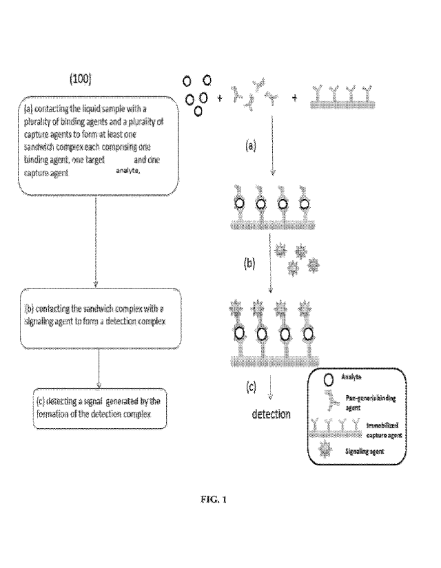
[0062] FIG. 1 shows a flow chart of the sequence of steps in a method for
detecting a
plurality of target analytes in a liquid sample according to some
implementations.
[0063] FIG. 2 shows formation of sandwich complexes via two-step routes
according to
some implementations.
[0064] FIG. 3 shows a schematic illustration of the top view of a sequential
lateral flow
device according to some implementations.
[0065] FIG. 4 shows a schematic illustration of a capillary flow bed residing
inside the
cavity of a housing unit according to some implementations.
[0066] FIG. 5A shows a schematic illustration of a capillary flow bed,
according to some
implementations. FIG. 5B shows a schematic illustration showing positioning of
datum
points in reference to the capillary flow bed, according to some
implementations.
[0067] FIG. 6 shows a schematic illustration of the configuration of the
sample-receiving
zone, according to some implementations.
[0068] FIG. 7 shows a schematic illustration of the side view of the sample-
receiving
zone, according to some implementations.
[0069] FIG. 8A shows a cut-away view of a housing alignment feature of
friction-fit pins,
according to some implementations. FIG. 8B shows each of the friction-fit pins
having a
hard stop figure that prevents over-closure of the device during assembly,
according to
16
CA 03071328 2020-01-27
WO 2019/023597
PCT/US2018/044123
some implementations. FIG. 8C shows that each of the friction-fit pins fully
sits at the
bottom of the cassette, according to some implementations.
[0070] FIG. 9A shows a schematic illustration of guides for the capillary flow
bed and the
reservoir pad (terminal wick) datum, according to some implementations. FIG.
9B shows a
schematic illustration of alignment tabs to maintain the position of the
capillary flow bed
relative to housing features, according to some implementations. FIG. 9C shows
a
schematic illustration showing that alignment tabs slope away from the
capillary flow bed,
aiding assembly and preventing flooding, according to some implementations.
[0071] FIG. 10 shows a schematic illustration of liquid containment ribs that
serve as a
liquid reservoir inside the upper portion of the housing unit, according to
some
implementations.
[0072] FIG. 11 shows a photograph showing the bend of the capillary flow bed
inside the
cavity of the housing unit, according to some implementations.
[0073] FIG. 12A shows a schematic illustration of broad pinch contact points
inside the
housing unit to compress the conjugate pad to direct the liquid flow into the
capillary flow
path, according to some implementations. FIG. 12B shows a schematic
illustration
showing positioning of the cross-section view of the broad pinch point in FIG.
12C. FIG.
12C shows the cross-section view of the broad pinch point, in which the
housing plastic
slopes away from the contact point with the capillary flow bed, according to
some
implementations. FIG. 12D shows a schematic illustration of the cross-section
view of the
broad pinch point in reference to the sample-receiving zone according to some
implementations.
[0074] FIG. 13 shows a schematic illustration of the positions of pinch points
relative to
the capillary flow bed, according to some implementations.
[0075] FIG. 14 shows a quantitative comparison of detection signals for
detecting Pa
27853 at various dilutions using a non-sequential lateral flow device (direct
method) vs. a
sequential lateral flow device, according to some implementations.
Detailed Description
[0076] Unless otherwise defined herein, scientific and technical terms used in
this
application shall have the meaning commonly understood by those skilled in the
art. The
techniques and procedures described or referenced herein are well understood
and
17
CA 03071328 2020-01-27
WO 2019/023597
PCT/US2018/044123
commonly employed using conventional methodologies that are well known and
commonly used in the art.
[0077] All publications, patents and published patent applications referred to
in this
application are hereby specifically incorporated by reference herein.
[0078] Each implementation of the disclosure described herein may be taken
alone and/or
in combination with one or more other implementations of the disclosure.
[0079] Unless specified otherwise, the following definitions are provided for
specific
terms, which are used in the above written description.
[0080] Throughout this disclosure, the word "comprise" and/or variations such
as
"comprises" and "comprising" will be understood to imply the inclusion of a
stated integer
(or components and/or steps) or group of integers (or components and/or
steps), but not
the exclusion of any other integer (or components and/or steps) or group of
integers
(and/or components).
[0081] The singular forms "a," "an," and "the" include the plurals unless the
context
clearly dictates otherwise. "A" or "an" also means "one or more" or "at least
one."
[0082] Transitional terms such as "including," "having," "containing,"
"involving,"
"composed of," and the like are to be understood to be open-ended and are used
to mean
"including but not limited to." "Including" and "including but not limited to"
are used
interchangeably.
[0083] As used herein, a binding agent is an agent capable of binding more
than one genus
of bacteria, viruses, and/or fungi. Binding agents are capable of binding to a
common
antigen of more than one genus of bacteria, viruses, and/or fungi, when used
in the
methods and devices of the present disclosure, for example, two or more, three
or more,
four or more, five or more, six or more, seven or more, eight or more, nine or
more, ten or
more, eleven or more, twelve or more, thirteen or more, fourteen or more,
fifteen or more,
sixteen or more, seventeen or more, eighteen or more, nineteen or more, or
twenty or more
genera of bacteria, viruses, and/or fungi. In some implementations, the
binding agent is an
antibody. In some implementations, a plurality of binding agents are used in
the methods
and devices described herein. The plurality of binding agents can be one or
more than one
type of binding agents, such as one or more than one type of antibodies, each
adapted to
specifically bind to a common antigen of at least a subset of target analytes
and the target
analytes may include multiple subsets or types of analytes, each subset or
type having a
18
CA 03071328 2020-01-27
WO 2019/023597
PCT/US2018/044123
common antigen within the subsets. In some implementations, a binding agent
specifically
binds a common antigen in more than one genus of bacteria, viruses, and/or
fungi. By way
of non-limiting example, an antibody that specifically binds
lipopolysaccharide on two or
more genera of Gram-negative bacteria is a binding agent. Likewise, an
antibody that
specifically binds lipoteichoic acid (LTA) on two or more genera of Gram-
positive
bacteria is a binding agent. Such binding agents can be polyclonal and/or
monoclonal
antibodies. In some implementations, a binding agent comprises antibodies with
different
specificities in a mixture, such that the mixture binds more than one genus of
bacteria,
viruses, and/or fungi. Other non-antibody molecules may serve as binding
agents if they
have the capability of binding to components of bacteria, viruses, and/or
fungi (e.g.,
antibiotics such as polymyxin bind to lipopolysaccharides of multiple genera
of Gram-
negative bacteria, and vancomycin can bind to components of the cell wall of
Gram-
positive bacteria). These molecules, with a suitable linker, could be used as
binding
agents.
[0084] As used herein, "antigen" (for example, a Gram-negative bacterial
antigen and/or a
Gram-positive bacterial antigen) is used to mean any molecule, in any
structural
conformation that may be specifically bound by a binding agent. The site on
the antigen
that is bound by a binding agent is called a "binding site." An antigen may
be, without
limitation, a protein, a glycoprotein, a carbohydrate, and/or a lipid.
[0085] As used herein, "analyte" or "analytes" refers to species, substances,
and/or
compounds to be detected and/or quantitatively analyzed in a sample. Analytes
include but
are not limited to toxins, proteins, peptides, viruses, bacteria and/or
bacteria antigens,
nucleic acids, carbohydrates, fungi, steroids, hormones, polysaccharides,
carbohydrates,
pollutants, metabolites, antibodies, and/or any detectable substances from
human and/or
non-human sources, such as blood, tissue, water, soil, sewage, beverages. In
some
implementations of this disclosure, an analyte binds to a binding agent and
forms a first
complex, and then the first complex binds to the immobilized capture agents on
the
capillary flow bed in the capture zone and forms a sandwich complex between
the first
complex and the capture agent. In certain such implementations, the sandwich
complex
further binds to a signaling agent to form a detection complex. The formation
of each
complex is discrete and sequential.
19
CA 03071328 2020-01-27
WO 2019/023597
PCT/US2018/044123
[0086] As used herein, "a conjugation pair" or "a conjugate pair" refers to
two different
molecules/members in which the first and second molecule/member bind to each
other
through a covalent bond, affinity, and/or physical means. The binding or
interaction
between the members of the conjugation pair is specific and unique such that
the members
are capable of distinguishing their binding partners from various interactions
and/or
affinities from other components of an assay and/or surrounding substances. In
some
implementations, a conjugation pair can be a receptor and a ligand, such as an
antibody
and an antigen. In other implementations, a conjugation pair includes, but is
not limited to,
biotin and avidin, carbohydrates and lectins, complementary nucleotide
sequences,
complementary peptide sequences, effector and receptor molecules, enzyme
cofactors and
enzymes, enzyme inhibitors and enzymes, a peptide sequence and an antibody
specific for
the sequence or the entire protein, polymeric acids and bases, dyes and
protein binders,
peptides and specific protein binders (e.g., ribonuclease, S-peptide and
ribonuclease S-
protein), metals and their chelators, and the like. Furthermore, specific
binding pairs can
include members that are analogs and/or derivatives of the original specific
binding
member, for example, a specific binding member made by chemical modification,
recombinant techniques, and/or molecular engineering that still maintains
similar binding
properties to the other binding member. In some implementations, a first
member of a
conjugation pair is biotin and a second member of a conjugation pair is
selected from
avidin, NeutrAvidin, streptavidin, and/or any anti-biotin antibody.
[0087] This disclosure provides methods, sequential lateral flow devices, and
kits for
detecting one or more target analytes in a liquid sample. This disclosure also
provides
methods of using the disclosed devices and/or kits. It is to be understood
that this
disclosure is not limited to the implementations set forth herein. It is also
to be understood
that the implementations of this disclosure are intended for descriptive
purposes and
should not be deemed as limiting.
[0088] In some implementations, this disclosure provides methods, devices, and
kits with
broader reactivity and higher sensitivity than existing methods, devices, and
kits. In some
implementations, the methods, devices, and kits are capable of detecting a
broader range
of target analytes, such as a broader range of bacterial genera, species,
and/or strains of
bacteria than existing methods and devices. For example, the methods, devices,
and/or kits
may be capable of detecting at least 20, 40, 60, 80, 100, 150, 200, 250, 300,
350, 400, 450,
CA 03071328 2020-01-27
WO 2019/023597
PCT/US2018/044123
or 500 different bacteria, virus, or fungi. In some implementations, the
disclosure provides
methods, devices, and/or kits comprising a plurality of antibodies capable of
detecting
greater than 1 x 10⁷, 1 x 10⁶, 1 x 10⁵, 1 x 10⁴, 1 x 10³, or 1 x 10² colony
forming units
(CFU) per mL of bacteria and/or an equivalent concentration of antigens
derived from that
level of bacteria, each of which antibodies can bind to a common antigen of at
least a
subset of the target analytes.
[0089] Detailed descriptions of certain implementations suitable for the
devices, kits, and
methods of this disclosure are discussed as follows, but not limited to:
[0090] Figure 1 shows an illustrative method (100) for detecting a plurality
of target
analytes in a liquid sample, according to some implementations. In step (a),
the liquid
sample is brought into contact with a plurality of binding agents and a
plurality of capture
agents on a solid support in a lateral flow device under conditions that
permit formation of
at least one sandwich complex. The sandwich complex is formed between one or
more of
the plurality of the binding agents, one or more of the target analytes, and
on one more of
the capture agents. The sandwich complex may be formed in a step-wise manner,
such as
the target analyte may bind to the binding agent first before binding with the
capture agent
or the target analyte may bind to the capture agent first before binding with
the binding
agent. In some events, the target analyte, binding agent, and the capture
agent may come
together to form the sandwich complex in one-step. In some implementations,
the plurality
of binding agents and the plurality of capture agents are antibodies. In some
implementations, the plurality of binding agents and the plurality of capture
agents are
monoclonal antibodies, polyclonal antibodies, and/or mixtures thereof In some
implementations, the plurality of binding agents and the plurality of capture
agents are
polyclonal antibodies. The sandwich complex includes at least one target
analyte (in the
middle), one or more of the capture agents (at the bottom, immobilized to a
solid support),
and one or more of the plurality of binding agents (on the top). Unbound
reagents, target
analytes, and/or binding agents may be optionally washed away and/or removed
prior to
the next step, for example, by washing the solid support with a buffer
solution and/or
spinning dry the solid support. When the method is carried out on a device,
such as a
lateral flow device described herein, unbound reagents may be carried away
from the
capture agents via capillary force.
21
CA 03071328 2020-01-27
WO 2019/023597
PCT/US2018/044123
[0091] In some implementations, the plurality of binding agents and/or the
plurality of
capture agents are antibodies (e.g., polyclonal antibodies or monoclonal
antibodies). In
some implementations, such antibodies can specifically bind a common antigen
of at least
a subset of Gram-positive and/or Gram-negative bacteria and/or at least a
subtype thereof
In certain implementations, the plurality of binding agents or the plurality
of capture
agents can be a polyclonal antibody (e.g., a multivalent polyclonal antibody),
a
monoclonal antibody, and/or a mixture of the foregoing. In some
implementations, each of
the plurality of binding agents is tagged with one member of a conjugate pair,
such as a
conjugate pair of biotin and a biotin-binding protein (e.g., avidin,
neutravidin, anti-biotin
antibody, streptavidin, and/or other biotin-binding proteins). Unlike the
binding agent-
signaling particle conjugates used in conventional methods, the binding agent
in the
present method is tagged with a first member of a conjugate pair, e.g., a
small molecule
such as biotin, according to some implementations. As such, the size of the
binding agent
is much smaller than the binding agent-signaling particle conjugates. Within
the same
reactive space, local concentrationsof binding agents can be greatly increased
thereby
yielding increased binding events to generate higher numbers of the first
complex for the
next step and eventually for detection. In some implementations, the binding
agent is
labeled with biotin. In some implementations, the binding agent is labeled
with a biotin-
binding protein. Suitable conditions that may facilitate the formation of the
sandwich
complex include carrying out the reaction in a buffer solution (e.g., a
phosphate buffer) at
room temperature, optionally with agitation and/or slight heating. Optionally,
unbound
target analytes or binding agents may be removed prior to the next step..
[0092] In some implementations, the plurality of capture agents may be one or
more types
of capture agents each adapted to specifically bind to a common antigen of at
least a
subset of target analytes in the liquid sample. For example, in some
implementations, the
capture agents can specifically bind a common antigen of at least a subset of
Gram-
positive and/or Gram-negative bacteria and/or at least a subtype thereof In
some
implementations, the capture agents are antibodies, such as polyclonal
antibodies (e.g., a
multivalent polyclonal antibody), monoclonal antibodies, and/or a mixture of
the
foregoing. In some implementations, at least some of the capture agents are
the same type
as at least some of the binding agents. In some implementations, at least some
of the
capture agents are different types from at least some of the binding agents.
In some
22
CA 03071328 2020-01-27
WO 2019/023597
PCT/US2018/044123
implementations, the plurality of capture agents are immobilized in groups on
a solid
support (e.g., a test strip and/or a capillary flow bed of a device described
herein)
covalently through a chemical bond and/or through physical absorption. The
groupings
may be spatially separated from each other with each grouping including an
antibody that
specifically binds to a different target analyte.
[0093] The process continues at step (b), a signaling agent (e.g., non-
enzymatic) is
brought in contact withthe sandwich complex under conditions that permit the
signaling
agent to bind to a binding agent of the sandwich complex to form a detection
complex.
The signaling agent is tagged with a second member of the conjugate pair, and
binding of
the signaling agent to the binding agent of the sandwich complex indicates the
presence of
one or more of the plurality of the target analytes in the liquid sample. As
such, the
detection complex will also be immobilized on the solid support where the
capture agents
are. The lateral flow device is adapted to inhibit the signaling agent from
contacting the
plurality of binding agents prior to formation of the sandwich complex. In
some
implementations, the lateral flow device includes a substantially impermeable
backing
disposed between a sample-receiving pad in a sample-receiving zone for
introducing the
liquid sample into the lateral flow device and a capillary flow bed in the
solid support that
facilitates a flow of the liquid sample, thereby reducing backflow of the
liquid sample in a
proximal direction of the lateral flow device. In some implementations, the
liquid sample
may be added into the sample-receiving pad using a dropper with fixed
volume/capacity
calibrated to the saturation capacity of the sample-receiving pad. In some
implementations, the amount of liquid sample added by using the dropper is
less than the
full saturation capacity of the sample-receiving pad so that the sample-
receiving pad may
be unsaturated.
[0094]The signaling agent provides a detectable signal where the detection
complex is
formed on the solid support to indicate the presence of the one or more of the
plurality of
target analytes in a liquid sample. In some implementations, the signaling
agent is a
colored particle, a latex particle, a metallic particle (e.g., gold, silver,
platinum
nanoparticles), a fluorescent particle, or a magnetic particle. In some
implementations, the
signaling agent is a colored dye and/or a fluorescence dye. In some
implementations, the
signaling agent is a catalytic enzyme. In some implementations, the signaling
agent is a
particle, such as a gold nanoparticle (e.g., a 40 nm, 60 nm, or 80 nm gold
nanoparticle). In
23
CA 03071328 2020-01-27
WO 2019/023597
PCT/US2018/044123
certain such implementations, the gold nanoparticle is tagged with a second
member of the
conjugate pair, and binding of the signaling agent to the binding agent of the
sandwich
complex (e.g., via the conjugate pair) indicates the presence of the one or
more of the
plurality of target analytes in the liquid sample. In certain such
implementations, the
signaling agent is a gold nanoparticle tagged with a biotin-binding molecule,
such as
avidin and/or streptavidin. Excess signaling agents may be washed away and/or
removed
before signal detection to minimize interference.
[0095] The process continues in step (c) where a signal generated by the
formation of the
detection complex is detected to determine the presence of the one or more of
the plurality
of target analytes in the liquid sample. The signal may be detected using
appropriate
means, such as visual, electrical, and/or optical detection. If the one or
more target
analytes are present in the liquid, and the detection complex is formed in a
sufficient
amount, a signal will be detected, e.g., on the solid support where the
capture agent is
immobilized, producing a positive result. However, in the absence of the one
or more
target analytes or if the one or more target analytes are not present in
clinically relevant
levels, neither the first, sandwich, nor detection complex will be formed in
sufficient
amounts and therefore, no detectable signal will be found, thereby producing a
negative
result.
[0096] Figure 2 shows two routes to form sandwich complexes, each route having
two
steps according to some implementations. In the first route (200), the liquid
sample is
brought into contact with the plurality of binding agents under conditions
that permit
formation of a first complex between at least some of the target analytes and
at least some
of the binding agents. Subsequently, the first complex is brought to contact
with the
plurality of capture agents to form the sandwich complex, such that the liquid
sample
contacts the plurality of capture agents after formation of the first complex.
In operation,
the liquid sample can be simply mixed with the plurality of binding agents in
a test tube
prior to flowing through a device described herein. The solution containing
the first
complex can also be brought into contact the solid support on which the
capture agent is
immobilized by dipping the solid support in the solution for a sufficient
amount of time.
Alternatively, the liquid sample can flow through a device, such as a lateral
flow device
(e.g., a device described herein) and/or on a test strip (e.g., a lateral flow
bed/path used in
a device described herein), pre-loaded with the plurality of binding agents
propelled by a
24
CA 03071328 2020-01-27
WO 2019/023597
PCT/US2018/044123
pulling force, such as capillary action. The device or strip is configured
such that the liquid
sample will come into contact with the plurality of binding agents prior to
contacting the
plurality of the capture agents in the capture zone. Suitable conditions may
be applied to
ensure complex formation between the target analyte and the binding agent
and/or the
capture agent. For example, in the first route (200), this first step can be
carried out at
room temperature, and/or optionally with slight heating provided sufficient
time is allowed
for the first complex and/or unbound target analytes and binding agents to
interact with the
plurality of capture agents so that the formation of the sandwich complex can
be as
complete as possible.
[0097] In the second route (300), the liquid sample is brought into contact
with a plurality
of capture agents disposed on a solid support in a lateral flow device or a
test strip under
conditions that permit formation of at least one complex between one target
analyte and
one capture agent prior to the liquid sample contacting the plurality of
binding agents to
form the sandwich complex. In practice, the liquid sample can flow through the
lateral
flow device first, followed by a separate subsequent flow of a buffer
containing the
plurality of binding agents.
[0098] According to one aspect, the sequential lateral flow device of this
disclosure
includes a housing unit having an inner surface that defines a cavity in the
housing unit.
[0099] Figures 3 and 4 show the top and inside views, respectively, of a
sequential lateral
flow device according to some implementations. As shown in Figure 3, the
housing unit
may include an upper portion (10) that can fit together with a lower portion
(11) (Figure 4)
to form the cavity. The upper portion of the housing unit may include a first
inlet (20) (a
portion of which is depicted in red and labeled with a "1" and a red outline
in Figure 3), a
second inlet (21) (a portion of which is depicted in white and labeled with a
"2" in Figure
3), and a reading window (22). A liquid sample may be introduced through the
first inlet
(20) into the device, while a reagent buffer may be introduced through the
second inlet
(21) into the device.
[0100] Figure 4 shows a capillary flow bed (30) residing inside the cavity of
the housing
unit, according to some implementations. In some implementations, the
capillary flow bed
has a proximal region and a distal region. In some implementations, the
capillary flow bed
is secured on the lower portion of the housing unit (11), which has limited
contact points
CA 03071328 2020-01-27
WO 2019/023597
PCT/US2018/044123
with the capillary flow bed (e.g., through discontinuous and isolated supports
under the
capillary flow bed), as shown in Figure 4.
[0101] In some implementations, the housing unit may further include features
such as
datum and alignment tabs inside to keep the capillary flow bed properly
aligned and
secured inside the housing unit, e.g., on the lower portion of the housing
unit. In some
implementations, the housing unit may further include features, such as ribs,
discontinuous
supports, and/or pinch points inside, to control the flow of liquids passing
through the
capillary flow bed, such as in a sequential manner. In one aspect, the
capillary flow bed is
configured to transport liquids (e.g., the sample and the reagent buffer) from
the proximal
region to a distal region of the capillary flow bed through capillary action.
Further features
of the capillary flow bed are depicted herein, using like numerals as shown in
Figure 4,
according to some implementations.
[0102] Figure 5a shows the capillary flow bed and various components on the
bed,
according to some implementations. In some implementations, the capillary flow
bed may
include the following zones: a buffer receiving zone, a sample-receiving zone,
and a
capture zone. The buffer-receiving zone is disposed on the capillary flow bed
and includes
a buffer-receiving pad (PB, 40) and a conjugate pad (Pa, 41). The conjugate
pad includes
(e.g., as attached thereto and/or associated therewith) a signaling agent
(e.g., gold
nanoparticles labeled with biotin-binding proteins) disposed therein for
providing a
detectable signal. The signaling agent may be releasably dried inside the
conjugate pad
(41). A reagent buffer may be introduced to the buffer-receiving pad through
the second
inlet (12, 21) to mobilize the signaling agent retained in the conjugate pad
to flow through
the capillary bed toward the capture zone. The capture zone (C, 44) contains a
plurality of
capture agents (e.g., a plurality of capture agents of one or more than one
type of capture
agents), such as antibodies that specifically bind to a common antigen of a
subset of the
plurality of target analytes, immobilized on the capillary flow bed (e.g., in
one or more
than one groupings). In some implementations, the sequential lateral flow
device may
further include a reservoir pad (R, 45) disposed in the distal region of the
capillary flow
bed (30). The reservoir pad is adapted to draw the sample and the buffer
carrying the
mobilized signaling agent in the distal flow direction. In some
implementations, the
sample-receiving zone is disposed between the buffer-receiving zone and the
capture zone.
The sample-receiving zone includes a sample-receiving pad (Ps, 42) and a
transfer pad (Pt,
26
CA 03071328 2020-01-27
WO 2019/023597
PCT/US2018/044123
43). According to some implementations, the sample-receiving zone also
includes an
impermeable backing (60). Figure 5b shows the positions of datum points in
reference to
the capillary flow bed.
[0103] In some implementations, the device of the present disclosure comprises
a
capillary flow bed, such as those shown in Figures 5a and 5b. Figure 5a shows
the
capillary flow bed with various components on the bed, according to some
implementations. In certain such implementations, a sample-receiving zone
(comprising a
sample-receiving pad (42) and a transfer pad (43)), a buffer-receiving zone (a
buffer-
receiving pad (40) and a conjugate pad (41)), and a reservoir pad (45) are
disposed on the
capillary flow bed (30).
[0104] In some implementations, the capillary flow bed (30), and/or the sample-
receiving
pad (42), and/or the transfer pad (43), and/or the buffer-receiving pad (40),
and/or the
conjugate pad (41) may be made of a bibulous material, such as a
nitrocellulose
membrane. Bibulous material of the present disclosure is a porous material
having pores,
capable of transporting liquids through the material in response to capillary
forces. The
hibulOUS material comprises a series of fibers drawn together in parallel to
form an open
wick. The space between the fibers forms channels to draw liquids through
capillary
actions.
[0105] Generally, bibulous materials are hydrophilic in nature. Suitable
bibulous materials
include but are not limited to hydrophilic inorganic powders, such as silica
gel, alumina,
diatomaceous earth and the like, sponge materials, glass fibers, argillaceous
substances,
cloth, hydrophilic natural polymeric materials, particularly cellulosic
material, like
cellulosic beads, and especially fiber-containing papers such as filter paper
and/or
chromatographic paper, synthetic and/or modified naturally occurring polymers,
such as
nitrocellulose, cellulose acetate, polyvinyl chloride, polyacrylamide,
polyethylene, glass
fiber, polyacrylates, polyurethanes, crosslinked dextran, agarose, and other
such
crosslinked and non-crosslinked water-insoluble hydrophilic polymers. Suitable
bibulous
material of the present disclosure can be functionalized on the surface, for
example,
forming covalent bonds with antibodies and/or receptors.
[0106] In some implementations, the bibulous material can be in a form of pad,
sheet,
and/or compressed fibers. In some implementations, the bibulous material is
27
CA 03071328 2020-01-27
WO 2019/023597
PCT/US2018/044123
nitrocellulose, such as nitrocellulose having a pore size from about 0.4
microns to about
15 microns.
[0107] In some implementations, the capillary flow bed is disposed on a solid
support,
such as a substantially impermeable support, including a water-insoluble, non-
porous,
flexible plastic strip, e.g., a polyester strip. In general, the solid support
is of the same or
slightly different dimension as the capillary flow bed. Suitable materials for
the solid
support include, but are not limited to, polyethylene, polypropylene,
polystyrene,
polymethacrylate, nylon, glass, ceramics, metals, polyurethane, neoprene,
latex, silicone
rubber, polyester, poly(ethylene terephthalate), poly(vinyl butyrate) and the
like.
[0108] In some implementations, the capillary flow bed may include at least
one capture
zone (44), as shown in Figures 5a and 5b. A capture zone includes one or more
than one
capture agents immobilized on the capillary flow bed via chemical and/or
physical means.
[0109] In some implementations, when the liquid sample is added to the sample-
receiving
pad (42), the sample flows to the transfer pad (43) under capillary forces and
mixes with
the plurality of binding agents to form a first complex between at least some
of the binding
agents and some of the target analytes. The sample carrying the first complex
continues to
flow via the capillary flow bed and through the capture zone. The plurality of
capture
agents then binds to the target analytes and forms a sandwich complex between
the
capture agents, target analytes, and the binding agents. Subsequently, as the
buffer
carrying the signaling agent flows through the capture zone, the signaling
agent will bind
to a binding agent of the sandwich complex through the conjugation pair and
form a
detection complex between the signaling agent, the binding agent, the analyte,
and the
capture agent. In some implementations, the first complex contacts the
plurality of capture
agents to form the sandwich complex such that the liquid sample contacts the
plurality of
capture agents only after formation of the first complex. The retained
signaling agent
thereby provides a detectable signal, indicating the presence of one or more
than one of
the plurality of target analytes in the sample. As such, the flow of liquids
is tightly
controlled in a sequential fashion, and the devices of this disclosure are
designed in such a
way to ensure the sequential and stepwise flow of liquids passing through the
capillary
bed.
[0110] In some aspects, various features of the capillary flow bed described
herein may be
incorporated to ensure sequential flow of fluids across the capillary flow bed
(30) through
28
CA 03071328 2020-01-27
WO 2019/023597
PCT/US2018/044123
the immobilized capture agents in the capture zone (44) and to minimize mixing
of fluids
at their interfaces. The sensitivity of the devices of this disclosure may
result from a
combination of the factors discussed herein, in particular, the sequential
flow of the fluids
and minimized mixing of liquids. In this disclosure, the liquids are referring
to (1) a liquid
sample mixed with a plurality of binding agents (e.g., antibodies, such as
polyclonal
antibodies); or (2) a buffer carrying signaling agents (e.g., streptavidin
labeled gold
nanoparticles).
[0111] In some implementations, the capillary flow bed may include a control
zone, where
the signalizing agent is being captured and retained even in the absence of
the analyte as a
means to indicate proper functioning of the device and/or to confirm the
validity of the
method performed. In some implementations, the control zone includes an
immobilized
member of the conjugation pair (e.g., biotin), which is able to bind to the
signaling agent
(e.g., streptavidin-labeled gold nanoparticles). In some implementations, the
control zone
may be disposed after the capture zone on the capillary flow in the distal
region but before
the reservoir pad.
[0112] In some implementations, as shown in Figures 5a and 5b, the capillary
flow bed
includes two fluid addition zones, namely, a sample-receiving zone and a
buffer-receiving
zone. In some implementations, the sample-receiving zone includes a sample-
receiving
pad (42) and a transfer pad (43). In some implementations, the buffer-
receiving zone
includes a buffer-receiving pad (40) and a conjugate pad (41).
[0113] As shown in Figures5a and 5b, the sample-receiving zone is disposed in
the middle
of the capillary flow bed (e.g., in the middle lower third of the capillary
bed) between the
buffer-receiving zone and the capture zone (45), according to some
implementations.
[0114] Figure 6 shows an illustration of the sample-receiving zone, including
a sample-
receiving pad (Ps, 42) and a transfer pad (PT, 43), according to some
implementations. The
sample-receiving zone of Figure 6 may be used in any of the devices and/or
methods
disclosed herein. As shown in Figure 6, a substantially impermeable backing
(60) is
disposed between the sample-receiving zone and the capillary flow bed and
extending at
least partially underneath the sample-receiving pad and the transfer pad. In
some
implementations, when the sample is introduced through the first inlet (Ii,
20) to the
sample-receiving pad, the impermeable backing (60) inhibits contact between
the sample
and the capillary flow bed in the sample-receiving zone, thereby reducing
backflow of the
29
CA 03071328 2020-01-27
WO 2019/023597
PCT/US2018/044123
sample in a proximal direction. In some implementations, the plurality of
binding agents,
such as antibodies (e.g., biotin-labeled polyclonal antibodies) may be
disposed inside the
transfer pad. In some implementations, the binding agents may be dried inside
the transfer
pad and can bind to a common antigen of at least a subset of target analytes.
In some
implementations, the binding agents are contained within the sample-receiving
pad.
[0115] In some implementations, the sequential lateral flow device is
configured, and the
method of use performed, such that the liquid sample flows along the capillary
flow bed
(30) into the capture zone (45) before the mobilized signaling agent flows
into the capture
zone (45). In some implementations, when in operation, the reading window (22)
of the
sequential lateral flow device is positioned over the capture zone (44) for
observing a
detectable signal produced by an interaction of the binding agent and the
signaling agent
in the presence of the target analytes.
The construction of the sample-receiving zone has been optimized to control
and minimize
the backward flow of the sample mixed with the binding agent (such as
antibodies, e.g.,
biotinylated antibodies). In some implementations, the transfer pad (43)
includes dried
binding agents (e.g., antibodies) that will be reconstituted and mobilized
upon addition of
the sample to the sample-receiving pad through a first inlet (20). In some
implementations,
in order to minimize and control the backward flow of the sample and/or the
first complex
between the sample and the binding agent, the sample pad rests on a plastic
backing (60)
that is substantially impermeable and acts as barrier between most of the
transfer pad and
the capillary flow bed underneath (the capillary flow bed comprises a
nitrocellulose pad).
The only contact the transfer pad has with the capillary flow bed is via the
forward lip of
the transfer pad. It is only through this contact that any fluid will flow
from the transfer
pad onto the capillary flow bed.
[0116] Figure 7 shows a schematic illustration of the side view of the sample-
receiving
zone inside the housing unit, according to some implementations. As shown in
Figure 7,
the transfer pad may be bent at the forward tip section to come into contact
with the
capillary flow bed due to the impermeable backing (60) underneath, according
to some
implementations.
[0117] In some implementations, upon addition of the sample, the fluid will
flow from the
sample-receiving pad through the transfer pad and onto the capillary flow bed.
This fluid
CA 03071328 2020-01-27
WO 2019/023597
PCT/US2018/044123
will then flow forward toward the capture zone(s) and backwards toward the
conjugate
pad.
[0118] In some implementations, the buffer-receiving zone comprises a buffer-
receiving
pad (40) and a conjugate pad (41), as shown in Figures 4a and 4b. The buffer-
receiving
pad and the conjugate pad each comprise a bibulous material of the same or
different
kinds. Suitable buffer solutions to be used in this disclosure include but are
not limited to a
acetate buffer, buffered saline, citrate buffer, barbital buffer, phosphate
buffer, or buffer
prepared with tris(hydroxyl-methyl)aminomethane (TRIS), N-(2-
acetamido)iminodiacetic
acid (ADA), piperazine-1,4-bis(2-ethanesulfonic acid) (PIPES), N-(2-acetamido)-
2-
aminoethanesulfonic acid (ACES), N,N-bis(2-hydroxyethyl)-2-aminoethanesulfonic
acid
(BES), 3-morpholinopropanesulfonic acid (MOPS), N-tris(hydroxymethyl) methy1-2-
aminoethanesulfonic acid (TES), 2,4-(2-hydroxyethyl)-1-piperazinyl
ethanesulfonic acid
(HEPES), 3,4-(2-hydroxyethyl)-1-piperazinyl propanesulfonic acid (EPPS), N-
tris(hydroxymethyl) methylglycine (Tricine), N,N-bis (2-hydroxyethyl)glycine
(Bicine),
N-cyclohexy1-2-aminoethanesulfonic acid (CHES), N-cyclohexy1-3-
aminopropanesulfonic
acid (CAPS), and/or buffer containing one or more than one buffer salts
selected from
K2HPO4, KH2PO4, Na2HPO4, NaH2PO4, NaHCO3, NaB04, (NH4)2CO3.
[0119] After the liquid sample is added through the first inlet (20), the
reagent buffer (such
as a chase buffer, e.g., a phosphate buffer) may be introduced into the device
through the
second inlet (21) and onto the buffer-receiving pad (40) at the distal
upstream end of the
capillary flow bed. To avoid the situation where the binding agent (e.g.,
biotinylated
antibodies) encounters and conjugates with the signaling agent (e.g.,
streptavidin-
conjugated gold nanoparticles) beyond an insignificant amount at their fluid
interface and
to prevent sample fluid backflow to the conjugate pad, the buffer may be added
as soon as
the sample fluid front flows past the viewing window over the capture zone.
The timing of
adding the buffer ensures that there is a forward fluid counter-flow to the
sample mixture
flowing backwards. As a result, these two fluids will meet in a very small
volume dictated
by the pore size of the nitrocellulose pad of the capillary flow bed. Hence,
very little
mixing occurs between these two fluids.
[0120] The forward flow of the sample will reach the reservoir pad (45), which
acts as a
sump to draw fluid to itself As long as this forward flow is imposed by the
reservoir pad,
there is very little mixing between the two fluids at their interface and
their sequential flow
31
CA 03071328 2020-01-27
WO 2019/023597
PCT/US2018/044123
is maintained. In some implementations, in roughly five to seven minutes, the
sample
fluid flow is complete and the flow front of the liquid containing the
signaling agents
should begin to appear in the reading window.
[0121] In some implementations, the device of this disclosure includes a
reservoir pad
(45). In some implementations, the reservoir pad is disposed in the distal
region of the
capillary flow bed, e.g., at the end of the distal region of the capillary
flow bed. In some
implementations, the reservoir pad also contains a bibulous material (e.g., a
nitrocellulose
pad), which serves as a liquid sink and has a liquid-absorbing capacity
exceeding the sum
capacity of the capillary flow bed, sample-receiving zone, and buffer-
receiving zone
combined. In this way, there is generated a continuous capillary force across
the capillary
flow bed from the proximal region to the distal region during the process of
conducting the
method. This continuous capillary force is important to keep the flow in one
direction and
lower the risk of backflow and/or fluids mixing.
[0122] In some aspects, there are many considerations for designing the
housing unit,
including manufacturability of the device, tolerances of the components,
component sizes
(e.g., component assembly and placement), functionality of the assay, (e.g.,
variability of
how the assay is run due to user-related factors), and overall aesthetics. In
some
implementations, the device components may be secured in the housing unit so
that
various components of the device are properly aligned to inlets (or the liquid
reagent
wells) and the reading window. For example, as shown in Figures 2 and 3, the
capture
zone is presented in the reading window and the housing unit does not hinder
testing
and/or alter test results using the method disclosed herein. Various device
components
include, but are not limited to, a capillary flow bed, a buffer-receiving
zone, a capture
zone, a sample-receiving zone, and a reservoir pad to enable the method of
detection. In
other implementations, the housing unit may include features to facilitate the
control and
sequential flow of liquids (e.g., samples and the reagent buffer).
[0123] In some implementations, the dimensions of the housing are dictated in
part by the
requirement that the device can optionally be read by an off-the-shelf lateral
flow assay
(LFA) reader. Such LFA reader, e.g., Qiagen ESEQuant LR3, may dictate the
device
housing to be a certain size, for example, a size of less than about 110 mm
long, about 50
mm wide and about 12.5 mm high. In addition to considerations of a LFA reader,
the
architecture of the capillary flow bed with various components of the device
also
32
CA 03071328 2020-01-27
WO 2019/023597
PCT/US2018/044123
influences the overall housing dimensions. In some implementations, the length
of the
capillary flow bed should be at least about 80 mm, at least about 90 mm, at
least about 100
mm, or at least about 110 mm in order to facilitate the sequential flow of
liquids and to
accommodate liquid volume along with all the other components of the device.
In some
implementations, the length of the capillary flow bed may be in the range from
about 50
mm to about 200 mm, from about 55 mm to about 180 mm, from about 60 mm to
about
160 mm, from about 75 mm to about 140 mm, or from about 80 mm to about 100 mm.
In
some implementations, the capillary flow bed has a length of about 85 mm.
However, the
housing may be any suitable shape and/or size, including sizes that are not
compatible
with automated assay readers.
[0124] Figures 3 and 4 show the top and inside views of the housing unit,
according to
some implementations. In some implementations, the housing unit includes an
upper
portion (10) coupled to a lower portion (11). In certain such implementations,
the upper
portion of the housing unit includes a reading window (22). In some
implementations, the
upper portion of the housing unit further includes a first inlet (20), such as
a first inlet for
introducing the sample into the sample-receiving pad (42). In some
implementations, the
upper portion of the housing unit further includes a second inlet (21), such
as a second
inlet for introducing a buffer into the buffer-receiving pad (40).
[0125] In some implementations, to clearly differentiate the control line from
other capture
zones so that users could correctly interpret test results, the reading window
is designed
and disposed so as to clearly identify and isolate the control line from other
capture zones.
[0126] Figure 3 shows that the reading window is narrowed at the distal end,
such as at the
location of the control line, according to some implementations. In certain
such
implementations, the reading window may be narrowed from about 5 mm to about 3
mm.
The narrowing creates a physical and visual differentiation that intuitively
alerts the user
that the control line is differentiated from other capture lines. In some
implementations,
the read window has a slope on the side walls away from the strip surface to
minimize
casting of shadows that can make it more difficult to properly visualize
positive results.
[0127] In some implementations, the reading window has a length of from about
10-50
mm, from about 20-40 mm, or about 20 mm, about 30 mm, about 40 mm, or about 50
mm.
In certain such implementations, the reading window has a length of about 23
mm.
33
CA 03071328 2020-01-27
WO 2019/023597
PCT/US2018/044123
[0128] In some implementations, the width of the reading window is less than
the width of
the capillary flow bed. In certain such implementations, the width of the
reading window
is from about 2 mm to 10 mm, from about 4 mm to 8 mm, from about 5 mm to 6 mm,
or
about 3 mm, about 4 mm, about 5 mm, or about 6 mm. In some implementations,
the
width of the reading window is about 4.7 mm. In other implementations, the
width of the
reading window is about 2.6 mm.
[0129] Figure 3 shows that the upper portion of the housing (10) has a flat
area on the top
surface for labeling, such as for putting on a bar-code sticker, according to
some
implementations. In some implementations, the upper portion of the housing
unit includes
markings on its top surface to identify where sample and reagents are to be
added. In some
implementations, the upper portion of the housing unit has concaved surfaces
around the
first inlet and second inlet to form liquid reservoirs, which facilitate the
transfer of liquids
with a disposable pipette and dropper bottle.
[0130] In some implementations, the housing unit is designed for ease of
manufacturing.
For example, the upper and lower edges of the housing unit may be made
parallel for a
short segment to support automated sorting and alignment of the upper portion
and the
lower portion. In addition, the upper portion of the housing unit is modified
in such a way
that a standard roller-closure could be utilized for assembling the devices.
[0131] In some implementations, the upper portion of the housing unit includes
at least
one friction-fit pin in its inner surface. Figures 8a-8c show the friction-fit
pins with
alignment guide elements, according to some implementations. In certain such
implementations, the area under the friction-fit pins is substantially
flattened to allow for
uniform pressure against the pins critical for device closure. In some
implementations, as
shown in Figure 8a, the friction-fit pins are cone-shaped so that the upper
and lower
portions of the housing unit can be aligned approximately and "honed in"
during paring. In
some implementations, the upper portion of the housing unit comprises one or
more than
one friction-fit pins, such as one, two, three, four, five, six, seven, eight,
nine, or ten
friction-fit pins.
[0132] Figures 8b and 8c show that, in addition to incorporating an alignment
feature to
the friction-fit pins, each of the friction-fit pins further has a hard stop
feature that prevents
over-closure of the device during assembly, which can potentially damage the
capillary
flow bed, according to some implementations.
34
CA 03071328 2020-01-27
WO 2019/023597
PCT/US2018/044123
[0133] The strategy for the design of the press-fit feature requires
consideration of a
number of other factors that are not readily apparent from a structural point
of reference.
One such consideration is the limitations of readily available toolmakers used
to create the
injection-molded housing unit. The toolmaker may have a limitation of not
being able to
reasonably rework the mold to create what is known as a "steel-safe" part. In
some
situations, the toolmaker, chosen for industry-leading rapid prototyping of
injection-
molded parts, can't rework the tool to tolerances less than 0.003 inches.
Because of this
limitation, the toolmaker cannot guarantee a snap and/or press-fit feature. To
address this
limitation, in some implementations, the friction fit feature incorporates a
pin that would
deform upon closure. In other implementations, the female component of the
part can also
deform to accommodate the pin. By designing the press-fit feature as
deformable
elements, the tolerance for acceptable interference fit is increased to about
0.005 inches
and within tolerances of the conventional vendor's toolmaking capabilities.
[0134] One aspect of the housing design is the need to properly secure the
capillary flow
bed and each component on top of it into the desired position. Figures 9a-9c
show
alignment tabs and datum and their positioning in the inner surface of the
housing unit,
according to some implementations. Figure 9a shows that the capillary flow bed
and the
housing unit may reference a common datum located at the distal region of the
capillary
flow bed, such as located at the reservoir pad (or terminal wick) end of the
capillary flow
bed, according to some implementations. In certain such implementations, it is
from this
common datum that the capillary flow bed and the housing unit tolerances are
derived. In
some implementations, the datum for the housing unit is a tab that the distal
region of the
capillary flow bed is butted.
[0135] Figure 9b shows alignment tabs in the inner surface of the housing unit
to maintain
the position of the capillary flow bed, according to some implementations. In
certain such
implementations, besides the terminal-wick-datum-point-tab in the housing
unit, the
devices of this disclosure further include a series of alignment tabs (such as
two or more
alignment tabs, e.g., two, four, six, or eight alignment tabs) that align the
capillary flow
bed to the appropriate features of the housing unit. These alignment tabs keep
the capillary
flow bed centered with the first and second inlet (i.e., sample and buffer
wells) as well as
the critical pinch points that are at the interface between the housing unit
and the capillary
flow bed.
CA 03071328 2020-01-27
WO 2019/023597
PCT/US2018/044123
[0136] Figure 9c shows alignment tabs slope away from the capillary flow bed
to aid
assembly and prevent flooding, according to some implementations. In certain
such
implementations, the capillary flow bed and the components thereon may be
properly
placed either manually and/or by automation. To make this task easier, the
alignment tabs
are sloped in such a way to help guide the capillary flow bed into proper
position. In some
instances, the housing may provide a potential point of wetting and
unintentionally flood
the interior and/or cavity of the housing unit. In some implementations, to
avoid situations
like unintentional flooding, the alignment tabs may slope aggressively away
from the
surface of the capillary flow bed so that the liquid reagents are contained
within the
confines of the pads and features of the housing unit.
[0137] In some aspects, the methods and devices of this disclosure are
designed to detect
multiple target analytes, such as bacterial antigens from a vast variety of
bacterial species
and strains. In some implementations, the methods and devices of this
disclosure may use
a large number of complex antibody mixtures. In some implementations, multiple
capture
zones of antibodies may be immobilized on the capillary flow bed (e.g., a
nitrocellulose
pad) in the devices of this disclosure. The wide variation of antigen targets
means that in
order to facilitate detection, the detector antibodies must be equally diverse
and present in
a sufficient quantity to label enough antigens to produce a detectable signal.
It is
determined that better performance of using the devices of this disclosure is
achieved by
sequentially flowing the sample, including labeled antibodies, across the
capture zones and
then followed by the detector component (such as a signaling agent, e.g.,
streptavidin-
conjugated colloidal gold particles). The specific need to sequentially flow
the sample and
reagents (e.g., a buffer solution) in a controlled, reproducible and robust
manner through
the device presents unique challenges for the device design.
[0138] A number of features may be incorporated in the sequential lateral flow
device to
ensure containment and controlled release of liquids, according to some
implementations.
For example, the housing unit of the devices of the present disclosure may be
involved in
the handling of liquids within the devices during use. In some
implementations, to
facilitate a sequential delivery of liquids (e.g., the sample and the reagent
buffer), crude
and variable amounts of liquids must be contained and later be released from a
reservoir
and/or area of containment. In some implementations, in practice, the user
will add a
sample through the first inlet (e.g., the sample well). In some
implementations, the sample
36
CA 03071328 2020-01-27
WO 2019/023597
PCT/US2018/044123
will wet out the sample-receiving pad and then wet out the transfer pad
containing binding
agents, such as antibodies.
[0139] In some aspects, the sample-receiving pad and the transfer pad do not
have the
capacity to completely contain the liquid sample. However, the liquid sample
must still be
controlled and/or held back to prevent flooding out of the flow path on the
capillary bed
because the flooding will result in the sample liquid flowing over instead of
through the
pores of the nitrocellulose pad of the capillary flow bed that contains the
capture agents
(e.g., the capture antibodies). Furthermore, if the liquid sample and/or
buffer is not
adequately contained, the sample and buffer could leave the desired flow path
and flood
out the interior and/or cavity of the housing unit. In either circumstance,
the detection will
be compromised. To support a sequential delivery of each liquid independently,
it is
challenging to retain and release the liquid in the device and to control the
flow of any
liquid into the flow path (e.g., nitrocellulose flow path) at a desired time
point. The same
challenge applies to the buffer and buffer-receiving pad, as well as the
conjugate pad.
[0140] In some implementations, the housing unit may interact with the pads
and facilitate
containment and release of the liquid sample and buffer to the capillary flow
bed (the
nitrocellulose pad) in a sequential manner. Figure 10 shows a schematic
illustration of the
inner surface of the housing unit (e.g., the upper portion) having a series of
ribs, according
to some implementations. In certain such implementations, a series of ribs is
incorporated
into the housing unit, such as in the inner surface of the upper portion of
the housing unit,
to facilitate the sequential flow of liquid sample and the buffer. In some
implementations,
the ribs are approximately perpendicular and/or approximately parallel to the
capillary
flow bed (e.g., the nitrocellulose flow path).
[0141] Figure 10 shows that the ribs may be disposed on the inner surface of
the upper
portion of the housing unit, such as over the sample-receiving pad and/or over
the buffer-
receiving pad, according to some implementations.
[0142] In some implementations, the housing unit may comprise a plastic
material as the
surface energy and/or wettability of plastic material can contribute to the
liquid
containment and/or release. For example, plastic material may have a lesser
ability to hold
onto liquids than the bibulous material of the capillary flow bed. As such,
plastic material
may be particularly suitable for the housing unit due to the need to
facilitate readily
releasing liquids into the capillary flow bed (e.g., nitrocellulose) to the
point of capillary
37
CA 03071328 2020-01-27
WO 2019/023597
PCT/US2018/044123
saturation. As liquids move through the capillary flow bed to the reservoir
pad (e.g., the
terminal wick), fluid contained in the housing unit is sequentially released
to flow through
the sample-receiving pad, to the capillary flow bed, and toward the reservoir
pad (e.g., the
terminal wick). In some implementations, the ribs can hold the liquids in
place until the
capillary flow bed (e.g., nitrocellulose) becomes incrementally less
saturated. At this
point, more liquid is then transferred to the transfer pad from the sample-
receiving pad that
remains saturated by the liquids held by the wetted housing features. The size
and spacing
of the ribs determine how much excess liquid may be contained in the device.
[0143] Interestingly, according to some implementations, once the liquid
sample and/or
the buffer is depleted to the point of becoming less saturated, the excess
liquid from the
inlet (e.g., the sample and/or buffer well) of the device becomes available to
the capillary
flow bed. In certain such implementations, this process continues as the
bibulous material
(e.g., nitrocellulose pad) of the capillary flow bed becomes incrementally
less saturated
and it, hence, becomes a source of capillarity and draws liquid from the
liquid-saturated
conjugate pad. As analogously described for the sample-receiving pad and the
transfer
pad, the over-saturated buffer-receiving pad, in turn, saturates the conjugate
pad. The
buffer-receiving pad is maintained in an oversaturated state by drawing a
buffer that is
held in place by the ribs on the inner surface of the upper portion of the
housing unit just
under the second inlet (i.e., the buffer well), according to some
implementations.
[0144] In some implementations, to run an assay with the disclosed devices,
the liquid
sample and buffer are added to the device in excess of any of the pads'
capacity to fully
absorb the volume required. In some implementations, excess liquid is
introduced to the
device because the pads must "give up" the liquids to the nitrocellulose of
the capillary
flow bed. This oversaturated state inevitably leads to a propensity of the
liquids to flood
within the device and/or not be sufficiently contained to prevent method
failure. To
prevent potential flooding in the housing unit, a number of structural
features may be
incorporated into the device, such as in the housing unit, according to some
implementations.
[0145] The capillary flow bed and the reservoir pad comprise the primary
liquid flow
pathway in the device. Feeding into this primary flow pathway is the liquid
sample and the
reagent buffer originating from the first inlet and the second inlet,
respectively. Once these
liquids are introduced to the device, the liquids will saturate the sample-
receiving pad and
38
CA 03071328 2020-01-27
WO 2019/023597
PCT/US2018/044123
the buffer-receiving pad, respectively, and begin to pool on any wettable
surface. This
situation produces a dynamic in which part of the liquids will flow over the
surface of the
capillary flow bed and have no contact with the capture zone within the pores
(e.g.,
nitrocellulous pores) of the capillary flow bed.
[0146] Figure 11 shows that, to prevent overflow, the capillary flow bed may
be bent so
that the liquids enter into the pore structures (e.g., nitrocellulose pores)
of the capillary
flow bed, according to some implementations. Basically, the bending in the
capillary flow
bed forces the liquids to enter into the nitrocellulose pore structures of the
capillary flow
bed though capillary action.
[0147] Figure 11 also shows bending the capillary flow bed to prevent liquid
from flowing
over the surface of the bed, according to some implementations. In certain
such
implementations, the capillary flow bed is bent so that the contact point of
the transfer pad
is anywhere from about 0.02 mm to about 1 mm lower than one or more adjacent
flow bed
supports. This (roughly) amounts to about 0.05 degrees to about 10 degrees of
a bending
angle. The bend encourages capillarity flow and mitigating flow over the
capillary bed
surface, so that excess reagent added to the wells and some liquid on the
capillary flow
bed will pool in locations adjacent to the liquid reservoirs, thereby reducing
localized
flooding, which will diminish the sequential delivery of fluids from the
reservoirs.
[0148] Figures 12a-12d show the broad pinch points incorporated on the
underside of the
housing unit, according to some implementations. In certain such
implementations, a
broad area of contact is incorporated in the housing unit, such as in the
upper portion of
the housing unit, to compress the conjugate pad and thereby direct the
reconstituted
conjugate to flow into the nitrocellulose pores of the capillary bed. It was
determined that
this broad area contact, acting as a broad pinch-point, may facilitate a more
compact and
discrete flow of conjugate through the device.
[0149] Theoretically, every point where the capillary flow bed contacts the
housing unit
may become a location that can create a capillary action. This occurs because
liquids are
able to wet the surfaces of plastic materials and, once wet, the contact point
becomes a
point for internal flooding. In some implementations, features of the housing
unit that
contacts the capillary flow bed may be incorporated to prevent capillarity
from creating an
undesired and/or otherwise uncontrolled pathway for the liquids to follow.
39
CA 03071328 2020-01-27
WO 2019/023597
PCT/US2018/044123
[0150] Figure 4 shows an open view of the sequential lateral flow device,
having the
capillary flow bed secured in the lower portion of the housing unit, according
to some
implementations. In some implementations, the housing unit, such as the lower
portion of
the housing unit, may also incorporate features that represent limited contact
points with
the capillary flow bed that are isolated from other portions of the capillary
flow bed. In
certain such implementations, the lower portion of the housing unit may
include discrete
and discontinuous supporting blocks, as shown in Figure 4. In certain such
implementations, the supporting blocks are disposed under the capillary flow
bed at
positions corresponding to the sample-receiving pad. In other implementations,
the
supporting blocks are disposed under the capillary flow bed at positions
corresponding to
the buffer-receiving pad, e.g., aligned to the second inlet. In some
implementations, the
supporting blocks are disposed under the capillary flow bed at positions
corresponding to
the reservoir pad.
[0151] In some implementations, the supporting blocks under the first inlet
are only long
enough to provide support for the capillary pad, the first inlet, and
associated pinch points.
In some implementations, the supporting blocks under the second inlet are only
long
enough to provide support for the capillary pad, the second inlet, and
associated pinch
points.
[0152] Figure 13 shows positions of the pinch points in the housing unit in
reference to the
capillary flow bed, according to some implementations. In certain such
implementations,
the housing unit may include one or more than one pinch point in the housing
unit, such as
in the inner surface of the housing unit. The purpose of pinch points is to
assure that the
different components on the capillary flow bed (such as sample-receiving pad,
buffer-
receiving pad, reservoir, conjugate pad, and transfer pad) are in contact with
adjacent
components so that a reliable and reproducible flow path is established in the
device. In
some implementations, a pinch point presses the buffer-receiving pad into the
conjugate
pad thereby creating a flow path for the buffer to flow to the conjugate pad.
In some
implementations, a pinch point presses the conjugate pad into the capillary
flow bed
(nitrocellulose) thereby creating a flow path for the sample and the analyte-
binding agent
to flow to the capillary flow bed. In some implementations, a pinch point
presses the
sample-receiving pad into the transfer pad to create a flow path for the
sample to flow
from the sample-receiving pad to the transfer pad. In some implementations, a
pinch point
CA 03071328 2020-01-27
WO 2019/023597
PCT/US2018/044123
presses the transfer pad into the capillary flow bed to create a flow path for
the sample to
flow from the transfer pad to the capillary flow bed. In some implementations,
the pinch
points exert sufficient pressure to facilitate a reproducible liquid flow path
in materials that
are compressible, such as the capillary flow bed. For example, the pinch point
can exert
enough forces to assure a continuous flow pathway, but not restrictive and/or
damaging to
the individual components of the device, according to some implementations.
[0153] The sequential lateral flow devices suitable for use in the methods of
the present
disclosure may be selected from any of the implementations of the sequential
lateral flow
devices and/or kits described above and/or below.
[0154] Reagents suitable to use in the methods, devices, and/or kits of the
present
disclosure include the following, but are not limited to the following.
[0155] In some implementations, the methods and devices of the present
disclosure
include a signaling agent. The signaling agent is capable of producing a
detectable signal,
such as a visually detectable signal, a chemically detectable signal, an
electromagnetically
detectable signal, and/or a signal detectable by an instrument to report the
presence of
analytes in a tested sample. Various signaling agents suitable for use in the
methods and/or
devices of this disclosure include agents that produce signals through
chemical and/or
physical means. For example, the signaling agent may be a particle, such as a
colored
particle, a latex particle, a metallic particle (e.g., gold, silver, platinum
nanoparticles,
and/or colloidal metallic particles), a fluorescent particle, a magnetic
particle, a
chemiluminescent particle, a non-metallic colloidal particle, and/or a
luminescent particle.
Other suitable signaling agents include, but are not limited to, liposomes,
plastic and/or
polymeric particles, stained microorganisms, cells, enzymatic substrates
and/or enzymes
(e.g., catalytic enzymes), specific-binding substances, and/or any vesicles
containing
visible substances and the like.
[0156] In some implementations, when the signaling agent is a particle, the
particle size
can be in a range from about 30 nm to about 120 nm in diameter, or from about
40 nm to
about 100 nm in diameter, or from about 50 nm to about 90 nm in diameter, or
from about
60 nm to about 80 nm in diameter. In some implementations, the particle size
is about 40
nm, about 60 nm, or about 80 nm in diameter. In some implementations, the
particle size
is about 40 nm in diameter.
41
CA 03071328 2020-01-27
WO 2019/023597
PCT/US2018/044123
[0157] In some implementations, the signaling agent provides a detectable
signal, such as
a detectable signal in the capture zone of the device where the binding agent
bound
analytes are captured by the capture agent.
[0158] In some implementations, the signaling agent (e.g., signaling particle)
is dried
within the conjugation pad.
[0159] In some implementations, the signaling agent is attached and/or labeled
with a first
or second member of a conjugation pair. In certain such implementations, the
signaling
agent is associated and/or labeled with a first or second member of a
conjugation pair and
the binding agent is attached and/or labeled with a second or first member of
a conjugation
pair.
[0160] In some implementations, the signaling agent comprises gold
nanoparticles (such
as about 40 nm, about 60 nm, or about 80 nm gold nanoparticles) labeled with a
first
member of a conjugation pair, for example avidin, streptavidin, and/or other
biotin binding
proteins and/or antibodies, while the binding agent may be labeled to a second
member of
the conjugation pair, such as biotin and/or any avidin binding moieties.
[0161] In some implementations, the methods and devices of the present
disclosure may
include a plurality of binding agents. In certain such implementations, each
of the plurality
of binding agents may be labeled with a first or second member of a conjugate
pair, such
as biotin and/or any avidin binding moieties and/or molecules. In some
implementations,
the binding agent may be present in the transfer pad, such as in a dry form in
the transfer
pad. In some implementations, the binding agent may be contained in the sample-
receiving pad.
[0162] In some implementations, the plurality of binding agents of this
disclosure are
antibodies and/or a functional fragment thereof, e.g., antibodies that
specifically bind a
common antigen of at least a subset of a plurality of Gram-negative bacteria
and/or
antibodies that specifically bind a common antigen of at least a subset of
Gram-positive
bacteria. In some implementations, the binding agents are polyclonal
antibodies, such as
one or more than one type of a multivalent polyclonal antibody. In other
implementations,
the binding agents are monoclonal antibodies. In some implementations, the
plurality of
binding agents comprises one or more than one type of antibodies or two or
more than two
types of antibodies, wherein each antibody specifically binds a common antigen
of a
subset of bacteria in a liquid sample.
42
CA 03071328 2020-01-27
WO 2019/023597
PCT/US2018/044123
[0163] In some implementations, the polyclonal antibody binds lipoteichoic
acid (LTA).
In some implementations, the polyclonal antibody binds a bacterial
lipopolysaccharide
structure (LPS). In some implementations, at least one type of the plurality
of binding
agents specifically bind a Gram-positive bacterial antigen and at least one
type of the
binding agents specifically bind a Gram-negative bacterial antigen. In some
implementations, the plurality of binding agents are capable of binding one or
more than
one (e.g., two or more, three or more, four or more) genera of bacteria.
[0164] In some implementations, the antibody is selected from a polyclonal
antibody, a
monoclonal antibody and a combination of polyclonal and monoclonal antibodies.
In some
implementations, the antibody is polyclonal and binds a common antigen of a
plurality of
bacteria. In some implementations, the antibody is polyclonal and binds a
common antigen
of a plurality of Gram-positive bacteria and/or a plurality of Gram-negative
bacteria,
and/or both. In some implementations, at least one antibody is a monoclonal
antibody and
at least one antibody is a polyclonal antibody. In some implementations, at
least one
antibody specifically binds a common antigen of Gram-positive bacteria and at
least one
antibody specifically binds a common antigen of Gram-negative bacteria. In
some
implementations, the antibody is capable of binding one or more than one
(e.g., two or
more, three or more, four or more) genera of bacteria.
[0165] In some implementations, such binding agents comprise antibodies which
bind
under physiological conditions to an antigen-containing epitope of a
lipopolysaccharide
(LPS) structure of a Gram-negative bacteria and/or a lipoteichoic acid (LTA)
structure of a
Gram-positive bacteria.
[0166] Antibodies useful in the methods and devices of the disclosure include,
but are not
limited to, a monoclonal antibody, a polyclonal antibody, a single-chain
antibody, a
synthetic antibody, a recombinant antibody, a chimeric antibody, and/or any
antigen-
binding fragment of the above, including, but not limited to, F(ab), F(ab'),
F(ab)2, scFy
fragments and recombinant fragments. The antibodies may be from non- species,
for
example, a chicken antibody, and/or from a mammalian species, including but
not limited
to rabbits, rodents (including mice, rats and guinea pigs), goats, pigs,
sheep, camels and
humans. The antibodies also may be humanized and/or chimeric antibodies.
[0167] Those skilled in the art are enabled to make any such antibody
derivatives using
standard art-recognized techniques. For example, Jones et al. (Nature 321: 522-
525
43
CA 03071328 2020-01-27
WO 2019/023597
PCT/US2018/044123
(1986)) discloses replacing the CDRs of a human antibody with those from a
mouse
antibody. Marx (Science 229: 455-456 (1985)) discusses chimeric antibodies
having
mouse variable regions and human constant regions. Rodwell (Nature 342: 99-100
(1989))
discusses lower molecular weight recognition elements derived from antibody
CDR
information. Clackson (Br. J. Rheumatol. 3052: 36-39 (1991)) discusses
genetically
engineered monoclonal antibodies, including Fv fragment derivatives, single-
chain
antibodies, fusion proteins, chimeric antibodies and humanized rodent
antibodies.
Reichman et al. (Nature 332: 323-327 (1988)) discloses a human antibody on
which rat
hypervariable regions have been grafted. Verhoeyen et al. (Science 239: 1534-
1536
(1988)) teaches grafting of a mouse antigen binding site onto a human
antibody.
[0168] Preferably, the antibodies of the present disclosure are polyclonal
antibodies and/or
monoclonal antibodies. Generation of monoclonal and polyclonal antibodies is
well within
the knowledge of one of ordinary skill in the art of biology (see, e.g.,
Ausubel et al.,
Current Protocols in Molecular Biology, John Wiley & Sons, New York, N.Y.,
1994). A
number of procedures are useful in producing antibodies to the desired unique
target
antigens. Traditional immunization and harvesting techniques will result in
the creation of
polyclonal antibodies directed against the common determinants of the target
bacterial
species including determinants such as LPS and LTA. Additionally, cellular
hybridization
techniques can be utilized to produce immortal hybridoma cell lines that
generate specific
monoclonal antibodies to the target species.
[0169] Antibodies having potential utility for broadly detecting Gram-positive
bacteria
include those described in Fisher et al., PCT Publication No. W098/57994;
Jackson, D. E.
et al., Infection and Immunity 43: 800 (1984); Hamada, S. et al, Microbiol.
Immunol. 28:
1009 (1984); Aasjord, P. et al., Acta Path. Microbiol. Immunol. Scand. Sect.
C, 93: 245
(1985); McDaniel, L. S. et al., Microbial Pathogenesis 3: 249 (1987); Tadler,
M. B. et al.,
Journal of Clinical Laboratory Analysis 3: 21(1989); and Stuertz, K et al.,
Journal of
Clinical Microbiology 36: 2346 (1998).
[0170] Antibodies having potential utility for broadly detecting Gram-negative
bacteria
include those described in Nelles, M. J. et al, Infect. Immun. 46: 677 (1984);
Teng, N. N.
H. et al, Proc. Natl. Acad. Sci. USA 82: 1790 (1985); Dunn, D. L. et al.,
Surgery 98: 283
(1985); De Jongh-Leuvenink, J. et al, Eur. J. Clin. Microbiol. 5: 148 (1986);
Bogard, W.
C. et al., Infect. Immun. 55: 899 (1987); Pollack, M. et al., Bacterial
Endotoxins:
44
CA 03071328 2020-01-27
WO 2019/023597
PCT/US2018/044123
Pathophysiological Effects, Clinical Significance, and Pharmacological
Control. pp. 327-
338 Alan R. Liss, Inc. (1988); Priest, B. P. et al., Surgery 106: 147 (1989);
Tyler, J. W.
et al., Journal of Immunological Methods 129: 221 (1990); Siegel, S. A. et
al., Infect.
Immun. 61: 512 (1993); Shelburne, C. E. et al., J. Periodont. Res. 28:
1(1993); Di
Pardova, F. E. et al., Infect. Immun. 61: 3863 (1993); and De Kievit, T. R.
and Lam, J. S.
J. Bacteriol. 176: 7129 (1994).
[0171] The antibody or antibodies used can be selected using classical
techniques.
Antibody specificity, binding extent and kinetics can be characterized by
empirically
testing each antibody in an empirical format. Micro-titer screening formats
are well
documented in the literature to aid in characterizing specific antibody
response in any
given immunoassay format. Likewise, the activities of detectably labeled
antibodies can be
characterized by executing a variety of chemical conjugation techniques and
screening the
resulting product for the optimal performance parameters. The capture antibody
and
detectably labeled antibody can be screened against the clinical isolates of
bacteria from
retained platelet or red cell samples to emulate final assay performance as
close to final
product embodiment as possible. This experimentation leads to the selection
and
optimization of antibody reagents for application in the various assay formats
described
below.
[0172] Monoclonal antibodies with specificity towards cross-genus targets on
the bacterial
cell surfaces may be utilized in methods and of the present disclosure. In
some
implementations, blends of monoclonal antibodies may be utilized. Polyclonal
antibodies,
including polyclonal antisera and/or polyclonal mixtures made by blending
monoclonal
and/or polyclonal antibodies with broad specificity across the different Gram-
negative and
Gram-positive species are useful in the methods and devices of the present
disclosure.
[0173] In some implementations, the antibodies disclosed herein can be
utilized as
described and/or modified as necessary to produce a useful agent.
[0174] In some implementations, the methods and devices of this disclosure may
include a
plurality of capture agents. The plurality of capture agents can be
immobilized on a solid
support, such as immobilized on a capillary flow bed covalently through a
chemical bond,
and/or the plurality of capture agents can be immobilized through absorption,
according to
some implementations.
CA 03071328 2020-01-27
WO 2019/023597
PCT/US2018/044123
[0175] In some implementations, the plurality of capture agents are antibodies
and/or a
functional fragment thereof, such as any of the antibodies described in this
disclosure. In
some implementations, the capture agents include the same type of antibodies
as the
plurality of binding agents. In other implementations, the capture agents
include different
types of antibodies from the plurality of binding agents. In some
implementations, the
plurality of capture antibodies are immobilized in groups in one or more than
one location
on a solid support (e.g., on a capillary flow bed), such as in one or more
than one location
on the solid support (e.g., one or more than one location within the capture
zone of a
capillary flow bed). In some implementations, the capture agents are
immobilized in one
or more than one locations within the capture zone on a capillary flow bed. In
some
implementations, the plurality of capture agents comprise one or more than one
capture
agent grouping on the solid support, the groupings spatially separated from
each other and
each grouping includes an antibody that specifically binds to a different
common antigen
of at least a subset of the target analytes.
[0176] In one aspect, this disclosure provides a kit for detecting and/or
screening a
plurality of target analytes (e.g., bacteria, viruses, and/or fungi) in a
liquid sample. The kit
may include any one of the devices of this disclosure, a dropper with fixed
volume
calibration to the capacity of the sample-receiving pad, a buffer solution
and/or buffer salts
for preparing a buffer solution, and one or more than one reagent for pre-
treating the liquid
sample. The kit may further include instructions providing procedural steps
for using the
device and/or control reagents as positive and/or negative controls. In some
implementations, the kit may be used to perform any method of the present
disclosure. In
some implementations, detecting and/or screening a plurality of target
analytes comprises
detecting the presence of a plurality of target analytes, such as target
analytes of a
clinically relevant amount.
[0177] Variations and modifications will occur to those of skill in the art
after reviewing
this disclosure. The disclosed features may be implemented, in any combination
and sub-
combination (including multiple combinations and sub-combinations), with one
or more
other features described herein. The various features described and/or
illustrated above,
including any components thereof, may be combined and/or integrated in other
systems.
Moreover, certain features may be omitted and/or not implemented.
46
CA 03071328 2020-01-27
WO 2019/023597
PCT/US2018/044123
[0178] It is important to note that the constructions and arrangements of
features and/or
the components thereof as shown in the various exemplary implementations are
illustrative
only. Although only a few implementations have been described in detail in
this
disclosure, those skilled in the art who review this disclosure will readily
appreciate that
many modifications are possible (e.g., variations in sizes, dimensions,
structures, shapes
and proportions of the various elements, values of parameters, mounting
arrangements,
use of materials, colors, orientations, etc.) without materially departing
from the teachings
and advantages of the subject matter disclosed. For example, elements shown as
integrally
formed may be constructed of multiple parts and/or elements, the position of
elements
may be reversed and/or otherwise varied, and the nature and/or number of
discrete
elements and/or positions may be altered and/or varied. The order and/or
sequence of any
process and/or method steps may be varied and/or re-sequenced according to
alternative
implementations. Other substitutions, modifications, changes and omissions may
also be
made in the design, operating conditions and arrangement of the various
exemplary
implementations without departing from the scope of the present disclosure.
[0179] While various implementations have been described and illustrated
herein, those of
ordinary skill in the art will readily envision a variety of other mechanisms
and/or
structures for performing the function and/or obtaining the results and/or one
or more of
the advantages described herein, and each of such variations and/or
modifications is
deemed to be within the scope of the implementations described herein. More
generally,
those skilled in the art will readily appreciate that, unless otherwise noted,
any parameters,
dimensions, materials, and configurations described herein are meant to be
exemplary and
that the actual parameters, dimensions, materials, and/or configurations will
depend upon
the specific application and/or applications for which the teachings is/are
used. Those
skilled in the art will recognize, and/or be able to ascertain using no more
than routine
experimentation, many equivalents to the specific implementations described
herein. It is,
therefore, to be understood that the foregoing implementations are presented
by way of
example only and that, within the scope of the appended claims and equivalents
thereto;
implementations may be practiced otherwise than as specifically described and
claimed.
Implementations of the present disclosure are directed to each individual
feature, system,
article, material, kit, and/or method described herein. In addition, any
combination of two
or more such features, systems, articles, materials, kits, and/or methods, if
such features,
47
CA 03071328 2020-01-27
WO 2019/023597
PCT/US2018/044123
systems, articles, materials, kits, and/or methods are not mutually
inconsistent, is included
within the inventive scope of the present disclosure.
[0180] In this disclosure, a sample can be any liquid sample that is suspected
of containing
bacteria, virus, and/or fungi. In some implementations, the sample is a
biological fluid,
including urine, sputum, spinal fluid, ascites, blood and blood products,
e.g., blood and/or
a blood product. In some implementations, the blood and/or blood product is
selected from
the group consisting of: whole blood, leukocytes, hematopoietic stem cells,
platelets, red
blood cells, plasma, bone marrow and serum.
[0181] In some implementations, this disclosure provide devices, kits, and
methods for
detecting a broader range of bacterial genera, species, and/or strains of
bacteria in platelet
storage samples. Platelets and/or thrombocytes are blood components that are
responsible
for coagulation and wound-healing. Platelets are transfused to supplement
patients with
low platelet counts, such as leukemia patients, cancer patients undergoing
chemotherapy
and/or radiation treatment, patients with auto-immune diseases, patients in
surgery and/or
who have traumatic injuries and/or infections. In general, it is difficult to
preserve and/or
store platelets for long term use as platelets have a short shelf life of 5
days and have to be
stored at room temperature (chilled platelets are quickly scavenged by the
liver). Thereby,
they are susceptible to bacterial contamination during storage from the
donor's skin flora.
Transfusion of a contaminated platelet unit can cause sepsis in the patient
resulting in
morbidity and/or death; therefore, it is critical to detect bacteria
contamination in platelet
storage and prior to transfusion.
[0182] Current practice in determining safety of platelets for transfusion
depends on
sample storage bag 24-hour post-collection and using a culture growth method
for bacteria
detection. The culture based tests cannot eliminate false negatives due to
insufficient
sampling of ultra low CFUs at the early lag phase of bacterial growth. In
addition, when
used within hours of transfusion, the culture-based tests may still need a
minimum number
of hours for results.
[0183] In some implementations, the blood and/or a blood product such as
platelets is
from a donor for transfusion to a recipient. In some implementations, the
sample is a
dialysis sample, such as a dialysis sample selected from hemodialysis fluid
and peritoneal
dialysis fluid. In some implementations, the sample is a fluid in which a
tissue such as a
tissue from a donor for transplanting to a recipient has been stored. In
certain such
48
CA 03071328 2020-01-27
WO 2019/023597
PCT/US2018/044123
implementations, the tissue is selected from the group consisting of: blood
cell cultures,
stem cell cultures, skin and bone and cartilage graft materials. In some
implementations,
the sample is derived from lung, bronchoalvealor, peritoneal, and/or
arthroscopic lavage.
In other implementations, the sample is an environmental sample such as water
and soil.
In some implementations, the sample is food and/or a beverage. Those of skill
in the art
will recognize that, in cases where the sample source is in solid form, such
as soil and/or
solid foods, the sample may be a liquid extract of the solid form and/or
liquid that has
been in contact with the solid form. In some implementations, the sample is a
biological
sample, for example, urine, tears, sputum and/or cerebrospinal fluid.
[0184] In some implementations, the sample of this disclosure is pre-treated
via a
chemical and/or mechanical means. For example, in some implementations, an
appropriate
volume of the liquid sample is mixed with a pre-treatment reagent such that
fragments of
the cell wall structure are knocked off into the sample. This treatment
results in the
exposure of a binding site of the target analyte (e.g., exposure of antigen
and/or antigenic
component of bacteria). The suitable pre-treatment reagent may be a chemical,
such as a
surfactant, a chelator, and/or an enzyme to degrade macromolecular structures
and expose
the native antigenic structure. The treatment may also be by mechanical means,
using
mechanical fragmentation (such as sonication) and/or kinetic energy (such as
boiling) to
break down the cell wall into its sub-components, thereby exposing a binding
site of a
binding agent on an antigenic component. In other implementations, the sample
is based-
shocked and followed by neutralization by a neutralizing agent. For example,
the sample is
mixed with a base solution (e.g., NaOH solution), which acts as a lysing
solution to break
down the cell walls, then followed by neutralization with a neutralizing
agent. Such
sample pretreatment also serves to dissociate bacteria and bacterial fragments
that may be
bound to endogenous antibodies and binding factors from the donor.
[0185] In some implementations, the pre-treated sample is mixed with a
plurality of
binding agents. In certain such implementations, the plurality of binding
agents comprise
antibodies, e.g., a detector antibody, such as polyclonal antibodies or
multivalent
polyclonal antibodies. In some such implementations, the binding agents are in
a solution
containing the neutralizing agent.
[0186] In some implementations, the sample is treated prior to or
concomitantly with
contacting the sample with an antibody. In some implementations, the antibody
can bind
49
CA 03071328 2020-01-27
WO 2019/023597
PCT/US2018/044123
to a common antigen of at least a subset of the target analytes. For example,
the antibody
can bind bacterial lipoteichoic acid (LTA) or bacterial lipopolysaccharide
structure (LPS).
In some implementations, the antibody may be an antibody capable of binding
more than
one genus of bacteria, viruses, and/or fungi. For example, the treatment
exposes a binding
site on the Gram-negative bacterial antigen and/or on the Gram-positive
bacterial antigen
for the antibody. A binding site on a bacterial antigen may be exposed by, for
example,
cleaving an antigen from the cell wall and/or cell pad of the bacteria,
thereby exposing the
binding site; inducing the bacteria to secrete the antigen, thereby exposing
the binding site;
lysing the bacteria, thereby releasing an intracellular bacterial antigen and
thus exposing
the binding site on the antigen; and/or by inducing a conformational change on
the
bacterial antigen, thereby exposing the binding site. Such treatments include
mechanical
disruption of the bacterial cells in the sample by physical means, including,
without
limitation, sonication, boiling, and/or homogenization, using, for example, a
Dounce
homogenizer. The treatment may also be treatment of the sample by chemical
means with
a compound and/or composition, such as a detergent, a basic solution (for
alkaline lysis),
an acidic solution (for acidic lysis), EDTA, EGTA, a metal ion, an anion, a
cation, a
surfactant, a chelator, and/or an enzyme (e.g., lysostaphin, lysozyme,
mutanolysin, labiase,
achromopeptidase, trypsin, proteinase K, an autolysin, bacteriophage-encoded
lytic
enzymes, and combinations thereof). The treatment exposes a binding site for
the
antibody on the Gram-negative bacterial antigen and/or on the Gram-positive
bacterial
antigen. The treatment also dissociates bacteria or bacterial antigen bound to
endogenous
antibodies and binding factors.
[0187] It is anticipated that the various implementations discussed herein are
combinable,
and may indeed be combined with any particular implementation of the system,
device, or
method without departing from the scope of the invention.
Examples
[0188] 1. Detecting various bacteria in blood samples using the sequential
lateral flow
device in comparison with detection with a non-sequential lateral flow device
(direct
assay)
Table 1.
Direct Assay Sequential Dipstick
Strain
Improvement
LOD LOD
CA 03071328 2020-01-27
WO 2019/023597
PCT/US2018/044123
Pseudomonas aeruginosa 2.15e6 3.7Ee4 ¨2 logs
10145
Staphylococcus epidermidis 1.15e5 <1.2e4 >1 log N
147
Staphylococcus aureus 25923 5.90e5 1.1e5 1/2 log
Bacillus cereus 7064 2.60e4 3.0e3 1 log
Streptococcus mitis 6249 ND at 1e6 1.1e5 >1 log
Streptococcus pneumoniae ND at 1e6 1.2e4 >2 logs
6303
Streptococcus oralis 9811 ND at 1e6 3.7e4 ¨2 logs
Clostridium perfringes 13124 3.40e5 <1.2e4 >1 log
[0189] 2. Use of a sequential lateral flow device of this disclosure for
bacteria detection.
The aim of this experiment was to compare the ability to detect bacterial
antigen
by performing an assay in a sequential and non-sequential delivery of sample
and reagents
through a test strip. The sample was prepared from 10-fold dilutions of
Pseudomonas
aeruginosa ATCC 27853. Detection of bacterial antigen was facilitated by
scanning the
signal generated from colloidal gold immobilized on the nitrocellulose through
an
immunological assay within striped bands of immobilized capture antibodies.
Quantification of immobilized colloidal gold was facilitated by scanning with
a Qiagen
ESEQuant LR3 reader utilizing LFStudio software ver. 3.6.0 with the baseline
set at 30
units. Quantification of the Pseudomonas aeruginosa ATCC 27853 bacteria was
performed using a dilution plate count (DPC) method. This is the typical
method used by
microbiologists and is known to those skilled in the art.
[0190] In this Dilution Plate Count method, 100 [IL of platelet sample
containing Pa
27853 was serially diluted by 10-fold with a fresh platelet sample. After a 36
hour
incubation of time, the Colony Forming Units (CFU) were counted to determine
the
number of bacteria contained in the initial sample. These 10-fold diluted
samples were
then run in an assay utilizing either a sequential or non-sequential delivery
of sample and
reagents to the test strip. To make the direct comparison of sequential to non-
sequential
51
CA 03071328 2020-01-27
WO 2019/023597
PCT/US2018/044123
performance, all components including the prepared sample were identical. The
results of
this experiment are shown in Figure 14.
[0191] Variations and modifications will occur to those of skill in the art
after reviewing
this disclosure. The disclosed features may be implemented, in any combination
and sub-
combination (including multiple dependent combinations and sub-combinations),
with one
or more other features described herein. The various features described and/or
illustrated
above, including any components thereof, may be combined and/or integrated in
other
systems. Examples of changes, substitutions, and alterations are ascertainable
by one
skilled in the art and could be made without departing from the scope of the
information
disclosed herein. Moreover, certain features may be omitted and/or not
implemented. All
references cited are hereby incorporated by reference herein in their
entireties and made
part of this application.
52