Note: Descriptions are shown in the official language in which they were submitted.
CA 03071379 2020-01-28
WO 2019/023599 PCT/US2018/044125
INTRAVENOUS CATHETER AND BLOOD COLLECTION DEVICE
CROSS REFERENCE TO RELATED APPLICATION
This application claims the benefit of priority to U.S. Provisional Patent
Application
Serial No. 62/538,381, filed July 28, 2017, incorporated herein by reference.
FILED OF THE INVENTION
Embodiments of the present invention relate, in general, to medical catheters
and
devices for collecting blood, and, in particular, to such combinations that
include needles
that retract after use and provisions for attachment to a blood collection
vial.
BACKGROUND OF THE INVENTION
Fluid access into the vasculature of a patient may be necessary, or desirable,
for
any of several different reasons. When such access is desirable, a fluid flow
path is
generally established between an extracorporeal fluid source and the
vasculature.
Moreover, when an infusion protocol is involved that requires periodic
injections, an
established fluid access site that can be repetitively used for a sequence of
different
injections may be required. Establishing such an access site, however, can be
problematic.
Further, obtaining a sample of the patient's blood is performed separately
from any
infusion, and typically the blood collection and infusion require separate
penetrations of the
patient's skin.
Various embodiments of the inventions shown herein provide for a single
penetration that not only provides the blood sample, but also establishes an
infusion
access site.
1
CA 03071379 2020-01-28
WO 2019/023599 PCT/US2018/044125
SUMMARY OF THE INVENTION
Various embodiments of the present invention pertain to improved method and
apparatus for using a single needle stick on a patient to both obtain a blood
sample, and
also to insert a catheter into the circulatory system of the patient.
Various embodiments of the present invention include a single device that
includes
a retractable needle that is in fluid communication by way of a fitting
suitable for an
evacuated blood collection vial, and a catheter having a flexible lumen that
surrounds the
needle when it is in the non-retracted, ready to use configuration.
Various embodiments of the present invention include a variable length fluid
conduit
from the needle assembly to the blood vial collection fitting. In some
embodiments this
variable length fluid communication is accomplished by stretching or
compressing a flexible
tube. In yet other embodiments it is accomplished by a tube that has a
compacted, stored
length that is greater than the extended length. In still further embodiments
the fluid conduit
has a substantially fixed length, and the conduit slides and advances in a
proximal direction
from the proximal end of the device when the needle is retracted.
Various embodiments of the invention include means for retaining the sliding
needle
body in the extended position, but permitting the needle body to retract with
the catheter
assembly is removed from contact with the needle assembly. In some embodiments
this is
accomplished by establishing a friction fit between the sliding needle body
and the housing
of the device.
It will be appreciated that the various apparatus and methods described in
this
summary section, as well as elsewhere in this application, can be expressed as
a large
number of different combinations and subcombinations. All such useful, novel,
and
inventive combinations and subcombinations are contemplated herein, it being
recognized
that the explicit expression of each of these combinations is unnecessary.
2
CA 03071379 2020-01-28
WO 2019/023599 PCT/US2018/044125
BRIEF DESCRIPTION OF THE DRAWINGS
The accompanying drawings incorporated in and forming a part of the
specification
illustrate several aspects of the present disclosure, and together with the
description serve
to explain the principles of the invention; it being understood, however, that
the described
embodiments are not limited to the precise arrangements shown. In the
drawings, a first
set of reference numerals are used in FIGS. 1-27, and a second set of
reference numerals
are used in FIGS. 28-38.
FIG. 1 is a side view of one version of a safety catheter shown with a shield
engaged with a holder of the safety catheter.
FIG. 2 is an exploded perspective view of the safety catheter of FIG. 1 having
a luer
assembly, a shuttle assembly, and a holder assembly.
FIG. 3 is a side cross-section view of the luer assembly of the safety
catheter shown
in FIG. 2.
FIG. 4 is a side cross-section view of the shuttle assembly of the safety
catheter
shown in FIG. 2.
FIG. 5 is a side cross-section view of the holder assembly of the safety
catheter
shown in FIG. 2.
FIG. 6 is a perspective view of the shield shown in FIG. 2.
FIG. 7 is a perspective view of the holder shown in FIG. 2.
FIG. 8 is a perspective view of the luer or hollow outer portion shown in FIG.
2.
FIG. 9 is a perspective view of the body top shown in FIG. 2.
FIG. 10 is a perspective view of the shuttle body shown in FIG. 2.
FIG. 11 is a side view of the stylet shown in FIG. 2.
FIG. 12 is a perspective view of the eyelet shown in FIG. 2.
FIG. 13 is a perspective view of the spring shown in FIG. 2.
FIG. 14 is a side view of the catheter shown in FIG. 2.
FIG. 15 is a perspective view of the one-way valve shown in FIG. 2.
FIG. 16 is a perspective view of the filter or stop shown in FIG. 2.
FIG. 17 is a perspective view of the actuator shown in FIG. 2.
FIG. 18 is a side cross-section view of the safety catheter of FIG. 1 shown in
a pre-
use configuration with the shield in place.
3
CA 03071379 2020-01-28
WO 2019/023599 PCT/US2018/044125
FIG. 19 is a side cross-section view of the safety catheter of FIG. 18, shown
rotated
ninety degrees.
FIG. 19A is a side view of the safety catheter of FIG. 19.
FIG. 20 is a side cross-section view of the safety catheter of FIG. 1, shown
with the
shield removed in a configuration for accessing the vasculature of a patient.
FIG. 21 is a side cross-section view of the safety catheter of FIG. 20, shown
rotated
ninety degrees.
FIG. 21A is a side view of the safety catheter of FIG. 20.
FIG. 22 is a side cross-section view of the safety catheter of FIG. 1, shown
with the
actuator and the luer assembly distally advanced.
FIG. 23 is a side cross-section view of the safety catheter of FIG. 22, shown
rotated
ninety degrees.
FIG. 23A is a side view of the safety catheter of FIG. 23.
FIG. 24 is a side cross-section view of the safety catheter of FIG. 1, shown
with the
luer assembly disengaged from the shuttle assembly and handle assembly.
FIG. 25 is a side cross-section view of the safety catheter of FIG. 24, shown
rotated
ninety degrees.
FIG. 25A is a side view of the safety catheter of FIG. 25.
FIG. 26 is a side cross-section view of the safety catheter of FIG. 1, shown
with the
shuttle assembly retracted into the handle assembly.
FIG. 27 is a side cross-section view of the safety catheter of FIG. 26, shown
rotated
ninety degrees.
FIG. 27A is side view of the safety catheter of FIG. 27.
FIG. 28 is a side, partially cross sectional, schematic representation of a
combined
blood collection device and intravenous catheter according to one embodiment
of the
present invention in the fully extended position.
FIG. 29 is a side, partially cross sectional, schematic representation of the
needle
assembly of FIG. 28.
FIG. 30 is a side, partially cross sectional schematic representation from
above of
the catheter of FIG. 28.
FIG. 31 is a side, partially cross sectional schematic representation of a
portion of
the assembly of FIG. 28.
4
CA 03071379 2020-01-28
WO 2019/023599 PCT/US2018/044125
FIG. 32 is a view of the apparatus of FIG. 28 with the needle in the fully
extended
position and with the catheter and adapter removed for the sake of clarity.
FIG. 33 is a view of the apparatus of FIG. 32 with the needle shown in the
fully
retracted position.
FIG. 34 is a side, partially cross sectional, schematic representation of a
blood
collection and catheter device according to another embodiment of the present
invention,
with the needle shown in the extended position.
FIG. 35 is a view of the apparatus of FIG. 34 with the needle shown in the
fully
retracted position.
FIG. 36 is a side, partially cross sectional, schematic representation of a
combined
blood collection device and intravenous catheter according to yet another
embodiment of
the present invention.
FIG. 37 is a side, partially cross sectional, schematic representation of a
blood
collection and catheter device according to the device shown in FIG. 36, with
several
components removed for the sake of clarity, with the needle shown in the
extended
position.
FIG. 38 is a view of the apparatus of FIG. 37 with the needle shown in the
fully
retracted position.
CA 03071379 2020-01-28
WO 2019/023599 PCT/US2018/044125
ELEMENT NUMBERING - FIGS. 1-27
The following is a list of element numbers and at least one noun used to
describe
that element. It is understood that none of the embodiments disclosed herein
are limited to
these nouns, and these element numbers can further include other words that
would be
understood by a person of ordinary skill reading and reviewing this disclosure
in its entirety.
lo catheter 59 distal end
11 61 annular flange
12 handle 62 flash window
13 textured surface 63 handle assembly
14 cap; shield 65 outer portion
15 flats 66 resilient fingers
16 luer assembly 67 inner portion
17 projections 68 projections
20 body portion 69 distal end
22 cannula 70 spring
24 eyelet 71 stops
26 one-way valve 72 annular flange
28 guides 73 gaps
30 proximal end 75 proximal end
32 distal end 77 internal chamber
33 proximal end 80 actuator
34 distal end 81 lateral flanges
35 textured surface 90 flat
36 stylet 92 annular collar
38 shuttle body 94 distal valve portion
40 plug 95 base
42 distal tip 96 slit
44 proximal end 97 dorsal thumb pad
45 proximal end 98 proximal end
46 distal end or proximal end 100 distal end
47 channel 102 rails
48 indents or flats 104 lateral arms
49 cavity 106 distal retention latches
50 shuttle body assembly 108 lateral projections
52 handle body 110 proximal living hinges
54 body top 112 bands
58 proximal end 115 conical bevel
6
CA 03071379 2020-01-28
WO 2019/023599 PCT/US2018/044125
ELEMENT NUMBERING ¨ FIGS. 28 ¨ 38
The following is a list of element numbers and at least one noun used to
describe
that element. It is understood that none of the embodiments disclosed herein
are limited to
these nouns, and these element numbers can further include other words that
would be
understood by a person of ordinary skill reading and reviewing this disclosure
in its entirety.
220, 320, 420 apparatus, device .4 outer surface
.1 extended; prior to use 62 flexible tube
.2 extended; after use .1 fluid path
.3 retracted; after use .2 needle body connection
30 catheter assembly .3 collection vial connection
31 body .4 storage
.1 inner surface .5 after usage
32 flexible lumen; tube .6 compressed
.2 fluid path .7 free state
33 luer fitting .8 extended
36 wings 63 luer fitting; vial collection
40 needle assembly fitting
41 body 64 cannula; second needle
.1 notches; means for axial 65 protective casing
restraint; means for .1 first compartment
limiting retraction .2 second compartment
42 cannula; first needle .3 travel stop; abutment
.1 fluid path .4 distal face
43 tubing connector 66 spring
45 casing .1 compressed
.1 filter .2 released
.2 one way valve; vent 67 end cap
.3 spring shoulder; spring
travel stop
.4 travel stop; abutment
46 sheath
60 housing assembly
61 adaptor
.1 projections; means for
axial restraint; means for
limiting retraction
.2 flexible arms
.3 proximal flange
7
CA 03071379 2020-01-28
WO 2019/023599 PCT/US2018/044125
DETAILED DESCRIPTION OF ONE OR MORE EMBODIMENTS
For the purposes of promoting an understanding of the principles of the
invention,
reference will now be made to the embodiments illustrated in the drawings and
specific
language will be used to describe the same. It will nevertheless be understood
that no
limitation of the scope of the invention is thereby intended, such alterations
and further
modifications in the illustrated device, and such further applications of the
principles of the
invention as illustrated therein being contemplated as would normally occur to
one skilled
in the art to which the invention relates. At least one embodiment of the
present invention
will be described and shown, and this application may show and/or describe
other
embodiments of the present invention, and further permits the reasonable and
logical
inference of still other embodiments as would be understood by persons of
ordinary skill in
the art.
It is understood that any reference to the invention" is a reference to an
embodiment of a family of inventions, with no single embodiment including an
apparatus,
process, or composition that should be included in all embodiments, unless
otherwise
stated. Further, although there may be discussion with regards to "advantages"
provided
by some embodiments of the present invention, it is understood that yet other
embodiments may not include those same advantages, or may include yet
different
advantages. Any advantages described herein are not to be construed as
limiting to any of
the claims. The usage of words indicating preference, such as "preferably,"
refers to
features and aspects that are present in at least one embodiment, but which
are optional
for some embodiments, it therefore being understood that use of the word
"preferably"
implies the term "optional."
Although various specific quantities (spatial dimensions, temperatures,
pressures,
times, force, resistance, current, voltage, concentrations, wavelengths,
frequencies, heat
transfer coefficients, dimensionless parameters, etc.) may be stated herein,
such specific
quantities are presented as examples only, and further, unless otherwise
explicitly noted,
are approximate values, and should be considered as if the word "about"
prefaced each
quantity. Further, with discussion pertaining to a specific composition of
matter, that
description is by example only, and does not limit the applicability of other
species of that
composition, nor does it limit the applicability of other compositions
unrelated to the cited
8
CA 03071379 2020-01-28
WO 2019/023599 PCT/US2018/044125
composition.
Various references may be made to one or more methods of manufacturing. It is
understood that these are by way of example only, and various embodiments of
the
invention can be fabricated in a wide variety of ways, such as by casting,
sintering,
sputtering, welding, electrodischarge machining, milling, as examples.
Further, various
other embodiment may be fabricated by any of the various additive
manufacturing
methods, some of which are referred to 3-D printing.
This document may use different words to describe the same element number, or
to
refer to an element number in a specific family of features. It is understood
that such
multiple usage is not intended to provide a redefinition of any language
herein. It is
understood that such words demonstrate that the particular feature can be
considered in
various linguistical ways, such ways not necessarily being additive or
exclusive.
As shown in the figures, versions illustrated herein may useful as a device
for
manipulating a stylet and/or any other stiffening or penetration element to
position a
catheter in fluid communication with the vasculature of a patient, and for
subsequently
concealing the stylet to prevent inadvertent "sticks" with the stylet. In one
version, the
retraction of the stylet or needle cannula is performed in a controlled
manner, where
controlled retraction may limit or mitigate tissue damage that can be
associated with
uncontrolled retraction that occurs automatically without input from a
clinician.
When a catheter is used to establish a fluid access site into the vasculature
of a
patient, the catheter is generally flexible. Once positioned, a flexible
cannula may be
beneficial in reducing patient discomfort and in minimizing tissue damage. The
flexible
catheter, however, may need to be stiffened for insertion so that the distal
end of the
catheter, or cannula, can be passed through tissue and positioned in the
vasculature. This
stiffening can be accomplished, for example, by using a stylet that can be
selectively
inserted into the lumen of the catheter to stiffen the catheter during
insertion. After the
stiffened catheter has been properly positioned in the vasculature, the stylet
can be
removed from the catheter to leave the flexible catheter in fluid
communication with the
vasculature for delivery or removal of fluid therefrom.
Versions of the safety catheter described herein provide for a stylet or
needle
cannula that is passively retracted from a flexible catheter after the
flexible catheter is
9
CA 03071379 2020-01-28
WO 2019/023599 PCT/US2018/044125
properly positioned. Passively retracting the stylet after positioning the
catheter may reduce
the risk of accidental needle sticks by safely securing the stylet upon
completion of the
catheter insertion. In at least one version, a passive release refers to
automatically
releasing a needle or shuttle assembly for retraction. However, it will be
appreciated that
upon passive release, where a needle assembly is free to pass into a secured
position, a
user may still control the timing of the actual release to provide controlled
retraction.
Versions herein provide for the controlled retraction of the stylet after
positioning the
flexible catheter, where controlled retraction may allow the stylet to be
safely secured
without causing tissue damage that may be associated with an abrupt or
uncontrolled
retraction.
Versions described herein are directed to a catheter device and system that
can be
positioned to establish a single fluid access site for multiple infusions of a
fluid medicament
into the vasculature. The safety catheter system may be configured with a
single-step
operation such that the flexible catheter is separated from the stylet in an
automated
manner and the stylet is concealed after placement of the catheter to prevent
accidental
needle sticks and can include an actuator and/or other release device,
mechanism, or
component to facilitate controlled retraction.
FIGS. 1-27 are prior art, as patented in U.S. Patent No. 8,216,188, issued
July 10,
2012.
Referring to FIGS. 1 and 2, one version of a safety catheter 10 is provided
that is
configured for insertion into the vasculature of a patient. The safety
catheter 10 may be
used to establish a single fluid access site into the vasculature of the
patient that can be
repetitively or sequentially used by extracorporeal fluid sources such as, but
not limited to,
a hypodermic syringe or IV pump (not shown). Generally, versions of the safety
catheter 10
are configured to stiffen a flexible catheter or cannula 22 for insertion into
the vasculature
of a patient. Once the cannula 22 is properly positioned, the safety catheter
10 is
configured to passively and/or automatically release a stylet 36, or any other
suitable
stiffening and/or penetration element, for withdrawal from the catheter. In
one version,
upon passive release of the stylet 36 from the cannula 22, the stylet 36 is
configured for
controlled retraction via an actuator 80 (FIG. 17) into a handle body 52 such
that the sharp
distal tip 42 of the stylet 36 is concealed to prevent accidental needle
sticks. Controlled
CA 03071379 2020-01-28
WO 2019/023599 PCT/US2018/044125
retraction of the stylet 36, after passive release from the cannula 22, may
reduce or
prevent tissue damage associated with an uncontrolled or abrupt retraction.
Referring to FIG. 1, one version of a safety catheter 10 is shown in a pre-
deployment or pre-use configuration. More specifically, the safety catheter 10
is shown
having a handle 12 with a textured surface 13 and a cap or shield 14. As
illustrated, the
shield 14 is engaged with the handle 12 to prevent exposure and contamination
of the
safety catheter 10. The shield 14 may have any suitable configuration designed
to prevent
exposure of the cannula 22 and stylet 36 (FIG. 2). Shield 14 comprises any
suitable shape
or grip and may be transparent or semitransparent to facilitate visualization
of the cannula
22 and stylet 36. Referring to FIG. 6, the shield 14 may include flats 15 to
prevent the
safety catheter 10 from rolling on a flat surface and to facilitate automated
engagement
with the handle 12. The shield 14 may further include one or a plurality of
projections 17, or
any other suitable coupling, configured to engage the handle 12 to provide a
secure
coupling. The handle 12 may include any suitable corresponding coupling means.
Referring to FIG. 7, the handle 12 may have any suitable gripping surface,
such as
textured surface 13, to facilitate handling, separation from the shield,
operation, or the like.
The handle 12 of the safety catheter, in one version, includes an elongated
handle body 52
that has a proximal end 75 and a distal end 59. It is also formed with an
internal chamber
77. During operation, after a catheter has been inserted into the vasculature
of a patient,
the handle body 52 is configured to retain the shuttle assembly 50 (FIG. 4)
upon retraction
of the stylet. The chamber 77 serves as a compartment for the stylet 36 to
prevent
accidental needle sticks and to prevent re-use.
In FIG. 1, the shield 14 is shown engaged with the handle 12, where any
suitable
coupling between the shield 14 and the handle, including a friction fit, a
snap fit, a threaded
fit, shrink wrap, tamper evident packaging, or the like, is contemplated. In
one version,
once the shield 14 is removed from the rest of the safety catheter 10 it
cannot be
reattached to the handle 12.
FIG. 2 illustrates an exploded view of the safety catheter 10 showing the
various
components of one version of the safety catheter 10. In addition to the shield
14, the safety
catheter 10 includes a luer or hollow body portion 20, a cannula 22, an eyelet
24, and a
one-way valve 26. In combination, these components comprise the luer assembly
16,
11
CA 03071379 2020-01-28
WO 2019/023599 PCT/US2018/044125
which is shown and described in more detail with reference to FIG. 3. The
safety catheter
further comprises a shuttle body assembly 50 including a stylet 36, a filter
plug 40, and
a shuttle body 38, which is shown in more detail in FIG. 4. FIG. 2 further
illustrates an
exploded view of a handle assembly 63 comprising a body top 54 engaged with a
handle
body 52 configured to retain a spring 70 therein. The handle assembly 63
further
comprises an actuator 80 that is retained on the body top 54 and is configured
for
longitudinal movement relative thereto. In the illustrated version, the luer
assembly 16, the
shuttle assembly 50, and the handle assembly 63 interact with one another in
multiple
stages to provide a method of accurately and effectively accessing the
vasculature of a
patient and reducing the risk of accidental needle sticks after the
vasculature has been
properly accessed.
FIG. 3 illustrates a more detailed cross-section view of the luer assembly 16.
In one
version, the cannula 22 comprises a proximal end 30 and a distal end 34, where
the
proximal end 30 of the cannula 22 is bonded or otherwise attached to the
eyelet 24. During
assembly of the luer assembly 16, the eyelet 24, attached to the cannula 22,
is fitted into
the proximal end 33 of the hollow body portion 20 such that the cannula 22
extends from
the distal end 32 of the body portion 20, as illustrated. After engaging the
cannula 22 and
eyelet 24 with the body portion 20, a one-way valve 26 may be positioned
inside the hollow
body portion 20 proximate the eyelet 24. The one-way valve, or other suitable
blocking
and/or selectively accessible component, allows for the stylet 36 of the
shuttle assembly 50
(FIG. 4) to pass therethrough during operation of the safety catheter 10, but
seals upon
removal of the stylet 36 to prevent fluid from passing out of the luer
assembly 16 until a
proper attachment with a syringe, or the like, is created. In this manner, the
luer assembly
16 can remain within the vasculature while various components are connected
thereto via
the one-way value for fluid delivery or removal.
The cannula 22 may be configured from any material, such as a flexible, bio-
compatible elastomeric material, suitable for insertion into the vasculature
of a patient. It
will be appreciated that the cannula 22 may be transparent or semi-transparent
to allow
visualization of blood or other fluid, have any suitable internal diameter,
have a bias toward
a particular shape or configuration, be rigid or semi-rigid, and/or have any
suitable
geometry at the distal end 33 thereof. In an alternate version, the cannula 22
is integral
12
CA 03071379 2020-01-28
WO 2019/023599 PCT/US2018/044125
with the eyelet 24 and/or one-way valve 26. It will be appreciated that the
coupling of the
components of the luer assembly 16 may be accomplished with any suitable
engagement
means such as with an adhesive, snap fit, friction fit, or the like.
Still referring to FIG. 3, the illustrated version of the hollow body portion
20 has a
generally frustoconical shape tapering from the proximal end 33 to the distal
end 32. The
proximal end of the eyelet 24 is configured to initially accept the stylet 36
and the distal end
46 of the shuttle body 38 during engagement of the luer assembly 16 and
shuttle assembly
50. The outer surface of the luer or hollow body portion 20 may include a
textured surface
35, such as a knurled surface or ridged surface, configured to be gripped by a
user during
operation of the safety catheter 10. The illustrated version of the hollow
body portion 20
further comprises a pair of guides 28 configured to engage resilient fingers
66 on the body
portion 54. It will be appreciated that the hollow body portion 20 may have
any suitable
shape or configuration designed to retain a cannula 22, to be advanced
distally by a user,
and/or to engage resilient fingers 66 associated with the body portion 54. It
will be
appreciated that the hollow body portion 20 may include any suitable number of
guides 28,
such as one or a plurality of guides, configured to engage a corresponding one
or a
plurality of resilient fingers 66. With reference to FIG. 1 and FIG. 8, the
hollow body portion
20 further comprises a pair of lateral flanges 81 configured to engage the
actuator 80 (FIG.
1), as will be described with more detail in reference to FIGS. 18-27a.
The luer assembly 16 is configured for removal from the safety catheter 10 and
is
designed to establish the site for fluid access into the vasculature of the
patient. After the
luer assembly has been properly positioned within a patient's vasculature, the
other
components of the safety catheter 10 will be removed such that an I.V. line,
or the like, may
be coupled with the luer assembly 16. It will be appreciated that the luer
assembly 16 can
include any suitable access means to the vasculature of a patient and/or means
for
coupling to a fluid delivery or extraction means.
FIG. 4 illustrates a cross-section view of one version of the shuttle assembly
50
associated with safety catheter 10. The shuttle assembly 50 comprises a
shuttle body 38
having a proximal end 45 and a distal end 46. In the illustrated version, the
distal end 46 of
the shuttle body 38 comprises a channel 47 configured to retain a needle or
stylet 36
having a proximal end 44 and a distal end 42. The channel 47 extends
proximally from the
13
CA 03071379 2020-01-28
WO 2019/023599 PCT/US2018/044125
distal end 46 of the shuttle body 38 along a portion of the shuttle body 38
and may be
configured to retain the stylet 36 in any suitable manner such as, for
example, with an
adhesive, a bonding, a friction fit, or any other suitable mechanical
engagement. In one
version, the stylet 36 is integral with the shuttle body 38. The proximal end
45 of the shuttle
body 38 comprises a laterally projecting annular flange 72 where, as shown in
more detail
in FIGS. 18-27a, the spring 70 is retained between the annular flange 72 and
an annular
flange 61 on the body top 54.
In one version, the shuttle body 38 is configured from a transparent or semi-
transparent material to facilitate the visualization of fluid, such as blood,
therein. In one
version, the shuttle body 38 further comprises a cavity 49 defined by the
outer wall of the
shuttle body 38, the proximal end of the channel 47, and the filter plug 40.
The stylet 36, in
one version, has a lumen therethrough to facilitate the flow of blood, or
other fluid, from the
distal tip 42 to the proximal end 46 and into the cavity 49. Fluid entering
the cavity 49 is
trapped by the filter plug 40. In one version, at least the portion of the
shuttle body 38
defining the cavity 49 is transparent, where upon accessing a patient's
vasculature, blood
will pass through the stylet 36 lumen and into the cavity 49 such that a
clinician can see
that the vasculature was successfully accessed. The stylet 36 may have any
suitable
configuration, such as a beveled distal tip 42, to facilitate access to a
patient's vasculature.
The filter plug 40 is configured to prevent fluid from passing out of the
shuttle body 38 and
may be integral with the shuttle body or bonded to the shuttle body 38.
Still referring to FIG. 4, in the illustrated version, the shuttle body 38
comprises a
pair of opposing indents or flats 48 at or near the distal end 46. The flats
48 are configured
to engage a corresponding pair of projections 68 located on resilient arms 66
associated
with the body portion 54 (FIG. 5). It will be appreciated that the shuttle
body 38 may include
any suitable number of flats 48, or other coupling feature, configured to
engage any
suitable number of resilient arms 66 and/or projections 68. It will be
appreciated that the
features of the illustrated components are provided by way of example only,
where any
components suitable for facilitating the operation of the device in accordance
versions and
methods described herein are contemplated.
During assembly of the luer assembly 16 (FIG. 3) and the shuttle assembly
(FIG.
4), the distal tip 42 of the stylet 36 is inserted into the proximal end 33 of
the hollow body
14
CA 03071379 2020-01-28
WO 2019/023599 PCT/US2018/044125
portion 20, through the one way valve 26, and through the flexible cannula 22.
In this
manner, the stylet 36 operates to stiffen the cannula 22 such that it can be
inserted into the
vasculature of the patient. The outer diameter of the stylet 36 may configured
to be
approximately the same as the inner diameter of the cannula 22 such that a
seal is created
between the cannula 22 and stylet 36, however, any suitable relationship
between the
cannula 22 and stylet 36 is contemplated. The stylet 36 may be inserted
through the
cannula 22 until the distal end 46 of the shuttle body abuts or nears the
proximal end of the
one way valve 26. The safety catheter 10 may include a cannula 22 and stylet
36 of any
suitable length. In one version, the cannula 22 has a length that is
approximately a
centimeter shorter than the length of the exposed stylet 36 when initially
engaged with the
hollow body portion 20, however, any suitable dimension and relationship is
contemplated.
Generally, the shuttle assembly 50 is configured to provide sufficient
rigidity to the
cannula 22 of the luer assembly 16 until the luer assembly 16 is properly
positioned. Once
positioned, as shown in more detail with reference to FIGS. 18-27a, the luer
assembly 16 is
removed from the shuttle assembly by initially advancing the luer assembly 16
with an
actuator 80 and then manually removing the luer assembly 16 completely from
the rest of
the safety catheter 10. Once the shuttle assembly 50 is removed, the cannula
22 may
regain its flexibility and the lumen of the cannula 22 will be clear for the
transfer of fluid
therethrough.
Referring to FIG. 5, one version of a handle assembly 63 is illustrated
comprising a
handle body 52, a body top 54, an actuator 80 (FIG. 17), and a spring 70. In
the illustrated
version, the body top 54 has a generally cylindrical proximal end 58 that is
configured to be
inserted into and bonded with the distal end 59 of the handle body 52. The
body top 54
comprises an annular flange 61 having an outer portion 65 that abuts the
distal end 59 of
the handle body when engaged. The body top 54 and handle body 52 may have any
suitable coupling including a bonding, a snap fit, a friction fit or, in an
alternative
embodiment, can be configured as an integral structure. The annular flange 61
of the body
top 54 further comprises an inner portion 67 configured to retain a spring 70
within the
handle body 52 in combination with the shuttle body 38 of the shuttle assembly
50 (shown
in FIGS. 18-27a). More specifically, when the safety catheter 10 is assembled,
the spring
70 is positioned between the annular flange 72 on the shuttle body 38 (FIG. 4)
and the
CA 03071379 2020-01-28
WO 2019/023599 PCT/US2018/044125
inner portion 67 of the annular flange 61. In one version, the spring 70 is
used to selectively
provide a motive force that is configured to translate the shuttle assembly 50
relative to the
handle assembly 63 during operation.
Referring to FIG. 9, a distal end 69 of the body top 54 comprises a pair of
stops 71
projecting laterally outward from the body top 54. The stops 71 define a pair
of gaps 73
(FIG. 2) therebetween. An actuator 80 is configured to engage the distal end
69 of the body
top 54 and is configured to translate axially relative thereto. The operation
of the actuator
80 relative to the body top 54 will be described in more detail with reference
to FIGS. 17-
27a. Projecting proximally from the distal end 69 of the body top 54 are a
pair of resilient
arms 66 having projections 68 projecting laterally inward from the distal ends
thereof. The
resilient arms 66, in the illustrated version, are configured to pivot as a
living hinge about
the connection point between the resilient arms 66 and the distal end 69 of
the body top
54. The projections 68 are configured to engage the flats 48 on the shuttle
body 38 of the
shuttle assembly 50 as will be described in more detail with reference to
FIGS. 18-27a.
Still referring to FIG. 5, one, version of the body top 54 comprises providing
a least a
portion of the body top 54 that is transparent or semi-transparent. In one
version, when the
handle assembly 63 is engaged with the shuttle assembly 50, as will be
described in more
detail herein, the cavity 49 of the shuttle body 38 is aligned with the distal
portion of the
body top 54. With reference to FIGS. 20-21a, by providing a transparent distal
portion 69 of
the body top 54, which aligns with the transparent portion of the shuttle body
38 covering
the cavity 49, a flash window 62 is created that allows a clinician to see
that a patient's
vasculature has been properly accessed. Providing a flash window 62 may
eliminate a
clinician having to guess as to the proper placement of the safety catheter 10
within the
patient. After access to the vasculature has been confirmed, the safety
catheter 10 may be
further operated in accordance with FIGS. 18-27a. It will be appreciated that
the luer
assembly 16, the shuttle assembly 50, and the handle assembly 63 are described
by way
of example only, where any suitable components in any suitable configuration
may be
provided in accordance with versions described herein. Components may be
separate or
integral.
FIG. 6 illustrates a more detailed perspective view of the shield 14 and FIG.
7
illustrates a more detailed perspective view of the handle 12. FIG. 8
illustrates a more
16
CA 03071379 2020-01-28
WO 2019/023599 PCT/US2018/044125
detailed perspective view of the hollow body portion 20. FIG. 9 illustrates a
more detailed
perspective view of the body top 54. FIG. 10 illustrates a more detailed
perspective view of
the shuttle body 38, where in one version the shuttle body 38 comprises a flat
90. FIG. 11
illustrates a more detailed side view of the stylet 36. FIG. 12 illustrates a
more detailed
perspective view of the eyelet 24 having, in one version a conical bevel 115.
FIG. 13
illustrates a more detailed side view of the spring 70. FIG. 14 illustrates a
side view of
cannula 22, where in one version the cannula 22 comprises a distal end 34
having a taper.
FIG. 15 illustrates a more detailed perspective view of the one-way valve 26.
The one-way
valve may be any suitable valve and may include, for example, an annular
collar 92 and a
distal valve portion 94 having a slit 96 therein. The valve portion 94 may be
configured from
any suitable material such that the slit 96 is normally sealed unless
penetrated, for
example, by the stylet 36 or other vasculature access or delivery device or
component. It
will be appreciated that any suitable valve or component that selectively
restricts the
movement of fluid is contemplated. FIG. 16 illustrates a more detailed
perspective view of
the filter plug 40. It will be appreciated that filter plug 40 may be
configured from any
suitable material and may have any suitable configuration to prevent or
obstruct the flow of
fluid while allowing displacement of air or another gas.
FIG. 17 illustrates one version of an actuator 80 having a proximal end 98 and
a
distal end 100. Actuator 80 comprises a base 95 having a dorsal thumb pad 97,
a pair of
distally extending rails 102, and a pair of lateral arms 104. The lateral arms
104 further
comprise a pair of distal retention latches 106 having inwardly projecting
lateral projections
108 and a pair of proximal living hinges 110. The proximal ends of the living
hinges 110 are
joined by a pair of crescent-shaped bands 112 that form a partial annular band
at the
proximal end of the actuator 80. The operation of actuator 80 will be
described in more
detail with reference to FIGS. 18-27a.
With reference to FIGS. 18-27a, one version of the operation of the safety
catheter
is illustrated. Generally, the operation of the safety catheter is to
transition the shuttle
assembly 50 from a first position distal to the handle 12 to a second location
inside the
chamber 77 of the handle 12. More specifically, in one version, when the
shuttle assembly
50 in its first location on the handle 12, the safety catheter 10 can be used
to establish fluid
access for the luer assembly 16 into the vasculature of the patient. To
maintain this fluid
17
CA 03071379 2020-01-28
WO 2019/023599 PCT/US2018/044125
access site, the luer assembly 16 is separated from the rest of the safety
catheter 10. After
separating the luer assembly 16 from the rest of the safety catheter 10, the
shuttle
assembly 50 is retracted to its second location inside the handle 12. When in
the second
position, the sharp distal tip 42 of the stylet 36 is effectively concealed
inside the chamber
77 of the handle 12 to prevent inadvertent or accidental "sticks" by the
stylet 36.
Referring FIGS. 18-19A, the safety catheter 10 is shown in it pre-use
configuration
with the shield 14 engaged with the handle 12 to effectively conceal the
stylet 36. The
safety catheter 10 may be packaged in any suitable manner for the safe
transport and/or
storage on the device.
FIGS. 20-21A illustrate one version of the safety catheter 10 after removal of
the
shield 14 therefrom such that the safety catheter 10 is in a configuration
designed to
access the vasculature of a patient. When the shuttle assembly 50 is in its
first location, the
luer assembly 16, the shuttle assembly 50 and the handle assembly 63 all
interact with
each other. As illustrated, the stylet 36 of the shuttle assembly 50 is
retained within the
cannula 22 of the luer assembly 16 and the distal end 46 of the shuttle body
38 is
positioned proximal to and adjacent the one-way valve 26 of the luer assembly
16 within
the guides 28. The stylet 36 extends distally from the shuttle body 38,
through the one-way
valve 26, and through the cannula 22. In the illustrated configuration, the
stylet 36 stiffens
the cannula 22 for insertion into the vasculature of a patient.
At the same time, the proximal end 33 of the hollow body portion 20 of the
luer
assembly 16 is positioned over the resilient fingers 66 of the body top 54,
where the
projections 68 on the resilient fingers are engaged, as best seen in FIG. 21,
with the flats
48 of the shuttle body 38. As illustrated in FIGS. 20 and 21, positioning the
hollow body
portion over the resilient arms 66 maintains the projections 68 within the
flats 48 such that
the shuttle assembly 50 is unable to move relative to the handle assembly 63.
This
interaction between the luer assembly 16, the shuttle assembly 50, and the
handle
assembly 63 effectively holds the shuttle assembly 50 in its first location
relative to the
handle 12. While the shuttle assembly 50 is in its first location, as shown in
FIGS. 20-21a,
the spring 70 is compressed between the annular flange 72 on the shuttle body
38 and the
annular flange 61 on body top 54. The spring 70 is configured to bias the
shuttle assembly
50 proximally into the holder 12, however, the retention of the projections 68
of the resilient
18
CA 03071379 2020-01-28
WO 2019/023599 PCT/US2018/044125
arms 66 within the flats 48 prevents the proximal retraction of the shuttle
assembly 50. The
spring 70 will remain compressed until the shuttle assembly is released from
both the luer
assembly 16 and the actuator 80.
Still referring to FIGS. 20-21A, the actuator 80 is shown engaged with the
body top
54 and with the proximal end 33 of the hollow body portion 20 of the luer
assembly 16.
More specifically, in the illustrated version, the retention latches 106,
having lateral
projections 108, are engaged with the lateral flanges 81 on the hollow body
portion 20. In
this configuration, the luer assembly 16 is secured to the rest of the safety
catheter 10. As
best seen in FIG. 21a, the neck of the lateral arms 104 is positioned in the
gaps 73
between the stops 71 on the body top 54. The bands 112 of the actuator
substantially
encircle the distal end of the body top 54 adjacent the annular band 61. In
this position, the
actuator 80 is secured to the body top 54 and the living hinges 110 of the
lateral arms 104
are in a relaxed position, where only the necks of the lateral arms 104 are
positioned within
the gaps 73 between the stops 71 of the body top 54.
As shown in FIGS. 20-21A, the safety catheter is configured for insertion into
the
vasculature of a patient. Upon insertion of the cannula 22 and stylet 36 into
the patient,
versions herein comprise confirming that the safety catheter 10 has been
properly
positioned such that the luer assembly 16 is in fluid communication with the
vasculature of
the patient. After successfully accessing the vasculature, blood will pass
through the lumen
of the stylet 36 and into the cavity 49 within the shuttle body 38. Because,
in one version,
the shuttle body 38 and surrounding body top 54 are transparent, the blood
will be visible
through this flash window. Visualizing blood through the flash window 62 will
indicate to the
clinician that the vasculature has been properly accessed. The filter plug 40
confines the
blood that enters into the cavity 49 of the shuttle assembly 50 and prevents
blood borne
pathogens from leaking out of the safety catheter 10.
With reference to FIGS. 22-23A, after the vasculature of a patient has been
accessed, the cannula 22 can be advanced beyond the distal tip 42 of the
stylet 36 and/or
farther into the vasculature. Advancing the cannula 22 is accomplished by the
clinician
placing, for example, their index finger on the dorsal pad 97 (FIGS. 23-23a)
and distally
advancing the actuator. As the actuator 80 is advanced, the retention latches
106 flex
outward to disengage the lateral projections 108 from the lateral flanges 81
on the hollow
19
CA 03071379 2020-01-28
WO 2019/023599 PCT/US2018/044125
body portion 20 of the luer assembly 16. This disengagement frees the luer
assembly 16
for removal from the rest of the safety catheter 10. Concurrently, as the
actuator 80 is
advanced, the rails 102 push the luer assembly 16 distally, thus advancing the
cannula 22
farther into the vascular of the patient. As the luer assembly 16 is pushed
distally by the
actuator 80, the base 95 of the actuator moves to cover the resilient arms 66
(FIG. 23) of
the body top 54 such that the projections 68 are still retained within the
flats 48 on the
shuttle body 38. In this position, the hollow body portion 20 of the luer
assembly 16 is no
longer retaining the resilient arms, however, the actuator 80 prevents the
resilient arms
from expanding laterally to release and allow retraction of the shuttle
assembly 50. In this
manner the cannula 22 is extended further into the vasculature of a patient
before allowing
for the release of shuttle assembly. This configuration may be beneficial as
it allows the
cannula 22 to be advanced with some stiffness, and to be repositioned if
necessary, before
the stylet 36 is retracted into the handle 12. As the actuator 80 is advanced,
the living
hinges 110 (FIG. 23a) on the actuator are drawn and contracted through the
gaps 73
between the stops 71 of the body top 54. This contraction biases the actuator
80 in a
proximal direction, which upon release of the actuator, or by decreasing
distal force on the
actuator, will move the actuator proximally.
With reference to FIGS. 24-25A, after the cannula 22 has been further
advanced,
the luer assembly 16 can removed from the rest of the safety catheter 10 and
remain within
the vasculature of a patient. With the clinician's finger still positioned on
the actuator 80,
retaining the stylet 36 and shuttle assembly 50 in the first position, the
luer assembly 16
may be guided off the stylet 36. As illustrated, the actuator 80 (FIG. 25)
will maintain the
projections 68 of the resilient arms 66 within the flats 48 until the actuator
80 is allowed to
retract, thereby securing the shuttle assembly in the first position until
release of the stylet
is desired.
With reference to FIGS. 26-27A, the shuttle assembly 50 can be released for
retraction into the handle 12 at any time after the luer assembly 16 has
advanced. The luer
assembly 16 can be partially or fully removed from the shuttle assembly 50
before allowing
the shuttle assembly 50 to retract. Retraction is caused by the clinician
releasing distal
pressure on the actuator 80 such that the proximal bias of the living hinges
110 (FIG. 27a)
urges the actuator proximally. As the actuator 80 moves proximally, the
resilient arms 66
CA 03071379 2020-01-28
WO 2019/023599 PCT/US2018/044125
(FIG. 27), which are biased outwardly, are no longer retained within the flats
48. Once the
resilient arms 66 are able to expand laterally, the projections 68 on the
resilient arms
disengage the flats 48. The shuttle body 38 of the shuttle assembly 50, once
disengaged
from the holder assembly 63, is urged to move proximally by the spring 70
retained within
the handle 12. The spring 70 will urge the shuttle assembly 50 proximally into
the chamber
77 of the handle 12, thus concealing the distal tip 42 of the stylet inside
the handle 12.
Concealing the stylet 36 in this manner can reduce the risks associated with
accidental
needle sticks. Once the shuttle assembly 50 is retained within the handle 12,
in one
version, the distal tip 42 of the stylet will be aligned with the flash window
62 on the body
top 54. In this version, the clinician will be able to see the distal tip 42
and know that the
stylet 36 is properly retained and no longer presents a risk.
FIGS. 28-38 present various side, partially cross sectional, schematic
representations of devices for collecting blood and simultaneously inserting a
catheter into
a patient. Those of ordinary skill in the art will recognize that various
aspects and features
of FIGS. 28-35 bear similarities to features shown in FIGS. 1-27, and the
embodiments of
FIGS. 28-35 contemplate incorporation of such similar figures.
It is noted that the numbering system of FIGS. 28-38 is different than the
numbering
system of FIGS. 1-27. The use of a "2", "3" or "4" prefix for an element
number (NXX.X)
refers to an element that is the same as the non-prefixed element (XX.X),
except as shown
and described. As an example, an element 320.1 would be the same as element
220.1,
except for those different features of element 320.1 shown and described.
Further,
common elements and common features of related elements may be drawn in the
same
manner in different figures, and/or use the same symbology in different
figures. As such, it
is not necessary to describe the features of 220.1 and 320.1 that are the
same, since these
common features are apparent to a person of ordinary skill in the related
field of
technology. Further, it is understood that the features 220 and 420 may be
backward
compatible, such that a feature (4XX.X) may include features compatible with
other various
embodiments (2XX.X), as well as the inventions shown in FIGS. 1-27, as would
be
understood by those of ordinary skill in the art.
Referring to FIG. 28, a side elevational semi-cross sectional view of
apparatus 220
is shown. In one embodiment, device 220 includes a catheter 230, a needle
assembly 240,
21
CA 03071379 2020-01-28
WO 2019/023599 PCT/US2018/044125
and a housing assembly 260. Preferably, these 3 components are aligned
linearly along a
common axis, although such linear arrangement is not required. Apparatus 220
in FIG. 28
is shown in position 220.1, which is the fully extended position. It can be
seen that a sharp
tipped cannula extends from a distalmost end. Cannula 242 is in fluid
communication with
an internal flexible tube 262 that extends to the proximal end of device 220.
As shown in
FIG. 28, the protective cap 14 has been removed from the distal end, such that
the sharp
tip is exposed. Device 220 as shown in FIG. 28 is ready to be inserted into
the circulatory
system of a biological unit by a user.
Referring to FIGS. 28-31, device 220 includes needle assembly 240 that is held
in
an extended position by the combined action of an adapter 261 and the body of
catheter
230. Catheter body 230 includes an inner surface 233 that slides over the
outer surface
261.4 of adapter 261, and slightly bends a pair of arms 261.2 toward an outer
diameter of
body 241. In a manner similar to that previously described, the arms 261.2
each include a
projection 261.1 that is received within notches 241.1 of body 240. The
projections are
received snuggly within the notches, providing means for limiting retraction
of needle
assembly 4 while catheter 230 is kept on the distal end of device 220.
In the fully extended, prior to use configuration 220.1, the notches and
projections
physically interfere with one another to prevent movement of needle assembly
240.
Although this restraint can be accomplished solely with interference, still
further assistance
in maintaining this interference is provided by the snug fit of the inner
diameter of catheter
assembly 230 over the outer diameter of the arms of the adapter. However, it
is understood
that various other embodiments contemplate limiting the relative movement of
needle
assembly 240 by friction only (such as friction against the inner diameter of
the adapter),
which in yet other embodiments is assisted by additional friction (and
compression) by the
inner diameter of the catheter body around the arms of the adapter. This
retention is in a
manner similar to that described previously with regards to the projections 68
of resilient
fingers 66 being located within indents 48. As shown and discussed herein, it
is understood
that means for limiting retraction, as well as means for retaining in a
position, and means
for axially restraining, can be by interference between features of adjacent
components, or
friction between adjacent components, or combinations of both.
22
CA 03071379 2020-01-28
WO 2019/023599 PCT/US2018/044125
So long as catheter 230 is kept in place as shown in FIG. 28, the flexible
arms 261.2
are maintained in an interlocking manner with needle assembly 240. However,
once
catheter 230 is removed, the arms 261.2 are free to slightly flex outward, and
out of
interlocking engagement and interference with notches 241.1. Still further, it
can be seen
that in some embodiments the projections 261.1 and notches 241.1 have beveled
leading
and trailing surfaces. These surfaces are beveled in such a manner that any
axial
movement of needle assembly body 240 relative to adapter 261 will result in a
force that
attempts to radially outwardly displace arms 261.2, even if the arms are not
pre-biased to
flex outward.
Although a means for limiting retraction of (or axially restraining) needle
assembly
240 has been shown and described, still other means for limiting retraction
are
contemplated. For example, the arms 261.2 can be biased radially inward (i.e.,
toward the
device centerline), neutrally biased (i.e., oriented parallel to the outer
surface the needle
body), or biased outwardly (i.e., biased away from engagement of the notches
with the
projections). Still further, although beveled leading and trailing edges have
been shown
and described, the present invention includes any type of shapes for the
notches and
projections, including hemispherical projections received within hemispherical
dimples,
square-edged leading and trailing edges fitting within squared-edged notches,
or the like.
Further, it is not necessary that the projections and notches be complementary
in shape,
such as, for example the use of squared-edged projections within curving,
rounded, or
hemispherical notches.
Referring to FIG. 29, a needle 240 is shown, which includes an internal
flowpath
242.1 (partially shown), that extends from the distal tip of cannula 242 to a
proximal
connector 243. In a manner well known, the insertion of needle assembly 240
into a
circulatory system provides a path for the circulated fluid through the
cannula. Briefly
referring to FIG. 28, this fluid path continues through a tube 262 all the way
to a connector
263. Fluid received from cannula 242 passes within the needle body 241, and
through a
filter 245.1 located within a casing 245 at the proximal end of assembly 240.
This optional
filter 245.1 preferably keeps any particulates within flowpath 242.1 from
reaching the fluid
collection device. Still further, needle assembly 24 includes a one-way valve
245.2 that
assists in purging air from flowpath 242.1. Device 245.1 is a porous filter,
preferably of the
23
CA 03071379 2020-01-28
WO 2019/023599 PCT/US2018/044125
type in which the porosity of the filter is maintained only so long as the
filter does not
contact flood, one example of such a material being Porex material. Upon
contact with
blood, the filter loses porosity, such that there can be no further leakage of
either trapped
air or blood through device 245.2.
FIG. 30 shows a top view of a partially cross sectioned catheter assembly 230.
Catheter assembly 230 includes a body 231 which includes a proximal portion
that includes
external butterfly wings 236 and a luer fitting 233, and a distal section
including a flexible
lumen 232 which is adapted and configured to provide fluid communication with
the
circulatory system of a biological unit.
Preferably, the butterfly wings 236 are attached to the outer diameter of body
231,
and extend radially outward on opposite sides of assembly 230. These wings 236
function
as handles by which a user's fingers can grip assembly 230. For example, after
cannula
242 and the distal tip of lumen 232 have been inserted into the circulatory
system, the user
can pinch or fold the flexible wings together (such as with the thumb and fore
finger of a
single hand) and hold the catheter in position as the assembly of the needle
assembly 240
and housing assembly 260 are moved out of engagement with the catheter body.
This
manipulation permits the user to statically maintain flexible lumen 232 in
fluid
communication with the circulatory system, and at the same time remove cannula
242 from
the circulatory system.
FIG. 31 is a cross sectional representation of an adapter 261 according to one
embodiment of the present invention. Adapter 261 provides an interface from
protective
casing 265 to the proximal ends of needle assembly 240 and catheter assembly
230. As
previously discussed, a pair of flexible arms 261.2 (similar to resilient
fingers 66 discussed
earlier) extend axially forward from a circular flange at the proximal end.
This flange
includes a proximal face that is preferably attached to a distal face 265.4 of
protective
casing 265. It is understood that the arrangement of the flexible arms and the
other
features of adapter 261 can be incorporated into an apparatus such as
apparatus 220 in a
variety of manners. Two such examples include multiple separate components
that are
adhered together, cast together or separately, 3-D printed separately or
together, or the
like. It is further understood that the adapter 261 is similar to the body top
54 previously
discussed, and various features of body top 54 can be incorporated into an
adapter such
24
CA 03071379 2020-01-28
WO 2019/023599 PCT/US2018/044125
as adapter 261. Still further, although it has been shown and described that
the assembly
of adapter 261, needle assembly 240, and catheter assembly 230 provide not
only for
means for limiting retraction, but also means for permitting axial movement,
similar to the
body top 54 and actuator 80 previously shown and described. Various
embodiments of the
present invention include features and aspects of actuator 80 in devices such
as apparatus
220.
It is further understood that adapter 261 is preferably attached to, or an
integral part
of, protective casing 265. In the various embodiments described herein,
protection of the
user is provided by retracting the sharp tip of the cannula into the interior
of any portion of
the device that remains attached to the housing after the needle retracts to
the interior,
protected position. As one example, a placement of the needle between
projections 261.1
would provide protection from the sharp tip to the user. Still further, the
reference to the first
distal compartment, the compartment that encloses the retracted needle,
includes the
interior space up to the distalmost front faces of the flexible arms 261.2.
FIG. 32 is a side elevational view in partial cross section of portions of the
apparatus
shown in FIG. 28. In FIG. 32, catheter assembly 230 has been removed. This
permits the
arms 261.2 of adapter 261 (removed from FIG. 32 for clarity) to no longer
provide means
for limiting retraction or means for axial restraint of needle assembly 240,
the force of
compressed spring 266 being larger than any frictional or interfering force
attempting to
maintain the position of assembly 240. Therefore, needle assembly 240 in FIG.
32 is
shown ready to move into its second, retracted position. This retraction is
accomplished by
a spring 266 shown in the compressed state 266.1. One end of this spring
pushes against
an inner, distal face 265.4 of casing 265, and the other end of the spring
pushes against a
surface of shoulder 245.3 of needle assembly 240 (as best seen in FIG. 29).
Spring 266 in
its compressed state biases needle assembly 240 to the retracted position.
FIG. 32 shows a casing 265 that includes a first compartment 265.1 that is
preferably axially aligned with a second internal compartment 265.2. In one
embodiment,
the casing 265 is generally cylindrical, and a second, smaller internal
cylinder establishes
the length of the second compartment 265.2, such that the remainder of
protective casing
265 is established as the first compartment 265.1. Although shown and
described as an
internal cylinder, various other embodiments contemplate any manner of
delineating and
CA 03071379 2020-01-28
WO 2019/023599 PCT/US2018/044125
separating the two compartments, including simple internal stops or abutments
located
within the inner diameter or interior of casing 265. This demarcation between
the first and
second compartments establishes the retraction limit of needle assembly 240
within casing
265.
As will be seen in FIG. 33, a second compartment that is established by a
thicker
wall, or alternatively, a separate internal cylinder, open on both ends, and
supported from
the proximal end of casing 265, can provide, in combination with a cylindrical
annular
member 245.3, a physical separation between a first compartment that contains
the used
needle, and the open second compartment. This physical separation further
minimizes any
splashing or ejection of blood toward the opened proximal end of the second
compartment
(i.e., toward the user). In some embodiments, there can be a resilient seal
(not shown)
between the proximal face of annular member 245.3 and the travel stop or
proximal end
265.3 to further minimize any splashing of blood.
Further, although various aspects of protective casing 260 have been shown and
described as being cylindrical, it is understood that various embodiments of
the present
invention contemplate any outer or internal shapes, including as one example
an external
shape including ridges or dimples to act as finger or hand grips.
FIG. 32 further shows that the end cap 267 has been removed from the proximal
end of casing 265, such that the internal flexible tube 262 can be extended
outward and
rearward from second compartment 265.2. As long as cap 267 remains in place,
flexible
tube 262 is placed in a slightly compressed, curving shape, as seen in FIG.
28. Preferably,
the free length of tube 262 is greater than the combined length of the
compartments 265.1
and 265.2. However, yet other embodiments contemplate a tube having a free
length that
is about the same as the distance traveled by the needle assembly from the
fully extended
position to the fully retracted position. Removal of end cap 267 permits
relief of this
compressive state, such that the collection vial fitting 263 extends outward
for manipulation
by the user. In some embodiments, Fitting 263 is a luer fitting, but can be of
any type of
fitting through which the connected vial receives fluid from the circulatory
system of the
biological unit through flowpath 262.1. During usage, cap 267 would be removed
and the
collection vial placed in fluid communication with path 262.1 while cannula
242 is in fluid
communication with the circulatory system, and prior to removal of needle
assembly 240
26
CA 03071379 2020-01-28
WO 2019/023599 PCT/US2018/044125
from the inserted catheter assembly 230. Various embodiments contemplate
fitting 263 of
any type that facilitate removal of fluid from the circulatory system.
FIG. 33 schematically shows the needle assembly 240 in its fully retracted
position.
Spring 266 has expanded to its fully released state 266.2, the final installed
length of spring
266 being established by the abutment of travel stop 245.3 against travel stop
265.3.
Preferably, the free length of spring 266 is longer than the length of first
compartment
265.1, so as to maintain a net force holding the needle assembly in the
retracted position.
The sharp tip of cannula 242 is preferably located within the interior of
casing 265, and fully
within first compartment 265.1, although other embodiments are not so limited,
and
contemplate maintaining the distal end of the first needle in any protected
space, such as
within adapter 261 or any other components that remain attached to the device
after
needle retraction occurs. FIG. 33 shows the apparatus in the retracted state
220.3, after full
retraction of the needle assembly, and after removal of the collection vial.
The user can
now dispose of assembly 220. If desired, the extended flexible tube 262 can be
pushed
back into cavity 265.2, and the cap 267 replaced on the proximal end to
maintain the tube
in the compartment for safe and convenient disposal.
FIGS 34 and 35 show views of an apparatus 320 according to another embodiment
of the present invention. It is recognized that apparatus 320 uses a common
numbering
system with apparatus 220, with like features being identified by similar
digits XX.X. Still
other similar features can be identified visually within these figures. Some
of the
differences between device 320 and 220 will now be explained.
FIG. 34 shows device 320 in the extended position, prior to usage (similar to
FIG.
28). Adaptor 361 is shown attached (or integral) with the proximal end of
protective casing
365. The catheter assembly 30 is radially compressing and maintaining the arms
361.2 into
notches 340.1, so as to maintain needle assembly 340 in the fully extended
position.
Needle assembly 340 is shown being biased to the retracted position by a coil
spring 366
in a compressed state 366.1.
A flexible tube 362 is shown connecting the connector port 343 to the vial
tubing port
on connector 363. Apparatus 320 preferably includes a coiled tube 362
extending within
portions of the first and second compartments 365.1 and 365.2. As shown in
FIG. 34, tube
362 is fully extended and in a state of tension, with the spacing between
coils being
27
CA 03071379 2020-01-28
WO 2019/023599 PCT/US2018/044125
expanded, and the diameter of the coils being reduced. Extended tube 362
applies a force
that biases toward each other the needle assembly 340 and the collection
fitting 363.
Because of this pulling force, collection port 363 is attached to the proximal
end of casing
360 in order to maintain the collection port in position at the proximal end
of casing 365.
Fitting 363 now extends in some embodiments as a circular plate across the
otherwise
opened end of casing 365. The end cap 367 is now of reduced size in some
embodiments,
and covers and protects only the vial fitting (or luer fitting) itself, as
well the tip of any
needle 362.2 contained therein.
FIG. 35 shows needle assembly 340 in the fully retracted position. Similar to
as
seen before, spring 362 is likewise in its fully extended state, with its
length limited by the
travel stops. Tubing 362 has likewise changed length, and is now shown in its
retracted
state. The spacing between coils has decreased, and the outer diameter of the
tube has
increased. In some embodiments, a reduced state of tension is still maintained
in tube 362
in tis retracted state. Therefore, it can be seen that during the retraction
of needle
assembly 240, the needle assembly is both pushed by spring 362, but also
pulled by tubing
366. Preferably, the free length of coiled tube 366 is less than the length of
the second
compartment 365.2. As shown in FIG. 35, the assembly 230 is ready for
disposal.
It is further understood that a coiled flexible tube can be utilized in
devices such as
device 220. Referring to FIG. 28, such a coiled spring would be in a mild
state of
compression when the device is fully extended. The free length of this
alternative coil
spring in some embodiments would preferably be greater than the combined
length of the
first and second compartment. Referring to FIG. 32, if the end cap is removed
the
compressed coil spring would extend out of the proximal end of the device,
thus eliminating
any circumstances under which the user of the device would have to reach into
the second
compartment to pull out the tube. It is noted that the difference in length
between tube 362
and the ready to use position 362.4 and the retracted position 362.7 is about
the same as
the net distance of retraction of the needle assembly.
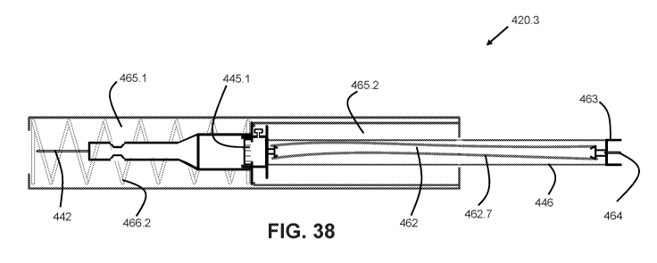
FIGS. 36-38 show views of an apparatus 420 according to another embodiment of
the present invention. It is recognized that apparatus 420 uses a common
numbering
system with apparatus 220 or 320, with like features being identified by
similar digits XX.X.
28
CA 03071379 2020-01-28
WO 2019/023599 PCT/US2018/044125
Still other similar features can be identified visually within these figures.
Some of the
differences between devices 420, 320, and 220 will now be explained.
FIG. 36 shows an apparatus 420 in the fully extended, unused state. Apparatus
420
is similar to apparatus 220, except that flexible tube 462 is contained within
a rigid
protective sheath 446. Sheath 446 is coupled to the end of needle assembly
440, and
moves from the extended to retracted positions concurrently with needle
assembly 440. As
shown in FIG. 36 in the fully extended position, sheath 446 is contained
entirely within the
first and second compartments of housing assembly 460. It is further
understood that in yet
other embodiments there is a single tube that provides both fluid
communication between
needles 442 and 464, and further a fixed connection between the body of the
needle
assembly and the fitting 463.
The proximal end of sheath 446 incorporates a fitting 463 adapted and
configured
for easy coupling and decoupling from the blood collection vial. In some
embodiments,
fitting 463 includes a second cannula 464 which pierces a seal on the inlet of
the collection
vial. This hollow second cannula 464 establishes fluid communication between
the interior
of the vial (which may be evacuated) and the hollow cannula 442. FIG. 37 shows
the
apparatus 420.2 in the extended state, with the end cap 467 removed. After
removal of the
end cap 467, the blood collection vial can be attached to fitting 463.
FIG. 37 shows device 420 in the extended position, but after usage (similar to
FIG.
32). Further, adaptor 461 is not shown for sake of clarity. Needle assembly
440 is shown in
the extended position, although being biased to the retracted position by a
coil spring 466
in a compressed state 466.1.
FIG. 38 shows device 420.3 in the fully retracted, post-usage state. As shown
previously with apparatus 220.3, the first cannula 442 is contained entirely
within the first
compartment 465.1. Because of the connection of sheath 446 to needle assembly
440, this
retractive movement results in sheath 446 extending rearward, with fitting 463
now
extending out of compartment 465.2 at the proximal end of device 420.3. In
some
embodiments, cap 467 can be placed back onto fitting 463, so as to prevent
inadvertent
contact with second cannula 464.
Various aspects of different embodiments of the present invention are
expressed in
paragraphs X1, X2, and X3 as follows:
29
CA 03071379 2020-01-28
WO 2019/023599 PCT/US2018/044125
X1 . One aspect of the present invention pertains to a device for
collecting blood
in a container and connecting to intravenous tubing. The device preferably
includes a first
distal compartment generally aligned with a second proximal compartment. The
device
preferably includes a needle assembly having proximal and distal ends and
including a
needle defining a first lumen and having a sharp tip on the distal end, the
needle assembly
being slidable within the first compartment. The device preferably includes a
spring biasing
the needle assembly toward a retracted position. The device preferably
includes a tube in
fluid communication with the first lumen and extending toward the second
proximal
compartment. The device preferably includes a sheath containing therein the
tube, the
sheath being slidable with the needle assembly; and a catheter assembly having
a flexible
second lumen supported by a body, the second lumen surrounding at least a
portion of the
needle.
X2. Another aspect of the present invention pertains to a device for
collecting
blood in a container and connecting to intravenous tubing. The device
preferably includes a
first compartment and a second compartment. The device preferably includes a
needle
assembly having proximal and distal ends and including a needle being slidable
between
an extended position in which the sharp tip is external to the housing and a
retracted
position. The device preferably includes means for biasing the needle assembly
to either
the retracted or extended position. The device preferably includes a fluid
conduit in fluid
communication with the first lumen and extending toward the proximal end of
the device.
And a catheter assembly having a flexible second lumen supported by a body,
the second
lumen surrounding at least a portion of the needle, the catheter assembly
including a pair
of opposing flexible wings adapted and configured for grasping by a user.
X3. Yet another aspect of the present invention pertains to a device for
collecting
blood in a container and connecting to intravenous tubing. The device
preferably includes a
first compartment and a second compartment. The device preferably includes a
needle
defining a first lumen and having a sharp tip on the distal end, the needle
being slidable
between two positions. The device preferably includesa flexible member biasing
the needle
assembly toward a position. The device preferably includes a tube in fluid
communication
with the first, the proximal end of the tube having a fitting adapted and
configured for
readily attaching and releasing the container for collecting blood; and a
catheter assembly
CA 03071379 2020-01-28
WO 2019/023599 PCT/US2018/044125
having a second flexible lumen supported by a body, the second lumen
surrounding at
least a portion of the needle.
Yet other embodiments pertain to any of the previous statements X1, X2, or X3,
which are combined with one or more of the following other aspects. It is also
understood
that any of the aforementioned X paragraphs include listings of individual
features that can
be combined with individual features of other X paragraphs.
Which further comprises a gas purging valve that permits expulsion of trapped
gas
within the needle assembly.
Which further comprises a filter having an inlet in fluid communication with
the first
lumen and an outlet in fluid communication with the inlet of the tube.
Which further comprises means for retaining the needle assembly in the ready
to
use position.
Which further comprises a readily removable cap covering the proximal end of
the
device.
Wherein the spring is a coil spring that is either compressed when the first
needle is
in the extended position, or extended when the first needle is in the extended
position.
Wherein the catheter assembly includes a fitting for readily attaching and
releasing
intravenous tubing, including luer-type fittings, threaded fittings, bayonet-
type fittings, or the
like.
Wherein the housing includes an abutment that limits the sliding of the needle
assembly to the retracted position; preferably the abutment being a surface of
a sliding
component or a surface of a stationary component into which the sliding
component slides.
Wherein the needle assembly abuts the distal end of the second compartment in
the
retracted position.
Wherein the catheter assembly includes a pair of opposing flexible wings
adapted
and configured for grasping by a user.
Which further comprises a rigid sheath containing therein the first tube, the
rigid
sheath preferably being slidable with the needle assembly from the extended
position to
the retraced position. Preferably the rigid sheath has a length that is about
the same as the
length of the tube contained therein, although other embodiments contemplate
rigid
sheaths that are longer or shorter than the tube contained therein.
31
CA 03071379 2020-01-28
WO 2019/023599 PCT/US2018/044125
Which further comprises a rigid sheath surrounding the tube, the rigid sheath
and
the tube being located within the housing when the needle assembly is in the
extended
position, and the proximal end of the tubing and the proximal end of the rigid
sheath
extending out of the second compartment when the needle assembly is in the
retracted
position.
Wherein the tube comprises a flexible material and has a free length that is
less
than the length of the second compartment, the tube being extended beyond the
free
length when the needle assembly is in the extended position.
Wherein the tube comprises a flexible material and has a free length that is
greater
than the length of the second compartment, the tube being compressed from the
free
length and fitting within the second compartment when the needle assembly is
in the
extended position.
Wherein the tube has a free length that is greater than the combined length of
first
compartment and the second compartment.
Wherein the tube has a first length prior to use of the device, a second
length after
the device has been used and the tube is free to extend at the distal end of
the device, and
the difference between the first length and the second length is about the
same as the
distance traveled by the sliding needle assembly when moving from the extended
position
to the retracted position.
Which further preferably comprises a fitting on the end of the tube within the
second
compartment, the fitting being adapted and configured for readily attaching
and releasing
the container for collecting blood. The fitting can be of any type suitable
for blood collection
vials, including screw-type, press to fit-type, bayonet-type, and others.
Wherein the proximal end of the tube has a fitting adapted and configured for
readily
attaching and releasing the container for collecting blood.
Wherein the fitting includes a second needle adapted and configured for
puncturing
a seal of the container for collecting blood. Preferably, the second needle
extends a short
distance from the fitting, and the tip of the needle is safely enclosed by the
endcap when
the device is in the ready to use configuration.
32
CA 03071379 2020-01-28
WO 2019/023599 PCT/US2018/044125
Wherein the needle is a first needle, and which further comprises a second
needle
in fluid communication with the tube and adapted and configured for receiving
blood from
the tube, the second needle being located on the proximal end of the device.
Wherein the first needle slides linearly along an axis from the extended
position to
the retracted position.
Wherein the sharp tip of the first needle extends out from the distal end of
the
second lumen when the first needle is in the extended position.
While the inventions have been illustrated and described in detail in the
drawings
and foregoing description, the same is to be considered as illustrative and
not restrictive in
character, it being understood that only certain embodiments have been shown
and
described and that all changes and modifications that come within the spirit
of the invention
are desired to be protected.
33