Language selection

Search

Patent 3071473 Summary

Third-party information liability

Some of the information on this Web page has been provided by external sources. The Government of Canada is not responsible for the accuracy, reliability or currency of the information supplied by external sources. Users wishing to rely upon this information should consult directly with the source of the information. Content provided by external sources is not subject to official languages, privacy and accessibility requirements.

Claims and Abstract availability

Any discrepancies in the text and image of the Claims and Abstract are due to differing posting times. Text of the Claims and Abstract are posted:

  • At the time the application is open to public inspection;
  • At the time of issue of the patent (grant).
(12) Patent: (11) CA 3071473
(54) English Title: ENDOSCOPE DEVICE
(54) French Title: DISPOSITIF ENDOSCOPIQUE
Status: Granted
Bibliographic Data
(51) International Patent Classification (IPC):
  • A61B 1/00 (2006.01)
(72) Inventors :
  • RIES, WOLFGANG (Germany)
  • GERMANN, MARTIN (Germany)
(73) Owners :
  • JOIMAX GMBH (Germany)
(71) Applicants :
  • JOIMAX GMBH (Germany)
(74) Agent: GOWLING WLG (CANADA) LLP
(74) Associate agent:
(45) Issued: 2024-04-09
(86) PCT Filing Date: 2018-08-20
(87) Open to Public Inspection: 2019-03-07
Examination requested: 2023-08-09
Availability of licence: N/A
(25) Language of filing: English

Patent Cooperation Treaty (PCT): Yes
(86) PCT Filing Number: PCT/EP2018/000405
(87) International Publication Number: WO2019/042578
(85) National Entry: 2020-01-29

(30) Application Priority Data:
Application No. Country/Territory Date
10 2017 008 148.4 Germany 2017-08-29
20 2017 004 822.1 Germany 2017-09-15

Abstracts

English Abstract


The invention proposes, for determining the precise
orientation and positioning of an endoscope in an
electromagnetic field, an endoscope device having a proximal
insertion head and having a shaft extending distally
therefrom having a centre axis, which extends with at least
one oblong lumen through the device, which is characterized
by a sensor rod having at least two sensor coils arranged
with finite spacing in relation to one another in the
longitudinal direction, which are oriented in relation to
one another at a finite angle.


French Abstract

L'invention concerne un dispositif endoscopique destiné à la détermination de l'orientation et du positionnement précis d'un endoscope dans un champ électromagnétique, comprenant une tête d'insertion proximale et, s'étendant distalement de cette dernière, un tube doté d'un axe central qui s'étend avec au moins un lumen allongé à travers le dispositif, caractérisé en ce qu'une tige de détection est pourvue d'au moins deux bobines de détection qui sont mutuellement disposées à une distance finie dans la direction longitudinale et qui sont orientées l'une par rapport à l'autre en formant un angle fini.

Claims

Note: Claims are shown in the official language in which they were submitted.


7
Patent Claims
1. An endoscope device comprising:
a proximal insertion head with an attachment of the
insertion head;
a shaft extending distally from the insertion head, the
shaft having a center axis;
at least one elongate lumen extending through the device;
and
a sensor rod having at least two sensor coils arranged at a
finite spacing in relation to one another in a sensor rod
longitudinal direction, the at least two sensor coils being
oriented in relation to one another at a finite angle and
wherein the at least two sensor coils comprise a first sensor
coil oriented in parallel to the center axis and a second sensor
coil located in the attachment of the insertion head and
oriented at the finite angle in relation to the center axis.
2. An endoscope device according to claim 1, wherein the first
sensor coil is arranged in the shaft.
3. An endoscope device according to claim 1, wherein the rod
bearing the two sensor coils extends through the lumen.
4. An endoscope device according claim 1, further comprising a
holder, wherein the holder is attachable to the attachment of
the insertion head and the rod is connected axially fixed to the
holder.
5. An endoscope device according to claim 4, wherein the

8
attachment of the insertion head and the holder comprise
corresponding Luer adapter parts facing toward one another
forming a Luer adapter, and the attachment and the holder are
fixedly connectable to one another by the Luer adapter.
6. An endoscope device according to claim 1, wherein the
sensor rod further comprises a connecting wire that extends from
each sensor coil through the rod up to a sensor rod proximal end
for connection to an analysis unit.
7. An endoscope device comprising:
a proximal insertion head with an attachment of the
insertion head;
a shaft extending distally from the insertion head, the
shaft having a center axis, wherein the attachment of the
insertion head extends at a finite angle with respect to the
center axis;
at least one elongate lumen extending through the device,
the lumen having a lumen section that extends in the attachment
of the insertion head and at the finite angle in relation to the
center axis;
a sensor rod;
a first sensor coil supported by the sensor rod and located
at a distal end of the sensor rod, inside the shaft, and
extending in parallel to the center axis and oriented in
parallel to the center axis; and
a second sensor coil supported by the sensor rod and
arranged with a spacing in relation to the first sensor coil at
the sensor rod and located in the attachment of the insertion
head, the attachment of the insertion head extending at the
finite angle in relation to the center axis, wherein an
orientation or extension of the second sensor coil in the

9
attachment of the insertion head also encloses the finite angle
in relation to the center axis and the first sensor coil and the
second sensor coil are not parallel to one another and the
second sensor coil is oriented at the finite angle in relation
to the first sensor coil, wherein the rod bearing the two sensor
coils extends through the lumen with the second sensor coil
arranged in the lumen section extending at the finite angle in
relation to the center axis.
8. An endoscope device according claim 7, further comprising a
holder attachable to the attachment of the insertion head,
wherein the rod is axially fixed to the holder whereby upon
attaching the holder to the attachment, the first sensor coil is
located at a first predefined position inside the shaft and the
second sensor coil is located at a second predefined position
inside the lumen section extending at the finite angle in
relation to the center axis.
9. An endoscope device according to claim 8, wherein the
attachment of the insertion head and the holder comprise
corresponding Luer adapter parts facing toward one another
forming a Luer adapter, and the attachment and the holder are
fixedly connectable to one another by the Luer adapter to set
the defined positions in a fixed state.
10. An endoscope device according to claim 7, wherein the
sensor rod further comprises a connecting wire that extends from
each sensor coil through the rod up to a sensor rod proximal end
for connection to an analysis unit.
11. An endoscope device comprising:

10
a proximal insertion head with an attachment of the
insertion head;
a shaft extending distally from the insertion head, the
shaft having a center axis;
at least one elongate lumen extending through the device,
the lumen having a lumen section that extends in the attachment
of the insertion head and at a finite angle in relation to the
center axis;
a sensor rod;
a first sensor coil supported by and located at the distal
end of the sensor rod, inside the shaft, and extending in
parallel to the center axis and oriented in parallel to the
center axis;
a second sensor coil supported by and arranged with a
spacing in relation to the first sensor coil at the sensor rod
and located in the attachment of the insertion head, the
attachment of the insertion head extending at the finite angle
in relation to the center axis, wherein an orientation or
extension of the second sensor coil in the attachment of the
insertion head also encloses the finite angle in relation to the
center axis and the first sensor coil and the second sensor coil
are not parallel to one another and the second sensor coil is
oriented at the finite angle in relation to the first sensor
coil, wherein the rod bearing the two sensor coils extends
through the lumen with the second sensor coil arranged in the
lumen section extending at the finite angle in relation to the
center axis; and
a holder attachable to the attachment of the insertion
head, wherein the rod is axially fixed to the holder whereby
upon attaching the holder to the attachment, the first sensor
coil is located at a first predefined position inside the shaft
and the second sensor coil is located at a second predefined

11
position inside the lumen section extending at the finite angle
in relation to the center axis.
12. An endoscope device according to claim 11, wherein the
attachment of the insertion head and the holder comprise
corresponding Luer adapter parts facing toward one another
forming a Luer adapter, and the attachment and the holder are
fixedly connectable to one another by the Luer adapter to set
the defined positions in a fixed state.
13. An endoscope device according to claim 12, wherein the
sensor rod further comprises a connecting wire that extends from
each sensor coil through the rod up to a sensor rod proximal end
for connection to an analysis unit.

Description

Note: Descriptions are shown in the official language in which they were submitted.


CA 03071473 2020-01-29
- 1 -
Endoscope device
The invention relates to an endoscope device having a
proximal insertion head and having a shaft extending
distally therefrom having a centre axis, which extends
with at least one oblong lumen through the device.
Minimally invasive operations are already carried out
presently by means of navigation-assisted operating
methods. Different navigation systems are used for this
purpose. Active and passive systems are used. In active
systems, a part introduced into the body of a patient,
such as an instrument or surgical tool, is provided
with a transmitter, via which the position of the
instrument or tool, in particular the distal end
located at the engagement location, may be externally
determined. In passive systems, a field is generated,
which is detected via a sensor, whereby the position
and alignment of the instrument or surgical tool, in
particular its distal end, can in turn be detected
directly or indirectly. Direct detection of the distal
end of a surgical part includes the arrangement of the
sensor on the distal end of the part itself; indirect
detection includes the fixed rigid attachment of the
sensor in a defined point, in particular axial
position, on the surgical part. Inferences about the
position and possibly the orientation of the distal end
can be seen on the basis of the measured sensor signal.
In passive navigation, in particular electromagnetic
navigation has proven itself, in which an
electromagnetic field is generated externally around
the operation region, for example, by a generator of an
electromagnetic field in a cushion on which the patient

CA 03071473 2020-01-29
- 2 -
lies. Coil-type sensors installed in the surgical part
enable the locating of the instruments, whereupon a
representation can be performed in CT or MRT images.
This method does not include a radiation exposure and
thus overall reduces the radiation exposure, also due
to a reduced use of x-rays. The image quality is not
impaired, nor can sensors be concealed, since they are
not optical sensors. The freedom of movement of the
operator is not restricted, as is the case with optical
systems. The work of the operator is significantly
facilitated.
The invention is based on the object of providing a
device, in which the location and orientation of an
endoscope, in particular of the distal end, can be
precisely determined using the above-described system
while avoiding the mentioned disadvantages.
The mentioned object is achieved according to the
invention by an endoscope device of the type mentioned
at the outset, which is characterized by a sensor rod
having at least two sensor coils arranged in the
longitudinal direction at a finite spacing in relation
to one another, which are oriented in relation to one
another at a finite angle.
Because the two sensor coils are arranged at a finite
angle in relation to one another in the endoscope
device, because of the different arrangement, the
orientation of the endoscope device in the magnetic
field of the detection system and thus also in space
can be precisely determined.

CA 03071473 2020-01-29
- 3 -
In one preferred embodiment, it is provided in this
case that the first sensor coil is oriented in parallel
in relation to the centre axis and the second sensor
coil is oriented at a finite angle in relation to the
centre axis, wherein in particular the first sensor
coil is arranged in the shaft and the second sensor
coil is arranged in an attachment of the insertion
head.
In one refinement, it is provided that the rod bearing
the two sensor coils extends through a lumen of the
device, wherein in particular the rod is connected in
an axially fixed manner to a holder which is attachable
to the attachment of the insertion head. In this way,
the location of the one sensor coil, in particular the
first sensor coil, is accurately defined in the
endoscope device and therefore on the basis of the
determination of the location of the first sensor coil
by the detection system in the magnetic field, the
exact determination of the precise location of the
distal end of the sensor device and thus of the working
location is also possible, since the spacing of the
(first) sensor coil from the distal end of the
endoscope shaft is fixedly predetermined in this
manner.
Due to the fixed connection of the rod bearing the
sensor coil to a separable holder, furthermore, on the
one hand, after positioning, the rod bearing the sensor
coil is removed, and the lumen occupied thereby for the
positioning can be released for other usage purposes.
Furthermore, the sensor device formed by the rod having
the sensor coils is thus separable from the actual

4
endoscope and can be used in another way. This also
enables simpler sterilization.
Further advantages and features of the invention result
from the claims and from the following description, in
which an exemplary embodiment of the invention is
explained in detail with reference to the drawing.
In this case, the single figure is a longitudinal section
view of an endoscope device according to the invention.
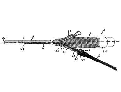
The endoscope device 1 according to the invention
comprises in the illustrated exemplary embodiment a
proximal insertion head 2 and a shaft 3 extending
distally therefrom. In the illustrated exemplary
embodiment, the insertion head 2 comprises three branches
or attachments 2.1 to 2.3, namely one attachment 2.1 for
introducing a light guide from the proximal end of the
device 1 up to its distal end and also two attachments
2.2, 2.3, which each also comprise a lumen or a channel
which extends from the distal end of the elongated shaft
3 with a centre axis A up to an exit from the respective
adapter 2.2, 2.3.
A rod 4, which bears two coils 4.1 and 4.2 with spacing
in its longitudinal extension, extends through the lumen
2.3.1 of the attachment 2.3. The rod can be formed as a
solid or flexible rod or also as a rod coiled in a helix.
A connecting wire (not shown) extends from each of the
coils 4.1, 4.2 in the proximal direction up to a
respective proximal connecting or contact end of the
respective wire, possibly in the
Date Recite/Date Received 2023-10-11

CA 03071473 2020-01-29
- 5 -
formation of a plug for connection of the wires to an
analysis unit (not shown).
The coil 4.1 is located at the distal end of the rod 4
and thus inside the shaft 3 extending in parallel to
its centre axis A and is therefore also oriented in
parallel to the centre axis A or axially-parallel. The
coil 4.2 arranged with spacing in relation to the coil
4.1 on the rod 4 is located in the attachment 2.3 of
the insertion head extending at a finite angle in
relation to the centre axis A. In that the lumen
section of the lumen 2.3.1 also extends at a finite
angle in relation to the centre axis A, the orientation
or extension of the coil 4.2 in the attachment 2.3 also
encloses a finite angle in relation to the centre axis
A. The two coils 4.1, 4.2 are therefore not parallel to
one another, but rather are oriented at a finite angle
in relation to one another.
In the case of an externally applied inhomogeneous
electromagnetic field, in which the coils 4.1, 4.2 are
located, these coils therefore perceive the field
differently and transmit different signals to the
analysis unit. Due to this different orientation of the
coils 4.1, 4.2, the orientation of the endoscope device
in the electromagnetic field and thus in space can
therefore be exactly determined.
The rod 4 is arranged axially fixed in a holder 5. The
holder 5 and the attachment 2.3 are connectable to one
another by.a Luer adapter 6, wherein each of the parts
bears a respective part 6.1, 6.2 corresponding to one
another of the Luer adapter 6, so that the attachment

CA 03071473 2020-01-29
-6-
2.3 and the holder 5 can be fixedly connected to one
another like a bayonet by the Luer adapter 6 in a way
known per se. The location of the holder 5 in relation
to the attachment 2.3 and also in relation to the head
2 and the shaft 3 of the endoscope device 1 is thus
defined in the fastened state. Since the shaft 4, as
stated, is arranged axially fixed in the holder 5, the
longitudinal position of the coils 4.1, 4.2 and in
particular of the distal coil 4.1 in the shaft 3 and
thus the endoscope device 1 is therefore also defined
and therefore the spacing, in particular the axial
spacing of the coil 4.1 from the distal end 3.1 of the
shaft 3, is also defined. By way of the sensor signal
of the coil 4.1 in the applied electromagnetic field,
its location and, because of the fixed spacing in
relation to the distal end 3.1 of the shaft 3, the
location of the distal end 3.1 of the shaft 3 in the
electromagnetic field and thus in space can also thus
be determined.
An operator therefore recognizes, on the basis of the
analysis of the sensor signals and an image display on
a display screen of the analysis unit, the position of
the distal end 3.1 of the shaft 3 and thus also of the
endoscope device 1 accurately and thus knows where
exactly they are working with their instruments, which
they possibly introduce into other lumens of the
endoscope device and through them.

Representative Drawing
A single figure which represents the drawing illustrating the invention.
Administrative Status

For a clearer understanding of the status of the application/patent presented on this page, the site Disclaimer , as well as the definitions for Patent , Administrative Status , Maintenance Fee  and Payment History  should be consulted.

Administrative Status

Title Date
Forecasted Issue Date 2024-04-09
(86) PCT Filing Date 2018-08-20
(87) PCT Publication Date 2019-03-07
(85) National Entry 2020-01-29
Examination Requested 2023-08-09
(45) Issued 2024-04-09

Abandonment History

There is no abandonment history.

Maintenance Fee

Last Payment of $210.51 was received on 2023-08-02


 Upcoming maintenance fee amounts

Description Date Amount
Next Payment if small entity fee 2024-08-20 $100.00
Next Payment if standard fee 2024-08-20 $277.00

Note : If the full payment has not been received on or before the date indicated, a further fee may be required which may be one of the following

  • the reinstatement fee;
  • the late payment fee; or
  • additional fee to reverse deemed expiry.

Patent fees are adjusted on the 1st of January every year. The amounts above are the current amounts if received by December 31 of the current year.
Please refer to the CIPO Patent Fees web page to see all current fee amounts.

Payment History

Fee Type Anniversary Year Due Date Amount Paid Paid Date
Application Fee 2020-01-29 $400.00 2020-01-29
Maintenance Fee - Application - New Act 2 2020-08-20 $100.00 2020-07-14
Maintenance Fee - Application - New Act 3 2021-08-20 $100.00 2021-07-28
Maintenance Fee - Application - New Act 4 2022-08-22 $100.00 2022-07-27
Maintenance Fee - Application - New Act 5 2023-08-21 $210.51 2023-08-02
Request for Examination 2023-08-21 $816.00 2023-08-09
Final Fee $416.00 2024-02-27
Owners on Record

Note: Records showing the ownership history in alphabetical order.

Current Owners on Record
JOIMAX GMBH
Past Owners on Record
None
Past Owners that do not appear in the "Owners on Record" listing will appear in other documentation within the application.
Documents

To view selected files, please enter reCAPTCHA code :


To view images, click a link in the Document Description column. To download the documents, select one or more checkboxes in the first column and then click the "Download Selected in PDF format (Zip Archive)" or the "Download Selected as Single PDF" button.

List of published and non-published patent-specific documents on the CPD .

If you have any difficulty accessing content, you can call the Client Service Centre at 1-866-997-1936 or send them an e-mail at CIPO Client Service Centre.


Document
Description 
Date
(yyyy-mm-dd) 
Number of pages   Size of Image (KB) 
Abstract 2020-01-29 1 13
Claims 2020-01-29 2 43
Drawings 2020-01-29 1 24
Description 2020-01-29 6 204
Representative Drawing 2020-01-29 1 29
International Search Report 2020-01-29 5 197
Amendment - Abstract 2020-01-29 2 89
Declaration 2020-01-29 1 56
National Entry Request 2020-01-29 3 76
Cover Page 2020-03-20 1 49
Electronic Grant Certificate 2024-04-09 1 2,527
Final Fee 2024-02-27 3 83
Representative Drawing 2024-03-08 1 16
Cover Page 2024-03-08 1 48
Request for Examination / PPH Request / Amendment 2023-08-09 19 820
Claims 2023-08-09 5 262
Description 2023-08-09 6 324
PPH OEE 2023-08-09 4 387
Examiner Requisition 2023-08-22 4 205
Amendment 2023-10-11 11 321
Description 2023-10-11 6 368
Claims 2023-10-11 5 265