Note: Descriptions are shown in the official language in which they were submitted.
CA 03071628 2020-01-30
WO 2018/144631
PCT/US2018/016287
STIMULATOR SYSTEMS AND METHODS FOR OBSTRUCTIVE SLEEP APNEA
RELATED APPLICATION DATA
[0001] Pursuant to 35 U.S.C. 119(e), this application claims the benefit of
U.S.
Provisional Patent Application 62/453,311, filed February 1, 2017, which is
expressly
incorporated herein by reference.
FIELD OF THE INVENTION
[0002] The present invention relates to systems and methods for the treatment
of
obstructive sleep apnea (OSA).
BACKGROUND
[0003] OSA is a highly prevalent sleep disorder that is caused by the collapse
of or
increase in the resistance of the pharyngeal airway, often resulting from
tongue
obstruction. The obstruction of the upper airway is mainly caused by reduced
genioglossus muscle activity during the deeper states of NREM sleep.
Obstruction
of the upper airway causes breathing to pause during sleep. Cessation of
breathing
causes a decrease in the blood oxygen saturation level, which is eventually
corrected when the person wakes up and resumes breathing. The long-term
effects
of OSA include high blood pressure, heart failure, strokes, diabetes,
headaches, and
general daytime sleepiness and memory loss, among other symptoms.
[0004] OSA is extremely common, having a similar prevalence as diabetes or
asthma. Over 100 million people worldwide suffer from OSA, with about 25% of
those being treated. Continuous Positive Airway Pressure (CPAP) is the usual
established therapy for people who suffer from OSA. More than five million
patients
own a CPAP machine in North America, but many do not comply with use of these
1
CA 03071628 2020-01-30
WO 2018/144631
PCT/US2018/016287
machines, because they cover the mouth and nose and, hence, are cumbersome
and uncomfortable.
[0005] The use of neurostimulators to open the upper airway has been explored
by
several companies as a treatment for alleviating apneic events. Such therapy
involves stimulating the nerve fascicles of the hypoglossal nerve (HGN) that
innervate the intrinsic and extrinsic muscles of the tongue in a manner that
prevents
retraction of the tongue, which would otherwise close the upper airway during
inspiration of the respiratory cycle.
[0006] ImThera Medical is currently in FDA clinical trials for a stimulator
system that
is used to stimulate the trunk of the HGN with a nerve cuff electrode. The
stimulation
system does not provide a sensor or sensing, and therefore, the stimulation
delivered to the HGN trunk is not synchronized to the respiratory cycle. Thus,
the
tongue and other muscles that are innervated by nerve fascicles of the HGN
trunk
are stimulated irrespective of the respiratory cycle.
[0007] The rationale for this treatment method appears to be that it is enough
simply
to tone the tongue muscle and other nearby muscles, so that the tongue muscle
does not retract in a manner that would cause OSA. The belief is that it is
not
necessary to specifically target the protraction (i.e., anterior movement) of
the tongue
muscle and to synchronize the occurrence of tongue protraction when it is most
needed, i.e., during inspiration. The nerve cuff electrode of the ImThera
Medical
system has multiple electrode contacts helically surrounding the proximal part
of the
HGN nerve trunk. So, instead, each electrode contact delivers stimulation in a
sequential order to the HGN trunk. For example, if a three-electrode contact
nerve
cuff is used, electrode contact #1 stimulates, then stops, electrode contact
#2
stimulates, then stops, electrode contact #3 stimulates, then stops, then
electrode
2
CA 03071628 2020-01-30
WO 2018/144631
PCT/US2018/016287
contact #1 stimulates, then stops and so on. Since all or most electrode
contacts
deliver stimulation, there is no selection process to choose the best one or
two
electrode contacts that is finally used to deliver the best stimulation to
alleviate sleep
apnea.
[0008] A disadvantage of the ImThera Medical system is that it does not target
tongue protraction coincident with the inspiration phase of respiration, since
it does
not have a sensor to enable synchronization of stimulation to the respiratory
cycle.
Since there is no attempt to synchronize the stimulation with the respiratory
cycle,
the tongue protraction does not occur when it would appear to help the
most¨during
inspiration when OSA can occur. Also, because the HGN trunk contains nerve
fascicles that innervate muscles other than the muscle that extend the tongue,
the
lmthera Medical method of stimulation at the HGN trunk does not just target
the
specific protrusor muscles of the tongue muscle, but other muscles that are
not
targeted. Thus, stimulating the HGN trunk in an arbitrary manner may recruit
other
nerve fascicles of the HGN trunk that may not contribute to the protraction of
the
tongue.
[0009] Another company, Inspire Medical Systems, Inc., does offer a
stimulation
system with a sensor, and therefore does attempt to time the onset of
stimulation to
the breathing cycle. This system, which is FDA approved for sale in the United
States since April 2010, uses a simple, bipolar electrode (two electrode
contacts
only) within a nerve cuff electrode and implants the electrode at the branch
of the
HGN that is responsible for protruding the tongue. A simple, two-electrode
contact
or three-electrode contact cuff electrode can be used at the branch nerve,
unlike the
HGN trunk, because at the distal branch location, the nerve fascicles
generally
innervate the specific tongue protrusor muscle and not other muscles.
3
CA 03071628 2020-01-30
WO 2018/144631
PCT/US2018/016287
[0010] However, implanting the electrode at a branch of the HGN takes
additional
surgery time, which increases trauma to the patient and increases the
substantial
expense of operating room time. By attaching the nerve cuff electrode to the
proximal section of the main trunk of the HGN, compared to placing the nerve
cuff
electrode at the more distal end of the HGN, it estimated that the surgical
time will be
reduced by approximately one hour. Even more importantly, because the branch
nerve is small and more difficult to isolate than the HGN trunk, implanting a
nerve
cuff electrode at the branch site demands heightened expertise from the
otolaryngologist/Ear Nose and Throat (ENT) surgeon or neurosurgeon, which
significantly increases the chance for error and surgical risks. Furthermore,
because
the distal location of the HGN has a smaller diameter of nerves, and hence the
required electrodes need to be smaller, the smaller nerve cuff electrode may
be
more difficult to manufacture.
[0011] Thus, it is certainly desirable to implant the nerve cuff electrode at
the trunk of
the hypoglossal nerve. However, one must then deal with the fact that the
target
nerve fascicles may be near the center of the nerve trunk and are not easily
isolated
and stimulated, while at the same time avoiding stimulating other non-targeted
fascicles in the same nerve trunk.
[0012] Furthermore, a pressure sensor is connected to neurostimulator of the
Inspire
system by a lead, thereby allowing the pressure sensor to be placed remotely
from
the implanted site of the neurostimulator. However, the fact that the pressure
sensor
has a lead connected to the stimulator necessitates some additional surgery,
because the sensor lead is another appendage that must be implanted.
[0013] There, thus, remains a need for improved systems and methods for
selectively recruiting only the fascicles of the hypoglossal nerve in
synchronization
4
CA 03071628 2020-01-30
WO 2018/144631
PCT/US2018/016287
with the respiratory cycle for treating OSA of a patient, while minimizing the
surgery
time and effort required to implant the neurostimulation components in the
patient.
SUMMARY
[0014] In accordance with a first aspect of the present inventions, an
electrode lead
comprises an elongated lead body having a proximal end and a distal end, and
at
least three connector contacts affixed to the proximal end of the lead body.
In one
embodiment, the lead body has at least one portion that is S-shaped to provide
strain relief. The electrode lead further comprises a biologically compatible,
flexible,
electrically insulative cuff body affixed to the distal end of the lead body.
The cuff
body is pre-shaped to transition from an unfurled state to a furled state,
wherein the
cuff body, when in the furled state has an inner surface for contacting a
nerve. In
one embodiment, the inner surface of the furled cuff body has a diameter in
the
range of 2.5 mm to 4.0 mm. In another embodiment, the cuff body is self-
adjusting,
such that the cuff body accommodates different sized nerve diameters, and
diameter
changes overtime.
[0015] The electrode lead further comprises at least three axially aligned
electrode
contacts circumferentially disposed along the inner surface of the cuff body
when in
the furled state, and at least three electrical conductors extending through
the lead
body respectively between the at least three connector contacts and the
electrode
contacts. In one embodiment, when the cuff body is in the furled state, the
electrode
contacts circumferentially span at least a 180-degree arc around the inner
surface of
the cuff body. In another embodiment, when the cuff body is in the furled
state, the
electrode contacts circumferentially span at least a 270-degree arc around the
inner
surface of the cuff body. In still another embodiment, when the cuff body is
in the
unfurled state, a center-to-center spacing of each pair of adjacent ones of
electrode
CA 03071628 2020-01-30
WO 2018/144631
PCT/US2018/016287
contacts is equal to or less than twice the width of each electrode contact of
the
respective pair of electrode contacts.
[0016] A neurostimulation system may comprise the electrode lead described
above,
and a neurostimulator comprising a connector configured for receiving the
proximal
contacts of the electrode lead, stimulation circuitry configured for
generating an
electrical pulse train, and control circuitry configured for causing the
stimulation
circuitry to deliver the electrical pulse train to at least one of the
electrode contacts of
the electrode lead. A method of using the electrode lead described above may
comprise maintaining the cuff body in the unfurled state while placing the
cuff body in
contact with the nerve, placing the cuff body from the unfurled state into the
furled
state, such that the cuff body wraps around the nerve, and delivering an
electrical
pulse train to at least one of the electrode contacts of the electrode lead,
thereby
stimulating the nerve.
[0017] In accordance with a second aspect of the present inventions, a
neurostimulation system comprises an electrode lead having a lead body. In one
embodiment, the lead body has at least one portion that is S-shaped to provide
strain relief. The electrode lead further comprises a biologically compatible
electrically insulative cuff body affixed to the distal end of the lead body.
The cuff
body is pre-shaped and flexible to transition from an unfurled state to a
furled state.
The cuff body, when in the furled state has an inner surface for contacting a
nerve.
In one embodiment, the inner surface of the furled cuff body has a diameter in
the
range of 2.5 mm to 4.0 mm. In another embodiment, the cuff body is self-
adjusting,
such that the cuff body accommodates different sized nerve diameters, and
diameter
changes overtime.
6
CA 03071628 2020-01-30
WO 2018/144631
PCT/US2018/016287
[0018] The electrode lead further comprises at least three axially aligned
electrode
contacts circumferentially disposed along the inner surface of the cuff body
when in
the furled state. In one embodiment, when the cuff body is in the furled
state, the
electrode contacts circumferentially span at least a 180-degree arc around the
inner
surface of the cuff body. In another embodiment, when the cuff body is in the
furled
state, the electrode contacts circumferentially span at least a 270-degree arc
around
the inner surface of the cuff body. In still another embodiment, when the cuff
body is
in the unfurled state, a center-to-center spacing of each pair of adjacent
ones of
electrode contacts is equal to or less than twice the width of each electrode
contact
of the respective pair of electrode contacts.
[0019] The neurostimulation system further comprises a neurostimulator
configured
for delivering an electrical pulse train to at least one of the electrode
contacts of the
electrode lead. By way of example, the electrical pulse train may have an
initial,
preconditioning current or voltage amplitude and a subsequent higher
stimulating
current or voltage amplitude. In one embodiment, the electrode contacts
comprise a
pair of adjacent ones of the electrode contacts, and the neurostimulator is
configured
for delivering the electrical pulse train between the pair of adjacent ones of
the
electrode contacts in a bipolar mode. In another embodiment, the
neurostimulator is
further configured for sensing physiological artifacts that are caused by
respiration,
and delivering the electrical pulse train to the electrode contacts in
synchronization
with a respiratory cycle based on the sensed physiological artifacts. As one
example, the neurostimulator may be configured for determining the next
projected
onset of an inspiratory phase of the respiratory cycle based on the sensed
physiological artifacts, and delivering the electrical pulse train to the at
least one
electrode contact immediately before, at, or right after the next projected
onset of the
7
CA 03071628 2020-01-30
WO 2018/144631
PCT/US2018/016287
inspiratory phase of the respiratory cycle. In another embodiment, the
neurostimulator is configured for storing data representative of the
physiological
artifacts sensed by the sensing circuitry.
[0020] The neurostimulation system may optionally comprise a clinician
programmer
configured for selecting the electrode contacts, and transcutaneously
communicating
with the neurostimulator, and programming the neurostimulator to deliver the
electrical pulse train to the selected electrode contact; a patient programmer
configured for transcutaneously communicating with the neurostimulator, and
toggling the neurostimulator between an OFF position and an ON position, such
that
in the OFF position, no stimulation is delivered; and/or an external charger
configured for inductively and transcutaneously charging the neurostimulator.
[0021] The neurostimulation system may optionally comprise a feedback
mechanism
configured for measuring a physiological parameter of the patient indicative
of the
efficacy of the delivered electrical pulse train in treating obstructive sleep
apnea of a
patient. As examples, the feedback mechanism may comprise one or more
temperature sensors configured for measuring the temperature of inhaled and
exhaled air of a patient, one or more carbon dioxide (002) sensors configured
for
measuring a concentration of CO2 in inhaled and exhaled air of the patient,
one or
more electro-myographic (EMG) sensors configured for measuring the electrical
potential generated by the muscle cells of a tongue of the patient, one or
more
cameras configured for capturing pictures of the airway of the patient, or one
or more
inertial sensors configured for measuring the movement of the tongue of the
patient.
If the neurostimulation system comprises a clinician programmer, it can be
configured for computing a score of the at least one electrode based on the
measured physiological parameter.
8
CA 03071628 2020-01-30
WO 2018/144631
PCT/US2018/016287
[0022] In accordance with a third aspect of the present inventions, a method
of
stimulating a nerve (e.g., a trunk of a hypoglossal nerve (HGN)) of a patient
to treat
an ailment (e.g., obstructive sleep apnea (OSA) comprises circumferentially
disposing at least three axially aligned electrode contacts around the nerve
(e.g., on
the HGN trunk proximal to a medical branch of the HGN trunk). In one method,
the
nerve has a diameter in the range of 2.5 mm to 4.0 mm. In another method, the
electrode contacts circumferentially span at least a 180-degree arc around
nerve. In
still another method, the electrode contacts circumferentially span at least a
270-
degree arc around the nerve. In yet another method, a center-to-center spacing
of
each pair of adjacent ones of electrode contacts is equal to or less than
twice the
width of each electrode contact of the respective pair of electrode contacts.
[0023] The method further comprises delivering an electrical pulse train to at
least
one of the electrode contacts, thereby stimulating the nerve to treat the
ailment. The
electrode contacts may comprise a pair of adjacent ones of the electrode
contacts,
and the electrical pulse train is delivered between the pair of adjacent ones
of the
electrode contacts in a bipolar mode. In one exemplary method, the electrical
pulse
train has an initial, preconditioning current or voltage amplitude and a
subsequent
higher stimulating current or voltage amplitude, such that one or more
peripherally
located nerve fascicles in the nerve are pre-conditioned by the initial
preconditioning
current or voltage amplitude, and one or more centrally located nerve
fascicles in the
nerve further away from the at least one electrode than the peripherally
located
nerve fascicles are triggered by the higher stimulating current or voltage
amplitude,
while the one or more pre-conditioned peripherally located nerve fascicles are
not
triggered by the higher stimulating current or voltage amplitude.
9
CA 03071628 2020-01-30
WO 2018/144631
PCT/US2018/016287
[0024] An optional method further comprises sensing physiological artifacts
that are
caused by respiration, and delivering the electrical pulse train to the
electrode
contacts in synchronization with a respiratory cycle based on the sensed
physiological artifacts. As one example, the method may further comprise
determining the next projected onset of an inspiratory phase of the
respiratory cycle
based on the sensed physiological artifacts, and delivering the electrical
pulse train
to the electrode contacts immediately before, at, or right after the next
projected
onset of the inspiratory phase of the respiratory cycle. The method may
further
comprise storing data representative of the sensed physiological artifacts.
[0025] Another optional method further comprises measuring a physiological
parameter of the patient indicative of the efficacy of the delivered
electrical pulse
train in treating an ailment of a patient. As examples, the physiological
parameter
may comprise one or more of measuring the temperature of inhaled and exhaled
air
of a patient, measuring a concentration of CO2 in inhaled and exhaled air of
the
patient, measuring the electrical potential generated by the muscle cells of a
tongue
of the patient, capturing pictures of the airway of the patient, and measuring
the
movement of the tongue of the patient. The method may further comprise
computing
a score of the at least one electrode based on the measured physiological
parameter.
[0026] In accordance with a fourth aspect of the present inventions, another
method
of stimulating a nerve (e.g., a trunk of a hypoglossal nerve (HGN)) of a
patient to
treat an ailment (e.g., obstructive sleep apnea (OSA) comprises disposing at
least
one electrode contact adjacent the nerve (e.g., on the HGN trunk proximal to a
medical branch of the HGN trunk), and delivering an electrical pulse train to
the
electrode contact(s), thereby treating the ailment. The nerve has one or more
CA 03071628 2020-01-30
WO 2018/144631
PCT/US2018/016287
peripherally located nerve fascicles and one or more centrally located nerve
fascicles
further away from the electrode contact(s) than the peripherally located nerve
fascicles, and electrical pulse train has an initial, preconditioning current
or voltage
amplitude and a subsequent higher stimulating current or voltage amplitude,
such
that the one or more peripherally located nerve fascicles are pre-conditioned
by the
initial preconditioning current or voltage amplitude, and the centrally
located nerve
fascicle(s) are triggered by the higher stimulating current or voltage
amplitude, while
the one or more pre-conditioned peripherally located nerve fascicles are not
triggered by the higher stimulating current or voltage amplitude.
[0027] In one method, the nerve has a diameter in the range of 2.5 mm to 4.0
mm.
In another method, the electrode contact(s) comprises a plurality of electrode
contacts circumferentially disposed around the nerve. The electrode contacts
may,
e.g., be axially aligned with each other, and may circumferentially span at
least a
180-degree arc around the nerve, or even at least a 270-degree arc around the
nerve. In still another method, a center-to-center spacing of each pair of
adjacent
ones of electrode contacts is equal to or less than twice the width of each
electrode
contact of the respective pair of electrode contacts. In still another method,
the
electrode contact(s) comprises a pair of adjacent ones of the electrode
contacts, and
the electrical pulse train is delivered between the pair of adjacent ones of
the
electrode contacts in a bipolar mode.
[0028] An optional method further comprises sensing physiological artifacts
that are
caused by respiration, and delivering the electrical pulse train to the
electrode
contact(s) in synchronization with a respiratory cycle based on the sensed
physiological artifacts. As one example, the method may further comprise
determining the next projected onset of an inspiratory phase of the
respiratory cycle
11
CA 03071628 2020-01-30
WO 2018/144631
PCT/US2018/016287
based on the sensed physiological artifacts, and delivering the electrical
pulse train
to the electrode contact(s) immediately before, at, or right after the next
projected
onset of the inspiratory phase of the respiratory cycle. The method may
further
comprise storing data representative of the sensed physiological artifacts.
[0029] Another optional method further comprises measuring a physiological
parameter of the patient indicative of the efficacy of the delivered
electrical pulse
train in treating an ailment of a patient. As examples, the physiological
parameter
may comprise one or more of measuring the temperature of inhaled and exhaled
air
of a patient, measuring a concentration of CO2 in inhaled and exhaled air of
the
patient, measuring the electrical potential generated by the muscle cells of a
tongue
of the patient, capturing pictures of the airway of the patient, and measuring
the
movement of the tongue of the patient. The method may further comprise
computing
a score of the at least one electrode based on the measured physiological
parameter.
[0030] In accordance with a fifth aspect of the present inventions, a
neurostimulation
system for treating obstructive sleep apnea (OSA) in a patient comprises an
electrode lead carrying at least one of the electrode contacts. In one
embodiment,
the electrode lead comprises a lead body, and a biologically compatible,
flexible,
electrically insulative cuff body affixed to distal end of the lead body. In
this case, the
cuff body may be pre-shaped to transition from an unfurled state to a furled
state, the
cuff body, when in the furled state has an inner surface for contacting a
nerve, and
the at least one electrode contact(s) comprises a plurality of electrode
contacts
circumferentially disposed along the inner surface of the cuff body when in
the furled
state. The inner surface of the furled cuff body has a diameter in the range
of 2.5
mm to 4.0 mm, the cuff body may be self-adjusting, such that the cuff body
12
CA 03071628 2020-01-30
WO 2018/144631
PCT/US2018/016287
accommodates different sized nerve diameters, and diameter changes over time,
and the electrode contacts may be axially aligned with each other.
[0031] In one embodiment, when the cuff body is in the furled state, the
electrode
contacts circumferentially span at least a 180-degree arc around the inner
surface of
the cuff body. In another embodiment, when the cuff body is in the furled
state, the
electrode contacts circumferentially span at least a 270-degree arc around the
inner
surface of the cuff body. In still another embodiment, when the cuff body is
in the
unfurled state, a center-to-center spacing of each pair of adjacent ones of
electrode
contacts is equal to or less than twice the width of each electrode contact of
the
respective pair of electrode contacts.
[0032] The neurostimulation system further comprises a neurostimulator
configured
for delivering an electrical pulse train to the electrode contact(s). By way
of example,
the electrical pulse train may have an initial, preconditioning current or
voltage
amplitude and a subsequent higher stimulating current or voltage amplitude. In
one
embodiment, the electrode contact(s) comprises a pair of adjacent ones of the
electrode contacts, and the neurostimulator is configured for delivering the
electrical
pulse train between the pair of adjacent ones of the electrode contacts in a
bipolar
mode. In another embodiment, the neurostimulator is further configured for
sensing
physiological artifacts that are caused by respiration, and delivering the
electrical
pulse train to the electrode contacts in synchronization with a respiratory
cycle based
on the sensed physiological artifacts. As one example, the neurostimulator may
be
configured for determining the next projected onset of an inspiratory phase of
the
respiratory cycle based on the sensed physiological artifacts, and delivering
the
electrical pulse train to the at least one electrode contact immediately
before, at, or
right after the next projected onset of the inspiratory phase of the
respiratory cycle.
13
CA 03071628 2020-01-30
WO 2018/144631
PCT/US2018/016287
In another embodiment, the neurostimulator is configured for storing data
representative of the physiological artifacts sensed by the sensing circuitry.
[0033] The neurostimulation system may optionally comprise a clinician
programmer
configured for selecting the electrode contacts, and transcutaneously
communicating
with the neurostimulator, and programming the neurostimulator to deliver the
electrical pulse train to the selected electrode contact; a patient programmer
configured for transcutaneously communicating with the neurostimulator, and
toggling the neurostimulator between an OFF position and an ON position, such
that
in the OFF position, no stimulation is delivered; and/or an external charger
configured for inductively and transcutaneously charging the neurostimulator.
[0034] The neurostimulation system further comprises a feedback mechanism
configured for measuring a physiological parameter of the patient indicative
of an
efficacy of the delivered electrical pulse train in treating the OSA. As
examples, the
feedback mechanism may comprise one or more temperature sensors configured for
measuring the temperature of inhaled and exhaled air of a patient, one or more
carbon dioxide (002) sensors configured for measuring a concentration of CO2
in
inhaled and exhaled air of the patient, one or more electro-myographic (EMG)
sensors configured for measuring the electrical potential generated by the
muscle
cells of a tongue of the patient, one or more cameras configured for capturing
pictures of the airway of the patient, and one or more inertial sensors
configured for
measuring the movement of the tongue of the patient.
[0035] If the neurostimulation system comprises a clinician programmer, it can
be
configured for computing a score of the at least one electrode based on the
measured physiological parameter. For example, the clinician programmer may be
configured for determining the efficiency of each inspiration phase in the
respiratory
14
CA 03071628 2020-01-30
WO 2018/144631
PCT/US2018/016287
cycle based on the measured physiological parameter, and computing the score
based on the determined efficiency of each inspiration phase in the
respiratory cycle.
[0036] In one embodiment, the feedback mechanism comprises one or more
temperature sensors, the measured physiological parameter is a peak-to-peak
difference in temperature of inhaled and exhaled air of the patient, the
clinician
programmer determines the efficiency of each inspiration phase in the
respiratory
cycle based on the measured physiological parameter, and computes the score
based on the determined efficiency of each inspiration phase in the
respiratory cycle.
[0037] In another embodiment, the feedback mechanism comprises one or more
carbon dioxide (002) sensors, the measured physiological parameter is a peak-
to-
peak difference in the concentration of CO2 in inhaled and exhaled air of the
patient,
the clinician programmer determines the efficiency of each inspiration phase
in the
respiratory cycle based on the measured physiological parameter, and computes
the
score based on the determined efficiency of each inspiration phase in the
respiratory
cycle.
[0038] In still another embodiment, the feedback mechanism comprises one or
more
electro-myographic (EMG) sensors, the measured physiological parameter is an
electrical potential generated by the muscle cells of a tongue of the patient,
and the
clinician programmer determines the extent to which one or more tongue
protusor
muscles are activated based on the measured physiological parameter, and
computes the score based on the determined extent to which the one or more
tongue protrusor muscles are activated.
[0039] In yet another embodiment, the feedback mechanism comprises one or more
cameras, the physiological parameter is a picture of the airway of the
patient,
clinician programmer determines the extent to which the airway of the patient
is
CA 03071628 2020-01-30
WO 2018/144631
PCT/US2018/016287
obstructed based on the measured physiological parameter, and computes the
score
based on the determined extent to which the airway of the patient is
obstructed.
[0040] In still yet another embodiment, the feedback mechanism comprises one
or
more inertial sensors, the measured physiological parameter comprises is the
movement of the tongue of the patient, the clinician programmer determines the
extent to which the tongue of the patient protrudes based on the measured
physiological parameter, and computes the score based on the determined extent
to
which the tongue of the patient protrudes.
[0041] In accordance with a sixth aspect of the present inventions, a method
of
titrating (or equivalently, "fitting") a neurostimulation system that treats
obstructive
sleep apnea (OSA) comprises circumferentially disposing a plurality of
electrode
contacts around a trunk of a hypoglossal nerve (HGN) (e.g., on the HGN trunk
proximal to a medial branch of the HGN trunk). In one method, the nerve has a
diameter in the range of 2.5 mm to 4.0 mm. In another method, the electrode
contacts circumferentially span at least a 180-degree arc around nerve. In
still
another method, the electrode contacts circumferentially span at least a 270-
degree
arc around the nerve. In yet another method, a center-to-center spacing of
each pair
of adjacent ones of electrode contacts is equal to or less than twice the
width of each
electrode contact of the respective pair of electrode contacts.
[0042] The method further comprises sequentially delivering an electrical
pulse train
to each of a plurality of sets of the electrode contacts. As one example, each
set of
electrode contacts may comprise a pair of adjacent ones of the electrode
contacts, in
which case, the electrical pulse train may be sequentially delivered to each
set of
electrode contacts in a bipolar mode. As another example, each set of
electrode
contacts may comprise a single electrode contact and the neurostimulator (or
IPG)
16
CA 03071628 2020-01-30
WO 2018/144631
PCT/US2018/016287
housing the indifferent or return electrode, in which case, the electrical
pulse may be
sequentially delivered to each electrode contact or set of contacts in a
monopolar
mode. And in some cases, non-adjacent electrode contact pairs or even more
than
two non-adjacent contacts may be chosen for either bipolar or monopolar
stimulation.
[0043] In one exemplary method, the electrical pulse train has an initial,
preconditioning current or voltage amplitude and a subsequent higher
stimulating
current or voltage amplitude, such that one or more peripherally located nerve
fascicles in the nerve are pre-conditioned by the initial preconditioning
current or
voltage amplitude, and one or more centrally located nerve fascicles in the
nerve
further away from the at least one electrode than the peripherally located
nerve
fascicles are triggered by the higher stimulating current or voltage
amplitude, while
the one or more pre-conditioned peripherally located nerve fascicles are not
triggered by the higher stimulating current or voltage amplitude.
[0044] The method further comprises measuring a physiological parameter of the
patient indicative of an efficacy of the delivered electrical pulse train in
treating the
OSA. As examples, the physiological parameter may comprise one or more of
measuring the temperature of inhaled and exhaled air of a patient, measuring a
concentration of CO2 in inhaled and exhaled air of the patient, measuring the
electrical potential generated by the muscle cells of a tongue of the patient,
capturing
pictures of the airway of the patient, and measuring the movement of the
tongue of
the patient.
[0045] The method further comprises selecting one of the sets of electrode
contacts based on the measured physiological parameter. In one method, the
electrical pulse train is delivered from a neurostimulator, in which case, the
method
17
CA 03071628 2020-01-30
WO 2018/144631
PCT/US2018/016287
may further comprise programming the neurostimulator with the selected set of
electrode contacts. The method may further comprise computing a score of each
of
the electrode contact sets based on the respective measured physiological
parameter. One method further comprises determining the efficiency of each
inspiration phase in the respiratory cycle based on the measured physiological
parameter, and computing the score based on the determined efficiency of each
inspiration phase in the respiratory cycle
[0046] The measured physiological parameter may comprise a peak-to-peak
difference in temperature of inhaled and exhaled air of the patient, and the
method
may further comprise determining the efficiency of each inspiration phase in
the
respiratory cycle based on the measured peak-to-peak difference in temperature
of
inhaled and exhaled air of the patient, in which case, the score may be
computed
based on the determined efficiency of each inspiration phase in the
respiratory cycle.
[0047] The measured physiological parameter may comprise a peak-to-peak
difference in the concentration of CO2 in inhaled and exhaled air of the
patient, and
the method further comprises determining the efficiency of each inspiration
phase in
the respiratory cycle based on the measured peak-to-peak difference in the
concentration of CO2 in inhaled and exhaled air of the patient, in which case,
the
score may be computed based on the determined efficiency of each inspiration
phase in the respiratory cycle.
[0048] The measured physiological parameter may comprise an electrical
potential
generated by the muscle cells of a tongue of the patient, the method further
comprising determining the extent to which one or more tongue protusor muscles
are
activated based on the measured electrical potential generated by the muscle
cells
18
CA 03071628 2020-01-30
WO 2018/144631
PCT/US2018/016287
of a tongue of the patient, in which case, the score may be computed based on
the
determined extent to which the one or more tongue protrusor muscles are
activated.
[0049] The measured physiological parameter may comprise a picture of the
airway
of the patient, and the method may further comprises determining the extent to
which
the airway of the patient is obstructed based on the picture of the airway of
the
patient, in which case, the score may be computed based on the determined
extent
to which the airway of the patient is obstructed.
[0050] The measured physiological parameter may comprise a movement of the
tongue of the patient, and the method may further comprise determining the
extent to
which the tongue of the patient protrudes based on the movement of the tongue
of
the patient, in which case, the score may be computed based on the determined
extent to which the tongue of the patient protrudes.
[0051] In accordance with a seventh aspect of the present inventions, an
implantable
neurostimulator for use in a patient having obstructive sleep apnea comprises
a case
and stimulation circuitry contained within the case. The stimulation circuitry
is
configured for generating an electrical pulse train. In one embodiment, the
electrical
pulse train has an initial, preconditioning current or voltage amplitude and a
subsequent higher stimulating current or voltage amplitude. The
neurostimulator
further comprises sensing circuitry comprising a sensor (e.g., at least one of
a
pressure sensor and an inertial sensor) affixed directly to or within the
case. The
sensor is configured for sensing physiological artifacts that are caused by
respiration.
[0052] The neurostimulator further comprises control circuitry contained
within the
case. The control circuitry configured for causing the stimulation circuitry
to deliver
the electrical pulse train to at least one electrode contact in
synchronization with a
19
CA 03071628 2020-01-30
WO 2018/144631
PCT/US2018/016287
respiratory cycle based on the sensed physiological artifacts. In one
embodiment,
the control circuitry is configured for determining the next projected onset
of an
inspiratory phase of the respiratory cycle based on the sensed physiological
artifacts,
and causing the stimulation circuitry to deliver the electrical pulse train to
the
electrode contact(s) immediately before, at, or right after the next projected
onset of
the inspiratory phase of the respiratory cycle.
[0053] In one embodiment, the neurostimulator further comprises a receptacle
configured for receiving at least one proximal contact of an electrode lead
that
carries the electrode contact(s). In another embodiment, the neurostimulator
further
comprises memory configured for storing data representative of the
physiological
artifacts sensed by the e sensor(s). The neurostimulator may optionally
comprise a
motion detector affixed directly to or within the case. The sensor(s) may
comprise
the motion detector. The motion detector may be configured for sensing a tap
on the
neurostimulator, and the control circuitry may be configured for toggling the
neurostimulator between an ON position and an OFF position in response at
least
one tap, such that in the OFF position, no stimulation energy is delivered to
the at
least one electrode contact. As one example, the control circuitry may be
configured
for toggling the neurostimulator between an ON position and an OFF position in
response to a plurality of successive taps (e.g., less than one second apart).
[0054] A neurostimulation system may comprise an electrode lead carrying the
electrode contact(s), and the neurostimulator, with the receptacle being
configured
for receiving the electrode lead. The electrode lead may carry a plurality of
electrode
contacts, in which case, the neurostimulation system may further comprise a
clinician programmer configured for selecting the electrode contact(s) from
the
electrode contacts, transcutaneously communicating with the neurostimulator,
and
CA 03071628 2020-01-30
WO 2018/144631
PCT/US2018/016287
programming the control circuitry to deliver the electrical pulse train to the
selected
electrode contact(s). The neurostimulation system may further comprise a
patient
programmer configured for transcutaneously communicating with the
neurostimulator, and toggling the neurostimulator between the OFF position and
the
ON position. The neurostimulator may further comprise a rechargeable battery
contained within the case, in which case, the neurostimulation system may
further
comprise an external charger configured for inductively and transcutaneously
charging the rechargeable battery of the neurostimulator.
[0055] In accordance with an eighth aspect of the present inventions, an
implantable
neurostimulator for use in a patient having an ailment comprises a case and
stimulation circuitry contained within the case. The stimulation circuitry is
configured
for generating an electrical pulse train. In one embodiment, the electrical
pulse train
has an initial, preconditioning current or voltage amplitude and a subsequent
higher
stimulating current or voltage amplitude.
[0056] The neurostimulator further comprises control circuitry contained
within the
case. The control circuitry is configured for causing the stimulation
circuitry to deliver
the electrical pulse train to at least one electrode contact. The
neurostimulator may
further comprise a receptacle configured for receiving at least one proximal
contact
of an electrode lead that carries the electrode contact(s).
[0057] The neurostimulator further comprises a motion detector (e.g., one of a
pressure sensor and an inertial sensor) affixed directly to or within the
case, the
motion detector configured for sensing a tap on the neurostimulator, wherein
the
control circuitry is configured for toggling the neurostimulator between an ON
position and an OFF position in response to a plurality of successive taps,
such that
in the OFF position, no stimulation energy is delivered to the at least one
electrode
21
CA 03071628 2020-01-30
WO 2018/144631
PCT/US2018/016287
contact. In one embodiment, the control circuitry is configured for toggling
the
neurostimulator between an ON position and an OFF position in response to a
plurality of successive taps less than one second apart.
[0058] In another embodiment, the neurostimulator further comprises sensing
circuitry comprising at least one sensor affixed directly to or within the
case. The
sensor(s) may comprise the motion detector. The sensor(s) is configured for
sensing physiological artifacts that are caused by respiration, and the
control circuitry
is configured for causing the stimulation circuitry to deliver the electrical
pulse train in
synchronization with a respiratory cycle based on the sensed physiological
artifacts.
In this case, the control circuitry may be configured for determining the next
projected onset of an inspiratory phase of the respiratory cycle based on the
sensed
physiological artifacts, and causing the stimulation circuitry to deliver the
electrical
pulse train to the electrode contact(s) immediately before, at, or right after
the next
projected onset of the inspiratory phase of the respiratory cycle. The
neurostimulator
may further comprise memory configured for storing data representative of the
physiological artifacts sensed by the sensor(s).
[0059] A neurostimulation system may comprise an electrode lead carrying the
electrode contact(s), and the neurostimulator, with the receptacle being
configured
for receiving the electrode lead. The electrode lead may carry a plurality of
electrode
contacts, in which case, the neurostimulation system may further comprise a
clinician programmer configured for selecting the electrode contact(s) from
the
electrode contacts, transcutaneously communicating with the neurostimulator,
and
programming the control circuitry to deliver the electrical pulse train to the
selected
electrode contact(s). The neurostimulation system may further comprise a
patient
programmer configured for transcutaneously communicating with the
22
CA 03071628 2020-01-30
WO 2018/144631
PCT/US2018/016287
neurostimulator, and toggling the neurostimulator between the OFF position and
the
ON position. The neurostimulator may further comprises a rechargeable battery
contained within the case, in which case, the neurostimulation system may
further
comprise an external charger configured for inductively and transcutaneously
charging the rechargeable battery of the neurostimulator.
[0060] In accordance with a ninth aspect of the present inventions, an
implantable
neurostimulator is provided for use in a patient having an ailment comprises a
case
and stimulation circuitry contained within the case. The stimulation circuitry
is
configured for generating an electrical pulse train. In one embodiment, the
electrical
pulse train has an initial, preconditioning current or voltage amplitude and a
subsequent higher stimulating current or voltage amplitude.
[0061] The neurostimulator further comprises sensing circuitry comprising at
least
one sensor (e.g., one of a pressure sensor and an inertial sensor) affixed
directly to
or within the case. The sensor(s) is configured for sensing a physiological
parameter
of patient and sensing a tap on the neurostimulator.
[0062] The neurostimulator further comprise control circuitry contained within
the
case. The control circuitry is configured for causing the stimulation
circuitry to deliver
the electrical pulse train to at least one electrode contact based on the
sensed
physiological parameter, and for toggling the neurostimulator between an ON
position and an OFF position in response at least one tap, such that in the
OFF
position, no stimulation energy is delivered to the electrode contact(s). As
one
example, the control circuitry may be configured for toggling the
neurostimulator
between an ON position and an OFF position in response to a plurality of
successive
taps (e.g., less than one second apart). In one embodiment, the
neurostimulator
23
CA 03071628 2020-01-30
WO 2018/144631
PCT/US2018/016287
further comprises a receptacle configured for receiving at least one proximal
contact
of an electrode lead that carries the electrode contact(s).
[0063] Other and further aspects and features of the invention will be evident
from
reading the following detailed description of the preferred embodiments, which
are
intended to illustrate, not limit, the invention.
BRIEF DESCRIPTION OF THE DRAWINGS
[0064] The drawings illustrate the design and utility of preferred embodiments
of the
present invention, in which similar elements are referred to by common
reference
numerals. In order to better appreciate how the above-recited and other
advantages
and objects of the present inventions are obtained, a more particular
description of
the present inventions briefly described above will be rendered by reference
to
specific embodiments thereof, which are illustrated in the accompanying
drawings.
Understanding that these drawings depict only typical embodiments of the
invention
and are not therefore to be considered limiting of its scope, the invention
will be
described and explained with additional specificity and detail through the use
of the
accompanying drawings in which:
[0065] Fig. 1 is a cut-away anatomical drawing of the head and neck area
illustrating
the muscles that control movement of the tongue and the hypoglossal nerve and
its
branches that innervate these muscles;
[0066] Fig. 2 is a plan view of a stimulation system constructed in accordance
with
one embodiment of the present inventions;
[0067] Fig. 3 is a block diagram of the internal components of an implantable
pulse
generator of the stimulation system of Fig. 2;
[0068] Fig. 4 is a perspective view of a lead electrode that may be used in
the
stimulation system of Fig. 2;
24
CA 03071628 2020-01-30
WO 2018/144631
PCT/US2018/016287
[0069] Fig. 5 is a plan view of a nerve cuff electrode of the lead electrode
of Fig. 4,
particularly shown in an unfurled state;
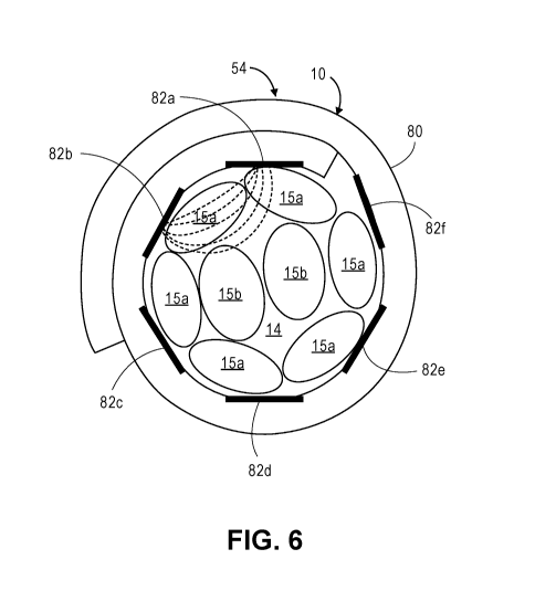
[0070] Fig. 6 is a cross-sectional view of the nerve cuff electrode of Fig. 5,
particularly shown in a furled state;
[0071] Fig. 7a is a diagram of an electrical pulse train that can be generated
by the
stimulation system of Fig. 2;
[0072] Fig. 7b is a diagram of a bi-phasic, charge-balanced, symmetrical
electrical
pulse train that can be generated by the stimulation system of Fig. 2;
[0073] Fig. 7c is a diagram of a bi-phasic, charge-balanced, asymmetrical
electrical
pulse train that can be generated by the stimulation system of Fig. 2;
[0074] Fig. 7d is a diagram of a bi-phasic, asymmetrical electrical pulse
train having
a quiescent period that can be generated by the stimulation system of Fig. 2;
[0075] Fig. 8 is a diagram of a pre-conditioning electrical pulse train that
can be
generated by the stimulation system of Fig. 2;
[0076] Fig. 9 is a flow diagram illustrating one method of implanting and
fitting the
stimulation system of Fig. 2 to a patient.
[0077] Fig. 10 is a plan view of a stimulation system constructed in
accordance with
another embodiment of the present inventions;
[0078] Fig. 11 is a diagram of an exemplary temperature change measurement
taken by a feedback mechanism of the stimulation system of Fig. 10 from a
breath
during respiration of a patient;
[0079] Fig. 12 is a diagram of an exemplary CO2 concentration measurement
taken
by a feedback mechanism of the stimulation system of Fig. 10 from a breath
during
respiration of a patient; and
CA 03071628 2020-01-30
WO 2018/144631
PCT/US2018/016287
[0080] Fig. 13 is a flow diagram illustrating one method of titrating or
fitting the
stimulation system of Fig. 10 to a patient.
DETAILED DESCRIPTION
[0081] Referring first to Fig. 1, it is desirable to locate a nerve cuff
electrode 10
around a trunk 14 of a hypoglossal nerve (HGN) 12 for purposes of stimulating
the
muscles that move the tongue 16 forward, and in particular, the fascicles of
the HGN
12 that innervate the tongue protrusor muscles, such as the genioglossus 18
and/or
the geniohyoid muscles 20, thereby preventing or alleviating obstructive
apneic
events. As shown, the nerve cuff electrode 10 is positioned on the HGN trunk
14
immediately before it branches out, and hence at a proximal position 22 to the
HGN
branches 24. In the illustrated embodiment, the proximal position 22 is just
prior to
the medial branch of the HGN 12 that innervates the tongue protrusor muscles.
[0082] As briefly discussed above, the implantation of the nerve cuff
electrode 10 at
this proximal position 22 reduces the surgical time and effort, allows more
surgeons
to perform the surgery, reduces the risk and trauma to the patient, and
reduces
engineering design complexity and cost. However, it introduces the problem of
inadvertently stimulating other fascicles of the HGN trunk 14 that innervate
muscles
in opposition to the tongue protrusor muscles, i.e., the tongue retractor
muscles, e.g.,
the hyoglossus 26 and styloglossus muscles 28, as well as the intrinsic
muscles of
the tongue 16.
[0083] As also briefly discussed above, it is further desirable to synchronize
the
stimulation of the HGN 12 with the respiratory cycle of the patient, so that
tongue 16
is anteriorly moved in response to the stimulation of the HGN 12 when it is
most
needed, and in particular, right before the onset of the next inspiratory
phase of the
respiratory cycle. Such synchronization requires detection or prediction of
the onset
26
CA 03071628 2020-01-30
WO 2018/144631
PCT/US2018/016287
of the inspiratory phase using one or more sensors. The conventional thought
is that
the sensor(s) should be implanted within anatomical structures, such as the
ribcage
and abdomen, the movement of which strongly correlates to the respiratory
cycle of
the patient. However, because the neurostimulator will typically be implanted
in the
upper chest portion of the patient away from these anatomical structures, one
or
more leads must be used to implant the sensor(s) within these anatomical
structures
remote from the neurostimulator, thereby requiring additional surgical time
and effort.
[0084] Systems and methods are described herein that selectively stimulate the
fascicles of the HGN 12 at the proximal position 22 of the HGN 12 that
innervate the
genioglossus 18 and/or the geniohyoid muscles 20, while synchronizing the
stimulation with the respiratory cycle of the patient without the need to
implant
sensor(s) remotely from the neurostimulator.
[0085] Referring to Fig. 2, one embodiment of a stimulation system 50 that
selectively stimulates the fascicles of the trunk 14 of the HGN 12 that
innervate the
tongue protrusor muscles for treating obstructive sleep apnea (OSA) will now
be
described. The system 50 generally comprises an implantable device 52, an
electrode lead 54, an external charger 55, a clinician programmer 56, and a
patient
programmer 58. The electrode lead 54 and the implantable device 52, or
alternatively, an implantable pulse generator ("IPG") or a "neurostimulator,"
can be
implanted within a patient. In this patent disclosure we will use terms "IPG"
and
"neurostimulator", equivalently.
[0086] The electrode lead 54 comprises the aforementioned nerve cuff electrode
10
and a lead body 60 coupling the nerve cuff electrode 10 to the implantable
device 52
via a proximal lead connector 62 and a corresponding connector receptacle 64.
Although the lead body 60 can be straight, in the illustrated embodiment, the
lead
27
CA 03071628 2020-01-30
WO 2018/144631
PCT/US2018/016287
body 60 may have one or more S-shaped sections in order to provide strain
relief,
thereby accommodating body movement at the location where the lead body 60 is
implanted. This strain relief feature is advantageous, since the lead body 60
is
intended to be implanted in a body location such as the neck, where the lead
body
60 is subjected to frequent movement and stretching. Thus, the S-shape of the
lead
body 60 can help prevent damage to the HGN trunk 14, resulting from sometimes,
unavoidable pulling of the nerve cuff electrode 10 as a result of neck
movements.
As will be described in further detail, the nerve cuff electrode 10 comprises
an array
of circumferentially disposed electrode contacts.
[0087] Although only a single electrode lead 54 is shown in Fig. 2, some
embodiments of the present system may have an IPG 52 having two receptacles 64
(not shown) for attaching two electrode leads, each electrode lead having a
nerve
cuff electrode 10. In such a two-electrode lead system, each nerve cuff
electrode 10
can be implanted bilaterally to each of the HGN trunks 14. However, it has
been
determined that only a single nerve cuff electrode 10 implanted at the HGN
trunk 14
on either side (unilaterally) can provide sufficiently effective stimulation
to protrude
the tongue to control OSA. A unilateral stimulation system is advantageous,
since it
is simpler in numbers of components used and requires only half the surgery to
implant only a single nerve cuff electrode 10, instead of two.
[0088] The IPG 52 comprises an outer case 66 for housing the electronic and
other
components (described in further detail below). In one embodiment, the outer
case
66 may comprise an electrically conductive, biocompatible material, such as
titanium
or titanium alloy, and form a hermetically sealed compartment wherein the
internal
electronics are protected from the body tissue and fluids. In some cases, the
outer
case 66 may serve as an electrode. As briefly discussed above, the IPG 52
further
28
CA 03071628 2020-01-30
WO 2018/144631
PCT/US2018/016287
comprises a receptacle 64 to which the proximal end of the lead body 60 mates
in a
manner that electrically couples the nerve cuff electrode 10 to the internal
electronics
(described in further detail below) within the outer case 66.
[0089] Referring now to Fig. 3, the components and circuitry housed in the
outer
case 66 comprise stimulation circuitry 68, control circuitry 70, communication
circuitry 72, memory 74, sensing circuitry 76, a rechargeable power source 77,
and
power circuitry 79, which all may be conveniently mounted on a printed circuit
board
(PCB) (not shown).
[0090] In one embodiment, the sensing circuitry 76 comprises one or more
sensor(s)
(not shown) that are contained in the outer case 66 or otherwise attached as
an
integral part of the IPG 52, such as affixed to the exterior of the outer case
66.
Further details on incorporation of sensors within or on the outer case of an
IPG 52
are described in U.S. Patent Application Ser. No. 15/374,538, entitled
"Implantable
Pressure Sensors and Medical Devices," which is expressly incorporated herein
by
reference. In other alternative embodiments, the sensor(s) can be positioned
at a
site remote from the IPG 52 coupled by a connecting lead, e.g., as described
in U.S.
Patent Application Ser. No. 15/093,495, entitled "Upper Airway Stimulator
Systems
for Obstructive Sleep Apnea," which is expressly incorporated herein by
reference,
although as can be appreciated, this would require additional surgery and time
to
implant the sensing lead.
[0091] The sensing circuitry 76 is used primarily to sense the respiration
cycle and
can, in embodiments of the invention, determine a projected onset of the
inspiratory
phase of the breathing cycle, or alternatively, may determine the projected
onset of
the expiratory phase of the breathing cycle. In particular, the sensing
circuitry 76 is
configured for detecting physiological artifacts that are caused by
respiration (e.g.,
29
CA 03071628 2020-01-30
WO 2018/144631
PCT/US2018/016287
movement or expansion of ribcage and/or abdomen), which are proxies for
respiratory phases, such as inspiration and expiration or, if no movement
occurs, to
indicate when breathing stops. For example, the sensing circuitry 76 may sense
movement of the thoracic cavity and/or detect changes in pressure/force in the
thoracic cavity. Thus, the sensing circuitry 76 is configured for acquiring,
conditioning, and processing signals related to respiration. The sensor(s) of
the
sensing circuitry 76 can take the form of, e.g., inertial sensors (e.g.,
accelerometers
or gyroscopes), pressure sensors, bioimpedance sensors, ECG electrodes,
temperature sensors, GPS sensors, or some combination thereof.
[0092] The stimulation circuitry 68 is coupled to the nerve cuff electrode 10
via the
lead body 60, and is configured for delivering stimulation to the HGN trunk 14
via
selected ones of the electrode contacts 82. The control circuitry 70 is
coupled to the
stimulation circuitry 68 and controls when, and for how long, the stimulation
circuitry
68 applies stimulation to the HGN trunk 14. The control circuitry 70 may also
control
the intensity of the stimulation applied by the stimulation circuitry 68 to
the HGN
trunk 14, e.g., by varying the amplitude, pulse width, or frequency of the
stimulation.
[0093] As will be described in further detail below, the control circuitry 70
may select
the optimal electrode contact(s) of the nerve cuff electrode 10 used for
stimulating
the HGN trunk 14, and in particular, the electrode contact(s) that stimulate
the
fascicles of the HGN 12 innervating the tongue protrusor muscles, e.g., the
genioglossus 18 or geniohyoid 20 muscles, to thereby prevent or alleviate
obstructive apneic events. However, stimulation of nerve fascicles innervating
the
tongue retractor muscles, e.g., the hyoglossus 26 and styloglossus muscles 28,
as
well as the intrinsic muscles of the tongue 16, should be avoided to the
extent
possible.
CA 03071628 2020-01-30
WO 2018/144631
PCT/US2018/016287
[0094] The memory 74 is configured for storing specific data gathered by the
sensing circuitry 76 and programming instructions and stimulation parameters.
The
control circuitry 70 may recall the sensed data from the memory 74 and analyze
it to
determine when stimulation should be delivered to the HGN trunk 14 to
synchronize
the stimulation delivery with the respiratory cycle. In some embodiments, the
sensor
data may be analyzed to predict the onset of the next inspiratory phase of the
breathing cycle and to deliver stimulation right before, at, or slightly after
the
predicted onset of the inspiratory phase.
[0095] Thus, when the patient is in the inspiratory portion of the respiratory
cycle¨
where the patient is breathing in or attempting to breath in, the control
circuitry 70
may, in some embodiments, apply stimulation, thereby causing forward
displacement of the tongue, and causing the upper airway to remain un-
obstructed
during inspiration while sleeping. The control circuitry 70 causes the
stimulation
circuitry 68 to apply stimulation during these inspiratory phases of the
respiratory
cycle (or applying stimulation starting slightly before the inspiration and
ending at the
end of inspiration), and not the remainder of the respiration cycle, when all
other
conditions for stimulation are met.
[0096] The IPG 52 may be toggled between an ON position and an OFF position
using one of a variety of techniques. In one embodiment, the IPG 52 may have a
magnetic reed switch (not shown) contained within the outer case 66 that can
sense
a magnetic field from an external magnet. An external magnet may be used to
toggle the IPG 52 to the OFF position or alternatively to an ON position.
Oftentimes,
patients may need to undergo an MRI scan. A reed switch in the IPG 52 may make
it MRI incompatible. In another embodiment, the IPG 52 may have a sensor (not
shown) that is sensitive to movement (i.e., a motion detector), such as an
inertial
31
CA 03071628 2020-01-30
WO 2018/144631
PCT/US2018/016287
sensor (e.g., an accelerometer or gyroscope), and a switch that can be toggled
between a closed state and an open state to place the implanted IPG 52 between
an
ON position and an OFF position by tapping the implanted IPG 52 with the hand.
For example, one tap may switch the IPG 52 from an ON position to an OFF
position, and another tap may switch the IPG 52 from an OFF position to an ON
position. In one preferred embodiment, the same sensing circuitry 76, along
with the
sensor, that is used for detecting physiological artifacts that are caused by
respiration, may be used to sense the tapping of the implanted IPG 52 to
toggle the
IPG 52 between the ON position and the OFF position.
[0097] In another preferred embodiment, the IPG 52 can be toggled between an
ON
position and an OFF position in response to multiple quick successive taps
(e.g.,
less than a second between taps), as opposed to a single tap, which may occur
by
accidental bumping and cause an inadvertent turn off of the IPG, for example,
two
taps to switch the IPG 52 from an ON position to an OFF position, and two taps
to
switch the IPG 52 from an OFF position to an ON position. As a redundancy, the
patient programmer 58 or the clinician programmer 56 may also be configured to
be
able to toggle the IPG 52 from ON to OFF and from OFF to ON.
[0098] In an optional embodiment, the sensing circuitry 76 comprises a body
position
sensor (not shown) (e.g., an inertial sensor) configured for measuring an
orientation
of the patient's body. In this case, the control circuitry 70 determines the
orientation
of the patient's body, and activates the portions of the sensing circuitry 76
that
monitor the physiological artifacts that are caused by respiration when the
orientation
indicates that the patient is in an apneic position (i.e., a position in which
the patient
is likely to experience apneic events). The most common apneic position is
supine,
but can include left side, right side, or both. Patients with positional sleep
apnea
32
CA 03071628 2020-01-30
WO 2018/144631
PCT/US2018/016287
experience significantly more apneic events while in particular apneic
positions,
thereby allowing the neurostimulator 52 to preserve battery life by monitoring
the
physiological artifacts that are caused by respiration only when the patient
is likely to
experience apneic events. The memory 74 may store positional sleep apnea data
for the patient that can be consulted by the control circuitry 70 when
determining
whether the patient is in an apneic position.
[0099] In another optional embodiment, the sensing circuitry 76 comprises a
sleep
sensor (not shown) configured for measuring a physiological parameter
indicative of
whether the patient is sleeping. The sleep sensor may comprise sensors used in
polysomnography, such as an EMG sensor across the jaw line, an EEG sensor, and
an EOG sensor, an inertial sensor, or a temperature sensor. In this case, the
control circuitry 70 determines whether the patient is asleep, and activates
the
portions of the sensing circuitry 76 that monitor the physiological artifacts
that are
caused by respiration only when the patient is asleep. This preserves battery
life
since sensing and monitoring only occurs when the patient is actually asleep.
[00100] Further details describing the use of body orientation and sleep
sensors are
discussed in U.S. Patent Application Ser. No. 15/093,627, entitled "Upper
Airway
Stimulator Systems for Obstructive Sleep Apnea," which is expressly
incorporated
herein by reference.
[00101] The communication circuitry 72 is configured for wirelessly
communicating
transcutaneously (through the patient's skin) with the clinician programmer 56
and
patient programmer 58 using radio frequency (RF) signals, e.g., via an Off The
Shelf
(OTS) lnductive/Bluetooth/MICS radio link. The communication circuitry 72 may
include one or more AC coils for transmitting and receiving the RF signals to
and
from the clinician programmer 56 and patient programmer 58.
33
CA 03071628 2020-01-30
WO 2018/144631
PCT/US2018/016287
[00102] The rechargeable power source 77, for example, a rechargeable battery,
and power circuitry 79 are configured for providing operating power to the IPG
52.
The rechargeable power source 77 may comprise a lithium-ion or lithium-ion
polymer
battery, and provide an unregulated voltage to the power circuitry 79. The
power
circuitry 79, in turn, generates regulated or unregulated voltage to the
various circuits
located within the IPG 52. The rechargeable power source 77 is recharged using
rectified AC power received by an AC receiving coil (such as one of the coils
coupled
to the communication circuitry 72) from the external charger 55. The AC
magnetic
field emitted by the external charger 55 induces AC currents in the AC
receiving coil
(not shown), which is rectified by circuitry (not shown) that rectifies the AC
current to
produce DC current that is used to charge the power source 77.
[00103] Referring back to Fig. 2, to recharge the IPG 52, the external charger
55 or
a part of the charger having a coil, which generates the AC magnetic field, is
placed
against, or otherwise adjacent, to the patient's skin over the implanted IPG
52. The
clinician programmer 56 may be used to transcutaneously communicate with the
implanted IPG 52 for programming the IPG 52 and querying the IPG 52 for
status.
For example, the clinician programmer 56 can be used to configure certain
programs
and processes used by the control circuitry 70 in the IPG 52 to determine when
the
stimulation pulses are to be delivered to electrode contacts of the nerve cuff
electrode 10. The clinician programmer 56 can also be used to program specific
stimulus parameters, such as stimulus pulse width, stimulus frequency,
duration of a
train pulses and pulse amplitude. The amplitude may be expressed in current,
for
example, milliamperes, or it could be expressed in volts, such as 0.3 volts.
The
choice between milliamperes or volts to express stimulus amplitude will depend
on
whether the design of the stimulation circuitry 68 provides stimulus pulses
that are
34
CA 03071628 2020-01-30
WO 2018/144631
PCT/US2018/016287
constant voltage or constant current. Another important function of the
clinician
programmer 56 is the ability to select modes of stimulation. For example, the
IPG 52
may operate in a monopolar stimulation mode (also sometimes referred to as a
"unipolar" mode) and in a bipolar stimulation mode.
[00104] As used in this present disclosure, a monopolar stimulation mode means
that one of the electrode contacts used is at least a portion of the outer
case 66 that
will function as an indifferent/anode electrode. The indifferent electrode is
part of the
electrical circuit with at least one electrode contact of the nerve cuff
electrode 10 as
the active/cathode electrode contact that stimulates the HGN trunk 14.
Generally,
that part of the outer case 66 that is acting as the indifferent electrode
does not
stimulate any tissue or nerve, but merely functions as a return electrode and
may be
a biocompatible, conductive metal such as a titanium alloy, as discussed
above.
[00105] A bipolar stimulation mode means, for purposes of this disclosure,
that the
outer case 66 is not part of the stimulation circuit. At least two electrode
contacts of
the nerve cuff electrode 10 must be selected and will be part of the bipolar
mode
electrical stimulation circuit. Sometimes a stimulation circuit can have three
or even
more electrode contacts functioning together. This may also be referred to as
"bipolar" stimulation mode even though there are sometimes more than two
active
electrode contacts in the stimulation circuit. Sometimes a three-electrode
contact
system may be referred to as a tripolar circuit. For purposes of this
disclosure and
application, we will consider a three or more electrode-contact stimulation
circuit (if it
excludes the outer case 66) as variants of a bipolar stimulation mode and will
be
included as within a "bipolar" stimulation mode. The present stimulation
system in
its various embodiments, thus, may operate in either monopolor or bipolar
stimulation modes.
CA 03071628 2020-01-30
WO 2018/144631
PCT/US2018/016287
[00106] Significantly, to facilitate selective stimulation of the fascicles of
the HGN 12
that innervate the tongue protrusor muscles, the clinician programmer 56 also
selects which electrode contacts of the nerve cuff electrode 10 or the
indifferent
electrode of the outer case 66 are to be in the stimulation circuit. The
clinician
programmer 56 may also be able to query the status of the IPG 52 for a number
of
status functions, such as battery status. Another query may be whether the IPG
52
is in an ON position or an OFF position. In the ON position, the stimulation
circuitry
68 within the IPG 52 is enabled and stimulation pulses can be delivered via
the
selected electrode contact or contacts of the nerve cuff electrode 10. When
the
patient is awake, the IPG may be placed automatically or by choice into the
OFF
position or mode, and the stimulation circuitry 68 is not enabled and no
stimulation
can occur.
[00107] The patient programmer 58 offers more limited programming options than
the clinician programmer 56. The patient programmer 58 may provide the option
to
toggle the IPG 52 into the OFF position or into the ON position. Also, the
stimulus
pulse amplitudes may be adjusted for a limited range of up and down. Often the
patient programmer 58, because of limited functionality, may be in a package
or form
that is much smaller in size than the clinician programmer 56. The clinician
programmer 56 and patient programmer 58 may take the form of commercial
electronic smart devices on which there are installed customized applications
for
performing the afore-described functions.
[00108] Referring now to Figs. 4-6, an embodiment of an electrode lead 54 that
may
be used in the stimulation system will now be described in further detail. The
proximal lead connector 62 comprises a linear array of connector contacts 78a-
78f
36
CA 03071628 2020-01-30
WO 2018/144631
PCT/US2018/016287
(in this case, six) for connecting to the connector receptacle 64 of the IPG
52 when
the proximal lead connector 62 is inserted into the connector receptacle 64.
[00109] The nerve cuff electrode 10 further comprises a nerve cuff body 80
that is
capable of substantially or completely encircling the HGN trunk 14. The nerve
cuff
electrode 10 may be, in some embodiments, manufactured to be self-curling, and
may be designed to self-adjust in accordance with the diameter of the HGN
trunk 14.
The material used for the electrode substrate can be typical implantable
electrode
materials, such as silicone, polyurethane or other less conventional implant
materials, e.g., liquid crystal polymers. The material consistency of the
formed cuff
body 80 should be pliable enough to allow the clinician to unfold the cuff, as
shown
in Fig. 5, and placed around the HGN trunk 14 and to have the nerve cuff
electrode
curl back around itself, as shown in Fig. 6. Although Fig. 5 illustrates the
lead
body 60 in the middle of the cuff body 80, the lead body 60 can be positioned
at
either the left or right end of the cuff body 80, and the lead body 60 may
even point
90 degrees from the direction the lead body 80 is aligned as shown in Fig. 5.
The
substrate material of the nerve cuff body 80, therefore, should have a memory
property to the extent that it will tend to return to its original curled
shape. In one
advantageous manufacturing process, the nerve cuff electrode 10, lead body 60,
and
proximal lead connector 62 may be constructed of a flexible circuit, as
described in
U.S. Patent Application Ser. Nos. 15/634,057 and 15/634,134, both entitled
"Nerve
Cuff Electrodes Fabricated Using Over-Molded LCP Substrates," which are
expressly incorporated by reference.
[00110] The nerve cuff electrode 10, as shown, will also have some give, so
that if
the nerve swells during the inflammatory phase post-surgery, the inner lumen
size of
the nerve cuff electrode 10 can expand and accommodate to the nerve swelling.
37
CA 03071628 2020-01-30
WO 2018/144631
PCT/US2018/016287
This capability of self-adjustment over time is important because once tissue
has
been dissected from around the nerve, there often will be an inflammatory
response
around the damaged tissue and also in response to the presence of foreign
matter
that may be introduced during the surgical implantation of the nerve cuff
electrode
10. Indeed, the nerve cuff electrode 10, itself, is likely seen as a foreign
matter
contributing to inflammation. The inflammatory response may be ongoing over a
period of months. During this period, the nerve, itself, may swell up and
increase
substantially in diameter, perhaps up to 50% more than before the surgery.
Once
past this inflammatory response, the nerve diameter may then decrease in size,
closer to its original diameter. If the inner lumen size of the nerve cuff
electrode 10
does not adjust in size to accommodate the increase in the nerve diameter,
constriction of the target nerve can result in traumatic cell damage and nerve
death.
Further details describing various self-expanding nerve cuff electrodes are
set forth
in U.S. Provisional Patent Application Ser. No. 62/500,080, entitled "Nerve
Cuff
Electrode Locking Mechanism," and U.S. Provisional Patent Application Ser. No.
62/500,091, entitled "Self-Expanding Nerve Cuff Electrode," which are both
expressly incorporated herein by reference.
[00111] The nerve cuff electrode 10 further comprises an array of electrode
contacts
82a-82f (in this case, six) affixed to an inner surface of the cuff body 80
(when
furled), such that when the cuff body 80 encircles the HGN trunk 14, the
electrode
contacts 82a-82f are in contact with the HGN trunk 14. To facilitate selective
activation of the fascicles of the HGN trunk 14 that innervate the protrusor
muscles,
the electrode contacts 82 are affixed to the cuff body 80 in a manner, such
that when
the cuff body 80 encircles the HGN trunk 14, the electrode contacts 82 are
circumferentially disposed about the HGN trunk 14. In this case, the electrode
38
CA 03071628 2020-01-30
WO 2018/144631
PCT/US2018/016287
contacts 82 span the cuff body 80 circumferentially around the HGN trunk 14.
The
electrode contacts 82a-82f preferably circumferentially span at least a 180-
degree
arc of the HGN trunk 14, and more preferably span at least a 270-degree arc of
the
HGN trunk 14, so that any fascicle within the HGN trunk 14 can be selectively
stimulated by delivering stimulation energy from the electrode contact or
contacts 82
adjacent to the fascicle, as described in further detail below. To facilitate
coverage
of all of the fascicles, the number of electrode contacts 82 preferably equals
at least
three, and more preferably, at least six. The electrode contacts 82 are also
aligned
on the inner surface of the cuff body 80, such that, when the cuff body 80
encircles
the HGN trunk 14, the electrode contacts 82 are axially aligned with each
other. For
the purposes of this specification, electrode contacts 82 are axially aligned
with each
other if they lie in the same plane that is perpendicular to the axis of the
cuff body 80
or axis of the HGN trunk 14. In addition, although Fig. 5 shows an embodiment
of
cuff having electrode contacts aligned in a single row, in other embodiments,
it is
possible to construct a cuff having two or even more parallel rows of
electrode
contacts (not shown) that are parallel to the direction defined by electrode
contacts
82a - 82f. The latter electrode contact arrangement(s) will provide an
additional
degree of freedom in stimulating a target nerve fascicle.
[00112] Although the exemplary nerve cuff electrode 10 comprises six electrode
contacts 82a-82f, other nerve cuff electrodes may have two to five electrode
contacts
82 or more than six electrode contacts 82. The preferred range, however, of
the
numbers of electrode contacts 82 on any particular nerve cuff electrode is
between
three to eight electrode contacts 82, so as to surround the circumference of
the HGN
trunk 14, and provide a sufficient number of independent electrode channels
from
which to select and to recruit the protrusor muscles without recruiting the
retractor
39
CA 03071628 2020-01-30
WO 2018/144631
PCT/US2018/016287
muscles. The connector contacts 82a-82f are respectively and independently
electrically coupled to the electrode contacts 82a-82f via electrical
conductors (not
shown), such that the electrode contacts 82a-82f may be independently
activated in
either monopolar stimulation mode or bipolar stimulation mode. In the
monopolar
stimulation mode, one or more of the electrode contacts 82a-82f will
preferably be
activated as cathode(s), whereas in the bipolar stimulation mode, one or more
of the
electrode contacts 82a-82f will be activated as cathode(s), and one or more
other
electrode contacts 82a-82f will be activated as anode(s).
[00113] Although in some embodiments, the nerve cuff electrode 10 may be
operated in a monopolar stimulation mode, requiring that only one electrode
contact
82 of the nerve cuff electrode 10 be activated at any given time, as will be
described
in further detail below, it is desirable that the nerve cuff electrode 10 be
operated in a
bipolar stimulation mode to facilitate selective recruitment of the fascicles
15 in the
HGN trunk 14, requiring that at least two electrode contacts 82 of the nerve
cuff
electrode 10 be activated at any given time.
[00114] That is, monopolar stimulation results in a more diffuse electrical
field that
will tend to recruit most fascicles 15 in the HGN trunk 14 including those
unwanted
fascicles, whereas bipolar stimulation results in a more specific and confined
electrical field that will tend to recruit only the targeted fascicles 15 in
the HGN trunk
14. Thus, the fascicles 15 in the HGN trunk 14 that innervate the tongue
protrusor
muscles can be more selectively activated via bipolar stimulation. Because the
electrode contacts 82 will circumferentially surround the HGN trunk 14, the
electrical
field generated by the nerve cuff electrode 10 in the bipolar stimulation mode
can be
selectively steered around the HGN trunk 14 to recruit the desired fascicles
15 within
the HGN trunk 14. It is further noted that, because the fascicles 15
innervating the
CA 03071628 2020-01-30
WO 2018/144631
PCT/US2018/016287
tongue protrusor muscles sometimes, depending on the individual anatomy, may
be
more peripherally located within the nerve bundle that is located at the
proximal
position 20 to the HGN branches 24, it is desirable that adjacent electrode
contacts
82 can be activated in the bipolar arrangement, such that the electrical field
extends
only peripherally into the HGN trunk 14.
[00115] Thus, with reference to Fig. 6, it may be desirable to activate
electrode
contact pair 82a-82b, electrode contact pair 82b-82c, electrode contact pair
82c-82d,
electrode contact combination 82d-82e, electrode contact combination 82e-82f,
or
electrode combination 82f-82a. As shown in Fig. 6, electrode combination 82a-
82b,
when activated, create a confined bipolar electrical field therebetween that
recruits
one or more of the peripheral, less centrally located fascicles 15a, as
opposed to
recruiting the more centrally located fascicles 15b. Of course, any of the
other
electrode contact combinations can be operated in a bipolar manner to recruit
other
peripherally located fascicles 15a. The first one of the electrode contacts 82
in the
combination can be a cathode, and the second one of the electrode contacts 82
in
the combination can be an anode, or vice versa. There are other possible
electrode
contact combinations or contact sets that can be possibly chosen, which are
described later in this disclosure in relation to titrating and fitting
electrode contacts
to determine the optimal set(s).
[00116] Notably, the strongest electrical field generated by the nerve cuff
electrode
will be beneath an active electrode contact/cathode. Thus, in order to
effectively
employ bipolar stimulation, the nerve cuff electrode 10 may have the following
design constraint: where W is the width of each electrode contact 82, and L
is the center-to-center distance between two adjacent electrode contacts 82,
as
illustrated in Fig. 5. This constraint is based on the commercial needs in
41
CA 03071628 2020-01-30
WO 2018/144631
PCT/US2018/016287
neuromodulation therapies to cover the most distance with spatial separations
L and
using the fewest number of electrode contacts 82. The width of the electrode
contacts 82 will typically be based on the particular neural element that will
be
stimulated or the size of the cuff body 80, or a combination thereof, and will
set the
strength ranges of the electric fields generated by the nerve cuff electrode
10. As
the center-to-center distance L exceeds the L2W design constraint, the
electric field
generated by a bipolar pair of electrode contacts 82 quickly starts to
resemble a
monopolar electric field as if there was a remote anode (unless there is a
dramatic
increase in the electric field amplitude). The ability to perform current
steering
between two or more adjacent electrode contacts 82 also weakens. In contrast,
if
adjacent electrode contacts 82 are too close or touching each other, there may
be
bleeding of electrical fields across the active contacts 82 at a higher
amplitude,
thereby creating a short that reduces the ability to spatially select
fascicles. Thus, it
is important that the center-to-center distance L between adjacent electrode
contacts
82 and the width W of the electrode contacts 82 be constrained.
[00117] To maintain the distance between the electrode contacts 82a, 82f in
accordance with the L2W design constraint over a variety of different nerve
sizes,
thereby ensuring that bipolar stimulation using the electrode contacts 82a,
82f is
effective, the nerve cuff electrode 10 may optionally be designed in the
manner
described in U.S. Provisional Patent Application Ser. No. 62/552,266, entitled
"Stimulator Systems and Methods or Selectively Recruiting Fascicles in
Hypoglossal
Nerve Trunk," which is expressly incorporated herein by reference.
[00118] The stimulation energy generated by the stimulation circuitry 68 takes
the
form of a train of electrical pulses under control of the control circuitry
70. The
electrical pulse train may be set to a constant time duration or it may be
adaptive,
42
CA 03071628 2020-01-30
WO 2018/144631
PCT/US2018/016287
meaning that duration of the train of pulses can change dynamically based on a
predictive algorithm that determines the duration of the inspiratory phase of
the
respiratory cycle.
[00119] Referring now to Fig. 7, various electrical pulse trains that can be
generated
by the stimulation circuitry 76 will now be described. In one embodiment
illustrated
in Fig. 7a, an electrical pulse train comprises a single stimulation pulse Si
or S2.
The stimulus pulse Si or S2 has stimulus parameters: stimulus pulse width, the
stimulus pulse current or voltage amplitude, and stimulus frequency. The
frequency
determines the time duration between two consecutive pulses Si, S2. These
multiple stimulus pulses may also be called a "train" of pulses or a "burst"
of pulses.
Usually there is a quiescent period, as shown in Fig. 7d, between two trains
or burst
of stimulus pulses, in this case, between stimulation pulses S6-S9 and S10.
During
this quiescent time, there is no stimulation occurring. The stimulus pulses
can be
cathodic (upper Y-axis direction in Fig. 7) or anodic (lower Y-axis direction
in Fig. 7).
The stimulus could be bi-phasic, and symmetric (Fig. 7b), meaning the
electrical
charge in the cathodic direction and anodic direction of a single pulse S3 are
the
same. Sometimes stimulation pulses S4, S5 can be bi-phasic, charge-balanced,
but
not symmetric (Fig. 7c). The cathodic amplitude is greater than the anodic
amplitude, but the total charge delivered out through an electrode contact
during the
cathodic phase is balanced by the same quantity of electrical charge incoming
into
the same electrode contact. Area C equals area D and area E is equal to area
F. A
stimulus that is charge-balanced is desirable in order to ensure that the
electrode
contacts do not erode prematurely during chronic implantation. For example, a
charge-unbalanced cathodic pulse as shown in Fig. 7a can stimulate a nerve,
but is
not a desirable stimulus choice for an IPG. Although it is possible to elicit
nerve
43
CA 03071628 2020-01-30
WO 2018/144631
PCT/US2018/016287
stimulation using an anodic pulse, a cathodic pulse is generally used to
stimulate a
nerve, since the nerve stimulation threshold (stimulus amplitude that just
triggers
nerve conduction) needed is much lower with a cathodic pulse than an anodic
pulse.
[00120] Referring now to Fig. 8, shows a train of biphasic, charge-balanced
pulses
comprising initial pre-conditioning pulses, designated by X, followed by
larger
amplitude stimulus pulses, designated by Y, which are at a higher amplitude
compared to the pre-conditioning pulses. Both X and Y stimuli may be charge-
balanced. The X preconditioning pulses may be as low as 10% and more typically
50-90% of the Y stimulus pulses. This particular stimulation pattern can be
used to
"precondition" the peripheral, less centrally located nerve fascicles 15a
(i.e., the
nerve fascicles closer to the outer circumference of the HGN trunk 14) over
the more
centrally located nerve fascicles 15b (i.e., the nerve fascicles closer to the
center of
the HGN trunk 14), as shown in Fig. 6. Although it is a general assumption
that it is
the peripherally located target fascicles 15a that innervate the tongue
protrusor
muscles, it is dependent on individual anatomy and the target fascicles may in
fact
be more centrally located in the HGN trunk. Thus, the usefulness of
preconditioning
will depend on the anatomy of an individual and the location of the target
nerve
fascicles in relation to the center of the nerve bundle. Preconditioning, as
described,
is a therefore a stimulation tool that may be optionally applied depending on
circumstances.
[00121] In operation, the initial pre-conditioning pulses X at the lower
amplitude will
stimulate the more peripherally located nerve fascicles 15a of the HGN trunk
14.
This causes these nerve fascicles to be "pre-conditioned," so that they are
not
inclined to be stimulated later by a larger amplitude stimulation pulses Y.
The lower
amplitude pre-conditioning pulses X are low enough in amplitude that they will
not
44
CA 03071628 2020-01-30
WO 2018/144631
PCT/US2018/016287
stimulate the more centrally located nerve fascicles 15b of the HGN trunk 14.
While
the peripherally located nerve fascicles 15a of the HGN trunk 14 are in their
"pre-
conditioned" state and not excitable, the larger amplitude stimulation pulses
Y will
reach into the center of the HGN trunk 14 with sufficient charge density to
stimulate
and activate the more centrally located nerve fascicles 15a.
[00122] Thus, in the case where the more centrally located nerve fascicles 15b
of
the HGN trunk 14 happen to innervate the tongue protrusor muscles, this
preconditioning stimulation pattern can provide selective stimulation of these
centrally located nerve fascicles 15b without stimulating nerve fascicles that
innervate other extraneous muscles, namely the peripherally located nerve
fascicles
15a. In other embodiments, instead of increasing the stimulus amplitude in the
latter part of pulse train Y, another possible way of achieving higher
electrical charge
intensity to activate the centrally located nerve fascicles 15b of the HGN
trunk 14 is
to, relative to the pre-conditioning pulses, increase stimulus pulse width,
increase
frequency of stimulation, or provide some combination thereof. It should be
appreciated that the use of the preconditioning stimulation pattern is not
limited to
HGN trunks 14, but can be used with any nerve trunk where it is desirable to
selectively stimulate more centrally located fascicles over more peripherally
located
nerve fascicles.
[00123] Thus, selective targeting of the tongue protrusor muscles can be
achieved
by spatially stimulating the fascicles innervating these tongue protrusor
muscles by
selectively using the electrode contacts 82 of the nerve cuff electrode 10 to
stimulate
the HGN trunk 14. Furthermore, by using lower amplitude pre-conditioning
pulses to
desensitize the fascicles innervating the tongue retractor muscles, followed
by a
cathodic stimulation to target the fascicles innervating the tongue protrusor
muscles,
CA 03071628 2020-01-30
WO 2018/144631
PCT/US2018/016287
even better targeting of the tongue protrusor muscles can be achieved in the
case
where the fascicles innervating the tongue protrusor muscles are deeper in the
HGN
trunk 14. Thus, it can be appreciated from the foregoing that the combination
of
several electrode contacts 82 on the nerve cuff electrode 10 that provide
multiple
independent electrode channels and the pre-conditioning stimulation trains
provides
a margin for placement of the nerve cuff electrode 10 on the HGN trunk 14 and
variation in the surgical approach across various surgeons.
[00124] Having described the arrangement and function of the stimulation
system
10, one embodiment of a method of using the stimulation system 10 to treat OSA
in
a patient will now be described with reference to Fig. 9.
[00125] First, the electrode contacts 82 are circumferentially disposed around
the
HGN trunk 14. In particular, the cuff body 80 is maintained in the unfurled
state (Fig.
5) while placing the cuff body 80 around the HGN trunk 14 (step 102). For
example,
the unfurled cuff body 80 may be placed underneath the HGN trunk 14. The cuff
body 80 may be maintained in the unfurled state by, e.g., holding it open,
although
the cuff body will tend to return to its furled, resting state. The HGN trunk
14 may
have a diameter, e.g., typically in the range of 2.5 mm to 4.0 mm.
[00126] Next, the cuff body 80 is transitioned from the unfurled state into
the furled
state (Fig. 6), such that the cuff body 80 wraps around the HGN trunk 14 (step
104).
The cuff body 80 may be placed from the unfurled state into the furled state
by letting
go of both ends of the cuff body 80, such that the cuff body 80 automatically
transitions from the unfurled state to the furled state. The electrode
contacts 82
preferably circumferentially span at least a 180-degree arc around the HGN
trunk 14,
and more preferably, at least a 270-degree arc around the HGN trunk 14.
Preferably, the center-to-center spacing L of each pair of adjacent ones of
the
46
CA 03071628 2020-01-30
WO 2018/144631
PCT/US2018/016287
electrode contacts 82 is equal to or less than twice the width W of each
electrode
contact of the respective pair of adjacent electrode contacts 82.
[00127] Next, the IPG 52 is implanted within the patient (step 106), and the
proximal
lead connector 62 is mated with the receptacle 64 of the IPG 52 (step 108).
Next,
the system 50 is titrated to determine at least one electrode contact 82 (an
"electrode
contact set") that provides the best treatment of the OSA by delivering
electrical
stimulation energy to different electrode contact sets 82 to determine the
optimal
electrode contact set(s) (step 110). For example, electrode contact set 82 may
be
selected, an electrical pulse train may be delivered to the selected electrode
contact
set to stimulate the HGN trunk 14, and preferably, the fascicles of the HGN
trunk 14
innervating the tongue protrusor muscles, and then repeated for all possible
electrode sets to determine the optimal electrode contact set(s). Further
details of
several techniques for titrating the neurostimulation system 10 are discussed
below.
In one method, each electrode contact set comprises a pair of adjacent ones of
the
electrode contacts 82, in which case, the pairs of electrode contacts are
selected for
delivery of the electrical pulse trains in a bipolar mode. In another method,
each
electrode contact set comprises a single electrode contact 82, in which case,
individual ones of the electrode contacts 82 are selected one at a time for
delivery of
the electrical pulse trains in a monopolar mode.
[00128] Initially, in order to trigger a peripherally located fascicle or
fascicles 15a
innervating the tongue protrusor muscles, all of the electrode contact sets
may be
tested using regular, constant amplitude pulse trains having a defined pulse
duration
and frequency, as shown in Fig. 7. If no peripherally located fascicle 15a
innervating
the tongue protrusor muscles is triggered, all of the electrode contact sets
may be
tested using pre-conditioning pulse trains, such as that shown in Fig. 8, in
order to
47
CA 03071628 2020-01-30
WO 2018/144631
PCT/US2018/016287
trigger any centrally located fascicle or fascicles 15b that may possibly
innervate the
tongue protrusor muscles.
[00129] Once an optimal electrode contact set (or sets) is determined, the IPG
52 is
programmed to the selected optimal electrode contact set (or sets) using the
clinician
programmer 56 (step 112). The IPG 52 can be turned on with the clinician
programmer 56 or the patient programmer 58. Or, the patient can turn the IPG
52 to
an ON setting, e.g., by tapping the area of the body over the implanted IPG 52
multiple times in quick succession or by using the patient programmer 64 (step
114).
The IPG 52 may be turned ON when the patient wishes to sleep, or OFF, when the
patient is awake during the day. In the ON position, the IPG 52 will provide,
through
the selected electrode contact or contacts, a stimulus train (or stimulus
burst) at the
programmed setting. Stimulation will move the tongue forward while the
patient is
asleep, so that during any obstructive apneic event, the patient will not be
prevented
from breathing. As such, when the IPG 52 is in the ON position, a train of
pulse
stimulation occurs at every breathing inspiration.
[00130] To save battery power, the IPG 52 may only provide therapy under a
specific set of circumstances, e.g., if the patient is sleeping in an apneic
position. To
this end, the IPG 52 may determine if the patient is in an apneic position by
measuring an orientation of the body of the patient (step 116), and if so,
then
determine if the patient is sleeping by measuring a physiological parameter
indicative
of whether the patient is sleeping (step 118).
[00131] If the patient is sleeping in an apneic position, the physiological
artifacts
caused by respiration are sensed and stored (step 120), the next projected
onset of
an inspiratory phase of the respiratory cycle is determined based on the
sensed
physiological artifacts (step 122), and electrical stimulation energy (e.g.,
an electrical
48
CA 03071628 2020-01-30
WO 2018/144631
PCT/US2018/016287
pulse train) is delivered to the programmed electrode contact set in
synchronization
with a respiratory cycle based on the sensed physiological artifacts, and in
particular,
immediately before, at, or right after the next projected onset of the
inspiratory phase
of the respiratory cycle (step 124), thereby treating the OSA.
[00132] If the electrical pulse train is conventional in nature, one or more
peripherally
located nerve fascicles 15a in the HGN trunk 14 (presumably, the peripherally
located nerve fascicle(s) 15a adjacent to the programmed electrode contact
set) are
triggered. If the electrical pulse train is a pre-conditioning pulse train,
one or more
peripherally located nerve fascicles 15a in the HGN trunk 14 will be pre-
conditioned,
and rendered not excitable, by the initial preconditioning current or voltage
amplitude, while one or more centrally located nerve fascicles 15b in the HGN
14 will
be triggered/activated by the subsequently delivered higher stimulating
current or
voltage amplitude.
[00133] Referring now to Fig. 10, another embodiment of a stimulation system
50'
that selectively stimulates the fascicles of the trunk 14 of the HGN 12 that
innervate
the tongue protrusor muscles for treating OSA will now be described. The
stimulation system 50' is similar to the stimulation system 50' illustrated in
Fig. 2,
with the exception that the stimulation system 50' additionally comprises a
feedback
mechanism 90 that can titrate stimulation system 50' in a clinical setting in
order to
provide chronic therapy to a patient suffering from OSA.
[00134] The feedback mechanism 90 of the system 50 is capable of sensing a
physiological parameter indicative of the efficacy in treating a patient's
obstructive
sleep apnea. In conjunction with the feedback mechanism 90, the clinician
programmer 56 can be used to determine the best set or sets of electrode
contacts
82, along with other stimulation parameters (e.g., stimulation pulse
amplitude,
49
CA 03071628 2020-01-30
WO 2018/144631
PCT/US2018/016287
stimulation pulse width, stimulation pulse frequency, and number of stimulus
pulses
in a pulse train, or burst and frequency of the stimulation pulses), that
optimally
recruits the target fascicles of the HGN trunk 14 that controls the protrusor
muscle
which moves the tongue forward.
[00135] The system 50' can be titrated the first time right after implantation
of the
nerve cuff electrode 10 or during clinical follow-up sessions. The system 50'
can
also be titrated during the implantation surgery itself, during a traditional
sleep study,
a drug induced sleep study, or other appropriate setting. The system 50' can
be
titrated while the patient is asleep, or it could be performed sometimes with
the
patient awake.
[00136] The clinician programmer 56 may be operated to iteratively test
different
sets of electrode contacts 82 of the nerve cuff electrode 10 to determine the
set or
sets electrode contacts 82, along with the corresponding types of electrical
pulse
trains, that provides the best therapy for treating the OSA of the patient.
For the
purposes of this patent disclosure, a set of electrode contacts 82 may include
only
one electrode contact 82 or may include multiple electrode contacts 82.
[00137] As an example, using an asymmetrical (peak cathodic amplitude larger
than
peak anodic amplitude), biphasic, charge-balanced, stimulus such as shown in
Fig.
7(c) and referring to Fig. 5 or Fig. 6, the electrode contact 82 selected
could be a
monopolar set, with the outer case 66 of the IPG 52 turned on as indifferent,
return
electrode contact. Then each electrode contact, 82a, 82b, 82c, 82d, 82e, and
82f
can be tested for successively for its efficacy.
[00138] Sometimes a monopolar stimulation can have two stimulating
(functioning
cathodic) electrode contacts 82, e.g., the outer case 66 of the IPG 52
functions as
the indifferent electrode (anode), electrode contact 82a and electrode contact
82b
CA 03071628 2020-01-30
WO 2018/144631
PCT/US2018/016287
concurrently function as stimulating cathodes. In this latter arrangement,
using two
stimulating cathodes concurrently, true current steering can be accomplished.
For
example, if electrode contact 82a outputs 50% of the total current while, at
the same
time, electrode contact 82b outputs 50% of the total current, the net effect
is to
create a virtual electrode that appears to be an electrode contact that is
positioned
right in between the two electrode contacts, 82a and 82b. In other cases, the
current
output may be uneven, for example 70% of total current output for contact 82a
and
30% of total current output from contact 82b. In that case the virtual
electrode will be
in between but closer to contact 82a than 82b. Current steering means that the
center of electrical current density is somewhere between the centers of two
adjacent electrodes and is an extremely powerful tool to "steer" the
stimulation
precisely between two electrode contacts.
[00139] In addition, the titration or fitting may test various sets of
electrode contacts
in bipolar stimulation mode. Examples of bipolar modes include: (A) 82a
(stimulating cathode) and 82b (functioning anode); (B) 82a (stimulating
cathode) and
82c (functioning anode); and (C) three electrode contacts in a tripolar
arrangement,
but still bipolar stimulation ¨ 82b (cathode), 82a (return anode) and 82c
(return
anode). Note there are opportunities for field shaping because 82a and 82c may
both return 50% of the total current output from contact 82b. Alternatively,
contacts
82a and 82c could sink different values of the total current, e.g. 40% and
60%. Of
course, contact 82b has a throughput of 100% of the stimulating current at any
instant in time. As the numbers of electrode contact increase, e.g. six
electrode
contacts, there are many combinations of electrode contacts that can be used,
hence the need for a fitting or titrating step to choose an optimal set of
electrode
51
CA 03071628 2020-01-30
WO 2018/144631
PCT/US2018/016287
contacts, bipolar or monopolar stimulation, and stimulus amplitudes of
individual
contacts.
[00140] The clinician programmer 56 may automatically or manually cycle
through
each possible set of electrode contacts 82, score the set of electrode
contacts 82,
along with the electrical pulse train types, based on the output of the
feedback
mechanism 90, and select the best set or sets of electrode contacts 82 and
electrical
pulse trains based on the corresponding efficacy scores.
[00141] The efficacy of the selected set of electrode contacts 82, along with
the
corresponding electrical pulse train types, can be quantified using a scoring
system,
e.g., by assigning a score from 1 to 100 to the selected set of electrode
contacts 82
and corresponding stimulation parameters, with 1 being the least effective,
and 100
being the most effective. The clinician programmer 56 may apply a preset
series of
stimulation patterns for each of the sets of electrode contacts 82, while
utilizing the
feedback mechanism 90 to score the effectiveness of the settings. The clinical
programmer 56 may use algorithms that recursively apply test stimulation
patterns
based upon earlier computed scores to converge on the optimal settings for a
given
patient.
[00142] If monopolar stimulation is assumed, different electrode contacts 82
may be
selected in combination with the outer case 66, which serves as the return
electrode
contact. For example, if the nerve cuff electrode 10 has four electrode
contacts
labeled, #1, #2, #3, and #4, along with the outer case 66 as the first
electrode
contact, electrode contact #1 can be selected and tested, then electrode
contact #2
can be selected and tested, then electrode contact #3 can be selected and
tested,
and then electrode contact #4 can be selected and tested. Of course, various
combinations of electrode contacts can be tested. For example, along with the
outer
52
CA 03071628 2020-01-30
WO 2018/144631
PCT/US2018/016287
IPG case 66 as the indifferent or return electrode contact, electrode contact
combination #1, #2 can be selected and tested, electrode contact combination
#2, #3
can be selected and tested, electrode contact combination #3, #4 can be
selected
and tested, and electrode contact combination #3, #4 can be selected and
tested. If
bipolar stimulation is assumed, pairs of electrode contacts 82 may be
selected. For
example, twelve total bipolar, two electrode contact combinations can be
selected¨
noting pair #1, and #4 can be different than #4 and #1, depending on which
electrode contact 82 is functioning mainly as the active, stimulating contact.
[00143] The feedback mechanism 90 can take the form of any mechanism that can
output a signal indicative of the efficacy of the treatment of OSA. Generally,
during
titration, the patient is either asleep or under general anesthesia. In one
embodiment, the feedback mechanism 90 comprises a temperature sensor that can
be located under the nose and/or close to the mouth of the patient to measure
the
temperature of the inhaled and exhaled air of the patient. Since inhaled air
has a
lower temperature than that of the exhaled air, the temperature of the inhaled
and
exhaled air is a good indicator of the respiratory cycle. The temperature
change
between the inhaled air and the exhaled air is best illustrated in Fig. 11,
which shows
an approximate 2 K peak-to-peak difference in temperature between air inhaled
through the nose and air exhaled from the nose. Thus, based on the signal
output
by the temperature sensor, the clinician programmer 56 can determine the
beginning
and ending of each inspiration phase in the respiratory cycle, as well as the
efficiency of the inspiration phase (i.e., whether the patient is taking a
full breath
during the inspiration phase).
[00144] Based on this, the clinician programmer 56 can compute a score of the
therapy provided by the current set up of the system 50, and in particular,
the
53
CA 03071628 2020-01-30
WO 2018/144631
PCT/US2018/016287
currently selected set of electrode contacts and corresponding stimulation
parameters. For example, if the valleys of the signal output by the
temperature
sensor indicate a regular and normal pattern of inspiration during the
respiratory
cycle, the therapy score assigned to the current set up may be relatively
high,
whereas if the valleys of the signal output by the temperature sensor indicate
a non-
regular or abnormal pattern of inspiration during the respiratory cycle, the
therapy
score assigned to the current set up may be relatively low. Thus, the more
efficient
the inspiration phase of the respiratory cycle, the higher the therapy score,
and the
less efficient the inspiration phase of the respiratory cycle, the lower the
therapy
score.
[00145] In another embodiment, the feedback mechanism 90 comprises a carbon
dioxide (002) sensor that can be located under the nose and/or close to the
mouth
of the patient to measure the concentration of CO2 in the inhaled and exhaled
air of
the patient. Since inhaled air has a lower CO2 concentration than that of the
exhaled air, the CO2 concentration of the inhaled and exhaled air is a good
indicator
of the respiratory cycle. The CO2 concentration change between the inhaled air
and
the exhaled air is best illustrated in Fig. 12, which shows an approximate 85
ppm
peak-to-peak difference in CO2 concentration between air inhaled through the
nose
and air exhaled from the nose. Thus, based on the signal output by the CO2
sensor,
the clinician programmer 56 can determine the beginning and ending of each
inspiration phase in the respiratory cycle, as well as the efficiency of the
inspiration
phase (i.e., whether the patient is taking a full breath during the
inspiration phase).
[00146] In a similar manner described above with respect to the temperature
sensor,
the clinician programmer 56 can compute a score of the therapy provided by the
current set up of the system 50, and in particular, the currently selected set
of
54
CA 03071628 2020-01-30
WO 2018/144631
PCT/US2018/016287
electrode contacts and corresponding stimulation parameters. That is, if the
valleys
of the signal output by the CO2 sensor indicate a regular and normal pattern
of
inspiration during the respiratory cycle, the therapy score assigned to the
current set
up may be relatively high, whereas if the valleys of the signal output by the
CO2
sensor indicate a non-regular or abnormal pattern of inspiration during the
respiratory cycle, the therapy score assigned to the current set up may be
relatively
low. Thus, the more efficient the inspiration phase of the respiratory cycle,
the
higher the therapy score, while the less efficient the inspiration phase of
the
respiratory cycle, the lower the therapy score.
[00147] In still another embodiment, the feedback mechanism 90 comprises one
or
more electro-myographic (EMG) sensors that measure the electrical potential
generated by the muscle cells of the tongue in response to electrical
stimulation of
the HGN 12. The EMG sensor(s) may be incorporated into an oral appliance or
mouth guard (not shown), such that the EMG sensor(s) is in surface contact
with the
appropriate muscle(s) of the tongue when the oral appliance or mouth guard is
worn
by the patient, or the EMG sensor(s) can take the form of needle electrodes
that can
be placed into the appropriate muscle(s) of the tongue. Alternatively, non-
invasive
EMG sensor(s) may be placed on the neck region of the patient to detect
movement
of the tongue caused by the electrical stimulation of the HGN.
[00148] Based on the EMG signals output by the EMG sensor(s), the clinician
programmer 56 can compute a score of the therapy provided by the current set
up of
the system 50, and in particular, the currently selected set of electrode
contacts and
corresponding stimulation parameters. That is, the EMG activity sensed by the
EMG
sensor(s) in synchronization with the stimulation of the HGN 12 is indicative
of
activation of the tongue protrusor muscles. If the magnitude of the EMG
activity is
CA 03071628 2020-01-30
WO 2018/144631
PCT/US2018/016287
relatively high, indicating a strong activation of the tongue protrusor
muscles, the
therapy score assigned to the current set up may be relatively high, whereas
if the
magnitude of the EMG activity is relatively low, indicating no or weak
activation of the
tongue protrusor muscles, the therapy score assigned to the current set up may
be
relatively low.
[00149] In still another embodiment, the feedback mechanism 90 comprises a
camera that captures pictures of the airway of the patient, thereby providing
an
indication of how well the obstruction in the airway of the patient is
eliminated in
response to electrical stimulation of the HGN 12. The camera may be on the end
of
an endoscope typically inserted through the nasal cavity. The clinician
programmer
56 may have image analysis software that computes the area of the airway
opening
shown in the picture provided by the camera, and based on this computed area,
computes a score of the therapy provided by the current set up of the system
50,
and in particular, the currently selected set of electrode contacts and
corresponding
stimulation parameters. Thus, if the computed area of the airway opening is
relatively large, the therapy score assigned to the current set up may be
relatively
high, whereas if the computed area of the airway opening is relatively low,
the
therapy score assigned to the current set up may be relatively low.
[00150] In yet another embodiment, the feedback mechanism 90 comprises an
inertial sensor (e.g., an accelerometer or gyroscope) that measures the
movement of
the tongue in response to electrical stimulation of the HGN 12. The inertial
sensor
may be incorporated into an oral appliance or mouth guard (not shown). Based
on
the signals output by the inertial sensor, the clinician programmer 56 can
compute a
score of the therapy provided by the current set up of the system 50, and in
particular, the currently selected set of electrode contacts and corresponding
56
CA 03071628 2020-01-30
WO 2018/144631
PCT/US2018/016287
stimulation parameters. That is, the motion activity sensed by the inertial
sensor in
synchronization with the stimulation of the HGN 12 is indicative of movement
of the
tongue. If the magnitude of the signals is relatively high, indicating a
strong
activation of the tongue protrusor muscles, the therapy score assigned to the
current
set up may be relatively high, whereas if the magnitude of the signals is
relatively
low, indicating no or weak activation of the tongue protrusor muscles, the
therapy
score assigned to the current set up may be relatively low.
[00151] Referring now to Fig. 13, one embodiment of the method 150 of fitting
or
titrating the system 10 to a particular patient in a clinical setting in order
to
subsequently provide chronic therapy will be described. By "fitting" or
"titrating" is
meant herein the identification and selection of an optimal set of electrode
contacts,
selection of monopolar or bipolar stimulation, the determination of stimulus
parameters, e.g., amplitude, frequency, pulsewidth, and whether to apply
preconditioning pulses. Advantageously, once the nerve cuff electrode 10 is
implanted within the patient, and in an embodiment of the method, around the
trunk
portion 14 of the HGN 12 of the patient, embodiments of the fitting procedure
will aid
in identifying the preferred electrode contact or contacts of the nerve cuff
electrode
to use in order to optimally recruit the fascicles of the HGN trunk 14 that
innervate
the muscles that control protraction of the tongue. In this fitting procedure,
it is
important also to avoid stimulating muscles that control tongue retraction.
The fitting
procedure may also be used to set the stimulus parameters, e.g., stimulus
pulse
width, stimulus frequency, number of stimulus pulses in a pulse train, or
burst and
frequency of the stimulus pulses, and if a pre-conditioning stimulus pulse
train is
used, the fitting procedure may be used to determine the intensity of the
stimulation
pulses relative to the pre-conditioning pulses in terms of pulse amplitudes or
pulse
57
CA 03071628 2020-01-30
WO 2018/144631
PCT/US2018/016287
widths. The fitting procedure can also be used to initially determine whether
a pre-
conditioning pulse train is needed at all, or whether a simple electrical
pulse train
without pre-conditioning will suffice to activate the desired fascicles in the
HGN 12.
[00152] First, a set of electrode contacts 82 of the nerve cuff electrode 10
is selected
(step 152), and the type of the electrical pulse train (e.g., amplitude, pulse
width,
pulse duration, monopolar or bipolar mode, conventional or pre-conditioning,
etc.))
generated by the system 50' will be selected (step 154). The electrical pulse
train
may be, e.g., a regular, constant amplitude pulse train having a defined train
duration
and frequency, such as that shown in Fig. 7, or a pre-conditioning electrical
pulse
train, such as that shown in Fig. 8, and may be either bipolar or monopolar
stimulation.
[00153] Then, the electrical pulse train is delivered to the selected
electrode
contact(s), for example, 82 (a)-(f), in accordance with the selected
electrical pulse
parameters (step 156), and the intensity of the stimulation is adjusted (by
adjusting
pulse amplitude and/or pulse width) from below nerve stimulation threshold to
slightly
above nerve stimulation threshold (step 158). Next, the efficacy of the
selected set
electrode contacts 82 in treating the OSA of the patient is determined.
[00154] In particular, the feedback mechanism 90 senses a physiological
parameter
indicative of the efficacy of the currently selected set of electrode contacts
82 and
corresponding set of stimulation parameters in treating the OSA of the patient
(step
160). The clinician programmer 56 then scores the set of electrode contacts
82,
along with the set of stimulation parameters, based on the output of the
feedback
mechanism 90 (step 162). If all possible selections of electrical pulse trains
for
currently selected set of electrode contacts 82 have not yet been tested (step
164),
the clinician programmer 56 returns to step 154, where a different set of
stimulation
58
CA 03071628 2020-01-30
WO 2018/144631
PCT/US2018/016287
parameters is selected for the currently selected electrode contact set, and
steps
156-164 are repeated. If all types of electrical pulse trains for the
currently selected
set of electrode contacts 82 has been tested (step 164), and if not all
possible
electrode sets of electrode contacts 82 have been tested (step 166), the
fitting
procedure returns to step 152, where a different set of electrode contacts 82
is
selected and tested by repeating steps 154-166. If all possible electrode sets
of
electrode contacts 82 have been tested (step 166), the clinician, using the
clinician
programmer 56 selects the best set or sets of electrode contacts 82 and
corresponding type of electrical pulse train, preferably, those with the best
therapy
score(s) (step 168), and programs the IPG 52 with the best set or sets of
electrode
contacts 82 and corresponding types of electrical pulse trains (step 170).
[00155] Although particular embodiments of the present inventions have been
shown and described, it will be understood that it is not intended to limit
the present
inventions to the preferred embodiments, and it will be obvious to those
skilled in the
art that various changes and modifications may be made without departing from
the
spirit and scope of the present inventions. Thus, the present inventions are
intended
to cover alternatives, modifications, and equivalents, which may be included
within
the spirit and scope of the present inventions as defined by the claims.
59