Note: Descriptions are shown in the official language in which they were submitted.
CA 03071766 2020-01-31
WO 2019/025610 PCT/EP2018/071179
1
Lateral flow assay and device for skin care application
Field of the invention
The present invention relates to diagnostic kits and methods based on lateral
flow assay devices for detecting the presence or quantity of one or more test
analytes within a test sample taken from the skin of a mammal.
Background of the invention
Fast development of genomics, transcriptomics, proteomics and regulomics
has made it possible to analyze molecular and cellular mechanisms at large
scale. One of the important results of these studies has been development of
functional genomics and the understanding that cells from different
individuals
have significant differences in genome structure, gene and protein expression
profiles and regulatory mechanisms that control specific cellular functions.
This
has resulted in an interest in detecting and/or quantifying biomarkers to
assess
the current state of a mammal by way of presence, absence and/or
concentration of one or more biomarkers.
Also there is a need for evaluating how effective treatments are on a personal
level, such as in the fields of personalized medicine and personalized skin
care.
In relation to personalized skin care the claimed effects of anti-wrinkle and
anti-
aging effects of cosmetic products are typically based on the assumption that
these products have similar effect on all individuals. However, this is not
the
case. Different people and different skin types react differently to cosmetic
products, hence the need for point-of-care devices that can determine the
effects or responsiveness of an individual to a particular type of skin care
product.
CA 03071766 2020-01-31
WO 2019/025610 PCT/EP2018/071179
2
Skin "quality" depends on the biological processes that control and regulate
skin morphology, structure and function. Basic biological mechanisms that are
responsible for skin performance are related to maintenance, renewal and
function of diferent cell populations in the skin. For example dermal
fibroblasts
control homeostasis of extracellular matrix, keratinocytes control barrier
function of the skin, immune cells and factors are responsible for
inflammatory
processes and fighting with infections. Functional networks (molecular
mechanisms) that control these processes are relatively well known and key
players in these networks have been identified. Levels and activity of
different
.. cytokines and growth factors regulate balance of cellular processes such as
proliferation and differentiation of different cell populations, synthesis and
degradation of extracellular matrix, metabolic activity etc. in the skin.
Combination of these activities results in the skin "quality" and aesthetic
look
of the skin.
Levels of interleukins may be used to determine the status of the skin and
also
provide recommendations how to improve skin "quality" (appearance, function,
structure).
One of the challenges faced with lateral flow assay methods are the provision
of a sample to test, in particular the provision of a sample form on the skin,
and
in particular to provide samples from on the skin in a reproducible and/or
uniform manner.
.. WO 2014184151 Al describes a point-of-care diagnostic device that is based
on lateral flow assay technology and enables non-invasive analysis of secreted
and diffusible factors from the skin surface.
US 2005/0175992 describes a method for the rapid diagnosis of targets in
.. human body fluids. In particular a lateral flow assay method is employed,
CA 03071766 2020-01-31
WO 2019/025610 PCT/EP2018/071179
3
where a sample is collected non-invasively from eye fluid using a swab
member.
Consequently there is a need in the art for kits and methods for obtaining and
analysing analytes from the skin, in particular point-of-care devices that
allows
for rapid detection. There is also a need in the art for sampling methods for
point-of-care devices that can provide a sample in a reproducible and/or
uniform manner compared to the prior art.
Summary of the invention
The present invention was made in view of the prior art described above, and
the object of the present invention is to provide a diagnostic kit for
detecting
the presence or quantity of one or more test analytes within a test sample
taken
from a skin surface of a mammal.
One aspect of the present invention provides a diagnostic kit for detecting
the
presence or quantity of one or more test analytes within a test sample
obtained
from a skin surface of a mammal, the diagnostic kit comprising:
a) a separate swab (200, 301) configured to be used for collecting said
test sample, wherein said swab comprising a sample collection pad
(201, 101) attached to a supporting member (202),
b) a lateral flow assay device (300) configured to accept and hold said
separate swab.
The inventors further provide a modified lateral flow assay to analyze e.g. IL-
la, IL-1RA and IL-8 levels in the skin. Accordingly, a second aspect of the
present invention provides a method for detecting the presence or quantity of
one or more test analytes, the method comprising the following steps:
CA 03071766 2020-01-31
WO 2019/025610 PCT/EP2018/071179
4
a) provide a separate swab (200) comprising a sample collection
pad (201) as defined herein, wherein said sample pad comprises a test sample
obtained from the skin surface of a subject using said separate swab;
b) insert said swab comprising a sample collection pad containing
said test sample in the lateral flow assay device adapted to receive said
separate swab insert as defined in any of the preceding claims;
c) developing the lateral flow assay.
Brief description of the drawings
Figure 1 shows perspective views of different embodiments, of the present
invention, of a porous support assembly (100) (also referred to as a lateral
flow
assay strip). In figure la a porous support assembly (100) is shown with a
sample pad (101), an conjugate pad (102), a detection zone (105) and an
indicator zone (106), both zones immobilized on porous support (107), a
wicking pad (104) and a backing material (108). "L" shows the direction of the
lateral flow and the area "DA" defines the detection area. Figure lb
illustrates
the porous support assembly, where the sample pad (101) is detached from
the remaining porous support assembly. Figure 1 c shows an alternative
embodiment of figure la, where the sample pad (101); conjugate pad (102);
detection zone (105) and indicator zone (106) on porous support (107); and
wicking pad (104) is adjoining or overlapping, and placed on a backing
material
(108). Figure id illustrates the lateral flow strip of figure 1 c, where the
sample
pad (101) is detached from the remaining porous support assembly.
Figure 2 shows in an embodiment of the present invention different views of a
separate swab (200) comprising a supporting member (202) with an aperture
(205) at the distal end (204) of the supporting member (202). The separate
swab (200) is disclosed without and with the sample collection pad (201)
attached and covering the periphery of the aperture. The supporting member
(202) comprises an incision (206) on one of the edges of the supporting
CA 03071766 2020-01-31
WO 2019/025610 PCT/EP2018/071179
member (202). The incision (206) is configured to interact with the bulge
(303)
on the lateral flow device (300) to orientate and secure the position of the
swab
in the inserted position in the lateral flow device (300). Figure 2 further
discloses an embodiment of the separate swab (200), where the width of the
5 proximal end (203) of the supporting member (202) is extended to form a
finger
grip.
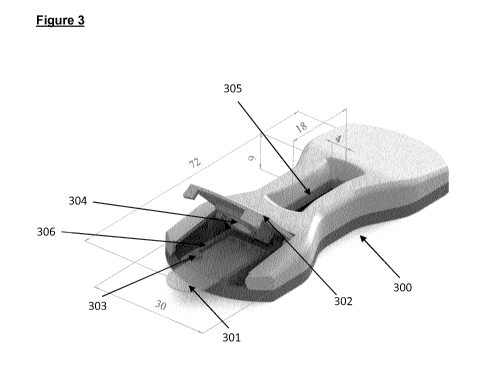
Figure 3 shows in an embodiment of the present invention different views of
the lateral flow device (300) with the of the separate swab (200) inserted in
a
sample pad slot (306), which slot comprises a bulge (303) configured to
interact with the incision (206) of the separate swab (200) to orientate and
position the separate swab (200) in the lateral flow device (300). The lateral
flow device (300) comprises a holding member (302), which may be closed
down over the separate swab (200) and hold and lock the swab in the inserted
position in the lateral flow device (300). The holding member (302) comprises
an opening (304), which in the locked position of the holding member aligns
with the sample collection pad (201) such that the sample collection pad is
exposed and accessible to running buffer introduced through the opening
(304). The lateral flow device (300) further comprises a reaction window
(305),
which allows visual inspection of the detection area (DA).
Figure 4 shows the results of in vitro testing different materials for sample
collection. 80p1 of a standard protein solution (PBS containing 2ng/m1 IL8,
4ng/m1 ILIA and 8ng/m1 IL1 RA recombinant proteins) was used as test
sample. Signal intensities are measured as mV.
Figure 5 shows the results of in vivo testing selected materials for sample
collection. Figure 5A (Forehead) and Figure 5B (Inner arm). Signal intensities
are measured as mV.
CA 03071766 2020-01-31
WO 2019/025610 PCT/EP2018/071179
6
Figure 6 shows the results of comparing blocked sample pads (0083) and the
corresponding unblocked sample pad on inner side of a forearm skin. Signal
intensities are measured as mV.
Figure 7 shows the results of comparing sample collection procedures (in
vivo).
Figure 7A (forehead) and Figure 7B (cheek). Three volunteers (JA, AL, and
AS). Signal intensities are measured as mV.
Figure 8 shows the results of comparing different swabbing procedures in vivo
on two volunteers (JA and AL). Collection by a Z-shape motion versus 5
seconds within an area of 5 cm2. Signal intensities are measured as mV.
Figure 9 shows the effect of pre-treatment of the skin prior to sample
collection
and how long the pre-treatment sustains. Figure 9A (water pre-treatment of
skin), Figure 9B (Et0H pre-treatment of skin) and Figure 90 (pre-treatment of
skin using a cosmetic wipe). Light grey bar (IL1A). Dark grey bar (IL1RA).
Signal intensities are measured as mV.
It will be recognized by the person of ordinary skill in the art, given the
benefit
of this disclosure that certain features shown in figures 1-5 are not
necessarily
drawn to scale. The dimensions and characteristics of some features in the
figures may have been enlarged, distorted or altered relative to other
features
in the figures to facilitate a better understanding of the illustrative
examples
disclosed herein.
It will further be recognized by the person of ordinary skill in the art that
the
individual features of the figures may be interchanged to obtain further
embodiments.
Detailed description of the invention
CA 03071766 2020-01-31
WO 2019/025610 PCT/EP2018/071179
7
In describing the embodiments of the invention specific terminology will be
resorted to for the sake of clarity. However, the invention is not intended to
be
limited to the specific terms so selected, and it is understood that each
specific
term includes all technical equivalents which operate in a similar manner to
accomplish a similar purpose.
A first aspect of the present invention provides a diagnostic kit for
detecting
the presence or quantity of one or more test analytes within a test sample
taken
from a skin surface of a mammal, the diagnostic kit comprising:
a) a separate swab (200, 301) configured to be used for collecting said
test sample, wherein said swab comprising a sample collection pad
(201, 101) attached to a supporting member (202),
b) a lateral flow assay device (300) configured to accept and hold said
separate swab.
The diagnostic kit of the present invention may be employed in point-of-care
devices to detect the presence or absence of one or more test analytes within
a test sample obtained from the skin using the separate swab (200) of the
diagnostic kit.
The separate swab of the diagnostic kit of the present invention is configured
to be suitable for collecting a test sample from the skin surface of a mammal.
In a preferred embodiment, the mammal is a human being. The swab (200,
301) comprises a supporting member (202) to which a sample collection pad
(201, 101) is attached, preferably on one side of the supporting member. The
supporting member (202) is typically used as a handle when the sample is
collected from the skin, e.g. by placing the sample collection pad (201, 101)
on
the skin and moving the pad around on the skin using the supporting member
(202) to control the movement.
CA 03071766 2020-01-31
WO 2019/025610 PCT/EP2018/071179
8
In one embodiment, the supporting member is elongated, for example the
length of the member is at least 2 times the width of the member, such as 2.5
times the width of the member, such as 3 times the width of the member, such
as at least 4 times the width of the member. In one embodiment, the supporting
member is configured with one proximal end (203) configured as a finger grip
and opposite distal end (204) to which said sample collection pad (201, 101)
is attached. The shape of the proximal end (203) may be configured to allow a
firm grip of the supporting member (202) between two or more finger. Figure 2
discloses an example, where the width of the proximal end (203) of the
supporting member (202) is extended to provide a better finger grip. Thus in
one embodiment, proximal end (203) of the supporting member is wider than
the distal end (204). In one embodiment, the area and shape of the proximal
end (203) of the supporting member corresponds to the pulp of an thumb of
an adult human being, which allows a firm grip of the supporting member.
In one embodiment, the supporting member (202) is flexible along the
longitudinal axis of supporting member. The supporting member (202) may be
made of a material that is flexible material such that the supporting member
(202) will bend slightly when the sample collection pad (201, 101) is pressed
against the skin and moved around on the skin to collect the sample material.
The flexibility of the supporting member (202) reduces the risk of injuring
the
skin. In a preferred embodiment, the supporting member (202) is made of a
plastic material, for example the supporting member (202) may be made of a
plastic material, where the thickness of the plastic material is less than
about
2 mm, such as 1 mm or less, such as between 2 and 0.5 mm, such as between
2 and 1 mm, which makes the supporting member (202) to flexible along the
longitudinal axis.
In a preferred embodiment of the present invention, the distal (204) end of
the
supporting member (202) comprises an aperture (205) configured to be
covered by the sample collection pad (201, 101). In this embodiment, the
CA 03071766 2020-01-31
WO 2019/025610 PCT/EP2018/071179
9
sample collection pad (201, 101) attached to the supporting member (202)
covers aperture and the perimeter of the same. In one embodiment, said
sample collection pad (201, 101) is attached to the supporting member (202)
such that said sample collection pad covers said aperture (205).
The sample collection pad (201, 101) may be attached to the supporting
member (202) close to the perimeter of the aperture. The sample collection
pad (201, 101) may be attached to the supporting member (202) further away
from the perimeter of the aperture. The sample collection pad (201, 101) is
typically attached to one side of the supporting member (202). In one
embodiment, the area of the aperture (205) corresponds to at least 50% of the
area of the sample collection pad (201, 101), such as at least 60% of the area
of the sample collection pad, for example at least 70% of the sample
collection
pad, such as at least 70% of the sample collection pad, for example at least
80% of the sample collection pad, such as at least 90% of the sample
collection
pad, for example at least 95% of the sample collection pad.
Inserted in the lateral flow device (300), the sample collection pad (201,
101)
of the swab (200) forms part of the porous support assembly (100), i.e. the
sample collection pad is in contact with the other elements of the assembly.
The aperture allows for access to the sample collection pad (201, 101) for the
addition of a running buffer to facilitate the lateral flow in the porous
support
assembly. In the context of the present invention a running buffer is any
liquid
buffer suitable for facilitate the lateral flow in the porous support
assembly,
such as a PBS buffer.
The sample collection pad (201, 101) is made of a material that is suitable
for
collecting the test sample on the skin and subsequent be mated with and form
part of the porous support assembly (100) and release the test sample to the
porous support assembly (100). In one embodiment, the sample collection pad
(201, 101) is made of a cellulose material, a cellulose derivative such as
CA 03071766 2020-01-31
WO 2019/025610 PCT/EP2018/071179
nitrocellulose, polyether sulfone, polyethylene, nylon polyvinylidene fluoride
(PVDF), polyester, polypropylene, glass fibers, cotton, or cloth. In a
preferred
embodiment, the sample collection pad (201, 101) is made of a cellulose
material, a cellulose derivative such as nitrocellulose.
5
The sample collection pad (201, 101) may be in the form of a sheet or the
like.
In one embodiment, the sample collection pad (201, 101) is in the form of a
layer of one or more sheets or the like, such as a lawyer of two sheets.
10 The average thickness the sample collection pad (201, 101) is preferably
less
than 2 mm, such as in the range of 1 to 0.80 mm, preferably less than 1 mm,
such as less than 0.95 mm, for example less than 0.85 mm, such as in the
range of 0.85 to 0.80 mm, such as 0.83 mm. In one embodiment, the sample
collection pad (201, 101) is in the form of a layer of two sheets, wherein the
thickness of each sheet is less than 0.50 mm such as in the range of 0.49 to
0.40 mm.
In order to prevent or reduce any bias between the absorption of the one or
more test analytes or any bias in the release of the one or more test analytes
from the sample collection pad (201, 101), the sample collection pad (201,
101) may be pre-treated with a blocking buffer. In one embodiment, the
blocking buffer is a PBS buffer comprising 1% BSA or a buffer comprising
10mM Borate, 3% BSA, 1% PVP-40 and 0.25% Triton X100 pH 8Ø
In a particular embodiment, the sample collection pad (201, 101) is in the
form
of a cellulose material or a cellulose derivative such as nitrocellulose pre-
treated with a blocking buffer, wherein the sample collection pad (201, 101)
has a thickness in the range of 0.85 to 0.80 mm, such as 0.83 mm.
In a preferred embodiment of the present invention, the supporting member
(202) of the separate swab (200) comprises an incision (206) on or near the
CA 03071766 2020-01-31
WO 2019/025610 PCT/EP2018/071179
11
edge of the distal end (204) of said supporting member (202). The lateral flow
device (300) comprises a bulge (303) that fits with the incision (206) on the
supporting member (202) and orientates and positions the distal end (204) of
said supporting member (202) when the swab is inserted in the lateral flow
device (300). Thus, the bulge/incision configuration secures that the swab and
in particular the sample collection pad (201, 101) is orientated and
positioned
correctly in the lateral flow device (300). Thus, the bulge/incision
configuration
ensures that the swab and in particular the sample collection pad (201, 101)
can only be inserted in the lateral flow device (300) such that the the sample
collection pad (201, 101) of the swab (200) forms part of the porous support
assembly (100), i.e. the sample collection pad is in contact with the other
elements of the assembly. Accordingly, in one embodiment, the separate swab
(200) comprises an incision (206) on or near the edge of the distal end (204)
of said supporting member (202) and the lateral flow device (300) comprises
a bulge (303) that fits with the incision (206) on the supporting member such
that when inserted in the lateral flow device (300), the sample collection pad
(201, 101) of the swab (200) forms part of the porous support assembly (100),
i.e. the sample collection pad is in contact with the other elements of the
assembly.
In another embodiment of the present invention, the lateral flow device (300)
comprises an opening (304) configured to align with the aperture (205) of the
swab such the sample collection pad (201, 101) is exposed through said
opening (304), when the swab is inserted in the lateral flow device. Although
the sample collection pad (201, 101) may be pre-wetted with running buffer,
the opening allows the addition of (further) running buffer, where the
separate
swab (100) is in an inserted state in the lateral flow device (300).
When used in combination with the bulge/incision configuration, the sample
collection pad (201, 101) is aligned correctly relative to the opening such
that
the running buffer is added to the sample collection pad (201, 101).
CA 03071766 2020-01-31
WO 2019/025610 PCT/EP2018/071179
12
In one embodiment, the lateral flow device comprises a sample pad slot
(306) configured to accept the distal end of said swab such that the position
of the sample collection pad (201, 101, 301) in the lateral flow device is
secured.
In another embodiment, the lateral flow device comprises a holding member
(302) configured to hold the distal end of said swab and secure the position
of
the distal end of said swab comprising said sample collection pad (201, 101).
In a further embodiment, the holding member is attached to the body of the
lateral flow device by a hinge. In the open state, the distal end (204) of
said
supporting member (202) comprising the sample collection pad (201, 101,
301) may be inserted in the device, the insertion may be further facilitated
by
the presence of a sample pad slot (306) configured to accept the distal end of
said swab. In the close position, the holding member is closed around the
sample collection pad (201, 101, 301), which holds the sample collection pad
(201, 101, 301) firmly in the lateral flow device (300). In one embodiment,
the
holding member (302) is configured to be closed down over the distal end of
said swab and locked to the body of the lateral flow device. Preferably, the
holding member (302) configuration of the device is used in combination with
the bulge/incision configuration secures that the swab and in particular the
sample collection pad (201, 101) is orientated and positioned correctly in the
lateral flow device (300).
In a preferred embodiment, the holding member (302) comprises the opening
(304) configured to align with the aperture (205) of the swab such the sample
collection pad (201, 101) is exposed through said opening (304), when the
swab is inserted in the lateral flow device. The opening (304) may be in the
form of port such as in the form of a conical port with the wide base facing
upwards and the narrow base facing downwards. In this configuration,
CA 03071766 2020-01-31
WO 2019/025610 PCT/EP2018/071179
13
(further) running buffer may be added to the sample collection pad (201, 101)
in order to facilitate the lateral flow in the porous support assembly (100).
In the context of the present invention the term lateral flow refers to a
liquid
flow in which the dissolved or dispersed component(s) of the liquid (including
the test analytes) migrates laterally with the liquid through the porous
support
assembly (100, referred to as capillary bed or lateral flow strip) with the
proviso
that component(s) are not permanently entrapped or by other means excluded
from migrating in the liquid. Assay relying on such lateral flow of are
referred
to as lateral flow assay. Where the porous support assembly is preferable
made of a non-bibulous material, the components in the liquid will travel at
an
essential equal speed through the capillary bed. If the porous support
assembly is made of a bibulous material, the migration of one of more of the
component may be affected by the material. If the porous support assembly
comprises or consist of a bibulous material, the material may be treated with
a
blocking agent, such as PBS buffer comprising BSA and/or Triton X-100, in
order to change the properties of the porous support assembly such that the
flow characteristics is identical or essentially identical that of a non-
bibulous
material.
The lateral flow assay is based on the porous support assembly (100) ¨ a
capillary bed (such as porous paper or sintered polymer) ¨ having the capacity
to transport fluid by action of capillary forces. The porous support assembly
(100) is an assembly of porous support elements, which elements are in in
fluid communication with each other when fluid (such as a running buffer) is
applied to the assembly. One of the porous support elements of the porous
support assembly (100) is the sample collection pad (101, 200), which become
part of the porous support assembly (100) when the swab is in the inserted
position in the lateral flow device. The porous support assembly (100) is also
referred to as the lateral flow assay strip.
CA 03071766 2020-01-31
WO 2019/025610 PCT/EP2018/071179
14
In one embodiment of the present invention, the lateral flow device is
constructed so as to form a porous support assembly (100), when it is mated
with the sample collection pad attached to said swab, wherein the lateral flow
device (300) mated with the sample collection pad comprise an elution zone
(101), a conjugate zone (102) and a detection area (DA).
The conjugation zone may be an integrated part of a larger porous element of
the porous support assembly (100), such as a porous support strip (107). The
conjugation zone may also be in the form of an element of the porous support
assembly (100). In a preferred embodiment, the conjugation zone is in the form
of a conjugate pad (102).
The sample collection pad (101, 200) functions as a sponge and holds the test
sample. Once it is soaked, the test sample, containing one or more test
analytes, will migrate from sample collection pad (101, 200) into the adjacent
element of the porous support assembly (100). The interphase between the
sample collection pad (101, 200) and the adjacent element of the porous
support assembly is referred to as the elution zone. The adjacent element of
the porous support assembly is typically a conjugate zone, preferably in the
form of a conjugate pad (102). The conjugate zone/conjugate pad (102)
typically contains one or more indicator affinity molecule(s), such as
affinity
molecules tagged with detection probe designed to bind to the one or more
test analytes within the test sample. The test sample and one or more affinity
molecules are mixed and the one or more affinity molecules having affinity for
one or more test analytes within the test sample will bind to each other while
migrating further to a detection area (DA) that may contain a detection zone
(105), and may contain an indicator zone (106), both with one or more stripes,
where another set of one or more affinity molecules have been immobilized.
By the time the test sample mixed with the affinity molecule(s) from the
conjugate pad reaches the detection area (DA), the one or more analytes in
the test sample will have been bound to the affinity molecule(s) from the
CA 03071766 2020-01-31
WO 2019/025610 PCT/EP2018/071179
conjugate pad. This complex will then in turn be bound by the affinity
molecule(s) on the stripe(s) in the detection zone (105). After a while, when
more and more fluid has passed the detection zone, detection probes
accumulate, and the stripe changes color. The detection probes may e.g. be
5 gold or latex particles conjugated to the affinity molecule(s) to prepare
affinity
molecules tagged with detection probes. The detection area (DA) may also
comprise an indicator zone (106) which can function as a control to verify
that
the lateral flow assay has been conducted properly. Such indicator zone (106)
may also comprise one or more stripes with affinity molecules immobilized that
10 only binds to the affinity molecule(s) tagged with detection probes from
the
conjugate pad, whereas the affinity molecule(s) in the detection zone (105)
bind to the complex between the analyte(s) and the indicator affinity
molecule(s), such as the affinity molecule(s) tagged with detection probes
from
the conjugate pad. After passing the detection area (DA) the fluid enters the
15 wicking pad (104), which generally receives fluid that has migrated
through the
entire porous support assembly (100). Thus in one embodiment, the detection
area (DA) comprise a detection zone (105) containing one or more affinity
molecule(s) for selectively retaining one or more test analyte(s) and
optionally
an indicator zone (106) containing one or more affinity molecule(s) for
selectively retaining one or more indicator affinity molecule(s).
The detection zone (105) may be located upstream or downstream of the
indicator zone (106). The lines or stripes in the detector zone or indicator
zone
may be disposed in a direction that is substantially perpendicular to the flow
of
the test sample. In some embodiments the lines may be in a direction that is
substantially parallel to the flow of the test sample. The lines or stripes in
the
detection zone (105) or indicator zone (106) does not need to be lines or
stripes, and can also be other shapes, such as e.g. dots or patterns.
In one embodiment, the lateral flow device further comprises a wicking pad
(104). The wicking pad is part of the porous support assembly (100) and may
CA 03071766 2020-01-31
WO 2019/025610 PCT/EP2018/071179
16
assist in promoting capillary action and fluid flow from the sample pad (101),
conjugate pad (102) through the detection area (DA).
In another embodiment, the lateral flow device comprises a backing material
(108) on the backside of said porous support assembly (100) facing away
from the elution zone. The backing layer (108) is liquid-impermeable so that
fluid flowing through porous support assembly (100) does not leak through
the backing layer (108). Examples of suitable materials for the support
include, but are not limited to, glass; polymeric materials, such as
polystyrene, polypropylene, polyester, polybutadiene, polyvinylchloride,
polyamide, polycarbonate, epoxides, methacrylates, and polymelamine.
The porous support assembly (100) is an assembly of two or more porous
elements, for example one or more porous elements and the sample collection
pad (201, 101), where the swab (200) comprising the sample collection pad is
inserted in the lateral flow assay device (300). The elements are preferably
in
the form of membranes, such as sheet like membranes. The porous support
assembly (100) may have a thickness equal to or less than 4 mm (such as less
than 4, 3, 2, 1 mm), and a width and a length both greater than the thickness.
.. In some embodiments the width and length of the porous support assembly
(100) are both greater (e.g. 3, 4, 5, 6, 7, 8, 9, 10, 50 times greater or up
to 4,
5, 6, 7, 8, 9, 10, 50 times greater) than the thickness. In some embodiments
the porous support assembly (100) is a square, such as a rectangle, and in
some embodiments the porous support assembly (100) is circular. If the
porous support assembly (100) is an irregular shape, i.e. different from a
square or rectangle, then the width, length and thickness refers to the
maximum values for such an irregular shape. For example the width of a circle
will be the diameter. Examples of widths and lengths may be 4, 5, 6, 7, 8, 9,
10, 11, 12, 13, 14, 15, 16, 17, 18, 19, 20, 21, 22, 23, 24, 25, 26, 27, 28,
29, 30,
35, 40 mm, such as e.g. range of widths and lengths from 5-30 mm.
CA 03071766 2020-01-31
WO 2019/025610 PCT/EP2018/071179
17
Thus in one embodiment, the porous support assembly (100) of the lateral flow
device (300) has an average thickness equal to 4 mm or less, and a width and
a length, both greater than the thickness, wherein the lateral flow device is
configured to have a lateral flow direction (L) in the direction of a plane
created
by the width and the length of the porous support assembly.
In one embodiment, the lateral flow device comprises a reaction window (305)
configured for visual inspection of the detection area (DA).
The diagnostic kit of the present invention may be used for testing analytes
present on the skin and obtainable using the separate swab (200). In one
embodiment, the one or more test analyte(s) are selected from the list
consisting of: chemokines, interleukins, growth factors, hormones, enzymes,
and other molecules present on the skin of a mammal, such as selected from
the list consisting of: IL-la, IL-1b, IL-1RA, IL-8, CCL-2, CCL-5, CCL-27,
CXCL-1, CXCL-2, CXCL-9, Trappin2/Elafin, hBD-1, hBD-2, VEGF, and TSLP.
In a preferred embodiment the test analytes are the combination of IL-8, IL-la
and IL-1RA.
As mentioned, the diagnostic kit of the present invention may be employed in
point-of-care devices to detect the presence or absence of one or more test
analytes within a test sample obtained from the skin using the separate swab
(200) of the diagnostic kit. The readout may be done visually, i.e. presence
or
absence of a one or more coloured test lines also referred to as test stripes
in
a detection zone (105), and the confirmation/validation of the test may be
done
by the presence and/or absence of one or more coloured indicator lines/stripes
in an indicator zone (106). The test may be qualitative (presence or absence)
as well as quantitative, and the detection/quantification may be aided by
reading equipment, or can be purely visual detection by the eye of the user of
the lateral flow assay.
CA 03071766 2020-01-31
WO 2019/025610 PCT/EP2018/071179
18
One aspect of the present invention provides a method for detecting the
presence or quantity of one or more test analytes, the method comprising the
following steps:
a) provide a separate swab (200) comprising a sample collection pad
(201) as defined herein, wherein said sample pad comprises a test
sample obtained from the a skin surface of a subject using said
separate swab;
b) insert said swab comprising a sample collection pad containing said
test sample in the lateral flow assay device adapted to receive said
separate swab insert as defined in any of the preceding claims;
c) developing the lateral flow assay.
Another aspect of the present invention provides a method for detecting the
presence or quantity of one or more test analytes, the method comprising the
following steps:
a) collecting a test sample from the a skin surface of a subject using a
separate swab (200) comprising a sample collection pad (201) as
defined,
b) insert said swab comprising a sample collection pad containing said
test sample in the lateral flow assay device adapted to receive said
separate swab insert as defined in any of the preceding claims;
c) developing the lateral flow assay.
The subject is a mammal, preferably a human being. The test sample is
obtained using the separate swab (200) comprising a sample collection pad
(201), which is applied to the skin of the mammal, preferably the skin of a
human being. The area of the skin may for example be the forehead, cheek,
the inner arm or a part of the arm which is normally exposed to the sun. The
separate swab may be applied to a pre-determined area, such an area not
exceeding 5 cm2. The separate swab may also be applied in pre-determined
time, such 5 seconds or 30 seconds. The test sample may also be collected
CA 03071766 2020-01-31
WO 2019/025610 PCT/EP2018/071179
19
by applying a pre-determined motion of the swab, such as a z-shaped motion
of the swab on the skin.
The sampling may be assisted by wetting the sample collection pad (201) with
a fixed volume of fluid. In a preferred embodiment, the sample collection pad
(201) of the separate swab (200) is pre-wetted with a buffer before the sample
collection, such as a with a fixed volume of a buffer. The buffer may be any
suitable buffer such as a PBS buffer. The buffer used for pre-wetting the
sample collection pad may be the same buffer used as running buffer in the
lateral flow assay step of the procedure.
In another preferred embodiment, running buffer is added to sample collection
pad inserted in the lateral flow assay device. The running buffer is added to
the sample collection pad inserted in the lateral flow assay device to
facilitate
or provide sufficient fluid for the lateral flow in the porous support
assembly
(100) and the development of the assay. Where an opening (304) is present
in the lateral flow device (300), the opening may be used as a port to add
running buffer to the sample collection pad (201) inserted in the device.
.. In one embodiment, the lateral flow assay device comprises an elution zone
(101) and a detection area (DA), and wherein said sample collection pad is the
elution zone (101).
In another embodiment, the lateral flow device is constructed so as to form a
porous support assembly (100), when it is mated with the sample collection
pad attached to said swab, wherein the lateral flow device (300) mated with
the sample collection pad comprise an elution zone (101), a conjugate zone
(102) and a detection area (DA).
In one embodiment, the porous support assembly (100) has a thickness equal
to 4 mm or less, and a width and a length, both greater than the thickness,
CA 03071766 2020-01-31
WO 2019/025610 PCT/EP2018/071179
wherein the lateral flow device is configured to have a lateral flow direction
(L)
substantially in the direction of a plane created by the width and the length
of
the porous support assembly (100).
5 In another embodiment, the detection area (DA) comprise a detection zone
(105) containing one or more affinity molecule(s) for selectively retaining
one
or more test analyte(s) and optionally an indicator zone (106) containing one
or more affinity molecule(s) for selectively retaining one or more indicator
affinity molecule(s).
In general the present invention is directed to a diagnostic kit that provides
an
integrated system for detecting the presence or absence of one or more test
analytes within a test sample obtained from the skin, over a broad range of
possible concentrations of the one or more test analytes. In some
.. embodiments the quantity of the one or more test analytes are also detected
in a quantitative assay. The diagnostic kit employs a lateral flow assay
device
(300) and a separate swab (200, 301) and one or more assay reagents for
detecting the one or more test analytes within the test sample. The assay
reagents include affinity molecule(s) tagged with detection probes that are
capable of producing a detection signal representing the presence or quantity
of the one or more test analyte(s) in the test sample. One way of quantifying
one or more of the test analyte(s) is by preparing suitable standard curves
using known concentrations of the one or more test analyte(s).
The one or more test analyte(s) assayed using the method of the invention
may be selected from the list consisting of: chemokines, interleukins, growth
factors, hormones, enzymes, and other molecules present on the skin of a
mammal, such as selected from the list consisting of: IL la, IL 1 b, IL 1RA,
IL
8, CCL 2, CCL 5, CCL 27, CXCL 1, CXCL 2, CXCL 9, Trappin2/Elafin, hBD 1,
.. hBD 2, VEGF, and TSLP. In a preferred embodiment, the test analytes are the
combination of IL-8, IL-la and IL-1 RA.
CA 03071766 2020-01-31
WO 2019/025610 PCT/EP2018/071179
21
If desired, a suitable reading equipment, such as an optical reader may be
used in some embodiments to measure the intensity of the probes. The actual
configuration and structure of the optical reader may generally vary depending
on the probes, which are to be measured. For example, optical detection
techniques that may be utilized include, but are not limited to, luminescence
(e.g. fluorescence, phosphorescence, etc.), absorbance (e.g. fluorescent or
non-fluorescent), diffraction, and so on. Qualitative, quantitative, or semi-
quantitative determination of the presence or concentration of an analyte may
be achieved in accordance with the present invention. For instance, the
amount of the analyte may be quantitatively or semi-quantitatively determined
by using the intensities of the signals produced by detection probes bound at
the detection zone (105) and the indicator zone (106).
In a preferred embodiment, an image of the detection area (DA) is captured
using a suitable device for capturing images, such as a cell-phone comprising
a camera. The image may subsequently be transmitted to a computer system
(for example a remotely located server) comprising an image processor and a
database, where the image is analysed, e.g. by extracting the image features
and compare the features with corresponding features stored in a database.
The computer system may then generate an output datum based on said
image features, which may be transmitted to the user, e.g. back to the cell-
phone used for capturing image.
Thus in one embodiment of the present invention, the method of the invention
further comprises a step d) of capturing an image of the detection area (DA)
and transmitting said image to a computer system comprising an image
processor and a database, wherein the image features are extracted from the
image by the image processor and said image features is stored in said
database and, wherein said computer system generates a least one output
datum based on said image features. In a further embodiment, the image is
CA 03071766 2020-01-31
WO 2019/025610 PCT/EP2018/071179
22
captured using a mobile device, such a cell phone configured to capture
images. In yet a further embodiment, the output datum generated by the
computer system is transferred to the mobile device.
When describing the embodiments of the present invention, the combinations
and permutations of all possible embodiments have not been explicitly
described. Nevertheless, the mere fact that certain measures are recited in
mutually different dependent claims or described in different embodiments
does not indicate that a combination of these measures cannot be used to
.. advantage. The present invention envisages all possible combinations and
permutations of the described embodiments.
The terms "comprising", "comprise" and "comprises" herein are intended by
the inventors to be optionally substitutable with the terms "consisting of",
"consist of" and "consists of", respectively, in every instance.
The invention is further described in the following non-limiting items.
Item 1. A diagnostic kit for detecting the presence or quantity of one or more
.. test analytes within a test sample taken from a skin surface of a mammal,
the
diagnostic kit comprising:
a) a separate swab (200, 301) configured to be used for
collecting
said test sample, wherein said swab comprising a sample collection pad (201,
101) attached to a supporting member (202),
b) a lateral flow assay device (300) configured to accept and hold
said separate swab.
Item 2. The diagnostic kit of item 1 characterized in that the lateral flow
assay
device (300) comprises one or more porous elements and wherein the said
sample collection pad (201, 101) is configured to form part of a porous
support
CA 03071766 2020-01-31
WO 2019/025610 PCT/EP2018/071179
23
assembly (100) when the separate swab (200, 301) is inserted in said lateral
flow assay device (300).
Item 3. The diagnostic kit of item 1 or 2, characterized in that said
supporting
member is configured with one proximal end (203) configured as a finger grip
and opposite distal end (204) to which said sample collection pad (201) is
attached.
Item 4. The diagnostic kit according to any one of items 1 to 3, characterized
in that said is supporting member (202) is flexible along the longitudinal
axis of
supporting member.
Item 5. The diagnostic kit according to any one of items 1 to 4, characterized
in that said distal (204) end of said supporting member (202) comprises an
aperture (205) configured to be covered by the sample collection pad (201).
Item 6. The diagnostic kit according to any one of items 1 to 5, characterized
in that said sample collection pad (201, 101) is attached to the supporting
member (202) such that said sample collection pad covers said aperture (205).
Item 7. The diagnostic kit according to any one of items 1 to 6, characterized
in that the area of the aperture (205) corresponds to at least 50% of the area
of the sample collection pad (201, 101), such as at least 60% of the area of
the sample collection pad, for example at least 70% of the sample collection
pad, such as at least 70% of the sample collection pad, for example at least
80% of the sample collection pad, such as at least 90% of the sample
collection
pad, for example at least 95% of the sample collection pad.
Item 8. The diagnostic kit according to any one of items 1 to 7, characterized
in that the sample collection pad (201, 101) is made of a cellulose material,
a
cellulose derivative such as nitrocellulose, polyether sulfone, polyethylene,
CA 03071766 2020-01-31
WO 2019/025610 PCT/EP2018/071179
24
nylon polyvinylidene fluoride (PVDF), polyester, polypropylene, glass fibers,
cotton, or cloth.
Item 9. The diagnostic kit according to any one of items 1 to 8, characterized
in that the sample collection pad (201, 101) is pre-treated with a blocking
buffer, such as a PBS buffer comprising 1`)/0 BSA or a buffer comprising 10mM
Borate, 3% BSA, 1% PVP-40 and 0.25% Triton X100 pH 8Ø
Item 10. The diagnostic kit according to any one of items 1 to 9,
characterized
in that the sample collection pad (201, 101) is in the form of a sheet or the
like.
Item 11. The diagnostic kit according to any one of items 1 to 10,
characterized
in that the sample collection pad (201, 101) is in the form of a layer of one
or
more sheets or the like, such as a lawyer of two sheets.
Item 12. The diagnostic kit according to any one of items 1 to 11,
characterized
in that the thickness of the sample collection pad (201, 101) is less than 2
mm,
such as in the range of 1 to 0.80 mm, preferably less than 1 mm, such as less
than 0.95 mm, for example less than 0.85 mm, such as in the range of 0.85 to
0.80 mm.
Item 13. The diagnostic kit according to any one of items 1 to 12,
characterized
in that the sample collection pad (201, 101) is in the form of a layer of two
sheets, wherein the thickness of each sheet is less than 0.50 mm such as in
the range of 0.49 to 0.40 mm.
Item 14. The diagnostic kit according to any one of items 1 to 13,
characterized
in that the sample collection pad (201, 101) is in the form of a cellulose
material, a cellulose derivative such as nitrocellulose pre-treated with a
blocking buffer, wherein the sample collection pad (201, 101) has a thickness
in the range of 0.85 to 0.80 mm.
CA 03071766 2020-01-31
WO 2019/025610 PCT/EP2018/071179
Item 15. The diagnostic kit according to any one of items 1 to 14,
characterized
in that the supporting member is made of a plastic material.
5 Item 16. The diagnostic kit according to any one of items 1 to 15,
characterized
in that the supporting member (202) is be made of a material that is flexible
material such that the supporting member (202) will bend slightly when the
sample collection pad (201, 101) is pressed against the skin and moved
around on the skin to collect the test sample.
Item 17. The diagnostic kit according to any one of items 1 to 16,
characterized
in that the supporting member is made of a plastic material, where the
thickness of the plastic material is less than about 2 mm, such as 1 mm or
less,
such as between 2 and 0.5 mm, such as between 2 and 1 mm
Item 18. The diagnostic kit according to any one of items 1 to 17,
characterized
in that one edge of the said distal end (204) of said supporting member (202)
comprises an incision (206) and wherein the lateral flow device comprises a
bulge (303) configured to orientate and position the distal end of said
.. supporting member when the swab is inserted in the lateral flow device.
Item 19. The diagnostic kit according to any one of items 1 to 18,
characterized
in that the lateral flow device comprises an opening (304) configured to align
with the aperture (205) of the swab such the sample collection pad (201, 101)
is exposed through said opening (304), when the swab is inserted in the
lateral
flow device.
Item 20. The diagnostic kit according to any one of items 1 to 19,
characterized
in that the lateral flow device comprises a sample pad slot (306) configured
to
accept the distal end of said swab such that the position of the sample
collection pad (201, 101, 301) in the lateral flow device is secured.
CA 03071766 2020-01-31
WO 2019/025610 PCT/EP2018/071179
26
Item 21. The diagnostic kit according to any one of items 1 to 20,
characterized
in that the lateral flow device comprises a holding member (302) configured to
hold the distal end of said swab and secure the position of the distal end of
said swab comprising said sample collection pad (201, 101).
Item 22. The diagnostic kit according to any one of items 1 to 21,
characterized
in that the holding member is attached to the body of the lateral flow device
by
a hinge.
Item 23. The diagnostic kit according to any one of items 1 to 22,
characterized
in that the holding member (302) is configured to be folded over the distal
end
of said swab and locked to the body of the lateral flow device.
Item 24. The diagnostic kit according to any one of items 1 to 23,
characterized
in that the holding member (302) comprises said opening (304).
Item 25. The diagnostic kit according to any one of items 1 to 24,
characterized
in that the said opening (304) is in the form of port such as in the form of a
conical port with the wide base facing upwards and the narrow base facing
downwards.
Item 26. The diagnostic kit according to any one of items 1 to 25,
characterized
in that the mammal is a human being.
Item 27. The diagnostic kit according to any one of items 1 to 26,
characterized
in that the lateral flow device is constructed so as to form a porous support
assembly (100), when it is mated with the sample collection pad attached to
said swab, wherein the lateral flow device (300) mated with the sample
collection pad comprise an elution zone (101), a conjugate zone (102) and a
detection area (DA).
CA 03071766 2020-01-31
WO 2019/025610 PCT/EP2018/071179
27
Item 28. The diagnostic according to any one of items 1 to 27, characterized
in that the conjugation zone is in the form of a conjugate pad (102).
Item 29. The diagnostic according to any one of items 1 to 28, characterized
in that the lateral flow device further comprises a wicking pad (104).
Item 30. The diagnostic according to any one of items 1 to 29, characterized
in that the lateral flow device comprises a backing material (108) on the
backside of said porous support assembly (100) facing away from the elution
zone.
Item 31. The diagnostic according to any one of items 1 to 30, characterized
in that the porous support assembly has an average thickness equal to 4 mm
or less, and a width and a length, both greater than the thickness, wherein
the
lateral flow device is configured to have a lateral flow direction (L) in the
direction of a plane created by the width and the length of the porous support
assembly (100).
Item 32. The diagnostic according to any one of items 1 to 31, characterized
in that the detection area (DA) comprise a detection zone (105) containing one
or more affinity molecule(s) for selectively retaining one or more test
analyte(s)
and optionally an indicator zone (106) containing one or more affinity
molecule(s) for selectively retaining one or more indicator affinity
molecule(s).
Item 33. The diagnostic according to any one of items 1 to 32, characterized
in that the lateral flow device comprises a reaction window (305) configured
for
visual inspection of the detection area (DA).
Item 34. The diagnostic kit according to any one of items 1 to 33,
characterized
in that the one or more test analyte(s) are selected from the list consisting
of:
CA 03071766 2020-01-31
WO 2019/025610 PCT/EP2018/071179
28
chemokines, interleukins, growth factors, hormones, enzymes, and other
molecules present on the skin of a mammal, such as selected from the list
consisting of: IL la, IL 1 b, IL 1RA, IL 8, CCL 2, CCL 5, CCL 27, CXCL 1, CXCL
2, CXCL 9, Trappin2/Elafin, hBD 1, hBD 2, VEGF, and TSLP.
Item 35. The diagnostic kit according to any one of items 1 to 34,
characterized
in that the test analytes are IL-8, IL la and IL 1RA.
Item 36. The diagnostic kit according to any one of items 1 to 35,
characterized
in that it further comprises a separate container comprising a buffer suitable
for prewetting the sample pad of said separate swab (200).
Item 37. The diagnostic kit according to item 36, characterized in that the
separate container comprises running buffer.
Item 38. The diagnostic kit according to any one of items 1 to 37,
characterized
in that the separate swab (200) comprises an incision (206) on or near the
edge of the distal end (204) of said supporting member (202) and the lateral
flow device (300) comprises a bulge (303) that fits with the incision (206) on
the supporting member such that when inserted in the lateral flow device
(300),
the sample collection pad (201, 101) of the swab (200) forms part of the
porous
support assembly (100).
Item 39. Method for detecting the presence or quantity of one or more test
analytes, the method comprising the following steps:
a) provide a separate swab (200) comprising a sample collection
pad
(201) as defined in any of the preceding items, wherein said sample pad
comprises a test sample obtained from the a skin surface of a mammal using
said separate swab;
CA 03071766 2020-01-31
WO 2019/025610 PCT/EP2018/071179
29
b) insert said swab comprising a sample collection pad containing
said test sample in the lateral flow assay device adapted to receive said
separate swab insert as defined in any of the preceding items;
c) developing the lateral flow assay device.
Item 40. The method according to item 39 further comprising adding running
buffer to sample collection pad inserted the lateral flow assay device.
Item 41. The method to any one of items 39 to 40, wherein the lateral flow
assay device comprises an elution zone (101) and a detection area (DA), and
wherein said sample collection pad is the elution zone (101).
Item 42. The method according to any one of items 39 to 41, wherein the skin
surface of the mammal is the skin of a human being.
Item 43. The method according to any one of items 39 to 42, wherein the
lateral
flow device is constructed so as to form a porous support assembly (100),
when it is mated with the sample collection pad attached to said swab, wherein
the lateral flow device (300) mated with the sample collection pad comprise an
elution zone (101), a conjugate zone (102) and a detection area (DA).
Item 44. The method according to any one of items 39 to 43, wherein the
porous support assembly (100) has a thickness equal to 4 mm or less, and a
width and a length, both greater than the thickness, wherein the lateral flow
device is configured to have a lateral flow direction (L) substantially in the
direction of a plane created by the width and the length of the the porous
support assembly (100).
Item 45. The method according to any one of items 39 to 44, wherein the
detection area (DA) comprise a detection zone (105) containing one or more
affinity molecule(s) for selectively retaining one or more test analyte(s) and
CA 03071766 2020-01-31
WO 2019/025610 PCT/EP2018/071179
optionally an indicator zone (106) containing one or more affinity molecule(s)
for selectively retaining one or more indicator affinity molecule(s).
Item 46. The method according to any one of items 39 to 45, wherein said test
5 analytes are IL-8, IL la and IL 1RA.
Item 47. The method according to any one of items 39 to 46 further comprising
a step d) of capturing an image of the detection area (DA) and transmitting
said image to a computer system comprising an image processor and a
10 database, wherein the image features are extracted from the image by the
image processor and said image features is stored in said database and,
wherein said computer system generates a least one output datum based on
said image features.
15 Item 48. The method according to item 47, wherein the image is captured
using
a mobile device, such a cell phone configured to capture images.
Item 49. The method according to item 48, wherein output datum generated
by the computer system is transferred to the mobile device.
Item 50. The method according a to any one of items 39 to 49, wherein the
sample collection pad (201, 101) is pre-treated with a blocking buffer, such
as
a PBS buffer comprising 1% BSA or a buffer comprising 10mM Borate, 3%
BSA, 1% PVP-40 and 0.25% Triton X100 pH 8Ø
Item 51. The method according a to any one of items 39 to 50, wherein the
sample collection pad (201, 101) is prewetted prior to sample collection.
Examples
Sample pad material and treatment
CA 03071766 2020-01-31
WO 2019/025610 PCT/EP2018/071179
31
Example 1 ¨ In vitro testing of sample pad materials
Different materials were tested as a sample collection material.
In vitro testing of the best material for sample collection and release was
performed. 80p1 Standard protein solution (PBS containing 2ng/m1 IL8, 4ng/m1
ILIA and 8ng/m1 !URA recombinant proteins) was pipetted onto parafilm and
adsorbed with 1x1 cm pieces of different possible sample pad material (blocked
and unblocked). Sample pads were incubated 5min at room temperature,
inserted into SELF cassette, covered with FibroTx sample pad carrier (clear
plastic strip, sample pad removed), cassette closed and 80p1 running buffer
(PBS + 1%Tween20) applied, results were read after 20min run with Qiagen
ESEQuant LR3 lateral flow reader, signal intensity results are shown in mV.
Material IL8 (mV) ILIA (mV) !URA (mV)
FibroTx
sample pad Cellulose,
0.95mm 367.7 119.7 20.7
C095 Cellulose,
0.95mm 448.1 99.1 0.0
C095, blocked
with 1%BSA/
PBS Cellulose,
0.95mm 162.0 89.4 51.8
Cellulose,
0.83mm 303.4 108.6 0.0
C083, blocked Cellulose,
0.83mm 337.7 151.2 78.4
CA 03071766 2020-01-31
WO 2019/025610 PCT/EP2018/071179
32
2x 0048 Cellulose,
0.48mm 266.6 81.1 0.0
2x C048, Cellulose,
blocked 0.48mm 330.7 167.3 112.9
2x111 Glass
Microfiber,
0.28mm 278.2 151.5 47.2
2x111, Glass
blocked Microfiber,
0.28mm 297.5 132.3 92.0
2x 226 Cotton
0.83mm 337.1 105.5 0.0
2x226, Cotton
blocked 0.83mm 220.3 125.3 41.8
222 Cotton,
0.83mm 277.5 112.3 0.0
222 blocked Cotton,
0.34mm 327.9 126.9 85.2
The data are presented in Figure 4.
Unblocked sample pads resulted in lower signal intensities on ILIA test line
compared to blocked sample pads of the same material and at the same time
failed to release enough !URA to result in detectable test line for most
sample
pads tested. Blocked sample pads resulted in detectable !URA.
Example 2 - In vivo testing of sample pad materials
For in vivo testing best materials based on the in vitro results were
selected:
C083, C048 (2 layers), 111 (2 layers), 222, all blocked, were selected for
testing on skin. First FibroTx sample pad (C095, unblocked) was also included.
CA 03071766 2020-01-31
WO 2019/025610 PCT/EP2018/071179
33
Sample pads were placed on skin and covered with FibroTx sample pad
bandage (sample pad was removed). 90p1 PBS was applied and sample pads
were incubated on skin for 15 minutes. Sample pads were inserted into
cassettes (including pad carriers), 80p1 running buffer (PBS + 1%Tween20)
applied, results were read after 20min run with Qiagen ESEQuant LR3 lateral
flow reader, signal intensity results are performed in mV. Skin from sun-
exposed area (forehead) and sun-nonexposed area (inner side of a forearm)
were tested in parallel.
Area tested: Forehead
Sample pad IL8 (mV) ILIA (mV) ILI RA (mV)
FibroTx pad 0 32,4 68,1
0 14,8 46,3
0083, blocked 0 56,7 349,0
2x 0048, blocked 0 29,4 274,1
2x 111, blocked 0 60,9 324,9
222 blocked 0 25,9 196,9
Area tested: Inner arm
Sample pad ILEI (mV) ILIA (mV) ILIRA (mV)
FibroTx pad 0 30,5 0
0083, blocked 0 167,0 70,8
2x 0048, blocked 0 73,0 32,1
2x 111, blocked 0 69,5 17,7
222 blocked 0 59,9 18,2
The data are presented in Figure 5A (Forehead) and Figure 5B (Inner arm)
CA 03071766 2020-01-31
WO 2019/025610
PCT/EP2018/071179
34
On skin, 0083 gave strongest signals, both from forehead and inner arm skin
therefore this material was selected as a sample collection pad for FibroTx
SELF.
Example 3 ¨ In vivo testing block buffers
For this material 2 different blocking buffers were tested:
= Simpler solution: 1% BSA + PBS
= More complex solution: 10mM Borate, 3% BSA, 1% PVP-40, 0.25%
Triton-X100, pH8.0
Blocked sample pads (0083) were tested on inner side of a forearm skin and
compared to unblocked sample pad.
Sample pads were placed on skin and covered with FibroTx sample pad
bandage (sample pad was removed). 90p1 PBS was applied and sample pads
were incubated on skin for 15 minutes. Sample pads were inserted into
cassettes (including pad carriers), 80p1 running buffer (PBS + 1%Tween20)
applied, results were read after 20min run with Qiagen ESEQuant LR3 lateral
flow reader, signal intensity results are shown in mV.
1L8 (mV) ILIA IL1R
(mV) A
0083 unblocked 0 44.1 0
0083 blocked with 1%BSA+PBS 0 61.8 18.8
0083 blocked with 10mM Borate + 3%BSA 0 104.9 27.4
+ 1% PVP-40 + 0.25% Triton-X100, pH8.0
0083 sample pad blocked with more complex blocking buffer shows clearly
best results for testing skin with FibroTx SELF. The data are presented in
Figure 6.
CA 03071766 2020-01-31
WO 2019/025610 PCT/EP2018/071179
Example 4 ¨ In vivo sample collection procedure
Biomarker sample can be obtained from skin by incubating the sample pad on
5 skin (secured to skin with a bandage) or by swabbing (wiping/rubbing) the
sample pad on the skin. These methods were compared to see if the swabbing
could be used for simplifying the sample collection for the customer and to
shorten the overall test time.
Sample collection by sample pad incubation on skin:
10 Sample pads (0083 blocked) were placed on skin and covered with FibroTx
sample pad bandage. 2 drops of activation buffer (PBS) was applied and
sample pads were hold on skin for 10 minutes.
Alternative sample collection method- rubbing the sample pad on skin:
15 Volunteers were given directions to add 2 drops of PBS onto the sample
pad,
remove the pad with the carrier from the bandage and rub the sample pad on
skin, 2 different ways (not to touch the sample pad area from either side).
a) Wipe the sample collection area in a Z-shape motion
b) Wipe the sample collection area 5 seconds within approximately 5cm
20 area with circular motions.
After sample collection in either method described above, the sample pads
were inserted into cassettes (including pad carriers), 2 drops of running
buffer
(PBS) applied, results were read after 20min run with Qiagen ESEQuant LR3
25 lateral flow reader, signal intensity results are shown in mV.
Volunteer Area Sample IL8 ILIA !URA
tested collection (mV) (mV) (mV)
JA Forehead Z-motion 49.6 68.7 360.8
5" rubbing 29.4 70.8 277.0
CA 03071766 2020-01-31
WO 2019/025610 PCT/EP2018/071179
36
10' incub. 0.0 43.5 320.3
Cheek Z-motion 16.1 40.5 186.0
5" rubbing 0.0 99.5 248.0
incub. 0.0 68.1 237.7
AL Forehead Z-motion 0.0 0.0 138.7
5" rubbing 11.6 49.1 346.5
10' incub. 0.0 30.7 104.0
Cheek Z-motion 12.1 12.1 106.4
5" rubbing 0.0 30.0 252.2
10' incub. 0.0 84.4 249.9
AS Forehead Z-motion 0.0 0.0 92.8
5" rubbing 0.0 32.1 225.8
10' incub. 0.0 37.4 298.8
Cheek Z-motion 0.0 25.0 72.4
5" rubbing 0.0 20.1 93.3
10' incub. 0.0 91.9 122.0
The data are presented in Figure 7A (forehead) and Figure 7B (cheek).
Swabbing the skin in Z-motion could obtain too little amount of sample
5 (forehead).
For cosmetic purposes sample collection by swabbing was chosen as it
considerably shortens and simplifies sample collection for the customer.
Longer swabbing time was tested also.
10 2 drops of PBS was added to sample pad and swabbed on skin in 3
different
ways:
= Wipe the sample collection area in a Z-shape motion
= Wipe the sample collection area 5 seconds within approximately 5cm
area with circular motions.
CA 03071766 2020-01-31
WO 2019/025610 PCT/EP2018/071179
37
= Wipe the sample collection area 30 seconds within approximately 5cm
area with circular motions.
After sample collection the sample pads were centrifuged to eluate the PBS
containing the collected sample, biomarker levels were determined using
Enzyme-Linked ImmunoSorbent Assay, biomarker levels shown in ng/ml.
Sample ILIA !URA
Volunteer
collection (ng/nril) (ng/n11)
Z-motion 0.11 1.28
5" swabbing 0.22 3.79
JA
30"
0.53 4.46
swabbing
Z-motion 0.03 2.17
5" swabbing 0.09 5.07
AL
30"
0.38
swabbing 6.60
The data are presented in Figure 8.
5" swabbing obtains 2 times more material from skin compared to Z-motional
swabbing. Longer swabbing time (30") does increase the amount of obtained
material even more, but swabbing for over 10" causes crumbling of the sample
pad material (depending on the intensity and pressure of swabbing by the
customer) and therefore could not be suggested for functional tests.
Example 5 ¨ In vivo testing of effect of skin treatment before testing
The effect of skin treatment/washing prior to SELF testing was analysed to
determine time needed before applying SELF bandage on skin after such
treatments.
CA 03071766 2020-01-31
WO 2019/025610 PCT/EP2018/071179
38
Inner arm was wiped 10x with cotton pad wetted with mQ water/ethanol or
with cosmetic wipe.
Each treatment had control/untreated samples next to it, average of 3
controls was taken.
Sample pads (0083 blocked) were placed on skin and covered with FibroTx
sample pad bandage at various times after this treatment. 80p1 of activation
buffer (PBS) were applied and sample pads were incubated on skin for 15
minutes. Sample pads were inserted into cassettes, 80p1 running buffer (PBS)
applied, results were read after 20min run with Qiagen ESEQuant LR3 lateral
flow reader, signal intensity results are shown in mV.
Wiped with water
Time after IL8 (mV) ILIA (mV) IL1RA (mV)
treatment
Omin 0 122.2 49.5
5min 0 185.4 42.5
10min 0 155.4 45.8
15min 0 166.7 23.3
30min 0 250.7 59.9
Wiped with 70% Et0H
Time after IL8 (mV) ILIA (mV) IL1RA (mV)
treatment
Omin 0 123.8 32.6
5min 0 195.7 0
10min 0 196.6 16.2
15min 0 177.4 0
Wiped with cosmetic wipe
Time after IL8 (mV) ILIA(mV) !URA(mV)
treatment
Omin 0 173.8 34.1
CA 03071766 2020-01-31
WO 2019/025610 PCT/EP2018/071179
39
5min 0 237 42.4
10min 0 195.1 53.7
15min 0 261.1 27.2
Control
IL8 (nnV) ILIA(mV) !URA(mV)
Parallel 1 0 278.2 24.5
Parallel 2 0 180.9 19.2
Parallel 3 0 134.6 61.2
Average 0 197.9 34.96667
StDev 0 73.29386 22.87276
CV% 37.03581 65.41305
The data are presented in Figure 9.
a) Washing with 70% ethanol seemed to have largest effects, mainly on
!LIRA levels.
b) Right after treatments of ILIA and IL1 RA had decreased slightly in all
treatments (IL8 was not detected from healthy skin).
c) After 5 minutes, levels of ILIA and !URA seemed to have restored.
d) Before skin testing customer can follow his/her regular skin care routine:
perform regular washing/ cleaning and apply everyday cream/ lotion/
serum or such
e) Still it not advised to use extreme procedures to Your skin e.g heavy
sunbathing, chemical/ mechanical peels etc (unless it is required by the
nature of the study) 3 days before the skin test and not wearing heavy/
oily cream/ serum/ lotion or heavy make-up (e.g concealer, make-up
cream, compact powder etc) during skin testing as it may affect the
outcome of the result.