Note: Descriptions are shown in the official language in which they were submitted.
CA 03072338 2020-02-06
WO 2019/032987
PCT/US2018/046255
PARESTHESIA-FREE SPINAL CORD STIMULATION SYSTEM
HELD OF THE INVENTION
[001] This application relates to Implantable Medical Devices (IMDs),
generally, Spinal Cord
Stimulators, more specifically, and to methods of control of such devices.
INTRODUCTION
[002] Implantable neurostimulator devices are devices that generate and
deliver electrical
stimuli to body nerves and tissues for the therapy of various biological
disorders, such as
pacemakers to treat cardiac arrhythmia, defibrillators to treat cardiac
fibrillation, cochlear
stimulators to treat deafness, retinal stimulators to treat blindness, muscle
stimulators to
produce coordinated limb movement, spinal cord stimulators to treat chronic
pain, cortical
and deep brain stimulators to treat motor and psychological disorders, and
other neural
stimulators to treat urinary incontinence, sleep apnea, shoulder subluxation,
etc. The
description that follows will generally focus on the use of the invention
within a Spinal Cord
Stimulation (SCS) system, such as that disclosed in U.S. Patent 6,516,227.
However, the
present invention may find applicability with any implantable neurostimulator
device system.
[003] An SCS system typically includes an Implantable Pulse Generator (IPG) 10
shown in
Figure 1. The IPG 10 includes a biocompatible device case 12 that holds the
circuitry and
battery 14 necessary for the IPG to function. The IPG 10 is coupled to
electrodes 16 via one
or more electrode leads 15 that form an electrode array 17. The electrodes 16
are configured
to contact a patient's tissue and are carried on a flexible body 18, which
also houses the
individual lead wires 20 coupled to each electrode 16. The lead wires 20 are
also coupled to
proximal contacts 22, which are insertable into lead connectors 24 fixed in a
header 23 on the
IPG 10, which header can comprise an epoxy for example. Once inserted, the
proximal
contacts 22 connect to header contacts within the lead connectors 24, which
are in turn
coupled by feedthrough pins through a case feedthrough to circuitry within the
case 12,
although these details aren't shown.
[004] In the illustrated IPG 10, there are sixteen lead electrodes (E1-E16)
split between two
leads 15, with the header 23 containing a 2x1 array of lead connectors 24.
However, the
number of leads and electrodes in an IPG is application specific and therefore
can vary. The
1
CA 03072338 2020-02-06
WO 2019/032987
PCT/US2018/046255
conductive case 12 can also comprise an electrode (Ec). In a SCS application,
the electrode
leads 15 are typically implanted proximate to the dura in a patient's spinal
column on the
right and left sides of the spinal cord midline. The proximal electrodes 22
are tunneled
through the patient's tissue to a distant location such as the buttocks where
the IPG case 12 is
implanted, at which point they are coupled to the lead connectors 24. In other
IPG examples
designed for implantation directly at a site requiring stimulation, the IPG
can be lead-less,
having electrodes 16 instead appearing on the body of the IPG for contacting
the patient's
tissue. The IPG leads 15 can be integrated with and permanently connected the
case 12 in
other IPG solutions. The goal of SCS therapy is to provide electrical
stimulation from the
electrodes 16 to alleviate a patient's symptoms, most notably chronic back
pain.
[005] IPG 10 can include an antenna 26a allowing it to communicate bi-
directionally with a
number of external devices, as shown in Figure 4. The antenna 26a as depicted
in Figure 1 is
shown as a conductive coil within the case 12, although the coil antenna 26a
can also appear
in the header 23. When antenna 26a is configured as a coil, communication with
external
devices preferably occurs using near-field magnetic induction. IPG may also
include a
Radio-Frequency (RF) antenna 26b. In Figure 1, 1ff antenna 26b is shown within
the header
23, but it may also be within the case 12. RF antenna 26b may comprise a
patch, slot, or
wire, and may operate as a monopole or dipole. RF antenna 26b preferably
communicates
using far-field electromagnetic waves RF antenna 26b may operate in accordance
with any
number of known RF communication standards, such as Bluetooth, Zigbee, WiFi,
M1CS, and
the like.
[006] Stimulation in IPG 10 is typically provided by pulses, as shown in
Figure 2.
Stimulation parameters typically include the amplitude of the pulses (A;
whether current or
voltage); the frequency (F) and pulse width (PW) of the pulses; the electrodes
16 (E)
activated to provide such stimulation; and the polarity (P) of such active
electrodes, i.e.,
whether active electrodes are to act as anodes (that source current to the
tissue) or cathodes
(that sink current from the tissue). These stimulation parameters taken
together comprise a
stimulation program that the IPG 10 can execute to provide therapeutic
stimulation to a
patient.
[007] In the example of Figure 2, electrode E5 has been selected as an anode,
and thus
provides pulses which source a positive current of amplitude +A to the tissue.
Electrode E4
has been selected as a cathode, and thus provides pulses which sink a
corresponding negative
current of amplitude -A from the tissue. This is an example of bipolar
stimulation, in which
only two lead-based electrodes are used to provide stimulation to the tissue
(one anode, one
2
CA 03072338 2020-02-06
WO 2019/032987
PCT/US2018/046255
cathode). However, more than one electrode may act as an anode at a given
time, and more
than one electrode may act as a cathode at a given time (e.g., tripole
stimulation, quadripole
stimulation, etc.).
[008] The pulses as shown in Figure 2 are biphasic, comprising a first phase
30a, followed
quickly thereafter by a second phase 30b of opposite polarity. As is known,
use of a biphasic
pulse is useful in active charge recovery. For example, each electrodes'
current path to the
tissue may include a serially-connected DC-blocking capacitor, see, e.g., U.S.
Patent
Application Publication 2016/0144183, which will charge during the first phase
30a and
discharged (be recovered) during the second phase 30b. In the example shown,
the first and
second phases 30a and 30b have the same duration and amplitude (although
opposite
polarities), which ensures the same amount of charge during both phases.
However, the
second phase 30b may also be charged balance with the first phase 30a if the
integral of the
amplitude and durations of the two phases are equal in magnitude, as is well
known. The
width of each pulse, PW, is defined here as the duration of first pulse phase
30a, although
pulse width could also refer to the total duration of the first and second
pulse phases 30a and
30b as well. Note that an interphase period (IP) during which no stimulation
is provided may
be provided between the two phases 30a and 30b.
[009] IPG 10 includes stimulation circuitry 28 that can be programmed to
produce the
stimulation pulses at the electrodes as defined by the stimulation program
Stimulation
circuitry 28 can for example comprise the circuitry described in U.S.
Provisional Patent
Application Serial Nos. 62/386,000 and 62/393,003, both filed September 10,
2016, or
described in USPs 8,606,362 and 8,620,436.
WM] Figure 3 shows an external trial stimulation environment that may precede
implantation of an IPG 10 in a patient. During external trial stimulation,
stimulation can be
tried on a prospective implant patient without going so far as to implant the
IPG 10. Instead,
one or more trial leads 15' are implanted in the patient's tissue 32 at a
target location 34, such
as within the spinal column as explained earlier. The proximal ends of the
trial lead(s) 15'
exit an incision 36 and are connected to an External Trial Stimulator (ETS)
40. The ETS 40
generally mimics operation of the IPG 10, and thus can provide stimulation
pulses to the
patient's tissue as explained above. See, e.g., 9,259,574, disclosing a design
for an ETS. The
ETS 40 is generally worn externally by the patient for a short while (e.g.,
two weeks), which
allows the patient and his clinician to experiment with different stimulation
parameters to try
and find a stimulation program that alleviates the patient's symptoms (e.g.,
pain). If external
trial stimulation proves successful, trial lead(s) 15' are explanted, and a
full IPG 10 and
3
CA 03072338 2020-02-06
WO 2019/032987
PCT/US2018/046255
lead(s) 15 are implanted as described above; if unsuccessful, the trial
lead(s) 15' are simply
ex pl anted.
[0011] Like the IPG 10, the ETS 40 can include one or more antennas to enable
bi-directional
communications with external devices, explained further with respect to Figure
4. Such
antennas can include a near-field magnetic-induction coil antenna 42a, and/or
a far-field RF
antenna 42b, as described earlier. ETS 40 may also include stimulation
circuitry 44 able to
form the stimulation pulses in accordance with a stimulation program, which
circuitry may be
similar to or comprise the same stimulation circuitry 28 present in the IPG
10. ETS 40 may
also include a battery (not shown) for operational power.
[0012] Figure 4 shows various external devices that can wirelessly communicate
data with
the IPG 10 and the ETS 40, including a patient, hand-held external controller
45, and a
clinician programmer 50. Both of devices 45 and 50 can be used to send a
stimulation
program to the IPG 10 or ETS 40¨that is, to program their stimulation
circuitries 28 and 44
to produce pulses with a desired shape and timing described earlier. Both
devices 45 and 50
may also be used to adjust one or more stimulation parameters of a stimulation
program that
the IPG 10 or ETS 40 is currently executing. Devices 45 and 50 may also
receive
information from the IPG 10 or ETS 40, such as various status information,
etc.
[0013] External controller 45 can be as described in U.S. Patent Application
Publication
201 5/008098 2 for example, and may comprise either a dedicated controller
configured to
work with the IPG 10. External controller 45 may also comprise a general
purpose mobile
electronics device such as a mobile phone which has been programmed with a
Medical
Device Application (MDA) allowing it to work as a wireless controller for the
IPG 10 or ETS
40, as described in U.S. Patent Application Publication 2015/0231402. External
controller 45
includes a user interface, including means for entering commands (e.g.,
buttons or icons) and
a display 46. The external controller 45's user interface enables a patient to
adjust
stimulation parameters, although it may have limited functionality when
compared to the
more-powerful clinician programmer 50, described shortly.
[0014] The external controller 45 can have one or more antennas capable of
communicating
with the IPG 10 and ETS 40. For example, the external controller 45 can have a
near-field
magnetic-induction coil antenna 47a capable of wirelessly communicating with
the coil
antenna 26a or 42a in the IPG 10 or ETS 40. The external controller 45 can
also have a far-
field RF antenna 47b capable of wirelessly communicating with the RF antenna
26b or 42b in
the IPG 10 or ETS 40.
[0015] The external controller 45 can also have control circuitry 48 such as a
microprocessor,
4
CA 03072338 2020-02-06
WO 2019/032987
PCT/US2018/046255
microcomputer, an FPGA, other digital logic structures, etc., which is capable
of executing
instructions an electronic device. Control circuitry 48 can for example
receive patient
adjustments to stimulation parameters, and create a stimulation program to be
wirelessly
transmitted to the IPG 10 or ETS 40.
[0016] Clinician programmer 50 is described further in U.S. Patent Application
Publication
2015/0360038, and is only briefly explained here. The clinician programmer 50
can
comprise a computing device 51, such as a desktop, laptop, or notebook
computer, a tablet, a
mobile smart phone, a Personal Data Assistant (PDA)-type mobile computing
device, etc. In
Figure 4, computing device 51 is shown as a laptop computer that includes
typical computer
user interface means such as a screen 52, a mouse, a keyboard, speakers, a
stylus, a printer,
etc., not all of which are shown for convenience. Also shown in Figure 4 are
accessory
devices for the clinician programmer 50 that are usually specific to its
operation as a
stimulation controller, such as a communication "wand" 54, and a joystick 58,
which are
coupleable to suitable ports on the computing device 51, such as USB ports 59
for example.
[0017] The antenna used in the clinician programmer 50 to communicate with the
IPG 10 or
EFS 40 can depend on the type of antennas included in those devices. If the
patient's IPG 10
or ETS 40 includes a coil antenna 26a or 42a, wand 54 can likewise include a
coil antenna
56a to establish near-filed magnetic-induction communications at small
distances. In this
instance, the wand 54 may be affixed in close proximity to the patient, such
as by placing the
wand 54 in a belt or holster wearable by the patient and proximate to the
patient's IPG 10 or
ETS 40.
[0018] If the IPG 10 or ETS 40 includes an RF antenna 26b or 42b, the wand 54,
the
computing device 51, or both, can likewise include an RF antenna 56b to
establish
communication with the IPG 10 or ETS 40 at larger distances. (Wand 54 may not
be
necessary in this circumstance). The
clinician programmer 50 can also establish
communication with other devices and networks, such as the Internet, either
wirelessly or via
a wired link provided at an Ethernet or network port.
[0019] To program stimulation programs or parameters for the IPG 10 or ETS 40,
the
clinician interfaces with a clinician programmer graphical user interface
(GUI) 64 provided
on the display 52 of the computing device 51. As one skilled in the art
understands, the GUI
64 can be rendered by execution of clinician programmer software 66 on the
computing
device 51, which software may be stored in the device's non-volatile memory
68. One
skilled in the art will additionally recognize that execution of the clinician
programmer
software 66 in the computing device 51 can be facilitated by control circuitry
70 such as a
CA 03072338 2020-02-06
WO 2019/032987
PCT/US2018/046255
microprocessor, microcomputer, an FPGA, other digital logic structures, etc.,
which is
capable of executing programs in a computing device. Such control circuitry
70, in addition
to executing the clinician programmer software 66 and rendering the GUI 64,
can also enable
communications via antennas 56a or 56b to communicate stimulation parameters
chosen
through the GUI 64 to the patient's IPG 10.
[0020] A portion of the GUI 64 is shown in one example in Figure 5. One
skilled in the art
will understand that the particulars of the GUI 64 will depend on where
clinician programmer
software 66 is in its execution, which will depend on the GUI selections the
clinician has
made. Figure 5
shows the GUI 64 at a point allowing for the setting of stimulation
parameters for the patient and for their storage as a stimulation program. To
the left a
program interface 72 is shown, which as explained further in the '038
Publication allows for
naming, loading and saving of stimulation programs for the patient. Shown to
the right is a
stimulation parameters interface 82, in which specific stimulation parameters
(A, D, F, E, P)
can be defined for a stimulation program. Values for stimulation parameters
relating to the
shape of the waveform (A; in this example, current), pulse width (PW), and
frequency (F) are
shown in a waveform parameter interface 84, including buttons the clinician
can use to
increase or decrease these values.
[0021] Stimulation parameters relating to the electrodes 16 (the electrodes E
activated and
their polarities P), are made adjustable in an electrode parameter interface
86 Electrode
stimulation parameters are also visible and can be manipulated in a leads
interface 92 that
displays the leads 15 (or 15') in generally their proper position with respect
to each other, for
example, on the left and right sides of the spinal column. A cursor 94 (or
other selection
means such as a mouse pointer) can be used to select a particular electrode in
the leads
interface 92. Buttons in the electrode parameter interface 86 allow the
selected electrode
(including the case electrode, Ec) to be designated as an anode, a cathode, or
off The
electrode parameter interface 86 further allows the relative strength of
anodic or cathodic
current of the selected electrode to be specified in terms of a percentage, X.
This is
particularly useful if more than one electrode is to act as an anode or
cathode at a given time,
as explained in the '038 Publication. In accordance with the example waveforms
shown in
Figure 2, as shown in the leads interface 92, electrode E5 has been selected
as the only anode
to source current, and this electrode receives X = 100% of the specified
anodic current, +A.
Likewise, electrode E4 has been selected as the only cathode to sink current,
and this
electrode receives X = 100% of that cathodic current, -A.
[0022] The GUI 64 as shown specifies only a pulse width PW of the first pulse
phase 30a.
6
CA 03072338 2020-02-06
WO 2019/032987
PCT/US2018/046255
The clinician programmer software 66 that runs and receives input from the GUI
64 will
nonetheless ensure that the IPG 10 and ETS 40 are programmed to render the
stimulation
program as biphasic pulses if biphasic pulses are to be used. For example, the
clinician
programming software 66 can automatically determine durations and amplitudes
for both of
the pulse phases 30a and 30b (e.g., each having a duration of PW, and with
opposite
polarities +A and -A). An advanced menu 88 can also be used (among other
things) to define
the relative durations and amplitudes of the pulse phases 30a and 30b, and to
allow for other
more advance modifications, such as setting of a duty cycle (on/off time) for
the stimulation
pulses, and a ramp-up time over which stimulation reaches its programmed
amplitude (A),
etc. A mode menu 90 allows the clinician to choose different modes for
determining
stimulation parameters. For example, as described in the '038 Publication,
mode menu 90
can be used to enable electronic trolling, which comprises an automated
programming mode
that performs current steering along the electrode array by moving the cathode
in a bipolar
fashion.
[0023] While GUI 64 is shown as operating in the clinician programmer 50, the
user interface
of the external controller 45 may provide similar functionality.
SUMMARY
[0024] In a first example, a method is disclosed for programming a spinal cord
stimulator
having a plurality of electrodes comprising an array, which may comprise:
programming the
spinal cord stimulator implanted in a patient to generate stimulation pulses
of a shape
comprising a frequency and a pulse width to at least two of a plurality of
electrodes, wherein
the frequency and the pulse width are selected based on information relating
frequencies and
pulse widths at which stimulation pulses are formed to provide pain relief to
the patient
without paresthesia.
[0025] The stimulation pulses may form a bipole in the patient's tissue. The
spinal cord
stimulator may be programmed to generate stimulation pulses to at least three
of the plurality
of electrodes to form a virtual bipole in the patient's tissue.
[0026] The spinal cord stimulator may further comprise control circuitry,
wherein the
information is stored in the control circuitry. The frequency may be provided
to the control
circuitry, and the pulse width may be determined using the information. The
pulse width
may be provided to the control circuitry, and the frequency may be determined
using the
information. The information may be stored in control circuitry of an external
device used to
program the spinal cord stimulator. The control circuitry may determine using
the
7
CA 03072338 2020-02-06
WO 2019/032987
PCT/US2018/046255
information at least one of the frequency or the pulse width at which
stimulation pulses are
formed to provide pain relief without paresthesia, and the control circuitry
may further
wirelessly transmit the at least one of the frequency or the pulse width to
the spinal cord
stimulator. The frequency and pulse width may be selected using the
information as a
frequency and pulse width that requires a lowest amount of power for the
stimulation pulses.
[0027] Each of the stimulation pulses may comprise a biphasic pulse having a
first phase of a
first polarity and a second phase of a second polarity opposite the first
polarity, wherein the
first and second phases are actively driven by stimulation circuitry in the
spinal cord
stimulator. Each of the stimulation pulses may comprise a symmetric biphasic
pulse, wherein
a duration of the first phase is equal to a duration of the second phase, and
wherein an
amplitude of the first phase is equal but of opposite polarity to an amplitude
of the second
phase. The pulse width may comprise (i) a total duration of the first and
second phases, or
(ii) a duration of either the first phase or the second phase.
[0028] The frequency may be 1 kHz, or lower than 1 kHz. The frequency and
pulse width at
which stimulation pulses are formed to provide pain relief without paresthesia
may be on or
within a linearly-bounded region defined by points
(10 Hz, 265 vs), (10 Hz, 435 p.$), (50 Hz, 370 vs), and (50 Hz, 230 vs),
(50 Hz, 230 vs), (50 Hz, 370 vs), (100 Hz, 325 vs), and (100 Hz, 195 [ts),
(100 Hz, 195 vs), (100 Hz, 325 us), (200 Hz, 260 is), and (200 Hz, 160 us),
(200 Hz, 160p), (200 Hz, 260 vs), (400 Hz, 225 vs), and (400 Hz, 140 vs),
(400 Hz, 140 vs), (400 Hz, 225 vs), (600 Hz, 200 vs), and (600 Hz, 120 vs),
(600 Hz, 120 vs), (600 Hz, 200 vs), (800 Hz, 175 vs), and (800 Hz, 105 vs), or
(800 Hz, 105 vs), (800 Hz, 175 vs), (1000 Hz, 150 vs), and (1000 Hz, 90 vs).
[0029] The frequency and pulse width at which stimulation pulses are formed to
provide pain
relief without paresthesia may not comprise a duty cycle relating frequency
and pulse width
that is constant lower than 1 kHz.
[0030] The frequency may be in a range of 1 kHz to 10 kHz. The frequency and
pulse width
at which stimulation pulses are formed to provide pain relief without
paresthesia may be on
or within one or more linearly-bounded regions defined by points:
(i) (1 kHz, 98.3 p.$), (1 kHz, 109 vs), (4 kHz, 71.4 vs), and (4 kHz, 64.6
vs); or
(ii) (4 kHz, 71.4 vs), (4 kHz, 64.6 vs), (7 kHz, 44.2 vs), and (7 kHz, 48.8
vs); or
(iii) (7 kHz, 44.2 vs), (7 kHz, 48.8 lits), (10 kHz, 29.9 vs), and (10 kHz,
27.1 vs).
or
(i) (1 kHz, 96.3 p.$), (1 kHz, 112 vs), (4 kHz, 73.8 vs), and (4 kHz, 62.2
vs); or
CA 03072338 2020-02-06
WO 2019/032987
PCT/US2018/046255
(ii) (4 kHz, 73.8 [ts), (4 kHz, 62.2 is), (7 kHz, 43.6 [ts), and (7 kHz, 49.4
[is); or
(iii) (7 kHz, 43.6 [is), (7 kHz, 49.4 [is), (10 kHz, 30.0 [is), and (10 kHz,
27.0 jts).
or
(i) (1 kHz, 69.6 [is), (1 kHz, 138.4 [is), (4 kHz, 93.9 [ts), and (4 kHz, 42.1
[is); or
(ii) (4 kHz, 93.9 [ts), (4 kHz, 42.1 jis), (7 kHz, 33.4 [ts), and (7 kHz, 59.6
[ts); or
(iii) (7 kHz, 33.4 [is), (7 kHz, 59.6 is), (10 kHz, 35.2 [is), and (10 kHz,
21.8 jis).
or
(i) (1 kHz, 50.0 [is), (1 kHz, 200.0 [is). (4 kHz. 110.0 jis), and (4 kHz,
30.0 [is): or
(ii) (4 kHz, 110.0 jis), (4 kHz, 30.0 [is), (7 kHz, 30.0 [ts), and (7 kHz,
60.0 [ts); or
(iii) (7 kHz, 30.0 [ts), (7 kHz, 60.0 [is), (10 kHz, 40.0 as), and (10 kHz,
20.0 p.$).
[0031] The method may further comprise steering current between the plurality
of electrodes
to adjust a location at which the stimulation pulses are applied to the
patient. The method
may further comprise adjusting an amplitude of the stimulation pulses based on
the adjusted
location at which the stimulation pulses are applied to the patient.
[0032] The frequency, pulse width, and amplitude may comprise three of a set
of stimulation
parameters used to generate the stimulation pulses, and the method may further
comprise
reducing at least one of the stimulation parameters to or by a set amount or
percentage in
response to an instruction. The stimulation circuitry in response to the
instruction may
reduce the amplitude of the stimulation pulses to or by a set amount or
percentage
[0033] The frequency and pulse width may comprise two of a set of stimulation
parameters
used to generate the stimulation pulses, and the method may further comprise
adjusting at
least one of the stimulation parameters in response to a change in position or
activity of the
patient. The spinal cord stimulator may be programmed during a programming
session, and
the stimulation pulses may be washed in for a period of one hour or less
during the
programming session to provide pain relief to the patient without paresthesia.
[0034] In a second example, a system is disclosed, which may comprise: a
spinal cord
stimulator, comprising stimulation circuitry programmed to generate
stimulation pulses of a
shape comprising a frequency and a pulse width to at least one of a plurality
of electrodes,
wherein the frequency and the pulse width are selected based on information
relating
frequencies and pulse widths at which stimulation pulses are formed to provide
pain relief
without paresthesia.
[0035] The stimulation pulses may be configured to form a bipole in the
patient's tissue. The
stimulation circuitry may be programmed to generate stimulation pulses to at
least three of
the plurality of electrodes to form a virtual bipole in the patient's tissue.
The spinal cord
9
CA 03072338 2020-02-06
WO 2019/032987
PCT/US2018/046255
stimulator may further comprise control circuitry, wherein the information is
stored in the
control circuitry. The frequency may be provided to the control circuitry, and
the pulse width
may be determined using the information. The pulse width may be provided to
the control
circuitry, and the frequency may be determined using the information.
100361 The system may further comprise an external device comprising control
circuitry,
wherein the information is stored in the control circuitry. The control
circuitry may be
configured to determine using the information at least one of the frequency or
the pulse width
at which stimulation pulses are formed to provide pain relief without
paresthesia, and wherein
the control circuitry is further configured to wirelessly transmit the at
least one of the
frequency or the pulse width to the spinal cord stimulator.
100371 The frequency and pulse width may be selected using the information as
a frequency
and pulse width that requires a lowest amount of power for the stimulation
pulses.
100381 Each of the stimulation pulses may comprise a biphasic pulse having a
first phase of a
first polarity and a second phase of a second polarity opposite the first
polarity, wherein the
first and second phases are actively driven by stimulation circuitry in the
spinal cord
stimulator. Each of the stimulation pulses may comprise a symmetric biphasic
pulse, wherein
a duration of the first phase is equal to a duration of the second phase, and
wherein an
amplitude of the first phase is equal but of opposite polarity to an amplitude
of the second
phase The pulse width may comprise (i) a total duration of the first and
second phases, or
(ii) a duration of either the first phase or the second phase.
100391 The frequency may be 1 kHz or lower thanl kHz. The frequency and pulse
width at
which stimulation pulses are formed to provide pain relief without paresthesia
are on or
within a linearly-bounded region defined by points
(10 Hz, 265 [Is), (10 Hz, 435 ps), (50 Hz, 370 ps), and (50 Hz, 230 ps),
(50 Hz. 230 is), (50 Hz, 370 ps), (100 Hz, 325 ps), and (100 Hz, 195 p.$),
(100 Hz, 195 ps), (100 Hz, 325 1is), (200 Hz, 260 i.ts), and (200 Hz, 160
[ts),
(200 Hz, 160 Rs), (200 Hz, 260 ps), (400 Hz, 225 p.$), and (400 Hz, 140 !us),
(400 Hz, 140 ps), (400 Hz, 225 [ts), (600 Hz, 200 Rs), and (600 Hz, 1201.1s),
(600 Hz, 120 ps), (600 Hz, 200 p.$), (800 Hz, 175 p..$), and (800 Hz, 105 ps),
or
(800 Hz, 105 ps), (800 Hz, 175 p.$), (1000 Hz, 150 ps), and (1000 Hz, 90 ps).
100401 The frequency and pulse width at which stimulation pulses are formed to
provide pain
relief without paresthesia may not comprise a duty cycle relating frequency
and pulse width
that is constant in a range of 10 Hz through 1 kHz.
100411 The frequency may be in a range of 1 kHz to 10 kHz. The frequency and
pulse width
CA 03072338 2020-02-06
WO 2019/032987
PCT/US2018/046255
at which stimulation pulses are formed to provide pain relief without
paresthesia may be on
or within one or more linearly-bounded regions defined by points:
(i) (1 kHz, 98.3 ps), (1 kHz, 109 p.$), (4 kHz, 71.4 ps), and (4 kHz, 64.6
ps); or
(ii) (4 kHz, 71.4 ps), (4 kHz, 64.6 !As), (7 kHz, 44.2 ps), and (7 kHz, 48.8
p.$); or
(iii) (7 kHz, 44.2 p.$), (7 kHz, 48.8 ps), (10 kHz, 29.9 p.$), and (10 kHz,
27.1 is).
or
(i) (1 kHz, 96.3 !is), (1 kHz, 112 p.$), (4 kHz, 73.8 !is), and (4 kHz, 62.2
!is); or
(ii) (4 kHz, 73.8 ps), (4 kHz, 62.2 !As), (7 kHz, 43.6 ps), and (7 kHz, 49.4
p.$); or
(iii) (7 kHz, 43.6 p.$), (7 kHz, 49.4 .is), (10 kHz, 30.0 as), and (10 kHz,
27.0 is).
or
(i) (1 kHz, 69.6 !is), (1 kHz, 138.4 gs), (4 kHz, 93.9 !is), and (4 kHz, 42.1
las); or
(ii) (4 kHz, 93.9 ps), (4 kHz, 42.1 !is), (7 kHz, 33.4 ps), and (7 kHz, 59.6
ii.$); or
(iii) (7 kHz, 33.4 p.$), (7 kHz, 59.6 is), (10 kHz, 35.2 p.$), and (10 kHz,
21.81,1s).
or
(i) (1 kHz, 50.0p), (1 kHz, 200.0 [is), (4 kHz, 110.0 [is), and (4 kHz, 30.0
las); or
(n) (4 kHz, 110.0 !is), (4 kHz, 30.0 gs), (7 kHz, 30.0 [is), and (7 kHz, 60.0
p,$); or
(iii) (7 kHz, 30.0 ps), (7 kHz, 60.0 ps), (10 kHz, 40.0 ps), and (10 kHz, 20.0
is).
[0042] The stimulation circuitry may be configurable to steer current between
the plurality of
electrodes to adjust a location at which the stimulation pulses are applied to
the patient The
stimulation circuitry may be further configured to adjust an amplitude of the
stimulation
pulses based on the adjusted location at which the stimulation pulses are
applied to the
patient.
[0043] The frequency, pulse width, and amplitude may comprise three of a set
of stimulation
parameters used to generate the stimulation pulses, wherein the stimulation
circuitry is
configurable in response to an instruction to reduce at least one of the
stimulation parameters
to or by a set amount or percentage.
[0044] The frequency and pulse width may comprise two of a set of stimulation
parameters
used to generate the stimulation pulses, and wherein the stimulation circuitry
is configurable
to adjust at least one of the stimulation parameters in response to a change
in position or
activity of the patient.
[0045] The spinal cord stimulator may be configured to be programmable during
a
programming session, and wherein the spinal cord stimulator is configured to
wash in the
stimulation pulses for a period of one hour or less during the programming
session to provide
pain relief to the patient without paresthesia.
11
CA 03072338 2020-02-06
WO 2019/032987
PCT/US2018/046255
[0046] In a third example, a method is disclosed for programming a spinal cord
stimulator
having a plurality of electrodes comprising an array, which may comprise: (a)
providing to
the spinal cord stimulator a plurality of different sets of first stimulation
parameters, wherein
each first stimulation parameters set causes the spinal cord stimulator to
form biphasic test
pulses at at least two of the electrodes, wherein each biphasic test pulse
comprises a first
phase of a first polarity and a second phase of a second polarity opposite the
first polarity,
wherein the first and second pulse phases are both actively driven by
stimulation circuitry in
the spinal cord stimulator, and wherein each first stimulation parameters set
causes supra-
perception stimulation to occur at different locations relative to the array;
(b) determining a
set of the first stimulation parameters that treats a pain symptom of the
patient, the
determined first stimulation parameters set corresponding to a therapy
location relative to the
array: and (c) providing to the spinal cord stimulator a set of second
stimulation parameters to
cause the spinal cord stimulator to form therapeutic pulses at at least two of
the electrodes,
wherein the second stimulation parameters set causes sub-perception
stimulation to occur at
the therapy location.
[0047] The biphasic test pulses may be formed at 130 Hz or less. A charge of
the first phase
may equal a charge of the second phase. A duration of the first phase may be
different from a
duration of the second phase, and an amplitude of the first phase may be
different from an
amplitude of the second phase. The hi ph asi c test pulses may comprise sy
mmetri c hi ph a si c
pulses, wherein a duration of the first phase is equal to a duration of the
second phase, and
wherein an amplitude of the first phase is equal to but of opposite polarity
to an amplitude of
the second phase. A charge of the first phase may not equal a charge of the
second phase.
[0048] The therapeutic pulses may comprise biphasic pulses having a first
phase of a first
polarity and a second phase of a second polarity opposite the first polarity.
The therapeutic
pulses may comprise symmetric biphasic pulses, wherein a duration of the first
phase is equal
to a duration of the second phase, and wherein an amplitude of the first phase
is equal but of
opposite polarity to an amplitude of the second phase.
[0049] The second stimulation parameters set may determined by adjusting at
least one of the
stimulation parameters of the determined first stimulation parameters set
without adjusting
the therapy location relative to the array. The determined first stimulation
parameters set
may comprise a set of stimulation parameters to which the patient responds
favorably to
treatment of the pain symptom.
[0050] Each first stimulation parameters set may cause supra-perception
stimulation to occur
as a multipole at the different locations. At least some or all of the first
stimulation
12
CA 03072338 2020-02-06
WO 2019/032987
PCT/US2018/046255
parameters sets may cause supra-perception to occur as a bipole at the
different locations. Al
least some of the first stimulation parameters sets may cause supra-perception
stimulation to
occur as a virtual bipole at the different locations. The second stimulation
parameters may
cause sub-perception stimulation to occur as a multipole at the therapy
location. The second
stimulation parameters may cause sub-perception stimulation to occur as a
bipole at the
therapy location. The second stimulation parameters may cause sub-perception
stimulation to
occur as a virtual bipole at the therapy location.
[0051] The determined first stimulation parameters set may be determined using
feedback
from the patient. The determined first stimulation parameters set may comprise
an amplitude
of the test pulses, and wherein the set of second stimulation parameters
comprises an
amplitude of the therapeutic pulses, and wherein the amplitude of the
therapeutic pulses is
lower than the amplitude of the test pulses. The determined first stimulation
parameters set
may differ from the second stimulation parameters set only in the amplitudes
of the test and
therapeutic pulses.
[0052] Each first stimulation parameters set and the second stimulation
parameters set may
comprise an indication of which of the at least two electrodes are active, an
indication of the
polarity of the at least two electrodes, and an indication of an amplitude of
a current at the at
least two electrodes.
[0053] The second stimulation parameters set may comprise a frequency and
pulse width of
the therapeutic pulses, wherein the frequency is 10 kHz or lower, and wherein
at least one of
the frequency and the pulse width are selected to cause the sub-perception
stimulation to
occur. The selected frequency and pulse width may be on or within one or more
linearly-
bounded regions defined by points:
(i) (10 Hz, 265 las), (10 Hz, 435 [is), (50 Hz, 370 s), and (50 Hz, 230 s);
or
(ii) (50 Hz. 230 s), (50 Hz, 370 [is), (100 Hz, 325 [is), and (100 Hz, 195
s); or
(iii) (100 Hz, 195 s), (100 Hz, 325 [is), (200 Hz, 260 [is), and (200 Hz, 160
[is); or
(iv) (200 Hz, 160 [is), (200 Hz, 260 0), (400 Hz, 225 s), and (400 Hz, 140
ps); or
(v) (400 Hz, 140 [is), (400 Hz, 225 [is), (600 Hz, 200 [is), and (600 Hz, 120
s); or
(vi) (600 Hz, 120 s), (600 Hz, 200 s), (800 Hz, 175 s), and (800 Hz, 105
p.$); or
(vii) (800 Hz, 105 [is), (800 Hz, 175 [is), (1000 Hz, 150 [is), and (1000 Hz,
90 s).
[0054] The selected frequency and pulse width may be on or within one or more
linearly-
bounded regions defined by points:
(i) (1 kHz, 98.3 [is), (1 kHz, 109 ps), (4 kHz, 71.4 [is), and (4 kHz, 64.6
[is); or
(ii) (4 kHz, 71.4 [is), (4 kHz, 64.6 s), (7 kHz, 44.2 s), and (7 kHz, 48.8
s); or
13
CA 03072338 2020-02-06
WO 2019/032987
PCT/US2018/046255
(iii) (7 kHz, 44.2 ps), (7 kHz, 48.8 as), (10 kHz, 29.9 as), and (10 kHz, 27.1
lis).
Or
(i) (1 kHz, 96.3 as), (1 kHz, 112 las), (4 kHz, 73.8 as), and (4 kHz, 62.2
as); or
(ii) (4 kHz, 73.8 as), (4 kHz, 62.2 as), (7 kHz, 43.6 ps), and (7 kHz, 49.4
las); or
(iii) (7 kHz, 43.6 as), (7 kHz, 49.4 vs), (10 kHz, 30.0 as), and (10 kHz, 27.0
s).
or
(i) (1 kHz, 69.6 las), (1 kHz, 138.4 ps), (4 kHz, 93.9 las), and (4 kHz, 42.1
las); or
(ii) (4 kHz, 93.9 las), (4 kHz, 42.1 as), (7 kHz, 33.4 ps), and (7 kHz, 59.6
las); or
(iii) (7 kHz, 33.4 ifs), (7 kHz, 59.6 as), (10 kHz, 35.2 las), and (10 kHz,
21.8 as).
or
(i) (1 kHz, 50.0 las), (1 kHz, 200.0 ps), (4 kHz, 110.0 as), and (4 kHz, 30.0
ps); or
(ii) (4 kHz, 110.0 as), (4 kHz, 30.0 ifs), (7 kHz, 30.0 las), and (7 kHz, 60.0
las), or
(iii) (7 kHz, 30.0 ifs), (7 kHz, 60.0 lis), (10 kHz, 40.0 las), and (10 kHz,
20.0 as).
[0055] The frequency and the pulse width may be selected based on infofination
relating
frequencies and pulse widths at which the therapeutic pulses are formed to
cause sub-
perception stimulation to occur at the therapy location. The first and second
stimulation
parameters set may be provided to the spinal cord stimulator by an external
device, and
wherein the information is stored on the external device. The information may
be stored in
the spinal cord stimulator. The frequency and pulse width may be selected
using the
information as a frequency and pulse width that requires a lowest amount of
power for the
therapeutic pulses.
[0056] The method may further comprise steering current between the plurality
of electrodes
to adjust the therapy location to a new therapy location relative to the
array. The method may
further comprise adjusting an amplitude of the therapeutic pulses based on the
new therapy
location.
[0057] The determined first stimulation parameters set may comprises an first
amplitude of
the test pulses, and the method may further comprise, in response to an
instruction, deriving
the second stimulation parameters set from the determined first stimulation
parameter set by
reducing the first amplitude to a second amplitude for the therapeutic pulses.
The first
amplitude may be reduced to the second amplitude to or by a set amount or
percentage.
[0058] The method may further comprise adjusting at least one of the
stimulation parameters
of the second stimulation parameters set in response to a change in position
or activity of the
patient. The spinal cord stimulator may programmed during a programming
session, and the
therapeutic pulses may be washed in for a period of one hour or less during
the programming
14
CA 03072338 2020-02-06
WO 2019/032987
PCT/US2018/046255
session to causes sub-perception stimulation to occur at the therapy location.
[0059] In a fourth example, a system is for programming a spinal cord
stimulator having a
plurality of electrodes comprising an array, which may comprise: an external
system a non-
transitory computer readable media containing instructions that when executed
allows the
external device to provide to the spinal cord stimulator a plurality of
different sets of first
stimulation parameters, wherein each first stimulation parameters set causes
the spinal cord
stimulator to form biphasic test pulses at at least two of the electrodes,
wherein each biphasic
test pulse comprises a first phase of a first polarity and a second phase of a
second polarity
opposite the first polarity, wherein the first and second pulse phases are
both actively driven
by stimulation circuitry in the spinal cord stimulator, and wherein each first
stimulation
parameters set causes supra-perception stimulation to occur at different
locations relative to
the array; wherein after determining a set of the first stimulation parameters
that treats a pain
symptom of the patient, the determined first stimulation parameters set
corresponding to a
therapy location relative to the array. the instructions when executed further
allow the
external device to provide to the spinal cord stimulator a set of second
stimulation parameters
to cause the spinal cord stimulator to form therapeutic pulses at at least two
of the electrodes,
wherein the second stimulation parameters set causes sub-perception
stimulation to occur at
the therapy location.
[0060] The biphasic test pulses may he formed at 130 Hz or less A charge of
the first phase
may equal a charge of the second phase. A duration of the first phase may be
different from a
duration of the second phase, and an amplitude of the first phase may be
different from an
amplitude of the second phase. The biphasic test pulses may comprise symmetric
biphasic
pulses, wherein a duration of the first phase is equal to a duration of the
second phase, and
wherein an amplitude of the first phase is equal to but of opposite polarity
to an amplitude of
the second phase. A charge of the first phase may not equal a charge of the
second phase.
[0061] The therapeutic pulses may comprise biphasic pulses having a first
phase of a first
polarity and a second phase of a second polarity opposite the first polarity.
The therapeutic
pulses may comprise symmetric biphasic pulses, wherein a duration of the first
phase is equal
to a duration of the second phase, and wherein an amplitude of the first phase
is equal but of
opposite polarity to an amplitude of the second phase.
[0062] The non-transitory computer readable media may be configured to
determine the
second stimulation parameters set by adjusting at least one of the stimulation
parameters of
the determined first stimulation parameters set without adjusting the therapy
location relative
to the array. The determined first stimulation parameters set may comprise a
set of
CA 03072338 2020-02-06
WO 2019/032987
PCT/US2018/046255
stimulation parameters to which the patient responds favorably to treatment of
the pain
symptom.
[0063] Each first stimulation parameters set may cause supra-perception
stimulation to occur
as a multipole at the different locations. At least some or all of the first
stimulation
parameters sets may cause supra-perception to occur as a bipole at the
different locations. At
least some of the first stimulation parameters sets may cause supra-perception
stimulation to
occur as a virtual bipole at the different locations. The second stimulation
parameters may
cause sub-perception stimulation to occur as a multipole at the therapy
location. The second
stimulation parameters may cause sub-perception stimulation to occur as a
bipole at the
therapy location. The second stimulation parameters may cause sub-perception
stimulation to
occur as a virtual bipole at the therapy location.
[0064] The determined first stimulation parameters set may be determined using
feedback
from the patient. The determined first stimulation parameters set may comprise
an amplitude
of the test pulses, and wherein the set of second stimulation parameters
comprises an
amplitude of the therapeutic pulses, and wherein the amplitude of the
therapeutic pulses is
lower than the amplitude of the test pulses. The determined first stimulation
parameters set
may differ from the second stimulation parameters set only in the amplitudes
of the test and
therapeutic pulses.
[0065] Each first stimulation parameters set and the second stimulation
parameters set may
comprise an indication of which of the at least two electrodes are active, an
indication of the
polarity of the at least two electrodes, and an indication of an amplitude of
a current at the at
least two electrodes.
[0066] The second stimulation parameters set may comprise a frequency and
pulse width of
the therapeutic pulses, wherein the frequency is 10 kHz or lower, and wherein
at least one of
the frequency and the pulse width are selected by the computer readable media
to cause the
sub-perception stimulation to occur. The selected frequency and pulse width
may be on or
within one or more linearly-bounded regions defined by points:
(i) (10 Hz, 265 vs), (10 Hz, 435 is), (50 Hz, 370 vs), and (50 Hz, 230 vs); or
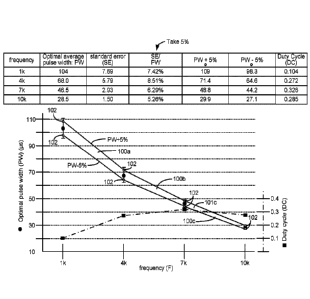
(ii) (50 Hz, 230 vs), (50 Hz, 370 p.$), (100 Hz, 325 vs), and (100 Hz, 195
p.$); or
(iii) (100 Hz, 195 vs), (100 Hz, 325 vs), (200 Hz, 260 vs), and (200 Hz, 160
s); or
(iv) (200 Hz, 160 s), (200 Hz, 260 vs), (400 Hz, 225 vs), and (400 Hz, 140
vs); or
(v) (400 Hz, 140 vs), (400 Hz, 225 vs), (600 Hz, 200 vs), and (600 Hz, 120
vs); or
(vi) (600 Hz, 120 s), (600 Hz, 200 vs), (800 Hz, 175 vs), and (800 Hz, 105
vs); or
(vii) (800 Hz, 105 vs), (800 Hz, 175 lits), (1000 Hz, 150 vs), and (1000 Hz,
90 vs).
16
CA 03072338 2020-02-06
WO 2019/032987
PCT/US2018/046255
[0067] The selected frequency and pulse width may be on or within one or more
linearly-
bounded regions defined by points:
(i) (1 kHz, 98.3 s), (1 kHz, 109 s), (4 kHz, 71.4 s), and (4 kHz, 64.6 s);
or
(ii) (4 kHz, 71.4 s), (4 kHz, 64.6 us), (7 kHz, 44.2 s), and (7 kHz, 48.8
s); or
(iii) (7 kHz, 44.2 us), (7 kHz, 48.8 iLts), (10 kHz, 29.9 us), and (10 kHz,
27.1 s).
or
(i) (1 kHz, 96.3 s), (1 kHz, 112 s), (4 kHz, 73.8 s), and (4 kHz, 62.2 s);
or
(ii) (4 kHz, 73.8 s), (4 kHz, 62.2 us), (7 kHz, 43.6 s), and (7 kHz, 49.4
s); or
(iii) (7 kHz, 43.6 us), (7 kHz, 49.4 us), (10 kHz, 30.0 us), and (10 kHz, 27.0
us).
or
(i) (1 kHz, 69.6 s), (1 kHz, 138.4 s), (4 kHz, 93.9 s), and (4 kHz, 42.1
iLts); or
(ii) (4 kHz, 93.9 s), (4 kHz, 42.1 us), (7 kHz, 33.4 s), and (7 kHz, 59.6
s); or
(iii) (7 kHz, 33.4 us), (7 kHz, 59.6 us), (10 kHz, 35.2 us), and (10 kHz, 21.8
us).
or
(i) (1 kHz, 50.0 us), (1 kHz, 200.0 us), (4 kHz, 110.0 us), and (4 kHz, 30.0
us); or
(n) (4 kHz, 110.0 us), (4 kHz, 30.0 s), (7 kHz, 30.0 us), and (7 kHz, 60.0
us); or
(iii) (7 kHz, 30.0 s), (7 kHz, 60.0 s), (10 kHz, 40.0 s), and (10 kHz, 20.0
lis).
[0068] The frequency and the pulse width may be selected by the computer
readable media
based on information relating frequencies and pulse widths at which the
therapeutic pulses
are formed to cause sub-perception stimulation to occur at the therapy
location. The
frequency and pulse width may be selected using the information as a frequency
and pulse
width that requires a lowest amount of power for the therapeutic pulses.
[0069] The computer readable media may contains instructions that when
executed allow the
external device to steer current between the plurality of electrodes to adjust
the therapy
location to a new therapy location relative to the array. The computer
readable media may
further contains instructions that when executed allow the external device to
adjust an
amplitude of the therapeutic pulses based on the new therapy location. The
computer
readable media may further contain instructions that when executed allow the
external device
to reduce at least one of the stimulation parameters of the second stimulation
parameters set
to or by a set amount or percentage.
[0070] The computer readable media may further contain instructions that when
executed
allow the external device to adjust at least one of the stimulation parameters
of the second
stimulation parameters set in response to a change in position or activity of
the patient. The
computer readable media may further contain instructions that when executed
allow the
17
86019694
external device to program the spinal cord stimulator during a programming
session, and
wherein the instructions are configured to wash in the therapeutic pulses for
a period of
one hour or less during the programming session to causes sub-perception
stimulation to
occur at the therapy location.
[0070a] According to one embodiment of the present invention, there is
provided an
external device for programming a patient's spinal cord stimulator system,
wherein the
spinal cord stimulator system comprises an electrode array comprising a
plurality of
electrodes, wherein the external device comprises control circuitry programmed
to: (a)
produce a first bipole comprising a first amplitude at a first set of active
electrodes of the
electrode array, the first bipole further comprising symmetric biphasic pulses
at the first
set of active electrodes of the electrode array at a frequency of 130 Hz or
less, each
symmetric biphasic pulse of the first bipole comprising a first phase of a
first polarity and
a second phase of a second polarity opposite the first polarity, the first and
second phases
of the first bipole both actively driven by stimulation circuitry of the
spinal cord stimulator
system, the first bipole causing stimulation above a perception threshold of
the patient; (b)
move the first bipole from the first set of active electrodes of the electrode
array to a
second set of active electrodes of the electrode array, the first bipole at
the second set of
active electrodes covering a patient's pain; and (c) produce, at the second
set of electrodes
of the electrode array, a second bipole different from the first bipole, the
second bipole
comprising symmetric biphasic pulses at the second set of active electrodes of
the
electrode array at a frequency of 130 Hz or less, each symmetric biphasic
pulse of the
second bipole comprising a first phase of a first polarity and a second phase
of a second
polarity opposite the first polarity, the first and second phases of the
second bipole both
actively driven by the stimulation circuitry of the spinal cord stimulator
system, the second
bipole comprising a second amplitude lower than the first amplitude of the
first bipole, the
second bipole causing stimulation below the perception threshold of the
patient, the second
bipole providing sub-perception pain relief within one hour or less after
generating the
second bipole at the second set of active electrodes.
10070b]
According to another embodiment of the present invention, there is
provided an external device for programming a patient's spinal cord stimulator
system,
wherein the spinal cord stimulator system comprises an electrode array
comprising a
plurality of electrodes, wherein the external device comprises control
circuitry
18
Date Recue/Date Received 2022-04-19
86019694
programmed to: (a) produce a first bipole comprising a first anode pole formed
at a first set of
two or more active electrodes and a first cathode pole formed at a second set
of two or more
active electrodes, the first bipole further comprising symmetric biphasic
pulses at the first and
second sets of two or more active electrodes at a frequency of 130 Hz or less,
each symmetric
biphasic pulse of the first bipole comprising a first phase of a first
polarity and a second phase of
a second polarity opposite the first polarity, the first and second phases of
the first bipole both
actively driven by stimulation circuitry of the spinal cord stimulator system,
the first bipole
causing stimulation above a perception threshold of the patient; (b) move the
first anode pole to a
third set of two or more active electrodes and the first cathode pole to a
fourth set of two or more
active electrodes to cover a patient's pain; and (c) produce a second bipole
comprising a second
anode pole at the third set of two or more active electrodes and a second
cathode pole at the
fourth set of two or more active electrodes, the second bipole comprising
symmetric biphasic
pulses third and fourth sets of two or more active electrodes at a frequency
of 130 Hz or less,
each symmetric biphasic pulse of the second bipole comprising a first phase of
a first polarity
and a second phase of a second polarity opposite the first polarity, the first
and second phases of
the second bipole both actively driven by the stimulation circuitry of the
spinal cord stimulator
system, the second bipole comprising a an amplitude lower than an amplitude of
the first bipole,
the second bipole causing stimulation below the perception threshold of the
patient, the second
bipole providing sub-perception pain relief within one hour or less after
generating the second
bipole at the second set of active electrodes.
BRIEF DESCRIPTION OF THE DRAWINGS
[0071] Figure 1 shows an Implantable Pulse Generator (IPG) useable for Spinal
Cord
Stimulation (SCS), in accordance with the prior art.
[0072] Figure 2 shows an example of stimulation pulses producible by the IPG,
in accordance
with the prior art.
[0073] Figure 3 shows use of an External Trial Stimulator (ETS) useable to
provide stimulation
before implantation of an IPG, in accordance with the prior art.
[0074] Figure 4 shows various external devices capable of communicating with
and
programming stimulation in an IPG and ETS, in accordance with the prior art.
18a
Date Recue/Date Received 2022-04-19
86019694
[0075] Figure 5 shows a Graphical User Interface (GUI) of a clinician
programmer external
device for setting or adjusting stimulation parameters, in accordance with the
prior art.
[0076] Figure 6 shows sweet spot searching to determine effective electrodes
for a patient using
a movable sub-perception bipole.
[0077] Figures 7A-7D show sweet spot searching to determine effective
electrodes for a patient
using a movable supra-perception bipole.
[0078] Figure 8 shows stimulation circuitry useable in the IPG or ETS capable
of providing
Multiple Independent Current Control to independently set the current at each
of the electrodes.
[0079] Figure 9 shows a flow chart of a study conducted on various patients
with back pain
designed to determine optimal sub-perception SCS stimulation parameters over a
frequency
range of 1 kHz to 10 kHz.
[0080] Figures 10A-10C show various results of the study as a function of
stimulation frequency
in the 1 kHz to 10 kHz frequency range, including average optimal pulse width
(Fig. 10A), mean
charge per second and optimal stimulation amplitude (Fig. 10B), and back pain
scores (Fig.
10C).
[0081] Figures 11A-11C shows further analysis of relationships between average
optimal pulse
width and frequency in the 1 kHz to 10 kHz frequency range, and identifies
statistically-
significant regions of optimization of these parameters.
18b
Date Recue/Date Received 2022-04-19
CA 03072338 2020-02-06
WO 2019/032987
PCT/US2018/046255
[0082] Figure 12A shows results of patients tested with sub-perception therapy
at frequencies
at or below 1 kHz, and shows optimal pulse width ranges determined at tested
frequencies,
and optimal pulse width Y. frequency regions for sub-perception therapy.
100831 Figure 12B shows various modelled relationships between average optimal
pulse
width and frequency at or below 1 kHz.
[0084] Figure 12C shows the duty of cycle of the optimal pulse widths as a
function of
frequencies at or below 1 kHz.
[0085] Figure 12D shows the average battery current and battery discharge time
at the
optimal pulse widths as a function of frequencies at or below 1 kHz.
[0086] Figure 13 shows a fitting module showing how the relationships and
regions
determined relating optimal pulse width and frequency (-10 kHz) can be used to
set sub-
perception stimulation parameters for an IPG or ETS.
[0087] Figure 14 shows an algorithm used for supra-perception sweet spot
searching
followed by sub-perception therapy, and possible optimization of the sub-
perception therapy
using the fitting module.
100881 Figure 15 shows an alternative algorithm for optimization of the sub-
perception
therapy using the fitting module.
DETAILED DESCRIPTION
[0089] While Spinal Cord Stimulation (SCS) therapy can be an effective means
of alleviating
a patient's pain, such stimulation can also cause paresthesia.
Paresthesia¨sometimes
referred to a "supra-perception" therapy¨is a sensation such as tingling,
prickling, heat, cold,
etc. that can accompany SCS therapy. Generally, the effects of paresthesia are
mild, or at
least are not overly concerning to a patient. Moreover, paresthesia is
generally a reasonable
tradeoff for a patient whose chronic pain has now been brought under control
by SCS
therapy. Some patients even find paresthesia comfortable and soothing.
[0090] Nonetheless, at least for some patients, SCS therapy would ideally
provide complete
pain relief without paresthesia¨what is often referred to as "sub-perception"
or sub-
threshold therapy that a patient cannot feel. Effective sub-perception therapy
may provide
pain relief without paresthesia by issuing stimulation pulses at higher
frequencies.
Unfortunately, such higher-frequency stimulation may require more power, which
tends to
drain the battery 14 of the IPG 10. See, e.g., U.S. Patent Application
Publication
2016/0367822. If an IPG's battery 14 is a primary cell and not rechargeable,
high-frequency
stimulation means that the IPG 10 will need to be replaced more quickly.
Alternatively, if an
19
CA 03072338 2020-02-06
WO 2019/032987
PCT/US2018/046255
IPG battery 14 is rechargeable, the IPG 10 will need to be charged more
frequently, or for
longer periods of time. Either way, the patient is inconvenienced.
[0091] In an SCS application, it is desirable to determine a stimulation
program that will be
effective for each patient. A significant part of determining an effective
stimulation program
is to determine a "sweet spot- for stimulation in each patient, i.e., to
select which electrodes
should be active (E) and with what polarities (P) and relative amplitudes (X%)
to recruit and
thus treat a neural site at which pain originates in a patient. Selecting
electrodes proximate to
this neural site of pain can be difficult to determine, and experimentation is
typically
undertaken to select the best combination of electrodes to provide a patient's
therapy.
[0092] As described in U.S. Provisional Patent Application Serial No.
62/680,539, filed June
4, 2018, selecting electrodes for a given patient can be even more difficult
when sub-
perception therapy is used, because the patient does not feel the stimulation,
and therefore it
can be difficult for the patient to feel whether the stimulation is "covering"
his pain and
therefore whether selected electrodes are effective. Further, sub-perception
stimulation
therapy may require a "wash in" period before it can become effective. A wash
in period can
take up to a day or more, and therefore sub-perception stimulation may not be
immediately
effective, making electrode selection more difficult.
[0093] Figure 6 briefly explains the '539 Application's technique for a sweet
spot search, i.e.,
how electrodes can be selected that are proximate to a neural site of pain 298
in a patient,
when sub-perception stimulation is used. The technique of Figure 6 is
particularly useful in a
trial setting after a patient is first implanted with an electrode array,
i.e., after receiving their
IPG or ETS.
[0094] In the example shown, it is assumed that a pain site 298 is likely
within a tissue region
299. Such region 299 may be deduced by a clinician based on the patient
symptoms, e.g., by
understanding which electrodes are proximate to certain vertebrae (not shown),
such as
within the T9-T10 interspace. In the example shown, region 299 is bounded by
electrodes
E2, E7, E15, and E10, meaning that electrodes outside of this region (e.g.,
El, E8, E9, E16)
are unlikely to have an effect on the patient's symptoms. Therefore, these
electrodes may not
be selected during the sweet spot search depicted in Figure 6, as explained
further below.
100951 In Figure 6, a sub-perception bipole 297a is selected, in which one
electrode (e.g., E2)
is selected as an anode that will source a positive current (+A) to the
patient's tissue, while
another electrode (e.g., E3) is selected as a cathode that will sink a
negative current (-A) from
the tissue. This is similar to what was illustrated earlier with respect to
Figure 2, and biphasic
stimulation pulses can be used employing active charge recovery. Because the
bipole 297a
CA 03072338 2020-02-06
WO 2019/032987
PCT/US2018/046255
provides sub-perception stimulation, the amplitude A used during the sweet
spot search is
titrated down until the patient no longer feels paresthesia. This sub-
perception bipole 297a is
provided to the patient for a duration, such as a few days, which allows the
sub-perception
bipole's potential effectiveness to "wash in," and allows the patient to
provide feedback
concerning how well the bipole 297a is helping their symptoms. Such patient
feedback can
comprise a pain scale ranking. For example, the patient can rank their pain on
a scale from 1-
using a Numerical Rating Scale (NRS) or the Visual Analogue Scale (VAS), with
1
denoting no or little pain and 10 denoting a worst pain imaginable. As
discussed in the '539
Application, such pain scale ranking can be entered into the patient's
external controller 45.
[0096] After the bipole 297a is tested at this first location, a different
combination of
electrodes is chosen (anode electrode E3, cathode electrode E4), which moves
the location of
the bipole 297 in the patient's tissue. Again, the amplitude of the current A
may need to be
titrated to an appropriate sub-perception level. In the example shown, the
bipole 297a is
moved down one electrode lead, and up the other, as shown by path 296 in the
hope of
finding a combination of electrodes that covers the pain site 298. In the
example of Figure 6,
given the pain site 298's proximity to electrodes E13 and E14, it might be
expected that a
bipole 297a at those electrodes will provide the best relief for the patient,
as reflected by the
patient's pain score rankings. The particular stimulation parameters chosen
when forming
bipole 297a can he selected at the GUI 64 of the clinician programmer 50 or
other external
device (such as a patient external controller 45) and wirelessly telemetered
to the patient's
IPG or ETS for execution.
[0097] While the sweet spot search of Figure 6 can be effective, it can also
take a
significantly long time when sub-perception stimulation is used. As noted, sub-
perception
stimulation is provided at each bipole 297 location for a number of days, and
because a large
number of bipole locations are chosen, the entire sweep spot search can take
up to a month to
complete.
[0098] The inventors have determined via testing of SCS patients that even if
it is desired to
eventually use sub-perception therapy for a patient going forward after the
sweet spot search,
it is beneficial to use supra-perception stimulation during the sweet spot
search to select
active electrodes for the patient. Use of supra-perception stimulation during
the sweet spot
search greatly accelerates determination of effective electrodes for the
patient compared to
the use of sub-perception stimulation, which requires a wash in period at each
set of
electrodes tested. After determining electrodes for use with the patient using
supra-
perception therapy, therapy may be titrated to sub-perception levels keeping
the same
21
CA 03072338 2020-02-06
WO 2019/032987
PCT/US2018/046255
electrodes determined for the patient during the sweet spot search. Because
the selected
electrodes are known to be recruiting the neural site of the patient's pain,
the application of
sub-perception therapy to those electrodes is more likely to have immediate
effect, reducing
or potentially eliminating the need to wash in the sub-perception therapy that
follows. In
short, effective sub-perception therapy can be achieved more quickly for the
patient when
supra-perception sweet spot searching is utilized. Preferably, supra-
perception sweet spot
searching occurs using symmetric biphasic pulses occurring at low
frequencies¨such as
between 40 and 200 Hz in one example.
[0099] In accordance with one aspect of the disclosed technique, a patient
will be provided
sub-perception therapy. Sweet spot searching to determine electrodes that may
be used
during sub-perception therapy may precede such sub-perception therapy. In some
aspects,
when sub-perception therapy is used for the patient, sweet spot searching may
use a bipole
297a that is sub-perception (Fig. 6), as just described. This may be relevant
because the sub-
perception sweet spot search may match the eventual sub-perception therapy the
patient will
receive.
[00100] However, the inventors have determined that even if sub-perception
therapy is
eventually to be used for the patient, it can be beneficial to use supra-
perception
stimulation¨that is, stimulation with accompanying paresthesia¨during the
sweet spot
search This is shown in Figure 7A, where the movable bipole 301a provides
supra-
perception stimulation that can be felt by the patient. Providing bipole 30Ia
as supra-
perception stimulation can merely involve increasing its amplitude (e.g.,
current A) when
compared to the sub-perception bipole 297a of Figure 6, although other
stimulation
parameters might be adjusted as well, such as by providing longer pulse
widths.
[00101] The inventors have determined that there are benefits to employing
supra-perception
stimulation during the sweet spot search even though sub-perception therapy
will eventually
be used for the patient.
[00102] First, as mentioned above, the use of supra-perception therapy by
definition allows
the patient to feel the stimulation, which enables the patient to provide
essentially immediate
feedback to the clinician whether the paresthesia seems to be well covering
his pain site 298.
In other words, it is not necessary to take the time to wash in bipole 301a at
each location as
it is moved along path 296. Thus, a suitable bipole 301a proximate to the
patient's pain site
298 can be established much more quickly, such as within a single clinician's
visit, rather
than over a period of days or weeks. In one example, when sub-perception
therapy is
preceded with supra-perception sweet spot searching, the time needed to wash
in the sub-
22
CA 03072338 2020-02-06
WO 2019/032987
PCT/US2018/046255
perception therapy can be one hour or less, ten minutes or less, or even a
matter of seconds.
This allows wash in to occur during a single programming session during which
the patient's
IPG or ETS is programmed, and without the need for the patient to leave the
clinician's
office.
[00103] Second, use of supra-perception stimulation during the sweet spot
search ensures that
electrodes are determined that well recruit the pain site 298. As a result,
after the sweet spot
search is complete and eventual sub-perception therapy is titrated for the
patient, wash in of
that sub-perception therapy may not take as long because the electrodes needed
for good
recruitment have already been confidently determined.
[00104] Figures 7B-7D show other supra-perception bipoles 301b-301d that may
be used,
and in particular show how the virtual bipoles may be formed using virtual
poles by
activating three or more of the electrodes 16. Virtual poles are discussed
further in U.S.
Provisional Patent Application Serial No. 62/598,114, filed December 13, 2017,
and thus
virtual poles are only briefly explained here. Forming virtual poles is
assisted if the
stimulation circuitry 28 or 44 used in the IPG or ETS is capable of
independently setting the
current at any of the electrodes¨what is sometimes known as a Multiple
Independent
Current Control (MICC), which is explained further below with reference to
Figure 8.
[00105] When a virtual bipole is used, the GUI 64 (Fig. 5) of the clinician
programmer 50
(Fig 4) can he used to define an anode pole (+) and a cathode pole (-) at
positions 291 (Fig
7B) that may not necessarily correspond to the position of the physical
electrodes 16. The
control circuitry 70 in the clinician programmer 50 can compute from these
positions 291 and
from other tissue modeling information which physical electrodes 16 will need
to be selected
and with what amplitudes to form the virtual anode and virtual cathode at the
designated
positions 291. As described earlier, amplitudes at selected electrodes may be
expressed as a
percentage X% of the total current amplitude A specified at the GUI 64 of the
clinician
programmer 50.
[00106] For example, in Figure 7B, the virtual anode pole is located at a
position 291
between electrodes E2, E3 and E10. The clinician programmer 50 may then
calculate based
on this position that each of these electrodes (during first pulse phase 30a)
will receive an
appropriate share (X%) of the total anodic current +A to locate the virtual
anode at this
position. Since the virtual anode's position is closest to electrode E2, this
electrode E2 may
receive the largest share of the specified anodic current +A (e.g., 75%*+A).
Electrodes E3
and E10 which are proximate to the virtual anode pole's position but farther
away receive
lesser shares of the anodic current (e.g., 15%*+A and 10%*+A respectively).
Likewise, it
23
CA 03072338 2020-02-06
WO 2019/032987
PCT/US2018/046255
can be seen that from the designated position 291 of the virtual cathode pole,
which is
proximate to electrodes E4, Ell, and El 2, that these electrodes will receive
an appropriate
share of the specified cathodic current ¨A (e.g., 20%*-A, 20%*-A, and 60%*-A
respectively,
again during the first pulse phase 30a). These polarities would then be
flipped during the
second phases 30b of the pulses, as shown in the wavefofins of Figure 7B. In
any event, the
use of virtual poles in the formation of bipole 301b allows the field in the
tissue to be shaped,
and many different combinations of electrodes can be tried during the sweet
spot search. In
this regard, it is not strictly necessary that the (virtual) bipole be moved
along an orderly path
296 with respect to the electrodes, and the path may be randomized, perhaps as
guided by
feedback from the patient.
[00107] Figure 7C shows a useful virtual bipole 301c configuration that can be
used during
the sweet spot search. This virtual bipole 301c again defines a target anode
and cathode
whose positions do not correspond to the position of the physical electrodes.
The virtual
bipole 301c is formed along a lead¨essentially spanning the length of four
electrodes from
El to E5. This creates a larger field in the tissue better able to recruit the
patient's pain site
298. 'Ibis bipole configuration 301c may need to be moved to a smaller number
of locations
than would a smaller bipole configuration compared 301a of Fig. 7A) as it
moves along path
296, thus accelerating pain site 298 detection. Figure 7D expands upon the
bipole
configuration of Figure 7C to create a virtual bipole 301d using electrodes
formed on both
leads, e.g., from electrodes El to E5 and from electrodes E9 to E13. This
bipole 301d
configuration need only be moved along a single path 296 that is parallel to
the leads, as its
field is large enough to recruit neural tissue proximate to both leads. This
can further
accelerate pain site detection.
[00108] In some aspects, the supra-perception bipoles 301a-301d used during
the sweet spot
search comprise symmetric biphasic waveforms having actively-driven (e.g., by
the
stimulation circuitry 28 or 44) pulse phases 30a and 30b of the same pulse
width PW and the
same amplitude (with the polarity flipped during the phases) (e.g., A3oa = A
_30b, and PW30a ¨
PW30b)- This is beneficial because the second pulse phase 30b provides active
charge
recovery, with in this case the charge provided during the first pulse phase
30a (Q30a)
equaling the charge of the second pulse phase 30b (Q3ob), such that the pulses
are charge
balanced. Use of biphasic waveforms are also believed beneficial because, as
is known, the
cathode is largely involved in neural tissue recruitment. When a biphasic
pulse is used, the
positions of the (virtual) anode and cathode will flip during the pulse's two
phases. This
effectively doubles the neural tissue that is recruited for stimulation, and
thus increases the
24
CA 03072338 2020-02-06
WO 2019/032987
PCT/US2018/046255
possibility that the pain site 298 will be covered by a bipole at the correct
location.
[00109] The supra-perception bipoles 301a-301d do not however need to comprise
symmetric biphasic pulses as just described. For example, the amplitude and
pulse width of
the two phases 30a and 30b can be different, while keeping the charge (Q) of
the two phases
balanced (e.g., Q30a = A30a*PW30a ¨ A30b*PW30b ¨ Q304 Alternatively, the two
phases 30a
and 30b may be charge imbalanced (e.g.,
<30a ¨ A30a*PW30a A30b*PW30b = Q30b, or Q30a ¨
A30a*PW30a < A30b*PW30b = Q30b)= In short, the pulses in bipoles 301-301d can
be biphasic
symmetric (and thus inherently charge balanced), biphasic asymmetric but still
charge
balanced, or biphasic asymmetric and charge imbalanced.
[00110] In a preferred example, the frequency F of the supra-perception pulses
301a-301d
used during the supra-perception sweet spot search may be 10 kHz or less, 1
kHz or less, 500
Hz or less, 300 Hz or less, 200 Hz or less, 130 Hz or less, or 100 Hz or less,
or ranges
bounded by two of these frequencies (e.g., 100 ¨ 130 Hz, or 100 ¨ 200 Hz). In
particular
examples, frequencies of 90 Hz, 40 Hz, or 10 Hz can be used, with pulses
comprising
biphasic pulses which are preferably symmetric. However, a single actively-
driven pulse
phase followed by a passive recovery phase could also be used. '[he pulse
width PW may
also comprise a value in the range of hundreds of microseconds, such as 150 to
400
microseconds. Because the goal of supra-perception sweet spot searching is
merely to
determine electrodes that appropriately cover a patient's pain, frequency and
pulse width may
be of less importance at this stage. Once electrodes have been chosen for sub-
perception
stimulation, frequency and pulse width can be optimized, as discussed further
below.
[00111] It should be understood that the supra-perception bipoles 301a-301d
used during
sweet spot searching need not necessarily be the same electrodes that are
selected when later
providing the patient with sub-perception therapy. Instead, the best location
of the bipole
noticed during the search can be used as the basis to modify the selected
electrodes. Suppose
for example that a bipole 301a (Fig. 7A) is used during sweep spot searching,
and it is
determined that bipole provides the best pain relief when located at
electrodes E13 and E14.
At that point, sub-perception therapy using those electrodes E13 and E14 can
be tried for the
patient going forward. Alternatively, it may be sensible to modify the
selected electrodes to
see if the patient's symptoms can be further improved before sub-perception
therapy is tried.
For example, the distance (focus) between the cathode and anode can be varied,
using virtual
poles as already described. Or, a tripole (anode/cathode/anode) consisting of
electrodes
E12/E13/E14 or E13/E14/E15 could be tried. See U.S. Provisional Patent
Application Serial
No. 62/598,114, filed December 13, 2017 (discussing tripoles). Or electrodes
on a different
CA 03072338 2020-02-06
WO 2019/032987
PCT/US2018/046255
lead could also be tried in combination with E13 and E14. For example, because
electrodes
E5 and E6 are generally proximate to electrodes E13 and E14, it may be useful
to add E5 or
E6 as sources of anodic or cathodic current (again creating virtual poles).
All of these types
of adjustments should be understood as comprising "steering" or an adjustment
to the
"location" at which therapy is applied, even if a central point of stimulation
doesn't change
(as can occur for example when the distance or focus between the cathode and
anode is
varied).
[00112] Multiple Independent Current Control (MICC) is explained in one
example with
reference to Figure 8, which shows the stimulation circuitry 28 (Fig. 1) or 44
(Fig. 3) in the
IPG or ETS used to form prescribed stimulation at a patient's tissue. The
stimulation
circuitry 28 or 44 can control the current or charge at each electrode
independently, and using
GUI 64 (Fig. 5) allows the current or charge to be steered to different
electrodes, which is
useful for example when moving the bipole 301i along path 296 during the sweet
spot search
(Fig. 7A-7D). The stimulation circuitry 28 or 44 includes one or more current
sources 440i
and one or more current sinks 442i. The sources and sinks 440i and 442i can
comprise
Digital-to-Analog converters (DACs), and may be referred to as PDACs 4401 and
NDACs
442i in accordance with the Positive (sourced, anodic) and Negative (sunk,
cathodic) currents
they respectively issue. In the example shown, a NDAC/PDAC 440/442 pair is
dedicated
(hardwired) to a particular electrode node ei 39 Each electrode node ei 39 is
preferably
connected to an electrode Ei 16 via a DC-blocking capacitor Ci 38, which act
as a safety
measure to prevent DC current injection into the patient, as could occur for
example if there
is a circuit fault in the stimulation circuitry 28 or 44. PDACs 440i and NDACs
442i can also
comprise voltage sources.
[00113] Proper control of the PDACs 4401 and NDACs 442i via GUI 64 allows any
of the
electrodes 16 and the case electrode Ec 12 to act as anodes or cathodes to
create a current
through a patient's tissue. Such control preferably comes in the form of
digital signals Tip
and fin that set the anodic and cathodic current at each electrode Ei. If for
example it is
desired to set electrode El as an anode with a current of + 3mA, and to set
electrodes E2 and
E3 as cathodes with a current of -1.5 mA each, control signal lip would be set
to the digital
equivalent of 3 mA to cause PDAC 4401 to produce + 3 mA, and control signals
I2n and I3n
would be set to the digital equivalent of 1.5 mA to cause NDACs 4420 and 4423
to each
produce -1.5 mA. Note that definition of these control signals can also occur
using the
programmed amplitude A and percentage X% set in the GUI 64. For example, A may
be set
to 3 mA, with El designated as an anode with X = 100%, and with E2 and E3
designated at
26
CA 03072338 2020-02-06
WO 2019/032987
PCT/US2018/046255
cathodes with X = 50%. Alternatively, the control signals may not be set with
a percentage,
and instead the GUI 64 can simply prescribe the current that will appear at
each electrode at
any point in time.
[00114] In short, the GUI 64 may be used to independently set the current at
each electrode,
or to steer the current between different electrodes. This is particularly
useful in forming
virtual bipoles, which as explained earlier involve activation of more than
two electrodes.
MICC also allows more sophisticated electric fields to be formed in the
patient's tissue.
[00115] Other stimulation circuitries 28 can also be used to implement MICC.
In an example
not shown, a switching matrix can intervene between the one or more PDACs 440i
and the
electrode nodes ei 39, and between the one or more NDACs 442i and the
electrode nodes.
Switching matrices allows one or more of the PDACs or one or more of the NDACs
to be
connected to one or more electrode nodes at a given time. Various examples of
stimulation
circuitries can be found in USPs 6,181,969, 8,606,362, 8,620,436, U.S. Patent
Application
Publication 2018/0071513, 2018/0071520, and U.S. Provisional Patent
Application Serial
No. 62/559,247, filed September 15, 2017.
[00116] Much of the stimulation circuitry 28 or 44, including the PDACs 440i
and NDACs
442i, the switch matrices (if present), and the electrode nodes ei 39 can be
integrated on one
or more Application Specific Integrated Circuits (ASICs), as described in U.S.
Patent
Application Publications 2012/0095529, 2012/0092031, and 2012/0095519 As
explained in
these references, AS1C(s) may also contain other circuitry useful in the 1PG
10, such as
telemetry circuitry (for interfacing off chip with the IPG's or ETS's
telemetry antennas),
circuitry for generating the compliance voltage VH that powers the stimulation
circuitry,
various measurement circuits, etc.
[00117] While it is preferred to use sweet spot searching, and in particular
supra-perception
sweet spot searching, to determine the electrodes to be used during subsequent
sub-
perception therapy, it should be noted that this is not strictly necessary.
Sub-perception
therapy can be preceded by sub-perception sweet spot searching, or may not be
preceded by
sweet spot searching at all. In short, sub-perception therapy as described
next is not reliant
on the use of any sweet spot search.
[00118] In another aspect of the invention, the inventors have determined via
testing of SCS
patients that statistically significant correlations exists between pulse
width (PW) and
frequency (F) where an SCS patient will experience a reduction in back pain
without
paresthesia (sub-perception). Use of this information can be helpful in
deciding what pulse
width is likely optimal for a given SCS patient based on a particular
frequency, and in
27
CA 03072338 2020-02-06
WO 2019/032987
PCT/US2018/046255
deciding what frequency is likely optimal for a given SCS patient based on a
particular pulse
width. Beneficially, this information suggests that paresthesia-free sub-
perception SCS
stimulation can occur at frequencies of 10 kHz and below. Use of such low
frequencies
allows sub-perception therapy to be used with much lower power consumption in
the
patient's IPG or ETS.
[00119] Figures 9-11C shows results derived from testing patients at
frequencies within a
range of 1 kHz to 10 kHz. Figure 9 explains how data was gathered from actual
SCS
patients, and the criteria for patient inclusion in the study. Patients with
back pain, but not
yet receiving SCS therapy, were first identified. Key patient inclusion
criteria included
having persistent lower back pain for greater than 90 days; a NRS pain scale
of 5 or greater
(NRS is explained below); stable opioid medications for 30 days; and a
Baseline Oswestry
Disability index score of greater than or equal to 20 and lower than or equal
to 80. Key
patient exclusion criteria included having back surgery in the previous 6
months; existence of
other confounding medical/psychological conditions; and untreated major
psychiatric
comorbidity or serious drug related behavior issues.
[00120] After such initial screening, patients periodically entered a
qualitative indication of
their pain (i.e., a pain score) into a portable e-diary device, which can
comprise a patient
external controller 45, and which in turn can communicate its data to a
clinician programmer
50 (Fig 4) Such pain scores can comprise a Numerical Rating Scale (NRS) score
from 1-10,
and were input to the e-diary three times daily. As shown in Figure 10C, the
baseline NRS
score for patients not eventually excluded from the study and not yet
receiving sub-
perception stimulation therapy was approximately 6.75/10, with a standard
error, SE
(sigma/SQRT(n)) of 0.25.
[00121] Returning to Figure 9, patients then had trial leads 15' (Fig. 3)
implanted on the left
and right sides of the spinal column, and were provided external trial
stimulation as explained
earlier. A clinician programmer 50 was used to provide a stimulation program
to each
patient's ETS 40 as explained earlier. This was done to make sure that SCS
therapy was
helpful for a given patient to alleviate their pain. If SCS therapy was not
helpful for a given
patient, trial leads 15' were explanted, and that patient was then excluded
from the study.
[00122] Those patients for whom external trial stimulation was helpful
eventually received
full implantation of a permanent 1PG 10, as described earlier. After a healing
period, and
again using clinician programmer 50, a "sweet spot" for stimulation was
located in each
patient, i.e., which electrodes should be active (E) and with what polarities
(P) and relative
amplitudes (X%) to recruit and thus treat a site 298 of neural site in the
patient. The sweet
28
CA 03072338 2020-02-06
WO 2019/032987
PCT/US2018/046255
spot search can occur in any of the manners described earlier with respect to
Figures 6-7D,
but in a preferred embodiment would comprise supra-perception stimulation
(e.g., e.g., 7A-
7D) because of the benefits described earlier. However, this is not strictly
necessary, and
sub-perception stimulation can also be used during the sweet spot search. In
the example of
Figure 9, sweet spot searching occurred at 10 kHz, but again the frequency
used during the
sweet spot search can be varied. Symmetric biphasic pulses were used during
sweet spot
searching, but again, this is not strictly required. Deciding which electrodes
should be active
started with selecting electrodes 16 present between thoracic vertebrae T9 and
T10.
However, electrodes as far away as T8 and T11 were also activated if
necessary. Which
electrodes were proximate to vertebrae T8, T9, T10, and Ti was determined
using
fluoroscopic images of the leads 15 within each patient.
[00123] During sweet spot searching, bipolar stimulation using only two
electrodes was used
for each patient, and using only adjacent electrodes on a single lead 15,
similar to what was
described in Figures 6 and 7A. Thus, one patient's sweet spot might involve
stimulating
adjacent electrodes E4 as cathode and E5 as anode on the left lead 15 as shown
earlier in
Figure 2 (which electrodes may be between '1'9 and '110), while another
patient's sweet spot
might involve stimulating adjacent electrodes E9 as anode and El as cathode
on the right
lead 15 (which electrodes may be between T10 and T11). Using only adjacent-
electrode
bipolar stimulation and only between vertebrae T8 to T11 was desired to
minimize variance
in the therapy and pathology between the different patients in the study.
However, more
complicated bipoles such as those described with respect to Figures 7B-7D
could also be used
during sweet spot searching. If a patient had sweet spot electrodes in the
desired thoracic
location, and if they experienced a 30% or greater pain relief per an NRS
score, such patients
were continued in the study; patients not meeting these criteria were excluded
from further
study. While the study started initially with 39 patients, 19 patients were
excluded from
study up to this point in Figure 9, leaving a total of 20 patients remaining.
[00124] The remaining 20 patients were then subjected to a "washout" period,
meaning their
IPGs did not provide stimulation for a time. Specifically, patients' NRS pain
scores were
monitored until their pain reached 80% of their initial baseline pain. This
was to ensure that
previous benefits of stimulation did not carry over to a next analysis period.
[00125] Thereafter, remaining patients were subjected to sub-perception SCS
therapy at
different frequencies in the range from 1 kHz to 10 kHz using the sweet spot
active electrodes
determined earlier. This however isn't strictly necessary, because as noted
earlier the current
at each electrode could also be independently controlled to assist in shaping
of the electric
29
CA 03072338 2020-02-06
WO 2019/032987
PCT/US2018/046255
filed in the tissue. As shown in Figure 9, the patients were each tested using
stimulation
pulses with frequencies of 10 kHz, 7 kHz, 4 kHz, and I kHz Figure 9 for
simplicity shows
that these frequencies were tested in this order for each patient, but in
reality the frequencies
were applied to each patient in random orders. Testing at a given frequency,
once complete,
was followed by a washout period before testing at another frequency began.
[00126] At each tested frequency, the amplitude (A) and pulse width (PW)
(first pulse phase
30a; Fig. 2) of the stimulation was adjusted and optimized for each patient
such that each
patient experienced good pain relief possible but without paresthesia (sub-
perception).
Specifically, using clinician programmer 50, and keeping as active the same
sweet spot
electrodes determined earlier (although again this isn't strictly necessary),
each patient was
stimulated at a low amplitude (e.g., 0), which amplitude was increased to a
maximum point
(perception threshold) where paresthesia was noticeable by the patient.
Initial stimulation
was then chosen for the patient at 50% of that maximum amplitude, i.e., such
that stimulation
was sub-perception and hence paresthesia free. However, other percentages of
the maximum
amplitude (80%, 90%, etc.) could be chosen as well, and can vary with patient
activity or
position, as explained further below. In one example, the stimulation
circuitry 28 or 44 in
the IPG or ETS is configurable to receive an instruction from the GUI 64 via a
selectable
option (not shown) to reduce the amplitude of the stimulation pulses to or by
a set amount or
percentage to render the so that the pulses can be made sub-perception if they
are not already.
Other stimulation parameters may also be reduced (e.g., pulse width, charge)
to the same
effect.
[00127] The patient would then leave the clinician's office, and thereafter
and in
communication with the clinician (or her technician or programmer) would make
adjustments
to his stimulation (amplitude and pulse width) using his external controller
45 (Fig. 4). At the
same time, the patient would enter NRS pain scores in his e-diary (e.g., the
external
controller), again three times a day. Patient adjustment of the amplitude and
pulse width was
typically an iterative process, but essentially adjustments were attempted
based on feedback
from the patient to adjust the therapy to decrease their pain while still
ensuring that
stimulation was sub-perception. Testing at each frequency lasted about three
weeks, and
stimulation adjustments might be made every couple of days or so. At the end
of the testing
period at a given frequency, optimal amplitude and pulse widths had been
determined and
were logged for each patient, along with patient NRS pain scores for those
optimal
parameters as entered in their e-diaries.
[00128] In one example, the percentage of the maximum amplitude used to
provide sub-
CA 03072338 2020-02-06
WO 2019/032987
PCT/US2018/046255
perception stimulation could be chosen dependent on an activity level or
position of the
patient. In regard, the IPG or ETS can include means for determining patient
activity or
position, such as an accelerometer. If the accelerometer indicates a high
degree of patient
activity or a position where the electrodes would be farther away from the
spinal cord (e.g.,
lying down), the amplitude could be increased to a higher percentage to
increase the current
(e.g., 90% of the maximum amplitude). If the patient is experiencing a lower
degree of
activity or a position where the electrodes would be closer to the spinal card
(e.g., standing),
the amplitude can be decreased (e.g., to 50% of the maximum amplitude).
Although not
shown, the GUI 64 of the external device (Fig. 5) can include an option to set
the percentage
of the maximum amplitude at which paresthesia become noticeable to the
patient, thus
allowing the patient to adjust the sub-perception current amplitude.
[00129] Preferably, Multiple Independent Current Control (MICC) is used to
provide or
adjust the sub-perception therapy, as discussed earlier with reference to
Figure 8. This allows
the current at each electrode to be independently set, which promotes the
steering of current
or charge between electrodes, facilitates the formation of virtual bipoles,
and more generally
allows the electric field to be shaped in the patient's tissue. In particular,
MICC, can be used
to steer sub-perception therapy to different locations in the electrode array
and thus the spinal
cord. For example, once a set of sub-perception stimulation parameters has
been chosen for
the patient, one or more of the stimulation parameters can be changed Such
changes may be
warranted or dictated by the therapy location. The physiology of the patient
may vary at
different vertebral positions, and tissue may be more or less conductive at
different therapy
locations. Therefore, if the sub-perception therapy location is steered to a
new location along
the spinal cord (which location change may comprise changing the anode/cathode
distance or
focus), it may be warranted to adjust at least one of the stimulation
parameters, such as
amplitude. As noted earlier, making sub-perception adjustment is facilitated,
and can occur
within a programming session, because a substantial wash in period may not be
necessary.
[00130] Adjustment to sub-perception therapy can also include varying other
stimulation
parameters, such as pulse width, frequency, and even the duration of the
interphase period
(IP) (Fig. 2). The interphase duration can impact the neural dose, or the rate
of charge
infusion, such that higher sub-perception amplitudes would be used with
shorter interphase
durations. In one example, the interphase duration can be varied between 0-3
ms. After a
washout period, a new frequency was tested, using the same protocol as just
described.
[00131] The sub-perception stimulation pulses used were symmetric biphasic
constant
current amplitude pulses, having first and second pulses phases 30a and 30b
with the same
31
CA 03072338 2020-02-06
WO 2019/032987
PCT/US2018/046255
duration (see Fig. 2). However, constant voltage amplitude pulses could be
used as well.
Pulses of different shapes (triangles, sine waves, etc.) could also be used.
Pre-pulsing¨that
is, providing a small current prior to providing the actively-driven pulse
phase(s)¨to affect
polarization or depolarization of neural tissue can also occur when providing
sub-perception
therapy. See, e.g., USP 9,008,790.
[00132] Figures 10A-10C show the results of testing the patients at 10 kHz,
7kHz, 4Hz and 1
kHz. Data is shown in each figure as average values for the 20 remaining
patients at each
frequency, with error bars reflecting standard error (SE) between the
patients.
[00133] Starting with Figure 10B, the optimized amplitude A for the 20
remaining patients
are shown at the tested frequencies. Interestingly, the optimal amplitude at
each frequency
was essentially constant¨around 3 mA. Figure 10B also shows the amount of
energy
expended at each frequency, more specifically a mean charge per second (MCS)
(in mC/s)
attributable to the pulses. MCS is computed by taking the optimal pulse width
(Fig. 10A,
discussed next) and multiplying it by the optimal amplitude (A) and the
frequency (F), which
MCS value can comprise a neural dose. MCS correlates to the current or power
that the
battery in the 1PG 10 must expend to form the optimal pulses. Significantly,
the MCS is
significantly lower at lower frequencies: for example, the MCS at F = lkHz is
approximately
1/3 of its value at higher frequencies (e.g., F = 7 kHz or 10 kHz). This means
that optimal
SCS therapy¨that
alleviates back pain without paresth es i a¨i s achievable at lower
frequencies like F = 1 kHz, with the added benefit of lower power draws that
are more
considerate of the IPG 10's (or ETS 40's) battery.
[00134] Figure 10A shows optimal pulse width as a function of frequency for
the 1 kHz to 10
kHz frequency range tested. As shown, the relationship follows a statistically
significant
trend: when modeled using linear regression 98a, PW = -8.22F + 106, where
pulse width is
measured in microseconds and frequency is measured in kiloHertz, with a
correlation
coefficient R2 of 0.974; when modeled using polynomial regression 98b, PW =
0.486F2 ¨
13.6F + 116, again with pulse width measured in microseconds and frequency
measured in
kiloHertz, with an even better correlation coefficient of R2 = 0.998. Other
fitting methods
could be used to establish other information relating frequency and pulse
width at which
stimulation pulses are formed to provide pain relief without paresthesia in
the frequency
range of 1 kHz to 10 kHz.
[00135] Note that the relationship between optimal pulse width and frequency
is not simply
an expected relationship between frequency and duty cycle (DC), i.e., the
duration that a
pulse is 'on' divided by its period (1/F). In this regard, notice that a given
frequency has a
32
CA 03072338 2020-02-06
WO 2019/032987
PCT/US2018/046255
natural effect on pulse width: one would expect that a higher frequency pulses
would have
smaller pulse widths. Thus, it might be expected for example that a 1 kHz
waveform with a
100 microsecond pulse width would have the same clinical results as a 10 kHz
waveform
with a 10 microsecond frequency, because the duty cycle of both of these
waveforms is 10%.
Figure 11A shows the resulting duty cycle of the stimulation waveforms using
the optimal
pulse width in the frequency range of lkHz to 10 kHz. Here, duty cycle is
computed by
considering the total 'on' time of the first pulse phase 30a (Fig. 2) only;
the duration of the
symmetric second pulse phase is ignored. This duty cycle is not constant over
the 1 kHz to
kHz frequency range: for example, the optimal pulse width at 1 kHz (104
microseconds)
is not merely ten times the optimal pulse width at 10 kHz (28.5 microseconds).
Thus, there is
significance to the optimal pulse widths beyond a mere scaling of the
frequency.
[00136] Figure 10C shows average patient pain scores at the optimal
stimulation parameters
(optimal amplitude (Fig. 7B) and pulse width (Fig. 7A)) for each frequency in
the range of 1
kHz to 10 kHz. As noted earlier, patients in the study, prior to receiving SCS
therapy,
initially reported pain scores with an average of 6.75. After SC S
implantation and during the
study, and with amplitude and pulse width optimized during the provisional of
sub-perception
therapy, their average pain scores dropped significantly, to an average score
of about 3 for all
frequencies tested.
[00137] Figure 11A provides a deeper analysis of the resulting relationship
between optimal
pulse width and frequency in the frequency range of 1 kHz to 10 kHz. The chart
in Figure
11A shows the average optimal pulse width for the 20 patients in the study at
each frequency,
along with the standard error resulting from variations between them. These
are normalized
at each frequency by dividing the standard error by the optimal pulse width,
ranging in
variations at each frequency between 5.26 % and 8.51 %. From this, a 5%
variance (lower
than all computed values) can be assumed as a statistically-significant
variance at all
frequencies tested.
[00138] From this 5% variance, a maximum average pulse width (PW + 5%) and a
minimum
average pulse width (PW + 5%) can be calculated for each frequency. For
example, the
optimal average pulse width PW at 1 kHz is 104 microseconds, and 5% above this
value
(1.05*104 tis) is 109 i.ts; 5% below this value (0.95*104) is 98.3 is.
Likewise, the optimal
average pulse width AVG(PW) at 4 kHz is 68.0 microseconds, and 5% above this
value
(1.05*68.0 [is) is 71.4 .is; 5% below this value (0.95*68.0 [is) is 64.6 !us.
Thus, a
statistically-significant reduction in pain without paresthesia occurs in or
on the linearly
bounded region 100a of points 102 of (1 kHz, 98.3 ns), (1 kHz, 109 ps), (4
kHz, 71.4 p.$),
33
CA 03072338 2020-02-06
WO 2019/032987
PCT/US2018/046255
and (4 kHz, 64.6 [is). A linearly bounded region 100b around points 102 is
also defined for
frequencies greater than or equal to 4 kHz and less than or equal to 7 kHz. (4
kHz, 71.4 las),
(4 kHz, 64.6 ms), (7 kHz, 44.2 las), (7 kHz, 48.8 p.$). A linear bounded
region 100c around
points 102 is also defined for frequencies greater than or equal to 7 kHz and
less than or equal
to 10 kHz: (7 kHz, 44.2 vis), (7 kHz, 48.8 [is), (10 kHz, 29.9 vis), (10 kHz,
27.1 [is). Such
regions 100 thus comprise information relating frequency and pulse width at
which
stimulation pulses are formed to provide pain relief without paresthesia in
the frequency
range of 1 kHz to 10 kHz.
[00139] Figure 11B provides an alternative analysis of the resulting
relationship between
optimal pulse width and frequency. In this example, regions 100a-100c are
defined based
upon the standard error (SE) calculated at each frequency. Thus, points 102
defining the
comers of the regions 100a-c are simply located at the extent of the SE error
bars at each
frequency (PW + SE, and PW ¨ SE), even though these error bars are of
different magnitudes
at each frequency. Thus, a statistically-significant reduction in pain without
paresthesia
occurs in or on the linearly bounded region 100a of points (1 kHz, 96.3 p), (1
kHz, 112 p),
(4 kHz, 73.8 s), and (4 kHz, 62.2 p3). The linear bounded regions 100b and
100c are
similar, and because the points 102 defining them are set forth in chart at
the top of Figure
11B, they are not repeated here.
[00140] Figure 11C provides another analysis of the resulting relationship
between optimal
pulse width and frequency. In this example, regions 100a-100c are defined
based upon the
standard deviation (SD) calculated at each frequency, which is larger than the
standard error
(SE) metric used to this point. Points 102 defining the comers of the regions
100a-c are
located at the extent of the SD error bars at each frequency (PW + SD, and PW
¨ SD),
although points 102 could also be set within the error bars, similar to what
was illustrated
earlier with respect to Figure 11A. In any event, a statistically-significant
reduction in pain
without paresthesia occurs in or on the linearly bounded region 100a of points
(1 kHz, 69.6
1.1s), (1 kHz, 138.4 [is), (4 kHz, 93.9 !is), and (4 kHz, 42.1 !is). The
linear bounded regions
100b and 100c are similar, and because the points 102 defining them are set
forth in chart at
the top of Figure 11C, they are not repeated here.
[00141] More generally, although not illustrated, regions within the frequency
range of 1 kHz
to 10 kHz where sub-perception efficacy was achieved comprises linearly-
bounded region
100a (1 kHz, 50.0 [ts), (1 kHz, 200.0 [ts), (4 kHz, 110.0 [is), and (4 kHz,
30.0 Rs); and/or
linearly-bounded region 100b (4 kHz, 110.0 ps), (4 kHz, 30.0 is), (7 kHz, 30.0
ns), and (7
kHz, 60.0 p); and/or linearly-bounded region 100c (7 kHz, 30.0 p.$), (7 kHz,
60.0 ps), (10
34
CA 03072338 2020-02-06
WO 2019/032987
PCT/US2018/046255
kHz, 40.0 !is), and (10 kHz, 20.0 is).
[00142] In summary, one or more statistically-significant regions 100 can be
defined for the
optimal pulse width and frequency data taken for the patients in the study to
arrive at
combinations of pulse width and frequency that reduce pain without the side
effect of
paresthesia within the frequency range of 1 kHz to 10 kHz, and different
statistical measures
of error can be used to so define the one or more regions.
[00143] Figures 12A-12D show the results of testing other patients with sub-
perception
stimulation therapy at frequencies at or below 1 kHz. Testing of the patients
generally
occurred after supra-perception sweep spot searching occurred to select
appropriate
electrodes (E), polarities (P) and relative amplitudes (X%) for each patient
(see Figs. 7A-7D),
although again the sub-perception electrodes used could vary from those used
during the
supra-perception sweet spot search (e.g., using MICC). Patients were tested
with sub-
perception stimulation using symmetric biphasic bipoles, although the form of
pulses used
during sub-perception therapy could vary.
[00144] Figure 12A shows the relationship between frequency and pulse width at
which
effective sub-perception therapy was reported by patients for frequencies of 1
kHz and below.
Note that the same patient selection and testing criteria described earlier
(Fig. 9) can be used
when evaluating frequencies at or below 1 kHz, with the frequencies adjusted
as appropriate.
[00145] As can be seen, at each frequency tested, the optimal pulse width
again fell within a
range. For example, at 800 Hz, patients reported good results when the pulse
width fell
within a range of 105-175 microseconds. The upper end of the pulse width range
at each
frequency is denoted PW(high), while the lower end of the pulse width range at
each
frequency is denoted PW(low). PW(middle) denotes the middle (e.g., average) of
the
PW(high) and PW(low) at each frequency. At each of the tested frequencies the
amplitude of
the current provided (A) was titrated down to sub-perception levels, such that
the patient
could not feel paresthesia. Typically, the current was titrated to 80% of the
threshold at
which paresthesia could be sensed. Because each patient's anatomy is unique,
the sub-
perception amplitude A could vary from patient to patient. The pulse width
data depicted
comprises the pulse width of only the first phase of the stimulation pulses.
[00146] Table 1 below expresses the optimal pulse width versus frequency data
of Figure
12A in tabular form for frequencies at or below 1 kHz, with the pulse widths
expressed in
microseconds:
CA 03072338 2020-02-06
WO 2019/032987
PCT/US2018/046255
Frequency PW(low) PW(middle) PW(high)
(Hz) (is) (us) (Rs)
1000 90 120 150
800 105 140 175
600 120 160 200
400 140 183 225
200 160 210 260
100 195 260 325
50 230 300 370
265 350 435
TABLE 1
[00147] As with the analysis described earlier for frequencies in a range of 1
kHz to 10 kHz
(Figs. 10A-11C), the data may be broken down to define different regions 300i
at which
effective sub-perception therapy is realized below 1 kHz. For example, regions
of effective
sub-perception therapy may be linearly bounded between various frequencies and
the high
and low pulse widths that define effectiveness. For example, at 10 Hz, PW(low)
= 265
microseconds and PW(high) = 435 microseconds. At 50 Hz, PW(low) = 230
microseconds
and PW(high) = 370 microseconds. Therefore, a region 300a that provides good
sub-
perception therapy is defined by the linearly bounded region of points (10 Hz,
265 tts), (10
Hz, 435 vs), (50 Hz, 370 vs), and (50 Hz, 230 vs). Table 2 defines the points
that linearly
bind each of the regions 300a-300g shown in Figure 12A:
region Bounded by points (Hz, tts)
300a (10, 265), (10, 435), (50, 370), (50, 230)
300b (50, 230), (50, 370), (100, 325), (100, 195)
300c (100, 195), (100, 325), (200, 260), (200, 160)
300d (200, 160), (200, 260), (400, 225), (400, 140)
300e (400, 140), (400, 225), (600, 200), (600, 120)
300f (600, 120), (600, 200), (800, 175), (800, 105)
300g (800, 105), (800, 175), (1000, 150), (1000, 90)
TABLE 2
36
CA 03072338 2020-02-06
WO 2019/032987
PCT/US2018/046255
[00148] Regions of sub-perception therapeutic effectiveness at frequencies at
or below 1 kHz
may be defined in other statistically-significant ways, such as those
described earlier for
frequencies in the range of I kHz to 10 kHz (Figs. 11A-11C). For example,
regions 300i may
be defined by reference to the pulse width at the middle of the ranges at each
frequency,
PW(middle). PW(middle) may comprise for example an average optimal pulse width
reported by patients at each frequency, rather than as a strict middle of an
effective range
reported by those patients. PW(high) and PW(low) may then be determined as a
statistical
variance from the average PW(middle) at each frequency, and used to set the
upper and lower
bounds of effective sub-perception regions. For example, PW(high) may comprise
average
PW(middle) plus a standard deviation or standard error, or a multiples of such
statistical
measures; PW(low) may likewise comprise average PW(middle) minus a standard
deviation
or standard error, or a multiple of such statistical measures. PW(high) and
PW(low) may also
be determined from average PW(middle) in other ways. For example, PW(high) may
comprise average PW(middle) plus a set percentage, while PW(low) may comprise
PW(middle) minus a set percentage. In summary, one or more statistically-
significant
regions 300 can be defined for the optimal pulse width and frequency data at
frequencies at or
below 1 kHz that reduce pain using sub-perception stimulation without the side
effect of
paresthesia.
[00149] Also shown in Figure 12A are average patient pain scores (NRS scores)
reported by
patients when optimal pulse widths are used for different frequencies at 1 kHz
or below.
Prior to receiving SCS therapy, patients initially reported pain scores with
an average of 7.92.
After SCS implantation, and using the sub-perception stimulation at optimal
pulse widths
with the ranges shown at each frequency, the patients' average pain scores
dropped
significantly. At 1 kHz, 200 Hz, and 10 Hz, patients reported average pain
scores of 2.38,
2.17, and 3.20 respectively. Thus clinical significance with respect to pain
relief is shown
when the optimal pulse widths are used at or below 1 kHz with sub-perception
therapy.
[00150] The optimal pulse width versus frequency data of Figure 12A for
frequencies at or
below 1 kHz is analyzed in Figure 12B from the perspective of the middle pulse
width,
PW(middle) at each frequency (F). As shown, the relationships 310a-310d
follows
statistically significant trends, as evidenced by the various regression
models shown in Figure
12B and summarized in Table 3 below:
37
CA 03072338 2020-02-06
WO 2019/032987 PCT/US2018/046255
Regression Relationship (PW(middle) in is) Correlation
model coefficient
R2
Linear PW(mi ddl e)=-0. 2F +294.4 0.835
(310a)
Polynomial PW(middle)=0.0002F2-0.461F+332.38 0.936
(310b)
Power PW(middle)=679.1x-"3 0.935
(310c)
Logarithmic PW(middle)=-50. 831n(F)+482. 8 0.982
(310d)
TABLE 3
[00151] Other fitting methods could be used to establish other information
relating frequency
and pulse width at which stimulation pulses are formed to provide sub-
perception pain relief
without paresthesia.
[00152] Regression analysis can also be used to define statistically relevant
regions such as
300a-300g where sub-perception therapy is effective at or below 1 kHz. For
example, and
although not shown in Figure 12B, regression can be performed for PW(low) v. F
to set a
lower boundary of relevant regions 300i, and regression can be performed for
PW(high) v. F
to set an upper boundary of relevant regions 300i.
[00153] Note that the relationship between optimal pulse width and frequency
depicted in
Figure 12A is not simply an expected relationship between frequency and duty
cycle (DC), as
Figure 12C shows. As was the case when the 1 kHz to 10 kHz frequency range was
tested
(Fig. 11A), the duty cycle of the optimal pulse widths is not constant at 1
kHz and below.
Again, there is significance to the optimal pulse widths beyond a mere scaling
of the
frequency. Nonetheless, most of the pulse widths observed to be optimal at 1
kHz and below
are greater than 100 microseconds. Such pulse widths are not even possible at
higher
frequencies. For example, at 10 kHz, both pulse phases have to fit within a
100 us period, so
PW longer than 100 are not even possible.
[00154] Figure 12D shows further benefits achieved in using sub-perception at
frequencies of
1 kHz and below, namely reduced power consumption. Two sets of data are
graphed. The
first data set comprises the average current drawn by the battery in the
patients' IPG or ETS
(AVG lbat) at each frequency using the optimal pulse width for that patient
(Fig. 12A) and
the current amplitude A necessary to achieve sub-perception stimulation for
that patient
(again, this amplitude can vary for each of the patients). At 1 kHz, this
average battery
current is about 1700 microamps. However, as the frequency is reduced, this
average battery
38
CA 03072338 2020-02-06
WO 2019/032987
PCT/US2018/046255
current drops, to about 200 microamps at 10 Hz. The second data set looks at
power
consumption from a different vantage point, namely the number of days that an
IPG or ETS
with a fully-charged rechargeable battery can operate before recharge is
required (-discharge
time"). As would be expected based on the average battery current data, the
discharge time is
lower at higher frequencies when the average battery current is higher (e.g.,
about 3.9 days at
1 kHz, depending on various charging parameters and settings), and is higher
at lower
frequencies when the average battery current is lower (e.g., about 34 days at
10 Hz,
depending on various charging parameters and settings). This is significant:
not only can
effective sub-perception therapy be provided at 1 kHz and below when optimal
pulse widths
are used; power consumptions is greatly lowered, which places less stress on
the IPG or ETS,
and allows it to operate from longer periods of time. As noted above,
excessive power
consumption is a significant problem when sub-perception therapy is
traditionally used at
higher frequencies. Note that the data of Figure 12D could also be analyzed in
terms of mean
charge-per-second (MSC), as described earlier for the 1 kHz to 10 kHz data
(Fig. 10B).
[00155] Once determined, the information 350 relating frequency and pulse
width for optimal
sub-perception therapy without paresthesia can be stored in an external device
used to
program the IPG 10 or ETS 40, such as the clinician programmer 50 or external
controller 45
described earlier. This is shown in Figure 13, in which the control circuitry-
70 or 48 of the
clinician programmer or extern al controller is associated with region
information 100i or
relationship information 98i for frequencies in the 1 kHz to 10 kHz range, and
region
information 300i or relationship information 310i for frequencies at or below
1 kHz. Such
information can be stored in memory within or associated with the control
circuitry. Storing
of this information with the external device is useful to assisting the
clinician with sub-
perception optimization, as described further below. Alternatively, and
although not shown,
the information relating frequency and pulse width can be stored in the IPG 10
or ETS 40.
thus allowing the IPG or ETS to optimize itself without clinician or patient
input.
[00156] Information 350 can be incorporated into a fitting module. For
example, fitting
module 350 could operate as a software module within clinician programmer
software 66,
and may perhaps be implemented as an option selectable within the advanced 88
or mode 90
menu options selectable in the clinician programmer GUI 64 (Fig. 6). Fitting
module 350
could also operate in the control circuitry of the IPG 10 or ETS 40.
[00157] The fitting module 350 can be used to optimize pulse width when
frequency is
known, or vice versa. As shown at the top of Figure 13, the clinician or
patient can enter a
frequency F into the clinician programmer 50 or external controller 45. This
frequency F is
39
CA 03072338 2020-02-06
WO 2019/032987
PCT/US2018/046255
passed to the fitting module 350 to determine a pulse width PW for the
patient, which is
statistically likely to provide suitable pain relief without paresthesia.
Frequency F could for
example be input to the relationships 98i or 310i to determine the pulse width
PW. Or, the
frequency could be compared to the relevant region 100i or 300i within which
the frequency
falls. Once the correct region 100i or 300i is determined, F can be compared
to the data in
regions to determine a pulse width PW, which may perhaps be a pulse width
between the PW
+ X and PW ¨ X boundaries at the given frequency, as described earlier. Other
stimulation
parameters, such as amplitude A, active electrodes E, their relative
percentage X%, and
electrode polarity P can be determined in other manners, such as those
described below, to
arrive at a complete stimulation program (SP) for the patient. Based on the
data from Figure
10B, an amplitude near 3.0 mA might be a logical starting point, as this
amplitude was show
to be preferred by patients in the 1 kHz to 10 kHz range. However, other
initial starting
amplitudes may be chosen as well, which amplitudes for sub-perception therapy
may be
dependent on frequency. The bottom of Figure 13 shows use of the fitting
module 350 in
reverse¨that is to pick a frequency given a pulse width. Note that in the
algorithms that
follow or even when used outside of any algorithm, in one example, the system
can allow the
user to associate the frequency and pulse width such that when the frequency
or pulse width
is changed, the other of the pulse width or frequency is automatically changed
to correspond
to an optimal setting In some embodiments, associating the frequency and pulse
width in
this manner can comprise a selectable feature (e.g., in GUI 64) useable when
sub-perception
programming is desired, and associating the frequency and pulse width can be
unselected or
unselectable for use with other stimulation modes.
[00158] Figure 14 shows an algorithm 355 that can be used to provide sub-
perception therapy
to an SCS patient at frequencies of 10 kHz or lower, and summarizes some of
the steps
already discussed above. Steps 320-328 describe the supra-perception sweep
spot search. A
user (e.g., clinician) selects electrodes to create a bipole for the patient
(320), for example, by
using the GUI of the clinician programmer. This bipole is preferably a
symmetric biphasic
bipole and may comprise a virtual bipole, as described earlier.
[00159] This bipole is telemetered along with other simulation parameters to
the 1PG or ETS
for execution (321). Such other stimulation parameters can also be selected in
the clinician
programmer using the GUI. As a default, the frequency F can equal 90 Hz and
the pulse
width (PW) can equal 200 microseconds, although this is not strictly necessary
and these
values can be modified. At this point, if the bipole provided by the 1PG or
ETS is not supra-
perception, i.e., if paresthesia is not felt by the patient, the amplitude A
or other stimulation
CA 03072338 2020-02-06
WO 2019/032987
PCT/US2018/046255
parameters can be adjusted to make it so (322). The bipole's effectiveness is
then gauged by
the patient (324) to see how well the bipole is covering the patient's pain
site. NRS or other
score rating systems can be used to judge effectiveness.
[00160] If the bipole is not effective, or if it is still desired to search, a
new bipole can be
tried (326). That is new electrodes can be selected preferably in manner which
moves the
bipole to a new location, along a path 296 as described earlier with reference
to Figures 7A-
7D. This new bipole can then again be telemetered to the IPG or ETS (321) and
adjustments
made if necessary to render the bipole supra-perceptive (322). If the bipole
is effective, or if
the searching is done and a most effective bipole has been located, that
bipole may optionally
be modified (328) prior to sub-perception therapy. Such modification as
described above can
involve selecting other electrodes proximate to the selected bipole's
electrodes to modify the
field shape in the tissue to perhaps better cover the patient's pain. As such,
the modification
of step 328 may change the bipole used during the search to a virtual bipole,
or a tripole, etc.
[00161] Modification of other stimulation parameters can also occur at this
point. For
example, the frequency and pulse width can also be modified. In one example, a
working
pulse width can be chosen which provides good, comfortable paresthesia
coverage (> 80%).
This can occur by using a frequency of 200 Hz for example, and starting with a
pulse width
of 120 microseconds for example. The pulse width can be increased at this
frequency until
good paresthesia coverage is noted An amplitude in the range of 4 to 9 mA may
he used for
example.
[00162] At this point, the electrodes chosen for stimulation (E), their
polarities (P), and the
fraction of current they will receive (X%) (and possible a working pulse
width) are known
and will be used to provide sub-perception therapy. To ensure that sub-
perception therapy is
provided, the amplitude A of the stimulation is titrated downward to a sub-
perception,
paresthesia free level (330), and telemetered to the IPG or ETS. As described
above, the
amplitude A may be set below an amplitude threshold (e.g., 80% of the
threshold) at which
the patient can just start to feel paresthesia.
[00163] At this point, it can be useful to optimize the frequency and pulse
width of the sub-
perception therapy that is being provided to the patient (332). While the
frequency (F) and
pulse width (PW) used during sweet spot searching can be used for sub-
perception therapy,
benefit is had by additionally adjusting these parameters to optimal values in
accordance with
the regions 100i or relationships 98i established at frequencies in the 1 kHz
to 10 kHz range,
or the regions 300i or relationships 310i established at frequencies at or
below 1 kHz. Such
optimization may use the fitting module 350 of Figure 13, and can occur in
different ways,
41
CA 03072338 2020-02-06
WO 2019/032987
PCT/US2018/046255
and a few means of optimization 332a-332c are shown in Figure 14. Option 332a
for
instance allows the software in either the clinician programmer or the IPG or
ETS to
automatically select both a frequency 10kHz) and
pulse width using the region or
relationship data correlating frequency to pulse width. Option 332a might use
the working
pulse width determined earlier (328), and choose a frequency using the regions
or
relationships. Option 332b by contrast allows the user (clinician) to specify
(using the GUI
of the clinician program) either the frequency 10kHz) or
the pulse width. The software
can then select an appropriate value for the other parameter (pulse width or
frequency (<
10kHz), again using regions or the relationships. Again, this option might use
the working
pulse width determined earlier to select an appropriate frequency. Option 332c
allows the
user to enter both the frequency (< 10kHz) and the pulse width PW, but in a
manner that is
constrained by the regions or the relationships. Again, this option may allow
the use to enter
the working pulse width and a frequency that is appropriate for that working
frequency,
depending on the regions or relationships. The GUI 64 of the clinician
programmer might in
this example not accept inputs for F and PW that do not fall within the
regions or along the
relationships because such values would not provide optimal sub-perception
therapy.
[00164] Frequency or pulse width optimization can occur other ways that more
effectively
search the desired portion of the parameter space. For example, a gradient
descent, binary
search, simplex method, genetic algorithm, etc can be used for the search A
machine
learning algorithm that has trained using data from patients could be
considered.
[00165] Preferably, when optimizing the frequency (< 10kHz) and pulse width at
step 332,
these parameters are selected in a manner that reduces power consumption. In
this regard, it
is preferable that the lowest frequency be chosen, as this will reduce mean
charge per second
(MCS), reduce the average current drawn from the battery in the IPG or ETS,
and thus
increase the discharge time, as discussed earlier with respect to Figures 10B
and 12D.
Lowering the pulse width if possible will also reduce battery draw and
increase the discharge
time.
[00166] At this point all relevant stimulation parameters (E, P. X, I, PW, and
F (< 10 kHz))
are determined and can be sent from the clinician programmer to the IPG or ETS
for
execution (334) to provide sub-perception stimulation therapy for the patient.
It is possible
that adjustment of the optimal pulse width and frequency 10 kHz)
(332) may cause these
stimulation parameters to provide paresthesia. Therefore, the amplitude of the
current A can
once again be titrated downward to sub-perception levels if necessary (336).
If necessary, the
prescribed sub-perception therapy can be allowed a period of time to wash in
(338), although
42
CA 03072338 2020-02-06
WO 2019/032987
PCT/US2018/046255
as mentioned earlier this may not be necessary as the supra-perception sweet
spot search
(320-328) has selected electrodes for situation that well recruit the
patient's pain site.
[00167] If sub-perception therapy is not effective, or could use adjustment,
the algorithm can
return to step 332 to selection of a new frequency (< 10 kHz) and/or pulse
width in
accordance with the regions or relationships defined earlier.
[00168] It should be noted that not all parts of steps of the algorithm of
Figure 14 need be
performed in an actual implementation. For example, if effective electrodes
are already
known (i.e., E, P, X), then the algorithm may begin with sub-perception
optimization using
the information relating frequency and pulse width.
[00169] Figure 15 shows another manner in which fitting module 350 (Fig. 13)
can be used
to determine optimal sub-perception stimulation for a patient at frequencies
of 10 kHz or less.
In Figure 15, the fitting module 350 is again incorporated within or used by
an algorithm 105,
which again can be executed on the external device's control circuitry as part
of its software,
or in the IPG 10. In the algorithm 105, the fitting module 350 is used to pick
initial pulse
widths given a particular frequency. Algorithm 105 is however more
comprehensive as it
will test and optimize amplitudes and further optimize pulse widths at
different frequencies.
As explained further below, algorithm 105 further optionally assists in
picking optimized
stimulation parameters that will result in the lowest power requirements that
are most
considerate of the IPG's battery 14 Some steps illustrated in Figure 15 for
algorithm 105 are
optional, and other steps could be added as well. It is assumed that a sweet
spot search for a
patient being tested by algorithm 105 has already occurred, and that
electrodes (E, P, X) have
already been chosen and preferably will remain constant throughout operation
of the
algorithm. However, this is not strictly required, as these electrode
parameters can also be
modified, as described above.
[00170] Algorithm 105 begins by picking an initial frequency (e.g., Fl) within
the range of
interest (e.g., < 10 kHz). Algorithm 105 then passes this frequency to the
fitting module 350,
which uses the relationships and/or regions determined earlier to pick an
initial pulse width
PW1. For simplicity, fitting module 350 is illustrated in Figure 15 as a
simple look up table
of pulse width versus frequency, which can comprise another form of
information relating
frequency and pulse width at which stimulation pulses are formed to provide
pain relief
without paresthesia. Selection of a pulse width using fitting module 350 could
be more
sophisticated, as described earlier.
[00171] After selection of a pulse width for the given frequency, stimulation
amplitude A is
optimized (120). Here, a number of amplitudes are chosen and applied to the
patient. In this
43
CA 03072338 2020-02-06
WO 2019/032987
PCT/US2018/046255
example, the chosen amplitudes are preferably determined using an optimal
amplitude A
determined at each frequency (see, e.g., Fig. 10B). Thus, amplitudes at A =
A2, below (Al),
and above (A3) are tried by the patient for a period (e.g., two days each). A
best of these are
picked by the patient. At this point, further adjustments to amplitude can be
tried to try and
hone in on an optimal amplitude for the patient. For example, if A2 is
preferred, amplitudes
slightly above (A2+A) and below (A2-A) below this can be tried for a period.
If a lower
value of Al was preferred, an even lower amplitude (Al -A) can be tried. If a
higher value of
A3 was preferred, an even higher amplitude (A3+A) can be tried. Ultimately,
such iterative
testing of amplitude arrives at an effective amplitude for the patient that
does not induce
paresthesia,
[00172] Next, the pulse width can be optimized for the patient (130). As with
amplitude, this
can occur by slightly lowering or increasing the pulse width chosen earlier
(350). For
example, at a frequency of Fl and an initial pulse width of PW1, the pulse
width may be
lowered (PW1-A) and increased (PW1+A) to see if such settings are preferred by
the patient.
Further iterative adjustment of amplitude and pulse width may occur at this
point, although
this is not illustrated.
[00173] In short, at a given frequency, an initial pulse width (350) (and
preferably also an
initial amplitude (120)) are chosen for a patient, because it would be
expected that these
values would likely provide effective and paresthesia-free pain relief
Nonetheless, because
each patient is different, the amplitude (120) and pulse width (130) are also
adjusted from the
initial values for each patient.
[00174] Thereafter, the optimal stimulation parameters determined for the
patient at the
frequency being tested are stored in the software (135). Optionally, a mean
charge per
second (MCS) indicative of the neural dose the patient receives, or other
information
indicative of power draw (e.g., average Ibat, discharge time) is also
calculated and also
stored. If still further frequencies in the range of interest have not been
tested (e.g., F2), they
are then tested as just described.
[00175] Once one or more frequencies have been tested, stimulation parameters
can be
chosen for the patient (140), using the optimal stimulation parameters stored
earlier for the
patient at each frequency (135). Because the stimulation parameters at each
frequency are
suitable for the patient, the stimulation parameters chosen can comprise that
which results in
the lowest power draw (e.g., the lowest) MSC. This is desired, because these
stimulation
parameters will be easiest on the IPG's battery. It might be expected that the
stimulation
parameters determined by algorithm 105 to have the lowest MCS would comprise
those taken
44
CA 03072338 2020-02-06
WO 2019/032987
PCT/US2018/046255
at the lowest frequency. However, every patient is different, and therefore
this might not be
the case. Once the stimulation parameters have been chosen, further amplitude
optimization
can be undertaken (150), with the goal of choosing a minimum amplitude that
provides sub-
perception pain relief without paresthesia.
[00176] It should be noted the use of the disclosed technique should not
necessarily be
limited to the specific frequencies tested. Other data suggests applicability
of the disclosed
technique to provide pain relief without paresthesia at frequencies as low as
2 Hz.
[00177] Various aspects of the disclosed techniques, including processes
implementable in
the IPG or ETS, or in external devices such as the clinician programmer or
external controller
to render and operate the GUI 64, can be formulated and stored as instructions
in a computer-
readable media associated with such devices, such as in a magnetic, optical,
or solid state
memory. The computer-readable media with such stored instructions may also
comprise a
device readable by the clinician programmer or external controller, such as in
a memory stick
or a removable disk, and may reside elsewhere. For example, the computer-
readable media
may be associated with a server or any other computer device, thus allowing
instructions to
be downloaded to the clinician programmer system or external controller or to
the 1PG or
ETS, via the Internet for example.