Note: Descriptions are shown in the official language in which they were submitted.
CA 03072346 2020-02-06
WO 2019/036430
PCT/US2018/046634
METHODS OF TREATING LIVER DISEASES
CROSS REFERENCE TO RELATED APPLICATIONS
[00011 The presents application claims priority to U.S. provisional
application 62/544,968,
filed August 14,2017, entitled METHODS OF TREATING LIVER DISEASES; and U.S.
provisional application 62/653,744, filed April 6, 2018, entitled METHODS OF
TREATING
LIVER DISEASES, the contents of each of which are hereby incorporated by
reference herein in
their entirety.
SEQUENCE LISTING
100021 The present application is being filed along with a Sequence Listing
in electronic
format. The Sequence Listing file, entitled SEQ_LST_20931007PCT.txt, was
created on August
13, 2018, and is 31,445 bytes in size. The information in electronic format of
the Sequence
Listing is incorporated herein by reference in its entirety.
FIELD OF THE INVENTION
(00031 The present invention provides compositions and methods for
treatment of liver
diseases in humans. In particular, the invention relates to the use of
compounds that modulate
Patatin-like phospholipase domain-containing protein 3 (PNPLA3) for treating
PNPLA3-related
diseases, e.g., nonalcoholic fatty liver disease (NAFLD), nonalcoholic
steatohepatitis (NASH),
and/or alcoholic liver disease (ALD).
BACKGROUND OF THE INVENTION
100041 Nonalcoholic fatty liver disease (NAFLD) is one of the most common
hepatic
disorders worldwide. In the United States, it affects an estimated 80 to 100
million people.
=NAFLD occurs in every age group but especially in people in their 40s and
50s. NAFLD is a
buildup of excessive fat in the liver that can lead to liver damage resembling
the damage caused
by alcohol abuse, but this occurs in people who drink little to no alcohol.
The condition is also
associated with adverse metabolic consequences, including increased abdominal
fat, poor ability
to use the hormone insulin, high blood pressure and high blood levels of
triglycerides.
100051 In some cases, NAFLD leads to inflammation of the liver, referred to
as non-
alcoholic steatohepatitis (NASH). NASH is a progressive liver disease
characterized by fat
accumulation in the liver leading to liver fibrosis. About 20 percent of
people with NASH will
progress to fibrosis. NASH affects approximately 26 million people in the
United States. With
continued inflammation, fibrosis spreads to take up more and more liver
tissue, leading to liver
cancer and/or end-stage liver failure in most severe cases. NASH is highly
correlated to obesity,
diabetes and related metabolic disorders. Genetic and environmental factors
also contribute to the
development of NASH.
- 1 -
CA 03072346 2020-02-06
WO 2019/036430
PCT/US2018/046634
[0006] Currently, no drug treatment exists for NAFLD or NASH. The condition
is primarily
managed in early stages through lifestyle modification (e.g., physical
exercise, weight loss, and
healthy diet) which may encounter poor adherence. Losing weight addresses the
conditions that
contribute to nonalcoholic fatty liver disease. Weight-loss surety is also an
option for those who
need to lose a great deal of weight. Anti-diabetic medication, vitamins or
dietary supplements can
be useful for controlling the condition. For those who have cirrhosis due to
NASH, liver
transplantation may be an option. This is the jni most common reason for liver
transplants in the
US and is projected to become most common reason in three years.
100071 Alcoholic liver disease (ALD) accounts for the majority of chronic
liver diseases in
Western countries. It encompasses a spectrum of liver manifestations of
alcohol
overconsumption, including fatty liver, alcoholic hepatitis, and alcoholic
cirrhosis. Alcoholic
liver cirrhosis is the most advanced form of ALD and is one of the major
causes of liver failure,
hepatocellular carcinoma and liver-related mortality causes. Restricting
alcohol intake is the
primary treatment for ALD. Other treatment options include supportive care
(e.g., healthy diet,
vitamin supplements), use of corticosteroids, and sometimes liver
transplantation.
100081 Therefore, there is a need for developing effective therapeutics for
the treatment of
NAFLD, NASH and/or ALD.
SUMMARY OF THE INVENTION
100091 The present invention discloses the mapping and identification of
gene signaling
network(s) associated with the Patatin-like phospholipase domain-containing
protein 3
(PNPLA3) gene, which has been linked to liver diseases such as NAFLD, NASH and
ALD. By
perturbing the components of the gene signaling network(s), the inventors have
identified novel
targets, compounds and/or methods that could be utilized to modulate PNPLA3
expression. Such
methods and compositions may be used to develop various therapies for a PNPLA3-
related
disorder (e.g., NAFLD, NASH or ALD) to prevent and/or alleviate the symptoms
of such a
disease.
100101 Accordingly, provided herein is a method of treating a subject with
a PNPLA3-
related disorder by administering to the subject an effective amount of a
compound capable of
modulating the expression of the PNPLA3 gene. Such compound may be a small
molecule, a
polypeptide, an antibody, a hybridizing oligonucleotide, or a genome editing
agent.
100111 In some embodiments, the compound administered to the subject for
treating a
PNPLA3-related disorder may include an inhibitor of the JAK/STAT pathway. Such
compound
may include at least one of Ruxolitinib, Oclacitinib, Baricitinib, Filgotinib,
Gandotinib,
Lestaurtinib, PF-04965842, Upadacitinib, Cucurbitacin I. CHZ868, Fedratinib,
AC430, A1'9283,
ati-50001 and ati-50002, AZ 960, AZD1480, BMS-911543, CEP-33779, Cerdulatinib
- 2 -
CA 03072346 2020-02-06
WO 2019/036430
PCT/US2018/046634
(PRT062070, PRT2070), Curcumol, Decernotinib (VX-509), Fedratinib (SAR302503,
TG101348), FLLL32, FM-381, GLPG0634 analogue, Go6976, JANEX-1 (WHI-P131),
Momelotinib (CYT387), NVP-BSK805, Pacritinib (SB1518), Peficitinib (ASP015K,
JNJ-
54781532), PF-06651600, PF-06700841, R256 (AZD0449), Solcitinib (G5K2586184 or
GLPG0778), S-Ruxolitinib (INCB018424), TG101209, Tofacitinib (CP-690550), WHI-
P154,
WP1066, XL019, ZM 39923 HC1, or a derivative or an analog thereof In one
embodiment, the
compound includes Momelotinib, or a derivative or an analog thereof. In one
embodiment, the
compound includes Pacritinib, or a derivative or an analog thereof.
100121 In some embodiments, the compound administered to the subject for
treating a
PNPLA3-related disorder may include an inhibitor of the mTOR pathway. Such
compound may
include at least one of Apitolisib (GDC-0980, R67422), AZD8055, BGT226 (NVP-
BGT226),
CC-223, Chrysophanic Acid, CZ415, Dactolisib (BEZ235, NVP-BEZ235), Everolimus
(RAD001), GDC-0349, Gedatolisib (PF-05212384, P1<I-587), G5K1059615, INK 128
(MLN0128), KU-0063794, LY3023414, MHY1485, Omipalisib (G5K2126458, G5K458),
OS!-
027, Palomid 529 (P529), PF-04691502, PI-103, PP121, Raparnycin (Sirolimus),
Ridaforolimus
(Deforolimus, MK-8669), SF2523, Tacrolimus (FK506), Temsirolimus (CCI-779, NSC
683864),
Torin 1, Torin 2, Torkinib (PP242), Vistusertib (AZD2014), Voxtalisib
(5AR245409, XL765)
Analogue, Voxtalisib (XL765, 5AR245409), WAY-600, WYE-125132 (WYE-132), WYE-
354,
WYE-687, XL388, Zotarolirnus (ABT-578), or a derivative or an analog thereof.
In one
embodiment, the compound includes WYE-125132 (WYE-132), or a derivative or an
analog
thereof.
100131 In some embodiments, the compound administered to the subject for
treating a
PNPLA3-related disorder may include an inhibitor of the Syk pathway. Such
compound may
include at least one of R788, tamatinib (R406), entospletinib (GS-9973),
nilvadipine, TAK-659,
BAY-61-3606, MNS (3,4-Methylenedioxy-O-nitrostyrene, MDBN), Piceatannol, PRT-
060318,
PRT062607 (P505-15, BIIB057), PRT2761, R09021, cerdulatinib, ibrutinib, ONO-
4059. ACP-
196, idelalisib, duvelisib, pilaralisib, TGR-1202, GS-9820, ACP-319, SF2523,
or a derivative or
an analog thereof. In one embodiment, the compound includes R788, or a
derivative or an analog
thereof.
100141 In some embodiments, the compound administered to the subject for
treating a
PNPLA3-related disorder may include an inhibitor of an inhibitor of the GSK3
pathway. Such
compound may include at least one of MO, AZD2858, 1-Azakenpaullone, AR-
A014418,
AZD1080, Bikinin, BIO-acetoxime, CHIR-98014, CHIR-99021 (C'T99021), IM-12,
Indirubin,
LY2090314, SB216763, SB415286, TDZD-8, Tideglusib, TWS119, or a derivative or
an analog
thereof.
- 3 -
CA 03072346 2020-02-06
WO 2019/036430
PCT/US2018/046634
[0015] In some embodiments, the compound administered to the subject for
treating a
PNPLA3-related disorder may include an inhibitor of the TGF-beta/SMAD pathway.
Such
compound may include at least one of Momelotinib (CYT387), BML-275, DM.H-1,
Dorsomorphin, Dorsomorphin dihydrochloride, K 02288, LDN-193189, LDN-212854,
ML347,
SIS3, or a derivative or an analog thereof. In some embodiments, the compound
may be
[0016] In some embodiments, the compound administered to the subject for
treating a
PNPLA3-related disorder may include an inhibitor of the NF-KB pathway. Such
compound may
include at least one of ACHP, 10Z-Hymenialdisine, Amlexanox, Andrographolide,
Arctigenin,
Bay 11-7085, Bay 11-7821, Bengamide B, BI 605906, BMS 345541, Caffeic acid
phenethyl
ester, Cardamonin, C-DIM 12, Celastrol, CID 2858522, FPS ZM1, Gliotoxin, GSK
319347A,
Honokiol, HU 211, 1KK 16, IMD 0354, 1P7e, IT 901, Luteolin, MG 132, ML 120B
dihydrochloride, ML 130, Parthenolide, PF 184, Piceatannol, PR 39 (porcine),
Pristimerin, PS
1145 dihydrochloride, PSI. Pyrrolidinedithiocarbamate ammonium, RAGE
antagonist peptide,
Ro 106-9920, SC 514, SP 100030, Sulfasalazine, Tanshinone ITA, TPCA-1,
Withaferin A,
Zoledronic Acid, or a derivative or an analog thereof.
[0017] In some embodiments, the compound administered to the subject for
treating a
PNPLA3-related disorder may include Amuvatinib or a derivative or an analog
thereof. In some
embodiments, the compound administered to the subject for treating a PNPLA3-
related disorder
may include BMS-754807 or a derivative or an analog thereof. In some
embodiments, the
compound administered to the subject for treating a PNPLA3-related disorder
may include BMS-
986094 or a derivative or an analog thereof. In some embodiments, the compound
administered
to the subject for treating a PNPLA3-related disorder may include LY294002 or
a derivative or
an analog thereof. In some embodiments, the compound administered to the
subject for treating a
PNPLA3-related disorder may include Piflthrin- or a derivative or an analog
thereof. In some
embodiments, the compound administered to the subject for treating a PNPLA3-
related disorder
may include XMU-MP-1 or a derivative or an analog thereof.
100181 In some embodiments, the compound administered to the subject may
include at least
one compound selected from the group consisting of aminopyridyloxypyrazole
compounds that
inhibit activity of transforming growth factor beta receptor 1 (TGF R1),
LY582563, mFL1NT,
4,4,4-trifluoro-N-((2S)-1-09-methoxy-3,3-dimethyl-5-oxo-2,3,5,6-tetrahydro-1H-
benzo[f]pyrrolo[1,2-a]azepin-6-yl)amino)-1-oxopropan-2-y1)butanamide or N4(25)-
14(8,8-
dimethyl-6-oxo-6,8,9,10-tetrahydro-5H-pyrido[3,2-f]pyrrolo[1,2-a]azepin-5-
yl)amino)-1-
oxopropan-2-y1)-4,4,4-trifluorobutanamide, N-(6-Fluoro-l-oxo-1,2-
dihydroisoquinolin-7-y1)-5-
[(3R)-3-hydroxypyrrolidin-l-yl]thiophene-2-sulfonamide, N-(6-Fluoro-l-oxo-1,2-
dihydroisoquinolin-7-y1)-5-[(35)-3-hydroxypyrrolidin-l-yl]thiophene-2-
sulfonamide, 5-[(35,4R)-
- 4 -
CA 03072346 2020-02-06
WO 2019/036430
PCT/US2018/046634
3-Fluoro-4-hydroxy-pyrrolidin-l-y1W-(6-fluoro-1-oxo-1,2-dihydroisoquinolin-7-
ypthiophene-2-
sulfonamide, 5-(3,3-Difluoro-(4R)-4-hydroxy-pyrrolidin-1-y1)-N-(6-fluoro-1-oxo-
1,2-
dihydroisoquinolin-7-yl)thiophene-2-sulfonamide, 5-(5,5-Dimethy1-6-oxo-1,4-
dihydropyridazin-
3-y1)-N-(6-fluoro-l-oxo-1,2-dihydroisoquinolin-7-yl)thiophene-2-sulfonamide, N-
(6-Fluoro-l-
oxo-1,2-dihydroisoquinolin-7-y1)-5-[(1R,3R)-3-
hydrox3,,,cyclopent3,71]thiophene-2-sulfonamide, N-
(6-Fluoro-l-oxo-1,2-dihydroisoquinolin-7-y1)-5-[(3R)-3-hydroxypyrrolidin-l-
yl]thiophene-2-
sulfonamide, 8-Methyl-244-(pyrimidin-2-ylmethyl)piperazin-l-y11-3,5,6,7-
tetrahydropyrido[2,3-
d]pyrimidin-4-one, 8-Methy1-244-(1-pyrimidin-2-ylethyppiperazin-l-y11-3,5,6,7-
tetrahydropyrido[2,3-d]pyrimidin-4-one, 244-[(4-Chloropyrimidin-2-
yOmethyl]piperazin-1-y1J-8-
methyl-3, 5,6,7-tetrahydropyrido[2,3-d]pyrimidin-4-one, 244-[(4-
methoxypyrimidin-2-
yOmethyl]piperazin-1-y1]-8-methy1-3, 5,6,7-tetrahydropyrido[2,3-d]pyrimidin-4-
one, 2444(3-
Bromo-2-pyridyl)methylipiperazin-l-y1]-8-methy1-3,5,6,7-tetrahydropyrido[2,3-
d]pyrimidin-4-
one, 244-[(3-Chloro-2-pyridyl)methyl]piperazin-l-y11-8-methy1-3,5,6,7-
tetrahydropyrido[2,3-
d]pyrimidin-4-one, 244-[(3-Fluoro-2-pyridyl)methyl]piperazin-l-y1]-8-methy1-
3,5,6,7-
tetrahydropyrido[2,3-d]pyrimidin-4-one, 24[4-(8-Methy1-4-oxo-3,5,6,7-
tetrahydropyrido[2,3-
d]pyrimidin-2-yl)piperazin-l-yllmethylipyridine-3-carbonitrile, 2-hydrox),7-2-
methyl-N4242-(3-
pyridyloxy)acety1]-3,4-dihydro-1H-isoquinolin-6-yl]propane-l-sulfonamide or 2-
methoxy-N42-
[2-(3-pyridyloxy)acety1]-3,4-dihydro-1H-isoquinolin-6-yl]ethanesulfonamide,
4,4,4-trifluoro-N-
[(1 S)-2-[[(7S)-5-(2-hydroxyethyl)-6-oxo-7H-pyrido[2,3-d][3]benzazepin-7-
yl]arnino]-1 -methyl-
2-oxo-ethyl]butanamide, 8-[5-(1-hydrox3;,-1-methylethyl)pyridin-3-y1]-1-[(2S)-
2-
methoxypropyl]-3-methyl-1,3-dihydro-2H-imidazo[4,5-c]quinolin-2-one, (R)45-(2-
methoxy-6-
methyl-pyridin-3-y1)-2H-pyrazol-3-y1146-(piperidin-3-yloxy)-pyrazin-2-y11-
amine, 4-fluoro-N-
methyl-N-(1 -(4-(I -methy1-1H-pyrazol-5-yl)phthalazin-1-yl)piperidin-4-y1)-2-
(trifluoromethypbenzamide, (E)-2-(4-(2-(5-(1-(3,5-dichloropyridin-4-ypethoxy)-
1H-indazol-3-
ypviny1)-1H-pyrazol-1-y1)ethanol or (R)-(E)-2-(4-(2-(5-(1-(3,5-dichloropyridin-
4-ypethoxy)- I H-
indazol-3 -yl)viny1)-1H-pyrazol-1-y1)ethanol, 5-(5-(2-(3-aminopropoxy)-6-
methoxypheny1)-1H-
pyrazol-3-ylamino) pyrazine-2-carbonitrile, Enzastaurin, tetrasubstituted
pyridazines, 1,4-
disubstituted phthalazines, disubstituted phthaIazines, uinoxaline-5,8-dione
derivatives,
Raloxifene, a substituted indole, benzofuran, benzothiophene, naphthalene, and
dihydronaphthalene.
[00191 In alternative embodiments, the compound administered to the subject
may include
one or more RNAi agents against a signaling molecule identified to regulate
PNPLA3
expression. In some embodiments, the compound includes one or more small
interfering RNA
(siRNA) targeting one or more genes selected from the group consisting ofJAK1,
jAK2, mTOR,
SYK, PDGFRA, PDGFRB, GSK3, ACVRI, SMAD3, SMAD4, and NF-KB.
- 5 -
CA 03072346 2020-02-06
WO 2019/036430
PCT/US2018/046634
100201 In any one of the embodiments disclosed above, the compound reduces
the expression
of the PNPLA3 gene in the subject. In some embodiments, the expression of the
PNPLA3 gene is
reduced by at least about 30%. In some embodiments, the expression of the
PNPLA3 gene is
reduced in the liver of the subject. The subject may have one or more
mutations in at least one
allele of the PNPLA3 gene. In some embodiments, the subject has the 1148M
mutation in at least
one allele of the PNPLA3 gene. In some embodiments, the expression of the
PNPLA3 gene is
reduced in the hepatocytes of the subject. In some embodiments, the expression
of the PNPLA3
gene is reduced in the hepatic stellate cells of the subject.
[0021] In any one of the embodiments disclosed above, the compound may also
reduce the
expression of the COL1A 1 gene. In some embodiments, the expression of the COL
11 gene is
reduced in the liver of the subject. In some embodiments, the expression of
the COL I A I gene is
reduced in the hepatocytes of the subject. In some embodiments, the expression
of the COL 1A1
gene is reduced in the hepatic stellate cells of the subject.
[0022] In any one of the embodiments disclosed above, the compound may also
reduce the
expression of the PNPLA5 gene. In some embodiments, the expression of the
PNPLA5 gene is
reduced in the liver of the subject.
[0023] In any one of the embodiments disclosed above, the PNPLA3-related
disorder may be
a non-alcoholic fatty liver disease (NAFLD). In some embodiments, the PNPLA3-
related
disorder is nonalcoholic steatohepatitis (NASH). In other embodiments, the
PNPLA3-related
disorder is alcoholic liver disease (ALD).
[0024] Also provided herein is a method of modulating the expression of a
PNPLA3 gene in
a cell by introducing to the cell an effective amount of a compound capable of
altering one or
more signaling molecules associated with a signaling center of the PNPLA3
gene. Such
compound may be a small molecule, a polypeptide, an antibody, a hybridizing
oligonucleotide,
or a genome editing agent.
100251 In some embodiments, the compound administered to the cell may alter
the
composition and/or the structure of the insulated neighborhood containing the
PNPLA3 gene.
The chromatin marks, or chromatin-associated proteins, identified at the
insulated neighborhood
include H3k27ac, BRD4, p300, H3K4me I and H3K4me3. Transcription factors
involved in the
insulated neighborhood include HNF3b, HNF4a, HNF4, HNF6, Myc, ONECUT2 and YY1.
Signaling proteins involved in the insulated neighborhood include TCF4, HIF1a,
HNF1, ERa,
GR, JUN, RXR, STAT3, VDR, NF-x13, SMAD2/3, STAT1, TEAD1, p53, SMAD4, and FOS.
Any components of these signaling centers and/or signaling molecules, or any
regions within or
near the insulated neighborhood, may be targeted or altered to change the
composition and/or
structure of the insulated neighborhood, thereby modulating the expression of
PNPLA3.
- 6 -
CA 03072346 2020-02-06
WO 2019/036430
PCT/US2018/046634
[0026] In some embodiments, the compound administered to the cell may
include an
inhibitor of the JAK/STAT pathway. Such compound may include at least one of
Momelotinib
(CYT387), Ruxolitinib, Oclacitinib, Baricitinib, Filgotinib, Gandotinib,
Lestaurtinib, PF-
04965842, Upadacitinib, Cucurbitacin I, CHZ868, Fedratinib, AC430, AT9283, ati-
50001 and
ati-50002, AZ 960, AZD1480, BMS-911543, CEP-33779, Cerdulatinib (PRT062070,
PRT2070),
Curcumol, Decemotinib (VX-509), Fedratinib (5AR302503, TG101348), FLLL32, FM-
381,
GLPG0634 analogue, Go6976, JANEX-1 (WHI-P131), NVP-BSK805, Pacritinib (01518),
Peficitinib (ASP015K, JNJ-54781532), PF-06651600, PF-06700841, R256 (AZD0449),
Solcitinib (GSK2586184 or GLPG0778), S-Ruxolitinib (INCB018424), TG101209,
Tofacitinib
(CP-690550), WHI-P154, WP1066, XL019, ZM 39923 HC1, or a derivative or an
analog thereof
In one embodiment, the compound includes Momelotinib, or a derivative or an
analog thereof. In
one embodiment, the compound includes Pacritinib, or a derivative or an analog
thereof.
[0027] In some embodiments, the compound administered to the cell may
include an
inhibitor of the mTOR pathway. Such compound may include at least one of
Apitolisib (GDC-
0980, RG7422), AZD8055, BGT226 (NVP-B6T226), CC-223, Chtysophanic Acid, CZ415,
Dactolisib (BEZ235, NVP-BEZ235), Everolimus (RAD001), GDC-0349, Gedatolisib
(PF-
05212384, PKI-587), G5K1059615, INK 128 (MLN0128), KU-0063794, LY3023414,
MHY1485, Omipalisib (GSK2126458, G5K458), OSI-027, Palomid 529 (P529), PF-
04691502,
PI-103, PP121, Rapamycin (Sirolimus), Ridaforolimus (Deforolimus, MK-8669),
SF2523,
Tacrolimus (FK506), Temsirolimus (CCI-779, NSC 683864), Torin 1, Torin 2,
Torkinib
(PP242), Vistusertib (AZD2014), Voxtalisib (5AR245409, XL765) Analogue,
Voxtalisib
(XL765, 5AR245409), WAY-600, WYE-125132 (WYE-132), WYE-354, WYE-687, XL388,
Zotarolimus (ABT-578), or a derivative or an analog thereof. In one
embodiment, the compound
includes WYE-125132 (WYE-132), or a derivative or an analog thereof
[0028] In some embodiments, the compound administered to the cell may
include an
inhibitor of the Syk pathway. Such compound may include at least one of R788,
tamatinib
(R406), entospletinib (GS-9973), nilvadipine, TAK-659, BAY-61-3606, MNS (3,4-
Methylenedioxy-fl-nitrostyrene, MDBN), Piceatannol, PRT-060318, PRT062607
(P505-15,
BIIB057), PRT2761, R09021, cerdulatinib, ibrutinib, ONO-4059, ACP-196,
idelalisib,
duvelisib, pilaralisib, TGR-1202, GS-9820, ACP-319, SF2523, or a derivative or
an analog
thereof. In one embodiment, the compound includes R788, or a derivative or an
analog thereof.
[0029] In some embodiments, the compound administered to the cell may
include an
inhibitor of an inhibitor of the GSK3 pathway. Such compound may include at
least one of BIO,
AZD2858, 1-Azakenpaullone, AR-A014418, AZD1080, Bikinin, BIO-acetoxime, CHIR-
98014,
- 7 -
CA 03072346 2020-02-06
WO 2019/036430
PCT/US2018/046634
CHM-99021 (CT99021), IM-12, Indirubin, LY2090314, 5B216763, 5B415286, TDZD-8,
Tideglusib, TWS119, or a derivative or an analog thereof.
[0030] In some embodiments, the compound administered to the cell may
include an
inhibitor of the TGF-beta/SMAD pathway. Such compound may include at least one
of
Momelotinib (CYT387), BML-275, DMH-1, Dorsomorphin, Dorsomorphin
dihydrochloride, K
02288, LDN-193189, LDN-212854, ML347, SIS3, or a derivative or an analog
thereof.
10031.1 In some embodiments, the compound administered to the cell may
include an
inhibitor of the NF-KB pathway. Such compound may include at least one of
ACHP, 10Z-
Hymenialdisine, Amlexanox, Andrographolide, Arctigenin, Bay 11-7085, Bay 11-
7821,
Bengamide B, BI 605906, BMS 345541, Caffeic acid phenethyl ester, Cardamonin,
C-DIM 12,
Celastrol, CID 2858522, FPS ZM1, Gliotoxin, GSK 319347A, Honokiol, HU 211,
TICK 16, TMD
0354, IP7e, IT 901, Luteolin, MG 132, ML 120B dihydrochloride, ML 130,
Parthenolide, PF
184, Piceatannol, PR 39 (porcine), Pristimerin, PS 1145 dihydrochloride, PSI,
Pyrrolidinedithiocarbamate ammonium, RAGE antagonist peptide, Ro 106-9920, SC
514, SP
100030, Sulfasalazine, Tanshinone IIA, TPCA-1, Withaferin A, Zoledronic Acid,
or a derivative
or an analog thereof.
100321 In some embodiments, the compound administered to the cell may
include
Amuvatinib or a derivative or an analog thereof. In some embodiments, the
compound
administered to the cell may include BMS-754807 or a derivative or an analog
thereof. In some
embodiments, the compound administered to the cell may include BMS-986094 or a
derivative
or an analog thereof. In some embodiments, the compound administered to the
cell may include
LY294002 or a derivative or an analog thereof. In some embodiments, the
compound
administered to the cell may include Piflthrin- or a derivative or an analog
thereof. In some
embodiments, the compound administered to the cell may include XMU-MP-1 or a
derivative or
an analog thereof.
[0033] In some embodiments, the compound administered to the cell may
include at least one
compound selected from the group consisting of aminopyridyloxypyrazole
compounds that
inhibit activity of transforming growth factor beta receptor 1 (TGF R1),
LY582563, mFLINT,
4,4,4-trifluoro-N-((2S)-1-09-methoxy-3,3-dimethy1-5-oxo-2,3,5,6-tetrahydro-IH-
benzo[f]pyrrolo[1,2-a]azepin-6-yl)amino)-1-oxopropan-2-yl)butanamide or N-
((25)-14(8,8-
dimethy1-6-oxo-6,8,9,10-tetrahydro-5H-pyrido[3,2-f]pyrrolo[1,2-a]azepin-5-
yl)amino)-1-
oxopropan-2-y1)-4,4,4-trifluorobutanamide, N-(6-Fluoro-l-oxo-1,2-
dihydroisoquinolin-7-y1)-5-
[(3R)-3-hydroxypyrrolidin-l-yl]thiophene-2-sulfonamide, N-(6-Fluoro-l-oxo-1,2-
dihydroisoquinolin-7-y1)-5-[(35)-3-hydroxypyrrolidin-l-yl]thiophene-2-
sulfonamide, 5-[(3S,4R)-
3-Fluoro-4-hydroxy-pyrrolidin-l-y1]-N-(6-fluoro-1-oxo-1,2-dihydroisoquinolin-7-
ypthiophene-2-
- 8 -
CA 03072346 2020-02-06
WO 2019/036430
PCT/US2018/046634
sulfonamide, 5-(3,3-Difluoro-(4R)-4-hydroxy-pyrrolidin-l-y1)-N-(6-fluoro-1-oxo-
1,2-
dihydroisoquinolin-7-yl)thiophene-2-sulfonamide, 5-(5,5-Dimethy1-6-oxo-1,4-
dihydropyridazin-
3-y1)-N-(6-fluoro-l-oxo-1,2-dihydroisoquinolin-7-yl)thiophene-2-sulfonamide, N-
(6-Fluoro-l-
oxo-1,2-dihydroisoquinolin-7-y1)-5-[(1R,3R)-3-hydroxycyclopentyllthiophene-2-
sulfonamide, N-
(6-Fluoro-l-oxo-1,2-dihydroisoquinolin-7-y1)-5-[(3R)-3-hydroxypyrrolidin-l-
yl]thiophene-2-
sulfonamide, 8-Methy1-244-(pyrimidin-2-ylmethyl)piperazin-l-y1]-3,5,6,7-
tetrahydropyrido[2,3-
d]pyrimidin-4-one, 8-Methy1-244-(1-pyrimidin-2-ylethyl)piperazin-1-y1]-3,5,6,7-
tetrahydropyrido[2,3-d]pyrimidin-4-one, 244-[(4-Chloropyrimidin-2-
yl)methyllipiperazin-l-y111-8-
methyl-3, 5,6,7-tetrahydropyrido[2,3-d]pyrimidin-4-one, 244-[(4-
methoxypyrimidin-2-
yl)methyl]piperazin-l-y1]-8-methy1-3, 5,6,7-tetrahydropyrido[2,3-d]pyrimidin-4-
one, 2444(3-
Bromo-2-pyridyl)methyl]piperazin-l-y1]-8-methy1-3,5,6,7-tetrahydropyrido[2,3-
d]pyrimidin-4-
one, 244-[(3-Chloro-2-pyridyl)methyl]piperazin-l-y1]-8-methy1-3,5,6,7-
tetrahydropyrido[2,3-
d]pyrimidin-4-one, 244-[(3-Fluoro-2-pyridypmethyllipiperazin-l-y11-8-methy1-
3,5,6,7-
tetrahydropyrido[2,3-d]pyrimidin-4-one, 24[4-(8-Methy1-4-oxo-3,5,6,7-
tetrahydropyrido[2,3-
d]pyrimidin-2-y1)piperazin-1-yllmethyllpyridine-3-carbonitrile, 2-hydroxy-2-
methyl-N4242-(3-
pyridyloxy)acetylj-3,4-dihydro-1H-isoquinolin-6-y1ipropane-1-sulfonamide or 2-
methoxy-N42-
[2-(3-pyridyloxy)acety1]-3,4-dihydro-1H-isoquinolin-6-yl]ethanesulfonamide,
4,4,4-trifluoro-N-
[(I S)-2-[[(7S)-5-(2-hydroxyethyl)-6-oxo-7H-pyrido[2,3-d][3]benzazepin-7-
yllaminol-1 -methyl-
2-oxo-ethylibutanamide, 8-[5-(1-hydroxy-1-methylethyppyridin-3-y1]-1-[(2S)-2-
methoxypropyl]-3-methyl-1,3-dihydro-2H-imidazo[4,5-c]quinolin-2-one, (R)45-(2-
methoxy-6-
methyl-pyridin-3-y1)-2H-pyrazol-3-y1H6-(piperidin-3-yloxy)-pyrazin-2-y1Famine,
4-fluoro-N-
methyl-N-0 -(4-(I -methy1-1H-pyrazol-5-y1)phthalazin-1-yppiperidin-4-y1)-2-
(trifluoromethyl)benzamide, (E)-2-(4-(2-(5-(1-(3,5-dichloropyridin-4-ypethoxy)-
1H-indazol-3-
yl)viny1)-1H-pyrazol-1-ypethanol or (R)-(E)-2-(4-(2-(5-( 1 -(3,5-
dichloropyridin-4-yl)ethoxy)-1H-
indazol-3 -yl)viny1)-1H-pyrazol-1-y1)ethanol, 5-(5-(2-(3-aminopropoxy)-6-
methoxypheny1)-1H-
pyrazol-3-ylarnino) pyrazine-2-carbonitrile, Enzastaurin, tetrasubstituted
pyridazines, 1,4-
disubstituted phthalazines, disubstituted phthalazines, uinoxaline-5,8-dione
derivatives,
Raloxifene, a substituted indole, benzofuran, benzothiophene, naphthalene, and
dihydronaphthalene.
100341 In
alternative embodiments, the compound administered to the cell may include one
or more RNAi agents against a signaling molecule identified to regulate PNPLA3
expression. In
some embodiments, the compound includes one or more small interfering RNA
(siRNA)
targeting one or more genes selected from the group consisting ofJAK1, JAK2,
mTOR, SYK,
PDGFRA, PDGFRB, GSK3, ACVR1, SMAD3, SMAD4, and NF-KB.
- 9 -
CA 03072346 2020-02-06
WO 2019/036430
PCT/US2018/046634
[0035] In any one of the cellular methods disclosed above, the compound
reduces the
expression of the PNPLA3 gene. In some embodiments, the expression of the
PNPLA3 gene is
reduced by at least about 30%. The cell may have one or more mutations in at
least one allele of
the PNPLA3 gene. In some embodiments, the cell has the I148M mutation in at
least one allele
of the PNPLA3 gene.
[0036] In any one of the cellular methods disclosed above, the compound may
also reduce
the expression of the COL1A I gene.
[0037] In any one of the cellular methods disclosed above, the compound may
also reduce
the expression of the PNPLA5 gene.
[0038] In any one of the cellular methods disclosed above, the cell may be
a mammalian cell.
In some embodiments, the cell is a human cell. In some embodiments, the cell
is a mouse cell. In
some embodiments, the cell is a hepatocyte. In some embodiments, the cell is a
hepatic stellate
cell.
[0039] Further provided herein is a method of modulating the expression of
a PNPLA3 gene
in a cell by introducing to the cell one or more compounds that alter one or
more of the upstream
or downstream neighborhood genes or its RSRs of the insulated neighborhood
comprising the
PNPLA3 gene. The insulated neighborhood may comprise the region on chromosome
22 at
position 43,782,676-45,023,137. In some embodiments, the one or more upstream
neighborhood
genes includes at least one of MPPED1, EFCAB6, SULT4A1, and PNPLA5. In some
embodiments; the one or more downstream neighborhood genes includes at least
one of
SAMM50, PARVB, and PARVG. In some embodiments, the cell is a hepatocyte. In
some
embodiments, the cell is a hepatic stellate cell.
BRIEF DESCRIPTION OF THE DRAWINGS
[0040] The foregoing and other objects, features and advantages will be
apparent from the
following description of particular embodiments of the invention, as
illustrated in the
accompanying drawings. The drawings are not necessarily to scale; emphasis
instead being
placed upon illustrating the principles of various embodiments of the
invention.
[0041] FIG. I illustrates the packaging of chromosomes in a nucleus, the
localized
topological domains into which chromosomes are organized, insulated
neighborhoods in TADs
and finally an example of an arrangement of a signaling center(s) around a
particular disease
gene.
[0042] FIG. 2A and FIG. 2B illustrate a linear and 3D arrangement of the
CTCF boundaries
of an insulated neighborhood.
[0043] FIG. 3A and FIG. 3B illustrate tandem insulated neighborhoods and
gene loops
formed in such insulated neighborhoods.
- I 0 -
CA 03072346 2020-02-06
WO 2019/036430
PCT/US2018/046634
[0044] FIG. 4 illustrates the concept of an insulated neighborhood
contained within a larger
insulated neighborhood and the signaling which may occur in each.
[0045] FIG. 5 illustrates the components of a signaling center; including
transcriptional
factors, signaling proteins, and/or chromatin regulators.
100461 FIG. 6 shows the dose response curve of Momelotinib in primary human
hepatocytes.
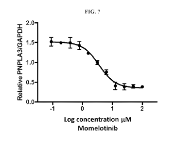
100471 FIG. 7 shows the dose response curve of Momelotinib in hepatic
stellate cells.
100481 FIG. 8 shows the dose response curve of Momelotinib in HepG2 cells.
100491 FIG. 9 shows the effect of Momelotinib treatment on PNPLA3
expression in mouse
liver.
100501 FIG. 10 shows the effect of WYE-125132 treatment on COL 11
expression in mouse
liver.
DETAILED DESCRIPTION OF THE INVENTION
I. INTRODUCTION
[0051] The present invention provides compositions and methods for the
treatment of liver
diseases in humans. In particular, the invention relates to the use of
compounds that modulate
Patatin-like phospholipase domain-containing protein 3 (PNPLA3) for the
treatment of PNPLA3-
related diseases, e.g., nonalcoholic fatty liver disease (NAFLD), nonalcoholic
steatohepatitis
(NASH) and/or alcoholic liver disease (ALD).
100521 The present invention also embraces the alteration, perturbation and
ultimate
regulated control of gene signaling networks (GSNs). Such gene signaling
networks include
genomic signaling centers found within insulated neighborhoods of the genomes
of biological
systems. Compounds modulating PNPLA3 expression may act through modulating one
or more
gene signaling networks.
[0053] As used herein, a "gene signaling network" or "GSN" comprises the
set of
biomolecules associated with any or all of the signaling events from a
particular gene, e.g., a
gene-centric network. As there are over 20,000 protein-coding genes in the
human genome,
there are at least this many gene signaling networks. And to the extent some
genes are non-
coding genes, the number increases greatly. Gene signaling networks differ
from canonical
signaling pathways which are mapped as standard protein cascades and feedback
loops.
[0054] Traditionally, signaling pathways have been identified using
standard biochemical
techniques and, for the most part, are linear cascades with one protein
product signaling the next
protein product-driven event in the cascade. While these pathways may
bifurcate or have
feedback loops, the focus has been almost exclusively at the protein level.
[0055] Gene signaling networks (GSNs) of the present invention represent a
different
paradigm to defining biological signaling¨taking into account protein-coding
and nonprotein-
- 11 -
CA 03072346 2020-02-06
WO 2019/036430
PCT/US2018/046634
coding signaling molecules, genomic structure, chromosomal occupancy,
chromosomal
remodeling, the status of the biological system and the range of outcomes
associated with the
perturbation of any biological systems comprising such gene signaling
networks.
100561 Genomic architecture, while not static, plays an important role in
defining the
framework of the GSNs of the present invention. Such architecture includes the
concepts of
chromosomal organization and modification, topologically associated domains
(TADs), insulated
neighborhoods (INs), genomic signaling centers (GSCs), signaling molecules and
their binding
motifs or sites, and of course, the genes encoded within the genomic
architecture.
100571 The present invention, by elucidating a more definitive set of
connectivities of the
GSNs associated with the PNPLA3 gene, provides a fine-tuned mechanism to
address PNPLA3-
related diseases, including NAFLD, NASH, and/or ALD.
Genomic architecture
[00581 Cells control gene expression using thousands of elements that link
cellular signaling
to the architecture of the genome. Genomic system architecture includes
regions of DNA, RNA
transcripts, chromatin remodelers, and signaling molecules.
Chromosomes
[00591 Chromosomes are the largest subunit of genome architecture that
contain most of the
DNA in humans. Specific chromosome structures have been observed to play
important roles in
gene control, as described in Hnisz et aL, Cell 167, November 17, 2016, which
is hereby
incorporated by reference in its entirety. The "non-coding regions" including
introns provide
protein binding sites and other regulatory structures, while the exons encode
for proteins such as
signaling molecules (e.g., transcription factors), that interact with the non-
coding regions to
regulate gene expression. DNA sites within non-coding regions on the
chromosome also interact
with each other to form looped structures. These interactions form a
chromosome scaffold that is
preserved through development and plays an important role in gene activation
and repression.
Interactions rarely occur among chromosomes and are usually within the same
domain of a
chromosome.
1.00601 In situ hybridization techniques and microscopy have revealed that
each interphase
chromosomes tends to occupy only a small portion of the nucleus and does not
spread throughout
this organelle. See, Cremer and Cremer, Cold Spring Harbor Perspectives in
Biology 2, a003889,
2010, which is hereby incorporated by reference in its entirety. This
restricted surface occupancy
area might reduce interactions between chromosomes.
Topologically associating domains (TADs)
100611 Topologically Associating Domains (TADs), alternatively known as
topological
domains, are hierarchical units that are subunits of the mammalian chromosome
structure. See,
-12-
CA 03072346 2020-02-06
WO 2019/036430
PCT/US2018/046634
Dixon el al., Nature, 485(7398):376-80, 2012; Filippova et al., Algorithms for
Molecular
Biology, 9:14, 2014; Gibcus and Dekker Molecular Cell, 49(5):773-82, 2013;
Nmunova etal.,
Science, 42(6161):948-53, 2013; which are hereby incorporated by reference in
their entireties.
TADs are megabase-sized chromosomal regions that demarcate a microenvironment
that allows
genes and regulatory elements to make productive DNA-DNA contacts. TADs are
defined by
DNA-DNA interaction frequencies. The boundaries of TADs consist of regions
where relatively
fewer DNA-DNA interactions occur, as described in Dixon eral., Nature,
485(7398):376-80,
2012; Nora etal., Nature, 485(7398):381-5, 2012; which are hereby incorporated
by reference in
their entirety. TADs represent structural chromosomal units that function as
gene expression
regulators.
100621 TADs may contain about 7 or more protein-coding genes and have
boundaries that are
shared by the different cell types. See, Smallwood el al., Current Opinion in
Cell Biology,
25(3):387-94, 2013, which is hereby incorporated by reference in its entirety.
Some TADs
contain active genes and others contain repressed genes, as the expression of
genes within a
single TAD is usually correlated. See, Cavalli etal., Nature Structural &
Molecular Biology,
20(3):290-9, 2013, which is hereby incorporated by reference in its entirety.
Sequences within a
TAD find each other with high frequency and have concerted, TAD-wide histone
chromatin
signatures, expression levels, DNA replication timing, lamina association, and
chromocenter
association. See, Dixon etal., Nature, 485(7398):376-80, 2012; Le Dily etal.,
Genes
Development; 28:2151-62, 2014; Dixon etal., Nature; 485(7398):376-80, 2012;
Wijchers,
Gcnome Research, 25:958-69, 2015, which are hereby incorporated by reference
in their
entireties.
100631 Gene loops and other structures within TADs influence the activities
of transcription
factors (TFs), cohesin, and 11-zinc fmger protein (CTCF), a transcriptional
repressor. See,
Baranello etal., Proceedings of the National Academy of Sciences, 111(3):889-
9, 2014, which is
hereby incorporated by reference in its entirety. The structures within TADs
include cohesin-
associated enhancer-promoter loops that are produced when enhancer-bound TFs
bind cofactors,
for example Mediator, that, in turn, bind RNA polymerase II at promoter sites.
See, Lee and
Young, Cell, 152(6):1237-51, 2013; Lelli etal., 2012; Roeder, Annual Reviews
Genetics 46:43-
68, 2005; Spitz and Furlong, Nature Reviews Genetics, 13(9):613-26, 2012;
Dowen etal., Cell,
159(2): 374-387, 2014; Lelli etal., Annual Review of Genetics, 46:43-68, 2012,
which are
hereby incorporated by reference in their entireties. The cohesin-loading
factor Nipped-B-like
protein (NIPBL) binds Mediator and loads cohesin at these enhancer-promoter
loops. See, Kagey
etal., Nature; 467(7314):430-5, 2010; which is hereby incorporated by
reference in its entirety.
- 13-
CA 03072346 2020-02-06
WO 2019/036430
PCT/US2018/046634
[0064] TADs have similar boundaries in all human cell types examined and
constrain
enhancer-gene interactions. See, Dixon etal., Nature, 518:331-336, 2015; Dixon
etal., Nature,
485:376-380, 2012, which are hereby incorporated by reference in their
entirety. This
architecture of the genome helps explain why most DNA contacts occur within
the TADs and
enhancer-gene interactions rarely occur between chromosomes. However, TADs
provide only
partial insight into the molecular mechanisms that influence specific enhancer-
gene interactions
within TADs.
10065] Long-range genomic contacts segregate TADs into an active and
inactive
compartment. See. Lieberman-Aiden et al., Science, 326:289-93, 2009, which is
hereby
incorporated by reference in its entirety. The loops formed between TAD
boundaries seem to
represent the longest-range contacts that are stably and reproducibly formed
between specific
pairs of sequences. See, Dixon etal., Nature, 485(7398):376-80, 2012, which is
hereby
incorporated by reference in its entirety.
[0066] in some embodiments, the methods of the present invention are used
to alter gene
expression from genes located in a TAD. In some embodiments, TAD regions are
modified to
alter gene expression of a non-canonical pathway as defined herein or as
defmable using the
methods described herein.
Insulated neighborhoods
[0067] As used herein, an "insulated neighborhood" (IN) is defmed as a
chromosome
structure formed by the looping of two interacting sites in the chromosome
sequence. These
interacting sites may comprise CCCTC-Binding factor (CTCF). These CTCF sites
are often co-
occupied by cohesin. The integrity of these cohesin-associated chromosome
structures affects
the expression of genes in the insulated neighborhood as well as those genes
in the vicinity of the
insulated neighborhoods. A "neighborhood gene" is a gene localized within an
insulated
neighborhood. Neighborhood genes may be coding or non-coding.
[0068] Insulated neighborhood architecture is defined by at least two
boundaries which come
together, directly or indirectly, to form a DNA loop. The boundaries of any
insulated
neighborhood comprise a primary upstream boundary and a primary downstream
boundary. Such
boundaries are the outermost boundaries of any insulated neighborhood. Within
any insulated
neighborhood loop, however, secondary loops may be formed. Such secondary
loops, when
present, are defined by secondary upstream boundaries and secondary downstream
boundaries.
relative to the primary insulated neighborhood. Where a primary insulated
neighborhood
contains more than one internal loop, the loops are numbered relative to the
primary upstream
boundary of the primary loop, e.g., the secondary loop (first loop within the
primary loop), the
-14-
CA 03072346 2020-02-06
WO 2019/036430
PCT/US2018/046634
tertiary loop (second loop within the primary loop), the quaternary loop (the
third loop within the
primary loop) and so on.
[0069] Insulated neighborhoods may be located within topologically
associated domains
(TADs) and other gene loops. Largest insulated neighborhoods may be TADs. TADs
are defined
by DNA-DNA interaction frequencies, and average 0.8 Mb, contain approximately
7 protein-
coding genes and have boundaries that are shared by the different cell types
of an organism.
According to Dowen, the expression of genes within a TAD is somewhat
correlated, and thus
some TADs tend to have active genes and others tend to have repressed genes.
See Dowen el al.,
Cell. 2014 Oct 9; 159(2): 374-387, which is hereby incorporated by reference
herein in its
entirety.
[0070] Insulated neighborhoods may exist as contiguous entities along a
chromosome or may
be separated by non-insulated neighborhood sequence regions. Insulated
neighborhoods may
overlap linearly only to be defined once the DNA looping regions have been
joined. While
insulated neighborhoods may comprise 3-12 genes, they may contain, 1, 2, 3, 4,
5, 6, 7, 8, 9, 10,
11, 12, 13 or more genes.
[0071] A "minimal insulated neighborhood" is an insulated neighborhood
having at least one
neighborhood gene and associated regulatory sequence region (RSRs) or regions
which facilitate
the expression or repression of the neighborhood gene such as a promoter
and/or enhancer and/or
repressor region, and the like. It is contemplated that in some instances
regulatory sequence
regions may coincide or even overlap with an insulated neighborhood boundary.
Regulatory
sequence regions, as used herein, include but are not limited to regions,
sections, sites or zones
along a chromosome whereby interactions with signaling molecules occur in
order to alter
expression of a neighborhood gene. As used herein, a "signaling molecule" is
any entity, whether
protein, nucleic acid (DNA or RNA), organic small molecule, lipid, sugar or
other biomolecule,
which interacts directly, or indirectly, with a regulatory sequence region on
a chromosome.
Regulatory sequence regions (RSRs) may also refer to a portion of DNA that
functions as a
binding site for a GSC.
100721 One category of specialized signaling molecules are transcription
factors.
"Transcription factors" are those signaling molecules which alter, whether to
increase or
decrease, the transcription of a target gene, e.g., a neighborhood gene.
[0073] According to the present invention, neighborhood genes may have any
number of
upstream or downstream genes along the chromosome. Within any insulated
neighborhood, there
may be one or more, e.g., one, two, three, four or more, upstream and/or
downstream
neighborhood genes relative to the primary neighborhood gene. A "primary
neighborhood gene"
is a gene which is most commonly found within a specific insulated
neighborhood along a
- 15-
CA 03072346 2020-02-06
WO 2019/036430
PCT/US2018/046634
chromosome. An upstream neighborhood gene of a primary neighborhood gene may
be located
within the same insulated neighborhood as the primary neighborhood gene. A
downstream
neighborhood gene of a primary neighborhood gene may be located within the
same insulated
neighborhood as the primary neighborhood gene.
[00741 The present invention provides methods of altering the penetrance of
a gene or gene
variant. As used herein, "penetrance" is the proportion of individuals
carrying a particular
variant of a gene (e.g., mutation, allele or generally a genotype, whether
wild type or not) that
also exhibits an associated trait (phenotype) of that variant gene. In some
situations of disease,
penetrance of a disease-causing mutation measured as the proportion of
individuals with the
mutation who exhibit clinical symptoms. Consequently, penetrance of any gene
or gene variant
exists on a continuum.
100751 Insulated neighborhoods are functional units that may group genes
under the same
control mechanism, which are described in Dowen et at., Cell, 159: 374-387
(2014), which is
hereby incorporated by reference in its entirety. Insulated neighborhoods
provide the mechanistic
background for higher-order chromosome structures, such as TADs which are
shown in FIG. 1.
Insulated neighborhoods are chromosome structures formed by the looping of the
two interacting
CTCF sites co-occupied by cohesin as shown in FIG. 2B. The integrity of these
structures is
important for proper expression of local genes. Generally, 1 to 10 genes are
clustered in each
neighborhood with a median number of 3 genes within each one. The genes
controlled by the
same insulated neighborhood are not readily apparent from a two-dimensional
view of DNA. In
humans, there are about 13,801 insulated neighborhoods in a size range of 25
kb-940 kb with a
median size of 186 kb. Insulated neighborhoods are conserved among different
cell types.
Smaller INs that occur within a bigger IN are referred to as nested insulated
neighborhoods
(NINs). TADs can consist of a single IN as shown in FIG. 1, or one IN and one
NIN and two
NINs as shown in FIG. 2B.
100761 As used herein, the term "boundary" refers to a point, limit, or
range indicating where
a feature, element, or property ends or begins. Accordingly, an "insulated
neighborhood
boundary" refers to a boundary that delimits an insulated neighborhood on a
chromosome.
According to the present invention, an insulated neighborhood is defined by at
least two insulated
neighborhood boundaries, a primary upstream boundary and a primary downstream
boundary.
The "primary upstream boundary" refers to the insulated neighborhood boundary
located
upstream of a primary neighborhood gene. The "primary downstream boundary"
refers to the
insulated neighborhood boundary located downstream of a primary neighborhood
gene.
Similarly, when secondary loops are present as shown in FIG. 2B, they are
defmed by secondary
upstream and downstream boundaries. A "secondary upstream boundary" is the
upstream
-16-
CA 03072346 2020-02-06
WO 2019/036430
PCT/US2018/046634
boundary of a secondary loop within a primary insulated neighborhood, and a
"secondary
downstream boundary" is the downstream boundary of a secondary loop within a
primary
insulated neighborhood. The directionality of the secondary boundaries follows
that of the
primary insulated neighborhood boundaries.
[0077] Components of an insulated neighborhood boundary may comprise the
DNA
sequences at the anchor regions and associated factors (e.g., CTCF, cohesin)
that facilitate the
looping of the two boundaries. The DNA sequences at the anchor regions may
contain at least
one CTCF binding site. Experiments using the ChIP-exo technique revealed a 52
bp CTCF
binding motif containing four CTCF binding modules (see Fig 1, Ong and Comes,
Nature
reviews Genetics, 12:283-293, 2011, which is incorporated herein by reference
in its entirety).
The DNA sequences at the insulated neighborhood boundaries may contain
insulators. In some
cases, insulated neighborhood boundaries may also coincide or overlap with
regulatory sequence
regions, such as enhancer-promoter interaction sites.
[0078] In some embodiments of the present invention, disrupting or altering
an insulated
neighborhood boundary may be accomplished by altering specific DNA sequences
(e.g., CTCF
binding sites) at the boundaries. For example, existing CTCF binding sites at
insulated
neighborhood boundaries may be deleted, mutated, or inverted. Alternatively,
new CTCF binding
sites may be introduced to form new insulated neighborhoods. In other
embodiments, disrupting
or altering an insulated neighborhood boundary may be accomplished by altering
the histone
modification (e.g., methylation, demethylation) at the boundaries. In other
embodiments,
disrupting or altering an insulated neighborhood boundary may be accomplished
by altering (e.g.,
blocking) the binding of CTCF and/or cohesin to the boundaries. In cases where
insulated
neighborhood boundaries coincide or overlap with regulatory sequence regions,
disrupting or
altering an insulated neighborhood boundary may be accomplished by altering
the regulatory
sequence regions (RSR) or the binding of the RSR-associated signaling
molecules.
Controlling expression from insulated neighborhoods: Signaling centers
[0079] Historically, the term "signaling center" has been used to describe
a group of cells
responding to changes in the cellular environment. See, Guger et at.,
Developmental Biology
172: 115-125 (1995), which is incorporated by reference herein in its
entirety. Similarly, the term
"signaling center", as used herein, refers to a defined region of a living
organism that interacts
with a defined set of biomolecules, such as signaling proteins or signaling
molecules (e.g.,
transcription factors) to regulate gene expression in a context-specific
manner.
[0080] Specifically, the term "genomic signaling center", i.e., a
"signaling center", as used
herein, refers to regions within insulated neighborhoods that include regions
capable of binding
context-specific combinatorial assemblies of signaling molecules/signaling
proteins that
-17-
CA 03072346 2020-02-06
WO 2019/036430
PCT/US2018/046634
participate in the regulation of the genes within that insulated neighborhood
or among more than
one insulated neighborhood.
100811 Signaling centers have been discovered to regulate the activity of
insulated
neighborhoods. These regions control which genes are expressed and the level
of expression in
the htunan genome. Loss of the structural integrity of signaling centers
contributes to
deregulation of gene expression and potentially causing disease.
100821 Signaling centers include enhancers bound by a highly context-
specific combinatorial
assemblies of transcription factors. These factors are recruited to the site
through cellular
signaling. Signaling centers include multiple genes that interact to form a
three-dimensional
transcription factor hub macrocomplex. Signaling centers are generally
associated with one to
four genes in a loop organized by biological function.
100831 The compositions of each signaling center has a unique composition
including the
assemblies of transcription factors, the transcription apparatus, and
chromatin regulators.
Signaling centers are highly context specific, permitting drugs to control
response by targeting
signaling pathways.
100841 Multiple signaling centers may interact to control the different
combinations of genes
within the same insulated neighborhood.
Binding sites for signaling molecules
100851 A series of consensus binding sites, or binding motifs for binding
sites, for signaling
molecules has been identified by the present inventors. These consensus
sequences reflect
binding sites along a chromosome, gene, or polynucleotide for signaling
molecules or for
complexes which include one or more signaling molecules.
100861 in some embodiments, binding sites are associated with more than one
signaling
molecule or complex of molecules.
Enhancers
[00871 Enhancers are gene regulatory elements that control cell type
specific gene expression
programs in humans. See, Buecker and Wysocka, Trends in genetics: TIG 28, 276-
284, 2012;
Heinz et al., Nature reviews Molecular Cell Biology, 16:144-154, 2015; Levine
etal.. Cell,
157:13-25, 2014; Ong and Corces, Nature reviews Genetics, 12:283-293, 2011;
Ren and Yue,
Cold Spring Harbor symposia on quantitative biology, 80:17-26, 2015, which are
hereby
incorporated by reference in their entireties. Enhancers are segments of DNA
that are generally a
few hundred base pairs in length that may be occupied by multiple
transcription factors that
recruit co-activators and RNA polymerase II to target genes. See, Bulger and
Groudine, Cell,
144:327-339, 2011; Spitz and Furlong, Nature reviews Genetics, 13:613-626,
2012; Tjian and
Maniatis, Cell, 77:5-8, 1994, which are hereby incorporated by reference in
their entireties.
-18-
CA 03072346 2020-02-06
WO 2019/036430
PCT/US2018/046634
Enhancer RNA molecules transcribed from these regions of DNA also "trap"
transcription
factors capable of binding DNA and RNA. A region with more than one enhancer
is a "super-
enhancer."
[0088] Insulated neighborhoods provide a microenvironment for specific
enhancer-gene
interactions that are vital for both normal gene activation and repression.
Transcriptional
enhancers control over 20,000 protein-coding genes to maintain cell type-
specific gene
expression programs in all human cells. Tens of thousands of enhancers are
estimated to be
active in any given human cell type. See, ENCODE Project Consortium et al.,
Nature, 489, 57-
74, 2012; Roadmap Epigenomics et al., Nature, 518, 317-330, 2015, which are
hereby
incorporated by reference in their entirety. Enhancers and their associated
factors can regulate
expression of genes located upstream or downstream by looping to the promoters
of these genes.
Cohesin ChIA-PET studies carried out to gain insight into the relationship
between
transcriptional control of cell identity and control of chromosome structure
reveal that majority
of the super-enhancers and their associated genes occur within large loops
that are connected
through interacting CTCF-sites co-occupied by cohesin. Such super-enhancer
domains (SD)
usually contain one super-enhancer that loops to one gene within the SD and
the SDs appear to
restrict super-enhancer activity to genes within the SD. The correct
association of super-
enhancers and their target genes in insulated neighborhoods is highly vital
because the mis-
targeting of a single super-enhancer is sufficient to cause disease. See
Groschel et al., Cell,
157(2):369-81, 2014.
[0089] Most of the disease-associated non-coding variation occurs in the
vicinity of
enhancers and hence might impact these enhancer target genes. Therefore,
deciphering the
features conferring specificity to enhancers is important for modulatory gene
expression. See,
Ernst etal., Nature, 473,43-49, 2011: Farb etal., Nature, 518, 337-343,2015:
Hnisz etal.. Cell,
155, 934-947, 2013; Maurano eral.. Science, 337, 1190-1195, 2012, which are
hereby
incorporated by reference in their entirety. Studies suggest that some of the
specificity of
enhancer-gene interactions may be due to the interaction of DNA binding
transcription factors at
enhancers with specific partner transcription factors at promoters. See,
Butler and Kadonaga,
Genes & Development, 15, 2515-2519, 2001; Choi and Engel, Cell, 55, 17- 26,
1988; Ohtsuki et
al., Genes & Development, 12, 547-556, 1998, which are hereby incorporated by
reference in
their entireties. DNA sequences in enhancers and in promoter-proximal regions
bind to a variety
of transcription factors expressed in a single cell. Diverse factors bound at
these two sites interact
with large cofactor complexes and interact with one another to produce
enhancer-gene
specificity. See, Zabidi etal., Nature, 518:556-559, 2015, which is hereby
incorporated by
reference in its entirety.
-19-
CA 03072346 2020-02-06
WO 2019/036430
PCT/US2018/046634
100901 In some embodiments, enhancer regions may be targeted to alter or
elucidate gene
signaling networks (GSNs).
Insulators
[0091] Insulators are regulatory elements that block the ability of an
enhancer to activate a
gene when located between them and contribute to specific enhancer-gene
interactions. See,
Chung etal., Cell 74:505-514, 1993; Geyer and Corces, Genes & Development
6:1865-1873,
1992; Kellum and Schedl, Cell 64:941-950, 1991; Udvardy etal., Journal of
molecular biology
185:341-358, 1985, which are hereby incorporated by reference in their
entirety. Insulators are
bound by the transcription factor CTCF but not all CTCF sites function as
insulators. See, Bell et
al., Cell 98: 387-396, 1999; Liu etal., Nature biotechnology 33:198-203, 2015,
which are hereby
incorporated by reference in their entireties. The features that distinguish
the subset of CTCF
sites that function as insulators have not been previously understood.
[0092] Genome-wide maps of the proteins that bind enhancers, promoters and
insulators,
together with knowledge of the physical contacts that occur between these
elements provide
further insight into understanding of the mechanisms that generate specific
enhancer-gene
interactions. See, Chepelev etal., Cell research, 22:490-503, 2012; DeMare
etal., Genome
Research, 23:1224-1234, 2013; Dowen et al., Cell, 159:374-387, 2014; Fullwood
etal., Genes &
Development 6:1865-1873, 2009: Handoko etal., Nature genetics 43:630-638,
2011; Phillips-
Cremins etal., Cell, 153:1281-1295, 2013; Tang etal., Cell 163:1611-1627,
2015, which are
hereby incorporated by reference in their entirety. Enhancer-bound proteins
are constrained such
that they tend to interact only with genes within these CTCF-CTCF loops. The
subset of CTCF
sites that form these loop anchors thus function to insulate enhancers and
genes within the loop
from enhancers and genes outside the loop, as shown in FIG. 3B. In some
embodiments,
insulator regions may be targeted to alter or elucidate gene signaling
networks (GSNs).
Cohesin and CTCF associated loops and anchor sites/regions
[0093] CTCF interactions link sites on the same chromosome forming loops,
which are
generally less than 1 Mb in length. Transcription occurs both within and
outside the loops, but
the nature of this transcription differs between the two regions. Studies show
that enhancer-
associated transcription is more prominent within the loops. Thus, the
insulator state is enriched
specifically at the CTCF loop anchors. CTCF loops thus either enclose gene
poor regions, with a
tendency for genes to be centered within the loops or leave out gene dense
regions outside the
CTCF loops. FIG. 2A and FIG. 2B compare the linear to the 3-dimensional (3D)
conformation of
the loops.
[0094] CTCF loops exhibit reduced exon density relative to their flanking
regions. Gene
ontology analysis reveals that genes located within CTCF loops are enriched
for response to
-20-
CA 03072346 2020-02-06
WO 2019/036430
PCT/US2018/046634
stimuli and for extracellular, plasma membrane and vesicle cellular
localizations. On the other
hand, genes present within the flanking regions just outside the loops exhibit
an expression
pattern similar to housekeeping genes i.e. these genes are on average more
highly expressed than
the loop-enclosed genes, are less cell-line specific in their expression
pattern, and have less
variation in their expression levels across cell lines. See Oti etal., BMC
Genomics, 17:252,
2016, which is hereby incorporated by reference in its entirety.
[0095] Anchor regions are binding sites for CTCF that influence
conformation of an
insulated neighborhood. Deletion of anchor sites may result in activation of
genes that are usually
transcriptionally silent, thereby resulting in a disease phenotype. In fact,
somatic mutations are
common in loop anchor sites of oncogene-associated insulated neighborhoods.
The CTCF DNA-
binding motif of the loop anchor region has been observed to be the most
altered human
transcription-factor binding sequence of cancer cells. See, Hnisz et al., Cell
167, November 17,
2016, which is incorporated by reference in its entirety.
[0096] Anchor regions have been observed to be largely maintained during
cell development,
and are especially conserved in the germline of humans and primates. In fact,
the DNA sequence
of anchor regions are more conserved in CTCF anchor regions than at CTCF
binding sites that
are not part of an insulated neighborhood. Therefore, cohesin may be used as a
target for ChIA-
PET to identify locations of both.
100971 Cohesin also becomes associated with CTCF-bound regions of the
genome, and some
of these cohesin-associated CTCF sites facilitate gene activation while others
may function as
insulators. See, Dixon et al., Nature, 485(7398):376-80, 2012; Parelho etal.,
Cell, 132(3):422-33,
2008; Phillips-Cremins and Corces, Molecular Cell, 50(4):461-74, 2013); Seitan
etal., Genome
Research, 23(12):2066-77, 2013; Wendt etal., Nature, 451(7180):796-801, 2008),
which are
hereby incorporated by reference in their entireties. Cohesin and CTCF are
associated with large
loop substructures within TADs, and cohesin and Mediator are associated with
smaller loop
structures that form within CTCF-bounded regions. See, de Wit etal., Nature,
501(7466):227-31,
2013; Cremins etal., Cell, 153(6):1281-95, 2013; Sofueva et aL, EMBO,
32(24):3119-29, 2013,
which are hereby incorporated by reference in their entireties. In some
embodiments, cohesin and
CTCF associated loops and anchor sites/regions may be targeted to alter or
elucidate gene
signaling networks (GSNs).
Genetic variants
[0098] Genetic variations within signaling centers are known to contribute
to disease by
disrupting protein binding on chromosomes, such as described in Hnisz etal.,
Cell 167,
November 17, 2016, which is hereby incorporated by reference in its entirety.
Variations of the
sequence of CTCF anchor regions of insulated neighborhood boundary sites that
interfere with
-21-
CA 03072346 2020-02-06
WO 2019/036430
PCT/US2018/046634
formation of insulated neighborhoods are observed to result in dysregulation
of gene activation
and repression. CTCF malfunctions caused by various genetic and epigenetic
mechanisms may
lead to pathogenesis. Therefore, in some embodiments, it is beneficial to
alter any one or more
gene signaling networks (GSNs) associated with such variant-driven etiology in
order to effect
one or more positive treatment outcomes.
Single nucleotide polvmorphisms (SNPs)
[0099] 94.2% of SNPs occur in non-coding regions, which include enhancer
regions. In some
embodiments, SNPs are altered in order to study and/or alter the signaling
from one or more
GSN.
Signaling molecules
[0100] Signaling molecules include any protein that functions in cellular
signaling pathways,
whether canonical or the gene signaling network pathways defined herein or
capable of being
defined using the methods described herein. Transcription factors are a subset
of signaling
molecules. Certain combinations of signaling and master transcription factors
associate to an
enhancer region to influence expression of a gene. Master transcription
factors direct
transcription factors in specific tissues. For example, in blood, GATA
transcription factors are
master transcription factors that direct TCF7L2 of the Wnt cellular signaling
pathway. In the
liver. HNF4A is a master transcription factor to direct SMAD in lineage
tissues and patterns.
[0101] Transcriptional regulation allows controlling how often a given gene
is transcribed.
Transcription factors alter the rate at which transcripts are produced by
making conditions for
transcription initiation more or less favorable. A transcription factor
selectively alters a signaling
pathway which in turn affects the genes controlled by a genomic signaling
center. Genomic
signaling centers are components of transcriptional regulators. In some
embodiments, signaling
molecules may be used, or targeted in order to elucidate or alter the
signaling of gene signaling
networks of the present invention.
[0102] Table 22 of International Application No. PCT/US18/31056, which is
hereby
incorporated by reference in its entirety, provides a list of signaling
molecules including those
which act as transcription factors (TF) and/or chromatin remodeling factors
(CR) that function in
various cellular signaling pathways. The methods described herein may be used
to inhibit or
activate the expression of one or more signaling molecules associated with the
regulatory
sequence region of the primary neighborhood gene encoded within an insulated
neighborhood.
The methods may thus alter the signaling signature of one or more primary
neighborhood genes
which are differentially expressed upon treatment with the therapeutic agent
compared to an
untreated control.
-22-
CA 03072346 2020-02-06
WO 2019/036430
PCT/US2018/046634
Transcription factors
[01031 Transcription factors generally regulate gene expression by binding
to enhancers and
recruiting coactivators and RNA polymerase II to target genes. See Whyte et
al., Cell, 153(2):
307-319, 2013, which is incorporated by reference in its entirety.
Transcription factors bind
"enhancers" to stimulate cell-specific transcriptional program by binding
regulatory elements
distributed throughout the genome.
101041 There are about 1800 known transcription factors in the human genome.
There are
epitopes on the DNA of the chromosomes that provide binding sites for proteins
or nucleic acid
molecules such as ribosomal RNA complexes. Master regulators direct a
combination of
transcription factors through cell signaling above and DNA below. These
characteristics allow
for determination of the location of the next signaling center. In some
embodiments, transcription
factors may be used or targeted, to alter or elucidate the gene signaling
networks of the present
invention.
Master transcription factors
101051 Master transcription factors bind and establish cell-type specific
enhancers. Master
transcription factors recruit additional signaling proteins, such as other
transcription factors, to
enhancers to form signaling centers. An atlas of candidate master TFs for 233
human cell types
and tissues is described in D'Alessio et al., Stem Cell Reports 5, 763-775
(2015), which is hereby
incorporated by reference in its entirety. In some embodiments, master
transcription factors may
be used or targeted, to alter or elucidate the gene signaling networks of the
present invention.
Signaling transcription factors
[0106] Signaling transcription factors are transcription factors, such as
homeoproteins, that
travel between cells as they contain protein domains that allow them to do the
so. Homeoproteins
such as Engrailed, Hoxa5, Hoxb4, Hoxc8, Emx 1, Emx2, 0tx2 and Pax6 are able to
act as
signaling transcription factors. The homeoprotein Engrailed possesses
internalization and
secretion signals that are believed to be present in other homeoproteins as
well. This property
allows homeoproteins to act as signaling molecules in addition to being
transcription factors.
Homeoproteins lack characterized extracellular functions leading to the
perception that their
paracrine targets are intracellular. The ability of homeoproteins to regulate
transcription and, in
some cases, translation is most likely to affect paracrine action. See
Prochiantz and Joliot, Nature
Reviews Molecular Cell Biology, 2003. In some embodiments, signaling
transcription factors
may be used or targeted, to alter or elucidate the gene signaling networks of
the present
invention.
-23-
CA 03072346 2020-02-06
WO 2019/036430
PCT/US2018/046634
Chromatin modifications
[0107] Chromatin remodeling is regulated by over a thousand proteins that
are associated with
histone modification. See, Ji etal., PNAS, 112(12):3841-3846(2015), which is
hereby
incorporated by reference in its entirety. Chromatin regulators are specific
sets of proteins
associated with genomic regions marked with modified histones. For example,
histones may be
modified at certain lysine residues: H3K20me3, H3K27ac, H3K4me3, H3K4me1,
H3K79me2,
H3K36me3, H3K9me2, and H3K9me3. Certain histone modifications mark regions of
the
genome that are available for binding by signaling molecules. For example,
previous studies have
observed that active enhancer regions include nucleosomes with H3K27ac, and
active promoters
include nucleosomes with H3K27ac. Further, transcribed genes include
nucleosomes with
H3K79me2. ChIP-MS may be performed to identify chromatin regulator proteins
associated with
specific histone modification. ChIP-seq with antibodies specific to certain
modified histones may
also be used to identify regions of the genome that are bound by signaling
molecules. In some
embodiments, chromatin modifying enzymes or proteins may be used or targeted,
to alter or
elucidate the gene signaling networks of the present invention.
RNAs derived.from regulatory sequence regions
[0108] Many active regulatory sequence regions (RSRs), such as regions from
enhancers,
signaling centers, and promoters of protein-coding genes, are known to produce
non-coding
RNAs. Transcripts produced at or in the vicinity of active regulatory sequence
regions have been
implicated in transcription regulation of nearby genes. Recent reports have
demonstrated that
enhancer-associated RNAs (eRNAs) are strong indicators of enhancer activity
(See Li etal., Nat
Rev Genet. 2016 Apr;17(4):207-23, which is hereby incorporated by reference in
its entirety).
Further, non-coding RNAs from active regulatory sequence regions have been
shown to be
involved in facilitating the binding of transcription factors to these regions
(Sigova et al.,
Science. 2015 Nov 20;350(6263):978-81, which is hereby incorporated by
reference in its
entirety). This suggests that such RNAs may be important for the assembly of
signaling centers
and regulation of neighborhood genes. In some embodiments, RNAs derived from
regulatory
sequence regions of the PNPLA3 gene may be used or targeted to alter or
elucidate the gene
signaling networks of the present invention.
[0109] In some embodiments, RNAs derived from regulatory sequence regions may
be an
enhancer-associated RNA (eRNA). In some embodiments. RNAs derived from
regulatory
sequence regions may be a promoter-associated RNA, including but not limited
to, a promoter
upstream transcript (PROMPT), a promoter-associated long RNA (PALR), and a
promoter-
associated small RNA (PASR). In further embodiments. RNAs derived from
regulatory sequence
- 24 -
CA 03072346 2020-02-06
WO 2019/036430
PCT/US2018/046634
regions may include but are not limited to transcription start sites (TSS)-
associated RNAs (TSSa-
RNAs), transcription initiation RNAs (tiRNAs), and terminator-associated small
RNAs (TASRs).
101101 In some embodiments. RNAs derived from regulatoiy sequence regions may
be long
non-coding RNAs (1ncRNAs) (i.e., >200 nucleotides). In some embodiments, RNAs
derived
from regulatory sequence regions may be intermediate non-coding RNAs. (i.e.,
about 50 to 200
nucleotides). In some embodiments, RNAs derived from regulatory sequence
regions may be
short non-coding RNAs (i.e., about 20 to 50 nucleotides).
101111 In some embodiments, eRNAs that may be modulated by methods and
compounds
described herein may be characterized by one or more of the following
features: (1) transcribed
from regions with high levels of monomethylation on lysine 4 of histone 3
(H3K4me1) and low
levels of trimethylation on lysine 4 of histone 3 (H3K4me3); (2) transcribed
from genomic
regions with high levels of acetylation on lysine 27 of histone 3 (H3K27ac);
(3) transcribed from
genomic regions with low levels of trimethylation on lysine 36 of histone 3
(H3K36me3); (4)
transcribed from genomic regions enriched for RNA polymerase Ii (P0111); (5)
transcribed from
genomic regions enriched for transcriptional co-regulators, such as the p300
co-activator; (6)
transcribed from genomic regions with low density of CpG island; (7) their
transcription is
initiated from Pol II-binding sites and elongated bidirectionally; (8)
evolutionarily conserved
DNA sequences encoding eRNAs; (9) short half-life; (10) reduced levels of
splicing and
polyadenylation, (11) dynamically regulated upon signaling; (12) positively
correlated to levels
of nearby mRNA expression; (13) extremely high tissue specificity; (14)
preferentially nuclear
and chromatin-bound; and/or (15) degraded by the exosome.
101121 Exemplary eRNAs include those described in Djebali et al., Nature.
2012 Sep
6;489(7414) (for example, Supplementary data file for Figure 5a) and Andersson
et al.. Nature.
2014 Mar 27;507(7493):455-461 (for example, Supplementary Tables S3, S12, S13,
S15, and
16), which are herein incorporated by reference in their entirety.
101131 In some embodiments, promoter-associated RNAs that may be modulated by
methods
or compounds described herein may be characterized by one or more of the
following features:
(1) transcribed from regions with high levels of H3K4me1 and low to medium
levels of
H3K4me3; (2) transcribed from genomic regions with high levels of H31(27ac;
(3) transcribed
from genomic regions with no or low levels of H3K36me3; (4) transcribed from
genomic regions
enriched for RNA polymerase II (Poll!); (5) transcribed from genomic regions
with high density
of CpG island; (6) their transcription is initiated from Pol TI-binding sites
and elongated in the
opposite direction from the sense strand (that is, mRNAs) or bidirectionally;
(7) short half-life;
(8) reduced levels of splicing and polyadenylation; (9) preferentially nuclear
and chromatin-
bound; and/or (10) degraded by the exosome.
-25-
CA 03072346 2020-02-06
WO 2019/036430
PCT/US2018/046634
(0114] In some embodiments, compositions and methods described herein may be
used to
modulate RNAs derived from regulatory sequence regions to alter or elucidate
the gene signaling
networks of the present invention. In some embodiments, methods and compounds
described
herein may be used to inhibit the production and/or function of an RNA derived
from regulatory
sequence regions. In some embodiments, a hybridizing oligonucleotide such as
an siRNA or an
antisense oligonucleotide may be used to inhibit the activity of the RNA of
interest via RNA
interference (RNAi), or RNase H-mediated cleavage, or physically block binding
of various
signaling molecules to the RNA. Exemplary hybridizing oligonucleotide may
include those
described in U.S. Pat No. 9,518,261 and PCT Publication No. WO 2014/040742,
which are
hereby incorporated by reference in their entirety. The hybridizing
oligonucleotide may be
provided as a chemically modified or unmodified RNA, DNA, locked nucleic acids
(LNA), or a
combination of RNA and DNA, a nucleic acid vector encoding the hybridizing
oligonucleotide,
or a virus carrying such vector. In other embodiments, genome editing tools
such as
CRISPR/Cas9 may be used to delete specific DNA elements in the regulatory
sequence regions
that control the transcription of the RNA or degrade the RNA itself. In other
embodiments,
genome editing tools such as a catalytically inactive CR1SPR/Cas9 may be used
to bind to
specific elements in the regulatory sequence regions and block the
transcription of the RNA of
interest. In further embodiments, bromodomain and extra-terminal domain (BET)
inhibitors (e.g.,
JQ1, I-BET) may be used to reduce RNA transcription through inhibition of
histone acetylation
by BET protein Brd4.
[0115] In alterative embodiments, methods and compounds described herein may
be used to
increase the production and/or function of an RNA derived from regulatory
sequence regions. In
some embodiments, an exogenous synthetic RNA that mimic the RNA of interest
may be
introduced into the cell. The synthetic RNA may be provided as an RNA, a
nucleic acid vector
encoding the RNA, or a virus carrying such vector. In other embodiments,
genome editing tools
such as CRISPR/Cas9 may be used to tether an exogenous synthetic RNA to
specific sites in the
regulatory sequence regions. Such RNA may be fused to the guide RNA of the
CR1SPR/Cas9
complex.
[0116] In some embodiments, modulation of RNAs derived from regulatory
sequence regions
increases the expression of the PNPLA3 gene. In some embodiments, modulation
of RNAs
derived from regulatory sequence regions reduces the expression of the PNPLA3
gene.
[0117] In some embodiments. RNAs modulated by compounds described herein
include
RNAs derived from regulatory sequence regions of the PNPLA3 in a liver cell
(e.g., a hepatocyte
or a stellate cell).
-26-
CA 03072346 2020-02-06
WO 2019/036430
PCT/US2018/046634
Perturbation of genomic systems
[0118] Behavior of one or more components of the gene signaling networks
(GSNs), genomic
signaling centers (GSCs), and/or insulated neighborhoods (INs) related to
PNPLA3 as described
herein may be altered by contacting the systems containing such features with
a perturbation
stimulus. Potential stimuli may include exogenous biomolecules such as small
molecules,
antibodies, proteins, peptides, lipids, fats, nucleic acids, and the like or
environmental stimuli
such as radiation, pH, temperature, ionic strength, sound, light and the like.
101191 The present invention selves, not only as a discovery tool for the
elucidation of better
defined gene signaling networks (GSNs) and consequently a better understanding
of biological
systems. The present invention allows the ability to properly define gene
signaling for PNPLA3
at the gene level in a manner which allows the prediction, a priori. of
potential treatment
outcomes, the identification of novel compounds or targets which may have
never been
implicated in the treatment of a PNPLA3-related disease or condition,
reduction or removal of
one or more treatment liabilities associated with new or known drugs such as
toxicity, poor half-
life, poor bioavailability, lack of or loss of efficacy or pharmacokinetic or
pharmacodynamic
risks.
101201 Treatment of disease by altering gene expression of canonical
cellular signaling
pathways has been shown to be effective. Even small changes in gene expression
may have a
significant impact on disease. For example, changes in signaling centers
leading to signaling
pathways affecting cell suicide suppression are associated with disease. The
present invention, by
elucidating a more definitive set of connectivities of the GSNs provides a
fine-tuned mechanism
to address disease, including genetic diseases. A method of treating a disease
may include
modifying a signaling center that is involved in a gene associated with that
disease. Such genes
may not presently be associated with the disease except as is elucidated using
the methods
described herein.
101211 A perturbation stimulus may be a small molecule, a known drug, a
biological, a
vaccine, an herbal preparation, a hybridizing oligonucleotide (e.g., siRNA and
antisense
oligonucleotide), a gene or cell therapy product, or other treatment product.
[0122] In some embodiments, methods of the present invention include
applying a
perturbation stimulus to perturb GSNs, genomic signaling centers, and/or
insulated
neighborhoods associated with the PNPLA3 gene. Perturbation stimuli that
causes changes in
PNPLA3 expression may inform the connectivities of the associated GSNs and
provide potential
targets and/or treatments for PNPLA3-related disorders.
-27-
CA 03072346 2020-02-06
WO 2019/036430
PCT/US2018/046634
Downstream targets
101231 In certain embodiments, a stimulus is administered that targets a
downstream product
of a gene of a gene signaling network. Alternatively, the stimulus disrupts a
gene signaling
network that affects downstream expression of at least one downstream target.
In some
embodiments, the gene is PNPLA3.
mRNA
101241 Perturbation of a single or multiple gene signaling network (GSN)
associated with a
single insulated neighborhood or across multiple insulated neighborhoods can
affect the
transcription of a single gene or a multiple set of genes by altering the
boundaries of the insulated
neighborhood due to loss of anchor sites comprising cohesins. Specifically,
perturbation of a
GSC may also affect the transcription of a single gene or a multiple set of
genes. Perturbation
stimuli may result in the modification of the RNA expression and/or the
sequences in the primary
transcript within the mRNA, i.e. the exons or the RNA sequences between the
exons that are
removed by splicing, i.e. the introns. Such changes may consequently alter the
members of the
set of signaling molecules within the gene signaling network of a gene,
thereby defining a variant
of the gene signaling network.
Proteins
101251 Perturbation of a single or multiple gene signaling networks
associated with a single
insulated neighborhood or across multiple insulated neighborhoods can affect
the translation of a
single gene or a multiple set of genes that are part of the genomic signaling
center, as well as
those downstream to the genomic signaling center. Specifically, perturbation
of a genomic
signaling center may affect translation. Perturbation may result in the
inhibition of the translated
protein.
Nearest neighbor gene
101261 Perturbation stimuli may cause interactions with signaling molecules
to occur in order
to alter expression of the nearest primary neighborhood gene that may be
located upstream or
downstream of the primary neighborhood gene. Neighborhood genes may have any
number of
upstream or downstream genes along the chromosome. Within any insulated
neighborhood, there
may be one or more, e.g., one, two, three, four or more, upstream and/or
downstream
neighborhood genes relative to the primary neighborhood gene. A "primary
neighborhood gene"
is a gene which is most commonly found within a specific insulated
neighborhood along a
chromosome. An upstream neighborhood gene of a primary neighborhood gene may
be located
within the same insulated neighborhood as the primary neighborhood gene. A
downstream
neighborhood gene of a primary neighborhood gene may be located within the
same insulated
neighborhood as the primary neighborhood gene.
-28-
CA 03072346 2020-02-06
WO 2019/036430
PCT/US2018/046634
canonical cell signaling pathways
101271 it is understood that there may be some overlap between the canonical
pathways
detailed in the art and the gene signaling networks (GSNs) defined herein.
101281 Whereas canonical pathways permit a certain degree of promiscuity of
members across
pathways (cross talk), gene signaling networks (GSN) of the invention are
defined at the gene
level and characterized based on any number of stimuli or perturbation to the
cell, tissue, organ
or organ system expressing that gene. Hence the nature of a GSN is both
structurally (e.g., the
gene) and situationally (e.g., the function, e.g., expression profile)
defined. And while two
different gene signaling networks may share members, they are still unique in
that the nature of
the perturbation can distinguish them. Hence, the value of GSNs in the
elucidation of the
function of biological systems in support of therapeutic research and
development.
101291 It should be understood that it is not intended that no connection
ever be made between
canonical pathways and gene signaling networks; in fact, the opposite is the
case. In order to
bridge the two signaling paradigms for further scientific insights, it will be
instructive to compare
the canonical signaling pathway paradigm with the gene signaling networks of
the present
invention.
101301 In some embodiments, methods of the present invention involve
altering the Janus
kinases (JAK)/signal transducers and activators of transcription (STAT)
pathway. The
JAK/STAT pathway is the major mediator for a wide array of cytokines and
growth factors.
Cytokines are regulatory molecules that coordinate immune responses. JAKs are
a family of
intracellular, nonreceptor tyrosine kinases that are typically associated with
cell surface receptors
such as cytokine receptors. Mammals are known to have 4 JAKs: JAK1, JAK2,
JAK3, and
Tyrosine kinase 2 (1'YK2). Binding of cytokines or growth factors to their
respective receptors at
the cell surface initiates trans-phosphotylation of JAKs, which activates
downstream STATs.
STATs are latent transcription factors that reside in the cytoplasm until
activated. There are
seven mammalian STATs: STAT1, STAT2, STAT3, STAT4, STAT5 (STAT5A and STAT5B),
and STAT6. Activated STATs translocate to the nucleus where they complex with
other nuclear
proteins and bind to specific sequences to regulate the expression of target
genes. Thus the
JAK/STAT pathway provides a direct mechanism to translate an extracellular
signal into a
transcriptional response. Target genes regulated by JAK/STAT pathway are
involved in
immunity, proliferation, differentiation, apoptosis and oncogenesis.
Activation of JAKs may also
activate the phosphatidylinositol 3-kinase (PI3K) and mitogen-activated
protein kinase (MAPK)
pathways.
[01311 In some embodiments, methods of the present invention involve
altering the p53
mediated apoptosis pathway. Tumor protein p53 regulates the cell cycle and
hence functions as a
-29-
CA 03072346 2020-02-06
WO 2019/036430
PCT/US2018/046634
tumor suppressor to prevent cancer. p53 plays an important role in apoptosis,
inhibition of
angiogenesis and genomic stability by activating DNA repair proteins,
arresting cell growth
though holding the cell cycle and initiating apoptosis. p53 becomes activated
in response to DNA
damage, osmotic shock, oxidative stress or other myriad stressors. Activated
p53 activates the
expression of several genes by binding DNA including p2I. p2I binds to the GI-
S/CDK
complexes which is an important molecule for the G I/S transition, then causes
cell cycle arrest.
p53 promotes apoptosis through two major apoptotic pathways: extrinsic pathway
and intrinsic
pathways. The extrinsic pathway involves activation of particular cell-surface
death receptors
that belong to the tumor necrosis factor (TNF) receptor family and, through
the formation of the
death-inducing signaling complex (DISC), leads to a cascade of activation of
caspases, including
Caspase8 and Caspase3, which in turn induce apoptosis. In the intrinsic
pathway, p53 participates
interacts with the multidomain members of the Bc1-2 family (e.g., Bc1-2, Bc1-
xL) to induce
mitochondrial outer membrane permeabilization.
[0132] In some embodiment, methods of the present invention involve
altering the
phosphoinositide 3-kinase (PI3K)/Akt signaling pathway. The PI3K/Akt signaling
pathway plays
a critical role in regulating various cellular functions including metabolism,
growth, proliferation,
survival, transcription and protein synthesis. The signaling cascade is
activated by receptor
tyrosine kinases, integrins, B and T cell receptors, cytokine receptors, G-
protein-coupled
receptors and other stimuli that induce production of phospha-tidylinositol
(3,4,5) trisphosphates
(PIP3) by PI3K. the serine/threonine kinase Akt (also known as protein kinase
B or PKB)
interacts with these phospholipids, causing its translocation to the inner
membrane, where it is
phosphorylated and activated by pyruvate dehydrogenase kinases PDKI and PD1(2.
Activated
Akt modulates the function of munerous substrates involved in the regulation
of cell survival,
cell cycle progression and cellular growth.
[0133] In some embodiment, methods of the present invention involve
altering the spleen
tyrosine kinase (Syk)-dependent signaling pathway. Syk is a protein tyrosine
kinase associated
with various inflammatory cells, including macrophages. Syk plays a key role
in the signaling of
activating Fc receptors and the B-cell receptor (BCR). When Fe-receptors for
IgG I, IIA, and
INA bind to their ligands, the receptor complex becomes activated and triggers
the
phosphorylation of the immunoreceptor-activating motifs (ITAMs). This
activates various genes,
which leads to a cytoskeletal rearrangement that mediates phagocytosis in
cells of the
monocyte/macrophage lineage. Because Syk plays an important role in Fe
receptor-mediated
signal transduction and inflammatory propagation, it is considered a good
target for the inhibition
of various autoimmtme conditions, such as rheumatoid arthritis and lymphoma.
-30-
CA 03072346 2020-02-06
WO 2019/036430
PCT/US2018/046634
[0134] In some embodiment, methods of the present invention involve
altering the insulin like
growth factor 1 receptor (IGF-1R)/insulin receptor (InsR) signaling pathway.
Insulin-like growth
factor I (IGF-1) controls many biological processes such as cellular
metabolism, proliferation,
differentiation, and apoptosis. These effects are mediated through ligand
activation of the
tyrosine kinase activity intrinsic to their receptors IGF-1R. InsR substrates
1 and 2 (IRS! and
IRS2) are key signaling intermediates, and their known downstream effectors
are PI3K/AKT and
MAPK/ERK1. The consequence of signaling results in a temporal transcriptional
response
leading to a wide range of biological processes including cell proliferation
and survival.
101351 in some embodiment, methods of the present invention involve
altering the Fins-like
Tyrosine Kinase-3 (FLT3) signaling pathway. FLT3, also known as FLK2 (Fetal
Liver Kinase-2)
and S'TK1 (human Stem Cell Kinase-1) is a cytokine receptor which belongs to
the receptor
tyrosine kinase class III. It is expressed on the surface of many
hematopoietic progenitor cells.
Signaling of FLT3 is important for the normal development of hematopoietic
stem cells and
progenitor cells. Binding of FLT3 ligand to FLT3 triggers the PI3K and RAS
pathways, leading
to increased cell proliferation and the inhibition of apoptosis.
[0136] in some embodiment, methods of the present invention involve altering
the
Hippo signaling pathway. The Hippo signaling pathway plays an important role
in tissue
regeneration, stem cell self-renewal and organ size control. It controls organ
size in animals
through the regulation of cell proliferation and apoptosis. The Mammalian
Sterile 20-like kinases
(MST1 and MST2) are key components of the Hippo signaling pathway in mammals.
[0137] In some embodiments, methods of the present invention involve
altering the
mammalian Target Of Rapamycin (mTOR) pathway. The mTOR pathway is a central
regulator
of cell metabolism, growth, proliferation and survival. mTOR is an atypical
serine/threonine
kinase that is present in two distinct complexes: mTOR complex I (mTORC I) and
mTORC2.
mTORC1 functions as a nutrient/energy/redox sensor and controls protein
synthesis. It senses
and integrates diverse nutritional and environmental cues, including growth
factors, energy
levels, cellular stress, and amino acids. mTORC2 has been shown to function as
an important
regulator of the actin cytoskeleton. In addition, mTORC2 is also involved in
the activation of
IGF-IR and InsR. Aberrant mTOR signaling is linked to many human diseases
including cancer,
cardiovascular disease, and diabetes.
[0138] In some embodiments, methods of the present invention involve altering
the Glycogen
synthase kinase 3 (GSK3) pathway. GSK3 is a constitutively active, highly
conserved
serine/dreonine protein kinase involved in numerous cellular functions
including glycogen
metabolism, gene transcription, protein translation, cell proliferation,
apoptosis, immune
response, and microtubule stability. GSK3 participates in a variety of
signaling pathways,
-31-
CA 03072346 2020-02-06
WO 2019/036430
PCT/US2018/046634
including cellular responses to WNT, growth factors, insulin, Reelin, receptor
tyrosine kinases
(RTK), Hedgehog pathways, and G-protein-coupled receptors (GPCR). GSK3 is
localized
predominantly in the cytoplasm but its subcellular localization is changed in
response to stimuli.
101391 In some embodiments, methods of the present invention involve
altering the
transforming growth factor-beta (TGF-beta)/SMAD signaling pathway. TGF-
beta/SMAD
signaling pathway is involved in many biological processes in both the adult
organism and the
developing embryo including cell growth, cell differentiation, apoptosis,
cellular homeostasis and
other cellular functions. TGF-beta superfamily ligands include Bone
morphogenetic proteins
(BMPs), Growth and differentiation factors (GDFs). Anti mullerian hormone
(AMH), Activin,
Nodal and TGF-beta. They act via specific receptors activating multiple
intracellular pathways
resulting in phosphorylation of receptor-regulated SMAD proteins that
associate with the
common mediator, SMAD4. Such complex translocates to the nucleus, binds to DNA
and
regulates transcription of many genes. BMPs may cause the transcription of
mRNAs involved in
osteogenesis, neurogenesis, and ventral mesoderm specification. TGF-betas may
cause the
transcription of mRNAs involved in apoptosis, extracellular matrix neogenesis
and
immunosuppression. It is also involved in GI arrest in the cell cycle. Activin
may cause the
transcription of mRNAs involved in gonadal growth, embryo differentiation and
placenta
formation. Nodal may cause the transcription of mRNAs involved in left and
right axis
specification, mesoderm and endoderm induction. The roles of TGF-beta
superfamily members
are reviewed in Wakefield et al, Nature Reviews Cancer I3(5):328-41, which is
hereby
incorporated by reference in its entirety.
101401 In some embodiments, methods of the present invention involve
altering the nuclear
factor-kappa B (NF-KB) signaling pathway. NF-KB is a transcription factor
found in all cell types
and is involved in cellular responses to stimuli such as stress and cytokines.
NF-KB signaling
plays an important role in inflammation, the innate and adaptive immune
response and stress. In
unstimulated cells NF-KB dimers are sequestered inactively in the cytoplasm by
a protein
complex called inhibitor of kappa B (IKB). Activation of NF-KB occurs via
degradation of IKB, a
process that is initiated by its phosphorylation by 'KB kinase (IKK). This
enables the active NF-
KB transcription factor subunits to translocate to the nucleus and induce
target gene expression.
NF-KB activation turns on expression of the IKBa gene, forming a negative
feedback loop.
Dysregulation of NF-KB signaling can lead to inflammatory and autoimmune
diseases and
cancer. The role of NF-KB pathway in inflammation is reviewed in Lawrence,
Cold Spring Harb
Perspect Biol. 2009; 1(6): a001651, which is hereby incorporated by reference
in its entirety.
II. FEATURES AND PROPERTIES OF THE PATAT1N-LIKE PHOSPHOL1PASE DOMAIN-
CONTAINING PROTEIN 3 (PNPLA3) GENE
-32-
CA 03072346 2020-02-06
WO 2019/036430
PCT/US2018/046634
[0141] In some embodiments, methods of the present invention involve
modulating the
expression of the Patatin-like phospholipase domain-containing protein 3
(PNPLA3) gene.
PNPLA3 may also be referred to as Adiponutrin, Calcium-Independent
Phospholipase A2-
Epsilon, Acylglycerol 0-Acyltransferase, Patatin-Like Phospholipase Domain-
Containing
Protein 3, Patatin-Like Phospholipase Domain Containing 3, Chromosome 22 Open
Reading
Frame 20, IPLA(2)Epsilon, IPLA2epsilon, IPLA2-Epsilon, C22orf20, ADPN, EC
2.7.7.56, EC
4.2.3.4, EC 3.1.1.3, and EC 2.3.1.-. PNPLA3 has a cytogenetic location of
22q13.31 and the
genomic coordinate are on Chromosome 22 on the forward strand at position
43,923,739-
43,964,488. PNPLA5 (ENSG00000100341) is the gene upstream of PNPLA3 on the
forward
strand and SAMM50 (ENSG00000100347) is the gene downstream of PNPLA3 on the
forward
strand. PNPLA3 has a NCBI gene ID of 80339, Uniprot ID of Q9NST1 and Ensembl
Gene ID of
ENSG00000100344. The nucleotide sequence of PNPLA3 is shown in SEQ ID NO: 1.
[0142] In some embodiments, methods of the present invention involve
altering the
composition and/or the structure of the insulated neighborhood containing the
PNPLA3 gene.
The present inventors have identified the insulated neighborhood containing
the PNPLA3 gene in
primary human hepatocytes. The insulated neighborhood that contains the PNPLA3
gene is on
chromosome 22 at position 43,782,676-45,023,137 with a size of approximately
1,240 kb. The
number of signaling centers within the insulated neighborhood is 12. The
insulated neighborhood
contains PNPLA3 and 7 other genes, namely MPPED1, EFCAB6, SULT4A1, PNPLA5,
SAMM50, PARVB, and PARVG. The chromatin marks, or chromatin-associated
proteins,
identified at the insulated neighborhood include H3k27ac, BRD4, p300, H3K4me1
and
H3K4me3. Transcription factors involved in the insulated neighborhood include
HNF3b, HNF4a,
HNF4, HNF6, Myc, ONECUT2 and YY1. Signaling proteins involved in the insulated
neighborhood include TCF4, RIF la, HNF1, ERa, GR, JUN, RXR, STAT3, VDR, NF-KB,
SMAD2/3, STAT1, TEAD1, p53, SMAD4, and FOS. Any components of these signaling
centers
and/or signaling molecules, or any regions within or near the insulated
neighborhood, may be
targeted or altered to change the composition and/or structure of the
insulated neighborhood,
thereby modulating the expression of PNPLA3.
[0143] PNPLA3 encodes a lipid droplet-associated, carbohydrate-regulated
lipogenic and/or
lipolytic enzyme. PNPLA3 is predominantly expressed in liver (hepatocytes and
hepatic stellate
cells) and adipose tissue. Hepatic stellate cells (HSCs, also called
perisinusoidal cells or Ito cells)
are contractile cells that reside between the hepatocytes and small blood
vessels in the liver.
HSCs have been identified as the main matrix-producing cells in the process of
liver fibrosis.
PNPLA3 is known to be involved in various metabolic pathways, such as
glycerophospholipid
biosynthesis, triacylglycerol biosynthesis, adipogenesis, and eicosanoid
synthesis.
-33-
CA 03072346 2020-02-06
WO 2019/036430
PCT/US2018/046634
[0144] Variations in PNPLA3 are associated with metabolic disorders such as
nonalcoholic
fatty liver disease, nonalcoholic steatohepatitis, hepatic steatosis,
alcoholic liver disease,
alcoholic liver cirrhosis, alcoholic steatosis, liver cancer, lipid storage
disease, obesity and other
inherited metabolic disorders. Any one or more of these disorders may be
treated using the
compositions and methods described herein.
[0145] A polymorphic variation rs738409 C/G of PNPLA3, encoding for the
isoleucine to
methionine substitution at residue 148 (I148M), has been linked to NAFLD,
hepatic steatosis and
nonalcoholic steatohepatitis (NASH) as well as its pathobiological sequelae
fibrosis, cirrhosis,
and hepatocellular cancer (Krawczyk M et al., Semin Liver Dis. 2013
Nov;33(4):369-79, which
is hereby incorporated by reference in its entirety). Research suggests that
the altered protein
leads to increased production and decreased breakdown of fats in the liver.
PNPLA3 I148M
enhances steatosis by impairing the liberation of triglycerides from lipid
droplets (Trepo E et al.,
J Hepatol. 2016 Aug;65(2):399-412, which is hereby incorporated by reference
in its entirety).
Recent data also suggests that PNPLA3 I148M protein evades degradation and
accumulates on
lipid droplets (BasuRay ei al., Hepatology. 2017 Oct;66(4):1111-1124, which is
hereby
incorporated by reference in its entirety). I148M variant is associated with
NAFLD in both adults
and in children, but is predominant in women, not in men. The specific
mechanism of the
PNPLA3 1148M variant in the development and progression of NAFLD is still not
clear.
However, it is thought that the PNPLA3 I148M variant may promote the
development of
fibrogenesis by activating the hedgehog signaling pathway, which, in turn,
leads to the activation
and proliferation of hepatic stellate cells, and excessive generation and
deposition of intrahepatic
extracellular matrix (Chen LZ, et al., World J Gastroenterol. 2015 Jan 21;
21(3): 794-802, which
is hereby incorporated by reference in its entirety).
[0146] The I148M variant has also been correlated with alcoholic liver
disease and clinically
evident alcoholic cirrhosis (Tian et al., Nature Genetics 42, 21-23 (2010),
which is hereby
incorporated by reference in its entirety). Moreover, it has been identified
as a prominent risk
factor for hepatocellular carcinoma in patients with alcoholic cirrhosis
(Nischalke et al., PLoS
One. 2011;6(11):e27087, which is hereby incorporated by reference in its
entirety).
[0147] The I I48M variant also influences insulin secretion levels and
obesity. In obese
subjects the body mass index and waist are higher in carriers of the variant
allele (Johansson LE
etal.. Eur J Endocrinol. 2008 Nov;159(5):577-83, which is hereby incorporated
by reference in
its entirety). The 1148M carriers display decreased insulin secretion in
response to oral glucose
tolerance test. I148M allele carriers are seemingly more insulin resistant at
a lower body mass
index.
- 34 -
CA 03072346 2020-02-06
WO 2019/036430
PCT/US2018/046634
[0148] The mutated PNPLA3 protein is not accessible by traditional antibody or
small
molecule approaches and its expression across hepatocytes and stellate cells
leads to significant
delivery challenges for oligo modality. This present invention provides novel
treatment options
for targeting PNPLA3 by altering the expression level of the mutant PNPLA3.
[0149] In some embodiments, methods of the present invention involve
modulating the
expression of the Collagen Type I Alpha 1 Chain (COL1A1) gene. COL1A1 is a
member of
group I collagen (fibrillar forming collagen). Activation of Hepatic stellate
cells (HSCs) in
damaged liver leads to secretion of collagen (such as COL 1A1) and formation
of scar tissue,
which contribute to chronic fibrosis or cirrhosis. Expression of PNPLA3
increases during the
early phases of activation and remains elevated in fully activated HSCs.
Emerging evidence
suggests that PNPLA3 is involved in HSC activation and its genetic variant
I148N1 potentiates
pro-fibrogenic features such as increased pro-inflammatory cytokine secretion.
Reduction of
PNPLA3 has been reported to affect the fibrotic phenotype in HSCs including
COL1A1 levels
(Bruschi etal., Hepatology. 2017;65(6):1875-1890, the content of which is
hereby incorporated
by reference in its entirety).
[0150] In some embodiments, methods of the present invention involve
modulating the
expression of the Patatin-like phospholipase domain-containing protein 5
(PNPLA5) gene.
PNPLA5, also known as GS2-like protein, is a member of the patatin-like
phospholipase family.
Inventors of the present invention discovered that PNPLA5 is located in the
same insulated
neighborhood as PNPLA3 in primary hepatocytes and responds to compound
treatment similarly
to PNPLA3. In fact, PNPLA3 was reported to be qualitatively expressed and
regulated in a
manner similar to PNPLA5 in mice (Lake etal., J Lipid Res. 2005;46(11):2477-
87, the content
of which is hereby incorporated by reference in its entirety). Lake et al.
also observed that
PNPLA3 expression was undetectable in the liver of C57B1/6J mice under both
fasting and fed
conditions, but was strongly induced in the liver of ob/ob mice, suggesting a
role in hepatic
lipogenesis.
III. COMPOSITIONS AND METHODS OF THE INVENTION
[0151] The present invention provides compositions and methods for modulating
the
expression of PNPLA3 to treat one or more PNPLA3-related disorders. Any one of
the
compositions and methods described herein may be used to treat a PNPLA3-
related disorder in a
subject. In some embodiments, a combination of the compositions and methods
described herein
may be used to treat a PNPLA3-related disorder.
[0152] As used herein, the term "PNPLA3-related disorder" refers to any
disorder, disease, or
state that is associated with the expression of the PNPLA3 gene and/or
function of the PNPLA3
gene product (e.g., mRNA, protein). Such disorders include but are not limited
to nonalcoholic
-35-
CA 03072346 2020-02-06
WO 2019/036430
PCT/US2018/046634
fatty liver disease (NAFLD), nonalcoholic steatohepatitis (NASH), hepatic
steatosis, alcoholic
liver disease (ALD), alcoholic liver cirrhosis, liver cancer, lipid storage
disease, obesity, and
other inherited metabolic disorders. In some embodiments, the PNPLA3-related
disorder is
NAFLD. In some embodiments, the PNPLA3-related disorder is NASH. In some
embodiments,
the PNPLA3-related disorder is ALD, including alcoholic liver cirrhosis.
[0153] The terms "subject" and "patient" are used interchangeably herein and
refer to an
animal to whom treatment with the compositions according to the present
invention is provided.
In some embodiments, the subject is a mammal. In some embodiments, the subject
is a human.
[0154] In some embodiments, subjects or patients may have been diagnosed with
or have
symptoms for a PNPLA3-related disorder, e.g., NAFLD, NASH, and/or ALD. In
other
embodiments, subjects or patients may be susceptible to a PNPLA3-related
disorder, e.g.,
NAFLD, NASH, and/or ALD. Subjects or patients may have dysregulated expression
of the
PNPLA3 gene and/or abnormal function of the PNPLA3 protein. Subjects or
patients may early
mutations within or near the PNPLA3 gene. In some embodiments, subjects or
patients may carry
the mutation I148M in the PNPLA3 gene. Subjects or patients carry one or two
I148M alleles of
the PNPLA3 gene.
[0155] In some embodiments, compositions and methods of the present invention
may be
used to decrease expression of the PNPLA3 gene in a cell or a subject. Changes
in gene
expression may be assessed at the RNA level or protein level by various
techniques known in the
art and described herein, such as RNA-seq, qRT-PCR, Western Blot, or enzyme-
linked
immunosorbent assay (ELISA). Changes in gene expression may be determined by
comparing
the level of PNPLA3 expression in the treated cell or subject to the level of
expression in an
untreated or control cell or subject. In some embodiments, compositions and
methods of the
present invention cause reduction in the expression of a PNPLA3 gene by at
least about 10%, at
least about 20%, at least about 30%, at least about 40%, at least about 50%,
from about 25% to
about 50%, from about 40% to about 60%, from about 50% to about 70%, from
about 60% to
about 80%, more than 80%, or even more than 90%, 95% or 99%.
[0156] In some embodiments, reduction in PNPLA3 expression induced by
compositions and
methods of the present invention may be sufficient to prevent or alleviate at
least one or more
signs or symptoms of NAFLD, NASH, and/or ALD.
Small molecules
[0157] In some embodiments, compounds used to modulate PNPLA3 gene expression
may
include small molecules. As used herein, the term "small molecule" refers to
any molecule
having a molecular weight of 5000 Daltons or less. In certain embodiments, at
least one small
molecule compounds described herein is applied to a genomic system to alter
the boundaries of
-36-
CA 03072346 2020-02-06
WO 2019/036430
PCT/US2018/046634
an insulated neighborhood and/or disrupt signaling centers, thereby modulating
the expression of
PNPLA3.
[0158] A small molecule screen may be performed to identify small molecules
that act
through signaling centers of an insulated neighborhood to alter gene signaling
networks which
may modulate expression of a select group of disease genes. For example, known
signaling
agonists/antagonists may be administered. Credible hits are identified and
validated by the small
molecules that are known to work through a signaling center and modulate
expression of the
target gene PNPLA3.
[0159] in some embodiments, small molecule compounds capable of modulating
PNPLA3
expression include but are not limited to Amuvatinib, BMS-754807, BMS-986094,
LY294002,
Momelotinib, Pacritinib, Pifithrin-p, R788, WYE-125132, XMU-MP-1 or
derivatives or analogs
thereof. Any one or more of such compounds may be administered to a subject to
treat a
PNPLA3-related disorder, e.g., NAFLD. NASH, and/or ALD.
Amuvatinib
[0160] In some embodiments, compounds capable of modulating the expression of
the
PNPLA3 gene may include Amuvatinib, or a derivative or an analog thereof.
Amuvatinib, also
known as MP-470, or HPK 56, is an orally bioavailable synthetic carbothioamide
with potential
antineoplastic activity. It has a CAS number of 850879-09-3 and PubChem
Compound ID of
11282283. The structure of Amuvatinib is shown below.
0
N
H
IP 0
0 j
[0161] Amuvatinib is a potent and multi-targeted inhibitor of stem cell
growth factor receptor
(SCFR or c-Kit), Platelet-derived growth factor receptor alpha (PDGFRa), and
FLT3 with IC50
of 10 nM, 40 nM, and 81 nM, respectively. Amuvatinib also inhibits clinically
mutant forms of c-
Kit, PDGFRa, and FLT3, which are often associated with cancer.
Mechanistically, Amuvatinib
inhibits tyrosine kinase receptor c-Kit through occupying its ATP binding
domain and disrupts
DNA repair through suppression of DNA repair protein Rad51 as well as
synergistic effects in
combination with DNA damage-inducing agents. Amuvatinib exhibits antitumor
activity against
several human cancer cell lines, such as GIST-48 human cell line.
-37-
CA 03072346 2020-02-06
WO 2019/036430
PCT/US2018/046634
[0162] Amuvatinib is currently in Phase 1/2 clinical trials as single agent
or in combination
with chemotherapies to treat solid tumors.
BMS'-754807
[0163] In some embodiments, compounds capable of modulating the expression of
the
PNPLA3 gene may include BMS-754807, or a derivative or an analog thereof BMS-
754807 is a
reversible, orally available dual inhibitor of the insulin-like growth factor
1 receptor (IGF-
IR)/insulin receptor (InsR) family kinases. It has a CAS number of 001350-96-4
and PubChem
Compound ID of 329774351. The structure of BMS-754807 is shown below.
<1
HN
NH
0
I pi-13 N
H
[0164] BMS-754807 inhibits IGF-1R and InsR with IC50 of 1.8 nM and 1.7 nM,
respectively.
It has minimal effect against an array of other tyrosine and serine/threonine
kinases (Wittman et
al.. Journal of Medicinal Chemisty 52, 7630-7363 (2009), which is hereby
incorporated by
reference in its entirety). BMS-754807 acts as a reversible ATP-competitive
antagonist of IGF-
IR by restricting the catalytic domain of the IGF-1R. BMS-754807 inhibits
tumor growth in
multiple xenografl ttunor models (e.g., epithelial, mesenchymal, and
hematopoietic).
Combination studies with BMS-754807 have been done on multiple human tumor
cell types and
mouse models, and showed synergies (combination index, <1.0) when combined
with cytotoxic,
hormonal, and targeted agents. See, Awasthi etal., Molecular Cancer
Therapeutics 11(12), 2644-
2653 (2012); Carboni etal., Mol Cancer Ther. 2009 Dec;8(12):3341-9; which are
hereby
incorporated by reference in their entirety.
BMS'-986094
[0165] In some embodiments, compounds capable of modulating the expression of
the
PNPLA3 gene may include BMS-986094, or a derivative or an analog thereof BMS-
986094,
also known as INX-08189, INX-189, or IDX-189, is a prodrug of a guanosine
nucleotide
analogue (2'-C-methylguanosine). It has a CAS number of 1234490-83-5 and
PubChem
Compound ID of 46700744. The structure of BMS-986094 is shown below.
-38-
CA 03072346 2020-02-06
WO 2019/036430
PCT/US2018/046634
...000 , ...-
LI
o.' .P:,* N'Yd. 0.fr )rc
.0
Lti,o pz-N
1 ...iN k,
, i \,....--\.,_õ,,,r, ,
FitY¨N, U 1 \
146 Ni,, ..,...,..:
I'
1114
[0166] BMS-986094 is an RNA-directed RNA polymerase (NS5B) inhibitor
originally
developed by Inhibitex (acquired by Bristol-Myers Squibb in 2012). It was in
phase II clinical
trials for the treatment of hepatitis C virus infection. However, the study
was discontinued due to
unexpected cardiac and renal adverse events.
L1294002
[0167] in some embodiments, compounds capable of modulating the expression of
the
PNPLA3 gene may include LY294002, or a derivative or an analog thereof
LY294002, also
known as 2-Morpholin-4-y1-8-phenylchromen-4-one, SF 1101, or NSC 697286, is a
cell
permeable, broad-spectrum inhibitor of Phosphatidylinosito1-4,5-bisphosphate 3-
kinases (PI3Ks).
It has a CAS munber of 154447-36-6 and PubChem Compound ID of 3973. The
structure of
LY294002 is shown below.
0
cr ---.-.
:- '=-=.:, L0
II
--.-7
101681 LY294002 inhibits P131(065/0 with IC50 of 0.5 M/0.57 1iM/0.9711M in
cell-free
assays, respectively. It acts as a competitor inhibitor of the ATP binding
site of the PI3Ks.
LY294002 does not affect the activities of EGF receptor kinase. MAP kinase,
PKC, P14-kinase,
S6 kinase and c-Src even at 50 M (Vlahos, C.J. et al. (1994) J Biol Chem 269,
5241-8, which is
hereby incorporated by reference in its entirety). LY294002 has been shown to
block PI3K-
dependent Akt phosphorylation and kinase activity. It has also been
established as an autophagy
inhibitor that blocks autophagosome. Besides PI3Ks, LY294002 is a potent
inhibitor of many
other proteins, such as casein kinase II, and BET bromodomains.
-39-
CA 03072346 2020-02-06
WO 2019/036430
PCT/US2018/046634
Momelotinib
[0169] in some embodiments, compounds capable of modulating the expression of
the
PNPLA3 gene may include Momelotinib, or a derivative or an analog thereof.
Momelotinib, also
known as N-(cyanomethyl)-4-{244-(morpholin-4-yl)anilinolpyrimidin-4-
y1}benzamide, CYT-
387, CYT-11387, or GS-0387, is an ATP-competitive inhibitor of Janus kinases
JAK1 and
JAK2. It has a CAS number of 1056634-68-4 and PubChem Compound ID of 25062766.
The
structure of Momelotinib is shown below.
õAN ,,,,,
)
ONN.2
[0170] Momelotinib inhibits JAK1 and JAK2 with IC50 of 11 nM and 18 nM,
respectively
(Pardanani A, et al. Leukemia, 2009, 23(8), 1441-1445, which is hereby
incorporated by
reference in its entirety). The activity is significantly less towards other
kinases, including JAK3
(IC50 = 160 nM). Inhibition of JAK1/2 activation leads to inhibition of the
JAK/STAT signaling
pathway, and hence the induction of apoptosis. Momelotinib shows
antiproliferative effects in
IL-3 stimulated Ba/F3 cells. It also causes the inhibition of cell
proliferation in several cell lines
constitutively activated by JAK2 or MPL signaling, including Ba/F3-MPLW515L
cells, CHRF-
288-11 cells and Ba/F3-TEL-JAK2 cells. In a murine myeloproliferative
neoplasms model,
Momelotinib induces hematologic responses and restores physiologic levels of
inflammatory
cytokines (Tyner JW, eral. Blood, 2010, 115(25), 5232-5240, which is hereby
incorporated by
reference in its entirety).
[0171] Momelotinib is also known to inhibit a spectrum of other kinases
including TYK2
with IC50 of ¨20 nM, and CDK2, JNK1, PKD3, PKCu, ROCK2 and TBK1 with IC50 of
less than
100 nM (Tyner AN, et al. Blood, 2010, 115(25), 5232-5240, which is hereby
incorporated by
reference in its entirety). TBK1 has been linked to the mTOR pathway. It was
recently
demonstrated that Momelotinib also inhibits BMPR kinase activin A receptor,
type I (ACVR1),
which is also called activin receptor-like kinase-2 (ALK2), with IC50 of 8 nM
(Asshoff M et al.,
Blood 2017 129:1823-1830, which is hereby incorporated by reference in its
entirety). ACVR1 is
known to be involved in the TGF-beta/SMAD signaling pathway.
[0172] Momelotinib is being developed by Gilead Sciences in a Phase III
trial for the
treatment of pancreatic and non-small cell lung cancers, and
myeloproliferative disorders
(including myelofibrosis, essential thrombocythemia and polycythemia vera).
-40 -
CA 03072346 2020-02-06
WO 2019/036430
PCT/US2018/046634
Pacritinib
[0173] in some embodiments, compounds capable of modulating the expression of
the
PNPLA3 gene may include Pacritinib, or a derivative or an analog thereof.
Pacritinib, also
known as SB1518, is an oral tyrosine kinase inhibitor developed by CTi
BioPharma. It has a
CAS number of 937272-79-2 and PubChem Compound ID of 46216796. The structure
of
Pacritinib is shown below.
Li
'N
[0174] Pacritinib is known to inhibit Janus Associated Kinase 2 (JAK2) and FMS-
like
tyrosine kinase 3 (FLT3) with reported 1C5o values of 23 nM and 22 nM in cell-
free assays,
respectively. The JAK family of enzymes is a family of intracellular,
nonreceptor tyrosine
kinases that transduce cytokine-mediated signals via the JAK/STAT pathway.
Pacritinib has
potent effects on cellular JAK/STAT pathways, inhibiting tyrosine
phosphorylation on JAK2
(Y221) and downstream STATs. Pacritinib induces apoptosis, cell cycle arrest
and
antiproliferative effects in JAK2-dependent cells. Pacritinib also inhibits
FLT3 phosphotylation
and downstream STAT, MAPK and PI3K signaling. See William etal., J. Med.
Chem., 2011, 54
(13), 4638-4658; Hart S etal.. Leukemia, 2011, 25(11), 1751-1759; Hart S
etal., Blood Cancer
J, 2011, 1(11), e44; which are hereby incorporated by reference in their
entirety.
[0175]
Pacritinib has demonstrated encouraging results in Phase 1 and 2 studies for
patients
with myelofibrosis and may offer an advantage over other JAK inhibitors
through effective
treatment of symptoms while having less treatment-emergent thrombocytopenia
and anemia than
has been seen in currently approved and in-development JAK inhibitors.
Pifithrin-g
[0176] In some embodiments, compounds capable of modulating the expression of
the
PNPLA3 gene may include Pifithrin- , or a derivative or an analog thereof.
Pifithrin-R, also
known as 2-Phenylethynesulfonamide or PFT- ., is an inhibitor of p53-mediated
apoptosis. It has
a CAS number of 64984-31-2 and PubChem Compound ID of 24724568. The structure
of
Pifithrin- is shown below.
-41 -
CA 03072346 2020-02-06
WO 2019/036430
PCT/US2018/046634
........................................ S .. Nt-12
0
101771 Pffithrin-p. interferes with p53 binding to mitochondria and
inhibits rapid p53-
dependent apoptosis of primary cell cultures of mouse thymocytes in response
to gamma
radiation (Strom E, eral. Nat Chem Biol. 2006, 2(9), 474-479, which is hereby
incorporated by
reference in its entirety). Pffithrin-p. reduces the binding affinity of p53
to the anti-apoptotic
proteins Bc1-xL and Bc1-2 at the mitochondria surface, while displaying no
effect on the
transactivational or cell cycle checkpoint control function of p53. Pffithrin-
protects mice from
doses of gamma radiation that cause lethal hematopoietic syndrome. Pffithrin-
n, reduces
apoptosis triggered by nutlin-3, which inhibits MDM2/p53 binding and
potentiates p53-mediated
growth arrest and apoptosis (Vaseva et at., Cell Cycle 8(11), 1711-1719
(2009), which is hereby
incorporated by reference in its entirety). Pffithrin- also interacts
selectively with heat shock
protein 70 (HSP70), leading to disruption of the association between HSP70 and
several of its co-
chaperones and substrate proteins (Len et al., Molecular Cell 36(1), 15-27
(2009), which is
hereby incorporated by reference in its entirety).
R788
[0178] In some embodiments, compounds capable of modulating the expression of
the
PNPLA3 gene may include R788, or a derivative or an analog thereof. R788, also
known as
fostamatinib disodium hexahydrate, tamatinib fosdium, NSC-745942; or R-935788,
is an orally
bioavailable inhibitor of the enzyme spleen tyrosine kinase (Syk). It has a
CAS number of
1025687-58-4 and PubChem Compound ID of 25008120. The structure of R788 is
shown below.
ON a4'
Na'
-0
1
õo õõ0
==:;.õ
= N
\
0
[0179] R788 is a methylene prodrug of active metabolite R406, which is an ATP-
competitive
inhibitor of Syk with ICso of 41 nM (Braselmann et al., J. Phanna. Exp. Ther.
2006, 319(3), 998-
1008, which is hereby incorporated by reference in its entirety). R406
inhibits phosphorylation of
Syk substrate linker for activation of T cells in mast cells and B-cell linker
protein SLP65 in B
cells. R406 is also a potent inhibitor of immunoglobulin E (IgE)- and IgG-
mediated activation of
-42 -
CA 03072346 2020-02-06
WO 2019/036430
PCT/US2018/046634
Fc receptor signaling. R406 blocks Syk-dependent Fc receptor-mediated
activation of
monocytes/macrophages and neutrophils and B-cell receptor (BCR)-mediated
activation of B
lymphocytes. In a large panel of diffuse large B-cell lymphoma cell lines,
R406 inhibited cellular
proliferation with ECso values ranging from 0.8 to 8.1 uM (Chen L, etal.
Blood, 2008, 111(4),
2230-2237, which is hereby incorporated by reference in its entirety). R788
was shown to
effectively inhibit BCR signaling in vivo, reduce proliferation and survival
of the malignant B
cells, and significantly prolong survival in treated mice (Suljagic M, etal.
Blood, 2010, 1.16(23),
4894-4905, which is hereby incorporated by reference in its entirety).
101801 R788 was developed by Rigel Pharmaceuticals and is currently in
clinical trials for
several autoimmune diseases, including rheumatoid arthritis, autoimmune
thrombocytopenia,
autoimmune hemolytic anemia, IgA nephropathy, and lymphoma.
WYE-125132
[0181.1 In some embodiments, compounds capable of modulating the expression of
the
PNPLA3 gene may include WYE-125132, or a derivative or an analog thereof WYE-
125132,
also known as WYE-132, is a highly potent, ATP-competitive mammalian Target Of
Rapamycin
(mTOR) inhibitor. It has a CAS number of 1144068-46-1 and PubChem Compound ID
of
25260757. The structure of WYE-125132 is shown below.
0
...,...%!.,õ,,
/ \
. \\\
N
\-----e"I'l
/1
tq
N=14
/ .i.= . -0
#
CI, )`----'
N'--N1H
d=
¨NH
[0182.1 WYE-125132 specifically inhibits mTOR with IC50 of 0.19 nM. It is
highly selective
for mTOR versus PI3Ks or PI3K-related kinases hSMG1 and ATR. Unlike rapamycin,
which
inhibits mTOR through allosteric binding to mTOR complex 1 (mTORC1.) only, WYE-
132
inhibits both inTORC1 and mTORC2. WYE-132 shows anti-proliferative activity
against a
variet3,7 of tumor cell lines, including MDA361 breast, U87MG glioma, A549 and
H1975 lung, as
well as A498 and 786-0 renal tumors. WYE-132 causes inhibition of protein
synthesis and cell
size, induction of apoptosis, and cell cycle progression.
-43 -
CA 03072346 2020-02-06
WO 2019/036430
PCT/US2018/046634
XtITI-MP-1
101831 In some embodiments, compounds capable of modulating the expression of
the
PNPLA3 gene may include XMU-MP-1, or a derivative or an analog thereof. MU-MP-
1, also
known as AKOS030621725; ZINC498035595; CS-5818; or HY-100526, is a reversible,
potent
and selective inhibitor of Mammalian sterile 20-like kinases 1 and 2 (MST1/2).
It has a CAS
number of 2061980-01-4 and PubChem Compound ID of 121499143. The structure of
XMU-
NIP-1 is shown below.
0.0
\ .0
====
2W..' N -->--
S
/
'N "" N ----/
101841 XMU-MP-1 inhibits MST1 and MS'T2 with IC50 values of 71.1 12.9 nM
and 38.1 6.9
nM, respectively. MST1 and MST2 are central components of the Hippo signaling
pathway that
play an important role in tissue regeneration, stem cell self-renewal and
organ size control.
Inhibition of MST1 /2 kinase activities activates the downstream effector Yes-
associated protein
and leads to cell growth. XMU-MP-1 displays excellent in vivo phannacokinetics
and promotes
mouse intestinal repair, as well as liver repair and regeneration, in both
acute and chronic liver
injury mouse models at a dose of 1 to 3 mg/kg via intraperitoneal injection.
XMU-MP-1
treatment exhibited substantially greater repopulation rate of human
hepatocytes in the Fah-
deficient mouse model than in the vehicle-treated control, indicating that XMU-
MP-1 treatment
might facilitate human liver regeneration. See, Fan etal., Sci Transl Med.
2016,
8(352):352ra108, which is hereby incorporated by reference in its entirety.
Other compounds
[01851 In some embodiments, compounds for treatment of a PNPLA3-related
disorder may
include compounds that are also used to treat other liver diseases, disorders,
or cancers. For
example, the compound may be selected those contemplated for treatment of
liver fibrosis, liver
failure, liver cirrhosis, or liver cancer shown in WO 2016057278A1 such as
aminopyridyloxypyrazole compounds that inhibit activity of transforming growth
factor beta
receptor 1 (TGF RI): WO 2003050129A1 such as LY582563; WO 1999050413A2 such as
mFLINT; WO 2017007702A1 such as 4,4,4-trifluoro-N-025)-1-09-methoxy-3,3-
dimethyl-5-
oxo-2,3,5,6-tetrahydro-1H-benzo[flpyrrolo[1,2-a]azepin-6-yl)amino)-1-oxopropan-
2-
yl)butanamide or N-((25)-1-08,8-dimethy1-6-oxo-6,8,9,10-tetrahydro-5H-
pyrido[3,2-
f]pyrrolo[1,2-a]azepin-5-yDamino)-1-oxopropan-2-y1)-4,4,4-trifluorobutanamide;
WO
-44 -
CA 03072346 2020-02-06
WO 2019/036430
PCT/US2018/046634
2016089670A1 such as N-(6-Fluoro-l-oxo-1,2-dihydroisoquinolin-7-y1)-5-[(3R)-3-
hydroxypyrrolidin-1-yl]thiophene-2-sulfonamide; N-(6-Fluoro-1-oxo-1,2-
dihydroisoquinolin-7-
y1)-5-[(3S)-3-hydroxypyrrolidin-l-yl]thiophene-2-sulfonamide; 5-[(3S,4R)-3-
Fluoro-4-hydroxy-
pyrrolidin-1-yl]-N-(6-fluoro-1-oxo-1,2-dihydroisoquinolin-7-yl)thiophene-2-
sulfonamide;
Difluoro-(4R)-4-hydroxy-pyrrolidin-1-y1)-N-(6-fluoro-1-oxo-1,2-
dihydroisoquinolin-7-
yl)thiophene-2-sulfonamide; 5-(5,5-Dimethy1-6-oxo-1,4-dihydropyridazin-3-y1)-N-
(6-fluoro-l-
oxo-1,2-dihydroisoquinolin-7-y1)thiophene-2-sulfonamide; or N-(6-Fluoro-I-oxo-
1,2-
dihydroisoquinolin-7-y1)-5-[(1R,3R)-3-hydroxycyclopentyl]thiophene-2-
sulfonamide; or N-(6-
Fluoro-1-oxo-1,2-dihydroisoquinolin-7-y1)-5-[(3R)-3-hydroxypyrrolidin-l-
yl]thiophene-2-
sulfonamide; WO 2015069512A1 such as 8-Methy1-244-(pyrimidin-2-
ylmethyl)piperazin-l-y1]-
3,5,6,7-tetrahydropyrido[2,3-d]pyrimidin-4-one; 8-Methy1-244-(1-pyrimidin-2-
ylethyDpiperazin-
1-y11-3,5,6,7-tetrahydropyrido[2,3-d]pyrimidin-4-one; 244-[(4-Chloropyrimidin-
2-
yl)methyl]piperazin-l-y1J-8-methyl-3, 5,6,7-tetrahydropyrido[2,3-d]pyrimidin-4-
one; 2444(4-
methoxypyrimidin-2-yl)methyllpiperazin-1-y11-8-methyl-3, 5,6,7-
tetrahydropyrido[2,3-
d]pyrimidin-4-one; 244-[(3-Bromo-2-pyridyl)methyl]piperazin-1-y11-8-methyl-
3,5,6,7-
tetrahydropyrido[2,3-d]pyrimidin-4-one; 244-[(3-Chloro-2-
pyridyl)methyllpiperazin-1-y11-8-
methy1-3,5,6,7-tetrahydropyrido[2,3-d]pyrimidin-4-one; 244-[(3-Fluoro-2-
pyridyl)methyl]piperazin-l-y1]-8-methy1-3,5,6,7-tetrahydropyrido[2,3-
d]pyrimidin-4-one; or 2-
[[4-(8-Methy1-4-oxo-3,5,6,7-tetrahydropyrido[2,3-d]pyrimidin-2-yppiperazin-1-
yljrnethyllipyridine-3-carbonitrile; WO 2015054060A1 such as 2-hydroxy-2-
methyl-N4242-(3-
pyridyloxy)acetyl]-3,4-dihydro-IH-isoquinolin-6-ylipropane-1-sulfonamide or 2-
methoxy-N42-
[2-(3-pyridyloxy)acety1]-3,4-dihydro-1H-isoquinolin-6-yliethanesulfonamide; WO
2013016081A1 such as 4,4,4-trifluoro-N-R1 S)-2-[[(7S)-5-(2-hydroxyethyl)-6-oxo-
7H-
pyrido[2,3-d]mbenzazepin-7-yl]amino]-1-methyl-2-oxo-ethyl]butanamide; WO
2012097039A1
such as 8-[5-(1-hydroxy-1-methylethyl)pyridin-3-y1]-1-[(2S)-2-methoxypropyl]-3-
methyl-1,3-
dihydro-2H-imidazo[4,5-c]quinolin-2-one; WO 2012064548A1 such as (R)-[5-(2-
methoxy-6-
methyl-pyridin-3-y1)-2H-pyrazol-3-y1]46-(piperidin-3-yloxy)-pyrazin-2-yll-
amine; WO
2010147917A1 such as 4-fluoro-N-methyl-N-(I -(4-(I -methy1-1H-pyrazol-5-
Aphthalazin-1-
y1)piperidin-4-y1)-2-(trifluoromethyl)benzamide; US 8,268,869B2 such as (E)-2-
(4-(2-(5-(1-(3,5-
dichloropyridin-4-ypethoxy)-1H-indazol-3-yl)viny1)-1H-pyrazol-1-y1)ethanol or
(R)-(E)-2-(4-(2-
(5-(1-(3,5-dichloropyridin-4-yl)ethoxy)-1H-indazol-3-yl)viny1)-1H-pyrazol-1-
yl)ethanol; WO
2010077758A1 such as 5-(5-(2-(3-aminopropoxy)-6-methoxypheny1)-1H-pyrazol-3-
ylamino)
pyrazine-2-carbonitrile; WO 2010074936A2 such as Enzastaurin; WO 2010056588A1
and WO
2010056620A1 such as tetrasubstituted pyridazines; WO 2010062507A1 such as 1,4-
disubstituted phthalazines; WO 2009134574A2 such as disubstituted
phthalazines; WO
-45 -
CA 03072346 2020-02-06
WO 2019/036430
PCT/US2018/046634
1999052365A1 such as uinoxaline-5,8-dione derivatives as inhibitors of GTP
binding to mutant
Ras; US 5,686,467A; US 5,574,047A such as Raloxifene; and US 6,124,311 such as
a substituted
indole, benzofuran, benzothiophene, naphthalene, or dihydronaphthalene; which
are incorporated
by reference herein in their entireties.
[0186] In some embodiments, compounds for treatment of a PNPLA3-related
disorder may
include compounds that inhibit the JAK/STAT pathway. In some embodiments, such
compounds
may be Janus kinase inhibitors, including but not limited to Ruxolitinib,
Oclacitinib, Baricitinib,
Filgotinib, Gandotinib, Lestaurtinib, PF-04965842, Upadacitinib, Cucurbitacin
I, CHZ868,
Fedratinib, AC430, AT9283, ati-50001 and ati-50002, AZ 960, AZD1480, BMS-
911543, CEP-
33779, Cerdulatinib (PRT062070, PRT2070), Curcumol, Decemotinib (VX-509),
Fedratinib
(5AR302503, TG101348), FLLL32, FM-381, GLPG0634 analogue, Go6976, JANEX-1 (WHI-
P131), Momelotinib (CYT387), NVP-B5K805, Pacritinib (01518), Peficitinib
(ASP015K, JNJ-
54781532), PF-06651600, PF-06700841, R256 (AZD0449), Solcitinib (G5K2586184 or
GLPG0778), S-Ruxolitinib (INCB018424), TG101209, Tofacitinib (CP-690550), WHI-
P154,
WP1066, XL019, ZM 39923 HCl, and those described herein.
[0187] In some embodiments, compounds for treatment of a PNPLA3-related
disorder may
include compounds that inhibit the mTOR pathway. In some embodiments, such
compounds may
be mTOR kinase inhibitors, including but not limited to Apitolisib (GDC-0980,
RG7422),
AZD8055, BGT226 (NVP-BGT226), CC-223, Chiysophanic Acid, CZ415, Dactolisib
(BEZ235,
NVP-BEZ235), Everolimus (RAD001), GDC-0349, Gedatolisib (PF-05212384, PKI-
587),
G5K1059615, INK 128 (MLN0128), KU-0063794, LY3023414, MHY1485, Omipalisib
(G5K2126458, G5K458), OSI-027, Palomid 529 (P529), PF-04691502, PI-103, PP121,
Rapamycin (Sirolimus), Ridaforolimus (Deforolimus, MK-8669), SF2523,
Tacrolimus (FK506),
Temsirolimus (CCI-779, NSC 683864), Torin 1, Torin 2, Torkinib (PP242),
Vistusertib
(AZD2014), Voxtalisib (5AR245409, XL765) Analogue, Voxtalisib (XL765,
5AR245409),
WAY-600, WYE-125132 (WYE-132), WYE-354, WYE-687, XL388, Zotarolimus (ABT-578),
and those described herein.
[0188] In some embodiments, compounds for treatment of a PNPLA3-related
disorder may
include compounds that inhibit the Syk pathway. In some embodiments, such
compounds may be
Syk inhibitors, including but not limited to R788, tamatinib (R406),
entospletinib (GS-9973),
nilvadipine, TAK-659, BAY-61-3606, MNS (3,4-Methylenedioxy-(-nitrostyrene,
MDBN),
Piceatannol, PRT-060318, PRT062607 (P505-15, BIIB057), PRT2761, R09021,
cerdulatinib,
and those described herein. In some embodiments, such compounds may be
Bruton's tyrosine
kinase (BTK) inhibitors, including but not limited to ibrutinib, ONO-4059. ACP-
196, and those
described herein. In some embodiments, such compounds may be PI3K inhibitors,
including but
-46 -
CA 03072346 2020-02-06
WO 2019/036430
PCT/US2018/046634
not limited to idelalisib, duvelisib, pilaralisib, TGR-1202, GS-9820, ACP-319,
SF2523, and
those described herein.
101891 In some embodiments, compounds for treatment of a PNPLA3-related
disorder may
include compounds that inhibit the GSK3 pathway. In some embodiments, such
compounds may
be GSK3 inhibitors, including but not limited to BIO, AZD2858, 1-
Azakenpaullone, AR-
A014418, AZD1080, Bikinin, BIO-acetoxime, CHIR-980I4, CHIR-99021 (CT99021), IM-
12,
Tndirubin, LY2090314. SB216763, 0415286, TDZD-8, Tideglusib, TWS119, and those
described herein.
101901 In some embodiments, compounds for treatment of a PNPLA3-related
disorder may
include compounds that inhibit the TGF-beta/SMAD pathway. In some embodiments,
such
compounds may be ACVR1 inhibitors, including but not limited to Momelotinib,
BML-275,
DMH-1, Dorsomorphin, Dorsomorphin dihydrochloride, K 02288, LDN-193189, LDN-
212854,
and ML347. In some embodiments, such compounds may be S1\IAD3 inhibitors,
including but
not limited to SIS3. In some embodiments, such compounds may be SMAD4
inhibitors.
101911 In some embodiments, compounds for treatment of a PNPLA3-related
disorder may
include compounds that inhibit the NF-KB pathway. In some embodiments, such
compounds may
include but not limited to ACHF', 10Z-Hy-menialdisine, Amlexanox,
Andrographolide,
Arctigenin, Bay 11-7085, Bay 11-7821, Bengamide B, BT 605906, BMS 345541,
Caffeic acid
phenethyl ester, Cardamonin, C-DIM 12, Celastrol, CID 2858522, FPS ZM1,
Gliotoxin, GSK
319347A, Honokiol, HU 211, MK 16, IMD 0354, IP7e, IT 901, Luteolin, MG 132, ML
120B
dihydrochloride, ML 130, Parthenolide, PF 184, Piceatannol, PR 39 (porcine),
Pristimerin, PS
1145 dihydrochloride, PSI, Pyrrolidinedithiocarbamate ammonium, RAGE
antagonist peptide,
Ro 106-9920, SC 514, SP 100030, Sulfasalazine, Tanshinone IIA, TPCA-1,
Withaferin A,
Zoledronic Acid, and those described in Tables 1-3 in International
Publication No.
W02008043157A1, the content of which is hereby incorporated by reference in
its entirety.
Polypeptides
101921 In some embodiments, compounds for altering expression of the PNPLA3
gene
comprise a polypeptide. As used herein, the term "polypeptide" refers to a
polymer of amino acid
residues (natural or unnatural) linked together most often by peptide bonds.
The term, as used
herein, refers to proteins, polypeptides, and peptides of any size, structure,
or function. In some
instances, the polypeptide encoded is smaller than about 50 amino acids and
the polypeptide is
then termed a peptide. If the polypeptide is a peptide, it will be at least
about 2, 3, 4, or at least 5
amino acid residues long. Thus, polypeptides include gene products, naturally
occurring
polypeptides, synthetic polypeptides, homologs, orthologs, paralogs, fragments
and other
equivalents, variants, and analogs of the foregoing. A polypeptide may be a
single molecule or
-47 -
CA 03072346 2020-02-06
WO 2019/036430
PCT/US2018/046634
may be a multi-molecular complex such as a dimer, trimer or tetramer. They may
also comprise
single chain or multichain polypeptides and may be associated or linked. The
term polypeptide
may also apply to amino acid polymers in which one or more amino acid residues
are an artificial
chemical analog of a corresponding naturally occurring amino acid.
Antibodies
[0193] in some embodiments, compounds for altering PNPLA3 expression comprise
an
antibody. In one embodiment, antibodies of the present invention comprising
antibodies,
antibody fragments, their variants or derivatives described herein are
specifically
immunoreactive with at least one molecule of the gene signaling network or
networks associated
with the insulated neighborhood which contain PNPLA3. Antibodies of the
present invention
comprising antibodies or fragments of antibodies may also bind to target sites
on PNPLA3.
[0194] As used herein, the term "antibody" is used in the broadest sense
and specifically
covers various embodiments including, but not limited to monoclonal
antibodies, polyclonal
antibodies, multispecific antibodies (e.g. bispecific antibodies formed from
at least two intact
antibodies), and antibody fragments such as diabodies so long as they exhibit
a desired biological
activity. Antibodies are primarily amino-acid based molecules but may also
comprise one or
more modifications such as with sugar moieties.
[0195] "Antibody fragments" comprise a portion of an intact antibody,
preferably comprising
an antigen binding region thereof. Examples of antibody fragments include Fab,
Fab', F(abl)2, and
Fv fragments; diabodies; linear antibodies; single-chain antibody molecules;
and multispecific
antibodies fonned from antibody fragments. Papain digestion of antibodies
produces two
identical antigen-binding fragments, called "Fab" fragments, each with a
single antigen-binding
site. Also produced is a residual "Fc" fragment, whose name reflects its
ability to crystallize
readily. Pepsin treatment yields an F(ab1)2 fragment that has two antigen-
binding sites and is still
capable of cross-linking antigen. Antibodies of the present invention may
comprise one or more
of these fragments. For the purposes herein, an "antibody" may comprise a
heavy and light
variable domain as well as an Fc region.
[0196] "Native antibodies" are usually heterotetrameric glycoproteins of
about 150,000
Daltons, composed of two identical light (L) chains and two identical heavy
(H) chains. Each
light chain is linked to a heavy chain by one covalent disulfide bond, while
the number of
disulfide linkages varies among the heavy chains of different immunoglobulin
isotypes. Each
heavy and light chain also has regularly spaced intrachain disulfide bridges.
Each heavy chain
has at one end a variable domain (Vii) followed by a number of constant
domains. Each light
chain has a variable domain at one end (VI) and a constant domain at its other
end; the constant
-48 -
CA 03072346 2020-02-06
WO 2019/036430
PCT/US2018/046634
domain of the light chain is aligned with the first constant domain of the
heavy chain, and the
light chain variable domain is aligned with the variable domain of the heavy
chain.
101971 As used herein, the term "variable domain" refers to specific
antibody domains that
differ extensively in sequence among antibodies and are used in the binding
and specificity of
each particular antibody for its particular antigen. As used herein, the term
"Fv" refers to
antibody fragments which contain a complete antigen-recognition and antigen-
binding site. This
region consists of a dimer of one heavy chain and one light chain variable
domain in tight, non-
covalent association.
101981 Antibody "light chains" from any vertebrate species can be assigned to
one of two
clearly distinct types, called kappa and lambda based on amino acid sequences
of their constant
domains. Depending on the amino acid sequence of the constant domain of their
heavy chains,
antibodies can be assigned to different classes. There are five major classes
of intact antibodies:
IgA, IgD, IgE, IgG, and IgM, and several of these may be further divided into
subclasses
(isotypes), e.g., IgG I, IgG2, IgG3, IgG4, TgA, and IgA2.
101991 "Single-chain Fv" or "scFv" as used herein, refers to a fusion
protein of VH and VL
antibody domains, wherein these domains are linked together into a single
polypeptide chain. In
some embodiments, the Fv polypeptide linker enables the scFv to form the
desired structure for
antigen binding.
102001 The term "diabodies" refers to small antibody fragments with two
antigen-binding
sites, which fragments comprise a heavy chain variable domain VH connected to
a light chain
variable domain VL in the same polypeptide chain. By using a linker that is
too short to allow
pairing between the two domains on the same chain, the domains are forced to
pair with the
complementary domains of another chain and create two antigen-binding sites.
Diabodies are
described more fully in, for example, EP 404,097; WO 93/11161; and Hollinger
etal., Proc. Natl.
Acad. Sci. USA, 90:6444-6448 (1993), the contents of each of which are
incorporated herein by
reference in their entirety.
102011 Antibodies of the present invention may be polyclonal or monoclonal or
recombinant,
produced by methods known in the art or as described in this application. The
term "monoclonal
antibody" as used herein refers to an antibody obtained from a population of
substantially
homogeneous cells (or clones), i.e., the individual antibodies comprising the
population are
identical and/or bind the same epitope, except for possible variants that may
arise during
production of the monoclonal antibody, such variants generally being present
in minor amounts.
In contrast to polyclonal antibody preparations that typically include
different antibodies directed
against different determinants (epitopes), each monoclonal antibody is
directed against a single
determinant on the antigen.
-49 -
CA 03072346 2020-02-06
WO 2019/036430
PCT/US2018/046634
[0202] The modifier "monoclonal" indicates the character of the antibody as
being obtained
from a substantially homogeneous population of antibodies, and is not to be
construed as
requiring production of the antibody by any particular method. The monoclonal
antibodies herein
include "chimeric" antibodies (immunoglobulins) in which a portion of the
heavy and/or light
chain is identical with or homologous to corresponding sequences in antibodies
derived from a
particular species or belonging to a particular antibody class or subclass,
while the remainder of
the chain(s) is identical with or homologous to corresponding sequences in
antibodies derived
from another species or belonging to another antibody class or subclass, as
well as fragments of
such antibodies.
102031 "Humanized" forms of non-human (e.g., murine) antibodies are
chimeric antibodies
that contain minimal sequence derived from non-human immunoglobulin. For the
most part,
humanized antibodies are human immunoglobulins (recipient antibody) in which
residues from
the hypervariable region from an antibody of the recipient are replaced by
residues from the
hypervariable region from an antibody of a non-human species (donor antibody)
such as mouse,
rat, rabbit or nonhuman primate having the desired specificity, affinity, and
capacity.
102041 The term "hypervariable region" when used herein in reference to
antibodies refers to
regions within the antigen binding domain of an antibody comprising the amino
acid residues
that are responsible for antigen binding. The amino acids present within the
hypervariable
regions determine the structure of the complementarity determining region
(CDR). As used
herein, the "CDR" refers to the region of an antibody that comprises a
structure that is
complimentary to its target antigen or epitope.
[0205] In some embodiments, the compositions of the present invention may
be antibody
mimetics. The term "antibody mimetic" refers to any molecule which mimics the
function or
effect of an antibody and which binds specifically and with high affinity to
their molecular
targets. As such, antibody mimics include nanobodies and the like.
102061 In some embodiments, antibody mimetics may be those known in the art
including, but
are not limited to affibody molecules, affilins, affitins, anticalins,
avimers, DARPins, Fynomers
and Kunitz and domain peptides. In other embodiments, antibody mimetics may
include one or
more non-peptide region.
[02071 As used herein, the term "antibody variant" refers to a biomolecule
resembling an
antibody in structure and/or function comprising some differences in their
amino acid sequence,
composition or structure as compared to a native antibody.
102081 The preparation of antibodies, whether monoclonal or polyclonal, is
known in the art.
Techniques for the production of antibodies are well known in the art and
described, e.g. in
Harlow and Lane "Antibodies, A Laboratory Manual", Cold Spring Harbor
Laboratory Press,
-50-
CA 03072346 2020-02-06
WO 2019/036430
PCT/US2018/046634
1988 and Harlow and Lane "Using Antibodies: A Laboratory Manual" Cold Spring
Harbor
Laboratory Press, 1999.
[0209] Antibodies of the present invention may be characterized by their
target molecule(s),
by the antigens used to generate them, by their function (whether as agonists
or antagonists)
and/or by the cell niche in which they function.
[0210] Measures of antibody function may be made relative to a standard under
normal
physiologic conditions, in vitro or in vivo. Measurements may also be made
relative to the
presence or absence of the antibodies. Such methods of measuring include
standard measurement
in tissue or fluids such as serum or blood such as Western blot, enzyme-linked
immtmosorbent
assay (ELISA), activity assays, reporter assays, luciferase assays, polymerase
chain reaction
(PCR) arrays, gene arrays, Real Time reverse transcriptase (RD PCR and the
like.
102111 Antibodies of the present invention exert their effects via binding
(reversibly or
irreversibly) to one or more target sites. While not wishing to be bound by
theory, target sites
which represent a binding site for an antibody, are most often formed by
proteins or protein
domains or regions. However, target sites may also include biomolecules such
as sugars, lipids,
nucleic acid molecules or any other form of binding epitope.
[0212] Alternatively, or additionally, antibodies of the present invention
may function as
ligand mimetics or nontraditional payload carriers, acting to deliver or ferry
bound or conjugated
drug payloads to specific target sites.
[0213] Changes elicited by antibodies of the present invention may result
in a neomorphic
change in the cell. As used herein, "a neomorphic change" is a change or
alteration that is new or
different. Such changes include extracellular, intracellular and cross
cellular signaling.
[0214] in some embodiments, compounds or agents of the invention act to alter
or control
proteoly-tic events. Such events may be intracellular or extracellular.
[0215] Antibodies of the present invention, as well as antigens used to
generate them, are
primarily amino acid-based molecules. These molecules may be "peptides,"
"polypeptides," or
"proteins."
[0216] As used herein, the term "peptide" refers to an amino-acid based
molecule having
from 2 to 50 or more amino acids. Special designators apply to the smaller
peptides with
"dipeptide" referring to a two amino acid molecule and "tripeptide" referring
to a three amino
acid molecule. Amino acid based molecules having more than 50 contiguous amino
acids are
considered poly-peptides or proteins.
[0217] The terms "amino acid" and "amino acids" refer to all naturally
occurring L-alpha-
amino acids as well as non-naturally occurring amino acids. Amino acids are
identified by either
the one-letter or three-letter designations as follows: aspartic acid (Asp:D),
isoleucine (Ile:I),
-51-
CA 03072346 2020-02-06
WO 2019/036430
PCT/US2018/046634
threonine (Thr:T), leucine (Leu:L), serine (Ser:S), tyrosine (Tyr:Y),
glutatnic acid (Glu:E),
phenylalanine (Phe:F), proline (Pro:P), histidine (His:H), glycine (Gly:G),
lysine (Lys:K),
alanine (Ala:A), arginine (Arg:R), cysteine (Cys:C), tryptophan (Trp:W),
valine (Val:V),
glutamine (Gln:Q) methionine (Met:M), asparagines (Asn:N), where the amino
acid is listed first
followed parenthetically by the three and one letter codes, respectively.
[0218] In some embodiments, an antibody, such as those shown in WO 2007044411
and WO
2015100104A1, may be used to treat NASH.
Hybridizing oligonucteotides
102191 In some embodiments, oligonucleotides, including those which
function via a
hybridization mechanism, whether single of double stranded such as antisense
molecules, RNAi
constructs (including siRNA, saRNA, microRNA, etc.), aptamers and ribozymes
may be used to
alter or as perturbation stimuli of the gene signaling networks associated
with PNPLA3.
[0220] In some embodiments, hybridizing oligonucleotides (e.g., siRNA) may
be used to
knock down signaling molecules involved in the pathways regulating PNPLA3
expression such
that PNPLA3 expression is reduced in the absence of the signaling molecule.
For example, once
a pathway is identified to positively regulate PNPLA3 expression, a component
of the pathway
(e.g., a receptor, a protein kinase, a transcription factor) may be knocked
down with an RNAi
agent (e.g., siRNA) to reduce the activation of PNPLA3 expression.
[0221] In some embodiments, the pathway targeted with a hybridizing
oligonucleotide (e.g.,
siRNA) of the present invention to reduce PNPLA3 expression is the JAK/STAT
pathway. In
one embodiment, the hybridizing oligonucleotide (e.g., siRNA) is used to knock
down JAK1. In
one embodiment, the hybridizing oligonucleotide (e.g., siRNA) is used to knock
down JAK2.
102221 In some embodiments, the pathway targeted with a hybridizing
oligonucleotide (e.g.,
siRNA) of the present invention to reduce PNPLA3 expression is the Syk
pathway. In one
embodiment, the hybridizing oligonucleotide (e.g., siRNA) is used to knock
down SYK.
[0223] In some embodiments, the pathway targeted with a hybridizing
oligonucleotide (e.g.,
siRNA) of the present invention to reduce PNPLA3 expression is the mTOR
pathway. In one
embodiment, the hybridizing oligonucleotide (e.g., siRNA) is used to knock
down mTOR.
[0224] In some embodiments, the pathway targeted with a hybridizing
oligonucleotide (e.g.,
siRNA) of the present invention to reduce PNPLA3 expression is the PDGFR
pathway. In one
embodiment, the hybridizing oligonucleotide (e.g., siRNA) is used to knock
down PDGFRA. In
one embodiment, the hybridizing oligonucleotide (e.g., siRNA) is used to knock
down PDGFRB.
[0225] In some embodiments, the pathway targeted with a hybridizing
oligonucleotide (e.g.,
siRNA) of the present invention to reduce PNPLA3 expression is the GSK3
pathway. In one
embodiment, the hybridizing oligonucleotide (e.g., siRNA) is used to knock
down GSK3.
-52-
CA 03072346 2020-02-06
WO 2019/036430
PCT/US2018/046634
102261 In some embodiments, the pathway targeted with a hybridizing
oligonucleotide (e.g.,
siRNA) of the present invention to reduce PNPLA3 expression is the TGF-
beta/SMAD pathway.
In one embodiment, the hybridizing oligonucleotide (e.g., siRNA) is used to
knock down
ACVR1. In another embodiment, the hybridizing oligonucleotide (e.g., siRNA) is
used to knock
down SMAD3. In yet another embodiment, the hybridizing oligonucleotide (e.g.,
siRNA) is used
to knock down SMAD4.
[0227] In some embodiments, the pathway targeted with a hybridizing
oligonucleotide (e.g.,
siRNA) of the present invention to reduce PNPLA3 expression is the NF-x13
pathway. In one
embodiment, the hybridizing oligonucleotide (e.g., siRNA) is used to knock
down NF-K13.
[0228] In some embodiments, a hybridizing oligonucleotide as described
above may be used
together with another hybridizing oligonucleotide to target more than one
components in the
same pathway, or more than one components from different pathways, to reduce
PNPLA3
expression. Such combination therapies may achieve additive or synergetic
effects by
simultaneously blocking multiple signaling molecules and/or pathways that
positively regulate
PNPLA3 expression.
[0229] As such oligonucleotides may also serve as therapeutics, their
therapeutic liabilities
and treatment outcomes may be ameliorated or predicted, respectively by
interrogating the gene
signaling networks of the invention.
Genome editing approaches
[0230] in certain embodiments, expression of the PNPLA3 gene may be modulated
by
altering the chromosomal regions defining the insulated neighborhood(s) and/or
genome
signaling center(s) associated with PNPLA3. For example, PNPLA3 production may
be reduced
or eliminated by targeting any one of the members of the molecules of the gene
signaling
network or networks associated with the insulated neighborhood which contain
PNPLA3.
[0231] Methods of altering the gene expression attendant to an insulated
neighborhood
include altering the signaling center (e.g. using CRISPR/Cas to change the
signaling center
binding site or repair/replace if mutated). These alterations may result in a
variety of results
including: activation of cell death pathways prematurely/inappropriately (key
to many immune
disorders), production of too little/much gene product (also known as the
rheostat hypothesis),
production of too little/much extracellular secretion of enzymes, prevention
of lineage
differentiation, switch of lineage pathways, promotion of stemness, initiation
or interference with
auto regulatory feedback loops, initiation of errors in cell metabolism,
inappropriate
imprinting/gene silencing, and formation of flawed chromatin states.
Additionally, genome
editing approaches including those well-known in the art may be used to create
new signaling
centers by altering the cohesin necklace or moving genes and enhancers.
-53-
CA 03072346 2020-02-06
WO 2019/036430
PCT/US2018/046634
[0232] In certain embodiments, genome editing approaches describe herein
may include
methods of using site-specific nucleases to introduce single-strand or double-
strand DNA breaks
at particular locations within the genome. Such breaks can be and regularly
are repaired by
endogenous cellular processes, such as homology-directed repair (HDR) and non-
homologous
end joining (NHEJ). HDR is essentially an error-free mechanism that repairs
double-strand DNA
breaks in the presence of a homologous DNA sequence. The most common form of
HDR is
homologous recombination. It utilizes a homologous sequence as a template for
inserting or
replacing a specific DNA sequence at the break point. The template for the
homologous DNA
sequence can be an endogenous sequence (e.g., a sister chromatid), or an
exogenous or supplied
sequence (e.g., plasmid or an oligonucleotide). As such, HDR may be utilized
to introduce
precise alterations such as replacement or insertion at desired regions. In
contrast, NHEJ is an
error-prone repair mechanism that directly joins the DNA ends resulting from a
double-strand
break with the possibility of losing, adding or mutating a few nucleotides at
the cleavage site.
The resulting small deletions or insertions (termed "Indels") or mutations may
disrupt or enhance
gene expression. Additionally, if there are two breaks on the same DNA, NHEJ
can lead to the
deletion or inversion of the intervening segment. Therefore, NHEJ may be
utilized to introduce
insertions, deletions or mutations at the cleavage site.
CR1SPR/Cas systems
[0233] In certain embodiments, a CRISPR/Cas system may be used to delete CTCF
anchor
sites to modulate gene expression within the insulated neighborhood associated
with that anchor
site. See, Hnisz et al., Cell 167, November 17, 2016, which is hereby
incorporated by reference
in its entirety. Disruption of the boundaries of insulated neighborhood
prevents the interactions
necessary for proper function of the associated signaling centers. Changes in
the expression
genes that are immediately adjacent to the deleted neighborhood boundary have
also been
observed due to such disruptions.
[0234] In certain embodiments, a CRISPR/Cas system may be used to modify
existing CTCF
anchor sites. For example, existing CTCF anchor sites may be mutated or
inverted by inducing
NHEJ with a CRISPR/Cas nuclease and one or more guide RNAs, or masked by
targeted binding
with a catalytically inactive CRISPR/Cas enzyme and one or more guide RNAs.
Alteration of
existing CTCF anchor sites may disrupt the formation of existing insulated
neighborhoods and
alter the expression of genes located within these insulated neighborhoods.
[0235] In certain embodiments, a CRTSPFt/Cas system may be used to introduce
new CTCF
anchor sites. CTCF anchor sites may be introduced by inducing HDR at a
selected site with a
CRISPR/Cas nuclease, one or more guide RNAs and a donor template containing
the sequence of
a CTCF anchor site. Introduction of new CTCF anchor sites may create new
insulated
- 54 -
CA 03072346 2020-02-06
WO 2019/036430
PCT/US2018/046634
neighborhoods and/or alter existing insulated neighborhoods, which may affect
expression of
genes that are located adjacent to these insulated neighborhoods.
[0236] In certain embodiments, a CRISPR/Cas system may be used to alter
signaling centers
by changing signaling center binding sites. For example, if a signaling center
binding site
contains a mutation that affects the assembly of the signaling center with
associated transcription
factors, the mutated site may be repaired by inducing a double strand DNA
break at or near the
mutation using a CRISPR/Cas nuclease and one or more guide RNAs in the
presence of a
supplied corrected donor template.
[0237] In certain embodiments, a CRISPR/Cas system may be used to modulate
expression of
neighborhood genes by binding to a region within an insulated neighborhood
(e.g., enhancer) and
block transcription. Such binding may prevent recruitment of transcription
factors to signaling
centers and initiation of transcription. The CRISPR/Cas system may be a
catalytically inactive
CRISPR/Cas system that do not cleave DNA.
[0238] In certain embodiments, a CRISPR/Cas system may be used to knockdown
expression
of neighborhood genes via introduction of short deletions in coding regions of
these genes. When
repaired, such deletions would result in frame shifts and/or introduce
premature stop codons in
mRNA produced by the genes followed by the mRNA degradation via nonsense-
mediated decay.
This may be useful for modulation of expression of activating and repressive
components of
signaling pathways that would result in decreased or increased expression of
genes under control
of these pathways including disease genes such as PNPLA3.
[0239] In other embodiments, a CRISPR/Cas system may also be used to alter
cohesion
necklace or moving genes and enhancers.
CRISPR/Cas enzymes
[0240] CRISPR/Cas systems are bacterial adaptive immune systems that utilize
RNA-guided
endonucleases to target specific sequences and degrade target nucleic acids.
They have been
adapted for use in various applications in the field of genome editing and/or
transcription
modulation. Any of the enzymes or orthologs known in the art or disclosed
herein may be
utilized in the methods herein for genome editing.
[0241] In certain embodiments, the CRISPR/Cas system may be a Type IT
CRTSPR/Cas9
system. Cas9 is an endonuclease that functions together with a trans-
activating CRISPR RNA
(tracrRNA) and a CRISPR RNA (crRNA) to cleave double stranded DNAs. The two
RNAs can
be engineered to form a single-molecule guide RNA by connecting the 3' end of
the crRNA to
the 5' end of tracrRNA with a linker loop. Jinek etal., Science, 337(6096):816-
821 (2012)
showed that the CRISPR/Cas9 system is useful for RNA-programmable genome
editing, and
international patent application W02013/176772 provides numerous examples and
applications
-55-
CA 03072346 2020-02-06
WO 2019/036430
PCT/US2018/046634
of the CRISPR/Cas endonuclease system for site-specific gene editing, which
are incorporated
herein by reference in their entirety. Exemplary CRISPR/Cas9 systems include
those derived
from Streptococcus pyogenes , Streptococcus thermophilus, Neisseria
meningitidis, Trepone ma
denticola, Streptococcus aureas, and Francisella tularensis.
[0242.1 In certain embodiments, the CRISPR/Cas system may be a Type V
CRISPR/Cpfl
system. Cpfl is a single RNA-guided endonuclease that, in contrast to Type II
systems, lacks
tracrRNA. Cpfl produces staggered DNA double-stranded break with a 4 or 5
nucleotide 5'
overhang. Zetsche etal. Cell. 2015 Oct 22;163(3):759-71 provides examples of
Cpfl
endonuclease that can be used in genome editing applications, which is
incorporated herein by
reference in its entirety. Exemplaly CRISPR/Cpfl systems include those derived
from
Francisella tularensis, Acidaminococcus sp., and Lachno.spiraceae bacterium.
102431 In certain embodiments, nickase variants of the CRISPR/Cas
endonucleases that have
one or the other nuclease domain inactivated may be used to increase the
specificity of CRISPR-
mediated genome editing. Nickases have been shown to promote HDR versus NHEJ.
HDR can
be directed from individual Cas nickases or using pairs of nickases that flank
the target area.
102441 In certain embodiments, catalytically inactive CRISPR/Cas systems may
be used to
bind to target regions (e.g., CTCF anchor sites or enhancers) and interfere
with their function.
Cas nucleases such as Cas9 and Cpfl encompass two nuclease domains. Mutating
critical
residues at the catalytic sites creates variants that only bind to target
sites but do not result in
cleavage. Binding to chromosomal regions (e.g., CTCF anchor sites or
enhancers) may disrupt
proper formation of insulated neighborhoods or signaling centers and therefore
lead to altered
expression of genes located adjacent to the target region.
102451 In certain embodiments, a CRISPR/Cas system may include additional
functional
domain(s) fused to the CRISPR/Cas endonuclease or enzyme. The functional
domains may be
involved in processes including but not limited to transcription activation,
transcription
repression, DNA methylation, histone modification, and/or chromatin
remodeling. Such
functional domains include but are not limited to a transcriptional activation
domain (e.g., VP64
or KRAB, SID or SID4X), a transcriptional repressor, a recombinase, a
transposase, a histone
remodeler, a DNA methyltransferase, a cryptochrome, a light
inducible/controllable domain or a
chemically inducible/controllable domain.
102461 In certain embodiments, a CRISPR/Cas endonuclease or enzyme may be
administered
to a cell or a patient as one or a combination of the following: one or more
polypeptides, one or
more mRNAs encoding the polypeptide, or one or more DNAs encoding the
polypeptide.
Guide nucleic acid
-56-
CA 03072346 2020-02-06
WO 2019/036430
PCT/US2018/046634
[0247] In
certain embodiments, guide nucleic acids may be used to direct the activities
of an
associated CRISPR/Cas enzymes to a specific target sequence within a target
nucleic acid. Guide
nucleic acids provide target specificity to the guide nucleic acid and
CRISPR/Cas complexes by
virtue of their association with the CRISPR/Cas enzymes, and the guide nucleic
acids thus can
direct the activity of the CRISPR/Cas enzymes.
[0248] In one aspect, guide nucleic acids may be RNA molecules. In one aspect,
guide RNAs
may be single-molecule guide RNAs. In one aspect, guide RNAs may be chemically
modified.
[0249] In certain embodiments, more than one guide RNAs may be provided to
mediate
multiple CRISPR/Cas-mediated activities at different sites within the genome.
[0250] In certain embodiments, guide RNAs may be administered to a cell or a
patient as one
or more RNA molecules or one or more DNAs encoding the RNA sequences.
Ribonucleoprotein complexes (RNPs)
[0251] In one embodiment, the CRISPR/Cas enzyme and guide nucleic acid may
each be
administered separately to a cell or a patient.
[0252] In another embodiment, the CRISPR/Cas enzyme may be pre-complexed with
one or
more guide nucleic acids. The pre-complexed material may then be administered
to a cell or a
patient. Such pre-complexed material is known as a ribonucleoprotein particle
(RNP).
Zinc Finger Nucleases
102531 In
certain embodiments, genome editing approaches of the present invention
involve
the use of Zinc finger nucleases (ZFNs). Zinc finger nucleases (ZFNs) are
modular proteins
comprised of an engineered zinc finger DNA binding domain linked to a DNA-
cleavage domain.
A typical DNA-cleavage domain is the catalytic domain of the type II
endonuclease FokI.
Because FokI functions only as a dimer, a pair of ZFNs must are required to be
engineered to
bind to cognate target "half-site" sequences on opposite DNA strands and with
precise spacing
between them to allow the two enable the catalytically active FokI domains to
dimerize. Upon
dimerization of the FokI domain, which itself has no sequence specificity per
se, a DNA double-
strand break is generated between the ZFN half-sites as the initiating step in
genome editing.
Transcription Activator-Like Effector Nucleases (TALENs)
[0254] In
certain embodiments, genome editing approaches of the present invention
involve
the use of Transcription Activator-Like Effector Nucleases (TALENs). TALENs
represent
another format of modular nucleases which, similarly to ZFNs, are generated by
fusing an
engineered DNA binding domain to a nuclease domain, and operate in tandem to
achieve
targeted DNA cleavage. While the DNA binding domain in ZFN consists of Zinc
finger motifs,
the TALEN DNA binding domain is derived from transcription activator-like
effector (TALE)
proteins, which were originally described in the plant bacterial pathogen
Xanthomonas sp.
-57-
CA 03072346 2020-02-06
WO 2019/036430
PCT/US2018/046634
TALEs are comprised of tandem arrays of 33-35 amino acid repeats, with each
repeat
recognizing a single basepair in the target DNA sequence that is typically up
to 20 bp in length,
giving a total target sequence length of up to 40 bp. Nucleotide specificity
of each repeat is
determined by the repeat variable diresidue (RVD), which includes just two
amino acids at
positions 12 and 13. The bases guanine, adenine, cytosine and thymine are
predominantly
recognized by the four RVDs: Asn-Asn, Asn-Ile, His-Asp and Asn-Gly,
respectively. This
constitutes a much simpler recognition code than for zinc fingers, and thus
represents an
advantage over the latter for nuclease design. Nevertheless, as with ZFNs, the
protein-DNA
interactions of TALENs are not absolute in their specificity, and TALENs have
also benefitted
from the use of obligate heterodimer variants of the FokI domain to reduce off-
target activity.
IV FORMULATIONS AND DELIVERY
Pharmaceutical Compositions
[02551 According to the present invention the compositions may be prepared as
pharmaceutical compositions. It will be understood that such compositions
necessarily comprise
one or more active ingredients and, most often, a pharmaceutically acceptable
excipient.
102561 Relative amounts of the active ingredient, a pharmaceutically
acceptable excipient,
and/or any additional ingredients in a pharmaceutical composition in
accordance with the present
disclosure may vary, depending upon the identity, size, and/or condition of
the subject being
treated and further depending upon the route by which the composition is to be
administered. For
example, the composition may comprise between 0.1% and 99% (w/w) of the active
ingredient.
By way of example, the composition may comprise between 0.1% and 100%, e.g.,
between .5
and 50%, between 1-30%, between 5-80%, at least 80% (w/w) active ingredient.
(02571 in some embodiments, the pharmaceutical compositions described herein
may
comprise at least one payload. As a non-limiting example, the pharmaceutical
compositions may
contain 1, 2, 3, 4 or 5 payloads.
102581 Although the descriptions of pharmaceutical compositions provided
herein are
principally directed to pharmaceutical compositions which are suitable for
administration to
humans, it will be understood by the skilled artisan that such compositions
are generally suitable
for administration to any other animal, e.g., to non-human animals, e.g. non-
human mammals.
Modification of pharmaceutical compositions suitable for administration to
humans in order to
render the compositions suitable for administration to various animals is well
understood, and the
ordinarily skilled veterinaty pharmacologist can design and/or perform such
modification with
merely ordinary, if any, experimentation. Subjects to which administration of
the pharmaceutical
compositions is contemplated include, but are not limited to, humans and/or
other primates;
mammals, including commercially relevant mammals such as cattle, pigs, horses,
sheep, cats,
-58-
CA 03072346 2020-02-06
WO 2019/036430
PCT/US2018/046634
dogs, mice, rats, birds, including commercially relevant birds such as
poultry, chickens, ducks,
geese, and/or turkeys.
[0259] In some embodiments, compositions are administered to humans, human
patients or
subjects.
Formulations
[0260] Formulations of the present invention can include, without
limitation, saline,
liposomes, lipid nanoparticles, polymers, peptides, proteins, cells
transfected with viral vectors
(e.g., for transfer or transplantation into a subject) and combinations
thereof.
[0261] Formulations of the pharmaceutical compositions described herein may be
prepared by
any method known or hereafter developed in the art of pharmacology. As used
herein the term
"pharmaceutical composition" refers to compositions comprising at least one
active ingredient
and optionally one or more pharmaceutically acceptable excipients.
[0262] In general, such preparatory methods include the step of associating
the active
ingredient with an excipient and/or one or more other accessory ingredients.
102631 Formulations of the compositions described herein may be prepared by
any method
knm n or hereafter developed in the art of pharmacology. In general, such
preparatory methods
include the step of bringing the active ingredient into association with an
excipient and/or one or
more other accessory ingredients, and then, if necessary and/or desirable,
dividing, shaping
and/or packaging the product into a desired single- or multi-dose unit.
[0264] A pharmaceutical composition in accordance with the present disclosure
may be
prepared, packaged, and/or sold in bulk, as a single unit dose, and/or as a
plurality of single unit
doses. As used herein, a "unit dose" refers to a discrete amount of the
pharmaceutical
composition comprising a predetermined amount of the active ingredient. The
amount of the
active ingredient is generally equal to the dosage of the active ingredient
which would be
administered to a subject and/or a convenient fraction of such a dosage such
as, for example, one-
half or one-third of such a dosage.
[0265] Relative amounts of the active ingredient, the pharmaceutically
acceptable excipient,
and/or any additional ingredients in a pharmaceutical composition in
accordance with the present
disclosure may vary, depending upon the identity, size, and/or condition of
the subject being
treated and further depending upon the route by which the composition is to be
administered. For
example, the composition may comprise between 0.1% and 99% (w/w) of the active
ingredient.
By way of example, the composition may comprise between 0.1% and 100%, e.g.,
between 0.5
and 50%, between 1-30%, between 5-80%, at least 80% (w/w) active ingredient.
Excipients and Diluents
-59-
CA 03072346 2020-02-06
WO 2019/036430
PCT/US2018/046634
[0266] In some embodiments, a pharmaceutically acceptable excipient may be
at least 95%, at
least 96%, at least 97%, at least 98%, at least 99%, or 100% pure. In some
embodiments, an
excipient is approved for use for humans and for veterinary use. In some
embodiments, an
excipient may be approved by United States Food and Drug Administration. In
some
embodiments, an excipient may be of pharmaceutical grade. In some embodiments,
an excipient
may meet the standards of the United States Pharmacopoeia (USP), the European
Pharmacopoeia
(EP), the British Pharmacopoeia, and/or the International Pharmacopoeia.
[0267] Excipients, as used herein, include, but are not limited to, any and
all solvents,
dispersion media, diluents, or other liquid vehicles, dispersion or suspension
aids, surface active
agents, isotonic agents, thickening or emulsifying agents, preservatives, and
the like, as suited to
the particular dosage form desired. Various excipients for formulating
pharmaceutical
compositions and techniques for preparing the composition are known in the art
(see Remington:
The Science and Practice of Pharmacy, 21st Edition, A. R. Gennaro, Lippincott,
Williams &
Wilkins, Baltimore, MD, 2006; incorporated herein by reference in its
entirety). The use of a
conventional excipient medium may be contemplated within the scope of the
present disclosure,
except insofar as any conventional excipient medium may be incompatible with a
substance or its
derivatives, such as by producing any undesirable biological effect or
otherwise interacting in a
deleterious manner with any other component(s) of the pharmaceutical
composition.
[02681 Exemplary diluents include, but are not limited to, calcium
carbonate, sodium
carbonate, calcium phosphate, dicalcium phosphate, calcium sulfate, calcium
hydrogen
phosphate, sodium phosphate lactose, sucrose, cellulose, microcrystalline
cellulose, kaolin,
mannitol, sorbitol, inositol, sodium chloride, dry starch, cornstarch,
powdered sugar, etc., and/or
combinations thereof.
Inactive Ingredients
[0269] In some embodiments, the pharmaceutical compositions formulations may
comprise at
least one inactive ingredient. As used herein, the term "inactive ingredient"
refers to one or more
agents that do not contribute to the activity of the active ingredient of the
pharmaceutical
composition included in formulations. In some embodiments, all, none or some
of the inactive
ingredients which may be used in the formulations of the present invention may
be approved by
the US Food and Drug Administration (FDA).
[0270] In one embodiment, the pharmaceutical compositions comprise at least
one inactive
ingredient such as, but not limited to, 1,2,6-Hexanetriol; 1,2-Dimyristoyl-Sn-
Glycero-3-
(Phospho-S-(1-Glycerol)); 1,2-Dimyiistoyl-Sn-Glycero-3-Phosphocholine; 1,2-
Dioleoyl-Sn-
Glycero-3-Phosphocholine; 1,2-Dipalmitoyl-Sn-Glycero-3-(Phospho-Rac-(1-
Glycerol)); 1,2-
Distearoyl-Sn-Glycero-3-(Phospho-Rac-(1-Glycerol)); 1,2-Distearoyl-Sn-Glycero-
3-
- 60 -
CA 03072346 2020-02-06
WO 2019/036430
PCT/US2018/046634
Phosphocholine; 1-0-Tolylbiguanide; 2-Ethyl-1,6-Hexanediol; Acetic Acid;
Acetic Acid,
Glacial; Acetic Anhydride; Acetone; Acetone Sodium Bisulfite; Acetylated
Lanolin Alcohols;
Acetylated Monoglycerides; Acetylcysteine; Acetyltryptophan, DL-; Acrylates
Copolymer;
Acrylic Acid-Isooctyl Act)/late Copolymer; Acrylic Adhesive 788; Activated
Charcoal; Adcote
72A103; Adhesive Tape; Adipic Acid; Aerotex Resin 3730; Alanine; Albumin
Aggregated;
Albumin Colloidal; Albumin Human; Alcohol; Alcohol, Dehydrated; Alcohol,
Denatured;
Alcohol, Diluted; Alfadex; Alginic Acid; Alkyl Ammonium Sulfonic Acid Betaine;
Alkyl Aryl
Sodium Sulfonate; Allantoin; Ally' .Alpha.-Ionone; Almond Oil; Alpha-
Terpineol; Alpha-
Tocopherol; Alpha-Tocopherol Acetate, D1-; Alpha-Tocopherol, D1-; Aluminum
Acetate;
Aluminum Chlorhydroxy Allantoinate; Aluminum Hydroxide; Aluminum Hydroxide -
Sucrose,
Hydrated; Aluminum Hydroxide Gel; Aluminum Hydroxide Gel F 500; Aluminum
Hydroxide
Gel F 5000; Aluminum Monostearate; Aluminum Oxide; Aluminum Polyester;
Aluminum
Silicate; Aluminum Starch Octenylsuccinate; Alumimun Stearate; Aluminum
Subacetate;
Aluminum Sulfate Anhydrous; Amerchol C; Amerchol-Cab; Aminomethylpropanol;
Ammonia;
Ammonia Solution; Ammonia Solution, Strong; Ammonium Acetate; Ammonium
Hydroxide;
Ammonium Lauryl Sulfate; Ammonium Nonoxyno1-4 Sulfate; Ammonium Salt Of C-12-C-
15
Linear Primary Alcohol Ethoxylate; Ammonium Sulfate; Ammonyx; Amphoteric-2;
Amphoteric-9; Anethole; Anhydrous Citric Acid; Anhydrous Dextrose; Anhydrous
Lactose;
Anhydrous Trisodium Citrate; Aniseed Oil; Anoxid Sbn; Antifoatn; Antipyrine;
Apaflurane;
Apricot Kernel Oil Peg-6 Esters; Aquaphor; Arginine; Arlacel; Ascorbic Acid;
Ascorbyl
Palmitate; Aspartic Acid; Balsam Peru; Barium Sulfate; Beeswax; Beeswax,
Synthetic;
Beheneth-10; Bentonite; Benzalkonium Chloride; Benzenesulfonic Acid;
Benzethonium
Chloride; Benzododecinium Bromide; Benzoic Acid; Benzyl Alcohol; Benzyl
Benzoate; Benzyl
Chloride; Betadex; Bibapcitide; Bismuth Subgallate; Boric Acid; Brocrinat;
Butane; Butyl
Alcohol; Butyl Ester Of Vinyl Methyl Ether/Maleic Anhydride Copolymer (125000
Mw); Butyl
Stearate; Butylated Hydroxyanisole; Butylated Hydroxytoluene; Butylene Glycol;
Butylparaben;
Butyric Acid; C20-40 Pareth-24; Caffeine; Calcium; Calcium Carbonate; Calcium
Chloride;
Calcium Gluceptate; Calcium Hydroxide; Calcium Lactate; Calcobutrol;
Caldiamide Sodium;
Caloxetate Trisodium; Caliendo' Calcium; Canada Balsam; Caprylic/Capric
Triglyceride;
Caprylic/Capric/Stearic Triglyceride; Captan; Captisol; Caramel; Carbomer
1342; Carbomer
1382; Carbomer 934; Carbomer 934p; Carbomer 940; Carbomer 941; Carbomer 980;
Carbomer
981; Carbomer Homopolymer Type B (Ally' Pentaerythritol Crosslinked); Carbomer
Homopolymer Type C (Ally' Pentaerydritol Crosslinked); Carbon Dioxide; Carboxy
Vinyl
Copolymer; Carboxymethylcellulose; Carboxymethylcellulose Sodium;
Carboxypolymethylene;
Carrageenan; Carrageenan Salt; Castor Oil; Cedar Leaf Oil; Cellulose;
Cellulose,
-61-
CA 03072346 2020-02-06
WO 2019/036430
PCT/US2018/046634
Microctystalline; Cerasynt-Se; Ceresin; Ceteareth-12; Ceteareth-15; Cetearedt-
30; Cetearyl
Alcohol/Ceteareth-20; Cetearyl Ethylhexanoate; Ceteth-10; Ceteth-2: Ceteth-20;
Ceteth-23;
Cetostearyl Alcohol; Cetrimonium Chloride; Cetyl Alcohol; Cetyl Esters Wax;
Cetyl PaImitate;
Cetylpyiidinitun Chloride; Chlorobutanol; Chlorobutanol Hemihydrate;
Chlorobutanol,
Anhydrous; Chlorocresol; Chloroxylenol; Cholesterol; Choleth; Choleth-24;
Citrate; Citric Acid;
Citric Acid Monohydrate; Citric Acid, Hydrous; Cocamide Ether Sulfate;
Cocamine Oxide; Coco
Betaine; Coco Diethanolamide; Coco Monoethanolamide; Cocoa Butter; Coco-
Glycerides;
Coconut Oil; Coconut Oil, Hydrogenated; Coconut Oil/Palm Kernel Oil
Glycerides,
Hydrogenated; Cocoyl Capylocaprate; Cola Nitida Seed Extract; Collagen;
Coloring
Suspension; Corn Oil; Cottonseed Oil; Cream Base; Creatine; Creatinine;
Cresol; Croscarmellose
Sodium; Crospovidone; Cupric Sulfate; Cupric Sulfate Anhydrous;
Cyclomethicone;
Cyclomethicone/Dimethicone Copolyol; Cysteine; Cysteine Hydrochloride;
Cysteine
Hydrochloride Anhydrous; Cysteine, D1-; D&C Red No. 28; D&C Red No. 33; D&C
Red No.
36; D&C Red No. 39; D&C Yellow No. 10; Dalfampridine; Daubert 1-5 Pestr
(Matte) 164z;
Decyl Methyl Sulfoxide; Dehydag Wax Sx; Dehydroacetic Acid; Dehymuls E;
Denatonium
Benzoate; Deoxycholic Acid; Dextran; Dextran 40; Dextrin; Dextrose; Dextrose
Monohydrate;
Dextrose Solution; Diatrizoic Acid; Diazolidinyl Urea; Dichlorobenzyl Alcohol;
Dichlorodifluoromethane; Dichlorotetrafluoroethane; Diethanolamine; Diethyl
Pyrocarbonate;
Diethyl Sebacate; Diethylene Glycol Monoethyl Ether; Diethylhexyl Phthalate;
Dihydroxyaluminum Aminoacetate; Diisopropanolamine; Diisopropyl Adipate;
Diisopropyl
Dilinoleate; Dimethicone 350; Dimethicone Copolyol; Dimethicone Mdx4-4210;
Dimethicone
Medical Fluid 360; Dimethyl Isosorbide; Dimethyl Sulfoxide; Dimethylaminoethyl
Methamylate-Butyl Methacrylate - Methyl Methaciylate Copolymer;
Dimethyldioctadecylanunonium Bentonite; Dimethylsiloxane/Methylvinylsiloxane
Copolymer;
Dinoseb Ammonium Salt; Dipalmitoylphosphatidylglycerol, D1-; Dipropylene
Glycol; Disodium
Cocoamphodiacetate; Disodium Lauredi Sulfosuccinate; Disodium Lauryl
Sulfosuccinate;
Disoditun Sulfosalicylate; Disofenin; Divinylbenzene Styrene Copolymer; Dmdm
Hydantoin;
Docosanol; Docusate Sodium; Duro-Tak 280-2516; Duro-Tak 387-2516; Duro-Tak 80-
1196;
Duro-Tak 87-2070; Duro-Tak 87-2194; Duro-Tak 87-2287; Duro-Tak 87-2296; Duro-
Tak 87-
2888; Duro-Tak 87-2979; Edetate Calcium Disodium; Edetate Disodium; Edetate
Disodium
Anhydrous; Edetate Sodium; Edetic Acid; Egg Phospholipids; Entsufon; Entsufon
Sodium;
Epilactose; Epitetracycline Hydrochloride; Essence Bouquet 9200; Ethan 'amine
Hydrochloride;
Ethyl Acetate; Ethyl Oleate; Ethylcelluloses; Ethylene Glycol; Ethylene Vinyl
Acetate
Copolymer; Ethylenediamine; Ethylenediamine Dihydrochloride; Ethylene-
Propylene
Copolymer; Ethylene-Vinyl Acetate Copolymer (28% Vinyl Acetate); Ethylene-
Vinyl Acetate
- 62 -
CA 03072346 2020-02-06
WO 2019/036430
PCT/US2018/046634
Copolymer (9% Vinylacetate); Ethylhexyl Hydroxystearate; Ethylparaben;
Eucalyptol;
Exametazime; Fat, Edible; Fat, Hard; Fatty Acid Esters; Fatty Acid
Pentaerythriol Ester; Fatty
Acids; Fatty Alcohol Citrate; Fatty Alcohols; Fd&C Blue No. 1; Fd&C Green No.
3; Fd&C Red
No. 4; Fd&C Red No. 40; Fd&C Yellow No. 10 (Delisted); Fd&C Yellow No. 5; Fd&C
Yellow
No. 6; Ferric Chloride; Ferric Oxide; Flavor 89-186; Flavor 89-259; Flavor Df-
119; Flavor Df-
1530; Flavor Enhancer; Flavor Fig 827118; Flavor Raspbeny Pfc-8407; Flavor
Rhodia
Pharmaceutical No. Rf 451; Fluorochlorohydrocarbons; Formaldehyde;
Formaldehyde Solution;
Fractionated Coconut Oil; Fragrance 3949-5; Fragrance 520a; Fragrance 6.007;
Fragrance 91-
122; Fragrance 9128-Y; Fragrance 93498g; Fragrance Balsam Pine No. 5124;
Fragrance Bouquet
10328; Fragrance Chemoderm 6401-B; Fragrance Chemoderm 6411; Fragrance Cream
No.
73457; Fragrance Cs-28197; Fragrance Felton 066m; Fragrance Firmenich 47373;
Fragrance
Givaudan Ess 9090/1c; Fragrance H-6540; Fragrance Herbal 10396; Fragrance Nj-
1085;
Fragrance P 0 F1-147; Fragrance Pa 52805; Fragrance Pera Derm D; Fragrance Rbd-
9819;
Fragrance Shaw Mudge U-7776; Fragrance Tf 044078; Fragrance Ungerer
Honeysuckle K 2771;
Fragrance Ungerer N5195; Fructose; Gadolinium Oxide; Galactose; Gamma
Cyclodextrin;
Gelatin; Gelatin, Crosslinked; Gelfoam Sponge; Gellan Gum (Low Acyl); Gelva
737; Gentisic
Acid; Gentisic Acid Ethanolamide; Gluceptate Sodium; Gluceptate Sodium
Dihydrate;
Gluconolactone; Glucuronic Acid; Glutamic Acid, D1-; Glutathione; Glycerin;
Glycerol Ester Of
Hydrogenated Rosin; Glyceryl Citrate; Glyceryl Isostearate; Glyceryl Laurate;
Glyceryl
Monostearate; Glyceryl Oleate; Glyceryl Oleate/Propylene Glycol; Glyceryl
Palmitate; Glyceryl
Ricinoleate; Glyceryl Stearate; Glyceryl Stearate - Laureth-23; Glyceryl
Stearate/Peg Stearate;
Glycetyl Stearate/Peg-100 Stearate; Glyceryl Stearate/Peg-40 Stearate;
Glyceryl Stearate-
Stearamidoethyl Diethylamine; Glyceryl Trioleate; Glycine; Glycine
Hydrochloride; Glycol
Distearate; Glycol Stearate; Guanidine Hydrochloride; Guar Gum; Hair
Conditioner (18n195-
1m); Heptane; Hetastarch; Hexylene Glycol; High Density Polyethylene;
Histidine; Human
Albumin Microspheres; Hyaluronate Sodium; Hydrocarbon; Hydrocarbon Gel,
Plasticized;
Hydrochloric Acid; Hydrochloric Acid, Diluted; Hydrocortisone; Hydrogel
Polymer; Hydrogen
Peroxide; Hydrogenated Castor Oil; Hydrogenated Palm Oil; Hydrogenated
Palm/Palm Kernel
Oil Peg-6 Esters; Hydrogenated Polybutene 635-690; Hydroxide Ion; Hydroxyethyl
Cellulose;
Hydroxyethylpiperazine Ethane Sulfonic Acid; Hydroxymethyl Cellulose;
Hydroxyoctacosanyl
Hydroxy, stearate; Hydroxypropyl Cellulose; Hydroxypropyl Methylcellulose
2906;
Hydroxy, propyl-Beta-cyclodextrin; Hypromellose 2208 (15000 Mpa.S); Hy-
promellose 2910
(15000 Mpa.S); Hypromelloses; Imidurea; Iodine; lodoxamic Acid; lofetamine
Hydrochloride;
Irish Moss Extract; Isobutane; Isoceteth-20; Isoleucine; isooctyl Actylate;
Isopropyl Alcohol;
Isopropyl Isostearate; Isopropyl Myristate; Isopropyl Myristate - Myristyl
Alcohol; Isopropyl
- 63 -
CA 03072346 2020-02-06
WO 2019/036430
PCT/US2018/046634
Paimitate; Isopropyl Stearate; Isostearic Acid; Isostemyl Alcohol; Isotonic
Sodium Chloride
Solution; jelene; Kaolin; Kathon Cg; Kathon Cg II; Lactate; Lactic Acid;
Lactic Acid, D1-;
Lactic Acid, L-; Lactobionic Acid; Lactose; Lactose Monohydrate; Lactose,
Hydrous; Laneth;
Lanolin; Lanolin Alcohol - Mineral Oil; Lanolin Alcohols; Lanolin Anhydrous;
Lanolin
Cholesterols; Lanolin Nonionic Derivatives; Lanolin, Ethoxylated; Lanolin,
Hydrogenated;
Lauralkonium Chloride; Lauramine Oxide; Laurdimonium Hydrolyzed Animal
Collagen;
Laura") Sulfate; Laureth-2; Laureth-23; Laureth-4; Laurie Diethanolamide;
Laurie Myristic
Diethanolamide; Lauroyl Sarcosine; Lauryl Lactate; Lamyl Sulfate; Lavandula
Angustifolia
Flowering Top; Lecithin; Lecithin Unbleached; Lecithin, Egg; Lecithin,
Hydrogenated; Lecithin,
Hydrogenated Soy; Lecithin, Soybean; Lemon Oil; Leucine; Levulinic Acid;
Lidofenin; Light
Mineral Oil; Light Mineral Oil (85 Ssu); Limonene, (+/-)-; Lipocol Sc-15;
Lysine; Lysine
Acetate; Lysine Monohydrate; Magnesium Aluminum Silicate; Magnesium Aluminum
Silicate
Hydrate; Magnesium Chloride; Magnesium Nitrate; Magnesium Stearate; Maleic
Acid;
Marmitol; Maprofix; Mebrofenin; Medical Adhesive Modified S-15; Medical
Antiform A-F
Emulsion; Medronate Disodium; Medronic Acid; Meglumine; Menthol; Metacresol;
Metaphosphoric Acid; Methanesulfonic Acid; Methionine; Methyl Alcohol; Methyl
Gluceth-10;
Methyl Gluceth-20; Methyl Gluceth-20 Sesquistearate; Methyl Glucose
Sesquistearate; Methyl
Laurate; Methyl Pyrrolidone; Methyl Salleylate; Methyl Stearate; Methylboronic
Acid;
Methylcellulose (4000 Mpa.S); Methylcelluloses; Methylchloroisothiazolinone;
Methylene Blue;
Methylisothiazolinone; Methylparaben; Microm,istalline Wax; Mineral Oil; Mono
And
Diglyceride; Monostearyl Citrate; Monothioglycerol; Multisterol Extract;
Myristyl Alcohol;
Myristyl Lactate; Myristyl-.Gamma.-Picolinium Chloride; N-(Carbamoyl-Methoxy
Peg-40)-1,2-
Distearoyl-Cephalin Sodium; N,N-Dimethylacetamide; Niacinamide; Nioxime;
Nitric Acid;
Nitrogen; Nonoxynol Iodine; Nonoxynol-15; Nonoxyno1-9; Norflurane; Oatmeal;
Octadecene-
1/Maleic Acid Copolymer; Octanoic Acid; Octisalate; Octoxyno1-1; Octoxyno1-40;
Octoxyno1-9;
Octyldodecanol; Octylphenol Polymethylene; Oleic Acid; Oleth-10/01eth-5; Oleth-
2; Oledi-20;
Oleyl Alcohol; Oleyl Oleate; Olive Oil; Oxidronate Disodium; Oxyquinoline;
Palm Kernel Oil;
Palmitamine Oxide; Parabens; Paraffin; Paraffin, White Soft; Parfum Creme
45/3; Peanut Oil;
Peanut Oil, Refmed; Pectin; Peg 6-32 Stearate/Glycol Stearate; Peg Vegetable
Oil; Peg-100
Stearate; Peg-12 Glyceryl Laurate; Peg-120 Glyceryl Stearate; Peg-120 Methyl
Glucose
Dioleate; Peg-15 Cocamine; Peg-150 Distearate; Peg-2 Stearate; Peg-20 Sorbitan
Isostearate;
Peg-22 Methyl Ether/Dodecyl Glycol Copolymer; Peg-25 Propylene Glycol
Stearate; Peg-4
Dilaurate; Peg-4 Laurate; Peg-40 Castor Oil; Peg-40 Sorbitan Diisostearate;
Peg-45/Dodecyl
Glycol Copolymer; Peg-5 Oleate; Peg-50 Stearate; Peg-54 Hydrogenated Castor
Oil; Peg-6
Isostearate; Peg-60 Castor Oil; Peg-60 Hydrogenated Castor Oil; Peg-7 Methyl
Ether; Peg-75
- 64 -
CA 03072346 2020-02-06
WO 2019/036430
PCT/US2018/046634
Lanolin; Peg-8 Laurate; Peg-8 Stearate; Pegoxol 7 Stearate; Pentadecalactone;
Pentaerythritol
Cocoate; Pentasodium Pentetate; Pentetate Calcium Trisodium; Pentetic Acid;
Peppermint Oil;
Perflutren; Perfume 25677; Perfume Bouquet; Perfume E-1991; Perfume Gd 5604;
Perfume
Tana 90/42 Scba; Perfume W-1952-1; Petrolatum; Petrolatum, White; Petroleum
Distillates;
Phenol; Phenol, Liquefied; Phenonip; Phenoxyethanol; Phenylalanine;
Phenyleth),71 Alcohol;
Phenylmercuric Acetate; Phenylmercuric Nitrate; Phosphatidyl Glycerol, Egg;
Phospholipid;
Phospholipid, Egg; Phospholipon 90g; Phosphoric Acid; Pine Needle Oil (Pinus
Sylvestris);
Piperazine Hexahydrate; Plastibase-50w; Polidronium Chloride; Poloxamer
124;
Poloxamer 181; Poloxamer 182; Poloxamer 188; Poloxamer 237; Poloxamer 407;
Poly(Bis(P-
Carboxyphenoxy)Propane Anhydride):Sebacic Acid;
Poly(Dimethylsiloxane/Methylvinylsiloxane/Methylhydrogensiloxane)
Dimethylvinyl Or
Dimethylhydroxy Or Tiimethyl Endblocked; Poly(D1-Lactic-Co-Glycolic Acid),
(50:50;
Po1)01-Lactic-Co-Glycolic Acid), Ethyl Ester Terminated, (50:50; Polyacrylic
Acid (250000
Mw); Polybutene (1400 Mw); Polycarbophil; Polyester; Polyester Polyamine
Copolymer;
Polyester Rayon; Polyethylene Glycol 1000; Polyethylene Glycol 1450;
Polyethylene Glycol
1500; Polyethylene Glycol 1540; Polyethylene Glycol 200; Polyethylene Glycol
300;
Polyethylene Glycol 300-1600; Polyethylene Glycol 3350; Polyethylene Glycol
400;
Polyethylene Glycol 4000; Polyethylene Glycol 540; Polyethylene Glycol 600;
Polyethylene
Glycol 6000; Polyethylene Glycol 8000; Polyethylene Glycol 900; Polyethylene
High Density
Containing Ferric Oxide Black (<1%); Polyethylene Low Density Containing
Barium Sulfate
(20-24%); Polyethylene T; Polyethylene Terephthalates; Polyglactin;
Polyglyceiy1-3 Oleate;
Polyglycery1-4 Oleate; Polyhydroxyethyl Methacrylate; Polyisobutylene;
Polyisobutylene
(1100000 Mw); Polyisobutylene (35000 Mw); Polyisobutylene 178-236;
Polyisobutylene 241-
294; Polyisobutylene 35-39; Polyisobutylene Low Molecular Weight;
Polyisobutylene Medium
Molecular Weight; Polyisobutylene/Polybutene Adhesive; Polylactide; Polyols;
Polyoxyethylene
- Polyoxypropylene 1800; Polyoxyethylene Alcohols; Polyoxyethylene Fatty Acid
Esters;
Polyoxyethylene Propylene; Polyoxyl 20 Cetostearyl Ether; Polyoxyl 35 Castor
Oil; Polyoxyl 40
Hydrogenated Castor Oil; Polyoxyl 40 Stearate; Polyoxyl 400 Stearate; Polyoxyl
6 And Polyoxyl
32 Palmitostearate; Polyoxyl Distearate; Polyoxyl Glyceryl Stearate; Polyoxyl
Lanolin; Polyoxyl
PaImitate; Polyoxyl Stearate; Polypropylene; Polypropylene Glycol;
Polyquaternitun-10;
Polyquaternium-7 (70/30 Acrylamide/Dadmac; Polysiloxane; Polysorbate 20;
Polysorbate 40;
Polysorbate 60; Polysorbate 65; Polysorbate 80; Polyurethane; Polyvinyl
Acetate; Polyvinyl
Alcohol; Polyvinyl Chloride; Polyvinyl Chloride-Polyvinyl Acetate Copolymer;
Polyvinylpyridine; Poppy Seed Oil; Potash; Potassium Acetate; Potassium Alum;
Potassium
Bicarbonate; Potassium Bisulfite; Potassium Chloride; Potassium Citrate;
Potassium Hydroxide;
- 65 -
CA 03072346 2020-02-06
WO 2019/036430
PCT/US2018/046634
Potassium Metabisulfite; Potassium Phosphate, Dibasic; Potassium Phosphate,
Monobasic;
Potassium Soap; Potassium Sorbate; Povidone Aciylate Copolymer; Povidone
Hydrogel;
Povidone K17; Povidone K25; Povidone K29/32; Povidone K30; Povidone K90;
Povidone K90f;
Povidone/Eicosene Copolymer; Povidones; Ppg-12/Smdi Copolymer; Ppg-15 Stearyl
Ether; Ppg-
20 Methyl Glucose Ether Distearate; Ppg-26 Oleate; Product Wat; Proline;
Promulgen D;
Promulgen G; Propane; Propellant A-46; Propyl Gallate; Propylene Carbonate;
Propylene
Glycol; Propylene Glycol Diacetate; Propylene Glycol Dicaprylate; Propylene
Glycol
Monolaurate; Propylene Glycol Monopalmitostearate; Propylene Glycol
Palmitostearate;
Propylene Glycol Ricinoleate; Propylene Glycol/Diazolidinyl
Urea/Methylparaben/Propylparben; Propylparaben; Protamine Sulfate; Protein
Hydrolysate;
Pvm/Ma Copolymer; Quaternium-l5; Quaternium-15 Cis-Form; Quatemium-52; Ra-
2397; Ra-
3011; Saccharin; Saccharin Sodium; Saccharin Sodium Anhydrous; Safflower Oil;
Sd Alcohol
3a; Sd Alcohol 40; Sd Alcohol 40-2; Sd Alcohol 40b; Sepineo P 600; Serine;
Sesame Oil; Shea
Butter; Silastic Brand Medical Grade Tubing; Silastic Medical Adhesive,
Silicone Type A;
Silica, Dental; Silicon; Silicon Dioxide; Silicon Dioxide, Colloidal;
Silicone; Silicone Adhesive
4102; Silicone Adhesive 4502; Silicone Adhesive Bio-Psa Q7-4201; Silicone
Adhesive Bio-Psa
Q7-4301; Silicone Emulsion; Silicone/Polyester Film Strip; Simethicone;
Simethicone Emulsion;
Sipon Ls 20np; Soda Ash; Sodium Acetate; Sodium Acetate Anhydrous; Sodium
Alkyl Sulfate;
Sodium Ascorbate; Sodium Benzoate; Sodium Bicarbonate; Sodium Bisulfate;
Sodium Bisulfite;
Sodium Borate; Sodium Borate Decahydrate; Sodium Carbonate; Sodium Carbonate
Decahydrate; Sodium Carbonate Monohydrate; Sodium Cetostearyl Sulfate; Sodium
Chlorate;
Sodium Chloride; Sodium Chloride Injection; Sodium Chloride Injection,
Bacteriostatic; Sodium
Cholesteryl Sulfate; Sodium Citrate; Sodium Cocoyl Sarcosinate; Soditun
Desoxycholate;
Sodium Dithionite; Sodium Dodecylbenzenesulfonate; Sodium Formaldehyde
Sulfoxylate;
Sodium Gluconate; Sodium Hydroxide; Sodium Hypochlorite; Sodium Iodide; Sodium
Lactate;
Sodium Lactate, L-; Sodium Laureth-2 Sulfate; Sodium Laureth-3 Sulfate; Sodium
Laureth-5
Sulfate; Sodium Lauroyl Sarcosinate; Sodium Lauryl Sulfate; Sodium Lauryl
Sulfoacetate;
Sodium Metabisulfite; Sodium Nitrate; Sodium Phosphate; Sodium Phosphate
Dihydrate;
Sodium Phosphate, Dibasic; Sodium Phosphate, Dibasic, Anhydrous; Sodium
Phosphate,
Dibasic, Dihydrate; Sodium Phosphate, Dibasic, Dodecahydrate; Sodium
Phosphate, Dibasic,
Heptahydrate; Sodium Phosphate, Monobasic; Sodium Phosphate, Monobasic,
Anhydrous;
Sodium Phosphate, Monobasic, Dihydrate; Sodium Phosphate, Monobasic,
Monohydrate;
Sodium Polyacrylate (2500000 Mw); Sodium Pyrophosphate; Sodium Pyrrolidone
Carboxylate;
Sodium Starch Glycolate; Sodium Succinate Hexahydrate; Sodium Sulfate; Sodium
Sulfate
Anhydrous; Sodium Sulfate Decahydrate; Sodium Sulfite; Sodium Sulfosuccinated
Undecyclenic
- 66 -
CA 03072346 2020-02-06
WO 2019/036430
PCT/US2018/046634
Monoalkylolamide; Sodium Tartrate; Sodium Thioglycolate; Sodium Thiomalate;
Sodium
Thiosulfate; Sodium Thiosulfate Anhydrous; Sodium Trimetaphosphate; Sodium
Xylenesulfonate; Somay 44; Sorbic Acid: Sorbitan; Sorbitan Isostearate;
Sorbitan Monolaurate;
Sorbitan Monooleate; Sorbitan Monopalmitate; Sorbitan Monostearate; Sorbitan
Sesquioleate;
Sorbitan Trioleate; Sorbitan Tristearate; Sorbitol; Sorbitol Solution; Soybean
Flour; Soybean Oil;
Spearmint Oil; Spermaceti; Squalane; Stabilized Oxychloro Complex; Stannous 2-
Ethylhexanoate; Stannous Chloride; Stannous Chloride Anhydrous; Stannous
Fluoride; Stannous
Tartrate; Starch; Starch 1500, Pregelatinized; Starch, Corn; Stearalkonium
Chloride;
Stearalkonium Hectorite/Propylene Carbonate; Stearamidoethyl Diethylamine;
Steareth-10;
Steareth-100; Steareth-2; Steareth-20; Steareth-21; Steareth-40; Stearic Acid;
Stearic
Diethanolamide; Stearoxytrimethylsilane; Steartrimonium Hydrolyzed Animal
Collagen; Steatyl
Alcohol; Sterile Water For Inhalation; Styrene/lsoprene/Styrene Block
Copolymer; Succimer;
Succinic Acid; Sucralose; Sucrose; Sucrose Distearate; Sucrose Polyesters;
Sulfacetamide
Sodium; Sulfobutylether .Beta.-Cyclodextrin; Sulfur Dioxide; Sulfuric Acid;
Sulfurous Acid;
Surfactol Qs; Tagatose, D-; Talc; Tall Oil; Tallow Glycerides; Tartaric Acid;
Tartaric Acid, D1-;
Tenox; Tenox-2; Tert-Butyl Alcohol; Tert-Butyl Hydroperoxide; Tert-
Butylhydroquinone;
Tetrakis(2-Methoxyisobutylisocyanide)Copper(I) Tetrafluoroborate; Tetrapropyl
Orthosilicate;
Tetrofosmin; Theophylline; Thimerosal; Threonine; Thymol; Tin; Titanium
Dioxide;
Tocopherol; Tocophersolan; Total parenteral nutrition, lipid emulsion;
Triacetin; Tricaptylin;
Trichloromonofluoromethane; Trideceth-10; Triethanolamine Laur3,71 Sulfate;
Trifluoroacetic
Acid; Triglycerides, Medium Chain; Trihydroxystearin; Trilaneth-4 Phosphate;
Trilaureth-4
Phosphate; Trisodium Citrate Dihydrate; Trisodium Hedta; Triton 720; Triton X-
200; Trolamine;
Tromantadine; Tromethamine (TRIS); Tryptophan; Tyloxapol; Tyrosine;
Undecylenic Acid;
Union 76 Amsco-Res 6038; Urea; Valine; Vegetable Oil; Vegetable Oil Glyceride,
Hydrogenated; Vegetable Oil, Hydrogenated; Versetamide; Viscarin;
Viscose/Cotton; Vitamin E;
Wax, Emulsifying; Wecobee Fs; White Ceresin Wax; White Wax; Xanthan Gum; Zinc;
Zinc
Acetate; Zinc Carbonate; Zinc Chloride; and Zinc Oxide.
[0271] Pharmaceutical composition formulations disclosed herein may include
cations or
anions. In one embodiment, the formulations include metal cations such as, but
not limited to,
Zn2+, Ca2+, Cu2+, Mn2+, Mg2+ and combinations thereof. As a non-limiting
example,
fonnulations may include polymers and complexes with a metal cation (See e.g.,
U.S. Pat. Nos.
6,265,389 and 6,555,525, each of which is herein incorporated by reference in
its entirety).
102721 Formulations of the invention may also include one or more
pharmaceutically
acceptable salts. As used herein, "pharmaceutically acceptable salts" refers
to derivatives of the
disclosed compounds wherein the parent compound is modified by converting an
existing acid or
- 67 -
CA 03072346 2020-02-06
WO 2019/036430
PCT/US2018/046634
base moiety to its salt form (e.g., by reacting the free base group with a
suitable organic acid).
Examples of pharmaceutically acceptable salts include, but are not limited to,
mineral or organic
acid salts of basic residues such as amines; alkali or organic salts of acidic
residues such as
carboxylic acids; and the like. Representative acid addition salts include
acetate, acetic acid,
adipate, alginate, ascorbate, aspartate, benzenesulfonate, benzene sulfonic
acid, benzoate,
bisulfate, borate, butyrate, camphorate, camphorsulfonate, citrate,
cyclopentanepropionate,
digluconate, dodecylsulfate, ethanesulfonate, fiimarate, glucoheptonate,
glycerophosphate,
hemisulfate, heptonate, hexanoate, hydrobromide, hydrochloride, hydroiodide, 2-
hydroxy-
ethanesulfonate, lactobionate, lactate, laurate, lauryl sulfate, malate,
maleate, malonate,
methanesulfonate, 2-naphthalenesulfonate, nicotinate, nitrate, oleate,
oxalate, palmitate, pamoate,
pectinate, persulfate, 3-phenylpropionate, phosphate, picrate, pivalate,
propionate, stearate,
succinate, sulfate, tartrate, thiocyanate, toluenesulfonate, undecanoate,
valerate salts, and the like.
Representative alkali or alkaline earth metal salts include sodium, lithium,
potassium, calcium,
magnesium, and the like, as well as nontoxic ammonium, quaternary ammonium,
and amine
cations, including, but not limited to ammonium, tetramethylammonium,
tetraethylanunonium,
methylamine, dimethylamine, trimethylamine, triethylamine, ethylamine, and the
like. The
pharmaceutically acceptable salts of the present disclosure include the
conventional non-toxic
salts of the parent compound formed, for example, from non-toxic inorganic or
organic acids.
102731 Solvates may be prepared by crystallization, recrystallization, or
precipitation from a
solution that includes organic solvents, water, or a mixture thereof. Examples
of suitable
solvents are ethanol, water (for example, mono-, di-, and tri-hydrates), N-
methylpyrrolidinone
(NMP), dimethyl sulfoxide (DMSO), N,N'-dimethylformamide (DMF), NAP-
dimethylacetamide
(DMAC), 1.,3-dimethy1-2-imidazolidinone (DMEU), 1.,3-dimethy1-3,4,5,6-
tetrahydro-2-(1H)-
pyrimidinone (DMF'U), acetonitrile (ACN), propylene glycol, ethyl acetate,
benzyl alcohol, 2-
pyrrolidone, benzyl benzoate, and the like. When water is the solvent, the
solvate is referred to
as a "hydrate."
V. ADMINISTRATION AND DOSING
Administration
[0274] The terms "administering" and "introducing" are used interchangeable
herein and refer
to the delivery of the pharmaceutical composition into a cell or a subject. In
the case of delivery
to a subject, the pharmaceutical composition is delivered by a method or route
that results in at
least partial localization of the introduced cells at a desired site, such as
hepatocytes, such that a
desired effect(s) is produced.
[0275] In one aspect of the method, the pharmaceutical composition may be
administered via
a route such as, but not limited to, enteral (into the intestine),
gastroenteral, epidural (into the
- 68 -
CA 03072346 2020-02-06
WO 2019/036430
PCT/US2018/046634
dura matter), oral (by way of the mouth), transdermal, peridural,
intracerebral (into the
cerebrum), intracerebroventricular (into the cerebral ventricles),
epicutaneous (application onto
the skin), intradermal, (into the skin itself), subcutaneous (under the skin),
nasal administration
(through the nose), intravenous (into a vein), intravenous bolus, intravenous
drip, intraarterial
(into an artery), intramuscular (into a muscle), intracardiac (into the
heart), intraosseous infusion
(into the bone marrow), intrathecal (into the spinal canal), intraperitoneal,
(infusion or injection
into the peritoneum), intravesical infusion, intravitreal, (through the eye),
intracavernous
injection (into a pathologic cavity) intracavitary (into the base of the
penis), intravaginal
administration, intrauterine, extra-amniotic administration, transdemial
(diffusion through the
intact skin for systemic distribution), transmucosal (diffusion through a
mucous membrane),
transvaginal, insufflation (snorting), sublingual, sublabial, enema, eye drops
(onto the
conjunctiva), in ear drops, auricular (in or by way of the ear), buccal
(directed toward the cheek),
conjunctival, cutaneous, dental (to a tooth or teeth), electro-osmosis,
endocervical, endosinusial,
endotracheal, extracorporeal, hemodialysis, infiltration, interstitial, intra-
abdominal, intra-
amniotic, intra-articular, intrabiliary, intrabronchial, intrabursal,
intracartilaginous (within a
cartilage), intracaudal (within the cauda equine), intracistemal (within the
cistema magna
cerebellomedularis), intracomeal (within the cornea), dental intracomal,
intracoronary (within the
coronary arteries), intracorporus cavemosum (within the dilatable spaces of
the corporus
cavemosa of the penis), intradiscal (within a disc), intraductal (within a
duct of a gland),
intraduodenal (within the duodenum), intradural (within or beneath the dum),
intraepidennal (to
the epidermis), intraesophageal (to the esophagus), intragastric (within the
stomach),
intragingival (within the gingivae), intraileal (within the distal portion of
the small intestine),
intralesional (within or introduced directly to a localized lesion),
intraluminal (within a hunen of
a tube), intralymphatic (within the lymph), intramedullaiy (within the marrow
cavity of a bone),
intrameningeal (within the meninges), intramyocardial (within the myocardium),
intraocular
(within the eye), intraovarian (within the ovary), intrapericardial (within
the pericardium),
intrapleural (within the pleura), intraprostatic (within the prostate gland),
intrapulmonary (within
the lungs or its bronchi), intrasinal (within the nasal or periorbital
sinuses), intraspinal (within the
vertebral column), intrasynovial (within the symovial cavity of a joint),
intratendinous (within a
tendon), intratesticular (within the testicle), intrathecal (within the
cerebrospinal fluid at any level
of the cerebrospinal axis), intrathoracic (within the thorax), intratubular
(within the tubules of an
organ), intratumor (within a tumor), intratympanic (within the aurus media),
intravascular (within
a vessel or vessels), intraventricular (within a ventricle), ion tophoresis
(by means of electric
current where ions of soluble salts migrate into the tissues of the body),
irrigation (to bathe or
flush open wounds or body cavities), laryngeal (directly upon the larynx),
nasogastric (through
- 69 -
CA 03072346 2020-02-06
WO 2019/036430
PCT/US2018/046634
the nose and into the stomach), occlusive dressing technique (topical route
administration which
is then covered by a dressing which occludes the area), ophthalmic (to the
external eye),
oropharyngeal (directly to the mouth and pharynx), parenteral, percutaneous,
periarticular,
peridural, perineural, periodontal, rectal, respiratory (within the
respiratory tract by inhaling
orally or nasally for local or systemic effect), retrobulbar (behind the pons
or behind the eyeball),
intramyocardial (entering the myocardium), soft tissue, subarachnoid,
subconjunctival,
submucosal, topical, transplacental (through or across the placenta),
transtracheal (through the
wall of the trachea), transtympanic (across or through the tympanic cavity),
ureteral (to the
ureter), urethral (to the urethra), vaginal, caudal block, diagnostic, nerve
block, biliary perfusion,
cardiac perfusion, photopheresis and spinal.
[0276] Modes of administration include injection, infusion, instillation,
and/or ingestion.
"Injection" includes, without limitation, intravenous, intramuscular, intra-
arterial, intrathecal,
intraventricular, intracapsular, intraorbital, intracardiac, intradermal,
intraperitoneal,
transtracheal, subcutaneous, subcuticular, intraarticular, sub capsular,
subarachnoid, intraspinal,
intracerebro spinal, and intrastemal injection and infusion. In some examples,
the route is
intravenous. For the delivery of cells, administration by injection or
infusion can be made.
[0277] The cells can be administered systemically. The phrases "systemic
administration,"
"administered systemically", "peripheral administration" and "administered
peripherally" refer to
the administration other than directly into a target site, tissue, or organ,
such that it enters,
instead, the subject's circulatory system and, thus, is subject to metabolism
and other like
processes.
Dosing
[0278] The term "effective amount" refers to the amount of the active
ingredient needed to
prevent or alleviate at least one or more signs or symptoms of a specific
disease and/or condition,
and relates to a sufficient amount of a composition to provide the desired
effect. The term
"therapeutically effective amount" therefore refers to an amount of active
ingredient or a
composition comprising the active ingredient that is sufficient to promote a
particular effect
when administered to a typical subject. An effective amount would also include
an amount
sufficient to prevent or delay the development of a symptom of the disease,
alter the course of a
symptom of the disease (for example but not limited to, slow the progression
of a symptom of the
disease), or reverse a symptom of the disease. It is understood that for any
given case, an
appropriate "effective amount" can be determined by one of ordinary skill in
the art using routine
experimentation.
[0279] The pharmaceutical, diagnostic, or prophylactic compositions of the
present invention
may be administered to a subject using any amount and any route of
administration effective for
- 70 -
CA 03072346 2020-02-06
WO 2019/036430
PCT/US2018/046634
preventing, treating, managing, or diagnosing diseases, disorders and/or
conditions. The exact
amount required will vary from subject to subject, depending on the species,
age, and general
condition of the subject, the severity of the disease, the particular
composition, its mode of
administration, its mode of activity, and the like. The subject may be a
human, a mammal, or an
animal. Compositions in accordance with the invention are typically formulated
in unit dosage
form for ease of administration and uniformity of dosage. It will be
understood, however, that
the total daily usage of the compositions of the present invention may be
decided by the attending
physician within the scope of sound medical judgment. The specific
therapeutically effective,
prophylactically effective, or appropriate diagnostic dose level for any
particular individual will
depend upon a variety of factors including the disorder being treated and the
severity of the
disorder; the activity of the specific payload employed; the specific
composition employed; the
age, body weight, general health, sex and diet of the patient; the time of
administration, and route
of administration; the duration of the treatment; drugs used in combination or
coincidental with
the active ingredient; and like factors well known in the medical arts.
102801 In
certain embodiments, pharmaceutical compositions in accordance with the
present
invention may be administered at dosage levels sufficient to deliver from
about 0.01 mg/kg to
about 100 mg/kg, from about 0.01 mg/kg to about 0.05 mg/kg, from about 0.05
mg/kg to about
0.5 mg/kg, from about 0.01 mg/kg to about 50 mg/kg, from about 0.1 mg/kg to
about 40 mg/kg,
from about 0.5 mg/kg to about 30 mg/kg, from about 0.01 mg/kg to about 10
mg/kg, from about
0.1 mg/kg to about 10 mg/kg, or from about 1 mg/kg to about 25 mg/kg, of
subject body weight
per day, one or more times a day, to obtain the desired therapeutic,
diagnostic, or prophylactic,
effect.
102811 The desired dosage of the composition present invention may be
delivered only once,
three times a day, two times a day, once a day, every other day, every third
day, every week,
every two weeks, every three weeks, or every four weeks. In certain
embodiments, the desired
dosage may be delivered using multiple administrations (e.g., two, three,
four, five, six, seven,
eight, nine, ten, eleven, twelve, thirteen, fourteen, or more
administrations). When multiple
administrations are employed, split dosing regimens such as those described
herein may be used.
As used herein, a "split dose" is the division of "single unit dose" or total
daily dose into two or
more doses, e.g., two or more administrations of the "single unit dose". As
used herein, a "single
unit dose" is a dose of any therapeutic administered in one dose/at one
time/single route/single
point of contact, i.e., single administration event.
VI. DEFINITIONS
[02821 The term
"analog", as used herein, refers to a compound that is structurally related to
the reference compound and shares a common functional activity with the
reference compound.
-71-
CA 03072346 2020-02-06
WO 2019/036430
PCT/US2018/046634
[0283] The term "biologic", as used herein, refers to a medical product made
from a variety of
natural sources such as micro-organism, plant, animal, or human cells.
[0284] The term "boundary", as used herein, refers to a point, limit, or
range indicating where
a feature, element, or property ends or begins.
[0285] The term "compound", as used herein, refers to a single agent or a
phartnaceutically
acceptable salt thereof, or a bioactive agent or drug.
[0286] The term "derivative", as used herein, refers to a compound that
differs in structure
from the reference compound, but retains the essential properties of the
reference molecule.
[0287] The term "downstream neighborhood gene", as used herein, refers to a
gene
downstream of primary neighborhood gene that may be located within the same
insulated
neighborhood as the primary neighborhood gene.
[0288] The term "drug", as used herein, refers to a substance other than food
intended for use
in the diagnosis, cure, alleviation, treatment, or prevention of disease and
intended to affect the
structure or any function of the body.
102891 The term "enhancer", as used herein, refers to regulatory DNA sequences
that, when
bound b transcription factors, enhance the transcription of an associated
gene.
[0290] The term "gene", as used herein, refers to a unit or segment of the
genomic
architecture of an organism, e.g., a chromosome. Genes may be coding or non-
coding. Genes
may be encoded as contiguous or non-contiguous polynucleotides. Genes may be
DNA or RNA.
[0291] The term "genomic signaling center", as used herein, refers to
regions within insulated
neighborhoods that include regions capable of binding context-specific
combinatorial assemblies
of signaling molecules that participate in the regulation of the genes within
that insulated
neighborhood.
[0292] The term "genomic system architecture", as used herein, refers to
the organization of
an individual's genome and includes chromosomes, topologically associating
domains (TADs),
and insulated neighborhoods.
[0293] The term "herbal preparation", as used herein, refers to herbal
medicines that contain
parts of plants, or other plant materials, or combinations as active
ingredients.
[0294] The term "insulated neighborhood" (IN), as used herein, refers to
chromosome
structure formed by the looping of two interacting sites in the chromosome
sequence that may
comprise CCCTC-Binding factor (CTCF) co-occupied by cohesin and affect the
expression of
genes in the insulated neighborhood as well as those genes in the vicinity of
the insulated
neighborhoods.
- 72 -
CA 03072346 2020-02-06
WO 2019/036430
PCT/US2018/046634
[0295] The term `insulator", as used herein, refers to regulatory elements
that block the ability
of an enhancer to activate a gene when located between them and contribute to
specific enhancer-
gene interactions.
[0296] The term "master transcription factor", as used herein, refers to a
signaling molecule
which alter, whether to increase or decrease, the transcription of a target
gene, e.g., a
neighborhood gene and establish cell-type specific enhancers. Master
transcription factors recruit
additional signaling proteins, such as other transcription factors to
enhancers to form signaling
centers.
[0297] The term "minimal insulated neighborhood", as used herein, refers to
an insulated
neighborhood having at least one neighborhood gene and associated regulatory
sequence region
or regions (RSRs) which facilitate the expression or repression of the
neighborhood gene such as
a promoter and/or enhancer and/or repressor region, and the like.
[0298] The term "modulate, as used herein, refers to an alteration (e.g.,
increase or decrease)
in the expression of the target gene and/or activity of the gene product.
[0299] The term "neighborhood gene", as used herein, refers to a gene
localized within an
insulated neighborhood.
[0300] The term "pcnetrance", as used herein, refers to the proportion of
individuals carry, ing
a particular variant of a gene (e.g., mutation, allele or generally a
genotype, whether wild type or
not) that also exhibits an associated trait (phenotype) of that variant gene
and in some situations
is measured as the proportion of individuals with the mutation who exhibit
clinical symptoms
thus existing on a continuum.
[0301] The term "polypeptide", as used herein, refers to a polymer of amino
acid residues
(natural or unnatural) linked together most often by peptide bonds. The term,
as used herein,
refers to proteins, polypeptides, and peptides of any size, structure, or
function. In some
instances, the poly-peptide encoded is smaller than about 50 amino acids and
the polypeptide is
then termed a peptide. If the polypeptide is a peptide, it will be at least
about 2, 3, 4, or at least 5
amino acid residues long.
[0302] The term "primary neighborhood gene" as used herein, refers to a gene
which is most
commonly found within a specific insulated neighborhood along a chromosome.
[0303] The term "primary downstream boundary", as used herein, refers to the
insulated
neighborhood boundary located downstream of a primary neighborhood gene.
[0304] The term "primary upstream boundary", as used herein, refers to the
insulated
neighborhood boundary located upstream of a primary neighborhood gene.
- 73 -
CA 03072346 2020-02-06
WO 2019/036430
PCT/US2018/046634
[0305] The term "promoter" as used herein, refers to a DNA sequence that
defines where
transcription of a gene by RNA polymerase begins and defines the direction of
transcription
indicating which DNA strand will be transcribed.
103061 The term "regulatory sequence regions", as used herein, include but
are not limited to
regions, sections or zones along a chromosome whereby interactions with
signaling molecules
occur in order to alter expression of a neighborhood gene.
[0307] The term "repressor", as used herein, refers to any protein that binds
to DNA and
therefore regulates the expression of genes by decreasing the rate of
transcription.
[0308] The term "secondary downstream boundary", as used herein, refers to the
downstream
boundary of a secondary loop within a primary insulated neighborhood.
[0309] The term "secondary upstream boundary", as used herein, refers to the
upstream
boundary of a secondary loop within a primary insulated neighborhood.
[0310] The term "signaling center", as used herein, refers to a defined
region of a living
organism that interacts with a defined set of biomolecules, such as signaling
proteins or signaling
molecules (e.g., transcription factors) to regulate gene expression in a
context-specific manner.
[0311] The term "signaling molecule", as used herein, refers to any entity,
whether protein,
nucleic acid (DNA or RNA), organic small molecule, lipid, sugar or other
biomolecule, which
interacts directly, or indirectly, with a regulatory sequence region on a
chromosome.
[0312] The term "signaling transcription factor", as used herein, refers to
signaling molecules
which alter, whether to increase or decrease, the transcription of a target
gene, e.g., a
neighborhood gene and also act as cell-cell signaling molecules.
[0313] The term "small molecule", as used herein, refers to a low molecular
weight drug, i.e.
<900 Daltons organic compound with a size on the order of 10-9 in that may
help regulate a
biological process.
[0314] The terms "subject" and "patient" are used interchangeably herein
and refer to an
animal to whom treatment with the compositions according to the present
invention is provided.
[0315] The term "super-enhancers", as used herein, refers to are large
clusters of
transcriptional enhancers that drive expression of genes that define cell
identity.
[0316] The term "therapeutic agent", as used herein, refers to a substance
that has the ability
to cure a disease or ameliorate the symptoms of the disease.
[0317] The term "therapeutic or treatment outcome", as used herein, refers
to any result or
effect (whether positive, negative or null) which arises as a consequence of
the perturbation of a
GSC or GSN. Examples of therapeutic outcomes include, but are not limited to,
improvement or
amelioration of the unwanted or negative conditions associated with a disease
or disorder,
- 74 -
CA 03072346 2020-02-06
WO 2019/036430
PCT/US2018/046634
lessening of side effects or symptoms, cure of a disease or disorder, or any
improvement
associated with the perturbation of a GSC or GSN.
[0318] The term "topologically associating domains" (TADs), as used herein,
refers to
structures that represent a modular organization of the chromatin and have
boundaries that are
shared by the different cell types of an organism.
[0319] The term "transcription factors", as used herein, refers to
signaling molecules which
alter, whether to increase or decrease, the transcription of a target gene,
e.g., a neighborhood
gene.
[0320] The term "therapeutic or treatment liability", as used herein,
refers to a feature or
characteristic associated with a treatment or treatment regime which is
unwanted, harmful or
which mitigates the therapies positive outcomes. Examples of treatment
liabilities include for
example toxicity, poor half-life, poor bioavailability, lack of or loss of
efficacy or
pharmacokinetic or pharmacodynamic risks.
[0321] The term "upstream neighborhood gene", as used herein, refers to a gene
upstream of a
primary neighborhood gene that may be located within the same insulated
neighborhood as the
primary neighborhood gene.
[0322] Described herein are compositions and methods for perturbation of
genomic signaling
centers (GSCs) or entire gene signaling networks (GSNs) for the treatment of
liver diseases (e.g.,
NASH). The details of one or more embodiments of the invention are set forth
in the
accompanying description below. Although any materials and methods similar or
equivalent to
those described herein can be used in the practice or testing of the present
invention, the
preferred materials and methods are now described. Other features, objects and
advantages of the
invention will be apparent from the description. In the description, the
singular forms also
include the plural unless the context clearly dictates otherwise. Unless
defined otherwise, all
technical and scientific terms used herein have the same meaning as commonly
understood by
one of ordinary skill in the art to which this invention belongs. In the case
of conflict, the present
description will control.
[0323] The present invention is further illustrated by the following non-
limiting examples.
EXAMPLES
Example 1. Experitnental procedures
A. Human hepatocyte cell culture
103241 Human hepatocytes were obtained from two donors from Massachusetts
General
Hospital, namely MGH54 and MGH63, and one donor from Lonza, namely HUM4111B.
Cryopreserved hepatocytes were cultured in plating media for 16 hours,
transferred to
maintenance media for 4 hours. Cultured on serum-free media for 2 hours, then
a compound was
- 75 -
CA 03072346 2020-02-06
WO 2019/036430
PCT/US2018/046634
added. The hepatocytes were maintained on the serum-free media for 16 hours
prior to gene
expression analysis. Primary Human Hepatocytes were stored in the vapor phase
of a liquid
nitrogen freezer (about -130 C).
103251 To seed the primary human hepatocytes, vials of cells were retrieved
from the LN2
freezer, thawed in a 37 C water bath, and swirled gently until only a sliver
of ice remains. Using
a 10m1 serological pipet, cells were gently pipetted out of the vial and
gently pipetted down the
side of 50mL conical tube containing 20mL cold thaw medium. The vial was
rinsed with about
lmL of thaw medium, and the rinse was added to the conical tube. Up to 2 vials
may be added to
one tube of 20mL thaw medium.
103261 The conical tube(s) were gently inverted 2-3 times and centrifuged
at 100 g for 10
minutes at 4 C with reduced braking (e.g. 4 out of 9). The thaw medium slowly
was slowly
aspirated to avoid the pellet. 4mL cold plating medium was added slowly down
the side (8mL if
combined 2 vials to 1 tube), and the vial was inverted gently several times to
resuspend cells.
[0327] Cells were kept on ice until 100p.1 of well-mixed cells were added
to 400 1 diluted
Ttypan blue and mixed by gentle inversion. They were counted using a
hemocytometer (or
Cellometer), and viability and viable cells/mL were noted. Cells were diluted
to a desired
concentration and seeded on collagen I-coated plates. Cells were pipetted
slowly and gently onto
plate, only 1-2 wells at a time. The remaining cells were mixed in the tubes
frequently by gentle
inversion. Cells were seeded at about 8.5x10⁶ cells per plate in 6mL cold
plating medium (10cm).
Alternatively, 1.5x10⁶ per well for a 6-well plate (1mL medium/well); 7x10⁵
per well for 12-well
plate (0.5mL/well); or 3.75x10⁵ per well for a 24-well plate (0.5mL/well)
103281 After all cells and medium were added to the plate, the plate was
transferred to an
incubator (37 C, 5% CO2, about 90% humidity) and rocked forwards and
backwards, then side to
side several times each to distribute cells evenly across the plate or wells.
The plate(s) were
rocked again every 1.5 minutes for the first hour post-plating. About 4 hours
post-plating (or first
thing the morning if cells were plated in the evening), cells were washed once
with PBS and
complete maintenance medium was added. The primary human hepatocytes were
maintained in
the maintenance medium and transferred to fresh medium daily.
B. Starvation and compound treatment of human hepatocvtes
103291 Human hepatocytes cultured as described above were plated in 24-well
format, adding
375,000 cells per well in a volume of 500u1 plating medium. Four hours before
treatment, cells
were washed with PBS and the medium was changed to either: fresh maintenance
medium
(complete) or modified maintenance medium.
[03301 Compound stocks were prepared at 1000x final concentration and added in
a 2-step
dilution to the medium to reduce risk of a compound precipitating out of
solution when added to
- 76 -
CA 03072346 2020-02-06
WO 2019/036430
PCT/US2018/046634
the cells, and to ensure reasonable pipetting volumes. One at a time, each
compound was first
diluted 10-fold in warm (about 37 C) modified maintenance medium (initial
dilution = ID),
mixed by vortexing, and the ID was diluted 100-fold into the cell culture
(e.g. 5.1 1 into 1 well of
a 24-well plate containing 0.5mL medium). The plate was mixed by carefully
swirling and after
all wells were treated and returned to the incubator overnight. If desired,
separate plates/wells
were treated with vehicle-only controls and/or positive controls. If using
multi-well plates,
controls were included on each plate. After about 18 hours, cells were
harvested for further
analysis, e.g., ChIP-seq, RNA-seq, ATAC-seq, etc.
C. Mouse hepatocyte cell culture and compound treatment
103311 Female C57BL/6 mouse hepatocy-tcs (F005152-cryopreserved) were
purchased from
BioreclamationIVT as a pool of 45 donors. Cells were plated in InvitroGRO CP
Rodent Medium
(Z990028) and Torpedo Rodent Antibiotic Mix (Z99027) on Collagen-coated 24-
well plates for
24 hours at 200K cells/well in 0.5mL media. Compound stocks in 10mM DMSO, were
diluted to
10uM (with final concentration of 1% DMSO), and applied on cells in biological
triplicates.
Medium was removed after 20 hours and cells processed for further analysis,
e.g. qRT-PCR.
D. Stellate cell culture and compound treatment
103321 Human
Primaiy Stellate cells (HSC) (ScienceCell Cat#5300) were originally isolated
from the liver of a 15-year-old female donor. Cells were plated in Stellate
Cell Medium (SteCM)
(ScienCell Cat#5301) on black clear bottom plates (GREINER BIO-ONE:82050-730)
coated
with 2 g/cm2 PolyLLysine (PLL) (ScienceCell Cat#0413). Cells were plated at a
density of
17000 cells/well in a 96-well plate and allowed to adhere overnight. The
following day cell
culture media was replenished with the indicated concentration(s) of compound
for 18 hours. All
wells possessed 1% DMSO. Medium was removed after 18 hours and cells were
processed for
further analysis, e.g. qRT-PCR.
E. HenG2 cell culture and compound treatment
103331 HepG2 cells were plated in 24 well format at 100,000 cells per well in
500 I DMEM.
After 48 hours, the medium was removed and replaced with fresh medium
containing 10 M
Momelotinib or DMSO. The following morning, the cells were harvested for RNA
extraction.
F. Media composition
10334] The thaw medium contained 6mL isotonic percoll and 14mL high glucose
DMEM
(Invitrogen #11965 or similar). The plating medium contained 100mL Williams E
medium
(Invitrogen #A1217601, without phenol red) and the supplement pack #CM3000
from
ThermoFisher Plating medium containing 5mL FBS, 10 1dexamediasone, and 3.6mL
plating/maintenance cocktail. Stock trypan blue (0.4%, lnvitrogen #15250) was
diluted 1:5 in
PBS. Normocin was added at 1:500 to both the thaw medium and the plating
medium.
- 77 -
CA 03072346 2020-02-06
WO 2019/036430
PCT/US2018/046634
103351 The 'ThermoFisher complete maintenance medium contained supplement pack
#CM4000 (10 dexamethasone and 4mL maintenance cocktail) and 100mL Williams E
(Invitrogen #A1217601, without phenol red).
103361 The modified maintenance media had no stimulating factors
(dexamethasone, insulin,
etc.), and contained100mL Williams E (Invitrogen #A1217601, without phenol
red), lmL L-
Glutamine (Sigma #G7513) to 2mM, 1.5mL HEPES (VWR#J848) to 15mM, and 0.5mL
penicillin/streptomycin (Invitrogen #15140) to a final concentration of 50U/mL
each.
G. DNA purification
103371 DNA purification was conducted as described in Ji et cd., PNAS
112(12):3841-3846
(2015) Supporting Information, which is hereby incorporated by reference in
its entirety. One
milliliter of 2.5 M glycine was added to each plate of fixed cells and
incubated for 5 minutes to
quench the formaldehyde. The cells were washed twice with PBS. The cells were
pelleted at
1,300 g for 5 minutes at 4 C. Then, 4 x 10⁷ cells were collected in each tube.
The cells were
lysed gently with 1 mL of ice-cold Nonidet P-40 lysis buffer containing
protease inhibitor on ice
for 5 minutes (buffer recipes are provided below). The cell lysate was layered
on top of 2.5
volumes of sucrose cushion made up of 24% (wt/vol) sucrose in Nonidet P-40
lysis buffer. This
sample was centrifuged at 18,000 g for 10 minutes at 4 C to isolate the nuclei
pellet (the
supernatant represented the cytoplasmic fraction). The nuclei pellet was
washed once with PBS/1
mM EDTA. The nuclei pellet was resuspended gently with 0.5mL glycerol buffer
followed by
incubation for 2 minutes on ice with an equal voltune of nuclei lysis buffer.
The sample was
centrifuged at 16,000 g for 2 minutes at 4 C to isolate the chromatin pellet
(the supernatant
represented the nuclear soluble fraction). The chromatin pellet was washed
twice with PBS/1
mM EDTA. The chromatin pellet was stored at -80 C.
103381 The Nonidet P-40 lysis buffer contained 10 mM Tris=FICI (pH 7.5), 150
mM NaCI,
and 0.05% Nonidet P-40. The glycerol buffer contained 20 mM Tris=HC1 (pH 7.9),
75 mM NaCl,
0.5 mM EDTA, 0.85 mM DTI', and 50% (vol/vol) glycerol. The nuclei lysis buffer
contained 10
mM Hepes (pH 7.6), 1 mM MT, 7.5 mM MgCl2, 0.2 mM EDTA, 0.3 M NaCl, 1 M urea,
and
1% Nonidet P-40.
H. Chromatin immunoprecipitation seauencine (ChIP-seal
103391 ChIP-seq was performed using the following protocol for primary
hepatocytes and
HepG2 cells to determine the composition and confirm the location of signaling
centers.
i. Cell cross-linking
[0340] 2 x 10 cells were used for each run of ChIP-seq. Two ml of fresh 11%
formaldehyde
(FA) solution was added to 20m1 media on 15cm plates to reach a 1.1% final
concentration.
Plates were swirled briefly and incubated at room temperature (RI) for 15
minutes. At the end of
- 78 -
CA 03072346 2020-02-06
WO 2019/036430
PCT/US2018/046634
incubation, the FA was quenched by adding lml of 2.5M Glycine to plates and
incubating for 5
minutes at RT. The media was discarded to a IL beaker, and cells were washed
twice with 20m1
ice-cold PBS. PBS (10m1) was added to plates, and cells were scraped off the
plate. The cells
were transferred to 15m1 conical tubes, and the tubes were placed on ice.
Plates were washed
with an additional 4m1 of PBS and combined with cells in 15ml tubes. Tubes
were centrifuged
for 5 minutes at 1,500 rpm at 4 C in a tabletop centrifuge. PBS was aspirated,
and the cells were
flash frozen in liquid nitrogen. Pellets were stored at ¨80 C until ready to
use.
ii. Pre-block magnetic beads
[0341] Thirty Al Protein G beads (per reaction) were added to a 1.5m1
Protein LoBind
Eppendorf tube. The beads were collected by magnet separation at RT for 30
seconds. Beads
were washed 3 times with lml of blocking solution by incubating beads on a
rotator at 4 C for 10
minutes and collecting the beads with the magnet. Five lag of an antibody was
added to the 250 1
of beads in block solution. The mix was transferred to a clean tube, and
rotated overnight at 4 C.
On the next day, buffer containing antibodies was removed, and beads were
washed 3 times with
1.1m1 blocking solution by incubating beads on a rotator at 4 C for 10 minutes
and collecting the
beads with the magnet. Beads were resuspended in 50n1 of block solution and
kept on ice until
ready to use.
iii. Cell lvsis, Renomic fraementation. and chromatin iinmunonrecipitation
[0342] COMPLETE protease inhibitor cocktail was added to lysis buffer I (LB1)
before
use. One tablet was dissolved in I ml of I-120 fora 50x solution. The cocktail
was stored in
aliquots at -20 C. Cells were resuspended in each tube in 8m1 of LB I and
incubated on a rotator
at 4 C for 10 minutes. Nuclei were spun down at 1,350 g for 5 minutes at 4 C.
LB1 was
aspirated, and cells were resuspended in each tube in 8m1 of LB2 and incubated
on a rotator at
4 C for 10 minutes.
[0343] A COVARIS E220EVOLUTION' ultrasonicator was programmed per the
manufacturer's recommendations for high cell numbers. HepG2 cells were
sonicated for 12
minutes, and primary hepatocyte samples were sonicated for 10 minutes. Lysates
were
transferred to clean 1.5m1 Eppendorf tubes, and the tubes were centrifuged at
20,000 g for 10
minutes at 4 C to pellet debris. The supernatant was transferred to a 2m1
Protein LoBind
Eppendorf tube containing pre-blocked Protein G beads with pre-bound
antibodies. Fifty I of
the supernatant was saved as input. Input material was kept at ¨80 C until
ready to use. Tubes
were rotated with beads overnight at 4 C.
iv. Wash, elution, and cross-link reversal
[0344] All washing steps were performed by rotating tubes for 5 minutes at 4
C. The beads
were transferred to clean Protein LoBind Eppendorf tubes with every washing
step. Beads were
collected in 1.5ml Eppendorf tube using a magnet. Beads were washed twice with
1.1m1 of
- 79 -
CA 03072346 2020-02-06
WO 2019/036430
PCT/US2018/046634
sonication buffer. The magnetic stand was used to collect magnetic beads.
Beads were washed
twice with 1.1m1 of wash buffer 2, and the magnetic stand was used again to
collect magnetic
beads. Beads were washed twice with 1.1m1 of wash buffer 3. All residual Wash
buffer 3 was
removed, and beads were washed once with 1.1m1 TE + 0.2% Triton X-100 buffer.
Residual TE
+ 0.2% Triton X-100 buffer was removed, and beads were washed twice with TE
buffer for 30
seconds each time. Residual TE buffer was removed, and beads were resuspended
in 300 1 of
ChIP elution buffer. Two hundred fifty I of ChTP elution buffer was added to
50 1 of input, and
the tubes were rotated with beads 1 hour at 65 C. Input sample was incubated
overnight at 65 C
oven without rotation. Tubes with beads were placed on a magnet, and the
eluate was transferred
to a fresh DNA LoBind Eppendorf tube. The eluate was incubated overnight at 65
C oven
without rotation
v. Chromatin extraction and precipitation
103451 Input
and immunoprecipitant (IP) samples were transferred to fresh tubes, and 300 1
of TE buffer was added to IP and Input samples to dilute SDS. RNase A
(20mg/m1) was added
to the tubes, and the tubes were incubated at 37 C for 30 minutes. Following
incubation, 3 1 of
1M CaCl2 and 7111 of 20mg/m1 Proteinase K were added, and incubated 1.5 hours
at 55 C.
MaXtract High Density 2m1 gel tubes (Qiagen) were prepared by centrifugation
at full speed for
30 seconds at RT. Six hundred gl of phenol/chloroform/isoamyl alcohol was
added to each
proteinase K reaction and transferred in about 1.2m1 mixtures to the MaXtract
tubes. Tubes were
spun at 16,000 g for 5 minutes at RT. The aqueous phase was transferred to two
clean DNA
LoBind tubes (300 1 in each tube), and 1.5 1 glycogen, 30111 of 3M sodium
acetate, and 900 1
ethanol were added. The mixture was precipitated overnight at -20 C or for 1
hour at -80 C, and
spun down at maximum speed for 20 minutes at 4 C. The ethanol was removed, and
pellets were
washed with lml of 75% ethanol by spinning tubes down at maximum speed for 5
minutes at
4 C. Remnants of ethanol were removed, and pellets were dried for 5 min at RT.
Twenty-five Al
of H20 was added to each immunoprecipitant (IP) and input pellet, left
standing for 5 minutes,
and vortexed briefly. DNA from both tubes was combined to obtain 50 1 of IP
and 501.d of input
DNA for each sample. One Al of this DNA was used to measure the amount of
pulled down
DNA using Qubit dsDNA HS assay (ThermoFisher, #Q32854). The total amount of
immunoprecipitated material ranged from several ng (for 'TFs) to several
hundred ng (for
chromatin modifications). Six I of DNA was analyzed using qRT-PCR to
determine
enrichment. The DNA was diluted if necessary. If enrichment was satisfactory,
the rest was used
for library preparation for DNA sequencing.
- 80 -
CA 03072346 2020-02-06
WO 2019/036430
PCT/US2018/046634
vi. Library preparation for DNA sequencing
103461 Libraries were prepared using NEBNext Ultra II DNA library prep kit for
Illumina
(NEB, #E7645) using NEBNext Multiplex Oligos for Illutnina (NEB, #16609S)
according to
manufacturer's instructions with the following modifications. The remaining
ChIP sample (about
431.1.1) and 1pg of input samples for library preparations were brought up the
volume of 50p1
before the End Repair portion of the protocol. End Repair reactions were run
in a PCR machine
with a heated lid in a 96-well semi-skirted PCR plate (ThermoFisher, #AB1400)
sealed with
adhesive plate seals (ThermoFisher, #AB0558) leaving at least one empty well
in-between
different samples. Undiluted adapters were used for input samples, 1:10
diluted adapters for 5-
10Ong of ChIP material, and 1:25 diluted adapters for less than 5ng of ChIP
material. Ligation
reactions were run in a PCR machine with the heated lid off. Adapter ligated
DNA was
transferred to clean DNA LoBind Eppendorf tubes, and the volume was brought to
96.5 1 using
H20.
103471 200-600bp ChIP fragments were selected using SPRIselect magnetic beads
(Beckman
Coulter, #B23317). Thirty RI of the beads were added to 96.5111 of ChIP sample
to bind
fragments that are longer than 600 bp. The shorter fragments were transferred
to a fresh DNA
LoBind Eppendorf tube. Fifteen Ill of beads were added to bind the DNA longer
than 200bp, and
beads were washed with DNA twice using freshly prepared 75% ethanol. DNA was
eluted using
171.1.1 of 0.1X TE buffer. About 15111 was collected.
103481 Three Al
of size-selected Input sample and all (15 1) of the ChIP sample was used for
PCR. The amount of size-selected DNA was measured using a Qubit dsDNA HS
assay. PCR was
run for 7 cycles of for Input and ChIP samples with about 5-i Ong of size-
selected DNA, and 12
cycles with less than 5 ng of size-selected DNA. One-half of the PCR product
(25p1) was
purified with 22.5p1 of AMPure XP beads (Beckman Coulter, #A63880) according
to the
manufacturer's instructions. PCR product was eluted with 17 1 of 0.1X TE
buffer, and the
amount of PCT product was measured using Qubit dsDNA HS assay. An additional 4
cycles of
PCR were run for the second half of samples with less than 5ng of PCR product,
DNA was
purified using 22.5111 of AMPure XP beads. The concentration was measured to
determine
whether there was an increased yield. Both halves were combined, and the
volume was brought
up to 50111 using H2O.
103491 A second round of purifications of DNA was run using 451.d of AMPure XP
beads in
171.il of 0.1X TE, and the fmal yield was measured using Qubit dsDNA HS assay.
This protocol
produces from 2Ong to lmg of PCR product. The quality of the libraries was
verified by diluting
1111 of each sample with H20 if necessary using the High Sensitivity
BioAnalyzer DNA kit
(Agilent, #5067-4626) based on manufacturer's recommendations.
-81-
CA 03072346 2020-02-06
WO 2019/036430
PCT/US2018/046634
vii. Reagents
103501 11% Formaldehyde Solution (50mL) contained 14.9m1 of 37%
formaldehyde (final
conc. 11%), 1 ml of 5M NaC1 (final conc. 0.1 M), 1001.11 of 0.5M EDTA (pH 8)
(final conc.
1mM), 50 1 of 0.5M EGTA (pH 8) (final conc. 0.5mM), and 2.5 ml 1M Hepes (pH
7.5) (final
conc. 50 mM).
[0351] Block Solution contained 0.5% BSA (w/v) in PBS and 500mg BSA in 100m1
PBS.
Block solution may be prepared up to about 4 days prior to use.
[0352] Lysis buffer 1 (LB1) (500m1) contained 25m1 of 1 M Hepes-KOH, pH 7.5;
14ml of
5M NaCl; 1 ml of 0.5M EDTA, pH 8.0; 50m1 of 100% Glycerol solution; 25m1 of
10% NP-40;
and 12.5m1 of 10% Triton X-100. The pH was adjusted to 7.5. The buffer was
sterile-filtered, and
stored at 4 C. The pH was re-checked immediately prior to use.
[0353] Lysis buffer 2 (LB2) (1000m1) contained 10m1 of 1 M Tris-HCL, pH 8.0;
40m1 of 5 M
NaCl; 2m1 of 0.5M EDTA, pH 8.0; and 2m1 of 0.5M EGTA, pH 8Ø The pH was
adjusted to 8Ø
The buffer was sterile-filtered, and stored at 4 C. The pH was re-checked
immediately prior to
use.
[0354] Sonication buffer (500m1) contained 25m1 of 1M Hepes-KOH, pH 7.5; 14m1
of 5M
NaCl; lml of 0.5M EDTA, pH 8.0; 50m1 of 10% Triton X-100; 10m1 of 5% Na-
deoxycholate;
and 5m1 of 10% SDS. The pH was adjusted to 7.5. The buffer was sterile-
filtered, and stored at 4
C. The pH was re-checked immediately prior to use.
[0355] Proteinase inhibitors were included in the LB1, LB2, and Sonication
buffer.
[0356] Wash Buffer 2 (500m1) contained 25m1 of 1M Hepes-KOH, pH 7.5; 35 ml of
5M
NaCl; lml of 0.5M EDTA, pH 8.0; 50m1 of 10% Triton X-100; 10m1 of 5% Na-
deoxycholate;
and 5m1 of 10% SDS. The pH was adjusted to 7.5. The buffer was sterile-
filtered, and stored at 4
C. The pH was re-checked immediately prior to use.
[0357] Wash Buffer 3 (500m1) contained 10m1 of 1M Tris-HCL, pH 8.0; lml of
0.5M EDTA,
pH 8.0; 125m1 of 1M LiC1 solution; 25m1 of 10% NP-40; and 50m1 of 5% Na-
deoxycholate. The
pH was adjusted to 8Ø The buffer was sterile-filtered, and stored at 4 C.
The pH was re-
checked immediately prior to use.
[0358] ChIP elution Buffer (500m1) contained 25m1 of 1 M Tris-HCL, pH 8.0;
10m1 of 0.5M
EDTA, pH 8.0; 50m1 of 10% SDS; and 415m1 of ddH20. The pH was adjusted to 7.5.
The buffer
was sterile-filtered, and stored at 4 C. The pH was re-checked immediately
prior to use.
I. Analysis of ChIP-seo results
[0359] All obtained reads from each sample were trimmed using trim_galore
0.4.1 requiring a
Phred score 20 and a read length 30. The trimmed reads were mapped against the
human
genome (hg19 build) using Bowtie (version 1.1.2) with the parameters: -v 2 -m
1 -S -t. All
- 82 -
CA 03072346 2020-02-06
WO 2019/036430
PCT/US2018/046634
unmapped reads, non-uniquely mapped reads and PCR duplicates were removed. All
the ChIP-
seq peaks were identified using MACS2 with the parameters: -q 0.01 ¨SPMR. The
ChIP-seq
signal was visualized in the UCSC genome browser. ChIP-seq peaks that are at
least 2 kb away
from annotated promoters (RefSeq, Ensemble and UCSC Known Gene databases
combined)
were selected as distal ChIP-seq peaks.
J. RNA-seq
[0360] This protocol is a modified version of the following protocols: MagMAX
mirVana
Total RNA Isolation Kit User Guide (Applied Biosystems #IMAN0011131 Rev B.0),
NEBNext
Poly(A) mRNA Magnetic Isolation Module (E7490), and NEBNext Ultra Directional
RNA
Library Prep Kit for Illumina (E7420) (New England Biosystems #E74901).
[0361] The MagMAX mirVana kit instructions (the section titled Isolate RNA
from cells" on
pages 14-17) were used for isolation of total RNA from cells in culture. Two
hundred I of Lysis
Binding Mix was used per well of the multiwell plate containing adherent cells
(usually a 24-well
plate).
[0362] For mRNA isolation and library prep, the NEBNext Poly(A) mRNA Magnetic
Isolation Module and Directional Prep kit was used. RNA isolated from cells
above was
quantified, and prepared in 500 mg of each sample in 50 1 of nuclease-free
water. This protocol
may be run in microfuge tubes or in a 96-well plate.
[0363] The 80% ethanol was prepared fresh, and all elutions are done in 0.1X
TE Buffer. For
steps requiring Ampure XP beads, beads were at room temperature before use.
Sample volumes
were measured first and beads were pipetted. Section 1.9B (not 1.9A) was used
for NEBNext
Multiplex Oligos for Illumina (4E6609). Before starting the PCR enrichment,
cDNA was
quantified using the Qubit (DNA High Sensitivity Kit, ThermoFisher #Q32854).
The PCR
reaction was run for 12 cycles.
[0364] After
purification of the PCR Reaction (Step 1.10), the libraries were quantified
using
the Qubit DNA High Sensitivity Kit. 1 1 of each sample were diluted to 1-2ng/
1 to run on the
Bioanalyzer (DNA High Sensitivity Kit, Agilent # 5067-4626). If Bioanalyzer
peaks were not
clean (one narrow peak around 300bp), the AMPure XP bead cleanup step was
repeated using a
0.9X or 1.0X beads:sample ratio. Then, the samples were quantified again with
the Qubit, and
run again on the Bioanalyzer (1-2ng/1.11).
[0365] Nuclear RNA from INTACT-purified nuclei or whole neocortical nuclei was
converted to cDNA and amplified with the Nugen Ovation RNA-seq System V2.
Libraries were
sequenced using the Illumina HiSeq 2500.
- 83 -
CA 03072346 2020-02-06
WO 2019/036430 PCT/US2018/046634
K. RNA-seq data analysis
103661 All obtained reads from each sample were mapped against the human
genome (hg19
build) using STAR 2.5.2b, which allows mapping across splice sites by reads
segmentation
(Dobin et al., Bioinformatics (2012) 29 (1): 15-21, which is hereby
incorporated by reference in
its entirety). The uniquely mapped reads were subsequently assembled into
transcripts guided by
reference annotation (RefSeq gene models) (Pruitt etal., Nucleic Acids Res.
2012 Jan:
40(Database issue): DI 30¨D135, which is incorporated by reference in its
entirety) with
Cuffiiorm v2.2.1 (Trapnell etal., Nature Protocols 7, 562-578 (2012), which is
hereby
incorporated by reference in its entirety). The expression level of each gene
was quantified with
normalized FPKM (fragments per kilobase of CX011 per million mapped
fragments). The
differentially expressed genes were called using Cuffdiff v2.2.1 with q value
< 0.01 and log2fold
change >=1 or <= -1.
L. ATAC-seq
[0367] Hepatocytes were seeded overnight, then the serum and other factors
were removed.
After 2-3 hours, the cells were treated with the compound and incubated
overnight. The cells
were harvested and the nuclei were prepared for the transposition reaction.
50,000 bead bound
nuclei were transposed using Tn5 transposase (Illumina FC-121-1030) as
described in Mo etal.,
2015, Neuron 86, 1369-1384, which is hereby incorporated by reference in its
entirety. After 9-
12 cycles of PCR amplification, libraries were sequenced on an Illumina HiSeq
2000. PCR was
performed using barcoded primers with extension at 72 C for 5 minutes, PCR,
then the final PCR
product was sequenced.
[0368] All obtained reads from each sample were trimmed using trim_galore
0.4.1 requiring
Phred score? 20 and read length > 30 for data analysis. The trimmed reads were
mapped against
the human genome (hg19 build) using Bowtie2 (version 2.2.9) with the
parameters: -t -q -N 1 -L
25 -X 2000 no-mixed no-discordant. All unmapped reads, non-uniquely mapped
reads and PCR
duplicates were removed. All the ATAC-seq peaks were called using MACS2 with
the
parameters --nolambda ¨nomodel -q 0.01 --SPMR. The ATAC-seq signal was
visualized in the
UCSC genome browser. ATAC-seq peaks that were at least 2 kb away from
annotated promoters
(RefSeq, Ensemble and UCSC Known Gene databases combined) were selected as
distal ATAC-
seq peaks.
M. aRT-PCR
[0369] qRT-PCR was performed as described in North etal., PNAS, 107(40) 17315-
17320
(2010), which is hereby incorporated by reference in its entirety. Prior to
qRT-PCR analysis, cell
medium was removed and replaced with RLT Buffer for RNA extraction (Qiagen
RNeasy 96
QIAcube HT Kit Cat#74171). Cells were processed for RNA extraction using
RNeasy 96 kit
- 84 -
CA 03072346 2020-02-06
WO 2019/036430
PCT/US2018/046634
(Qiagen Cat#74182). For Taqman qPCR analysis, cDNA was synthesized using High-
Capacity
cDNA Reverse Transcription Kit (ThermoFisher Scientific cat:4368813 or
4368814) according
to manufacturer instructions. qRT-PCR was performed with cDNA using the iQ5
Multicolor
rtPCR Detection system from BioRad with 60 C annealing. Samples were amplified
using the
following Taqman probes from ThermoFisher for each target: Hs01552217_ml
(human
PNPLA3), Mm00504420_m1 (mouse PNPLA3); Hs00164004_ml (COL1A1): Hs01078136 ml
(JAK2); Hs00895377_m1 (SYK); Hs00234508_m1 (mTOR): Hs00998018_ml (PDGFRA);
Hs00909233_ml (GFAP); 4352341E (ACTB); 4326320E (GUSB); 4326319E (B2M); and
4326317E (GAPDH).
103701 Analysis of the fold changes in expression as measured by qRT-PCR were
performed
using the technique below. The control was DMSO, and the treatment was the
selected
compound (CPD). The internal control was GAPDH or B-Actin (or otherwise
indicated), and the
gene of interest is the target. First, the averages of the 4 conditions were
calculated for
normalization: DMSO:GAPDH, DMSO:Target, CPD: GAPDH, and CPD:Target. Next, the
ACT
of both control and treatment were calculated to normalize to internal control
(GAPDH) using
(DMSO:Target) - (DMSO:GAPDH) = ACT control and (CPD:Target) - (CPD: GAPDH) =
ACT
experimental. Then, the AACT was calculated by ACT experimental - ACT control.
The
Expression Fold Change (or Relative Quantification, abbreviated as RQ) was
calculated by 2 -
(AACT) (2-fold expression change was shown by RNA-Seq results provided
herein).
[03711 In some examples, RQ Min and RQ Max values are also reported. RQ Min
and RQ
Max are the minimum and maximum relative levels of gene expression in the test
samples,
respectively. They were calculated using the confidence level set in the
analysis settings and the
confidence level was set to one standard deviation (SD). These values were
calculated using
standard deviation as follows: RQ Min= 2 -(AACT-SD): and RQ Max= 2 -(AACT-
FSD).
N. Chromatin Interaction Analysis by Paired-End Tae Seauencine (ChIA-PET)
103721 ChIA-PET is performed as previously described in Chepelev et al.
(2012) Cell Res. 22,
490-503; Fullwood etal. (2009) Nature 462, 58-64; Goh et al. (2012)J. Vis.
Exp.,
http://dx.doi.org/10.3791/3770; Li et al (2012) Cell 148, 84-98; and Dowen
etal. (2014) Cell
159, 374-387, which are each hereby incorporated by reference in their
entireties. Briefly,
embryonic stem (ES) cells (up to 1x10⁸ cells) are treated with 1% formaldehyde
at room
temperature for 20 minutes and then neutralized using 0.2M glycine. The
crosslinked chromatin
is fragmented by sonication to size lengths of 300-700 bp. The anti-SMC I
antibody (Bethyl,
A300-055A) is used to enrich SMC1-bound chromatin fragments. A portion of ChIP
DNA is
eluted from antibody-coated beads for concentration quantification and for
enrichment analysis
using quantitative PCR. For ChIA-PET library construction ChIP DNA fragments
are end-
- 85 -
CA 03072346 2020-02-06
WO 2019/036430
PCT/US2018/046634
repaired using T4 DNA polymerase (NEB). ChIP DNA fragments are divided into
two aliquots
and either linker A or linker B is ligated to the fragment ends. The two
linkers differ by two
nucleotides which are used as a nucleotide barcode (Linker A with CG; Linker B
with AT). After
linker ligation, the two samples are combined and prepared for proximity
ligation by diluting in a
20m1 volume to minimize ligations between different DNA-protein complexes. The
proximity
ligation reaction is performed with T4 DNA ligase (Fermentas) and incubated
without rocking at
22 C for 20 hours. During the proximity ligation DNA fragments with the same
linker sequence
are ligated within the same chromatin complex, which generated the ligation
products with
homodimeric linker composition. However, chimeric ligations between DNA
fragments from
different chromatin complexes could also occur, thus producing ligation
products with
heterodimeric linker composition. These heterodimeric linker products are used
to assess the
frequency of nonspecific ligations and were then removed.
i. DAY 1
[0373] The cells are crosslinked as described for ChIP. Frozen cell pellets
are stored in the -
80 C freezer until ready to use. This protocol requires at least 3x10⁸ cells
frozen in six 15m1
Falcon tubes (50 million cells per tube). Six 1001.d Protein G Dynabeads (for
each ChIA-PET
sample) are added to six 1.5m1 Eppendorf tubes on ice. Beads are washed three
times with 1.5 ml
Block solution, and incubated end over end at 4 C for 10 minutes between each
washing step to
allow for efficient blocking. Protein G Dynabeads are resuspended in 250g1 of
Block solution in
each of six tubes and lOgg of SMC1 antibody (Bethyl A300-055A) is added to
each tube. The
bead-antibody mixes are incubated at 4 C end-over-end overnight.
DAY 2
[0374] Beads are washed three times with 1.5ml Block solution to remove
unbound IgG and
incubated end-over-end at 4 C for 10 minutes each time. Simi-bound beads are
resuspended in
100111 of Block solution and stored at 4 C. Final lysis buffer 1 (8m1 per
sample) is prepared by
adding 50x Protease inhibitor cocktail solution to Lysis buffer 1 (LB1)
(1:50). Eight ml of Final
lysis buffer I was added to each frozen cell pellet (8m1 per sample x 6). The
cells are thoroughly
resuspended and thawed on ice by pipetting up and down. The cell suspension is
incubated again
end-over-end for 10 minutes at 4 C. The suspension is centrifuged at 1,350 g
for 5 minutes at 4
C. Concurrently, Final lysis buffer 2 (8m1 per sample) is prepared by adding
50x Protease
inhibitor cocktail solution to lysis buffer 2 (LB2) (1:50)
[0375] After centrifugation, the supernatant is discarded, and the nuclei
are thoroughly
resuspended in 8m1 Final lysis buffer 2 by pipetting up and down. The cell
suspension is
incubated end-over-end for 10 minutes at 4 C. The suspension is centrifuged at
1,350 g for 5
minutes at 4 C. During incubation and centrifugation. the Final sonication
buffer (15m1 per
- 86 -
CA 03072346 2020-02-06
WO 2019/036430
PCT/US2018/046634
sample) is prepared by adding 50x Protease inhibitor cocktail solution to
sonication buffer (1:50).
The supernatant is discarded, and the nuclei are fully resuspended in 15ml
Final sonication buffer
by pipetting up and down. The nuclear extract is extracted to fifteen lml
Covaris Evolution E220
sonication tubes on ice. An aliquot of 10111 is used to check the size of
unsonicated chromatin on
a gel.
103761 A Covaris sonicator is programmed according to manufacturer's
instructions (12
minutes per 20 million cells = 12x15= 3 hours). The samples are sequentially
sequenced as
described above. The goal is to break chromatin DNA to 200-600 bp. If
sonication fragments are
too big, false positives become more frequent. The sonicated nuclear extract
is dispensed into
1.5m1Eppendorf tubes. 1.5m1 samples are centrifuged at full speed at 4 C for
10 minutes.
Supernatant (SNE) is pooled into a new pre-cooled 50m1 Falcon tube, and
brought to a volume of
18m1 with sonication buffer. Two tubes of 50111 were taken as input and to
check the size of
fragments. 250111 of ChIP elution buffer is added and reverse crosslinking
occurs at 65 C
overnight in the oven After reversal of crosslinking, the size of sonication
fragments is
determined on a gel.
103771 Three ml of sonicated extract is added to 100 Ml Protein G beads with
SMCI
antibodies in each of six clean 15m1 Falcon tubes. The tubes containing SNE-
bead mix are
incubated end-over-end at 4 C overnight (14 to 18 hours)
iii. DAY 3
103781 Half the volume (1.5m1) of the SNE-bead mix is added to each of six pre-
chilled tubes
and SNE is removed using a magnet. The tubes are sequentially washed as
follows: 1) 1.5ml of
Sonication buffer is added, the beads are resuspended and rotated for 5
minutes at 4 C for
binding, then the liquid was removed (step performed twice); 2) 1.5m1 of high-
salt sonication
buffer is added, and the beads are resuspended and rotated for 5 minutes at 4
C for binding, then
the liquid is removed (step performed twice); 3) 1.5m1 of high-salt sonication
buffer is added,
and the beads are resuspended and rotated for 5 minutes at 4 C for binding,
then the liquid is
removed (step performed twice); 4) 1.5m1 of LiC1 buffer is added, and the
cells are resuspended
and incubated end-over-end for 5 minutes for binding, then the liquid is
removed (step performed
twice): 5) 1.5m1 of IX TE + 0.2% Triton X-I00 is used to wash the cells for 5
minutes for
binding, then the liquid is removed; and 1.5ml of ice-cold TE Buffer is used
to wash the cells for
30 seconds for binding, then the liquid is removed (step performed twice).
Beads from all six
tubes are sequentially resuspended in beads in one 1,000u1 tube of IX ice-cold
TE buffer.
103791 ChIP-DNA is quantified using the following protocol. Ten percent of
beads (by
volume), or 100 1, are transferred into a new 1.5ml tube, using a magnet.
Beads are resuspended
in 300111 of ChIP elution buffer and the tube is rotated with beads for 1 hour
at 65 C. The tube
- 87 -
CA 03072346 2020-02-06
WO 2019/036430
PCT/US2018/046634
with beads is placed on a magnet and the eluate was transferred to a fresh DNA
LoBind
Eppendorf tube. The eluate is incubated overnight at 65 C oven without
rotating. Iminuno-
precipitated samples are transferred to fresh tubes, and 300 1 of TE buffer is
added to the
immuno-precipitants and Input samples to dilute. Five I of RNase A (20mg/m1)
is added, and
the tube is incubated at 37 C for 30 minutes.
103801 Following incubation, 3 1 of 1M CaCl2 and 7111 of 20 mg/ml Proteinase K
is added to
the tube and incubated 1.5 hours at 55 C. MaXtract High Density 2m1 gel tubes
(Qiagen) were
prepared by centrifuging them at full speed for 30 seconds at RT. 6001.d of
phenol/chloroform/isoamyl alcohol is added to each proteinase K reaction.
About 1.2m1 of the
mixtures is transferred to the MaXtract tubes. Tubes are spun at 16,000 g for
5 minutes at RT.
The aqueous phase is transferred to two clean DNA LoBind tubes (300 I in each
tube), and 1 I
glycogen, 30 I of 3M sodium acetate, and 900 1 ethanol is added. The mixture
is allowed to
precipitate overnight at -20 C or for 1 hour at -80 C.
103811 The mixture is spun down at maximum speed for 20 minutes at 4 C,
ethanol is
removed, and the pellets are washed with 1ml of 75% ethanol by spinning tubes
down at
maximum speed for 5 minutes at 4 C. All remnants of ethanol are removed, and
pellets are dried
for 5 minutes at RT. H20 is added to each tube. Each tube is allowed to stand
for 5 minutes, and
vortexed briefly. DNA from both tubes is combined to obtain 50 1 of IP and 100
1 of Input
DNA.
103821 The amount of DNA collected is quantitated by ChIP using Qubit
(Invitrogen
#Q32856). One I intercalating dye is combined with each measure 1 1 of
sample. Two
standards that come with the kit are used. DNA from only 10% of the beads is
being measured.
About 400ng of chromatin in 900111 of bead suspension is obtained with a good
enrichment at
enhancers and promoters as measured by qPCR.
iv. DAY 3 or 4
103831 End-blunting of ChIP-DNA is performed on the beads using the following
protocol.
The remaining chromatin/beads are split by pipetting, and 450 1 of bead
suspension is aliquoted
into 2 tubes. Beads are collected on a magnet. Supernatant is removed, and
then the beads are
resuspended in the following reaction mix: 70 1 10X NEB buffer 2.1 (NEB,
M0203L), 7111
10mM dNTPs, 615.8111 dH20, and 7.2 1 of 3U/ 1 T4 DNA Polymerase (NEB, M0203L).
The
beads are incubated at 37 C with rotation for 40 minutes. Beads are collected
with a magnet, then
the beads are washed 3 times with lml ice-cold ChIA-PET Wash Buffer (30
seconds per each
wash).
103841 On-Bead A-tailing was performed by preparing Klenow (3'to 5'exo-)
master mix as
stated below: 70 I 10X NEB buffer 2, 7 I 10mM dA'TP, 616 1dH20, and 71.d of
3U/ I Klenow
- 88 -
CA 03072346 2020-02-06
WO 2019/036430
PCT/US2018/046634
(3 'to 5'exo-) (NEB, M0212L). The mixture is incubated at 37 C with rotation
for 50 minutes.
Beads are collected with a magnet, then beads are washed 3 times with lml of
ice-cold ChIA-
PET Wash Buffer (30 seconds per each wash).
103851 Linkers are thawed gently on ice. Linkers are mixed well with water
gently by
pipetting, then with PEG buffer, then gently vortexed. Then, 13940 of master
mix and 60 of
ligase is added per tube and mixed by inversion. Parafilm is put on the tube,
and the tube is
incubated at 16 C with rotation overnight (at least 16 hours). The
biotinylated linker was ligated
to ChIP-DNA on beads by setting up the following reaction mix and adding
reagents in order:
1110 1dH20, 4 1 200ng/ I biotinylated bridge linker, 280 1 5X T4 DNA ligase
buffer with PEG
(Invitrogen), and 6 130 U/ 1 T4 DNA ligase (Fermentas).
v. DAY 5
103861 Exonuclease lambda/Exonuclease I On-Bead digestion was performed using
the
following protocol. Beads were collected with a magnet and washed 3 times with
lml of ice-cold
ChIA-PET Wash Buffer (30 seconds per each wash). The Wash buffer is removed
from beads,
then resuspended in the following reaction mix: 7011 10X lambda nuclease
buffer (NEB,
M0262L), 618 1 nuclease-free dH20, 6 15 U/1.1 Lambda Exonuclease (NEB,
M0262L), and 6 1
Exonuclease I (NEB, M0293L). The reaction is incubated at 37 C with rotation
for 1 hour. Beads
are collected with a magnet, and beads are washed 3 times with lml ice-cold
ChIA-PET Wash
Buffer (30 seconds per each wash).
103871 Chromatin complexes are eluted off the beads by removing all
residual buffer and
resuspending the beads in 3000 of ChIP elution buffer. The tube with beads is
rotated 1 hour at
65 C. The tube is placed on a magnet and the eluate is transferred to a fresh
DNA LoBind
Eppendorr tube. The eluate is incubated overnight at 65 C in an oven without
rotating.
vi. DAY 6
103881 The eluted sample is transferred to a fresh tube and 300111 of TE
buffer is added to
dilute the SDS. Three tl of RNase A (30mg/m1) is added to the tube, and the
mixture is
incubated at 37 C for 30 minutes. Following incubation, 41 of 1M CaCl2 and 7 1
of 20 mg/ml
Proteinase K is added, and the tube is incubated again for 1.5 hours at 55 C.
MaXtract High
Density 2m1 gel tubes (Qiagen) are precipitated by centrifuging them at full
speed for 30 seconds
at RT. Six hundred I of phenol/chloroform/isoamyl alcohol is added to each
proteinase K
reaction, and about 1.2m1 of the mixture is transferred to the MaXtract tubes.
Tubes are spun at
16,000 g for 5 minutes at RT.
103891 The aqueous phase is transferred to two clean DNA LoBind tubes (300 1
in each
tube), and 11.11 glycogen, 30111 of 3M sodium acetate, and 900 1 ethanol is
added. The mixture is
precipitated for 1 hour at -80 C. The tubes are spun down at maximiun speed
for 30 minutes at
- 89 -
CA 03072346 2020-02-06
WO 2019/036430
PCT/US2018/046634
4 C, and the ethanol is removed. The pellets are washed with lml of 75%
ethanol by spinning
tubes down at maximum speed for 5 minutes at 4 C. Remnants of ethanol are
removed, and the
pellets are dried for 5 minutes at RT. Thirty I of H20 is added to the pellet
and allowed to stand
for 5 minutes. The pellet mixture is vortexed briefly, and spun down to
collect the DNA.
[0390] Qubit and DNA High Sensitivity ChIP are performed to quantify' and
assess the quality
of proximity ligated DNA products. About 120 ng of the product is obtained.
vii. DAY 7
[0391] Components for Nextera tagmentation are then prepared. One hundred ng
of DNA is
divided into four 25 1 reactions containing 12.5 1 2X Tagmentation buffer
(Nextera), 11.d
nuclease-free dH20, 2.5 1 Tn5 enzyme(Nextera), and 9 1 DNA (25ng). Fragments
of each of the
reactions are analyzed on a Bioanalyzer for quality control.
[0392] The reactions are incubated at 55 C for 5 minutes, then at 10 C for
10 minutes.
Twenty-five I of I-120 is added, and tagmented DNA is purified using Zymo
columns. Three
hundred fifty I of Binding Buffer is added to the sample, and the mixture is
loaded into a
column and spun at 13,000 rpm for 30 seconds. The flow through is re-applied
and the columns
are spun again. The columns are washed twice with 200111 of wash buffer and
spun for 1 minute
to dry the membrane. The column is transferred to a clean Eppendorf tube and
25 1 of Elution
buffer is added. The tube is spun down for I minute. This step is repeated
with another 25 1 of
elution buffer. All tagmented DNA is combined into one tube.
[0393] ChIA-PETs are immobilized on Streptavidin beads using the following
steps. 2X
B&W Buffer (40m1) is prepared as follows for coupling of nucleic acids: 400 1
1M Tris-HCl pH
8.0 (10mM final), 80 1 1M EDTA (1mM final), 16m1 5M NaCl (2M final), and
23.52m1 dH20.
1 X B&W Buffer (40m1 total) is prepared by adding 20m1c1H20 to 20m1 of the 2X
B&W Buffer.
[0394] MyOne Streptavidin Dynabeads M-280 are allowed to come to room
temperature for
30 minutes, and 30 1 of beads are transferred to a new 1.5m1 tube. Beads are
washed with 1501.t1
of 2X B&W Buffer twice. Beads are resuspended in 100p1 of iBlock buffer
(Applied
Biosystems), and mixed. The mixture is incubated at RT for 45 minutes on a
rotator.
[0395] I-BLOCK Reagent is prepared to contain: 0.2% I-Block reagent (0.2 g),
IX PBS or IX
TBS (10 ml 10X PBS or 10X TBS), 0.05% Tween-20 (50 I), and I-120 to 100m1.
10X PBS and
I-BLOCK reagent is added to H20, and the mixture is microwaved for 40 seconds
(not allowed
to boil), then stirred. Tween-20 is added after the solution is cooled. The
solution remains
opaque, but particles are dissolved. The solution is cooled to RT for use.
[0396] During incubation of beads, 500ng of sheared genomic DNA is added to 50
1 of H20
and 50 1 of 2X B&W Buffer. When the beads finish incubating with the iBLOCK
buffer, they
are washed twice with 2001t1 of IX B&W buffer. The wash buffer is discarded,
and 100 1 of the
- 90 -
CA 03072346 2020-02-06
WO 2019/036430
PCT/US2018/046634
sheared genomic DNA is added. The mixture is incubated with rotation for 30
minutes at RT.
The beads are washed twice with 200p1 of IX B&W buffer. Tagmented DNA is added
to the
beads with an equal volume of 2X B&W buffer and incubated for 45 minutes at RT
with rotation.
The beads are washed 5 times with 5000 of 2xSSC/0.5% SDS buffer (30 seconds
each time)
followed by 2 washes with 500m1 of IX B&W Buffer and incubating each after
wash for 5
minutes at RT with rotation. The beads are washed once with 100p1 elution
buffer (EB) from a
Qiagen Kit by resuspending beads gently and putting the tube on a magnet. The
supernatant is
removed from the beads, and they were resuspended in 300 of EB.
10391 A paired end sequencing library is constructed on beads using the
following protocol.
Ten I of beads are tested by PCR with 10 cycles of amplification. The 50111
of the PCR mixture
contains: 10p1 of bead DNA, 15111NPM mix (from Illumina Nextera kit), 511 of
PPC PCR
primer, 5111 of Index Primer 1 (i7), 5 1 of Index Primer 2 (i5), and 1011 of
H20. PCR is
performed using the following cycle conditions: denaturing the DNA at 72 C for
3 minutes, then
10-12 cycles of 98 C for 10 seconds, 63 C for 30 seconds, and 72 C for 50
seconds, and a final
extension of 72 C for 5 minutes. The number of cycles is adjusted to obtain
about 300ng of DNA
total with four 25 I reactions. The PCR product may be held at 4 C for an
indefinite amount of
time.
[03981 The PCR product was cleaned-up using AMPure beads. Beads are allowed to
come to
RT for 30 minutes before using. Fifty I of the PCR reaction is transferred to
a new Low-Bind
Tube and (I.8x volume) 90 I of AMPure beads is added. The mixture is pipetted
well and
incubated at RT for 5 minutes. A magnet is used for 3 minutes to collect beads
and remove the
supernatant. Three hundred gl of freshly prepared 80% ethanol is added to the
beads on the
magnet, and the ethanol is carefully dicarded. The wash is repeated, and then
all ethanol is
removed. The beads are dried on the magnet rack for 10 minutes. Ten pl EB is
added to the
beads, mixed well, and incubated for 5 minutes at RT. The eluate is collected,
and 1 1 of eluate is
used for Qubit and Bioanalyzer.
[03991 The
library is cloned to verify complexity using the following protocol. One I of
the
library is diluted at 1:10. A PCR reaction is performed as described below.
Primers that anneal to
Illumina adapters are chosen (Tm=52.2 C). The PCR reaction mixture (total
volume: 501.11)
contains the following: 10111 of 5X GoTaq buffer, lid of 10 mM dNTP, 5p1 of
101.tM primer mix,
0.25 1 of GoTaq polymerase, I pl of diluted template DNA, and 32.750 of H20.
PCR is
performed using the following cycle conditions: denaturing the DNA at 95 C for
2 minutes and
20 cycles at the following conditions: 95 C for 60 seconds, 50 C for 60
seconds, and 72 C for 30
seconds with a final extension at 72 C for 5 minutes. The PCR product may be
held at 4 C for an
indefinite amount of time.
-91-
CA 03072346 2020-02-06
WO 2019/036430
PCT/US2018/046634
(0400] The PCR product is ligated with the pGEMO T-Easy vector (Promega)
protocol. Five
Al of 2X T4 Quick ligase buffer, 10 of pGEMO T-Easy vector, 11.11 of T4
ligase, 11.11 of PCR
product, and 41 of I-120 are combined to a total volume of 101.11. The product
is incubated for 1
hour at RT and 411 is used to transform Stellar competent cells. Two hundred
ttl of 500111 of cells
are plated in SOC media. The next day, 20 colonies are selected for Sanger
sequencing using a
T7 promoter primer. 60% clones had a full adapter, and 15% had a partial
adapter.
viii. Reagents
104011 Protein G Dynabeads for 10 samples are from Invitrogen Dynal, Cat#
10003D. Block
solution (50m1) contains 0.25g BSA dissolved in 50m1 of ddH20 (0.5% BSA, w/v),
and is stored
at 4 C for 2 days before use.
104021 Lysis buffer 1 (LB1) (500m1) contains 25m1 of 1M Hepes-KOH, pH 7.5;
14m1 of 5M
NaCl; 1ml of 0.5 M EDTA, pH 8.0; 50m1 of 100% Glycerol solution; 25m1 of 10%
NP-40; and
12.5m1 of 10% Triton X-100. The pH is adjusted to 7.5. The buffer is sterile-
filtered, and stored
at 4 C. The pH is re-checked immediately prior to use. Lysis buffer 2 (LB2)
(1000m1) contains
10m1 of 1M Tris-HCL, pH 8.0; 40m1 of 5 M NaCl; 2m1 of 0.5 M EDTA, pH 8.0: and
2m1 of 0.5
M EGTA, pH 8Ø The pH is adjusted to 8Ø The buffer is sterile-filtered, and
stored at 4 C. The
pH is re-checked immediately prior to use.
[04031 Sonication buffer (500m1) contains 25m1 of 1M Hepes-KOH, pH 7.5; 14ml
of 5M
NaCl; 1ml of 0.5 M EDTA, pH 8.0; 50m1 of 10% Triton X-100; 10m1 of 5% Na-
deoxycholate;
and 5m1 of 10% SDS. The buffer is sterile-filtered, and stored at 4 C. The pH
is re-checked
immediately prior to use. High-salt sonication buffer (500m1) contains 25m1 of
1M Hepes-KOH,
pH 7.5; 35ml of 5M NaCl; 1ml of 0.5 M EDTA, pH 8.0; 50m1 of 10% Triton X-100;
10m1 of 5%
Na-deoxycholate; and 5m1 of 10% SDS. The buffer is sterile-filtered, and
stored at 4 C. The pH
is re-checked immediately prior to use.
[04041 LiC1 wash buffer (500 ml) contains 10m1 of 1M Tris-HCL, pH 8.0; lml of
0.5M
EDTA, pH 8.0: 125m1 of 1M LiC1 solution: 25m1 of 10% NP-40; and 50m1 of 5% Na-
deoxycholate. The pH is adjusted to 8Ø The buffer is sterile-filtered, and
stored at 4 C. The pH
is re-checked immediately prior to use.
104051 Elution buffer (500m1) used to quantify the amount of ChIP DNA contains
25m1 of
1M Tris-HCL, pH 8.0: 10m1 of 0.5M EDTA, pH 8.0; 50m1 of 10% SDS; and 415m1 of
ddH20.
The pH is adjusted to 8Ø The buffer is sterile-filtered, and stored at 4 C.
The pH is re-checked
immediately prior to use.
[0406] ChIA-PET Wash Buffer (50m1) contains 500111 of 1M Tris-HCl, pH 8.0
(final 10mM);
1000 of 0.5M EDTA, pH 8.0 (final 1mM); 5m1 of 5M NaCl (final 500mM); and
44.4m1 of
dH20.
- 92 -
CA 03072346 2020-02-06
WO 2019/036430
PCT/US2018/046634
0. HiChIP
[0407] Alternatively to ChIA-PET, HiChIP was used to analyze chromatin
interactions and
conformation. HiChIP requires fewer cells than ChIA-PET.
i. Cell crosslinking
[0408] Cells were cross-linked as described in the ChIP protocol above.
Crosslinked cells
were either stored as pellets at -80 C or used for HiChIP immediately after
flash-freezing the
cells.
Lvsis and restriction
[0409] Fifteen million cross-linked cells were resuspended in 500 pt of ice-
cold Hi-C Lysis
Buffer and rotated at 4 C for 30 minutes. For cell amounts greater than 15
million, the pellet was
split in half for contact generation and then recombined for sonication. Cells
were spun down at
2500g for 5 minutes, and the supernatant was discarded. The pelleted nuclei
were washed once
with 500 AL of ice-cold Hi-C Lysis Buffer. The supernatant was removed, and
the pellet was
resuspended in 100AL of 0.5% SDS. The resuspension was incubated at 62 C for
10 minutes, and
then 285AL of H20 and 504 of 10% Triton X-100 were added to quench the SDS.
The
resuspension was mixed well, and incubated at 37 C for 15 minutes. Fifty AL of
10X NEB Buffer
2 and 375 U of MboI restriction enzyme (NEB. R0147) was added to the mixture
to digest
chromatin for 2 hours at 37 C with rotation. For lower starting material, less
restriction enzyme is
used: 154 was used for 10-15 million cells, 8 L for 5 million cells, and 4 L
for 1 million cells.
Heat (62 C for 20 minutes) was used to inactivate MboI.
iii. Biotin Incorporation and Proximity Ligation
[0410] To lull in the restriction fragment overhangs and mark the DNA ends
with biotin, 521AL
of fill-in master mix was reacted by combining 37.51.uL of 0.4mM biotin-dATP
(Thermo
19524016); 1.5AL of 10mM dCTP, dGTP, and dTTP; and 101.tL of 5U/AL DNA
Polymerase I.
Large (Klenow) Fragment (NEB, M0210). The mixture was incubated at 37 C for 1
hour with
rotation.
104111 9481iL of ligation master mix was added. Ligation Master Mix
contains 1501iL of 10X
NEB T4 DNA ligase buffer with 10mM ATP (NEB, B0202); 125 L of 10% Triton X-
100; 31.tL
of 50mg/mL BSA; 10 L of 400 U/AL T4 DNA Ligase (NEB, M0202), and 660 L of
water. The
mixture was incubated at room temperature for 4 hours with rotation. The
nuclei were pelleted at
2500g for 5 minutes, and the supernatant was removed.
iv. Sonication
[0412] For sonication, the pellet was brought up to 1000 AL in Nuclear
Lysis Buffer. The
sample was transferred to a Covaris millitube, and the DNA was sheared using a
Covarie
E220EvolutionTM with the manufacturer recommended parameters. Each tube (15
million cells)
- 93 -
CA 03072346 2020-02-06
WO 2019/036430
PCT/US2018/046634
was sonicated for 4 minutes under the following conditions: Fill Level 5; Duty
Cycle 5%; PIP
140; and Cycles/Burst 200.
v. Preclearine, Immunoprecipitation, W Bead Capture. and Washes
[0413] The
sample was clarified for 15 minutes at 16,100g at 4 C. The sample is split
into 2
tubes of about 4004 each and 75011L of ChIP Dilution Buffer is added. For the
Smcla antibody
(Bethyl A300-055A), the sample is diluted 1:2 in ChIP Dilution Buffer to
achieve an SDS
concentration of 0.33%. 604 of Protein G beads were washed for every 10
million cells in ChIP
Dilution Buffer. Amounts of beads (for preclearing and capture) and antibodies
were adjusted
linearly for different amounts of cell starting material. Protein G beads were
resuspended in 504
of Dilution Buffer per tube (1004 per HiChIP). The sample was rotated at 4 C
for 1 hour. The
samples were put on a magnet, and the supernatant was transferred into new
tubes. 7.51.1g of
antibody was added for every 10 million cells, and the mixture was incubated
at 4 C overnight
with rotation. Another 604 of Protein G beads for every 10 million cells in
ChIP Dilution
Buffer was added. Protein G beads were resuspended in 504 of Dilution Buffer
(100 4 per
HiChIP), added to the sample, and rotated at 4 C for 2 hours. The beads were
washed three times
each with Low Salt Wash Buffer, High Salt Wash Buffer, and LiC1 Wash Buffer.
Washing was
performed at room temperature on a magnet by adding 5004 of a wash buffer,
swishing the
beads back and forth twice by moving the sample relative to the magnet, and
then removing the
supernatant
vi. ChIP DNA Elution
[0414] ChIP sample beads were resuspended in 1004 of fresh DNA Elution Buffer.
The
sample beads were incubated at RT for 10 minutes with rotation, followed by 3
minutes at 37 C
with shaking. ChIP samples were placed on a magnet, and the supernatant was
removed to a
fresh tube. Another 1004 of DNA Elution Buffer was added to ChIP samples and
incubations
were repeated. ChIP sample supernatants were removed again and transferred to
a new tube.
There was about 2004 of ChIP sample. Ten 4 of Proteinase K (20mg/m1) was added
to each
sample and incubated at 55 C for 45 minutes with shaking. The temperature was
increased to
67 C, and the samples were incubated for at least 1.5 hours with shaking. The
DNA was Zymo-
purified (Zytno Research, #D4014) and eluted into 104 of water. Post-ChIP DNA
was
quantified to estimate the amount of Tn5 needed to generate libraries at the
correct size
distribution. This assumed that contact libraries were generated properly,
samples were not over
sonicated, and that material was robustly captured on streptavidin beads. SMC1
HiChIP with 10
million cells had an expected yield of post-ChIP DNA from 15ng-50ng. For
libraries with
greater than 15Ong of post-ChIP DNA, materials were set aside and a maximum of
15Ong was
taken into the biotin capture step.
- 94 -
CA 03072346 2020-02-06
WO 2019/036430
PCT/US2018/046634
vii. Biotin Pull-Down and Preparation for Illumina Sequencing
104151 To prepare for biotin pull-down, 54 of Streptavidin C-1 beads were
washed with
Tween Wash Buffer. The beads were resuspended in 104 of 2X Biotin Binding
Buffer and
added to the samples. The beads were incubated at RT for 15 minutes with
rotation. The beads
were separated on a magnet, and the supernatant was discarded. The beads were
washed twice by
adding 5004, of Tween Wash Buffer and incubated at 55 C for 2 minutes while
shaking. The
beads were washed in 1004 of 1X (diluted from 2X) TD Buffer. The beads were
resuspended in
254, of 2X TD Buffer, 2.54 of Tn5 for each 50ng of post-ChIP DNA, and water to
a volume of
504.
104161 The Tn5 had a maximum amount of 4 AL. For example, for 25ng of DNA
transpose,
1.254 of Tn5 was added, while for 125ng of DNA transpose, 44, of Tn5 was used.
Using the
correct amount of Tn5 resulted in proper size distribution. An over-transposed
sample had shorter
fragments and exhibited lower alignment rates (when the junction was close to
a fragment end).
An undertransposed sample has fragments that are too large to cluster properly
on an Illumina
sequencer. The library was amplified in 5 cycles and had enough complexity to
be sequenced
deeply and achieve proper size distribution regardless of the level of
transposition of the library.
104171 The beads were incubated at 55 C with interval shaking for 10 minutes.
Samples were
placed on a magnet, and the supernatant was removed. Fifty mM EDTA was added
to samples
and incubated at 50 C for 30 minutes. The samples were then quickly placed on
a magnet, and
the supernatant was removed. The samples were washed twice with 50mM EDTA at
50 C for 3
minutes, then were removed quickly from the magnet. Samples were washed twice
in Tween
Wash Buffer for 2 minutes at 55 C, then were removed quickly from the magnet.
The samples
were washed with 10mM Tris-HCI, pH8Ø
viii. PCR and Post-PCR Size Selection
104181 The beads were resuspended in 504 of PCR master mix (use Nextera XT DNA
library preparation kit from Illumina, #15028212 with dual-Index adapters #
15055289). PCR
was performed using the following program. The cycle number was estimated
using one of two
methods: (1) A first run of 5 cycles (72 C for 5 minutes, 98 C for 1 minute,
98 C for 15 seconds,
63 C for 30 seconds, 72 C for 1 minute) is performed on a regular PCR and then
the product is
removed from the beads. Then, 0.25X SYBR green is added, and the sample is run
on a qPCR.
Samples are pulled out at the beginning of exponential amplification: or (2)
Reactions are run on
a PCR and the cycle number is estimated based on the amount of material from
the post-ChIP
Qubit (greater than 5Ong is run in 5 cycles, while approximately 5Ong is run
in 6 cycles, 25ng is
run in 7 cycles, 12.5ng is run in 8 cycles, etc.).
- 95 -
CA 03072346 2020-02-06
WO 2019/036430
PCT/US2018/046634
104191 Libraries were placed on a magnet and eluted into new tubes. The
libraries were
purified using a kit form Zymo Research and eluted into 104 of water. A two-
sided size
selection was performed with AMPure XP beads. After PCR, the libraries were
placed on a
magnet and eluted into new tubes. Then, 25 L of AMPure XP beads were added,
and the
supernatant was kept to capture fragments less than 700 bp. The supernatant
was transferred to a
new tube, and 154 of fresh beads were added to capture fragments greater than
300 bp. A final
elution was performed from the Ampure XP beads into 104 of water. The library
quality was
verified using a Bioanalyzer.
ix. Buffers
104201 Hi-C Lysis Buffer (10mL) contains 1004 of 1M Tris-HC1 pH 8.0; 204 of 5M
NaCl;
200 L of 10% NP-40; 2004 of 50X protease inhibitors; and 9.68mL of water.
Nuclear Lysis
Buffer (10mL) contains 5004 of IM Tris-HC1 pH 7.5; 2004 of 0.5M EDTA; I mL of
10%
SDS; 2004 of 50X Protease Inhibitor; and 8.3mL of water. ChIP Dilution Buffer
(10mL)
contains 10 L of 10% SDS; 1.1mL of 10% Triton X-100; 244 of 500mM EDTA; 1674
of 1M
Tris pH 7.5; 3344 of 5M NaCl; and 8.365mL of water. Low Salt Wash Buffer
(10mL) contains
1004 of 10% SDS; lmL of 10% Triton X-100; 404 of 0.5M EDTA; 2004 of 1M Tris-
HC1
pH 7.5; 3004 of 5M NaCl; and 8.36mL of water. High Salt Wash Buffer (10mL)
contains
1004 of 10% SDS; lmL of 10% Triton X-100; 404 of 0.5M EDTA; 200 L of 1M Tris-
HC1
pH 7.5; lmL of 5M NaCI; and 7.66mL of water. LiC1 Wash Buffer (10mL) contains
1004 of
1M Tris pH 7.5; 5004 of 5M LiCl; lmL of 10% NP-40; lmL of 10% Na-deoxycholate;
20 L of
0.5M EDTA; and 7.38mL of water.
104211 DNA Elution Buffer (5mL) contains 2504 of fresh 1M NaHCO3; 5004 of 10%
SDS; and 4.25mL of water. Tween Wash Buffer (50mL) contains 2504 of 1M Tris-
HC1 pH 7.5;
504 of 0.5M EDTA; 10mL of 5M NaCl; 2504 of 10% Tween-20; and 39.45mL of water.
2X
Biotin Binding Buffer (10mL) contains 1004 1M Tris-HCl pH 7.5; 20 L of 0.5M;
4mL of 5M
NaCI; and 5.88mL of water. 2X TD Buffer (1mL) contains 204 of 1M Tris-HC1 pH
7.5; 10 L
of I NI MgCl2; 2004 of 100 /oDimethylformamide: and 770 L of water.
P. Drug dilutions for administration to henatocytes
104221 Prior to compound treatment of hepatocy-tes, 100mM stock drugs in DMSO
were
diluted to 10mM by mixing 0.1mM of the stock drug in DMSO with 0.9m1 of DMSO
to a final
volume of 1.0m1. Five I of the diluted drug was added to each well, and 0.5ml
of media was
added per well of drug. Each drug was analyzed in triplicate. Dilution to
1000x was performed
by adding 5 1 of drug into 45 I of media, and the 500 being added to 450 1 of
media on cells.
- 96 -
CA 03072346 2020-02-06
WO 2019/036430
PCT/US2018/046634
104231 Bioactive compounds were also administered to hepatocytes. To obtain
1000x stock of
the bioactive compounds in lml DMSO, 0.1 ml of 10,000X stock was combined with
0.9m1
DMSO.
Q. siRNA knockdown
[04241 Primary human hepatocytes were reverse transfected with siRNA with
6pmo1 siRNA
using RNAiMAX Reagent (ThennoFisher Cat#13778030) in 24 well format, lull per
well. The
following morning, the medium was removed and replaced with modified
maintenance medium
for an additional 24 hours. The entire treatment lasted 48 hours, at which
point the medium was
removed and replaced with RLT Buffer for RNA extraction (Qiagen RNeasy 96
QIAcube HT Kit
Cat#74171). Cells were processed for qRT-PCR analysis and then levels of
target mRNA were
measured.
[04251 siRNAs were obtained from Dhannacon and are a pool of four siRNA duplex
all
designed to target distinct sites within the specific gene of interest (known
as "SMARTpool").
The following siRNAs were used: D-001206-13-05 (non-targeting); M-003145-02-
0005 (JAK1):
M-003146-02-0005 (JAK2); M-003176-03-0005 (SYK); M-003008-03-0005 (mTOR); M-
003162-04-0005 (PDGFRA), M-012723-01-0005 (SMAD1); M-003561-01-0005 (SMAD2); M-
020067-00-0005 (SMAD3); M-003902-01-0005 (SMAD4); M-015791-00-0005 (SMAD5):
and
M-016192-02-0005 (SMAD9); M-004924-02-0005 (ACVR1); and M-003520-01-0005 (NF-
KB).
R. Mice studies
[04261 A group of 6 mice (C57BL/6J strain), 3 male and 3 female, were
administered with a
candidate compound once daily via oral gavage for four consecutive days. Mice
were sacrificed
4 hours post-last dose on the fourth day. Organs including liver, spleen,
kidney, adipose, plasma
were collected. Mouse liver tissues were pulverized in liquid nitrogen and
aliquoted into small
microtubes. TRIzol (Invitrogen Cat# 15596026) was added to the tubes to
facilitate cell lysis
from tissue samples. The TitIzol solution containing the disrupted tissue was
then centrifuged
and the supernatant phase was collected. Total RNA was extracted from the
supernatant using
Qiagen RNA Extraction Kit (Qiagen Cat#74182) and the target mRNA levels were
analyzed
using qRT-PCR.
Examnle 2. RNA-sea study for stimulated henatocytes
1.04271 To identify small molecules that modulate PNPLA3, primary human
hepatocytes were
prepared as a monoculture, and at least one small molecule compound was
applied to the cells.
(0428] RNA-seq was performed to determine the effects of the compounds on
PNPLA3
expression in hepatocytes. Fold change was calculated by dividing the level of
expression in the
cell system that had been perturbed by the level of expression in an
unperturbed system. Changes
in expression having a p-value 5. 0.05 were considered significant.
- 97 -
CA 03072346 2020-02-06
WO 2019/036430
PCT/US2018/046634
10429l Compounds used to perturb the signaling centers of hepatocytes
include at least one
compound listed in Table 1. In the table, compounds are listed with their ID,
target, pathway, and
pharmaceutical action. Most compounds chosen as perturbation signals are known
in the art to
modulate at least one canonical cellular pathway. Some compounds were selected
from
compounds that failed in Phase III clinical evaluation due to lack of
efficacy.
Table I. Compounds used in RNA-seq
ID Compound Name Target Pathway Action
.
1 Sinwastatin HMG-CoA reductase 'Metabolic Inhibitor
2 Adapin (doxepin) Hi histamine, u-adrenoreceptors
!Histamine receptor signaling Antagonist
4 Danazol ER, AR, Progesteron receptor 'Estrogen
signaling Agonist
Nefazodone HTR2A 'Calcium signaling Antagonist
6 Rosiglimz.one maleate PP.ARg PPAR signaling Agonist
7 Sulpiride D2 dopamine IcAiMP signaling Antagonist
8 Captopril NIMP2 Estrogen signaling Inhibitor .
9 ate nolol ADRB1 lAdrenergic signaling Antagonist
Ranitidinc H2 histamine receptor !Histamine receptor signaling
Antagonist.
11 Meifonnin AMPK 'Insulin & AMPK signaling
Activator
12 imatinib RTK, Bcr-Abl IPDGFR, AK, signaling Inhibitor
13 Papaverine phosphodiesterase 1AMPK signaling Inhibitor
14 Amiodarone Adrenergic receptor li, CYP !Adrenergic
signaling Antagonist
Nitrofttrautoin pyrtwate-flavodoxin oxidoreductase antibiotic Activator
.
16 prednisone GR 1GR signaling Agonist
17 Penicillamine(D-) .29.PPer copper chclation
Chelator
18 Disopy ramide SCN5A .Adninergic signaling Inhibitor
19 Rifampicin PXR iPXR Inhibitor
Benzbromarone xanthine oxidase. CYP2C9 uric acid formation
Inhibitor
21 isoniazid CYP2C19. CYP3A4 unknown Inhibitor
22 Acetaminophen COX1/2 COX Inhibitor
(paracetamol) .
23 Ritomavir CYP3A4, Pol polyprotein ,HIV Transcription Inhibitor
24 SG!-1776 NM JAKISTAT signaling Inhibitor
Vaiproate HDAC9, gluctirortyl transferase, 'unknown
Inhibitor
epoxide hvdrolase i
26 Ibuprofen COX, PTGS2 !COX Inhibitor
1
27 Propyithioutacil thy roperoxidase !Thyroid hormone
synthesis Inhibitor
28 rapamvcin mTOR I inTOR signaling Inhibitor
29 BIO GSK-3 WNT, TGF beta signaling
Inhibitor .
ATRA RXRb, RXRg, RARg IRAR signaling Agonist
31 Xay939 tankvrase !Vv'NT & PARP pathway Inhibitor
32 bms189453 RARB 'Nuclear Receptor t rause ripi ion
Agonist
33 dorsomomhin ALK ITGF beta signaling Inhibitor
34 BMP2 'BMPR1A FroF beta signaling Agonist
BMS777607 Met !Ras signaling Inhibitor
36 bms833923 SMO Hedgehog signaling Antagonist .
37 dinPGE2 EPR, PGDIL 1EP receptor signaling Agonist
38 MK-0752 y-seeretase NOTCH signaling Inhibitor
39 N-Acetv Ipurinonwein SnoN. SKI. SKIL ITGF beta signaling Modulator
LY 364947 TGF-fl RI, TGER-I, TOR-I, ALK-5 I TGE beta signaling
Inhibitor
41 Ervastaurirt PKC lEpigertetics. TGF-beialSmad
Inhibitor
42 D1VEKAA Unclear !Tumor necrosis Inhibitor
43 BSI-201 PARP Cell Cycle/DNA Damage: Inhibitor
Epigenetics
44 Dantpladib Phospholipase 'Others Inhibitor
Sciumetinib MEK MAPK/ERK Pathway Inhibitor
46 Peramiyir (trihydrate) Influenza Virus lAnti-infection
Inhibitor
47 Palifosfamide DNA alkylatorkrosslinker !Cell Cycle/DNA Damage
- 98 -
CA 03072346 2020-02-06
WO 2019/036430
PCT/US2018/046634
48 Evacetrapib CETP 'Others Inhibitor
49 Cediranib VEGFR Protein Tyrosine Kinase/RTK
Inhibitor
50 R788 (fostrimatinib, Syk Protein Tyrosine Kinase/RTK
Inhibitor
disodium hexalwdrate)
51 Torcetrapib CETP !Others Inhibitor
52 Tivozanib VEG FR IProtein Ty rosi ne K itiase/RTK
Inhibitor
53 17-AAG HSP Cell Cycle/DNA Damage Inhibitor
(Tanespi my ci n) Metabolic Enzyme/Protease
54 Zibotentan Endo Inel ti Receptor I GPCR/G protein Antagonist
55 Semagacestat y-secretase Neuronal Signaling Stein
Inhibitor
Cells/Writ
56 Dalcetrapib CETP Others Inhibitor
57 Latrepirdine AMPK Epigenetics; Pl3K/Akt/mTOR Activator
(dihydrochloride)
58 CMX001 CMV Anti-infection NA
(Brincidofovir)
59 Victiviroc (maleate) CCR GPCR/G protein;
Antagonist
lintnunology/Inflammation
60 Tetrisirolimus mTOR I PI3K/Akt/InTOR Inhibitor
61 Preladenant Adenosine Receptor GPCR/G protein Antagonist
62 EVP-6124 nACIIR Membrane Transporter/ion
Activator
(hydrochloride) Channel
(eneetticline)
63 Bitopertin Gly T1 Membrane Transporter/Ion
Inhibitor
Channel
64 Latrepirdirie AMPK lEpigerietics P13K/AktlinTOR
Inhibitor
65 Vanoxerine Dopamine Reuptake Inhibitor Neuronal
Signaling Inhibitor
(dihy,.drochloride)
66 CO-1686 (Rociletinib) EGFR JAK/STAT Signaling Protein Inhibitor
Tyrosine Kinase/RTK
67 Laiopiprant (tredaptive) Prostaglandin Receptor 1GPCRIG protein
Antagonist
68 Bardoxolone Keapl-N rf2 NF-KB Activator
69 VX-661 (tez.acaptor) CFIR Membrane
transporter/ion Corrector
channel
70 INNO-206 Topoisomerase Cell Cycle/DNA Damage NA
(aldoxonibicin)
71 LY404039 mGhiR GPCR/G protein Inhibitor
(pomaglumetad
methionil (niGlii2/3))
72 Perifosine (KRX-0401) AKT PI3K1AKT Inhibitor
73 Cabozantinib (XL184, VEGFR2, MET, Ret, Kit, Flt-1/3/4, MET Inhibitor
BMS-907351) Tie2, and AXL
74 Dacoinitinib (PF299804. EGFR, ErbB2, Erb84 .AKT/ERK, HER
inhibitor
PF299)
75 Pacritinib (SB1518) FLT3, JAK2, TYK2,
JAK3, JAK1 IJAK-STAT Inhibitor
76 TH-302 (Evofosfamide) hypoxic regions Unclear NA
77 a-PI1P Unclear 'Unclear NA.
78 LY 2140023 mGlu2 & mGlu3 Gcri/o protein-dependent
Activator
(Pomaglumetad
methionil-LY404039)
79 TP-434 (Etavacycli ne ) Antibiotic resistance mechanisms 'Tel racy c I
ne-specific efliux inhibitor
80 TC-5214 (S-(+)- Nicotinic acetylcholine receptors Base excision
repair and Antagonist
MecaMy laMine homologous recombination
Hydrochloride) repair
81 Rolofylline (KW-3902) Al adenosine teceptor Unclear
Antagonist.
82 Amigal a-galactosidase Stress signaling Inhibitor
(Deo xygrilactonqj ri tnyc
in hydrochloride)
83 NOV-002 (oxidized L- gamma-glutamyl-transpeptidase Glutathione
pathway NA
Glutathione) (GGT)
84 bms-986094 (inx-189) NS58 I Unclear inhibitor
- 99 -
CA 03072346 2020-02-06
WO 2019/036430
PCT/US2018/046634
85 TC-5214 (11- Nicotinic receptors Base excision repair and
Antagonist
Mecamylamine homologous recombination
hydrochloride) repair
86 Ganaxolone GBAA receptors , Unclear Modulator
87 Irinotecan DNA Topo I Unclear Inhibitor
Hydrochloride .
Trilly,.drate i
88 TFP D2R, Calmodulin i Calmodulin Inhibitor
:
89 Perphenazine D2R, Calmodulin ICalmodulin Inhibitor
90 A3-HO CKI, CKII, PKC, PKA 1WNT, Hedgehog, PKC, PKA Inhibitor
91 FICZ Aryl hydrocarbon receptor Aryl hydrocarbon
receptor Agonist .
92 Pifithrin-a p53 ip53 Inhibitor
93 Deferoxamine mesylate HIP illvpoxia activated Inhibitor
94 Insulin InsR 11GF-1R/hisR Activator
95 Photbol 12,13- PKC PKC Activator
dibuty rate
96 RU 28318 MR iMirieralconicoid receptor
Antagonist
97 Bryostatin I PKC 1PKC Activator
98 DY 268 FX12 FX12. Antagonist.
99 GVv' 7647 PPARa I PPAR Agonist
100 CI-4AS-1 AR +i
Agonist:Androgen receptor Agonist
101 T0901317 LXR 1LXR Agonist
:
102 BlvIP2 BMPR IA 1TGF-B Activator
103 22S-Hydroxycholesterol LXR 1LXR Inhibitor
104 CALP I Calmodulin iCalmodulin Activator
105 CALP3 Calmodulin Calinodulin Activator
106 Forskolin A.deny ly I cyclase IcAMP related Activator
:
107 Dexamethasone GR iGlucocorticoid receptor
Activator
108 IFN-v 1FNGRI/IFNGR2 IJAK/sTAT Activator
109 TGF-b TGF-beta Receptor 1TGF-B Activator
110 INF-a INF-R 1/INF-R2 INF-KB, MAPK, Apoptosis Activator
111 PDGF Pan-PDGFR +1PDGFR Activator
112 IGF-1 KIF-1R IIGF-1R/InsR Activator
113 EGF FGFR I FGFR Activator
114 EGF Pan-ErbB EGFR Activator .
115 HGF/SE c-Met 1c-MET Activator
116 TCS 359 FLT3 !Protein Tyrosine Kinase/RTK
Inhibitor
117 Cobalt chloride REF 1 illy poxia activated Inducer
118 CH223191 Ala i Arvl hydrocarbon receptor
Antagonist
119 Echinotimin HIP IHvpoxia activated Inhibitor
120 PAF C-1.6 MEK I MAPK. Activator
121 Bcxarotene RXR RXR Agonist .
122 CD 2665 RAR 1R.AR Antagonist
123 Pilithfin-p P53 p53 Inhibitor
124 EB1089 \TR Vitamin 1) Receptor Agonist
125 BMP4 TGF-beta ITGF-B Activator
126 IWP-2 Wnt iwNT Inhibitor
3 127 RITA (NSC 652287) p53 553 Inhibitor
128 Calcitriol \TR i Vitamin 1) Receptor Agonist
.
129 ACEA Cal iCannabinoid receptor Agonist
130 Rimonabant CBI Cannabinoid receptor Antagonist.
131 Otenabant CBI Cannabinoid receptor Antagonist
132 DLPC LRH-1/NR5A2 LHR-1 Agonist
133 LRH-1 antagonist LRH-1./NR5A3 LHR-1
Antagonist
134 Wnt3a FRIZZLED IWNT Activator
135 Activin TGF-bcta TGF-B Activator .
136 Nodal TGF-beta 1TGF-B Activator
137 Anti mullerian hormone TGF-beta TGF-B Activator
138 GDF2 (BMP9) IGF-beta TGF-B Activator
139 GDF10 (BMP3b) TGF-beta ITGF-B Activator
-100-
CA 03072346 2020-02-06
WO 2019/036430
PCT/US2018/046634
140 Oxoglaucine PI3K/Akt iPI3KJAKT Activator
141 BNIS 195614 RAR RAR Antagonist
142 LDNI. 93189 ALK2/3 I TGF-I3 Inhibitor
143 Varenicline Tartrate AchR Acetylcholine
receptor Agonist
144 Histamine Histamine receptor 'Histamine receptor Activator
145 Chloroquine phosphate ATM/A1R 1ATM/ATR Activator
146 LIE308 RSK 1/2/3 1S6K Inhibitor
147 GSK621 AMPK +1AMPK Activator
148 STA-2I STAT3 1JAK/STAT Inhibitor
149 SMI-4a Piml I PIM Inhibitor
150 AMG 337 c-Met c-MET Inhibitor .
151 Win agonist 1 Writ iwNT Activator
152 PRI-724 Writ 1WNT Inhibitor
153 ABT-263 Pan-Bc1-2 1BCL2 Inhibitor
154 Axitinib Pan-VEGFR I VEGFR Inhibitor
155 Alatinib Pan-ErbB IEGFR Inhibitor
156 Bosutinib Src I Src Inhibitor
157 Dasatinib Bcr-Abl ABL Inhibitor .
158 Masitinib c-Kit Ic-KIT Inhibitor
159 Crizotinib c-Met c-MET Inhibitor
160 PHA-665752 c-Met Ic-MET Inhibitor
161 GSK1904529A IGF-1R/InsR I IGF-1121InsR Inhibitor
162 GDC-0879 Raf IMAPK Inhibitor
163 LY294002 Pan-PI3K IPI3K/AKT Inhibitor
164 OSU-03012 PDK-1 PDK-1 Inhibitor .
165 JNJ-38877605 c-Met Ic-MET Inhibitor
166 BMS-754807 IGF-1R/InsR 11GF-1.R/InsR Inhibitor
167 TGX-221 pl 10b 1PI3K/AKT Inhibitor
168 Regorafenib Pan-VEGFR I VEGFR Inhibitor
169 Thalidomide AR INP-KB Antagonist
170 Anawatinib PDGFRA I PDGFR Inhibitor
171 Etomidate GABA GABAergic receptor Inhibitor .
172 Glimepiride Potassium channel !Potassium channel Inhibitor
173 Omeprazole Proton pump Proton pump Agonist
174 Tipitarnib Ras RAS Inhibitor
175 SP600125 ink IMAPK Inhibitor
176 Quizartinib FLT3 IFL13 Inhibitor
177 CP-673451 Pan-PDGFR 1PDGFR Inhibitor
178 Pomalidomide TNF-a NF-KB Inhibitor .
179 KU-60019 ATM kinase 'DNA. Damage Inhibitor
180 BIRB 796 P38 MAPK Inhibitor
181 R04929097 Gamnia-secretase NOTCH Inhibitor
182 Hydrocortisone GR IGlucoconicoid receptor Agonist
183 AICAR AMPK 1AMPK Activator
t
184 Amlodipine Besylate Calcium channel !Calcium channel
Inhibitor
185 DPH Bcr-Abl .ABI., Activator .
186 Taladegib Hedgehog/Smoothened IHedgehog/Smoothened Inhibitor
187 AZD1480 JAK2 JAK/STAT Inhibitor
188 AST-1306 Pan-ErbB I EGFR Inhibitor
189 AZD8931 Pan-ErbB EGFR Inhibitor
190 Motnelotinib Pan-Jak IJAK/STAT Inhibitor
191 Cryptotanshinone STAT3 1JAK/STAT
Inhibitor
192 Bethanechol chloride AchR I.Acetylcholine
receptor Activator
193 Clozapine 5-FIT i 5-HT Antagonist
194 Dopamine Dopamine Dopamine receptor Agonist
195 Phenfonnin AMPK I AMPK. Activator
196 Mifepristone GR Glucoconicoid receptor Antagonist
197 0W3965 LXR ILXR Agonist
198 WYE-125132 (WYE- mTOR mTOR Inhibitor
132)
- 101 -
CA 03072346 2020-02-06
WO 2019/036430
PCT/US2018/046634
199 Crenolanib Pan-PDGFR i PDGFR Inhibitor
200 PF-04691502 Pan-Akt PI3KJAKT Inhibitor
201 GVv'4064 FXR I FXR Agonist
202 Sotrastaurin PKC PKC Inhibitor
203 Ipatasertib Pan-Akt 1PI3K/1KT Inhibitor
204 ARN-509 AR i Androgen receptor Inhibitor
205 10070907 PPARg 1PPAR Antagonist
206 006983 PKC +1PKC Inhibitor
207 Epinephrine Adrenergic lAdrenergic receptor Agonist
208 E kid ptan 5-HT I 5-1-cr Agonist
209 Tlifittoperazine Dopamine Dopamine receptor
Inhibitor .
210 Fexofenadine Histamine 'Histamine receptor Inhibitor
211 Corticosterone MR I Mineralcorticoid receptor
Agonist
212 Tamibarotene RA R 1RAR Agonist
213 Leucine inTOR I mTOR Activator
214 G ly co py rro late AchR lAcety lc hol i
tie receptor Antagonist
215 Tiagabine GA BA I GABAergic receptor Inhibitor
216 Fluoxy mesterone AR Androgen receptor
Agonist .
217 Tamsulosin Ad renergic Ad re nergic receptor Antagonist
hydrochloride
218 Ceritinib ALK I ALK Inhibitor
219 0SK2334470 PDK-1 1PDK - I Inhibitor
220 AZD1208 Pan-NM i PL'vl Inhibitor
221 C0K733 ATIv1/ATR DNA Damage Inhibitor .
222 LDN-212854 Pan-IGFB 1TGF-B Inhibitor
223 GZD824 Dimesy late Bcr-Abl ABL Inhibitor
224 AZD2858 Pan-GSK -3 I GSK -3 Inhibitor
225 FRA.X597 PAK PAK Inhibitor
226 5C75741 NF-03 INF-KB Inhibitor
227 SH-4-54 Pan-STAT IJAK/STAT Inhibitor
228 HS-173 pi 10a I PS K/AKT Inhibitor .
229 K02288 Pan-IGFB 1TGF-B Inhibitor
230 EW-7197 Pan-TGFB TGF-B Inhibitor
231 Decemotinib Pan-Jak I JAK/STAT Inhibitor
232 MI-773 P53 p53 Inhibitor
233 PND- I 186 I:AK 1FAK Act ivator
234 Kartogenin SMAD4/5 ITGF-B Activator
235 Picropodophy Ilin IGF- I R I IGF- I R/ItisR
Inhibitor
236 AZD6738 ATR IATM/AIR Inhibitor
237 Smoothened Agonist Hedgehog/Smoothened
Hedgehog/Smoothened Agonist
238 E doll nib EGFRIfirbB1 I EGFR Inhibitor
239 MHY1485 inTOR inTOR Activator
240 SC79 Pan-Akt 1PI3K/AKT Activator
241 meBIO AhR i Aryl hydrocarbon receptor
Agonist
242 Huperzine AchE I .Acety ic boil ne receptor
Inhibitor
243 B0.1398 Pan-FGFR i FGFR Inhibitor
244 Netarsudil ROCK ROCK Inhibitor
245 Acetycholine AchR I Acety icholine receptor Agonist
246 Purmorphamine Hedgehog/Smoothened Hedgehog/Smoothened Agonist
.
247 LY2584 702 p70 56K i S6K Inhibitor
248 Dorsomorpitin AM:PK ' AMPK Inhibitor
249 Giasdegib (PF- Hedgehog/S mom hened Hedgehog/Smoothened Inhibitor
04449913)
250 LDN193189 Pan-TGFB I TGF-B Inhibitor
251 Oligomycin A ATPase ATP channel Inhibitor .
252 BAY 87-2243 HIF I I Hy poxia activated Inhibitor
253 SIS3 SMAD3 1TGF-B Inhibitor
254 B DA-366 Bc1-2 I8C1-2 Antagonist
255 XMU-MP-1 MST1/2 'Hippo Inhibitor
256 Semax i nib Pan-VEGFR IVEGFR Inhibitor
- 102 -
CA 03072346 2020-02-06
WO 2019/036430
PCT/US2018/046634
257 BAM7 BcI-2 iBCL1 Activator
258 GDC-0994 Erk MAPK Inhibitor
259 SKL2001 Wut WNT Agonist
260 Merestinib c-Met c-MET Inhibitor
261 APS-2-79 MEK ,MAPK Antagonist
262 NSC228155 Pan-EibB EGFR Activator
263 740 Y-P Pan-PE3K 'MK/AU Activator
264 b-Estradiol ER + ER _Activator
265 -Glucose GLUTs --rnetabolic/glycolysis Activator
266 Transferrin Transferrin Receptor Iron transport Activator
267 AM 580 RAR jRAR Activator
Example 3. Identification of compounds that modulate PNPLA3 expression
10430] Analysis of RNA-seq data revealed 23 compounds that caused significant
changes in
the expression of PNPLA3 (p<0.01). Among these compounds, 9 compounds were
observed to
result in reduction in PNPLA3 expression with a minimum 1og2 fold change of -
0.5. The results
are presented in Table 2.
Table 2. PNPLA3 expression modulated by compounds
ID Compound Fold change (Log 2)
vs untreated
50 R788 (fostamatinib, disodium -1.38
hexahydrate)
75 Pacritinib -1.32
84 BMS-986094 -0.69
123 Pifithrin-p -0.68
163 LY294002 -0.76
166 BMS-754807 -0.53
170 Aniuvatintb -0.52
190 Momelotinib -0.78
198 WYE-125132 (WYE-132) -0.86
255 )(MU-MI?-1 -0.66
[04311 Two identified compounds, Pacritinib and Momelotinib, are known
inhibitors of the
JAK/STAT pathway. Pacritinib mainly inhibits Janus kinase 2 (JAK2) and Fms-
like tyrosine
kinase 3 (FLT3). Momelotinib is an ATP competitor that specifically inhibits
Janus kinases
JAK1 and JAK2. This finding strongly suggests that PNPLA3 expression may be
regulated by
the JAK/STAT pathway. Inhibiting signaling molecules, particularly JAK1 and
JAK2, in the
JAK/STAT pathway may potentially downregulate PNPLA3.
104321 The results also suggest that PNPLA3 expression may be associated with
other
signaling pathways. R788 (fostamatinib, disodium hexahydrate) is an inhibitor
of spleen tyrosine
kinase (Syk), which selectively inhibits Syk-dependent signaling. BMS-986094
is a guanosine
nucleotide analog that inhibits the nucleotide polymerase nonstructural
protein 5B (NS5B) from
Hepatitis C virus. Pifithrin-p. inhibits p53 binding to mitochondria by
reducing its affinity for
antiapoptotic proteins Bc1-2 and Bc1-XL, thereby inhibiting p53-dependent
apoptosis. LY294002
is a potent inhibitor of many proteins and a strong phosphoinositide 3-kinases
(PI3Ks) inhibitor.
BMS-754807 is a potent and reversible inhibitor of insulin-like growth factor
1 receptor (IGF-
1R)/insulin receptor family kinases (InsR). Amuvatinib is a multi-targeted
inhibitor of c-Kit,
- 103 -
CA 03072346 2020-02-06
WO 2019/036430
PCT/US2018/046634
Platelet-derived growth factor receptor alpha (PDGFRa) and FLT3. WYE-125132
(WYE-132) is
a highly potent, ATP-competitive mammalian Target Of Rapamycin (mTOR)
inhibitor. XMU-
MP-1 is an inhibitor of Mammalian sterile 20-like kinases 1 and 2 (MST1 and
MST2), which are
kinases involved in the Hippo signaling pathway. Targeting these targets
and/or associated
pathways may be potentially effective to reduce PNPLA3 expression in
hepatocytes.
Example 4. Determining genomic position and composition of signaling centers
[0433] A multilayered approach was used herein to identify locations or the
"footprint" of
signaling centers. The linear proximity of genes and enhancers is not always
instructive to
determine the 3D conformation of the signaling centers.
[0434] ChIP-seq was used to determine the genomic position and composition of
signaling
centers. The ChIP-seq experiments and analysis were performed according to
Example 1.
Antibodies specific to 67 targets, including transcription factors, signaling
proteins, and
chromatin modifications or chromatin-associated proteins, were used in ChIP-
seq studies. These
antibody targets are shown in Table 3. In the signaling proteins column, the
associated canonical
pathway is included after the "-".
Table 3. ChIP-seq targets for primary human hepatocytes
Chromatin Transcription factors Signaling proteins
H3K4me3 FINFlA RNA Poll! STATI - JAK/STAT NR3C I (glucocorticoid
receptor,
GR) -- nuclear receptor signaling
H3K27ac FOXA1 ONECUT2 STAT3 -JAK/STAT AR (androgen receptor) -
nuclear
receptor signaling
113K4tnel HNF4A PROXI TP53 - p53, mTOR, AMPK ESR1 (estrogen receptor,
ERat -
nuclear receptor signaling
113K27me3 NROB2 'YYI TEAL) 1/2 -Hippo NR 1143 (liver X receptor
alpha,
LXRa) nuclear receptor signaling
p300 FOXA2 CTCF NF-x13 (RelA/p65) - NF-KB NR IH4 (famesoid X
receptor, FXR)
(HNF3b) - nuclear receptor signaling
BRD4 CUX2 ONECUT1 CREB1 - MAPK AHR (aryl hydwaibon ieceptor) -
(HNF6) aryl hydrocarbon signaling
SMC1 HHEX MYC CREB2 - MAPK NR112 (pregnane X receptor,
PXR) -
nuclear receptor signaling
ZGPAT ATF5 JUN-TLR, MAPK HIFla (hypoxia inducible
factor) -
hypoxia activated signaling
NR113 FOS - TLR, MAPK TCF7L2 (TCF4) - WNT
_______________________ ELKI - MAPK CTNNB I - WNT
SMAD2/3 RBPJ NOTCH
SMAD4 - TGF-I3 SREBP1 - cholesterol
biosynthesis
SMAD1/5/8 TGF-13 SREBP2 -= cholesterol
biosynthesis
ETV4 - ERK MAPK RXR (RA pathway) - nuclear
receptor signaling
RARA nuclear receptor signaling NR3(72 (Mineralocorticoid receptor)
- nuclear receptor signaling =
NR III (Vitamin D receptor, 'VDR) STAT5 - JAK/STAT
nuclear receptor signaling
NR5A2 (liver receptor homolog 1, PPARG - nuclear receptor signaling
LRH-1) - nuclear receptor
signaling
YAP I - Hippo signaling PPAR.A =-= nuclear receptor
signaling
- 104 -
CA 03072346 2020-02-06
WO 2019/036430
PCT/US2018/046634
TAZ ¨ Hippo signaling mTOR ¨ mTOR signaling
MLXIPL ¨ carbohydrate response GLI3 ¨ Hedgehog signaling
signaling
GLI1 ¨ Hedgehog signaling AIR DNA damage response
signaling
WWTR1 ¨ Hippo signaling
[0435] In primary human hepatocytes, the insulated neighborhood that contains
the PNPLA3
gene was identified to be on chromosome 22 at position 43,782,676-45,023,137
with a size of
approximately 1,240 kb. 12 signaling centers were found within the insulated
neighborhood. The
chromatin marks or chromatin-associated proteins, transcription factors and
signaling proteins
that were found in the insulated neighborhood are presented in Table 4.
Table 4. Insulated neighborhood containing PNPLA3
Chromatin Transcription factors Signaling proteins
H3k27ac HNF3b TCF4
BRD4 HNF4a HIF la
p300 HNF4 FINT I
H3K4me I HNF6 ERa
Ii3K4trre3 MYC OR
ONECUT2 KIN
YY I RXR
STAT3
VOR
NF-KB
SMAD2/3
STATI
TEAD1
p53
SMAD4
FOS
104361 The ChIP-seq profile suggests that the insulated neighborhood
containing PNPLA3
may be regulated by JAK/STAT signaling, TGF-beta/SMAD signaling, BMP
signaling, nuclear
receptor signaling, VDR signaling, NF-KB signaling, MAPK signaling, and/or
Hippo signaling
pathways. STAT1 and STAT3, both associated with the JAK/STAT pathway, were
observed to
bind to the signaling centers within the neighborhood, which coincides with
the finding that
disrupting the JAK/STAT pathway with compounds altered PNPLA3 expression.
Moreover, the
insulated neighborhood is also enriched with NF-KB, which is a transcription
factor regulated by
the mTOR pathway. Targeting one or more of these pathways may be effective in
downregulating PNPLA3 expression.
Example 5. Determining genome architecture in hepatocytes
[0437] HI-ChIP was performed as described in Example 1 to decipher genome
architecture. In
some cases, ChIA-PET for SMC I structural protein was used for the same
purpose. These
techniques identify portions of the chromatin that interact to form 3D
structures, such as
insulated neighborhood and gene loops.
- 105 -
CA 03072346 2020-02-06
WO 2019/036430
PCT/US2018/046634
104381 The insulated neighborhood containing the PNPLA3 gene was identified to
be on
chromosome 22 at position 43,782,676-45,023,137 with a size of approximately
1,240 kb. The
insulated neighborhood contains PNPLA3 and 7 other genes, with four genes
upstream of
PNPLA3, namely MPPED1, EFCAB6, SULT4A1, and PNPLA5, and three genes downstream
of
PNPLA3, namely SAMM50, PARVB, and PARVG.
Example 6. Validating compounds and pathways in human hepatocytes
[0439] Initial
RNA-seq screen and ChIP-seq profile identified compounds and pathways that
may be utilized to downregulate PNPLA3 expression. The aim of the validation
studies was to
test the identified compounds from key pathways, and expand the compound
franchise to identify
other potential hits. Candidate compounds were subjected to validation with
qRT-PCR in human
hepatocytes. qRT-PCR was performed on samples of primary human hepatocytes
from a second
donor treated with the candidate compounds. Compounds were tested at
concentrations ranging
from 0.01 fAM to 50 LIM, with the majority tested at 10 j.tM. Fold change in
PNPLA3 expression
observed via qRT-PCR was analyzed as described in Example 1. Compounds that
caused robust
reduction of PNPLA3 expression were selected for further characterization.
[0440] initial RNA-seq screen and ChIP-seq data suggested that the JAIQSTAT
pathway may
play a role in controlling PNPLA3 expression. The two JAK inhibitors
identified from the RNA-
seq screen, Momelotinib and Pacritinib, and an additional panel of JAK
inhibitors were tested in
human hepatocytes. As expected, both Momelotinib and Pacritinib induced a
substantial decrease
in PNPLA3 expression in human hepatocytes. Two other JAK inhibitors,
Oclacitinib and
AZD1480, also showed efficient dow-nregulation of PNPLA3. 'This confirms JAK
inhibitors
reduce PNPLA3 expression. qRT-PCR results from human hepatocytes treated with
10 p.M of
selected JAK inhibitors are shown in Table 5. Each value is the mean of three
replicates
standard deviation.
Table 5. JAK inhibitors in human hepatocytes
Compound Relative PNPLA3 mRNA levels...
DMSO I.10 0.09
Momelotinib 0.38 0.01
Pacritinib 0.23 0.02
Oclacitinib 0.56 0.03
AZD1480 0.69 0.10
[0441] PNPLA3 expression in human hepatocytes exhibited a dose-dependent
response to
Momelotinib (see FIG. 6), indicating a drug-specific action. Furthermore, no
cytotoxicity was
observed with Momelotinib at any tested concentration (0.01-50 04).
[0442] An mTOR inhibitor, WYE-125132 (WYE-132), was identified in the initial
RNA-seq
experiment. In addition, Momelotinib is also known to inhibit a spectrum of
kinases, including
TANK-binding kinase 1 (TBK1), which has been linked to the mTOR pathway.
Therefore, a
- 106 -
CA 03072346 2020-02-06
WO 2019/036430
PCT/US2018/046634
number of mTOR inhibitors were tested in human hepatocytes. Several mTOR
inhibitors showed
inhibition of PNPLA3 expression in htunan hepatocytes, reaffirming the role of
mTOR signaling
in PNPLA3 gene expression control. qRT-PCR results from human hepatocytes
treated with 1
ti.M of WYE-125132 or 101.IM of selected mTOR pathway inhibitors are presented
in Table 6.
Each value is the mean of three replicates standard deviation.
Table 6. mTOR inhibitors in human hepatocytes
Compound Relative PNPLA3 mRNA levels
DMSO 1.00 0.12
WYE-125132 0.67 0.09
CZ415 0.62 0.07
AZD-8055 0.52 0.16
PP242 0.50 0.02
104431 Initial RNA-seq screen also demonstrated downregulation of PNPLA3
expression by
R788 (fostamatinib, disodium hexahydrate), which is a Syk inhibitor. R788 and
an additional
panel of Syk pathway inhibitors were thus tested in human hepatocytes. At 10
LIM, R788 and 6
other Syk pathway inhibitors reduced PNPLA3 expression from about 22% to 55%
in human
hepatocytes. This shows that targeting the Syk pathway can also effectively
downregulate
PNPLA3. qRT-PCR results from human hepatocytes treated with 10 tIM of selected
Syk
pathway inhibitors are presented in Table 7. Relative PNPLA3 mRNA levels were
normalized to
B2M. Each value is the mean of three replicates standard deviation.
Table 7. Syk inhibitors in human hepatocytes
Compound Relative PNPLA3 mRNA levels
DMSO 1.00 0.10
R788 0.77 0.02
tamatinib 0.63 0.06
entosplet nib 0.67 0.05
nilvadipine 0.61 0.08
ibrutinib 0.50 0.06
idelalisib 0.65 0.01
TAK-659 0.44 0.11
Example 7. lnterro2ating pathways of interest via siRNA
10444) The aim of this experiment was to confirm relative roles of the
identified signaling
pathways (e.g., JAK/STAT, Syk, mTOR and PDGFR) that are controlling PNPLA3
expression.
The end component of each pathway was targeted via siRNA-mediated knock-down.
Primary
human hepatocytes were reverse transfected with 10 nM siRNA targeting one or
more of the
following mRNAs: JAK1, JAK2, SYK, mTOR and/or PDGFRA. After 48 hours of
treatment,
levels of the target mRNA were measured via qRT-PCR and compared with a non-
targeting
siRNA control to evaluate the known-down efficiency (reported as percent
decrease). PNPLA3
mRNA levels were then assayed via qRT-PCR and normalized to the geometric mean
of two
internal controls, GAPDH and B2M.
- 107 -
CA 03072346 2020-02-06
WO 2019/036430
PCT/US2018/046634
104451 The knock-down efficiency of the siRNA experiments ranged from 50% -
95%. The
knock-down was also highly specific. Knocking down JAK1, JAK2, SYK, mTOR or
PDGFRA
each led to a decrease of PNPLA3 mRNA levels, consistent with previous
observations.
However, the data also suggest that inhibition of a single kinase is not
sufficient to decrease
PNPLA3. This indicates that PNPLA3 expression is well regulated through a
signaling network
including functions from at least the JAK/STAT, Syk, mTOR and/or PDGFR
pathways. Results
of the siRNA experiments are presented in Table 8.
Table 8. Knock-down of signaling proteins via siRNA
mRNA targeted Target mRNA Relative PNPLA3
knock-down efficiency mRNA levels
Non-targeting 1.00 0.06
JAI() 0.95 0.01 0.87 0.05
JAK2 0.77 0.05 0.72 1 0.06
JAKI-i=JAK2 JAK 1: 0.93 0.4)1 0.90 0.06
JAK2: 0.76 0.02
SYK 0.52 0.10 0.69 0.03
mTOR 0.88 1 0.01 0.81 0.03
PDGFRA 0.93 0.05 0.64 1 0.02
Example 8. Compound validation in mouse hepatocvtes
[04461 Selected compounds were tested in mouse hepatocytes to confirm their
ability to
downregulate PNPLA3. qRT-PCR was performed on samples of mouse hepatocytes
treated with
the candidate compounds. Compounds were tested at concentrations ranging from
0.01 M to 50
M. Fold change in PNPLA3 expression observed via qRT-PCR was analyzed as
described in
Example 1. PNPLA3 levels were normalized to the level of a house keeping gene
ACTB.
Compounds that caused robust reduction of PNPLA3 expression were selected for
further
characterization.
104471 The effect of Momelotinib and Pacritinib on PNPLA3 expression was
validated in
mouse hepatocytes. Both Momelotinib and Pacritinib induced significant
reduction of PNPLA3
mRNA levels in mouse hepatocytes, with respective fold changes of 10% and 13%
relative to the
control. While slight cytotoxicity was observed with Pacritinib at 10 M,
Momelotinib was well
tolerated at 10 LIM by mouse hepatocytes.
104481 Downregulation of PNPLA3 expression by mTOR pathway inhibitors was also
observed in mouse hepatocytes, consistent with the data in human primary
hepatocytes. qRT-
PCR results from mouse hepatocytes treated with selected mTOR pathway
inhibitors are
presented in Table 9. In the table, all compounds were tested at 1 M, except
for Torin I, which
was at 10 M.
- 108 -
CA 03072346 2020-02-06
WO 2019/036430
PCT/US2018/046634
Table 9. in TOR inhibitors in mouse hepatocytes
Compound Relative PNPLA3 mRNA levels
Everolimus 0.31 0.10
Torin 1 0.53 0.25
PP242 0.44 0.10
WAY600 0.67 0.21
CZ415 0.20 0.11
1NK128 0.30 0.14
TAK659 0.31 0.12
AZD-8055 0.21 0.11
PP-04691502 0.21 : 0.10
Voxtalisib 0.31 0.08
DeforoliMUS 0.30 0.10 =
081-027 0.24 0.12
Example 9. Compound testing in hepatic stellate cells
[0449] Hepatic stellate cells (HSCs, also called perisinusoidal cells or
Ito cells) are contractile
cells that wrap around the endothelial cells. In normal liver, they are
present in a quiescent state
and make about 10% of the liver. When liver is damaged, they change to
activated state and play
a major role in liver fibrosis. PNPLA3 is expressed in stellate cells as well
as hepatocytes.
Emerging evidence suggests that PNPLA3 is involved in HSC activation and its
genetic variant
II 48M potentiates pro-fibrogenic features such as increased pro-inflammatory
cytokine secretion.
Therefore, candidate compounds were tested for their effect on PNPLA3
expression in stellate
cells. Besides PNPLA3, compound effect on collagen lal (Col lal, encoded by
the COLIAI
gene) expression was also evaluated in stellate cells as Col lal plays a major
role in fibrosis and
decreasing Col lal levels are predicted to improve fibrosis. The COL 1A1 gene
is not typically
expressed in hepatocytes, but is expressed at a much higher level in HSCs.
Reduction of
PNPLA3 has been reported to affect the fibrotic phenotype in HSCs including
Col lal levels.
Therefore, compounds that are capable of decreasing levels of both PNPLA3 and
Col lal may
provide additional benefits for treating NASH.
[0450] Candidate compounds were tested in stellate cells for their ability
to modulate
PNPLA3 and COL 1A1. Stellate cells were treated with serial dilutions of the
compounds,
ranging from 0.1 M to 100 M. Changes in PNPLA3 (or COL1A1) mRNA levels in
stellate
cells were analyzed with qRT-PCR. Once compounds capable of downregulating
PNPLA3
and/or COL 1A1 were identified, additional compounds that are known to act in
the same
pathways were also tested. Transforming growth factor beta (TGF-beta) is known
to induce
fibrotic genes including COL IA1 in vitro, and was thus chosen as a positive
control (i.e.,
positively regulate COL 1AI expression).
[0451] Momelotinib reduced PNPLA3 mRNA levels in stellate cells in a dose-
dependent
manner (see FIG. 7), consistent with previous observations in human and mouse
hepatocytes.
- 109 -
CA 03072346 2020-02-06
WO 2019/036430
PCT/US2018/046634
However, at the tested concentrations (0.01 p.M, 0.1 M, 1 LtM and 10 p.M),
Momelotinib did not
alter COL1A1 expression.
104521 Encouragingly, the mTOR inhibitor WYE-125132 (WYE-132) decreased both
PNPLA3 and COL1A1 in HSCs in a dose-dependent manner (see Table 10).
Additional mTOR
compounds were then tested, including everolimus, Torin 1, PP242, CZ415, 1NK-
128, and AZD-
8055. Serial dilutions of the mTOR compounds had robust effects on PNPLA3 and
COL 1A1
gene expression in HSCs. All tested mTOR. inhibitors decreased PNPLA3 levels
and all tested
mTOR inhibitors, with the exception of everolimus, decreased COL1A1 levels.
Results of mTOR
compound treatments in HSCs are presented in Table 10. Fold change, expressed
as Relative
Quantification (RQ), RQ Min, and RQ Max values were calculated as described in
Example 1.
These results were obtained from four technical replicates.
Table 10. mTOR inhibitors in hepatic stellate cells
Compound Concentration Retail e PNPLA3 niRNA levels Relative COLIA1 in1RNA
levels
RQ RQ Min RQ
Max RQ RQ Min RQ Max ,
1)MS0 i 1 00 0.90 1.11 1.00 0.95 1.05
TGF-beta 0.1 nclml 1.66 1.57 1.76 1.59 1.50
1.69
(positive 1 ng/m1 1.97 1.83 . 2.11 2.03 1.94
2.14 .
control) 10 ug/ml 1.88 1.71 2.05 1.80 1.70
1.90
100 ng/m1 3.06 .... 2.71 3.45 1.82 1.80 1.84
-WYE-125132 0.01 p.M 0.44 0.38 0.50 0.92 0.88
0.97
0.1 glvl 0.36 0.32 0.40 0.42 0.38 0.46
1 gM 0.42 0.38 0.46 0.26 0.25 0.27
10 pM 0.42 0.40 0.45 0.29 0.28 0.31
everolimus 0.01 M 0.64 0.60 . 0.68 1.07 0.92
1.24 .
0.1 M 0.47 0.41 0.55 1.01 0.94 1.08
1 NI 0.56 0.52 0.59 1.12 1.04 1.21
10 NI 0.44 0.34 0.57 1.19 1.16 1.22
Torin 1 0.01 pM 0.34 0.29 0.40 0.29 0.28
0.29 ,
01 p.M 0.65 0.60 0.70 0.41 0.39 0.43
1 p.M 0.99 0.91 1.07 0.43 0.42 0.44
10 M 2.39 2.14 . 2.67 0.36 0.35 0.37
PP242 0.01 M 1.07 0.97 1.18 1.09 1.02
1.15
0.1 pM 0.74 0.67 0.82 1.02 22_..._ 1.05
1 AI 0.39 0.36 0.41 0.40 0.38 0.41
10 pM 0.63 0.60 0.67 0.18 0.17 0.18 ,
CZ415 0.01 pM 087 0.74 1.02 1.01 0.94
1.08
0.1 p.M 0.47 0.42 0.53 0.86 0.84 0.89
1 pM 0.31 0.24 . 0.39 0.28 0.26 0.30 .
10 pM 0.35 0.32 0.38 0.27 0.26 0.28
INK-128 0.01 pM 0.40 0.31 0.52 1.05 1.02
1.07
0.1 pM 0.28 0.26 0.30 0.27 0.26 0.28
- 1 M 0.58 0.49 0.69 0.32 0.30 0.35
10 pM 0 58 0.52 0.64 0.21 0.21 0.22
AZD-8055 0.01 p.M 0.38 0.36 0.40 0.71 0.68
0.73
0.10M 0.44 0.42 . 0.47 0.58 1).56 0.60
1 01 0.45 0.27 0.57 0.35 0.34 0.37
10 pM 0.39 0.27 0.57 0.27 0.26 0.28
104531 Surprisingly, compound screening in HSCs also identified two
additional compounds,
BIO and AZD2858, which modestly decreased both PNPLA3 and COL1A1 in a dose
dependent
manner. BIO and AZD2858 are inhibitors of Glycogen synthase kinase 3 (GSK3).
Results of
- 110 -
CA 03072346 2020-02-06
WO 2019/036430 PCT/US2018/046634
GSK3 inhibitors in HSCs are presented in Table 11. Fold change, expressed as
Relative
Quantification (RQ), RQ Min, and RQ Max values were calculated as described in
Example 1.
These results were obtained from four technical replicates.
Table 11. GSK3 inhibitors in hepatic stellate cells
Compound Concentration Relath e PNPLA3 mRNA levels Relative COLIA1 mRNA levels
RQ RQ Min RQ Max RQ RQ Min RQ Max
DMSO 1.00 0.90 1.11 1.00 0.86 -- 1.16
BIO 1 M 1.09 0.96 1.23 0.74 0.68 0.79
fiM 0.54 0.43 0.67 0.59 0.56 0.62
AZD2858 1 AM 4S 0.38 0.60 0.83 0.78 0.89
10 '&1 :) 7.! 0.65 0.79 0.72 0.66 0.79
Example 10. Compound testing in PNPLA3 mutant cell line HepG2
104541 Candidate compounds were evaluated in a PNPLA3 mutant cell line HepG2
to test
their effects on mutant PNPLA3 expression. The HepG2 cells have the 1148M
mutation in
PNPLA3. Changes in PNPLA3 expression in HepG2 cells were analyzed with qRT-
PCR.
PNPLA3 mRNA levels were normalized to the geometric mean of two internal
controls, GUSB
and B2M.
104551 Momelotinib showed consistent downregulation of PNPLA3 in HepG2 cells.
At 10
Momelotinib treatment caused an approximately 85% drop in PNPLA3 mRNA level
compared to the DMSO control. The effect is compatible with results from other
tested cells.
Moreover, mutant PNPLA3 mRNA levels in HepG2 cells responded to Momelotinib in
a dose-
dependent manner (see FIG. 8). These experiments demonstrated that Momelotinib
can decrease
mutant PNPLA3 expression as well.
Example 11. Momelotinib mechanism of action studies
104561 As Momelotinib consistently exhibited downregulation of PNPLA3 in
multiple
experiments, its mechanism of action was further investigated. The siRNA knock-
down
experiments (see Example 7) demonstrated that knocking down JAKI or JAK2,
whether alone or
jointly, failed to fully recapitulate the effect of Momelotinib on PNPLA3,
which prompted the
hypothesis that Momelotinib may exert its activities through additional
pathways. In fact,
Momelotinib is known to inhibit a spectrum of kinases with submicromolar
affinity in addition to
JAKI and JAK2 (Tyner JViT etal., Blood, 2010, 115(25), 5232-5240, which is
hereby
incorporated by reference in its entirety). Among the list of Momelotinib
targets, TBKI and
ACVRI (Activin A receptor, type I) were of particular interest. TBK I, also
known as the NF-KB-
activating kinase, can mediate NF-KB activation in response to certain growth
factors. ACVRI is
a member of the TGF-beta family subgroup of receptors and can activate SMAD
transcriptional
regulators upon ligand binding. This coincides with the Ch1P-seq data
(described in Example 4)
which showed that the insulated neighborhood of PNPLA3 is bound by a number of
signaling
proteins including NF-KB, SIV1AD2/3 and SIV1AD4. This is further supported by
the observation
- 1 1 1 -
CA 03072346 2020-02-06
WO 2019/036430
PCT/US2018/046634
that Activin and bone morphogenic proteins (BMPs), such as BMP2 and GDF2, were
the best
upregulators of PNPLA3 and PNPLA5 in the RNA-seq study. Therefore, signaling
proteins in
the NF-KB pathway and ACVR1/SMAD pathway were targeted via siRNA to test their
effect on
PNPLA3. Additionally, as PNPLA5 is located in the same insulated neighborhood
as PNPLA3
and has been observed to respond similarly to compound treatments as PNPLA3,
PNPLA5
expression was included in the analysis as a second readout.
[0457] Primary human hepatocytes were reverse transfected with 10 nM siRNA
specific for
each of the six SMAD proteins: SMAD1, SMAD2, SMAD3, SMAD4, SMAD5, and SMAD9.
The knock-down treatment was performed in the presence of either BMP2 (220 nM)
or TGF-beta
(100 ng/mL) to stimulate SMAD activation. After 72 hours of treatment, levels
of target mRNAs
were evaluated for knock-down efficiency and the effect of each knock-down on
PNPLA3 and
PNPLA5 expression was examined. Each target mRNA was efficiently knocked down
by the
siRNA. The result of the SMAD protein knock-down experiments are presented in
Table 12. The
data showed that PNPLA3 and PNPLA5 expression can be reduced by SMAD3 or SMAD4
knock-down, consistent with the ChIP-seq data.
Table 12. Knock-down of SMAD proteins via siRNA
mRNA targeted Ligand Relative PNPLA3 Relative PNPLA5
treatment tnRNA levels mRNA levels
Non-targeting BMP2 1.00 0.09 1.05 0.38
Non-tamettng TGF-beta 1.00 0.05 1.01 0.20
SMADI BMP2 0.92 0.06 0.61 0.03
SMAD2 TGF-beta 1.19 0.14 1.47 0.09
SMAD3 TGF-beta 0.44 0.02 0.23 0.04
SMAD4 TGF-beta 0.50 0.10 0.17 0.03
SMAD5 BMP2 1.01 0.05 0.98 0.05
SMAD9 BMP2 0.99 0.04 1.15 0.06
[0458] The experiment was repeated for a longer siRNA treatment time of 36
hours in the
absence of BMP2 or TGF-beta stimulation. Additional targets, ACVR1 and NF-KB,
were
targeted via siRNA-mediated knock-down. Relative PNPLA3 or PNPLA5 mRNA levels
were
normalized to GUSB. The results are presented in Table 13.
Table 13. Knock-down of SMAD proteins, ACVR1 and NF-KB via siRNA
rtiRNA targeted Relative PNPLA3 Relative PNPLAS
mRNA levels mRNA levels
Non-targeting 1.00 0.03 1.01 0.17
SMAD3 0.79 0.03 0.58 0.09
SMAD4 0.63 0.05 0.40 0.05
SMAD5 0.83 0.04 1.89 0.17
ACAIR I 0.82 0.05 0.44 0.04
NF-03 0.72 0.04 0.37 0.03
[0459] The above experiments confirmed that ACVR1, SMAD3, SMAD4, and NF-KB
contribute to the regulation of PNPLA3 expression. It is likely that
Momelotinib acts through
- 112 -
CA 03072346 2020-02-06
WO 2019/036430
PCT/US2018/046634
inhibiting the TGF-beta/SMAD and NF-x13 pathways in addition to JAK/STAT
inhibition to
downregulate PNPLA3.
Example 12. In vivo compound testing in mice
[0460] Compounds that showed effective downregulation in ex vivo validation
studies were
chosen for in vivo testing in mice. Candidate compounds were administered at
an appropriate
dose once daily to a group of wild-type mice consisting of 3 male and 3 female
mice. Mice were
sacrificed on the fourth day and liver tissue was collected and analyzed for
PNPLA3 (or
COL1A1) expression by qRT-PCR. PNPLA3 expression was observed to be higher and
more
variable in females than in males, and therefore the data was analyzed
separately for each gender.
When COL 1A1 was analyzed, a stellate cell specific gene GFAP was used as a
house-keeping
control.
[0461] Momelotinib was dosed at 50 mg/kg and treatment of Momelotinib reduced
PNPLA3
significantly in mouse liver. Albeit different baseline PNPLA3 levels, both
male and female mice
responded to momelotinib treatment (see FIG. 9). No change was observed in
animal body
weight, organ weight or in many other liver genes such as albumin, ASGR1, and
HAMPl.
[0462] WYE-125132 (WYE-132) was dosed at 50 mg/kg and treatment of WYE-125132
reduced COL 1A1 expression in mouse liver (see FIG. 10), more predominantly in
female mice.
This is consistent with the observation that WYE-125132 decreased COL 1A1 mRNA
in HSCs.
The reduction of COL 1A1 expression levels indicates conserved mechanism
between in vitro and
in vivo animals.
Example 13. Compound testing in patient cells
[0463] Candidate compounds are evaluated in patient derived induced
pluripotent stem (iPS)¨
hepatoblast cells to confirm their efficacy. Selected patients have the I148M
mutation in the
PNPLA3 gene. Changes in PNPLA3 expression in hepatoblast cells are analyzed
with qRT-PCR.
Results are used to confirm if the pathway is similarly functional in patient
cells and if the
compounds have the same impact.
Example 14. Compound testing in a mouse model
[0464] Candidate compounds are evaluated in a mouse model of PNPLA3-mediated
liver
disease (e.g., NASH) for in vivo activity and safety.
Equivalents and Scope
[0465] Those skilled in the art will recognize, or be able to ascertain
using no more than
routine experimentation, many equivalents to the specific embodiments in
accordance with the
invention described herein. The scope of the present invention is not intended
to be limited to the
above Description, but rather is as set forth in the appended claims.
-113-
CA 03072346 2020-02-06
WO 2019/036430
PCT/US2018/046634
[0466] In the claims, articles such as "a," "an," and "the" may mean one or
more than one
unless indicated to the contrary or otherwise evident from the context. Claims
or descriptions
that include "or" between one or more members of a group are considered
satisfied if one, more
than one, or all of the group members are present in, employed in, or
otherwise relevant to a
given product or process unless indicated to the contrary or otherwise evident
from the context.
The invention includes embodiments in which exactly one member of the group is
present in,
employed in, or otherwise relevant to a given product or process. The
invention includes
embodiments in which more than one, or the entire group members are present
in, employed in,
or otherwise relevant to a given product or process.
[0467] It is also noted that the term "comprising" is intended to be open
and permits but does
not require the inclusion of additional elements or steps. When the term
"comprising" is used
herein, the term "consisting of' is thus also encompassed and disclosed.
[0468] Where ranges are given, endpoints are included. Furthermore, it is
to be understood
that unless otherwise indicated or otherwise evident from the context and
understanding of one of
ordinary skill in the art, values that are expressed as ranges can assume any
specific value or
subrange within the stated ranges in different embodiments of the invention,
to the tenth of the
unit of the lower limit of the range, unless the context clearly dictates
otherwise.
[0469] In addition, it is to be understood that any particular embodiment
of the present
invention that falls within the prior art may be explicitly excluded from any
one or more of the
claims. Since such embodiments are deemed to be known to one of ordinary skill
in the art, they
may be excluded even if the exclusion is not set forth explicitly herein. Any
particular
embodiment of the compositions of the invention (e.g., any antibiotic,
therapeutic or active
ingredient; any method of production; any method of use; etc.) can be excluded
from any one or
more claims, for any reason, whether or not related to the existence of prior
art.
[0470] It is to be understood that the words which have been used are words of
description
rather than limitation, and that changes may be made within the purview of the
appended claims
without departing from the true scope and spirit of the invention in its
broader aspects.
[0471] While the present invention has been described at some length and with
some
particularity with respect to the several described embodiments, it is not
intended that it should
be limited to any such particulars or embodiments or any particular
embodiment, but it is to be
construed with references to the appended claims so as to provide the broadest
possible
interpretation of such claims in view of the prior art and, therefore, to
effectively encompass the
intended scope of the invention.
- 114 -