Note: Descriptions are shown in the official language in which they were submitted.
CA 03072591 2020-02-10
WO 2019/033065 PCT/US2018/046360
DIGITAL AMPLIFICATION WITH PRIMERS OF LIMITED NUCLEOTIDE COMPOSITION
CROSS-REFERENCE TO RELATED APPLICATION
[0001] The present application claims the benefit of US 62/544,605 filed
August 11,
2017 incorporated by reference in its entirety, for all purposes.
SEQUENCE LISTING
[0002] The application includes sequences in a txt filing designated
517594W0SL, of 4
kbytes, created August 10, 2018, which is incorporated by reference.
BACKGROUND
[0003] The polymerase chain reaction (PCR) is used to quantify nucleic
acids by
amplifying a nucleic acid molecule with the enzyme DNA polymerase.
Conventional PCR is
based on the theory that amplification is exponential. Therefore, nucleic
acids may be
quantified by comparing the number of amplification cycles and amount of PCR
end-product to
those of a reference sample.
[0004] Digital PCR (or dPCR) is a variation of PCR in a sample is
partitioned so that
individual nucleic acid molecules within the sample are localized and
concentrated within many
discrete regions, such as micro well plates, capillaries, oil emulsion, and
arrays of miniaturized
chambers. Each region is subject to an individual PCR. The PCR solution is
divided into smaller
reactions and are then made to run PCR individually. After multiple PCR
amplification cycles,
the samples are checked for fluorescence with a binary readout of "0" or "1".
The number of
fluorescing samples provides an indication of the number of target molecules
in the initial
sample. Although there is growing interest in dPCR, interpretation of results
can be
complicated due to unintended amplification products resulting in intermediate
values
between the expected binary readouts.
1
CA 03072591 2020-02-10
WO 2019/033065 PCT/US2018/046360
SUMMARY
[0005] The invention provides a method of performing a digital
amplification on a target
nucleic acid in a sample comprising: partitioning a sample comprising a target
nucleic acid into
aliquots, conducting amplification reactions in the aliquots wherein an
amplified segment of
the target nucleic acid is formed by extension of a pair of forward and
reverse primers on the
target nucleic acid if the target nucleic acid is present in the aliquot;
wherein the primers are
underrepresented in one or more of the four standard nucleotide types, the
underrepresented
nucleotide type(s) being the same in the primers, and detecting an amplified
segment, if
present, in each aliquot. Optionally, the amplified segment is the predominant
amplification
product formed from by extension of the forward and/or reverse primers.
[0006] Optionally, the copy number of the target nucleic acid is
determined by the
number of aliquots containing or lacking the amplified segment, e.g.,
following a Poisson
distribution. Optionally, the sample comprises a plurality of target nucleic
acids, and the
amplification is performed with a plurality of forward and reverse primer
pairs corresponding to
the respective targets.
[0007] Optionally the sample comprises a plurality of target nucleic
acids, and the
amplification is performed with a plurality of forward and reverse primer
pairs corresponding to
the respective targets, each of which is underrepresented in the same standard
nucleotide
type(s), optionally wherein the pluralities are each at least 2, 3, 4, 5, 6,
7, 8, 9 or 10. Optionally,
each of the primer pairs is underrepresented in the same one and only one
standard nucleotide
type.
[0008] Optionally, the target nucleic acids are from different chromosomes
or the same
chromosome. Optionally, the target nucleic acid is DNA, RNA, cDNA, cell-free
DNA, cell-free
fetal DNA, or circulating tumor DNA. Optionally, the sample is a tissue, or a
body fluid.
Optionally, the amplification reactions in the aliquots are polymerase chain
reactions.
Optionally, the amplification reactions in the aliquots are isothermal
amplification reactions.
Optionally, the amplification reactions in the aliquots are a combination of
isothermal and
polymerase chain reactions.
2
CA 03072591 2020-02-10
WO 2019/033065 PCT/US2018/046360
[0009] In some methods, before or after partitioning a sample comprising
a target
nucleic acid into aliquots, the target nucleic acid is pre-amplified. In some
methods, before or
after partitioning a sample comprising a target nucleic acid into aliquots,
the target nucleic acid
is treated with a chemical, protein or enzyme. In some methods, the target
nucleic acid is
treated with bisulfite to determine methylation state of the target nucleic
acid.
[0010] Optionally, the detecting indicates whether a predefined genetic
abnormality is
present in the target nucleic acid. Optionally, the predefined genetic
abnormality is a
chromosome aneuploidy, single nucleotide polymorphism (SNP), insertion, or
deletion.
Optionally, the chromosome aneuploidy is trisomy 21, trisomy 18, trisomy 13,
triple X, or
monosomy X. Optionally, a chromosome aneuploidy is determined based on a ratio
of copy
numbers of target nucleic acids on the two chromosomes.
[0011] Optionally, a chromosome aneuploidy is determined based on a ratio
of copy
numbers of target nucleic acids on two chromosomes, one of which is subject to
the aneuploidy
and the other of which is not. Optionally, the method is performed on a
plurality of target
nucleic acids including a target nucleic acid from chromosome 21, a target
nucleic acid from
chromosome 18 and a target nucleic acid from chromosome 13, wherein the
detecting
indicates one of the target nucleic acids includes the aneuploidy. Optionally,
the method is
performed on samples from a population, wherein the method identifies samples
containing
the chromosome aneuploidy, chromosomes lacking the aneuploidy and inconclusive
samples,
and the method further comprising sequencing DNA from the inconclusive samples
to
determine whether the samples determined to be inclusive by the digital
amplification analysis
have the chromosome aneuploidy. Optionally, the sequencing is by a next
generation
technique. Optionally, the sample is a cell-free nucleic acid sample.
Optionally, the cell-free
nucleic acid sample from a pregnant female and the target nucleic is a fetal
nucleic acid.
Optionally the fetal nucleic acid is a segment of the Y-chromosome or encoded
by the Y-
chromosome. Optionally, the fetal nucleic acid is differentially methylated
compared with a
corresponding maternal nucleic acid. Optionally, the method is performed with
a plurality of
target nucleic acids which include a fetal nucleic acid target and a
corresponding maternal
target nucleic acid target. Optionally, the method is performed with a
plurality of target nucleic
3
CA 03072591 2020-02-10
WO 2019/033065 PCT/US2018/046360
acids which include a genomic target released by lysed blood cells and a cell
free nucleic acid
target.
[0012] Optionally, the target nucleic acid includes a site of a single
nucleotide
polymorphism (SNP), insertion, or deletion. Optionally, the digital PCR is
droplet digital PCR
(ddPCR). Optionally, the amplified segment is detected with an intercalating
dye. Optionally,
the DNA intercalating dye is EVAGreen . Optionally, the amplified segment is
detected with a
fluorophore labeled oligonucleotide probe. Optionally, the fluorophore labeled
oligonucleotide
probe is a Taqman probe, Molecular Beacon probe or ying yang probe.
Optionally, the
fluorophore is FAM, or HEX. Optionally, the plurality of target nucleic acids
are detected in a
single droplet reaction. Optionally, the plurality of targets are detected
based on amplicon
signal intensity. Optionally, amplicon signal intensities of the plurality of
target nucleic acids
are distinguishable due to differences in amplicon sizes and/or primer
concentrations.
Optionally, the amplified segments are detected using a DNA intercalating dye
and a
fluorophore labeled oligonucleotide probe. Optionally, two target nucleic acid
are components
of the same contiguous nucleic acid. Optionally, the forward primer and /or
reverse primer is
linked at its 5' end to an artificial sequence underrepresented in the
nucleotide. Optionally, the
multiple target nucleic acids are amplified with the same or different
artificial sequence
underrepresented in the nucleotide linked to the primer pairs. Optionally, the
amplified
segment is detected by melting curve analysis.
[0013] In some methods, the forward and reverse primers are
underrepresented in only
one of the four standard nucleotide types. In some method, the forward and
reverse primers
contain no more than two nucleotides of the underrepresented nucleotide type.
In some
methods, primer binding sites for the forward and reverse primers are
identified by searching
the target nucleic acid for primer binding sites underrepresented in the
complement of the
nucleotide type(s) underrepresented in the forward and reverse primers. In
some methods, the
amplified segment is the predominant amplification product formed by extension
of the
forward and/or reverse primers.
[0014] In some methods, the primers have one and only one
underrepresented
standard nucleotide type, and the complement of the underrepresented standard
nucleotide
4
CA 03072591 2020-02-10
WO 2019/033065 PCT/US2018/046360
type is present at the 3' terminal position of at least one of the primers. In
some methods, the
complement of the underrepresented standard nucleotide type is present at the
3' terminal
position of each of the primers. In some methods, the primers have one and
only one
underrepresented standard nucleotide type, and the underrepresented nucleotide
type is
present at the 5' terminal position of one of the primers. In some methods,
the
underrepresented standard nucleotide type is present at the 5' terminal
position of all of the
primers.
BRIEF DESCRIPTION OF THE DRAWINGS
[0015] Fig. 1 shows a target nucleic acid and exemplary three nucleotide
primers and
primer binding sites. The upper portion of the figure shows one strand of the
target nucleic
acid containing the complement of the forward primer binding site (ATC
nucleotides)
contiguous with the reverse primer binding site (ATG site). The lower portion
shows the
primers bound to their respective binding sites on opposing strands.
Amplification can proceed
in the presence of dTTP, dATP, and dGTP (and other typical PCR components) but
dCTP is not
required because there are no G nucleotides in the strands of the target
nucleic acid being
amplified. The sequences in Fig. 1 are (top to bottom) SEQ. ID NO:72, SEQ. ID
NO:73 (reversed
from as shown so as to depict 5' to 3' in SL), SEQ. ID NO:74, SEQ. ID NO:75
(reversed from as
shown so as to depict 5' to 3' in SL).
[0016] Fig. 2 shows a template in which primer binding sites show three
mismatches
(forward primer) or two mismatches (reverse primer) to primers of three
nucleotide-type
composition. The sequences in Fig. 2 (top to bottom) are SEQ. ID NO:76
(reversed from as shown
so as to depict 5' to 3' in SL), SEQ. ID NO:77, SEQ. ID NO:78(reversed from as
shown so as to
depict 5' to 3' in SL), SEQ. ID NO:79, SEQ. ID NO:80, SEQ. ID NO:81 (reversed
from as shown so as
to depict 5' to 3' in SL), SEQ. ID NO:82, SEQ. ID NO:83 (reversed from as
shown so as to depict 5'
to 3' in SL).
CA 03072591 2020-02-10
WO 2019/033065 PCT/US2018/046360
[0017] Fig. 3 shows examples of mismatch binding reagents. The sequences
in Fig. 3 are
(top to bottom) SEQ. ID NO:84 (reversed from as shown so as to depict 5' to 3'
in SL); SEQ. ID
NO:85; SEQ. ID NO:86 (reversed from as shown so as to depict 5' to 3' in SL);
and SEQ. ID NO:87.
[0018] Fig. 4 shows amplification of a template in which three nucleotide-
type primer
binding sites are separated by a segment including all four-nucleotide-types.
Amplification is
performed in the presence of all four-nucleotide-types mononucleotide
triphosphates.
[0019] Fig. 5 shows primers with underrepresented nucleotide types
attached to
fluorophores suitable for digital amplification.
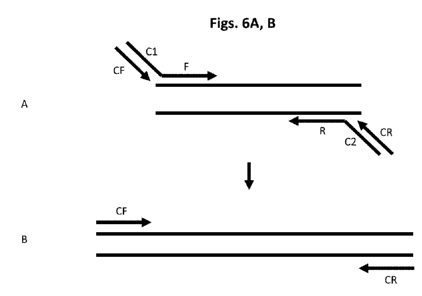
[0020] Figs. 6A, B show a two-step dPCR amplification method using a
three nucleotide-
type primer underrepresentative primer.
[0021] Figs. 7A, B compares background fluorescence between three
nucleotide primers
and four nucleotide primers in a digital PCR platform
[0022] Fig. 8 shows the results from a 5-multiplex dPCR reaction to
distinguish between
trisomic and euploidy samples.
[0023] Fig. 9 shows the results from a 14-multiplex dPCR reaction to
detect or quantify
copy number variations in cffDNA.
[0024] Fig. 10 shows results from a 15-multiplex dPCR assay to detect or
quantify copy
number variations in cffDNA.
DEFINITIONS
[0025] Unless defined otherwise, all technical and scientific terms used
herein have the
same meaning as commonly understood in the art to which the invention
pertains. The
following definitions supplement those in the art and are directed to the
current application
and are not to be imputed to any related or unrelated case, e.g., to any
commonly owned
patent or application. Although any methods and materials similar or
equivalent to those
described herein can be used in the practice for testing of the present
invention, the preferred
materials and methods are described herein. Accordingly, the terminology used
herein is for
the purpose of describing particular embodiments only, and is not intended to
be limiting. The
6
CA 03072591 2020-02-10
WO 2019/033065 PCT/US2018/046360
term "a" or "an" entity refers to one or more of that entity; for example, "a
nucleic acid,"
represents one or more nucleic acids. Therefore, the terms "a" (or "an"), "one
or more," and
"at least one" can be used interchangeably herein.
[0026] Nucleic acids include DNA and RNA and DNA-RNA chimeras can be
double-
stranded or single- stranded. DNA can be genomic, cDNA, methylated DNA or
synthetic DNA.
RNA can be mRNA, miRNA, tRNA, rRNA, hnRNA, methylated RNA among others. The
term
"nucleic acid" encompasses any physical string of monomer units that can be
corresponded to a
string of nucleotides, including a polymer of nucleotides (e.g., a typical DNA
or RNA polymer),
peptide nucleic acid (PNA), modified oligonucleotides (e.g., oligonucleotides
comprising bases
that are not typical to biological RNA or DNA in solution, such as 2'-0-
methylated
oligonucleotides), and the like. A nucleic acid can be e.g., single-stranded
or double-stranded.
[0027] The four conventional nucleotide bases are A, T/U, C and G with T
being present
in DNA and U in RNA. The nucleotides found in targets are usually natural
nucleotides
(deoxyribonucleotides or ribonucleotides). Such is also the case is
nucleotides forming primers.
[0028] Complementarity of nucleic acid strands means that the strands
form a stabile
duplex due to hydrogen bonding between their nucleobase groups. The
complementary bases
are in DNA, A with T and C with G, and, in RNA, C with G, and U with A.
Nucleotides in
respective strands are complementarity when they form one of these (Watson-
Crick pairings)
when the strands are maximally aligned. Nucleotides are mismatched when they
do not form a
complementarity pair when their respective strands are maximally aligned.
Complementarity
of strands can be perfect or substantial. Perfect complementarity between two
strands means
that the two strands can form a duplex in which every base in the duplex is
bonded to a
complementary base by Watson-Crick pairing. Substantial complementary means
most but not
necessarily all bases in strands form Watson-Crick pairs to form a stable
hybrid complex in set
of hybridization conditions (e.g., salt concentration and temperature). For
example, some
primers can duplex with a primer binding site notwithstanding up to 1, 2 or 3
positions of
mismatch, provided such mismatches are not at the 3' end and preferably not
proximate
thereto (e.g., within 4 nucleotides). Such conditions can be predicted by
using the sequences
and standard mathematical calculations to predict the Tm of hybridized
strands, or by empirical
7
CA 03072591 2020-02-10
WO 2019/033065 PCT/US2018/046360
determination of Tm by using routine methods. Tm refers to the temperature at
which a
population of hybridization complexes formed between two nucleic acid strands
are 50%
denatured. At a temperature below the Tm, formation of a hybridization complex
is favored,
whereas at a temperature above the Tm, melting or separation of the strands in
the
hybridization complex is favored. Tm may be estimated for a nucleic acid
having a known G+C
content in an aqueous 1 M NaCI solution by using, e.g., Tm=81.5+0.41(% G+C) -
675/N - %
mismatch, where N = total number of bases.
[0029] A mismatch means that a nucleotide in one strand of nucleic acid
does not or
cannot pair through Watson-Crick base pairing with a nucleotide in an opposing
complementary nucleic acid strand. Examples of mismatches are but not limited
to AA, AG, AC,
GG, CC, TT, TG, TC, UU, UG, UC, and UT base pairs. Mismatches can happen
between DNA and
DNA molecules, DNA and RNA molecules, RNA and RNA molecules, and among other
natural or
artificial nucleic acid analogs.
[0030] Mismatch binding reagents or agents are any molecules or any
modification in
underrepresented primers that can stabilize the underrepresented primer
hybridization with
underrepresented primer binding sites through chemical interaction or physical
interaction.
Modification of underrepresented primers may be modified in any way, as long
as a given
modification is compatible with the desired function of a given
underrepresented primers as
can be easily determined. Modifications include base modifications, sugar
modifications or
backbone modifications. Some small molecules can bind to mismatched bases
through
hydrogen bonds presumably complementary to those in the unpaired base and
stabilize the
duplex with a high base selectivity. Metal ions have been shown to interact
with nucleic acids
for their structure formation and folding. Ono A., Togashi H. (Ono & Togashi,
2004, Angewandte
Chemie (International Ed. in English), 43(33), 4300-4302) showed that addition
of mercury ion
in solution increases the Tm DNA duplex with T-T mismatch by 5 C. Torigoe H.,
Okamoto I. et al.
(Torigoe et al., 2012, Biochimie, 94(11), 2431-2440) showed that silver ion
selectively bind and
stabilize C-C mismatch. A series of rhodium complexes capable of high-
selectivity mismatch site
recognition has been designed and synthesized by Cordier C., Pierre V.C. et
al. (Cordier, Pierre,
& Barton, 2007, Journal of the American Chemical Society, 129(40), 12287-
12295). Nakatani K.,
8
CA 03072591 2020-02-10
WO 2019/033065 PCT/US2018/046360
Sando S., et al. (Nakatani, Sando, Kumasawa, Kikuchi, & Saito, 2001, Journal
of the American
Chemical Society, 123(50), 12650-12657) have developed a series of
naphthyridine based small
molecules to selectively recognize mismatched DNA.
[0031] Hybridization or annealing conditions include chemical components
and their
concentrations (e.g., salts, chelating agents, formamide) of an aqueous or
organic solution
containing the nucleic acids, and the temperature of the mixture in which one
nucleic acid
strand bonds to a second nucleic acid strand by complementary strand
interactions to produce
a hybridization complex.
[0032] A sample is a composition in which one or more target nucleic
acids of interest
may be present, including patient samples, plant or animal materials, waste
materials,
materials for forensic analysis, environmental samples, Circulation tumor cell
(CTC), cell free
DNA, liquid biopsy, and the like. Samples include any tissue, cell, or extract
derived from a
living or dead organism which may contain a target nucleic acid, e.g.,
peripheral blood, bone
marrow, plasma, serum, biopsy tissue including lymph nodes, respiratory tissue
or exudates,
gastrointestinal tissue, urine, feces, semen, or other body fluids. Samples of
particular interest
are tissue samples (including body fluids) from a human or an animal having or
suspected of
having a disease or condition, particularly infection by a virus. Other
samples of interest
include industrial samples, such as for water testing, food testing,
contamination control, and
the like. Sample components may include target and non-target nucleic acids,
and other
materials such as salts, acids, bases, detergents, proteins, carbohydrates,
lipids and other
organic or inorganic materials. A sample may or may not be subject of
processing to purify a
target nucleic acid before amplification. Further processing can treatment
with a detergent or
denaturant to release nucleic acids from cells or viruses, removal or
inactivation of non-nucleic
acid components and concentration of nucleic acids.
[0033] A "target nucleic acid" refers to a nucleic acid molecule or
population of related
nucleic acid molecules that is or may be present within a sample. A target
nucleic acid can
include a segment to be amplified defined by primer binding sites. The segment
can be the
entire nucleic acid or any segment thereof of length amenable to
amplification. A target nucleic
acid can be an entire chromosome, gene or cDNA, and a target segment can be
for example,
9
CA 03072591 2020-02-10
WO 2019/033065 PCT/US2018/046360
only 40-500 of these nucleotides. A target segment can present on any strand
(sense or anti-
sense) of the structure. A target nucleic acid can be RNA (e.g., viral RNA,
microRNA, mRNA,
cRNA, rRNA, hnRNA, cfRNA, or DNA (genomic, somatic, cfDNA, cffDNA, or cDNA)
among others.
[0034] The target nucleic acid can be from a pathogenic microorganism,
such as a virus,
bacteria or fungus, or can be endogenous to a patient. Viral nucleic acids
(e.g., genomic,
mRNA) form a useful target for analyses of viral sequences. Some examples of
viruses that can
be detected include HIV, hepatitis (A, B, or C), herpes virus (e.g., VZV, HSV-
1, HAV-6, HSV-II,
CMV, and Epstein Barr virus), adenovirus, XMRV, influenza virus, flaviviruses,
echovirus,
rhinovirus, coxsackie virus, cornovirus, respiratory syncytial virus, mumps
virus, rotavirus,
measles virus, rubella virus, parvovirus, vaccinia virus, HTLV virus, dengue
virus, MLV-related
Virus, papillomavirus, molluscum virus, poliovirus, rabies virus, JC virus and
arboviral
encephalitis virus. Examples of such bacteria include chlamydia, rickettsial
bacteria,
mycobacteria, staphylococci, treptocci, pneumonococci, meningococci and
conococci,
klebsiella, proteus, serratia, pseudomonas, legionella, diphtheria,
salmonella, bacilli, cholera,
tetanus, botulism, anthrax, plague, leptospirosis, Lymes disease bacteria,
streptococci, or
neisseria. rRNA is a particularly useful target nucleic acid for typing
bacteria. Detection of
human or animal genes is useful for detecting presence or susceptibility to
disease. Examples
of genes that can be the subject of detection include cancer gene fusions,
BRACA-1 or BRAC-2,
p53, CFTR, cytochromes P450), for genotyping (e.g., forensic identification,
paternity testing,
heterozygous carrier of a gene that acts when homozygous, HLA typing),
determining drug
efficacy on an individual (e.g., companion diagnostics) and other uses.
[0035] An underrepresented nucleotide type is one present in no more than
20% of
positions in a primer or primer binding site. Typically if one nucleotide type
is
underrepresented in a primer, its complement is underrepresented in the primer
binding site
(or vice versa). Typically a primer has nucleotide composition of, A, G, C, T
or, A, G, C, U,
although in the present methods one or more of the standard nucleotide types
may be absent.
A primer may include unnatural nucleotide, such as Is C and IsoG, deaza G or
deaza A. These
are scored the same way as corresponding standard nucleotides in determining
the number or
percentage of underrepresented nucleotides. An analog corresponds with a
natural nucleotide
CA 03072591 2020-02-10
WO 2019/033065 PCT/US2018/046360
if it has the same relative pairing affinity with other natural nucleotides.
Thus deaza G or
inosine are analogs of G because they pair more strongly with C than any of
the other natural
nucleotides. As an example, if G is an underrepresented nucleotide type, to
determine a
percentage of the underrepresented nucleotide type in a primer, deaza G is
included in the
numerator (as well as the denominator) and deaza A only in the denominator.
Thus, the
percentage of underrepresented nucleotide in a primer containing one G, one
deaza G and 20
nucleotides total is 10%. Typically an underrepresented nucleotide type is
present in 0, 1 or 2
units at internal positions and optionally one at the 5' terminal position in
each primer and 0, 1,
2, 3 or 4 units in each primer binding sites, and in 0 units in an artificial
sequence. Ideally one
and only unit of the underrepresented nucleotide type is at the 5' terminal
position. If one and
only one of the four-nucleotide-types is underrepresented it is the least
represented (including
null representation) of the four standard nucleotide types. If the primer
contains a degenerate
position, the position is counted as being an underrepresented nucleotide type
position (i.e., in
the numerator as well as the denominator) if the degeneracy includes the
underrepresented
nucleotide type and in the denominator only otherwise. A nucleotide analog
having no
preference among binding to the natural nucleotide types is treated the same
as a degenerate
position. A primer containing underrepresented nucleotide type(s) is called an
underrepresented primer. A probe containing underrepresented nucleotide
type(s) called
underrepresented probe.
[0036] The term "dNTP " generally refers to an individual or combination
of
deoxynucleotides containing a phosphate, sugar and organic base in the
triphosphate form,
that provide precursors required by a DNA polymerase for DNA synthesis. A dNTP
mixture may
include each of the naturally occurring deoxynucleotides (i.e., adenine (A),
guanine (G), cytosine
(C), uracil (U), and Thymine (T)). In some embodiments, each of the naturally
occurring
deoxynucleotides may be replaced or supplemented with a synthetic analog; such
as inosine,
isoG, IsoC, deaza G, deaza A, and so forth. When nucleotides are
underrepresented in a primer
or a probe, the nucleotides are called underrepresented nucleotides. The
underrepresented
nucleotides can be included in a reaction system as the form of
deoxynucleotides or
dideoxynucleotides or ribonucleotides. Their complements are called
complementary
11
CA 03072591 2020-02-10
WO 2019/033065 PCT/US2018/046360
nucleotides of underrepresented nucleotides. The term "ddNTP " generally
refers to an
individual or combination of dideoxynucleotides containing a phosphate, sugar
and organic
base in the triphosphate form, that provide precursors required by a DNA
polymerase for DNA
synthesis. A ddNTP mixture may include each of the naturally occurring
dideoxynucleotides
(i.e., adenine (A), guanine (G), cytosine (C), uracil (U), and Thymine (T)).
In some embodiments,
each of the naturally occurring dideoxynucleotides may be replaced or
supplemented with a
synthetic analog; such as inosine, isoG, IsoC, deazaG, deaza A, and so forth.
The term "NTP "
generally refers to an individual or combination of Ribonucleotides containing
a phosphate,
sugar and organic base in the triphosphate form, that provide precursors
required by a RNA
polymerase for RNA synthesis. A NTP mixture may include each of the naturally
occurring
Ribonucleotides (i.e., adenine (A), guanine (G), cytosine (C), uracil (U)). In
some embodiments,
each of the naturally occurring Ribonucleotides may be replaced or
supplemented with a
synthetic analog; such as inosine, isoG, IsoC, deazaG, deaza A, and so forth.
[0037] A primer binding site or probe binding site is interchangeable
with
underrepresented primer binding site or underrepresented probe binding site in
this invention.
A primer binding site is a complete or partial site in a target nucleic acid
to which a primer
hybridizes. A partial site can be supplemented by provision of toehold and
junction sequences,
which also contain partial primer binding sites as described in W02016/172632.
A partial
binding site from a toehold or junction sequence can combine with a partial
primer binding site
on a target nucleic acid to form a complete primer binding site.
[0038] The term "primer" or "probe" is interchangeable with
underrepresented primer
or underrepresented probe in this invention. A primer or a probe is an
oligonucleotide
complementary to primer or probe binding site contributed in whole or part by
a target nucleic
acid. A primer or a probe can be linked at its 5' end to another nucleic acid
(sometimes
referred to as a tail), not found in or complementary to the target nucleic
acid. A 5' tail can
have an artificial sequence. For a primer or probe exactly complementary to a
primer or a
probe binding site, the demarcation between primer or probe and tail is
readily apparent in
that the tail starts with the first noncomplementary nucleotide encountered
moving from the 3'
end of the primer or probe. For a primer substantially complementary to a
primer binding site,
12
CA 03072591 2020-02-10
WO 2019/033065 PCT/US2018/046360
the last nucleotide of the primer is the last nucleotide complementary to the
primer binding
site encountered moving away from the 3' end of the primer that contributes to
primer binding
to the target nucleic acid (i.e., primer with this 5' nucleotide has higher TM
for the target
nucleic acid than a primer without the 5' nucleotide). Complementarity or not
between
nucleotides in the primer and priming binding site is determined by Watson-
Crick pairing or not
on maximum alignment of the respective sequences.
[0039] A primer or a probe is an oligonucleotide. The term
"oligonucleotide"
encompasses a singular "oligonucleotide" as well as plural "oligonucleotides,"
and refers to any
polymer of two or more of nucleotides, nucleosides, nucleobases or related
compounds used as
a reagent in the amplification methods of the present invention, as well as
subsequent
detection methods. The oligonucleotide may be DNA and/or RNA and/or analogs
thereof
and/or DNA RNA chimeric. The term oligonucleotide does not denote any
particular function to
the reagent, rather, it is used generically to cover all such reagents
described herein. An
oligonucleotide may serve various different functions, e.g., it may function
as a primer if it is
capable of hybridizing to a complementary strand and can further be extended
in the presence
of a nucleic acid polymerase, it may provide a promoter if it contains a
sequence recognized by
an RNA polymerase and allows for transcription, it may contain detection
reagents for signal
generation/amplification, and it may function to prevent hybridization or
impede primer
extension if appropriately situated and/or modified. Specific oligonucleotides
of the present
invention are described in more detail below. As used herein, an
oligonucleotide can be
virtually any length, limited only by its specific function in the
amplification reaction or in
detecting an amplification product of the amplification reaction.
Oligonucleotides of a defined
sequence and chemical structure may be produced by conventional techniques,
such as by
chemical or biochemical synthesis, and by in vitro or in vivo expression from
recombinant
nucleic acid molecules, e.g., bacterial or viral vectors. As intended by this
disclosure, an
oligonucleotide does not consist solely of wild-type chromosomal DNA or the in
vivo
transcription products thereof. Oligonucleotides may be modified in any way,
as long as a given
modification is compatible with the desired function of a given
oligonucleotide as can be easily
determined. Modifications include base modifications, sugar modifications or
backbone
13
CA 03072591 2020-02-10
WO 2019/033065 PCT/US2018/046360
modifications. Base modifications include, but are not limited to the use of
the following bases
in addition to adenine, cytidine, guanosine, thymine and uracil: C-5 propyne,
2-amino adenine,
5-methyl cytidine, inosine, and dP and dK bases. The sugar groups of the
nucleoside subunits
may be ribose, deoxyribose and analogs thereof, including, for example,
ribonucleosides having
a 2'-0-methyl (2'-0-ME) substitution to the ribofuranosyl moiety. See "Method
for Amplifying
Target Nucleic Acids Using Modified Primers," (Becker, Majlessi, & Brentano,
2000, U.S. Pat. No.
6,130,038). Other sugar modifications include, but are not limited to 2'-
amino, 2'-fluoro, (L)-
alpha-threofuranosyl, and pentopuranosyl modifications. The nucleoside
subunits may be
joined by linkages such as phosphodiester linkages, modified linkages or by
non-nucleotide
moieties which do not prevent hybridization of the oligonucleotide to its
complementary target
nucleic acid sequence. Modified linkages include those linkages in which a
standard
phosphodiester linkage is replaced with a different linkage, such as a
phosphorothioate linkage
or a methylphosphonate linkage. The nucleobase subunits may be joined, for
example, by
replacing the natural deoxyribose phosphate backbone of DNA with a pseudo
peptide
backbone, such as a 2-aminoethylglycine backbone which couples the nucleobase
subunits by
means of a carboxymethyl linker to the central secondary amine. (DNA analogs
having a pseudo
peptide backbone are commonly referred to as "peptide nucleic acids" or "PNA"
and are
disclosed by Nielsen et al., "Peptide Nucleic Acids," (Nielsen, Buchardt,
Egholm, & Berg, 1996,
U.S. Pat. No. 5,539,082). Other linkage modifications include, but are not
limited to, morpholino
bonds. Non-limiting examples of oligonucleotides or oligomers contemplated by
the present
invention include nucleic acid analogs containing bicyclic and tricyclic
nucleoside and nucleotide
analogs (LNAs). See lmanishi et al., "Bicyclonucleoside and Oligonucleotide
Analogues,"
(Imanishi & Obika, 2001, U.S. Pat. No. 6,268,490); and Wengel et al.,
"Oligonucleotide
Analogues," (Wengel & Nielsen, 2003, U.S. Pat. No. 6,670,461). Any nucleic
acid analog is
contemplated by the present invention provided the modified oligonucleotide
can perform its
intended function, e.g., hybridize to a target nucleic acid under stringent
hybridization
conditions or amplification conditions, or interact with a DNA or RNA
polymerase, thereby
initiating extension or transcription. In the case of detection probes, the
modified
oligonucleotides must also be capable of preferentially hybridizing to the
target nucleic acid
14
CA 03072591 2020-02-10
WO 2019/033065 PCT/US2018/046360
under stringent hybridization conditions. The 3'-terminus of an
oligonucleotide (or other nucleic
acid) can be blocked in a variety of ways using a blocking moiety, as
described below. A
"blocked" oligonucleotide is not efficiently extended by the addition of
nucleotides to its 3'-
terminus, by a DNA- or RNA-dependent DNA polymerase, to produce a
complementary strand
of DNA. As such, a "blocked" oligonucleotide cannot be a "primer."
[0040] The term "degenerate primer" refers to a mixture of similar primers
with
differing bases at the varying positions (Mitsuhashi, J. Clin. Lab. Anal.,
10(5): 285 93 (1996); von
Eggeling et al., Cell. Mol. Biol., 41(5):653 70 (1995); (Zhang et al., Proc.
Natl. Acad. Sci. USA,
89:5847 5851 (1992); Telenius et al., Genomics, 13(3):718 25 (1992)). Such
primers can include
inosine, as inosine is able to base pair with adenosine, cytosine, guanine or
thymidine.
Degenerate primers allow annealing to and amplification of a variety of target
sequences that
can be related. Degenerate primers that anneal to target DNA can function as a
priming site for
further amplification. A degenerate region is a region of a primer that
varies, while the rest of
the primer can remain the same. Degenerate primers (or regions) denote more
than one primer
and can be random. A random primer (or regions) denotes that the sequence is
not selected,
and it can be degenerate but does not have to be. In some embodiments, the 3'
target specific
regions have a Tm of between about 5 C and 50 C. In some embodiments, a 15-
mer has a Tm
of less than about 60 C.
[0041] A primer "3 segment or 3' binding region or 3' binding site or 3'
hybridization
region" is able to bind to a genomic sequence occurring in a genome at a
particular frequency
or other nucleic acid sequence. In some embodiments, this frequency is between
about 0.01%
and 2.0%, such as, between about 0.05% and 0.1% or between about 0.1% and
0.5%. In some
embodiments, the length of the "binding site" of a primer depends mainly on
the averaged
lengths of the predicted PCR products based on bioinformatic calculations. The
definition
includes, without limitation, a "binding region" of between about 4 and 12
bases in length. In
more particular embodiments, the length of the 3' binding region can be, for
example, between
about 4 and 20 bases, or between about 8 and 15 bases. Binding regions having
a Tm of
between about 10 C. and 60 C. are included within the definition. The term,
"primer binding
segment," when used herein refers to a primer of specified sequence.
CA 03072591 2020-02-10
WO 2019/033065 PCT/US2018/046360
[0042] A polymerase is an enzyme that can perform template directed
extension of a
primer hybridized to the template. It can be a DNA polymerase, an RNA
polymerase or a
reverse transcriptase. Examples of DNA polymerases include: E. coli DNA
polymerase I, Taq
DNA polymerase, S. pneumonioe DNA polymerase I, Tfl DNA polymerase, D.
radiodurans DNA
polymerase I, Tth DNA polymerase, Tth XL DNA polymerase, M. tuberculosis DNA
polymerase
I, M. thermoautotrophicum DNA polymerase I, Herpes simplex-1 DNA polymerase,
T4 DNA
polymerase, thermosequenase or a wild-type or modified T7 DNA polymerase, 029
Polymerase, Bst Polymerase, Vent Polymerase, 9 Nm Polymerase, Klenow fragment
of DNA
Polymerase I. Examples of reverse transcriptase: AMV Reverse Transcriptase,
MMLV Reverse
Transcriptase, HIV Reverse Transcriptase. Examples of RNA polymerases include:
T7 RNA
polymerase or SP6 RNA polymerase, bacterial RNA polymerases and eukaryotic RNA
polymerases.
[0043] Amplification refers to either producing an additional copy or
copies of all or a
segment of a target nucleic acid by template-directed primer extension (target
amplification) or
amplifying detection signal for qualitatively/quantitatively measurement
(signal amplification)
or both. Amplification can be performed under temperature cycled or isothermal
conditions or
combined. Amplification can be linear or exponential.
[0044] Many well-known methods of nucleic acid target amplification
require
thermocycling to alternately denature double-stranded nucleic acids and
hybridize primers;
however, other well-known methods of nucleic acid amplification are
isothermal. The
polymerase chain reaction, commonly referred to as PCR (Mullis, 1987 U.S.
Patent No.
4,683,202; Saiki et al., 1985, Science (New York, N.Y.), 230(4732), 1350-
1354), uses multiple
cycles of denaturation, annealing of primer pairs to opposite strands, and
primer extension to
exponentially increase copy numbers of the target sequence. In a variation
called RT-PCR,
reverse transcriptase (RT) is used to make a complementary DNA (cDNA) from
mRNA, and the
cDNA is then amplified by PCR to produce multiple copies of DNA (Gelfand et
al., "Reverse
Transcription with Thermostable DNA Polymerases¨High Temperature Reverse
Transcription,"
(Gelfand, 1994, U.S. Pat. Nos. 5,322,770; Gelfand & Myers, 1994, U.S. Pat.
Nos. 5,310,652).
Another method of amplifying nucleic acid is called the LCR method (ligase
chain reaction,
16
CA 03072591 2020-02-10
WO 2019/033065 PCT/US2018/046360
Laffler, Carrino, & Marshall, 1993, Anna/es De Biologie Clinique, 5/(9), 821-
826). LCR (Laffler et
al., 1993, Anna/es De Biologie Clinique, 5/(9), 821-826) is based on the
reaction in which two
adjacent probes are hybridized with a target sequence and ligated to each
other by a ligase. The
two probes could not be ligated in the absence of the target nucleotide
sequence, and thus the
presence of the ligated product is indicative of the target nucleotide
sequence. The LCR
method also requires control of temperature for separation of a complementary
chain from a
template. Another method is strand displacement amplification (George T.
Walker, Little, &
Nadeau, 1993, U.S. Pat. No. 5,270,184; George T. Walker, 1995, U.S. Pat. No.
5,455,166; G. T.
Walker et al., 1992, Nucleic Acids Research, 20(7), 1691-1696, 1992,
Proceedings of the
National Academy of Sciences of the United States of America, 89(1), 392-396),
commonly
referred to as SDA, which uses cycles of annealing pairs of primer sequences
to opposite
strands of a target sequence, primer extension in the presence of a dNTP to
produce a duplex
hemiphosphorothioated primer extension product, endonuclease-mediated nicking
of a
hemimodified restriction endonuclease recognition site, and polymerase-
mediated primer
extension from the 3' end of the nick to displace an existing strand and
produce a strand for the
next round of primer annealing, nicking and strand displacement, resulting in
geometric
amplification of product. Thermophilic SDA (tSDA) uses thermophilic
endonucleases and
polymerases at higher temperatures in essentially the same method (Fraiser,
Spargo, Van,
Walker, & Wright, 2002, European Pat. No. 0 684 315). Other amplification
methods include:
nucleic acid sequence based amplification (Compton, 1991, Nature, 350(6313),
91-92, Malek,
Davey, Henderson, & Sooknanan, 1992), commonly referred to as NASBA; one that
uses an RNA
replicase to amplify the probe molecule itself (Lizardi, Guerra, Lomeli,
Tussie-Luna, & Russell
Kramer, 1988, Nature Biotechnology, 6(10), 1197-1202), commonly referred to as
013 replicase;
a transcription-based amplification method (Kwoh et al., 1989, Proceedings of
the National
Academy of Sciences of the United States of America, 86(4), 1173-1177); self-
sustained
sequence replication (35R), (Guatelli et al., 1990, Proceedings of the
National Academy of
Sciences of the United States of America, 87(5), 1874-1878; Landgren (1993)
Trends in
Genetics 9, 199-202; and Lee, H. et al., NUCLEIC ACID AMPLIFICATION
TECHNOLOGIES (1997));
and, transcription-mediated amplification(Kwoh et al., 1989, Proceedings of
the National
17
CA 03072591 2020-02-10
WO 2019/033065 PCT/US2018/046360
Academy of Sciences of the United States of America, 86(4), 1173-1177; Kacian
& Fultz, 1995,
U.S. Pat. No. 5,480,784; Kacian & Fultz, 1996, U.S. Pat. No. 5,399,491),
commonly referred to as
TMA. For further discussion of known amplification methods see Persing, David
H., 1993, "In
Vitro Nucleic Acid Amplification Techniques" in Diagnostic Medical
Microbiology: Principles and
Applications (Persing et al., Eds.), pp. 51-87 (American Society for
Microbiology, Washington,
D.C.). Other illustrative amplification methods suitable for use in accordance
with the present
invention also include rolling circle amplification (RCA) (Fire & Xu, 1995,
Proceedings of the
National Academy of Sciences, 92(10), 4641-4645; Lizardi, 1998, U.S. Pat. No.
5,854,033);
Nucleic Acid Amplification Using Nicking Agents (Van Ness, Galas, & Van Ness,
2006, U. S. Pat.
No. 7,112,423); Nicking and Extension Amplification Reaction (NEAR) (Maples et
al., 2009, US
2009-0017453 Al); Helicase Dependent Amplification (HDA) (Kong, Vincent, & Xu,
2004, US
2004-0058378 Al; Kong, Vincent, & Xu, 2007 US pat. U52007/0254304 Al); and
Loop-Mediated
Isothermal Amplification (LAMP) (Notomi & Hase, 2002, U.S. Pat. No.
6,410,278), and
Quadruplex priming amplification (Analyst, 2014,139, 1644-1652). Expar
amplification (PNAS
April 15, 2003 100, 4504-4509). Cross priming amplification (Sci Rep. 2012; 2:
246). SMAP
amplification (Nature Methods 04/2007; 4(3):257-62). Multiple displacement
amplification
(MDA, Proceedings of the National Academy of Sciences 2005, 102 (48): 17332-
6.),
Recombinase Polymerase Amplification (Journal of Clinical Virology 54 (4): 308-
12). Single
primer isothermal amplification (SPIA) (clinical chemistry, 2005 vol. 51 no.
10 1973-1981).
[0045] Another aspect of amplification is signal amplification. When a
sufficient amount
of nucleic acids to be detected is available, there are advantages to
detecting that sequence
directly, instead of making more copies of that target, (e.g., as in PCR and
LCR). Traditional
methods of direct detection including Northern and Southern blotting and RNase
protection
assays usually require the use of radioactivity and are not amenable to
automation. Other
techniques have sought to eliminate the use of radioactivity and/or improve
the sensitivity in
automatable formats. The cycling probe reaction (CPR) (Duck, Alvarado-Urbina,
Burdick, &
Collier, 1990b, BioTechniques, 9(2), 142-148), uses a long chimeric
oligonucleotide in which a
central portion is made of RNA while the two termini are made of DNA.
Hybridization of the
probe to a target DNA and exposure to a thermostable RNase H causes the RNA
portion to be
18
CA 03072591 2020-02-10
WO 2019/033065 PCT/US2018/046360
digested. This destabilizes the remaining DNA portions of the duplex,
releasing the remainder
of the probe from the target DNA and allowing another probe molecule to repeat
the process.
Branched DNA (bDNA), described by Urdea et al., 1987, Gene, 6/(3), 253-264,
involves
oligonucleotides with branched structures that allow each individual
oligonucleotide to carry 35
to 40 labels (e.g., alkaline phosphatase enzymes). While this enhances the
signal from a
hybridization event, signal from non-specific binding is similarly increased.
Other signal
amplification include: Invasive Cleavage of Nucleic Acids (Prudent, Hall,
Lyamichev, Brow, &
Dahlberg, 2006, U.S. Pat. No. 7,011,944); Hybridization Chain Reaction (HCR)
(R. M. Dirks &
Pierce, 2004, Proceedings of the National Academy of Sciences of the United
States of America,
101(43), 15275-15278, R. Dirks & Pierce, 2012, U. S. Pat. No. 8,105,778) and G-
quadruplex
DNAzyme-based colorimetric detection. CHA amplification (J. Am. Chem. Soc.,
2013, 135 (20),
pp 7430-7433). SMART signal amplification (Biotechniques 2002 Mar; 32(3):604-
6, 608-11.)
[0046] Amplification products can be detected qualitatively (i.e.,
positive signal relative
to control) or quantitatively (signal intensity related to absolute amount or
relative amount of
analyte giving rise to amplification product). Detection can include but does
not require further
analysis, such as sequencing of an amplification product. The methods provided
by the
invention may also include directly detecting a particular nucleic acid in a
capture reaction
product or amplification reaction product, such as a particular target
amplicon or set of
amplicons. Accordingly, mixtures of the invention can comprise specialized
probe sets
including TAQMANTm, which uses a hydrolyzable probe containing detectable
reporter
and quencher moieties, which can be released by a DNA polymerase with 5T->3'
exonuclease
activity (Livak, Flood, & Marmaro, 1996, U.S. Pat. No. 5,538,848); molecular
beacon, which uses
a hairpin probe with reporter and quenching moieties at opposite termini
(Tyagi, Kramer, &
Lizardi, 1999, U.S. Patent No. 5,925,517); Fluorescence resonance energy
transfer (FRET)
primers, which use a pair of adjacent primers with fluorescent donor and
acceptor moieties,
respectively (Wittwer, Ririe, & Rasmussen, 2001, U.S. Patent No. 6, 174,670);
and LIGHTUPTm, a
single short probe which fluoresces only when bound to the target (Kubista &
Svanvik, 2001,
U.S. Patent No. 6,329,144). Similarly, SCORPIONTM (Whitcombe, Theaker, Gibson,
& Little, 2001,
U.S. Patent No. 6,326,145) and SIMPLEPROBES"' (Wittwer et al., 2003, U.S.
Patent No.
19
CA 03072591 2020-02-10
WO 2019/033065 PCT/US2018/046360
6,635,427) use single reporter/dye probes. Amplicon-detecting probes can be
designed
according to the particular detection modality used, and as discussed in the
above-referenced
patents. Other detection methods include: gel electrophoresis, mass
spectrometry, or capillary
electrophoresis, melting curve, nucleic acid-based fluorescent chelating dye
such as SYBRTM
green, or detection of amplification products using a fluorescent label and a
soluble quencher
(Will, Gupta, & Geyer, 2014, U.S. Patent No. 8,658,366).
[0047] The term "multiplex amplification" refers to the amplification of
more than one
nucleic acid of interest. For example, it can refer to the amplification of
multiple sequences
from the same sample or the amplification of one of several sequences in a
sample as
discussed, for example, in George T. Walker, Nadeau, & Little, 1995 U.S. Pat.
Nos. 5,422,252;
and George T. Walker, Nadeau, Spears, et al., 1995, U.S. Pat. Nos. 5,470,723,
which provide
examples of multiplex strand displacement amplification. The term also refers
to the
amplification of one or more sequences present in multiple samples either
simultaneously or in
step-wise fashion.
[0048] The term "digital polymerase chain reaction" or "dPCR" refers to a
refined
version of conventional polymerase chain reaction (PCR) methods used to
directly quantify and
clonally amplify nucleic acids including DNA, cDNA or RNA, such that the
amount of target
nucleic acid can be directly quantitatively measured. Digital PCR achieves
this direct
quantitative measurement by partitioning individual target nucleic acid
molecules present in a
sample into multiple aliquots within many separate reaction chambers that are
able to localize
and concentrate the amplification product to detectable levels. Preferably,
the sample is
partitioned such that most aliquots (e.g., at least 50%, 75%, 90%, 95% or 99%)
receive zero or
one molecule of each target nucleic acid to be detected. After PCR
amplification, the presence
of a signal in any chamber is an indication the target nucleic is present and
a count of chambers
containing the PCR end-product is a direct measure of the absolute nucleic
acid quantity. The
capture or isolation of individual nucleic acid molecules, typically by way of
dilution, may be
effected in capillaries, microemulsions, arrays of miniaturized chambers, or
on nucleic acid
binding surfaces. The basic methodology of digital PCR is described in, e.g.,
Sykes et al.,
Biotechniques 13 (3): 444-449, 1992; and Vogelstein and Kinzler, Proc Natl
Acad Sci U S A 1999;
CA 03072591 2020-02-10
WO 2019/033065 PCT/US2018/046360
96:9236-41. Other forms of amplification described herein, such as
transcription mediated
amplification, can analogously be performed digitally.
[0049] The term "real-time amplification" refers to an amplification
reaction for which
the amount of reaction product, i.e. amplicon, is monitored as the reaction
proceeds. Forms of
real-time amplification differ mainly in the detection mechanisms used for
monitoring the
reaction products. Detection methods are reviewed in Mackay, Arden, & Nitsche,
2002, Nucleic
Acids Research, 30(6), 1292-1305, which is incorporated herein by reference.
[0050] The term "detection label" refers to any atom or molecule which
can be used to
provide or aid to provide, a detectable (preferably quantifiable) signal, and
can be attached to a
nucleic acid or protein. Labels may provide signals detectable by
fluorescence, radioactivity,
colorimetry, gravimetry, magnetism, enzymatic activity and the like. Detection
labels can be
incorporated in a variety of ways: (1) the primers comprise the label(s), for
example, attached
to the base, a ribose, a phosphate, or analogous structures in a nucleic acid
analog; (2)
nucleotides triphosphates are modified at either the base or the ribose (or to
analogous
structures in a nucleic acid analog) with the label(s); the label-modified
nucleotides are then
incorporated into a newly synthesized strand by an extension enzyme such as a
polymerase; (3)
modified nucleotides are used that comprise a functional group that can be
used (post-
enzymatic reaction) to add a detectable label; (4) modified primers are used
that comprise a
functional group that can be used to add a detectable label in a similar
manner; (5) a label
probe that is directly labeled and hybridizes to a portion of the amplicon can
be used; (6) a label
that can be incorporated into amplified products; (7) a label that can react
with byproducts of
amplification reaction.
[0051] The terms "thermally cycling," "thermal cycling", "thermal cycles"
or "thermal
cycle" refer to repeated cycles of temperature changes from a total denaturing
temperature, to
an annealing (or hybridizing) temperature, to an extension temperature and
back to the total
denaturing temperature. The terms also refer to repeated cycles of a
denaturing temperature
and an extension temperature, where the annealing and extension temperatures
are combined
into one temperature. A totally denaturing temperature unwinds all double-
stranded fragments
into single strands. An annealing temperature allows a primer to hybridize or
anneal to the
21
CA 03072591 2020-02-10
WO 2019/033065 PCT/US2018/046360
complementary sequence of a separated strand of a nucleic acid template. The
extension
temperature allows the synthesis of a nascent DNA strand of the amplicon.
[0052] The term "reaction mixture", "amplification mixture" or "PCR
mixture" refer to a
mixture of components necessary to amplify at least one amplicon from nucleic
acid templates.
The mixture may comprise nucleotides (dNTPs), a thermostable polymerase,
primers, and a
plurality of nucleic acid templates. The mixture may further comprise a Tris
buffer, a
monovalent salt, and Mg'. The concentration of each component is well known in
the art and
can be further optimized.
[0053] The terms "amplified product" or "amplicon" refer to a fragment of
DNA
amplified by a polymerase using a pair of primers in an amplification method
such as PCR.
[0054] The term "fluorophore" refers to a moiety that absorbs light
energy at a defined
excitation wavelength and emits light energy at a different defined
wavelength.
[0055] The term "quencher" includes any moiety that is capable of
absorbing the
energy of an excited fluorescent label when it is located in close proximity
to the fluorescent
label and is capable of dissipating that energy. A quencher can be a
fluorescent quencher or a
non-fluorescent quencher, which is also referred to as a dark quencher. The
fluorophores listed
above can play a quencher role if brought into proximity to another
fluorophore, wherein
either FRET quenching or contact quenching can occur. It is preferred that a
dark quencher
which does not emit any visible light is used. Examples of dark quenchers
include, but are not
limited to, DABCYL (4-(4'-dimethylaminophenylazo) benzoic acid) succinimidyl
ester,
diarylrhodamine carboxylic acid, succinimidyl ester (QSY-7), and 4',5'-
dinitrofluorescein
carboxylic acid, succinimidyl ester (QSY-33), quencher!, or Black Hole
Quencher (BHQ-1, BHQ-2
and BHQ-3), nucleotide analogs, nucleotide G residues, nanoparticles, and gold
particles.
[0056] The term "mutation" refers to one or more nucleotides in a target
nucleic acid
sequence that differ from a prototypical form of the target nucleic acid
designated wildtype.
The sequence designated wildtype is the most common allelic form of a
sequence, the first
discovered form of the sequence, and/or a form of the sequence associated with
a normal
(non-diseased phenotype). Single nucleotide polymorphisms (SNPs) are one form
of mutation.
22
CA 03072591 2020-02-10
WO 2019/033065 PCT/US2018/046360
[0057] The term "surface" refers to any solid surface to which nucleic
acids can be
covalently attached, such as for example latex beads, dextran beads,
polystyrene,
polypropylene surface, polyacrylamide gel, gold surfaces, glass surfaces and
silicon wafers.
Preferably the solid support is a glass surface.
[0058] The term "attached to surface" refers to any chemical or non-
chemical
attachment method including chemically-modifiable functional groups.
"Attachment" relates to
immobilization of nucleic acid on solid supports by either a covalent
attachment or via
irreversible passive adsorption or via affinity between molecules (for
example, immobilization
on an avidin-coated surface by biotinylated molecules). The attachment must be
of sufficient
strength that it cannot be removed by washing with water or aqueous buffer
under DNA-
denaturing conditions.
[0059] A sticky end is a single-stranded end of a nucleic acid adjacent a
double-stranded
segment of the nucleic acid. Nucleic acids with stick ends with complementary
sequences can
anneal via the sticky ends and undergo ligation to one another.
[0060] An artificial sequence is a sequence lacking complementarity to or
at least not
intended to have complementarity to a naturally occurring target nucleic acid
known or
suspected may be present in a sample. Artificial sequences can serve as
linkers joining
segments hybridizing to a target nucleic acid, or as tails for labelling
primers, among other
purposes.
[0061] The term "chromosomal aneuploidy," refers to any genetic defect
exhibiting an
abnormal number of chromosomes. For example, chromosomal aneuploidy can
include but is
not limited to, including having more or fewer than normal number of any one
chromosome, as
well as having an extra portion of any one chromosome in addition to the
normal pair, or
missing a portion of any one chromosome in the normal pair. In some cases, the
abnormality
can involve more than one chromosome, or more than one portion of one or more
chromosomes. Common chromosome aneuploidy diseases, include but are not
limited to,
trisomy, e.g., trisomy 21, where the genome of an afflicted patient has three
rather than the
normal two (i.e., a pair) chromosome 21. In rarer cases, the patient may have
an extra piece of
chromosome 21 (less than full length) in addition to the normal pair. In other
cases, a portion of
23
CA 03072591 2020-02-10
WO 2019/033065 PCT/US2018/046360
chromosome 21 may be translocated to another chromosome, e.g. chromosome 14.
In this
example, chromosome 21 is referred as the "chromosome relevant to the
chromosomal
aneuploidy" and a second, irrelevant chromosome, i.e., one that is present in
the normal pair in
the patient's genome, for example chromosome 1, is a "reference chromosome."
There are also
cases where the number of a relevant chromosome is less than the normal number
of 2. Turner
syndrome is one example of a chromosomal aneuploidy where the number of X
chromosome in
a female subject has been reduced from two to one.
[0062] A "genetic marker," refers to a polynucleotide sequence or a
modification to a
polynucleotide sequence present in the genomic sequence of a reference
chromosome with a
known physical location that permits identification. Examples of some genetic
markers include
but are not limited to, different alleles (e.g., alleles from two different
individuals, such as
alleles from a fetus v. alleles from the pregnant woman) to be distinguished
from each other
based on difference in the polynucleotide sequence (e.g., polymorphism), or
presence or
absence of the sequence at all (e.g., a sequence present on the Y chromosome
from a male
fetus but not present in the pregnant woman's genome). In this context, a
"methylation
marker" located on a chromosome relevant to the chromosomal aneuploidy refers
to a
genomic polynucleotide sequence on a chromosome having an abnormal number; or
in the
case where there is an extra piece of the chromosome or a portion of the
chromosome is
missing, the "methylation marker" is located within the piece or portion of
the relevant
chromosome. Difference in methylation profiles of the methylation marker
allows distinction of
the corresponding methylation marker from two different individuals, e.g., a
fetus and the
pregnant woman.
[0063] The term "single nucleotide polymorphism" or "SNP" refers to the
polynucleotide sequence variation present at a single nucleotide residue among
different
alleles of the same gene, which may be the same gene located on the two copies
of the same
chromosome from the same individual (e.g., two alleles from a fetus) or may be
the same gene
from two different individuals (e.g., fetus and pregnant woman). This
variation may occur
within the coding region or non-coding region (e.g., the promoter region or
its proximity, or the
24
CA 03072591 2020-02-10
WO 2019/033065 PCT/US2018/046360
intron) of a gene, or in the intergenic region. Detection of one or more SNP
allows
differentiation of different alleles of a single gene.
[0064] The term "simple tandem repeat polymorphism" refers to the
polynucleotide
sequence variation demonstrated in the varying number of tandem repeats of a
nucleotide
sequence (e.g., a tandem repeat of 1 or more nucleotides) among different
alleles of the same
gene, which may be the same gene located on two copies of the same chromosome
from the
same individual (e.g., fetus) or may be the same gene from two different
individuals (e.g., fetus
and pregnant woman). This variation often occurs within the non-coding region
(e.g., the
promoter region or its proximity, or intron) of a gene, or in the intergenic
region. Detection of
difference in tandem repeat numbers allows differentiation of different
alleles of a single gene.
[0065] The term "insertion-deletion polymorphism" refers to the
polynucleotide
sequence variation demonstrated in the presence or absence of a short
nucleotide sequence
(e.g., 1-3 nucleotides) among different alleles of the same gene, which may be
the same gene
located on two copies of the same chromosome from the same individual (e.g.,
fetus) or may
be the same gene from two different individuals (e.g., fetus and pregnant
woman). This
variation can occurs within both the coding region and the non-coding region
(e.g., the
promoter region or its proximity, or intron) of a gene, or in the intergenic
region. Detection of
whether a short nucleotide sequence is present allows differentiation of
different alleles of a
single gene.
[0066] The term "blood" refers to a blood sample. The term encompasses
whole blood
or any fractions of blood, such as serum, cell-free DNA in blood plasma, and
plasma as
conventionally defined. Examples of blood samples include but are not limited
to, preparation
from a pregnant woman or a woman being tested for possible pregnancy, a person
with a
disease or infection monitoring for a possible disease or infection.
[0067] The term "bisulfite" refers to all types of bisulfites, such as
sodium bisulfite, that
are capable of chemically converting a cytosine (C) to a uracil (U) without
chemically modifying
a methylated cytosine and therefore can be used to differentially modify a DNA
sequence
based on the methylation status of the DNA.
CA 03072591 2020-02-10
WO 2019/033065 PCT/US2018/046360
[0068] The term "locus" refers to a segment of DNA defined by a start
nucleotide
position to an end nucleotide position on a chromosome (i.e., a genomic
location, or a
chromosomal location) of a reference genome assembly (e.g., the Human Genome
March 2006
assembly (hg18) on the UCSC Genome Browser). A locus may or may not overlap
with the
genomic location of a gene, a CpG island, or any product of
transcription/translation. For
example, a locus usually can include but is not limited to a continuous
segment of DNA
identified by experimental data (e.g., a MeDIP-chip dataset) and the
subsequent data analysis
(e.g., MAT, TAS) to contain different DNA methylation levels. A locus may
contain one or more
CpG sites. A locus may be sub-divided into shorter segments (e.g., CpG-
containing genomic
sequences, fragments or regions) that are amenable to analysis (e.g., Epityper
assay, bisulfite
sequencing, polynucleotide amplification and determination). A locus may be
developed into
one or more fetal epigenetic markers. In some context of this application, a
locus also refers to
a continuous segment of DNA identified by certain bioinformatics criteria.
[0069] The term "molecular counting" refers to any method that allows
quantitative
measurement of the number of a molecule or molecular complex, often the
relative number in
the context of other co-existing molecules or complexes of distinct
characteristics. Various
methods of molecular counting are described in, e.g., Leaner et al.,
Analytical Chemistry
69:2115-2121, 1997; Hirano and Fukami, Nucleic Acids Symposium Series No.
44:157-158, 2000;
Chiu et al., Trends in Genetics 25:324-331, 2009; and U.S. Pat. No. 7,537,897.
DETAILED DESCRIPTION
I. General Overview
[0070] Methods of amplification using primers of limited nucleotide
composition are
described by W02016/172632, which claims the benefit of U562/152,752, filed
April 24, 2015,
each incorporated by reference in its entirety for all purposes. The present
discloses the use of
such primers in digital amplification, for example, digital PCR.
II. Primer Design
[0071] The invention uses methods of amplification from a single primer or
a pair of
forward and reverse primers of limited nucleotide composition. Limited
nucleotide
26
CA 03072591 2020-02-10
WO 2019/033065 PCT/US2018/046360
composition means that the primers are underrepresented in at least one
nucleotide type.
Such primers have much reduced capacity to prime from each other or to extend
initiated by
mispriming from other than at their intended primer binding sites in a target
nucleic acid. The
use of such primers for target-specific amplification requires identification
of primer binding
sites in a target nucleic acid that support primer binding and amplification.
In some target
nucleic acids, primer binding sites having complete complementarity to primers
of limited
nucleotide composition can be identified. More often, segments of limited
nucleotide
composition in target nucleic acids are too short by themselves to serves as
primer binding
sites. However, such sites can be adapted to undergo amplification with
primers of limited
nucleotide composition by a variety of techniques described below including
the use of ancillary
toehold or junction oligonucleotide, primer with mismatch hybridization to
primer binding site,
mismatch stabilizing agents and presence of limited numbers of the
underrepresented
nucleotide in the primers as further described in W02016/172632.
a. Basic Principles
[0072] The present method start with a basic concept of a limited
nucleotide
composition of primers in which one or more nucleotide type(s) is
underrepresented (e.g., A, T,
C and no G) and then selects the best primer binding sites within a target
nucleic acid for
pairing with primers of that composition (e.g., A, T and G). Depending on the
primer binding
sites selected, the nucleotide composition of the primers may then be further
adjusted (e.g., by
allowing a limited number units of an underrepresented nucleotide) to improve
complementarity with to the primer binding sites.
[0073] A preferred primer design is that one and only one of the four
standard
nucleotide types is underrepresented in both the forward and reverse primers.
In other words,
such primers can consist of A, T/U and C with G underrepresented, A, T/U, G
with C
underrepresented, A, G and C with T underrepresented or T, G and C with A
underrepresented.
The underrepresented nucleotide type is preferably G or C. If the
underrepresented nucleotide
type is present at all in a primer, it is preferably at position(s) other than
the 3' nucleotide, most
preferably as the 5' nucleotide or a 5' tail nucleotide linked to the 5'
nucleotide of the primer.
27
CA 03072591 2020-02-10
WO 2019/033065 PCT/US2018/046360
Inclusion of a 5' underrepresented nucleotide increases the melting
temperature (TM) of
primer binding without significantly increasing in unintended amplification
products.
[0074] The 3' nucleotide of a primer is preferably occupied by the
complement of the
underrepresented nucleotide type. For example, if the underrepresented
nucleotide type is G,
then the 3' nucleotide is preferably C and vice versa. The terminal C or G
inhibits primer dimer
extension because there is no complementary base on the primers for it to pair
with. The
elimination or underrepresentation of one nucleotide type substantially limits
the number of
nucleotides than can form Watson-Crick pairs between the primers or between
primers and
mismatched primer binding sites. Correct base paring of the 3' nucleotide of a
primer is of
greatest importance in its ability to support template dependent extension.
Use of the
complement of the underrepresented nucleotide type at this position
substantially reduces
primer dimer and primer mismatch extension.
[0075] Other features of primer design are similar to conventional
primers. A primer
has a sequence complementary to its primer binding site. Some primers are at
least 15, 20, 25,
30, 35 or 40 nucleotides long. Some primers are no more than 25, 30, 40, 50 or
75 nucleotides
long. Primers can have any permutation of these lower and upper lengths, e.g.,
from 15-50 of
20-30 or 30-40 nucleotides. The melting temperature of a primer to its primer
binding site can
be for example 45-80 C or preferably 55-65 C. By convention, for primers
binding to opposite
strands, one of which is the coding strand, the forward primer is
complementary to the non-
coding strand so the extended product is the coding strand, and the reverse
primer to the
coding strand so the extended product is the noncoding strand. For target
nucleic acids not
having coding and noncoding strands, designation of forward and reverse primer
is arbitrary.
Such is also the case when forward and reverse primers bind to primer binding
sites on the
same strand. Primers can have 5' tails not complementary to a target nucleic
acid. Such tails
can be used for attaching fluorophore or quenchers, or can contain
identification codes, or can
link discontinuous segments of primer complementary to its target nucleic
acid.
[0076] Amplification conditions are usually similar to conventional
primers in terms of
buffers, Mg', enzymes, temperatures and so forth. Conventional amplification
is performed
with all four standard nucleotide types present as dNTP monomers.
Amplification with primers
28
CA 03072591 2020-02-10
WO 2019/033065 PCT/US2018/046360
of limited nucleotide composition can be so performed, but can also be
performed with the
complements of the underrepresented nucleotide type(s) absent or present at
reduced
concentration or provided as ddNTP(s).
[0077] Usually but not invariably forward and reverse primers bind to
opposite strands
of a target nucleic acid. Thus, one strand of a target nucleic acid contains
for example, the
complement of the forward primer binding site and the reverse primer binding
site, and the
other strand contains the forward primer binding site and complement of the
reverse primer
binding site. In some formats, forward and reverse primer binding sites are on
the same strand.
For example, linked forward and reverse primers can bind to binding sites on
the same strand
and amplify by a rolling circle mechanism. Some pairs of three way junction
primers can also
bind to sites on the same nucleic acid strand, such that one primer serves as
a template for the
other.
[0078] The search for suitable primer binding sites in a target nucleic
acid is informed by
the principles of primer design in that the primer binding sites should be
complementary to the
primers. For example, for use with primers that are underrepresented in a
single nucleotide
type, one can search a target nucleic acid for a forward primer binding site
and a reverse primer
binding site that are underrepresented in the complement of the nucleotide
type
underrepresented in the primers. Preferably, a forward primer binding site and
a reverse
primer binding site are identified in which the complement of the
underrepresented nucleotide
type is absent. However, if such sites cannot be found, other primer binding
sites can be still be
used, preferably those in which the number of units of the complement of the
underrepresented nucleotide type is minimized. Often, the complement of the
underrepresented nucleotide type in the primers is itself underrepresented in
the primer
binding sites, but this is not essential. Some forward and reverse primer
binding sites each
have no more than 4, 3, 2 or 1 units of the complement of the nucleotide
underrepresented in
the primers.
[0079] For ATC primers, software can be used to look for contiguous or
proximate ATC
and ATG regions representing the complement of the forward primer binding site
and reverse
primer binding site respectively. To use ATG primers, software can look for
ATG and ATC
29
CA 03072591 2020-02-10
WO 2019/033065 PCT/US2018/046360
regions for the complement of the forward primer binding site and the reverse
primer binding
site respectively. To use CGA primers, software can look for CGA and CGT
regions representing
the complement of the forward primer binding site and the reverse primer
binding site
respectively. To use CGT primers, software can look for CGT and CGA regions
for the
complement of the forward primer binding site and the reverse primer binding
site
respectively.
[0080] The complement of the forward primer binding site (or the forward
primer
binding site itself if on the same strand as the reverse primer) and the
reverse primer binding
site can be contiguous with one another or separated by intervening
nucleotides in a strand of
the target nucleic acid. The intervening nucleotides, if any, may exclude the
underrepresented
nucleotide in the primers and its complement, or may include one or both of
these nucleotides
and either of the other two of the four standard nucleotide types. If non-
contiguous, the
complement of the forward primer binding site (or the forward primer binding
site itself) and
reverse primer binding site should be close enough together to prime extension
compatible
with the amplification technique (e.g., no more than 100, 500, 1000, or 10000
nucleotides).
[0081] Fig. 1 shows a simple representation of the method in which the
forward and
reverse primers each consist of A, T and C nucleotides, with a C nucleotide at
the 3' positions.
In other words G is the underrepresented nucleotide type. The reverse primer
binding site
consists of A, T and G (the complement of the C, and underrepresented in the
primers). The
complement of the forward primer binding site shown consists of A, T and C,
implying that the
forward primer binding site (like the reverse primer binding site) consists of
A, T and G. The
forward and reverse primers are perfectly complementary to the forward and
reverse primer
binding sites, respectively. The complement of the forward primer binding site
and the reverse
primer binding sites are contiguous. An amplification product can form when a
reaction is
supplied with the three nucleotide triphosphate monomers complementary to the
three-
nucleotide-types in the forward and reverse primers, A, T and G. Primer dimer
formation and
mispriming are inhibited as described because few bases can pair between
primers and or
between a primer and a mismatched primer binding site. But even if the primers
could
sufficiently bind to unintended primer binding sites sufficient to initiate
extension, no
CA 03072591 2020-02-10
WO 2019/033065 PCT/US2018/046360
amplification product would form because the omitted nucleotide triphosphate
monomer in
the amplification mix brings amplification to a stop whenever the extended
chain need to
incorporate a C.
[0082] Alternatively, the primer binding sites can be noncontiguous and
separated by a
region including all four of the standard nucleotides, as shown in Fig. 4. In
such a case,
amplification is performed with all four of the standard nucleotide
triphosphate monomers.
b. Mismatches Between Primer Binding Sites and Primers
[0083] Fig. 2 shows a more typical situation in which a search of a
target nucleic acids
for forward and reverse primer binding sites showed no suitable pair of
forward and reverse
prime binding sites having complete complementarity to primers consisting of
A, T, C
nucleotides (i.e., no primer binding sites in which the underrepresented
nucleotide type is
entirely absent). The longest ATC region contains 7 nucleotides (CATCCTC) and
the longest ATG
region (CGATTGGTATG) contains 12 nucleotides. These regions are not long
enough to use as
primers because their Tm's are too low. In such cases, primers mismatched with
the primer
binding sites can be used. In Fig. 2 the forward primer binding site has three
units of C and the
reverse primer binding site has two units of C aligned with C-nucleotides in
the primers.
Accordingly when such primers and primer binding sites are hybridized with one
another there
are three mismatch positions between forward primer and its binding site and
two mismatches
between the reverse primer and its binding site. Nevertheless hybridization
and extension can
still occur albeit with reduced efficiency. Hybridization and extension can be
increased if the
reaction mix is supplied with a mismatch stabilizing agent. Mismatch binding
or stabilizing
agent are any molecules or any modification in underrepresented primers that
can stabilize the
underrepresented primer hybridization with underrepresented primer binding
sites through
chemical interaction or physical interaction (se Fig. 3). Modification of
underrepresented
primers may be modified in any way, as long as a given modification is
compatible with the
desired function of a given underrepresented primers as can be easily
determined.
Modifications include base modifications, sugar modifications or backbone
modifications, such
as PNA, LNA, or 2' Fluorine 2' methyloxy. Rhodium metalloinsertors as examples
of mismatch
stabilizing agents are described by Ernst et al. J. Am. Chem. Soc. 131,2359-
2366 (2009).
31
CA 03072591 2020-02-10
WO 2019/033065 PCT/US2018/046360
Chemicals such as rhodium metalloinsertors can specifically bind DNA
mismatches and have a
binding constant of 2.0 x 10⁷ M-1 at a CC mismatch. Binding of rhodium
metalloinsertors can
increase the melting temperature of double-stranded DNA including a mismatch
by 18.7 C.
Therefore such mismatch binding reagents can be added to three-nucleotide-type
primer PCR
reactions to specifically stabilize mismatches and increase PCR efficiency. As
well as C-C
mismatches, T-C or A-C mismatches can be stabilized by such reagents among
other
possibilities. Even with such stabilizing agents, mismatched primers may
hybridize to a
template with slightly reduced efficiency but amplification can proceed.
c. Inclusion of a Few Units of Underrepresented Nucleotide
[0084] Alternatively or additionally to using a mismatch stabilizing
agent, the number of
mismatches can be reduced by introducing a limited number of units of the
underrepresented
nucleotide type (typically up to 2 internal position) at positions in a primer
that reduce the
number of mismatches with its primer binding site. An underrepresented
nucleotide can also
be used at the 5' position of the primer or in a tail immediately 5' to the 5'
end of the primer.
For example, with the primers and primer binding sites shown in Fig. 2,
introduction of two G's
into each of the forward and reverse primers reduces the mismatches to one in
the case of the
forward primer and none in the case of the reverse primer.
[0085] The choice whether to use a mismatch stabilizing agent or to
include one or
more units of the underrepresented nucleotide type in the primers depends on
the number of
mismatch positions between hypothetical forward and reverse primers completely
lacking the
underrepresented nucleotide types and their respective binding sites. If there
are more than
two mismatches between such a primer and its binding site or a mismatch occurs
close (e.g.,
within 4 nucleotides) to the 3' end of a primer, it is preferred to eliminate
one or more
mismatches by inclusion of one or more underrepresented nucleotides in the
primer.
[0086] In the case of ATC primers, instead of introducing G into the
underrepresented
primer, one or more unnatural bases can be introduced as alternative as long
as the unnatural
bases can help to reduce primer dimer interaction comparing to conventional
ATGC primers. An
example of the unnatural bases is inosine. Introducing G increases the
hybridization efficiency
of primer to its binding site, but also increases inter- and intra- primer
interactions because CG
32
CA 03072591 2020-02-10
WO 2019/033065 PCT/US2018/046360
pairs are present now. lnosine on the other hand maintains the hybridization
efficiency of
primer to its binding site with the help of flanking bases pairs. But a single
or a few of C and I
pairs between or within primers make little contribution to binding and do no
result in
substantial primer dimer formation. Preferably such primers consist of a 3'
segment that
contains only A, T, and C to minimize the mismatch effect on primer extension
efficiency, and a
5' segment including only any number of inosine residues (e.g., 1-10)
[0087] In situations in which the primer binding sites are not perfectly
matched with
primers in which an underrepresented nucleotide type is entirely absent, the
amplification can
still occur without the complement of the underrepresented nucleotide type in
the primers
being supplied as a nucleotide triphosphate monomer, but proceeds more
efficiently if this
nucleotide type is supplied. This nucleotide type can however by supplied at
reduced
concentration compared with the others of the standard four nucleotides (e.g.,
< 10x, <100X or
<1000X each of the other nucleotide triphosphate monomers), or can be supplied
as a dideoxy
NTP. Extension resulting from mispriming is terminated by the dideoxy NTP. Use
of either
strategy (reducing nucleotide concentration or use of ddNTP) decreases
unintended
amplification products from mispriming or primer dimers. The primers with
inosine
substitutions require dCTP in the reaction for efficient extension on the
inosine bases. The dCTP
however can be supplied at reduced concentration compared with the other types
of
nucleotide triphosphate monomers.
[0088] When target sequences are from organisms of a variety of species
or genotypes,
the template is a mixture of more than one allele. Primer with
underrepresented nucleotide
can contain degenerate bases at certain positions to match different sequence
variations.
[0089] Underrepresented primers with mismatches or inosine substitutions
can be used
in combination with the conventional primers of their original sequences (i.e.
no mismatches or
inosine substitutions) in amplification reactions. However, a conventional
primer should have
reduced concentrations, between 0.1% to 50% of an underrepresented primer's
concentration.
The conventional primers hybridize to their binding sites more efficiently
than the
underrepresented primers and their extension products provide the
underrepresented primers
with more templates. The types of dNTP which are complement of the
underrepresented
33
CA 03072591 2020-02-10
WO 2019/033065 PCT/US2018/046360
nucleotide are provided at reduced concentrations as mentioned above or are
completely
omitted depending on the composition of the underrepresented primers. Such
combination of
conventional and underrepresented primers facilitates the amplification from
underrepresented primers and maintains the low primer-primer interactions.
d. Primers Underrepresented in More Than One Nucleotide Type
[0090] The strategy and principles for primers with a single
underrepresented
nucleotide type can be applied to primers or with two or even three
underrepresented
nucleotides can be applied to primers (or in other words consisting entirely
or primarily of a
single nucleotide). Use of primers underrepresented in a single nucleotide has
wider
applicability in natural target nucleic acids because binding sites for such
primers occur at
statistically greater frequency. However, some forms of amplification, such as
immune-PCR,
amplify nucleic acids of artificial sequences. Such artificial sequences can
be designed to be
amplified with primers with two or even three underrepresented nucleotide type
as with one
underrepresented nucleotide type.
[0091] In primers underrepresented in two nucleotide types, the two
underrepresented
nucleotide types should not be complementary to one another. In others words,
the
underrepresented nucleotide types can be A with C, A with G, T/U with C or T/U
with G. This
leaves primers consisting entirely or primarily of the same two
noncomplementary nucleotide
types. Such primers have reduced ability to support primer-dimer or primer-
mismatch
extension. Primers have three nucleotides underrepresented or in other words,
consisting
entirely or substantially of a single nucleotide type also have reduced
ability to support primer
dimer or mismatched primer extension. Primer binding sites are selected by
analogous
principles to those described above, and primer sequences can be adjusted to
accommodate a
small number of underrepresented nucleotide(s) if necessary. Toehold and
junction primer
strategies can also be used as described in W02016/172632. Amplification with
such primers is
performed at least with the complements of the nucleotides not
underrepresented in the
primers, and optionally, with the complements of the underrepresented
nucleotide(s) as well,
which as noted can be supplied in reduced concentration or as dideoxy
nucleotides.
34
CA 03072591 2020-02-10
WO 2019/033065 PCT/US2018/046360
III. Sample Preparation
[0092] A target nucleic acid can be prepared from various biological
samples of interest.
Examples of samples include but are not limited to, fetal or maternal genetic
material, maternal
plasma or blood, a biopsy sample of a subject having a cancer or being suspect
of having a
cancer, any human of known or unknown status with respect to genetic
variations, blood cells,
blood cells enriched for (a) particular cell type(s), bone marrow derived
mononuclear cells,
placenta cells, umbilical cord sample, fetal tissue, fetal fibroblasts or
blood cells, tissue from
infant or child, neonatal tissue, non-cellular entity comprising nucleic acid
(e.g. virus), cell-based
organisms (e.g. plant, fungi, eubacteria, archeabacteria, protist, or animal),
plants or food
products.
[0093] Before analyzing the sample using the methods provided herein, it
may be
desirable to perform one or more sample preparation operations on the sample.
Examples of
sample preparation operations may include extraction of intracellular material
from a cell,
tissue, blood, or microorganisms. Extracted intracellular material can include
nucleic acids,
protein, or other macromolecules from the samples. In some applications, a
sample is prepared
using formalin-fixed, paraffin embedded (FFPE), or frozen section for
sectioning. In some
applications the sample is microdissected with a laser before any extraction
methods are used.
Another example of sample preparation operations may include extraction of
cell free DNA
from plasma or blood.
[0094] A biological sample can be obtained by methods known in the art
including
swabbing, scraping, phlebotomy, a biopsy (e.g., excisional, fine needle
aspiration, incisional,
core needle), or any other suitable method particularly for subjects having or
suspected of
having a disease or infection. In some applications, serial biopsies are
obtained from a diseased
tissue or organ.
[0095] Biological samples can be obtained from any of the tissues
provided herein.
Examples of biological samples include but not limited to, lung, respiratory
tract, nasal cavity,
gastrointestinal tract, mouth, skin, heart, lung, kidney, breast, pancreas,
liver, blood, muscle,
smooth muscle, bladder, gall bladder, colon, intestine, brain, prostate,
esophagus, or thyroid.
CA 03072591 2020-02-10
WO 2019/033065 PCT/US2018/046360
a. Nucleic Acid Extraction
[0096] For whole cells, viruses or other tissue samples to be analyzed,
nucleic acids are
typically extracted from these samples.
[0097] The target nucleic acid can be isolated from the biological
samples using any
techniques known in the art. In some applications, DNA or RNA can be extracted
from a
biological sample before analysis by either physical, chemical methods, or a
combination of
both. Extraction can be by means including, but not limited to, the use of
detergent lysates,
sonication, or vortexing with glass beads. In particular embodiments, DNA can
be extracted
according to standard methods from blood, e.g., with the use of the Qiagen
UltraSens DNA
extraction kit. In some applications, nucleic acid molecules can be isolated
using gradient
centrifugation (e.g., cesium chloride gradients, sucrose gradients, glucose
gradients),
centrifugation protocols, boiling, purification kits (e.g., Qiagen
purification systems; Promega
purification systems; Amersham purification systems; Invitrogen Life
Technologies Purification;
Mo-Bio Laboratories purification systems, etc.). Methods of extracting nucleic
acids can also
include the use of liquid extraction using Trizol or DNAzol.
[0098] RNA can be isolated from various body fluids. Methods of isolating
RNA analysis
from blood, plasma and serum. See, for example Tsui N B et al. Clin. Chem. 48,
1647-53, 2002.
[0099] In some applications, the target nucleic acid is prepared from an
RNA using RT-
PCR. In some applications, the target nucleic acid is prepared by an RT-PCR
and following dPCR,
which can be carried out in two distinct steps or a single step. In some
applications, the target
nucleic acid is pre-amplified in a separate reaction to specifically or non-
specifically enrich the
target sequences of interest.
IV. Amplification Methods
[00100] The strategy and principles described above can be incorporated
into any
amplification method involving template-directed extension from single or
paired primers. The
polymerase chain reaction is one implementation including optionally RT-PCR.
PCR is
characterized by temperature cycling to permit primer annealing, primer
extension and
denaturation of an extended strand from its template.
36
CA 03072591 2020-02-10
WO 2019/033065 PCT/US2018/046360
[00101] Transcription mediated amplification (TMA) is an alternative
isothermal form in
which one or both of the primers is linked to a promoter at its 5' end,
usually a T7 promoter.
Once the double-stranded promoter is formed, the RNA polymerase starts
transcription
amplification. The amplification product is single stranded RNA molecules. TMA
can also be
coupled to reverse transcription.
[00102] Another isothermal amplification format amenable to use with
primers of the
invention is the nicking amplification reaction (NEAR). NEAR exponentially
amplifies DNA at a
constant temperature using a polymerase and nicking enzyme. The primers for
nicking
amplification are linked to artificial segments at their 5' ends, the 5'
segments containing a
cleavage site for the nicking enzyme. In the first cycle both primers
hybridize to a template and
extend. In the next cycle, both primers can hybridizes to the first cycle
products and extend to
generate the full nicking site on the artificial tail. Once a nicking site is
formed, nicking enzyme
nicks and releases one strand. Extension and nicking repeat in the next cycle.
[00103] Another isothermal amplification procedure amenable to use with
primers of the
invention is loop mediated isothermal amplification or (LAMP). LAMP uses one
or more
primers having underrepresented nucleotides in accordance with the invention.
In LAMP, the
target sequence is amplified at a constant temperature of 60 ¨ 65 C using
either two or three
sets of primers and a polymerase with high strand displacement activity in
addition to a
replication activity. Typically, 4 different primers are used to identify 6
distinct regions on the
target gene, which adds highly to the specificity. An additional pair of "loop
primers" can
further accelerate the reaction.
[00104] Another isothermal amplification format is Recombinase Polymerase
Amplification (RPA) is a single tube, isothermal alternative to the Polymerase
Chain Reaction
(PCR). The RPA process employs three core enzymes ¨ a recombinase, a single-
stranded DNA-
binding protein (SSB) and strand-displacing polymerase. Recombinases are
capable of pairing
oligonucleotide primers with homologous sequence in duplex DNA. SSB bind to
displaced
strands of DNA and prevent the primers from being displaced. Finally, the
strand displacing
polymerase begins DNA synthesis where the primer has bound to the target DNA.
By using two
opposing primers, much like PCR, if the target sequence is indeed present, an
exponential DNA
37
CA 03072591 2020-02-10
WO 2019/033065 PCT/US2018/046360
amplification reaction is initiated. The two primers can both be primers with
underrepresented
nucleotide types as described above.
[00105] Still other amplification format in which primers of the invention
can be used
include strand displacement assay, transcription-based amplification systems,
self-sustained
sequence replication (35R), a ligation chain reaction (sometimes referred to
as oligonucleotide
ligase amplification OLA), cycling probe technology (CPT), rolling circle
amplification (RCA),
Recombinase Polymerase Amplification (RPA), nucleic acid sequence bases
amplification
(NASBA), invasive cleavage technology, Helicase dependent amplification (I),
Exponential
amplification (EXPAR), Hybridization chain reaction (HCR), and catalyzed
hairpin assembly
(CHA).
[00106] Another amplification format is immune-PCR in which an analyte is
linked to a
nucleic acid (which can have an artificial sequence) and the analyte is
detected by amplification
of the nucleic acid. Such amplification can be performed with a primer pair
with
underrepresented nucleotide types (e.g., completely absent) complementary to
primer binding
sites underrepresented in the complements of the underrepresented
nucleotide(s).
[00107] The above methods amplify a specific predetermined target nucleic
acid or
segment thereof determined by the selected primers and their complementary
primer binding
sites (in other words, target-specific amplification). The amplification
product from a pair of
primers binding to its intended primer binding sites predominates over any or
all other
amplification products primed from the same primer pair either by primer dimer
binding or
mispriming on the target sequence. Preferably the amplification product from
primers binding
to their intended primer binding sites is present in at least 10, 50, 100 or
1000 fold excess (by
moles, mass or copy number) of any or all other amplification products primed
from the primer
pair. In some methods, a single pair of primers is used in amplification. In
other methods,
multiple primer pairs are used in a multiplex amplification. The number of
primer pairs can be
for example 2-50 or more, preferably 5-25 or 10-20, or at least 2, 3, 4, 5, 6,
7, 8, 9, 10, 11, 12,
13, 14, 15, 16, 17, 18, 19 or 20. When multiple primer pairs are used the
intended amplification
product of each primer pair from binding of the primer pair to its intended
primer binding sites
is present in at least 10, 50, 100 or 1000 fold excess (by moles, mass or copy
number) to any or
38
CA 03072591 2020-02-10
WO 2019/033065 PCT/US2018/046360
all other amplification products primed by that primer pair. When multiple
primer pairs are
present in the same reaction, preferably each primer of each primer pair has
the same
underrepresented standard nucleotide type or types. Preferably one and only
one standard
nucleotide type is underrepresented in each primer of each primer pair.
Primers used in the
methods are not random primers in which most or all primer positions are
occupied by random
or degenerate selections of nucleotides varying among primers. Rather each
primer pair is
designed to hybridize to specific primer binding sites in a target nucleic
acid, and typically
different primer pairs are unrelated from each other as required by the
different primer binding
sites in target nucleic acids being detected. For example, one primer pair can
be designed to
bind to primer binding sites on a target nucleic acid in one pathogen and a
second primer pair
to primer binding sites on a different target nucleic acid in a different
pathogen. Except by
coincidence the different target nucleic acids and consequently primer binding
sites and
primers are unrelated to one another.
V. Digital amplification
[00108] Digital amplification (e.g., digital PCR or dPCR) is a highly
sensitive quantitation
method for nucleic acids. The method can detect and quantify nucleic acids by
directly
measuring the number of target molecules without relying on any normalization
standard or
external standards. In this manner, the absolute number of target molecules
can be
determined, with a lower limit being a single copy of the molecule.
[00109] The strategy used by dPCR or analogously other form of digital
amplification can
be referred to as "Divide and Conquer" strategy where a sample can be first
diluted and divided
into thousands to tens of thousands of micro reaction chambers, so that most
reaction
chambers (e.g., at least 50%, 75%< 90%, 95 or 99%) contains only either zero
or one copy of the
target gene sequence (a small number of reaction chambers may contain multiple
copies). By
counting the number of reaction chambers with positive amplification results,
the absolute
number of target gene molecules in the original sample can be determined.
[00110] The distribution of the target molecules across the partitions can
be seen as a
Poisson process (the targets end up in partitions independently and with a
fixed rate). Poisson
statistics thus allow a more accurate calculation of the initial number of
targets from the
39
CA 03072591 2020-02-10
WO 2019/033065 PCT/US2018/046360
number of positive and negative partitions taking into account that some
reaction chambers
receive multiple copies.
[00111] In comparison to traditional PCR technology, dPCR or other digital
amplification
method is considered to have multiple advantages, including low required
sample amount,
reduced consumption of reagents, absolute quantification of nucleic acid
molecules, reduced
interference among different copies within a sample, and superb sensitivity
and specificity.
Furthermore, the standard division process of the reaction system in digital
amplification can
greatly reduce the concentration of background sequences that could compete
with the target
sequence; therefore, digital amplification may be especially suitable for
detection of rare
mutations in a complex biological background, such for example DNA from
circulating tumor
cells and cffDNA for NIPT (non-invasive prenatal testing) applications.
a. Digital amplification primers
[00112] Digital amplification amplifies a single copy of a template. Like
traditional or
first-generation PCR, digital amplification also requires a high concentration
of primers in the
reaction mix. However, having a high concentration primers results in the
production of non-
targeted products, such as primer-dimer or non-specific amplifications.
[00113] Non-targeted primer-dimer products tend to have a smaller size
than the
intended amplification product, and owing to their small size are amplified
with higher
efficiency than the intended product. This creates competition within the
reaction between
these products and in many cases the intended amplification products cannot be
differentiated
from the non-targeted primer-dimer products, where the reaction is monitored
by DNA
intercalating dyes. Consequently, primer dimer formation hinders the
efficiency, sensitivity, and
specificity of digital amplification. Furthermore, the problem of primer-dimer
formation and
non-specific amplification becomes significantly more pronounced as more
primers are
multiplexed in high-throughput digital amplifications.
[00114] These types of problems can be reduced or avoided by performing
digital PCR or
other amplification with limited composition primers as described herein. Such
primers can
increase efficiency, sensitivity, and specificity of in the digital
amplification.
CA 03072591 2020-02-10
WO 2019/033065 PCT/US2018/046360
[00115] Any of the primers described herein by used for a digital
amplification. Fig. 5
shows examples of primers with an underrepresented nucleotide type comprising
fluorophore
labeled probes which can be used in a digital amplification.
[00116] In some applications primers with an underrepresented nucleotide
type
provided herein can be linked to a peptide nucleic acid (PNA) for higher
sensitivity.
[00117] In some applications, the primers of limited nucleotide
composition reduce
primer dimer formation in a single or multiplex digital amplification by
greater than 70%, 75%,
80%, 90%, 95%, 97%, or 99% compared with an otherwise comparable reaction with
conventional primers.
b. dPCR platforms
[00118] The compositions, methods, and kits can be used with various
commercial dPCR
platforms currently know or developed in the future.
[00119] Depending on the application a clinician or researcher can choose
a dPCR
platform that meet its technical demands of required throughput and accuracy
requirements or
its application. For example, a microfluidic-chip-based dPCR can have up to
several hundred
partitions per panel. Droplet-based dPCR usually has approximately 20,000
partitioned
droplets, but it can have up to 10,000,000.
[00120] In some application the dPCR compositions and methods provided by
the
disclosure can be used with microfluidic-chip-based dPCR. In some application
the dPCR
compositions and methods provided by the disclosure can be used with Droplet-
based dPCR.
[00121] In some applications the dPCR compositions and methods provided by
the
disclosure can be used with microfluidic-chamber-based BioMark dPCR from
Fluidigm. In
some applications the dPCR compositions and methods provided by the disclosure
can be used
with micro-well chip-based QuantStudio12k flex dPCR. In some applications the
dPCR
compositions and methods provided by the disclosure can be used with 3D dPCR
from Life
Technologies. In some applications the dPCR compositions and methods provided
by the
disclosure can be used with droplet-based ddPCR (ddPCR) QX100 and 0X200 from
Bio-Rad . In
some applications the dPCR compositions and methods provided by the disclosure
can be used
with RainDrop from RainDance .
41
CA 03072591 2020-02-10
WO 2019/033065 PCT/US2018/046360
[00122] In some applications, the methods provided herein enhance accuracy
of
quantification or detection in a dPCR reaction by greater than 70%, 75%, 80%,
90%, 95%, 97%,
or 99% relative to otherwise comparable assays with conventional primers not
containing an
underrepresented nucleotide. In some applications, the methods provided herein
enhance
sensitivity of quantification or detection in a dPCR reaction by greater than
70%, 75%, 80%,
90%, 95%, 97%, or 99% relative to otherwise comparable assays with
conventional primers.
[00123] Other forms of digital amplification can be performed with the
same or similar
platforms.
c. Multiplexing dPCR Assays
[00124] Digital PCR or other amplification can be performed as a
multiplex. A multiplex
assay is particularly useful in applications were the biological material is
limited or rare so that
splitting the sample for separate analysis is infeasible or difficult. In some
cases, the multiplex
amplification assay enables the detection of quantification of at least 2, 3,
4, 5, 6, 7, 8, 9, 10, 11,
12, 13, 14, 15, 16, 17, 18, 19, 20, 21, different genetic variations, such as
single nucleotide
polymorphisms (SNPs), insertions, inversions, rearrangements, transversions,
deletions, indels,
microsatellite repeats, minisatellite repeats, short tandem repeats,
transposable elements,
large scale structural chromosomal variants, methylation, and combinations
thereof. In some
applications, the genetic variations assayed are a combination of different
genetic variations.
[00125] In some applications, the multiplexing dPCR enables detection of
least 2, 3, 4, 5,
6, 7, 8, 9, 10, 15, 20, 25, 30, 40, 50, 60, 70, 80, 90, or 100 different
genetic alternations in a
single reaction tube. In some applications, the multiplexing dPCR enables
detection of least
100, 200, 300, 400, 500, 600, 700, 800, 900, or 1000 different genetic
alternations in a single
reaction tube.
d. Detection Methods
[00126] Amplification reactions used with the methods, compositions, and
kits of the
disclosure can generate one or more signals. In some applications, labels are
used in or after
the amplification reaction to generate the signal. In some applications, dyes
are used in or after
the amplification reaction to generate the signal.
42
CA 03072591 2020-02-10
WO 2019/033065 PCT/US2018/046360
[00127] In some applications, the target nucleic acid is detected with DNA
intercalating
dye. Examples of intercalating dyes that can be used with the disclosure
include but are not
limited to, ethidium bromide, propidium iodide, acridine orange, 9-amino-6-
chloro-2-
methoxyacridine (ACMA), SYBRTM Green, SYBRTM Green II, SYBRTM Gold, YO
(Oxazole Yellow), TO
(Thiazole Orange), PG (PicoGreen ), or EvaGreen . Positive dPCR reactions are
distinguished
from negative dPCR reactions by their elevated signal intensity over the
background signal
generated by primers.
[00128] In some applications, multiple target nucleic acids or genetic
variants are
detected simultaneously in one dPCR reaction. In some applications, multiple
targets are two or
more alleles of the same target locus. In some cases, multiplex targets are
different target
locus. The reaction mixture comprises two or more primer pairs, each pair
comprising a forward
primer and a reverse primer. By varying the lengths of amplified products
and/or primer
concentration, the different targets can be discriminated by their signal
intensity of amplified
products. The space between background signal generated by primers alone and
the saturation
signal intensity limited by dPCR machine determines how many targets can be
multiplexed in
one dPCR reaction. The three-nucleotide-type primers (3N primers) greatly
reduce primer-
primer interaction, and thus reduce the background signal, allowing more
targets to be
multiplexed. In some embodiments, multiple amplicons are detected for
quantification of one
target. The three-nucleotide-type primers (3N primers) greatly increase dPCR
multiplicity
allowing accurate quantification where the target nucleic acid in at extremely
low amounts in
the biological sample.
[00129] In some applications, the target nucleic acid of the dPCR is
detected with
fluorophore labeled probes, such as Taqman probe, Molecular Beacon, and
fluorophore labeled
primers provided with a quencher labeled complementary oligo (Fig. 5). In
another
embodiment, the target nucleic acid of the dPCR is detected with a combination
of DNA
intercalating dyes and fluorophore labeled probes.
[00130] A "fluorescent label" or "fluorophore" can be a compound with a
fluorescent
emission maximum between about 350 and 900 nm.
43
CA 03072591 2020-02-10
WO 2019/033065 PCT/US2018/046360
[00131] Examples of fluorophores used with the disclosure include but are
not limited to,
5-FAM (also called 5-carboxyfluorescein; also called Spiro(isobenzofuran-
1(3H)' 9'-
(9H)xanthene)-5-carboxylic aci',3',6'-dihydroxy-3-oxo-6- carboxyfluorescein);
5-Hexachloro-
Fluorescein; ([4,',2',4',5',7'-hexachloro'(3',6'-dipivaloyl- fluoresceinyI)-6-
carboxyli- c acid]); 6-
Hexachloro-Fluorescein; ([4,',2',4',5',7'-hexachloro'(3',6'-
dipivaloylfluoresceinyI)-5-carboxylic
acid]); 5-Tetrachloro-Fluorescein; ([4,',2',7'-tetra-chloro'(3',6'-
dipivaloylfluoresceinyI)-5-
carboxylic acid]); 6-Tetrachloro-Fluorescein; ([4,',2',7'- tetrachloro'(3',6'-
dipivaloylfluoresceiny1)-6-carboxylic acid]); 5-TAMRA (5-
carboxytetramethylrhodamine);
Xanthylium, 9-(2,4-dicarboxyphenyI)-3 ,6-bis(dimethyl-amino); 6-TAMRA (6-
carboxytetramethylrhodamine); 9-(2,5-dicarboxyphenyI)-3,6-bis(dimethylamino);
EDANS ¨5 -
((2-aminoethypamino)naphthalene-1-sulfonic acid); 1,5-IAEDANS (5-(W2-
iodoacetypamino)ethyl)amino)naphthalene-1-sulfonic acid); Cy5
(Indodicarbocyanine-5); Cy3
(Indo-dicarbocyanine-3); and BODIPY FL (2,6-dibromo-4,4-difluoro-5,7-dimethy1-
4-bora-3a,4a-
diaza-s-indacene-3-pr- oprionic acid); QuasarTm-670 dye (Biosearch
Technologies); Cal FluorTM
Orange dye (Biosearch Technologies); Rox dyes; Max dyes (Integrated DNA
Technologies), as
well as derivatives thereof.
[00132] A "quencher" can be a molecule or part of a compound, which is
capable of
reducing the emission from a fluorescent donor when coupled to or in proximity
to the donor.
Quenching can occur by any of several mechanisms including fluorescence
resonance energy
transfer, photo-induced electron transfer, and paramagnetic enhancement of
intersystem
crossing, Dexter exchange coupling, and exciton coupling such as the formation
of dark
complexes.
[00133] Fluorescence can be "quenched" when the fluorescence emitted by
the
fluorophore is reduced as compared with the fluorescence in the absence of the
quencher by at
least 10%, for example, at least 15%, 20%, 25%, 30%, 35%, 40%, 45%, 50%, 55%,
60%, 65%,
70%, 75%, 80%, 85%, 90%, 95%, 98%, 99%, or more. The selection of the quencher
can depend
on the identity of the fluorophore. Examples of quenchers used with the
disclosure include but
are not limited to, DABCYL, Black HoleTM Quenchers (BHQ-1, BHQ-2, and BHQ-3),
Iowa BlackTM
FQ and Iowa BlackTM RQ.
44
CA 03072591 2020-02-10
WO 2019/033065 PCT/US2018/046360
[00134] In some applications, both a fluorophore and a quencher can be
coupled to the
primer using methods known in the art. In general, a fluorophore can be
coupled to the 5'
portion of a hot-start primer 5' of the cleavage site. Fluorophores can be
added during
oligonucleotide synthesis through standard phosphoramidite chemistry. They can
also be
added post synthesis by introducing a linker with an appropriate functional
group during oligo
synthesis. Following synthesis, the fluorophore can be coupled to the
oligonucleotide functional
group. For longer sequences, to permit efficient quenching, the sequence
immediately 3' of the
fluorophore and outside the target region of the primer can be made to be
partially
complementary to permit the formation of a stem-group of a hairpin (i.e.,
molecular beacon).
Thus, the fluorophore can remain with the primer while the primer is
hybridized to the target
polynucleotide and extended by the polymerase. The quencher can be coupled to
the 3' portion
of the hot-start primer 3' of the cleavage site. Thus, the quencher can be
released from the
primer while the primer is hybridized to the target polynucleotide and thus no
longer quench
the fluorophore that remains coupled to the primer. The proper site of
coupling a fluorophore
and quencher and the distance between the fluorophore and the quencher can be
known in the
art. In some cases, a fluorophore is positioned about, more than, less than,
or at least 10, 11,
12, 13, 14, 15, 16, 17, 18, 19, 20, 21, 22, 23, 24, 25, 26, 27, 28, 29, 30,
31, 32, 33, 34, 35, 36, 37,
38, 39, 40, 41, 42, 43, 44, 45, 46, 47, 48, 49, or 50 bases from a quencher in
a primer.
[00135] In some applications, detection of the target nucleic acid can be
achieved, using
a two-step method as shown in Figs. 6A-B. In one embodiment, the forward three-
nucleotide-
type primer is linked to the 5' end of a universal artificial 3N sequence
(Cl), and the other
reverse three-nucleotide-type primers is linked to the 5' end of a different
universal artificial 3N
sequence (C2). Next, a pair of common primers (CF and CR) are provided in the
dPCR reaction.
The CF primer has the same sequence as Cl, and the CR primer has the same
sequence as C2. In
initial stages of reaction, specific primers C1-F and C2-R hybridize to the
template and generate
amplicons with Cl and C2 at two ends. In later stages of the reaction, common
primers CF and
CR participate and dominate the reaction. In some applications of detecting
multiple targets in
a single reaction solution, the specific primers for each specific target may
have the same or
different common sequences attached at its 5' end. Multiple targets in a
single reaction can be
CA 03072591 2020-02-10
WO 2019/033065 PCT/US2018/046360
detected by one or more than one set of common primers as described in Fig. 5
by using
detection probes associated with common forward primer sequences (CF) or
common reverse
primer sequences (CR).
e. Analyses
[00136] The compositions, methods, and kits can be used to conduct analyses
of various
genomic alterations associated with a diseased state or other phenotype.
[00137] The methods can be used to carry out analyses of chromosomal
abnormalities of
multiple genes or whole chromosomes for a particular disease. A representative
procedure can
include comparing either the copy number of a target chromosome (or segment
thereof)
against that of a reference chromosome or segment thereof or the copy number
of a mutant
allele relative to a wild-type allele. Such an analyses can be used, for
example, in trisomy
detection or sex determination.
[00138] The methods can be used to carry out analyses of single gene, such
as a deletion
or point mutation (e.g., single nucleotide change) for a particular disease. A
representative
procedure for detecting a point mutation or a small deletion in a gene can
include comparing
the amount of the mutant allele relative to the wild-type allele. A
representative procedure for
detecting a deletion of a single gene can include comparing either the copy
number of a target
deleted gene against that of a reference gene.
[00139] The methods can be used to carry out SNP-association analyses for a
particular
disease. A representative procedure can include comparing the dosage of
paternally or
maternally-inherited single nucleotide polymorphisms (SNPs) to a relative
reference haplotype
dosage. In some applications, a representative procedure can include
preferential allelic
imbalance analysis (PAI). PAI occurs when a disease-associated heritable SNP
is preferentially
retained relative to the wild-type allele. PAI analysis can include comparing
the copy number of
an allele with a disease-associated heritable SNP to a wild-type allele.
[00140] The methods can be used to carry out epigenetic analyses. DNA
methylation
plays an important role in cancer and neurodegenerative disorders. A
representative
procedure can include comparing methylation levels from the target region of a
diseased
sample to a methylation level of the target region from a control sample.
46
CA 03072591 2020-02-10
WO 2019/033065
PCT/US2018/046360
[00141]
Other analyses that can be used with the present disclosure are described or
referenced in Butchbach, M.E.R. Biomolecular Detection and Quantification 10:
9-14 (2016).
VI. Computer Implementation
[00142]
Selection of primer binding sites and primers can be performed by computer-
implemented analysis of a target nucleic acid in a computer programmed by non-
transitory
computer readable storage media. The sequence of a target nucleic acid (one or
both strands)
is received in a computer. The computer also stores or receives by user input
desired
nucleotide compositions of primers (e.g., A, T, C). The computer is then
programmed to search
the target sequence to identify forward and reverse primer binding sites
within a distance of
one another compatible with amplification that most closely correspond to the
primer
composition. If the primer composition is A, T, C, then forward and reverse
primer binding sites
should most closely correspond to A, T and G. The computer can identify
forward and reverse
primer binding sites on opposite strands or can identify a complement of the
forward primer
binding sites and reverse primer binding site on the same strand and calculate
the forward
primer binding site from its complement. The computer can then provide output
of candidate
pairs of primer binding sites, which may differ to varying degrees with the
ideal composition
sought. The computer can also show primer designs that hybridize to each of
the primer
binding site pairs. Multiple primer designs can be shown for the same primer
binding site pair
with different numbers of units of the underrepresented nucleotide and
different numbers of
mismatches.
[00143] A
computer system can include a bus which interconnects major subsystems
such as a central processor, a system memory, an input/output controller, an
external device
such as a printer via a parallel port, a display screen via a display adapter,
a serial port, a
keyboard, a fixed disk drive, and an internet connection. Many other devices
can be connected
such as a scanner via I/O controller, a mouse connected to serial port or a
network interface.
Many other devices or subsystems may be connected in a similar manner. Also,
it is not
necessary for all of the devices to be present to practice the present
invention, as discussed
below. The devices and subsystems may be interconnected in different ways.
Source code to
implement the present invention may be operably disposed in system memory or
stored on
47
CA 03072591 2020-02-10
WO 2019/033065 PCT/US2018/046360
storage media such as a fixed disk, compact disk or the like. The computer
system can be a
mainframe, PC, table or cell phone among other possibilities.
VII. Kits
[00144] Kits are provided for carrying out the methods and applications
described herein.
[00145] Kits may often comprise compositions, reagents, devices, and
instructions on
how to perform the methods or test on a particular biological sample type.
Depending on the
method desired a kits can comprise one or more of the following components:
reagents,
primers, reaction mixtures, buffers, enzymes (e.g. endonucleases,
exonucleases, ligases,
polymerases, RNA polymerases, DNA polymerases, Hot-start polymerases, reverse
transcriptases, topoisomerases, kinases, phosphatases), antibodies, primers,
probes, dyes,
experimental standards (e.g. nucleic acids, DMR-nucleic acids and the like),
computer software
(e.g. computer-executable logic that instructs a processor) to drive and
instruct the devices, and
instructions for the user or technical staff such as, researchers or
clinicians for implementing
the methods provided herein. The DNA polymerase and underrepresentative
primers used in
the assay can be stored in a state where they exhibit long-term stability,
e.g., in suitable storage
buffers or in a lyophilized or freeze dried state. In addition, the kits can
further comprise a
buffer for the DNA polymerase.
[00146] In some embodiments, the kits can further comprise reagents or
devices to
enable the detection by additional downstream methods that can enhance or add
in further
clinical detection, prognosis, drug response determination, and diagnosis of a
patient suffering
from a disease.
[00147] In other embodiments, a kit can further comprise a software
package for data
analysis of genetic profiling, which can include reference genetic profiles
for comparison. In
some applications the kits software package including connection to a central
server to conduct
for data analysis and where a report is generated which comprises with
recommendation on
disease state, drug interactions, or treatment suggestions to a health care
provider. In some
applications, the report provided with the kit can be a paper or electronic
report. It can be
generated by computer software provided with the kit, or by a computer sever
which the user
uploads to a website wherein the computer server generates the report.
48
CA 03072591 2020-02-10
WO 2019/033065 PCT/US2018/046360
[00148] In some embodiments, the report may include prognosis such as
predicted
overall survival, predicted response to therapy, predicted disease-free
survival, predicted
progression-free survival, or predicted non-reoccurrence survival. The report
may include a
diagnosis of a condition. The report may include a recommendation for a
treatment modality
such as treatment or stopping treatment with of a particular drug.
[00149] In some embodiments, the kits further include reaction mixtures
provided by the
disclosure. In some embodiment the kits further including a set of
underrepresentative primers
for amplifying a particular species-specific gene, wherein the primers for
amplifying at least one
end of the target sequence include hot-start primers, and optionally non-hot-
start primers.
[00150] In some embodiments, a kit comprises a underrepresentative primer
that is
complementary to a first sequence of a target polynucleotide, comprising: (a)
a first nucleoside
residue at a position corresponding to a target locus residing within the
first sequence; (b) a
fluorophore coupled to the primer and immediately 5' of the position
corresponding to the
target locus; and (c) a quencher coupled to the first primer and immediately
3' of the position
corresponding to the target locus.
[00151] In some embodiments, the kit comprises reagents for selective
amplification of a
nucleic acid from a sample. The kit comprises (a) a first and/or a second
underrepresentative
primer, each having' 3' end an' 5' end, wherein each primer is complementary
to a portion of a
nucleic acid to be amplified or its complement, and wherein at least one
underrepresentative
primer comprises (i) a first underrepresentative primer at a position
corresponding to a target
locus residing within the first sequence; (ii) a fluorophore coupled to the
primer an' 5' of the
position corresponding to the target locus; and (iii) a quencher coupled to
the first primer an' 3'
of the position corresponding to the target locus; (b) an instruction manual
for amplifying the
nucleic acid. The kit can optionally include a DNA polymerase.
[00152] In a further embodiment, the kit comprises reagents for selective
amplification
of a nucleic acid. The kit comprises (i) an oligonucleotide probe having' 3'
end and' 5' end
comprising an RNaseH cleavable domain, (ii) a fluorophore and a quencher,
wherein the
cleavable domain is positioned between the fluorophore and the quencher, and
wherein the
49
CA 03072591 2020-02-10
WO 2019/033065 PCT/US2018/046360
probe is complementary to a portion of the target nucleic acid to be amplified
or its
complement.
[00153] Any of the disclosed primers or probes can be incorporated into
kits. In some
applications the kit includes at least 1, 2, 3, 4, 5, 6, 7, 8, 9, 10, 11, 12,
13, 14, 15, 16, 17, 18, 19,
20, 21, 22, 23, 24, 25, 26, 27, 28, 29, 30, 31, 32, 33, 34, 35, 36, 37, 38,
39, 40, 41, 42, 43, 44, 45,
46, 47, 48, 49, 50, 51, 52, 53, 54, 55, 56, 57, 58, 59, 60, 61, 62, 63, 64,
65, 66, 67, 68, 69, 70, 71,
72, 73, 74, 75, 76, 77, 78, 79, 80, 81, 82, 83, 84, 85, 86, 87, 88, 89, or 90
primer pairs or probes.
Such a kit preferably includes at least one primer pair and preferably at
least 5, 20 or 20 primer
pairs. The primer pairs in a kit are preferably capable of use in the same
multiplex reaction
meaning that they have compatible melting temperatures as well as the same
underrepresented nucleotide type(s).
a. Applications and Methods
[00154] The current disclosure provides compositions, methods, and kits
that can be
used to detect, diagnose, prognose, monitor, or predict drug response to
various diseases, type
of infection, and research-related applications.
b. Cancer
[00155] Cancer is a disease of genetic aberrations. In the advent of
personalized
oncological medicine, the clinical management of cancer has move towards tumor
genotyping.
The present disclosure provides methods for detecting, diagnosing, prognosing,
monitoring or
predicting drug response in a cancer patient.
[00156] Briefly, a representative process starts with obtaining a
biological sample from a
patient that has or is suspected to have cancer. Next, the nucleic acids of
interest are extracted
and isolated from the biological sample. Then the isolated nucleic acids are
subjected to
amplification thereby making PCR amplicons to the target DNA. The PCR
amplicons are purified
and then diluted and partitioned with the dPCR reaction mixture. After the
dPCR reaction is
finished, the data is then read and analyzed to identify the status or
absolute quantification one
or more genetic aberrations such as, mutational status of a specific allele,
presence of SNPs,
loss of heterozygosity, quantification of copy number variation,
quantification of gene
CA 03072591 2020-02-10
WO 2019/033065
PCT/US2018/046360
expression level, nucleic acid methylation status, or detection of gene
rearrangements.
Generally, genetic aberrations affecting oncogenes, tumor suppressor genes,
DNA
amplification, DNA replication, DNA recombination, or other genes known to be
correlated with
cancer onset, progression, or drug response (e.g., BRCA1 gene, p53 gene, APC
gene, Her2/Neu
amplification, Bcr/AB1, K-ras gene, and human papillomavirus Types 16 and 18
or other driver
or passenger mutations of cancer, as described in Vogelstein, et al., Science.
2013 March 29;
339(6127). In some applications the method can also comprise using the
presence or absence
of clinical symptoms associated with a particular cancer.
[00157] In
some embodiments, detection of a mutation in a specific allele, increase in
copy number variation, increase gene expression level, hypermethylation
status, or detection of
a gene rearrangement, or a combination thereof diagnoses the presence of a
cancer or grade of
cancer. In some embodiments, detection of a lack of a mutation in a specific
allele, decrease in
copy number variation, decrease gene expression level, hypomethylation status,
or detection of
no gene rearrangement, or a combination thereof diagnoses the presence of a
cancer or grade
of cancer.
[00158] In
some embodiments, one or more mutations in a specific allele, increase in
copy number variation, increase gene expression level, hypermethylation
status, or detection of
gene rearrangements, or a combination thereof diagnoses the absence of cancer.
In some
embodiments, detection of a lack of a mutation in a specific allele, decrease
in copy number
variation, decrease gene expression level, hypomethylation status, or
detection of no gene
rearrangement, or a combination thereof diagnoses the absence of cancer.
[00159] In
some embodiments, detection of a mutation in a specific allele, increase in
copy number variation, increase gene expression level, hypermethylation
status, or detection of
a gene rearrangement, or a combination thereof predicts a good outcome. In
some
embodiments, detection of a lack of a mutation in a specific allele, decrease
in copy number
variation, decrease gene expression level, hypomethylation status, or
detection of no gene
rearrangement, or a combination thereof predicts a good outcome.
[00160] In
some embodiments, detection of a mutation in a specific allele, increase in
copy number variation, increase gene expression level, hypermethylation
status, or detection of
51
CA 03072591 2020-02-10
WO 2019/033065 PCT/US2018/046360
a gene rearrangement, or a combination thereof predicts a bad outcome. In some
embodiments, detection of a lack of a mutation in a specific allele, decrease
in copy number
variation, decrease gene expression level, hypomethylation status, or
detection of no gene
rearrangement, or a combination thereof predicts a bad outcome.
[00161] Types of cancers that can be used with the methods include but are
not limited
to, acute myeloid leukemia, bladder cancer, including upper tract tumors and
urothelial
carcinoma of the prostate, bone cancer, including chondrosarcoma, Ewing's
sarcoma, and
osteosarcoma, breast cancer, including noninvasive, invasive, phyllodes tumor,
Paget's disease,
and breast cancer during pregnancy, central nervous system cancers, adult low-
grade
infiltrative supratentorial astrocytoma/oligodendroglioma, adult intracranial
ependymoma,
anaplastic astrocytoma/anaplastic oligodendroglioma/glioblastoma multiforme,
limited (1-3)
metastatic lesions, multiple (>3) metastatic lesions, carcinomatous
lymphomatous meningitis,
nonimmunosuppressed primary CNS lymphoma, and metastatic spine tumors;
cervical cancer;
chronic myelogenous leukemia (CML); colon cancer, rectal cancer, anal
carcinoma; esophageal
cancer; gastric (stomach) cancer; head and neck cancers, including ethmoid
sinus tumors,
maxillary sinus tumors, salivary gland tumors, cancer of the lip, cancer of
the oral cavity, cancer
of the oropharynx, cancer of the hypopharynx, occult primary, cancer of the
glottic larynx,
cancer of the supraglottic larynx, cancer of the nasopharynx, and advanced
head and neck
cancer; hepatobiliary cancers, including hepatocellular carcinoma, gallbladder
cancer,
intrahepatic cholangiocarcmoma, and extrahepatic cholangiocarcmoma, Hodgkin
disease/lymphoma, kidney cancer, melanoma, multiple myeloma, systemic light
chain
amyloidosis, Waldenstrom's macro globulinemia, myelodysplasia syndromes;
neuroendocrine
tumors, including multiple endocrine neoplasia, type 1, multiple endocrine
neoplasia, type 2,
carcinoid tumors, islet cell tumors, pheochromocytoma, poorly
differentiated/small
cell/atypical lung carcinoids; Non-Hodgkin's Lymphomas, including chronic
lymphocytic
leukemia/small lymphocytic lymphoma, follicular lymphoma, marginal zone
lymphoma, mantle
cell lymphoma, diffuse large B-Cell lymphoma, Burkitt's lymphoma,
lymphoblastic lymphoma,
AIDS-Related B-Cell lymphoma, peripheral T-Cell lymphoma, and mycosis
fungoides/Sezary
Syndrome; non-melanoma skin cancers, including basal and squamous cell skin
cancers,
52
CA 03072591 2020-02-10
WO 2019/033065 PCT/US2018/046360
dermatofibrosarcoma protuberans, Merkel cell carcinoma; non-small cell lung
cancer (NSCLC),
including thymic malignancies; occult primary; ovarian cancer, including
epithelial ovarian
cancer, borderline epithelial ovarian cancer (Low Malignant Potential), and
less common
ovarian histologies; pancreatic adenocarcinoma; prostate cancer; small cell
lung cancer and
lung neuroendocrine tumors; soft tissue sarcoma, including soft-tissue
extremity,
retroperitoneal, intra-abdominal sarcoma, and desmoid; testicular cancer;
thymic malignancies,
including thyroid carcinoma, nodule evaluation, papillary carcinoma,
follicular carcinoma,
Hiirthle cell neoplasm, medullary carcinoma, and anaplastic carcinoma; uterine
neoplasms,
including endometrial cancer and uterine sarcoma.
c. Autoimmune Disease
[00162] Autoimmune diseases are common conditions which appear to develop
in
genetically susceptible individuals. Genome-wide analyses have resulted in the
discovery of
more than 300-susceptability loci for autoimmune disease. See, Gutierrez-
Arcelus et al. Nature
Reviews Genetics 17, 160-174, Feb (2016). Furthermore, genome-wide
microsatellite screens
and large-scale single nucleotide polymorphism (SNP) association studies have
identified
chromosomal loci that are associated with specific autoimmune diseases
including systemic
lupus erythematosus, rheumatoid arthritis, juvenile arthritis, multiple
sclerosis, and diabetes
See, Gutierrez-Roelens et al., Curr Mol Med. 2008 Sep;8(6):551-61.
[00163] Additional applications of the disclosure provide methods to
detect, diagnose,
prognose, or monitor an autoimmune disease by determining if a biological
sample has one or
more of the 300-susceptability or chromosomal loci known to be associated to
risk of
developing an autoimmune disease.
[00164] Briefly, a representative process starts with obtaining a
biological sample from a
patient that has or is suspected to have cancer. Next, the nucleic acids of
interest are extracted
and isolated from the biological sample. Then the isolated nucleic acids are
subjected to
amplification thereby making PCR amplicons to the target DNA. The PCR
amplicons are purified
and then diluted and partitioned with the dPCR reaction mixture. After the
dPCR reaction is
finished, the data is the reactions are read and analyzed to identify the
presence or absence of
one or more susceptibility loci as provided herein. The analysis used will
depend on which loci
53
CA 03072591 2020-02-10
WO 2019/033065 PCT/US2018/046360
is being investigated. The method can also comprise assessing the presence or
absence of
clinical symptoms associated with a particular autoimmune disease.
[00165] In some embodiments, the determination that the biological sample
has one or
more susceptibility or chromosomal loci (e.g., set of loci hallmarks, such as,
microsatellite,
SNPs) relative to wild type indicates an increased risk for developing an
autoimmune disease. In
some embodiments, the determination that the biological sample does not
comprise one or
more susceptibility or chromosomal loci relative to wild type indicates an
increased risk for
developing an autoimmune disease.
[00166] In some embodiments, the determination that the biological sample
has one or
more susceptibility or chromosomal loci (e.g., a set of loci hallmarks, such
as, microsatellite,
SNPs) relative to wild type indicates a decreased risk for developing an
autoimmune disease. In
some embodiments, the determination that the biological sample does not
comprise one or
more susceptibility or chromosomal loci relative to wild type indicates a
decreased risk for
developing an autoimmune disease.
[00167] In some embodiments, the determination that the biological sample
has one or
more susceptibility or chromosomal loci relative to wild type predicts a good
outcome. In some
embodiments, the determination that the biological sample does not comprise
one or more
susceptibility or chromosomal loci relative to wild type predicts a good
outcome.
[00168] In some embodiments, the determination that the biological sample
has one or
more susceptibility or chromosomal loci relative to wild type predicts a bad
outcome. In some
embodiments, the determination that the biological sample does not comprise
one or more
susceptibility or chromosomal loci relative to wild type predicts a bad
outcome.
[00169] Moreover, the methods of the disclosure can further include other
means of
investigating autoimmune diseases as described in, U.S. Patent Nos. 5641864
and 6617171.
d. Neurological Disorders
[00170] Many neurological disorders are caused by single mutations in
genes or
chromosomal mutations that affect the normal function of the brain, spinal
cord, peripheral
nerves or muscles. Many of these types of mutations cause pediatric
neurological disorders.
54
CA 03072591 2020-02-10
WO 2019/033065 PCT/US2018/046360
Yet, other neurological disorders are complex disorders, caused by several
genetic and
environmental factors.
[00171] Here we provide methods to detect, diagnose, prognose, or monitor
a
neurological disease caused by chromosomal aberrations and single gene
deletions. In short, a
representative process starts with obtaining a biological sample from a
patient that has or is
suspected to have cancer. Next, the nucleic acids of interest are extracted
and isolated from
the biological sample. Then the isolated nucleic acids are subjected to
amplification thereby
making PCR amplicons to the target DNA. The PCR amplicons are purified and
then diluted and
partitioned to come in contact with a dPCR reaction mixture. After the dPCR
reaction is
finished, the data is then read and positive reactions are counted to
determine the
presence/absence, mutational status, or absolute quantification of the target
DNA comprising
the chromosomal aberration associated with the disease. The methods can also
comprise using
the presence or absence of clinical symptoms associated with a neurological
disease.
[00172] Examples of neurological disease cause by chromosomal aberrations
used with
the methods provide herein include but are not limited to, trisomy 13; trisomy
18; and trisomy
21; Pa!lister-Killian by detection of the presence of a mosaic supernumerary
marker
isochromosome 12p (iso12p); Chromosome 22q11 Microdeletion Syndrome, by
quantify copy
number changes within the deleted region; Autosomal Recessive Nonsyndromic
Sensorineural
hearing loss by quantitatively measuring the deletions within the DFNB1 locus
of Chromosome
22q11.
[00173] Examples of neurological disease caused by single gene deletions
or point
mutations used with the methods provide herein include but are not limited to,
SMA disorder
can be determined by detecting the deletion of a single gene (SMN1 (survival
motorneuron 1));
East Asian-type alpha(0)-thalassemia disorder by the detection of the deletion
of the gene
(HBA1/HBA2 (alpha-globin)); Hypoparathyroidism type-diseases, by determining
the mutational
status of GCM2 (glial cells missing homolog 2; GCM2(T370M) and
GCM2(R367Tfs*)); Verrucous
Venus Malformation type diseases by determining the mutational status of
MAP3K3 (mitogen-
activated protein kinase 3; MAP3K3(I441M)); Lympathic malformation and Klippel-
Trenaunay
syndrome determining the mutational status of PIK3CA (alpha catalytic subunit
of
CA 03072591 2020-02-10
WO 2019/033065 PCT/US2018/046360
phosphatidylinosito1-4,5-bisphosphate 3-kinase;PIK3CA(C420R), PIK3CA(E542K),
PIK3CA(E545K),
PIK3CA(H1047R)and PIK3CA(H1047L)); Trenaunay syndrome by determining the
mutational
status of SMN1 (SMN1(Y272C)) with SMA; McCune-Albright syndrome and in GNAS
(stimulatory
alpha-subunit of G protein, Gs-alpha; GNAS(R201C)). In many cases, the disease-
associated
intragenic mutations were initially identified using next generation
sequencing.
[00174] Examples of neurological disorders that can be use with present
disclosure
include but are not limited to, Adie's syndrome, adrenoleukodystrophy,
agenesis of the corpus
callosum, agnosia, Aicardi syndrome, Aicardi-Goutieres syndrome disorder, AIDS
- neurological
complications, akathisia, alcohol related disorders, Alexander disease, Alien
hand syndrome
(anarchic hand), allochiria, Alpers' disease, altitude sickness, alternating
hemiplegia, Alzheimer's
disease, amyotrophic lateral sclerosis, anencephaly, aneurysm, Angelman
syndrome,
angiomatosis, anoxia, Antiphospholipid syndrome, aphasia, apraxia, arachnoid
cysts,
arachnoiditis, arnold-chiari malformation, Asperger syndrome, arteriovenous
malformation,
ataxia, ataxias and cerebellar or spinocerebellar degeneration, ataxia
telangiectasia, atrial
fibrillation, stroke, attention deficit hyperactivity disorder, auditory
processing disorder, autism,
autonomic dysfunction, back pain, Barth syndrome, Batten disease, becker's
myotonia, Behcet's
disease, bell's palsy, benign essential blepharospasm, benign focal
amyotrophy, benign
intracranial hypertension, Bernhardt- Roth syndrome, bilateral frontoparietal
polymicrogyria,
Binswanger's disease, blepharospasm, Bloch-Sulzberger syndrome, brachial
plexus birth
injuries, brachial plexus injury, Bradbury- Eggleston syndrome, brain or
spinal tumor, brain
abscess, brain aneurysm, brain damage, brain injury, brain tumor, Brown-
Sequard syndrome,
bulbospinal muscular atrophy, CADASIL (cerebral autosomal dominant
arteriopathy subcortical
infarcts and leukoencephalopathy), Canavan disease, Carpal tunnel syndrome,
causalgia,
cavernomas, cavernous angioma, cavernous malformation, Central cervical cord
Syndrome,
Central cord syndrome, Central pain syndrome, central pontine myelinolysis,
centronuclear
myopathy, cephalic disorder, ceramidase deficiency, cerebellar degeneration,
cerebellar
hypoplasia, cerebral aneurysm, cerebral arteriosclerosis, cerebral atrophy,
cerebral beriberi,
cerebral cavernous malformation, cerebral gigantism, cerebral hypoxia,
cerebral palsy, cerebral
vasculitis, Cerebro-Oculo-Facio-Skeletal syndrome (COFS), cervical spinal
stenosis, Charcot-
56
CA 03072591 2020-02-10
WO 2019/033065 PCT/US2018/046360
Marie-Tooth disease, chiari malformation, Cholesterol ester storage disease,
chorea,
choreoacanthocytosis, Chronic fatigue syndrome, chronic inflammatory
demyelinating
polyneuropathy (CIDP), chronic orthostatic intolerance, chronic pain, Cockayne
syndrome type
II, Coffm-Lowry syndrome, colpocephaly, coma, Complex regional pain syndrome,
compression
neuropathy, concussion, congenital facial diplegia, congenital myasthenia,
congenital
myopathy, congenital vascular cavernous malformations, corticobasal
degeneration, cranial
arteritis, craniosynostosis, cree encephalitis, Creutzfeldt- Jakob disease,
cumulative trauma
disorders, Cushing's syndrome, Cytomegalic inclusion body disease (CIBD),
cytomegalovirus
infection, Dancing eyes-dancing feet syndrome (opsoclonus myoclonus syndrome),
Dandy-
Walker syndrome (DWS), Dawson disease, decompression sickness, De morsier's
syndrome,
dejerine-klumpke palsy, Dejerine-Sottas disease, Delayed sleep phase syndrome,
dementia,
dementia - multi-infarct, dementia - semantic, dementia - subcortical,
dementia with lewy
bodies, dentate cerebellar ataxia, dentatorubral atrophy, depression,
dermatomyositis,
developmental dyspraxia, Devic's syndrome, diabetes, diabetic neuropathy,
diffuse sclerosis,
Dravet syndrome, dysautonomia, dyscalculia, dysgraphia, dyslexia, dysphagia,
dyspraxia,
dyssynergia cerebellaris myoclonica, dyssynergia cerebellaris progressiva,
dystonia, dystonias,
Early infantile epileptic, Empty sella syndrome, encephalitis, encephalitis
lethargica,
encephalocele, encephalopathy, encephalopathy (familial infantile),
encephalotrigeminal
angiomatosis, encopresis, epilepsy, epileptic hemiplegia, erb's palsy, erb-
duchenne and
dejerine- klumpke palsies, erythromelalgia, essential tremor, extrapontine
myelinolysis, Fabry's
disease, Fahr's syndrome, fainting, familial dysautonomia, familial
hemangioma, familial
idiopathic basal ganglia calcification, familial periodic paralyses, familial
spastic paralysis,
Farber's disease, febrile seizures, fibromuscular dysplasia, fibromyalgia,
Fisher syndrome, floppy
infant syndrome, foot drop, Foville's syndrome, Friedreich's ataxia,
frontotemporal dementia,
Gaucher's disease, generalized gangliosidoses, Gerstmann's syndrome, Gerstmann-
Straussler-
Scheinker disease, giant axonal neuropathy, giant cell arteritis, Giant cell
inclusion disease,
globoid cell leukodystrophy, glossopharyngeal neuralgia, Glycogen storage
disease, gray matter
heterotopia, Guillain-Barre syndrome, Hallervorden-Spatz disease, head injury,
headache,
hemicrania continua, hemifacial spasm, hemiplegia alterans, hereditary
neuropathies,
57
CA 03072591 2020-02-10
WO 2019/033065 PCT/US2018/046360
hereditary spastic paraplegia, heredopathia atactica polyneuritiformis, herpes
zoster, herpes
zoster oticus, Hirayama syndrome, Holmes-Adie syndrome, holoprosencephaly,
HTLV-1
associated myelopathy, HIV infection, Hughes syndrome, Huntington's disease,
hydranencephaly, hydrocephalus, hydrocephalus - normal pressure, hydromyelia,
hypercortisolism, hypersomnia, hypertension, hypertonia, hypotonia, hypoxia,
immune -
mediated encephalomyelitis, inclusion body myositis, incontinentia pigmenti,
infantile
hypotonia, infantile neuroaxonal dystrophy, Infantile phytanic acid storage
disease, Infantile
refsum disease, infantile spasms, inflammatory myopathy, inflammatory
myopathies,
iniencephaly, intestinal lipodystrophy, intracranial cyst, intracranial
hypertension, Isaac's
syndrome, Joubert syndrome, Karak syndrome, Kearns-Sayre syndrome, Kennedy
disease,
Kinsbourne syndrome, Kleine-Levin syndrome, Klippel feil syndrome, Klippel-
Trenaunay
syndrome (KTS), Kluver-Bucy syndrome, Korsakoff s amnesic syndrome, Krabbe
disease,
Kugelberg-Welander disease, kuru, Lafora disease, lambert-eaton myasthenic
syndrome,
Landau-Kleffner syndrome, lateral femoral cutaneous nerve entrapment, Lateral
medullary
(wallenberg) syndrome, learning disabilities, Leigh's disease, Lennox-Gastaut
syndrome, Lesch-
Nyhan syndrome, leukodystrophy, Levine-Critchley syndrome, lewy body dementia,
Lipid
storage diseases, lipoid proteinosis, lissencephaly, Locked-In syndrome, Lou
Gehrig's, lumbar
disc disease, lumbar spinal stenosis, lupus - neurological sequelae, lyme
disease- neurological
sequelae, Machado-Joseph disease (spinocerebellar ataxia type 3),
macrencephaly, macropsia,
megalencephaly, Melkersson-Rosenthal syndrome, Menieres disease, meningitis,
meningitis
and encephalitis, Menkes disease, meralgia paresthetica, metachromatic
leukodystrophy,
metabolic disorders, microcephaly, micropsia, migraine, Miller fisher
syndrome, mini-stroke
(transient ischemic attack), misophonia, mitochondria! myopathy, Mobius
syndrome, Moebius
syndrome, monomelic amyotrophy, mood disorder, Motor neurone disease, motor
skills
disorder, Moyamoya disease, mucolipidoses, mucopolysaccharidoses, multi-
infarct dementia,
multifocal motor neuropathy, multiple sclerosis, multiple system atrophy,
multiple system
atrophy with orthostatic hypotension, muscular dystrophy, myalgic
encephalomyelitis,
myasthenia - congenital, myasthenia gravis, myelinoclastic diffuse sclerosis,
myoclonic
encephalopathy of infants, myoclonus, myopathy, myopathy - congenital,
myopathy -
58
CA 03072591 2020-02-10
WO 2019/033065 PCT/US2018/046360
thyrotoxic, myotonia, myotonia congenita, myotubular myopathy, narcolepsy,
neuroacanthocytosis, neurodegeneration with brain iron accumulation,
neurofibromatosis,
Neuroleptic malignant syndrome, neurological complications of AIDS,
neurological
complications of lyme disease, neurological consequences of cytomegalovirus
infection,
neurological manifestations of AIDS, neurological manifestations of pompe
disease,
neurological sequelae of lupus, neuromyelitis optica, neuromyotonia, neuronal
ceroid
lipofuscinosis, neuronal migration disorders, neuropathy- hereditary,
neurosarcoidosis,
neurosyphilis, neurotoxicity, neurotoxic insult, nevus cavernosus, Niemann-
pick disease, Non
24-hour sleep-wake syndrome, nonverbal learning disorder, normal pressure
hydrocephalus,
O'Sullivan-McLeod syndrome, occipital neuralgia, occult spinal dysraphism
sequence, Ohtahara
syndrome, olivopontocerebellar atrophy, opsoclonus myoclonus, Opsoclonus
myoclonus
syndrome, optic neuritis, orthostatic hypotension, Overuse syndrome, chronic
pain, palinopsia,
panic disorder, pantothenate kinase-associated neurodegeneration, paramyotonia
congenita,
Paraneoplastic diseases, paresthesia, Parkinson's disease, paroxysmal attacks,
paroxysmal
choreoathetosis, paroxysmal hemicrania, Parry-Romberg syndrome, Pelizaeus-
Merzbacher
disease, Pena shokeir II syndrome, perineural cysts, periodic paralyses,
peripheral neuropathy,
periventricular leukomalacia, persistent vegetative state, pervasive
developmental disorders,
photic sneeze reflex, Phytanic acid storage disease, Pick's disease, pinched
nerve, Piriformis
syndrome, pituitary tumors, PMG, polio, polymicrogyria, polymyositis, Pompe
disease,
porencephaly, Post-polio syndrome, postherpetic neuralgia (PHN),
postinfectious
encephalomyelitis, postural hypotension, Postural orthostatic tachycardia
syndrome, Postural
tachycardia syndrome, Prader-Willi syndrome, primary dentatum atrophy, primary
lateral
sclerosis, primary progressive aphasia, Prion diseases, progressive hemifacial
atrophy,
progressive locomotor ataxia, progressive multifocal leukoencephalopathy,
progressive
sclerosing poliodystrophy, progressive supranuclear palsy, prosopagnosia,
Pseudo-Torch
syndrome, Pseudotoxoplasmosis syndrome, pseudotumor cerebri, Rabies, Ramsay
hunt
syndrome type I, Ramsay hunt syndrome type II, Ramsay hunt syndrome type III,
Rasmussen's
encephalitis, Reflex neurovascular dystrophy, Reflex sympathetic dystrophy
syndrome, Refsum
disease, Refsum disease - infantile, repetitive motion disorders, repetitive
stress injury, Restless
59
CA 03072591 2020-02-10
WO 2019/033065 PCT/US2018/046360
legs syndrome, retrovirus-associated myelopathy, Rett syndrome, Reye's
syndrome, rheumatic
encephalitis, rhythmic movement disorder, Riley-Day syndrome, Romberg
syndrome, sacral
nerve root cysts, saint vitus dance, Salivary gland disease, Sandhoff disease,
Schilder's disease,
schizencephaly, schizophrenia, Seitelberger disease, seizure disorder,
semantic dementia,
sensory integration dysfunction, septo-optic dysplasia, severe myoclonic
epilepsy of infancy
(SMEI), Shaken baby syndrome, shingles, Shy-Drager syndrome, Sjogren's
syndrome, sleep
apnea, sleeping sickness, snatiation, Sotos syndrome, spasticity, spina
bifida, spinal cord
infarction, spinal cord injury, spinal cord tumors, spinal muscular atrophy,
spinocerebellar
ataxia, spinocerebellar atrophy, spinocerebellar degeneration, Steele-
Richardson-Olszewski
syndrome, Stiff-Person syndrome, striatonigral degeneration, stroke, Sturge-
Weber syndrome,
subacute sclerosing panencephalitis, subcortical arteriosclerotic
encephalopathy, SUNCT
headache, superficial siderosis, swallowing disorders, Sydenham's chorea,
syncope,
synesthesia, syphilitic spinal sclerosis, syringohydromyelia, syringomyelia,
systemic lupus
erythematosus, tabes dorsalis, tardive dyskinesia, tardive dysphrenia, tarlov
cyst, Tarsal tunnel
syndrome, Tay-Sachs disease, temporal arteritis, tetanus, Tethered spinal cord
syndrome,
Thomsen disease, thomsen's myotonia, Thoracic outlet syndrome, thyrotoxic
myopathy, tic
douloureux, todd's paralysis, Tourette syndrome, toxic encephalopathy,
transient ischemic
attack, transmissible spongiform encephalopathies, transverse myelitis,
traumatic brain injury,
tremor, trigeminal neuralgia, tropical spastic paraparesis, Troyer syndrome,
trypanosomiasis,
tuberous sclerosis, ubisiosis, uremia, vascular erectile tumor, vasculitis
syndromes of the central
and peripheral nervous systems, viliuisk encephalomyelitis (VE), Von economo's
disease, Von
Hippel-Lindau disease (VHL), Von recklinghausen's disease, Wallenberg's
syndrome, Werdnig-
Hoffman disease, Wernicke-Korsakoff syndrome, West syndrome, Whiplash,
Whipple's disease,
Williams syndrome, Wilson's disease, Wolman's disease, X-linked spinal and
bulbar muscular
atrophy, or Zellweger syndrome. Further, the above method can further comprise
additional
means for investigating neurological conditions as described in U.S. Patent
Application
Publication No. 20120207726 and 2011018390.
CA 03072591 2020-02-10
WO 2019/033065 PCT/US2018/046360
e. Infectious Diseases
[00175] Additional applications of the disclosure provide methods to
detect infectious
diseases caused by bacterial, viral, parasite, and fungal infectious agents.
[00176] In some applications, the disclosure can be used to monitor a
viral infection of a
cell. Viral genomes can evolve quickly and often differ from each other in a
few nucleotides or
segments, whereas the remaining genome remains unaltered. As a result, primers
or probes
can be made to recognize the conserved regions and to identify the particular
variable
nucleotide(s) that are unique to the strain. See for example US patent
application
U520160259881 which described a dPCR for detecting virus and viral
recombination. Examples
of viruses used with the disclosure can include but are not limited to, human
immunodeficiency
virus (HIV), or papilloma virus (HPV), influenza strains such as, H1N1, H5N1,
H3N2, H7N9, or
H1N2, or a recombinant thereof.
[00177] In some applications, the methods can be used to detect or monitor
a viral or
microbial infection associated with a bioterrorist or biowarfare attack. In
some applications, the
methods are used to monitor an epidemic or a pandemic in a population or a
patient. In some
applications, disclosure can be used to detect and diagnosis drug resistance
caused by
infectious agents. Non-limiting examples of drug-resistance infectious agents
that can be used
with the disclosure are, vancomycin-resistant Enterococcus faecium,
methicillin-resistant
Staphylococcus aureus, penicillin-resistant Streptococcus pneumonia, multi-
drug resistant
Mycobacterium tuberculosis, and AZT-resistant human immunodeficiency virus.
[00178] Additional applications of the disclosure provide methods to
detect or quantify
antibiotic resistant strain of a pathogen in a biological sample. In one
embodiment, the method
can comprise the steps of: (a) performing real-time PCR on nucleic acids from
the test sample,
wherein the PCR reaction mixture is a reaction mixture described herein; (b)
determining the Ct
values of the signals generated by the probes that detect a pathogen-specific
sequence,
whether a gene or intergenic region (herein referred to as a "pathogen-
specific gene"), and a
polynucleotide sequence that confers antibiotic resistance (herein referred to
as an "antibiotic
resistance gene"); and (c) comparing the Ct value of the pathogenic-specific
gene to the Ct
value of the antibiotic resistance gene. In another embodiment, the method can
further
61
CA 03072591 2020-02-10
WO 2019/033065 PCT/US2018/046360
comprise the steps of (d) amplifying a "bridging region" (a region connecting
the usual point of
insertion of an element containing an antibiotic resistance gene [the
"insertion point"] and a
known location in the genome of the target pathogen) and; (e) determining the
Ct value of the
bridging region.
[00179] Additional applications of the disclosure provide methods to
detect, diagnose,
prognose, or monitor microorganism infections or contaminants in the food or
feed industry.
For example the disclosure can be used for the identification and
characterization of production
organisms such as yeast for production of beer, wine, cheese, yogurt, bread,
and so forth. In
some applications, the disclosure can be used for quality control and
certification of products
and processes (e.g., livestock, pasteurization, and meat processing) for
contaminants. In some
applications, the disclosure can be applied to the characterization of plants,
bulbs, and seeds
for breeding purposes, identification of the presence of plant-specific
pathogens, and detection
and identification of veterinary infections and in animal breeding programs.
[00180] Additional applications of the disclosure provide methods to
detect or monitor
environmental contaminants. Examples of environmental monitoring methods used
with the
disclosure include but are not limited to, detection, identification, and
monitoring of
pathogenic and indigenous microorganisms in natural and engineered ecosystems
and
microcosms such as in municipal waste water purification systems and water
reservoirs or in
polluted areas undergoing bioremediation. It is also possible to detect
plasmids containing
genes that can metabolize xenobiotics, to monitor specific target
microorganisms in population
dynamic studies, or either to detect, identify, or monitor genetically
modified microorganisms
in the environment and in industrial plants.
[00181] Additional applications of the disclosure provide methods for
forensics or
epidemiology studies. Examples of forensic methods used with the disclosure
can include but
are not limited to, human identification for military personnel and criminal
investigation,
paternity testing and family relation analysis, HLA compatibility typing,
Short Tandem Repeats
(STR) and screening blood, sperm, or transplantation organs for contamination.
In some
applications, the disclosure provides methods and applications for
archaeological studies.
62
CA 03072591 2020-02-10
WO 2019/033065 PCT/US2018/046360
[00182] Additional applications of the disclosure provide methods to
detect or quantify
copy number variations. Examples of diseases associated with copy number
variations used
with the disclosure can include but are not limited to, trisomy 13, trisomy,
21, trisomy 18,
Diverge/ velocardiofacial syndrome (22q1I .2 deletion), Prader-Willi syndrome
(15q11-q13
deletion), Williams-Beuren syndrome (7q1 1.23 deletion), Miller-Dieker
syndrome (MDLS)
(17p13.3 microdeletion), Smith- Magenis syndrome (SMS) (17p11 .2
microdeletion),
Neurofibromatosis Type 1 (NF1) (17q1I.2 microdeletion), Phelan-McErmid
Syndrome (22qI3
deletion), Rett syndrome (loss-of-function mutations in MECp2 on chromosome
Xq28),
Merzbacher disease (CNV of PLP1), spinal muscular atrophy (SMA) (homozygous
absence of
telomerec SMNI on chromosome 5q13), Potocki-Lupski Syndrome (PTLS, duplication
of
chromosome 17p.I 1.2). Additional copies of the PMP22 gene can be associated
with Charcot-
Marie-Tooth neuropathy type IA (CMTI A) and hereditary neuropathy with
liability to pressure
palsies (HNPP). The methods of detecting CNVs described herein can be used to
diagnose CNV
disorders described herein and in publications incorporated by reference.
Additional diseases
that can be used with the disclosure are described in Lupski J. (2007) Nature
Genetics 39: S43-
S47.
[00183] Additional applications of the disclosure provide methods to
detect or quantify
fetal aneuploidies from a maternal sample (e.g., maternal blood sample,
chorionic villus
sample, or amniotic fluid). Examples of fetal aneuploidies used with the
disclosure can include
but are not limited to, trisomy 13, trisomy 18, trisomy 21 (Down Syndrome),
Klinefelter
Syndrome (XXY), monosomy of one or more chromosomes (X chromosome monosomy,
Turner's syndrome), trisomy X, trisomy of one or more chromosomes, tetrasomy
or pentasomy
of one or more chromosomes (e.g., XXXX, XXYY, XXXY, XYYY, XXXXX, XXXXY, XXXYY,
XYYYY and
XXYYY), triploidy (three of every chromosome, e.g. 69 chromosomes in humans),
tetraploidy
(four of every chromosome, e.g. 92 chromosomes in humans), and multiploidy. In
some
applications, the aneuploidy can be a segmental aneuploidy. Examples of
segmental
aneuploidies used with the disclosure can include but are not limited to, Ip36
duplication,
dup(17)(pl 1.2p1 1.2) syndrome, Down syndrome, Pelizaeus-Merzbacher disease,
dup(22)(q1
1.2q1 1.2) syndrome, and cat-eye syndrome.
63
CA 03072591 2020-02-10
WO 2019/033065 PCT/US2018/046360
[00184] Additional applications of the disclosure provide methods to
detect or quantify
an abnormal fetal genotype caused by one or more deletions of sex or autosomal
chromosomes. Examples of abnormal fetal genotype caused by one or more
deletions which
used with the disclosure can include but are not limited to, Cri-du-chat
syndrome, Wolf-
Hirschhorn, Williams-Beuren syndrome, Charcot-Marie-Tooth disease, Hereditary
neuropathy
with liability to pressure palsies, Smith-Magenis syndrome, Neurofibromatosis,
Alagille
syndrome, Velocardiofacial syndrome, DiGeorge syndrome, Steroid sulfatase
deficiency,
Kal!mann syndrome, Microphthalmia with linear skin defects, Adrenal
hypoplasia, Glycerol
kinase deficiency, Pelizaeus-Merzbacher disease, Testis-determining factor on
Y, Azospermia
(factor a), Azospermia (factor b), Azospermia (factor c), or Ip36 deletion. In
some cases, a
decrease in chromosomal number results in an XO syndrome. In another
application, the above
method can further comprise other methods for detecting fetal aneuploidy as
described in U.S.
Patent No. 8293470.
[00185] Additional applications of the disclosure provide methods to
detect or quantify
genomic copy number variations. Examples of excessive genomic DNA copy number
variations
used with the disclosure can include but are not limited to, Li-Fraumeni
cancer predisposition
syndrome (Shlien et al. (2008) PNAS 105: 11264-9), CNV is associated with
malformation
syndromes, including CHARGE (coloboma, heart anomaly, choanal atresia,
retardation, genital,
and ear anomalies), Peters-Plus, Pitt-Hopkins, or thrombocytopenia-absent
radius syndrome
(see e.g., Ropers HH (2007) Am J of Hum Genetics 81 : 199-207). The
relationship between copy
number variations and cancer is described, e.g., in Shlien A. and Malkin D.
(2009) Genome Med.
1(6): 62. Copy number variations are also associated with, e.g., autism,
schizophrenia, and
idiopathic learning disability. See e.g., Sebat J., et al. (2007) Science 316:
445-9; Pinto J. et al.
(2010) Nature 466: 368-72; Cook E.H. and Scherer S.W. (2008) Nature 455: 919-
923. Copy
number variations can be associated with resistance of cancer patients to
certain therapeutics.
For example, amplification of thymidylate synthase can result in resistance to
5- fluorouracil
treatment in metastatic colorectal cancer patients. See Wang et al. (2002)
PNAS USA vol. 99,
pp. 16156-61. Methods of determining CNVs are described, e.g., in PCT
Application Publication
No. W02012/109500.
64
CA 03072591 2020-02-10
WO 2019/033065 PCT/US2018/046360
[00186] Additional applications of the disclosure provide methods to
detect or quantify
gene expression level of RNA. (e.g., messenger RNA level). In some
applications, the method is
used to detect or quantify lower RNA expression levels as compared to a wild-
type or standard.
In some applications, the method is used to detect or quantify higher RNA
expression levels as
compared to a wild-type or standard.
[00187] Additional applications of the disclosure provide methods to
detect and conduct
various genetic analyses. Example of genetic analyses used with the disclosure
can include but
is not limited to, if a sequence is: mutated state or in a wild-type state,
has one or more
mutations (e.g., a de novo mutation, nonsense mutation, missense mutation,
silent mutation,
frameshift mutation, insertion, substitution, point mutation, single
nucleotide polymorphism
(SNP), single nucleotide variant, de novo single nucleotide variant, deletion,
rearrangement,
amplification, chromosomal translocation, interstitial deletion, chromosomal
inversion, loss of
heterozygosity, loss of function, gain of function, dominant negative, or
lethal). In some
applications, the disclosure can be used for the detection of specific point
mutations associated
with the onset and progression of cancers. In another application, the above
method can
further comprise additional methods for analyzing nucleic acids, e.g., for
detecting mutations,
gene expression, or copy number variation, as described in U.S. Patent
Application Publication
Nos. 20120252015, 2012021549, 20120214163, 20120225428, 20120245235,
20120252753,
20100196898, 20120270739, 20110171646, and U.S. Patent Nos. 8304194.
[00188] Additional applications of the disclosure provide methods to
detect or quantify
fetal genetic abnormalities that involve quantitative differences between
maternal and fetal
genetic sequences. Examples of differences in genetic abnormalities between
the a mother and
her fetus used with the disclosure can include but are not limited to,
heterozygous and
homozygous between maternal and fetal DNA, and aneuploidies. For example, a
missing copy
of chromosome X (monosomy X) results in Turner's Syndrome, while an additional
copy of
chromosome 21 results in Down Syndrome. Other diseases such as Edward's
Syndrome and
Patau Syndrome are caused by an additional copy of chromosome 18, and
chromosome 13,
respectively.
CA 03072591 2020-02-10
WO 2019/033065 PCT/US2018/046360
[00189] Additional applications of the disclosure provide methods to
detect or quantify a
various chromosomal aneuploidies. Examples of chromosomal aneuploidies used
with the
disclosure can include but are not limited to, translocation, insertion,
amplification, additions,
transversion, inversion, aneuploidy, polyploidy, monosomy, trisomy, trisomy
21, trisomy 13,
trisomy 14, trisomy 15, trisomy 16, trisomy 18, trisomy 22, triploidy,
tetraploidy, and sex
chromosome abnormalities including but not limited to XO, XXY, XYY, and XXX.
Examples of
diseases where the target sequence may exist in one copy in the maternal DNA
(heterozygous)
but cause disease in a fetus (homozygous), include but are not limited to,
sickle cell anemia,
cystic fibrosis, hemophilia, and Tay Sachs disease. Accordingly, using the
methods described
here, one may distinguish genomes with one mutation from genomes with two
mutations.
[00190] Additional applications of the disclosure provide methods to
detect or quantify
inherited genetic diseases. Genetic disease can be screened either prenatal or
postnatally.
Examples of detectable genetic diseases used with the present disclosure can
include but are
not limited to, 21 hydroxylase deficiency, cystic fibrosis, Fragile X
Syndrome, Turner Syndrome,
Duchenne Muscular Dystrophy, Down Syndrome or other trisomies, heart disease,
single gene
diseases, HLA typing, phenylketonuria, sickle cell anemia. Tay-Sachs Disease,
thalassemia,
Klinefelter Syndrome, Huntington Disease, autoimmune diseases, lipidosis,
obesity defects,
hemophilia, inborn errors of metabolism, and diabetes. Additional gene
mutations used are
described in the following databases: The GDB Human Genome Database, The
Official World-
Wide Database for the Annotation of the Human Genome Hosted by RTI
International, North
Carolina USA.
[00191] Additional applications of the disclosure provide methods to
detect or quantify
sickle-cell anemia. Sickle-cell anemia is an autosomal recessive disease. Nine-
percent of US
blacks are heterozygous, while 0.2% are homozygous recessive. The recessive
allele causes a
single amino acid substitution in the beta chains of hemoglobin.
[00192] Additional applications of the disclosure provide methods to
detect or quantify
Tay-Sachs Disease. Tay-Sachs Disease is an autosomal recessive resulting in
degeneration of
the nervous system. Symptoms manifest after birth. Children homozygous
recessive for this
66
CA 03072591 2020-02-10
WO 2019/033065 PCT/US2018/046360
allele rarely survive past five years of age. Sufferers lack the ability to
make the enzyme N-
acetyl-hexosaminidase, which breaks down the GM2 ganglioside lipid.
[00193] Additional applications of the disclosure provide methods to
detect or quantify
Phenylketonuria (PKU). PKU is a recessively inherited disorder whose sufferers
lack the ability to
synthesize an enzyme to convert the amino acid phenylalanine into tyrosine
Individuals
homozygous recessive for this allele have a buildup of phenylalanine and
abnormal breakdown
products in the urine and blood.
[00194] Additional applications of the disclosure provide methods to
detect or quantify
Hemophilia. Hemophilia is a group of diseases in which blood does not clot
normally. Factors in
blood are involved in clotting. Hemophiliacs lacking the normal Factor VIII
have Hemophilia A,
and those who lack Factor IX have hemophilia B. These genes are carried on the
X chromosome,
so primers and probes may be used in the present method to detect whether or
not a fetus
inherited the mother's defective X chromosome, or the father's normal allele.
[00195] Additional applications of the disclosure provide methods to
detect and quantify
genetic abnormalities caused by a differentially methylated regions (DMRs) on
a nucleic acid.
The term "differentially methylated region" or "DMR" is intended to refer to a
region in
chromosomic DNA that is differentially methylated between fetal and maternal
DNA. The
invention provides a new approach for noninvasive prenatal test (NIPT) based
on the detection
of cffDNA. In some applications DMRs selected for assaying are those that are
hypermethylated
in fetal DNA and hypomethylated in maternal. That is, these selected DMRs
exhibit a greater
degree (i.e., more) methylation in fetal DNA as compared to maternal DNA. In
other
applications, DMRs are those that are hypomethylated in fetal DNA and
hypermethylated in
maternal blood. In some applications, a large panel (approximately 2000) of
DMRs for each of
the chromosomes 13, 18, 21, X and Y can be assayed using the methods provided
herein. In
some applications, the disclosure can be used for detect or quantify a
plurality of DMRs on
chromosome 21 for diagnosis of trisomy 21.
[00196] A plurality of DMRs on chromosome 21 comprise two or more, three
or more,
four or more, five or more, six or more, seven or more, eight or more, nine or
more, ten or
more, eleven or more, or twelve regions. In various other embodiments, the
plurality of DMRs
67
CA 03072591 2020-02-10
WO 2019/033065 PCT/US2018/046360
are on a chromosome selected from the group consisting of chromosome 13,
chromosome 18,
X chromosome and Y chromosome, to allow for diagnosis of aneuploidies of any
of these
chromosomes.
[00197] A reagent is used to differentially modify methylated as compared
to non-
methylated DNA. For example, treatment of DNA with bisulfite converts cytosine
to uracil, but
leaves 5-methylcytosine residues unaffected, and results in a new sequence
with A, T, G, and U,
which is a perfect template for the primers with limited nucleotide
composition. In particular
embodiment, at least one of the DMRs is located in RASSF1A and/or TBX3 gene,
or selected
from the genes: RASSF1A, TBX3, HLCS, ZFY, CDC42EP1, MGC15523, SOX14, GSTP1,
DAPS, ESR1,
ARC, HSD1784, H1C1, and SPN.
[00198] In one aspect, the invention provides NIPT method using a sample
of maternal
blood. The hypomethylated DNA is chemically altered or enzymatically digested,
leaving
hypermethylated DNA unaffected. The levels of plurality of DMRs of the targets
of interest are
determined with primers with limited nucleotide composition. The term "level
of the plurality
of DMRs" means the amount, prevalence, or copy number of the DMRs. In a fetus
with a fetal
aneuploidy, as compared to a normal fetus, there is a larger amount of the
DMRs as a result of
the aneuploidy. The fetal aneuploidy is diagnosed by comparing the level of
plurality of DMRs
of target of interests to the reference targets of the same sample or the
reference targets of a
normal maternal reference sample.
[00199] Additional applications of the disclosure provide methods for
determining the
allele identity in a biological sample for the diagnosis of drug metabolism
(e.g.,
pharmacokinetics or pharmacodynamics), drug safety, toxicity or adverse
reactions. In some
applications the disclosure can be used to for determining the
pharmacogenetics of a biological
sample from a patient's germline DNA (e.g., SNP). In some applications the
disclosure can be
used to for determining the pharmacogenomics of a biological sample from a
patient's somatic
DNA (e.g., tumoral DNA). In some applications, the disclosure can be applied
to mutations that
are acquired during the course of therapeutic intervention in the treatment of
a disease that
can decide the course and efficacy of a given treatment. Some non-limiting
examples of
measuring the efficacy of a drug treatment are detecting or quantifying:
minimal residual
68
CA 03072591 2020-02-10
WO 2019/033065 PCT/US2018/046360
disease (MRD) in a leukemia patient or sample, a germline SNP(s), or a somatic
mutation(s).
Examples of pharmacogenetic genes and their variants used with the disclosure
can be found in
various databases, such as the "Pharmacogenomics Knowledge Base" and are
published
regularly in the journal Pharmaco genetics and Genomics.
[00200] Additional applications of the disclosure provide methods to
conduct association
studies to determine genetic variations underlying disease risk or
pharmacogenetics. Genetic
association studies test is conducted to find a correlation between disease
and a genome
region(s) (e.g., locus, haplotype, using statistical methods such quantitative
trait loci (QTL)). A
higher frequency of a genetic variation in a population of affected with the
disease is generally
interpreted as being associated with an increase in disease risk. Examples of
genetic variations
used with the disclosure to conduct an association study can include but are
not limited to,
SNPs, microsatellite markers, insertion, deletions, variable-number tandem
repeats (VNTRs),
and copy-number variants (CNVs). In some applications, a case-control
association study is
conducted. In some applications, a family-based association study or a QTL.
[00201] Additional applications of the disclosure provide methods to
conduct sequencing
library preparation for NGS, single cell amplification, and detection by RNA-
seq.
[00202] A further method of the disclosure is a combination diagnostic for
presence of
aneuploidy in a cell. The most common chromosomal aneuploidy is Down syndrome
(Trisomy
21), Edwards syndrome (Trisomy 18), and Patau syndrome (Trisomy 13). It is
estimated that T21
occurs in 1 out of 691 live births, T18 occurs in 1 out of 3762 live births,
and T13 occurs in 1 out
of 7906 live births. The rate is higher in the fetuses that do not reach term.
[00203] The invasive prenatal diagnostic test such as amniocentesis or
chorionic villus
sampling are the current gold standard for chromosomal aneuploidy, but are
associated with
0.2-0.5% risk of fetal loss. Non-invasive prenatal test (NIPT) such as
massively parallel
sequencing (MPS) or next generation sequencing (NGS) of libraries generated
from cell free
DNA (cfDNA) of maternal blood has been shown to be a reliable method of
chromosomal
aneuploidy detection. However, the NGS assays have to be performed in
centralized
laboratories, and the current work flow of NGS is very time consuming and
expensive. Thus the
69
CA 03072591 2020-02-10
WO 2019/033065 PCT/US2018/046360
current NGS NIPT assays are not suitable for large size patient samples or
world regions that
have no ready access to centralized laboratories.
[00204] The invention provides a digital amplification based NIPT for
chromosomal
aneuploidy of T21, T18, and T13, among other aneuploidies. The test utilizes
cfDNA from
maternal blood and simultaneously quantifies any or preferably all of
chromosomes 21, 18, and
13. The digital amplification can be performed on any regular PCR machine that
supports 96
well plate, and the end point reading is performed on a ddPCR reader. Maternal
blood is
collected during pregnancy. cfDNA is purified from the maternal blood,
preferably from
plasma. Samples are tested by ddPCR to identify true negatives and positives
with an
acceptable confidence (e.g., at least 95% confidence). Samples that cannot be
classified as true
negatives or positives with acceptable confidence (in other words inconclusive
samples) are
then subjected to sequencing, preferably by a next generation technique.
[00205] The disclosure further provides methods for fetal fraction
determination and
fetal gender determination from a maternal sample that contains fetal
material. Examples of
fetal specific targets include Y chromosome for a male fetus, and
differentially methylated
regions (DMR) between fetal and maternal DNA. The presence of Y chromosome
indicates a
male fetus. The fetal fraction (FF) can be calculated by the following
equation, FF =
(NY/NYC)/(NY/NYC+NM/NMC)*2*100%, where NY is the copy number of Y specific
targets
determined by digital amplification, NYC is the number of copies of the Y
specific targets
present in 1 genome equivalent material, NM is the copy number of total
maternal specific
targets, and NMC is the number of copies of the maternal specific targets in 1
genome
equivalent material. The fetal fraction can be calculated by another equation,
FF =
(NDMT/NDMC-NDMMT/NDMC)/(NDMUT/NDMC), where NDMT is the copy number of DMRs
after methylation specific treatment determined by digital amplification, NDMC
is the number
of copies of the DMRs in 1 genome equivalent material, NDMMT is the copy
number of DMRs in
the maternal control sample that does not contain fetal material after
methylation specific
treatment, NDMUT is the copy number of DMRs without methylation specific
treatment.
Examples of methylation specific treatment includes but is not limited to
methylation sensitive
restriction enzyme digestion, and bisulfide treatment.
CA 03072591 2020-02-10
WO 2019/033065 PCT/US2018/046360
[00206] The disclosure also provides methods to measure a level of released
DNA from
cells potentially contaminating cfDNA in maternal blood. Maternal blood is
collected, stored,
transported, and treated before cfDNA is extracted from the plasma.
Preservation of the blood
and prevention of blood cell from lysis is critical in this process to ensure
the quality of cfDNA.
Release of blood DNA into the plasma changes the size distribution of
extracted cfDNA and
reduces fetal fraction, which complicates the downstream applications. The
sizes of targeted
amplicons fit the size distribution of cfDNA and the sizes of targeted genomic
regions exceed
90%, 95%, 99% of the sizes of cfDNA. Multiple targets are quantified
simultaneously by dPCR
and their amplitudes are adjusted so that positive droplets for each of the
targets form a
distinct cluster on the 2-dimensional graph and are readily differentiated
from multi-target
positive droplets. The number of multi-target positive droplets are then
calculated theoretically
to compare with the observed multi-target positive droplets. The extra amount
of multi-target
positive droplets indicate the presence of blood cell DNA in the cfDNA
extraction and its
percentage is calculated by XCMT/CGE*100%, where XCMT is copy number of multi-
target
regions and CGE is copy number of genome equivalent material in the sample.
[00207] The disclosure further provides a method to reduce the fragment
sizes for dPCR
partitioning. The process of genomic DNA purification results in fragmented
DNA with different
sizes depending on the methods and operation. Large fragments are more
difficult to denature
due to their structures and stronger hybridizations. In the dPCR format,
accurate quantification
rely on successful partitioning of the targets of interest into individual
compartment following a
Poisson distribution. Therefore multi-target detection or multi-copy gene
detection would
result in false quantification if two or more targets or two or more copies of
the same target are
present on the same DNA fragment and are partitioned into 1 compartment. The
desired DNA
fragment sizes are smaller than 500bp, 400bp, 300bp, 200bp, 170bp, 150bp, or
100bp.
Examples of DNA fragmentation include physical fragmentation methods, such as
sonication,
acoustic shearing, hydrodynamic shearing, and shearing by electromagnetic
force, etc;
enzymatic fragmentation methods, such as single or multiple restriction
endonuclease
digestion, fragmentase treatment, DNasel treatment, and transposase treatment,
etc. In some
71
CA 03072591 2020-02-10
WO 2019/033065 PCT/US2018/046360
embodiment, DNA fragmentation is performed before dPCR assembly. In some
embodiment,
DNA fragmentation and dPCR assembly is combined.
[00208] The disclosure further provides methods for target copy number
calculation in
dPCR when the multiple targets are detected in the same fluorescence
channel(s) and are
resolved by different signal intensities or different combinations of signal
intensities on 1-D
amplitude, 2-D amplitude, or 3-D amplitude. The assay is designed so that in
each of the
fluorescence channels two or more targets are detected. In a given channel,
target one (Ti) is
associated with higher amplitude and the other target (T2) is associated with
lower amplitude.
When two types of targets are in the same partition, the amplitude of the
partition is the same
as the higher amplitude. Assume the number of total partitions is T, the
number of partitions
with higher amplitude is H, and the number of partitions with lower amplitude
is L, copy
number of Ti is (In(T)-In(T-H))*(RWPV), copy number of T2 is
In(T-H))*(RWPV), where RV is total reaction volume and PV is partition volume.
The same
principle applies to reactions in other fluorescence channels and to reactions
with more than
two types of targets.
[00209] To carry out the various applications of the methods provide by the
present
disclosure, various conventional techniques can be used in combination to
tailor the methods
to the particular application. Such conventional techniques can be found in
standard laboratory
manuals such as Genome Analysis: A Laboratory Manual Series (Vols. I-IV),
Using Antibodies: A
Laboratory Manual, Cells: A Laboratory Manual, PCR Primer: A Laboratory
Manual, and
Molecular Cloning: A Laboratory Manual (all from Cold Spring Harbor Laboratory
Press); Stryer,
L. (1995) Biochemistry (4th Ed.) Freeman, New York; Gait, "Oligonucleotide
Synthesis: A
Practical Approach" 1984, IRL Press, London, Nelson and Cox (2000), Lehninger,
(2004)
Principles of Biochemistry 4th Ed., W. H. Freeman Pub., New York, N.Y. and
Berg et al. (2006)
Biochemistry, 6th Ed., W. H. Freeman Pub., New York, N.Y., all of which are
herein incorporated
in their entirety by reference for all purposes.
EXAMPLES
Example 1: Underrepresented primers reduce background signal in dPCR
72
CA 03072591 2020-02-10
WO 2019/033065 PCT/US2018/046360
[00210] This experiment was conducted to determine if primers with an
underrepresented nucleotide type (e.g., three nucleotide-type primers) can
reduce the
background signal in a dPCR reaction as compared to a traditional primer
comprising four
nucleotides. Primers with an underrepresented nucleotide are sometimes
referred to as
underrepresented primers.
[00211] In the experiment the fluorescence signals generated from a dPCR
reaction
comprising the underrepresentative primers was compared to a PCR reaction
comprising one
pair of four nucleotide primers after performing droplet digital PCR reactions
(ddPCR) on
template containing (n=4) and no template reactions (n=2).
[00212] Briefly, a 20 uL ddPCR reaction comprising 1000 copies of E.coli
genomic DNA
template, 10 uL 0X200 EvaGreen Digital PCR Supermix (2X), and 800 nM of each
primer. Both
the three nucleotide-type primers and the traditional primers targeted the
same genomic
region in E.coli. The traditional four nucleotides primers contained two
guanines and the three
nucleotide-type primer consisted of the ATC nucleotides. Lastly, negative
controls comprising
no DNA template but otherwise the same 20 uL ddPCR reaction mixture as above
were also ran.
[00213] Next, water-in-oil droplets were generated with a commercial
droplet generator
(Bio-Rad Laboratories, Inc.). The cycle for the PCR reaction were as follows:
95 C for 5 min,
followed by 40 cycles of: 95 C for 15 secs, 60 C for 30 sec, 4 C for 5 min,
and heated at 95 C for
min. The fluorescence signal was measured and analyzed using a 0X200 droplet
reader and
QuantaSoftTM software (both from Bio-Rad Laboratories, Inc.).
[00214] The results are shown in Figs. 7A, B. Positive template containing
droplets of
both three nucleotide-type primer reactions (A) and four nucleotide primer
reactions (B) both
generated fluorescence signals at 28,000-31,000. While the background signal
of three
nucleotide-type primer reactions gave extremely low background signal at 2000
compared the
(B) four nucleotide primer reactions is at 16,000-18,000.
[00215] Taken together, these results indicate that three nucleotide-type
primers greatly
reduce background signal compared the conventional four nucleotide primers.
Additionally,
these results suggest that the nucleotide-type primer reduces primer-primer
interactions in the
reaction.
73
CA 03072591 2020-02-10
WO 2019/033065 PCT/US2018/046360
Example 2: Discrimination of trisomic and euploid DNA
[00216] This experiment was conducted to determine if underrepresentative
primers
allows accurate and sensitive multiplexing with dPCR platforms.
[00217] In the experiment, a 5-multiplex ddPCR reaction were ran using
five pairs of
primers in a single reaction tube, to quantify copy number variation of
chromosomes (Chr) 21,
18, 13, X, and Y.
[00218] A 20 uL ddPCR reaction contained 250 copies of template DNA
(either a first or a
second DNA template type), 10 uL 0X200 EvaGreen Digital PCR Supermix (2X), a
pair of Chr 21
primers, a pair of Chr 18 primers, a pair of Chr13 primers, a pair of Chr X
primers, and a pair of
Chr Y primers. The PCR conditions were as follows: 95 C for 5 min, then 40
cycles of: 95 C for
15 sec, 60 C for 30 sec, 4 C for 5 min, and heated at 95 C for 5 min.
[00219] An average of 11%-13.4% of cell-free DNA in maternal blood is of
fetal origin.
The first DNA template was a euploid sample comprised of normal human genomic
DNA. The
second DNA template was a trisomic sample comprised of normal human genomic
DNA spiked
with 10% trisomy 21 genomic DNA from a Down Syndrome patient which typically
contains
additional full or partial copy of Chr 21.
[00220] Thirty-two replicates were performed for each type of template DNA
to reach
the desired number of positive droplets. The fluorescence signal was measured
and analyzed
using a 0X200 droplet reader and QuantaSoftTM software (both from Bio-Rad
Laboratories, Inc.).
[00221] The results are shown in Fig. 8. The graph shows five species of
positive droplets
indicating that all five chromosomes 21, 18, 13, X and Y (from top to bottom)
are detected and
distinguished from the background signal (black dots). Further, the copy
numbers of all five
chromosomes were also quantified, (see Table 1A below).
[00222] Table 1A: Copy numbers of each chromosome detected in 5-multiplex
dPCR
XY 13 18 21
WT 249.15 239.61 244.11 245.32
Ratio 1.00 0.96 0.98 0.98
10% T21 250.44 246.90 249.50 261.46
Ratio 1,00 0.99 1.00 1.04
74
CA 03072591 2020-02-10
WO 2019/033065 PCT/US2018/046360
[00223] The first DNA template was a euploid sample (indicated as WT),
comprising a
balanced number of chromosomes. In this sample, chromosome 21 was estimated as
245.32
copies, chromosome 18 was estimated as 244.11 copies, chromosome 13 was
estimated as
239.61 copies, and chromosome XY was estimated together as one species at
249.15 copies.
[00224] In second DNA template was a trisomic sample, comprising 10%
additional copy
of Chr 21 (indicated as 10% T21). In this sample chromosome 21 was estimated
as 261.46
copies, chromosome 18 was estimated as 249.50 copies, chromosome 13 was
estimated as
246.90 copies, and chromosome XY was estimated together as one species at
250.44 copies.
And the ratios using chromosome XY as a reference are shown in the table.
[00225] T-tests were performed between each pair of chromosomes for both
DNA
template types (WT and 10% T21) to determine if they were significantly
different from each
other (see Table 1B).
[00226] Table 1B: Shows the p-value obtained by the -test between each pair
of
chromosomes for the WT and 10% T21 DNA templates.
WT XY 13 18 21
XY 1.00 0.05 0.26 0.42
13 0.05 1.00 0.41 0.32
18 0.26 0.41 1.00 0.82 [00227] In
21 0.42 0.32 0.82 1.00
the WT DNA templates, no
10% T21 XY 13 18 21
chromosome XY 1.00 0.46 0.83 0.04 aneuploidy
were
13 0.46 1.00 0.50 0.00
found. In the 18 0.83 0.50 1.00 0.01 10% T21
sample,
21 0.04 0.00 0.01 1.00
the copy number of Chr
21 was significantly higher while the copy numbers of chromosomes 18, 13, and
XY were not
significantly different from each another.
[00228] These results indicated that using the underrepresentative primers
and methods
provided by the disclosure can accurately quantify copy number variation in a
5-multiplex
reaction to five different chromosomes in a single reaction and obtain
statistically significant
discrimination between trisomic and euploid samples.
CA 03072591 2020-02-10
WO 2019/033065 PCT/US2018/046360
Example 3: Determining Chromosome Aneuploidy in cffDNA using a 14-Multiplex
dPCR
[00229] In addition to fetal DNA being at low abundance in maternal blood,
it is also
fragmented as it enters the bloodstream. Therefore more amplicons per target
chromosome
are needed to detect enough positive droplets in non-invasive prenatal testing
(NIPT)
applications.
[00230] This experiment was conducted to determine if a 14-multiplex dPCR
assay using
the underrepresentative primers in a single reaction tube can accurately
quantify and detect a
very low amount of trisomic DNA templates.
[00231] A 20 uL ddPCR reaction contained 500 copies of template DNA
(comprising
either a first, second or third DNA sample), 10 uL 0X200 EvaGreen Digital PCR
Supermix (2X),
Chr 21 primers, and Chr 18 primers. Because dPCR assay designed with more
amplicons per
target chromosome requires less amount of cffDNA input seven different
amplicons were
chosen for chromosome 21 (Chr 18) and seven different amplicons were chosen
for
chromosome 18 (Chr 18). The PCR conditions were: 95 C for 5 min, followed by
40 cycles of:
95 C for 15 sec, 60 C for 30 sec, 4 C for 5 min, and heated at 95 C for 5 min.
The fluorescence
signal was measured and analyzed using a 0X200 droplet reader and QuantaSoftTM
software
(both from Bio-Rad Laboratories, Inc.).
[00232] Eight replicates were performed for each type of genomic DNA to
reach the
desired number of positive droplets. The first DNA sample was comprised of
normal human
genomic DNA, the second DNA sample was comprised of normal human genomic DNA
spiked
with 10% trisomy 21, genomic DNA from a Down Syndrome patient, and the third
DNA sample
was comprised of normal human genomic DNA spiked with 10% trisomy 18, genomic
DNA from
a Edwards Syndrome patient.
[00233] Fig. 9 shows the positive droplets detected in each DNA sample
type. In Fig. 9
normal human genomic DNA (indicated as WT), normal human genomic DNA spiked
with 10%
DNA from trisomy 21 (indicated as 10% T21), and normal human genomic DNA
spiked with 10%
trisomy 18 (indicated as 10% T18). Droplets containing Chr 21 amplicons are
located at 14,000-
20,000 on the graph; droplets containing Chr 18 amplicons are located at 7,000-
12,000 on the
graph.
76
CA 03072591 2020-02-10
WO 2019/033065 PCT/US2018/046360
[00234] Table 2A shows the copy numbers detected for Chr 21, Chr 18 in
each replicate,
and the ratio of Chr 21 to Chr 18 for three DNA sample types.
21 18 21/18
WY 3851.53903.4 0.99
3907.7 3994.9 0.98
3824.5 4095.1 0_93
3854.3 3967.9 0.97
3816.9 3890.6 0.98 21 CV 1.66%
3904.4 4180.8 0.93 18 CV 3.30%
3704.1 3795.9 0.98 ratio CV 2.42%
3822.2 3831.7 1.00 Total CV 2.32%
10% 3997.4 3873.1 1.03
T21 4078.7 3829.5 1_07
4115.2 3791.3 1_09
4125.5 3982.2 1_04
3930.3 3975.9 0.99 21 CV 2.62%
4039.2 4034.9 1.00 18 CV 2.52%
4265.0 4053.2 1-05 ratio CV 3.06%
4195.1 4015.1 1.04 Total CV 2.07%
10% 4089.1 4270.5 0.96
T18 4109.7 4532.9 0_91
4137.9 4555.2 0_91
4271.1 4472.5 0.95
4057.0 4452.8 0.91 21 CV 3.14%
4003.0 4168.4 0.96 18 CV 4.49%
3878.0 4123.8 0.94 ratio CV 2.61%
3906.4 4074.7 0.96 Total CV 3.67%
[00235] Table 2B, shows the mean copy numbers for the three DNA sample
types and p-
values obtained from the t-test.
21 18 21/18 TTest
WT 3835.7 3957.6 0.97
10%T21 4093.3 3944.4 1.04 0.00
10%T18 4056.5 4331.4 0.94 0.02
[00236] The p-values obtained from the t-test indicate that the multiplex
assay was able
to detect statistically significant differences in copy number variation
between trisomy T21 and
T18 samples and the normal, euploid samples.
[00237] These results show that underrepresentative primers and methods
provided by
the disclosure can detect chromosomal abnormalities with the sensitivity of
detection as low as
10% DNA concentration difference in a sample using a 14-multiplex dPCR assay.
Additionally,
this experiment shows that underrepresentative primers and can be used with
dPCR platforms
77
CA 03072591 2020-02-10
WO 2019/033065 PCT/US2018/046360
to detect or quantify trisomy of Chr 21 and Chr 18 in a low abundant cffDNA,
as required for
non-invasive prenatal testing (NIPT) methods.
Example 4: Determination of Down Syndrome, Edwards Syndrome, and Patau
Syndrome in
cffDNA using a 15-Multiplexing dPCR
[00238] In this experiment, 15-multiplexing dPCR was conducted in a single
reaction
tube using five different amplicons were chosen for chromosome 21, each with
the same FAM
fluorophore labeled probe, five different amplicons were chosen for chromosome
18 each
detected with the same FAM fluorophore labeled probe, and 5 amplicons were
chosen for
chromosome 13 each detected with the same HEX fluorophore labeled probe.
[00239] Four types of DNA samples were tested, normal human genomic DNA,
normal
human genomic DNA spiked with 10% trisomy 21 genomic DNA, from a Down Syndrome
patient, normal human genomic DNA spiked with 10% trisomy 18 genomic DNA, from
a
Edwards Syndrome patient, and normal human genomic DNA spiked with 10% trisomy
13
genomic DNA from a Patau Syndrome patient.
[00240] A 20 uL ddPCR reaction contained 500 copies of template DNA, 10 uL
0X200
Probe Digital PCR Supermix (2X), Chr 21 primers, Chr18 primers, Chr 13
primers, probe 1, probe
2, and probe 3. The PCR conditions were, 95 C for 5 min, followed by 50 cycles
of: 95 C for 15
sec, 60 C for 45 sec, 98 C for 10 min, and incubated at 4 C for 5 min. Eight
replicates were
performed for each DNA sample type to reach the desired number of positive
droplets. The
fluorescence signal was measured and analyzed using a 0X200 droplet reader and
QuantaSoft
software (both from Bio-Rad Laboratories, Inc.).
[00241] Fig. 10 shows the positive amplicon droplets for each DNA sample
type. Positive
droplets containing Chr 21 amplicons are at located cluster (A), droplets
containing Chr 18
amplicons are located at cluster (B) and droplets containing Chr 13 amplicons
located at cluster
(C).
[00242] Tables 3A, 3B, and 3C show copy number quantified for Chr 21, 13,
and 18,
respectively, and the ratios for each replicate for the four DNA sample types
(WT, 10% T21, 10%
T18, and 10% T13). At the bottom of Tables 3A, 3B, and 3C the mean copy number
and p-value
78
CA 03072591 2020-02-10
WO 2019/033065
PCT/US2018/046360
obtained from the t-tests between the WT (normal) and T21, T18, and T13
trisomic samples is
shown.
[00243] Table 3A: Quantification of Copy Number for Chromosome 21
500WT 500-10%T21
21+18 21 18 13 21+18 21 18 13
3886.13 1947.13 1938.99 1919.03 3946.81 2002.70H.944.11 1944.06
+
3916.62 1943.51 1973.12 2014.30 3776.63 1919.721856.91 1708.56
3784.30 1883.061901.23 2014.90 3934.02 2005.60 1928.42 2015.58
3880.90 2014.60 1866.30 2079.43 3935.26 2084.82 1850.44 1947.06
3874.87 1963.88)910.99 1978.89 4061.44 2131.48, 1929.96 2027.19
3880.69 1925.12 1955.58 1972.96 4076.68 2121.37 1955.32 2055.14
3885.15 1886.78+1998.37 1977.26 3938.14 2020.44 1917.70 1899.01
3798.14 1846.55 1951.59A.934.42 3900.51 1983.86 1916,64 1921.92
Mean 481.58 484.26 496.60 Mean 508.44 478.11 484.95
Ratio 21/18 21/13 18/13 Ratio 21/18 21/13 18/13
1 1.00 1.01 1.01 1 1.03 1.03 1.00
2 0.98 0.96 0.98 2 1.03 1.12 1.09
3 0.99 0.93 0,94 3 1.04 1,00 0.96
4 1.08 0.97 0.90 4 1.13 1.07 0.95
1.03 0.99 0.97 5 1.10 1.05 0.95
6 0.98 0.98 0.99 6 1.08 1.03 0.95
7 0.94 0.95 1.01 7 1.05 1.06 1.01
8 0.95 0.95 1.01 8 1.04 1.03 1.00
Mean 1.00 0.97 0.98 Mean 1.06 1.05 0.99
TTest 0.00 0.00 0.59
[00244] Table 3B: Quantification of Copy Number for Chromosome 13
500WT 500-10%T13
21+18 21 18 13 21+18 21 18 13
3808.92 1925.70 1883.231960.65 3797.54 1928.23 1869.31 2035.39
;
3695.42 1764.97 1930_4511960.80 3798.13 1824.59 1973.54 2101.79
3791.28 1882.92 1908.36 1979.30 3703.7111822.78 1880.93 2161.46
3764.12 1885.58 187854,2001.61 3783.33 1846.10 1937.23 2143.86
3663.60 1854.14 2009.462001.80 3856.88 1930.98 1925.89 2147.23
t
3739.17 1860.01 1879.162002.80 3832.75 1946.50 1886.25 2064.22
3872.64 1892.81 1979.832164.76 3974.42 1897.64 2076.79 2104.37
t
3730.44 1906.16 1824.271951.59 3793.16 1942.02 1851.142187.52
,
Mean 467.88 477.92 500.73 Mean 473.09 481.28 529.56
Ratio 21/18 21/13 18/13 Ratio 21/18 21/13 18/13
1 1.02 0_98 0.96 1 1.03 0.95 0.92
1
2 0.91 0.90 0.98 2 0.92 0.87 0.94
3 0.99 0,95 ' 0.96 3 0.97 0.84 0.87
4 1.00 0.94 0.94 4 0.95 0.86 0.90
t
5 0.92 0.93 1.00 5 1.00 0.90 0.90
6 0.99 0.93 0.94 6 1.03 0.94 0.91
t
7 0.96 0.87 0.91 7 0.91 0.90 0.99
8 1.04 0.98 0.93 8 1.05 0.89 0.85
Mean 0.98 0.94 1 0.95 Mean 0.98
0.89 0.91
1
TTest 0.86 0.04 0.03
,
79
CA 03072591 2020-02-10
WO 2019/033065 PCT/US2018/046360
[00245] Table 3C: Quantification of Copy Number for Chromosome 18
500WT 500-10%T18
21+18 21 18 13 21+18 21 18 13
3808.92 1925.70 1883.23i1960.65 4144.68 1917.74 2226.94 2184.66
3695.42 1764.97 1930.451960.80 4031.93 1941.23 2090.70 2023.47
k :
3791.28 1882.92 1908_361979.30 3965.90 1905.40 2060.50 2113.21
;
3764.12 1885.58 1878.542001.61 4084.25 1919.66 2164.59 2108.15
;
3863.60 1854.14 2009.4612001.80 4187.42 1999.15 2188.27 2135.89
1
3739.17 1860.01 1879.16 2002.80 4219.012042.31 2176.73 2155.85
3872.64 1892.81 1979.83,2164.76 4127.07p1954.72 2172.35 2098.22
3730.44 1906.16 1824.27 i 1951.59 4146.76 2004.90 2141.86 2036.32
Mean 467.88 477.92 500.73 Mean 490.16 1 538,19 526.74
Ratio 21/18 21/13 18/13 Ratio 21/18 21/13 18/13
1 1.02 0.98 0.96 1 0.86 0.88 1.02
2 0.91 0.90 0.98 2 0.93 0.96 1.03
3 0.99 0.95 0.96 3 0.92 0.90 0.98
4 1.00 0.94 0.94 4 0.89 0.91 1.03
0.92 0_93 1.00 5 0.91 0.94 1.02
6 0.99 0.93 0.94 6 0.94 0.95 1.01
7 0.96 0.87 , 0.91 7 0.90 0.93 1.04
k
8 1.04 0.98 i 0.93 8 0.94 0.98 1.05
I
Mean 0.98 0.94 0.95 Mean 0.91 0.93 1.02
TTest 0.00 0.82 0.00
[00246] All of the t-tests performed had significant p-values as shown in
Tables 3A, #E3
and 3C. These results indicate the 15-multiplex dPCR assay using the
underrepresentative
primers in a single reaction can detect chromosomal aneuploidy in Ch 21, Ch
13, and Chr 18 in a
small amount of cffDNA.
Example 5: Determining copy number variation based on conversion of methylated
cytosine.
[00247] The conversion of methylated cytosine was performed on commercially
available
genomic DNA purified from E.coli (ATCC 700927D-5), with the EZ DNA
MethylationTM Kit by
ZYMO RESEARCH following manufacturer's instruction. 200ng E.coli genomic DNA
was
converted in the experiment and purified afterwards. We next mixed converted
DNA with
original DNA (mimicking the methylated DNA) at ratios ranging from 1:100
(converted:original)
to 100:1 (converted:original).
[00248] Two ddPCR reactions were performed on each of the different
mixtures, one
targeting the converted DNA sequence and the other one targeting the original
DNA sequence.
A 20 uL ddPCR reaction contained 500 copies of template DNA, 10 uL 0X200
Evagreen Digital
CA 03072591 2020-02-10
WO 2019/033065 PCT/US2018/046360
PCR Supermix (2X), and the primer mix. The PCR conditions were, 95 C for 5
min, followed by
40 cycles of: 95 C for 15 sec, 60 C for 30 sec, 4 C for 5 min, 90 C for 10
min, and incubated at
4 C for 5 min. Eight replicates were performed for each DNA sample type to
reach the desired
number of positive droplets. The fluorescence signal was measured and analyzed
using a 0X200
droplet reader and QuantaSoftTM software (both from Bio-Rad Laboratories,
Inc.).
[00249] This example demonstrates that majority of cytosines in the DNA
were
converted to uracils, and primers that target converted DNA or original DNA
successfully
quantified the corresponding targets.
[00250] Although the invention has been described in detail for purposes of
clarity of
understanding, certain modifications may be practiced within the scope of the
appended
claims. All publications including accession numbers, websites and the like,
and patent
documents cited in this application are hereby incorporated by reference in
their entirety for all
purposes to the same extent as if each were so individually denoted. To the
extent difference
version of a sequence, website or other reference may be present at different
times, the
version associated with the reference at the effective filing date is meant.
The effective filing
date means the earliest priority date at which the accession number at issue
is disclosed.
Unless otherwise apparent from the context any element, embodiment, step,
feature or aspect
of the invention can be performed in combination with any other.
81
CA 03072591 2020-02-10
WO 2019/033065 PCT/US2018/046360
Sequence Listing
The sequence listing provides sequences of nucleic acids used in the Examples.
1.
Eco I i-3N- F1 TTTAATACCTCAATCTCTATCACAATATCCACATTC Example 1
2.
Eco I i-3 N- R1 AACAACAATATCTACTCAATCTCCACACTCCCCTAC Example 1
3.
Eco I i-4N- F1 TTTAATACCTCAATGTGTATCACAATATCCACATTC Example 1
4.
Ecoli-4N-R1 AACAAGAATATCTACTCAATCTCCAGACTCCCCTAC Example 1
5. Ch
r21- F1 CCCACACTCTTCTTCAAGGTTCACCTTCC Example 2
6. Ch
r21- R1 G AC CTTCATCAC CTTTTGTTTCATCTC Example 2
7. Ch
r18- F1 CCTACTTCTGTCAATTCATCAGACTCATTCTCCATCC Example 2
8. Ch
r18- R1 AACATCTCTTCCCCAAAGGATCACAACTCCTC Example 2
9. Ch
r13- F1 CTCTTGCCTACACCTGCATTTACCCCAAC Example 2
10. Ch r13- R1
TCCACAG CTCCTGCTTATATCAAAACC Example 2
11. ChrX-F1
CATACCTCCTTGTCTTGAACCCCAAACCTTCC Example 2
12. Ch rX- R1
AATCTTCTACCGATGCCTTTCTTATTTCCCC Example 2
13. Ch rY- F1
CATACCTCCTTGTCTTGAACCCCAAACCTTCC Example 2
14. Ch rY- R1
AATCTTCTACCGATGCCTTTCTTATTTCCCC Example 2
15. Ch r21- F2
CCCACACTCTTCTTCAAGGTTCACCTTCC Example 3
16. Ch r21- R2 G
AC CTTCATCAC CTTTTGTTTCATCTC Example 3
17. Ch r21- F3
CTCTCAAAGTTTTCTG CCTCAAATTCC Example 3
18. Ch r21- R3 TTTCGAAACCCCCTCATTCCACGAAAAATACCC
Example 3
19. Ch r21- F4
TGTCCCCCTAAAATTCATATGCCAATCTTAACCTCC Example 3
20. Ch r21- R4 TCTCCACCCCCATGACCTAATCACCTCCCAAAGTTCCCCACCTTC
Example 3
21. Ch r21- F5
AAC CCTTATAACCAG AG ATCTTTCTCC Example 3
22. Ch r21- R5
TCAAGTCCCTCTCATG CTTCTAATCAC Example 3
23. Ch r21- F6
AACACTCC CATG ATTCAG TTATCTCC CAC Example 3
24. Ch r21- R6 TCCTCACCCAAATCTCCTATTGTAG CTTC CATAATTCC CAC
Example 3
82
CA 03072591 2020-02-10
WO 2019/033065 PCT/US2018/046360
25. Ch r21- F7 TG
TCCTAACCCAAATCC CATCTTG AATTTTAATCC CC Example 3
26. Ch r21- R7
GAAACTACTCCCATGATTCAATTACCTCCTACC Example 3
27. Chr18-F2
TTTGAAAGTATTCCCTCCTCCTC Example 3
28. Chr18-R2
CTTCCCTGCTGAATTCTATCAAAC Example 3
29. Ch r18- F3
TTGTCCTTTCCCAGTTATTTCCCTCAAC Example 3
30. Ch r18- R3
CACTTTG CTTCCAATCATTGATTCCACCC Example 3
31. Ch r18- F4
CCCCCCCCCAAAAAAAGGAAATACAAATC Example 3
32. Ch r18- R4 TCTTTAG CTAAATCAG CTCACTACCC
Example 3
33. Ch r18- F5
CAAAGCCTTCTCTCGCACATTCTTTC Example 3
34. Ch r18- R5 AACCACGTCCTTTCCTCCGTCATCCCTACACCAAC
Example 3
35. Ch r18- F6
CCCCCACTCAAATCTCATCTTGTAG CTCCCATAATCCCC Example 3
36. Ch r18- R6 ACTTGTCCCCATGATTCAATTATCTCCCACC
Example 3
37. Ch r18- F7
TTCTAAAACTCTTTGCTGCACCCCCATTTAAC Example 3
38. Ch r18- R7 AATTTCAGCCTAAATTTCCCGACACCTTCATTTTCTCCC
Example 3
39. cCh r21- F1 TCCTATCCGGCCTTCCATATCACCCCTCCCCACACTCTTCTTCAAGGTTCACCTTCC
Exam ple 4
40. cCh r21- R1 G AC CTTCATCAC CTTTTGTTTCATCTC
Example 4
41. cCh r21- F2 TCCTATCCGGCCTTCCATATCACCCCTCCCCACACTCTTCTTCAAGGTTCACCTTCC
Exa nn p I e 4
42. cCh
r21- R2 G AC CTTCATCAC CTTTTGTTTCATCTC Example 4
43. cCh r21- F3 TCCTATCCGGCCTTCCATATCACCCCTCCTCTCAAAGTTTTCTG CCTCAAATTCC
Example 4
44. cCh r21- R3 TTTCG AAACCCC CTCATTCCACG AAAAATAC CC
Example 4
45. cCh r21- F4 TCCTATCCGGCCTTCCATATCACCCCTCTGTCCCCCTAAAATTCATATGCCAATCTTAAC
Exann p I e 4
CTCC
46. cCh r21- R4 TCTCCACCCCCATGACCTAATCACCTCCCAAAGTTCCCCACCTTC
Example 4
47. cCh r21- F5 TCCTATC CG G C CTTC CATATCACC CCTCAACCCTTATAACCAG AG
ATCTTTCTCC Example 4
48. cCh r21- R5 TCAAGTCCCTCTCATG CTTCTAATCAC
Example 4
49. cCh r18- F1 ACCACTCTTCCTCAGAAGATATCCTTCCCCTACTTCTGTCAATTCATCAGACTCATTCTCC
Exam ple 4
ATCC
83
CA 03072591 2020-02-10
WO 2019/033065 PCT/US2018/046360
50. cCh r18- R1 AACATCTCTTCCCCAAAGGATCACAACTCCTC
Example 4
51. cCh r18- E2 ACCACTCTTCCTCAGAAGATATCCTTCCTTTGAAAGTATTCCCTCCTCCTC
Example 4
52. cChr18-R2 CTTCCCTGCTGAATTCTATCAAAC
Example 4
53. cCh r18- E3 ACCACTCTTCCTCAGAAGATATCCTTCCTTGTCCTTTCCCAGTTATTTCCCTCAAC
Example 4
54. cCh r18- R3 CACTTTG CTTCCAATCATTGATTCCACCC
Example 4
55. cCh r18- E4 ACCACTCTTCCTCAGAAGATATCCTTCCCCCCCCCCCAAAAAAAGGAAATACAAATC Exam
pie 4
56. cCh r18- R4 TCTTTAG CTAAATCAG CTCACTACCC
Example 4
57. cCh r18- F5 ACCACTCTTCCTCAGAAGATATCCTTCCCAAAGCCTTCTCTCGCACATTCTTTC
Example 4
58. cCh r18-RS AACCACGTCCTTTCCTCCGTCATCCCTACACCAAC
Example 4
59. cCh r13- F1 AACCCCGTACAAAATCGCCACCACCAACCTCTTGCCTACACCTGCATTTACCCCAAC
Exam pie 4
60. cCh r13- R1 TCCACAG CTCCTGCTTATATCAAAACC
Example 4
61. cCh r13- E2 AACCCCGTACAAAATCGCCACCACCAACTAAAACACATTCAACACTGTCTCCCAGACAC
Exam ple 4
CCAAAC
62. cChr13-R2 CTCTTCCCCACCATGTGTTCATTCATTC
Example 4
63. cCh r13- E3 AACCCCGTACAAAATCGCCACCACCAACTCCTCTAGCATTAATAGTTACCACACCTC Exam
ple 4
64. cCh r13-R3 TACTG AC CAATCCAATG TCAAATTCCTCTACCAC
Example 4
65. cCh r13- E4 AACCCCGTACAAAATCGCCACCACCAACAAACCCCAATGCCCAAATCTTGCCATTTTTT
Exam pie 4
CAC
66. cCh r13-R4 TACCCTTCCTTCCCTGAACACAGTCAATCATTTCTC
Example 4
67. cCh r13- F5 AACCCCGTACAAAATCGCCACCACCAACCTCTGCCCTCACGACCCAATCACCTTTCAAA
Exam pie 4
C
68. cCh r13-RS CCTCCAAAACTCATCTGGAAATTTATTCCCCCAC
Example 4
69. Pr1 FAM-
AGGAGTTCCTATCCGGCCTTCCATATCACCCCTCACTCCT Example 4
70. Pr2 FAM-
AGGAGTACCACTCTTCCTCAGAAGATATCCTTCCACTCCT Example 4
71. Pr3 H EX-
AG G AGTAAC CCCG TACAAAATCG C CACCACCAACACTCCT Example 4
84