Note: Descriptions are shown in the official language in which they were submitted.
OCCLUSION DEVICE
RELATED APPLICATIONS
[001] N/A
FIELD OF THE INVENTION
[002] The occlusion device disclosed herein relates generally to the field
of occlusion devices and/or occlusion device systems and/or implantable
occlusion
devices and the use of the same for the occlusion of vessels and/or the
treatment
and/or amelioration of aneurysms and/or for peripheral vascular embolization
(a
process well known in the art and known to involve the shutdown of blood flow
distal
to a specified vascular point), for example, in the treatment and/or
amelioration of
peripheral arterial or venous pathologies and/or any related pathologies
requiring
vessel occlusion for the treatment thereof.
BACKGROUND OF THE DISCLOSURE
[003] There is a significant demand for the development of improved
occlusion devices and/or systems for the treatment and/or amelioration of
aneurysms.
This observation is supported by the abundance and wide-range of current
occlusion
devices and/or systems currently in the aneurysm peripheral vascular
embolization
treatment field. However, there still remains an unmet need for providing
aneurysm
treatment and/or amelioration, particularly for neurovascular aneurysms, via
occlusion
devices comprised of a deployable material designed to achieve greater flow
disruption and compartmentalization to introduce stasis and/or designed in
such a
manner so as to occlude larger and/or more irregularly shaped aneurysms.
[004] It is well known that an aneurysm forms when a dilated portion of
an artery is stretched thin from the pressure of the blood. The weakened part
of the
artery forms a bulge, or a ballooning area, that risks leak and/or rupture.
When a
neurovascular aneurysm ruptures, it causes bleeding into the compartment
surrounding the brain, the subarachnoid space, causing a subarachnoid
hemorrhage.
Subarachnoid hemorrhage from a ruptured neurovascular aneurysm can lead to a
- 1 -
Date Regue/Date Received 2023-01-19
hemorrhagic stroke, brain damage, and death. Approximately 25 percent of all
patients with a neurovascular aneurysm suffer a subarachnoid hemorrhage.
Neurovascular aneurysms occur in two to five percent of the population and
more
commonly in women than men. It is estimated that as many as 18 million people
currently living in the United States will develop a neurovascular aneurysm
during
their lifetime. Annually, the incidence of subarachnoid hemorrhage in the
United
States exceeds 30,000 people. Ten to fifteen percent of these patients die
before
reaching the hospital and over 50 percent die within the first thirty days
after rupture.
Of those who survive, about half suffer some permanent neurological deficit.
[005] Smoking, hypertension, traumatic head injury, alcohol abuse, use
of hounonal contraception, family history of brain aneurysms, and other
inherited
disorders such as Ehlers-Danlos syndrome (EDS), polycystic kidney disease, and
Marfan syndrome possibly contribute to neurovascular aneurysms.
[006] Most unrupturecl aneurysms are asymptomatic. Some people with
unruptured aneurysms experience some or all of the following symptoms:
peripheral
vision deficits, thinking or processing problems, speech complications,
perceptual
problems, sudden changes in behavior, loss of balance and coordination,
decreased
concentration, short term memory difficulty, and fatigue. Symptoms of a
ruptured
neurovascular aneurysm include nausea and vomiting, stiff neck or neck pain,
blurred
or double vision, pain above and behind the eye, dilated pupils, sensitivity
to light,
and loss of sensation. Sometimes patients describing "the worst headache of my
life"
are experiencing one of the symptoms of a ruptured neurovascular aneurysm.
[007] Most aneurysms remain undetected until a rupture occurs.
Aneurysms, however, may be discovered during routine medical exams or
diagnostic
procedures for other health problems. Diagnosis of a ruptured cerebral
aneurysm is
commonly made by finding signs of subarachnoid hemorrhage on a CT scan
(Computerized Tomography). If the CT scan is negative but a ruptured aneurysm
is
still suspected, a lumbar puncture is performed to detect blood in the
cerebrospinal
fluid (CSF) that surrounds the brain and spinal cord.
[008] To determine the exact location, size, and shape of an aneurysm,
neuroradiologists use either cerebral angiography or tomographic angiography.
Cerebral angiography, the traditional method, involves introducing a catheter
into an
artery (usually in the leg) and steering it through the blood vessels of the
body to the
artery involved by the aneurysm. A special dye, called a contrast agent, is
injected
- 2 -
Date Regue/Date Received 2023-01-19
into the patient's artery and its distribution is shown on X-ray projections.
This
method may not detect some aneurysms due to overlapping structures or spasm.
[009] Computed Tomographic Angiography (CTA) is an alternative to
the traditional method and can be performed without the need for arterial
catheterization. This test combines a regular CT scan with a contrast dye
injected into
a vein. Once the dye is injected into a vein, it travels to the brain
arteries, and images
are created using a CT scan. These images show exactly how blood flows into
the
brain arteries. New diagnostic modalities promise to supplement both classical
and
conventional diagnostic studies with less-invasive imaging and possibly
provide more
accurate 3-dimensional anatomic information relative to aneurismal pathology.
Better
imaging, combined with the development of improved minimally invasive
treatments,
will enable physicians to increasingly detect, and treat, more silent
aneurysms before
problems arise.
[0010] Several methods of treating aneurysms have been attempted,
with
varying degrees of success. For example, open craniotomy is a procedure by
which
an aneurysm is located, and treated, extravascularly. This type of procedure
has
significant disadvantages. For example, the patient undergoes a great deal of
trauma
in the area of the aneurysm by virtue of the fact that the surgeon must sever
various
tissues in order to reach the aneurysm. In treating cerebral aneurysms
extravascularly,
for instance, the surgeon must typically remove a portion of the patient's
skull, and
must also traumatize brain tissue in order to reach the aneurysm. As such,
there is a
potential for the development of epilepsy in the patients due to the surgery.
[0011] Other techniques used in treating aneurysms are performed
endovascularly. Such techniques typically involve attempting to form a mass
within
the sac of the aneurysm. Typically, a microcatheter is used to access the
aneurysm.
The distal tip of the microcatheter is placed within the sac of the aneurysm,
and the
microcatheter is used to inject embolic material into the sac of the aneurysm.
The
embolic material includes, for example, detachable coils or an embolic agent,
such as
a liquid polymer. The injection of these types of embolic materials suffers
from
disadvantages, most of which are associated with migration of the embolic
material
out of the aneurysm into the parent artery. This can cause permanent and
irreversible
occlusion of the parent artery.
[0012] For example, when detachable coils are used to occlude an
aneurysm which does not have a well-defined neck region, the detachable coils
can
- 3 -
Date Regue/Date Received 2023-01-19
migrate out of the sac of the aneurysm and into the parent artery. Further, it
is at
times difficult to gauge exactly how full the sac of the aneurysm is when
detachable
coils are deployed. Therefore, there is a risk of overfilling the aneurysm in
which
case the detachable coils also spill out into the parent artery.
[0013] Another disadvantage of detachable coils involves coil
compaction
over time. After filling the aneurysm, there remains space between the coils.
Continued hemodynamic forces from the circulation act to compact the coil mass
resulting in a cavity in the aneurysm neck. Thus, the aneurysm can recanalize.
[0014] Embolic agent migration is also a problem. For instance,
where a
liquid polymer is injected into the sac of the aneurysm, it can migrate out of
the sac of
the aneurysm due to the hemodynamics of the system. This can also lead to
irreversible occlusion of the parent vessel.
[0015] Techniques have been attempted in order to deal with the
disadvantages associated with embolic material migration to the parent vessel.
Such
techniques are, without limitation, temporary flow arrest and parent vessel
occlusion,
and typically involve temporarily occluding the parent vessel proximal of the
aneurysm, so that no blood flow occurs through the parent vessel, until a
thrombotic
mass has formed in the sac of the aneurysm. In theory, this helps reduce the
tendency
of the embolic material to migrate out of the aneurysm sac. However, it has
been
found that a thrombotic mass can dissolve through normal lysis of blood. Also,
in
certain cases, it is highly undesirable from a patient's risk/benefit
perspective to
occlude the parent vessel, even temporarily. Therefore, this technique is, at
times, not
available as a treatment option. In addition, it is now known that even
occluding the
parent vessel may not prevent all embolic material migration into the parent
vessel.
[0016] Another endovascular technique for treating aneurysms
involves
inserting a detachable balloon into the sac of the aneurysm using a
microcatheter.
The detachable balloon is then inflated using saline and/or contrast fluid.
The balloon
is then detached from the microcatheter and left within the sac of the
aneurysm in an
attempt to fill the sac of the aneurysm. However, detachable balloons also
suffer
disadvantages and as such this practice has all but been superseded by the
current
practice of deployment of coils or other types of occlusion devices. For
example,
detachable balloons, when inflated, typically will not conform to the interior
configuration of the aneurysm sac. Instead, the detachable balloon requires
the
aneurysm sac to conform to the exterior surface of the detachable balloon.
Thus,
- 4 -
Date Regue/Date Received 2023-01-19
there is an increased risk that the detachable balloon will rupture the sac of
the
aneurysm. Further, detachable balloons can rupture and migrate out of the
aneurysm.
[0017] Another endovascular technique for treating aneurysms
involves
occlusion devices having two expandable lobes and a waist, or an expandable
body
portion, a neck portion, and a base portion.
[0018] Still another endovascular technique for treating aneurysms
involves occlusion devices for intrasaccular implantation having a body
portion
designed to fill and/or expand radially into the space within the sac of the
aneurysm.
[0019] Still other endovascular techniques are disclosed in the co-
owned
pending applications, U.S. Serial Number 14/699,188 and U.S Serial Number
15/372,128.
[0020] Many current occlusion devices are not designed for
treatment of
large aneurysms or for aneurysms of irregular shapes and sizes, including wide-
and
narrow-necked aneurysms, side-wall and bifurcation aneurysms, for example.
Many
current occlusion devices are constructed of braided or woven mesh designs and
such
designs, if reconfigured for a large and irregular shaped aneurysm, would
typically
utilize too much material. This would make it difficult to collapse down into
a
constrained, low profile, delivery configuration small enough to be delivered
and
deployed without excess friction on the walls of the delivery catheter or
other delivery
lumen. The sheer bulkiness of these devices would make them inconvenient or
inappropriate for intra-cranial delivery.
[0021] Therefore, the occlusion device disclosed herein provides
innovative improvements and several advantages in the field of vascular
occlusion
devices because the occlusion device disclosed herein provides aneurysm and/or
body
lumen treatment and/or amelioration, particularly for neurovascular aneurysms
of
large and irregular sizes, via the use of super compactable continuous mesh-
based
fully-retrievable deployable material. The occlusion device disclosed herein
relates to
a continuous configuration comprising disproportionate mesh bodies on opposing
sides of a medial pinch point or marker. On one side of the pinch point or
marker is a
disc-shaped mesh body which caves inward like a cup. On the other opposing
side of
the pinch point or marker is a compressible mesh basket-shaped carriage
defined on
either axial end by a pinch point of the mesh or by a pinch point-encircled
marker.
This novel design achieves greater flow disruption and compartmentalization
within
- 5 -
Date Regue/Date Received 2023-01-19
the aneurysm or body lumen and results in increased stasis particularly so as
to
occlude larger and more irregularly shaped aneurysms.
[0022] N/A.
SUMMARY OF THE INVENTION
[0023] The present inventor has designed an intra-aneurysmal
occlusion
device for deploying into the aneurysm sac providing aneurysm treatment and/or
amelioration through the creation of flow disruption and ultimate stasis. The
occlusion device uniquely comprises a continuous mesh configuration having
disproportionate mesh bodies on either side of a medial pinch point or marker
providing a continuous 3-dimensional mesh network inside the aneurysm for flow
disruption, thrombus establishment, and/or a framework for cell growth. Such
an
implantable occlusion device is also used for treatment of vessel occlusion
and/or
peripheral vascular embolization.
[0024] Disclosed herein is an occlusion device comprising: (a)
continuous
mesh structure comprising a medial pinch point; (b) a resilient mesh disc-
shaped body
extending distally and outward from the medial pinch point; and (c) a
compressible
mesh carriage extending distally from the medial pinch point on an opposing
side of
the resilient mesh body of (b), wherein the compressible mesh carriage
comprises a
pinch point on each end of the carriage, wherein one of the pinch points is
the medial
pinch point of (a); wherein the continuous mesh structure has a first delivery
shape
and a second expandable deployed shape, and wherein the length (x) of the
resilient
mesh body is greater than the length (y) of the compressible mesh carriage in
free air
and in the deployed shape.
[0025] In one embodiment, a marker encircles at least one pinch
point of
the continuous mesh structure. In a further embodiment, the marker is
radiopaque. In
further embodiments, a marker located at the distal (non-medial) end of the
mesh
carriage is a detachment junction to deploy the occlusion device and/or an
attachment
junction to retrieve the occlusion device. In still further embodiments, the
marker
comprises or the markers comprise a rigid member, and/or the marker is (or
markers
are) a solid ring(s).
[0026] In another embodiment, the continuous mesh structure
expands to a
deployed shape and fills the body lumen or aneurysm.
- 6 -
Date Regue/Date Received 2023-01-19
[0027] In one
embodiment, the continuous mesh structure has an open
mesh density for enhanced tissue integration and/or stabilization of the
occlusion
device.
[0028] In
another embodiment, the resilient mesh body of the occlusion
device is single-layer mesh.
[0029] In
another embodiment, the resilient mesh body of the occlusion
device is a dual or double layer mesh. In a further embodiment, the dual layer
of
mesh comprises a single layer of mesh folded circumferentially.
[0030] In
another embodiment, at the mesh carriage of the continuous
mesh structure comprises an inner coaxial mesh carriage or inner coaxial mesh
carriages. In a further embodiment, the inner coaxial mesh carriage or
carriages is
dissimilar material to its outer mesh carriage. In a further embodiment, the
inner
coaxial mesh carriages are two (2) or three (3) inner coaxial mesh carriages.
In
another further embodiment, the inner coaxial mesh carriage or carriages is
dissimilar
mesh density to its outer mesh carnage.
[0031] In
another embodiment, the continuous mesh structure is
constructed from a super elastic material. In a further embodiment, the
resilient mesh
body is constructed from nitinol. In yet another embodiment, the resilient
mesh body
is constructed from DFT platinum core nitinol.
[0032] Also
disclosed herein is a kit comprising the occlusion device
disclosed herein and a delivery means for deploying the occlusion device.
[0033]
Additionally disclosed herein are methods for manufacture and/or
delivery and/or deployment of the occlusion device disclosed herein.
[0033a] In accordance with one aspect, there is provided an occlusion device
comprising a continuous mesh structure comprising: a. a medial
pinch point; b.
a resilient mesh disc-shaped body extending distally and outward from the
medial pinch point, said disc-shaped body having no other pinch point than
said
medial pinch point and in free air caving inward in an open cupped
configuration; and
c. a
compressible mesh carriage extending proximally from the medial pinch
point on an opposing side of the resilient mesh body of (b), wherein the
compressible
mesh carriage comprises a pinch point on each end of the carriage, and one of
the
carriage end pinch points is the medial pinch point of (a);
wherein the continuous mesh structure has a first delivery shape and a second
expandable deployed shape, and wherein a length (x) of the resilient mesh body
is
- 7 -
Date Regue/Date Received 2023-01-19
greater than a length (y) of the compressible mesh carriage in free air and in
the
deployed shape.
[0033b] In accordance with one aspect, there is provided a kit for treatment
and/or amelioration of an aneurysm; the kit comprising a. an
occlusion device
according to the present disclosure; and b. a delivery system or detachment
system
corresponding to the occlusion device.
[0034] In other
embodiments, the occlusion device in the preceding
paragraphs may incorporate any of the preceding or subsequently disclosed
embodiments.
[0035] The
Summary of the Invention is not intended to define the claims
nor is it intended to limit the scope of the invention in any manner.
[0036] Other
features and advantages of the invention will be apparent
from the following Drawings, Detailed Description, and the Claims.
- 8 -
Date Regue/Date Received 2023-01-19
BRIEF DESCRIPTION OF THE FIGURES
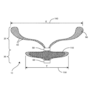
[0037] Figure 1 illustrates a cross section of an embodiment of
the
occlusion device disclosed herein showing length (x) of the resilient mesh
disc-shaped
body is greater than the length (y) of the compressible mesh carriage in free
air.
[0038] Figure 2 illustrates an embodiment of the occlusion device
disclosed herein deployed in an aneurysm showing the length (x) of the
resilient mesh
disc-shaped body is greater than the length (y) of the compressible mesh
carriage in
the deployed shape.
[0039] Figure 3 illustrates an embodiment of the occlusion device
disclosed herein showing the length (x) of the resilient mesh disc-shaped body
is
greater than the length (y) of the compressible mesh carriage in free air.
[0040] Figure 4 illustrates the delivery of an embodiment of the
occlusion
device disclosed herein.
DETAILED DESCRIPTION
[0041] The present invention is illustrated in the drawings and
description
in which like elements are assigned the same reference numerals. However,
while
particular embodiments are illustrated in the drawings, there is no intention
to limit
the present invention to the specific embodiment or embodiments disclosed.
Rather,
the present invention is intended to cover all modifications, alternative
constructions,
and equivalents falling within the spirit and scope of the invention. As such,
the
drawings are intended to be illustrative and not restrictive.
[0042] Unless otherwise defined, all technical twits used herein
have the
same meaning as commonly understood by one of ordinary skill in the art to
which
this technology belongs.
[0043] Exemplary embodiments of the occlusion device disclosed
herein
are depicted in Figures 1-2.
[0044] For the purposes of the occlusion device 10 disclosed
herein, the
terminology "corresponds to" means there is a functional and/or mechanical
relationship between objects which correspond to each other. For example, an
occlusion device delivery system corresponds to (or is compatible with) an
occlusion
device for deployment thereof.
- 9 -
Date Regue/Date Received 2023-01-19
[0045] For the purposes of the occlusion device 10 disclosed
herein, the
terminology "occlusion device" means and/or may be interchangeable with
terminology such as, without limitation, "device" or "occlusion device system"
or
"occlusion system" or "system" or "occlusion device implant" or "implant" or
"intrasaccular implant" or "intra-aneurysmal implant" and the like.
[0046] Occlusion device delivery systems are well known and
readily
available in the art. For example, such delivery technologies may be found,
without
limitation, in US Patent and Publication Numbers 4,991,602; 5,067,489;
6,833,003;
2006/0167494; and 2007/0288083. For the purposes of the occlusion device
disclosed herein, any type of occlusion device delivery means and/or delivery
system
and/or delivery technology and/or delivery mechanism and/or detachment (and/or
attachment) means and/or detachment system and/or detachment technology and/or
detachment mechanism may be utilized and/or modified in such a manner as to
make
compatible (so as to correspond) with the occlusion device disclosed herein.
Exemplary occlusion device delivery mechanisms and/or systems include, without
limitation, guide wires, pusher wires, catheters, micro-catheters, and the
like.
Exemplary occlusion device detachment mechanisms include, without limitation,
fluid pressure, electrolytic mechanisms, hydraulic mechanisms, interlocking
mechanisms, and the like. In one embodiment, the occlusion device disclosed
herein
is used in a method of electrolytic detachment. Electrolytic detachment is
well known
in the art and can be found, for example, in US Patent Numbers 5,122,136;
5,423,829;
5,624,449; 5,891,128; 6,123,714; 6,589,230; and 6,620,152.
[0047] The occlusion device 10 disclosed herein relates to a
continuous
mesh configuration comprising disproportionate mesh structures on opposing
sides of
a medial pinch point or pinch point-encircled marker 50. On one side of the
pinch
point or marker is a disc-shaped mesh body 20 which caves inward like a cup.
The
disc-shaped expansion of the mesh body 20 is the result of the body having no
other
pinch point or pinch point-encircled marker than the medial pinch point or
pinch
point-encircled marker 50. Therefore, there is no pinch point or marker
conforming
the ends of the body into a sphere and rather the ends of the disc-shaped body
20
extend distally and outward allowing the protruding mesh 90 to appose the dome
of
the aneurysm 70, and effectively conform to the walls of the aneurysm 70 as
the mesh
disc-shaped body 20 caves inward in a cupped configuration. On the opposing
side of
the disc-shaped body 20 extending distally from the medial pinch point or
pinch
- 10 -
Date Regue/Date Received 2023-01-19
point-encircled marker 50 is a compressible mesh basket-shaped carriage 30
defined
on either axial end by a pinch point of the mesh or by a pinch point-encircled
marker
50, 60.
[0048] Figures 1-4 show a continuous mesh structure comprising a
medial
pinch point 50, a resilient mesh disc-shaped body 20 extending distally and
outward
from the medial pinch point 50 and a compressible mesh carriage 30 extending
distally from the medial pinch point on an opposing side of the resilient mesh
disc-
shaped body 20. The compressible mesh carriage 30 comprises a pinch point on
each
end of the carriage, wherein one of the pinch points is the medial pinch point
50 of the
entire continuous mesh structure. The device 10 disclosed herein has a
continuous
mesh structure which is capable of a first delivery shape and a second
expandable
deployed shape. Figure 1 shows that, in one embodiment of the device 10
disclosed
herein, the length (x) 140 of the resilient mesh body is greater than the
length (y) 150
of the compressible mesh carriage 30 in free air and in the deployed shape.
The
continuous mesh structure of the device 10 disclosed herein and its uniquely
disproportionate mesh substructures on either side of a medial pinch point 50
promotes more effective endothelialization around the device 10 as shown when
the
device is in the deployed shape in Figures 2 & 4. As such, the occlusion
device 10
design is one continuous 3-dimensional mesh network which, when deployed in an
aneurysm 70 or body lumen, provides flow disruption 130, thrombus 120
establishment, a framework for cell growth, and/or ultimate blood stasis 80.
[0049] For the purposes of the claimed invention, a "carriage" 30
is an
axial segment of mesh between a pinch point of mesh or marker-encircled pinch
point
50, 60 of mesh which causes the mesh to expand in a puffed yet compressed
basket-
shaped 110 manner. A "pinch point" is located at and defines the ends of an
axial
segment of mesh. Such segmented mesh carriages 30 and pinch points are
configured
to be within a continuous mesh structure or network. A "pinch point" is a
constrained
and gathered location on the mesh structure which functions to restrict
movement of
the adjacent carriage 30 at an isolated point and thereby stabilizes the
carriage 30. In
one embodiment of the continuous mesh structure of the device 10 disclosed
herein,
the pinch points stabilize the carriage 30 relative to the disc-shaped mesh
body 20
extending distally therefrom a pinch point located at one axial end of the
carriage 30
which is located medially 50 in relation to the entire continuous mesh
structure. In
one further embodiment, the continuous mesh structure comprises more than one
-11 -
Date Regue/Date Received 2023-01-19
mesh carriage 30. The number (n) of carriages 30 is as many as clinically and
practically possible, and determined by a clinician in accordance with known
diagnostic techniques in advance, for treating large and/or irregular-sized
aneurysms
70, and for delivery through about a 150 centimeter (cm) catheter (or micro-
catheter).
The axial length (1) of each carriage 30 can vary depending on the number (n)
of
carriages 30 deemed appropriate to occlude an aneurysm 70 of a given size so
long as
the length (1) is sufficient to permit the carriage 30 to expand and compress
to a
dimension (or width) greater than its original width. As is accepted in the
art, the
diameter of such an occlusion device 10 is measured in free air. The width (w)
of
each carriage 30 ranges (in free air) from about 2 millimeters (mm) to about
50 mm in
order to be clinically practical. When deployed, the carriage 30 compresses in
such a
manner where the width (w) grows or expands up to about a factor of two (2)
such
that a carriage 30 of dimension (w) is capable of growing to approximately 2
times w
(or 2w). In other words, each carriage 30 compresses like a marshmallow which
causes its length (1) to be reduced and its width (w) to expand. In one
embodiment, in
free air, each carriage 30 can be designed in such a manner that the length
(1) is
greater or equal to its width (w) but in the deployed (compressed) shape, w is
greater
than 1. Such an occlusion device 10 comprising a compressible carriage 30 can
be
constructed in a variable manner so as to select the number (n) of carriages
30 as well
as the length (1) and corresponding width (w) of each carriage 30 to
accommodate a
wide range of sizes and shapes of aneurysms 70 or body lumen to be treated. As
such, in another embodiment, in free air, a carriage 30 can be designed in
such a
manner that its length (1) is equal to or less than its width (w) and in the
deployed
(compressed) shape, its width (w) remains greater than 1.
[0050] The disc-
shaped resilient mesh body 20 of the continuous mesh
structure extends distally from a pinch point of the opposing carriage 30 yet
located
medially 50 within the entire continuous mesh structure. In one embodiment,
the
disc-shaped mesh body 20 has a deployed shape that caves inward like a cup.
The
disc-shaped expansion of the mesh body 20 is the result of the body 20 having
no
other pinch point or pinch point-encircled marker than the medial pinch point
or pinch
point-encircled marker 50. Therefore, there is no pinch point or marker
conforming
the ends of the body into a sphere or closed, puffed shape and rather the ends
of the
disc-shaped body 20 extend distally and outward allowing the mesh to appose
the
dome of the aneurysm 70 in a low-profile manner, and effectively conform to
the
- 12 -
Date Regue/Date Received 2023-01-19
walls of the aneurysm 70 as the mesh disc-shaped body 20 caves inward in a
cupped
configuration.
[0051] For the purposes of the present invention, the terminology
"low
profile" means that the mesh disc-shaped body 20, in free air, has a height
that is
between about 10-20% of its width, and therefore in its deployed shape the
resilient
mesh body 20 lays flush, in a flattened manner, up against the aneurysm 70
walls. In
this manner, the disc-shaped body 20 of the device 10 disclosed herein is
lower and/or
slimmer than typical occlusion devices readily available in the art which
expand to fill
the space of the aneurysm dome (fully and/or partially with respect to the
majority of
the space in the aneurysm) and which expand radially and/or which expand in a
spherical manner. In one embodiment, the resilient mesh disc-shaped body 20,
in free
air, has a height between about 12-18% of its width. In another embodiment,
the
resilient disc-shaped body 20, in free air, has a height between about 14-16%
of its
width. In another embodiment, the resilient mesh disc-shaped body 20, in free
air, has
a height of about 15% of its width.
[0052] In another embodiment, as shown in Figure 3, the low
profile,
disc-shaped body 20 is a single layer of resilient mesh material. In another
embodiment, as shown in Figure 1, the low profile, disc-shaped body 20 is a
dual (or
double) layer 40 of resilient mesh material. As such, the resilient mesh body
confer
its capabilities for conforming to the inner surface of the walls of the
aneurysm 70
(via the opposing pressure of the protruding body 90 against the aneurysm
walls)
thereby providing a stabilizing effect for anchoring the device 10 in the
aneurysm 70
(and thereby minimizing the need for anti-coagulation therapy and lessening
the risk
of clot emboli formation which could flow deeper into the vascular tree
inducing
stroke). Such a low profile configuration facilitates blood stasis 80, clot
foimation
120 and/or healing and/or shrinkage of the aneurysm 70 which is particularly
advantageous if the size or mass of the aneurysm 70 is causing pain or other
side
effects within the patient. Moreover, such an occlusion device 10 is well
suited for
conformability across a broad range of aneurysm morphologies, particularly
since it is
well known and generally accepted that aneurysms are not perfectly round in
shape.
[0053] In another embodiment, a dual layer 40 disc-shaped body 20
of an
occlusion device 10 disclosed herein is a configuration of wire mesh which is
folded
circumferentially (circumferential fold line) and therefore doubled back on
itself. The
dual 40 or doubled back layers continuously intersect with the mesh pinch
point or
- 13 -
Date Regue/Date Received 2023-01-19
pinch point-encircled marker 50 of the compressible carriage 30. This
intersection of
the mesh of the disproportionate configurations of the device 10 is located at
a medial
pinch point 50 defining one axial end of the compressing carriage 30 and the
proximal
central portion of the disc-shaped mesh body 20. This medial pinch point 50 is
approximately the core of the entire continuous structure of the occlusion
device 10.
Without wishing to be bound by theory, this doubled back or dual layer 40 of
wire
mesh material triggers a mechanism of action believed to contribute to the
enhanced
acute thrombogenicity 120 of the device 10 in animal studies. It is believed
that the
localizing of a small volume of clot between the dual/double layers 40, which
have a
high surface area contribution from the wire strands, facilitates blood stasis
80, and
nucleating and stabilizing thrombus 120. In the deployed shape, the disc-
shaped body
20 having a folded back dual layer 40 is deeper when compared to a non-
deployed
dual layer occlusion device accounting for a change in width of approximately
15%
which translates to an increase in the diameter of the body when pressure is
applied at
the pinch point or marker 50, 60. This change in width/increase in diameter
contributes to an effective anchoring effect of the deployed device 10 as
blood applies
pressure to the protruding mesh body 90 distributed along the aneurysm 70
walls.
Such a configuration also provides sufficient apposition of the protruding
body 90 of
the device 10 against the aneurysm 70 wall or vessel wall for peripheral
arterial or
venous occlusion. Based on animal studies, such a disc-shaped body 20 provides
sufficient mesh density to confer stasis 80 acutely. It is further known,
based on
analyzing such a body configuration in post-deployment that the wire
mesh/braid
distribution remains relatively uniform.
[0054] In
another embodiment of the occlusion device disclosed herein, a
compressible axial mesh carriage 30 comprises a coaxial inner mesh carriage.
Such a
coaxial mesh inner carriage creates greater flow disruption 130 and
compartmentalization 100 than an axial mesh carriage without a coaxial mesh
inner
carriage, thereby triggering enhanced stasis 80 and thrombus 120
stabilization. In
another embodiment, the axial carriage 30 and the coaxial carriage (or
carriages) are
constructed of dissimilar metal mesh. In a further embodiment, the dissimilar
metal
mesh creates a galvanic effect which can further enhance thrombus 120
development.
In another further embodiment, the dissimilar metal mesh can be comprised of
one
metal in one carriage which possesses radiopaque properties relative to the
metal in
the other carriage and thus enhances visualization of the device. In such
- 14 -
Date Regue/Date Received 2023-01-19
embodiments, braid mesh density can be the same or different in axial outer
carriages
30 and coaxial inner carriages and wires of the inner and outer mesh can have
different numbers of strands and wire diameters. Such a coaxial carriage or
coaxial
carriages are variable in dimension compared to the outer axial carriage 30.
For
example, in one embodiment, a coaxial carriage or carriages can range from
about 5%
to about 95% of the dimensions of the outer axial carriage of which the
coaxial
carriage or coaxial carriages is/are comprised within.
[0055] In one embodiment, the device 10 is constructed of a metal
braid of
readily available material such as, without limitation, nitinol (NiTi), cobalt
chrome
(CoCr) alloy, stainless steel, tungsten iridium alloy or a combination
thereof. For
example, the mesh of the continuous mesh structure is woven with the most
clinically
relevant and practical braided mesh in a range of as few as 36 braids to as
many as
144 braids. In another embodiment, the angle of the weave of the metal braid
construction creates the softest compressible mesh design. For example, the
mesh is
braided with a wire diameter of about 0.0075 inches up to about .005 inches.
Prior to
use of such an occlusion device 10, a clinician or physician determines the
size and
shape of the aneurysm 70 or body lumen to be treated using readily available
diagnostic techniques. The physician or clinician is then able to best choose
the
occlusion device having a dimension or dimensions which corresponds to the
given
aneurysm 70 or body lumen to be treated.
[0056] "Markers" 50, 60 are well known and readily available in
the
medical device art. In some embodiments, a marker 50, 60 consists of metallic
material, often radiopaque material, and takes the form of a shape such as a
band-
shaped marker, a ring-shaped marker, a tube-shaped marker, and the like, so as
to
encircle a pinch point of mesh of continuous mesh structure of the occlusion
device
disclosed herein. Alternatively, a marker 50, 60 may consist of wire strands
wound
around and therefore encircling a given pinch point. In one embodiment, the
marker
or markers 50, 60 which encircle each pinch point provide positional reference
under
X-Ray as to where the device 10 is located in the catheter (or microcatheter)
and
where the device 10 is located once deployed in an aneurysm 70 or body lumen.
[0057] In one embodiment, a marker 50, 60 such as a ring encircles
the
pinch points defining each axial end of the compressible carriage 30 of the
continuous
mesh structure. As such, the marker 50, 60 of the occlusion device 10
disclosed
herein is a substantially solid collar or rigid member such as, without
limitation a
- 15 -
Date Regue/Date Received 2023-01-19
solid ring or band comprised of materials such as, without limitation, gold,
platinum,
stainless steel, and/or combinations thereof. In another embodiment,
radiopaque
materials such as, without limitation, gold, platinum, platinum/iridium alloy,
and/or
combinations thereof, can be used. Such a marker 50, 60 provides positional
visualization of the device during delivery and placement. The markers 50, 60
are
located on the occlusion device 10 encircling pinch points on each axial end
of the
carriage 30. In this manner, the marker located at the distal axial end of the
carriage
30 is capable of resting above or within the neck of an aneurysm 70. The
solidness of
the markers 50, 60 help confer stability of the device 10 within the aneurysm
70 and
prevents movement or the transfer of forces through the compressible mesh
carriage
30 and resilient mesh disc-shaped body 20 thereby preventing misplacement or
accidental movement of the device 10. The markers 50, 60 are also configured
with a
junction to cooperate and release from/attach to a corresponding delivery
means such
as, without limitation, a delivery catheter or guide wire and/or pusher wire
technologies. It also advantageously provides for full retrievability of the
device 10
disclosed herein.
[0058] In another embodiment, the substantially solid marker 50,
60
comprises a radiopaque material (such as for example, without limitation,
platinum,
gold, platinum/iridium alloy, and/or combinations thereof) to facilitate
visualization
of the occlusion device 10 under fluoroscopy during delivery, placement and/or
deployment. The marker 50, 60 comprises a proximal end and a distal end.
Occlusion devices 10 disclosed herein may be configured to incorporate the use
of
markers 50, 60 to influence shape, diameter, and/or curvature of the
compressible
carriage 30 upon expansion during deployment. Additionally, the marker 50, 60
may
be designed in various shapes to influence the overall profile of the
occlusion device
to ensure a proper fit of the expanded/deployed occlusion device 10 within the
aneurysm sac 70.
[0059] Without wishing to be bound by theory, this configuration
of a
continuous compressible mesh structure divided into disproportionate mesh
segments
triggers a mechanism of action believed to contribute to enhanced acute
thrombogenicity 120 of the device in animal studies. It is also believed that
the
localizing of a small volume of clot between the disc-shaped mesh dual-layers
40 and
basket 30 compartments, which have a high surface area contribution from the
wire
strands, facilitates nucleating and stabilizing thrombus 120 in an aneurysm
70. This
- 16 -
Date Regue/Date Received 2023-01-19
compartmentalization 100 of the occlusion device in its deployed shape is an
effective
stabilizing or anchoring feature of the deployed device 10 as blood applies
pressure to
the mesh structure distributed across or within the neck of the aneurysm 70.
Such a
configuration also provides sufficient apposition of the compressible device
against
the aneurysm 70 wall or vessel wall for peripheral arterial or venous
occlusion. The
device 10 disclosed herein provides sufficient mesh density to confer stasis
80 acutely
and the wire mesh/braid distribution remains relatively uniform in deployment.
[0060] In another embodiment of an occlusion device disclosed
herein, the
occlusion device 10 is constructed or partially constructed with a relatively
uniform
distribution of wire mesh strands or braids such as, without limitation, a 72
NiTi wire
mesh strand braided configuration or a combination of 72 NiTi and CoCr wire
mesh
strand braided configuration. In other embodiments, the occlusion device 10
comprises or partially comprises wire mesh strands or braids that range from
36 to
144 NiTi strand braided configuration.
[0061] For the purposes of the present invention, the terminology
"mesh
density" means the level of porosity or the ratio of metal to open area of the
mesh
device. Mesh density relates to the number and size of the openings or pores
of the
mesh and by the extent that the pores are open or closed in situations where
opening
or pore openness varies between delivery and deployment. Generally, a high
mesh
density region of a resilient mesh material has approximately about 70% or
more
metal area and about 60% or less open area.
[0062] In one embodiment, the continuous mesh structure has or
partially
has an "open mesh density" for enhanced tissue integration and/or
stabilization of the
occlusion device. Open mesh density is greater than about 40% open area in the
mesh. Open mesh density is known to have a low number, usually between about
40
and 80, picks per inch (PPI) to represent the porosity of the mesh layers. PPI
is the
number of repeat cross overs of braiding material in a linear inch. A high
number of
repeats (or PPI), usually between about 100 and 180, is an indicator that the
mesh is
dense. A lower number of repeats (or PPI) is an indicator that the mesh is
porous
(open). In an additional embodiment, the continuous mesh structure is
constructed
from or partially constructed from a super elastic material, such as, without
limitation,
nitinol. In yet another embodiment, the structure is constructed or partially
constructed from DFT platinum core nitinol. In other embodiments, when the
structure is partially constructed of nitinol and partially constructed of DFT
platinum
- 17 -
Date Regue/Date Received 2023-01-19
core nitinol. DFT platinum core nitinol is used for enhancing visualization of
the
device during deployment and implantation.
[0063] In one embodiment, as shown in Figure 4, the occlusion
device 10
disclosed herein is delivered to an aneurysm 70 or lumen via electrolytic
delivery
and/or deployment and/or detachment of the occlusion device 10 disclosed
herein
through an artery and/or vessel adjacent to the aneurysm 70 or body lumen.
Electrolytic detachment means and methods such as those disclosed in U.S.
Patent
5,122,136 are well known in the art. In one embodiment, a coil-wound core wire
(or
guide wire or pusher wire) of the catheter (or micro-catheter) is attached
inside the
marker 60 at its most distal end to the occlusion device 10 disclosed herein.
The coil
wind maintains a constant diameter (4)) so as not to impact upon flexibility
or stiffness
of the delivery catheter or micro-catheter or guide wire. In certain
embodiments, FEP
(Fluorinated Ethylene Propylene) heat shrink tubing encases the coil-wound
portion
of the core wire. Numerous readily available and well known attachment
techniques
in the medical device arts can be used to attach the distal end of the core
wire inside
the marker 60 and to the occlusion device 10 or implant. Such attachment
techniques
include, without limitation, adhesives, laser melting, laser tack, spot,
and/or
continuous welding. In one embodiment, an adhesive is used to attach the
distal end
of the core wire inside the marker 60. In a further embodiment, the adhesive
is an
epoxy material which is cured or hardened through the application of heat or
UV
(ultra-violet) radiation. In an even further embodiment, the epoxy is a
thermal cured,
two-part epoxy such as EPO-TEKO 353ND-4 available from Epoxy Technology,
Inc., 14 Fortune Drive, Billerica, Mass. Such an adhesive or epoxy material
encapsulates the junction of the core wire inside the marker 60 and increases
its
mechanical stability.
[0064] In another embodiment, during and/or after deployment of
the
device 10, the coil-wound core wire detaches the occlusion device 10 disclosed
herein
at an electrolytic detachment site (or zone) on the core wire itself in such a
manner so
that the core wire is severed and/or dissolved through electrolytic action at
the base of
the marker 60. Such action then releases and/or places the occlusion device 10
into an
aneurysm 70 or vessel to be treated.
[0065] In certain embodiments, the compressible mesh structure of
the
occlusion device 10 disclosed herein can be filled with an embolic material to
promote clotting and closure of the aneurysm 70.
- 18 -
Date Regue/Date Received 2023-01-19
[0066] In other embodiments, the occlusion device 10 disclosed
herein
may further incorporate adjunctive elements and/or members such as coiling
techniques, framing coils, embolic agents, additional markers, polymers,
resorbent
polymers and/or a combination thereof.
[0067] Resilient and compressible mesh materials for design and/or
manufacture of occlusion devices are readily available and well known by those
skilled in the relevant art. As such, resilient and compressible mesh
materials range
from a wide variety of available materials such as, without limitation, nickel
titanium
(nitinol or otherwise known as NiTi), stainless steel, polymers, and/or
combinations
thereof. Exemplary known biomedical polymeric families include, without
limitation,
polymers such as polyphosphazenes, polyanhydrides, polyacetals, poly(ortho
esters),
polyphosphoesters, polycaprolactones, polyurethanes, polylactides,
polycarbonates,
polyamides, and/or a combination thereof. (See, e.g., J Polym Sci B Polym
Phys.
Author manuscript; available in PMC 2012 June 15.)
[0068] In one exemplary embodiment, the resilient and compressible
mesh
material is formed of woven strands of polymer material, such as, without
limitation,
nylon, polypropylene or polyester. The polymer strands can be filled with a
radiopaque material which allows the physician treating the aneurysm to
fluoroscopically visualize the location of the device within the vasculature.
Radiopaque filler materials preferably include bismuth trioxide, tungsten,
titanium
dioxide or barium sulfate, or radiopaque dyes such as iodine. The resilient
and
compressible mesh material can be formed by strands of radiopaque material.
The
radiopaque strands allow the physician and/or radiologist to fluoroscopically
visualize
the location of the mesh, without the use of filled polymer materials. Such
radiopaque
strands may be formed with materials such as, without limitation, gold,
platinum, a
platinum/iridium alloy, and/or a combination thereof. In one embodiment, the
resilient mesh material is constructed of 10%-45% platinum core NiTi. In
another
embodiment, the resilient mesh material is constructed of 10% platinum core
NiTi,
15% platinum core NiTi, 20% platinum core NiTi, or 45% platinum core NiTi. 10%
platinum core NiTi construction is sufficient to provide a ghost image of the
occlusion
device under x-ray.
[0069] Such constructed combination wires or composite wires
having a
radiopaque core and non-radiopaque outer layer or casing are readily available
and
well known in the medical device and metallic arts as DFT (drawn-filled-tube)
- 19 -
Date Regue/Date Received 2023-01-19
wires, cables or ribbons. DFT wire is a metal-to-metal composite constructed
to
combine the desired physical and mechanical attributes of two or more
materials into
a single wire. By placing the more radiopaque, but more ductile material in
the core
of the wire, the NiTi outer layer is able to provide the resulting composite
wire with
similar mechanical properties of a 100% NiTi wire. DFT wires are available
from
Fort Wayne Metals Corp., Fort Wayne, hid., U.S.A. See also, for example, the
journal article entitled Biocompatible Wire by Schaffer in Advanced Materials
&
Processes, Oct 2002, pages 51-54.
[0070] Where the compressible mesh structure is formed of
radiopaque
metal strands, the strands may be covered with a polymer coating or extrusion.
The
coating or extrusion over the radiopaque wire strands provides fluoroscopic
visualization but also increases the resistance of the strands to bending
fatigue and
may also increase lubricity of the strands. The polymer coating or extrusion,
in one
embodiment, is coated or treated with an agent which tends to resist clotting,
such as
heparin. Such clot resistant coatings are generally known. The polymer coating
or
extrusion can be any suitable extnidable polymer, or any polymer that can be
applied
in a thin coating, such as Teflon or polyurethane.
[0071] In yet another embodiment, the strands of the compressible
mesh
structure are formed using both metal and polymer braided strands. Combining
the
metal strands with the polymer strands into a braid changes the flexibility
characteristics of mesh. The force required to deploy and/or collapse such a
mesh
portion is significantly reduced over that required for a mesh portion that
includes
only metal mesh strands. However, the radiopaque characteristics of the mesh
for
fluoroscopic visualization are retained. Metal strands forming such a device
includes,
without limitation, stainless steel, gold, platinum, platinum/iridium,
nitinol, and/or
combinations thereof. Polymer strands forming the device can include nylon,
polypropylene, polyester, Teflon , and/or combinations thereof. Further,
polymer
strands of the mesh material can be chemically modified to make them
radiopaque
with known techniques such as, without limitation, by using gold deposition
onto the
polymer strands, or by using ion beam plasma deposition of suitable metal ions
onto
the polymer strands.
[0072] The compressible mesh structure can also be fonned with
filaments
or strands of varying diameter and/or varying flexibility. For example, wire
diameters
for use in the occlusion device disclosed herein range from about 0.0075
inches up to
- 20 -
Date Regue/Date Received 2023-01-19
about .005 inches. By varying the size or flexibility of the polymer strands,
the
flexibility characteristics of the mesh, upon deployment, can also be varied.
By
varying the flexibility characteristics, both the deployed (compressed) and
delivery
(constrained) configuration of the resilient and compressible mesh structure
can be
varied or changed to substantially any desired shape.
[0073] Not only
can the mesh be formed of both polymer strands or
filaments and metal strands or filaments, but it can be formed using filaments
of
different polymer materials. For example, different polymer materials having
different flexibility characteristics can be used in forming the mesh. This
alters the
flexibility characteristics to change the resultant configuration of the mesh
structure in
both the deployed and the collapsed positions. Such biomedical polymers are
readily
known and available in the art and can be derived from polymeric families such
as,
without limitation, polyphosphazenes, polyanhydrides, polyacetals, poly (ortho
esters), polyphospho esters, po ly
caprolacton es, polyurethanes, poly lactides,
polycarbonates, polyamides, and/or a combination thereof.
[0074]
Compressible mesh materials suitable for use within the mesh
carriages may take the form of a flat woven sheet, knitted sheet, or a laser
cut wire
mesh. In general, the material should include two or more sets of
substantially
parallel strands, with one set of parallel strands being at a pitch of between
45 degrees
and 135 degrees with respect to the other set of parallel strands. In some
embodiments, the two sets of parallel strands forming the mesh material are
substantially perpendicular to each other. The pitch and general construction
of the
mesh material may be optimized to meet the performance needs of the occlusion
device 10.
[0075] The wire
strands of the metal fabric used in the occlusion device 10
disclosed herein should be formed of a material which is both resilient and
compressible and can be heat-treated to substantially set a desired shape.
Materials
which are believed to be suitable for this purpose include a cobalt-based low
thermal
expansion alloy referred to in the field of occlusion devices as Elgiloy 0,
nickel-based
high-temperature high-strength "superalloys" commercially available from
Haynes
International under the trade name Hastelloy , nickel-based heat treatable
alloys sold
under the name Incoloy0 by International Nickel, and a number of different
grades of
stainless steel. The important factor in choosing a suitable material for the
wires is
that the wires retain a suitable amount of the deformation induced by the
molding
- 21 -
Date Regue/Date Received 2023-01-19
surface (or shape memory, as described below) when subjected to a
predetermined
heat treatment.
[0076] One class of materials which meet these qualifications are
so-called
shape memory alloys. Such alloys tend to have a temperature induced phase
change
which will cause the material to have a preferred configuration which can be
fixed by
heating the material above a certain transition temperature to induce a change
in the
phase of the material. When the alloy is cooled, the alloy will "remember" the
shape
it was in during the heat treatment and will tend to assume that same and/or
similar
configuration unless constrained from doing so.
[0077] One particular shape memory alloy for use in the occlusion
device
disclosed herein is nitinol, an approximately stoichiometric alloy of nickel
and
titanium, which may also include other minor amounts of other metals to
achieve
desired properties. NiTi alloys such as nitinol, including appropriate
compositions
and handling requirements, are well known in the art and such alloys need not
be
discussed in detail here. For example, United States Patent Numbers 5,067,489
and
4,991,602, discuss the use of shape memory NiTi alloys in guide wire-based
technologies. Such NiTi alloys are preferred, at least in part, because they
are
commercially available and more is known about handling such alloys than other
known shape memory alloys. NiTi alloys are also very elastic. Indeed, they are
said
to be known as "superelastic" or "pseudoelastic." This elasticity will help an
occlusion device 10 as disclosed herein return to prior expanded configuration
for
deployment thereof.
[0078] The wire strands can comprise a standard monofilament of
the
selected material, i.e., a standard wire stock may be used. In some
embodiments, 72
wire strands and/or 72 strand braid configuration is used. In other
embodiments, the
occlusion device comprises wire mesh strands or braids that range from 36 to
144
NiTi strand braided configurations. If so desired, though, the individual wire
strands
may be formed from "cables" made up of a plurality of individual wires. For
example, cables formed of metal wires where several wires are helically
wrapped
about a central wire are commercially available and NiTi cables having an
outer
diameter of 0.003 inches or less can be purchased. One advantage of certain
cables is
that they tend to be "softer" than the monofilament wires having the same
diameter
and formed of same material. Additionally, the use of a cable can increase the
effective surface area of the wire strand, which will tend to promote
thrombosis 120.
- 22 -
Date Regue/Date Received 2023-01-19
[0079] An
occlusion device 10 disclosed herein is configured with a
continuous mesh structure having a mesh density sufficient for functioning in
such a
manner as an endothelial cell scaffold layers or compai _____________ intents
100 filling a vessel or
body lumen or aneurysm 70 and thereby reducing blood flow 130 by about 60% to
trigger clot formation and/or healing of the aneurysm 70 and/or ultimate
stasis 80.
For the purposes of the occlusion device 10 disclosed herein, the terminology
"mesh
density" means the level of porosity or the ratio of metal to open area of the
mesh
structure. Mesh density relates to the number and size of the openings or
pores of the
mesh and by the extent that the pores are open or closed in situations where
opening
or pore openness varies between delivery and deployment. Generally, a high
mesh
density region of a resilient mesh material has approximately about 40% or
more
metal area and about 60% or less open area.
[0080] In some
embodiments, the compressible mesh structure may be
formed unifomily of the same material, however such material may have
different
knitted, stitched, braided, and/or cut construction.
[0081] In other
embodiments, the implantable occlusion device 10
disclosed herein can be used for the process of peripheral vascular
embolization (a
process well known in the art and known to involve the shutdown of blood flow
130
distal to a specified vascular point), for example, in the treatment and/or
amelioration
of peripheral arterial or venous pathologies and/or any related pathologies
requiring
vessel occlusion for the treatment thereof.
[0082] The
occlusion device 10 of the invention disclosed herein may
incorporate reasonable design parameters, features, modifications, advantages,
and
variations that are readily apparent to those skilled in the art in the field
of occlusion
devices.
[0083] EXAMPLES
[0084] A study
protocol with respect to the occlusion device 10 disclosed
herein and justification for animal use will be reviewed and approved by the
Institutional Animal Care and Use Committee (IACUC) at ISIS Services and the
procedures carried out under veterinarian supervision.
[0085] The
rabbit elastase aneurysm model is a well-accepted and art-
recognized model for testing novel neurointerventional devices and has been
the
subject of a number of clinical publications regarding efficacy and similarity
to
human response. (See, e.g., Altes et al. Creation of Saccular Aneurysms in the
Rabbit:
- 23 -
Date Regue/Date Received 2023-01-19
A Model Suitable for Testing Endovascular Devices. AJR 2000; 174: 349-354.) It
therefore is readily accepted by the regulatory agencies as an appropriate
test model.
The model's coagulation system is highly similar to that of humans. In
addition, the
model has advantageous anatomical aspects in that the diameters of the
rabbits' extra-
cranial carotid arteries are highly similar to the diameter of extra-cranial
carotid
arteries in humans. Moreover, elastase-induced aneurysms have been shown to
behave in a histologically similar manner as human aneurysms.
[0086] A number
of embodiments of the invention have been described.
Without departing from the scope and spirit of the occlusion device 10
disclosed
herein, reasonable features, modifications, advantages, and design variations
of the
claimed apparatus will become readily apparent to those skilled in the art by
following the guidelines set forth in the preceding detailed description and
embodiments. Accordingly, other embodiments are within the scope of the
following
claims.
-24 -
Date Regue/Date Received 2023-01-19