Note: Descriptions are shown in the official language in which they were submitted.
CA 03074689 2020-03-03
WO 2019/051244 PCT/US2018/049976
SYSTEMS AND METHODS FOR NON-INVASIVE PREIMPLANTATION GENETIC
DIAGNOSIS
FIELD
[0001] The embodiments disclosed herein are generally directed towards systems
and methods
for non invasive genetic screening and/or diagnosis of embryos prior to
implantation in an in
vitro fertilization procedure. More specifically, there is a need for non
invasive preimplantation
screening and/or diagnostic systems and methods which can aid clinicians in
the selection of
embryos with the lowest risk of genetic abnormalities/defects and have the
highest probability of
uterine implantation success.
BACKGROUND
[0002] In vitro fertilization (IVF) is an assisted reproductive technology has
become increasingly
popular for women of advanced maternal age, couples with difficulties
conceiving and as a
means for facilitating gestational surrogacy. The process of fertilization
involves extracting
eggs, retrieving a sperm sample, and then manually combining an egg and sperm
in a laboratory
setting. The embryo(s) is then implanted in the host uterus to carry the
embryo to term.
[0003] IVF procedures are expensive and can exact a significant
emotional/physical toll on
patients, so genetic screening of embryos prior to implantation is becoming an
increasingly
common for patients undergoing an IVF procedure. Current methods of diagnosing
genetic
abnormalities in embryos and screening for viability of transfer (i.e., embryo
implantation
viability) require a biopsy of embryos, which can affect embryo quality and
requires specialized
laboratory techniques that can be prohibitively expensive and time consuming.
[0004] As such, there is a need for non-invasive genetic screening and/or
diagnostic (NI PGS)
systems and methods for genetically screening embryos which avoid the need for
embryo biopsy
and thus substantially increase the safety for the preimplanted embryo.
1
CA 03074689 2020-03-03
WO 2019/051244 PCT/US2018/049976
SUMMARY
[0005] In one aspect, a method for determining copy number variation in an
embryo candidate
for in vitro fertilization (IVF) implantation is disclosed. An embryo
candidate is isolated from a
plurality of embryos. The embryo candidate is incubated in media that is
substantially free of
DNA. A portion of the media is transferred to an amplification vessel, wherein
the portion of
media includes genomic fragments shed or secreted from the embryo candidate. A
plurality of
genomic linker segments and ligase enzyme is added to the amplification vessel
in conditions
that catalyze the formation of concatenated genomic fragments containing at
least one genomic
linker segment and at least one genomic fragment from the isolated embryo
candidate. The
concatenated genomic fragments are amplified in the amplification vessel.
Sequence
information is obtained from the amplified concatenated genomic fragments. The
sequence
information is aligned (mapped) against a reference genome. Copy number
variations are
identified in the embryo candidate when a frequency of genomic fragment
sequence reads
aligned to a chromosomal position on the reference genome deviates from a
frequency threshold.
[0006] In another aspect, a method is for identifying genomic features in an
embryo candidate is
disclosed. An embryo candidate is isolated from a plurality of embryo
candidates. The embryo
candidate is incubated in media that is substantially free of DNA. A portion
of the media is
transferred to an amplification vessel, wherein the portion of media includes
one more genomic
fragments shed or secreted from the embryo candidate. A plurality of genomic
linker segments
and a ligase enzyme is added to the amplification vessel in conditions that
catalyze the formation
of concatenated genomic fragments containing at least one genomic linker
segment and at least
one genomic fragment from the isolated embryo candidate. The concatenated
genomic
fragments are amplified in the amplification vessel. Sequence information is
obtained from the
concatenated genomic fragments. The sequence information is aligned against a
reference
genome. Genomic features are identified on the aligned genomic fragment
sequences.
[0007] In still another aspect, a system for identifying genomic features in
an embryo candidate
is disclosed. The system includes a genomics sequencer, a computing device and
a display.
[0008] The genomic sequencer is configured to obtain sequence information from
concatenated
genomic fragments derived from an embryo candidate. The concatenated genomic
fragments
each contain at least one genomic linker segment and at least one genomic
fragment from the
embryo candidate.
2
CA 03074689 2020-03-03
WO 2019/051244 PCT/US2018/049976
[0009] The computing device is communicatively connected to the genomic
sequencer and
includes a sequence alignment engine and a genomic features identification
engine. The
sequence alignment engine is configured to subtract out sequence information
related to the
genomic linker segment portion of the concatenated genomic fragments and align
the genomic
fragment sequences to a reference genome. The genomic features identification
engine is
configured to identify genomic features in the aligned genomic fragment
sequences. The display
is communicatively connected to the computing device and configured to display
a report
containing the identified genomic features.
[0010] In yet another aspect, a method for identifying genomic features in a
tissue sample is
disclosed. Concatenated genomic fragment sequence reads are received
containing at least one
genomic linker segment sequence and at least one genomic fragment sequence
from a tissue
sample. The genomic linker segment sequence portion of the concatenated
genomic fragment
sequence reads is subtracted out. The concatenated genomic fragment sequence
reads are
aligned (mapped) to a reference genome. Genomic features are identified on the
aligned
genomic fragment sequences.
[0011] In still another aspect, a non-transitory computer-readable medium is
provided in which a
program is stored for causing a computer to perform a method for identifying
genomic features
in a tissue sample. Concatenated genomic fragment sequence reads are received
containing at
least one genomic linker segment sequence and at least one genomic fragment
sequence from a
tissue sample. The genomic linker segment sequence portion of the concatenated
genomic
fragment sequence reads are subtracted out. The concatenated genomic fragment
sequence reads
are aligned (mapped) to a reference genome. Genomic features are identified on
the aligned
genomic fragment sequences.
3
CA 03074689 2020-03-03
WO 2019/051244 PCT/US2018/049976
BRIEF DESCRIPTION OF THE DRAWINGS
[0012] For a more complete understanding of the principles disclosed herein,
and the advantages
thereof, reference is now made to the following descriptions taken in
conjunction with the
accompanying drawings, in which:
[0013] Figure 1 illustrates a workflow for non-invasive preimplantation
genetic screening of
embryos, in accordance with some embodiments of the disclosure.
[0014] Figure 2 is an exemplary flowchart depicting an amplification protocol
for amplifying
short genomic fragments, in accordance with some embodiments of the
disclosure.
[0015] Figure 3 illustrates the formation of concatenated fragments, in
accordance with some
embodiments of the disclosure.
[0016] Figure 4 is a block diagram that illustrates a computer system, in
accordance with various
embodiments.
[0017] Figure 5 is a schematic diagram of a system for non-invasive
preimplantation genetic
screening of embryos, in accordance with various embodiments
[0018] Figure 6 is a depiction of how concatenated fragment reads are mapped
to a reference
genome, in accordance with various embodiments.
[0019] Figure 7 is an exemplary flowchart showing a method for aligning
genomic fragment
reads to identify various types of genomic features, in accordance with
various embodiments.
[0020] Figure 8 is a flowchart showing a method for determining copy number
variation in an
embryo candidate, in accordance with various embodiments.
[0021] Figure 9 is a flowchart showing a method of identifying genomic
features in an embryo
candidate, in accordance with various embodiments.
[0022] Figure 10 is a flowchart showing a method for identifying genomic
features from
concatenated genomic fragment reads, in accordance with various embodiments.
[0023] It is to be understood that the figures are not necessarily drawn to
scale, nor are the
objects in the figures necessarily drawn to scale in relationship to one
another. The figures are
depictions that are intended to bring clarity and understanding to various
embodiments of
apparatuses, systems, and methods disclosed herein. Wherever possible, the
same reference
numbers will be used throughout the drawings to refer to the same or like
parts. Moreover, it
should be appreciated that the drawings are not intended to limit the scope of
the present
teachings in any way.
4
CA 03074689 2020-03-03
WO 2019/051244 PCT/US2018/049976
DETAILED DESCRIPTION
[0024] This specification describes exemplary embodiments and applications of
the disclosure.
The disclosure, however, is not limited to these exemplary embodiments and
applications or to
the manner in which the exemplary embodiments and applications operate or are
described
herein. Moreover, the figures may show simplified or partial views, and the
dimensions of
elements in the figures may be exaggerated or otherwise not in proportion. In
addition, as the
terms "on," "attached to," "connected to," "coupled to," or similar words are
used herein, one
element (e.g., a material, a layer, a substrate, etc.) can be "on," "attached
to," "connected to," or
"coupled to" another element regardless of whether the one element is directly
on, attached to,
connected to, or coupled to the other element or there are one or more
intervening elements
between the one element and the other element. In addition, where reference is
made to a list of
elements (e.g., elements a, b, c), such reference is intended to include any
one of the listed
elements by itself, any combination of less than all of the listed elements,
and/or a combination
of all of the listed elements. Section divisions in the specification are for
ease of review only and
do not limit any combination of elements discussed.
[0025] Unless otherwise defined, scientific and technical terms used in
connection with the
present teachings described herein shall have the meanings that are commonly
understood by
those of ordinary skill in the art. Further, unless otherwise required by
context, singular terms
shall include pluralities and plural terms shall include the singular.
Generally, nomenclatures
utilized in connection with, and techniques of, cell and tissue culture,
molecular biology, and
protein and oligo- or polynucleotide chemistry and hybridization described
herein are those well
known and commonly used in the art. Standard techniques are used, for example,
for nucleic
acid purification and preparation, chemical analysis, recombinant nucleic
acid, and
oligonucleotide synthesis. Enzymatic reactions and purification techniques are
performed
according to manufacturer's specifications or as commonly accomplished in the
art or as
described herein. The techniques and procedures described herein are generally
performed
according to conventional methods well known in the art and as described in
various general and
more specific references that are cited and discussed throughout the instant
specification. See,
e.g., Sambrook et al., Molecular Cloning: A Laboratory Manual (Third ed., Cold
Spring Harbor
Laboratory Press, Cold Spring Harbor, N.Y. 2000). The nomenclatures utilized
in connection
CA 03074689 2020-03-03
WO 2019/051244 PCT/US2018/049976
with, and the laboratory procedures and techniques described herein are those
well known and
commonly used in the art.
[0026] The phrase "next generation sequencing" (NGS) refers to sequencing
technologies having
increased throughput as compared to traditional Sanger- and capillary
electrophoresis-based
approaches, for example with the ability to generate hundreds of thousands of
relatively small
sequence reads at a time. Some examples of next generation sequencing
techniques include, but
are not limited to, sequencing by synthesis, sequencing by ligation, and
sequencing by
hybridization. More specifically, the MISEQ, HISEQ and NEXTSEQ Systems of
Illumina and
the Personal Genome Machine (PGM) and SOLiD Sequencing System of Life
Technologies
Corp, provide massively parallel sequencing of whole or targeted genomes. The
SOLiD System
and associated workflows, protocols, chemistries, etc. are described in more
detail in PCT
Publication No. WO 2006/084132, entitled "Reagents, Methods, and Libraries for
Bead-Based
Sequencing," international filing date Feb. 1, 2006, U.S. patent application
Ser. No. 12/873,190,
entitled "Low-Volume Sequencing System and Method of Use," filed on Aug. 31,
2010, and
U.S. patent application Ser. No. 12/873,132, entitled "Fast-Indexing Filter
Wheel and Method of
Use," filed on Aug. 31, 2010, the entirety of each of these applications being
incorporated herein
by reference thereto.
[0027] The phrase "sequencing run" refers to any step or portion of a
sequencing experiment
performed to determine some information relating to at least one biomolecule
(e.g., nucleic acid
molecule).
[0028] As used herein, the phrase "genomic features" can refer to a genome
region with some
annotated function (e.g., a gene, protein coding sequence, mRNA, tRNA, rRNA,
repeat
sequence, inverted repeat, miRNA, siRNA, etc.) or a genetic/genomic variant
(e.g., single
nucleotide polymorphism/variant, insertion/deletion sequence, copy number
variation, inversion,
etc.) which denotes a single or a grouping of genes (in DNA or RNA) that have
undergone
changes as referenced against a particular species or sub-populations within a
particular species
due to mutations, recombination/crossover or genetic drift.
[0029] Genomic variants can be identified using a variety of techniques,
including, but not
limited to: array-based methods (e.g., DNA microarrays, etc.), real-
time/digital/quantitative PCR
instrument methods and whole or targeted nucleic acid sequencing systems
(e.g., NGS systems,
6
CA 03074689 2020-03-03
WO 2019/051244 PCT/US2018/049976
Capillary Electrophoresis systems, etc.). With nucleic acid sequencing,
coverage data can be
available at single base resolution.
[0030] DNA (deoxyribonucleic acid) is a chain of nucleotides consisting of 4
types of
nucleotides; A (adenine), T (thymine), C (cytosine), and G (guanine), and that
RNA (ribonucleic
acid) is comprised of 4 types of nucleotides; A, U (uracil), G, and C. Certain
pairs of nucleotides
specifically bind to one another in a complementary fashion (called
complementary base
pairing). That is, adenine (A) pairs with thymine (T) (in the case of RNA,
however, adenine (A)
pairs with uracil (U)), and cytosine (C) pairs with guanine (G). When a first
nucleic acid strand
binds to a second nucleic acid strand made up of nucleotides that are
complementary to those in
the first strand, the two strands bind to form a double strand. As used
herein, "nucleic acid
sequencing data," "nucleic acid sequencing information," "nucleic acid
sequence," "genomic
sequence," "genetic sequence," or "fragment sequence," or "nucleic acid
sequencing read"
denotes any information or data that is indicative of the order of the
nucleotide bases (e.g.,
adenine, guanine, cytosine, and thymine/uracil) in a molecule (e.g., whole
genome, whole
transcriptome, exome, oligonucleotide, polynucleotide, fragment, etc.) of DNA
or RNA. It
should be understood that the present teachings contemplate sequence
information obtained
using all available varieties of techniques, platforms or technologies,
including, but not limited
to: capillary electrophoresis, microarrays, ligation-based systems, polymerase-
based systems,
hybridization-based systems, direct or indirect nucleotide identification
systems,
pyrosequencing, ion- or pH-based detection systems, electronic signature-based
systems, etc.
[0031] A "polynucleotide", "nucleic acid", or "oligonucleotide" refers to a
linear polymer of
nucleosides (including deoxyribonucleosides, ribonucleosides, or analogs
thereof) joined by
internucleosidic linkages. Typically, a polynucleotide comprises at least
three nucleosides.
Usually oligonucleotides range in size from a few monomeric units, e.g. 3-4,
to several hundreds
of monomeric units. Whenever a polynucleotide such as an oligonucleotide is
represented by a
sequence of letters, such as "ATGCCTG," it will be understood that the
nucleotides are in 5'->3'
order from left to right and that "A" denotes deoxyadenosine, "C" denotes
deoxycytidine, "G"
denotes deoxyguanosine, and "T" denotes thymidine, unless otherwise noted. The
letters A, C,
G, and T may be used to refer to the bases themselves, to nucleosides, or to
nucleotides
comprising the bases, as is standard in the art.
7
CA 03074689 2020-03-03
WO 2019/051244 PCT/US2018/049976
[0032] The phrase "fragment library" refers to a collection of nucleic acid
fragments, wherein
one or more fragments are used as a sequencing template. A fragment library
can be generated,
for example, by cutting or shearing a larger nucleic acid into smaller
fragments. Fragment
libraries can be generated from naturally occurring nucleic acids, such as
mammalian or bacterial
nucleic acids. Libraries comprising similarly sized synthetic nucleic acid
sequences can also be
generated to create a synthetic fragment library.
[0033] In various embodiments, a sequence alignment method can align a
fragment sequence to
a reference sequence or another fragment sequence. The fragment sequence can
be obtained
from a fragment library, a paired-end library, a mate-pair library, a
concatenated fragment
library, or another type of library that may be reflected or represented by
nucleic acid sequence
information including for example, RNA, DNA, and protein based sequence
information.
Generally, the length of the fragment sequence can be substantially less than
the length of the
reference sequence. The fragment sequence and the reference sequence can each
include a
sequence of symbols. The alignment of the fragment sequence and the reference
sequence can
include a limited number of mismatches between the symbols of the fragment
sequence and the
symbols of the reference sequence. Generally, the fragment sequence can be
aligned to a portion
of the reference sequence in order to minimize the number of mismatches
between the fragment
sequence and the reference sequence.
[0034] In particular embodiments, the symbols of the fragment sequence and the
reference
sequence can represent the composition of biomolecules. For example, the
symbols can
correspond to identity of nucleotides in a nucleic acid, such as RNA or DNA,
or the identity of
amino acids in a protein. In some embodiments, the symbols can have a direct
correlation to
these subcomponents of the biomolecules. For example, each symbol can
represent a single base
of a polynucleotide. In other embodiments, each symbol can represent two or
more adjacent
subcomponent of the biomolecules, such as two adjacent bases of a
polynucleotide.
Additionally, the symbols can represent overlapping sets of adjacent
subcomponents or distinct
sets of adjacent subcomponents. For example, when each symbol represents two
adjacent bases
of a polynucleotide, two adjacent symbols representing overlapping sets can
correspond to three
bases of polynucleotide sequence, whereas two adjacent symbols representing
distinct sets can
represent a sequence of four bases. Further, the symbols can correspond
directly to the
subcomponents, such as nucleotides, or they can correspond to a color call or
other indirect
8
CA 03074689 2020-03-03
WO 2019/051244 PCT/US2018/049976
measure of the subcomponents. For example, the symbols can correspond to an
incorporation or
non-incorporation for a particular nucleotide flow.
[0035] In various embodiments, a computer program product can include
instructions to select a
contiguous portion of a fragment sequence; instructions to map the contiguous
portion of the
fragment sequence to a reference sequence using an approximate string matching
method that
produces at least one match of the contiguous portion to the reference
sequence.
[0036] In various embodiments, a system for nucleic acid sequence analysis can
include a data
analysis unit. The data analysis unit can be configured to obtain a fragment
sequence from a
sequencing instrument, obtain a reference sequence, select a contiguous
portion of the fragment
sequence, and map the contiguous portion of the fragment sequence to the
reference sequence
using an approximate string mapping method that produces at least one match of
the contiguous
potion to the reference sequence.
[0037] As used herein, "substantially" means sufficient to work for the
intended purpose. The
term "substantially" thus allows for minor, insignificant variations from an
absolute or perfect
state, dimension, measurement, result, or the like such as would be expected
by a person of
ordinary skill in the field but that do not appreciably affect overall
performance. When used
with respect to numerical values or parameters or characteristics that can be
expressed as
numerical values, "substantially" means within ten percent.
[0038] The term "ones" means more than one.
[0039] As used herein, the term "plurality" can be 2, 3, 4, 5, 6, 7, 8, 9, 10,
or more.
[0040] As used herein, the term "cell" is used interchangeably with the term
"biological cell."
Non-limiting examples of biological cells include eukaryotic cells, plant
cells, animal cells, such
as mammalian cells, reptilian cells, avian cells, fish cells, or the like,
prokaryotic cells, bacterial
cells, fungal cells, protozoan cells, or the like, cells dissociated from a
tissue, such as muscle,
cartilage, fat, skin, liver, lung, neural tissue, and the like, immunological
cells, such as T cells, B
cells, natural killer cells, macrophages, and the like, embryos (e.g.,
zygotes), oocytes, ova, sperm
cells, hybridomas, cultured cells, cells from a cell line, cancer cells,
infected cells, transfected
and/or transformed cells, reporter cells, and the like. A mammalian cell can
be, for example,
from a human, a mouse, a rat, a horse, a goat, a sheep, a cow, a primate, or
the like.
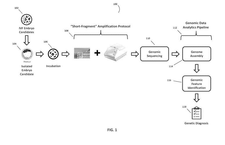
[0041] Figure 1 illustrates a workflow 100 for non-invasive preimplantation
genetic screening
of embryos, in accordance with some embodiments of the disclosure. As depicted
herein, an
9
CA 03074689 2020-03-03
WO 2019/051244 PCT/US2018/049976
embryo candidate 104 for IVF implantation can be isolated from a pool of
embryos and
incubated for a period of time in a sample holder containing media that is
substantially free of
DNA 106 or other polynucleotides that can interfere with the genetic screening
analysis. Some
examples of a sample holder may include, but are not limited to, a test tube,
pipette tube, petri
dish, or a well/partition within a multi-partition/well plate. In various
embodiments, the embryo
candidate 104 can also be incubated in a continuous culture system whereby
"fresh" culture
media 106 is introduced using a continuous media feed line to the sample
holder and "old"
culture media 106 is continuously removed (and sampled) from the sample holder
to maintain a
substantially constant volume of media in the sample holder.
[0042] During incubation, genomic fragments are regularly secreted by
and/or shed from the
embryo into the surrounding DNA-free media. An example of DNA free media that
can be
utilized in this workflow is ORIGIO SEQUENTIAL BLASTTm culture media of The
Cooper
Companies. In some embodiments, the embryo can be incubated in the culture
media for a
minimum of about 18 hrs. In other embodiments, the embryo can be incubated in
the culture
media between about 18 hours and about 144 hours. It should be understood that
the embryos
can be incubated in DNA free media for as long a period of time as is
necessary for a sufficient
quantity of genomic fragments to be secreted by and/or shed from the embryo to
allow for a
genetic screening analysis to be performed using the workflow 100. In some
embodiments, the
embryo is in the blastocyst stage of development when it is isolated and
incubated in the DNA
free media. In other embodiments, the embryo is in a multi-cell pre-blastocyst
stage of
development when it is isolated and incubated in the DNA free media.
[0043] After the embryo is incubated for a required period of time to allow
for a threshold
quantity of genomic fragments to be secreted or shed into the DNA free media,
a portion of the
incubation media is transferred to a separate amplification vessel where the
fragments undergo
an amplification protocol 108 that is tailored for amplifying short genomic
fragment for later
genomic sequence analysis. In some embodiments, the amplification protocol 108
uses a
multiple displacement amplification (MDA) based whole genome amplification
(WGA)
technique. MDA is a non-PCR based DNA amplification technique which has been
shown to be
efficient in the amplification of small amounts of DNA. MDA relies on priming
of target DNA
with random primers and the use of the strand-displacing (p29 polymerase (or
its equivalent) to
amplify substantially the entire DNA in a given sample. Compared with PCR-
based WGA
CA 03074689 2020-03-03
WO 2019/051244 PCT/US2018/049976
methods, MDA reduces amplification bias by orders of magnitude, generates
longer genomic
fragments and exhibits better genome coverage. In other embodiments, the
amplification
protocol 108 uses a multiple annealing and looping-based amplification cycles
(MALBAC)
based WGA technique. The MALBAC amplification technique uses special primers
that
allow amplicons to have complementary ends and therefore to loop, preventing
DNA from being
copied exponentially. This results in amplification of only the original
genomic DNA. This
controlled amplification consequently can reduce amplification bias and, by
extension, can lower
production of artifacts and lower incidences of false positive and false
negative mutation calls on
the isolated embryo candidate.
[0044] It should be understood, however, that any type of WGA technique can be
used in
amplification protocol 108 as long as the technique generates sufficient
quality and/or quantities
of genomic fragments to be sequenced for a genetic screening analysis to be
run using workflow
100.
[0045] After the genomic fragments (from the isolated embryo 104) have been
amplified to a
sufficient quantity, they are sequenced 110 using a NGS or equivalent genomic
sequencing
system. The sequencing workflow can begin with the fragments being sequenced
110 on a
nucleic acid sequencer to provide hundreds, thousands or millions of nucleic
acid sequence reads
(i.e., sequence reads). The genomic fragment sequence information can then be
processed using
a genomic data analytics pipeline 112 whereby the genomic fragment sequences
are aligned
(mapped) 114 against a reference genome and one or more secondary analytics
tools/pipelines
are used to help identify one or more genomic features 116 present in the
genome of the embryo
104. In some embodiments, the genomic features 116 can be genomic variants
such as
insertions/deletions (INDEL), copy number variations (CNV), single nucleotide
polymorphisms
(SNP), duplications, inversions, translocations, etc. In other embodiments,
the genomic features
116 can be genomic regions that have some annotated function such as a gene,
protein coding
sequence, mRNA, tRNA, rRNA, repeat sequence, inverted repeat, miRNA, siRNA,
etc. In still
other embodiments, the genomic features 116 can be epigenetic changes on the
genome (e.g.,
methylation, acetylation, ubiquitylation, phosphorylation, sumoylation,
ribosylation,
citrullination, etc.) that can affect gene expression and activity.
[0046] In some embodiments, the reference genome is a human genome. In other
embodiments,
the reference genome is a genome of the animal species that the embryo
originates from. It
11
CA 03074689 2020-03-03
WO 2019/051244 PCT/US2018/049976
should be appreciated, however, that the reference genome can be an
artificially created genome
that is not associated with any particular animal species, but rather created
for a particular
analysis/application.
[0047] After the genomic features 116 have been identified, the analytics
pipeline 112 can
generate a genetic diagnostics report 118 providing information regarding
inherited or non-
inherited genetic conditions that the isolate embryo 104 has or is at risk
for.
[0048] In various embodiments, a "blank" or control sample is run side by side
with the embyro
candidate 104 through the entire workflow 100. That is, a portion of DNA free
media (which
was not used to incubate an embryo 104) is run through all the steps and
processes of workflow
100. The results from analyzing the blank sample can serve as a control to
ensure that the
genomic features identified in the genome of the embryo is not an artifact of
the amplification
and/or systemic errors during sequencing.
[0049] Figure 2 is an exemplary flowchart depicting an amplification protocol
200 for
amplifying short genomic fragments, in accordance with some embodiments of the
disclosure.
[0050] As depicted herein, the genomic fragments 202 (in the portion of media
incubating the
embryo) are combined with enzymes 204 and genomic linker segments 206 in
conditions that
catalyze the formation of concatenated fragments 208. The ligation reaction is
carried out at
room temperature (without agitation) for about 16-18 hours (overnight
incubation). The ligation
reaction mixture consists of 1 unit of DNA ligase in a buffer containing 50mM
Tris HC1, 10m1V1
MgCl2, 1mM ATP and 10m1V1 DTT at a pH of about 7.5 and a temperature of
between about
20 C and about 25 C temperature. The resulting concatenated fragments 208 are
longer than
the original genomic fragments 202, which helps to reduce amplification errors
(when compared
to amplifying the genomic fragments 202 individually) when the genomic
fragments are
amplified later in the protocol 200.
[0051] Concatenation can provide long templates (i.e., concatenated fragments)
that are optimal
for amplification using the (p29 enzyme, which isothermally amplifies DNA by
multiple
displacement amplification. (p29 enzyme cannot efficiently and/or accurately
amplify short
fragments (i.e., amplicons shorter than about 30 base pairs), which has been
demonstrated in
validation experiments and hence it is pertinent that we create long
concatenated fragments to
capture the entirety of the short fragments of DNA extruded by the embryo into
the culture
media. Moreover, concatenation also helps in creating adequate templates for
successful
12
CA 03074689 2020-03-03
WO 2019/051244 PCT/US2018/049976
amplification by other whole genome amplification strategies such as Sureplex
system
(I1lumina), MALBAC and DOP PCR. This reduction in amplification errors is
particularly
significant for short genomic fragments. In general, reducing amplification
error results in better
accuracy in the identification of genomic features when the genomic fragments
are later
sequenced and analyzed. In some embodiments, the genomic fragment is a short
genomic
fragment that has a length of between about 30 base pairs (bps) and about 800
bps. In other
embodiments, the genomic fragment is a short genomic fragment that has a
length of between
bout 150 bps to about 400 bps. In still other embodiments, the genomic
fragment is a short
genomic fragment that has a length of less than about 1000 bps.
[0052] The genomic linker segments 206 are essentially artificially created
double-stranded
"conjoint" oligonucleotide segments of a known length and nucleotide sequence.
In some
embodiments, the genomic linker segments 206 are between about 30 to 1000 bps
in length. In
other embodiments, the genomic linker segments 206 are between about 30 bps
and about 500
bps in length. In still other embodiments, the genomic linker segments 206 are
between about 50
bps to about 150 bps. In some embodiments, the genomic linker segments 206 are
homopolymer
oligonucleotide segments. In other embodiments, the genomic linker segments
206 are
heteropolymer oligonucleotide segments. In some embodiments, the genomic
linker segments
206 are blunt ended double-stranded oligonucleotide segments. In some
embodiments, the
genomic fragments 202 are enzymatically blunt ended prior to being ligated to
the genomic
linker segments 206.
[0053] Various types of prokaryotic and eukaryotic enzymes (i.e., ligases) can
be used to ligate
the genomic fragments 202 to the genomic linker segments 206 to form the
concatenated
genomic fragments 208. Some examples of ligases that can be used here include,
but are not
limited to, T3, T4, T7, or Ligase 1.
[0054] After the concatenated fragments are formed in their container (e.g.,
well, pipette tube,
etc.) they can be amplified 210 on a thermal cycler (or similar device) using
WGA techniques
such as MDA, MALBAC, etc.
[0055] Figure 3 illustrates the formation of concatenated fragments, in
accordance with some
embodiments of the disclosure. As depicted herein, the genomic fragments 302
are first blunt
ended using a blunting enzyme to fill-in or remove the 3' or 5' overhangs
(i.e., unpaired
nucleotides) 306 prior to the introduction of the genomic linker segments 308
and their ligation
13
CA 03074689 2020-03-03
WO 2019/051244 PCT/US2018/049976
with a ligase 310 to form concatenated fragments 312. That is, the blunting
enzyme employed
can exhibit exonuclease activity to digest (remove) the overhangs or
polymerase activity to
synthesize (fill-in) the missing complementary bases on the overhang. Some
examples of
blunting enzymes that can be used include, but are not limited to, DNA
Polymerase I Klenow
fragment, T4 DNA Polymerase, and Mung Bean Nuclease. In an exemplary
embodiment, the
blunting reagent mixture used to blunt the dsDNA concatenated fragments
includes T4 DNA
polymerase (which has 3"¨>5' exonuclease activity and 5'¨>3' polymerase
activity) and T4
Polynucleotide Kinase (which aids in phosphorylation of 5' ends of blunt ended
DNA, necessary
for subsequent ligation reaction).
[0056] After blunting ending 306 the 5' and 3' ends of the genomic fragments
302, a DNA
ligase can be introduced to ligate the genomic fragments 302 to the genomic
linker segments
308. During ligation 310, the DNA ligase seals the 5' and 3' polynucleotide
ends via nucleotidyl
transfer steps involving ligase-adenylate and DNA-adenylate intermediates. DNA
ligases fall
into two general categories: ATP-dependent DNA ligases (EC 6.5.1.1), and NAD
(+) dependent
DNA ligases (EC 6.5.1.2). NAD (+) dependent DNA ligases are found only in
bacteria (and
some viruses) while ATP-dependent DNA ligases are ubiquitous.
[0057] The ATP-dependent DNA ligases can be divided into four classes: DNA
ligase I, II, III,
and IV. DNA ligase I links Okazaki fragments to form a continuous strand of
DNA; DNA ligase
II is an alternatively spliced form of DNA ligase III, found only in non-
dividing cells; DNA
ligase III is involved in base excision repair; and DNA ligase IV is involved
in the repair of DNA
double-strand breaks by non-homologous end joining (NHEJ). Amongst all
ligases, there are
two types of prokaryotic and one type of eukaryotic ligases that are
particularly well suited for
facilitating the blunt ended double stranded DNA ligation: Prokaryotic DNA
ligases (T3 and T4)
and Eukaryotic DNA ligase (Ligase 1).
[0058] In some embodiments, T4 DNA ligase is used in the blunt end ligation
process 310 for
this protocol. Bacteriophage T4 DNA ligase is a single polypeptide with a M.W
of about 68,000
Daltons requiring ATP as energy source. The maximal activity pH range is
between about 7.5 to
about 8Ø The presence of Mg++ ion is preferred and the optimal concentration
is about 10mM.
T4 DNA ligase has the unique ability to join sticky and blunt ended fragments.
T4 DNA ligase
catalyzes phosphodiester bond formation between juxtaposed 5'and 3' termini in
the genomic
fragments 302 and genomic linker segments 308 in three steps: 1) enzyme-
adenylylate formation
14
CA 03074689 2020-03-03
WO 2019/051244 PCT/US2018/049976
by reaction with ATP; 2) adenylyl transfer to a 5-phosphorylated
polynucleotide to generate
adenylylated DNA; and 3) phosphodiester bond formation with release of AMP. In
an
exemplary embodiment, the ligation reaction can be carried out using 1 unit of
T4 DNA ligase in
a buffer consisting of 50mM Tris HC1, 10mM MgCl2, 1mM ATP and 10mM DTT at a pH
of
about 7.5 and at a temperature of about 23 C. The reaction mixture containing
the T4 ligase,
blunt ended DNA and the linker segments can be incubated for 16-18 hours,
without agitation.
The concentration of the linker segment can range from about 1pg to about lng.
[0059] A concatenated fragment 312 forms once a genomic fragment 302 is
ligated to a genomic
linker segment 308. In some embodiments, the concatenated fragment 312
includes a least one
genomic fragment 302 that is ligated to at least one genomic linker segment
308. In other
embodiments, the concatenated fragment 312 includes two or more genomic
fragments 302 and
at least one genomic linker segment 308, whereby the at least one genomic
fragment 302 is
ligated to each end of the genomic linker segment 308. It should be
appreciated, however, that a
concatenated fragment 312 can have essentially any combination of genomic
fragments 312 and
genomic linker segments 308 as long as the combination is suitable for the
purposes of
sequencing and subsequent genomic feature analysis
[0060] After the formation of the concatenated fragments 312, they are
amplified using WGA
amplification technique 313 (such as PicoPlex, MDA, MALBAC, DOPlify etc.) and
subsequently sequenced using a NGS (or equivalent) genomic sequencing system
316.
Computer-Implemented System
[0061] Figure 4 is a block diagram that illustrates a computer system 400,
upon which
embodiments of the present teachings may be implemented. In various
embodiments of the
present teachings, computer system 400 can include a bus 402 or other
communication
mechanism for communicating information, and a processor 404 coupled with bus
402 for
processing information. In various embodiments, computer system 400 can also
include a
memory, which can be a random access memory (RAM) 406 or other dynamic storage
device,
coupled to bus 402 for determining instructions to be executed by processor
404. Memory also
can be used for storing temporary variables or other intermediate information
during execution
of instructions to be executed by processor 404. In various embodiments,
computer system 400
can further include a read only memory (ROM) 408 or other static storage
device coupled to bus
CA 03074689 2020-03-03
WO 2019/051244 PCT/US2018/049976
402 for storing static information and instructions for processor 404. A
storage device 410, such
as a magnetic disk or optical disk, can be provided and coupled to bus 402 for
storing
information and instructions.
[0062] In various embodiments, computer system 400 can be coupled via bus 402
to a display
412, such as a cathode ray tube (CRT) or liquid crystal display (LCD), for
displaying
information to a computer user. An input device 414, including alphanumeric
and other keys, can
be coupled to bus 402 for communicating information and command selections to
processor 404.
Another type of user input device is a cursor control 416, such as a mouse, a
trackball or cursor
direction keys for communicating direction information and command selections
to processor
404 and for controlling cursor movement on display 412. This input device 414
typically has two
degrees of freedom in two axes, a first axis (i.e., x) and a second axis
(i.e., y), that allows the
device to specify positions in a plane. However, it should be understood that
input devices 414
allowing for 3 dimensional (x, y and z) cursor movement are also contemplated
herein.
[0063] Consistent with certain implementations of the present teachings,
results can be provided
by computer system 400 in response to processor 404 executing one or more
sequences of one or
more instructions contained in memory 406. Such instructions can be read into
memory 406
from another computer-readable medium or computer-readable storage medium,
such as storage
device 410. Execution of the sequences of instructions contained in memory 406
can cause
processor 404 to perform the processes described herein. Alternatively hard-
wired circuitry can
be used in place of or in combination with software instructions to implement
the present
teachings. Thus implementations of the present teachings are not limited to
any specific
combination of hardware circuitry and software.
[0064] The term "computer-readable medium" (e.g., data store, data storage,
etc.) or "computer-
readable storage medium" as used herein refers to any media that participates
in providing
instructions to processor 404 for execution. Such a medium can take many
forms, including but
not limited to, non-volatile media, volatile media, and transmission media.
Examples of non-
volatile media can include, but are not limited to, optical, solid state,
magnetic disks, such as
storage device 410. Examples of volatile media can include, but are not
limited to, dynamic
memory, such as memory 406. Examples of transmission media can include, but
are not limited
to, coaxial cables, copper wire, and fiber optics, including the wires that
comprise bus 402.
16
CA 03074689 2020-03-03
WO 2019/051244 PCT/US2018/049976
[0065] Common forms of computer-readable media include, for example, a floppy
disk, a
flexible disk, hard disk, magnetic tape, or any other magnetic medium, a CD-
ROM, any other
optical medium, punch cards, paper tape, any other physical medium with
patterns of holes, a
RAM, PROM, and EPROM, a FLASH-EPROM, any other memory chip or cartridge, or
any
other tangible medium from which a computer can read.
[0066] In addition to computer readable medium, instructions or data can be
provided as signals
on transmission media included in a communications apparatus or system to
provide sequences
of one or more instructions to processor 404 of computer system 400 for
execution. For
example, a communication apparatus may include a transceiver having signals
indicative of
instructions and data. The instructions and data are configured to cause one
or more processors
to implement the functions outlined in the disclosure herein. Representative
examples of data
communications transmission connections can include, but are not limited to,
telephone modem
connections, wide area networks (WAN), local area networks (LAN), infrared
data connections,
NFC connections, etc.
[0067] It should be appreciated that the methodologies described herein flow
charts, diagrams
and accompanying disclosure can be implemented using computer system 400 as a
standalone
device or on a distributed network of shared computer processing resources
such as a cloud
computing network.
[0068] Figure 5 is a schematic diagram of a system for non-invasive
preimplantation genetic
screening of embryos 500, in accordance with various embodiments. As depicted
herein, the
system 500 includes a genomic sequencing system 502, a computing device 504
and a
display/client terminal 510.
[0069] In various embodiments, the computing device 504 can be communicatively
connected to
the genomic sequencing system 502 via a network connection that can be either
a "hardwired"
physical network connection (e.g., Internet, LAN, WAN, VPN, etc.) or a
wireless network
connection (e.g., Wi-Fi, WLAN, etc.). In various embodiments, the computing
device 504 can
be a workstation, mainframe computer, distributed computing node (part of a
"cloud computing"
or distributed networking system), personal computer, mobile device, etc.
In various
embodiments, the genomic sequencing system 504 can be a nucleic acid sequencer
(e.g., NGS,
Capillary Electrophoresis system, etc.), real-time/digital/quantitative PCR
instrument, microarray
scanner, etc. It should be understood, however, that the genomic sequencing
system 504 can
17
CA 03074689 2020-03-03
WO 2019/051244 PCT/US2018/049976
essentially be any type of instrument that can generate nucleic acid sequence
data from samples
containing genomic fragments.
[0070] It will be appreciated by one skilled in the art that various
embodiments of genomic
sequencing system 502 can be used to practice variety of sequencing methods
including ligation-
based methods, sequencing by synthesis, single molecule methods, nanopore
sequencing, and
other sequencing techniques. Ligation sequencing can include single ligation
techniques, or
change ligation techniques where multiple ligation are performed in sequence
on a single
primary nucleic acid sequence strand. Sequencing by synthesis can include the
incorporation of
dye labeled nucleotides, chain termination, ion/proton sequencing,
pyrophosphate sequencing, or
the like. Single molecule techniques can include continuous sequencing, where
the identity of the
nuclear type is determined during incorporation without the need to pause or
delay the
sequencing reaction, or staggered sequence, where the sequencing reactions is
paused to
determine the identity of the incorporated nucleotide.
[0071] In various embodiments, the genomic sequencing system 502 can determine
the sequence
of a nucleic acid, such as a polynucleotide or an oligonucleotide. The nucleic
acid can include
DNA or RNA, and can be single stranded, such as ssDNA and RNA, or double
stranded, such as
dsDNA or a RNA/cDNA pair. In various embodiments, the nucleic acid can include
or be
derived from a fragment library, a mate pair library, a chromatin immuno-
precipitation (ChIP)
fragment, or the like. In particular embodiments, the genomic sequencing
instrument 502 can
obtain the sequence information from a single nucleic acid molecule or from a
group of
substantially identical nucleic acid molecules.
[0072] In various embodiments, the genomic sequencing system 502 can output
nucleic acid
sequencing read data (genomic sequence information) in a variety of different
output data file
types/formats, including, but not limited to: *.fasta, *.csfasta, *.xsq,
*seq.txt, *qseq.txt, *.fastq,
*.sff, *prb.txt, *.sms, *srs and/or *.qv.
[0073] The analytics computing device 504 can be configured to host a sequence
read alignment
engine 506 and a genomic features identification engine 508. The read
alignment engine 506 can
be configure to receive genomic fragment sequence information generated by the
genomic
sequence system 502 and align (map) the genomic fragment sequences to a
reference genome.
Examples of publically available sequence alignment software that can be used
to align the
fragment sequences include BLAT, BLAST, Bowtie, BWA, drFAST LAST, MOSAIK,
18
CA 03074689 2020-03-03
WO 2019/051244 PCT/US2018/049976
NEXTGENMAP, etc. Once the fragment sequences have been aligned, the genomic
features
identification engine 508 can be configured to identify genomic features on
the aligned
sequences. That is, the genomic features identification engine 508 can be
communicatively
connected (e.g., a network connection to the analytics computing device 504, a
serial bus
connection to database storage that is local to the analytics computing device
504, a peripheral
device connection to a peripheral storage device connected to the analytics
computing device
504, etc.) to various public (e.g., the RefGene Database (UCSC), the
Alternative Splicing
Database (EBI), the dbSNP database (NCBI), the Genomic Structural Variation
database
(NCBI), the GENCODE database (UCSC), the PolyPhen database (Harvard), the SIFT
database
(NCBI), the 3000 Genomes Project database, the Database of Genomic Variants
database (EBI),
the Biomart database (EBI), Gene Ontology database (public), the
BioCyc/HumanCyc database,
the KEGG pathway database, the Reactome database, the Pathway Interaction
Database (NTH),
the Biocarta database, PANTHER database, etc.) and private databases to
identify the genomic
features in the aligned sequences.
[0074] In some embodiments, the genomic features can be genomic variants such
as
insertions/deletions (INDEL), copy number variations (CNV), single nucleotide
polymorphisms
(SNP), duplications, inversions, translocations, etc. In other embodiments,
the genomic features
can be genomic regions that have some annotated function such as a gene,
protein coding
sequence, mRNA, tRNA, rRNA, repeat sequence, inverted repeat, miRNA, siRNA,
etc. In still
other embodiments, the genomic features can be epigenetic changes on the
genome (e.g.,
methylation, acety lati on, ubiquitylation, phosphorylation, sumoy lati on,
ribosy lati on,
citrullination, etc.) that can affect gene expression and activity.
[0075] It should be appreciated that the functionalities of the read alignment
engine 506 and
genomic features identification engine 508 can be implemented as hardware,
firmware, software,
or any combination thereof. Furthermore, the various engines depicted in
Figure 5 can be
combined or collapsed into a single engine, component or module, depending on
the
requirements of the particular application or system architecture. Moreover,
in various
embodiments, the read alignment engine 506 and genomic features identification
engine 508 can
comprise additional engines or components as needed by the particular
application or system
architecture.
19
CA 03074689 2020-03-03
WO 2019/051244 PCT/US2018/049976
[0076] After the genomic features have been identified, the results can be
displayed on a display
or client terminal 510 that is communicatively connected to the computing
device 504. In
various embodiments, client terminal 510 can be a thin client computing
device. In various
embodiments, client terminal 510 can be a personal computing device having a
web browser
(e.g., IN ________________________________________________________________
lERNET EXPLORERTM, FIREFOXTM, SAFARITM, etc) that can be used to control the
operation of the sequence alignment engine 506 and/or genomic features
identification engine
508. That is, the client terminal 510 can access the sequence alignment engine
506 using a
browser to control the operation of the sequence alignment engine 506. For
example, the
sequence alignment criteria or logic can be modified depending on the
requirements of the
particular application.
Similarly, client terminal 510 can access the genomic features
identification engine 508 using a browser to control the database sources
(e.g., the RefGene
Database (UCSC), the Alternative Splicing Database (EBI), the dbSNP database
(NCBI), the
Genomic Structural Variation database (NCBI), the GENCODE database (UCSC), the
PolyPhen
database (Harvard), the SIFT database (NCBI), the 3000 Genomes Project
database, the
Database of Genomic Variants database (EBI), the Biomart database (EBI), Gene
Ontology
database (public), the BioCyc/HumanCyc database, the KEGG pathway database,
the Reactome
database, the Pathway Interaction Database (NTH), the Biocarta database,
PANTHER database,
etc.) used to identify the genomic features in the aligned sequences or the
modify the summary
reports generated.
[0077] Figure 6 is a depiction of how concatenated fragment reads are mapped
to a reference
genome, in accordance with various embodiments. As discussed previously,
concatenated
fragments are comprised of both genomic fragments that the candidate embryo
has secreted or
shed (in the media that it was incubated in) and artificially created double-
stranded "conjoint"
oligonucleotide segments (i.e., genomic linker segments) of a known length and
nucleotide
(base) sequence. Therefore, as depicted herein Figure 6, the concatenated
fragment reads 602
are comprised of sequence reads of both the artificially synthesized genomic
linker segments 604
and the genomic fragments 606 obtained from the embryo test media.
[0078] The concatenated fragment reads 602 are aligned (mapped) 608 to a
reference genome
610 using any number of publically available sequence alignment tools
including, but not limited
to: BLAT, BLAST, BWA, Bowtie, drFAST LAST, MOSAIK, NEXTGENMAP, etc. In some
embodiments, the parameters of the sequence alignment tool are modified to
accommodate short
CA 03074689 2020-03-03
WO 2019/051244 PCT/US2018/049976
fragment sequence read alignments. In some embodiments, the short genomic
fragment reads
have a length of between about 30 base pairs (bps) and about 800 bps. In other
embodiments,
the short genomic fragment reads have a length of between bout 150 bps to
about 400 bps. In
still other embodiments, the short genomic fragment reads have a length of
less than about 1000
bps.
[0079] In some embodiments, the genomic linker segments sequence reads are
between about 30
to 1000 bps in length. In other embodiments, the genomic linker segment
sequence reads are
between about 30 bps and about 500 bps in length. In still other embodiments,
the genomic
linker segment sequence reads are between about 50 bps to about 150 bps. In
some
embodiments, the genomic linker segment sequence reads are homopolymer
sequences. In other
embodiments, the genomic linker segment sequence reads are heteropolymer
oligonucleotide
sequences.
[0080] In some embodiments, since the genomic linker segment sequence reads
are not naturally
occurring they are algorithmically filtered out during the alignment of the
concatenated fragment
reads to the reference genome. That is, the alignment tool subtracts out the
known sequences
associated with the genomic linker segments and only aligns the sequences
associated with the
genomic fragments portion of the concatenated fragment reads to the reference
genome.
[0081] In some embodiments, the alignment tool selects the best alignment for
each genomic
fragment sequence read by determining the longest matching alignment position
on the reference
genome for each genomic fragment sequence read. That is, the alignment
location where the
longest consecutive sequence of bases on the genomic fragment sequence read
matches to the
reference genome. In other embodiments, the alignment tool selects the best
alignment for each
genomic fragment sequence read by determining the position on the reference
genome where the
most number of bases from the genomic fragment sequence reads match,
regardless of whether
they are consecutive or not.
[0082] In some embodiments, genomic fragment sequence reads that align equally
well to
multiple locations on the reference genome are automatically discarded and not
used in the
identification of genomic features (e.g., SNPs, CNVs, Indels, etc.).
[0083] Figure 7 is an exemplary flowchart showing a method for aligning
concatenated genomic
fragment sequence reads to identify various types of genomic features, in
accordance with
various embodiments. As depicted herein, the concatenated genomic fragment
sequence reads
21
CA 03074689 2020-03-03
WO 2019/051244 PCT/US2018/049976
702 are first aligned to a reference genome 704. The alignments are made using
any number of
publically available sequence alignment tools including, but not limited to:
BLAT, BLAST,
BWA, Bowtie, drFAST LAST, MOSAIK, NEXTGENMAP, etc. As discussed above, the
concatenated genomic fragment reads are sequence reads of both the
artificially synthesized
genomic linker segments and the genomic fragments obtained from the test
sample (e.g., tissue,
embryo, etc.).
[0084] In some embodiments, since the genomic linker segments are not
naturally occurring (in
the human genome) they are algorithmically filtered out during the alignment
of the
concatenated fragment reads to the reference genome. That is, the alignment
tool subtracts out
the known sequences associated with the genomic linker segments and only
aligns the sequences
associated with the genomic fragments portion of the concatenated fragment
reads to the
reference genome.
[0085] The alignment tool selects the best alignment for each genomic fragment
sequence read
based on a set of parameters or factors 706, including, but not limited to,
alignment score and
whether there are multiple alignments for the genomic fragment reads. In some
embodiments,
the alignment score for a genomic fragment read alignment can be calculated
(using Equation 1)
as a function of a match criteria (e.g., a number of consecutive bases of the
genomic fragment
sequence read that matches to the reference genome, the absolute number of
bases from the
genomic fragment sequence read that matches to the reference genome, the
percent sequence
identity between the sequence and its match in the genome, etc.), a mismatch
criteria and gap
penalties. Within the construct of Equation 1, mismatches and gaps in
alignment are penalized
from the overall alignment score.
Equation 1: Alignment Score = f(match criteria) -f(mismatch criteria) - f(Gap
Penalties)
[0086] In some embodiments, genomic fragment sequence reads that align equally
well (e.g.,
have the same alignment score, etc.) to multiple locations on the reference
genome are
automatically discarded and not used in the identification of genomic
features.
[0087] After the genomic fragment sequence reads 702 are aligned to the
reference genome,
various analytics tools or callers can be used to identify genomic features on
the aligned
sequences 708. In various embodiments, these tools or callers can be
configured to access
22
CA 03074689 2020-03-03
WO 2019/051244 PCT/US2018/049976
various public (e.g., the RefGene Database (UCSC), the Alternative Splicing
Database (EBI), the
dbSNP database (NCBI), the Genomic Structural Variation database (NCBI), the
GENCODE
database (UCSC), the PolyPhen database (Harvard), the SIFT database (NCBI),
the 3000
Genomes Project database, the Database of Genomic Variants database (EBI), the
Biomart
database (EBI), Gene Ontology database (public), the BioCyc/HumanCyc database,
the KEGG
pathway database, the Reactome database, the Pathway Interaction Database
(NTH), the Biocarta
database, PANTHER database, etc.) and/or private databases to identify the
genomic features.
[0088] In some embodiments, the genomic features can be genomic variants such
as
insertions/deletions (INDEL), copy number variations (CNV), single nucleotide
polymorphisms
(SNP), duplications, inversions, translocations, etc. In other embodiments,
the genomic features
can be genomic regions that have some annotated function such as a gene,
protein coding
sequence, mRNA, tRNA, rRNA, repeat sequence, inverted repeat, miRNA, siRNA,
etc. In still
other embodiments, the genomic features can be epigenetic changes on the
genome (e.g.,
methylation, acetylation, ubiquitylation, phosphorylation, sumoylation,
ribosylation,
citrullination, etc.) that can affect gene expression and activity.
[0089] In various embodiments, SNPs can be called via local de-novo assembly
of haplotypes
710. In various embodiments, aneuploiday can be called using an aneuploidy
caller 714. In
various embodiments, Copy Number Variants (CNVs) can be identified using a
modified CNV
caller 712. The modified CNV caller can be configured to differentiate between
biological and
technical variation by normalization to a normal sample. Technical variations
can occur due to
bias in technology, for example, some regions in the genome can have more or
less reads when
sequenced due to high GC content bias (i.e., the proportion of G and C bases
in a region and the
count of fragments mapped to it), amplification bias, linker ligation etc. so
they are not real CNV
deletions or duplications; but instead, are merely experimental artifacts. On
the other hand,
biological variations are due to actual CNV deletions/duplications in the
genome. For example,
when the genome region (i.e., chromosomal position) of the sample (e.g.,
tissue, embryo, etc.)
being tested has a CNV deletion it will have less reads in that region and
when the genome has a
CNV duplication it means that it has more reads in that region. In various
embodiments, in order
to remove bias from technical variations and be able to differentiate between
"real" biological
variations from "fake" technical variations a circular binary segmentation
(CBS) based algorithm
23
CA 03074689 2020-03-03
WO 2019/051244 PCT/US2018/049976
is applied and spline normalization is performed using an Interpolated
Univariate smoothing
model.
[0090] That is, normalizations are performed to compare regions of one sample
to all other
samples that have been previously tested. The logic being if there are
technical variations they
will affect all the samples within a sample test batch (i.e., the samples that
are run through the
amplification and sequencing workflow steps together) and not just one sample
within a batch of
samples. So if a sample shows a drop in the quantity of reads in a region
which is also seen in
other samples of the same sample batch then it is safe to conclude that it was
a technical
variation. However, if the drop is only seen in one sample in a sample batch
and in no other
sample in the same sample batch then it is highly likely to be a biological
variation. This
comparison can be done only when all samples are normalized to the same scale.
To do this,
gene regions of interest are typically split into many small intervals of
approximately 100 bps
and the average depths (i.e., quantity of aligned reads) of the samples are
calculated for each
region. Even if individual interval shows variation, the Spline normalization
performed smooths
over the region, so that it removes smaller errors so that only significant
variations in each region
will be detectable. CNVs can then be identified by measuring significance
using techniques such
as Principal Component Analysis (PCA).
[0091] In various embodiments, the CBS algorithm is configured to identify the
start and end
positions for CNVs in a sample. That is, the CBS algorithm performs multiple
passes through a
sample whereby on the first pass the algorithm searches the entire sample,
compiling a list of
(start, end) position tuples in which statistically significant changes in
read depth appear to have
occurred. Among these tuples, the tuple containing the most dramatic change is
identified as a
CNV, and then the algorithm is reapplied recursively to the two pieces of the
sample on either
side of this tuple. The algorithm terminates when no statistically significant
changes in read
depth occur in any of the portions of the sample currently under evaluation.
[0092] Put another way, for every small interval, the CBS algorithm compares
the intervals
before and after it and if they both show the same drop/increase it moves to
the next interval. At
the boundary of the variation, one side will have the signal while the other
won't, which helps
define the boundaries.
[0093] In various embodiments, during the Spline normalization of the genome
regions (i.e.,
chromosomal positions) in the genome of a sample being tested for CNV, a
quantiling function is
24
CA 03074689 2020-03-03
WO 2019/051244 PCT/US2018/049976
used to partition by depth the reads for a particular sample to ascertain what
constitutes a low,
average and deep read depth for each genome region. The same procedure is then
repeated for
the median read depth at each genome region in the genome across all samples
in the batch.
[0094] The breakpoints which partition these read depths by low, average,
deep, etc. for a
particular sample are plotted on the x-axis, and the breakpoints which
partition the read depths
for the median across samples is plotted on the y-axis. These (x, y) values
are then interpolated
with a curve.
[0095] Next, for a particular sample, the read depth for a particular region
in said sample is
evaluated against the curve, by looking at the height on the curve
corresponding to its region on
the x-axis. By doing this, samples which have, for example, a large percentage
of low coverage
regions when compared to the median across samples will be modified in such a
way that the
upper portion of their low coverage regions will be re-interpreted as being of
average coverage.
Next, if a sample shows a drop in reads in a region which is also seen in
other samples then it can
be classified as a technical variation, however if the drop is only seen in
one sample and in no
other sample in the batch then it can be classified as a biological variation.
This is accounted for
by dividing a sample's read depth at a particular region by the median read
depth at that same
region across all samples in a batch.
[0096] Figure 8 is a flowchart showing a method for determining copy number
variation in an
embryo candidate, in accordance with various embodiments. As depicted herein,
method 800
details an exemplary workflow for identifying copy number variations in an
embryo candidate.
In step 802, an embryo candidate is isolated from a plurality of fertilized
embryos and placed
into a container. For example, the embryo candidate can be isolated from a
plurality of fertilized
embryos each of which can be a candidate for IVF implantation. In some
embodiments, the
embryo candidate is in the blastocyst stage of embryongenesis. In some
embodiments, the
embryo candidate is a human embryo.
[0097] Typically, isolation step 802 is performed using conventional sterile
techniques or in a
sterile hood to ensure that the isolated embryo candidate is not contaminated
with genomic
matter that may lead to erroneous test results.
[0098] In step 804, the embryo candidate is incubated in media that is
substantially free of DNA.
Typically, the embryo is incubated for as long of a period of time as is
required (while still
keeping the embryo candidate viable for IVF implantation) for a sufficient
quantity of DNA
CA 03074689 2020-03-03
WO 2019/051244 PCT/US2018/049976
fragments (i.e., genomic fragments) to be secreted or shed from the embryo
candidate to the
DNA free media for a copy number variation analysis to be performed using
method 800. In
some embodiments, the embryo can be incubated in the culture media for a
minimum of about
18 hrs. In other embodiments, the embryo can be incubated in the culture media
for between
about 18 hours and about 144 hours. An example of DNA free media that can be
utilized in this
workflow is ORIGIO SEQUENTIAL BLASTTm culture media of The Cooper Companies.
In
various embodiments, the media can be substantially free of oligonucletides
and not just DNA to
ensure the lowest possible chance of erroneous analysis results or artifact
formation during
amplification.
[0099] In step 806, a portion of the media is transferred to an amplification
vessel, wherein the
portion of media includes one or more genomic fragments (i.e., DNA fragment)
shed or secreted
from the embryo candidate. Examples of an amplification vessel that can be
used include, but
are not limited to, a test tube, pipette tube, petri dish, or a well/partition
within a multi-
partition/well plate.
[00100] In step 808, a plurality of linker segments and ligase enzyme is added
to the amplification
vessel in conditions that catalyze the formation of concatenated genomic
fragments containing at
least one genomic linker segment and at least one genomic fragment (from the
embryo
candidate). Typically, the genomic fragments obtained from the media are
considered "short"
genomic fragments. In some embodiments, the short genomic fragments have
lengths of
between about 30 base pairs (bps) and about 800 bps. In other embodiments, the
short genomic
fragments have a length of between about 150 bps to about 400 bps. In still
other embodiments,
the short genomic fragments have a length of less than about 1000 bps.
[00101] The genomic linker segments are essentially artificially created
double-stranded
"conjoint" oligonucleotide segments of a known length and nucleotide sequence.
In some
embodiments, the genomic linker segments are between about 30 to 1000 bps in
length. In other
embodiments, the genomic linker segments are between about 30 bps and about
500 bps in
length. In still other embodiments, the genomic linker segments are between
about 50 bps to
about 150 bps. In some embodiments, the genomic linker segments are
homopolymer
oligonucleotide segments. In other embodiments, the genomic linker segments
are
heteropolymer oligonucleotide segments. In some embodiments, the genomic
linker segments
are blunt ended double-stranded oligonucleotide segments. In some embodiments,
the genomic
26
CA 03074689 2020-03-03
WO 2019/051244 PCT/US2018/049976
fragments are enzymatically blunt ended prior to being ligated to the genomic
linker segments
using methods that were previously disclosed above.
[00102] Various types of prokaryotic and eukaryotic enzymes (i.e., ligases)
can be used to ligate
the genomic fragments to the genomic linker segments to form the concatenated
genomic
fragments. Some examples of ligases that can be used here include, but are not
limited to, T3,
T4, T7, or Ligase 1.
[00103] In step 810, the concatenated genomic fragments are amplified in the
amplification
vessel. In various embodiments, the concatenated genomic fragments are
amplified on a thermal
cycler (or similar device) using WGA techniques such as MDA, MALBAC, etc.
[00104] Because the concatenated fragments are significantly longer than the
original genomic
fragments isolated from the incubation media, amplification errors are
significantly reduced
(when compared to amplifying the genomic fragments individually).
[00105] In step 812, sequence information from the amplified concatenated
genomic fragments is
obtained from sequencing the concatenated fragments on a NGS or equivalent
genomic
sequencing system. In some embodiments, the sequence information includes both
genomic
fragment sequence reads (obtained from genomic fragments isolated from the
embryo candidate)
and genomic linker segment sequence reads (which were artificially created and
ligated to the
genomic fragments prior to amplification in step 810).
[00106] In step 814, the sequence information is aligned against a reference
genome using a
publically available or proprietary sequence alignment tool. Examples of
publically available
sequence alignment tools that can be used to align the fragment sequences
include, but are not
limited to, BLAT, BLAST, BWA, Bowtie, drFAST LAST, MOSAIK, NEXTGENMAP, etc. In
some embodiments, since the genomic linker segments are not naturally
occurring their
corresponding sequence reads are algorithmically filtered out during the
alignment of the
sequence information to the reference genome. That is, the alignment tool
subtracts out the
known sequences associated with the genomic linker segments and only aligns
the sequences
associated with the genomic fragments portion of the concatenated fragment
reads to the
reference genome.
[00107] In some embodiments, the alignment tool selects the best alignment for
each genomic
fragment sequence read by determining the longest matching alignment position
on the reference
genome for each genomic fragment sequence read. That is, the alignment
location where the
27
CA 03074689 2020-03-03
WO 2019/051244 PCT/US2018/049976
longest consecutive sequence of bases on the genomic fragment sequence read
matches to the
reference genome. In other embodiments, the alignment tool selects the best
alignment for each
genomic fragment sequence read by determining the position on the reference
genome where the
most number of bases from the genomic fragment sequence reads match,
regardless of whether
they are consecutive or not. In some embodiments, genomic fragment sequence
reads that align
equally well to multiple locations on the reference genome are automatically
discarded and not
used.
[00108] In step 816, copy number variations in the embryo candidate's genome
are identified
when a frequency of genomic fragment sequence reads aligned to a chromosomal
position on the
reference genome deviates from a frequency threshold. In various embodiments,
a deviance
occurs when the frequency of genomic fragment sequences aligned to a
chromosomal position is
below the frequency threshold (i.e., fragment alignment frequency in a normal
genome). That is,
when the chromosomal position of the sample (e.g., tissue, embryo, etc.) being
tested has a CNV
deletion it will have less reads (i.e. frequency of reads aligned) in that
region than in a normal
genome. In various embodiments, a deviance occurs when the frequency of
genomic fragment
sequences aligned to a chromosomal position is above the frequency threshold.
That is, when
the chromosomal position has CNV duplication it means that it has more reads
in that region
than in a normal genome.
[00109] Figure 9 is a flowchart showing a method of identifying genomic
features in an embryo
candidate, in accordance with various embodiments. As depicted herein, method
900 details an
exemplary workflow for identifying genomic features in an embryo candidate. In
step 902, an
embryo candidate is isolated from a plurality of embryo candidates. For
example, the embryo
candidate can be isolated from a plurality of fertilized embryos each of which
can be a candidate
for IVF implantation. In some embodiments, the embryo candidate is in the
blastocyst stage of
embryongenesis. In some embodiments, the embryo candidate is a human embryo.
[00110] In step 904, the embryo candidate is incubated in media that is
substantially free of DNA.
Typically, the embryo is incubated for as long of a period of time as is
required (while still
keeping the embryo candidate viable for IVF implantation) for a sufficient
quantity of DNA
fragments (i.e., genomic fragments) to be secreted or shed from the embryo
candidate to the
DNA free media for a copy number variation analysis to be performed using
method 900. An
example of DNA free media that can be utilized in this workflow is ORIGIO
SEQUENTIAL
28
CA 03074689 2020-03-03
WO 2019/051244 PCT/US2018/049976
BLASTTm culture media of The Cooper Companies. In various embodiments, the
media can be
substantially free of oligonucleotides and not just DNA to ensure the lowest
possible chance of
erroneous analysis results or artifact formation during amplification.
[00111] In step 906, a portion of the media is transferred to an amplification
vessel, wherein the
portion of media includes one or more genomic fragments (i.e., DNA fragment)
shed or secreted
from the embryo candidate. Examples of an amplification vessel that can be
used include, but
are not limited to, a test tube, pipette tube, petri dish, or a well/partition
within a multi-
partition/well plate.
[00112] In step 908, a plurality of linker segments and ligase enzyme is added
to the amplification
vessel in conditions that catalyze the formation of concatenated genomic
fragments containing at
least one genomic linker segment and at least one genomic fragment from the
embryo candidate.
Typically, the genomic fragments isolated from the media are considered
"short" genomic
fragments. In some embodiments, the short genomic fragments have lengths of
between about
30 base pairs (bps) and about 800 bps. In other embodiments, the short genomic
fragments have
lengths of between bout 150 bps to about 400 bps. In still other embodiments,
the short genomic
fragments have lengths of less than about 1000 bps.
[00113] The genomic linker segments are essentially artificially created
double-stranded
"conjoint" oligonucleotide segments of a known length and nucleotide sequence.
In some
embodiments, the genomic linker segments are between about 30 to about 1000
bps in length. In
other embodiments, the genomic linker segments are between about 30 bps and
about 500 bps in
length. In still other embodiments, the genomic linker segments are between
about 50 bps to
about 150 bps. In some embodiments, the genomic linker segments are
homopolymer
oligonucleotide segments. In other embodiments, the genomic linker segments
are
heteropolymer oligonucleotide segments. In some embodiments, the genomic
linker segments
are blunt ended double-stranded oligonucleotide segments. In some embodiments,
the genomic
fragments are enzymatically blunt ended prior to being ligated to the genomic
linker segments
using methods that were previously disclosed above.
[00114] Various types of prokaryotic and eukaryotic enzymes (i.e., ligases)
can be used to ligate
the genomic fragments to the genomic linker segments to form the concatenated
genomic
fragments. Some examples of ligases that can be used here include, but are not
limited to, T3,
T4, T7, or Ligase 1.
29
CA 03074689 2020-03-03
WO 2019/051244 PCT/US2018/049976
[00115] In step 910, the concatenated genomic fragments are amplified in the
amplification
vessel. In various embodiments, the concatenated genomic fragments are
amplified on a thermal
cycler (or similar device) using WGA techniques such as MDA, MALBAC, etc.
[00116] In step 912, sequence information from the amplified concatenated
genomic features are
obtained from sequencing the concatenated fragments on a NGS or equivalent
genomic
sequencing system. In some embodiments, the sequence information includes both
genomic
fragment sequence reads (obtained from genomic fragments isolated from the
embryo candidate)
and genomic linker segment sequence reads (which were artificially created and
ligated to the
genomic fragments prior to amplification in step 910).
[00117] In step 914, the sequence information is aligned against a reference
genome using a
publically available or proprietary sequence alignment tool. Examples of
publically available
sequence alignment tools that can be used to align the fragment sequences
include, but are not
limited to, BLAT, BLAST, BWA, Bowtie, drFAST LAST, MOSAIK, NEXTGENMAP, etc. In
some embodiments, since the genomic linker segments are not naturally
occurring their
corresponding sequence reads are algorithmically filtered out during the
alignment of the
sequence information to the reference genome. That is, the alignment tool
subtracts out the
known sequences associated with the genomic linker segments and only aligns
the sequences
associated with the genomic fragments portion of the concatenated fragment
reads to the
reference genome.
[00118] In some embodiments, the alignment tool selects the best alignment for
each genomic
fragment sequence read by determining the longest matching alignment position
on the reference
genome for each genomic fragment sequence read. That is, the alignment
location where the
longest consecutive sequence of bases on the genomic fragment sequence read
matches to the
reference genome. In other embodiments, the alignment tool selects the best
alignment for each
genomic fragment sequence read by determining the position on the reference
genome where the
most number of bases from the genomic fragment sequence reads match,
regardless of whether
they are consecutive or not. In some embodiments, genomic fragment sequence
reads that align
equally well to multiple locations on the reference genome are automatically
discarded and not
used.
[00119] In step 916, genomic features are identified on the aligned genomic
fragment sequences
using a various publically available or proprietary genomic features analytics
tools or callers. In
CA 03074689 2020-03-03
WO 2019/051244 PCT/US2018/049976
various embodiments, these tools or callers can be configured to access
various public (e.g., the
RefGene Database (UCSC), the Alternative Splicing Database (EBI), the dbSNP
database
(NCBI), the Genomic Structural Variation database (NCBI), the GENCODE database
(UCSC),
the PolyPhen database (Harvard), the SIFT database (NCBI), the 3000 Genomes
Project
database, the Database of Genomic Variants database (EBI), the Biomart
database (EBI), Gene
Ontology database (public), the BioCyc/HumanCyc database, the KEGG pathway
database, the
Reactome database, the Pathway Interaction Database (NTH), the Biocarta
database, PANTHER
database, etc.) and/or private databases to identify the genomic features.
[00120] In some embodiments, the genomic features can be genomic variants such
as
insertions/deletions (INDEL), copy number variations (CNV), single nucleotide
polymorphisms
(SNP), duplications, inversions, translocations, etc. In other embodiments,
the genomic features
can be genomic regions that have some annotated function such as a gene,
protein coding
sequence, mRNA, tRNA, rRNA, repeat sequence, inverted repeat, miRNA, siRNA,
etc. In still
other embodiments, the genomic features can be epigenetic changes on the
genome (e.g.,
methylation, acetylation, ubiquitylation, phosphorylation, sumoylation,
ribosylation,
citrullination, etc.) that can affect gene expression and activity.
[00121] Figure 10 is a flowchart showing a method for identifying genomic
features from
concatenated genomic fragment sequence reads, in accordance with various
embodiments. As
depicted herein, method 1000 details an exemplary workflow for identifying
genomic features on
genomic fragment sequence reads that were obtained from concatenated fragments
(created by
ligating artificial genomic linker segments to genomic fragments that were
extracted from a
tissue sample) that were amplified and later sequenced on a NGS or equivalent
genomic
sequencing system. In step 1002, concatenated genomic fragment reads
containing at least one
genomic linker segment sequence and at least one genomic fragment sequence
from a tissue
sample is received on a computing device/server programmed with instructions
(software or
hardware) to analyze genomic sequence information (sequence reads) generated
by a genomic
sequencing system configured to determine the base sequence information of
genomic
fragments.
[00122] The genomic linker segments are artificially created so their length
and base sequence isn
known. In some embodiments, the genomic linker segment reads are between about
30 to about
1000 bps in length. In other embodiments, the genomic linker segment reads are
between about
31
CA 03074689 2020-03-03
WO 2019/051244 PCT/US2018/049976
30 bps and about 500 bps in length. In still other embodiments, the genomic
linker segment
reads are between about 50 bps to about 150 bps. In some embodiments, the
genomic linker
segment reads are homopolymer sequences. In other embodiments, the genomic
linker segment
reads are heteropolymer sequences.
[00123] In step 1004, the genomic linker segment sequence portion of the
concatenated genomic
fragment sequence reads is subtracted out prior to the concatenated genomic
fragment sequence
reads being aligned to a reference genome in step 1006. That is, the known
sequences associated
with the genomic linker segments is subtracted out from the concatenated
genomic fragment
sequence reads first and then only the genomic fragments portion of the
concatenated fragment
reads are aligned to the reference genome.
[00124] In step 1008, genomic features are identified on the aligned genomic
fragment sequences
using various publically available or proprietary genomic features analytics
tools or callers. In
various embodiments, these tools or callers can be configured to access
various public (e.g., the
RefGene Database (UCSC), the Alternative Splicing Database (EBI), the dbSNP
database
(NCBI), the Genomic Structural Variation database (NCBI), the GENCODE database
(UCSC),
the PolyPhen database (Harvard), the SIFT database (NCBI), the 3000 Genomes
Project
database, the Database of Genomic Variants database (EBI), the Biomart
database (EBI), Gene
Ontology database (public), the BioCyc/HumanCyc database, the KEGG pathway
database, the
Reactome database, the Pathway Interaction Database (NTH), the Biocarta
database, PANTHER
database, etc.) and/or private databases to identify the genomic features.
[00125] In some embodiments, the genomic features can be genomic variants such
as
insertions/deletions (INDEL), copy number variations (CNV), single nucleotide
polymorphisms
(SNP), duplications, inversions, translocations, etc. In other embodiments,
the genomic features
can be genomic regions that have some annotated function such as a gene,
protein coding
sequence, mRNA, tRNA, rRNA, repeat sequence, inverted repeat, miRNA, siRNA,
etc. In still
other embodiments, the genomic features can be epigenetic changes on the
genome (e.g.,
methylation, acetylation, ubiquitylation, phosphorylation, sumoylation,
ribosylation,
citrullination, etc.) that can affect gene expression and activity.
EXPERIMENTAL RESULTS
32
CA 03074689 2020-03-03
WO 2019/051244 PCT/US2018/049976
[00126] The following illustrative example is a representative embodiment of
the software
applications, systems, and methods described herein and are not meant to be
limiting in any way.
[00127] As shown in the Table 1, 26 embryos were analyzed for chromosomal
abnormalities (i.e.,
CNV) using both a conventional embryo trophectoderm biopsy method and the
novel non-
invasive sampling of embryo culture media methods disclosed above. The samples
were either
analyzed immediately or stored in temperature conditions ranging from -20 C to
-80 C.
TABLE 1
Aneuploidy Euploidy Individual Chromosome
100 % (8/8) 89% (16/18) 99% (615/624)
[00128] The DNA of each traditional embryo biopsy was analyzed using ILLUMINA'
s
VERISEQTm PGS workflow and analysis. IVF culture media for each respective
embryo was
subjected to noninvasive analysis via a novel amplification method, sequenced
on an
ILLUMINA NGS sequencer and their chromosome copy numbers were calculated using
a
custom bioinformatics pipeline. The results clearly show high concordance in
the aneupoloidy
(chromosomal abnormality) and euploidy (normal genetic makeup) calls between
the industry
accepted trophectoderm biopsy method and the non-invasive embryo culture media
methods
disclosed above. Moreover, there was a high concordance rate for each of the
24 chromosomes
that were compared across each respective sample.
[00129] The methodologies described herein may be implemented by various means
depending
upon the application. For example, these methodologies may be implemented in
hardware,
firmware, software, or any combination thereof. For a hardware implementation,
the processing
unit may be implemented within one or more application specific integrated
circuits (ASICs),
digital signal processors (DSPs), digital signal processing devices (DSPDs),
programmable logic
devices (PLDs), field programmable gate arrays (FPGAs), processors,
controllers, micro-
controllers, microprocessors, electronic devices, other electronic units
designed to perform the
functions described herein, or a combination thereof.
[00130] In various embodiments, the methods of the present teachings may be
implemented as
firmware and/or a software program and applications written in conventional
programming
33
CA 03074689 2020-03-03
WO 2019/051244 PCT/US2018/049976
languages such as C, C++, Python, etc. If implemented as firmware and/or
software, the
embodiments described herein can be implemented on a non-transitory computer-
readable
medium in which a program is stored for causing a computer to perform the
methods described
above. It should be understood that the various engines described herein can
be provided on a
computer system, such as computer system 400 of Figure 4, whereby processor
404 would
execute the analyses and determinations provided by these engines, subject to
instructions
provided by any one of, or a combination of, memory components 406/4008/410
and user input
provided via input device 414.
[00131] While the present teachings are described in conjunction with various
embodiments, it is
not intended that the present teachings be limited to such embodiments. On the
contrary, the
present teachings encompass various alternatives, modifications, and
equivalents, as will be
appreciated by those of skill in the art.
[00132] Further, in describing various embodiments, the specification may have
presented a
method and/or process as a particular sequence of steps. However, to the
extent that the method
or process does not rely on the particular order of steps set forth herein,
the method or process
should not be limited to the particular sequence of steps described. As one of
ordinary skill in the
art would appreciate, other sequences of steps may be possible. Therefore, the
particular order of
the steps set forth in the specification should not be construed as
limitations on the claims. In
addition, the claims directed to the method and/or process should not be
limited to the
performance of their steps in the order written, and one skilled in the art
can readily appreciate
that the sequences may be varied and still remain within the spirit and scope
of the various
embodiments.
[00133] The embodiments described herein, can be practiced with other computer
system
configurations including hand-held devices, microprocessor systems,
microprocessor-based or
programmable consumer electronics, minicomputers, mainframe computers and the
like. The
embodiments can also be practiced in distributing computing environments where
tasks are
performed by remote processing devices that are linked through a network.
[00134] It should also be understood that the embodiments described herein can
employ various
computer-implemented operations involving data stored in computer systems.
These operations
are those requiring physical manipulation of physical quantities. Usually,
though not necessarily,
these quantities take the form of electrical or magnetic signals capable of
being stored,
34
CA 03074689 2020-03-03
WO 2019/051244 PCT/US2018/049976
transferred, combined, compared, and otherwise manipulated. Further, the
manipulations
performed are often referred to in terms, such as producing, identifying,
determining, or
comparing.
[00135] Any of the operations that form part of the embodiments described
herein are useful
machine operations. The embodiments, described herein, also relate to a device
or an apparatus
for performing these operations. The systems and methods described herein can
be specially
constructed for the required purposes or it may be a general purpose computer
selectively
activated or configured by a computer program stored in the computer. In
particular, various
general purpose machines may be used with computer programs written in
accordance with the
teachings herein, or it may be more convenient to construct a more specialized
apparatus to
perform the required operations.
[00136] Certain embodiments can also be embodied as computer readable code on
a computer
readable medium. The computer readable medium is any data storage device that
can store data,
which can thereafter be read by a computer system. Examples of the computer
readable medium
include hard drives, network attached storage (NAS), read-only memory, random-
access
memory, CD-ROMs, CD-Rs, CD-RWs, magnetic tapes, and other optical, FLASH
memory and
non-optical data storage devices. The computer readable medium can also be
distributed over a
network coupled computer systems so that the computer readable code is stored
and executed in
a distributed fashion.
CA 03074689 2020-03-03
WO 2019/051244 PCT/US2018/049976
RECITATION OF SELECTED EMBODIMENTS
[00137] Embodiment 1. A method is provided for determining copy number
variation in an
embryo candidate for in vitro fertilization (IVF) implantation is disclosed.
An embryo candidate
is isolated from a plurality of embryos. The embryo candidate is incubated in
media that is
substantially free of DNA. A portion of the media is transferred to an
amplification vessel,
wherein the portion of media includes genomic fragments shed or secreted from
the embryo
candidate. A plurality of genomic linker segments and ligase enzyme is added
to the
amplification vessel in conditions that catalyze the formation of concatenated
genomic fragments
containing at least one genomic linker segment and at least one genomic
fragment from the
isolated embryo candidate. The concatenated genomic fragments are amplified in
the
amplification vessel. Sequence information is obtained from the amplified
concatenated
genomic fragments. The sequence information is aligned (mapped) against a
reference genome.
Copy number variations are identified in the embryo candidate when a frequency
of genomic
fragment sequence reads aligned to a chromosomal position on the reference
genome deviates
from a frequency threshold.
[00138] Embodiment 2. The method of Embodiment 1, further including:
subtracting sequence
information related to the genomic linker segment from the concatenated
genomic fragment
sequence prior to aligning the concatenated genomic fragment sequence to the
reference genome.
[00139] Embodiment 3. The method of Embodiment 2, further including:
normalizing the
frequency of genomic fragment sequence reads aligned to each chromosomal
position; and
determining a frequency threshold for each chromosomal position.
[00140] Embodiment 4. The method of Embodiment 3, further including: applying
a circular
binary segmentation (CBS) analysis to determine whether the identified
deviance from the
frequency threshold identified is due to technical bias.
[00141] Embodiment 5. The method of Embodiment 3, wherein the normalization is
performed
using a Spline normalization method.
[00142] Embodiment 6. The method of Embodiment 1, further including: blunting
the genomic
fragment ends using a modified polymerase prior to ligating them to the
genomic linker
segments.
36
CA 03074689 2020-03-03
WO 2019/051244 PCT/US2018/049976
[00143] Embodiment 7. The method of Embodiment 6, wherein the modified
polymerase is a
Klenow T4 DNA polymerase.
[00144] Embodiment 8. The method of Embodiment 1, wherein the ligase enzyme is
one of a T3,
T4 or T7 prokaryotic DNA ligase.
[00145] Embodiment 9. The method of Embodiment 1, wherein the embryo candidate
is a human
embryo.
[00146] Embodiment 10. The method of Embodiment 1, wherein the embryo
candidate is a
blastocyst.
[00147] Embodiment 11. The method of Embodiment 1, wherein the frequency
threshold is a
frequency of genomic fragment reads that map to a normal chromosome.
[00148] Embodiment 12. A method is provided for identifying genomic features
in an embryo
candidate is disclosed. An embryo candidate is isolated from a plurality of
embryo candidates.
The embryo candidate is incubated in media that is substantially free of DNA.
A portion of the
media is transferred to an amplification vessel, wherein the portion of media
includes one more
genomic fragments shed or secreted from the embryo candidate. A plurality of
genomic linker
segments and a ligase enzyme is added to the amplification vessel in
conditions that catalyze the
formation of concatenated genomic fragments containing at least one genomic
linker segment
and at least one genomic fragment from the isolated embryo candidate. The
concatenated
genomic fragments are amplified in the amplification vessel. Sequence
information is obtained
from the concatenated genomic fragments. The sequence information is aligned
against a
reference genome. Genomic features are identified on the aligned genomic
fragment sequences.
[00149] Embodiment 13. The method of Embodiment 12, further including:
subtracting sequence
information related to the genomic linker segment from the concatenated
genomic fragment
sequence prior to aligning the concatenated genomic fragment sequence to the
reference genome.
[00150] Embodiment 14. The method of Embodiment 12, further including:
blunting the genomic
fragment ends using a modified polymerase prior to ligating them to the
genomic linker
segments.
[00151] Embodiment 15. The method of Embodiment 14, wherein the modified
polymerase is a
Klenow T4 DNA polymerase.
[00152] Embodiment 16. The method of Embodiment 12, wherein the ligase enzyme
is one of a
T3, T4 or T7 prokaryotic DNA ligase.
37
CA 03074689 2020-03-03
WO 2019/051244 PCT/US2018/049976
[00153] Embodiment 17. The method of Embodiment 12, wherein the embryo
candidate is a
human embryo.
[00154] Embodiment 18. The method of Embodiment 12, wherein the embryo
candidate is a
blastocyst.
[00155] Embodiment 19. The method of Embodiment 12, wherein the genomic
feature is a single
nulceotide polymorphism.
[00156] Embodiment 20. The method of Embodiment 12, wherein the genomic
feature is an
indel.
[00157] Embodiment 21. The method of Embodiment 12, wherein the genomic
feature is an
inversion.
[00158] Embodiment 22. A system is provided for identifying genomic features
in an embryo
candidate. The system includes a genomics sequencer, a computing device and a
display.
[00159] The genomic sequencer is configured to obtain sequence information
from concatenated
genomic fragments derived from an embryo candidate. The concatenated genomic
fragments
each contain at least one genomic linker segment and at least one genomic
fragment from the
embryo candidate.
[00160] The computing device is communicatively connected to the genomic
sequencer and
includes a sequence alignment engine and a genomic features identification
engine. The
sequence alignment engine is configured to subtract out sequence information
related to the
genomic linker segment portion of the concatenated genomic fragments and align
the genomic
fragment sequences to a reference genome. The genomic features identification
engine is
configured to identify genomic features in the aligned genomic fragment
sequences. The display
is communicatively connected to the computing device and configured to display
a report
containing the identified genomic features.
[00161] Embodiment 23. The system of Embodiment 22, wherein the genomic
feature is a copy
number variation.
[00162] Embodiment 24. The system of Embodiment 23, wherein the genomic
features
identification engine is further configured to: normalize a frequency of
genomic fragment
sequences aligned to each chromosomal position on the reference genome;
determine a genomic
fragment sequence alignment frequency threshold to make a copy number
variation call for each
chromosomal position; and make a copy number variation call for each
chromosomal positon
38
CA 03074689 2020-03-03
WO 2019/051244 PCT/US2018/049976
with genomic fragment sequence alignment frequencies that deviate from the
frequency
threshold.
[00163] Embodiment 25. The system of Embodiment 24, wherein the genomic
features
identification engine is further configured to apply a circular binary
segmentation (CBS) analysis
to determine whether the identified deviance from the frequency threshold
identified is due to
technical bias.
[00164] Embodiment 26. The system of Embodiment 24, wherein the normalization
is performed
using a Spline normalization method.
[00165] Embodiment 27. The system of Embodiment 24, wherein a deviance occurs
when the
frequency of genomic fragment sequences aligned to a chromosomal position is
below the
frequency threshold.
[00166] Embodiment 28. The system of Embodiment 24, wherein a deviance occurs
when the
frequency of genomic fragment sequences aligned to a chromosomal position is
above the
frequency threshold.
[00167] Embodiment 29. The system of Embodiment 22, wherein the embryo
candidate is a
human embryo.
[00168] Embodiment 30. The system of Embodiment 22, wherein the embryo
candidate is a
blastocyst.
[00169] Embodiment 31. The system of Embodiment 22, wherein the genomic
feature is a single
nulceotide polymorphism.
[00170] Embodiment 32. The system of Embodiment 22, wherein the genomic
feature is an
indel.
[00171] Embodiment 33. The system of Embodiment 22, wherein the genomic
feature is an
inversion.
[00172] Embodiment 34. The system of Embodiment 22, wherein the genomic linker
segment
sequence is a known sequence.
[00173] Embodiment 35. A method is provided for identifying genomic features
in a tissue
sample is disclosed. Concatenated genomic fragment sequence reads are received
containing at
least one genomic linker segment sequence and at least one genomic fragment
sequence from a
tissue sample. The genomic linker segment sequence portion of the concatenated
genomic
fragment sequence reads is subtracted out. The concatenated genomic fragment
sequence reads
39
CA 03074689 2020-03-03
WO 2019/051244 PCT/US2018/049976
are aligned (mapped) to a reference genome. Genomic features are identified on
the aligned
genomic fragment sequences.
[00174] Embodiment 36. The method of Embodiment 35, further including:
deleting
concatenated genomic fragment sequence reads that map to more than one
location on a
reference genome.
[00175] Embodiment 37. The method of Embodiment 35, wherein the genomic
feature is a copy
number variation.
[00176] Embodiment 38. The method of Embodiment 37, further including:
normalizing a
frequency of genomic fragment sequences aligned to each chromosomal position;
determining a
genomic fragment sequence alignment frequency threshold to make a copy number
variation call
for each chromosomal position; and making a copy number variation call for
each chromosomal
positon with genomic fragment sequence alignment frequencies that deviate from
the frequency
threshold.
[00177] Embodiment 39. The method of Embodiment 38, further including:
applying a circular
binary segmentation (CBS) analysis to determine whether the identified
deviance from the
frequency threshold is identified due to technical bias.
[00178] Embodiment 40. The method of Embodiment 38, wherein a deviance occurs
when the
frequency of genomic fragment sequences aligned to a chromosomal position is
below the
frequency threshold.
[00179] Embodiment 41. The method of Embodiment 38, wherein a deviance occurs
when the
frequency of genomic fragment sequences aligned to a chromosomal position is
above the
frequency threshold.
[00180] Embodiment 42. The method of Embodiment 35, wherein the tissue sample
is an
embryonic tissue.
[00181] Embodiment 43. The method of claim 35, wherein the tissue sample is a
blastocyst.
[00182] Embodiment 44. The method of claim 35, wherein the genomic feature is
a single
nulceotide polymorphism.
[00183] Embodiment 45. The method of claim 35, wherein the genomic feature is
an indel.
[00184] Embodiment 46. The method of claim 35, wherein the genomic feature is
an inversion.
[00185] Embodiment 47. A non-transitory computer-readable medium is provided
in which a
program is stored for causing a computer to perform a method for identifying
genomic features
CA 03074689 2020-03-03
WO 2019/051244 PCT/US2018/049976
in a tissue sample. Concatenated genomic fragment sequence reads are received
containing at
least one genomic linker segment sequence and at least one genomic fragment
sequence from a
tissue sample. The genomic linker segment sequence portion of the concatenated
genomic
fragment sequence reads are subtracted out. The concatenated genomic fragment
sequence reads
are aligned (mapped) to a reference genome. Genomic features are identified on
the aligned
genomic fragment sequences.
[00186] Embodiment 48. The method of Embodiment 47, further including:
deleting
concatenated genomic fragment sequence reads that map to more than one
location on a
reference genome.
[00187] Embodiment 49. The method of Embodiment 47, wherein the genomic
feature is a copy
number variation.
[00188] Embodiment 50. The method of Embodiment 47, wherein the genomic
feature is an
indel.
[00189] Embodiment 51. The method of Embodiment 47, wherein the genomic
feature is an
inversion.
[00190] Embodiment 52. The method of Embodiment 49, further including:
normalizing a
frequency of genomic fragment sequences aligned to each chromosomal position;
determining a
genomic fragment sequence alignment frequency threshold to make a copy number
variation call
for each chromosomal position; and making a copy number variation call for
each chromosomal
positon with genomic fragment sequence alignment frequencies that deviate from
the frequency
threshold.
[00191] Embodiment 53. The method of Embodiment 52, further including:
applying a circular
binary segmentation (CBS) analysis to determine whether the identified
deviance from the
frequency threshold is identified due to technical bias.
[00192] Embodiment 54. The method of Embodiment 52, wherein a deviance occurs
when the
frequency of genomic fragment sequences aligned to a chromosomal position is
below the
frequency threshold.
[00193] Embodiment 55. The method of Embodiment 52, wherein a deviance occurs
when the
frequency of genomic fragment sequences aligned to a chromosomal position is
above the
frequency threshold.
41
CA 03074689 2020-03-03
WO 2019/051244 PCT/US2018/049976
[00194] Embodiment 56. The method of Embodiment 47, wherein the tissue sample
is an
embryonic tissue.
[00195] Embodiment 57. The method of Embodiment 47, wherein the tissue sample
is a
blastocyst.
[00196] Embodiment 58. The method of Embodiment 47, wherein the genomic
feature is a single
nulceotide polymorphism.
[00197] Embodiment 59. The method of Embodiment 47, wherein the genomic
feature is an
indel.
[00198] Embodiment 60. The method of Embodiment 47, wherein the genomic
feature is an
inversion.
42