Note: Descriptions are shown in the official language in which they were submitted.
CA 03074901 2020-03-05
WO 2019/048689 PCT/EP2018/074382
1
Tumor organoid model
The invention relates to the field of artificial tissue mod-
els grown in vitro.
Background of the invention
Malignant brain tumors are among the most devastating can-
cers with almost negligible survival rates that have not im-
proved in decades. Development of suitable brain cancer models
and effective therapies is challenging due to their enormous ge-
netic (McLendon et al., 2008, Nature, 455, 1061-8) and morpho-
logical (Louis et al., 2016, Acta Neuropathol, 131, 803-20) het-
erogeneity. In addition, the obvious morphological and physio-
logical differences between human and rodent brains limit the
development of appropriate animal models (Lui et al., 2011,
Cell, 146, 18-36). Human brain cancer cell lines as well as can-
cer stem cells cultured in 2D (Hu et al., 2016, Cell, 167, 1281-
1295.e18) have served as surrogate models but do not recapitu-
late the 3D tumor environment.
The recent development of organoid culture models has opened
new avenues for modelling disease directly in human tissues. Re-
capitulating either organ regeneration from adult stem cells
(ASCs) (Sato et al., 2009, Nature, 459, 262-5) or organ develop-
ment from pluripotent stem cells (PSCs) (Kelava and Lancaster,
2016, Cell Stem Cell, 18, 736-48), organoids resemble organ his-
tology and physiology in a strikingly accurate manner (Lancaster
and Knoblich, 2014, Science, 345, 1247125).
US 2014/302491 Al relates to a culture system for long term
cultures of mammalian tissues.
Xiaolei et al., Cell Stem Cell 18 (1) (2016): 25-38 is a re-
view on to stem-cell based organoids.
WO 00/75286 A2 describes in vitro 3D models of various can-
cer tissues, which can be used for screening applications.
Ridder et al., International Journal of Cancer Research and
Treatment 17 (6B) (1997), relates to brain tumor spheroids that
are attached to human dermal spheroids in order to test
tumor invasiveness.
Nygaard et al., Journal of Neurosurgery 89 (3) (1998): 2843-
2857, describes spheroids of glioblastoma that are cocultured
with rat brain aggregates.
CA 03074901 2020-03-05
WO 2019/048689 PCT/EP2018/074382
2
Zhu et al., The Journal of Experimental Medicine 214 (10)
(2017): 2843-2857, describes an oncolytic effect of Zika virus
against glioblastoma.
Wang et al., PLOS ONE 9 (4) (2014): 1, describes studying 3D
organoids as disease development models.
Organoids have been used to model various human diseases
(Johnson and Hockemeyer, 2015, Curr Opin Cell Biol, 37, 84-90),
including cancer (Neal and Kuo, 2016, Annu Rev Pathol). For ASC-
derived neoplastic organoids, this can be achieved by using ge-
netically modified ASCs (Barker et al., 2009, Nature, 457, 608-
11; Drost et al., 2015, Nature, 521, 43-7; Matano et al., 2015,
Nature Medicine, 21, 256-62) or primary tumors (Boj et al.,
2015, Cell, 160, 324-38) as a starting material. For PSC-derived
organoids, however, this approach is difficult as the growth re-
quirements of these organoids are often not compatible with
adult tumor cells or will impose selective pressure on them.
Therefore, there remains a goal to produce improved cancer
model cultures, especially 3D cultures of cancer and in particu-
lar to model additional characteristics of cancer in vitro cul-
tures that closely resemble in vivo cancer.
Summary of the invention
In particular, the invention has the goal of recapitulation
of life-like circumstances during cancer development. This goal
is solved by introducing tumorigenesis together with development
of normal, non-cancerous tissues in organoids.
The invention provides a method of generating an artificial
3D tissue culture of a cancer grown in non-cancerous tissue,
comprising the steps of providing an aggregate of pluripotent
stem or progenitor cells, culturing and expanding said stem or
progenitor cells in a 3D biocompatible matrix, wherein the cells
are allowed to differentiate to develop the aggregate into a
tissue culture of a desired size; wherein at least a portion of
said cells are subjected to carcinogenesis by expressing a onco-
gene and/or by suppressing a tumor suppressor gene during any of
said steps or in the tissue culture, and further comprising the
step of allowing said cells with an expressed oncogene or sup-
pressed tumor suppressor to develop into cancerous cells.
The inventive method is also useful in testing unknown genes
instead of one or more (known) oncogenes or tumor suppressors.
CA 03074901 2020-03-05
WO 2019/048689 PCT/EP2018/074382
3
The culture may also be used to test candidate agents for its
carcinogenesis potential. Accordingly, the invention also pro-
vides a method of screening one or more candidate genes or
agents for their effects on carcinogenesis, comprising generat-
ing an artificial 3D tissue culture, comprising the steps of
providing an aggregate of pluripotent stem or progenitor cells,
culturing and expanding said stem or progenitor cells in a 3D
biocompatible matrix, wherein the cells are allowed to differen-
tiate to develop the aggregate into a tissue culture of a de-
sired size; wherein at least a portion of said cells are sub-
jected to carcinogenesis by expressing or suppressing the candi-
date gene or by treating the cells with the candidate agent dur-
ing any of said steps or in the tissue culture, and further com-
prising the step of culturing said cells in conditions that al-
low an expressed or supressed candidate gene to develop into
cancerous cells.
The invention further provides an artificial 3D tissue cul-
ture, for example an organoid, comprising non-cancerous tissue
and cancerous tissue. In the artificial 3D tissue culture, pref-
erably the cancerous tissue overexpresses one or more oncogenes
and/or has suppressed (e.g. expression or activity) of one or
more tumor suppressors, wherein preferably gene expression of
other genes than said oncogene or tumor suppressor is substan-
tially unmodified in the cancerous tissue as compared to the
non-cancerous tissue, wherein said tissue (i) is obtainable by a
method according to the invention; and/or (ii) comprising a
transgene or a construct for suppression of a tumor suppressor
at least in cells of the cancerous tissue; and/or (iii) compris-
ing a 3D biocompatible matrix, preferably gel, a collagenous
gel, or a hydrogel.
Further provided is a method of testing a candidate compound
for carcinogenesis or for its effect on cancer tissue, compris-
ing contacting cells or a tissue in a method of the invention
with the candidate compound or contacting a tissue of the inven-
tion with the candidate compound and maintaining said contacted
tissue in culture, and observing any changes in the tissue as
compared to said tissue without contacting by said candidate
compound. Likewise, the invention provides exposing the tissue
or the cells in the inventive method to a condition instead of
contacting it with a candidate compound. Such a condition may be
CA 03074901 2020-03-05
WO 2019/048689 PCT/EP2018/074382
4
e.g. elevated temperature, limited or increased nutrients or al-
tered redox potential, to which cancer cells may react and ex-
hibit a different behaviour or growth rate.
Further provided is a method of testing a candidate oncolytic
virus for carcinogenesis or for its effect on cancer tissue,
comprising contacting cells or a tissue in a method of the in-
vention with the candidate oncolytic virus or contacting a tis-
sue of the invention with the candidate oncolytic virus and
maintaining said contacted tissue in culture, and observing any
changes in the tissue as compared to said tissue without con-
tacting by said candidate oncolytic virus. Likewise, the inven-
tion provides exposing the tissue or the cells in the inventive
method to a condition instead of contacting it with a candidate
oncolytic virus. Such a condition may be e.g. elevated tempera-
ture, limited or increased nutrients or altered redox potential,
to which cancer cells may react and exhibit a different behav-
iour or growth rate.
In a further aspect, the invention provides Zika virus for
use as a medicament, in particular as an oncolytic virus. In
particular provided is Zika virus for use in the treatment of
nervous system cancer. Related thereto is a method of treating a
nervous system cancer in a patient comprising treating a patient
having nervous system cancer with Zika virus to remove said can-
cer. Further provided is the use of Zika virus in the manufac-
ture of a medicament for the treatment of neuronal cancer. Nerv-
ous system cancer or neuronal cancer may e.g. be glioblastoma or
neuroblastoma or CNS-PNET (central nervous system primitive neu-
ro-ectodermal tumor).
Further provided is a pharmaceutical composition comprising
a replication competent Zika virus and a stabilizer for said vi-
rus.
Also provided is a kit suitable for providing a tissue cul-
ture according to the invention. The kit may comprise (i) a
transfection vector comprising an oncogene transgene or a con-
struct for disruption of a tumor suppressor, (ii) a 3D biocom-
patible matrix, preferably further comprising (iii) a tissue
differentiation agent, a stem cell culturing medium, a nu-
cleofection medium or a combination thereof.
All embodiments of the invention are described together in
the following detailed description and all preferred embodiments
CA 03074901 2020-03-05
WO 2019/048689 PCT/EP2018/074382
relate to all embodiments, aspects, methods, tissues and kits
alike. E.g. kits or their components can be used in or be suita-
ble for inventive methods. Any component used in the described
methods can be in the kit. Inventive tissues are the results of
inventive methods or can be used in inventive methods. Preferred
and detailed descriptions of the inventive methods read alike on
suitability of resulting tissues of the inventions. All embodi-
ments can be combined with each other, except where otherwise
stated.
Detailed description of the invention
The present invention relates to a method of generating an
artificial 3D (three-dimensional) tissue culture of a cancer
grown in non-cancerous tissue. Such a 3D tissue culture can be
created in vitro and shows all distinguishing characteristics of
in vitro cell cultures, such as due to lack of neighbouring ob-
stacles (such as other organs or bones found in vivo) a substan-
tially uniforms shape and/or no directional orientation - except
if such have been artificially introduced, as e.g. using direc-
tional growth substrates such as disclosed in WO 2017/121754 Al.
The produced 3D culture is preferably in all embodiments of the
invention an organold. An organoid is a collection of organ-
specific cell types that develops from stem cells or organ pro-
genitors and self-organizes through cell sorting and spatially
restricted lineage commitment in a manner similar to in vivo
(Lancaster and Knoblich, Science 345(6194), 2014: 1247125).
The invention provides 3D tissue cultures that comprise can-
cerous tissue and thus serve as in vitro models for cancer and
cancer development. This allows any research uses such as drug
screening or testing reactions of the tissue culture and/or the
cancer or non-cancer parts within to environmental influences,
such as nutritional or temperature changes or exposure to other
agents or compounds. Accordingly, the invention also relates to
a method of screening one or more candidate genes or agents for
their effects on carcinogenesis or cancer therapy. Said candi-
date genes or agents may be any such drug or influence or genet-
ic modification, like suppression of one or more suspected tumor
suppressors and/or enhancement of expression of one or more sus-
pected oncogenes.
A hallmark of the invention is that the cancerous tissue is
CA 03074901 2020-03-05
WO 2019/048689
PCT/EP2018/074382
6
grown on or starts growing from non-cancerous tissue. This al-
lows the recapitulation of more in vivo-like effects, like in-
filtration or invasion and monitoring of early cancerous chang-
es. Accordingly, non-cancerous or pre-cancerous cells are cul-
tured and then carcinogenesis is initiated at a stage of devel-
opment of the culture of the practitioners choosing, preferably
at a stage when progenitor cells have already developed. A pro-
genitor cell is a biological cell that, like a stem cell, has a
tendency to differentiate into a specific type of cell, but is
already more specific than a stem cell and is pushed to differ-
entiate into its target cell type. Progenitor cells are of the
tissue type the organoid is destined to develop into, for exam-
ple a neuronal progenitor cell. Carcinogenesis is also referred
to herein as oncogenesis or tumorigenesis.
As summarized in the background section, previous cultures,
including organoid were cultured from cancerous cells, e.g.
cells from a patient that a particular type of cancer, like gli-
oblastoma (Huber et al., Cancer Res 2016 76(8): 2465-2477). Such
organoids fail to develop the intricate organ structure of nor-
mal healthy tissue, including different areas of development or
differentiation, as the tumorous mass is the only product of
such organoids. Contrary thereto, the invention allows the de-
velopment of normal tissues with various areas of development or
layers of development as can be recapitulated by such 3D tissue
cultures or organoids. Upon carcinogenesis initiation, different
behaviours or differently differentiated cells can be observed.
In particular, like in in vivo situation, not all cell lineages
present in the 3D tissue cultures may give rise to a particular
type of cancer. This different behaviour of different cell types
can be observed by the invention. Accordingly, the invention fa-
cilitates tracking and identification of a particular cell line-
age that can give rise to a particular cancer in view of the
carcinogenesis. According to the invention, carcinogenesis is a
modification of the non-cancerous cells of the culture, i.e. in-
troduction of cancerous mutations in the cells of the tissue,
not an Infiltration by cancerous cells, such as investigated in
a metastatic organoid model disclosed in WO 2017/05173 Al. Ac-
cording to the invention, also non-metastatic cancer can be in-
vestigated. Accordingly, in a preferment, the cancerous cells of
the inventive tissue are non-metastatic, i.e. have no metastasis
CA 03074901 2020-03-05
WO 2019/048689
PCT/EP2018/074382
7
potential. In other embodiments, the cancerous cells are meta-
static. Such metastasis behaviours can e.g. be determined by
monitoring cells leaving the 3D tissue culture and being able to
form a new tumor or cancer in another organoid. In preferred em-
bodiments, the cancer in non-metalizing or is metastasizing with
the proviso that no metastasis of external or internal origin is
formed in the 3D tissue culture. "External origin metastasis"
means a metastasis from a cell that has not been developed in
the 2D tissue culture or aggregate. "Internal origin metastasis"
means a metastasis from a cell that has developed in the 2D tis-
sue culture or aggregate; this may be allowed or not.
The inventive method is based on known methods to generate
3D tissue cultures or organoids, such as disclosed in Lancaster,
M. A. et al. Nature 501, 373-379 (2013); Lancaster et al., Na-
ture Protocols 9 (10) (2014): 2329-2340; WO 2014/090993 Al; WO
2017/121754 Al; WO 2017 123791 Al; WO 2017 117547 Al; WO
2015/135893 Al (neuronal and neural organoids); WO 2017/142069
Al (gastric organoids); WO 2017 115982 Al (cartilage organoid);
WO 2017/059171 Al; WO 2016/183143 Al; WO 2015/184273 Al (cardiac
organoid); WO 2017/049243 Al; WO 2015/130919 Al (kidney organ-
oid); WO 2016/141137 Al (vascular organoid); WO 2016/174604 Al
(breast/ductal-lobular organoid); WO 2015/196012 Al (prostate
organoid); WO 2016/061464 Al (intestinal organoid); WO
2015/183920 Al (gastric organoid); WO 2014/127170 Al; WO
2012/014076 A2 (liver organoid) (all incorporated herein by ref-
erence). Any such organoid generation methods or the resulting
organoids can be used according to the invention.
Usually, the inventive method comprises the step of provid-
ing an aggregate of pluripotent stem or progenitor cells. Such
an aggregate may be developed from a single stem cell, such as
an induced pluripotent stem (iPS) cell or an isolated embryonic
pluripotent stem cell. For example, the stem cell or progenitor
may be a cell isolated from an early stage of an embryo. Such a
method does not require the destruction of the embryo. The cell
may be developed to an aggregate, also referred to "embryoid
body" in the field, as disclosed in the references cited in the
above paragraph, especially WO 2014/090993 Al. The aggregate is
used as a starting point for the inventive method but of course
the invention can also be defined by performing these steps to
arrive at the aggregate stage. The following applies to perform-
CA 03074901 2020-03-05
WO 2019/048689 PCT/EP2018/074382
8
ing said method but also to the resulting aggregate.
The use of iPS cells allows applications in personalized di-
agnostics and medicine. A cell from a patient, in particular a
tumor patient, can be transformed into an iPS cell and used in
the inventive methods. Said cell may be a normal, healthy cell
from said patient. The inventive carcinogenesis can be used to
recapitulate the tumor of the same patient, thereby allowing an
investigation of tumorigenesis based on such a tumor cell and
its carcinogenic modifications.
For example, a tumor cell from a patient can be examined for
aberrant modifications that may lead to tumor development. Such
modifications may be mutations or changes in DNA methylation or
gene expression. The inventive carcinogenesis can reflect said
modification, e.g. by mutating a gene that is modified, aber-
rantly methylated or expressed in said tumor cell. Assuming that
the gene expression is the final effectual cause of tumor devel-
opment, any such modification can be used to modify a cell ac-
cording to the carcinogenesis step in the inventive method, as
long as gene expression is altered, in particular as reminiscent
of the tumor cell from the patient.
If a particular modification has been confirmed by the in-
ventive method as cause of the disease, then said gene may be
specifically targeted by individualized medicine, such as by up-
or downregulating activity of said gene or its gene product into
the direction of the normal activity status. Up- or downregula-
tion can be achieved by conventional means as known in the art,
such as by administering a drug or transgene or inhibitory com-
pounds, like inhibitory nucleic acids.
The inventive aggregate can be obtained from culturing plu-
ripotent stem or progenitor cells or a single cell. In preferred
embodiments, the aggregate or the cells of the 3D tissue culture
are of the same genetic lineage, such as when derived from the
same single cell. In principle, the cells may also be totipo-
tent, if ethical reasons allow. A "totipotent" cell can differ-
entiate into any cell type in the body, including the germ line
following exposure to stimuli like that normally occurring in
development. Accordingly, a totipotent cell may be defined as a
cell being capable of growing, i.e. developing, into an entire
organism. The cells used in the methods according to the present
invention are preferably not totipotent, but (strictly) pluripo-
CA 03074901 2020-03-05
WO 2019/048689 PCT/EP2018/074382
9
tent.
In a particular preferred embodiment, the cells of the pre-
sent invention (including all further embodiments related there-
to), are human cells or non-human cells, e.g. primate cells. The
inventive cells are usually eukaryotic cells. Further non-human
animals as origin of the cells are mouse, cat, dog, hamster, ro-
dent, rat, cow, horse, sheep, etc.
A "pluripotent" stem cell is not able of growing into an en-
tire organism, but is capable of giving rise to cell types orig-
inating from all three germ layers, i.e., mesoderm, endoderm,
and ectoderm, and may be capable of giving rise to all cell
types of an organism. Pluripotency can be a feature of the cell
per see, e.g. in certain stem cells, or it can be induced arti-
ficially. E.g. in a preferred embodiment of the invention, the
pluripotent stem cell is derived from a somatic, multipotent,
unipotent or progenitor cell, wherein pluripotency is induced.
Such a cell is referred to as induced pluripotent stem cell
herein. The somatic, multipotent, unipotent or progenitor cell
can e.g. be used from a patient, which is turned into a pluripo-
tent cell, that is subject to the inventive methods. Such a cell
or the resulting tissue culture can be studied for abnormali-
ties, e.g. during tissue culture development according to the
inventive methods.
A "multipotent" cell is capable of giving rise to at least
one cell type from each of two or more different organs or tis-
sues of an organism, wherein the said cell types may originate
from the same or from different germ layers, but is not capable
of giving rise to all cell types of an organism.
In contrast, a "unipotent" cell is capable of differentiat-
ing to cells of only one cell lineage.
A "progenitor cell" is a cell that, like a stem cell, has
the ability to differentiate into a specific type of cell, with
limited options to differentiate, with usually only one target
cell. A progenitor cell is usually a unipotent cell, it may also
be a multipotent cell.
With decreasing differentiation capabilities, stem cells
differentiate in the following order: totipotent, pluripotent,
multipotent, unipotent. For example, during development of the
inventive organoid, stem cells differentiate from pluripotent
(also totipotent cells are possible) into multipotent neural
CA 03074901 2020-03-05
WO 2019/048689 PCT/EP2018/074382
stem cells, further into unipotent stem cells of a tissue type
and subsequently into non-stem tissue cells. Tissue cells may
e.g. be neuronal cells or neuroeplthelial cells, such as glial
cells.
The aggregate or tissue is preferably treated with a differ-
entiation factor in order to initiate differentiation into a
specific tissue type of interest. Alternatively, the aggregate
can already be induced for particular tissue type differentia-
tion, e.g. by treating the cells during its generation. Accord-
ingly, the aggregate preferably develops into a differentiated
tissue type of Interest. Since cancer may befall any tissue, any
tissue type is also possible for the invention. Likewise, cul-
turing is known for virtually any tissue type, including the
generation of organoids from any tissue. In preferred embodi-
ment, the aggregate or inventive tissue comprises, is or devel-
ops into ("tissue fate") neuronal, gastric, connective, carti-
lage, bone, bone marrow, cardiac, kidney, vascular, breast or
ductal-lobular, retinal, prostate, intestinal, gastric, lung,
endothelium or liver tissue. The progenitor cell may he of any
of these tissues or may be destined for development into any of
these tissues. In particular preferred is neuronal tissue, in
particular cerebral tissue. The differentiation factor that is
administered to the cells or aggregate is for differentiation
into any such a tissue. The aggregate or tissues may comprise
any stem or progenitor cell for such a tissue that has undergone
tissue specific differentiation. Preferably the tissues comprise
cells selected from neuronal or neurogenic, adipogenic, myogen-
ic, tenogenic, chondrogenic, osteogenic, ligamentogenic, der-
matogenic, hepatic, or endothelial cells. In some cases, also
combinations are possible, e.g. organ cells (e.g. neuronal, myo-
genic, hepatic) with cells that would develop into supporting
tissues (e.g. endothelial, adipogenic, ligamentogenic cells). In
the methods, differentiation may be initiated by commonly known
tissue specific growth or differentiation factors, also called,
differentiation-inducing agents. Such are e.g. known in the art
and are e.g. disclosed in WO 2009/023246 A2, WO 2004/084950 A2
and WO 2003/042405 A2. Further, the differentiating or growth
factor can be a bone morphogenetic protein, a cartilage- derived
morphogenic protein, a growth differentiation factor, an angio-
genic factor, a platelet-derived growth factor, a vascular endo-
CA 03074901 2020-03-05
WO 2019/048689
PCT/EP2018/074382
11
thelial growth factor, an epidermal growth factor, a fibroblast
growth factor, a hepatocyte growth factor, an insulin-like
growth factor, a nerve growth factor, a colony- stimulating fac-
tor, a neurotrophin, a growth hormone, an interleukin, a connec-
tive tissue growth factor, a parathyroid hormone-related pro-
tein, (e.g. disclosed in WO 2004/084950 A2). These fac-
tors/agents are commercially available and need no further de-
scription. Of course, such factors/agents for any one of the
above tissue types may be included in the inventive kit. Prefer-
ably, neural, neuronal or neurogenic differentiation factors are
used in the method or provided in the kit and preferably neural,
neuronal or neurogenic cells are present in the inventive tis-
sue. In a preferred method of the invention, the pluripotent
stem or progenitor cells are differentiated into neural or neu-
ronal cells and/or the tissue is developed into an organoid. In
case of neuronal or neural 3D tissue cultures with cancerous
tissue, the invention is also referred to as brain neoplastic
organoids herein.
The aggregate or tissue may comprise progenitor cells, such
as a stem cell, to any tissue, especially those described above.
The progenitor cell is preferably selected from the group con-
sisting of a totipotent stem cell, pluripotent stem cell, mul-
tipotent stem cell, mesenchymal stem cell, neural stem cell,
hematopoietic stem cell, pancreatic stem cell, cardiac stem
cell, embryonic stem cell, embryonic germ cell, neural stem
cell, especially a neural crest stem cell, kidney stem cell, he-
patic stem cell, lung stem cell, hemangioblast cell, and endo-
thelial progenitor cell. The pluripotent cell used in the method
or the progenitor cell can be derived from a de-differentiated
chondrogenic cell, myogenic cell, osteogenic cell, tendogenic
cell, ligamentogenic cell, adipogenic cell, neurogenic cell or
dermatogenic cell. A neuronal stem cell or progenitor cells may
be differentiated to astrocytes and/or oligodendrocytes and op-
tionally further to glia cells. At any such stage, carcinogene-
sis can take place. Preferably carcinogenesis is at a stage of
neuroectoderm, e.g. when the aggregate or tissue comprises neu-
roectoderm.
Differentiation can be achieved by contacting cells with a
tissue specific growth or differentiation factor. The cells may
then develop into the desired tissue. Such a tissue specific
CA 03074901 2020-03-05
WO 2019/048689 PCT/EP2018/074382
12
growth or differentiation factor may be a neuronal or neurogen-
ic, myogenic, tenogenic, chondrogenic, or osteogenic differenti-
ation factor, etc. Especially preferred is a neuronal differen-
tiation factor. This will determine the development into the re-
spective type of cellular tissue in later development. The cells
will thereby transit from pluripotent to multipotent cells. Oth-
er tissue types shall then be not or only by a return to a plu-
ripotent status be possible again. Usually not all cells are
differentiated to the selected tissue type. It usually suffi-
cient when about 30% or more or at least 40% or at least 50%, or
at least 60% or at least 70% or at least 80% of the cells initi-
ate differentiation towards the selected tissue type and trans-
form to reduce their differentiation potential by multipotent
cell with the respective tissue destiny (%-values as fractions
of the cell amount). Of course, this differentiation destiny on-
ly applies for the cells that are not returned to an un- or less
differentiated state by use of artificial growth and dedifferen-
tiation stimuli. Clearly, even somatic cells can be returned to
a pluripotent cell and this is not meant when defining a differ-
entiated state herein. Preferably, no factors are introduced to
the cells that would return the cells to pluripotent cells.
The aggregate of pluripotent stem or progenitor cells is
cultured and expanded in a 3D biocompatible matrix, wherein the
cells are allowed to differentiate to develop the aggregate into
a tissue culture of a desired size and/or further advanced dif-
ferentiation status. This advanced may be an increased number of
different tissue-specific differentiation states, such as for
example cells of at least two different progenitors and tissue-
specific (e.g. neural or neuronal) differentiation layers. In
case of neuronal tissue, it is preferred that at least one pro-
genitor layer comprises outer radial glia cells, cells of an
outer or extra cortical subventricular zone and/or cells of a
cortical inner fiber layer.
In the inventive method of creating a cancer-containing tis-
sue, at least a portion of said stem or progenitor cells are
subjected to carcinogenesis. "Stem or progenitor cells" for car-
cinogenesis of course also includes any descendent cell, includ-
ing cells into which these stem or progenitor cells have differ-
entiated in the aggregate or 3D tissue culture. As said above,
carcinogenesis can be initiated at any stage of development of
CA 03074901 2020-03-05
WO 2019/048689
PCT/EP2018/074382
13
the aggregate or 3D tissue, which includes any stage of develop-
ment of the cells, wherein preferably, tissue specificity or a
tissue fate has already been initiated when carcinogenesis
starts. Carcinogenesis is achieved by expressing an oncogene
and/or by suppressing a tumor suppressor gene during any of the
method steps (e.g. in cells of the aggregate before or during 3D
matrix culturing) or in the tissue culture. Said cells with an
expressed oncogene or suppressed tumor suppressor gene are al-
lowed to develop into cancerous cells.
In the inventive method of screening (i.e. testing) one or
more candidate genes or agents for their effects on carcinogene-
sis, at least a portion of said stem or progenitor cells includ-
ing their descendants are subjected to potential carcinogenesis
by expressing or suppressing the candidate genes or by treating
the cells with the candidate agents during any of the method
steps (e.g. in cells of the aggregate before or during 3D matrix
culturing) or in the tissue culture. The cells or tissue is cul-
tured in conditions that allow a cell with an expressed or su-
pressed candidate genes or subject to the agent to develop into
cancerous cells. Such conditions may be normal culturing condi-
tions usually used for 3D tissue cultures or organoids. These
include culturing in media with nutrients and at a suitable tem-
perature and pressure for the cells, non-cancerous or cancerous
alike.
Carcinogenesis (or tumorigenesis) is an artificial step in
the inventive method. In involves the expression or overexpres-
sion of an oncogene, also referred to as 'tumor gene" or the re-
duced or inhibited expression of a tumor suppressor gene, or
both, or combinations of more than one such expres-
sions/overexpressions of an oncogene and/or the reduced or in-
hibited expressions of tumor suppressor genes. This leads to the
development of a cancer in cells of the 3D artificial tissue
culture - that is, at least a portion of said cells, not neces-
sarily all cells.
Hundreds of oncogenes and tumor suppressor genes are known
in the art. Such genes, have been collected in data bases such
as "Cancer Gene Census" at cancer.sanger.ac.uk/census/ (Futreal
et al. Nature Reviews Cancer 4, 177-183 (2004)). Any known onco-
gene or tumor suppressor gene can be used in the inventive meth-
od, in particular to test its effect on carcinogenesis in the
CA 03074901 2020-03-05
WO 2019/048689
PCT/EP2018/074382
14
inventive tissue or organoid.
Preferably the oncogene, tumor suppressor or candidate gene
are selected from cancer genes selected from the group consist-
ing of ABL1, ABL2, AF15Q14, AF1Q, AF3p21, AF5q31, AKT, AKT2,
ALK, AL017, AML1, API, APC, ARHGEF, ARHH, ARNT, ASPSCR1, ATIC,
ATM, AXL, BCL10, BCL11A, BCL11B, BCL2, BCL3, BCL5, BCL6, BCL7A,
BCL9, BCR, BED, BIRC3, ELM, BMPR1A, BRCA1, BRCA2, BRD4, BIG1,
CBFA2T1, CBFA2T3, CBFB, CBL, CCND1, c-fos, CDH1, c-jun, CDK4, c-
kit, CDKN2A-p14ARF, CDKN2A-p16INK4A, CDX2, CEBPA, CEP1, CHEK2,
CHIC2, CHN1, CLTC, c-met, c-myc, COL1A1, COPEB, COX6C, CREBBP,
c-ret, CTNNB1, CYLD, D10S170, DDB2, DDIT3, DDX10, DEK, EGFR,
EIF4A2, ELKS, ELL, EP300, EPS15, erbB, ERBB2, ERCC2, ERCC3,
ERCC4, ERCC5, ERG, ETV1, ETV4, ETV6, EVI1, EWSR1, EXT1, EXT2,
FACL6, FANCA, FANCC, FANCD2, FANCE, FANCF, FANCG, FEV, FGFR1,
FGFR1OP, FGFR2, FGFR3, FH, FIP1L1, FLI1, FLT3, FLT4, FMS, FNBP1,
FOX01A, FOX03A, FPS, FSTL3, FUS, GAS7, GATA1, GIP, GMPS, GNAS,
GOLGA5, GPC3, GPHN, GRAF, HEI10, HER3, HIP1, HIST1H4I, HLF,
HMGA2, HOXA11, HOXA13, HOXA9, HOXC13, HOXD11, HOXD13, HRAS,
HRPT2, HSPCA, HSPCB, hTERT, IL21R, IRF4, IRTA1, JAK2, KIT,
KRAS2, LAF4, LASP1, LCK, LCP1, LCX, LHFP, LM01, LM02, LPP, LYL1,
MADH4, MALT1, MAML2, MAP2K4, MDM2, MECT1, MEN1, MET, MHC2TA,
MLF1, MLH1, MLL, MLLT1, MLLT10, MLLT2, MLLT3, MLLT4, MLLT6,
MLLT7, MLM, MN1, NSF, MSH2, MSH6, MSN, MTS1, MUTYH, NYC, MYCL1,
MYCN, MYH11, MYH9, MYST4, NACA, NBS1, NCOA2, NCOA4, NF1, NF2,
NOTCH1, NPM1, NR4 A3, NRAS, NSD1, NTRK1, NTRK3, NUMAI, NUP214,
NUP98, NUT, OLIG2, p53, p27, p57, p16, p21, p73, PAX3, PAX5,
PAX7, PAX8, PBX1, PCM1, PDGFB, PDGFRA, PDGFRB, PICALM, PIM1,
PML, PMS1, PMS2, PMX1, PNUTL1, POU2AF1, PPARG, PRAD-1, PRCC,
PRKAR1A, PR01073, PSIP2, PTCH, PTEN, PTPN11, RAB5EP, RAD51L1,
RAF, RAP1GDS1, RARA, RAS, Rb, RB1, RECQL4, REL, RET, RPL22,
RUNX1, RUNXBP2, SBDS, SDHB, SDHC, SDHD, SEPT6, SET, SFPQ,
SH3GL1, SIS, SMAD2, SMAD3, SMAD4, SMARCB1, SMO, SRC, SS18,
SS18L1, SSH3BP1, SSX1, SSX2, SSX4, Stathmin, STK11, STL, SUFU,
TAF15, TALI, TAL2, TCF1, TCF12, TCF3, TCL1A, TEC, TCF12, TFE3,
TFEB, TFG, TFPT, TFRC, TIF1, TLX1, TLX3, TNFRSF6, TOP1, TP53,
TPM3, TPM4, TPR, TRA, TRB, TRD, 1RIM33, TRIP11, TRK, TSC1, TSC2,
TSHR, VHL, WAS, WHSC1L18, WRN, WT1, XPA, XPC, ZNF145, ZNF198,
ZNF278, ZNF384, ZNFN1A1, and especially preferred from NYC,
CDKN2A, CDKN2B, EGFR, EGFRvIII, NF1, PTEN, p53 (TP53) or combi-
nations thereof. Also preferred are PTEN, ATM, ATR, EGFR, ERBB2,
CA 03074901 2020-03-05
WO 2019/048689 PCT/EP2018/074382
ERBB3, ERBB4, Notchl, Notch2, Notch3, Notch4, AKT, AKT2, AKT3,
HIF, HIF1a, HIF3a, Met, HRG, Bc12, PPAR alpha, PPAR gamma, WIl
(Wilms Tumor), FGF Receptor Family members (members 1, 2, 3, 4,
5), CDKN2a, APC, RB (retinoblastoma gene), MEN1, VHL, BRCA1,
BRCA2, AR (androgen receptor), TSG101, IGF, IGF receptor, Igfl,
Igf2, Igf 1 receptor, Igf 2 receptor, Fax, Bc12, caspase family
(members 1, 2, 3, 4, 6, 7, 8, 9, 12) Kras, Apc. Further and pre-
ferred cancer genes that can be used according to the invention
are one or more of the following genes: CDKN2A, CDKN2B, CDKN2C,
NF1, PTEN, p53 (1P53), ATRX, RB1, CDK4, CDK6, MDM2-B, EGFR, EG-
FRvIII, PDGFRA, H3F3A (preferably a K27M or G34R alteration),
MYC, SMARB1, PICH1, CTNNB1, MET, RTK, FGFR1, FGFR2, FGFR3, PI3-
kinase, PIK3CA, PIK3R1, PIK3C2G, PIK3CB, PIK3C2B, PIK3C2A,
PIK3R2, PTEN, BRAF, MDM2, MDM4, MDM1, IDH1, IDH2; or combina-
tions thereof such as CDKN2A and CDKN2B, CDKN2A and CDKN2B and
EGFR, CDKN2A and CDKN2B and EGFRvIII, CDKN2A and CDKN2B and EGFR
and EGFRvIII, CDKN2A and CDKN2B and PTEN, CDKN2A and CDKN2B and
p53, CDKN2A and CDKN2B and PDGFRA, EGFR and CDK4, EGFRvIII and
CDK4, EGFR and EGFRvIII and CDK4, MDM2-B and CDK4, NF1 and PTEN
and p53, EGFRvIII and CDKN2A and PTEN, H3F3A and ARTX and p53.
The gene abbreviations or gene names are used in the art and
full gene names are summarized in gene databases such as the
NCBI database or the EPI database. The database GeneCards
(www.genecards.org/) collects information from various databases
and provides accumulated summaries. Gene Cards is the preferred
database to procure further information from these genes. Pre-
ferred combinations are (i) CDKN2A, CDKN2B, EGFR, and EGFRvIII,
(ii) NF1, PTEN and p53, or (iii) EGFRvIII, CDKN2A and PTEN, or
(iv) NYC. Such combinations are for example (i) CDKN2A1CDKN2B-
/EGFR E/EGFRvIII E, (ii) NF1-/PTEN1p53-, or (iii) EG-
FRvIII E/CDKN2A1PTEN-, or (iv) MYC E, with a raised minus sign ('
") indicating a reduced or inhibited expression and raised let-
ters OE (\oEr, ) indicating an overexpression. The carcinogenic mu-
tation in these genes as used in the invention is preferably a
change as found in in vivo patients or cancerous cells. Such mu-
tations can be easily identified or have already been identified
by comparison of the genes with wild-type nucleic acid sequences
in healthy cells. Carcinogenic are known from various publica-
tions or the data bases mentioned herein.
Preferably an oncogene is selected from ras, raf, Bc1-2,
CA 03074901 2020-03-05
WO 2019/048689 PCT/EP2018/074382
16
Akt, Sis, src, Notch, Stathmin, mdm2, abl, hTERT, c-fos, c-jun,
c-myc, erbB, HER2/Neu, HER3, c-kit, c-met, c-ret, flt3, API,
AML1, axl, alk, fms, fps, gip, lck, MLM, PRAD-1, and trk. Pref-
erably a tumor suppressor genes is selected from p53, Rb, PTEN,
BRCA-1, BRCA-2, APC, p57, p27, p16, p21, p73, p14ARF, Chek2,
NF1, NF2, VHL, WRN, WT1, MEN1, MTS1, SMAD2, SMAD3, and SMAD4.
In the inventive screening methods, a candidate gene of
choice may be mutated or otherwise, mutations in random genes
may be introduced, such as by non-specific mutagenesis, like ir-
radiation or chemical carcinogenesis or by oncogenic virus expo-
sure, such as a retrovirus or a DNA virus, e.g. a papilloma vi-
rus. Such altered genes may be identified by genetic analysis.
In particular preferred are carcinogenic mutations found in
neuronal tumors, in particular in case the cells of the aggre-
gate or 3D tissue culture comprise neural cells. Such gen alter-
ations are known in the art and published in the above-mentioned
databases or in scientific literature, such as in Brennan et al.
Cell 2013; 155(2): 462-477 or McLendon et al., 2008, Nature,
455, 1061-8.
The selection of oncogenes and/or tumor suppressor genes may
also be selected according to the genotype in clinical cancer,
found in a patient. Accordingly, the invention also provides
method of detecting aberrantly expressed genes in a cancer cell
of a patient and expressing or suppressing such genes (according
to the expression pattern found in the patient), as above, in
the cells of the inventive aggregate or 3D tissue culture. The
detection of aberrantly expressed genes in the patient may be in
comparison with healthy cells of the patient or with cells of
other healthy individuals of the same species as controls, pref-
erably, wherein said comparison cells/control cells are also of
the same tissue or differentiation type as the cancer cells that
is analysed. An aberrant expression may be a deviation in ex-
pression level of at least 25%, preferably at least 30%, at
least 50% or at least 75% decrease or increase (all %-in mol.-
%). Other kinds of carcinogenic mutations are in addition or al-
ternatively to expression level changes in the coding sequence
and may include loss or gain of function mutations. Loss or gain
of function may be a change in gene product activity of at least
25%, preferably at least 30%, at least 50% or at least 75% de-
crease or increase (all %-in enzymatic activity unit U-% or
CA 03074901 2020-03-05
WO 2019/048689 PCT/EP2018/074382
17
katal-%). Expression levels and activities all relates to the
wild-type expression or activity of said gene/gene product. In
particular embodiments, tumor suppressor genes are prevented by
knock-out mutations. In particular embodiments, oncogenes may be
introduced (if they do not exist in healthy cells) or have an at
least 2-fold, preferably at least 4-fold expression/activity as
compared to the control (as above, mol.-% or enzymatic unit,
katal).
Methods to introduce such mutations are well-known in the
art, and include knock-out or knock-down methods or mutagenesis
by e.g. CRISPR-Cas or homologous recombination with a transgene.
For this purpose, genetic material able to cause said mutation
is Introduced in the cells. Genetic constructs may be used to
introduce such genetic material into the cells. Constructs may
e.g. expression vectors, integration vectors, transposons or a
virus.
Example methods for carcinogenic mutation by introduction of
a construct or other genetic material are transfection or trans-
duction. Transfection of cells typically involves opening tran-
sient pores or holes in the cell membrane to allow the uptake of
material (US 7,732,175 B2). Iransfection can be carried out us-
ing calcium phosphate (i.e. tricalcium phosphate), by electro-
poration, by cell squeezing or by mixing a cationic lipid with
the material to produce liposomes which fuse with the cell mem-
brane and deposit their cargo inside. There are various methods
of introducing foreign DNA into a eukaryotic cell: some rely on
physical treatment (electroporation, cell squeezing, nanoparti-
cies, magnetofection); others rely on chemical materials or bio-
logical particles (viruses) that are used as carriers. Non-viral
methods include physical methods such as electroporation, mi-
croinjection, gene gun, impalefection, hydrostatic pressure,
continuous infusion, cell squeezing, optical laser transfection,
gene gun transfection (particle bombardment), magnetofection,
and sonication (sonoporation) and chemical, such as lipofection,
which is a lipid-mediated DNA-transfection process utilizing
liposome vectors, calcium phosphate transfection, dendrimer
transfection, polycation transfection, FuGENE transfection. It
can also include the use of polymeric gene carriers (polyplex-
es). These methods may be combined with each other or other as-
sisting techniques, such as a heat shock.
CA 03074901 2020-03-05
WO 2019/048689 PCT/EP2018/074382
18
Virus-mediated gene delivery utilizes the ability of a virus
to inject its DNA inside a host cell. A gene that is intended
for delivery is packaged into a replication-deficient viral par-
ticle. Viruses used to date include retrovirus, lentivirus, ade-
novirus, adeno-associated virus and herpes simplex virus. Trans-
duction with viral vectors can be used to insert or modify genes
in mammalian cells. E.g., a plasmid is constructed in which the
genes to be transferred are flanked by viral sequences that are
used by viral proteins to recognize and package the viral genome
into viral particles. This plasmid is inserted (usually by
transfection) into a producer cell together with other plasmids
(DNA constructs) that carry the viral genes required for for-
mation of infectious virions. In these producer cells, the viral
proteins expressed by these packaging constructs bind the se-
quences on the DNA/RNA (depending on the type of viral vector)
to be transferred and insert it into viral particles. Prefera-
bly, none of the plasmids used contains all the sequences re-
quired for virus formation, so that simultaneous transfection of
multiple plasmids is required to get infectious virions. Also
preferred, only the plasmid carrying the sequences to be trans-
ferred contains signals that allow the genetic materials to be
packaged in virions, so that none of the genes encoding viral
proteins are packaged. Viruses collected from such production
cells are then applied to the cells of the 3d tissue culture or
the aggregate to be altered.
The introduction of conditional mutations is possible by us-
ing suitable conditional mutation vectors, e.g. comprising a re-
versible gene trap. Conditional mutations preferably facilitate
reversible mutations, which can be reversed to a gene-active or
inactive, respectively, state upon stimulation, e.g. as in the
double-Flex system (WO 2006/056615 Al; WO 2006/056617 Al; WO
2002/88353 A2; WO 2001/29208 Al). Mutations can either be random
or site-directed at specific genes. Thus, in some embodiments of
the invention, reversible mutations are introduced into the plu-
ripotent stem cells, either by random (forward) or site directed
(reverse) mutagenesis. Suitable vectors comprising insertion
cassette with a reversible mutation. Mutations can be switched
on or off at any stage of the inventive method. Preferred muta-
tions are non-reversible and Inheritable to the cancer or pre-
cancer cell progeny cells.
CA 03074901 2020-03-05
WO 2019/048689
PCT/EP2018/074382
19
Genetic material capable of carcinogenesis may encode any
agonist of an oncogene or inhibitor (or antagonist) of a tumor
suppressor gene. Such genetic elements may be expression vec-
tors, expressing integration vectors or knock-in vector (ago-
nists) or inhibitory nucleic acids, knock-out or knock-down vec-
tor (Inhibitors). Exemplary inhibitors of tumor suppressor genes
include antisense oligonucleotides or inhibitory RNA molecules,
such as small interfering RNAs (sIRNAs), short hairpin RNAs
(shRNAs), microRNAs (miRNAs), Piwi-interacting RNAs (piRNAs),
and small nuclear RNAs (snRNAs), and Clustered Regularly Inter-
spaced Short Palindromic Repeats (CRISPR) interference (CRISPRi)
systems comprising guide crRNAs and Cas protein that downregu-
late expression of one or more tumor suppressor genes. Gas may
be a nuclease-deficient Gas (e.g., dCas9). Such inhibitors may
again be encoded by expression systems of the inhibitor, e.g. as
the oncogene with an expression vector or expressing integration
vector.
Any type of promoter can be operably linked to a target nu-
cleic acid sequence. Examples of promoters include, without lim-
itation, tissue-specific promoters, constitutive promoters, in-
ducible promoters, and promoters responsive or unresponsive to a
particular stimulus.
Targeted editing tools can be used for both over or re-
duced/inhibited expression, e.g. by enhancing a promoter of an
oncogene, homologous recombination (knock-in) and introduction
of a gene for an oncogene, or disruption, ablation or inhibited
expression of a tumor suppressor gene. Genome editing tools such
as transcription activator-like effector nucleases (TALENs) and
zinc finger nucleases (ZFNs) have impacted the fields of bio-
technology, gene therapy and functional genomic studies in many
organisms. More recently, RNA-guided endonucleases (RGENs) are
directed to their target sites by a complementary RNA molecule.
The Cas9/CRISPR system is a REGEN. tracrRNA is another such
tool. These are examples of targeted nuclease systems: these
systems have a DNA-binding member that localizes the nuclease to
a target site. The site is then cut by the nuclease. TALENs and
ZFNs have the nuclease fused to the DNA-binding member.
Cas9/CRISPR are cognates that find each other on the target DNA.
The DNA-binding member has a cognate sequence in the chromosomal
DNA. The DNA-binding member is typically designed in light of
CA 03074901 2020-03-05
WO 2019/048689 PCT/EP2018/074382
the intended cognate sequence so as to obtain a nucleolytic ac-
tion at nor near an intended site. Certain embodiments are ap-
plicable to all such systems without limitation; including, em-
bodiments that minimize nuclease re-cleavage, embodiments for
making indels with precision at an intended residue, and place-
ment of the allele that is being introgressed at the DNA-binding
site. Methods of oncogenesis (= carcinogenesis) using the CRISPR
-Cas system are for example disclosed in WO 2014/204723 Al (in-
corporated herein by reference) and various other documents. Any
such method can be used according to the invention.
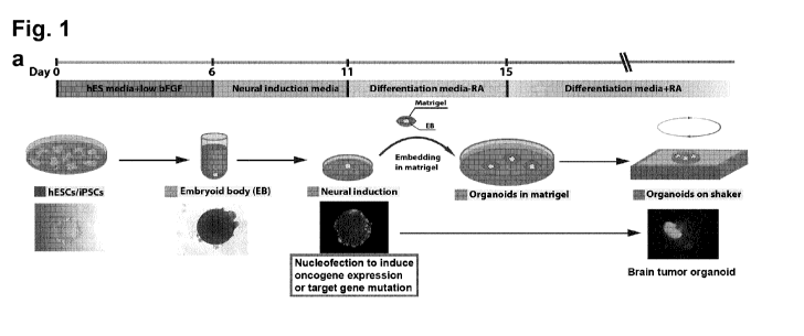
For example, as shown in the example section, which is pro-
vided for illustration only and not to limit the invention, a
combined transposon-mediated insertion with CRISPR/Cas9-mediated
genome editing was used to model human brain tumorigenesis in
cerebral organoids. By screening multiple combinations of gain-
and loss-of-function mutations found in cancer patients (McLen-
don et al., 2008, Nature, 455, 1061-8), it was demonstrated that
the growth of large, xeno-transplantable tumors can be classi-
fied as central nervous system primitive neuroectodermal tumor
(CNS-PNET) or glioblastoma (GBM) by marker expression and tran-
scriptome analysis. The approach initiates the transformation of
tumors carrying a specific set of driver mutations in the genet-
ic background of any patient, which allows the potential target-
ed drug testing in a personalized manner. Finally, the newly de-
veloped 3D brain tumor models were used to screen cancer medica-
tion and to demonstrate the oncolytic activity of the Zika fla-
vovirus, thereby establishing its potential suitability for
brain tumor therapy. It was demonstrated that these brain tumor
models can be used to evaluate drug efficacy on tumors with spe-
cific DNA aberrations.
The inventive method further comprises the step of culturing
and expanding said stem cells in a 3D biocompatible matrix. In
this step, the cells are allowed to differentiate and to develop
into a tissue culture of a desired size. As recapitulated above
with regard to references disclosing various organoids, such a
step is generally known and e.g. disclosed in W02014/090993 and
W02017/121754. The cells of the aggregate are placed in said 3D
biocompatible matrix preferably in form of said aggregate it-
self, i.e. without isolating cells from the aggregate.
Growth of the tissues to form the 3D tissue culture may be
CA 03074901 2020-03-05
WO 2019/048689 PCT/EP2018/074382
21
performed as known in the art for tissue culturing in a 3D
(three dimensional) matrix. A 3D matrix is distinct from 2D cul-
tures, such as 2D cultures in a dish on a flat surface. A "3D
culture" means that the culture can expand in all three dimen-
sions without being blocked by a one-sided wall (such as a bot-
tom plate of a dish). Such a culture is preferably in suspen-
sion. The 3D biocompatible matrix may be a gel, especially a
rigid stable gel, which results in further expansion of growing
cell culture/tissue and differentiation. A suitable 3D matrix
may comprise collagen. More preferably the 3D matrix comprises
extracellular matrix (ECM) or any component thereof selected
from collagen, laminin, entactin, and heparin-sulfated proteo-
glycan or any combination thereof. Extra-cellular matrix may be
from the Engelbreth-Holm-Swarm tumor or any component thereof
such as laminin, collagen, preferably type 4 collagen, entactin,
and optionally further heparan-sulfated proteoglycan or any com-
bination thereof. Such a matrix is Matrigel. Matrigel is known
in the art (US 4,829,000) and has been used to model 3D heart
tissue previously (WO 01/55297 A2) or neuronal tissue (WO
2014/090993). Preferably the matrix comprises laminin, collagen
and entactin, preferably in concentrations 30%-85% or 50%-85%,
laminin, 3%-50% collagen and sufficient entactin so that the ma-
trix forms a gel, usually 0.5%-10% entactin. Laminin may require
the presence of entactin to form a gel if collagen amounts are
insufficient for gel forming. Even more preferred, the matrix
comprises a concentration of at least 3.7 mg/ml containing in
parts by weight about 30%-85% laminin, 5%-40% collagen IV, op-
tionally 1%-10% nidogen, optionally 1%-10% heparan sulfate pro-
teoglycan and 1%-10% entactin. Matrigel's solid components usu-
ally comprise approximately 60% laminin, 30% collagen IV, and 8%
entactin. All %-values given for the matrix components are in
wt.-%. Entactin is a bridging molecule that interacts with lam-
inin and collagen. Such matrix components can be added in step
r). These components are also preferred parts of the inventive
kit. The 3D matrix may further comprise growth factors, such as
any one of EGF (epidermal growth factor), FGF (fibroblast growth
factor), NGF, PDGF, IGF (insulin-like growth factor), especially
IGF-1, TGF-13, tissue plasminogen activator. The 3D matrix may
also be free of any of these growth factors.
In general, the 3D matrix is a 3D structure of a biocompati-
CA 03074901 2020-03-05
WO 2019/048689 PCT/EP2018/074382
22
ble matrix. It preferably comprises collagen, gelatin, chitosan,
hyaluronan, methylcellulose, laminin and/or alginate. The matrix
may be a gel, in particular a hydrogel. Organo-chemical hydro-
gels may comprise polyvinyl alcohol, sodium polyacrylate, acry-
late polymers and copolymers with an abundance of hydrophilic
groups. Hydrogels comprise a network of polymer chains that are
hydrophilic, sometimes found as a colloidal gel in which water
is the dispersion medium. Hydrogels are highly absorbent (they
can contain 90 wt.-% water or more) natural or synthetic poly-
mers. Hydrogels also possess a degree of flexibility very simi-
lar to natural tissue, due to their significant water content.
It is possible that the 3D matrix, or its components, especially
ECM or collagen, still remains in the produced 3D tissue cul-
ture. Preferably the 3D matrix is a collagenous matrix, prefera-
bly it contains type I and/or type IV collagen. In particular
preferred, the 3D biocompatible matrix is a collagenous gel or a
collagenous hydrogel. Preferably, said aggregate of cells and/or
the 3D matrix are cultured in a suspension culture. A suspension
culture prevents contacts to solid walls of a cultivation vessel
and allows the 3D tissue culture during formation to expand in
all directions uniformly. The 3D tissue culture may be formed
without contacts to such a solid wall or without regions of
halted expansion due to contact to such a wall.
In summary of the above, carcinogenesis is preferably per-
formed after the pluripotent stem cells have been stimulated for
tissue-specific differentiation, such as neural differentiation.
For example, carcinogenesis is before expanding said stem cells
in a 3D biocompatible matrix. Carcinogenesis may be a recombi-
nant modification of said genes, preferably by introduction of a
transgene for expression of the oncogene or a gene inhibition
construct for suppression of the tumor suppressor. The transgene
or construct may be introduced into cells by nucleofection such
as electroporation.
In a further preferment of the invention the cancerous cells
are labelled with a marker, preferably a marker gene. Possible
markers or labels are reporter genes such as fluorescent pro-
teins, preferably GFP (green fluorescent protein), enhanced
green fluorescent protein (eGFP), d2EGFP, CFP (cyan fluorescent
protein), YFP (yellow fluorescent protein), RFP (drFP583; also
red fluorescent protein), BFP (blue fluorescent protein), smURFP
CA 03074901 2020-03-05
WO 2019/048689 PCT/EP2018/074382
23
(Small ultra red fluorescent protein), HcRed, DsRed, DsRed mono-
mer, ZsGreen, mCyan, ZsYellow enhanced blue fluo-rescent pro-
tein (eBFP), enhanced yellow fluorescent protein (eYFP), GFPuv,
enhanced cyan fluorescent protein (eCFP), far red Reef Coral
Fluorescent Protein; p-galactosidase; luciferase; a peroxidase,
e.g. horse radish peroxidase; alkaline phosphatases, e.g., SEAP,
and glucose oxidase, any cell sur-face marker such as Thy1.1.
Another type of marker is an enzymatic label. "Enzymatic la-
bel" means an enzyme that converts a substrate to a detectable
product. Suitable label enzymes for use in the present invention
include, but are not limited to, galactosidase, horseradish pe-
roxidase, luciferases, e.g., fire fly and renilla luciferase,
alkaline phosphatases, e.g., SEAP, and glucose oxidase. The
presence of the marker can be determined through the enzyme's
catalysis of substrate into an identifiable product.
Other markers are detectable proteins, in particular cell
surface proteins. Surface proteins can be detected by molecular
interaction with a binding partner through chemical or physical
interaction. A surface protein may be any partner in a "binding
pair". Binding pairs are molecules that interact with each other
through binding. "Partner of a binding pair" means one of a
first and a second moiety, wherein the first and the second moi-
ety have a suitable binding affinity for each other to detect
the pair with its members bound to each other. Suitable binding
pairs for use in the invention include, but are not limited to,
antigens/antibodies. Preferably, the cancer cells express a tu-
mor antigen that can be detected. Such a tumor antigen may be
one of the oncogenes that is artificially expressed as discussed
above.
Such a marker can be introduced together with the carcino-
genic elements as described above or separate from said ele-
ments. Preferred in all embodiments is the introduction together
with the carcinogenesis in order to track treated cells. In any
case, said labelling with a marker to identify cancerous cells.
Accordingly, the invention also comprises the step of identify-
ing cancerous cells in said tissue culture. Said identifying
step is preferably performed by identifying the marker. Such
methods of identification are well known in the art and include
cell sorting (e.g. FACS - fluorescence-activated cell sorting),
immunoassays, marker photo detection, magnetic separation etc..
CA 03074901 2020-03-05
WO 2019/048689 PCT/EP2018/074382
24
Preferably, the marker is a genetic marker that can be passed on
to progeny cells of the labelled cells. A labelled cell may be a
cell destined for carcinogenesis, it may or may not be a cancer
cell already. Preferred markers are different than the onco-
genes.
An artificial 3D tissue culture obtainable by any one of the
above described and below described methods and preferred embod-
iments, having accordingly bestowed characteristics, forms also
part of the invention. Producing such a 3D tissue culture is
usually a step in the inventive method. In addition to the above
characteristics, the 3D tissue culture may comprise non-
cancerous tissue and cancerous tissue. The cancerous tissue
overexpresses an oncogene and/or has suppressed expression of a
tumor suppressor as mentioned above, preferably in combination
with a marker gene that allows detection. The cancerous genes
usually have the same genetic background as the non-cancerous
cells, i.e. are from the same source original progenitor cells,
e.g. pluripotent stem cells. Accordingly, genes other than said
oncogene or tumor suppressor are preferably substantially unmod-
ified in the cancerous tissue as compared to the non-cancerous
tissue. Furthermore, said tissue (i) is obtainable by a method
according to the invention; and/or (ii) comprising a transgene
or a construct for suppression of a tumor suppressor at least in
cells of the cancerous tissue; and/or (iii) comprising a 3D bio-
compatible matrix, preferably gel, a collagenous gel, or a hy-
drogel as disclosed above. The 3D tissue culture may be an or-
ganoid or have any one of an organoid's characteristics, such as
they 1. contain multiple organ-specific cell types, i.e. the
cells have different differentiation types depending on the gen-
eral organ selected for differentiation (e.g. neural progenitor
cells may further differentiate into forebrain cells, cells of
cells of dorsal-lateral ganglionic eminence and caudal ganglion-
ic eminence identity, cells of ventral-medial ganglionic emi-
nence identity, cells of dorsal cortex identity, etc., in gen-
eral of any subdifferentiation as mentioned above); 2. are capa-
ble of recapitulating some specific function of the organ (eg.
excretion, filtration, neural activity, contraction); 3. are
grouped together and spatially organized similar to an organ.
Organoid formation recapitulates both major processes of self-
organization during development: cell sorting out and spatially
CA 03074901 2020-03-05
WO 2019/048689 PCT/EP2018/074382
restricted lineage commitment. This self-organization and dif-
ferentiation according to particular tissue parts reminiscent of
in vivo development is found in the inventive 3D tissue culture.
Of course, such a natural development is found in the non-
cancerous cells. Cancerous cells may differ from natural tissue
or organ development and exhibit a cancerous/tumor phenotype,
such as uncontrolled growth and in severe cancer case invasion
of non-cancerous tissue parts. Preferably, the tissue comprises
cells of a cancerous or proliferative central nervous system
disorder, in particular preferred glioblastoma, neuroblastoma or
CNS-PNET (central nervous system primitive neuro-ectodermal tu-
mor) as further described herein.
In preferred embodiments of the invention, the 3D tissue
culture is grown to a size or has a size of at least 100 pm,
preferably at least 150 um, especially preferred at least 200
pm. "Size" refers to the longest dimension in 3d space. Prefera-
bly the 3D tissue culture is globular in shape, in particular
with the shortest dimension being not less than 20% of the long-
est dimension, in particular not less than 30% or not less than
40% of the longest dimension. Preferably the volume of the 3D
tissue culture is at least 1x10⁶ pm3, in particular preferred at
least 2x10⁶ pm3, at least 4x10⁶ pm3, at least 6x10⁶ pm3, at least
8x10⁶ pm3, at least 10x106 pm3, at least 15x106 pm3 and/or sizes
of at least 250 pm, especially preferred at least 350 um.
The 3D tissue culture is usually of a size of at most 10 mm,
preferably of at most 5 mm, of at most 2 mm, of at most 1250 pm
or at most 800 pm, e.g. with volumes of at most at most 4200 mm3,
at most 2400 mm3, at most 1200 mm3, at most 800 mm3, at most 400
mm3, at most 100 mm3 at most 50 mm3, at most 8 mm3, at most 2
mm3 or at most at most 1 mm3. In some embodiments, the 3D tissue
culture may be larger with a size of at most 15 mm, preferably
of at most 10 mm or at most 5 mm, e.g. with volumes of at most
15000 mm3, at most 10000 mm3, or at most at most 8000 mm3.
The inventive 3D tissue culture may or may not comprise a
vascular network in all embodiments of the invention, in partic-
ular, the inventive 3D tissue culture may comprise only cells of
a single differentiation lineage, e.g. neural cells or organ,
such as neural, gastric, connective, cartilage, bone, bone mar-
row, cardiac, kidney, vascular, breast or ductal-lobular, reti-
nal, prostate, intestinal, gastric, lung, endothelium or liver
CA 03074901 2020-03-05
WO 2019/048689
PCT/EP2018/074382
26
tissue. This outcome may be controlled by the use of suitable
differentiation factors as disclosed above. It is also possible
to allow some variation in differentiation but maintain strin-
gent differentiation to tissues of only one germ layer selected
from mesoderm, endoderm, and ectoderm. Furthermore, the 3D tis-
sue culture may be homogenously constituted from said cell of
one differentiation lineage. Accordingly, other differentiation
lineages may not be present, such as a connective tissue layer
on the 3D tissue culture.
The 3D tissue culture may express certain differentiation
expression markers, or lack expression of such expression mark-
ers as signals of a specific differentiation.
Preferably, said tissue culture comprises neural tissue and
wherein the cancerous tissue is a neural tissue tumor.
Preferably the 3D tissue culture comprises cells, which ex-
press DLX2. DLX2 is expressed in cells of ventral forebrain
identity. Preferably this tissue type is comprised in the in-
ventive tissue.
Preferably the 3D tissue culture comprises cells, which ex-
press GSX2. GSX2 is expressed in cells of dorsal-lateral gangli-
onic eminence and caudal ganglionic eminence identity. Prefera-
bly this tissue type is comprised in the inventive tissue.
Preferably the 3D tissue culture comprises cells, which ex-
press NKX2-1. NKX2-1 is expressed in cells of ventral-medial
ganglionic eminence identity. Preferably this tissue type is
comprised in the inventive tissue.
Preferably the 3D tissue culture comprises cells, which ex-
press LHX6. LHX6 is expressed in cells of a subregion of ven-
tral-medial ganglionic eminence identity. Preferably this tissue
type is comprised in the inventive tissue.
Preferably the 3D tissue culture comprises cells, which ex-
press FoxG1. FoxG1 is expressed in cells of dorsal cortex iden-
tity. Preferably this tissue type is comprised in the inventive
tissue.
Preferably the 3D tissue culture comprises cells, which ex-
press TBR1. TBR1 is expressed in cells of dorsal forebrain iden-
tity. Preferably this tissue type is comprised in the inventive
tissue.
Preferably the 3D tissue culture comprises cells, which ex-
press TBR2. TBR2 is expressed in cells of dorsal cortical iden-
CA 03074901 2020-03-05
WO 2019/048689
PCT/EP2018/074382
27
tity. Preferably this tissue type is comprised in the inventive
tissue.
The inventive 3D tissue culture may contain a region of in-
vasion or fusion between the tissue (part) of non-cancerous
cells and the tissue (part) of cancerous cells. Such a region of
invasion or fusion may allow the cells of the tissue types to be
juxtaposed, resulting in a tissue that is a continuous tissue
where one side is of the cancerous type, while the other side is
of the non-cancerous type.
Preferably, non-cancerous tissue is at least at the core of
the tissue and the cancerous tissue at least at the surface of
the tissue. Since the aggregates are usually not disrupted be-
fore cultivation in the 3D matrix and the aggregate continues
growing to the state of the 3D tissue culture in the 3D biocom-
patible matrix, which enhances growth and differentiation to in
vivo-like lineages, and give that carcinogenesis is preferably
on surface contact of the aggregate or the 3D tissue culture, by
consequence, the first carcinogenic mutations will happen in
cells on the surface. Other aggregate or tissue treatment to in-
troduce carcinogenesis may be injection, accordingly, the cancer
growth may start at the place of injection, which may be the
core of the 3D tissue culture. "At least" means that the cells
are found in the specified tissue location but may also be found
in other parts of the tissue. In case of non-invasive cancerous
cells, the cancerous cells may remain at their original loca-
tion, such as the surface of the tissue. In case of invasive
cells, the cancerous cells may be found throughout the tissue.
For example, MYC-OE neoplastic cells are usually non-invasive.
In case of GBM neoplastic cells, the cancerous cells grow not
only on the surface, but also invade into the core of the tis-
sue. Normal non-cancerous cells also grow on the surface of or-
ganoids.
Also provided is a method of testing or screening a candi-
date compound or agent for carcinogenesis or for its effect on
cancer tissue, comprising contacting cells or a tissue in a
method of the invention with the candidate compound or agent or
contacting a tissue of the invention with the candidate compound
or agent and maintaining said contacted tissue in culture, and
observing any changes in the tissue as compared to said tissue
without contacting by said candidate compound. Likewise, the in-
CA 03074901 2020-03-05
WO 2019/048689 PCT/EP2018/074382
28
vention provides exposing the tissue or the cells in the in-
ventive method to a condition instead of contacting it with a
candidate compound. Such a condition may be e.g. elevated tem-
perature, limited nutrients or altered redox potential, to which
cancer cells may react and exhibit a different behaviour or
growth rate as compared to behaviour or growth without exposure
to said condition. Accordingly, the inventive 3D tissue culture
and the method of its generation can also be used as a research
tool to study the effects of any chemical (compounds, e.g. drugs
or other stimuli), (biological) agents (e.g. a virus, like an
oncolytic virus and/or a Flavivirus) environmental (e.g. temper-
ature, pressure, light exposure, redox potential, nutrients, ir-
radiation) Influences on growth and activity of cells in the
tissue, in particular of the cells undergoing carcinogenesis.
Temperature changes are preferably elevated temperature; altered
nutrients are e.g. lowered glucose or other carbohydrate energy
sources, increased fat or fatty acids; altered redox potential
may e.g. be the addition of oxidizing agents or reducing agents
or antioxidants, like vitamin C; light may be LW light; irradia-
tion may be by alpha or beta radiation sources; a virus may be
an oncolytic virus or a Flavivirus, a retrovirus or a DNA virus.
In an inventive tissue that also comprises non-cancerous cells,
it is further possible to compare the effects on the cancer
cells to the effects on the non-cancerous cells of the same or a
different 3D tissue culture. Accordingly, it is possible to
identify cancer specific compounds, agents or environmental fac-
tors that have a stronger effect on cancer cells than non-cancer
cells. In this case, compounds, agents or environmental factors
may be eligible cancer therapy candidates, vs. compounds or
agents or environmental factors that kill cancerous and non-
cancerous cells indiscriminately. The cancer specific effect
preferably kills or growth-inhibits 2 or more cancer cells for
every non-cancerous cell. Preferably, this ratio is 3 or more, 4
or more, 5 or more, 10 or more, 20 or more or 100 or more cancer
cells for every non-cancer cell. A therapeutic candidate thus
classified may be subject for further caner tests, e.g. in an
animal model or in patients.
The candidate compound or agent may be analysed and selected
according to a desired property on the development of cancer in
the 3D tissue culture. For example, compounds or agents may be
CA 03074901 2020-03-05
WO 2019/048689 PCT/EP2018/074382
29
analysed for their potential to slow or even halt cancer growth.
Also, it is possible to screen for their ability to destroy tu-
mor or cancer cells. Such effects can be screened in comparison
to the non-cancerous cells, which are preferably less affected
by such detrimental effects than the cancer cells, if the candi-
date compound should be further considered as a cancer treatment
drug. Any kind of activity of the inventive cells or tissue, in-
cluding metabolic turn-over or signalling can be searched for in
a candidate compound or agent. In essence, the inventive highly
differentiated tissue can be used as a model for tissue behav-
iour testing on any effects of any compound. Such a method might
also be used to test therapeutic drugs, intended for treating
cancer, for having side-effects on non-cancerous cells as can be
observed in the inventive tissue culture. As said, instead of
testing or screening a candidate compound or agent, also envi-
ronmental conditions can be analysed for the same effects and
purposes. Such effects may be elevated temperatures, such as
40 C and above, or reduced nutrients like withdrawal of a carbo-
hydrate or mineral source.
A candidate drug as candidate compound or agent may be a bi-
omolecule, like a protein, peptide, nucleic acid, or comprise or
be composed of such hiomolecules, such as a virus, or a small
molecule Inhibitor. Small molecules are usually small organic
compounds having a size of 5000 Dalton or less, e.g. 2500 Dalton
or less, or even 1000 Dalton or less. The candidate drug, agent
or compound may be known for other Indication and/or a known
chemical compound. Such known compounds are e.g. disclosed in
compound databases such as www.selleckchem.com, which collects
inhibitor compound information, including the cellular target of
a compound. Preferably, the candidate compound is an inhibitor
of an oncogene, in particular an oncogene that has been artifi-
cially introduced according to the inventive methods, either
targeted or by random mutagenesis (and that was then identified)
according to the inventive methods described above. Many and any
compound can be screened, for example for target gene EGFR,
www.selleckchem.com lists more than 50 Inhibitors that are all
eligible screening targets, of course. Further candidate com-
pounds are virus particle, in particular infectious virus parti-
cles, including wild type viruses or attenuated viruses. Effec-
tive viruses are called oncolytic viruses due to their anti-
CA 03074901 2020-03-05
WO 2019/048689
PCT/EP2018/074382
cancerous effect, although lysis of a tumor or of cancer cells
is not strictly necessary. Such a virus that was found to be a
viable treatment option for cancer is the Zika Flavivirus. The
examples herein show its oncolytic potential in a neural tumor
organoid. Its use is a further aspect of the invention.
In particular preferred embodiments, the candidate compound
or agent is tested or screened for its effects on any cancerous
or proliferative central nervous system disorder, in particular
preferred glioblastoma, neuroblastoma or CNS-PNET (central nerv-
ous system primitive neuro-ectodermal tumor). Accordingly, the
inventive tissue comprises such a disorder or in the inventive
method such a disorder is created in the carcinogenesis step.
Such a method is particularly used to screen for or test poten-
tial therapeutic compounds or agents.
The inventive 3D tissue culture can also be implanted into
an animal. Possible implantation sites are anywhere in the ani-
mal, such as subcutaneous or in an organ cavity, such as near
the kidney. Said screening or testing methods can also be per-
formed on in the animal model, e.g. by administering a candidate
compound or agent to the animal. Example animals are e.g. non-
human primates, rodents, non-human mammals, etc. The candidate
agent or drug may be administered in combination with a pharma-
ceutical preparation. Example pharmaceutical preparations are
further described below. The invention thus also provides ani-
mals comprising the inventive 3D tissue culture.
In a further aspect, the invention provides Zika virus for
use as a oncolytic virus. In particular provided is Zika virus
for use in the treatment of nervous system cancer. Related
thereto is a method of treating a nervous system cancer in a pa-
tient comprising treating a patient having nervous system cancer
with Zika virus to remove said cancer. Further provided is the
use of Zika virus in the manufacture of a medicament for the
treatment of nervous system cancer. Also provided is a method of
treating nervous system cancer cells with Zika virus. The cancer
cells may be in a patient or in vitro, e.g. in a 3D tissue cul-
ture as described. Nervous system cancer may e.g. be a brain
cancer or a spinal cord cancer. As shown herein, Zika virus
(ZIKV) preferentially infects tumor cells in organoids and se-
verely impairs tumor growth. The results show that cerebral or-
ganoids can be used to test strategies for brain tumor therapy
CA 03074901 2020-03-05
WO 2019/048689 PCT/EP2018/074382
31
and to shows the use of ZIKA as an oncolytic virus.
Oncolytic viruses and their use to treat cancer are well-
known in the art (see NO 1998/035028 A2, NO 2001/053506 A2, NO
2002/067861 A2, NO 2004/078206 Al, NO 2017/070110 Al, NO
2017/085175 Al, NO 2017/132552 Al, WC 2017/120670 Al; Russell et
al. Nat Biotechnol. 30(7): 658-670 (2012)) for use alone (i.e.
as only anti-cancer drug) or in combination with other cancer
therapies, in particular chemotherapy (WO 2008/043576 Al, NO
2017/121925 Al). The inventive therapy using Zika virus is an
oncolytic virus therapy and particular aspects can be used as
known in the art, such as administering the virus in a formula-
tion suitable for viral delivery and/or stability. Candidate on-
colytic viruses can be tested or screened for oncolytic effects
in the inventive methods and tissue cultures as candidate com-
pounds.
Zika virus (ZIKV) is a mosquito-borne flavivirus distributed
throughout much of Africa and Asia. Infection with the virus may
cause acute febrile illness that clinically resembles dengue fe-
ver. It has been characterized and is available in the art
(Haddow et al., PLoS neglected tropical diseases 6(2) 2012:
e1477, Incorporated herein by reference). Any strain can be
used, such as the African strain or Asian strain, including any
of its lineages, including MR 766, ArD 41519, IbH 30656, EC Yap,
P6-740 or FSS13025. In addition, Zika virus may be attenuated or
recombinantly engineered to include further antigens or attenua-
tion modifications. The genome of the Zika virus of the inven-
tion preferably still has at least 85% or at least 90% or at
least 95% sequence identity to any one of lineages MR 766, ArD
41519, IbH 30656, EC Yap, P6-740 or FSS13025 as deposited and
reviewed by Haddow et al., 2012, supra.
In some embodiments, the invention provides a method of
treating cancer in a subject in need thereof, comprising admin-
istering an oncolytic Zika virus described herein or composi-
tions thereof to the subject. In some embodiments, the subject
is a mouse, a rat, a rabbit, a cat, a dog, a horse, a non-human
primate, or a human. In some embodiments, the oncolytic virus or
compositions thereof are administered intravenously, subcutane-
ously, intratumorally, intramuscularly, intranasally, parenter-
ally, or intraperitoneally. The virus can be administered sys-
temically or topically. In case of metastasizing tumors, system-
CA 03074901 2020-03-05
WO 2019/048689 PCT/EP2018/074382
32
ic administration is preferred. In case of singular tumor, a
topical administration to the tumor site or cancerous organ is
also possible.
The invention further provides a pharmaceutical composition
comprising a replication competent Zika virus and a stabilizer
or carrier for said virus. The pharmaceutical composition may be
used in the treatment of neural cancer cells. A stabilizer may
be any stabilizer for viral formulations, preferably to create a
shelf-life of at least 3 months, at room temperature or under
cooled storage, such as at 1 C to 8 C. An example stabilizer may
be a carbohydrate (US 8,142,795 B2), including disaccharides (US
6,231,860 B1) or serum proteins like albumin (US 6,210,683 B1)
or salts comprising Mg2 and Ca2-' ions (US 3,915,794 A) or glutam-
ic acid and arginine (US 4,337,242 A) or combinations thereof.
Of course, the concentration of the stabilizer shall be adapted
to achieve a stabilized effect, such as maintaining the infec-
tious virus in solution, for at least 14 days or more like 2-3
months or more. The composition may further comprise a sensitiz-
er such as to remove or reduce protection of the cancer by the
patient's immune system. An example sensitizer is an IFN inhibi-
tor (Russell et al., 2012, supra) or a check-point inhibitor (WO
2017/120670 Al). The Zika virus may be a wild type virus, which
may require isolation of the patient in order to prevent infec-
tion of bystanders, or the Zika virus may be life-attenuated to
mitigate infection capacity and contagion of bystanders. Because
Zika virus only causes a mild infection in besides having cancer
otherwise healthy persons with the exception of pregnant women,
Zika virus may be wild type. Accordingly, the person treated
with Zika virus is not a pregnant female in such a case.
The pharmaceutical composition may comprise a carrier. As
used herein, "carrier" includes any and all solvents, dispersion
media, vehicles, coatings, diluents, isotonic and absorption de-
laying agents, buffers, carrier solutions, suspensions, disper-
sants, colloids, and the like. The use of such media and agents
for pharmaceutical active substances is well known in the art.
Except insofar as any conventional media or agent is incompati-
ble with the active ingredient, its use in the therapeutic com-
positions is contemplated. Supplementary active ingredients can
also be incorporated into the compositions. In one embodiment, a
composition comprising a carrier is suitable for parenteral ad-
CA 03074901 2020-03-05
WO 2019/048689 PCT/EP2018/074382
33
ministration, e.g., intravascular (intravenous or intraarteri-
al), intraperitoneal or intramuscular administration. Pharmaceu-
tically acceptable carriers include sterile aqueous solutions or
dispersions and sterile powders for the extemporaneous prepara-
tion of sterile Injectable solutions or dispersion. The use of
such media and agents for pharmaceutically active substances is
well known in the art. Except insofar as any conventional media
or agent is incompatible with a viral vector or nucleic acid
molecule, use thereof in the pharmaceutical compositions of the
invention is contemplated.
The pharmaceutical forms suitable for injectable use include
sterile aqueous solutions or dispersions and sterile powders for
the extemporaneous preparation of sterile injectable solutions
or dispersions (US 5,466,468, incorporated herein by reference
in its entirety). In all cases the form should be sterile and
should be fluid to the extent that easy syringability exists. It
should be stable under the conditions of manufacture and storage
and should be preserved against the contaminating action of mi-
croorganisms, such as bacteria and fungi. The composition may
further comprise antibacterial and/or antifungal agents or other
preservatives to increase shelf-life. Of course, sterile or the
presence shall not prevent Zika virus' oncolytic capability.
Sterility therefore does not extend to the removal or inactiva-
tion of Zika virus. Likewise, the preservative shall not pre-
serve against Zika virus.
The composition may further comprise an antioxidant. Exam-
ples of pharmaceutically-acceptable antioxidants include: (1)
water soluble antioxidants, such as ascorbic acid, cysteine hy-
drochloride, sodium bisulfate, sodium metabisulfite, sodium sul-
fite and the like; (2) oil-soluble antioxidants, such as ascor-
byl palmitate, butylated hydroxyanisole (BHA), butylated hydrox-
ytoluene (BHT), lecithin, propyl gallate, alpha-tocopherol, and
the like; and (3) metal chelating agents, such as citric acid,
ethylenediamine tetraacetic acid (EDTA), sorbitol, tartaric ac-
id, phosphoric acid, and the like.
An example composition may comprise combinations of all of
these components, such as the Zika virus, a stabilizer for the
virus, preferably PEG, a carrier, and an antioxidant, preferably
further a sensitizer. The composition may be sterile with the
exception of the presence of Zika virus, which shall remain in-
CA 03074901 2020-03-05
WO 2019/048689
PCT/EP2018/074382
34
fectious, and/or comprise a preservative that is not harmful to
Zika virus.
The patient to be treated may have been diagnosed with neu-
ronal cancer or neural cancer, such as neuroblastoma or glio-
blastoma. For example, the patient may have a glioma (glial cell
tumor), e.g. Gliomatosis cerebri, Oligoastrocytoma, Choroid
plexus papilloma, Ependymoma, Astrocytoma (Pilocytic astrocyto-
ma, Glioblastoma multiforme), Dysembryoplastic neuroepithelial
tumor, Oligodendroglioma, Medulloblastoma, Primitive neuroecto-
dermal tumor; a neuroepitheliomatous tumor, e.g. Ganglioneuroma,
Neuroblastoma, Atypical teratoid rhabdoid tumor, Retinoblastoma,
Esthesioneuroblastoma; or a nerve sheath tumor, e.g. Neurofibro-
ma (Neurofibrosarcoma, Neurofibromatosis), Schwannoma, Neurino-
ma, Acoustic neuroma, Neuroma. Any such tumor may also be mod-
elled by the inventive 3D tissue culture or organoid. Also, the
inventive method of treating neural cancer cell may comprise di-
agnosing or detecting neural cancer cells and then treating the
cells with Zika virus according to the invention. The treatment
with Zika virus may take precautionary preparations such as iso-
lating the patient to prevent further infection in other sub-
jects, in particular humans.
The invention also relates to a kit for providing a tissue
culture according to the invention. The kit may comprise (i) a
transfection vector comprising an oncogene transgene or a con-
struct for disruption of a tumor suppressor, (ii) a 3D biocom-
patible matrix, preferably further comprising (iii) a tissue
differentiation agent, a stem cell culturing medium, a nu-
cleofection medium or a combination thereof. The kit can be used
in the inventive method. Preferably the kit comprises any fur-
ther compound or means as disclosed above for the inventive
method. In particular, preferred, the kit also comprises a mark-
er as disclosed above in order to label mutated cells. The mark-
er is preferably an expression marker, such as a fluorescent
protein. The 3D matrix has been described in length above -
preferably it comprises a collagenous hydrogel or any other em-
bodiment disclosed herein. The kit further preferably comprises
a differentiation agent, a stem cell culturing medium, a nu-
cleofection medium or a combination thereof. Such media are
known in the art and usually Include one or more of the follow-
ing components:
CA 03074901 2020-03-05
WO 2019/048689
PCT/EP2018/074382
Differentiation agent: any one of the differentiation factors as
disclosed above, preferably a neuronal differentiation factor,
these are suitable for creating neuronal 3D tissue cultures;
Stem cell culturing medium: N2 supplement, B27 supplement, insu-
lin, 2-mercaptoethanol, glutamine, non-essential amino acids, or
any combination thereof (see WO 2014/090993 Al);
Nucleofection medium: e.g. Dulbecco's modified Eagle medium
(DMEM) or other nutrient and mineral source, glutamine, and FCS
or other serum or serum replacement (see also US 7,732,175 B2);
DMEM or the other nutrient source is preferable in a range of
80-95 w.-% of the medium. FCS or other serum or serum replace-
ment is preferably 5-20 w.-% of the medium. The nucleofector me-
dium should be suitable for nucleofection, preferably for elec-
troporation.
In addition to these components, the kit may also comprise
suitable containers, such as flasks or vials to hold its compo-
nents, preferably separately for each component or medium.
The invention is further illustrated in the following fig-
ures and examples, without being limited to these embodiments of
the invention.
Figures
Figure 1. Nucleofection of genome-editing constructs into
neural stem/precursor cells (NS/PCs) of cerebral organoids. a,
Schematic of the culture system of cerebral organoid system and
nucleofection strategy. Example images of each stage are pre-
sented. EBs were electroporated at the end of neural induction
stage, right before the matrigel embedding to initiate tumor-
igenesis. EB, embryoid body; bFGF, basic fibroblast growth fac-
tor; hESCs, human embryonic stem cells; hiPSCs, human induced
pluripotent stem cells; RA, retinoic acid. b, Immunofluorescence
photographs revealed that nucleofected cells (GFP, green) in EBs
at the end of neural induction stage are NS/PCs (S0X1, red; N-
CAD, red; NES, red; arrowheads), but neither mesodermal cells
(BRA, red; FOXF1, red; arrows) nor endodermal cells (S0X17, red;
CD31, red; arrows). N-CAD: N-CADHERIN; NES: NESTIN; BRA: BRACHY-
URY. Scale bar: b, upper panel: 200 pm; lower panel: 100 pm.
Figure 2. Clonal mutagenesis in organoids induces tumorous
overgrowth. Immunofluorescence photographs (a) and quantifica-
tion of the GFP fluorescence intensity of organoids 1 day (b)
CA 03074901 2020-03-05
WO 2019/048689 PCT/EP2018/074382
36
and 1 month (c) after nucleofection. Result showed that EBs from
all groups contains similar amount of nucleofected cells 1 day
after nucleofection, while organoids from four groups, including
MYC, CDKN2A /CDKN2B /EGFR E/EGFRvIII E, NF1 /PTEN /p53 , EG-
FRvIII/PTEN7CDK2A-, exhibit dramatic overgrowth of GFP-' cells in
cerebral organoids 1 month after nucleofection. Scale bar: a: 1
day: 200 pm; 1 month: 500 pm.
Figure 3. MYC E and GEM-like neoplastic cerebral organoids
have distinct transcriptional profiles and cellular identities.
a, Principle component analysis (PCA) of the top 500 variable
genes between normal cells from CTRL organoids and tumor cells
from different neoplastic cerebral organoid groups. b, Venn dia-
grams showed overlap of genes differentially expressed (DESeq,
adjusted p value<0.05) between the Cluster 2 (MYC E, n=3) and the
Cluster 3 (GEM-1, GBM-2, GBM-3, n=7) relative to the CTRL organ-
oids (Cluster 1; n=3). The p value for overlaps were calculated
by hypergeometric test. c, KEGG pathway enrichment analysis re-
vealed the differences in signalling pathways between neoplastic
cerebral organoids from the Cluster 2 and the Cluster 3. d, The
heatmap shows normalised expression levels for differentially
expressed genes (adjusted absolute 10g2fc value >1 or <-1 and
adjusted p value <0.05) between Cluster 2 and Cluster 3 (n=3 for
Cluster 2 and n=7 for Cluster 3 from one experiment) selected
from differentially expressed genes between human primary CNS-
PNET and GBM tumors. The heatmap was created from
1og2(Transcripts Per Kilobase Million, TPM) transformed data
that was row (gene) normalised using the 'Median Center
Genes/Rows" and "Normalise Genes/Rows" functions to report data
as relative expression between samples. e, Low-magnification im-
ages of DAPI (blue) and GFP (green) staining of control and neo-
plasm groups 4 months after nucleofection. f-k, Representative
immunofluorescence images and quantification of four-month-old
organoids from CTRL, MYC E, and GBM-1. The staining was performed
from six independent experiments with similar results. Quantifi-
cation was performed on organoids from three independent experi-
ments. Statistical analysis was performed using one-way ANOVA
with Dunnett's test. Data are presented as mean SD, with details
of sample sizes and values, as well as adjusted p value in
Source Data. *, p<0.05; **, p<0.01; ***, p<0.001. Scale bar: c:
100 pm.
CA 03074901 2020-03-05
WO 2019/048689 PCT/EP2018/074382
37
Figure 4. Neoplastic organoids expanded upon renal subscapu-
lar xenografts. a, Schematic of renal capsule xenograft proce-
dure. Two-month-old neoplastic organoids were implanted into
kidney capsule of nude mice, and tissues were collected 1.5
months after. b, Brightfield and immunofluorescence photographs
showed that neoplastic organoids were expanded, while control
organoids were largely absorbed. c, Photograph of H&E staining
of neoplastic organoids in renal capsule. Glial cells are point-
ed by arrows, and neurons are pointed by arrowhead. d, Immuno-
histochemical photographs of glial marker GFAP, precursor marker
SOX1, and cell cycle marker K167 on implanted organoids. e, Pho-
tographs of H&E staining of implanted MYC E organoids showed that
MYC E neoplasm exhibit CNS-PNET-like histopathological features.
f, Immunohistochemical photographs of neuronal marker MAP2 re-
vealed that MYC E neoplasm barely differentiated into neurons.
Scale bar: b: 500 mm; c, d, f: 200 lam and 50 lam (inset); e: 1000
pm; e', e", e"': 50 pm.
Figure 5.GBM neoplastic cerebral organoids exhibit features
of GBM invasion. a-c, Representative images of the tumor-normal
interface in GBM-1 neoplastic cerebral organoids. Images are
representative of at least three independent experiments. d, Im-
munohistochemical staining of GFAP in GBM-like neoplastic cere-
bral organoids. Images are representative of two independent re-
nal implantations. Dotted black lines indicate the boundary be-
tween implanted neoplastic cerebral organoids and murine kidney.
Dotted red line indicates the renal tubule. Arrowheads indicate
invaded tumor cells. e, Hierarchical clustering analysis of GBM
invasiveness-relevant genes from four-month-old organoids (n=3
for CTRL organoids; n=4 for MYC E, n=4 for GBM-1, n=4 for GBM-2,
and n=3 for GBM-3 neoplastic cerebral organoids, from three in-
dependent cultures for each group). The heatmap was created from
1og2(TPM) transformed data that was row (gene) normalised using
the 'Median Center Genes/Rows" and "Normalise Genes/Rows" func-
tions to report data as relative expression between samples. f,
Representative immunofluorescence staining of neoplastic cere-
bral organoids from GBM-1 group for the indicated mesenchymal
marker and Invasiveness markers; GFP is also shown. Images are
representative of two independent experiments. Scale bar: a,
1000 mm; b and c, 200 mm; d, 25 pm; f: 100 lam.
CA 03074901 2020-03-05
WO 2019/048689 PCT/EP2018/074382
38
Figure 6. Using brain neoplastic organoid model to investi-
gate potential brain tumor therapies. a, b, Images (a) and quan-
tification of FACS sorting (b) assay revealed that EGFR inhibi-
tors Afatinib was able to diminish most of GFP-' tumor cells in
GBM-1 (n=6) and GBM-3 (n=3) neoplastic cerebral organoids, but
exhibited no effect on tumor cells in MYC E and GBM-2 neoplastic
cerebral organoids compared to DMSO treatment. The percentage of
GFP-' cells in total cells from the drug-treated groups were nor-
malized to the percentage of GFP-' cells from DMSO-treated neo-
plastic cerebral organoids. Statistical analysis of quantifica-
tion was performed using unpaired two-tailed Student's t-test.
C, Schematic of ZIKV infection and experimental setups. d, Immu-
nofluorescence images of GFP and ZIKV of CTRL organoids treated
with either MOCK or ZIKV, as well as ZIKV-treated MYC E and GEM-1
neoplastic cerebral organoids. e, Quantification of ZIKV infec-
tion ratio showed significantly higher infection ratio of GFP-'
tumor cells from all neoplastic cerebral organoid groups com-
pared to non-tumor cells from CTRL organoids or neoplastic cere-
bral organoids. Statistical analysis of quantification was per-
formed using one-way ANOVA with Dunnett's test. f, Immunofluo-
rescence images of neural precursor marker MUSASHI1 (MSI1) of
CTRL organoids treated with either MOCK or ZIKV, as well as
ZIKV-treated MYC E and GBM-1 neoplastic cerebral organoids. g,
Immunofluorescence images of apoptosis marker activated Caspase3
(CASP3) of CTRL organoids treated with either MOCK or ZIKV, as
well as ZIKV-treated MYC E and GEM-1 neoplastic cerebral organ-
oids. h, Quantification of percentage of CASP3-' apoptosis cells
showed that ZIKV-infection induced significantly more cells
apoptosis in tumor regions compared to MOCK-treated tumor re-
gions, and MOCK- or ZIKV-treated non-tumor regions. CTRL-ZIKV
Statistical analysis of quantification was performed using one-
way ANOVA with Dunnett's test. i, Quantification of the yields
of progeny ZIKV by analysing the percentage of ZIKV-infected
Vero cells exposed to the supernatant from CTRL and neoplastic
cerebral organoids at 4 dpi. Compared to supernatant from CTRL
organoids, significantly more Vero cells were infected exposed
CA 03074901 2020-03-05
WO 2019/048689 PCT/EP2018/074382
39
to the supernatant from neoplastic cerebral organoid groups.
Statistical analysis of quantifications was performed using one-
way ANOVA with Dunnett's test. j, Epifluorescence iamges of neo-
plastic cerebral organoids from MYC E groups upon MOCK or ZIKV
exposure at 0, 6, and 14 dpi. k, FACS sorting analysis of GFP-'
tumor cell proportion in different neoplastic cerebral organoid
groups upon MOCK treatment at 14 dpi. The percentage of GFP-'
cells in total cells from the ZIKV-treated groups were normal-
ized to the percentage of GFP-' cells from MOCK-treated neoplastic
cerebral organoids. Statistical analysis of quantification was
performed using unpaired two-tailed Student's t-test. *, p<0.05;
** , p<0.01; ***, p<0.001. Scale bar: a, d, f, g, j, 1000
Figure 7. The strategy to introduce gene aberrations into
neural stem/precursor cells in cerebral organoids. a, Schematic
of the strategy of genome-editing techniques to introduce onco-
gene amplification and/or tumor suppressor mutation/deletion.
Sleeping Beauty transposon system was used to integrate onco-
gene-expression and GFP-expression elements into genome. CRISPR-
Cas9 system was applied to introduce mutation/deletion of tumor
suppressors. b, Quantification of cellular identities of nu-
cleofected cells in EBs 1 day after nucleofection by immunofluo-
rescence staining on serial cryo-sections. Results showed that
100% of GFP-' cells are SOX1-' (n=402), N-CADHERIW (N-CAD)
(n=451), and NESTIN-' (NES) (n=433) neural stem/precursor cells.
None of GFP-' cells is BRACHYURY-' (BRA) (n=398) or FOXF1+ (n=356)
mesodermal cells, or SOX17 (n=328) or CD31+ (n=267) endodermal
cells. c, d, Immunofluorescence images (c) and quantification (d)
of adherent cell culture of dissociated EBs 1 day after nu-
cleofection. Results showed that 100% of CFP- cells are SOX1-'
(n=549), N-CADHERIW (N-CAD) (n=403), and NESTIW (NES) (n=461)
neural stem/precursor cells. None of GFP-' cells is BRACHY1JRY-'
(BRA) (n=474) or FOXF1-' (n=402) mesodermal cells, or SOX17-'
(n=334) or CD31-' (n=415) endodermal cells. Scale bar: c, 50 lam.
Figure 8. Verification of gene aberrations introduced by ge-
nome-editing techniques. a, RNA-seq and RT-PCR analysis showed
CA 03074901 2020-03-05
WO 2019/048689 PCT/EP2018/074382
that tumor cells from MYC E neoplastic cerebral organoids exhibit
high MYC expression levels. b, Three example sequences of
CRISPR-Cas9 targeting CDKN2A and CDKN2B locus in tumor cells
from GBM-1 neoplastic cerebral organoids. RNA-seq and RI-PCR
analysis showed that tumor cells from GBM-1 neoplastic cerebral
organoids exhibit high expression levels of both EGFR and EG-
FRvIII. c, Three example sequences of CRISPR-Cas9 targeting NF1,
PTEN, and TP53 locus in tumor cells from GBM-2 neoplastic cere-
bral organoids. d, Three example sequences of CRISPR-Cas9 tar-
geting CDKN2A and PTEN locus in tumor cells from GBM-3 neo-
plastic cerebral organoids. RNA-seq and RI-PCR analysis showed
that tumor cells from GBM-3 neoplastic cerebral organoids exhib-
it high expression level of EGFRvIII, but not EGFR.
Figure 9. Low-magnification images revealed that 4-month-old
neoplastic organoids showed brain tumor subtype-specific cellu-
lar identities. a, Immunofluorescence photographs of control and
neoplastic groups 1 day and 4 months after nucleofection con-
firmed the tumor-initiation capability of genetic disruptions.
b, Immunofluorescence photographs of DAPI (blue) and GFP (green)
staining of control and neoplastic groups 4 months after nu-
cleofection. c-h, Immunofluorescence photographs and quantifica-
tion of neuronal marker HuC/D (c), precursor marker SOX2 (d,
red), cell cycle marker K167 (e, red), CNS-PNET marker CD99 (f,
red), as well as glial marker S1003 (h, red) and GFAP (g, red).
Scale bar: a: upper panel: 200 lam, lower panel: 1000 lam; b-g:
1000 lam.
Figure 10. High-magnification images revealed that 1-month-
old neoplastic organoids showed brain tumor subtype-specific
cellular identities. a, Immunofluorescence photographs of con-
trol and neoplastic groups 1 day and 1 months after nucleofec-
-Lion confirmed the tumor-initiation capability of genetic dis-
ruptions. b, Immunofluorescence photographs of DAPI (blue) and
GFP (green) of control and tumor groups 1 month after nucleofec-
tion. c-e, Immunofluorescence photographs and quantification of
neuronal marker HuC/D (c, red), precursor marker SOX2 (c, cyan),
cell cycle marker K167 (d, red), as well as glial marker S1003
(e, red). Scale bar: a: upper panel: 200 lam, lower panel: 1000
pm; b, 1000 pm; c-h: 100 lam.
CA 03074901 2020-03-05
W02019/048689 PCT/EP2018/074382
41
Figure 11. Low-magnification images revealed that one-month-
old Neoplastic organoids showed brain tumor subtype-specific
cellular identities. a, Immunofluorescence photographs of con-
trol and neoplastic groups 1 day and 1 month after nucleofection
confirmed the tumor-initiation capability of genetic disrup-
tions. b, Immunofluorescence photographs of DAPI (blue) and GFP
(green) staining of control and neoplastic groups 1 month after
nucleofection. c-e, Immunofluorescence photographs and quantifi-
cation of neuronal marker HuC/D (c, red), precursor marker SOX2
(c, cyan), cell cycle marker Ki67 (d), as well as glial marker
S1003 (e). Scale bar: a: upper panel: 200 lam, lower panel: 1000
pm; b-h: 1000 pm.
Figure 12. In vivo expansion of neoplastic cerebral organ-
oids. Neoplastic cerebral organoids from MYC E group and GBM-1
group were implanted into kidney capsule. Engrafted kidneys were
analysed at 1 week and 1.5 months after xenograft to evaluate
the in vivo expansion of neoplastic cerebral organoids.
Figure 13. Drug testing assay showed the drug screening po-
tential of neoplastic organoids. a, Schematic of luciferase as-
say-based drug testing strategy on neoplastic organoids. b,
Quantification of relative luciferase activity revealed that
EGFR inhibitors Afatinib and Erlotinib significantly reduced lu-
ciferase activity in GBM1 (CDKN2A/CDKN2B /EGFR E/EGFRvIII E) neo-
plastic organoids (CTRL group: n=3; DMSO: n=9; Canertinib: n=9;
Pelitinib: n=8; Afatinib: n=9; Gefitinib: n=10; Erlotinib: n=9).
Normalized luciferase activity was presented. Statistical analy-
sis of quantifications was performed using one-way ANOVA with
Dunnett's test. **, p<0.01.
Figure 14. ZIKV exhibits various tropism toward different
subtypes of neural cells. a,b, Immunofluorescence images and
quantifications of triple-staining of GFP (green), ZIKV (magen-
ta), and different neural cell subtype markers, including neural
precursor marker SOX2 (cyan) and MSII (cyan), glial marker S1003
(cyan), and neuronal marker HuC/D (cyan), as well as a double
staining for ZIKV (magenta) and GFP (green) that represent tumor
cells. Results showed significantly more GFP-' tumor cells co-
localized with ZIKV staining compared to other non-tumor neural
CA 03074901 2020-03-05
WO 2019/048689 PCT/EP2018/074382
42
cell types. In addition, ZIKV infection ratios of SOX2- and MSI1-'
non-tumor precursor cells are significantly higher than HuC/D-'
non-tumor neurons, which match the previous observations. c,d,
Immunofluorescence images and quantifications of ZIKV infection
ratio of different cell types within tumors from different neo-
plastic cerebral organoid groups. Results demonstrated that cell
type tropism within tumor regions. Statistical analysis of quan-
tifications was performed using one-way ANOVA with Dunnett's
test. *, p<0.05; ***, p<0.001. Scale bar: a, c, 100 pm.
Figure 15. Neoplastic cerebral organoids produce more ZIKV
progeny. a, Immunofluorescence images of ZIKV-infected Vero
cells exposed to the supernatant from CTRL and neoplastic cere-
bral organoids at 4 dpi. Cells were stained by DAPI (blue), and
ZIKV was stained as green. b, qPCR analysis revealed the signif-
icantly higher ZIKV gene expression in neoplastic cerebral or-
ganoids compared to CTRL organoids upon ZIKV infection. The ZIKV
vRNA level of neoplastic cerebral organoids were normalized to
the ZIKV vRNA level of CTRL organoids. Statistical analysis of
quantifications was performed using one-way ANOVA with Dunnett's
test. ***, p<0.001. Scale bar: 100 lam.
Figure 16. ZIKV infection in tumor regions of neoplastic
cerebral organoids resulted in a remarkable more cell apoptosis.
a, Immunofluorescence images of cell apoptosis marker CASP3 (red)
of ZIKV-infected non-tumor and tumor regions, as well as MOCK-
treated non-tumor and tumor regions. DAPI (blue) was stained for
nuclei, and GFP (green) was staining to represent tumor cells. b,
Immunofluorescence images of ZIKV staining and cell apoptosis
marker CASP3 of MYC E neoplastic cerebral organoids upon MOCK-
treatment, and ZIKV-infection at 6 dpi and 14 dpi. Scale bar: a,
100 pm; b, 1000 pm.
Examples
Example 1. Materials and Methods
1.1 Plasmid constructs and materials
For overexpression constructs, based on the Sleeping Beauty
Transposase System, the CMV promoter from pCMV(CAT)T7-SB100
(Addgene cat. No.: 34879; Mates at al., 2009, Nat Genet, 41,
753-61) was replaced with CAG promotor from pCAGEN (Addgene cat.
CA 03074901 2020-03-05
WO 2019/048689
PCT/EP2018/074382
43
No.: 11160; Matsuda and Cepko, 2004, Proc. Natl. Acad. Sci.
U.S.A., 101, 16-22). IRDR-R and IRDR-L sequences from pT2/LTR7-
GFP (Addgene cat. No.: 62541; Wang et al., 2014, Nature 516,
405-9) were cloned into pCAGEN to produce pCAG-GS/IR. cDNAs used
for overexpression were amplified from human cDNA and cloned in-
to the MCS of pCAG-GS/IR. With the help of sleeping beauty
transposase SB100X (pCAG-SB100X), CAG-GFP and CAG-oncogenes were
integrated into the genome of cells in organoids. To introduce
gene mutations, short guide RN/Is of tumor suppressors were
cloned into CRISPR/Cas9 vector pX330-U6-Chimeric BB-CBh-hSpCas9
(Addgene cat. No.: 42230; Ran et al., 2013, Nat Protoc, 8, 2281-
308). All cloning primers are listed in the Tables 1.
Table 1. Primers for cloning oncogenes into sleeping beauty con-
struct
Gene Primers
symbols
MYC up- GACGGCGCGCCCGCCACCATGCTGGATTTTTTTCGGGTAG (SEQ
stream ID NO: 1)
down- GACACCGGTTTACGCACAAGAGTTCCGTAG (SEQ ID NO: 2)
stream
EGFR/EG up- GACGGCGCGCCCGCCACCATGCGACCCTCCGGGACG (SEQ ID
FRvIII stream NO: 3)
down- GACACCGGTTCATGCTCCAATAAATTCACTG (SEQ ID NO:
stream 4)
PDGFRA up- GACGGCGCGCCCGCCACCATGGGGACTTCCCATCCGGCGTTC
stream (SEQ ID NO: 5)
down- GACACCGGTTTACAGGAAGCTGTCTTCCACCAG (SEQ ID NO:
stream 6)
CDK4 up- GACGGCGCGCCCGCCACCATGGCTACCTCTCGATATGAGC (SEQ
stream ID NO: 7)
down- GACACCGGTTCACTCCGGATTACCTTCATCC (SEQ ID NO:
stream 8)
MDM2-B up- GACGGCGCGCCCGCCACCATGTGCAATACCAACATGTCTG (SEQ
stream ID NO: 9)
down- GACACCGGTCTAGGGGAAATAAGTTAGCAC (SEQ ID NO:
stream 10)
H3F3A- up- CATTTTGGCAAAGAATTCCCTCGATACCGGGGGCGCGCCCGCCAC
K27M/ stream CATGGCTCGTACAAAGCAGACTGC (SEQ ID NO: 11)
H3F3A- down- CGGGAATGCTAGCAATCATTGGTTGATCAGCTTTGTTACCGGTTT
CA 03074901 2020-03-05
WO 2019/048689 PCT/EP2018/074382
44
G34R stream AAGCACGTTCTCCACGTATG (SEQ ID NO: 12)
1.2 Human embryonic stem cell (hESC) and human induced pluripo-
tent stem cell (iPSC) culture
Feeder-free (FF) H9 hESCs were obtained from WiCell with
verified normal karyotype and contamination free. FF H9 hESCs
were cultured in a feeder-free manner on Matrigel (Corning,
hESC-qualified Matrix)-coated plate with mTeSR medium (Stemcell
Technologies). Feeder-dependent (FD) H9 hESCs were obtained from
WiCell with verified contamination-free. FD 119 hESCs were cul-
tured on CF-1-gamma-irradiated mouse embryonic stem cells (MEFs)
(GSC-6001G, Global Stem) according to WiCell protocols. All cell
lines were routinely checked for mycoplasma-negative. All stem
cells were maintained in a 5% 002 incubator at 37 C. Standard
procedures were used for culturing and splitting hESCs as ex-
plained previously (Lancaster et al., 2013, Nature, 501, 373-9).
All hESCs were authenticated using Infinium PsychArray-24 Kit
(Illumina).
1.3 Generation of cerebral organoids
Cerebral organoids were cultured as previous described (Lan-
caster et al., 2013 Nature 501, 373-379; WO 2014/090993 Al; both
incorporated herein by reference). Briefly, to make EBs (embry-
aid bodies), hESCs/hiPSCs were trypsinized into single cells,
and 9,000 cells were plated into each well of an ultraplow-
binding 96-well plate (Corning) in human ES medium containing
low concentration basic fibroblast growth factor (bFGF, 4 ng/m1)
and 50 nM Rho-associated protein kinase (ROCK) inhibitor (Calbi-
ochem). EBs were fed every three days for 6 days then trans-
ferred to neural induction media to form neuroepithelial tis-
sues. After 5-7 days in neural induction media, EBs were embed-
ded into droplets of Matrigel (Corning) and cultured in differ-
entiation medium without vitamin A (Diff-A). Finally, the EB
droplets were transferred to 10 cm-dish containing differentia-
tion medium with vitamin A (Diff+A) and cultured on an orbital
shaker. Media were changed weekly.
1.4 Nucleofection of organoids to induce gene muta-
tion/amplification
CA 03074901 2020-03-05
WO 2019/048689 PCT/EP2018/074382
In order to initiate the brain tumors, we introduced the tu-
mor suppressor mutations and/or oncogene amplifications on neu-
roepithelial cells at the end of neural induction culture, right
before the Matrigel embedding. Briefly, 10-15 EBs were collect-
ed, resuspended in nucleofetion reagent (NucleofectorTm kits for
human stem cells, Lonza) containing plasmids and transferred in-
to nucleofection vials. Nucleofection was performed according to
the manufacturer's protocol. After electroporation, EBs were
carefully transferred to 6 cm-dish containing neural induction
medium, and cultured at 37 C incubator for 4 hours. Then nu-
cleofected EBs were embedded into Matrigel and cultured for or-
ganoids as described. The neoplastic cerebral organoids with
significant overgrowth of GFP+ cells were selected for further
investigations, in which the samples were randomly allocated.
1.5 Adherent cell culture of dissociated EBs.
One day after nucleofection, the EBs were trypsinized at
37 C for 20 min to make single cell suspension. Then cells were
plated on the poly-D-lysine- and laminin-coated coverglasses in
neural induction medium with ROCK inhibitor, and cultured in a
5% CO2 incubator at 37 C. The further immunofluorescence staining
and analysis were performed the day after.
1.6 RNA-seq analysis
Organoids from control and neoplastic groups were collected
40 days and four months after nucleofection, and trypsinised
with shaking at 37 C for half an hour. GFP+ cells were sorted ac-
cording to the example gating strategy, and total RNA was iso-
lated using RNeasy Micro kit (Qiagen) according to the manufac-
turer's instruction. RNA concentration and quality were analysed
using RNA 6000 Nano Chip (Agilent Technologies). Messenger RNA
(mRNA) was enriched using SMART-Seq v4 Ultra Low Input RNA Kit
(TaKaRa) according to manufacturer's protocol. Libraries were
prepared using NEB Next Ultra Directional RNA library Prep kit
for Illumina (NEB). Barcoded samples were multiplexed and se-
quenced 50 bp SE on a HighSeq 2500 (Illumina). mRNA sample iso-
CA 03074901 2020-03-05
WO 2019/048689 PCT/EP2018/074382
46
lation, library preparation, and sequencing were performed at
the VBCF NGS Unit (www.vbcf.ac.at).
The unstranded reads were screened for ribosomal RNA by
aligning with BWA (v0.7.12) against known rRNA sequences (Ref-
Seq). The rRNA subtracted reads were aligned with TopHat
(v2.1.1) against the Homo sapiens genome (hg38). Microexon-
search was enabled. Additionally, a gene model was provided as
GTF (UCSC, 201501, hg38). rRNA loci are masked on the genome
for downstream analysis. Aligned reads are subjected to Tran-
scripts Per Kilobase Million (TPM) estimation with Kallisto
(v0.43.0). Furthermore, the aligned reads were counted with
HTSeq (v0.6.1; intersection-nonempty) and the genes were sub-
jected to differential expression analysis with DESeq2
(v1.12.4).
Before the bioinformatics analysis, the expression of onco-
genes according to the genome editing manipulation was checked,
and one four-month-old sample from GBM-3 neoplastic cerebral or-
ganoid group was excluded from the further analysis because of
the failure of Introducing the overexpression of EGFRvIII.
PCA was performed using the top 500 variable genes between
normal cells from CTRL organoids and tumor cells from different
neoplastic cerebral organoid groups. Venn diagram hypergeometric
test was performed on differentially expressed genes between
Cluster 2 or Cluster 3 versus CTRL, and KEGG pathway enrichment
analysis were performed on differentially expressed genes be-
tween Cluster 2 and Cluster 3 with an adjusted absolute log2fc
value >0.5 and adjusted p value <0.05. Venn diagram hypergeomet-
ric test was performed via R language. KEGG pathway enrichment
was analysed using DAVID Bioinformatics (da-
vid.ncifcrf.gov) (Huang et al, 2009, Nature Protocol, 4, 44-57).
The heatmap of RNA-seq was generated using MeV (Saeed et al.,
2003, BioTechniques 34, 374-8). For the heatmap of tumor-subtype
gene profiling (Fig. 3c), the differentially expressed genes be-
tween Cluster 2 and Cluster 3 (adjusted absolute 10g2fc value >1
or <-1 and adjusted p value <0.05) were selected from the dif-
ferentially expressed gene list (adjusted absolute 1og2fc value
CA 03074901 2020-03-05
WO 2019/048689 PCT/EP2018/074382
47
>1 or <-1 and adjusted p value <0.05) from human primary tumor
transcriptome analysis (Sturm et al., 2016, Cell, 164, 1060-
1072). For the heatmap of hierarchical clustering analysis of
GBM invasiveness-relevant genes (Fig. 5e), differential ex-
pressed genes from any individual GBM groups versus CTRL organ-
olds with an adjusted absolute log2fc value >0.5 and adjusted p
value <0.05 were selected. The heatmap was created from
10g2(TPM) transformed data that was row (gene) normalised using
the "Median Center Genes/Rows" and "Normalise Genes/Rows" func-
tions to report data as relative expression between samples.
1.7 Verification of genome alteration introduced by SB and
CRISPR/Cas9
To test whether the genome editing techniques actually al-
tered the genome in tumor cells, GFP-' tumor cells were FACS sort-
ed for genomic DNAs isolation for genotyping and for RNAs to
verify the expression of oncogenes. RNAs were isolated using
RNeasy Micro kit (Qiagen), and cDNA was synthesised according to
previous description (Bagley et al., 2017, Nature Methods, 14,
743-751). RT-PCRs for MYC, EGFR/EGFRvIII, and TBP were performed
using the primers listed in Table 2. Genomic DNAs were isolated
using DNeasy Blood & Tissue Kits (Qiagen) according to the manu-
facturer's instruction. The CRISPR/0as9 targeted genome locus of
tumor suppressor genes were amplified using primers listed in
the Table 3. The PCR products were inserted into T vector
(Promega) according to the manufacturer's instruction. Nighty-
six colonies per gene were cultured for sequencing.
Table 2. Primers for RT-PCR
Gene symbols Primers
MYC Top TCGGATTCTCTGCTCTCCTC (SEQ ID NO: 33)
Bottom CCTGCCTCTTTTCCACAGAA (SEQ ID NO: 34)
EGFR/EGFRvIII Top CGGGCTCTGGAGGAAAAG (SEQ ID NO: 35)
Bottom GCCCTTCGCACTTCTTACAC (SEQ ID NO: 36)
TBP Top GGGCACCACTCCACTGTATC (SEQ ID NO: 37)
Bottom CGAAGTGCAATGGTCTTTAGG (SEQ ID NO: 38)
ZIKV Top TTGGTCATGATACTGCTGATTGC (SEQ ID NO: 39)
CA 03074901 2020-03-05
WO 2019/048689
PCT/EP2018/074382
48
Bottom CCTICCACAAAGICCCTATTGC (SEQ ID NO: 40)
Table 3. Primers for cloning tumor suppressor guide RNAs into
CRISPR-Cas9 construct
Gene Primers
symbols
CDKN2A Top CACCGTCCCGGGCAGCGTCGTGCAC (SEQ ID NO: 13)
Bottom AAACGTGCACGACGCTGCCCGGGAC (SEQ ID NO: 14)
CDKN2B Top CACCGACGGAGTCAACCGTTTCGGG (SEQ ID NO: 15)
Bottom AAACCCCGAAACGGTTGACTCCGTC (SEQ ID NO: 16)
NF1 Top CACCGCTCGTCGAAGCGGCTGACCA (SEQ ID NO: 17)
Bottom AAACTGGTCAGCCGCTTCGACGAGC (SEQ ID NO: 18)
PTEN Top CACCGAACTTGTCTTCCCGTCGTGT (SEQ ID NO: 19)
Bottom AAACACACGACGGGAAGACAAGTTC (SEQ ID NO: 20)
p53 Top CACCGTCGACGCTAGGATCTGACTG (SEQ ID NO: 21)
Bottom AAACCAGTCAGATCCTAGCGTCGAC (SEQ ID NO: 22)
RB1 Top CACCGCGGTGGCGGCCGTTTTTCGG (SEQ ID NO: 23)
Bottom AAACCCGAAAAACGGCCGCCACCGC (SEQ ID NO: 24)
ATRX Top CACCGAAATTCCGAGTTTCGAGCGA (SEQ ID NO: 25)
Bottom AAACTCGCTCGAAACTCGGAATTTC (SEQ ID NO: 26)
SMARCB1 Top CACCGAGAACCTCGGAACATACGG (SEQ ID NO: 27)
Bottom AAACCCGTATGTTCCGAGGTTCTC (SEQ ID NO: 28)
PTCH1 Top CACCGCAGATAGTCCCGGTCCGGCG (SEQ ID NO: 29)
Bottom AAACCGCCGGACCGGGACTATCTGC (SEQ ID NO: 30)
CTNNB1 Top CACCGAAACAGCTCGTTGTACCGCT (SEQ ID NO: 31)
Bottom AAACAGCGGTACAACGAGCTGTTTC (SEQ ID NO: 32)
1.8 Renal subcapsular engrafting
All procedures were performed in accordance with institu-
tional animal care guidelines. Briefly, adult MF1 nu/nu male
mice (8 to 12 weeks) were anesthetized with ketamine solution.
After disinfecting the surgical site with 70% alcohol, a 1.5-2
cm incision was made and the kidney was carefully exteriorized.
The renal capsule was incised for 2-4 mm using a pipette tip,
and a capsule pocket for the grafts was made using a blunted
glass Pasteur pipette. Two-month-old organoids from each group
were carefully implanted under the renal capsule, respectively.
Then kidney was gently replaced back into the retroperitoneal
CA 03074901 2020-03-05
WO 2019/048689
PCT/EP2018/074382
49
cavity. During the exteriorization, the kidney was kept hydra-
tion by applying PBS with penicillin/streptomycin. The kidneys
were collected one and half months after xenograft for further
analysis.
1.9 Immunofluorescence and immunohistochemistry
For immunofluorescence staining, tissues were fixed in 4%
paraformaldehyde (PFA) at 4 C for overnight. The tissues were
dehydrated in 30% sucrose overnight, embedded in Tissue-Tek
(VWR), and then cryosectioned at 16 pm. For immunofluorescence
staining, sections were blocked and permeabilized in 0.25% Tri-
ton X-100 and 4% normal donkey serum (NDS) in PBS at room tem-
perature (RT). Sections were incubated at 4 C with primary anti-
body in 0.1% Triton-X-100 and 4% NDS in PBS. After washing three
times for 10 min with PBS, sections were incubated with second-
ary antibodies in 0.1% Triton-X-100 and 4% NDS in PBS and DAPI
consecutively for visualizing the immunostains. The primary and
secondary antibodies were used for immunofluorescence were
listed in Tables 4, 5. Images were captured using a confocal mi-
croscope (Zeiss LSM 780). Quantification of images from three
independent preparations of neoplastic organoids was performed
using Fiji.
Table 4. Primary Antibodies
Antigen Species Company Catalog Dilution Appli-
No. cation
BRACHYURY Goat R&D Systems AF2085 1:200 IF
CD31 Mouse Dako M0832 1:200 IF
CD99 Rabbit Abcam ab108297 1:500 IF
Cleaved Rabbit Cell Signaling 9661S 1:200 IF
Caspase-3 Technology
Flavivirus Mouse Merck Millipore MAB10216 1:600 IF
antigen
FOXF1 Goat R&D Systems AF4798 1:200 IF
GFAP Rabbit DAKO Z0334 1:500 IF&IHC
GFP Chicken Abcam ab13970 1:500 IF&IHC
HuC/D Mouse Abcam ab21271 1:100 IF
K167 Mouse BD Pharmingen 550609 1:100 IF&IHC
MAP2 Rabbit Merck Millipore MAB3418 1:500 IHC
MSI1 Rabbit Abcam ab21628 1:200 IF
CA 03074901 2020-03-05
WO 2019/048689
PCT/EP2018/074382
N-CADHERIN Mouse BD Biosciences 610920 1:500 IF
NESTIN Mouse BD Transduction 611658 1:200 IF
Laboratories
S10013 Rabbit bcam ab52642 1:200 IF
SOX1 Goat R&D Systems AF3389 1:200 IF&IHC
SOX2 Rabbit Abcam ab97959
1:1000 IF
SOX17 Goat R&D Systems AF1924 1:100 IF
Zika Rabbit GeneTex GTX133314 1:600 IF
Table 5. Secondary Antibodies
Host Recognizes Fluorophore Company Catalog Dilu- Appli-
No. tion cation
Donkey Chicken Alexa Fluor Jackson 703- 1:500 IF
488 Immuno 605-155
Donkey Rabbit Alexa Fluor
Invitrogen A10042 1:500 IF
568
Donkey Rabbit Alexa Fluor
Invitrogen A31573 1:500 IF
647
Donkey Mouse Alexa Fluor
Invitrogen A31571 1:500 IF
647
Donkey Mouse Alexa Fluor
Invitrogen A10036 1:500 IF
568
Donkey Goat Alexa Fluor
Invitrogen A11057 1:500 IF
568
Goat Mouse Alexa Fluor
Invitrogen A21144 1:500 IF
IgG2b 568
Goat Rabbit n/a Dako E0432 1:500 IHC
Goat Chicken n/a Abcam Ab97135 1:500 IHC
Rabbit Goat n/a Dako F0250 1:500 IHC
For histologic and immunohistochemical staining, tissues
CA 03074901 2020-03-05
WO 2019/048689 PCT/EP2018/074382
51
were fixed in 4% paraformaldehyde overnight. Fixed tissues were
rinsed in PBS, dehydrated by immersion in an ascending ethanol
gradient (70%, 90%, and 100% ethanol), embedded in paraffin, and
sectioned at a thickness of 2 to 5 lam. Sections were stained by
a routine Hematoxylin and Eosin (H&E) protocol in a Microm HMS
740 automated stainer. Immunohistochemistry was performed using
the Leica Bond III automated immunostainer. The primary and sec-
ondary antibodies used in this study were listed in Table 4, 5.
Slides were reviewed with a Zeiss Axioskop 2 MOT microscope and
images were acquired with a SPOT Insight digital camera. Slides
were also scanned with a Pannoramic 250 Flash II Scanner (3D
Histech). Digital slides were reviewed and images acquired with
the Pannoramic Viewer software (3D Histech). Slides were re-
viewed by a board certified Veterinary Comparative Pathologist
(A.K.).
1.10 Drug testing on neoplastic organoids
For drug testing, neoplastic organoids were first grown for
2 months, followed by drug treatment for 40 days. EGFR inhibi-
tors Afatinib (www.selleckchem.com, cat. No.: S1011), Erlotinib
(www.selleckchem.com, cat. No.: S7786), Gefitinib
(www.selleckchem.com, cat. No.: S1025), Canertinib
(www.selleckchem.com, cat. No.: S1019), and Pelitinib (Sigma-
Aldrich, cat. #: 257933-82-7) (final concentration 1 WO were
applied, and DMSO was used as control. After drug treatment, ne-
oplastic organoids were trypsinized for single cell preparation,
followed by FACS sorting analysis. Total cell numbers were
counted to evaluate the cytotoxicity of the drugs.
1.11 ZIKV stock production and infections
The ZIKV strain (H/PF/2013) was passaged in Vero cells to
establish a viral stock. Briefly, Vero cells (maintained in DMEM
medium supplemented with 10% Fetal Bovine Serum, and 2 mM L-
Glutamine) were infected with ZIKV at MOI 0.1 and incubated at
37 C, in 5% CO2 humidified atmosphere. At 3 days post infection,
cell supernatants from infected cells were harvested and puri-
fied by centrifugation at 1500 rpm for 10 min to remove cellular
debris. Supernatant of non-infected cells was used as MOCK. Su-
pernatants were aliquoted and stored at -80 C. To determine vi-
ral titer, confluent Vero cells in 96-well plates were infected
with serially diluted ZIKV stock. The assay was carried out in
eight parallels wells for each dilution with the last column of
CA 03074901 2020-03-05
WO 2019/048689 PCT/EP2018/074382
52
96-well plate as cell control without virus. The cells were in-
cubated at 37 C in 5% CO2 humidified atmosphere. At 5 days post
infection, the appearance of cytopathic effects (CPE) were exam-
ined by microscope. The TCID50 was calculated from the CPE In-
duced in the cell culture. All ZIKV experiments were conducted
under Biosafety Level 2 Plus (BSL2+) containment. For infections
of organoids, 130 to 160-day-old CTRL or neoplastic organoids
cultured in Diff+A medium were transferred into 6 or 10 cm dish-
es. ZIKV stock and equivalent volume of MOCK were diluted in
Diff+A medium to 0.5 x10'6 TCID50 particles/ml and 2 ml /organoid
of diluted stocks (for a total of 10'6 TCID50particles/organoid)
were added to the dish and Incubated at 37 C, in 5% CO2 humidi-
fied atmosphere on an orbital shaker. Media were changed every 4
days. All the experiments performed in ZIKV studies were done
for at least three times Independently.
1.12 Statistical Analysis
Statistical analyses were performed with GraphPad Prism 7.
Statistical analysis of quantifications performed was done using
unpaired Student's two-tailed t-test for significance between
two experimental groups in all experiments, except for those in-
volving NGS-based approaches. Statistically significant thresh-
old was accepted as p < 0.05.
Example 2. Clonal mutagenesis in organoids induces tumorous
overgrowth
Brain tumors are characterized by a wide variety of DNA ab-
errations that either cause oncogene overexpression or loss of
tumor suppressor gene function (McLendon et al., 2008, Nature,
455, 1061-8). Importantly, a recent re-classification of brain
cancer subtypes includes DNA aberrations as a defining feature
(Louis et al., 2016, Acta Neuropathol, 131, 803-20), highlight-
ing the need for genetically defined human brain cancer models.
To recapitulate a wide variety of tumorigenic events, we com-
bined Sleeping Beauty (SB) transposon-mediated gene insertion
with CRISPR/Cas9-based mutagenesis. Combinations of plasmids en-
coding (1) the SB transposase, (2) GFP flanked by SB inverted
repeats (IRs), (3) any oncogene flanked by IRs and (4) multiple
plasmids expressing the Cas9 nuclease together with one or many
guide RNAs (gRNAs) were introduced into cerebral organoids by
electroporation before matrigel embedding (Fig. 7). At this
CA 03074901 2020-03-05
WO 2019/048689 PCT/EP2018/074382
53
stage of the protocol (Fig. la), neural induction is complete
and neural stem and progenitor cells (NS/PCs) are expanding on
the surface of embryoid bodies (EBs). Immunostaining of EBs 24 h
after nucleofection of pCAG-GFP showed that 100% of GFPE cells
are SOX2+, N-CADHERIN+ (N-CAD), and NESTIN+ (NES-) NS/PCs (Fig.
lb and Fig. 7b-d). None of GFP+ cells are BRACHYURY+ (BRA+) or
FOXF1- mesodermal cells, or SOX17+ or CD31E endodermal cells
(Fig. lb and Fig. 7b-d). Thus, the electroporated plasmids are
exclusively delivered into NS/PCs, which are often presumed to
be cells of origin for brain cancers (Chen et al., 2012, Cell,
149, 36-47).
To ask whether tumor-like overgrowth can be induced in cere-
bral organoids, we tested 18 single gene mutations or amplifica-
tions as well as 15 of the most common clinically-relevant com-
binations observed in GBM (McLendon et al., 2008, Nature, 455,
1061-8) (Table 6). As most electroporated cells carry the CAG-
GFP insertion, GFP intensity was used to quantify proliferation
of cells carrying gene aberrations. One day after electro-
poration, EBs from all groups contained similar amounts of GFP+
cells (Fig. 2a, b). One month later, however, striking over-
growth of GFP-' cells was observed in organoids carrying the MYC-
amplification (MYC E), and in organoids carrying CDKN2A-/CDKN2B-
/EGFR E/EGFRvIII0E, NF1 /PTEN /p53, and EGFRvIII E/CDKN2A-/PTEN
(Fig. 2a, c). As these combinations of gene aberrations are com-
monly found in GBM, we refer to them as GBM-1, GBM-2, and GBM-3,
respectively. Thus, cerebral organoids can be used to test the
tumorigenic capacity of different gene aberrations induced with-
in the same cell of origin.
Table 6. Genetic aberrations
Groups with gene aberrations Tumor subtypes
CDKN2A GBM
CDKN2B GBM
NF1 GBM
PTEN GBM
p53 GBM, Pediatric GBM
ATRX Pediatric GBM
RB1 GBM
CDK4 GBM, Pediatric GBM
CA 03074901 2020-03-05
WO 2019/048689 PCT/EP2018/074382
54
MDM2-B GBM, Pediatric GBM
EGFR GBM
EGFRvIII GBM
PDGFRA GBM, Pediatric GBM
H3F3A-K27M Pediatric GBM
H3F3A-G34R Pediatric GBM
MYC GBM, CNS-PNET, MB
SMARB1 AT/RT
PTCH1 MB
CTNNB1 MB
CDKN2A/CDKN2B GBM
CDKN2A/CDKN2B/EGFR GBM
CDKN2A/CDKN2B/EGFRvIII GBM
CDKN2A/CDKN2B/EGFR/EGFRvIII GBM
CDKN2A/CDKN2B/PTEN GBM
CDKN2A/CDKN2B/p53 GBM
CDKN2A/CDKN2B/PDGFRA GBM
EGFR/CDK4 GBM
EGFRvIII/CDK4 GBM
EGFR/EGFRvIII/CDK4 GBM
MDM2-B/CDK4 GBM
NF1/PTEN/p53 GBM
EGFRvIII/CDKN2A/PTEN GBM
H3F3A-K27M/ARTX/p53 Pediatric GBM
H3F3A-G34R/ARTX/p53 Pediatric GBM
Abbreviation
GEM: glioblastoma;
CNS-PNET: center nervous system primitive neuroectodermal tumor;
MB: medulloblastoma
AT/RI: atypical teratoid/rhabdoid tumor
To confirm that the genome editing techniques actually al-
tered the genome in tumor cells, the expression of oncogenes
and/or sequences of CRISPR-targeting regions were analysed, and
the results confirmed that tumor cells from different groups
carried the expected gene mutations/amplifications (Fig. 8a-d).
Thus, cerebral organoids can be used as a platform to test the
CA 03074901 2020-03-05
WO 2019/048689 PCT/EP2018/074382
tumorigenic capacity of different gene aberrations induced with-
in the same cell of origin.
Example 3. MYC E and GEM-like tumors have distinct transcription-
al profiles
To test whether brain tumor-like organoids resemble distinct
brain tumor-subtypes, we performed transcriptome analysis on GFP-'
cells isolated by FACS. Principal component analysis (PCA) of
genes expressed differently between groups identified three dis-
tinct clusters. Cluster one included all control (CTRL) organ-
oids which harbour only CAG-GFP and a control gRNA targeting
tdTomato (Fig. 3a). Cluster two included the organoids carrying
the MYC E construct, while cluster three contained the organoids
carrying genetic aberrations found in GBM (GBM-1, GBM-2, GBM-3).
Importantly, the majority of genes deregulated in the MYC E group
are distinct from those deregulated in the GBM- groups (Fig.
3b), confirming the PCA analysis. KEGG pathway analysis via the
DAVID Bioinformatics tools (Huang et al., 2009, Nature Protocol,
4, 44-57) confirmed a glioma signature in organoids in Cluster 3
and showed upregulation of the PI3K-Akt, Rap1, ErbB, HIFI, NF-
kappa B, and Estrogen signaling pathways that are also relevant
for GBM (Gutmann et al., 1997, Oncogene, 15, 1611-6; Clark et
al., 2012, NEO, 14, 420-IN13; Mayer et al., 2012, Int. J. On-
col., 41, 1260-70; Puliyappadamba et al., 2014, Mol Cell Oncol,
1, e963478) (Fig. 3c). In the organoids from the Cluster 2, we
detected upregulation of metabolic pathways and cell cycle
genes, but also the Hippo, WNT, TGFp, and p53 signalling path-
ways that are known to be connected to NYC (Rogers et al., 2012,
British Journal of Cancer, 107, 1144-52; Mutter et al., 2017,
Genes, 8, 107-19; Atkins et al., 2016, Curr. Biol., 26, 2101-13)
(Fig. 3c). In addition, the MYC E group showed upregulation of an
epithelial development signature, suggesting a CNS-PNET-like ne-
oplasm, which originates from neuroepithelial cells.
To confirm the similarity of the organoid tumors with prima-
ry tumor tissues, we tested the genes differentially expressed
between CNS-PNET and GBM (Sturm et al., 2016, Cell 164, 1060-72)
for their expression in neoplastic organoids. Hierarchical clus-
tering revealed that neoplastic organoids from the MYC E group
showed a strong CNS-PNET signature. Organoids from Cluster 3 ex-
CA 03074901 2020-03-05
WO 2019/048689 PCT/EP2018/074382
56
hibited upregulation of GBM genes (Fig. 3d) and we refer to this
cluster as the GBM-group below. Taken together, our observations
suggest that we succeeded in creating neoplastic organoids that
recapitulate two subtypes of brain tumors by inducing distinct
genetic modifications in the same cell of origin.
Example 4. MYC E and GEM organoid tumors have different cellular
identities
To characterize the cellular identities of MYC E and GBM ne-
oplastic organoids, we tested specific CNS-PNET and GBM markers
4 months after nucleofection. CNS-PNETs are characterized by un-
differentiated SOX2-' cells and high CD99 expression (Rocchi et
al., 2010, J. Clin. Invest., 120, 668-80), while the glial mark-
ers S10013 and GFAP and the proliferation marker Ki67 are diag-
nostic for GBM.
In CTRL organoids, the majority of GFP+ cells were HuC/D+
neurons (Fig. 3f and Fig. 9c), while only a small portion of GFP-'
cells maintained SOX2 (Fig. 3g and Fig. 9d) and Ki67 + (Fig. 3h
and Fig. 9e) and the glial markers S1003 (Fig. 3j and Fig. 9h)
and GFAP (Fig. 3k and Fig. 9g) were essentially absent in GFP-'
cells. In the MYC E group, very few GFP-' cells are HuC/D-' (Fig. 3f
and Fig. 9c), or express the glial markers S1003 (Fig. 3j and
Fig. 9h) or GFAP (Fig. 3k and Fig. 9g). Instead, the most GFP-'
cells were SOX2-' (Fig. 3g and Fig. 9d), and almost half of them
expressed Ki67 (Fig. 3h and Fig. 9e). In addition, most MY-
C E/GFP' cells expressed high levels of CD99 antigen (Fig. 41 and
Fig. 9f), further confirming their CNS-PNET-like cellular iden-
tities. In the GBM-relevant groups, GFP+ regions were positive
for S1003-' (Fig. 3j and Fig. 9h) and GFAP- (Fig. 3k and Fig. 9g)
glial cells and contained only few HuC/D+ neurons (Fig. 3f and
Fig. 9c). Compared with CTRL organoids, we also detected more
SOX2-' (Fig. 3g and Fig. 9d) and Ki67-' (Fig. 3h and Fig. 9e)
cells, which are also found in the central core of GBM tumors
(Schmitz et al., 2007, British Journal of Cancer, 96, 1293-301).
In addition, GFP-' regions in GBM-relevant groups showed elevated
CD99 levels (Fig. 41 and Fig. 9f), a feature also reported for
GBM tissues (Seol et al., 2012, Genes & Cancer, 3, 535-49).
We also examined tissue organization in the various groups
of organoid neoplasms. In CTRL organoids, GFP-' cells located in
the ventricular zone (labelled with dashed line) of rosette-like
CA 03074901 2020-03-05
WO 2019/048689 PCT/EP2018/074382
57
cortical regions, expressed SOX2 and Ki67, while GFP-7 HuC/D-'
neurons were located in the basal cortical regions (Fig. 3e-k
and Fig. 9b-g). In the MYC E group, GFP+ cells formed both large
sheets of cells and small rosette-like structures (Fig. 3e-k and
Fig. 9b-g), which are also often observed in CNS-PNET tissues.
GEM-groups, in contrast, showed a disorganized architecture with
disruption of orderly cortical architecture (Fig. 3e-k and Fig.
9b-g). Noteworthy, staining of 1-month-old control organoids and
neoplastic organoids showed similar trends of cellular identi-
ties and same histological features as 4-month-old organoids
(Fig. 10a-e and Fig. 11a-e).
Thus, neoplastic organoids induced through generating dis-
tinct genetic aberrations recapitulate the establishment of cel-
lular identities and histo-morphological structures of either
CNS-PNET or GEM, starting from the same cell of origin.
Example 5. Renal subscapular engrafting of neoplastic organoids
To confirm that organoid neoplasms can grow in vivo, we im-
planted them into renal subcapsular space of immunodeficient
mice, an environment that can provide abundant blood supply to
implanted cells (Fig. 4a). In controls, four out of five organ-
oids were resorbed within six weeks and the remaining organoid
was reduced to only a tiny cluster of cells (Fig. 4b) that had
lost cellularity and architectural detail (Fig. 4c). Thirteen
out of fifteen neoplastic organoids, in contrast, were retained
and often expanded even beyond the renal capsule (Fig. 4b and
Fig. 12). Transplanted organoids from the MYC E group proliferat-
ed massively often Invading the adjacent renal cortex. They
formed cell sheets and rosettes remarkably similar to CNS-PNET
(Fig. 4c, e', e"). Immunohistochemical analysis revealed many
neuro-epithelial areas positive for the NS/PC marker SOX1 (Fig.
4d), but very few cells positive for the glial marker GFAP (Fig.
4d) or the neuronal marker MAP2 (Fig. 4f), indicating their
primitive, poorly differentiated state. GEM groups instead dis-
played high expression of glial marker GFAP, NS/PC marker SOX1,
and cell cycle marker Ki67 (Fig. 4d). GBM-1 and GBM-3 organoids
displayed a glial (arrowhead) neoplasia like expansion (Fig.
4c), while GBM-2 showed glial (arrowhead) neoplasia like prolif-
eration with additional cells of mature neuronal appearance (ar-
row) reminiscent of glioneuronal tumors (Fig. 4c). Thus, neo-
CA 03074901 2020-03-05
WO 2019/048689 PCT/EP2018/074382
58
plastic organoids can engraft and expand in vivo and maintain
their subtype identity upon renal transplantation into nude
mice.
Example 6: GBM-like neoplastic cerebral organoids are suitable to
study interaction between tumorous and normal tissues
Compared to other in vitro brain tumor models such as 29
cell cultures or 3D tumor spheres, a distinct feature of the In-
ventive neoplastic cerebral organoids is that tumors were initi-
ated by introducing gene aberrations in a very small portion of
cells during cerebral organoid culture. This not only mimics hu-
man tumor initiation in vivo, but also results in a mixed struc-
ture which contains both tumor and normal tissues adjacent to
each other. This advantage allowed this approach an ideal plat-
form to study some essential tumor biological questions such as
invasiveness, which is one of the main causes of high mortality
in GBM patients.
GBMs are known to extensively infiltrate into adjacent brain
parenchyma. During GBM progression, epithelial-mesenchymal tran-
sition (EMT) confers essential migratory and invasive capabili-
ties to tumor cells. Therefore, high expression of transcription
factors inducing EMT are observed in GBMs, which may also acti-
vate mesenchymal features in them. With respect to invasiveness,
many proteases, including matrix metalloproteases, are also in-
volved in the interaction between GBM tumor cells and the extra-
cellular matrix (ECM).
To assess whether neoplastic cerebral organoids can be used
to study the invasiveness of GBM, we evaluated the neoplastic
and normo-cellular interface in GBM-like neoplastic cerebral or-
ganoids. We observed the Invasive presence of GFP-' tumor cells
within normal regions (Fig. 5a-c). Small invasive foci of tumor
cells that breached the renal capsule were also observed in the
renal xenografts of GBM-group neoplastic cerebral organoids
(Fig. 5d). To analyze the invasiveness of GBM-group tumor cells,
RNA-seq analysis was further performed to compare the expression
of invasion-related genes in tumor cells and normal cells from
4-month-old organoids. Hierarchical clustering analysis showed
that, compared to CTRL organoids, the tumor cells from different
GBM groups have higher expression level of GBM invasiveness
genes, including EMT-related transcriptional factors (TGFO,
CA 03074901 2020-03-05
WO 2019/048689 PCT/EP2018/074382
59
TGFO1I1, STAT3, SNAI2, ZEB1, ZEB2), migration-related receptor
(CXCR4), extracellular matrix molecules (ITGA5), proteases
(PLAU, CTSB, ADAM10, ADAM17, MMP2, MMP14), respectively (Fig.
5e). In addition, tumor cells from GBM groups exhibit downregu-
lation of many genes inhibiting tumor invasion compared to nor-
mal cells in CTRL organoids, such as tissue inhibitors of matrix
metalloproteinases (TIMP2, TIMP3), and tight junction components
(CLDN1, CLDN2, CLDN3, OCLN) (Fig. 5e). To confirm the RNA-seq
results, immunostaining was performed using antibodies against
the mesenchymal marker vimentin (VIM), invasion-associated pro-
teases urokinase (PLAU) and matrix metalloproteinase 2 (MMP2).
Results revealed that tumor cells in neoplastic cerebral organ-
oids expressed higher level of all those GBM invasiveness genes
compared to the surrounding normal tissues (Fig. 5f). Interest-
ingly, most invasion-related genes were downregulated in MYC E
neoplastic cerebral organoids compared to GBM groups (Fig. 5e),
which correlated with the lower regional Infiltration tendency
of embryonal neoplasms when compared to astrocytic neoplasms.
These observations confirmed the invasiveness of tumor cells
from the GBM group of neoplastic cerebral organoids, and sug-
gested the immense potential for using neoplastic cerebral or-
ganoids to study the properties of carcinogenic mutations and
the behavior of invasive cells at the interface between neo-
plastic and normal cells.
Example 7. Screening of EGFR inhibitors to reduce tumor growth
To evaluate the potential use of neoplastic cerebral organ-
oids in preciinicai investigation of human brain tumors, we
tested the suitability of using the model for targeted drug
testing. Since our approach initiated tumorigenesis by introduc-
ing defined gene aberrations, the neoplastic cerebral organoids
could be potentially used for targeted drug testing. To exam
this, we applied one EGFR inhibitor Afatinib, which is currently
in a clinical trial for GBM (ClinicalTrials.gov NCI No.:
NCT02423525), as a proof of principle. Forty days after treat-
ment, Afatinib significantly reduced the number of tumor cells
in GBM-1 and GBM-3 (Fig. 6a,b), but showed no effect on the MYC E
and GBM-2 groups (Fig. 6a,b). This is consistent with the fact
that only GBM-1 and GBM-3 organoids are mainly driven by EGFR
CA 03074901 2020-03-05
WO 2019/048689 PCT/EP2018/074382
over-activation. Thus, neoplastic cerebral organoids can be used
to test the effect of chemical compounds on tumors originating
from specific driver mutations.
In an effort toward adapting this method for large scale
screening, we modified the neoplastic cerebral organoid system
to include firefly luciferase for measurement of tumor size
(Fig. 13a). Five different EGFR inhibitors, including Afatinib,
Erlotinib, and Gefitinib, which are approved for different types
of cancers, and the experimental drugs Canertibib and Pelitinib,
were applied to organoids from GEM-1 groups, which are mainly
driven by EGFR signalling. Forty days after drug treatment,
Afatinib and Erlotinib significantly reduced firefly luciferase
activity, while the other inhibitors had only non-significant
effects (Fig. 13b). Thus, these results suggested that our model
could identify the efficacy of different compounds and is suita-
ble for drug screening.
Example 8. Tumor tropism and oncolytic effect of Zika virus
Neoplastic cerebral organoids contain both normal and tumor
tissues, which make them an ideal model to evaluate tumor tro-
pism and efficacy of oncolytic viral therapy. In this study, we
tested the neurotropic ZIKV as the proof of principle. In embry-
os, ZIKV infects neural precursors resulting in massive apopto-
sis and severe foetal microcephaly (Qian et al., 2016, Cell,
165, 1238-54; Tang et al., 2016, Cell Stem Cell, 18, 587-90). In
adults, the virus causes only mild symptoms and a connection
with severe diseases like Guillain-Barre syndrome is controver-
sial (Silva and Souza, 2016, Rev. Soc. Bras. Med. Trop., 49,
267-73). A recent study showed that ZIKV can specifically infect
GBM stem cells (Zhu et al., 2017, J. Exp. Med., 214, 2843-57),
which shares similarities to NS/PCs (Ward et al., 2007, Annu.
Rev. Pathol. Mech. Dis., 2, 175-89).
In this study, we used organoids older than 4 months that
consist mostly of differentiated neurons and glial cells (Pasca
et al., 2015, Nature Methods, 12, 671-8; Renner et al., 2017,
EMBO J, 36, 1316-29) (Fig. 6c). Six days post infection (dpi),
CA 03074901 2020-03-05
WO 2019/048689 PCT/EP2018/074382
61
immunofluorescent analysis from photographs and quantification
showed widespread infection of ZIKV in the GFP+ tumor regions,
with little infection in GFP- non-tumor regions (Fig. 6d,e). In-
terestingly, ZIKV + cells in the tumor regions expressed the neu-
ral precursor markers MIJSASHI1 (MST1) (Fig. 6f), which is also
highly expressed in gliomas (Kaneko et al., 2000, Dev. Neurosci.,
22, 139-53; Fox et al., 2015, Annu. Rev. Cell Dev. Biol., 31,
249-67). Comparison of ZIKV infection ratio in different sub-
types of neural cells in non-tumor regions and GFP+ tumor cells
revealed that ZIKV exhibited higher tropism towards tumor cells
than other neural cells, even including NS/PCs in the non-tumor
regions (Fig. 14a,b). Further quantification of the cell sub-
types infected by ZIKV in tumor regions showed that most ZIKV-
infected cells from GBM organoid tissue are SOX2+, MSI1+ NS/PCs,
or S100+ glial cells, but not EluC/D+ neurons, which is consistent
with previous work (Zhu et al., 2017, J.Exp. Med., 214, 2843-57)
(Fig. 14c,d). In MYC E neoplastic cerebral organoids, ZIKV-
infected cells are mainly SOX2+ and MSI1+ NS/PCs (Fig. 14c,d). In
addition, since it has been shown that MSI1 promotes ZIKV repli-
cation (Chavali et al., 2017, Science, 357, 83-8), we compared
the production of ZIKV particles from CTRL and neoplastic cere-
bral organoids. This experiment demonstrated that the yield of
progeny ZIKV from neoplastic cerebral organoids were signifi-
cantly higher than CTRL organoids at 4 dpi (Fig. 6i and Fig.
15a,b).
Next, we tested if ZIKV infection could cause tumor cell
apoptosis in neoplastic cerebral organoids. We stained for the
apoptosis marker activated Caspase3 (CASP3) and found that ZIKV-
infected tumor regions in organoids are largely CASP3+, while
non-tumor regions and CTRL organoids, as well as the MOCK-
exposed neoplastic cerebral organoids contained significantly
less CASP3+ cells (Fig. 6g, h and Fig. 16). In the MYC E group,
the oncolytic effect of ZIKV was particularly striking and could
even be observed by epifluorescence analysis (Fig. 6j). To fur-
ther confirm a preferential cytotoxicity of tumor cells over
non-tumor cells induced by ZIKV infection, we measured the frac-
CA 03074901 2020-03-05
WO 2019/048689 PCT/EP2018/074382
62
tion of GFP- cells in neoplastic cerebral organoids at 14 dpi.
The proportions of GFP-' cells in ZIKV-exposed neoplastic cerebral
organoids were significantly reduced compared to the proportion
in MOCK-exposed neoplastic cerebral organoids (Fig. 6k), indi-
cating that ZIKV exhibits tropism towards tumor cells and sig-
nificantly reduces the number of tumor cells in both PNET and
GBM neoplastic cerebral organoids, with minor damage to normal
cells.
Example 9. Recapitulation and comparison
By recapitulating genetic aberrations found in human brain
cancer patients, we were able to induce tumor-like over prolif-
eration in brain organoids. Neoplastic organoids showed many
cancer features, such as cellular identities, cancer pathway
specific transcriptome profiles, and capability of in vivo ex-
pansion and invasion. We tested three mutant combinations that
induce glial-orientated differentiation and abnormal overgrowth,
indicating their glial neoplasm-like identities. Furthermore, by
cverexpressing MYC, we could generate neoplastic organoids that
showed histopathological features, cellular identities and tran-
scriptome signatures very similar to those in human CNS-PNET
(Sturm et al., 2016, Cell, 164, 1060-70; Ellison et al., 2012,
Neuropathology), a tumor for which no successful animal or in
vitro model existed so far. It is interesting to note that am-
plification of MYC alone could initiate CNS-PNET-like neoplasia
in cerebral organoids within a very short period, while it re-
quires much longer time in animal models with low incidence
(Momota et al., 2008, Oncogene, 27, 4392-401).
Unlike previous GBM culture models (Hubert et al., 2016,
Cancer Res, 76: 2465-77), neoplastic cerebral organoids allow
the functional analysis of genome aberrations identified in can-
cer sequencing projects all within the same genetic background.
By introducing genome aberrations in organoids started from pa-
tient iPS cells, neoplastic organoids can also be used to test
the susceptibility of individual patients to different combina-
tions of driver mutations. Unlike glioblastoma cell lines, neo-
plastic organoids mimic, to a certain degree, the in vivo struc-
tural organization. They contain both tumor cells and normal
CA 03074901 2020-03-05
WO 2019/048689
PCT/EP2018/074382
63
cells within the same culture, so that interactions between
transformed and non-transformed cells can be analysed. For drug
screening, this particular situation allows for an analysis of
anti-tumor effects accompanied by a safety test in the same sys-
tem. Like all organoid systems, neoplastic organoids are limited
by the lack of vasculature so that certain features of GM such
as glomeruloid vascular proliferation and perivascular palisad-
ing necrosis are not be observable. Co-culture organoid systems
like the one that has been generated for microglia (Muffat et
al., 2016, Nat Med, 22, 1358-67) can overcome those limitations.
Our results add ZIKV to the list of oncolytic viruses that
might be used to selectively target tumor cells with minimal
disruption of non-neoplastic tissues. Viruses from different vi-
ral genera and families have been tested in human glioblastoma
multiforme and considered for clinical applications against GBM
(Russell et al., 2012, Nat Biotechnol., 30, 658-70). ZIKV is a
fetal neurotropic virus able to target neural progenitor cells,
astrocytes, oligodendrocyte precursors and to a minor extent
neurons in the developing fetus (Qian et al., 2016, Cell, 165,
1238-54). Interestingly, our data indicate that the tumor tro-
pism of ZIKV cannot simply be explained by the abundance of im-
mature progenitor cells as a portion of MSI1+ cells in non-tumor
areas or in CTRL organoids were not infected. In adults, the ef-
fects of ZIKV infection are mild with only very rare suspected
complications (Li et al., 2016, Neuron, 92, 949-58). Thus, a
clinical use of ZIKV should be feasible. In any case, our re-
sults showcase the power of brain neoplastic organoid models for
testing unconventional therapeutic approaches.