Note: Descriptions are shown in the official language in which they were submitted.
CA 03075036 2020-03-05
WO 2019/051248
PCT/US2018/049986
METHODS OF DIFFERENTIATING STEM CELL-DERIVED
ECTODERMAL LINEAGE PRECURSORS
CROSS-REFERENCE TO RELATED APPLICATIONS
This application claims priority to United States Provisional Application No.
62/555,629, filed September 7, 2017, the contents of which is hereby
incorporated by
reference in its entirety herein.
1. INTRODUCTION
The presently disclosed subject matter relates to cells of the four main
ectodermal lineages, CNS, neural crest, cranial placode, and non-neural
ectoderm,
derived from human stem cells, and uses thereof for cell-based treatment and
drug
discovery in neurological disorders.
2. BACKGROUND OF THE INVENTION
Early developmental cell types are difficult to isolate and study in humans.
The
directed differentiation of pluripotent stem cells (PSCs) offers a model
system to access
early fate decisions in a systematic manner for applications in basic and
translational
biology. Several strategies exist to differentiate PSCs into early lineages
such as
spontaneous differentiation paradigms and directed differentiation strategies
based on
the in vitro modulation of developmental pathways known to act during
development in
vivo. Factors that greatly affect outcome across various differentiation
platforms
include the use of feeder cells, monolayer versus embryoid body-based
strategies or
complex media compositions. For example, many published protocols involve
media
containing serum or serum-replacement factors such as KSR for deriving a
desired fate.
Batch-to-batch variability in the manufacturing of those reagents affects
reproducibility
of differentiation making it often necessary to pursue laborious lot testing
in order to
generate a specific cell type of interest (Blauwkamp et al., 2012). While such
extensive
quality control strategies for complex reagents such as KSR are feasible for
any single
protocol, they prevent the development of more ambitious strategies aimed at
generating dozens or possibly hundreds of defined cell types in a modular
fashion.
Protocols have been established to derive multiple cell types of the nervous
system based on the addition LDN193189 and SB431542, small molecules that
inhibit
the BMP and TGF[3 signaling pathways, respectively, which thereby inhibits
SMAD
signaling. This inhibitory cocktail combination, termed dual SMAD inhibition
CA 03075036 2020-03-05
WO 2019/051248
PCT/US2018/049986
(dSMADi), allows for the efficient generation of cells in the central nervous
system
(CNS) defaulting towards an anterior neuroectoderm (NE) marked by expression
of the
transcription factor Pax6 (Chambers et al., 2009). Modifications to dSMADi can
yield
many different neural subtypes along the neuraxis of the embryo including
forebrain,
midbrain and spinal cord progenitors. In addition, dSMADi can be adapted to
generate
non-CNS cell types such as neural crest (NC) (Mica et al., 2013), cranial
placode (CP)
and non-neural ectoderm (NNE) (Dincer et al., 2013). Overall, dSMADi is a
robust and
widely used platform that that will generate a near homogenous layer of Pax6+
NE.
However, even for deriving Pax6+ NE under dSMADi, the acquisition of the most
anterior, telencephalic marker FOXG1+ in PAX6+ cells, can be affected by KSR
batch
variability. Therefore, a scalable and fully modular differentiation platform
should be
devoid of KSR or other complex media factors.
3. SUMMARY OF THE INVENTION
The presently disclosed subject matter relates to neural crest (NC), cranial
placode (CP), and non-neural ectoderm (NNE) precursors derived from human stem
cells, e.g., by in vitro differentiation.
The presently disclosed subject matter is based at least in part on the
discovery
that the non-CNS ectodermal lineages of the neural crest, cranial placode and
non-neural ectoderm can be differentiated from human stem cells by inhibition
of
SMAD signaling (for example, by inhibition of TGF13/Activin-Nodal signaling)
along
with activation of BMP signaling, wherein BMP signaling is activated for at
least 2
days after initial contact of the cells to effective amounts of one or more
SMAD
inhibitor and one or more BMP activator, and wherein the cells are further
contacted
with effective amounts of one or more NC, CP or NNE lineage specific
activators and
inhibitors. For example, the stem cells can be differentiated to NC by further
contacting the cells with effective amounts of one or more Wnt activator; CP
can be
differentiated from the stem cells by further contacting the stem cells with
effective
amounts of one or more activator of FGF; and NNE can be differentiated from
the stem
cells by further contacting the stem cells with effective amounts of one or
more
.. inhibitor of FGF.
In certain embodiments, the cells are contacted to effective amounts of the
one
or more inhibitor of transforming growth factor beta (TGF13)/Activin-Nodal
signaling
and one or more activator of BMP signaling for at least about 2 days. In
certain
2
CA 03075036 2020-03-05
WO 2019/051248
PCT/US2018/049986
embodiment, the cells are contacted to effective amounts of one or more
inhibitor of
transforming growth factor beta (TGF[3)/Activin-Nodal signaling for at least
about 12
days. In certain embodiments, effective amounts of the one or more inhibitor
of
transforming growth factor beta (TGF[3)/Activin-Nodal signaling and one or
more
activator of BMP signaling are contacted to the population of cells
concurrently.
In certain embodiments, the presently disclosed subject matter provides for in
vitro methods for inducing differentiation of human stem cells into non-CNS NC
precursors comprising contacting a population of human stem cells with
effective
amounts of one or more inhibitor of TGF[3/Activin-Nodal signaling, effective
amounts
of one or more activator of BMP signaling, and effective amounts of one or
more
activator of wingless (Wnt) signaling. In certain embodiments, the cells are
contacted
with the effective amounts of one or more activator of BMP signaling for at
least about
2 days, or at least about 3 days, to produce a population of cells that
express detectable
levels of one or markers selected from TFAP2A, TFAP2B, NEUROG1, HAND1, ISL1,
BRN3a and/or MASH1. In certain embodiments, the cells are contacted with
effective
amounts of the foregoing agents for a period of time such that at least 10%,
20%, 30%,
40%, 50% or 60% or more of the cells express detectable levels of the
foregoing
markers. In certain embodiments, the effective amounts of one or more
activator of
Wnt signaling and inhibitor of TGF13/Activin-Nodal signaling are contacted to
the cells
concurrently, wherein the concentration of Wnt activator is increased about 2
days after
the cells are initially contacted with the Wnt activator, and wherein the
cells are
contacted with the increased level of Wnt for up to about 10, 11, or 12 days
or more. In
certain embodiment, the cells express detectable levels of SOX10, for example,
after
about 12 days after initially contacted with the effective amounts of the one
or more
inhibitor of TGF13/Activin-Nodal signaling. In certain embodiments, the cells
are
contacted with effective amounts of the foregoing agents for a period of time
such that
at least 10%, 20%, 30%, 40%, 50% or 60% or more of the cells express
detectable
levels of SOX10.
In certain embodiments, the presently disclosed subject matter provides for in
vitro methods for inducing differentiation of human stem cells into non-CNS CP
precursors comprising contacting a population of human stem cells with
effective
amounts of one or more inhibitor of TGF13/Activin-Nodal signaling, effective
amounts
of one or more activator of BMP signaling, and effective amounts of one or
more
3
CA 03075036 2020-03-05
WO 2019/051248
PCT/US2018/049986
activator of fibroblast growth factors (FGF) signaling. In certain
embodiments, the
cells are contacted with the effective amounts of one or more activator of BMP
signaling for at least about 2 days, or at least about 3 days, to produce a
population of
cells that express detectable levels of one or more markers selected from
TFAP2A,
TFAP2B, NEUROG1, HAND1, ISL1, BRN3a, and/or MASH1. In certain
embodiments, the cells are contacted with effective amounts of the foregoing
agents for
a period of time such that at least 10%, 20%, 30%, 40%, 50% or 60% or more of
the
cells express detectable levels of the foregoing markers. In certain
embodiments, the
effective amounts of one or more activator of FGF signaling is contacted to
the cells at
least 2 days after the cells are contacted with the inhibitor of TGF[3/Activin-
Nodal
signaling. In certain embodiment, the cells express detectable levels of SIX1
and/or
ELAVL4, for example, after about 12 days after initially contacted with the
inhibitor of
TGF[3/Activin-Nodal signaling. In certain embodiments, the cells express
detectable
levels of one or more lens placode precursor markers selected from SIX1, PAX6,
PITX3, Crystallin alpha A, and/or Crystallin alpha B about 12 days after the
cells are
contacted with the inhibitor of TGF13/Activin-Nodal signaling. In certain
embodiments,
the cells are contacted with effective amounts of the foregoing agents for a
period of
time such that at least 10%, 20%, 30%, 40%, 50% or 60% or more of the cells
express
detectable levels of the foregoing markers.
In certain embodiments, the cells are further contacted with effective amounts
of one or more activator of Wnt signaling at least 2 days after being
contacted with the
effective amounts of one or more inhibitor of TGF13/Activin-Nodal signaling,
wherein
the cells are contacted to the effective amounts of one or more Wnt activator
for about 2
days. In certain embodiments, the cells are not contacted with an activator of
FGF
during or after contact of the cells with an activator of Wnt. In certain
embodiments,
the cells express detectable levels of the trigeminal placode precursor marker
PAX3,
for example, after about 12 days after initially contacted with the effective
amounts of
one or more inhibitor of TGF13/Activin-Nodal signaling. In certain
embodiments, the
cells are contacted with effective amounts of the foregoing agents for a
period of time
such that at least 10%, 20%, 30%, 40%, 50% or 60% or more of the cells express
detectable levels of SIX1 and/or PAX3.
In certain embodiments, the presently disclosed subject matter provides for in
vitro methods for inducing differentiation of human stem cells into pituitary
cells, or
4
CA 03075036 2020-03-05
WO 2019/051248
PCT/US2018/049986
cranial placode precursors thereof, by contacting the cells with effective
amounts of
one or more inhibitor of TGF[3/Activin-Nodal signaling, effective amounts of
one or
more activator of BMP signaling, effective amounts of one or more activator of
Sonic
Hedgehog (SHH) signaling, and effective amounts of one, two or more activators
of
FGF signaling. In certain embodiments, the activators of FGF signaling
activate FGF8
and FGF10 signaling. In certain embodiments, the cells are contacted with the
effective
amounts of one or more activator of BMP signaling for at least about 2 days,
or at least
about 3 days. In certain embodiments, the cells are contacted with the
effective
amounts of one or more activator of SHH signaling and the effective amounts of
one,
two or more activators of FGF signaling at least 4 days after the cells are
contacted with
the effective amounts of one or more inhibitor of TGF[3/Activin-Nodal
signaling, and
wherein the cells are contacted for up to at least 26 days or more with the
one or more
SHH activator and the one, two or more FGF activators.
In certain embodiments, the foregoing methods to produce a population of
pituitary cells, or cranial placode precursors thereof, produces a population
of cells that
express detectable levels of one or more markers selected from PITX1, PITX2,
LUX,
LHX4, HESX1, 5IX6, TBX19, PAX6, or combinations thereof In certain
embodiments, the cells are contacted with effective amounts of the foregoing
agents for
a period of time such that at least 10%, 20%, 30%, 40%, 50% or 60% or more of
the
cells express detectable levels of the foregoing markers.
In certain embodiments, the cells are further contacted with effective amounts
of one or more dorsalizing agents, for example, an activator of FGF signaling;
effective
amounts of one or more ventralizing agent, for example, an activator of BMP
signaling;
or a combination thereof, wherein the cells are contacted with the agent(s) at
least 30
days after the cells are contacted to the effective amounts of one or more
inhibitor of
TGF13/Activin-Nodal signaling. In certain embodiments, the cells are contacted
with
the effective amounts of the agent(s) for at least 30 days or more.
In certain embodiments, the presently disclosed subject matter provides for in
vitro methods for inducing differentiation of human stem cells into non-CNS
NNE
precursors comprising contacting a population of human stem cells with
effective
amounts of one or more inhibitor of TGF13/Activin-Nodal signaling, effective
amounts
of one or more activator of BMP signaling, and effective amounts of one or
more
inhibitor of FGF signaling. In certain embodiment, the cells express
detectable levels
5
CA 03075036 2020-03-05
WO 2019/051248
PCT/US2018/049986
of TFAP2A, and do not express detectable levels of SIX1 and/or SOX10, for
example,
after about 12 days after initially contacted with the effective amounts of
one or more
inhibitor of TGF[3/Activin-Nodal signaling. In certain embodiments, the cells
are
contacted with effective amounts of the foregoing agents for a period of time
such that
at least 10%, 20%, 30%, 40%, 50% or 60% or more of the cells express
detectable
levels of TFAP2A.
In certain embodiments, the method comprises concurrently contacting said
population of human stem cells with said effective amounts of one or more
inhibitor of
TGF[3/Activin-Nodal signaling and said effective amounts of one or more
activator of
BMP signaling.
In certain embodiments, said population of human stem cells are differentiated
into a population of differentiated cells that express one or more neural
crest, cranial
placode or non-neural ectoderm lineage marker on or after about 12 days after
initial
contact with said effective amounts of one or more inhibitor of TGF13/Activin-
Nodal
signaling.
The present disclosure also provides for a population of in vitro
differentiated
cells expressing one or more neural crest, cranial placode or non-neural
ectoderm
lineage marker prepared according to the methods described herein. In certain
embodiments, the differentiated cell population is derived from a population
of human
stem cells. The presently disclosed subject matter further provides for
compositions
comprising such a differentiated cell population.
In certain embodiments, wherein a population of cells is differentiated into
an
ectodermal lineage as described herein, less than 50%, 40%, 30%, 20%, 15%,
10%, 5%,
2% or 1% of the population of cells express detectable levels of expression of
one or
more markers of other ectodermal lineages, as described herein.
Furthermore, the presently disclosed subject matter provides for kits for
inducing differentiation of stem cells.
In certain embodiments, the kit comprises (a) one or more inhibitor of
transforming growth factor beta (TGF13)/Activin-Nodal signaling, (b) one or
more
activator of BMP signaling, (c) one or more activator of Wnt signaling, and
(d)
instructions for inducing differentiation of the stem cells into a population
of
differentiated cells that express one or more neural crest lineage marker.
6
CA 03075036 2020-03-05
WO 2019/051248
PCT/US2018/049986
In certain embodiments, the kit comprises (a) one or more inhibitor of
transforming growth factor beta (TGF[3)/Activin-Nodal signaling, (b) one or
more
activator of BMP signaling, (c) one or more activator of FGF signaling, and
(d)
instructions for inducing differentiation of the stem cells into a population
of
differentiated cells that express one or more cranial placode lineage marker.
In certain
embodiments, the kit optionally comprises (e) one or more activator of Wnt
signaling.
In certain embodiments, the kit comprises (a) one or more inhibitor of
transforming growth factor beta (TGF[3)/Activin-Nodal signaling, (b) one or
more
activator of BMP signaling, (c) one or more inhibitor of FGF signaling, and
(d)
instructions for inducing differentiation of the stem cells into a population
of
differentiated cells that express one or more non-neural ectoderm lineage
marker.
In certain embodiments, the kit comprises (a) one or more inhibitor of
transforming growth factor beta (TGF[3)/Activin-Nodal signaling, (b) one or
more
activator of BMP signaling (c) one or more activator of SHH signaling, (d) two
or more
activators of FGF signaling, and (e) instructions for inducing differentiation
of the stem
cells into a population of differentiated cells that express one or more
pituitary cell or
pituitary cell precursor marker.
In certain embodiments, the present disclosure provides for kits comprising
the
stem cell-derived precursors prepared according to the methods described
herein. In
certain embodiments, the stem cell-derived cells are mature, differentiated
cells.
In certain embodiments, said one or more inhibitor of TGF13/Activin-Nodal
signaling is a small molecule selected from the group consisting of SB431542,
derivatives thereof, and mixtures thereof In certain embodiments, said one or
more
activator of Wnt signaling lowers glycogen synthase kinase 3 3 (GSK33) for
activation
of Wnt signaling. In certain embodiments, said one or more activator of Wnt
signaling
is a small molecule selected from the group consisting of CHIR99021, WNT3A,
derivatives thereof, and mixtures thereof In certain embodiments, said
activators of
FGF signaling are selected from the group consisting of FGF2, FGF8, FGF10,
derivatives thereof, and mixtures thereof In certain embodiments, said
inhibitor of
FGF signaling is a small molecule selected from the group consisting of
5U5402,
derivatives thereof, and mixtures thereof In certain embodiments, said
activator of
BMP signaling is selected from the group consisting of BMP4, BMP2, derivatives
thereof, and mixtures thereof In certain embodiments, the activator of SHH
signaling
7
CA 03075036 2020-03-05
WO 2019/051248
PCT/US2018/049986
is selected from the group consisting of Sonic hedgehog (SHH), C25II and
smoothened
(SMO) receptor small molecule agonists such as purmorphamine, derivatives
thereof,
and mixtures thereof
In certain embodiments, said human stem cells are selected from the group
consisting of human embryonic stem cells, human induced pluripotent stem
cells,
human parthenogenetic stem cells, primordial germ cell-like pluripotent stem
cells,
epiblast stem cells, and F-class pluripotent stem cells.
In certain embodiments, the method further comprises subjecting said
population of differentiated cells to conditions favoring maturation of said
differentiated cells into a population of NC-derived neurons, CP-derived
neurons, or
NNE-derived cells.
The presently disclosed subject matter further provides for a population of in
vitro differentiated cells expressing at least one neural crest lineage marker
comprising
SOX10, wherein said differentiated cell population is derived from a
population of
stem cells according to a method comprising: exposing a population of stem
cells to one
or more inhibitor of transforming growth factor beta (TGF13)/Activin-Nodal
signaling
and one or more activator of BMP signaling for at least about 2 or 3 days; and
exposing
the cells to one or more activator of Wnt signaling, wherein less than about
20% of the
population of differentiated cells express detectable levels of at least one
marker
selected from the group consisting of FOXG1, PAX6, SIX1, and combinations
thereof
The presently disclosed subject matter further provides for a population of in
vitro
differentiated cells expressing at least one cranial placode lineage marker
selected from
the group consisting of SIX1, PAX3, PITX3, Crystallin alpha A, crystallin
alpha B, and
combinations thereof, wherein said differentiated cell population is derived
from a
population of stem cells according to a method comprising: exposing a
population of
stem cells to one or more inhibitor of TGF13/Activin-Nodal signaling and one
or more
activator of BMP signaling for at least about 2 or 3 days; and exposing the
cells to one
or more activator of FGF signaling, wherein less than about 20% of the
population of
differentiated cells express detectable levels of at least one marker selected
from the
group consisting of FOXG1, PAX6, SOX10, and combinations thereof
The presently disclosed subject matter further provides for a population of in
vitro differentiated cells expressing one or more trigeminal placode lineage
marker
selected from the group consisting of SIX1, PAX3, and combinations thereof,
wherein
8
CA 03075036 2020-03-05
WO 2019/051248
PCT/US2018/049986
said differentiated cell population is derived from a population of stem cells
according
to a method comprising exposing a population of stem cells to one or more
inhibitor of
TGF[3/Activin-Nodal signaling and one or more activator of BMP signaling for
at least
about 2 or 3 days; and exposing the cells to one or more activator of Wnt
signaling,
wherein less than about 20% of the population of differentiated cells express
detectable
levels of at least one marker selected from the group consisting of FOXG1,
PAX6,
SOX10, and combinations thereof
The presently disclosed subject matter further provides for a population of in
vitro differentiated cells expressing at least one ore non-neural ectoderm
lineage
marker comprising TFAP2A, wherein said differentiated cell population is
derived
from a population of stem cells according to a method comprising exposing a
population of stem cells to one or more inhibitor of TGF13/Activin-Nodal
signaling
andone or more activator of BMP signaling for at least about 2 or 3 days; and
exposing
the cells to one or more inhibitor of FGF signaling, wherein less than about
20% of the
population of differentiated cells express detectable levels of at least one
marker
selected from the group consisting of FOXG1, PAX6, SOX10, SIX1 and
combinations
thereof.
In certain embodiments, the cells are exposed to the one or more activator of
Wnt signaling at a concentration of between about 600 nM and about 1.5 M. In
certain embodiments, the cells are exposed to the one or more activator of FGF
signaling at a concentration of between about 10 ng/mL and about 200 ng/mL. In
certain embodiments, the cells are exposed to the one or more inhibitor of FGF
signaling at a concentration of between about 11,1M and about 20 M. In
certain
embodiments, the stem cells are exposed to the one or more inhibitor of
TGF13/Activin-Nodal signaling at a concentration of between about 1 p,M and
about 20
1,1M, and the one or more activator of BMP signaling at a concentration of
between
about 0.01 ng/ml and about 30 ng/ml. In certain embodiments, the cells are
exposed to
the one or more activator of BMP signaling at a concentration of about 10
ng/ml or
about 20 ng/ml for about 2 days, followed by the one or more activator of BMP
signaling at a concentration of about 5 ng/ml.
In certain embodiments, said one or more inhibitor of TGF13/Activin-Nodal
signaling comprises a small molecule selected from the group consisting of
SB431542,
derivatives thereof, and mixtures thereof In certain embodiments, the
activator of
9
CA 03075036 2020-03-05
WO 2019/051248
PCT/US2018/049986
BMP signaling is selected from the group consisting of BMP2, BMP4, BMP6, BMP7,
derivatives thereof, and mixtures thereof In certain embodiments, said one or
more
activator of Wnt signaling is a small molecule selected from the group
consisting of
CHIR99021, WNT3A derivatives thereof, and mixtures thereof In certain
embodiments, said one or more activator of FGF signaling comprises FGF2,
derivatives thereof, and mixtures thereof In certain embodiments, said
inhibitor of
FGF signaling is a small molecule selected from the group consisting of
SU5402,
derivatives thereof, and mixtures thereof
The presently disclosed subject matter further provides for an in vitro method
for inducing differentiation of stem cells, comprising: exposing a population
of stem
cells to one or more inhibitor of TGF13/Activin-Nodal signaling and one or
more
activator of BMP signaling for at least about 2 or 3 days; and exposing the
cells to one
or more activator of Wnt signaling, to obtain a cell population of
differentiated cells
expressing at least one neural crest lineage marker.
The presently disclosed subject matter further provides for an in vitro method
for inducing differentiation of stem cells, comprising: exposing a population
of stem
cells to one or more inhibitor of TGF13/Activin-Nodal signaling and one or
more
activator of BMP signaling for at least about 2 or 3 days; and exposing the
cells to one
or more activator of Wnt signaling, to obtain a cell population of
differentiated cells
expressing at least one cranial placode lineage marker.
The presently disclosed subject matter further provides for an in vitro method
for inducing differentiation of stem cells, comprising: exposing a population
of stem
cells to one or more inhibitor of TGF13/Activin-Nodal signaling and one or
more
activator of BMP signaling for at least about 2 or 3 days; and exposing the
cells to one
or more activator of Wnt signaling, to obtain a cell population of
differentiated cells
expressing at least one trigeminal placode lineage marker.
The presently disclosed subject matter further provides for an in vitro method
for inducing differentiation of stem cells, comprising: exposing a population
of stem
cells to one or more inhibitor of TGF13/Activin-Nodal signaling and one or
more
activator of BMP signaling for at least about 2 or 3 days; and exposing the
cells to one
or more activator of Wnt signaling, to obtain a cell population of
differentiated cells
expressing at least one non-neural ectoderm lineage marker.
CA 03075036 2020-03-05
WO 2019/051248
PCT/US2018/049986
In certain embodiments, the cells are exposed to the one or more activator of
Wnt signaling at a concentration of between about 600 nM and about 1.5 M. In
certain embodiments, the cells are exposed to the one or more activator of FGF
signaling at a concentration of between about 10 ng/mL and about 200 ng/mL. In
certain embodiments, the cells are exposed to the one or more inhibitor of FGF
signaling at a concentration of between about 1 p.M and about 20 M. In
certain
embodiments, the stem cells are exposed to the one or more inhibitor of
TGF[3/Activin-Nodal signaling at a concentration of between about 1 p.M and
about 20
p.M, and the one or more activator of BMP signaling at a concentration of
between
about 0.01 ng/ml and about 30 ng/ml. In certain embodiments, the stem cells
are
exposed to the one or more activator of BMP signaling at a concentration of
about 10
ng/ml or about 20 ng/ml for about 2 days, followed by the one or more
activator of
BMP signaling at a concentration of about 5 ng/ml.
In certain embodiments, said one or more inhibitor of TGF13/Activin-Nodal
signaling comprises a small molecule selected from the group consisting of
SB431542,
derivatives thereof, and mixtures thereof In certain embodiments, said one or
more
activator of BMP signaling is selected from the group consisting of BMP2,
BMP4,
BMP6, BMP7, derivatives thereof, and mixtures thereof In certain embodiments,
said
one or more activator of Wnt signaling is a small molecule selected from the
group
consisting of CHIR99021, WNT3A, derivatives thereof, and mixtures thereof In
certain embodiments, said one or more activator of FGF signaling comprises
FGF2,
derivatives thereof, and mixtures thereof In certain embodiments, said
inhibitor of
FGF signaling is a small molecule selected from the group consisting of
SU5402,
derivatives thereof, and mixtures thereof
In certain embodiments, the at least one neural crest lineage marker comprises
SOX10. In certain embodiments, the at least one cranial placode lineage marker
is
selected from the group consisting of SIX1, PAX3, PITX3, Crystallin alpha A,
crystallin alpha B, and combinations thereof In certain embodiments, the at
least one
trigeminal placode lineage marker is selected from the group consisting of
SIX1, PAX3,
and combinations thereof In certain embodiments, the at least one non-neural
ectoderm lineage marker comprises TFAP2A. In certain embodiments, less than
about
20% of the population of differentiated cells expresses detectable levels of
at least one
11
CA 03075036 2020-03-05
WO 2019/051248
PCT/US2018/049986
marker selected from the group consisting of FOXG1, PAX6, SIX1, and
combinations
thereof
The presently disclosed subject matter further provides compositions
comprising the differentiated cell population described herein. In certain
embodiments,
the composition is a pharmaceutical composition and comprises a
pharmaceutically
acceptable excipient or carrier.
The presently disclosed subject matter further provides methods of treating a
neurodegenerative disorder or pituitary disorder in a subject. In certain
embodiments,
the method comprises administering to the subject an effective amount of the
differentiated cell population described herein or the composition described
herein to a
subject.
The presently disclosed subject matter further provides for the differentiated
cell population described herein or the composition described herein for
treating a
neurodegenerative disorder or pituitary disorder in a subject.
The presently disclosed subject matter further provides for uses of the
differentiated cell population described herein or the composition described
herein in
the manufacture of a medicament for treating a neurodegenerative disorder or
pituitary
disorder.
In certain embodiments, the pituitary disorder is a hypopituitary disorder.
4. BRIEF DESCRIPTION OF THE FIGURES
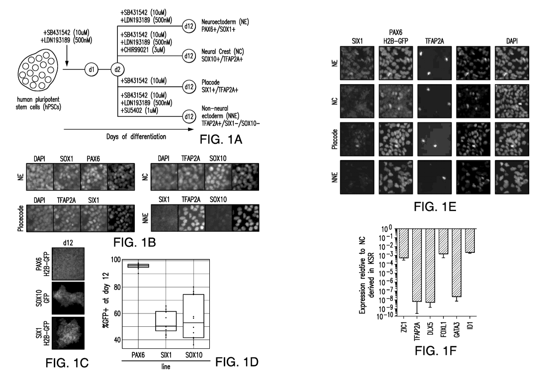
Figures 1A-1F Show (A) the protocol for differentiating neuroectoderm (NE),
neural crest (NC), cranial placode (CP), non-neural ectoderm (NNE) from human
pluripotent stem cells (hPSC) in KSR media. (B) Expression of SOX1, PAX6,
TFAP2A, SOX10 in NE, NC, CP and NNE differentiated in KSR. (C, D)
Differentiation of the human pluripotent stem cells in KSR media into specific
cell
types produced an average of 95%, 50% and 58% of NE, Placode and NC,
respectively.
(E) The percentage of cells expressing PAX6 was improved upon addition of
SB431542 (SB) or dSMADi (i.e., SB and LDN193189) to human pluripotent stem
cells
cultured in E6 media, wherein the percentage of PAX6 positive cells increased
to
.. nearly 90% and 80%, respectively. (F) A comparative gene expression
analysis of
hPSCs differentiated towards NC fate in KSR media versus E6 media revealed a
lack of
non-neural marker expression when cultured in E6 media.
12
CA 03075036 2020-03-05
WO 2019/051248
PCT/US2018/049986
Figures 2A-2J Show (A) BMP signaling has been shown to be important for
the formation of NNE and Placode in the developing chick embryo (Groves and
LaBonne, 2014). (B, C) TFAP2A expression is rapidly upregulated within three
days of
treatment with BMP in a dose dependent manner, in combination with SB431542 in
E6
media. (D) At 2Ong/m1BMP, cells become TFAP2A positive and lack the expression
of
SOX10 and SIXI implying that NNE is triggered by strong BMP signaling
activation.
(E) Culturing hPSC with SB, BMP and 5U5402 generated NNE progenitors, which
expressed immature (K14 positive) and mature (K18 positive) epidermal cell
markers.
(F) A three-day BMP pulse in combination with SB431542 resulted in
differentiation
of hPSCs into SIXI positive CP progenitors. (G) The addition of FGF2, but not
FGF8,
to the E6 culture comprising SB431542 and BMP during differentiation of hPSC
enhanced the formation of SIXI positive CP cells to nearly 50%. (H, I)
Terminal
differentiation of SIXI positive CP precursors (by culturing the cells for 30
days)
resulted in an increase in lens specific factors such as PITX3, Crystallin
Alpha A and B.
(J) Exposure of hPSC to Wnt activation in combination with SB431542, BMP and
FGF2 in E6 media differentiated the cells into CP precursors expressing SIXI
and
PAX3, indicative of trigeminal placode fate.
Figures 3A-3C Show (A) Activation of Wnt signaling in combination with a
short pulse of BMP4 (lng/nil) and SB431542 was capable to generate a nearly
homogenous SOX10 positive NC population. (B) The addition of both Wnt and BMP,
along with SB431542 to the E6 media, activated TFAP2A expression as well as
DLX3,
another marker of the non-neural ectodermal fates. (C) Differentiation of the
SOX10
positive NC precursors gave rise to autonomic and sensory neurons marked by
Isll and
Mash' positive expression.
Figures 4A-4F Show (A, B) the transcriptional expression signatures of all 4
human ectodermal lineages. NE clustered closely with hESCs, while NNE
clustered the
furthest apart from all other ectodermal lineages. NC and CP clustered closely
to each
other. (C D) The 4 ectodermal progenitors upregulate and downregulate
expression of
different genes, which were subjected to gene ontology analysis (Edgar et al.,
2013).
Genes associated with extracellular matrix reorganization were significantly
enriched
in all non-CNS derived cell types. Ontologies associated with NE involve
synaptic
transmission and nervous system development. (E) Genes specifically
upregulated
during ectoderm differentiation that are shared between the CNS and non-CNS
fates
13
CA 03075036 2020-03-05
WO 2019/051248
PCT/US2018/049986
include ANXA1, LGI1, NR2F2 and ZNF503. Factors expressed by non-CNS cell
precursors that distinguish the cells from CNS precursors include NEUROG1,
HAND1,
TFAP2A and TFAP2B. (F) Cells of the 4 ectoderm lineages also exhibited
differences
in gene expression. In NE, SOX1, Hes5 and PAX6 were upregulated, while low-
level
PAX6 transcripts could be found in all other lineages. High levels of the zinc
finger
protein ZNF229 were specifically observed in the NC lineage. ELAVL4 and SMYD1
were preferentially expressed in placode and NNE, respectively.
Figures 5A-5D Show (A) Knockout of TFAP2A expression in hESC resulted
in a loss of TFAP2A expression after a short 3-day induction in the presence
of high
BMP. (B) Wild-type hESCs exhibited robust upregulation of E-cadherin at day 6
of
differentiation under CP or NNE conditions, compared to TFAP2A knockout cells,
which did not express E-cadherin under the CP or NNE conditions. (C, D)
Differentiation of NE was not affected by TFAP2A knockout, as evident by PAX6
and
SOX1 expression. Although NC and CP protocols resulted in increased levels of
SOX1
and PAX6 expression in TFAP2A knockout versus wild type cells, SOX10 and SIX1
expression was also detected, indicating that abolishing TFAP2A expression is
not
sufficient to suppress non-CNS cell types.
Figures 6A-6E Show (A) a small molecule screen using the Library of
Pharmacologically Active Compounds (LOPAC) using the Sixl ::H2B-GFP reporter
line to identify compounds that enhance CP induction. (B, C) Three candidate
compounds that increased expression of SIX1 above the levels observed in
control
differentiations were identified: BRL-5443 a serotonin receptor agonist;
Parthenolide, a
plant hormone that has the capacity to inhibit NF-kB and STAT mediated
confirmation
transcription; and Phenanthroline, a metalloprotease inhibitor. (D)
Differentiation
towards CP showed a five-fold increase in SIX1 expression in the presence of
Phenanthroline over controls, without inducing the expression of other lineage
markers
such as Sox10, T, MyoD or 5ox17. (E) There was a nearly 4-fold increase (69%
versus
18%) of SIX1 positive cells upon addition of Phenanthroline to the CP protocol
in the
absence of FGF2. After the addition of FGF2, or FGF2 plus Phenanthroline, the
enrichment of SIX1 positive cells was decreased to, 34% and 46%, respectively.
Figures 7A-7B Show the culture protocols for the modular generation of NE,
NC, CP and NNE progenitors which comprises dose-dependent BMP exposure in E6
media. (A) The generation of NE was robust in both KSR and E6 systems without
14
CA 03075036 2020-03-05
WO 2019/051248
PCT/US2018/049986
modifications. (B) An initial BMP pulse increased differentiation of NC, CP
and NNE
fates.
Figures 8A-8F Show the generation of PAX6::H2B-GFP and SIX1::H2B-GFP
hESC reporter cell lines. To monitor the acquisition of those various
ectodermal
lineage markers under defined differentiation media, the present example used
three
GFP reporter lines, PAX6::H2B-GFP (A, B, C and D) SOX10::GFP and
SIX1::H2B-GFP (A, B, E and F).
Figures 9A-9E Show (A) KSR differentiation protocol used in the presence of
E6 media. (B) Although hESCs can differentiate into NE precursors in E6
without
addition of any small molecules, the percentage of cells expressing PAX6 was
further
improved upon addition of SB431542 or dSMADi to nearly 90% and 80%,
respectively.
(C, D) NC induction did not generate either PAX6 or SOX10 positive cells in E6
media,
indicating that Wnt activation may alter the regional identity of
differentiating cells
rather than inducing NC. (E) PAX6 positive NE efficiently differentiated
further into
Tbrl positive cortical neurons in E6 media.
Figure 10 Shows that a three-day pulse of BMP signaling in combination with
SB431542 was sufficient to generate SIX1 positive CP precursors in E6 media.
Dose-response studies showed that moderate concentrations of BMP4 (around
5ng/m1)
resulted in CP induction.
Figure 11 Shows that a three-day pulse of BMP signaling in combination with
SB431542 was sufficient to generate SOX10 positive NC precursors in E6 media.
Dose-response studies showed that low concentrations of BMP4 (around 1 ng/ml)
resulted in strong NC induction.
Figures 12A-12C Show the generation of TFAP2A knockout hESCs using the
CRISPR/Cas9 system. (A, B) Two guide RNAs were used to induce frame shift
deletions in TFAP2A, and positive clones were sequenced to determine the
extent and
the nature of the deletion. (C) Ablation of TFAP2A expression was confirmed
using a
short 3-day induction in the presence of high BMPs, which failed to elicit
TFAP2A
expression, compared to wild-type cells.
Figures 13A-13B Show that (A) BRL-5443 and (B) Parthenolide increased the
level of SIX1 expressing CP precursor cells differentiated from hESCs using
the CP
protocol.
CA 03075036 2020-03-05
WO 2019/051248
PCT/US2018/049986
Figures 14A-14C Show (A) that both Matrigel substrate (a coating substrate
composed of thousands of proteins) and Vitronectin substrate (a coating
substrate
composed of a single recombinant protein) yielded highly robust induction
efficiencies.
Differentiations using 50,000 to 300,000 cells/cm2 using both Matrigel (B) and
Vitronectin (C) did not affect cell fate determination.
Figures 15A-15C Show Differentiation of hPSCs into cranial placode using
chemically defined conditions. (A) Schematic representation of cranial placode
in vivo
development and protocol for directed differentiation of human pluripotent
stem cells.
(B) Real-time PCR gene expression time course of key cranial placode (SIX],
EYA1),
non-neural ectoderm (TFAP2A, DLX3/5, GATA3) genes as well as genes probing for
potential contaminates (S0X10, T, SOX] 7, MYOD). Values are normalized to
GAPDH
and expression on day 0 of differentiation (right before switch to
differentiation
medium) and plotted as mean SEM from 4 independent differentiations. (C)
Immunofluorescence analysis comparing protein expression on day 11 of cranial
placode induction protocol and LSB (neuroectoderm). Scale bars: 50 p.m.
Figures 16A-16C Show pituitary specification of anterior cranial placode
derived hPSCs. (A) Schematic representation of pituitary gland in vivo
development
and protocol for directed differentiation of human pluripotent stem cells into
anterior
pituitary-like cells. (B) Real-time PCR analysis comparing expression of key
genes
involved in pituitary development in LSB, Pituitary condition and medium
conditioned
by hypothalamic neuroectoderm (Hypothalamus CM) after 15 days of
differentiation in
the respective medium. Values are normalized to GAPDH and gene expression on
day
15 of lens differentiation (E6 only) and plotted as mean SEM of at least 4
independent
experiments. *: p <0.05, **: p <0.01, ***: p < 0.001 compared to E6 only
condition on
day 15. (C) Immunofluorescence analysis comparing expression of PITX1, LHX3
after
15 days of differentiation under lens or pituitary conditions as well as
expression of
HESX1 and 5IX3/6 on day 15 of pituitary differentiation. Scale bars: 50 pm.
Figures 17A-17C Show pituitary placode induction from unpatterned SIX1
purified cells. (A) Schematic representation of the experimental outline. hESC
were
differentiated under default conditions for 6 days. Unpatterned SIX1+ cells
were FACS
purified and cultured for additional 9 days in various conditions. Cells were
analyzed
on day 15. (B) Gene expression analysis of key pituitary genes in cells grown
in 3
conditions described in A. Values are normalized to GAPDH and gene expression
on
16
CA 03075036 2020-03-05
WO 2019/051248
PCT/US2018/049986
day 15 of lens differentiation (E6 only) and plotted as mean SEM of at least
4
independent experiments. *: p <0.05, **: p <0.01 compared to E6 only condition
on
day 15. (C) Immunofluorescence analysis of SIX1 sorted cells after 9 days of
differentiation in respective medium condition. Arrows indicated absence of
LHX3
expression in SIX1+ cells in co-culture condition. Scale bars: 50 p.m.
Figures 18A-18E Show functional characterization of anterior pituitary cells.
(A) Immunofluorescence analysis of anterior pituitary cells after 30 days of
differentiation. On day 30 the culture contains corticotrophs (ACTH),
somatotrophs
(GH) and gonadotrophs (FSH, LH). Scale bar: 50 jim. (B) In vitro basal hormone
release on day 30 of differentiation as assessed by ELISA. Data is plotted as
mean
SEM of 3 independent experiments. *: p <0.05, ***: p < 0.001, ****: p < 0.0001
compared to no cells (differentiation medium only). (C-E) Quantification of
hormone
levels after 24h of in vitro stimulation using compounds triggering hormone
release.
ACTH release was specifically induced by CRF, Stressin or Urocortin and not by
Somatocrinin or Ghrelin (C), GH release was induced by Somatocrinin but not
CRF (D)
and FSH release was induced by Nafarelin (E). Data is plotted as mean SEM of
3
independent experiments. *: p <0.05 compared to the solvent control.
Figures 19A-19E Show temporal single cell qRT-PCR analysis of anterior
pituitary development in vitro. (A,B) Principal component analysis of single
cells on
day 30 (black) and day 60 (green) of differentiation reveals two distinct
populations of
cells. (C) Unsupervised hierarchical clustering of day 30 and day 60 cells
using 34
different primer pairs identifies 2 clusters of cells with very few leading
cells (day 30
cells resembling day 60 cells) and cells lacking behind (day 60 cells still
more closely
resembling day 30). (D) Quantification of hormone expressing cells on day 30
and day
60 as well as percentage of cells expressing more than 1 hormonal transcript
per cell. (E)
Expression of individual hormones per single cell on day 30 and day 60
respectively.
Figures 20A-20E Show specification of hormonal cells of the pituitary in
vitro.
(A) Bulk qRT-PCR analysis of day 60 cells patterned with FGF8, FGF8/BMP2 or
BMP2 for 30 days. Patterning with BMP2 induced a more ventral cell identity
(PIT],
GATA2, GH], FSHB and LHB) while FGF8 suppressed dorsal cell types (FSHB). Data
is plotted as mean SEM of 2-4 independent experiments.. *: p < 0.05, **: p
<0.01,
***: p <0.001 compared to the "default" pituitary differentiation on day 60.
(B)
Unsupervised hierarchical clustering of FGF8, FGF8/BMP2 and BMP2 patterned
cells
17
CA 03075036 2020-03-05
WO 2019/051248
PCT/US2018/049986
using 34 primer pairs identified 3 larger clusters of cells with cluster 2
mainly
comprised of cells patterned by FGF8 (or FGF8/BMP2) and cluster 3 mainly
comprised
of cells patterned by BMP2 (or FGF8/BMP2). (C) Quantification of hormonal
transcripts per cell in different patterning conditions. Data is plotted as
percentage of
cells expressing the respective transcript (ct < 35 cycles in combination with
a proper
melting curve). (D) Immunofluorescence analysis (representative images) of
hormone
expression in cells patterned with FGF8, FGF8/BMP2 or BMP2 on day 60 of
differentiation. Scale bars: 50 um. (E) Quantification of hormone expressing
cells (per
subtype) in different patterning conditions on day 60 of differentiation. High
levels of
.. FGF8 induced dorsal fate (ACTH) while intermediate levels of FGF8 and BMP2
induced dorsal/ventral fates (PRL and GH) compared to the default condition
(E6 only).
Data is plotted as mean SEM of 2 independent experiments. *: p < 0.05, **: p
< 0.01
compared to the "default" (E6 only) pituitary differentiation on day 60.
Figures 21A-21G Show in vivo survival and function of hPSC-derived anterior
pituitary cells. (A) Schematic representation of experimental layout. After
surgical
removal of the pituitary gland and confirmation of hypopituitarism, cells
embedded in
Matrigel were transplanted subcutaneously. (B-E) ACTH (B), GH (C), LH (D) and
corticosterone (E) levels were quantified in serum for up to 7 weeks after
transplantation of the cells using ELISA. Data is plotted as mean SEM with
each dot
representing an individual animal. *: p <0.05, **: p <0.01 compared to the
corresponding sham control. (F) Immunohistological analysis of grafts 7 weeks
after
transplantation. Cells for each of the 6 hormonal lineages of the anterior
pituitary gland
were detectable within the graft. Scale bars: 50 jim. (G) Quantification of
cells
expressing ACTH and the corresponding graft volume 7 weeks after
transplantation.
Data is plotted as mean SEM with each dot representing an individual animal
(3
animals total).
Figures 22A-22C Show "default" conditions in chemically defined media
result in lens placode specification. (A) After 30 days of differentiation
under "default"
conditions (E6 only) lentoid bodies (circled structures in brightfield image)
staining
positive for the lens marker PAX6 are clearly identifiable. Scale bars: 50
jim. (B) After
an additional 90 days of differentiation (day 120) the majority of the cells
is expressing
crystallin the predominant structural proteins in the lens. Scale bars: 50
jim. (C)
qRT-PCR gene expression time course during lens differentiation. Cells
differentiated
18
CA 03075036 2020-03-05
WO 2019/051248
PCT/US2018/049986
for 120 days express the lens characteristic transcripts PITX3, CRYAA and
CRYAB.
Values have been normalized to GAPDH and expression in undifferentiated ES
cells
and are plotted as means +/- SEM of 4 independent consecutive experiments.
Figures 23A-23B Show quantification of ectodermal subtypes within the
pituitary differentiation using reporter cell lines. (A) Cells (different
reporter cell lines)
differentiated for 6 and 11 days under either default placode or pituitary
conditions
were analyzed using Flow Cytometry for SOX10, PAX6 or SIX1 expression.
Representative Flow Cytometry plots with percentages are shown. (B)
Quantification
of data form (A) reveals very few contaminating neural crest cells (SOX10+)
while
confirming the anterior cranial placode character of the cells (SIX1+, PAX6+).
Data is
plotted as mean SEM of 2-8 independent experiments.
Figures 24A-24B Show differentiation of hESCs into hypothalamic ectoderm.
(A) Immunofluorescence comparison of cells differentiated for 15 days under
either
pituitary or hypothalamus condition. Cells were stained for either FOXG1
(Pituitary) or
NKX2.1 (Hypothalamus). Scale bars: 50 tim. (B) qRT-PCR analysis of day 15
cells
differentiated under pituitary or hypothalamic ectoderm condition probing for
NKX2. 1
and FOXG1. Values have been normalized to GAPDH and expression in day 6
placode
cells and are plotted as means +/- SEM of 2-4 independent experiments.
Figures 25A-25E Show generation of the SIX1 knock-in reporter line. (A)
Schematic representation of the TALEN-based targeting of the endogenous SIX1
Stop
codon using an eGFP containing reporter cassette. (B) PCR screening of
targeted
clones using primers annealing to the genomic region just outside the SIX1
homology
arms. Clone #6 was selected and used in the study. (C) Karyogram of H9
SIX1H2B::GFP clone #6 showing a normal female (XX) karyotype. (D) qRT-PCR
analysis of SIX/ expression in SIX1H2B::GFP cells differentiated for 6 days
under
placode conditions sorted positively and negatively for GFP. Values are
normalized to
GAPDH and unsorted cells from the same experiment and are plotted as means +/-
SEM of a single experiment with 2 technical replicates. (E) Immunofluorescence
analysis of day 11 pituitary cells staining for endogenous SIX1 (red) and GFP
under the
control of the endogenous SIX1 promoter (green). Scale bar: 50 p.m.
Figure 26 Shows quantification of hormonal transcripts in single cells using
single cell qRT-PCR. Single cell PCR data from day 30 and day 60 of the
"default"
pituitary differentiation protocol were mined for cells expressing at least
one hormonal
19
CA 03075036 2020-03-05
WO 2019/051248
PCT/US2018/049986
transcript. Data is plotted as percentage of cells expressing the respective
transcript(s)
(ct < 35 cycles in combination with a proper melting curve).
Figure 27 Shows the list of primers used in the single cell qRT-PCR
experiments of Example 2.
Figure 28 Shows the list of antibodies used in Example 2.
Figures 29A-29B Show differentiation of pluripotent cells into neural crest
derived cells. (A) Pluripotent cells were cultured in E6 media supplemented
with
SB431542, BMP4 and CHIR99021 for two days (i.e., from dO to d2 of culture in
E6
media), and in E6 media supplemented with SB431542 and CHIR from d2 to dll, to
differentiate into neural crest progenitor cells. (B) The neural crest
progenitors
spontaneously differentiated into cells expressing MASH1 and ISL1 at d25.
Figures 30A-30B Show comparison of the traditional KSR-based pituitary
induction with the new cGMP-ready induction. Cells grown on feeders in KSR-
based
medium were differentiated using the old Dincer et. al. protocol (PIP-KSR). To
.. compensate for KSR lot-to-lot variation 2 concentrations of LDN-193189 were
used.
Cells grown under feeder-free Essential8 conditions using the PIP-E6 protocol
were
differentiated in parallel. (A) qRT-PCR analysis of day 15 cells
differentiated under
PIP-KSR and PIP-E6 condition probing for SIX1, TFAP2A, PAX6, PITX1, PITX2,
PITX3 and PITX4. Values have been normalized to GAPDH and expression in day 15
PIP-E6 cells and are plotted as means +/- SEM of 2 independent experiments.
(B)
Immunofluorescence comparison of cells differentiated for 15 days under either
PIP-KSR or PIP-E6 condition. Cells were stained for either SIX1 (pan placode)
or
LHX3 (pan pituitary). Scale bars: 50 [tm.
Figures 31A-31E Show cell line comparison of pituitary induction protocol in
E8/E6 and replacing recombinant SHH with small molecule smoothened agonists.
Four different hESC lines (including the H9 SIX1::H2B-GFP clone #6) and 1
hiPSC
cell line were differentiated in parallel using the cGMP-ready pituitary
induction
protocol. (A) qRT-PCR analysis of day 15 cells differentiated under pituitary
condition
probing for the pan placodal marker SIX1 as well as the pan anterior pituitary
genes
PITX1, PITX2, LHX3 and LHX4. Values have been normalized to GAPDH and
expression in day 30 wt H9 cells and are plotted as means +/- SEM of 2-4
independent
experiments. (B) qRT-PCR analysis of day 30 cells differentiated under
pituitary
condition without sorting on day 15 probing for 2 anterior pituitary hormone
transcripts
CA 03075036 2020-03-05
WO 2019/051248
PCT/US2018/049986
POMC and GH1 Values have been normalized to GAPDH and expression in day 30 wt
H9 cells and are plotted as means +/- SEM of 3 independent experiments. (C)
Immunofluorescence analysis comparing protein expression on day 15 of
pituitary
placode induction across different hPSC lines. Scale bars: 50 [tm. To
investigate
whether SHUT can be replaced by small molecules, cells were differentiated
using the
cGMP-ready pituitary placode induction protocol using either recombinant SHH
or one
of the small molecule agonists purmorphamine or SAG in combination with FGF8
and
FGF10. Lens placode differentiation was performed in parallel and served as a
negative
control. (D) qRT-PCR analysis of day 15 cells, differentiated under pituitary
conditions
using either SHH, purmorphamine or SAG from day 4 on, probing for the pan
pituitary
genes PITX1, PITX2, LHX3, LHX4, HESX1 as well as the hormone transcript
POMC1 as well as the lens marker PITX3. Values have been normalized to GAPDH
and expression in day 15 lens placode and are plotted as means +/- SEM of 3
independent experiments. (E) Immunofluorescence analysis comparing protein
expression on day 15 of pituitary placode and lens induction using SHH and the
small
molecule alternatives purmorphamine and SAG. Early lentoid bodies (circled
structures) start to downregulate expression of the pan placodal marker SIX1
while
pituitary placode retains high SIX1 expression in combination with expression
of
LHX3. Scale bars: 50 [tm.
Figure 32 Shows heatmaps of raw ct values for each cell and gene obtained by
single cell q-RT PCR. Raw ct values for every cell and gene obtained for every
single
cell PCR run are displayed as unprocessed heat maps.
Figure 33 Shows immunofluorescence validation of single cell q-RT PCR
results and quantification of hormonal transcripts in single cells using
single cell
qRT-PCR. Immunofluorescence analysis of day 15 and day 30 cells differentiated
under pituitary conditions. Cells were co-stained for the progenitor marker
HESX1 and
the transient cortiocotroph marker NEUROD1. Scale bars: 50 [tm.
Figures 34A-34B Show surface marker screen to identify hormonal subclass
specific markers. (A) Schematic representation of experimental procedure. (B)
Heatmap of screen results. Percentage indicates cells staining positive for
respective
marker.
Figure 35 Show comparison of endogenous rat hormones in unlesioned
animals and graft derived human hormones in lesioned animals. Graft-derived
21
CA 03075036 2020-03-05
WO 2019/051248
PCT/US2018/049986
hormone levels shown in Figure 21A-G (week 5) are compared to endogenous
hormone levels in unlesioned rats.
Figure 36 Shows the differentiation scheme in accordance with certain
embodiments of the presently disclosed subject matter.
Figures 37A-37G Show BMP signaling is necessary to obtain NNE. (A)
Diagram of differentiation strategies by replacement of KSR for E6. (B)
Representative immunofluorescence staining of particular lineage markers
during the
differentiation of hPSCs into the ectodermal lineages. (C) Representation of
the neural
plate border model and important signaling pathways that influence particular
cell fates.
(D) Immunofluorescence staining of differentiating cells treated with various
concentrations of BMP4 for 3 days. (E) (Quantification of TFAP2A+ cells at
various
BMP4 concentrations after 3 days of treatment. Values represent mean SEM.
(F)
The derivation of TFAP2A+, PAX6-, SIX1-, and SOX10- NNE is achieved by using a
high concentration of BMP4 (20 ng/ml). (G) Immunofluorescence staining of
keratinocyte markers K18 and K14 upon further differentiation of the NNE at
two
different time points. Scale bars, 50 p.m.
Figures 38A-381 Show a BMP gradient is sufficient to derive NC and CP cells.
(A) The expression of SIX1::GFP+ placode using a gradient of BMP4. Each bar
within
the group represents an independent replicate. (B) Quantification of SIX:: GFP
after
treatment of cells with FGF2 or FGF8 during the differentiation. (C)
Quantitative PCR
of anterior markers PAX6 and 5IX3 during two different time points along the
differentiation. Values represent mean SEM. (D) Immunofluorescence staining
of
CRYAA and CRYAB in lens placode cultures on day 30. (E) Immunofluorescence
staining of PAX6+ lens placode in the absence of WNT and PAX3+ trigeminal
placodes after the addition of WNT signals. (F) The expression of SOX10::GFP+
NC
using a gradient of BMP4. Each bar within the group represents an independent
replicate. (G) Immunofluorescence staining of differentiating cells treated
with various
concentrations of BMP4 with or without 600nM CHIR for 3 days. (H)
Immunofluorescence staining of spontaneously differentiated NC cells for their
ability
to generate ASCL1 and ISL1 neurons representing autonomic and sensory neurons,
respectively. (I) Calcium imaging was performed on differentiated sensory and
autonomic neurons for a response to glutamate. Values represent mean SEM.
Scale
bars, 50 mm.
22
CA 03075036 2020-03-05
WO 2019/051248
PCT/US2018/049986
Figures 39A-39B Show novel differentiation strategies are applicable to a
range of human embryonic and induced PSCs. (A) Representative images used for
high-content imaging of validated antibodies to mark the different ectodermal
lineages.
(B) Quantification of the percentage of positive cells during a particular
differentiation.
Biological replicates (n = 4) and technical replicates (n = 2 per biological
replicate)
were performed and quantified. Scale bars, 50 mm.
Figures 40A-40J Show differentiation of hPSCs towards the four ectodermal
lineages in serum replacement conditions affects regional patterning. (A)
Schematic of
the general strategies for differentiation into the four ectodermal lineages
using
knockout serum replacement. (B) Transcription factor expression combinations
that
distinguish cell identity at the end of the differentiation (i.e. day 12). (C)
Schematic of
targeting the donor plasmid into the Pax6 locus. (D) PCR of genomic DNA to
identify
clones that carry the reporter transgenes. E. Intracellular FACS for Pax6 and
GFP
during neuroectoderm formation. (F) Quantitative PCR for Sixl in unsorted,
Sixl-GFP
positive and Sixl-GFP negative cells. (G) PAX6, SIX1 and SOX10 GFP reporter
line
expression of GFP at day 12 of differentiation. (H) Quantification of the
percentage of
GFP undergoing differentiation towards specific lineages. (I) Several knockout
serum
replacement lots were tested for the ability to generate the neuroectoderm
(three are
presented), determined for the expression of PAX6 and downregulation of the
stem cell
factor OCT4. (J) Similar to I, the expression of SOX1 is consistent between
lots of
KSR, however the anterior marker FOXG1 is variable even in the presence of the
WNT
inhibitor, XAV-939. Scale bars 50um.
Figures 41A-41G Show differentiation toward the ectoderm is skewed toward
the CNS in the chemically defined system. (A) Quantification of Pax6::GFP
positive
cells in the absence of small molecules, addition of SB, and the addition of
LDN plus
SB. (B) Immunofluorescence staining of ZO-1 and 50X2 representing rosette
stage
cells. (C) Differentiation of the rosettes into neurons stained with TUJ-1 and
TBR1. (D)
Cortical neurons (day 50 of differentiation) exhibit response to glutamate.
(E)
Immunofluorescence of SOX10 and SIX1 with PAX6 in the differentiation towards
neural crest and placode, respectively. (F) Quantification of PAX6::H2B-GFP
expression in E. (G) Quantitative PCR of general immediate early genes during
the
differentiation of neural crest in KSR and E6.
Figures 42A-42D Show differentiation strategies towards the ectodermal
23
CA 03075036 2020-03-05
WO 2019/051248
PCT/US2018/049986
lineages are applicable to other pluripotent stem cell lines. (A)
Representative images
of immunofluorescence staining on differentiations of the ectoderm. (B)
Quantification on the percentage of cells positive for particular markers
during the
differentiation. (C) Comparative analysis of a dataset of purified chick
neural crest
compared to the purified human neural crest cells in this study. The overlap
significance was tested using the hypergeometric distribution. (D) The list of
genes in
common from the Venn diagram in C. Scale bars 5011m.
Figures 43A-43H Show validation and characterization of TFAP2A knockout
cells. (A) Schematic of the guide RNAs targeting TFAP2A. (B) Sequencing
results of
three potential clones indicate two had frameshift mutations (AP2A.10 and
AP2A.11)
and the other clone (AP2A.4) harbors a frameshift mutation on one allele and a
9bp on
the other allele. (C) Immunofluorescence of TFAP2A in different ES clones
treated
with 20ng/m1 BMP4 for 3 days. (D) A western blot was performed to validate the
loss
of protein expression in the mutant lines after 3 days of BMP4 treatment. (E)
Immunofluorescence of SOX10 positive cells lacking TFAP2A expression on day
12.
(F) Immunofluorescence of SIX1 positive cells lacking TFAP2A expression on day
12.
(G) Quantitative PCR analysis of the expression of KRT16 and WISP1 during the
derivation of NNE from the TFAP2A KO cells compared to wildtype. (H)
Immunofluorescence staining of TFAP2C in combination with Sox10 GFP and
Sixl-H2B GFP demonstrating little overlap. Scale bars 5011m.
Figures 44A-44B Show Other hit validation from the chemical screen did not
produce a significant difference in SIX1::GFP expression. (A) Quantification
of SIX1
expression after the treatment of differentiating placode cells with BRL-
54443. (B) As
in A, but with Parthenolide.
5. DETAILED DESCRIPTION OF THE INVENTION
The presently disclosed subject matter relates to in vitro methods for
inducing
differentiation of human stem cells to cells that express one or more
neuroectoderm,
neural crest, cranial placode, or non-neural ectoderm lineage marker, and
cells
produced by such methods, and compositions comprising such cells. Also
provided are
uses of such cells for treating neurodegenerative disorders.
For purposes of clarity of disclosure and not by way of limitation, the
detailed
description is divided into the following subsections:
5.1. Definitions;
24
CA 03075036 2020-03-05
WO 2019/051248
PCT/US2018/049986
5.2. Method of Differentiating Stem Cells;
5.3 Compositions Comprising Differentiated Cell Populations;
5.4. Method of Treating Neurodegenerative and Pituitary Disorders;
5.5. Kits;
5.6 Methods of Screening Therapeutic Compounds; and
5.7 Methods of Screening for Compounds that Increase NE, NC, CP or NNE
fate.
5.1 Definitions
The terms used in this specification generally have their ordinary meanings in
the art, within the context of this invention and in the specific context
where each term
is used. Certain terms are discussed below, or elsewhere in the specification,
to provide
additional guidance to the practitioner in describing the compositions and
methods of
the invention and how to make and use them.
The term "about" or "approximately" means within an acceptable error range
for the particular value as determined by one of ordinary skill in the art,
which will
depend in part on how the value is measured or determined, i.e., the
limitations of the
measurement system. For example, "about" can mean within 3 or more than 3
standard
deviations, per the practice in the art. Alternatively, "about" can mean a
range of up to
20%, e.g., up to 10%, up to 5%, or up to 1% of a given value. Alternatively,
particularly
with respect to biological systems or processes, the term can mean within an
order of
magnitude, e.g., within 5-fold, or within 2-fold, of a value.
As used herein, the term "signaling" in reference to a "signal transduction
protein" refers to a protein that is activated or otherwise affected by ligand
binding to a
membrane receptor protein or some other stimulus. Examples of signal
transduction
protein include, but are not limited to, a SMAD, a wingless (Wnt) complex
protein,
including beta-catenin, NOTCH, transforming growth factor beta (TGF[3),
Activin,
Nodal, glycogen synthase kinase 313 (GSK3 p) proteins, bone morphogenetic
proteins
(BMP) and fibroblast growth factors (FGF). For many cell surface receptors or
internal
receptor proteins, ligand-receptor interactions are not directly linked to the
cell's
response. The ligand activated receptor can first interact with other proteins
inside the
cell before the ultimate physiological effect of the ligand on the cell's
behavior is
produced. Often, the behavior of a chain of several interacting cell proteins
is altered
following receptor activation or inhibition. The entire set of cell changes
induced by
receptor activation is called a signal transduction mechanism or signaling
pathway.
CA 03075036 2020-03-05
WO 2019/051248
PCT/US2018/049986
As used herein, the term "signals" refer to internal and external factors that
control changes in cell structure and function. They can be chemical or
physical in
nature.
As used herein, the term "ligands" refers to molecules and proteins that bind
to
receptors, e.g., transforming growth factor-beta (TFG[3), Activin, Nodal, bone
morphogenic proteins (BMPs), etc.
"Inhibitor" as used herein, refers to a compound or molecule (e.g., small
molecule, peptide, peptidomimetic, natural compound, siRNA, anti-sense nucleic
acid,
aptamer, or antibody) that interferes with (e.g., reduces, decreases,
suppresses,
.. eliminates, or blocks) the signaling function of s molecule or pathway. An
inhibitor can
be any compound or molecule that changes any activity of a named protein
(signaling
molecule, any molecule involved with the named signaling molecule, a named
associated molecule, such as a glycogen synthase kinase 313 (GSK3[3)) (e.g.,
including,
but not limited to, the signaling molecules described herein). For example, an
inhibitor
of SMAD signaling can function, for one example, via directly contacting SMAD
signaling, contacting SMAD mRNA, causing conformational changes of SMAD,
decreasing SMAD protein levels, or interfering with SMAD interactions with
signaling
partners (e.g., including those described herein), and affecting the
expression of SMAD
target genes (e.g. those described herein). Inhibitors also include molecules
that
indirectly regulate SMAD biological activity by intercepting upstream
signaling
molecules (e.g., within the extracellular domain). Examples of a SMAD
signaling
inhibitor molecules and an effect include: Noggin which sequesters bone
morphogenic
proteins, inhibiting activation of ALK receptors 1,2,3, and 6, thus preventing
downstream SMAD activation. Likewise, Chordin, Cerberus, Follistatin,
similarly
sequester extracellular activators of SMAD signaling. Bambi, a transmembrane
protein,
also acts as a pseudo-receptor to sequester extracellular TGF[3 signaling
molecules.
Other SMAD inhibitors include dorsomorphin. Antibodies that block activins,
nodal,
TGF[3, and BMPs are contemplated for use to neutralize extracellular
activators of
SMAD signaling, and the like. Although the foregoing example relates to SMAD
signaling inhibition, similar or analogous mechanisms can be used to inhibit
other
signaling molecules. Examples of inhibitors include but are not limited to:
LDN193189 (LDN) and SB431542 (SB) (LSB) for SMAD signaling inhibition,
XAV939 (X) for Wnt inhibition, and 5U5402 (S) for FGF signaling inhibition.
26
CA 03075036 2020-03-05
WO 2019/051248
PCT/US2018/049986
Inhibitors are described in terms of competitive inhibition (binds to the
active
site in a manner as to exclude or reduce the binding of another known binding
compound) and allosteric inhibition (binds to a protein in a manner to change
the
protein conformation in a manner which interferes with binding of a compound
to that
protein's active site) in addition to inhibition induced by binding to and
affecting a
molecule upstream from the named signaling molecule that in turn causes
inhibition of
the named molecule. An inhibitor can be a "direct inhibitor" that inhibits a
signaling
target or a signaling target pathway by actually contacting the signaling
target.
"Activators," as used herein, refer to compounds that increase, induce,
stimulate,
activate, facilitate, or enhance activation the signaling function of the
molecule or
pathway, e.g., Wnt signaling, BMP signaling, FGF signaling, etc.
As used herein, the term "derivative" refers to a chemical compound with a
similar core structure.
As used herein, the term "a population of cells" or "a cell population" refers
to a
group of at least two cells. In non-limiting examples, a cell population can
include at
least about 10, at least about 100, at least about 200, at least about 300, at
least about
400, at least about 500, at least about 600, at least about 700, at least
about 800, at least
about 900, at least about 1000 cells, at least about 5,000 cells or at least
about 10,000
cells or at least about 100,000 cells or at least about 1,000,000 cells. The
population
may be a pure population comprising one cell type, such as a population of NE,
CP, NC
or NNE precursors, or a population of undifferentiated stem cells.
Alternatively, the
population may comprise more than one cell type, for example a mixed cell
population.
As used herein, the term "stem cell" refers to a cell with the ability to
divide for
indefinite periods in culture and to give rise to specialized cells. A human
stem cell
refers to a stem cell that is from a human.
As used herein, the term "embryonic stem cell" and "ESC" refer to a primitive
(undifferentiated) cell that is derived from preimplantation-stage embryo,
capable of
dividing without differentiating for a prolonged period in culture, and are
known to
develop into cells and tissues of the three primary germ layers. A human
embryonic
stem cell refers to an embryonic stem cell that is from a human. As used
herein, the
term "human embryonic stem cell" or "hESC" refers to a type of pluripotent
stem cells
derived from early stage human embryos, up to and including the blastocyst
stage, that
is capable of dividing without differentiating for a prolonged period in
culture, and are
27
CA 03075036 2020-03-05
WO 2019/051248
PCT/US2018/049986
known to develop into cells and tissues of the three primary germ layers.
As used herein, the term "embryonic stem cell line" refers to a population of
embryonic stem cells which have been cultured under in vitro conditions that
allow
proliferation without differentiation for up to days, months to years.
As used herein, the term "totipotent" refers to an ability to give rise to all
the
cell types of the body plus all of the cell types that make up the
extraembryonic tissues
such as the placenta.
As used herein, the term "multipotent" refers to an ability to develop into
more
than one cell type of the body.
As used herein, the term "pluripotent" refers to an ability to develop into
the
three developmental germ layers of the organism including endoderm, mesoderm,
and
ectoderm.
As used herein, the term "induced pluripotent stem cell" or "iPSC" refers to a
type of pluripotent stem cell, similar to an embryonic stem cell, formed by
the
introduction of certain embryonic genes (such as a OCT4, SOX2, and KLF4
transgenes)
(see, for example, Takahashi and Yamanaka Cell 126, 663-676 (2006), herein
incorporated by reference) into a somatic cell, for examples, CI 4, C72, and
the like.
As used herein, the term "somatic cell" refers to any cell in the body other
than
gametes (egg or sperm); sometimes referred to as "adult" cells.
As used herein, the term "somatic (adult) stem cell" refers to a relatively
rare
undifferentiated cell found in many organs and differentiated tissues with a
limited
capacity for both self-renewal (in the laboratory) and differentiation. Such
cells vary in
their differentiation capacity, but it is usually limited to cell types in the
organ of origin.
As used herein, the term "neuron" refers to a nerve cell, the principal
functional
units of the nervous system. A neuron consists of a cell body and its
processes¨ an
axon and one or more dendrites. Neurons transmit information to other neurons
or cells
by releasing neurotransmitters at synapses.
As used herein, the term "proliferation" refers to an increase in cell number.
As used herein, the term "undifferentiated" refers to a cell that has not yet
.. developed into a specialized cell type.
As used herein, the term "differentiation" refers to a process whereby an
unspecialized embryonic cell acquires the features of a specialized cell such
as a heart,
liver, or muscle cell. Differentiation is controlled by the interaction of a
cell's genes
28
CA 03075036 2020-03-05
WO 2019/051248
PCT/US2018/049986
with the physical and chemical conditions outside the cell, usually through
signaling
pathways involving proteins embedded in the cell surface.
As used herein, the term "directed differentiation" refers to a manipulation
of
stem cell culture conditions to induce differentiation into a particular (for
example,
desired) cell type, such as neural, neural crest, cranial placode, and non-
neural
ectoderm precursors.
As used herein, the term "directed differentiation" in reference to a stem
cell
refers to the use of small molecules, growth factor proteins, and other growth
conditions to promote the transition of a stem cell from the pluripotent state
into a more
mature or specialized cell fate.
As used herein, the term "inducing differentiation" in reference to a cell
refers
to changing the default cell type (genotype and/or phenotype) to a non-default
cell type
(genotype and/or phenotype). Thus, "inducing differentiation in a stem cell"
refers to
inducing the stem cell (e.g., human stem cell) to divide into progeny cells
with
characteristics that are different from the stem cell, such as genotype (e.g.,
change in
gene expression as determined by genetic analysis such as a microarray) and/or
phenotype (e.g., change in expression of a protein, such as SOX1, PAX6, SOX10,
SIX1,
PITX3, and TFAP2A).
As used herein, the term "cell culture" refers to a growth of cells in vitro
in an
artificial medium for research or medical treatment.
As used herein, the term "culture medium" refers to a liquid that covers cells
in
a culture vessel, such as a Petri plate, a multi-well plate, and the like, and
contains
nutrients to nourish and support the cells. Culture medium may also include
growth
factors added to produce desired changes in the cells.
As used herein, the term "contacting" cells with a compound (e.g., one or more
inhibitor, activator, and/or inducer) refers to exposing cells to a compound,
for example,
placing the compound in a location that will allow it to touch the cell. The
contacting
may be accomplished using any suitable methods. For example, contacting can be
accomplished by adding the compound to a tube of cells. Contacting may also be
accomplished by adding the compound to a culture medium comprising the cells.
Each
of the compounds (e.g., the inhibitors and activators disclosed herein) can be
added to a
culture medium comprising the cells as a solution (e.g., a concentrated
solution).
Alternatively or additionally, the compounds (e.g., the inhibitors and
activators
29
CA 03075036 2020-03-05
WO 2019/051248
PCT/US2018/049986
disclosed herein) as well as the cells can be present in a formulated cell
culture medium.
An effective amount is an amount that produces a desired effect.
As used herein, the term "in vitro" refers to an artificial environment and to
processes or reactions that occur within an artificial environment. In vitro
environments exemplified, but are not limited to, test tubes and cell
cultures.
As used herein, the term "in vivo" refers to the natural environment (e.g., an
animal or a cell) and to processes or reactions that occur within a natural
environment,
such as embryonic development, cell differentiation, neural tube formation,
etc.
As used herein, the term "expressing" in relation to a gene or protein refers
to
making an mRNA or protein which can be observed using assays such as
microarray
assays, antibody staining assays, and the like.
As used herein, the term "marker" or "cell marker" refers to gene or protein
that
identifies a particular cell or cell type. A marker for a cell may not be
limited to one
marker, markers may refer to a "pattern" of markers such that a designated
group of
markers may identity a cell or cell type from another cell or cell type.
As used herein, the term "derived from" or "established from" or
"differentiated
from" when made in reference to any cell disclosed herein refers to a cell
that was
obtained from (e.g., isolated, purified, etc.) a parent cell in a cell line,
tissue (such as a
dissociated embryo, or fluids using any manipulation, such as, without
limitation,
single cell isolation, culture in vitro, treatment and/or mutagenesis using
for example
proteins, chemicals, radiation, infection with virus, transfection with DNA
sequences,
such as with a morphogen, etc., selection (such as by serial culture) of any
cell that is
contained in cultured parent cells. A derived cell can be selected from a
mixed
population by virtue of response to a growth factor, cytokine, selected
progression of
cytokine treatments, adhesiveness, lack of adhesiveness, sorting procedure,
and the
like.
An "individual" or "subject" herein is a vertebrate, such as a human or
non-human animal, for example, a mammal. Mammals include, but are not limited
to,
humans, primates, farm animals, sport animals, rodents and pets. Non-limiting
examples of non-human animal subjects include rodents such as mice, rats,
hamsters,
and guinea pigs; rabbits; dogs; cats; sheep; pigs; goats; cattle; horses; and
non-human
primates such as apes and monkeys.
CA 03075036 2020-03-05
WO 2019/051248
PCT/US2018/049986
As used herein, the term "disease" refers to any condition or disorder that
damages or interferes with the normal function of a cell, tissue, or organ.
As used herein, the term "treating" or "treatment" refers to clinical
intervention
in an attempt to alter the disease course of the individual or cell being
treated, and can
be performed either for prophylaxis or during the course of clinical
pathology.
Therapeutic effects of treatment include, without limitation, preventing
occurrence or
recurrence of disease, alleviation of symptoms, diminishment of any direct or
indirect
pathological consequences of the disease, preventing metastases, decreasing
the rate of
disease progression, amelioration or palliation of the disease state, and
remission or
improved prognosis. By preventing progression of a disease or disorder, a
treatment
can prevent deterioration due to a disorder in an affected or diagnosed
subject or a
subject suspected of having the disorder, but also a treatment may prevent the
onset of
the disorder or a symptom of the disorder in a subject at risk for the
disorder or
suspected of having the disorder.
5.2 Method of Differentiatinji Stem Cells
The presently disclosed subject matter provides for in vitro methods for
inducing differentiation of stem cells (e.g., human stem cells). Non-limiting
examples
of human stem cells include human embryonic stem cells (hESC), human
pluripotent
stem cell (hPSC), human induced pluripotent stem cells (hiPSC), human
parthenogenetic stem cells, primordial germ cell-like pluripotent stem cells,
epiblast
stem cells, F-class pluripotent stem cells, somatic stem cells, cancer stem
cells, or any
other cell capable of lineage specific differentiation. In certain
embodiments, the
human stem cell is a human embryonic stem cell (hESC). In certain embodiments,
the
human stem cell is a human induced pluripotent stem cell (hiPSC).
The presently disclosed subject matter is based at least in part on the
discovery
that the non-CNS ectodermal lineages of the neural crest (NC), cranial placode
(CP)
and non-neural ectoderm (NNE) can be differentiated from human stem cells by
inhibition of SMAD signaling along with activation of BMP signaling, wherein
BMP
signaling is activated for at least 2 days after initial contact of the cells
to effective
amounts of one or more SMAD inhibitor and BMP activator, and wherein the cells
are
further contacted with effective amounts of one or more NC, CP or NNE lineage
specific activators and inhibitors. For example, the stem cells can be
differentiated to
NC by further contacting the cells with effective amounts of one or more Wnt
activator;
31
CA 03075036 2020-03-05
WO 2019/051248
PCT/US2018/049986
CP can be differentiated from the stem cells by further contacting the stem
cells with
effective amounts of one or more activator of FGF; and NNE can be
differentiated from
the stem cells by further contacting the stem cells with effective amounts of
one or more
inhibitor of FGF.
In certain embodiments, a presently disclosed differentiation method comprises
contacting a population of human stem cells with effective amounts of one or
more
inhibitor of transforming growth factor beta (TGF[3)/Activin-Nodal signaling,
which
results in inhibition of Small Mothers Against Decapentaplegic (SMAD)
signaling. In
certain embodiments, the inhibitor of TGF[3/Activin-Nodal signaling
neutralizes the
ligands including TGF[3s, bone morphogenetic proteins (BMPs), Nodal, and
activins,
or blocking their signal pathways through blocking the receptors and
downstream
effectors.
Non-limiting examples of inhibitors of TGF[3/Activin-Nodal signaling are
disclosed in WO/2010/096496, WO/2011/149762, WO/2013/067362,
WO/2014/176606, WO/2015/077648, Chambers et al., Nature Biotechnology 27,
275-280 (2009), and Chambers et al., Nature biotechnology 30, 715-720 (2012),
which
are incorporated by reference in their entireties herein for all purposes. In
certain
embodiments, the one or more inhibitor of TGF[3/Activin-Nodal signaling is a
small
molecule selected from the group consisting of SB431542, derivatives thereof,
and
mixtures thereof "SB431542" refers to a molecule with a number CAS 301836-41-
9, a
molecular formula of C22th8N403, and a name of
4-14-(1,3-benzodioxo1-5-y1)-5-(2-pyridiny1)-1H-imidazol-2-y11-benzamide, for
example, see structure below:
/--()
0
e ______________________
=-=" "*.
ci
5.2.1 Method of Differentiating Neural Crest (NC) Precursors
In certain embodiments, the presently disclosed subject matter provides for in
vitro methods for inducing differentiation of human stem cells into non-CNS
32
CA 03075036 2020-03-05
WO 2019/051248
PCT/US2018/049986
precursors, wherein the non-CNS precursors are neural crest (NC) precursors.
In
certain embodiments, the method comprises contacting a population of human
stem
cells with effective amounts of one or more inhibitor of TGF[3/Activin-Nodal
signaling,
effective amounts of one or more activator of BMP signaling (for example, at a
concentration of about 1 ng/mL), and effective amounts of one or more
activator of
wingless (Wnt) signaling.
In certain embodiments, the BMP activator is contacted to the cells for at
least
about 2 days, at least about 3 days, at least about 4 days, or at least about
5 days, or for
up to about 2 days, up to about 3 days, up to about 4 days, up to about 5 days
or more.
In a specific embodiment, the BMP active agent is contacted to the cells for
at least
about 2 days.
In certain embodiments, the activator of Wnt signaling and inhibitor of
TGF[3/Activin-Nodal signaling are contacted to the cells concurrently. In
certain
embodiments, the activator of Wnt signaling and inhibitor of TGF13/Activin-
Nodal
signaling are contacted to the cells for at least about 6 days, at least about
7 days, at least
about 8 days, at least about 9 days, at least about 10 days, at least about 11
days, at least
about 12 days, or more. In certain embodiments, the activator of Wnt signaling
and
inhibitor of TGF13/Activin-Nodal signaling are contacted to the cells for or
up to about 6
days, up to about 7 days, up to about 8 days, up to about 9, days, up to about
10 days, up
to about 11 days, up to about 12 days, or more. In a specific embodiment, the
cells are
contacted with the Wnt activator and inhibitor of TGF13/Activin-Nodal
signaling for
about 12 days or more.
In certain embodiments, the concentration of Wnt activator is increased after
about 2, or about 3, or about 4 days after the cells are initially contacted
with the Wnt
signaling activator. In certain embodiments, the concentration of the Wnt
signaling
activator is increased by about 50%, 60%, 70%, 80%, 90%, or 100%. In a
specific
embodiment the concentration of Wnt is increased by about 50% after 2 days of
contacting the cells to the Wnt activator.
In certain embodiments, the cells express detectable levels of SOX10, for
example, after about 12 days after initially contacted with the inhibitor of
TGF13/Activin-Nodal signaling. In certain embodiments, the cells are contacted
with
effective amounts of the foregoing agents for a period of time such that at
least about
33
CA 03075036 2020-03-05
WO 2019/051248
PCT/US2018/049986
10%, about 20%, about 30%, about 40%, about 50% or about 60% or more of the
cells
express detectable levels of SOX10.
In certain embodiments, the cells are contacted with an inhibitor of
transforming growth factor beta (TGF[3)/Activin-Nodal signaling at a
concentration of
between about 1 [tM and about 20 [tM, between about 2 [tM and about 18 [tM,
between
about 4 [tM and about 16 [tM, between about 6 [tM and about 14 [tM, or between
about
8 [tM and about 12 [tM. In a specific embodiment, the cells are contacted with
an
inhibitor of transforming growth factor beta (TGF[3)/Activin-Nodal signaling
at a
concentration of about 10 [tM.
In certain embodiments, said activator of BMP signaling is selected from the
group consisting of BMP2, BMP4, BMP6, BMP7, derivatives thereof, and mixtures
thereof In certain embodiments, the activator of BMP signaling is contacted to
the cells
at a concentration of between about 0.01 ng/ml and about 5 ng/ml, between
about 0.1
ng/ml and about 2 ng/mL, or between about 1 ng/ml and about 1.5 ng/mL. In a
specific
embodiment the activator of BMP signaling is contacted to the cells at a
concentration
of about 1 ng/mL.
In certain embodiments, the cells are contacted with an activator of Wnt
signaling at a concentration of between about 50 nM and 2 [tM, between about
100 nM
and 1.5 [tM, between about 150 nM and 1 [tM, between about 200 nM and 950 nM,
between about 250 nM and about 900 nM, between about 300 nM and about 850 nM,
between about 350 nM and about 800 nM, between about 400 nM and about 750 nM,
between about 450 nM and about 700 nM, between about 500 nM and about 650 nM,
or
between about 550 nM and about 600 nM. In a specific embodiment, the cells are
contacted with an activator of Wnt signaling at a concentration of about 600
nM or
about 1.5 [tM.
In a specific embodiment, the non-CNS NC precursors are differentiated from
the human stem cells by contacting the cells with one or more inhibitor of
TGF13/Activin-Nodal signaling (10 p,M) for up to 12 days or more, one or more
activator of BMP signaling (1 ng/mL) for up to 2 or 3 days, and one or more
activator of
wingless (Wnt) signaling (600 nM for days 0-2, and 1.5 [tM from day 2 or 3
onward)
for up to 12 days or more, wherein the inhibitor of TGF13/Activin-Nodal
signaling,
activator of BMP signaling, and activator of Wnt signaling are concurrently
contacted
to the cells.
34
CA 03075036 2020-03-05
WO 2019/051248
PCT/US2018/049986
In certain embodiments, the one or more activator of Wnt signaling lowers
glycogen synthase kinase 313 (GSK313) for activation of Wnt signaling. Thus,
the
activator of Wnt signaling can be a GSK313 inhibitor. A GSK313 inhibitor is
capable of
activating a WNT signaling pathway, see e.g., Cadigan, et al., J Cell Sci.
2006;119:395-402; Kikuchi, et al., Cell Signaling. 2007;19:659-671, which are
incorporated by reference herein in their entireties. As used herein, the term
"glycogen
synthase kinase 313 inhibitor" refers to a compound that inhibits a glycogen
synthase
kinase 313 enzyme, for example, see, Doble, et al., J Cell Sci. 2003;116:1175-
1186,
which is incorporated by reference herein in its entirety.
Non-limiting examples of activators of Wnt signaling or GSK313 inhibitors are
disclosed in W02011/149762, Chambers (2012), and Calder et al., J Neurosci.
2015
Aug 19;35(33):11462-81, which are incorporated by reference in their
entireties. In
certain embodiments, the one or more activator of Wnt signaling is a small
molecule
selected from the group consisting of CHIR99021, derivatives thereof, and
mixtures
thereof "CHIR99021" (also known as "aminopyrimidine" or
"3-13-(2-Carboxyethyl)-4-methylpyrrol-2-methylideny11-2-indolinone") refers to
IUPAC name
6-(2-(4-(2,4-dichloropheny1)-5-(4-methy1-1H-imidazol-2-yOpyrimidin-2-ylamino)
ethylamino)nicotinonitrile with the following formula.
HN
Cl
CI
CHIR99021 is highly selective, showing nearly thousand-fold selectivity
against a panel of related and unrelated kinases, with an IC50=6.7 nM against
human
GSK313 and nanomolar IC50 values against rodent GSK313 homologs.
5.2.2 Method of Differentiating Cranial Placode (CP) Precursors
In certain embodiments, the presently disclosed subject matter provides for in
vitro methods for inducing differentiation of human stem cells into non-CNS
precursors, wherein the non-CNS precursors are cranial placode (CP)
precursors. In
certain embodiments, the method comprises contacting a population of human
stem
CA 03075036 2020-03-05
WO 2019/051248
PCT/US2018/049986
cells with effective concentrations of one or more inhibitor of TGF[3/Activin-
Nodal
signaling, effective concentrations of one or more activator of BMP signaling
(for
example, at a concentration of about 5 ng/mL), and effective concentrations of
one or
more activator of fibroblast growth factors (FGF) signaling. In certain
embodiments,
the BMP activator is contacted to the cells for at least about 2 days, at
least about 3 days,
at least about 4 days, or at least about 5 days, or for up to about 2 days, up
to about 3
days, up to about 4 days, up to about 5 days or more. In a specific
embodiment, the
BMP active agent is contacted to the cells for at least about 2 days.
In certain embodiments, the inhibitor of TGF13/Activin-Nodal signaling is
contacted to the cells for at least about 6 days, at least about 7 days, at
least about 8 days,
at least about 9 days, at least about 10 days, at least about 11 days, at
least about 12 days,
or more. In certain embodiments, the inhibitor of TGF13/Activin-Nodal
signaling is
contacted to the cells for or up to about 6 days, up to about 7 days, up to
about 8 days,
up to about 9, days, up to about 10 days, up to about 11 days, up to about 12
days, or
more. In a specific embodiment, the cells are contacted with the inhibitor of
TGF13/Activin-Nodal signaling for about 12 days or more.
In certain embodiments, the activator of FGF signaling is contacted to the
cells
at least about 1, 2, 3, 4 or 5 days after the cells are contacted the
inhibitor of
TGF13/Activin-Nodal signaling.
In certain embodiments, the activator of FGF signaling is contacted to the
cells
for at least about 6 days, at least about 7 days, at least about 8 days, at
least about 9 days,
at least about 10 days, at least about 11 days, at least about 12 days, or
more. In certain
embodiments, the activator of FGF signaling is contacted to the cells for up
to about 6
days, up to about 7 days, up to about 8 days, up to about 9, days, up to about
10 days, up
to about 11 days, up to about 12 days, or more.
In a specific embodiment, the cells are contacted to the activator of FGF
signaling about 2 or 3 days after the cells are contacted to the inhibitor of
TGF13/Activin-Nodal signaling, wherein the cells are contacted to the FGF
activator for
about 10 days or more.
In certain embodiment, the cells express detectable levels of SIX1 and/or
ELAVL4, for example, after about 12 days after initially contacted with the
inhibitor of
TGF13/Activin-Nodal signaling. In certain embodiments, the cells express
detectable
levels of PAX6, PITX3, Crystallin alpha A, and/or Crystallin alpha B, wherein
the cells
36
CA 03075036 2020-03-05
WO 2019/051248
PCT/US2018/049986
are lens placode precursors. In certain embodiments, said activators of FGF
signaling
are selected from the group consisting of FGF2, derivatives thereof, and
mixtures
thereof
In certain embodiments, the cells are further contacted with an activator of
Wnt
signaling for at least about 2 days, at least about 3 days, at least about 4
days, or at least
about 5 days or more. In certain embodiments, the activator of Wnt signaling
is
contacted to the cells for up to about 2 days, up to about 3 days, up to about
4 days, up to
about 5 days, or more. In certain embodiments, then cells are contacted with
the
activator of Wnt signaling at least about 1, 2, 3, 4 or 5 days after the cells
are contacted
the inhibitor of TGF13/Activin-Nodal signaling. In a specific embodiment, the
cells are
contacted with the activator of Wnt signaling about 2 days after the cells are
contacted
with the inhibitor of TGF13/Activin-Nodal signaling, wherein the cells are
contacted
with the Wnt activator for about 2 days. In certain embodiments, the cells are
not
contacted with an activator of FGF signaling. In certain embodiments the CP
precursor
cells express detectable levels of PAX3 and are trigeminal placode precursors.
In certain embodiments, the cells are contacted with effective amounts of the
foregoing agents for a period of time such that at least about 10%, about 20%,
about
30%, about 40%, about 50% or about 60% or more of the cells express detectable
levels
of the foregoing markers.
In certain embodiments, the cells are contacted with an inhibitor of
transforming growth factor beta (TGF13)/Activin-Nodal signaling at a
concentration of
between about 1 [tM and about 20 [tM, between about 2 [tM and about 18 [tM,
between
about 4 [tM and about 16 [tM, between about 6 [tM and about 14 [tM, or between
about
8 [tM and about 12 [tM, or about 10 [1.M. In a specific embodiment, the cells
are
contacted with an inhibitor of transforming growth factor beta (TGF13)/Activin-
Nodal
signaling at a concentration of about 10 [1.M.
In certain embodiments, said activator of BMP signaling is selected from the
group consisting of BMP2, BMP4, BMP6, BMP7, derivatives thereof, and mixtures
thereof In certain embodiments, the activator of BMP signaling is contacted to
the cells
at a concentration of between about 0.01 ng/ml and about 10 ng/ml, between
about 0.1
ng/ml and about 8 ng/mL, between about 1 ng/ml and about 6 ng/mL, or between
about
2 ng/ml and about 5 ng/mL. In a specific embodiment the activator of BMP
signaling is
contacted to the cells at a concentration of about 5 ng/mL.
37
CA 03075036 2020-03-05
WO 2019/051248
PCT/US2018/049986
In certain embodiments, said activator of FGF signaling is selected from the
group consisting of FGF2, FGF4, FGF7, FGF8 and FGF10, derivatives thereof, and
mixtures thereof In certain embodiments, the cells are contacted with the
activator of
FGF signaling at a concentration of between about 10 ng/ml and about 200
ng/mL,
between about 20 ng/ml and about 150 ng/mL, between about 30 ng/ml and about
100
ng/mL, or between about 40 ng/ml and about 75 ng/mL. In a specific embodiment
the
activator of FGF signaling is contacted to the cells at a concentration of
about 50 ng/mL
or about 100 ng/mL.
In certain embodiments, the cells are contacted with the activator of Wnt
.. signaling at a concentration of between about 50 nM and about 2 M, between
about
100 nM and about 1.5 M, between about 150 nM and about 1 M, between about
200
nM and about 950 nM, between about 250 nM and about 900 nM, between about 300
nM and about 850 nM, between about 350 nM and about 800 nM, between about 400
nM and about 750 nM, between about 450 nM and about 700 nM, between about 500
.. nM and about 650 nM, or between about 550 nM and about 600 nM. In a
specific
embodiment, the cells are contacted with an activator of Wnt signaling at a
concentration of between about 600 nM and about 1.5 M.
In a specific embodiment, the non-CNS CP precursors are differentiated from
the human stem cells by contacting the cells with one or more inhibitor of
TGF13/Activin-Nodal signaling (10 p,M) for up to 12 days or more, one or more
activator of BMP signaling (5 ng/mL) for up to 2 or 3 days, one or more
activator of
FGF signaling (50 ng/mL) for up to 10 days or more, wherein the inhibitor of
TGF13/Activin-Nodal signaling and activator of BMP signaling are concurrently
contacted to the cells, and wherein the FGF activator is contacted to the
cells 2 or 3 days
.. after initial contact of the cells to the inhibitor of TGF13/Activin-Nodal
signaling and
activator of BMP signaling.
When a Wnt activator is further contacted to the cells to differentiate
trigeminal
placode precursors, the Wnt activator is contacted to the cells for up to 2
days or more,
wherein the Wnt activator is contacted to the cells 2 days after initial
contact of the cells
.. to the inhibitor of TGF13/Activin-Nodal signaling and activator of BMP
signaling. In
certain embodiments, cells that are contacted to the activator of Wnt
signaling are not
contacted to an activator of FGF signaling during or after contact of the
cells with the
activator of Wnt signaling.
38
CA 03075036 2020-03-05
WO 2019/051248
PCT/US2018/049986
5.2.2.1 Method of Differentiating Pituitary Cell Precursors
In certain embodiments, the presently disclosed subject matter provides for in
vitro methods for inducing differentiation of human stem cells into non-CNS
precursors, wherein the non-CNS precursors are pituitary placode precursors or
pituitary cells.
In certain embodiments, the stem cells are differentiated into pituitary
cells, or
pituitary placode precursors, wherein the human stem cells are contacted with
effective
amounts of one or more inhibitor of TGF[3/Activin-Nodal signaling, effective
amounts
of one or more activator of BMP signaling, effective amounts of one or more
activator
of Sonic Hedgehog (SHH) signaling, and effective amounts of one, two or more
activators of FGF signaling. In certain embodiments, the activators of FGF
signaling
activate FGF8 and FGF10 signaling. In certain embodiments, the inhibitor of
TGF13/Activin-Nodal signaling is contacted to the cells for at least about 6
days, at least
about 7 days, at least about 8 days, at least about 9 days, at least about 10
days, at least
about 11 days, at least about 12 days, at least about 13 days, at least about
14 days, at
least about 15 days, at least about 16 days, at least about 17 days, at least
about 18 days,
at least about 19 days, at least about 20 days or more. In certain
embodiments, the
inhibitor of TGF13/Activin-Nodal signaling are contacted to the cells for or
up to about 6
days, up to about 7 days, up to about 8 days, up to about 9, days, up to about
10 days, up
.. to about 11 days, up to about 12 days, up to about 13 days, up to about 14
days, up to
about 15 days, up to about 16 days, up to about 17 days, up to about 18 days,
up to about
19 days, up to about 20 days or more. In a specific embodiment, the cells are
contacted
with the inhibitor of TGF13/Activin-Nodal signaling for about 15 days or more.
In certain embodiments, the BMP activator is contacted to the cells for at
least
about 2 days, at least about 3 days, at least about 4 days, or at least about
5 days, or for
up to about 2 days, up to about 3 days, up to about 4 days, up to about 5 days
or more.
In a specific embodiment, the BMP active agent is contacted to the cells for
at least
about 3 days.
In certain embodiments, the one or more activator of SHH signaling and two or
.. more activators of FGF signaling are contacted to the cells for at least
about 20, 21, 22,
23, 24, 25, 26, 27, 28, 29, 30, 31, 32, 33, 34, 35, 36, 37, 38, 39, 40 days or
more; or for
up to about 20, 21, 22, 23, 24, 25, 26, 27, 28, 29, 30, 31, 32, 33, 34, 35,
36, 37, 38, 39,
days or more. In a specific embodiment, the one or more activator of SHH
signaling
39
CA 03075036 2020-03-05
WO 2019/051248
PCT/US2018/049986
and two or more activators of FGF signaling are contacted to the cells for at
least 26
days or more.
In certain embodiments, the one or more activator of SHH and two or more
activators of FGF are contacted to the cells at least about 2, 3, 4, 5, or 6
days after the
cells are contacted to the one or more inhibitor of TGF[3/Activin-Nodal
signaling. In a
specific embodiment, one or more activator of SHH and two or more activators
of FGF
are contacted to the cells at least 4 days after the cells are contacted to
the one or more
inhibitor of TGF13/Activin-Nodal signaling.
In certain embodiments, the cells are contacted with an inhibitor of
transforming growth factor beta (TGF13)/Activin-Nodal signaling at a
concentration of
between about 1 [tM and about 20 [tM, between about 2 [tM and about 18 [tM,
between
about 4 [tM and about 16 [tM, between about 6 [tM and about 14 [tM, or between
about
8 [1.M and about 12 M. In a specific embodiment, the cells are contacted with
an
inhibitor of transforming growth factor beta (TGF[3)/Activin-Nodal signaling
at a
concentration of about 10 M.
In certain embodiments, said activator of BMP signaling is selected from the
group consisting of BMP2, BMP4, BMP6, BMP7, derivatives thereof, and mixtures
thereof In certain embodiments, the activator of BMP signaling is contacted to
the cells
at a concentration of between about 0.01 ng/ml and about 10 ng/ml, between
about 0.1
ng/ml and about 8 ng/mL, between about 1 ng/ml and about 6 ng/mL, or between
about
2 ng/ml and about 5 ng/mL. In a specific embodiment the activator of BMP
signaling is
contacted to the cells at a concentration of about 5 ng/mL.
In certain embodiments, the cells are contacted with the two or more
activators
of FGF signaling, each at a concentration of between about 10 ng/ml and about
200
.. ng/mL, between about 20 ng/ml and about 150 ng/mL, between about 30 ng/ml
and
about 100 ng/mL, or between about 40 ng/ml and about 75 ng/mL, In a specific
embodiment, the cells are contacted with the one or more activator of FGF
signaling at
a concentration of about 50 ng/mL, or about 100 ng/mL.
In certain embodiments, the cells are contacted with the one or more activator
of
SHH signaling at a concentration of between about 10 ng/ml and about 400
ng/mL,
between about 50 ng/ml and about 350 ng/mL, between about 100 ng/ml and about
300
ng/mL, or between about 150 ng/ml and about 250 ng/mL. In a specific
embodiment,
CA 03075036 2020-03-05
WO 2019/051248
PCT/US2018/049986
the cells are contacted with the one or more activator of SHH signaling at a
concentration of about 200 ng/mL.
In certain embodiments, the activator of SHH signaling is selected from the
group consisting of Sonic hedgehog (SHH), C25II and smoothened (SMO) receptor
small molecule agonists such as purmorphamine, SAG (for example, as disclosed
in
Stanton et al, Mol Biosyst. 2010 Jan;6(1):44-54), derivatives thereof, and
mixtures
thereof
In certain embodiments, the cells express detectable levels of PITX1, PITX2,
LUX, LHX4, HESX1, 5IX6, TBX19, and/or PAX6, for example, at least about 9, 15,
30 or 60 days after the cells are contacted with the inhibitor of
TGF[3/Activin-Nodal
signaling. In certain embodiments, greater than about 10%, 20%, 30%, 40%, 50%,
60%, 70%, 80%, or 90% of the population of cells expresses detectable levels
of said
markers.
In certain embodiments, the cells express detectable levels of one or more
hormone, for example, adrenocorticotropic hormone (ACTH), growth hormone (GH),
prolactin (PRL), follicle-stimulating hormone (FSH), and/or luteinizing
hormone (LH),
for example, about 30 to 60 days after the cells are contacted with the
inhibitor of
TGF13/Activin-Nodal signaling. In certain embodiments, the cells express ACTH
upon
contact with CRF or stressin; the cells express GH upon contact with
somatocrinin;
and/or the cells express FSH upon contact with nafarelin. In certain
embodiments,
greater than about 30%, 40%, 50%, 60%, 70%, 80%, or 90% of the population of
cells
expresses detectable levels of said hormones. In certain embodiments, at least
about
5%, 10%, 15%, 20%, 25%, 30%, 35%, 40%, 45% or 50% of the cells express more
than
one hormone.
In certain embodiments, the cells are further contacted with a dorsalizing
agent,
a ventralizing agent, or a combination thereof In certain embodiments, the
dorsalizing
agent comprises an activator of FGF, for example FGF8. In certain embodiments,
the
ventralizing agent comprises an activator of BMP, for example, BMP2. In
certain
embodiments, the cells are contacted with the dorsalizing agent, ventralizing
agent, or
combination thereof, at least about 20, 25, 30, 35, 40 or more days after the
cells are
contacted with the inhibitor of TGF13/Activin-Nodal signaling. In certain
embodiments,
the cells are contacted with the dorsalizing agent, ventralizing agent, or
combination
thereof, for at least about 15, 20, 25, 30, 35, 40, 45 or more days, or for up
to about 15,
41
CA 03075036 2020-03-05
WO 2019/051248
PCT/US2018/049986
20, 25, 30, 35, 40, 45 or more days. In a specific embodiment, the cells are
contacted
with the dorsalizing agent, ventralizing agent, or combination thereof for at
least 30
days.
In certain embodiments, the cells are contacted with the dorsalizing agent at
a
concentration of between about 10 ng/mL and about 200 ng/mL, between about 20
ng/mL and about 150 ng/mL, between about 30 ng/mL and about 100 ng/mL, or
between about 40 ng/mL and about 75 ng/mL. In a specific embodiment, the cells
are
contacted with the dorsalizing agent at a concentration of about 50 ng/mL, or
about 100
ng/mL.
In certain embodiments, the cells are contacted with the ventralizing agent at
a
concentration of between about 1 ng/mL and about 30 ng/mL, between about 5
ng/mL
and about 25 ng/mL, or between about 10 ng/mL and about 20 ng/mL. In a
specific
embodiment, the cells are contacted with the ventralizing agent at a
concentration of
about 10 ng/mL, or about 20 ng/mL.
In certain embodiments, the cells contacted with the dorsalizing agent express
detectable levels of pro-opiomelanocortin (POMC).
In certain embodiments, the cells contacted with the ventralizing agent
express
detectable levels of FSH and/or LH.
In certain embodiments, the cells contacted with a mixture of the dorsalizing
and ventralizing agents express detectable levels of GH and/or TSHB.
In a specific embodiment, the stem cells are differentiated into pituitary
cells, or
pituitary placode precursors, wherein the human stem cells are contacted with
one or
more inhibitor of TGF13/Activin-Nodal signaling (10 p,M), one or more
activator of
BMP signaling (5 ng/mL) for about 3 days, one or more activator of Sonic
Hedgehog
(SHH) signaling (200 ng/mL), and two or more activators of FGF signaling
(e.g., 100
ng/mL FGF8 and 50 ng/mL FGF10). In certain embodiments, the activators of SHH
and FGF signaling are contacted to the cells about 4 days after the cells are
contacted to
the one or more inhibitor of TGF13/Activin-Nodal signaling, wherein the cells
are
contacted to the SHH and FGF signal activators for at least about 26 days.
5.2.3 Method of Differentiation of Non-Neural Ectoderm (NNE) Precursors
In certain embodiments, the presently disclosed subject matter provides for in
vitro methods for inducing differentiation of human stem cells into non-CNS
precursors, wherein the non-CNS precursors are non-neural ectoderm (NNE)
42
CA 03075036 2020-03-05
WO 2019/051248
PCT/US2018/049986
precursors. In certain embodiments, the method comprises contacting a
population of
human stem cells with effective amounts of one or more inhibitor of
TGFfl/Activin-Nodal signaling, effective amounts of one or more activator of
BMP
signaling (for example, at a concentration of about 10 ng/mL or 20 ng/mL), and
effective amounts of one or more inhibitor of FGF signaling.
Non-limiting examples of inhibitors of FGF signaling include SU5402 (Sun et
al., Journal of medicinal chemistry 42, 5120-5130 (1999); Paterson et al. Br.
J.
Haematol. 124 595 (2004); Tanaka et al., Nature 435:172 (2005)), PD 173074
(N42-[[4-(Diethylamino)butyllamino]-6-(3,5-dimethoxyphenyl)pyrido[2,3-
dlpyrimid
in-7-yll-N-(1,1-dimethylethyOurea; Bansal et al., J.Neurosci.Res.,
2003;74:486), FIIN
1 hydrochloride
(N-(3-((3-(2,6-dichloro-3,5-dimethoxypheny1)-7-(4-(diethylamino)butylamino)-2-
oxo
-3,4-dihydropyrimido[4,5-d]pyrimidin-1(2H)-yl)methyl)phenyl)acrylamide; Zhou,
Chem.Biol., 2010;17:285), 5U6668
(5-[1,2-Dihydro-2-oxo-3H-indo1-3-ylidene)methyll-2,4-dimethyl-1H-pyrrole-3-
propa
noic acid; Yamamoto et al., Cancer Res. 2008 Dec 1;68(23):9754-62), PD 166285
dihydrochloride
(6-(2,6-Dichloropheny1)-24[442-(diethylamino)ethoxylphenyl]amino]-8-
methylpyrid
o[2,3-dlpyrimidin-7(8H)-one dihydrochloride; Panek et al., J Pharmacol Exp
Ther.
1997 Dec;283(3):1433-44), PD 161570
(N46-(2,6-Dichloropheny1)-24[4-(diethylamino)butyllaminolpyrido[2,3-
d]pyrimidin-
7-yll-N'-(1,1-dimethylethypurea; Hamby et al., J Med Chem. 1997 Jul
18;40(15):2296-303), AP 24534
(3-(2-Imidazo[1,2-blpyridazin-3-ylethyny1)-4-methyl-N44-[(4-methyl-1-
piperazinyl)
methy1]-3-(trifluoromethyl)phenyll-benzamide; Huang et al., J.Med.Chem.,
2010;53:4701), or derivatives thereof
In certain embodiments, the term "5U5402" refers to a small molecule with a
chemical formula of C17H16N203 and chemical name:
2-[(1,2-Dihydro-2-oxo-3H-indo1-3-ylidene)methyll-4-methyl-1H-pyrrole-3-pr-
opanoic acid. In certain embodiments, 5U5402 has the following structure:
43
CA 03075036 2020-03-05
WO 2019/051248
PCT/US2018/049986
1102c
ij
f I
In certain embodiments, the inhibitor of TGF[3/Activin-Nodal signaling,
activator of BMP signaling, and inhibitor of FGF signaling are contacted to
the cells for
at least about 6 days, at least about 7 days, at least about 8 days, at least
about 9 days, at
least about 10 days, at least about 11 days, at least about 12 days, or more.
In certain
embodiments, the inhibitor of TGF[3/Activin-Nodal signaling, activator of BMP
signaling, and inhibitor of FGF signaling are contacted to the cells for or up
to about 6
days, up to about 7 days, up to about 8 days, up to about 9, days, up to about
10 days, up
to about 11 days, up to about 12 days, or more. In a specific embodiment, the
cells are
contacted with the inhibitor of TGF[3/Activin-Nodal signaling, activator of
BMP
signaling, and inhibitor of FGF signaling for about 12 days or more.
In certain embodiment, the cells express detectable levels of TFAP2A, and do
not express detectable levels of SIX1 and/or SOX10, for example, after about
12 days
after initially contacted with the inhibitor of TGF[3/Activin-Nodal signaling.
In certain embodiments, the cells are contacted with effective amounts of the
foregoing agents for a period of time such that at least about 10%, 20%, 30%,
40%, 50%
or 60% or more of the cells express detectable levels of TFAP2A.
In certain embodiments, the inhibitor of FGF signaling comprises SU5402
(Sun et al., Journal of medicinal chemistry 42, 5120-5130 (1999)), derivatives
thereof,
or mixtures thereof
In certain embodiments, the cells are contacted with an inhibitor of
transforming growth factor beta (TGF13)/Activin-Nodal signaling at a
concentration of
between about 1 [tM and about 20 [tM, between about 2 [tM and about 18 [tM,
between
about 4 [tM and about 16 [tM, between about 6 [tM and about 14 [tM, or between
about
8 [tM and about 12 [1.M. In a specific embodiment, the cells are contacted
with an
inhibitor of transforming growth factor beta (TGF[3)/Activin-Nodal signaling
at a
concentration of about 10 [1.M.
44
CA 03075036 2020-03-05
WO 2019/051248
PCT/US2018/049986
In certain embodiments, said activator of BMP signaling is selected from the
group consisting of BMP2, BMP4, BMP6, BMP7, derivatives thereof, and mixtures
thereof In certain embodiments, the activator of BMP signaling is contacted to
the cells
at a concentration of between about 0.01 ng/ml and about 30 ng/ml, between
about 1
ng/ml and about 25 ng/mL, between about 5 ng/ml and about 20 ng/mL, or between
about 10 ng/ml and about 15 ng/mL. In a specific embodiment the activator of
BMP
signaling is contacted to the cells at a concentration of about 10 ng/mL or
about 20
ng/mL
In certain embodiments, the cells are contacted with an inhibitor of FGF
signaling at a concentration of between about 1 [tM and about 20 M, between
about 2
[tM and about 18 M, between about 4 [tM and about 16 M, between about 6 [tM
and
about 14 M, or between about 8 [tM and about 12 M. In a specific embodiment,
the
cells are contacted with an inhibitor of FGF signaling at a concentration of
about 10
M.
In a specific embodiment, the non-CNS NNE precursors are differentiated from
the human stem cells by contacting the cells with one or more inhibitor of
TGF13/Activin-Nodal signaling (10 p,M) for up to 12 days, one or more
activator of
BMP signaling (10 ng/mL or 20 ng/mL for about 2 days, followed by 5 ng/mL) for
up
to 12 days, and one or more inhibitor of FGF signaling (10 [tM) for at least
or about 2
days, or up to 12 days, wherein the inhibitor of TGF13/Activin-Nodal
signaling,
activator of BMP signaling and inhibitor of FGF signaling are concurrently
contacted
to the cells.
In certain embodiments, the above-described inhibitors, activators and
molecules are added to a cell culture medium comprising the stem cells.
Suitable cell
culture media include, but are not limited to, Essential 8 /Essential 6
("E8/E6")
medium. E8/E6 medium is commercially available.
E8/E6 medium is a feeder-free and xeno-free medium that supports the growth
and expansion of human pluripotent stem cells. E8/E6 medium has been proven to
support somatic cell reprogramming. In addition, E8/E6 medium can be used as a
base
for the formulation of custom media for the culture of PSCs. One example E8/E6
medium is described in Chen et al., Nat Methods. 2011 May;8(5):424-9, which is
incorporated by reference in its entirety. One example E8/E6 medium is
disclosed in
W015/077648, which is incorporated by reference in its entirety. In certain
CA 03075036 2020-03-05
WO 2019/051248
PCT/US2018/049986
embodiments, an E8/E6 cell culture medium comprises DMEM/F12, ascorbic acid,
selenium, insulin, NaHCO3, transferrin, FGF2 and TGF[3. In certain
embodiments, the
E6 media does not include FGF2 and TGF[3. The E8/E6 medium differs from a KSR
medium in that E8/E6 medium does not include an active BMP or Wnt ingredient.
The differentiated cells can further express one or more reporter. Non-
limiting
examples of reporters include fluorescent proteins (such as green fluorescent
protein
(GFP), blue fluorescent protein (EBFP, EBFP2, Azurite, mKalamal), cyan
fluorescent
protein (ECFP, Cerulean, CyPet, mTurquoise2), and yellow fluorescent protein
derivatives (YFP, Citrine, Venus, YPet, EYFP)), [3-galactosidase (LacZ),
chloramphenicol acetyltransferase (cat), neomycin phosphotransferase (neo),
enzymes
(such as oxidases and peroxidases), and antigenic molecules. As used herein,
the terms
"reporter gene" or "reporter construct" refer to genetic constructs comprising
a nucleic
acid encoding a protein that is easily detectable or easily assayable, such as
a colored
protein, fluorescent protein such as GFP or an enzyme such as beta-
galactosidase (lacZ
gene). In certain embodiments, the reporter can be driven by a recombinant
promotor
of a NE lineage marker gene, a recombinant promotor of a NC lineage marker
gene, a
recombinant promotor of a CP lineage marker gene, or a recombinant promotor of
a
NNE lineage marker gene.
The differentiated cells can be purified after differentiation, e.g., in a
cell
culture medium. As used herein, the terms "purified," "purify,"
"purification,"
"isolated," "isolate," and "isolation" refer to the reduction in the amount of
at least one
contaminant from a sample. For example, a desired cell type is purified by at
least
about 10%, by at least about 30%, by at least about 50%, by at least about
75%, and by
at least about 90%, with a corresponding reduction in the amount of
undesirable cell
types. The term "purify" can refer to the removal of certain cells (e.g.,
undesirable cells)
from a sample.
The presently disclosed subject matter also provides a population of in vitro
differentiated cells expressing one or more NC, CP or NNE lineage marker
produced
by the methods described herein, and compositions comprising such in vitro
differentiated cells.
5.3 Compositions Comprising Differentiated Cell Populations
The present disclosure provides for a population of in vitro differentiated
cells
expressing one or more neural crest lineage marker, or precursor cells
thereof, prepared
46
CA 03075036 2020-03-05
WO 2019/051248
PCT/US2018/049986
according to the methods described herein. In certain embodiments, at least
about 10%,
20%, 30%, or 40% (e.g., at least about 45%, at least about 50%, at least about
55%, at
least about 60%, at least about 65%, at least about 70%, at least about 75%,
at least
about 80%, at least about 85%, at least about 90%, at least about 95%, or at
least about
99%, or at least about 99.5%, or at least 99.9%) of the population of cells
express one or
more neural crest lineage marker, for example, SOX10.
The present disclosure provides for a population of in vitro differentiated
cells
expressing one or more cranial lens placode lineage marker, or precursor cells
thereof,
prepared according to the methods described herein. In certain embodiments, at
least
about 10%, 20%, 30%, or 40% (e.g., at least about 45%, at least about 50%, at
least
about 55%, at least about 60%, at least about 65%, at least about 70%, at
least about
75%, at least about 80%, at least about 85%, at least about 90%, at least
about 95%, or
at least about 99%, or at least about 99.5%, or at least about 99.9%) of the
population of
cells express one or more cranial lens placode lineage marker, for example,
SIX1,
PAX6, PITX3, Crystallin alpha A, crystallin alpha B, or combinations thereof
The present disclosure provides for a population of in vitro differentiated
cells
expressing one or more cranial trigeminal placode lineage marker, or precursor
cells
thereof, prepared according to the methods described herein. In certain
embodiments, at
least about 10%, 20%, 30%, or 40% (e.g., at least about 45%, at least about
50%, at
least about 55%, at least about 60%, at least about 65%, at least about 70%,
at least
about 75%, at least about 80%, at least about 85%, at least about 90%, at
least about
95%, or at least about 99%, or at least about 99.5%, or at least about 99.9%)
of the
population of cells express one or more cranial trigeminal placode lineage
marker, for
example, SIX1, PAX3, or combinations thereof
The present disclosure provides for a population of in vitro differentiated
cells
expressing one or more non-neural ectoderm lineage marker, or precursor cells
thereof,
prepared according to the methods described herein. In certain embodiments, at
least
about 10%, 20%, 30%, or 40% (e.g., at least about 45%, at least about 50%, at
least
about 55%, at least about 60%, at least about 65%, at least about 70%, at
least about
75%, at least about 80%, at least about 85%, at least about 90%, at least
about 95%, or
at least about 99%, or at least about 99.5%, or at least about 99.9%) of the
population of
cells express one or more non-neural ectoderm lineage marker, for example,
TFAP2A ,
and less than about 15% (e.g., less than about 10%, less than about 5%, less
than about
47
CA 03075036 2020-03-05
WO 2019/051248
PCT/US2018/049986
4%, less than about 3%, less than about 2%, less than about 1%, less than
about 0.5%,
or less than about 0.1%) of the population of cells express one of more
markers selected
from the group consisting of SIX1, SOX10 and combinations thereof
The present disclosure provides for a population of in vitro differentiated
cells
expressing one or more pituitary cell marker, or pituitary placode precursors,
prepared
according to the methods described herein. In certain embodiments, at least
about 10%,
20%, or 30% (e.g., at least about 35%, at least about 40%, at least about 45%,
at least
about 45%, at least about 50%, at least about 55%, at least about 60%, at
least about
65%, at least about 70%, at least about 75%, at least about 80%, at least
about 85%, at
least about 90%, at least about 95%, or at least about 99%, or at least about
99.5%, or at
least about 99.9%) of the population of cells express one or more pituitary
cell marker,
for example, PITX1, PITX2, LUX, LHX4, HESX1, SIX6, TBX19, PAX6, or
combinations thereof
In certain embodiments, the differentiated cell population is derived from a
population of human stem cells. The presently disclosed subject matter further
provides for compositions comprising such differentiated cell population.
In certain embodiments, the composition comprises a population of from about
1 x 10⁴ to about 1 x 10¹⁰, from about 1 x 10⁴ to about 1 x 10⁵, from about 1 x
105 to
about 1 x 10⁹, from about 1 x 10⁵ to about 1 x 10⁶, from about 1 x 10⁵ to
about 1 x 10⁷,
from about 1 x 10⁶ to about 1 x 10⁷, from about 1 x 10⁶ to about 1 x 10⁸, from
about 1 x
107 to about 1 x 10⁸, from about 1 x 10⁸ to about 1 x 10⁹, from about 1 x 10⁸
to about 1
x 1010, or from about 1 x 10⁹ to about 1 x 10¹⁰ of the presently disclosed
stem-cell-derived cells.
In certain non-limiting embodiments, the composition further comprises a
biocompatible scaffold or matrix, for example, a biocompatible three-
dimensional
scaffold that facilitates tissue regeneration when the cells are implanted or
grafted to a
subject. In certain non-limiting embodiments, the biocompatible scaffold
comprises
extracellular matrix material, synthetic polymers, cytokines, collagen,
polypeptides or
proteins, polysaccharides including fibronectin, laminin, keratin, fibrin,
fibrinogen,
hyaluronic acid, heparin sulfate, chondroitin sulfate, agarose or gelatin,
and/or hydrogel.
(See, e.g., U.S. Publication Nos. 2015/0159135, 2011/0296542, 2009/0123433,
and
2008/0268019, the contents of each of which are incorporated by reference in
their
entireties).
48
CA 03075036 2020-03-05
WO 2019/051248
PCT/US2018/049986
In certain embodiments, the composition is a pharmaceutical composition that
comprises a pharmaceutically acceptable carrier, excipient, diluent or a
combination
thereof The compositions can be used for preventing and/or treating
neurodegenerative and pituitary disorders, as described herein.
5.4 Method of Treating Neurodegeneratiye and Pituitary Disorders
The in vitro differentiated cells that express one or more NC, CP or NNE
lineage marker (also referred to as "stem-cell-derived NC, CP or NNE
precursors") can
be used for treating a neurodegenerative disorder or pituitary disorders. The
presently
disclosed subject matter provides for methods of treating a neurodegenerative
disorder
or pituitary disorders comprising administering an effective amount of the
presently
disclosed stem-cell-derived precursors into a subject suffering from a
neurodegenerative disorder or pituitary disorders.
Non-limiting examples of a neurodegenerative disorders include Friedrich's
Ataxia.
Non-limiting examples of pituitary disorders include hypopituitary disorders.
The presently disclosed stem-cell-derived precursors can be administered or
provided systemically or directly to a subject for treating or preventing a
neurodegenerative disorder or pituitary disorder. In certain embodiments, the
presently
disclosed stem-cell-derived precursors are directly injected into an organ of
interest
(e.g., the central nervous system (CNS) or peripheral nervous system (PNS)).
The presently disclosed stem-cell-derived precursors can be administered in
any physiologically acceptable vehicle. Pharmaceutical compositions comprising
the
presently disclosed stem-cell-derived precursors and a pharmaceutically
acceptable
vehicle are also provided. The presently disclosed stem-cell-derived
precursors and the
pharmaceutical compositions comprising said cells can be administered via
localized
injection, orthotopic (OT) injection, systemic injection, intravenous
injection, or
parenteral administration. In certain embodiments, the presently disclosed
stem-cell-derived precursors are administered to a subject suffering from a
neurodegenerative disorder or pituitary disorder via orthotopic (OT)
injection.
The presently disclosed stem-cell-derived precursors and the pharmaceutical
compositions comprising said cells can be conveniently provided as sterile
liquid
preparations, e.g., isotonic aqueous solutions, suspensions, emulsions,
dispersions, or
viscous compositions, which may be buffered to a selected pH. Liquid
preparations are
49
CA 03075036 2020-03-05
WO 2019/051248
PCT/US2018/049986
normally easier to prepare than gels, other viscous compositions, and solid
compositions. Additionally, liquid compositions are somewhat more convenient
to
administer, especially by injection. Viscous compositions, on the other hand,
can be
formulated within the appropriate viscosity range to provide longer contact
periods
with specific tissues. Liquid or viscous compositions can comprise carriers,
which can
be a solvent or dispersing medium containing, for example, water, saline,
phosphate
buffered saline, polyol (for example, glycerol, propylene glycol, liquid
polyethylene
glycol, and the like) and suitable mixtures thereof Sterile injectable
solutions can be
prepared by incorporating the compositions of the presently disclosed subject
matter,
e.g., a composition comprising the presently disclosed stem-cell-derived
precursors, in
the required amount of the appropriate solvent with various amounts of the
other
ingredients, as desired. Such compositions may be in admixture with a suitable
carrier,
diluent, or excipient such as sterile water, physiological saline, glucose,
dextrose, or the
like. The compositions can also be lyophilized. The compositions can contain
auxiliary substances such as wetting, dispersing, or emulsifying agents (e.g.,
methylcellulose), pH buffering agents, gelling or viscosity enhancing
additives,
preservatives, flavoring agents, colors, and the like, depending upon the
route of
administration and the preparation desired. Standard texts, such as
"REMINGTON'S
PHARMACEUTICAL SCIENCE", 17th edition, 1985, incorporated herein by
reference, may be consulted to prepare suitable preparations, without undue
experimentation.
Various additives which enhance the stability and sterility of the
compositions,
including antimicrobial preservatives, antioxidants, chelating agents, and
buffers, can
be added. Prevention of the action of microorganisms can be ensured by various
antibacterial and antifungal agents, for example, parabens, chlorobutanol,
phenol,
sorbic acid, and the like. Prolonged absorption of the injectable
pharmaceutical form
can be brought about by the use of agents delaying absorption, for example,
alum inum
monostearate and gelatin. According to the presently disclosed subject matter,
however, any vehicle, diluent, or additive used would have to be compatible
with the
presently disclosed stem-cell-derived precursors.
Viscosity of the compositions, if desired, can be maintained at the selected
level
using a pharmaceutically acceptable thickening agent. Methylcellulose can be
used
because it is readily and economically available and is easy to work with.
Other
CA 03075036 2020-03-05
WO 2019/051248
PCT/US2018/049986
suitable thickening agents include, for example, xanthan gum, carboxymethyl
cellulose,
hydroxypropyl cellulose, carbomer, and the like. The concentration of the
thickener
can depend upon the agent selected. The important point is to use an amount
that will
achieve the selected viscosity. The choice of suitable carriers and other
additives will
depend on the exact route of administration and the nature of the particular
dosage form,
e.g., liquid dosage form (e.g., whether the composition is to be formulated
into a
solution, a suspension, gel or another liquid form, such as a time release
form or
liquid-filled form).
Those skilled in the art will recognize that the components of the
compositions
should be selected to be chemically inert and will not affect the viability or
efficacy of
the presently disclosed stem-cell-derived precursors. This will present no
problem to
those skilled in chemical and pharmaceutical principles, or problems can be
readily
avoided by reference to standard texts or by simple experiments (not involving
undue
experimentation), from this disclosure and the documents cited herein.
One consideration concerning the therapeutic use of the presently disclosed
stem-cell-derived precursors is the quantity of cells necessary to achieve an
optimal
effect. An optimal effect includes, but is not limited to, repopulation of CNS
and/or
PNS regions of a subject suffering from a neurodegenerative disorder, and/or
improved
function of the subject's CNS and/or PNS.
An "effective amount" (or "therapeutically effective amount") is an amount
sufficient to affect a beneficial or desired clinical result upon treatment.
An effective
amount can be administered to a subject in one or more doses. In terms of
treatment, an
effective amount is an amount that is sufficient to palliate, ameliorate,
stabilize, reverse
or slow the progression of the neurodegenerative disorder or pituitary
disorder, or
otherwise reduce the pathological consequences of the neurodegenerative
disorder or
pituitary disorder. The effective amount is generally determined by the
physician on a
case-by-case basis and is within the skill of one in the art. Several factors
are typically
taken into account when determining an appropriate dosage to achieve an
effective
amount. These factors include age, sex and weight of the subject, the
condition being
treated, the severity of the condition and the form and effective
concentration of the
cells administered.
In certain embodiments, an effective amount of the presently disclosed
stem-cell-derived precursors is an amount that is sufficient to repopulate CNS
and/or
51
CA 03075036 2020-03-05
WO 2019/051248
PCT/US2018/049986
PNS regions of a subject suffering from a neurodegenerative disorder or
pituitary
disorder. In certain embodiments, an effective amount of the presently
disclosed
stem-cell-derived precursors is an amount that is sufficient to improve the
function of
the CNS and/or PNS of a subject suffering from a neurodegenerative disorder or
pituitary disorder, e.g., the improved function can be about 1%, about 5%,
about 10%,
about 20%, about 30%, about 40%, about 50%, about 60%, about 70%, about 80%,
about 90%, about 95%, about 98%, about 99% or about 100% of the function of a
normal person's CNS and/or PNS.
The quantity of cells to be administered will vary for the subject being
treated.
In certain embodiments, from about 1 x 10⁴ to about 1 x 10¹⁰, from about 1 x
104 to
about 1 x 10⁵ , from about 1 x 10⁵ to about 1 x 10⁹, from about 1 x 10⁵ to
about 1 x 10⁶,
from about 1 x 10⁵ to about 1 x 10⁷, from about 1 x 10⁶ to about 1 x 10⁷, from
about 1 x
106 to about 1 x 10⁸, from about 1 x 10⁷ to about 1 x 10⁸, from about 1 x 10⁸
to about 1
x 109, from about 1 x 10⁸ to about 1 x 10¹⁰, or from about 1 x 10⁹ to about lx
1010 of the
presently disclosed stem-cell-derived precursors are administered to a
subject. In
certain embodiments, from about 1 x 10⁵ to about 1 x 10⁷ of the presently
disclosed
stem-cell-derived precursors are administered to a subject suffering from a
neurodegenerative disorder or pituitary disorder. In certain embodiments,
about 2 x 10⁶
of the presently disclosed stem-cell-derived precursors are administered to a
subject
suffering from a neurodegenerative disorder or pituitary disorder. In certain
embodiments, from about 1 x 10⁶ to about 1 x 10⁷ of the presently disclosed
stem-cell-derived precursors are administered to a subject suffering from a
neurodegenerative disorder or pituitary disorder. In certain embodiments, from
about 1
x 106 to about 4 x 10⁶ of the presently disclosed stem-cell-derived precursors
are
administered to a subject suffering from a neurodegenerative disorder or
pituitary
disorder. The precise determination of what would be considered an effective
dose
may be based on factors individual to each subject, including their size, age,
sex, weight,
and condition of the particular subject. Dosages can be readily ascertained by
those
skilled in the art from this disclosure and the knowledge in the art.
In certain embodiments, the cells that are administered to a subject suffering
from a neurodegenerative or pituitary disorder for treating a
neurodegenerative or
pituitary disorder are a population of neurons that are
differentiated/maturalized from
the presently disclosed stem-cell-derived NC or CP precursors.
52
CA 03075036 2020-03-05
WO 2019/051248
PCT/US2018/049986
5.5 Kits
The presently disclosed subject matter provides for kits for inducing
differentiation of stem cells. In certain embodiments, the kit comprises (a)
one or more
inhibitor of transforming growth factor beta (TGF[3)/Activin-Nodal signaling,
(b) one
or more activator of BMP signaling (c) one or more activator of Wnt signaling,
and (c)
instructions for inducing differentiation of the stem cells into a population
of
differentiated cells that express one or more neural crest lineage marker.
In certain embodiments, the kit comprises (a) one or more inhibitor of
transforming growth factor beta (TGF[3)/Activin-Nodal signaling, (b) one or
more
.. activator of BMP signaling, (c) one or more activator of FGF signaling, and
(d)
instructions for inducing differentiation of the stem cells into a population
of
differentiated cells that express one or more cranial placode lineage marker,
In certain
embodiments, the kit optionally comprises (e) one or more activator of Wnt
signaling.
In certain embodiments, the kit comprises (a) one or more inhibitor of
transforming growth factor beta (TGF[3)/Activin-Nodal signaling, (b) one or
more
activator of BMP signaling, (c) one or more inhibitor of FGF signaling, and
(d)
instructions for inducing differentiation of the stem cells into a population
of
differentiated cells that express one or more non-neural ectoderm lineage
marker.
In certain embodiments, the kit comprises (a) one or more inhibitor of
.. transforming growth factor beta (TGF[3)/Activin-Nodal signaling, (b) one or
more
activator of BMP signaling (c) one or more activator of SHEI signaling, (d)
two or more
activators of FGF signaling, and (e) instructions for inducing differentiation
of the stem
cells into a population of differentiated cells that express one or more
pituitary cell or
pituitary cell precursor marker.
In certain embodiments, the instructions comprise contacting the stem cells
with the inhibitor(s), activator(s) and molecule(s) in a specific sequence.
The sequence
of contacting the inhibitor(s), activator(s) and molecule(s) can be determined
by the cell
culture medium used for culturing the stem cells.
In certain embodiments, the instructions comprise contacting the stem cells
with the inhibitor(s), activator(s) and molecule(s) as described by the
methods of the
present disclosure (see, supra, Section 5.2).
In certain embodiments, the present disclosure provides for kits comprising an
effective amount of a population of the presently disclosed stem-cell-derived
53
CA 03075036 2020-03-05
WO 2019/051248
PCT/US2018/049986
precursors or a composition comprising said precursors in unit dosage form. In
certain
embodiments, the stem-cell-derived cells are mature differentiated cells. In
certain
embodiments, the kit comprises a sterile container which contains the
therapeutic
composition; such containers can be boxes, ampules, bottles, vials, tubes,
bags,
pouches, blister-packs, or other suitable container forms known in the art.
Such
containers can be made of plastic, glass, laminated paper, metal foil, or
other materials
suitable for holding medicaments.
In certain embodiments, the kit comprises instructions for administering a
population of the presently disclosed stem-cell-derived precursors or a
composition
comprising thereof to a subject suffering from a neurodegenerative disorder or
pituitary
disorder. The instructions can comprise information about the use of the cells
or
composition for treating or preventing a neurodegenerative disorder. In
certain
embodiments, the instructions comprise at least one of the following:
description of the
therapeutic agent; dosage schedule and administration for treating or
preventing a
neurodegenerative disorder or symptoms thereof; precautions; warnings;
indications;
counter-indications; over dosage information; adverse reactions; animal
pharmacology;
clinical studies; and/or references. The instructions can be printed directly
on the
container (when present), or as a label applied to the container, or as a
separate sheet,
pamphlet, card, or folder supplied in or with the container.
5.6 Methods of Screeninz Therapeutic Compounds
The presently disclosed stem-cell-derived NC and CP precursors can be used to
model neurodegenerative disorders or pituitary disorders, for example
Friedrich's
Ataxia or hypopituitary disorders, and can also serve as a platform to screen
for
candidate compounds that can overcome disease cellular phenotypes. The
capacity of a
candidate compound to alleviate a neurodegenerative disorder or a pituitary
disorder
can be determined by assaying the candidate compound's ability to rescue a
physiological or cellular defect, which causes a neurodegenerative disorder or
pituitary
disorder.
In certain embodiments, the method comprises: (a) providing (i) a population
of
the presently disclosed precursors derived from stem cells (e.g., human stem
cells)
wherein the progenitor cells are prepared from iPSCs from a subject with the
neurodegenerative disorder or pituitary disorder, or wherein the progenitor
cells
express cellular and/or metabolic characteristics of the disorder, and (ii) a
test
54
CA 03075036 2020-03-05
WO 2019/051248
PCT/US2018/049986
compound; (b) contacting the precursors with the test compound; and (c)
measuring
functional activity, or gene expression of the precursors. In certain
embodiments, the
precursors are contacted with the test compound for at least about 24 hours (1
day),
about 2 days, about 3 days, about 4 days, about 5 days, about 6 days, about 7
days,
about 8 days, about 9 days, or about 10 days. In certain embodiments, the
precursors
are matured into differentiated neurons prior to contacting with the test
compound.
5.7 Methods of Screeninz for Compounds that Increase NE, NC, CP or NNE fate
The present disclosure provides for methods of screening (for example, high
throughput screening) for compounds that promote NE, NC, CP or NNE progenitor
differentiation. In certain embodiments, NE, NC, CP, and NNE can be
differentiated
according to the methods described herein, wherein the cells are contacted
with a test
compound to determine if the test compound enhances NE, NC, CP or NNE
induction.
In certain embodiments, the cells express a reporter construct for determining
differentiation of the cells into NE, NC, CP or NNE fates, for example, a
detectable
reporter such as GFP operably linked to a promoter of a gene specific for NE,
CP, NC
or NNE progenitors. In certain embodiments, the reporter construct is selected
from the
group consisting of PAX6: :H2B-GFP, SOX10::GFP, SIX1::H2B-GFP, and a
combination thereof
In certain embodiments the screening method comprises culturing human stem
cells according to the methods described herein, wherein a test compound is
added to
the culture media at day 1, or day 2, or day 3, or day 4, or day 5, or day 6,
or day 7. In
certain embodiments, the screening method comprises determining the level of
detectable expression of one or more NE, NC, CP or NNE precursor markers, such
as
SOX1 and/or PAX6 (NE); SIX1, PAX6, PAX3, PITX3, Crystallin alpha A, crystallin
alpha B, and/or TFAP2A (CP); SOX10 and/or TFAP2A (NC); or TFAP2A and lack of
expression of detectable SIX1 and SOX10 (NNE), in cells cultured with the test
compound, and comparing said levels to the level of expression of the same
markers in
cells cultured in media without the test compound, wherein an increase in the
level of
marker expression in the cells cultured with the test compound indicates that
the test
compound enhances NE, NC, CP or NNE precursor induction.
CA 03075036 2020-03-05
WO 2019/051248
PCT/US2018/049986
6. EXAMPLES
The presently disclosed subject matter will be better understood by reference
to
the following Examples and Appendix, which are provided as exemplary of the
presently disclosed subject matter, and not by way of limitation.
6.1 EXAMPLE 1: Methods of preparing stem cell-derived progenitor cells of the
neuroectoderm (NE), neural crest (NC) cranial placode (CP) and non-neuro
ectoderm (NNE) ectodermal lineages
Summary
Human pluripotent cells (hPSCs) can give rise to potentially any cell type of
the
body. A long-term goal is the development of strategies to re-create the
complete
human lineage tree in vitro. Such an effort depends on establishing modular
differentiation platforms that provide access to each of the three germ-
layers. The
present example presents a strategy to derive all four main ectodermal
lineages (CNS,
neural crest, cranial placode, non-neural ectoderm) in parallel under fully
defined
conditions. Using genetic reporter lines, a dose- and time-dependent role for
BMP
signaling was demonstrated in driving non-CNS ectodermal derivatives. Gene-
editing
tools were applied to dissect mechanism of early cell fate decisions and
further
demonstrate the utility of the present platform in a chemical screen for
compounds that
increase cranial placode fate. Reproducible access to the four ectodermal
lineages on
demand is a first milestone on the road towards recreating the human
ectodermal
lineage diversity in vitro.
Results
dSMADi-based differentiation protocols for deriving the four ectodermal
lineages are
skewed towards NE fate using a chemically defined system
The four major ectodermal lineages comprise the neuroectoderm (NE), neural
crest (NC), cranial Placode (CP) and non-neural ectoderm (NNE). Each of those
lineages can be generated by modifying dSMADi conditions using traditional
KSR-based protocols as summarized in Figure 1A. Interestingly, under
KSR-conditions, the optimal time point to include or subtract patterning
factors is at 48
hours post induction (Dincer et al., 2013; Mica et al., 2013). At this time
point,
continuation with dSMADi generates anterior NE, activation of Wnt signaling
with
CHIR99021 generates cranial NC, removal of the BMP inhibitor LDN193189
generates cranial placode, or blocking FGF signaling with 5U5402 in
combination with
LDN193189 removal triggers NNE fates. Defined transcription factors and other
56
CA 03075036 2020-03-05
WO 2019/051248
PCT/US2018/049986
lineage-specific markers can be used to uniquely identify each of the early
ectodermal
lineages. The generation of NE is marked by the expression of the SOX1 and
PAX6 and
the absence of TFAP2A. The expression of TFAP2A separates the ectoderm from
the
non-neural ectoderm-derived cell types. In combination with TFAP2A, expression
of
SOX10 versus SIX1 specifically mark NC versus placode identity, respectively.
It
remains unclear if there is a specific transcription factor for NNE, however
the
expression of TFAP2A in the absence of both SOX10 and SIX1 appears to reliably
identify NNE under those culture conditions (Figure 1B).
To monitor the acquisition of those various ectodermal lineage markers under
defined differentiation media, the present example used three GFP reporter
lines,
PAX6::H2B-GFP (Figure 8A, B, C and D) SOX10::GFP (Chambers et al., 2012; Mica
et al., 2013) and SIX1::H2B-GFP (Figure 8A, B, E and F). Differentiation of
the lines
into specific cell types produced an average of 95%, 50% and 58% of NE,
Placode and
NC, respectively (Figure 1C and 1D). Although overall differentiation
efficiencies
were quite high, the yield of Placode and NC cells was variable across repeat
differentiations suggesting that certain factors in the differentiation media
may vary
and thereby affecting yield.
To transition these protocols into the more defined E6 media, the present
example adapted hPSCs in E8 for multiple passages and simply replaced the
KSR-based medium with an E6 based differentiation while maintaining factors
and
concentrations as described previously to trigger the 4 major ectodermal
lineages
(Figure 9A). The present example found that the original concentration for
some of the
small molecules, namely CHIR99021 and SU5402, effectively killed the cells
during
differentiation and had to be re-titrated. After determining an optimal non-
toxic
concentration for each of the small molecules, the present example observed
that NE
formation under E6 conditions was equivalent to that obtained with KSR-based
conditions. The formation of NE in the absence of small molecules under E6
conditions
was demonstrated previously (Lippmann et al., 2014). The present example
compared
the efficiency of generating PAX6 positive cells using either no small
molecules, using
dSMADi or using the single TGF[3 inhibitor SB431542 (SB). The present example
found robust PAX6 expression in the absence of dSMADi (-40% of total cells on
day
12 post induction). However, the percentage of cells expressing PAX6 was
further
improved upon addition of SB or dSMADi to nearly 90% and 80%, respectively
57
CA 03075036 2020-03-05
WO 2019/051248
PCT/US2018/049986
(Figure 1E and 9B). PAX6+ NE efficiently differentiated further into Tbrl
positive
cortical neurons (Figure 9E) indicating that these cells can progress through
the early
stages of cortical development. Surprisingly, high percentages of PAX6+ cells
were
observed in nearly all the treatment groups including in cells maintained
under CP or
NNE. Although CP cells can express PAX6, these cells did not co-express Sixl
suggesting they are not of CP origin (Figure 1E). Additionally, NC induction
did not
generate either PAX6 nor SOX10 positive cells, indicating that Wnt activation
may
alter the regional identity of differentiating cells rather than inducing NC
(Figure 9C,
D). Finally, a comparative gene expression analysis of hPSCs differentiated
towards
NC under KSR versus E6 conditions revealed a lack of non-neural marker
expression
under E6 (Figure 1F). Overall, the data suggest that E6 lacked factors to
induce
non-neural fates under the small molecule conditions developed for KSR based
differentiations.
BMP signaling through TFAP2A is necessary to generate non-CNS fates
In order to understand why most ectodermal lineage protocols defaulted to
PAX6 positive NE, the present example investigated the induction of
Transcription
Factor AP2a (TFAP2A). TFAP2A is highly expressed in NC, CP and NNE and is
upregulated within two days of differentiation preceding the expression of
other
lineage-restricted markers such as SOX10 and SIX1 for NC and CP, respectively
(Dincer et al., 2013). Many signaling molecules have been reported to induce
the
expression of TFAP2A such as retinoids and activators of WNT and BMP signaling
(Luo et al., 2003; Xie et al., 1998). Interestingly, under KSR conditions,
none of those
signaling factors were added during induction of the non-CNS fates despite
robust
TFAP2A expression (Dincer et al., 2013). Therefore, the present example
postulated
that KSR-based media can trigger endogenous signals sufficient for the
induction of
TFAP2A, while E6 lacked those factors. Accordingly, the present example
attempted
to restore TFAP2A expression by directly adding relevant signaling molecules.
Generation of Placode and Non-neural ectoderm
BMP signaling has been shown to be important for the formation of NNE and
Placode in the developing chick embryo (Figure 2A) (Groves and LaBonne, 2014).
The
present example sought to induce the expression of TFAP2A and suppress PAX6+
NE
by extrinsic stimulation of BMP signaling. The present example observed that
TFAP2A expression is rapidly upregulated within three days of treatment in a
dose
58
CA 03075036 2020-03-05
WO 2019/051248
PCT/US2018/049986
dependent manner (Figure 2B and C). At a high concentration (20ng/m1) cells
become
TFAP2A positive and lack the expression of SOX10 and SIX1 implying that NNE is
triggered by strong BMP signaling activation (Figure 2D). Additional
inhibition of the
FGF pathway further blocks CP induction and thereby increases the efficiency
of NNE
induction. When subjecting NNE to terminal differentiation towards
keratinocytes
using defined conditions, the present example was able to attain both immature
(K14
positive) and mature epidermal cells (K18 positive) (Figure 2E).
The present example next asked whether a three-day pulse of BMP signaling
was sufficient to generate SIX1 positive placode. Indeed, the addition of BMPs
triggered placode induction, but at low efficiencies (Figure 2F and 10). Dose-
response
studies showed that moderate concentrations of BMP4 (around 5 ng/ml) are
optimal for
CP induction. Intriguingly, the majority of the cells during placode
differentiation
resembled NNE indicating that the direct addition of FGF agonists may be
necessary to
boost the efficiency of placode generation at the expense of NNE. Indeed, the
addition
of FGF2, but not FGF8, during the differentiation enhanced the formation of
SIX1
positive cells to nearly 50% (Figure 2G).
It has previously been shown that the trigeminal placode is the default
placode
fate derived from hPSCs (Dincer et al., 2013). The present example terminally
differentiated the Sixl positive CP and observed expression of the anterior
placode
marker PAX6 rather than posterior markers such as PAX3 (Figure 2H). Expression
of
PAX6 in placode cells is compatible with lens, pituitary or olfactory
identity. Further
differentiation of these cells demonstrated expression of lens specific
factors such as
PITX3, Crystallin Alpha A and B confirming their fate as primarily lens rather
than
trigeminal placode (Figure 2H and I).
The present example next sought to identify factors that promote the
derivation
of trigeminal placodes at the expense of lens placode since these factors are
likely in the
KSR and not in E6. During development, trigeminal placodes are induced
posterior to
the PAX6+ lens, pituitary and olfactory placode. Therefore, the present
example tested
whether activation of canonical WNT signaling, known to trigger posterior cell
identity
during development may be sufficient to shift patterning towards the
trigeminal lineage.
Exposure to an additional pulse of CHIR99021 during the early stages of
differentiation,
after the placode-inducing BMP pulse, was capable of triggering PAX3
expression in
SIX1 placode cells indicative of trigeminal placode (Figure 2J). These data
show that
59
CA 03075036 2020-03-05
WO 2019/051248
PCT/US2018/049986
under minimal media conditions, the present example were able to closely
recapitulate
in vivo cell fate choices and regional specification during in vitro placode
induction.
Generation of Neural Crest
The present example observed that activation of WNT signaling in combination
with a short pulse of BMP4 (1 ng/ml) was capable to generate a nearly
homogenous
SOX10 positive NC population (Figure 3A and Figure 11). Interestingly, at such
low
concentrations of BMP4, TFAP2A is induced only weakly (Figure 2F) suggesting
that
NC may initially arise from early precursor cells expressing no or low levels
of
TFAP2A. However, the addition of both WNT and BMP act synergistically to
activate
TFAP2A as well as DLX3, another marker of the non-neural ectodermal fates
(Figure
3B). Furthermore, the differentiation of the NCs can give rise to autonomic
and
sensory neurons marked by Isll and Mashl staining respectively (Figure 3C).
The data
presented here demonstrate that an early, dose-dependent induction of BMP
signaling
allows for the formation of the non-CNS ectoderm fates including NC with
greater
efficiencies and lower variability than previously reported.
Expression analysis of purified cell types from the ectoderm
Since the present example generated reporter lines specific for the ectoderm,
the
present example wanted to determine the transcriptional expression signatures
of all 4
human ectodermal lineages. With the exception to the NNE lineage, the present
example sorted GFP positive cells using the respective reporter lines (all
lines were
derived from WA-09 hESCs) and performed RNA sequencing in those purified
cells.
Unbiased clustering algorithms showed that NE clustered closely with hESCs
while
NNE clustered the furthest apart from all other ectodermal lineages.
Interestingly,
based on principle component analysis, NC and CP clustered closely to each
other
suggesting that these cells have similar transcriptional profiles yet are
divergent in their
expression of SOX10 and SIX', respectively (Figure 4A and B). Differentially
expressed genes were then grouped into those with shared and unique expression
profiles (Figure 4C). Such expression patterns were then subjected to gene
ontology
analysis (Edgar et al., 2013) (Figure 4D). Genes associated with extracellular
matrix
.. reorganization were significantly enriched in all non-CNS derived cell
types. This
implies that the early BMP signal during the differentiation may act in part
through
ECM or at least induces cell types enriched for ECM related transcripts.
Conversely,
ontologies associated with NE involve synaptic transmission and nervous system
CA 03075036 2020-03-05
WO 2019/051248
PCT/US2018/049986
development. Individual ontologies specific for NC include cell adhesion and
calcium
binding while CP was enriched for synaptic transmission and ion membrane
transport.
Taken together, the transcriptional expression profiles for the four
ectodermal lineages
are globally distinct and capture functions associated with each of the
specific lineages
represented.
Given the paucity of early human ectodermal lineage data, the present example
next asked whether the present example could identify more specific markers
for each
of the ectodermal lineages. Transcripts with significant differential
expression among
ectodermal lineages as well as uniquely upregulated genes were subjected to
further
validation. Genes specifically upregulated during ectoderm differentiation
that are
shared between the CNS and non-CNS fates include ANXA1, LGI1, NR2F2 and
ZNF503. Additionally, factors that delineate the CNS versus non-CNS are
NEUROG1,
HAND], TFAP2A and TFAP2B (Figure 4E). In the NE, factors such as Soxl and Hes5
exclusively identify cells of the CNS while low-level Pax6 transcripts could
be found in
all other lineages (Figure 4F). Interestingly, the present example observed
high
expression of an uncharacterized zinc finger protein ZNF229 specifically in
the NC
lineage. As for other lineages, the present example identified ELAVL4 and
SMYD1 to
be preferentially expressed in placode and NNE, respectively. This analysis
provides a
subset of both known and novel genes for the identification of each of the
major
lineages during ectoderm formation.
Loss of TFAP2A impacts the derivation of non-CNS derivatives
The novel conditions presented here to derive the 4 major ectodermal lineages
are robust and efficient. Therefore, the present example wants to perform
perturbation
studies to identify key players during ectodermal lineage specification. One
prime
candidate in the present study for commitment to non-CNS fates was TFAP2A as
it is
highly expressed early during dSMADi. To address whether non-CNS lineages are
dependent on TFAP2A expression, the present example generated TFAP2A knockout
hESC lines using the CRISPR/Cas9 system. Two guide RNAs were used to induce
frame shift deletions and positive clones were sequenced to determine the
extent and
.. the nature of the deletion (Figure 12A and 12B). Ablation of TFAP2A
expression was
confirmed using a short 3-day induction in the presence of high BMPs (Figure
5A and
12C). Further comparative studies of TFAP2A versus wild-type hESCs showed
robust
upregulation of E-cadherin at day 6 of differentiation in wild type but not
TFAP2A
61
CA 03075036 2020-03-05
WO 2019/051248
PCT/US2018/049986
knockout cells under CP or NNE conditions (Figure 5B). These data suggest that
TFAP2A promotes expression of E-cadherin which may protect cells from an
EMT-like transition toward NC or CP fates.
Given the E-cadherin data, the present example postulated that the TFAP2A
knockout cells would not be able to transition towards non-CNS lineages and
default
towards CNS NE. In agreement with this hypothesis, the derivation of the NE
was not
impacted upon loss of TFAP2A (Figure 5C). CNS-enriched transcription factors
such
as Soxl and Pax6 were abundant with little contamination from the other
lineages.
Furthermore, NC and placode protocols resulted in increased levels of SOX1 and
PAX6 expression (Figure 5C and D) in TFAP2A knockout versus wild type cells.
Unexpectedly, however, during the derivation of the TFAP2A KO cells towards NC
or
CP, SOX10 and SIX1 positive cells could be found, albeit at lower numbers
compared
to wild type, respectively. This indicates that abolishing TFAP2A expression
is not
sufficient to suppress non-CNS cell types. Another surprising result was that
disruption
of TFAP2A did not affect the expression of other NNE related genes such as
Smydl.
Those data suggest that the formation of NNE does not solely rely on TFAP2A
expression. In fact, previous studies have shown that expression of TFAP2C
(AP2a) is
predominant in NNE (Li and Cornell, 2007).
The present novel platform for deriving the human ectodermal lineages has
demonstrated an important role for BMP signaling during ectodermal lineage
specification that mimics developmental programs in vivo. Additionally, the
present
example presents a proof-of-concept that the system can be genetically
manipulated to
uncover the role of defined developmental factors involved in determining fate
choice
such as TFAP2A.
Small molecule screen for factors to enhance placode formation
Converting to a chemically defined system yields a modular differentiation
platform with excellent yield for most major ectodermal lineages. However, the
derivation of the CP fate remained relatively low (-40%). To further enhance
the
efficiency of CP induction and to demonstrate the suitability of the present
platform in
HTS assays, the present example performed a small molecule screen using the
Library
of Pharmacologically Active Compounds (LOPAC) on the Six1::H2B-GFP reporter
line (Figure 6A). The present example identified three potential candidates
that
increased expression of Sixl above the levels observed in control
differentiations.
62
CA 03075036 2020-03-05
WO 2019/051248
PCT/US2018/049986
BRL-5443 a serotonin receptor agonist; Parthenolide, a plant hormone that has
the
capacity to inhibit NF-kB and STAT mediated confirmation transcription; and
Phenanthroline, a metalloprotease inhibitor. Upon further validation of the
primary hits,
Phenanthroline was confirmed to reliably enhance the percentage of cells
expressing
Sixl (Figure 6C, 13A, 13B).
Although there was no obvious link between the compound and CP
development, the present example next determined how Phenanthroline may act on
specifically enriching placode fate. Differentiation towards CP showed a five-
fold
increase in Sixl expression over controls without inducing the expression of
other
lineage markers such as Soxl 0, T, MyoD or 5ox17 (Figure 6D). The present
example
next assessed whether addition of Phenanthroline improved the efficiency of CP
induction in an additive or selective manner. Interestingly, there is a nearly
4-fold
increase (69% versus 18%) of Sixl positive cells upon addition of
Phenanthroline in the
absence of FGF2 (Figure 6E). After the addition of FGF2 or FGF2 plus
Phenanthroline,
the enrichment of Sixl positive cells was decreased to, 34% and 46%,
respectively.
These results suggest that Phenanthroline may selectively enrich for Sixl
positive CP
cells at the expense of other ectodermal lineages that may not be able to
survive in the
presence of the compound. In conclusion, the small molecule screen
demonstrates the
feasibility of using the present ectodermal lineage platform to identify novel
factors
that can further improve the efficiency of hPSC differentiation toward
lineages of
choice.
The modified approach for the modular generation of the NC, CP and NNE
relied on a dose-dependent BMP signaling response whereas the generation of NE
was
robust in both systems without modification (Figure 7A). The initial pulse of
BMP
signaling allowed for the increased efficiencies and reduced variability in
the
generation of NC and to a lesser extent, CP when compared to non-modular KSR-
based
differentiation conditions (Figure 7B versus Figure 1D).
The main focus was to establish a highly robust and modular ectodermal
differentiation platform that eliminates the variability caused by media-
related factors
such as KSR. However, there are additional potential sources of variability
such as
coating substrate and cell density. Matrigel is a commonly used coating
substrate
composed of thousands of proteins whereas Vitronectin, used in the current
study, is a
single recombinant protein. To determine whether use of Matrigel instead of
63
CA 03075036 2020-03-05
WO 2019/051248
PCT/US2018/049986
Vitronectin could negatively affect robustness of ectodermal lineage
specification, the
present example tested eleven batches of Matrigel in comparison to the
standard
Vitronectin-based protocol. Surprisingly, all but two batches yielded highly
robust
induction efficiencies (Figure 14A). Another potential factor known to affect
ectodermal lineage choice is cell density. Namely, the generation of PAX6
positive NE
versus Soxl 0 positive NC was shown to depend on the starting hPSC plating
density
(Chambers et al., 2009). The present example performed differentiations using
50,000
to 300,000 cells/cm2 using both Matrigel (Figure 14B) and Vitronectin (Figure
14C)
and found that cell density did not play an obvious role in cell fate
determination. Those
additional data further confirm the robustness the differentiation platform
that shows
minimal dependence on coating substrate or cell density for acquisition of the
four
ectodermal lineages.
Discussion
The goal of deriving a multitude of specific cell types on demand from hPSCs
is
dependent on the availability of a suitable differentiation platform.
Currently available
protocols are prone to many inconsistencies in media composition and culture
techniques. Here, the present example presents a strategy to derive all four
major
ectodermal lineages using a chemically defined system. The application of in
vivo
developmental cues greatly enhances the success of differentiation, and the
present
example show that the modulation of four signaling pathways is sufficient to
recreate
the full diversity of early ectodermal lineage choice. The delineation of CNS
versus
non-CNS fates relies on the dose-dependent treatment with BMPs. The specific
BMP
concentration to promote any specific lineage such as NC, CP and NNE is very
narrow.
BMPs act at least in part via upregulating TFAP2A that can work in concert
with WNT
activation to generate NC, with FGF activation to generate CP and with FGF
inhibition
to generate NNE. The results demonstrate that in a minimal media system, one
can
recapitulate ectodermal cell fate decision by mimicking in vivo development
using a
handful of small molecules. The ectodermal differentiation platform is highly
robust
and amenable for genetic dissection of developmental pathways as well as small
molecule screening. The present study also demonstrated that the loss of
TFAP2A does
not affect the formation of NE or NNE, but greatly affects the derivation of
NC and CP
fates.
64
CA 03075036 2020-03-05
WO 2019/051248
PCT/US2018/049986
Methods
hPSC culture and differentiation
H9 ESCs (WA09) and modified reporter lines (passage 40-70) were cultured in
the Knockout Serum Replacement (KSR) (Life Technologies) based media
(DMEM-F12, 20% KSR, L-glutamate and lOng/m1 FGF2) on mouse embryonic
fibroblast feeders. KSR hPSCs were directly transferred to the E8 media (Life
Technologies) coated with Vitronectin and adapted for 4-5 passages before
differentiations were performed. KSR based differentiations were performed as
previously described, neuroectoderm and neural crest (Mica et al., 2013), and
placode
and non-neural ectoderm (Dincer et al., 2013).
All differentiations starting from E8 were dissociated into single cells using
EDTA and replated at high density in E8 with the ROCK inhibitor (Tocris).
Cells were
typically seeded at a density of 200,000 cells/cm2 on Matrigel coated dishes.
Switching
from E8 to E6 triggered differentiations (i.e. d0). Neuroectoderm (NE)
formation was
generated by culturing the cells in E6 with 10 [tM SB and 500nM LDN until d12.
Neural Crest (NC) was first treated with 10 [tM SB, 600nM CHIR and lng/ml BMP4
from dO-d2. At d3, cells were maintained in 10 [tM SB with 1.5uM CHIR until
d12.
Lens placode was first treated with 10 [tM SB and 5ng/m1BMP4 from dO-d2. At
d3,
cells were maintained in 10 [tM SB with 5Ong/m1FGF2 until d12. Trigeminal
placode
was first treated with 10 [tM SB and 5ng/m1BMP4 from dO-d2. At d2, 600nm CHIR
was added. At d3, cells were treated with 10 [tM SB and 600 nM CHIR. From d4-
d12,
cells were treated with 10 [tM SB. Finally, non-neural ectoderm (NNE) was
treated
with 10 [tM SB, 101,tM 5U5402 and 10 ng/ml BMP4 from dO-dl, and then treated
with
10 [tM SB and 5 ng/ml BMP4 from d2-d12.
Generation of the Pax6 and Six] H2B-GFP knock-in reporter lines.
Donor plasmids were constructed and cloned into pUC19 using the Infusion
Cloning System (Clontech). TALEN sequences were predicted using the TAL
Effector
Nucleotide Targeter software (Cermak et al., 2011; Doyle et al., 2012).
Pax6 TALENS and Sixl TALENS:. TALENs were generated using the
TALEN Toolbox (Addgene) (Sanjana et al., 2012) and performed as described. The
donor plasmid (20ug) and TALEN pairs (5ug each) were nucleofected (Lonza Kit
V)
into H9 hESCs (passage 32-36). Nucleofected cells were seeded onto a MEF
feeder
layer in KSR media plus ROCK inhibitor. After 48 hours, puromycin (lug/nil)
was
CA 03075036 2020-03-05
WO 2019/051248
PCT/US2018/049986
added to select for positive clones. Puromycin resistant colonies were then
isolated and
genomic DNA was extracted, and targeting was confirmed using PCR. Further
validation included directed differentiation and co-labeling GFP with either
Pax6 or
Sixl antibody.
RNA sequencing and analysis.
Total RNA (Trizol) was isolated from GFP sorted cells (NE, NC and placode)
or in bulk (NNE) at day 12 of differentiation in at least duplicates. Sixty
million reads
were generated and aligned using Tophat v1.2 (Trapnell et al., 2012). Reads
were then
counted using HTseq (Anders et al., 2015) and differentially expressed genes
were
calculated using DESeq (Anders and Huber, 2010). Differentially expressed
groups
analyzed for their gene ontology classification and signaling pathway
enrichment using
LifeMap Gene Analytics (Edgar et al., 2013). Resulting RNA sequencing datasets
are
uploaded to GEO.
TFAP2A knockout line generation.
The CRISPR/Cas9 system was used to generate the knockout hESC lines.
Briefly, two guide RNAs were predicted using the CRISPR design tool (Cong et
al.,
2013) and cloned into the TOPO-Blunt vector (Mali et al., 2013)(Life
Technologies).
Cas9-GFP (5ug) and both guideRNAs (lug each) were nucleofected into H9 hESCs
and replated on a Matrigel coated dish in KSR media with ROCK inhibitor. After
24
hours, the cells were sorted for GFP and seeded on a MEF feeder layer in KSR
media
with ROCK inhibitor. Colonies were then isolated and the targeted region of
TFAP2A
was amplified by PCR and cloned into the TOPO TA vector and sequenced to
identify
frame shift mutants.
References
1. Anders, S., and Huber, W. (2010). Differential expression analysis for
sequence
count data. Genome biology 11, R106.
2. Anders, S., Pyl, P.T., and Huber, W. (2015). HTSeq--a Python framework to
work with high-throughput sequencing data. Bioinformatics 31, 166-169.
3. Blauwkamp, T.A., Nigam, S., Ardehali, R., Weissman, IL., and Nusse, R.
(2012). Endogenous Wnt signalling in human embryonic stem cells generates
an equilibrium of distinct lineage-specified progenitors. Nature
communications 3, 1070.
66
CA 03075036 2020-03-05
WO 2019/051248
PCT/US2018/049986
4. Cermak, T., Doyle, E.L., Christian, M., Wang, L., Zhang, Y., Schmidt, C.,
Bailer, J.A., Somia, N.V., Bogdanove, A.J., and Voytas, D.F. (2011). Efficient
design and assembly of custom TALEN and other TAL effector-based
constructs for DNA targeting. Nucleic acids research 39, e82.
5. Chambers, S.M., Fasano, C.A., Papapetrou, E.P., Tomishima, M., Sadelain,
M.,
and Studer, L. (2009). Highly efficient neural conversion of human ES and iPS
cells by dual inhibition of SMAD signaling. Nature biotechnology 27, 275-280.
6. Chambers, S.M., Qi, Y., Mica, Y., Lee, G., Zhang, X.J., Niu, L., Bilsland,
J.,
Cao, L., Stevens, E., Whiting, P., etal. (2012). Combined small-molecule
inhibition accelerates developmental timing and converts human pluripotent
stem cells into nociceptors. Nature biotechnology 30, 715-720.
7. Chen, G., Gulbranson, DR., Hou, Z., Bolin, J.M., Ruotti, V., Probasco,
M.D.,
Smuga-Otto, K., Howden, SE., Diol, N.R., Propson, N.E., etal. (2011).
Chemically defined conditions for human iPSC derivation and culture. Nature
methods 8, 424-429.
8. Cong, L., Ran, F.A., Cox, D., Lin, S., Barrett , R., Habib, N., Hsu, P.D.,
Wu,
X., Jiang, W., Marraffini, L.A., etal. (2013). Multiplex genome engineering
using CRISPR/Cas systems. Science 339, 819-823.
9. Dincer, Z., Piao, J., Niu, L., Ganat, Y., Kriks, S., Zimmer, B., Shi, S.H.,
Tabar,
V., and Studer, L. (2013). Specification of functional cranial placode
derivatives from human pluripotent stem cells. Cell reports 5, 1387-1402.
10. Doyle, EL., Booher, N.J., Standage, D.S., Voytas, D.F., Brendel, V.P.,
Vandyk, J.K., and Bogdanove, A.J. (2012). TAL Effector-Nucleotide Targeter
(TALE-NT) 2.0: tools for TAL effector design and target prediction. Nucleic
acids research 40, W117-122.
11. Edgar, R., Mazor, Y., Rinon, A., Blumenthal, J., Golan, Y., Buzhor, E.,
Livnat,
I., Ben-An, S., Lieder, I., Shitrit, A., etal. (2013). LifeMap Discovery: the
embryonic development, stem cells, and regenerative medicine research portal.
PloS one 8, e66629.
12. Groves, AK., and LaBonne, C. (2014). Setting appropriate boundaries: fate,
patterning and competence at the neural plate border. Developmental biology
389, 2-12.
67
CA 03075036 2020-03-05
WO 2019/051248
PCT/US2018/049986
13. Li, W., and Cornell, R.A. (2007). Redundant activities of Tfap2a and
Tfap2c are
required for neural crest induction and development of other non-neural
ectoderm derivatives in zebrafish embryos. Developmental biology 304,
338-354.
14. Lippmann, E.S., Estevez-Silva, M.C., and Ashton, R.S. (2014). Defined
human
pluripotent stem cell culture enables highly efficient neuroepithelium
derivation
without small molecule inhibitors. Stem cells 32, 1032-1042.
15. Luo, T., Lee, Y.H., Saint-Jeannet, J.P., and Sargent, T.D. (2003).
Induction of
neural crest in Xenopus by transcription factor AP2alpha. Proceedings of the
National Academy of Sciences of the United States of America 100, 532-537.
16. Mali, P., Yang, L., Esvelt, KM., Aach, J., Guell, M., DiCarlo, J.E.,
Norville,
J.E., and Church, G.M. (2013). RNA-guided human genome engineering via
Cas9. Science 339, 823-826.
17. Maroof, A.M., Keros, S., Tyson, J.A., Ying, S.W., Ganat, Y.M., Merkle,
FT.,
Liu, B., Goulburn, A., Stanley, E.G., Elefanty, A.G., etal. (2013). Directed
differentiation and functional maturation of cortical interneurons from human
embryonic stem cells. Cell stem cell 12, 559-572.
18. Mica, Y., Lee, G., Chambers, S.M., Tomishima, M.J., and Studer, L. (2013).
Modeling neural crest induction, melanocyte specification, and disease-related
pigmentation defects in hESCs and patient-specific iPSCs. Cell reports 3,
1140-1152.
19. Sanjana, N.E., Cong, L., Zhou, Y., Cunniff, MM., Feng, G., and Zhang, F.
(2012). A transcription activator-like effector toolbox for genome
engineering.
Nature protocols 7, 171-192.
20. Schorle, H., Meier, P., Buchert, M., Jaenisch, R., and Mitchell, P.J.
(1996).
Transcription factor AP-2 essential for cranial closure and craniofacial
development. Nature 381, 235-238.
21. Trapnell, C., Roberts, A., Goff, L., Pertea, G., Kim, D., Kelley, DR.,
Pimentel,
H., Salzberg, S.L., Rinn, J.L., and Pachter, L. (2012). Differential gene and
transcript expression analysis of RNA-seq experiments with TopHat and
Cufflinks. Nature protocols 7, 562-578.
68
CA 03075036 2020-03-05
WO 2019/051248
PCT/US2018/049986
22. Xie, W.F., Kondo, S., and Sandell, L.J. (1998). Regulation of the mouse
cartilage-derived retinoic acid-sensitive protein gene by the transcription
factor
AP-2. The Journal of biological chemistry 273, 5026-5032.
6.2 EXAMPLE 2: Derivation of diverse hormone-releasing pituitary cells
from
human pluripotent stem cells
Summary
Human pluripotent stem cells (hPSCs) represent a potentially unlimited cell
source for applications in regenerative medicine. Hormone producing cells may
be
particularly suitable for cell therapy applications, and the treatment of
pituitary gland
dysfunction may be a potential therapeutic target. Previous studies have
demonstrated
the derivation of pituitary lineages from mouse ESCs using 3D organoid
cultures that
mimic the developmental interactions involved in pituitary gland development
in vivo.
The present example describes a simple and highly efficient strategy to derive
anterior pituitary lineages from hPSCs using chemically defined monolayer
culture
conditions suitable for cell manufacturing. The present example demonstrate
that
purified placode lineage can be induced towards pituitary fate using defined
cues in the
absence of complex co-culture conditions. Using single cell gene expression
analysis
the present example define the diversity of hPSC-derived lineages with >80%
pituitary
hormone-expressing cells. Finally the present example demonstrate basal and
stimulus-induced hormone release in vitro and engraftment and hormone release
in vivo
after transplantation into a murine model of pituitary dysfunction.
Here the present example report the efficient derivation of hormone-producing
cells of the anterior pituitary from hPSC under fully defined cGMP-ready
monolayer
culture conditions. Additionally the present example present single cell mRNA
expression data to address the diversity of anterior pituitary subtypes that
can be
achieved in vitro. Furthermore, the present example show that the cells
generated are
functional in vitro by responding to appropriate stimuli and secrete hormones
in an
animal model of hypopituitarism in vivo. The data indicate, in contrast to
earlier reports
using mESC, that pituitary cell fate can be induced independent of mimicking
the
complex 3D organization of the developing gland and without direct contact
with
hypothalamic cells thought to induce anterior pituitary identity. The present
example
demonstrate that by providing appropriate signals to purified placode
precursor cells
pituitary identity can be specified at high efficiency and that further
manipulation of
morphogen gradients allows controlled changes in the relative composition of
69
CA 03075036 2020-03-05
WO 2019/051248
PCT/US2018/049986
hormonal cell types. The present example provide a robust differentiation
platform to
access diverse hormone producing cell types suitable for further development
towards a
cell-based treatment of hypopituitarism.
Results
Derivation of cranial placode from hPSC under fully defined conditions
The present example have recently reported the generation of cranial placode
including the adenohypophysis from hPSCs (Dincer et al., 2013). However the
previous protocol was not optimized to generate pituitary lineage cells and
contained
several ill-defined reagents such as knockout serum replacement (KSR),
Matrigel and
mouse embryonic fibroblast (iMEF) feeders. Those components are a significant
source of variability for the robustness of a given differentiation protocol
and
complicate the development of cell therapies suitable for human translation.
Furthermore, the efficiency of generating anterior pituitary lineage cells
from PSCs has
been low in all published studies for both mouse and human cells. Finally, it
was
important to conclusively assess whether direct interaction with hypothalamic
cells is
required during the induction process as suggested by the 3D culture studies
in mouse
ESCs (Suga et al., 2011) or whether a limited number of defined signals are
sufficient
to trigger pituitary fate.
The first step in the protocol is the efficient induction of early cranial
placode
cells competent to generate anterior pituitary lineages. The novel cranial
placode
induction protocol relies on serum-free monolayer-based induction conditions
and uses
fully defined cGMP-ready components. The specific signals used to trigger
placode
induction are based on the developmental signals thought to specify placode
induction
in vivo (Figure 15A). The present example observe that exposure to moderate
concentrations of BMP4 is required for the efficient induction of cranial
placode fate.
Furthermore lens appears to be the "default" placode fate under those
conditions
(Figure 22) in agreement with studies in the developing chick embryo in vivo
that also
report a lens default in the absence of FGF signals (Bailey et al., 2006). A
detailed
temporal expression analysis revealed the rapid loss of pluripotency markers
and the
robust induction of key placode genes such as SIX1, EYA1 and DLX3/5 by 6 days
of
differentiation at both mRNA (Figure 15B) and protein (Figure 15C) levels.
Transcripts
marking the presence of contaminating cell types such as SOX10 (neural crest),
SOX17
(endoderm), T/Brachyury (Mesoderm) or MYOD (myogenic lineages) were not
CA 03075036 2020-03-05
WO 2019/051248
PCT/US2018/049986
induced under those conditions (Figure 15B). The previous study reported PAX3+
trigeminal placode as the "default" identity of hPSC-derived placode cells
(Dincer et al.,
2013). The current protocol, using the defined placode induction conditions,
shows
PAX6 rather than PAX3 expression (Figure 15C). To further quantify the yield
and
selectivity of placode induction and PAX6 expression the present example used
hESC
genetic reporter lines for SIX1::H2B-GFP (Figure 25)., PAX6::H2B-GFP,
SOX10-GFP(Chambers et al., 2012; Mica et al., 2013). Flow cytometry at day 6
and
day 11 of differentiation confirmed robust induction of PAX6 positive and SIX1
positive anterior cranial placode in the absence of contaminating SOX10
positive
neural crest cells (Figure 23).
Patterning of anterior pituitary from hPSC-derived cranial placode
The complex morphogenetic development of the pituitary gland occurs during
early embryonic stages (¨ El in mouse). Both the anterior and intermediate
lobes of
the gland are derived from oral ectoderm, which corresponds to the pituitary
placode
while the posterior pituitary gland develops from the neural ectoderm (Figure
16A).
Inductive tissue interactions as well as various defined signaling pathways
including
FGFs, BMPs and SHH have been shown to be important for pouch invagination,
proper
gland development and hormonal subtype specifications in vivo (Figure 16A,
upper
panel) (for review see (Zhu et al., 2007)). Here the present example assessed
whether
modulating those key-signaling pathways directing hPSC differentiation towards
placode fates is sufficient to trigger pituitary identity (Figure 16A, lower
panel). The
data show that timed exposure to SHH, FGF8 and FGF10 robustly induces the
genes
associated with anterior pituitary development including PITX1/2, LHX3/4 as
well as
HESX1 and 5IX6 (Figure 16B). Expression under the pituitary differentiation
condition
was also confirmed on the protein level using antibodies against PITX1, LHX3,
LHX4
and 5IX6 (Figure 16C).
The present example compared the cGMP-ready E8/E6-based induction
protocol with the published KSR-based placode induction protocol (PIP) (Dincer
et al.,
2013). To compensate for KSR lot-to-lot variability, which can dramatically
affect
differentiation efficiency, the present example performed PIP using two
distinct
concentrations of the BMP inhibitor LDN-193189. After 15 days of
differentiation, the
cells were analyzed using qRT-PCR probing for pan-placodal markers such as
SIX1 as
well as the pan-pituitary markers PITX1, PITX2, LHX3, and LHX4. In addition,
the
71
CA 03075036 2020-03-05
WO 2019/051248
PCT/US2018/049986
neuroectoderm marker PAX6 and the non-neural ectoderm transcription factor
TFAP2A were included in the analysis (Figure 30A). Cell identity was further
confirmed at the protein level using immunofluorescence staining for SIX1 and
LHX3
(Figure 30B). The KSR lot used for these experiments failed to effectively
induce
pituitary or placode identity as shown by the low expression of SIX1 and
TFAP2A and
high expression of PAX6. Lowering the LDN-193189 concentration was able to
partially but not fully rescue that effect compared with the new E8/E6-based
protocol.
The cGMPready protocol presented here works reliably and with comparable
efficiency across various hESC and hiPSC lines (Figures 31A-31C). Furthermore,
the
.. present example confirmed that the recombinant protein SHH can be replaced
by
small-molecule smoothened agonists such as purmorphamine and SAG (Figures 31D
and 31E). However, despite robust induction of anterior pituitary-lineage
markers, the
present example observed an increase in cell death when using the
small-molecule-based induction conditions, which prompted us to use
recombinant
.. SHH for subsequent studies.
Interestingly, medium conditioned by hPSC-derived hypothalamic anlage
(Maroof et al., 2013; Merkle et al., 2015) (Figure 24) was not sufficient to
robustly
induce pituitary marker expression (Figure 16B). Specifically, there was a
lack of
induction of LHX4 as well as HESX1 and 5IX6 when using CM only. LHX4 and 5IX6
have been implicated in pituitary progenitor expansion, which could explain
the
reduced total numbers of cells generated in the presence of hypothalamic
lineage CM.
There is evidence in mouse ESCs that direct tissue interaction with the
hypothalamic
anlage may be needed during pituitary development (Suga et al., 2011).
However, the
data suggest that defined extrinsic cues may be sufficient to induce pituitary
placode
.. identity. In order to more directly assess the role of hypothalamic tissue
during pituitary
placode induction and differentiation, the present example made use of the
SIX1::H2BGFP reporter cell line (Figure 25). Early placode cells
differentiated under
default conditions were sorted at day 6 of differentiation for SIX1::H2B-GFP
expression followed by further differentiation under either lens conditions
(default), in
.. the presence of SHH, FGF8 and FGF10 (pituitary conditions) or in the
presence of
medium conditioned by hPSC-derived hypothalamic neuroectoderm (Figure 17A).
Gene expression analysis revealed that even in purified SIX1::H2B-GFP+ cells,
that
are devoid of any hypothalamic lineage cells, the defined induction conditions
are
72
CA 03075036 2020-03-05
WO 2019/051248
PCT/US2018/049986
sufficient to induce expression of all the key pituitary markers. In contrast,
hypothalamic CM failed to induce LHX4 expression above levels observed under
the
default lens conditions (Figure 17B). Since pituitary induction was started
later
compared to the standard protocol to exclude patterning of the preplacode
tissue (day 6
vs. day 4) the levels of induction, especially of LHX4, was slightly lower in
SIX1::H2B-GFP purified cells compared to the standard pituitary placode
induction
protocol on unsorted cells. Alternatively, the sorting process could also
decrease
induction efficiency.
In a different set of experiments the present example co-cultured the day 6
sorted SIX1::H2B-GFP+ cells in direct contact with hPSC-derived hypothalamic
anlage, and stained the cells for the plan-placodal marker SIX1 as well as the
pituitary
marker LHX3 at 9 days of additional differentiation (day 15). As additional
controls the
present example included the "default" lens conditions as well as the standard
pituitary
condition and hypothalamic conditioned medium (Figure 17A). While the lens
condition started to form lentoid-like clusters and downregulated SIX1
expression the
pituitary condition resulted in SIX1/LHX3 double positive cells. The
conditioned
medium was able to maintain SIX1 while inducing only weak levels of LHX3
expression. Although the co-culture condition was able to maintain SIX1
expression,
no LHX3 expression was observed (Figure 17C). These findings support the idea
that
exposure to the correct extrinsic developmental cure is sufficient to direct
hPSC into
pituitary lineages cells in the absence cell contact mediated signal from
hypothalamic
lineage cells. While the present example cannot rule out that further
optimization of the
co-culture conditions may eventually yield pituitary lineage cells the use of
defined
signals acting on a defined lineage of hPSC-derived cells may reduce
variability
inherent to more complex co-culture based systems, and present a platform more
suitable for subsequent translational applications.
hPSC derived adenohypophyseal cells are functional
The main function of the adenohypophysis is to secrete 6 different hormones
controlling key events in the human body including stress response
(adrenocorticotropic hormone [ACTH]), skeletal growth (growth hormone [GH]),
metabolism (thyroid-stimulating hormone [TSH]) and reproductive functions
(prolactin [PRL], follicle-stimulating hormone [FSH], luteinizing hormone
[LH]). The
present example therefore assessed the presence of hormonal subtypes in the
culture
73
CA 03075036 2020-03-05
WO 2019/051248
PCT/US2018/049986
after 30 days of differentiation. The present example were able to detect
ACTH, GH,
PRL as well as FSH and LH expressing cells in the culture (Figure 18A). ELISA
measurements of cell culture supernatant confirmed that the cells exhibit a
basal rate of
secretion for ACTH, GH and FSH (Figure 18B). Hormone release in the anterior
pituitary gland is tightly regulated by several feedback mechanisms from
various
factors secreted by various target organs as well as from upstream factors
released by
hypothalamus cells through the portal veins. Therefore the functional response
of
pituitary cells needs to be closely linked to those various regulatory
stimuli.
hPSC-derived pituitary cells at day 30 of differentiation showed induced
release of
ACTH in response to stimulation with CRF, stressin or urocortin. In contrast,
exposure
to inappropriate stimuli such as Ghrelin or somatocrinin did not trigger ACTH
release
(Figure 18C). On the other hand, somatocrinin but not CRF exposure triggered
robust
increase in GH release (Figure 18D). Finally, the present example were able to
show
induction of FSH upon exposure to Nafarelin (Figure 18E).
Single cell gene expression analysis reveals diversity of hormonal lineages
Differentiation of the various hormonal progenitor lineages within the
adenohypophysis (Tabar, 2011) is a tightly regulated spatial and temporal
process
involving various patterning events (Figure 16A). To address the diversity of
progenitor fates in the hPSC-based culture system the present example
performed a
single cell qRT-PCR experiment using the Fluidigm platform. The present
example
probed for 34 genes spanning the entire pituitary development from pluripotent
stem
cells to mature hormone-expressing cells. The present example also included
primers to
assay for potential mesodermal or endodermal contaminants such as T
(Brachyury),
MYOD and SOX17. Principal component analysis (PCA) of cells at day 30 and day
60
of differentiation showed that most cells show a clear time dependent change
with only
few cells moving ahead of schedule (i.e. day 30 cells showing a day 60
profile) or being
delayed (i.e. day 60 cells retaining a day 30 signature) (Figure 19A, B). The
scree plot
(Figure 19B) defined the PCA components that explain most of the variability
of the
data. Hierarchical clustering confirmed a separation of cells largely along
the time axis
resulting in two main clusters interspersed with several smaller subclusters
(Figure
19C).
In addition, heatmaps based on the raw ct values are provided in Figure 32.
The
present example further validated the single-cell data by immunofluorescence
staining
74
CA 03075036 2020-03-05
WO 2019/051248
PCT/US2018/049986
in day-30 cultures for the progenitor marker HESX1, and for NEUROD1, a more
mature marker transiently expressed in corticotrophs. Immunofluorescence
analysis at
day 15 of differentiation served as negative control for NEUROD1 (Figure 33A).
The
present example confirmed co-labeling of HESX1 and NEUROD1 in the same cell at
day 30 of differentiation. However, the levels of HESX1 expression were much
lower
at day 30 compared with day 15.
The analysis revealed that day 30 cultures contain a high percentage of
pituitary
like cells with ¨70% of cells co-expressing the pituitary markers PITX1 and
LHX3.
This percentage further increased by day 60. On the other hand, by day 30 the
present
example could only detect 4 cells (-5% of all cells analyzed) expressing T,
SOX17 or
MYOD suggesting a low percentage of potential contaminants. Most of the cells
expressed TBX19 (TPIT) a transcription factor shown to be key for the
development of
the POMC lineage in the pituitary gland (Lamolet et al., 2001). Furthermore,
most cells
(¨ 84%) expressed the pan placodal marker SIX]. Most SIX1+ cells also
expressed
PAX6, compatible with pituitary placode fate. However the present example also
observed expression of other placode fates including PAX2 (epibranchial), PAX3
(trigeminal) or PAX8 (otic) in small subsets of cells representing a total of
20% of the
cells in combination.
The ultimate functional unit of the anterior pituitary is cells expressing and
secreting specific hormones. The single cell analysis showed that at day 30 of
differentiation approximately 50% of the cells expressed at least one hormonal
mRNA
species. This percentage increased to about 80% by day 60, indicating further
in vitro
maturation (Figure 19D). There have been reports suggesting that both the
developing
and adult rodent pituitary gland can contain cells that express more than a
single
hormone (Nunez et al., 2003; Villalobos et al., 2004). Indeed, in the hPSC-
derived
cultures the present example could detect expression of more than one hormonal
transcript ("plurihormonal") in 10% of the cells by day 30 of differentiation.
By day 60
of differentiation, this percentage increased to ¨ 30% of the total cell
population (Figure
19D). The present example found that the majority of plurihormonal cells by
day 60
expressed both POMC and GH (¨ 10%). Cells expressing more than two transcripts
were only detected by day 60 and always contained POMC (Figure 26).
The most frequent hormonal transcript expressed in hPSC-derived pituitary
cells at day 30 of differentiation was POMC (30% of total cells), which is
thought to
CA 03075036 2020-03-05
WO 2019/051248
PCT/US2018/049986
emerge from the dorsal pituitary anlage. The more ventral cell types such as
GH or TSH
made up a lower percentage (about 20%) of the total cell population by day 30
of
differentiation. PRL was expressed in an even smaller subset of cells.
Finally, FSH and
LH, the two most ventral cell types, which appear only at later stages of
development,
were not detected by day 30 (Figure 19E). At day 60 of differentiation the
number of
POMC and GH expressing cells increased to 55% and 30% respectively. Still only
few
cells expressed FSH and LH at day 60 of differentiation (Figure 19E). In
addition to the
single-cell PCR the present example characterized the cell-surface marker
expression
of the day-30 culture using the commercially available BD Lyoplate screening
kit
.. (Figure 34).
Dorsal-ventral patterning of anterior pituitary cells in vitro using
morphogens
Hypopituitarism is a very diverse and complex disease. Depending on the cause
of pituitary dysfunction the type of hormones affected can vary. For example
GH
deficits are commonly observed in patients with inborn genetic disease (van
Gelderen
and van der Hoog, 1981) but can also occur in patients following radiation
treatment
(Sklar and Constine, 1995). In contrast, lymphocytic hypophysitis, an
autoimmune
disease of the pituitary gland, affects primarily ACTH (Rivera, 2006).
Therefore, for
the broad application of hPSC derived pituitary cells in the future, cell
replacement
therapy may need to be customized to the specific needs of a given patient
population.
Since the standard conditions mostly yield dorsal, ACTH+ cells, the present
example
asked whether additional signals can be used to enhance the production of more
ventral
cell types.
It has been shown that FGF8 and BMP2 signaling gradients play an important
role in dorsal-ventral patterning of the mouse pituitary gland (Rosenfeld et
al., 2000)
(Figure 16A). The present example therefore treated pituitary-lineage cells
with high
concentration of either FGF8 (dorsalizing) or BMP2 (ventralizing), or with a
mixture of
the two patterning factors at intermediate concentration levels to mimic
morphogen
gradients occurring in vivo. Gene expression studies for key transcription
factors of
pituitary precursor lineage and hormonal subtypes confirmed the need for BMP2
to
generate the most ventral cell types. FSHB and LHB were significantly
upregulated in
the presence of BMP2 while FGF8 exerted a negative effect on FSHB yield
(Figure
20A).
The present example next performed single cell qRT-PCR analysis to increase
76
CA 03075036 2020-03-05
WO 2019/051248
PCT/US2018/049986
the resolution of the analysis. Unsupervised hierarchical clustering of single
cells at day
60, treated with FGF8, BMP2 or a combination of both factors, revealed three
larger
clusters of cells (Figure 20B). In addition, heatmaps based on the raw ct
values are
provided in Figure 32. Cells corresponding to each of the three treatments
were
observed in every cluster. However, 49% of all the total cells in cluster 3
were from the
BMP2 treated group while 56% of all the cells in cluster 2 were derived from
the FGF8
group. Cluster 1 represented cells from all 3 treatments in roughly equal
proportions.
By analyzing single cell expression for each of the anterior pituitary
hormones the
present example were able to confirm findings predicted from mouse in vivo
studies
(Rosenfeld et al., 2000) in the hPSC-based culture system. High concentrations
of
FGF8 led to an increase in the number of cells expressing POMC compared to
high
concentrations of BMP2 (75% versus 40% respectively) (Figure 20C). Furthermore
intermediate concentrations of both signaling molecules resulted an increase
of cells
expressing GH and TSHB compared to either FGF8 or BMP2 alone (Figure 20C).
Finally, high concentrations of BMP2 not only decreased the number of ACTH+
dorsal
cell types but also increased the number of ventral cell types expressing FSHB
and
LHB.
The present example next validated the single cell qRT-PCR data using
traditional immunofluorescence staining for 4 different hormones under the
three
culture conditions (FGF8, FGF8/BMP2, BMP2; Figure 20D). Quantification of the
immunocytochemical data confirmed a bias towards dorsal ACTH expressing cells
in
the FGF8 treated culture. PRL and GH were the most abundant hormones observed
following the combination treatment with FGF8/BMP2 while FSH was the most
abundant cell type in BMP2 treated cultures (Figure 20E). POMC is the
precursor
polypeptide of ACTH and 44 amino acids are removed during translation, which
ultimately gives rise to the hormone ACTH.
Grafted human ESC-derived anterior pituitary cells are functional in an in
vivo model
of hypopituitarism
To assess the ability of hPSC-derived pituitary cells to survive and function
in
vivo the present example transplanted day 30 cells into hypophysectomized
rats. After
surgical removal of the pituitary gland (Figure 21A) using parapharyngeal
methods,
hypopituitarism in the rats was confirmed by measuring ACTH levels in the
blood. Rats
that were successfully hypopysectomized were divided into 2 experimental
groups, the
77
CA 03075036 2020-03-05
WO 2019/051248
PCT/US2018/049986
sham group (n=4) receiving Matrigel only injections and the grafting group
(n=7)
receiving Matrigel containing hESC-derived pituitary cells (day 30, standard
conditions without BMP2 treatment). Following transplantation of 2 x 10⁶ cells
subcutaneously, both treatment and control groups were followed for 7 weeks
post-transplantation monitoring pituitary hormone levels in the blood stream
(Figure
21B-D). Starting at 3 weeks after transplantation, hormone levels in the
grafted group
started to increase compared with the 1-week time point while the levels in
the sham
group remained largely unchanged. ACTH levels remained at constantly higher
levels
in the grafted versus control group for the 7 week period of the experiment
(Figure 21B).
Compared to ACTH levels observed in unlesioned animals, the grafted group
recovered about 60% of the ACTH levels (data not shown). Increases in the
level of two
other hormones, namely GH and LH were more variable and did not reach
significance
at all the time points tested. However the present example could detect
significant
increases in the GH levels 3 and 7 weeks after transplantation (Figure 21C).
No
significant increase in LH level was observed (Figure 21D). Hormone levels in
grafted
animals were compared with levels in intact age matched rats and found to be
¨40% for
ACTH, ¨28% for GH, and ¨20% for PRL (Figure 35). In a final step to evaluate
the
function of the transplanted cells the present example performed measurements
of the
downstream factors affected by hormone secretion. Upon release of ACTH there
is an
increase in glucocorticoids secreted by the adrenal glands as part of the
normal
HPA-axis response (Webster and Sternberg, 2004). In humans, ACTH triggers
release
of cortisol whereas the main glucocorticoid in rodents is corticosterone
(Wand, 2008).
The present example therefore measured corticosterone levels in both
experimental
groups (Figure 21E). The grafted group showed a consistently higher level of
corticosterone resulting in a statistical significant difference by 7 weeks
after
transplantation. Those data indicate that human ACTH released by hPSC-derived
cells
can trigger steroid hormone release from the host adrenal gland. At 7 weeks
after
transplantation, the animals were sacrificed, and the graft was analyzed
histologically
(Figure 21F-G). The present example were able to detect cells expressing each
of the 6
anterior pituitary hormones with the graft (Figure 21F). Those data confirm in
vivo cell
survival and suggest further in vivo differentiation and maturation of the
cells.
Stereological quantification of the grafts showed an average of 3.08 x 10⁶
0.42 x 10⁶
(average SD; n = 3) human cells per graft, with the majority (>95%) having a
placode
78
CA 03075036 2020-03-05
WO 2019/051248
PCT/US2018/049986
identity, as determined by co-expression of SIX1 and human nuclear antigen
(hNA) by
immunohistochemistry. The proportion of Ki67-positive proliferating cells in
the graft
was 9.6% 0.6% (average SD; n = 3) of the hNA+ population. The entire graft
stained negative for the pluripotency-associated surface markers SSEA-4 and
Tral-60.
No signs of tumors were detected up to 7 weeks after transplantation.
Stereological
quantification of the grafts showed an average of 18212 2969 ACTH + cells
per
animal (optical fractionator method), with a graft volume of 81 10 mm3 graft
(Cavalieri estimator; average SD; n=3).
Methods
.. ESCs and culture conditions
The human pluripotent stem cells H9 (WA-09, XX, passage 35-50), MEL-1
(XY, passage 20-40), HUES-6 (XX, passage 24-40), hiPSCs (in-house generated
hiPSCs derived from the fetal fibroblast cell line MRCS (ATCC CCL-171)
(Chambers
et al., 2009), XY, passage 15-30) and modified reporter cell lines (all H9
background,
passage 40-75) were maintained on VTN-N (Fisher Scientific) using Essential8
medium (E8) (Fisher Scientific) (Chen et al., 2011) and passaged twice a week
using
EDTA (Chen, 2008). Cells were tested for mycoplasma contamination once a
month.
ESC differentiation
Differentiation into neural ectoderm was performed as previously described
(Chambers 2009) with slight modifications. Briefly, cells were plated at 250
000
cells/cm2 on VTN-N coated dishes in E8 + Y-27632 (Tocris). After 24h (day0)
medium
was changed to Essential6 (E6) (Chen) supplemented with 10 [tM SB431542
(Tocris
Biosciences), 500 nM LDN193189 (Stem Cell Technologies) and l[tM XAV939
(Tocris Biosciences) (until day 5). From day 5 on XAV939 was removed from the
medium. Medium was changed every day until day 11.
Hypothalamic ectoderm differentiation was performed as described earlier
(Maroof et al., 2013; Merkle et al., 2015) with slight modifications. Briefly,
cells were
plated at 250 000 cells/cm2 on VTN-N coated dishes in E8 + Y-27632. After 24h
(day0)
medium was changed to E6 supplemented with 10 [tM SB431542 for 2 days. From
day
2 on E6 medium was supplemented with high concentrations of SHH (1 [tg/m1)
until
day 11. For conditioned medium preparation cells were cultured for 24h in E6
only and
washed twice afterwards to remove potential SHH from the induction medium. On
day
13 E6 only was added to the cells and conditioned for 24h. Prior to using it,
the
79
CA 03075036 2020-03-05
WO 2019/051248
PCT/US2018/049986
conditioned medium was sterile filtered to get rid of debris and dead cells.
For lens differentiation cells were plated at 250 000 cells/cm2 on VTN-N
coated
dishes in E8 + 10 [tM Y-27632. After 24h (day0) medium was changed to E6
supplemented with 10 [tM SB431542 and 5 ng/ml BMP4 (R&D Systems). Medium
was changed every day. On day 3 BMP4 was removed from the medium and cells
were
cultured in E6 + 10 [tM SB431542 until day 15. From day 15 on cells were
maintained
in E6 only for up to 120 days. From day 30 on, medium was supplemented with
VTN-N
(1:100) once a week during feeding to prevent cells from peeling of the plate.
For pituitary differentiation cells were plated at 250 000 cells/cm2 on VTN-N
coated dishes (differentiation works best in 24 well plates) in E8 + 10 [tM Y-
27632.
After 24h (day0) medium was changed to E6 supplemented with 10 [tM SB431542
and
5 ng/ml BMP4 (R&D Systems). Medium was changed every day. On day 3 BMP4 was
removed from the medium and cells were cultured for 1 day in E6 + 10 [tM
SB431542.
For the standard differentiation conditions, on day 4 E6 was supplemented with
10 [tM
SB431542, 200 ng/ml SHH (R&D Systems, C25I1), 100 ng/ml FGF8b (R&D Systems)
and 50 ng/ml FGF10 (Peprotech Inc.). For some experiments SHH was replaced by
1
[tM purmorphamine (Stemgent) or 1 [tM SAG (Stemcell Technologies). From now on
medium volume was doubled and cells were feed every other day until day 15. On
day
15 of differentiation SIX1 H2B::GFP+ cells were sorted using a BDFACS Aria III
cell
sorter. Purified cells were then plated as droplets (50 000 cells/10 IA drop)
in E6
supplemented with 10 [tM Y-27632, 200 ng/ml SHH, 100 ng/ml FGF8b and 50 ng/ml
FGF10 on polyomithine/laminin/fibronectin-coated plates. After 24h medium was
changed to E6 containing SHH, FGF8 and FGF10 until day 30. Medium was changed
every other day. For some experiments pituitary induction was started slightly
later
(day 6) or cells were differentiated in medium conditioned by hypothalamic
neuroectoderm from either day 4 or day 6 on. For the co-culture experiment
SIX1
H2B::GFP positive cells were sorted on day 6 and 50 000 cells/cm2 were plated
directly
on hypothalamic neuroectodermal cells in E6 only supplemented with 10 [tM
SB431542.
Pituitary cell maturation and subtype specification
For standard (unless mentioned otherwise in the text) pituitary maturation,
medium on day 30 was changed to "E6 only" for additional 30 days. For
patterning
experiments (indicated in the text) E6 medium was supplemented with either
high
CA 03075036 2020-03-05
WO 2019/051248
PCT/US2018/049986
concentrations of FGF8 (100 ng/ml, dorsalize), high concentrations of BMP2 (20
ng/ml,
ventralize) or intermediate concentrations of both (FGF8 50 ng/ml, BMP2 10
ng/ml).
RNA extraction and traditional quantitative Real time PCR
Total RNA was extracted from at least 3 independent experiments using the
TRIzol (Fisher Scientific) reagent in combination with Phase-lock tubes
(5Prime)
according to the manufactures protocol. 1 lag of total RNA was reverse
transcribed into
cDNA using iScript (BioRad). For quantitative RT PCR the present example used
the
SSoFast EvaGreen Mix (BioRad) in combination with QuantiTect primer assays
(Qiagen) on a BioRad CFX96 Thermal Cycler. All reactions were run according to
the
manufacturer's protocol. Gene expression was normalized to glyceraldehyde
3-phosphate dehydrogenase (GAPDH) and a control cell type (indicated in the
Figures).
Results are calculated using the AACt method (Livak and Schmittgen, 2001).
Single Cell quantitative RT-PCR
For single cell PCR analysis cells were detached using Accutase. After
filtering
through a 40 lam cell strainer DAPI negative cells were sorted on a BDFACS
Aria III
machine, essentially cleaning up the cell preparation of debris and dead
cells. Sorted
cell suspension was adjusted to a concentration of 400 000 cells/ml. Single
cells were
captured using the Fluidigm Cl system according to the manufactures manual.
Capture
rate of for each Cl chip was confirmed microscopically using a standard tissue
culture
bright flied microscope. Capture rates were as follows: day 30: 91% (87/96)
day 60: 94%
(90/96) day 60 FGF8: 93% (89/96) day 60 FGF8/BMP2: 89% (85/96) day 60 BMP2:
93% (89/96). Cells were lysed, RNA was extracted and transcribed into cDNA
using
the Cl in combination with wet-lab tested Fluidigm DELTAgene assays (Figure
27)
following the manufactures protocol. Wet-lab tested DELTAgene assays were
purchased directly from Fluidigm. The resulting cDNA was diluted 1:5 and
subjected
to Single cell PCR amplification using the Fluidigm BioMark system in
combination
with EvaGreen chemistry according to the manufactures manual ("Fast Gene
Expression Analysis Using EvaGreen on the BioMark or BioMark HD System"). PCR
was run using a Fluidigm 96.96 Dynamic Array. Each primer pair was run in
technical
duplicates on the chip. Only single cells with consistent amplification
results between
the technical primer replicates were considered to minimize false positive
calls on the
expense of increasing the number of false negatives. Overall discrepancy rate
was low (>
3% per primer pair). Expression data was analyzed using the Fluidigm Real-Time
PCR
81
CA 03075036 2020-03-05
WO 2019/051248
PCT/US2018/049986
analysis software in combination with the Fluidigm SINGuLAR Analysis Toolset
for R
(Version 3Ø2 (2013-09-25) "Frisbee Sailing").
Microscopy, Antibodies and Flow Cytometry
After washing the cells once with PBS, cells were fixed with 4% (v/v)
paraformaldehyde for 20 min, washed twice with PBS, permeabilized using 0.1%
(v/v)
Triton X-100 in PBS, Cells were blocked with 10% (v/v) FCS in PBS for 1-5h at
room
temperature. Cells were incubated with primary antibodies diluted in 2% FCS
(v/v) in
PBS at 4 C overnight. A list of the primary antibodies used in this study is
provided as
Figure 28. After primary antibody incubation cells were washed twice with PBS
followed by incubation with appropriate AlexaFluor-conjugated secondary
antibodies
diluted in PBS at room temperature for lh (1:1000; Molecular Fisher
Scientific). After
washing twice with PBS nuclei were stained using DAPI. After an additional 2
washing
steps, fluorescence images of the cells were taken using an Olympus IX71
inverted
microscope equipped with a Hamamatsu ORCA CCD camera.
For immunohistochemical analysis, the animals were perfused with PBS and
then 4% paraformaldehyde. Matrigel plugs were post-fixed in 4%
paraformaldehyde
and subsequently immersed in 30% sucrose. Matrigel plugs were cryosectioned at
30
lam for immunohistochemical analysis. The sections were pretreated with
Antigen
Retrieval Reagent-Universal solution (R&D systems). The sections were washed
with
PBS and then blocked with blocking solution (1%BSA-0.3%Triton-PBS) for 1 hour
at
room temperature. The sections were stained with hNA, Ki67, ACTH, GH, TSH,
PRL,
FSH and LH and subsequently with an Alexa-568 conjugated secondary antibody.
The
images were acquired using an Olympus BX51 Microscope equipped with a
Hamamatsu camera. Stereological quantification of the number of ACTH cells in
the
whole matrigel plug was conducted using the optical fractionator probe, and
the graft
volume was analyzed using the Cavalieri estimator method. (Stereo Investigator
Software, Microbrightfield Bioscience).
For Flow Cytometry analysis cells (different reporter cell lines) were
detached
from cell culture plastic using TrypLE (Fisher Scientific). After washing once
with
PBS cells were suspended in 2% FCS, 1 mM EDTA in PBS and DAPI. Cells were
filtered using a 40 lam cell strainer and analyzed on BD LSRFortessa Flow
Cytometer.
Only single (doublet exclusion) live (DAPI-) cells were analyzed. Data was
further
processed using FlowJo Version 9.7.6 (FLOWJO LLC).
82
CA 03075036 2020-03-05
WO 2019/051248
PCT/US2018/049986
Cell surface marker screen
For the BD LyoplateTM cell surface marker screen day 30 cells were replated at
a density of 100 000 cells/cm2 into 96 well imaging plates using Accutase.
After a 4
hour attachment phase cells were stained according to the user's manual for
bioimaging.
Cells were analyzed on an Operetta High Content Imaging System (Perkin Elmer).
Images were processed and analyzed using the Harmony Software package (Perkin
Elmer).
Stimulation of hormone release
To stimulate hormone release in vitro, cells were differentiated in 24 well
plates
as described above. On day 30 of differentiation cells were washed once with
PBS and
250 ill of fresh medium containing either the solvent or the stimulant were
added to
each well. After 12 h the supernatant was removed and centrifuged for 5 min at
2000g
to pellet debris. Supernatant was transferred into fresh reaction tubes, flash
frozen and
stored at -80 C until ELISA measurements. Stimulants used were: CRF (Tocris, 1
[tM),
Stressin I (Tocris, 2 [tM), Ghrelin (Tocris, 1 [tM), Somatocrinin (Accurate
Chemical, 1
jig/ml), Nafarelin (Tocris, 1 [tM) and Urocortin (Tocris, 500 nM).
ELISA measurements
Hormone concentration in the supernatant of cells or in animal serum was
analyzed using ELISA measurements. Hormone concentration in the cell culture
supernatant was assessed using traditional single hormone ELISA Kits according
to the
manufactures manual. ACTH (Calbiotech, detects rat and human ACTH), hGH (R&D
Systems, human specific), FSH (Calbiotech, FSH (lumELISA, human specific) and
corticosterone (Abcam). Plates were read using an EnSpire Multimode plate
reader
(PerkinElmer). Hormone concentration in in vivo samples was analyzed using
either
traditional ELISA (for ACTH only, serum diluted 1:2) or species specific
(human or rat)
Milliplex multiplex ELISA using Luminex technology (Millipore). Magnetic
bead-based sandwich immunoassay was performed according to the manufactures
manual. 25 ill of undiluted serum samples in duplicate wells were analyzed by
Luminex
FlexMap 3D (Luminex Corp, Austin, TX). Cytokine concentrations were determined
by Luminex Xponent 4.1 and EMD-Millipore Milliplex Analyst v5.1 using 5-p log
analysis.
Grafting of cells into hypophysectomized rats, sample preparation
Male Athymic nude rats (RNU rat Crl:NIH-Foxnlrnu, Charles River
83
CA 03075036 2020-03-05
WO 2019/051248
PCT/US2018/049986
Laboratories) were hypophysectomized using the parapharyngeal approach at the
age
of 8 weeks. Plasma ACTH was measured 1 week after hypophysectomy to confirm
hypopituitarism. The
hypophysectomized rats were randomized into two groups: Sham control (n=4),
Human ES derived pituitary cells subcutaneously grafted group (n=7). Syringes
(1m1,
BD biosciences) and matrigel (BD biosciences) were chilled on ice prior to
injection to
prevent gel forming of matrigel before injection. The neck of rat was shaved
and
prepared with Betadine and 70% Ethanol. 0.9 ml Matrigel was mixed with a 2
million
human pituitary cell suspension (in 100 ill essential E6 medium). The mixture
of
matrigel and cells was injected into subcutaneous tissue on the neck of rats.
Blood was taken by retro orbital bleeding before graft, 1 week, 3 weeks, 5
weeks and 7 weeks after the transplantation under isoflurane anesthesia at 8
a.m. Blood
was collected with K2 EDTA-treated BD Microtainer MAP (BD Biosciences) and
plasma was isolated and stored at -80 C.
Seven weeks after grafting, the animals were anesthetized (Fatal Plus,
60mg/kg)
and intracardially perfused with 0.1M phosphate buffered saline (PBS, pH 7.4)
and
then 4% paraformaldehyde (in 0.1M PBS, pH7.4). Matrigel plugs were excised and
post-fixed in 4% paraformaldehyde for 6 hours and subsequently immersed in 30%
sucrose for 24 hours at 4 C, then frozen in embedding compound (OCT, Tissue-
Tek,
Sakura Finetek USA, Inc.). Matrigel plugs were cryosectioned at 30 p.m for
immunohistochemical analysis. The sections were stained with ACTH, GH, TSH,
PRL,
FSH and LH (rabbit IgG, 1:100, the National Hormone and Peptide Program) and
subsequently with Alexa 568 conjugated goat anti-rabbit (life technologies).
The
images were taken by a Hamamatsu camera and an Olympus BX51 Microscope.
Stereological quantification of the number of ACTH cells in the whole matrigel
plug
was conducted using the optical fractionator probe, and the graft volume was
analyzed
using the cavalieri estimator method (Stereo Investigator Software,
Microbrightfield
Bioscience).
Statistical analysis
Data are presented as sample means SEM, as indicated in each figure legend.
Means represent the data of independent experiments (number indicated in the
corresponding figure legend). Differences between groups were analyzed by
unpaired t
tests or one-way ANOVA with Bonferroni multiple-comparison post hoc test. p
<0.05
84
CA 03075036 2020-03-05
WO 2019/051248
PCT/US2018/049986
= *, p < 0.01 = **, p <0.001 = ***, p < 0.0001 = ****
References
1. Bailey, A.P., Bhattacharyya, S., Bronner-Fraser, M., and Streit, A. (2006).
Lens
specification is the ground state of all sensory placodes, from which FGF
promotes olfactory identity. Developmental cell //, 505-517.
2. Chambers, S.M., Fasano, C.A., Papapetrou, E.P., Tomishima, M., Sadelain,
M.,
and Studer, L. (2009). Highly efficient neural conversion of human ES and iPS
cells by dual inhibition of SMAD signaling. Nature biotechnology 27, 275-280.
3. Chambers, S.M., Qi, Y., Mica, Y., Lee, G., Zhang, X.J., Niu, L., Bilsland,
J.,
Cao, L., Stevens, E., Whiting, P., etal. (2012). Combined small-molecule
inhibition accelerates developmental timing and converts human pluripotent
stem cells into nociceptors. Nature biotechnology 30, 715-720.
4. Chemaitilly, W., and Sklar, C.A. (2010). Endocrine complications in long-
term
survivors of childhood cancers. Endocrine-related cancer 17, R141-159.
5. Chen, G. (2008). Splitting hESC/hiPSC lines with EDTA in feeder free
conditions. In StemBook (Cambridge (MA)).
6. Chen, G., Gulbranson, DR., Hou, Z., Bolin, J.M., Ruotti, V., Probasco,
M.D.,
Smuga-Otto, K., Howden, SE., Diol, N.R., Propson, N.E., etal. (2011).
Chemically defined conditions for human iPSC derivation and culture. Nat
Methods 8, 424-429.
7. Dincer, Z., Piao, J., Niu, L., Ganat, Y., Kriks, S., Zimmer, B., Shi, S.H.,
Tabar,
V., and Studer, L. (2013). Specification of functional cranial placode
derivatives from human pluripotent stem cells. Cell reports 5, 1387-1402.
8. Dreser, N., Zimmer, B., Dietz, C., Sugis, E., Pallocca, G., Nyffeler, J.,
Meisig,
J., Bluthgen, N., Berthold, MR., Waldmann, T., etal. (2015). Grouping of
histone deacetylase inhibitors and other toxicants disturbing neural crest
migration by transcriptional profiling. Neurotoxicology 50, 56-70.
9. Ezzat, S., Asa, S.L., Couldwell, W.T., Barr, CE., Dodge, WE., Vance, ML.,
and McCutcheon, I.E. (2004). The prevalence of pituitary adenomas: a
systematic review. Cancer /0/, 613-619.
10. Lamolet, B., Pulichino, A.M., Lamonerie, T., Gauthier, Y., Brue, T.,
Enjalbert,
A., and Drouin, J. (2001). A pituitary cell-restricted T box factor, Tpit,
activates
CA 03075036 2020-03-05
WO 2019/051248
PCT/US2018/049986
POMC transcription in cooperation with Pitx homeoproteins. Cell 104,
849-859.
11. Lee, G., Ramirez, C.N., Kim, H., Zeltner, N., Liu, B., Radu, C., Bhinder,
B.,
Kim, Y.J., Choi, I.Y., Mukherjee-Clavin, B., etal. (2012). Large-scale
screening using familial dysautonomia induced pluripotent stem cells
identifies
compounds that rescue IKBKAP expression. Nature biotechnology 30,
1244-1248.
12. Leung, A.W., Kent Morest, D., and Li, J.Y. (2013). Differential BMP
signaling
controls formation and differentiation of multipotent preplacodal ectoderm
progenitors from human embryonic stem cells. Developmental biology 379,
208-220.
13. Livak, K.J., and Schmittgen, T.D. (2001). Analysis of relative gene
expression
data using real-time quantitative PCR and the 2(-Delta C(T)) Method. Methods
25, 402-408.
14. Maroof, A.M., Keros, S., Tyson, J.A., Ying, S.W., Ganat, Y.M., Merkle,
FT.,
Liu, B., Goulburn, A., Stanley, E.G., Elefanty, A.G., etal. (2013). Directed
differentiation and functional maturation of cortical interneurons from human
embryonic stem cells. Cell stem cell 12, 559-572.
15. Merkle, FT., Maroof, A., Wataya, T., Sasai, Y., Studer, L., Eggan, K., and
Schier, A.F. (2015). Generation of neuropeptidergic hypothalamic neurons
from human pluripotent stem cells. Development 142, 633-643.
16. Mica, Y., Lee, G., Chambers, S.M., Tomishima, M.J., and Studer, L. (2013).
Modeling neural crest induction, melanocyte specification, and disease-related
pigmentation defects in hESCs and patient-specific iPSCs. Cell reports 3,
1140-1152.
17. Murry, CE., and Keller, G. (2008). Differentiation of embryonic stem cells
to
clinically relevant populations: lessons from embryonic development. Cell 132,
661-680.
18. Nunez, L., Villalobos, C., Senovilla, L., and Garcia-Sancho, J. (2003).
Multifunctional cells of mouse anterior pituitary reveal a striking sexual
dimorphism. The Journal of physiology 549, 835-843.
86
CA 03075036 2020-03-05
WO 2019/051248
PCT/US2018/049986
19. Regal, M., Paramo, C., Sierra, S.M., and Garcia-Mayor, R.V. (2001).
Prevalence and incidence of hypopituitarism in an adult Caucasian population
in northwestern Spain. Clinical endocrinology 55, 735-740.
20. Rivera, J.A. (2006). Lymphocytic hypophysitis: disease spectrum and
approach
to diagnosis and therapy. Pituitary 9, 35-45.
21. Rosenfeld, M.G., Briata, P., Dasen, J., Gleiberman, AS., Kioussi, C., Lin,
C.,
O'Connell, S.M., Ryan, A., Szeto, D.P., and Treier, M. (2000). Multistep
signaling and transcriptional requirements for pituitary organogenesis in
vivo.
Recent progress in hormone research 55, 1-13; discussion 13-14.
22. Sklar, C.A., and Constine, L.S. (1995). Chronic neuroendocrinological
sequelae
of radiation therapy. International journal of radiation oncology, biology,
physics 31, 1113-1121.
23. Smith, J.C. (2004). Hormone replacement therapy in hypopituitarism. Expert
opinion on pharmacotherapy 5, 1023-1031.
24. Smith, S.M., and Vale, W.W. (2006). The role of the
hypothalamic-pituitary-adrenal axis in neuroendocrine responses to stress.
Dialogues Clin Neurosci 8, 383-395.
25. Steinbeck, J.A., Jaiswal, M.K., Calder, E.L., Kishinevsky, S., Weishaupt,
A.,
Toyka, K.V., Goldstein, P.A., and Studer, L. (2015). Functional Connectivity
under Optogenetic Control Allows Modeling of Human Neuromuscular
Disease. Cell stem cell.
26. Suga, H., Kadoshima, T., Minaguchi, M., Ohgushi, M., Soen, M., Nakano, T.,
Takata, N., Wataya, T., Muguruma, K., Miyoshi, H., etal. (2011).
Self-formation of functional adenohypophysis in three-dimensional culture.
Nature 480, 57-62.
27. Szarek, E., Farrand, K., McMillen, IC., Young, JR., Houghton, D., and
Schwartz, J. (2008). Hypothalamic input is required for development of normal
numbers of thyrotrophs and gonadotrophs, but not other anterior pituitary
cells
in late gestation sheep. The Journal of physiology 586, 1185-1194.
28. Tabar, V. (2011). Making a pituitary gland in a dish. Cell stem cell 9,
490-491.
29. van Gelderen, H.H., and van der Hoog, C.E. (1981). Familial isolated
growth
hormone deficiency. Clinical genetics 20, 173-175.
87
CA 03075036 2020-03-05
WO 2019/051248
PCT/US2018/049986
30. Villalobos, C., Nunez, L., and Garcia-Sancho, J. (2004). Phenotypic
characterization of multi-functional somatotropes, mammotropes and
gonadotropes of the mouse anterior pituitary. Pflugers Archiv : European
journal of physiology 449, 257-264.
31. Wand, G. (2008). The influence of stress on the transition from drug use
to
addiction. Alcohol Res Health 31, 119-136.
32. Webster, J.I., and Sternberg, E.M. (2004). Role of the
hypothalamic-pituitary-adrenal axis, glucocorticoids and glucocorticoid
receptors in toxic sequelae of exposure to bacterial and viral products. J
Endocrinol 181, 207-221.
33. Zhu, X., Gleiberman, AS., and Rosenfeld, M.G. (2007). Molecular physiology
of pituitary development: signaling and transcriptional networks. Physiol Rev
87, 933-963.
34. Zimmer, B., Lee, G., Balmer, N.V., Meganathan, K., Sachinidis, A., Studer,
L.,
and Leist, M. (2012). Evaluation of developmental toxicants and signaling
pathways in a functional test based on the migration of human neural crest
cells.
Environ Health Perspect 120, 1116-1122.
6.3 EXAMPLE 3: Derivation of neural crest from pluripotent stem cells
Pluripotent stem cells were cultured in E8/E6 media to differentiate into
neural
crest progenitor cells. Spontaneous differentiation of these neural crest
progenitor cells
generated both autonomic neurons, marked by MASH1 expression, and sensory
neurons, marked by ISL1 and/or BRN3a expression.
Pluripotent stem cells were differentiated in E8/E6 media as described by
Example 1, and as shown by Figure 29A. In particular, the pluripotent stem
cells were
differentiated in E6 media supplemented with SB431542, BMP4 and CHIR99021 for
two days (i.e., from dO to d2 of culture in E6 media). At d2, BMP4 was removed
from
the culture media, and the cells were cultured in E6 media supplemented with
SB431542 and CHIR99021 for culture days d2 to dl 1. At day 11, cells were FACS
sorted for cells expressing CD49d and Soxl 0, and further cultured in neural
crest
differentiation media. At d15, spheroids from the culture were selected and
replated,
which were again replated at d18. At d25, the sorted neural crest cells
expressed
MASH1 and ISL1 (Figure 29B).
88
CA 03075036 2020-03-05
WO 2019/051248
PCT/US2018/049986
6.4 EXAMPLE 4: A Modular Platform for Differentiation of Human PSCs into
All Major Ectodermal Lineages
Summary
Directing the fate of human pluripotent stem cells (hPSCs) into different
lineages requires variable starting conditions and components with undefined
activities,
introducing inconsistencies that confound reproducibility and assessment of
specific
perturbations. Here the present example introduce a simple, modular protocol
for
deriving the four main ectodermal lineages from hPSCs. By precisely varying
FGF,
BMP, WNT, and TGFb pathway activity in a minimal, chemically defined medium,
the
present example show parallel, robust, and reproducible derivation of
neuroectoderm,
neural crest (NC), cranial placode (CP), and non-neural ectoderm in multiple
hPSC
lines, on different substrates independently of cell density. The present
example
highlight the utility of this system by interrogating the role of TFAP2
transcription
factors in ectodermal differentiation, revealing the importance of TFAP2A in
NC and
CP specification, and performing a small-molecule screen that identified
compounds
that further enhance CP differentiation. This platform provides a simple stage
for
systematic derivation of the entire range of ectodermal cell types.
Introduction
Early developmental cell types are difficult to isolate and study in humans.
The
.. directed differentiation of pluripotent stem cells (PSCs) offers a model
system to access
early fate decisions in a systematic manner for applications in basic and
translational
biology. Several strategies exist to differentiate PSCs into early lineages
such as
spontaneous differentiation paradigms and directed differentiation strategies
based on
the in vitro modulation of developmental pathways known to act during
development in
.. vivo (Suzuki and Vanderhaeghen, 2015; Tabar and Studer, 2014). Factors that
greatly
affect outcome across various differentiation platforms include the use of
feeder cells,
monolayer versus embryoid-body (EB)-based strategies, or complex media
compositions. For example, many published protocols involve media containing
serum
or serum-replacement factors such as KSR (knockout serum replacement) for
deriving
a desired fate. Batch-to-batch variability in the manufacturing of those
reagents affects
reproducibility of differentiation, making it often necessary to pursue
laborious lot
testing in order to generate specific cell types of interest (Blauwkamp et
al., 2012;
Gadue et al., 2006; Zimmer et al., 2016). While such extensive quality control
89
CA 03075036 2020-03-05
WO 2019/051248
PCT/US2018/049986
strategies for complex reagents such as KSR are feasible for any single
protocol, they
prevent the development of more ambitious strategies aimed at generating
dozens or
possibly hundreds of defined cell types in a modular fashion.
The lab has established protocols to derive multiple cell types of the nervous
system based on the addition of LDN193189 and SB431542 (SB), small molecules
that
inhibit the BMP and TGFb signaling pathways, respectively. This inhibitory
cocktail
combination, termed dual SMAD inhibition (dSMADi), allows the efficient
generation
of cells in the central nervous system (CNS) defaulting toward an anterior
neuroectoderm (NE) marked by expression of the transcription factor PAX6
(Chambers
et al., 2009). Modifications of dSMADi can yield many different neural
subtypes along
the neuraxis of the embryo, including forebrain, midbrain, and spinal cord
progenitors
(Suzuki and Vanderhaeghen, 2015; Tabar and Studer, 2014). In addition, dSMADi
can
be adapted to generate non-CNS cell types such as neural crest (NC) (Menendez
et al.,
2011; Mica et al., 2013), cranial placode (CP), and non-neural ectoderm (NNE)
(Dincer
et al., 2013; Leung et al., 2013). Overall, dSMADi is a robust and widely used
platform
that will generate a near homogeneous layer of PAX6+ NE. However, even when
deriving PAX6+ NE under dSMADi, the acquisition of the most anterior,
telencephalic
marker FOXG1+ in PAX6+ cells can be affected by KSR batch variability, a
problem
that may necessitate the addition of an indirect inhibitor of the WNT
signaling pathway
(XAV09393) to fully restore telencephalic fate potential (Maroof et al.,
2013).
Therefore, a scalable and fully modular differentiation platform should be
devoid of
KSR or other complex media factors. Here the present example set out to
establish
such a defined platform to access in parallel all major ectodermal lineages
(CNS-NE,
NC, CP, and NNE).
Recently, several alternative base media have been developed that are
chemically defined and generated with fewer components. In particular, the
development of the Essential 8 (E8) media enables maintenance of hPSCs with
just
eight defined compounds and the lack of any animal proteins (Chen et al.,
2011). In
addition, removal of two components, namely TGFI31 and FGF2, triggers
spontaneous
differentiation of hPSCs. Transition from E8 to Essential 6 (E6) can give rise
to
PAX6+ NE without the addition of any small-molecule inhibitors (Lippmann et
al.,
2014), though addition of TGFI3 signaling inhibitors greatly improves speed
and
efficiency of neural fate acquisition. In this report, the present example
have
CA 03075036 2020-03-05
WO 2019/051248
PCT/US2018/049986
formulated a directed differentiation system to derive in parallel and with
high purity all
major ectodermal lineages from human PSCs under fully defined media
conditions.
The present example apply these differentiation strategies to pursue proof-of-
concept
studies that either address the effects of genetic perturbation experiments on
the entire
set of ectodermal lineages (rather than on a specific lineage) or demonstrate
the
feasibility of chemical screening to identify molecules that further enhance
differentiation toward specific ectodermal fates. The efficiency and
versatility of the
differentiation platform represents an important step toward the long-term
goal of
establishing modular directed differentiation conditions to access any human
cell type
from PSCs on demand in vitro.
Results
dSMADi-based Differentiation Protocols for Deriving the Four Ectodermal
Lineages
Are Skewed toward NE Fate when a Chemically Defined System Is Used
The four major ectodermal lineages comprise the NE, NC, CP, and NNE. Each
of those lineages can be generated by the modification of dSMADi conditions
using
traditional KSR-based protocols as summarized in Figure 40A. Under KSR
conditions,
the optimal time point to include or subtract patterning factors is at 48 hr
post induction
(Dincer et al., 2013; Mica et al., 2013). At this time point, continuation
with dSMADi
generates anterior NE, activation of WNT signaling with CHIR99021 generates
cranial
NC, removal of the BMP inhibitor LDN193189 generates CP, or blockage of FGF
signaling with 5U5402 in combination with LDN193189 removal triggers NNE
fates.
Defined transcription factors and other lineage-specific markers can be used
to
uniquely identify each of the early ectodermal lineages. The generation of NE
is
marked by the expression of SOX1 and PAX6 and the absence of Transcription
Factor
AP2a (TFAP2A). The expression of TFAP2A in combination with the presence or
absence of SOX10 and SIX1 separates the ectoderm from the NNE-derived cell
types.
In combination with TFAP2A, expression of SOX10 versus SIX1 specifically marks
NC versus CP identity, respectively. It remains unclear if there is a specific
transcription factor for NNE; however, the expression of TFAP2A in the absence
of
both SOX10 and SIX1 appears to reliably identify NNE under those culture
conditions
(Figure 40B).
To monitor the acquisition of those various ectodermal lineage markers in
defined differentiation media, the present example used three GFP reporter
lines,
91
CA 03075036 2020-03-05
WO 2019/051248
PCT/US2018/049986
PAX6::H2B-GFP (Figures 40C-40F), SOX10::GFP (Chambers et al., 2012; Mica et
al.,
2013), and SIX1::H2B-GFP (Figures 40C, 40G, and 40H). Differentiation of the
lines
under KSR-based conditions into specific cell types produced an average of
95%, 50%,
and 58% of NE, CP, and NC, respectively (Figures 401 and 40J). Although
overall
differentiation efficiencies were quite high, the yield of CP and NC cells was
variable
across repeat differentiations, suggesting that certain factors in the KSR-
based
conditions may vary and thereby affect yield. Furthermore, when the regional
patterning of the NE was assessed, the expression of PAX6 and SOX1 was very
robust
(Figure 401), but the anterior marker FOXG1 was variable even with WNT
inhibition in
one of the KSR batches tested (Figure 40J), implying that KSR contains
components
that can alter regional identity during differentiation.
To transition these protocols into the more defined E6 media, the present
example adapted hPSCs in E8 for multiple passages and initially replaced
simply the
KSR-based medium with an E6-based differentiation while maintaining factors
and
concentrations as described previously to trigger the four major ectodermal
lineages
(Figure 37A). The present example found that the original concentrations for
some of
the small molecules, namely CHIR99021 and SU5402, effectively killed the cells
during differentiation and had to be re-titrated (data not shown). After
determining an
optimal nontoxic concentration for each of the small molecules, the present
example
observed that NE formation under E6 conditions was equivalent to that obtained
with
KSR-based conditions. The formation of NE in the absence of small molecules
under
E6 conditions was demonstrated previously (Lippmann et al., 2014). The present
example compared the efficiency of generating PAX6+ cells using no small
molecules,
using dSMADi, or using the single TGFI3 inhibitor SB. The present example
found
upregulation of PAX6 expression in the absence of dSMADi (-40% of total cells
on
day 12 post induction). However, the percentage of cells expressing PAX6 was
further
improved upon the addition of SB or complete dSMADi to nearly 90% and 80%,
respectively (Figure 41A). PAX6+ NE can efficiently form neural rosettes
(Figure 41B)
that can be further differentiated into TBR1+ cortical neurons (Figure 41C),
indicating
that these cells can progress through the early stages of cortical
development. Upon
further culture (day 50 of differentiation), the resulting neurons displayed
responsiveness to glutamate treatment as shown by calcium imaging (Figure
41D).
Surprisingly, high percentages of PAX6+ cells were observed in nearly all the
92
CA 03075036 2020-03-05
WO 2019/051248
PCT/US2018/049986
treatment groups including in cells maintained under CP or NNE conditions
(Figure
37B). Although CP cells can express PAX6, these cells did not co-express SIX1,
suggesting that they are not of CP origin. Additionally, NC induction did not
generate
either PAX6 or SOX10+ cells efficiently (Figures 41E and 41F), suggesting that
WNT
activation may alter the regional identity of differentiating, putative NE
cells rather
than inducing NC. Finally, a comparative gene expression analysis of hPSCs
differentiated toward NC under KSR versus E6 conditions revealed a lack of
non-neural marker expression under E6 (Figure 41G). Overall, the data suggest
that E6
lacked factors to induce non-neural fates under the small-molecule conditions
developed for KSR-based differentiations.
MP Signaling through TFAP2A Is Necessary to Generate Non-CNS Fates
In order to understand why most ectodermal lineage protocols failed to induce
non-neural fates, the present example investigated the induction of TFAP2A.
TFAP2A
is highly expressed in NC, CP, and NNE and is upregulated within a few days of
differentiation, preceding the expression of other lineage-restricted markers
such as
SOX10 and SIX1 for NC and CP, respectively (Dincer et al., 2013). Many
signaling
molecules have been reported to induce the expression of TFAP2A such as
retinoids
and activators of WNT and BMP signaling (Luo et al., 2003; Xie et al., 1998).
Interestingly, under KSR conditions, none of those signaling factors were
directly added during induction of the non-CNS fates despite robust TFAP2A
expression (Dincer et al., 2013). Therefore, the present example postulated
that
KSR-based media could trigger endogenous signals sufficient for the induction
of
TFAP2A, while E6 lacks those factors. Accordingly, the present example
attempted to
restore TFAP2A expression by directly adding relevant signaling molecules.
BMP signaling has been shown to be important for the formation of NNE and
placode (Figure 37C) (Groves and LaBonne, 2014). The present example sought to
induce the expression of TFAP2A and sub-sequently suppress CNS differentiation
by
extrinsic stimulation of BMP signaling. The present example observed that
TFAP2A
expression is rapidly upregulated within 3 days of treatment with BMP4 in a
dose-dependent manner (Figures 1D and 1E). At a high concentration (20 ng/ml),
cells
become TFAP2A+ and lack expression of SOX10 and SIX', implying that NNE is
triggered by strong BMP signaling activation (Figure 37F). Additional
inhibition of the
FGF pathway further blocks CP induction and thereby increases the efficiency
of NNE
93
CA 03075036 2020-03-05
WO 2019/051248
PCT/US2018/049986
induction (data not shown). When subjecting NNE to terminal differentiation
toward
keratinocytes using defined conditions, the present example were able to
attain both
immature (K14+) and mature (K18+) epidermal cells (Figure 37G). These data
suggest
that BMP4 signaling is able to rapidly induce NNE formation.
Indeed, the addition of BMPs triggered placode induction, but at low
efficiencies (Figures 38A and 10). Dose-response studies showed that moderate
concentrations of BMP4 (around 5 ng/ml) are optimal for CP induction.
Intriguingly,
the majority of the cells during placode differentiation resembled NNE,
indicating that
the direct addition of FGF agonists may be necessary to boost the efficiency
of pla-code
generation at the expense of NNE. Indeed, the addition of FGF2, but not FGF8,
during
the differentiation enhanced the formation of SIX1+ cells to nearly 50%
(Figure 38B).
It has previously been shown that in KSR-based media the tri-geminal placode
is the default placode fate derived from hPSCs (Dincer et al., 2013). The
present
example terminally differentiated the SIX1+ CP and observed expression of the
anterior placode marker PAX6 and SIX3 (Figure 38C). Expression of PAX6 in
placode
cells is compatible with lens, pituitary, or olfactory identity. Further
differentiation of
these cells demonstrated expression of SIX3, CRYAA, and CRYAB by
immunocytochemistry and suggests that the initial placode identity is likely
corresponding to anterior lens rather than the posterior trigeminal placode
(Figures 38D
and 38E). Interestingly, these data are in agreement with work in the chick
embryo
suggesting that lens is the default placode during in vivo development (Bailey
et al.,
2006).
The present example next sought to identify factors that promote the
derivation
of trigeminal placode at the expense of lens placode since these factors are
likely in
KSR and not in E6. During development, tri-geminal placode is induced
posterior to
the PAX6+ lens, pituitary, and olfactory placode. Therefore, the present
example
tested whether activation of canonical WNT signaling, known to trigger
posterior cell
identity during development, may be sufficient to shift patterning toward the
trigeminal
lineage. Exposure to an additional pulse of CHIR99021 during the early stages
of
differentiation, after the placode-inducing BMP treatment, was capable of
triggering
PAX3+ and PAX6¨ expression in placode cells compatible with trigeminal placode
fate (Figure 38E). These data show that under minimal media conditions, the
present
example were able to closely recapitulate in vivo cell fate choices and
regional
94
CA 03075036 2020-03-05
WO 2019/051248
PCT/US2018/049986
specification during in vitro placode induction.
Intriguingly, the present example also observed that activation of WNT
signaling in combination with a short pulse of low dose BMP4 (1 ng/ml) was
capable of
generating a nearly homogeneous SOX10+ NC population (Figures 38F and 11). At
this concentration of BMP4, TFAP2A is induced only weakly, suggesting that NC
may
initially arise from early precursor cells expressing no or low levels of
TFAP2A.
However, the addition of both WNT and BMP acts synergistically to activate
TFAP2A
as well as DLX3, another marker of NNE fates (Figure 38G). Furthermore, the
spontaneous differentiation of the NC can give rise to neurons that express
ASCL1 or
ISL1, which is compatible with autonomic and sensory neuron fates,
respectively
(Figure 38H), and the resulting neurons are functionally active upon prolonged
culture
(Figure 381). The data presented here demonstrate that an early, dose-
dependent
induction of BMP signaling allows the formation of the non-CNS ectoderm fates,
including NC, several of which are formed with greater efficiencies and lower
variability than previously reported.
To address the robustness of the protocol across cell lines, the present
example
assessed seven additional ESC/iPSC lines and quantified yield of ectodermal
lineages
using validated antibodies. Each PSC line was capable of inducing appropriate
markers
for a particular differentiated lineage, but with varying degrees of
efficiency (Figures
39A and 39B). Demarcation of CNS and non-CNS lineages is marked by the
expression of TFAP2A. The percentages of TFAP2A+ cells ranged from 0%-5%
without treatment with BMP4 and 56%-95% with the addition of BMP4 in the cell
types tested. The generation of anterior NE from the various lines displayed
efficiencies ranging from 67% to 95% (FOXG1) and 56% to 91% (PAX6). NC
displayed 30%-58% (S0X10) and CP displayed 10%-45% (SIX1). Collectively, these
data demonstrate that the defined culture conditions allow access to all the
various NNE
ectodermal fates across hPSC lines.
Expression Analysis of Purified Cell Types from the Ectoderm
The four ectodermal lineages represent a typically transient stage during
early
development and obtaining large numbers of human cells at a highly defined
stage is
difficult. Typically, the reporter lines are utilized to distinguish a
particular cell type;
however, PAX6 can be found in both NE and CP and SOX10 can be found in both NC
and otic placodes (Taylor and Labonne, 2005). The present example next sought
to
CA 03075036 2020-03-05
WO 2019/051248
PCT/US2018/049986
determine transcriptional expression signatures from purified cells of each of
the four
ectodermal lineages. With the exception of the NNE lineage, the present
example
sorted GFP+ cells using the respective reporter lines (all lines were derived
from
WA-09 hESCs) and performed RNA sequencing in those purified cells. Unbiased
clustering algorithms showed that NE clustered closely with hESCs, while NNE
clustered the furthest apart from all other ectodermal lineages.
Interestingly, based on
principle component analysis, NC and CP clustered closely to each other,
suggesting
that these cells have similar transcriptional profiles (Figures 4A and 4B),
despite the
efforts in isolating pure NC and CP based on single markers such as SOX10 and
SIX',
respectively. Differentially expressed genes were then grouped into those with
shared
and unique expression profiles (Figure 4C). Such expression patterns were
further
subjected to gene ontology analysis (Edgar et al., 2013) (Figure 4D). Genes
associated
with ECM reorganization were significantly enriched in all non-CNSderived cell
types.
This implies that the early BMP signal during the differentiation may act in
part
through ECM or at least induces cell types enriched for ECM-related
transcripts.
Conversely, ontologies associated with NE involve synaptic transmission and
nervous
system development. Individual ontologies specific for NC include cell
adhesion and
calcium binding while CP was enriched for synaptic transmission and ion
membrane
transport. Taken together, the transcriptional expression profiles for the
four
ectodermal lineages are globally distinct and capture functions associated
with each of
the specific lineages represented.
Given the paucity of early human ectodermal lineage data, the present example
next asked whether the present example could identify more specific markers
for each
of the ectodermal lineages. Transcripts with significant differential
expression among
ectodermal lineages as well as uniquely upregulated genes were subjected to
further
validation. Genes specifically upregulated during ectoderm differentiation
that are
shared between the CNS and non-CNS fates include ANXA1, LGI1, NR2F2, and
ZNF503. Additionally, factors that delineate non-CNS versus CNS are NEUROG1,
HAND1, TFAP2A, and TFAP2B (Figure 4E). Not surprisingly, in the NE, factors
such
as SOX1 and HESS exclusively identify cells of the CNS while low-level PAX6
transcripts could be found in all other lineages (Figure 4F). Interestingly,
the present
example observed high expression of the uncharacterized zinc finger protein
ZNF229
specifically in the NC lineage. As for other lineages, the present example
identified
96
CA 03075036 2020-03-05
WO 2019/051248
PCT/US2018/049986
ELAVL4 and SMYD1 to be preferentially expressed in placode and NNE,
respectively
(Figure 4F). Furthermore, the present example compared the NC dataset to a
previously published study focused on identifying NC in chick (Simoes-Costa
and
Bronner, 2016). Of the genes significantly enriched in the NC, the present
example
found significant overlap (p = 3.56 3 10-7) between the two datasets
(reflecting key
conserved NC markers; Figures 42C and 42D). Interestingly, some candidate
markers
in the study such as ZNF229 are not conserved in the mouse, complicating
validation
studies. Nevertheless, comparative studies of the datasets, in particular for
CP and
NNE, would be valuable once additional, appropriate primary cell data become
-- available.
The present example validated expression of selected lineage markers from the
genomics analysis in the additional PSC lines described above. For a subset of
the
genes the present example also assessed expression by immunocytochemistry and
high
content imaging. The present example found that, although by RNA, the
expression of
-- several genes seemed to be present in multiple lineages, by protein
analysis those
markers were selectively expressed. SOX1 and ZBTB16 (PLZF) were indeed
primarily enriched in the NE while factors such as TFAP2B and HAND1 were
enriched
in either NC or NNE, respectively (Figures 42A and 42B). Identity of placode
was
defined by the expression of SIX1 and the absence of TFAP2B expression. This
-- analysis provides a comprehensive set of both known and novel markers
including
several still uncharacterized genes such as ZNF229 for the specific
identification of
each of the major lineages during human ectoderm formation.
Loss of TFAP2A Impacts the Derivation of Non-CNS Derivatives
The novel conditions presented here to derive the four major ectodermal
-- lineages are robust and efficient. Therefore, the present example used the
platform to
perform perturbation studies to identify key players during ectodermal lineage
specification. One prime candidate in the study for commitment to non-CNS
fates was
TFAP2A as it is highly expressed early during induction. To address whether
non-CNS
lineages are dependent on TFAP2A expression, the present example generated
-- TFAP2A knockout hESC lines using the CRISPR/Cas9 system (Mali et al.,
2013).
Two guide RNAs were used to induce frameshift deletions and positive clones
were
sequenced to determine the extent and the nature of the deletion (Figures 43A
and 43B).
Ablation of TFAP2A expression on the protein level was confirmed using a short
3-day
97
CA 03075036 2020-03-05
WO 2019/051248
PCT/US2018/049986
induction in the presence of high BMPs (Figures 5A, 43C, and 43D). Further
comparative studies of TFAP2A KO versus wild-type hESCs showed robust
expression of CDH1 at day 6 of differentiation in wild-type, but not TFAP2A,
knockout
cells under CP or NNE conditions (Figure 5B). These data suggest that TFAP2A
promotes expression of CDH1, which may protect cells from an EMT-like
transition
toward NC fates.
Given the CDH1 data, the present example postulated that the TFAP2A KO
cells would not be able to transition toward non-CNS lineages and default
toward CNS
NE. In agreement with this hypothesis, the derivation of the NE was not
impacted upon
loss of TFAP2A (Figure 5C). CNS-enriched transcription factors such as SOX1
and
PAX6 were abundant with little contamination from the other lineages.
Furthermore,
NC and placode protocols resulted in increased levels of SOX1 and PAX6
expression
(Figures 5C and 5D) in TFAP2A KO versus wild-type cells. Unexpectedly,
however,
during the derivation of the TFAP2A KO cells toward NC or CP, SOX10 and SIX1+
cells could be found, albeit in dramatically lower numbers compared to wild-
type,
respectively (Figures 43E and 43F). This implies that either abolishing TFAP2A
expression is not sufficient to suppress non-CNS cell types or other factors
are able to
weakly compensate and regulate SOX10 or SIX1 independently of TFAP2A.
Although the effects on the loss of TFAP2A in the derivation of NC and CP were
dramatic, the effect on NNE was not as pronounced. The present example did not
observe expression of PAX6 or SOX1 in the NNE condition, and the expression of
NNE-related genes such as KRT16 and WISP1 was only marginally impacted
(Figures
5C and 43G). These data suggest that the formation of NNE does not solely rely
on
TFAP2A expression. In fact, previous studies have shown that expression of
TFAP2C
(AP27) is predominant in NNE (Li and Cornell, 2007). Since NNE is the common
cell
type found in the NC and CP differentiations, the present example analyzed the
expression of TFAP2C in those populations and found little to no expression
(Figure
43H). This suggests that expression of TFAP2C is specific to the NNE lineage.
The novel platform for deriving the human ectodermal lineages has
demonstrated an important role for BMP signaling during ectodermal lineage
specification that mimics developmental programs in vivo. Additionally, the
present
example present a proof of concept that the system can be genetically
manipulated to
uncover the role of defined developmental factors, such as TFAP2A, involved in
98
CA 03075036 2020-03-05
WO 2019/051248
PCT/US2018/049986
determining fate choice.
Small-Molecule Screen for Factors to Enhance Placode Formation
In addition to the molecular dissection of neural differentiation, the modular
platform has the ability to generate unlimited numbers of cells with excellent
yield for
all four major early ectoderm lineages, thus making drug screening more
amenable for
these difficult to isolate cell types. However, the derivation efficiency for
the CP fate
remained relatively low (-40%). To further enhance the efficiency of CP
induction and
to demonstrate the suitability of the platform in high-throughput screening
(HTS)
assays, the present example performed a small-molecule screen using the
Library of
Pharmacologically Active Compounds (LOPAC) on the SIX1::H2B-GFP reporter line
(Figure 6A). The initial screen unveiled 11 compounds that promoted the
expression of
SIX1 over controls. Based on reproducibility and robustness of the fold
changes
observed, the list was narrowed down to three potential candidates: BRL-5443,
a
serotonin receptor agonist; Parthenolide, a plant hormone that has the
capacity to
inhibit NF-03 and STAT-mediated confirmation transcription; and
Phenanthroline, a
metal chelator that can act as a metallo-protease inhibitor (Figure 6B). These
candidate
compounds increased expression of SIX1 above the levels observed in control
differentiations. Upon further validation of the primary hits, Phenanthroline
was
confirmed to trigger a robust increase in the percentage of cells expressing
SIX1
(Figures 6C, 49A, and 49B).
Although there was no obvious link between the compound and CP
development, the present example next determined how Phenanthroline may act in
specifically enriching placode fate. Differentiation toward CP showed a 5-fold
increase
in SIX1 expression over controls without inducing the expression of other
lineage
markers such as SOX10, T, MYOD, or 50X17 (Figure 6D). The present example next
assessed whether the addition of Phenanthroline improved the efficiency of CP
induction in an additive or selective manner. Interestingly, there is a nearly
4-fold
increase (69% versus 18%) of SIX1+ cells upon the addition of Phenanthroline
in the
absence of FGF2 (Figure 6E). After the addition of FGF2 or FGF2 plus
Phenanthroline,
the enrichment of SIX1+ cells decreased to 34% and 46%, respectively. These
results
suggest that Phenanthroline may selectively enrich for SIX1+ CP cells at the
expense of
other ectodermal lineages that may not be able to survive in the presence of
the
compound except in the presence of FGF2. In conclusion, the small-molecule
screen
99
CA 03075036 2020-03-05
WO 2019/051248
PCT/US2018/049986
demonstrates the feasibility of using the ectodermal lineage platform to
identify novel
factors that can further improve the efficiency of hPSC differentiation toward
lineages
of choice.
The modified approach for the modular generation under defined media
conditions, i.e., E6 of the NC, CP, and NNE, relied on a dose-dependent BMP
signaling
response, whereas the generation of NE was robust in either system without
modification (Figure 7A). The initial pulse of BMP signaling allowed the
increased
efficiencies and reduced variability in the generation of NC and, to a lesser
extent, CP
when compared to KSR-based differentiation conditions (Figure 7B versus Figure
44A).
The main focus of this study was to establish a highly robust and modular
ectodermal differentiation platform that eliminates the variability caused by
media-related factors such as KSR. However, there are additional potential
sources of
variability such as coating substrate and cell density. Matrigel is a commonly
used
coating substrate composed of thousands of proteins, whereas Vitronectin is a
single
recombinant protein. To determine whether the use of Matrigel instead of
Vitronectin
could negatively affect robustness of ectodermal lineage specification, the
present
example tested 11 independent lots of Matrigel in comparison to the standard
Vitronectin-based protocol. Surprisingly, all but two batches yielded highly
robust
.. induction efficiencies (Figure 44A). Another potential factor known to
affect
ectodermal lineage choice is cell density. Namely, the generation of PAX6+ NE
versus
SOX10+ NC was shown to depend on the starting hPSC plating density (Chambers
et
al., 2009). The present example performed differentiations using 50,000 to
300,000
cells/cm2 using both Matrigel and Vitronectin (Figure 44B) and found that cell
density
did not play an obvious role in cell fate determination. Finally, growth
factors such as
recombinant BMP4 can exhibit technical variability in potency. Once the
threshold
concentration required to activate TFAP2A is determined, the effective dose of
BMP4
can be titrated, which should address this concern. BMP4 titration should
prove useful
for the generation of the various non-CNS lineages where low levels of TFAP2A
generate NC (in combination with WNT activation), while moderate levels
promote CP
and NNE fates. In conclusion, the data illustrate the robustness of the
differentiation
platform that shows minimal dependence on coating substrate or cell density
for
acquisition of the four key ectodermal lineages.
100
CA 03075036 2020-03-05
WO 2019/051248
PCT/US2018/049986
Discussion
The goal of deriving a multitude of specific cell types on demand from hPSCs
is
dependent on the availability of a suitable differentiation platform.
Currently available
protocols often use undefined media components prone to introducing
variability and
not suitable for ultimate clinical translation. Furthermore, such protocols
offer access
to only individual ectodermal lineages under conditions that each vary in cell
density,
in the use of EB versus monolayer cultures, and in the application of a broad
range of
media composition. Therefore, in past studies it was nearly impossible to
define the
impact of a specific genetic or chemical perturbation across all four lineages
given the
large number of confounding factors that differed for each lineage-specific
protocol.
Here, a strategy was presented to derive all four major ectodermal lineages
using a robust, modular, and chemically defined system. The application of in
vivo
developmental cues greatly enhances the success of differentiation in this
system, and
the present example show that the modulation of four signaling pathways is
sufficient
to recreate the full diversity of early ectodermal lineage choice. The
delineation of
CNS versus non-CNS fates relies on the dose-dependent treatment with BMPs. The
specific BMP concentration to promote any specific lineage such as NC, CP, and
NNE
is very precise. BMPs act at least in part via upregulating TFAP2A, which can
work in
concert with WNT activation to generate NC, with FGF activation to generate
CP, and
with FGF inhibition to generate NNE. The present results demonstrate that in a
minimal media system, one can recapitulate ectodermal cell fate decision by
mimicking
in vivo development using a handful of small molecules. The resulting system
is
technically simple and yields a near homogeneous population of specific
ectodermal
lineages that does not need additional sorting or selection. Beyond improving
reproducibility and technical ease, the use of highly defined media such as E6
will
greatly facilitate the production of clinical grade cells for future
translational
applications.
The present example demonstrate that the ectodermal differentiation platform
is
amenable for genetic dissection of developmental pathways as well as small-
molecule
screening. In the present proof-of-concept study the present example
demonstrate that
the loss of TFAP2A does not affect the formation of NE or NNE but greatly
affects the
derivation of NC and CP fates. The present data are in agreement with reports
in
Tfap2a knockout mice that display perinatal lethality with neural tube defects
and
defects in sensory organ development, yet non-neural fates were not affected
(Schorle
101
CA 03075036 2020-03-05
WO 2019/051248
PCT/US2018/049986
et al., 1996). There is likely compensation for TFAP2A loss via other TFAP2
proteins
such as TFAP2C (Li and Cornell, 2007). Preliminary data suggest that TFAP2C is
not
unregulated during any of the ectodermal differentiations. However, TFAP2B was
strongly unregulated, suggesting a possible compensatory function in the
formation of
NNE.
The use of the present differentiation platform in small-molecule screens
could
involve the identification of compounds for the directed differentiation
toward
particular region-specific precursor populations or specific classes of
neurons. Here,
the present example have demonstrated the feasibility of performing such a
screen for
the enrichment of the placode lineage. While the presently defined
differentiation
platform was significantly more efficient in generating NC or NNE lineages,
even
compared to conditions that used optimized KSR lots (Figures 44H and 7A), the
efficiency of deriving SIX1+ CP precursors was not improved. However, in the
presence of Phenanthroline (Figure 6E), the main hit compound in the present
small-molecule screen, the present example were able to trigger CP marker
expression
in the large majority of cells. The mechanism by which Phenanthroline acts
remains to
be determined. However, the data suggest that this drug primarily acts via
positive
selection of SIX1+ cells. Future studies will be required to address the
mechanism of
action for Phenanthroline during placode development and subtype
specification.
The present example envisage that beyond the derivation of the four major
ectodermal lineages, the next step will be the systematic derivation of
regionally biased
lineages as illustrated in the switch from a PAX6+ lens to a PAX3+ trigeminal
placode.
The ability to generate region-specific CNS lineages is well known in the
field and
similar strategies are currently under development for generating specific NC
lineages
such as the switch from the default cranial to vagal and enteric NC (Fattahi
et al., 2016).
The final step in a human ectodermal lineage project will be the induction,
isolation,
and molecular characterization of the various neural and non-neuronal subtypes
generated from each region-specific ectodermal lineage. The goal of developing
systematic differentiation conditions for each germ layer is further
illustrated by a
recent effort at recreating mesodermal lineage diversity from human PSCs (Loh
et al.,
2016). Combining such germ-layer-specific efforts may ultimately yield a
uniform
platform to access any human cell type on demand.
In conclusion, the present study offers a resource for the field that
represents a
102
CA 03075036 2020-03-05
WO 2019/051248
PCT/US2018/049986
blueprint for generating ectodermal lineage diversity and the dissection of
molecular
pathways involved in development of the four major ectodermal lineages in
parallel.
The study lays the foundation for many potential applications, including
mechanistic
studies for understanding the molecular mechanisms associated with cell fate
decisions
such as determining optimal chromatin stages for regional patterning and
terminal fate
specification. It also sets the stage for deriving a broad range of ectodermal
lineages
under conditions suitable for future clinical translation, including
applications aimed at
developing novel cell therapies for human disorders.
Methods
Human embryonic stem cell (hESC) lines H9 (XX, p31-40), H1 (XY, p35-40),
MEL1 (XY, p40-46), HUES6 (XX, p25-35), HUES8 (XY, p64-68) and induced
pluripotent stem cell (iPSC) lines BJ1, Sev6 and MRCS were cultured on a mouse
embryonic feeder layer in hESC media containing DMEM-F12, non-essential amino
acids, L-glutamine, 20% knockout serum replacement (KSR) and lOng/m1FGF2. The
.. PAX6 H2B::GFP, SOX10 GFP and SIX1 H2B::GFP parental line is H9. Cells were
adapted to Essential 8 (E8, Thermo Fisher Scientific) by dissociating the
pluripotent
stem cells with 0.5mM EDTA and plating them on Vitronectin coated dishes.
Lines
that did not initially adapt well were thrown out and attempted again on
Matrigel coated
dishes and transitioned to Vitronectin at the next passage. Cells were
considered
adapted after 3-4 weeks of culture (-6-8 passages). All cells were cultured at
37 C with
5% CO2. Media was changed every day. All cell lines are periodically
authenticated
using STR analysis, periodically tested for karyotype abnormalities, and
routinely
checked for mycoplasma.
For both types of differentiation (KSR and E6 based), sets of abbreviations
and
timing used are listed in Tables 1-2 below:
Table 1: Small molecule abbreviations
Small molecule Abbreviation
LDN193189 LDN
5B431542 SB
Y-27632 ROCKi
XAV939 XAV
CHIR99021 CHIR
SU5402 SU
Table 2: Differentiation days relative to time of exposure
103
CA 03075036 2020-03-05
WO 2019/051248
PCT/US2018/049986
Day Duration (hr)
Day 0 0-24
Day 1 24-48
Day 2 48-72
Day 3 72-96
Day 4 96-120
Day 5 120-144
Day 6 144-168
Day 7 168-192
Day 8 192-216
Day 9 216-240
Day 10 240-264
Day 11 264-288
KSR Based Differentiation (10-12 Days)
Table 3:Differentiation days relative to media gradient
% KSR differentiation media % N2 media
100 0
100 0
100 0
100 0
75 25
75 25
50 50
50 50
25 75
25 75
0 100
0 100
Preparation before start of differentiation
Dilute Matrigel (1:50) with DMEM-F12 base media and coat tissue culture
treated dishes. Parafilm and store dishes at 4 C overnight.
Media composition
KSR differentiation media (1L). KSR differentiation media is as follows:
820m1 Knockout DMEM (1X) medium, 150m1 Knockout Serum Replacement, 10m1
Pen Strep, 10m1 L-Glutamine 200mM, 10m1 MEM Non-Essential Amino Acids 100x,
and 1 mL 2-mercaptoethanol 1000x.
N2 media (1L). N2 media is as follows: 1L DMEM/F12 (1:1) 1X medium, lml
2-mercaptoethanol 1000x, 2.0g Sodium Bicarbonate, 1.56 g D-(+)- Glucose, and
20u1
progesterone (Stock: dissolve 0.032 g Progesterone in 100m1100% ethanol), with
10m1
N2 supplement B 100x.
104
CA 03075036 2020-03-05
WO 2019/051248
PCT/US2018/049986
Day ¨1 of differentiation
hPSC culture should be 70%-80% confluent before beginning differentiation.
Detach cells with Accutase dissociation buffer (30 min at 37 C) and gently
dissociate
the cells off the plate. Pass the cells through a 45-micron cell strainer and
pellet the
cells (200 x g for 5 min). Wash cells gently with PBS and pellet again. Plate
250-300,000 cells per cm2 in hESC medium with 1011M ROCKi. Incubate the cells
overnight in 37 C incubator.
Day 0 of differentiation
hPSCs should be appear as a high density monolayer. Minimal plastic should
be visible. Wash cells with PBS or KSR differentiation media before starting
the
particular differentiation below:
Neuroectoderm differentiation. Day 0 (0-24hrs): Add KSR differentiation
media containing 500nM LDN + 1011M SB to cells. To skew the differentiation
more
toward an anterior neuroectoderm, add KSR differentiation media containing
500nM
LDN + 1011M SB + 504 XAV to cells.
Day 1 (24-48hrs): Change media to KSR differentiation media containing
500nM LDN + 101.1.M SB to cells. If differentiating toward an anterior
neuroectoderm,
maintain KSR differentiation media containing 500nM LDN + 101.1.M SB + 511M
XAV
to cells.
Day 2 (48-72hrs): Change media to KSR differentiation media containing
500nM LDN + 101.1.M SB to cells. If differentiating toward an anterior
neuroectoderm,
maintain KSR differentiation media containing 500nM LDN + 101.1.M SB + 511M
XAV
to cells.
Day 3 (72-96hrs): Change media to KSR differentiation media containing
500nM LDN + 101.1.M SB to cells. If differentiating toward an anterior
neuroectoderm,
maintain KSR differentiation media containing 500nM LDN + 101.1.M SB + 511M
XAV
to cells.
Day 4 (96-120hrs): Change media to 75% KSR differentiation media and 25%
N2 media containing 500nM LDN + 1011M SB to cells. If differentiating toward
an
anterior neuroectoderm, 75% KSR differentiation media and 25% N2 media
containing
500nM LDN + 1011M SB + 511M XAV to cells.
Day 5 (120-144hrs): Change media to 75% KSR differentiation media and 25%
N2 media containing 500nM LDN + 1011M SB to cells. If differentiating toward
an
105
CA 03075036 2020-03-05
WO 2019/051248
PCT/US2018/049986
anterior neuroectoderm, remove XAV and continue treating cells with 75% KSR
differentiation media and 25% N2 media 500nM LDN + 1004 SB.
Day 6-11: Change media every day following the media gradient containing
500nM LDN + 10p,M SB.
Neural Crest differentiation. Day 0 (0-24hrs): Add KSR differentiation media
containing 500nM LDN + 10p,M SB to cells.
Day 1 (24-48hrs): Change media with KSR differentiation media containing
500nM LDN + 10p,M SB to cells + 31.1.M CHIR to cells.
Day 2-11: Change media every other day with media gradient containing 10[1.M
SB + 31.1.M CHIR to cells.
Trigeminal Placode differentiation. Day 0 (0-24hrs): Add KSR differentiation
media containing 500nM LDN +10p.M SB to cells.
Day 1 (24-48hrs): Change media with KSR differentiation media containing
500nM LDN + 10p,M SB to cells.
Day 2 (48-72hrs): Change media with KSR differentiation media containing
1004 SB to cells.
Day 3-11: Change media every other day with media gradient containing 10[1.M
SB to cells.
Non-neural ectoderm differentiation. Day 0 (0-24hrs): Add KSR differentiation
media containing 500nM LDN + 10p,M SB to cells.
Day 1 (24-48hrs): Change media with KSR differentiation media containing
500nM LDN + 10p,M SB to cells.
Day 2 (48-72hrs): Change media with KSR differentiation media containing
500nM LDN + 10p,M SB to cells.
Day 3-11: Change media every other day with media gradient containing 10[1.M
SB + 10p,M SU to cells.
Essential 6 Based Differentiation (10-12 days)
Preparation before start of differentiation
Dilute Matrigel (1:50) with DMEM-F12 base media or Vitronectin (1:100) with
PBS and coat tissue culture treated dishes. Parafilm and store dishes at 4 C
overnight.
Day ¨1 of differentiation
hPSC culture should be 70%-80% confluent before beginning differentiation.
Detach cells with EDTA dissociation buffer (5 min at 37 C) and gently
dissociate the
106
CA 03075036 2020-03-05
WO 2019/051248
PCT/US2018/049986
cells off the plate. Pass the cells through a 45-micron cell strainer and
pellet the cells
(200 x g for 5 min). Wash cells gently with PBS and pellet again. Plate 250-
300,000
cells per cm2 in E8 medium with 101.1.M ROCKi. Incubate the cells overnight in
37 C
incubator.
Day 0 of differentiation
hPSCs should be appear as a high density monolayer. Minimal plastic should
be visible. Wash cells with PBS or E6 media before starting the particular
differentiation below.
Neuroectoderm differentiation. Day 0 (0-24hrs): Add E6 media containing
500nM LDN + 1011M SB to cells. To skew the differentiation more toward an
anterior
neuroectoderm, add E6 media containing 500nM LDN + 101.1.M SB + 51.1.M XAV to
cells.
Day 3 (72-96hrs): Change media to E6 media containing 500nM LDN + 101.1.M
SB to cells. If differentiating toward an anterior neuro- ectoderm, remove XAV
and
continue treating cells with 500nM LDN + 101.1.M SB.
Day 4-12: Change media every other day with E6 media containing 500nM
LDN + 101.1.M SB.
Neural Crest differentiation. Day 0 (0-24hrs): Add E6 media containing lng/ml
BMP4 + 101.1.M SB + 600nM CHIR to cells.
Day 1 (24-48hrs): Change media with E6 media containing lng/ml BMP4 +
101.1.M SB + 600nM CHIR to cells.
Day 2 (48-72hrs): Change media with E6 media containing lng/ml BMP4 +
101.1.M SB + 600nM CHIR to cells.
Day 3 (72-96hrs): Change media with E6 media containing 101.1.M SB + 1.51.1.M
CHIR to cells.
Day 4-12: Change media every other day with E6 media containing 1011M SB +
1.51.1.M CHIR to cells
Cranial Placode differentiation. Day 0 (0-24hrs): Add E6 media containing
5ng/m1 BMP4 + 101.1.M SB to cells.
Day 1 (24-48hrs): Change media with E6 media containing 5ng/m1 BMP4 +
101.1.M SB to cells.
Day 2 (48-72hrs): Change media with E6 media containing 5ng/m1 BMP4 +
101.1.M SB to cells.
107
CA 03075036 2020-03-05
WO 2019/051248
PCT/US2018/049986
Day 3 (72-96hrs): Change media with E6 media containing 1011M SB +
5Ong/m1FGF2 to cells.
Day 4-12: Change media every day with E6 media containing 1011M SB +
5Ong/m1FGF2 to cells
Trigeminal Placode differentiation. Day 0 (0-24hrs): Add E6 media containing
5ng/m1 BMP4 + 1011M SB to cells.
Day 1 (24-48hrs): Change media with E6 media containing 5ng/m1 BMP4 +
1011M SB to cells.
Day 2 (48-72hrs): Change media with E6 media containing 5ng/m1 BMP4 +
1011M SB + 600nM CHIR to cells.
Day 3 (72-96hrs): Change media with E6 media containing 1011M SB + 600nM
CHIR to cells.
Day 4 (96-120hrs): Change media with E6 media containing 1011M SB Day 5
-12: Change media every day with E6 media containing 1011M SB to cells.
Non-neural ectoderm differentiation. Day 0 (0-24hrs): Add E6 media
containing lOng/m1 BMP4 + 1011M SB + 1011M SU to cells.
Day 1 (24-48hrs): Change media with E6 media containing 1 Ong/ml BMP4 +
1011M SB + 1011M SU to cells.
Day 2-12: Change media every other day with E6 media containing 5ng/m1
BMP4 + 1011.1. SB to cells.
Differentiation of progenitor cells from the ectodermal lineages
NE differentiation into neurons. Starting at d10, cultures are maintained an
extra 10 days in N2 media with B27 supplement and dissociated with Accutase,
passed
through a 45-micron cell strainer and the cells were washed 2 times in media
or PBS.
Cells were seeded at low density ¨50K cells/cm2 in Neuron differentiation
media
(Neurobasal, B27, 20ng/m1 BDNF, 100 M AA, 20ng/m1 GDNF with 10 M DAPT).
DAPT is removed when the cultures look neuronal (-5-6 days).
NC differentiation into sensory and autonomic neurons. Differentiated neural
crest cells were dissociated at d10 with Accutase and resuspended at 2
million/ml in
Neurobasal media supplemented with N2, B27, lOng/m1 FGF2 and 311M CHIR99021.
Cell suspension is transferred to ultra-low attachment plates for suspension
culture to
form NC spheres. Spheres are maintained in Neurobasal media supplemented with
N2,
B27, lOng/m1 FGF2 and 311M CHIR99021 until d15, then plated at a 1:1 ratio on
108
CA 03075036 2020-03-05
WO 2019/051248
PCT/US2018/049986
PO/LAM/FN coated plates in Neurobasal media supplemented with N2, B27 and
'Ong/nil GDNF to spontaneously differentiate into sensory and autonomic
neurons.
CP differentiation into lens placode and trigeminal neurons. Lens placode.
Starting at dl 0, cranial placode cultures where maintained an extra 10 days
in E6 with
1011M SB431542. Starting at d20, SB431542 was removed from the medium and
cultures where maintained in E6 only for the rest of the differentiation.
Medium was
changed 3 times a week (Monday, Wednesday, Friday).
Trigeminal neurons. On dl 0, trigeminal placode clusters were manually picked
using a 200 1 pipette. Picked clusters were transferred to N2 media with B27
supplement, 10 [tM DAPT, song/ml BDNF, song/ml GDNF, 100ng/m1 NGF and
10004 AA). Approximately 10-20 clusters/cm2 were plated on Matrigel. Medium
was changed 3 times a week (Monday, Wednesday, Friday).
NNE differentiation into keratinocytes. On d10 of differentiation, cells were
dissociated using Accutase, passed through a 45-micron cell strainer and the
cells were
washed once in media or PBS. Cells were passaged 1:2 onto cell culture plastic
previously coated using the Coating Matrix Kit Protein according to the
manufacturers
specification in E6 with 1011M SB431542, 'Ong/nil BMP4 and 1011M 5U5402. After
2 days, medium was changed to 75% E6 / 25% EpiLife with S7 supplement. After
additional 2 days medium was changed to 50% E6 / 50% EpiLife with S7
supplement.
2 days later medium was changed to 25% E6 / 75% EpiLife with S7 supplement.
Another 2 days later medium was changed to 100% EpiLife with S7 supplement.
From
now on medium (EpiLife with S7 supplement) was changed 3 times a week (Monday,
Wednesday, Friday).
Generation of the PAX6 and SIX! H2B GFP Reporter Lines
Donor plasmids were constructed and cloned into pUC19 using the Infusion
Cloning System (Clontech). TALEN sequences were predicted using the TAL
Effector
Nucleotide Targeter software (Cermak et al., 2011; Doyle et al., 2012). PAX6
TALENS: TGTCCTGTATTG TACCACT and TGTATACAAAGGTCCTTGT SIX]
TALENS: TCTCTGCTCGGCCCCCTCA and TTGGGGTCCTAAGTGGGGA.
TALENs were generated using the TALEN Toolbox (Addgene) (Sanjana et al., 2012)
and performed as described. The donor plasmid (20ug) and TALEN pairs (5ug
each)
were nucleofected (Lonza Kit V using the B-016 program) into H9 hESCs (passage
32-36). Nucleofected cells were seeded onto a MEF feeder layer in KSR media
plus
109
CA 03075036 2020-03-05
WO 2019/051248
PCT/US2018/049986
M ROCK inhibitor. After 48 hr, puromycin (1 ug/m1) was added to select for
positive clones. Puromycin resistant colonies were then isolated, genomic DNA
was
extracted and targeting was confirmed using PCR. Further validation included
directed
differentiation and co-labeling GFP with either PAX6 or SIX1 antibody.
5 Generation of a TFAP2A Knockout hESC Line
The CRISPR/Cas9 system was used to generate the knockout hESC lines.
Briefly, two guide RNAs were predicted using the CRISPR design tool (Cong et
al.,
2013) and cloned into the TOPO-Blunt vector. Cas9-GFP (5ug) and both guide
RNAs
(lug each) were nucleofected into H9 hESCs (Lonza Kit V using the B-016
program)
10 and seeded on a Matrigel coated dish in KSR media with 10 M ROCKi. After
24 hr,
the cells were sorted for GFP and seeded on a MEF feeder layer in KSR media
with
ROCK inhibitor. Colonies were then isolated and the targeted region of TFAP2A
was
amplified by PCR and cloned into the TOPO TA vector and sequenced to identify
frameshift mutants.
Hi2h Content Ima2e Analysis
Human embryonic (H9, H1, HUES8, HUES6 and MEL1) and induced
pluripotent stem cells (BJ1, MRCS and SeV6) were adapted in the E8 media for a
minimum of 4 passages (-2 weeks) and induced to differentiate into the four
ectodermal lineages. Bulk differentiations were dissociated using Accutase for
30 min
at 37 C at day 10 of differentiation and seeded in 96 well imaging plates.
Cells were
fixed two days later using 4% paraformaldehyde and permeabilized using 0.5%
Triton-X and maintained in 0.2% Tween all diluted with PBS. Cells were then
stained
using antibodies against various markers indicative of the ectodermal
lineages. Plates
were scanned and measurements recorded using an IN Cell Analyzer 6000 (GE) and
quantified using the Columbus image analysis software (PerkinElmer).
Experiments
involving these cell lines were biologically replicated (n = 4) and
technically replicated
(n = 2 per biological replicate).
RNA Sequencin2 and Analysis
Total RNA (Trizol) was isolated from GFP sorted cells (NE, NC and CP) or in
bulk (NNE) at day 12 of differentiation in at least duplicates. RNA sequencing
libraries
underwent ribosome depletion and roughly sixty million reads were generated
and
aligned to hg19 using Tophat v1.2 (Trapnell et al., 2012). Reads were then
counted
using HTseq (Anders et al., 2015) and differentially expressed genes were
calculated
110
CA 03075036 2020-03-05
WO 2019/051248
PCT/US2018/049986
using DESeq (Anders and Huber, 2010). Differentially expressed groups analyzed
for
their gene ontology classification and signaling pathway enrichment using
LifeMap
Gene Analytics (Edgar et al., 2013). Resulting RNA sequencing datasets are
uploaded
to GEO (GSE101661).
Small-Molecule Screen for Enhancers of Placode Fate
H9 SIX] H2B::GFP cells were differentiated into lens placode as described
earlier and plated on 96 well plates coated with Matrigel. On day 3 (day
before SIX]
H2B::GFP can be detected in individual cells) the medium was additionally
supplemented with compounds from the LOPAC library. Two different
concentrations
(1 uM and 10 uM) per compound were used. Medium was not changed until day 6 of
differentiation. On day 6 of differentiation plates were washed once with PBS
and cells
were fixed with 4% PFA. To increase the signal intensity over background, the
cells
were stained using an antibody against GFP. After labeling with an appropriate
Alexa488-conjugated secondary antibody and the nuclear counterstain DAPI,
cells
were analyzed using the Meta Express software (Meta-morph) by calculating the
percent nuclear GFP signal over DAPI positive cells.
Calcium Ima2in2 of Cortical and Sensory Neurons
Cortical neurons derived from NE and sensory neurons derived from NC
(described above) were plated on Ibidi plates coated with Poly-omithine,
Laminin and
Fibronectin. The cells were loaded with 2 umol/L Fluo-4 AM dissolved in 1:1
(v/v)
amount of 20% PluronicO-F127 and DMSO with stock concentration of 1 mmol/L for
45 min at RT in Tyrode solution consisting of (mmol/L): 140 NaCl, 5.4 KC1, 1
MgCl2,
1.8 CaCl2, 10 glucose and 10 HEPES at pH 7.4. The calcium transients in the
cells
were recorded on a heated stage using an inverted wide field imaging system
(Zeiss
Axio0bserver Inverted Wide field/Fluorescence Microscope) at intervals of 250
ms (4
frames per second). The regions of interest (ROIs) were then quantified as the
background subtracted fluorescence intensity changes normalized to the
background
subtracted baseline fluorescence using MetaXpress software.
Quantification And Statistical Analysis
During the development of the strategies to derive the various ectodermal
lineages, over 80 differentiations (biological and technical) were performed
to assess
reproducibility and robustness of the protocols by FACS analysis for the
lineage
specific GFP. All the data is plotted in the boxplots represented for that
particular
111
CA 03075036 2020-03-05
WO 2019/051248
PCT/US2018/049986
experiment. Specifically, Figure 37E (n = 3), Figures 38A, 38G (n = 3), Figure
38B (n
= 2), Figure 39 (n = 2 biological replicates, n = 4 technical replicates),
Figure 6C (n = 3),
Figure 6D (n = 2), Figure 6 E (n = 6) and Figure 7A (n = 8). Figure 40H (n = 5
biological, n = 2 technical), Figure 42 (n = 2 biological replicates, n = 4
technical
replicates), Figure 44 (n = 3).
The RNA sequencing data generated in this paper is uploaded to GEO with
accession number GEO: G5E101661. This dataset includes the counts-table output
from HTSeq and quantification of fold changes of each cell type compared to
the
starting hESCs.
References
Anders, S., and Huber, W. (2010). Differential expression analysis for
sequence count
data. Genome Biol. 11, R106.
Anders, S., Pyl, PT., and Huber, W. (2015). HTSeq¨a Python framework to work
with
high-throughput sequencing data. Bioinformatics 31, 166-169.
Bailey, A.P., Bhattacharyya, S., Bronner-Fraser, M., and Streit, A. (2006).
Lens
specification is the ground state of all sensory placodes, from which FGF
promotes
olfactory identity. Dev. Cell 11, 505-517.
Blauwkamp, T.A., Nigam, S., Ardehali, R., Weissman, IL., and Nusse, R. (2012).
Endogenous Wnt signalling in human embryonic stem cells generates an
equilibrium of
distinct lineage-specified progenitors. Nat. Commun. 3, 1070.
Cermak, T., Doyle, EL., Christian, M., Wang, L., Zhang, Y., Schmidt, C.,
Baller, J.A.,
Somia, N.V., Bogdanove, A.J., and Voytas, D.F. (2011). Efficient design and
assembly
of custom TALEN and other TAL effector-based constructs for DNA targeting.
Nucleic Acids Res. 39, e82.
Chambers, S.M., Fasano, C.A., Papapetrou, E.P., Tomishima, M., Sadelain, M.,
and
Studer, L. (2009). Highly efficient neural conversion of human ES and iPS
cells by dual
inhibition of SMAD signaling. Nat. Biotechnol. 27, 275-280.
Chambers, S.M., Qi, Y., Mica, Y., Lee, G., Zhang, X.J., Niu, L., Bilsland, J.,
Cao, L.,
Stevens, E., Whiting, P., et al. (2012). Combined small-molecule inhibition
accelerates
developmental timing and converts human pluripotent stem cells into
nociceptors. Nat.
Biotechnol. 30, 715-720.
Chen, G., Gulbranson, DR., Hou, Z., Bolin, J.M., Ruotti, V., Probasco, M.D.,
Smuga-Otto, K., Howden, SE., Diol, N.R., Propson, N.E., et al. (2011).
Chemically
112
CA 03075036 2020-03-05
WO 2019/051248
PCT/US2018/049986
defined conditions for human iPSC derivation and culture. Nat. Methods 8, 424-
429.
Cong, L., Ran, F.A., Cox, D., Lin, S., Barrett , R., Habib, N., Hsu, P.D., Wu,
X., Jiang,
W., Marraffini, L.A., and Zhang, F. (2013). Multiplex genome engineering using
CRISPR/Cas systems. Science 339, 819-823.
Dincer, Z., Piao, J., Niu, L., Ganat, Y., Kriks, S., Zimmer, B., Shi, S.H.,
Tabar, V., and
Studer, L. (2013). Specification of functional cranial placode derivatives
from human
pluripotent stem cells. Cell Rep. 5, 1387-1402.
Doyle, E.L., Booher, N.J., Standage, D.S., Voytas, D.F., Brendel, V.P.,
Vandyk, J.K.,
and Bogdanove, A.J. (2012). TAL Effector-Nucleotide Targeter (TALE-NT) 2.0:
tools
for TAL effector design and target prediction. Nucleic Acids Res. 40, W117-22.
Edgar, R., Mazor, Y., Rinon, A., Blumenthal, J., Golan, Y., Buzhor, E.,
Livnat, I.,
Ben-An, S., Lieder, I., Shitrit, A., et al. (2013). LifeMap Discovery _I : the
embryonic
development, stem cells, and regenerative medicine research portal. PLoS ONE
8,
e66629.
Fattahi, F., Steinbeck, J.A., Kriks, S., Tchieu, J., Zimmer, B., Kishinevsky,
S., Zeltner,
N., Mica, Y., El-Nachef, W., Zhao, H., et al. (2016). Deriving human ENS
lineages for
cell therapy and drug discovery in Hirschsprung disease. Nature 531, 105-109.
Felgentreff, K., Du, L., Weinacht, K.G., Dobbs, K., Bartish, M., Giliani, S.,
Schlaeger,
T., DeVine, A., Schambach, A., Woodbine, L.J., et al. (2014). Differential
role of
nonhomologous end joining factors in the generation, DNA damage response, and
myeloid differentiation of human induced plurip-otent stem cells. Proc. Natl.
Acad. Sci.
USA 111, 8889-8894.
Gadue, P., Huber, T.L., Paddison, P.J., and Keller, G.M. (2006). Wnt and TGF-
beta
signaling are required for the induction of an in vitro model of primitive
streak
formation using embryonic stem cells. Proc. Natl. Acad. Sci. USA 103, 16806-
16811.
Groves, A.K., and LaBonne, C. (2014). Setting appropriate boundaries: fate,
patterning
and competence at the neural plate border. Dev. Biol. 389, 2-12.
Kriks, S., Shim, J.W., Piao, J., Ganat, Y.M., Wakeman, D.R., Xie, Z., Carrillo-
Reid, L.,
Auyeung, G., Antonacci, C., Buch, A., et al. (2011). Dopamine neurons derived
from
human ES cells efficiently engraft in animal models of Parkinson's disease.
Nature 480,
547-551.
Leung, A.W., Kent Morest, D., and Li, J.Y. (2013). Differential BMP signaling
controls formation and differentiation of multipotent preplacodal ectoderm
progenitors
113
CA 03075036 2020-03-05
WO 2019/051248
PCT/US2018/049986
from human embryonic stem cells. Dev. Biol. 379,208-220.
Li, W., and Cornell, R.A. (2007). Redundant activities of Tfap2a and Tfap2c
are
required for neural crest induction and development of other non-neural
ectoderm
derivatives in zebrafish embryos. Dev. Biol. 304,338-354.
Lippmann, E.S., Estevez-Silva, M.C., and Ashton, R.S. (2014). Defined human
pluripotent stem cell culture enables highly efficient neuroepithelium
derivation
without small molecule inhibitors. Stem Cells 32,1032-1042.
Loh, K.M., Chen, A., Koh, P.W., Deng, T.Z., Sinha, R., Tsai, J.M., Barkal,
A.A., Shen,
K.Y., Jain, R., Morganti, R.M., et al. (2016). Mapping the Pairwise Choices
Leading
from Pluripotency to Human Bone, Heart, and Other Mesoderm Cell Types. Cell
166,
451-467.
Luo, T., Lee, Y.H., Saint-Jeannet, J.P., and Sargent, T.D. (2003). Induction
of neural
crest in Xenopus by transcription factor AP2alpha. Proc. Natl. Acad. Sci. USA
100,
532-537.
Mali, P., Yang, L., Esvelt, K.M., Aach, J., Guell, M., DiCarlo, J.E.,
Norville, J.E., and
Church, G.M. (2013). RNA-guided human genome engineering via Cas9. Science
339,
823-826.
Maroof, A.M., Keros, S., Tyson, J.A., Ying, S.W., Ganat, Y.M., Merkle, F.T.,
Liu, B.,
Goulburn, A., Stanley, E.G., Elefanty, A.G., et al. (2013). Directed
differentiation and
functional maturation of cortical interneurons from human embryonic stem
cells. Cell
Stem Cell 12,559-572.
Menendez, L., Yatskievych, T.A., Antin, P.B., and Dalton, S. (2011). Wnt
signaling
and a Smad pathway blockade direct the differentiation of human pluripotent
stem cells
to multipotent neural crest cells. Proc. Natl. Acad. Sci. USA 108,19240-19245.
.. Mica, Y., Lee, G., Chambers, S.M., Tomishima, M.J., and Studer, L. (2013).
Modeling
neural crest induction, melanocyte specification, and disease-related
pigmentation
defects in hESCs and patient-specific iPSCs. Cell Rep. 3,1140-1152.
Miller, J.D., Ganat, Y.M., Kishinevsky, S., Bowman, R.L., Liu, B., Tu, E.Y.,
Mandal,
P.K., Vera, E., Shim, J.W., Kriks, S., et al. (2013). Human iPSC-based
modeling of
late-onset disease via progerin-induced aging. Cell Stem Cell 13,691-705.
Sanjana, N.E., Cong, L., Zhou, Y., Cunniff, M.M., Feng, G., and Zhang, F.
(2012). A
transcription activator-like effector toolbox for genome engineering. Nat.
Protoc. 7,
171-192.
114
CA 03075036 2020-03-05
WO 2019/051248
PCT/US2018/049986
Schorle, H., Meier, P., Buchert, M., Jaenisch, R., and Mitchell, P.J. (1996).
Transcription factor AP-2 essential for cranial closure and craniofacial
development.
Nature 381,235-238.
Simoes-Costa, M., and Bronner, M.E. (2016). Reprogramming of avian neural
crest
axial identity and cell fate. Science 352,1570-1573.
Suzuki, I.K., and Vanderhaeghen, P. (2015). Is this a brain which I see before
me?
Modeling human neural development with pluripotent stem cells. Development
142,
3138-3150.
Tabar, V., and Studer, L. (2014). Pluripotent stem cells in regenerative
medicine:
challenges and recent progress. Nat. Rev. Genet. 15,82-92.
Taylor, K.M., and Labonne, C. (2005). SoxE factors function equivalently
during
neural crest and inner ear development and their activity is regulated by
SUMOylation.
Dev. Cell 9,593-603.
Trapnell, C., Roberts, A., Goff, L., Pertea, G., Kim, D., Kelley, DR.,
Pimentel, H.,
Salzberg, S.L., Rinn, J.L., and Pachter, L. (2012). Differential gene and
transcript
expression analysis of RNA-seq experiments with TopHat and Cufflinks. Nat.
Protoc. 7,
562-578.
Xie, W.F., Kondo, S., and Sandell, L.J. (1998). Regulation of the mouse
cartilage-derived retinoic acid-sensitive protein gene by the transcription
factor AP-2. J.
Biol. Chem. 273,5026-5032.
Zimmer, B., Piao, J., Ramnarine, K., Tomishima, M.J., Tabar, V., and Studer,
L. (2016).
Derivation of Diverse Hormone-Releasing Pituitary Cells from Human Pluripotent
Stem Cells. Stem Cell Reports 6,858-872.
Although the presently disclosed subject matter and its advantages have been
described in detail, it should be understood that various changes,
substitutions and
alterations can be made herein without departing from the spirit and scope of
the
invention as defined by the appended claims. Moreover, the scope of the
present
application is not intended to be limited to the particular embodiments of the
process,
machine, manufacture, and composition of matter, means, methods and steps
described
in the specification. As one of ordinary skill in the art will readily
appreciate from the
disclosure of the presently disclosed subject matter, processes, machines,
manufacture,
compositions of matter, means, methods, or steps, presently existing or later
to be
developed that perform substantially the same function or achieve
substantially the
115
CA 03075036 2020-03-05
WO 2019/051248
PCT/US2018/049986
same result as the corresponding embodiments described herein may be utilized
according to the presently disclosed subject matter. Accordingly, the appended
claims
are intended to include within their scope such processes, machines,
manufacture,
compositions of matter, means, methods, or steps.
Patents, patent applications, publications, product descriptions and protocols
are cited throughout this application the disclosures of which are
incorporated herein by
reference in their entireties for all purposes.
116