Note: Descriptions are shown in the official language in which they were submitted.
CA 03075415 2020-03-09
WO 2019/056022
PCT/US2018/051609
INTERFERENCE IMAGING DEVICE AND ITS APPLICATION
BACKGROUND OF THE INVENTION
[0001] According to the statistic of World Health Organization, skin cancer
has
grown year-on-year in the past decade globally, closely related to lifestyle,
aging
society, and the destruction of the global ozone layer.
[0002] Skin cancers are cancers that arise from the skin They are due to the
development of abnormal cells that have the ability to invade or spread to
other parts
of the body.
[0003] Optical Coherence Tomography (OCT) is a technique for performing
noninvasive high resolution cross-sectional imaging that can provide images of
tissue
structure (e.g., skin tissues) on the micron scale.
SUMMARY OF THE INVENTION
[0004] The present invention provides an invention device/system (i.e., an
interference imaging device/system), especially to a line scan interference
imaging
device having a two-dimensional camera to receive the interference signal and
achieve good quality of images and image resolution. The device comprises a
line
shaped reflective mirror on the interference objective module, thereby
increasing the
efficiency of utilizing light.
[0005] In some aspect provides a device/system comprising an illumination
module
configured to provide a source light to an optical interference module, which
converts
the source light to a line of light and processes light signal; an
interference objective
module, which handles light from the optical interference module and processes
light
signal generated from a sample; a two-dimensional camera configured to receive
a
backscattered interference signal from the sample, and a data processing
module
which processes the interference signal into an image.
[0006] In another aspect provides a device/system comprising an illumination
module configured to provide a source light (such as a line of light, or an
area of light)
to an optical interference module; an interference objective module comprising
an
objective and an interference means, which handles light from the optical
interference
module and process light signal generated from a sample; a two-dimensional
camera
to receive a backscattered interference signal from the sample; and a data
processing
1
CA 03075415 2020-03-09
WO 2019/056022
PCT/US2018/051609
module for analyzing light signals and providing a sample imaging, wherein
device/system is configured to make the objective to accept incident light in
an
arrangement having a focal spot of the incident light between a focal plane
and a
principal plane of the objective.
[0007] In yet another aspect provides a method for imaging a sample comprising
imaging test light in depth emerging from a sample, and imaging a contrast
image of
absorption, dispersion, and/or scattering from a substructure of the sample to
provide
a dynamic state of the sample, by a device or a system described herein.
[0008] In yet another aspect provides a method for imaging a sample comprising
making an objective in the invention interference objective module which
handles
light from the optical interference module and process light signal generated
from a
sample to accept incident light in an arrangement having a focal spot of the
incident
light between a focal plane and a principal plane of the objective from an
illumination
module, and processing an interference signal generated said interference
module into
an image by a data processing module.
INCORPORATION BY REFERENCE
[0009] All publications, patents and patent applications mentioned in this
specification are herein incorporated by reference to the same extent as if
each
individual publication, patent or patent application was specifically and
individually
indicated to be incorporated by reference.
BRIEF DESCRIPTION OF THE DRAWINGS
[0010] A better understanding of the features and advantages of the present
invention
will be obtained by reference to the following detailed description that sets
forth
illustrative embodiments, in which the principles of the invention are used,
and the
accompanying drawings of which:
[0011] FIG. 1A/B illustrate the block diagrams exemplifying the invention
device/system comprising an illumination module A, an optical interference
module
B, an interference objective module C next to an area of samples, a two-
dimensional
camera D, and an image processing module E (1A). The invention device/system
is
optionally included an imaging guiding module comprising another two-
dimensional
camera F (1B).
2
CA 03075415 2020-03-09
WO 2019/056022
PCT/US2018/051609
[0012] FIG. 2A/B illustrates an exemplary invention device/system without
incorporating a second two-dimensional (2D) camera (2A) and with a 2D camera
(2B).
[0013] FIG. 3A-C show the exemplary images produced by an embodiment of the
invention device/system. Image produced by a 1D-camera is shown in FIG. 3A.
Image produced by a 2D-camera is shown in FIG. 3C. FIG. 3B shows superimposing
several images to provide a low number of speckles in images.
[0014] FIG. 4A/B illustrate the designs of the exemplary interference
objective
module without a black spot (4A) or with a black spot (4B).
[0015] FIG. 5 illustrates an embodiment of the first glass having a line
shaped
reflective mirror.
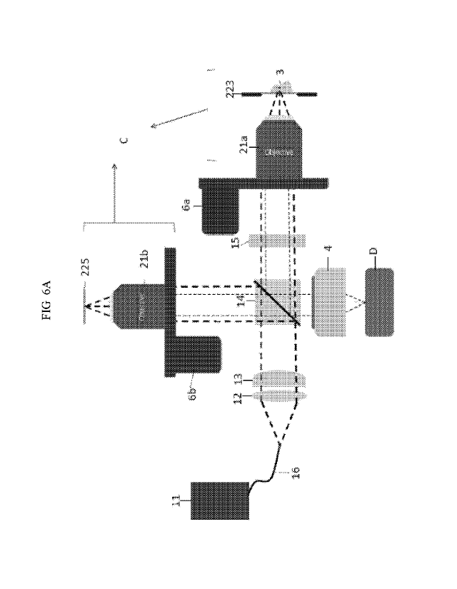
[0016] FIG. 6A/B illustrates yet another embodiment of the invention
device/system,
where a Michelson type objective is used without an imaging guiding module
(6A) or
with an imaging guiding module (6B).
[0017] FIG. 7 provides yet another embodiment of an exemplary design of the
invention device/system having a switch to change the illumination mode.
[0018] FIG. 8 illustrates an exemplary invention device/system comprising an
interference objective module wherein the objective 31 is configured to make
the
focal spot 44 of the incident light located between a focal plane 41 and a
principle
plane 42.
[0019] FIG. 9 further illustrates how an exemplary device/system is configured
to
make the stray light be focused on the edge and outside imaging range of the
2D
camera D
[0020] FIG. 10A-C illustrate examples of the reflective reference mirror with
different shapes.
DETAILED DESCRIPTION OF THE INVENTION
[0021] In recent years, optical coherence tomography (OCT) has been widely
applied
on three-dimensional (3-D) image reconstruction of skin tissue, or cornea. It
is known
that in epidermis, to non-invasively probe the layer parameters (LPs), such as
average
total thickness (a-TT), average number of layers (a-NOLs), and average
cellular layer
thickness (a-CLT), for stratum corneum (SC) becomes important for evaluating
the
skin moisturization of epidermis. However, to apply OCT technology to skin
tissue
3
CA 03075415 2020-03-09
WO 2019/056022
PCT/US2018/051609
imaging, axial resolution better than 1.2 1.tm in tissue is the doorsill to
measure LPs of
the SC. Besides, the morphology of single 3-D epidermal cell is also important
for
early detection of normal and abnormal cells of pre-cancer diagnosis. These
all
require sub-micron spatial resolution in tissue.
[0022] Provided herein are devices and systems that apply OCT technology
(e.g., a
FF-OCT) to skin tissue or cornea imaging applying a line light illuminated on
a
sample which produces a cross-sectional scanning image with unexpectedly clear
and
low speckles image quality, with a two-dimensional camera. Particularly, the
present
invention provides devices and systems having a line shaped reflective mirror
parallel
the line shaped light on an interference objective module to be detected by a
two-
dimensional camera, so as to achieve the efficiency of light utilization and
improve
the image scanning speed.
[0023] In some embodiments, there are provided a device/system comprising an
illumination module configured to provide a source light (such as a line of
light, or an
area of light) to an optical interference module; an interreference objective
module,
which handles light from the optical interference module and process light
signal
generated from a sample; a two-dimensional camera to receive a backscattered
interference signal from the sample; and a data processing module for
analyzing light
signals and providing a sample imaging.
[0024] There are provides an embodiment of the invention device/system as
shown
FIG. 1A, which comprises an illumination module A configured to provide a
source
light (e.g., a line of light, or an area of light) to an optical interference
module B, an
interference objective module C which processes and projects the light to the
interference objective module B therefrom, and direct the line of light on a
sample 3;
a two-dimensional camera D configured to receive a backscattered interference
signal
from the sample 3; and a data processing module E, which processes the
interference
signal into an image.
[0025] In some embodiments, the illumination module (such as a light source
11)
comprises a spontaneous emission light source, an amplified spontaneous
emission
light source, a superluminescent diode, a light emitting diode (LED), a
broadband
supercontinuum light source, a mode-locked laser, a tunable laser, a Fourier-
domain
mode-locked light source, an optical parametric oscillator (0P0), a halogen
lamp, or
a doped crystal fiber such as a Ce':YAG crystal fiber, a Ti3:A1203 crystal
fiber, a
4
CA 03075415 2020-03-09
WO 2019/056022
PCT/US2018/051609
Cr4+:YAG crystal fiber, or the like. In certain embodiments, the light source
module
comprises a Ce':YAG crystal fiber, Ti3:A1203 crystal fiber, or a Cr4+:YAG
crystal
fiber. In certain embodiments, the illumination module comprises a Ti3:A1203
crystal fiber. For example, the light source module is Ti3:A1203 crystal fiber
light
source with power of 0.5mW to 500 mW, or 4 to100 mW, or 10 to 50 mW, or 20 to
40 mW, or other suitable power range.
[0026] In some embodiments, the optical interference module is configured to
generate a line pattern light projected by a light source in the illumination
module. In
certain embodiments, the optical interference module comprises an anamorphic
lens
such as a cylindrical lens, or a round-to-linear fiber bundle, a diffractive
optical
element, a special-designed optical diffuser, or the like. A skilled person in
the art
would readily adapt other suitable means to produce line shaped light with
various the
aspect ratio such as 3 to 100, or 5 to 20, or other suitable ratios. Other
suitable optical
components known in the art to produce a thin light can be used without
limitations.
[0027] In some embodiments, the interference objective module comprises an
objective and an interference means configured to process the source light
such as a
line of light projected by an optical interference module, to a sample and
receive a
backscattered signal therefrom to generate an interference signal. In some
embodiments, the interference objective module is a Mirau-type interference
objective
module, a Michelson-type interference module, a Mach-Zehnder interference
objective module, or any suitable interference type objective module readily
recognized by a skilled person in the art.
[0028] In some embodiments, the objective is a Mirau-type interference
objective
module comprising an immersed objective having the immersed solution with a
refractive index approaching to the refractive index of the sample. For
example, if the
sample is a skin, the refractive index will be in a range of about 1.2 to
about 1.8,
preferably about 1.3 to about 1.5. In some embodiments, the media comprises
water,
silicone oil, ethanol, glycerol, pyrex, ultra sound gel, or combinations
thereof. In
certain embodiments, the media comprises water, silicone oil, or glycerol. In
certain
embodiments, the media comprises water.
[0029] In some embodiments, as shown in FIG. 1B, the invention device/system
further comprises an imaging guiding module F comprising a camera lens and a
second 2D-camera used for imaging guiding. The imaging guiding module provides
a
CA 03075415 2020-03-09
WO 2019/056022
PCT/US2018/051609
large area image of the sample (e.g., a detailed large area of sample
surface). The
imaging guiding module and the interference objective module share the same
optical
channel or path and thus provide overlapped field of views as illustrated in
FIG. 2B.
[0030] FIG. 2A provides an exemplary invention system/device. A light is
generated
by an illumination module comprising an exemplary light source 11 and
transport to
collimation lens 12 via an optical fiber 16. The light is transformed to a
line shaped
light by, for example, a cylindrical lens 13 and then passes through
polarization beam
splitter 14 and quarter wave plate 15 to convert the line shaped light with
circular
polarization. The light then enters an interference objective module C. In
some
embodiments, the interference objective module C comprises an objective 21 and
an
interference means 22. When the light (such as a line-shaped light) projects
to sample
3 through interference objective module C, the backscattered light by sample 3
passes
through interference objective module C to beam splitter 14 and provides light
signal
to a two-dimensional camera D via a projection lens 4. The signals are then
further
processed by a data processing module (not shown) to provide sample imaging.
It is
known in the art that a line scan light is processed by a one-dimensional
camera since
there is no need to record an area other than a one dimensional "line". It is
surprisingly found that by utilizing a 2-D camera D with a special design, a
high
image signal to noise ratio, high resolution cress-sectional image is produced
in
comparison with the use of a 1-D camera. It is designed to utilize a z-axial
piezoelectric transducer (PZT) 6 to scan interference objective module C in
the Z
direction. The line-shaped light with the interference signal will project
onto the two-
dimensional camera D with part of the pixel in a narrow rectangular area via a
projection lens 4. After recording the PZT scanned interference signal, each
column in
the narrow rectangular area is processed by a data processing module E to
produce
cross-sectional images. Thus, a scan can produce several cross-sectional
images.
After superimposing the several cross-sectional images, a high image signal to
noise
ratio, high resolution cress-sectional image is produced.
[0031] In some embodiment, the interference means comprises horizontally
arranged
glasses including a first glass plate, a second glass plate and a third glass
plate. The
first glass plate comprises the reflective mirror configured to have a shape
parallel the
line of light. The second glass plate is configured to have the light
transmitted
partially to the third glass plate. For instance, the reflective mirror can be
formed on
6
CA 03075415 2020-03-09
WO 2019/056022
PCT/US2018/051609
the first glass plate having a shape of thin line with aspect ratio of about 1
to 5000,
especially 4 to 1000, especially 8 to 250, especially 10 to 100, an artisan
can adjust
the ratio in need. The range of the aspect ratio of the reflective mirror can
determine
the range of field of view (FOV). In some embodiments, the second glass plate
can be
used as a beam splitter having a refractive ratio of about 5% to 30%,
preferably 5% to
20% to avoid the stray light reflected by the glass-sample interface.
Furthermore,
those three glass plates have a refractive index matching the sample's
refractive
index, for example in a range of about 1.2 to about 1.8, preferably about 1.3
to about
1.5, so as to avoid the stray light produced by the glass-sample interface.
[0032] It is found by utilizing a 2-D camera D instead of a 1-D camera
typically
associated with line-shaped light related optical module known in the art for
scanning
sample to acquire a cross-sectional image, an unexpected superior result with
high
image clarity and quality was achieved where such design effectively boost
image
signal to noise ratio and reduce number of image speckles. As evidenced by the
sample images in FIG. 3, obvious speckles are found in FIG. 3A image which was
acquired by a 1D camera. The image is blurry with poor quality. On the other
hand,
compared with the image from FIG. 3A, the image of FIG. 3C, which was produced
by a 2D camera, appears to have clear image signal with much less number of
speckles. The reason that the use of a 2D camera in such design associated
with a line
of light has better image quality compared with one from a 1D camera (which is
the
typical approach known in the art) is because 1D camera can only receive 1
pixel
wide sample image while the 2D camera can receive more than 1 pixel image
data.
By superimposing several images in accordance with the practice of the present
invention as illustrated in FIG. 3B, a clear image with a low number of
speckle was
acquired. However, it is also found that if the stacking thickness is too
thick, the
image becomes blurry in certain characters of the image. Thus, it is found
that the use
of a 2D camera in such design only works under a range of thickness of image
stacking. The optimum superposition thickness in some embodiments is 2 to 256
pixel, 4 to 128 pixel, or 4 to 64 pixel. In some embodiments, the optimum
superposition thickness is 4 to 64 pixel. For example, 8 pixel is used to
generate FIG.
3C. It is also surprisingly found that such 2-D camera design reduces the
number of
lenses needed making the module simpler due to the fact that the 2D camera can
be
flexibly adjusted for the measuring area allowing easy use of different aspect
ratio of
7
CA 03075415 2020-03-09
WO 2019/056022
PCT/US2018/051609
the optical designs. The reduced number of lenses significantly shortens the
manufacturing time and effort to produce the invention device/system.
[0033] In some embodiments, the device/system further comprise an imaging
guiding
module comprising a projection lens 104 and a second 2D camera 105 for imaging
guiding.
[0034] The device/system incorporates an imaging guiding module which provides
a
large (macro) image with skin surface detail. As shown in FIG. 2B, besides the
2D
camera D associated with an exemplary Mirau type interference objective
module,
which provides a high-resolution optical imaging, an image guiding module
comprising a camera lens 104 and a 2D-camera 105 is included in the
device/system
where the beam splitter 14a is used to direct the signals to 2D-camera 105.
The two
imaging systems share the same optical channel/path; therefore, the FOVs of
them are
overlapped and have a fixed relative position. A light source such as a LED
(L11)
cycling around the interference means 22 is incorporated to provide light for
the
imaging guiding module. The light source L11 has a different wavelength or
time
distribution from the illumination module, thus the signal produced by light
source
L11 by the sample are all collected by 2D-camera 105 to produce a large image
of
skin surface.
[0035] The FOV of the imaging guiding module is large than the FOVs of high-
resolution imaging module (i.e., the interference objective module). While
examining
a sample (e.g., a lesion, or a cornea), the imaging guiding module is used to
take a
large image of the sample area first. Then, the interference objective module
is
attached onto the sample allowing the image guiding module to image the
surface of
the sample. An algorithm is used to calculate where the FOV of guiding image
is on
the first large image. Because the position between guiding image and high-
resolution
image is fixed, the position of high resolution image can be pinpointed on the
large
image.
[0036] An exemplary invention interference objective module and how it works
is
illustrated in FIGs. 4A, 4B and 5.
[0037] An exemplary interference means 22 is shown in FIG. 4A. In some
embodiments, the interference means comprises a first glass plate 221 coated
with a
reflective mirror 224, a second glass plate 222, and a third glass plate 223
wherein the
reflective mirror 224 is coated to generate a reference arm and produce
interference
8
CA 03075415 2020-03-09
WO 2019/056022
PCT/US2018/051609
with the returned scattered light by sample 3. As shown in FIG. 4A, the
reflective
mirror 224 coated on the first glass plate 222 is linear and parallel to
focused line
shaped light 111.
[0038] In other embodiments, as illustrated in FIG. 4B, a first glass plate
221 further
comprises a black spot 225 on the opposite side of the first glass plate 221
at a
position corresponding to the reflective mirror 224.
[0039] The transparent first glass plate 221, which is closest to the
objective 21, is
partially coated with a reflective mirror 224 so that the central region of
the surface
toward the focal plane is highly reflective, while and the central region of
the surface
toward the objective 21 has a black spot 225, which is absorptive to block the
stray
light. In some embodiments, the position of the black spot is on the same side
of the
reflective mirror 224, where the black spot covers the reflective reference
mirror
224, so as to absorb the stray light from the first glass plate. The
transparent third
glass plate 223, which is (partially) contacted with the sample 3, is set to a
(range of)
position so that the focal plane of the objective lens is near the sample.
[0040] The second glass 222 is coated so that the surface toward the third
glass plate
223 is partially reflective. This coated surface is served as the beam
splitter in a Mirau
type interferometer, and the position of the transparent second plate 222 is
set to a
position so that the highly reflective region 224 is on the focal plane of the
objective
lens.
[0041] As illustrated in FIG. 4A, the deviation within 20 degrees of angle is
defined
as parallel. In some embodiments, it is within 15 degrees of angle, within 10
degrees
of angle, or within 5 degrees of angle. Furthermore, the reflective mirror 224
has an
adjustable aspect ratio about 3 to 10, and preferably about 5 to 8.
Accordingly, the
architecture of such design of a coated line shaped reflective mirror on the
first glass
plate makes the full use of light. In some embodiments, the second glass plate
222
has a refractive ratio of about 5% to 30%, preferably 10% to 20%, or any other
suitable ratio as needed based on the conditions. The third glass plate 223 is
fully
transparent for fitting with sample 3 allowing the line-shaped light to
penetrate and
illuminate sample 3.
[0042] In some embodiment, the interference means comprises horizontally
arranged
glasses including a first glass plate, a second glass plate and a third glass
plate. The
first glass plate comprises the reflective mirror configured to have a shape
parallel the
9
CA 03075415 2020-03-09
WO 2019/056022
PCT/US2018/051609
line of light. The second glass plate is configured to have the light
transmitted
partially to the third glass plate. For instance, the reflective mirror can be
formed on
the first glass plate having a shape of thin line with aspect ratio of about 1
to 5000,
especially 4 to 1000, especially 8 to 250, especially 10 to 100, an artisan
can adjust
the ratio in need. The range of the aspect ratio of the reflective mirror can
determine
the range of field of view (FOV). In some embodiments, the second glass plate
can be
used as a beam splitter having a refractive ratio of about 5% to 30%,
preferably 5% to
20%. Furthermore, those three glass plates have a refractive index matching
the
sample's refractive index, for example in a range of about 1.2 to about 1.8,
preferably
about 1.3 to about 1.5, so as to avoid the stray light produced by the glass-
sample
interface.
[0043] FIG. 6A/6B provides yet another embodiment where a Michelson-type
interference objective module is used in the invention device/system. In some
embodiments, the invention device/system is a Michelson-type interference
imaging
device/system, comprising the same illumination module and optical
interference
module, and optional imaging guiding module as in FIG. 2A/2B, except the use
of a
different interference objective module C. The interference objective module C
comprises an objective 21a and a third glass plate 223 attached to a sample 3
thereon
to produce a sample arm, and an objective 21b and a reflective mirror 225 to
produce
a reference arm. When the line light illuminated on the sample 3 and the
reflective
mirror 225 simultaneously and reflected therefrom, the interference signal
will be
created and collected by a two-dimensional camera D via a projection lens 4,
then
produces a cross-sectional image by a data processing module, while the
imaging
guiding module comprising a projection lens 104 and a 2D camera 105 to provide
a
large sample image for correlation of the cross-sectional image.
[0044] In some embodiments, the optical interference module further comprises
a
switch configured to toggle the light output between the line of light and an
area of
light, thereto switching between line-scan mode and full-field mode for the
device
allowing the user to acquire cross-sectional images and/or en-face images
(e.g., to
acquire a 3-D slice data) of a sample. Such design allows users to acquire the
whole
sample information.
[0045] In order to acquire more structure information from a sample, in some
embodiments, the optical interference module further comprises a switch 17
CA 03075415 2020-03-09
WO 2019/056022
PCT/US2018/051609
configured to provide different lighting mode as shown in FIG. 7. In some
embodiments, there are two modes included to be changed; one is line light
illuminating mode L, another is area light illuminating mode F, in which the
switch 17
(e.g., Thorlabs CFW6) is disposed between the collimation lens 12 and the
polarization beam splitter 14 to toggle the cylindrical lens 13 and the
achromatic lens
18 fitted in the lens holders of switch 17, so as the illumination mode is
switched to
line light illumination mode L for acquiring cross-sectional images, or to
area light
illumination mode F for acquiring en-face images, which can lead to three-
dimensional volumetric images. In some embodiments, such toggle switch design
is
not limited to the changes of line light illuminating mode and area
illuminating mode;
all other suitable modes with different lens may be used in accordance with
the
practice of the invention.
[0046] In some embodiments, the invention device/system is configured to make
the
stray light be focused on the edge and outside imaging range of the 2D camera
D.
[0047] For example, as shown in FIG. 8, the interference objective module
comprising an objective 31, and an interference means (comprising a first
glass
plate 321 with a reflectance reference mirror 324, a second glass plate 322
and a
third glass plate 323 next to a sample 3) wherein the objective 31 is
configured to
make the focal spot 44 of the incident light located between a focal plane 41
and a
principle plane 42. Such arrangement allows the focal spot 44 of the incident
light
be offset from the optical axis 111 of the objective 31, which makes the stray
light
be focused on the edge and outside imaging range of the 2D camera D (as shown
in FIG. 9). In some embodiments, the reflective reference mirror 324 is coated
on
the first glass plate 321 partially, for example, coated on the center of the
first
glass plate 321 wherein the reflective reference mirror 324 has a high
reflective
index made by silver, or other suitable metal used for coating.
[0048] In some embodiments, the incident light is configured to have an
incident
angle 01 which is greater than 0 and less than 45 to an optical axis of the
objective, Preferably, 01 is greater than 0 and less than 20 , more
preferably,
greater than 0 and less than 5 , but it is not limited thereto.
[0049] In some embodiments, the focal spot is configured to have a divergence
angle 02 in a range of about 0 to 70 . The value of 02 is depend on the field
of
view (FOV) and in a direct proportion to FOV. In some embodiments, an artisan
11
CA 03075415 2020-03-09
WO 2019/056022
PCT/US2018/051609
can choose 02 in a range of 00 to 20 or 50 to 150 to achieve small FOV, or
choose
40 to 70 or 500 to 60 to achieve large FOV.
[0050] In some embodiments, the objective has an NA value satisfying the
following formula (1):
NA = n x sin 0, and 0 = 03/(0.5-4.5) ....... (1),
[0051] : NA is a numerical aperture of the objective, n is a refractive index,
0 is
1/2 angular aperture, and 03 is a half spreading angle form the objective.
[0052] .Preferably, 0 = 03/(0.5-4.0). If the angle of 03 is too large, it will
reduce
the signal correction of the sample there to reduce the sample brightness.
[0053] In some embodiments, the invention device/system comprises an
illumination
module configured to provide a source light (such as a line of light, or an
area of light)
to an optical interference module; an interference objective module comprising
an
objective and an interference means, which handles light from the optical
interference
module and process light signal generated from a sample; a two-dimensional
camera
to receive a backscattered interference signal from the sample; and a data
processing
module for analyzing light signals and providing a sample imaging, wherein
device/system is configured to make the objective to accept incident light in
an
arrangement having a focal spot of the incident light between a focal plane
and a
principal plane of the objective.
[0054] As shown in FIG. 9, a light provided by an illumination module is
projected
to an optical interference module via an optical fiber 16. The light is
collected by
collimation lens 312 and then transformed to a line shaped light by
cylindrical lens
313 as shown (or an area light by achromatic lens as illustrated in FIG. 7)
and pass
through a beam splitter 14, which will be transmitted into the interference
objective module C. When the light passing through the interference objective
module C to a sample 3 through the third glass plate 323, the light is
absorbed,
reflected or backscattered. The backscattered light signal will be collected
by the
interference objective module C and interferes with the reference light, which
is
reflected from the reflective mirror 324 and the second glass plate 322, to
generate
an interference signal. Then the beam splitter 14 reflects the signal to the
projection lens 4 making the stray light be focused on the edge and outside
imaging range of the 2D camera D.
12
CA 03075415 2020-03-09
WO 2019/056022
PCT/US2018/051609
[0055] In some embodiments, the reflective reference mirror has a shape of
line,
polygon (such as a square), circle spot, or other shape suitable for the
device or
system.
[0056] For example, as shown in FIG. 10 (10A to 10C), the reflective reference
mirror 324 can have a shape of line (10A), polygon (a square, 10B), or circle
spot
(10C). In some embodiments, the size of the refractive reference mirror can be
in a
range of less than 1500 [tm2, preferably less than 1000 [tm2, preferably less
than
500 [tm2, and preferably less than 300 [tm2, and a skilled person in the art
would
readily adjust the size as suitably needed. By coating the reflective
reference
mirror on the first glass plate partially, the utilization of the light will
be
effectively improved.
[0057] In some embodiments provide a method for imaging a sample comprising
making an objective in the invention interference objective module which
handles
light from the optical interference module and process light signal generated
from said
sample to accept incident light in an arrangement having a focal spot of the
incident
light between a focal plane and a principal plane of the objective from an
illumination
module, and processing an interference signal generated said interference
module into
an image by a data processing module. In some embodiments, the interference
objective module is any invention interference objective module disclosed
herein.
[0058] The invention device/system is useful to imaging a sample in a cross-
sectional
as well as an en-face direction. It is particular useful in assisting in
providing
information of the sample surface and sub-surface such as a skin or cornea
condition.
The invention device/system uses a two-dimensional camera with a line-light
backscattering to acquire high noise to signal ratio cross-sectional images,
effectively
improving the image quality and reach the resolution of 1 p.m level. Also such
design
allows increasing the image scanning speed to 150 [tm/sec or more. The use of
an
imaging guiding module allows the user efficiently to pinpoint the area of
interest.
[0059] Although preferred embodiments of the present invention have been shown
and described herein, it will be obvious to those skilled in the art that such
embodiments are provided by way of example only. Numerous variations, changes,
and substitutions will now occur to those skilled in the art without departing
from the
invention. It should be understood that various alternatives to the
embodiments of the
invention described herein can be employed in practicing the invention. It is
intended
13
CA 03075415 2020-03-09
WO 2019/056022
PCT/US2018/051609
that the following claims define the scope of the invention and that methods
and
structures within the scope of these claims and their equivalents be covered
thereby.
14