Note: Descriptions are shown in the official language in which they were submitted.
CA 03075532 2020-03-10
WO 2019/055878
PCT/US2018/051240
MULTIPLEX PRODUCTION AND BARCODING OF GENETICALLY
ENGINEERED CELLS
.. CROSS-REFERENCE TO RELATED APPLICATIONS
This application claims priority to U.S. provisional application no.
62/559,493,
filed on September 15, 2017, which is incorporated herein by reference in its
entirety.
STATEMENT REGARDING FEDERALLY SPONSORED RESEARCH OR DEVELOPMENT
This invention was made with government support under contract HG000205
awarded by the National Institutes of Health and contract 70NANB15H268 awarded
by the National Institute of Standards and Technology. The government has
certain
rights in the invention.
TECHNICAL FIELD
The present disclosure pertains generally to the field of genome engineering
using RNA-guided nucleases. In particular, the disclosure relates to
compositions and
methods for multiplex high-throughput production and validation of genetically
engineered cells using RNA-guided nucleases and barcoding.
BACKGROUND
The advent of programmable genome editing via the CRISPR/Cas9 system is
enabling rapid advances in synthetic biology and genetic engineering. The
Streptococcus pyogenes bacterial type II clustered regularly-interspaced short
palindromic repeats (CRISPR)-associated protein 9 (Cas9) was the first RNA-
guided
nuclease (RGN) demonstrated to cut nearly any genomic location using a guide
RNA
(gRNA) with homology to the target region2. Utilizing conserved homologous
recombination-based DNA repair pathways present in the host cells, donor DNA
with
homology flanking the cut site can be used to repair that break and introduce
a genetic
change of interest. The short specificity-determining region of the gRNA
(generally
20 nucleotides (nt) in length) and donor DNA lengths (-100-150 nt) are
compatible
with highly parallel array-based oligonucleotide library synthesis, allowing
facile
creation of gRNA-donor libraries directed against thousands of targets"-g. To
date,
-1-
CA 03075532 2020-03-10
WO 2019/055878
PCT/US2018/051240
however, production of variant libraries has been restricted to pools, which
greatly
limits the options for characterizing the phenotype of individual variants.
For
example, microscopy, metabolomics, and many enzymatic assay reporters are not
amenable to a pooled format.
CRISPR editing is particularly efficient in yeast because of its strong
preference for using homologous recombination (HR) to repair double-strand
breaks
in the presence of donor DNA, eliminating the need for selectable markers when
making edits to the gen0me9-11. In contrast to the near 100% Cas9 editing
efficiency
reported in yeast'', gene editing in metazoan cells is hindered by the
preference for
non-homologous end joining (NHEJ) over HR, and editing by HR in human cells
has
only reached a maximal efficiency of about 10-60%15'16. Thus, in addition to
extending the well-established utility of yeast as a model system for
eukaryotic
biology, the Cas9 system magnifies the value of yeast as a host for
engineering
heterologous proteins and pathways.
Thus, there remains a need for more efficient and flexible methods of genome
editing that enhance repair of RGN-mediated double-strand breaks through the
HR
mechanism to allow genomes to be modified with precise genetic changes as
desired
and improved methods for high-throughput production of variant libraries.
SUMMARY
The present disclosure relates to multiplex production and validation of
genetically engineered cells using RNA-guided nucleases and barcoding. In
particular, high-throughput multiplex genome editing is achieved utilizing a
system
that facilitates precise genome editing at desired target chromosomal loci by
homology directed repair. Integration of guide RNA and donor DNA sequences as
a
genomic barcode at a chromosomal locus separate from the target loci being
edited
allows ready identification, isolation, and massively-parallel validation of
individual
variants from a pool of transformants. Strains can be arrayed according to
their
precise genetic modifications, as specified by donor DNA incorporation in
heterologous or native genes. The present disclosure further relates to a
method of
editing codons outside of canonical guide RNA recognition regions, which
enables
complete saturation mutagenesis of protein-coding genes, a marker-based
internal
cloning method, which removes background due to oligonucleotide synthesis
errors
-2-
CA 03075532 2020-03-10
WO 2019/055878
PCT/US2018/051240
and incomplete vector backbone cleavage, and a method of enhancing homology
directed repair by active donor recruitment.
Provided herein is a method for multiplex production of genetically
engineered cells, the method including: (a) transfecting a plurality of cells
with
plurality of different recombinant polynucleotides, each recombinant
polynucleotide
including a genome editing cassette including a first nucleic acid sequence
encoding a
first guide RNA (gRNA) capable of hybridizing at a genomic target locus to be
modified and a donor polynucleotide thereby forming a gRNA-donor
polynucleotide
combination, where each recombinant polynucleotide includes a different genome
editing cassette including a different gRNA-donor polynucleotide combination,
and
allowing each of the cells to express the first nucleic acid sequence thereby
forming
the gRNA; and (b) introducing an RNA-guided nuclease into each of the
plurality of
cells, where the RNA-guided nuclease in each cell forms a complex with the
gRNA
thereby forming a gRNA-RNA-guided nuclease complex, and allowing the gRNA-
RNA-guided nuclease complex to modify the genomic target locus by integrating
the
donor polynucleotide into the genomic target locus, thereby producing a
plurality of
genetically engineered cells.
In another aspect is provided a method for multiplex production of genetically
engineered cells, the method including: (a) transfecting a plurality of cells
with
plurality of different recombinant polynucleotides, each recombinant
polynucleotide
including a unique polynucleotide barcode and a genome editing cassette
including a
first nucleic acid sequence encoding a first guide RNA (gRNA) capable of
hybridizing at a genomic target locus to be modified and a donor
polynucleotide
thereby forming a gRNA-donor polynucleotide combination, where each
recombinant
polynucleotide includes a different genome editing cassette including a
different
gRNA-donor polynucleotide combination, and allowing each of the cells to
express
the first nucleic acid sequence thereby forming the gRNA; and (b) introducing
an
RNA-guided nuclease into each of the plurality of cells, where the RNA-guided
nuclease in each cell forms a complex with the gRNA thereby forming a gRNA-
RNA-guided nuclease complex, and allowing the gRNA-RNA-guided nuclease
complex to modify the genomic target locus by integrating the donor
polynucleotide
into the genomic target locus, thereby producing a plurality of genetically
engineered
cells.
-3-
CA 03075532 2020-03-10
WO 2019/055878
PCT/US2018/051240
In embodiments, the method further includes sequence verification and
arraying of the plurality of genetically modified cells, the method including:
(c)
plating the plurality of genetically modified cells in an ordered array on
media
suitable for growth of the genetically modified cells; (d) culturing the
plurality of
genetically modified cells under conditions whereby each genetically modified
cell
produces a colony of clones in the ordered array; (e) introducing a genome
editing
cassette from a colony in the ordered array into a barcoder cell, where the
barcoder
cell includes a nucleic acid including a recombination target site for a site-
specific
recombinase and a barcode sequence that identifies the position of the colony
in the
.. ordered array to which the genome editing cassette corresponds; (f)
translocating the
genome editing cassette to a position adjacent to the barcode sequence of the
barcoder
cell using a site-specific recombinase system, where site-specific
recombination with
the recombination target site of the barcoder cell generates a nucleic acid
including
the barcode sequence linked to the genome editing cassette; (g) sequencing the
nucleic acid including the barcode sequence of the barcoder cell linked to the
genome
editing cassette to identify the sequences of the guide RNA and the donor
polynucleotide of the genome editing cassette from the colony, where the
barcode
sequence of the barcoder cell is used to identify the position of the colony
in the
ordered array from which the genome editing cassette originated; and (h)
picking a
clone including the genome editing cassette from the colony in the ordered
array
identified by the barcode of the barcoder cell.
In another aspect is provided a method for localizing a donor polynucleotide
to a genomic target locus in a cell, the method including: (a) transfecting a
cell with a
recombinant polynucleotide, the recombinant polynucleotide including a genome
.. editing cassette including a donor polynucleotide and a DNA binding
sequence
known to bind a DNA binding domain; (b) introducing a nuclease into the cell,
where
the nuclease recognizes and causes a double-strand DNA break at the genomic
target
locus; (c) introducing a donor recruitment protein into the cell, the donor
recruitment
protein including the DNA binding domain and a DNA break site localizing
domain
and allowing the donor recruitment protein to selectively recruit the double-
strand
DNA break, thereby localizing the donor polynucleotide to the genomic target
locus.
In another aspect is provided a library of gene editing vectors, each gene
editing vector including a genome editing cassette including (i) a barcode,
(ii) a first
-4-
CA 03075532 2020-03-10
WO 2019/055878
PCT/US2018/051240
nucleic acid sequence encoding a first guide RNA (gRNA) capable of hybridizing
at a
genomic target locus to be modified, and (iii) a donor polynucleotide, thereby
forming
a barcode-gRNA-donor polynucleotide combination; where each recombinant
polynucleotide includes a different genome editing cassette including a
different
barcode-gRNA-donor polynucleotide combination.
In another aspect is provided a gene editing vector including a donor
polynucleotide and a first nucleic acid sequence encoding a first guide RNA
(guide X)
capable of hybridizing with the vector at a target site such that when the
guide X is
expressed by a cell, the guide X hybridizes to the vector and creates a double-
strand
DNA break at the target site.
In another aspect is provided a kit including: (a) a gene editing vector as
described herein including embodiments thereof; and (b) a nuclease or a
polynucleotide encoding a nuclease.
In another aspect is provided a kit including: (a) a gene editing vector as
described herein including embodiments thereof; and (b) a reagent for
genetically
modifying a cell.
In another aspect is provided a library of gene editing vectors, each gene
editing vector comprising a genome editing cassette comprising (i) a first
nucleic acid
sequence encoding a first guide RNA (gRNA) capable of hybridizing at a genomic
target locus to be modified, and (ii) a donor polynucleotide, thereby forming
a gRNA-
donor polynucleotide combination; wherein each recombinant polynucleotide
comprises a different genome editing cassette comprising a different gRNA-
donor
polynucleotide combination.
In embodiments, each recombinant polynucleotide further comprises a second
nucleic acid sequence encoding the RNA-guided nuclease.
In one aspect, the present disclosure includes a method for multiplex genetic
modification and barcoding of cells, the method comprising: a) providing a
plurality
of recombinant polynucleotides, wherein each recombinant polynucleotide
comprises
a genome editing cassette comprising a polynucleotide encoding a guide RNA
(gRNA) capable of hybridizing at a genomic target locus to be modified and a
donor
polynucleotide comprising a 5' homology arm that hybridizes to a 5' genomic
target
sequence and a 3' homology arm that hybridizes to a 3' genomic target sequence
flanking a nucleotide sequence comprising an intended edit to be integrated
into the
-5-
CA 03075532 2020-03-10
WO 2019/055878
PCT/US2018/051240
genomic target locus, wherein each recombinant polynucleotide comprises a
different
genome editing cassette comprising a different guide RNA-donor polynucleotide
combination, such that the plurality of recombinant polynucleotides is capable
of
producing a plurality of different intended edits at one or more genomic
target loci;
and b) transfecting the cells with the plurality of recombinant
polynucleotides; c)
culturing the transfected cells under conditions suitable for transcription,
wherein
guide RNAs are produced from each genome editing cassette; d) introducing an
RNA-
guided nuclease into the cells, wherein the RNA-guided nuclease forms
complexes
with the guide RNAs produced in the cells, said guide RNAs directing the
complexes
.. to the one or more genomic target loci, wherein the RNA-guided nuclease
creates
double-stranded breaks in the genomic DNA of the cells at the one or more
genomic
target loci, and the donor polynucleotide present in each cell is integrated
at the
genomic target locus recognized by its 5' homology arm and 3' homology arm by
homology directed repair (HDR) such that a plurality of genetically modified
cells are
.. produced; and e) barcoding the plurality of genetically modified cells by
integrating
the genome editing cassette present in each genetically modified cell at a
chromosomal barcode locus. In certain embodiments, the method further
comprises
performing additional rounds of genetic modification and genomic barcoding on
the
genetically modified cells by repeating steps (a)-(e) using different genome
editing
cassettes.
In certain embodiments, each recombinant polynucleotide is provided by a
vector. The vector can be, for example, a plasmid or viral vector. In certain
embodiments, the vector is a high copy number vector.
In certain embodiments, each recombinant polynucleotide is provided as linear
.. DNA. For example, the method may further comprise amplifying a recombinant
polynucleotide comprising a genome editing cassette, which is provided as a
PCR
product.
In certain embodiments, the RNA-guided nuclease is also provided by a
vector. In certain embodiments, the genome editing cassette and the RNA-guided
nuclease are provided by a single vector or separate vectors. In another
embodiment,
a recombinant polynucleotide encoding the RNA-guided nuclease is integrated
into
the genome of the host cells.
-6-
CA 03075532 2020-03-10
WO 2019/055878
PCT/US2018/051240
Transcription of a guide RNA will generally depend on the presence of a
promoter, which may be included in the genome editing cassette, or in a vector
or at a
chromosomal locus (e.g., the chromosomal barcode locus) in which the genome
editing cassette is inserted. The promoter may be a constitutive or an
inducible
promoter. In certain embodiments, each genome editing cassette comprises a
promoter operably linked to the polynucleotide encoding the guide RNA. In
other
embodiments, the chromosomal barcode locus comprises a promoter that becomes
operably linked to the polynucleotide encoding the guide RNA of any genome
editing
cassette that integrates at the chromosomal barcode locus. In another
embodiment,
each recombinant polynucleotide is provided by a vector, wherein the vector
comprises a promoter that is operably linked to the polynucleotide encoding
the guide
RNA.
In certain embodiments, the plurality of recombinant polynucleotides is
capable of producing mutations at multiple sites within a single gene. In
other
embodiments, the plurality of recombinant polynucleotides is capable of
producing
mutations at multiple sites in different genes or anywhere in the genome. For
example, each donor polynucleotide may introduce a different mutation into a
gene,
such as an insertion, deletion, or substitution. In another embodiment, at
least one
donor polynucleotide introduces a mutation that inactivates a gene. In another
embodiment, at least one donor polynucleotide removes a mutation from a gene.
In
another embodiment, at least one donor polynucleotide inserts a precise
genetic
change into the genomic DNA.
In certain embodiments, integration of the genome editing cassette present in
a
genetically modified cell at the chromosomal barcode locus is performed using
HDR.
Each recombinant polynucleotide may further comprise a pair of universal
homology
arms flanking the genome editing cassette that are capable of hybridizing to
complementary sequences at the chromosomal barcode locus to allow said
integration
of the genome editing cassette at the chromosomal barcode locus by the HDR. In
addition, each recombinant polynucleotide may further comprise a second guide
RNA
capable of hybridizing at the chromosomal barcode locus, wherein the RNA-
guided
nuclease further forms a complex with the second guide RNA, said second guide
RNA directing said complex to the chromosomal barcode locus, wherein the RNA-
guided nuclease creates a double-stranded break at the chromosomal barcode
locus,
-7-
CA 03075532 2020-03-10
WO 2019/055878
PCT/US2018/051240
and the genome editing cassette is integrated into the chromosomal barcode
locus by
the HDR.
In other embodiments, integration of the genome editing cassette present in a
genetically modified cell at the chromosomal barcode locus is performed using
a site-
specific recombinase system. Exemplary site-specific recombinase systems
include a
Cre-loxP site-specific recombinase system, a Flp-FRT site-specific recombinase
system, a PhiC31-att site-specific recombinase system, and a Dre-rox site-
specific
recombinase system. In certain embodiments, the chromosomal barcode locus
further
comprises a first recombination target site for a site-specific recombinase
and the
recombinant polynucleotide further comprises a second recombination target
site for
the site-specific recombinase, and site-specific recombination between the
first
recombination target site and the second site-specific recombination site
results in
said integrating of the genome editing cassette at the chromosomal barcode
locus.
In certain embodiments, the method further comprises using a selectable
marker that selects for clones that have undergone successful integration of
the donor
polynucleotide at the genomic target locus or successful integration of the
genome
editing cassette at the chromosomal barcode locus.
In certain embodiments, the cells to be genetically modified are eukaryotic or
prokaryotic. In some embodiments, the cells are yeast cells, which can be
haploid or
diploid yeast cells.
In certain embodiments, each recombinant polynucleotide further comprises a
pair of restriction sites flanking the genome editing cassette. In some
embodiments,
the restriction sites are recognized by a meganuclease (e.g., SceI) that
generates a
DNA double-strand break. The expression of the meganuclease may be controlled
by
an inducible promoter.
In another embodiment, the genome editing cassette further comprises a tRNA
sequence at the 5' end of the nucleotide sequence encoding the guide RNA.
In another embodiment, the genome editing cassette further comprises a
nucleotide sequence encoding a hepatitis delta virus (HDV) ribozyme at the 5'
end of
the nucleotide sequence encoding the guide RNA.
In another embodiment, the RNA-guided nuclease is a Cas nuclease (e.g.,
Cas9 or Cpfl) or an engineered RNA-guided FokI-nuclease.
-8-
CA 03075532 2020-03-10
WO 2019/055878
PCT/US2018/051240
In another embodiment, the genome editing cassette is flanked by restriction
sites recognized by a meganuclease.
In certain embodiments, each genome editing cassette further comprises a
unique barcode sequence for identifying the guide RNA and the donor
polynucleotide
encoded by each genome editing cassette. The unique barcode can be sequenced
in
place of the guide RNA and the donor polynucleotide to identify a genetic
modification to a cell. In another embodiment, the method further comprises
deleting
the polynucleotide encoding the guide RNA and the donor polynucleotide
integrated
at the chromosomal barcode locus while retaining the unique barcode at said
chromosomal barcode locus that represents the deleted sequences. In another
embodiment, the method further comprises sequencing the barcode at the
chromosomal barcode locus of at least one genetically modified cell to
identify the
genome editing cassette used in genetically modifying said cell.
In certain embodiments, the method further comprises sequencing each
genome editing cassette. Sequencing of a genome editing cassette to link a
barcode to
a particular gRNA-donor polynucleotide combination may be performed, for
example, at an intermediate cloning step prior to ligation of a genome editing
cassette
into a vector or prior to transfecting cells. Alternatively or additionally,
sequencing of
a genome editing cassette that has been integrated at the chromosomal barcode
locus
may be used to determine genome edits performed on a genetically modified
cell.
In certain embodiments, the method further comprises sequence verification
and arraying of the plurality of genetically modified cells, the method
comprising: a)
plating the plurality of genetically modified cells in an ordered array on
media
suitable for growth of the genetically modified cells; b) culturing the
plurality of
genetically modified cells under conditions whereby each genetically modified
cell
produces a colony of clones in the ordered array; c) introducing a genome
editing
cassette from a colony in the ordered array into a barcoder cell, wherein the
barcoder
cell comprises a nucleic acid comprising a recombination target site for a
site-specific
recombinase and a barcode sequence that identifies the position of the colony
in the
ordered array to which the genome editing cassette corresponds; d)
translocating the
genome editing cassette to a position adjacent to the barcode sequence of the
barcoder
cell using a site-specific recombinase system, wherein site-specific
recombination
with the recombination target site of the barcoder cell generates a nucleic
acid
-9-
CA 03075532 2020-03-10
WO 2019/055878
PCT/US2018/051240
comprising the barcode sequence linked to the genome editing cassette; e)
sequencing
the nucleic acid comprising the barcode sequence of the barcoder cell linked
to the
genome editing cassette to identify the sequences of the guide RNA and the
donor
polynucleotide of the genome editing cassette from the colony, wherein the
barcode
sequence of the barcoder cell is used to identify the position of the colony
in the
ordered array from which the genome editing cassette originated; and f)
picking a
clone comprising the genome editing cassette from the colony in the ordered
array
identified by the barcode of the barcoder cell.
For example, the genetically modified cells may be haploid yeast cells and the
barcoder cells may be haploid yeast cells capable of mating with the
genetically
modified cells, wherein introducing a genome editing cassette from a
genetically
modified haploid yeast colony in the ordered array into a barcoder haploid
yeast cell
comprises mating the haploid yeast clone from the colony with the barcoder
haploid
yeast cell to produce a diploid yeast cell. Subsequent site-specific
recombination, as
described herein, generates a nucleic acid comprising the barcode sequence
linked to
the genome editing cassette in the diploid yeast cell. The genetically
modified cells
may be strain MATa and the barcoder yeast cells may be strain MATa.
Alternatively, the genetically modified cells may be strain MATa and the
barcoder
yeast cells may be strain MATa.
In certain embodiments, the recombinase system in the barcoder cell is a Cre-
loxP site-specific recombinase system, a Flp-FRT site-specific recombinase
system, a
PhiC31-att site-specific recombinase system, or a Dre-rox site-specific
recombinase
system. In one embodiment, the recombination target site of the barcoder cell
comprises a loxP recombination site.
In another embodiment, the recombinase system in the barcoder cell uses a
meganuclease to generate a DNA double-strand break. In another embodiment, the
meganuclease in the barcoder cell is a galactose inducible SceI meganuclease.
In
another embodiment, the genome editing cassette is flanked by restriction
sites
recognized by the meganuclease.
In another embodiment, the method further comprises using a selectable
marker that selects for clones that have undergone successful site-specific
recombination.
-10-
CA 03075532 2020-03-10
WO 2019/055878
PCT/US2018/051240
In certain embodiments, the method further comprises inhibiting non-
homologous end joining (NHEJ). For example, NHEJ may be inhibited by
contacting
cells with a small molecule inhibitor selected from the group consisting of
wortmannin and Scr7. Alternatively, RNA interference or CRISPR-interference
can
be used to inhibit expression of a protein component of the NHEJ pathway.
In other embodiments, the method further comprises using an HDR enhancer
or active donor recruitment to increase the frequency of HDR in the cells.
In another embodiment, the method further comprises using a selectable
marker that selects for clones that have undergone successful integration of
the donor
polynucleotides at the one or more genomic target loci by HDR.
In another embodiment, the method further comprises phenotyping at least one
clone in the ordered array.
In another embodiment, the method further comprises sequencing an entire
genome of at least one clone in the ordered array.
In another embodiment, the method further comprises repeating steps (a)-(e)
with all the colonies in the ordered array to identify the sequences of the
guide RNAs
and the donor polynucleotides of the genome editing cassettes for every colony
in the
ordered array.
In another aspect, the present disclosure includes an ordered array of
colonies
comprising clones of the genetically modified cells produced by the methods
described herein, wherein the colonies are indexed according to the verified
sequences
of their guide RNAs and donor polynucleotides.
In another aspect, the present disclosure includes a kit for multiplex genetic
modification and barcoding of cells, the kit comprising: a) a plurality of
recombinant
polynucleotides, wherein each recombinant polynucleotide comprises a genome
editing cassette comprising a polynucleotide encoding a guide RNA (gRNA)
capable
of hybridizing at a genomic target locus to be modified and a donor
polynucleotide
comprising a 5' homology arm that hybridizes to a 5' genomic target sequence
and a 3'
homology arm that hybridizes to a 3' genomic target sequence flanking a
nucleotide
sequence comprising an intended edit to be integrated into the genomic target
locus,
wherein each recombinant polynucleotide comprises a different genome editing
cassette comprising a different guide RNA-donor polynucleotide combination,
such
that the plurality of recombinant polynucleotides is capable of producing a
plurality of
-11-
CA 03075532 2020-03-10
WO 2019/055878
PCT/US2018/051240
different intended edits at one or more genomic target loci; and b) an RNA-
guided
nuclease; and c) cells comprising a chromosomal barcode locus, wherein the
barcode
locus comprises a site for integration of the genome editing cassette of at
least one
recombinant polynucleotide. The kit may further comprise other reagents and
instructions for performing genome editing and barcoding as described herein.
In certain embodiments, each recombinant polynucleotide in the kit further
comprises a pair of universal homology arms flanking the genome editing
cassette
that are capable of hybridizing to complementary sequences at the site for
integration
at the chromosomal barcode locus to allow said integration of the genome
editing
cassette at the chromosomal barcode locus by homology directed repair (HDR).
In another embodiment, each recombinant polynucleotide further comprises a
second guide RNA capable of hybridizing at the chromosomal barcode locus.
In certain embodiments, the kit further comprises a site-specific recombinase
system (e.g., a Cre-loxP site-specific recombinase system, a Flp-FRT site-
specific
recombinase system, a PhiC31-att site-specific recombinase system, or a Dre-
rox site-
specific recombinase system). In another embodiment, the chromosomal barcode
locus further comprises a first recombination target site for a site-specific
recombinase and the recombinant polynucleotide further comprises a second
recombination target site for the site-specific recombinase, such that site-
specific
recombination can occur between the first recombination target site and the
second
site-specific recombination site to allow said integration of the genome
editing
cassette at the chromosomal barcode locus.
In another embodiment, the RNA-guided nuclease in the kit is a Cas nuclease
(e.g., Cas9 or Cpfl) or an engineered RNA-guided FokI-nuclease.
In certain embodiments, the kit further comprises a fusion protein designed to
carry out donor recruitment as described herein. Such a fusion protein
comprises a
polypeptide comprising a nucleic acid binding domain connected to a protein
that
selectively binds to a DNA break generated by the RNA-guided nuclease. In
another
embodiment, the donor polynucleotide further comprises a nucleotide sequence
sufficiently complementary to hybridize to a sequence adjacent to the DNA
break,
and a nucleotide sequence comprising a binding site recognized by the nucleic
acid
binding domain of the fusion protein. In certain embodiments, the nucleic acid
binding domain is a LexA DNA binding domain and the binding site is a LexA
-12-
CA 03075532 2020-03-10
WO 2019/055878
PCT/US2018/051240
binding site or the nucleic acid binding domain is a forkhead homolog 1 (FKH1)
DNA binding domain and the binding site is a FKH1 binding site. In some
embodiments, the polypeptide comprising the nucleic acid binding domain
further
comprises a forkhead-associated (FHA) phosphothreonine-binding domain. In
another embodiment, the polypeptide comprising the nucleic acid binding domain
comprises a LexA DNA binding domain linked to a FHA phosphothreonine-binding
domain.
In another aspect, the present disclosure includes a method of promoting
homology directed repair (HDR) by active donor recruitment to a DNA break, the
method comprising: a) introducing into a cell a donor recruitment protein
comprising
a polypeptide that selectively binds to the DNA break connected to a
polypeptide
comprising a nucleic acid binding domain; and b) introducing into the cell a
donor
polynucleotide comprising i) a nucleotide sequence sufficiently complementary
to
hybridize to a sequence adjacent to the DNA break, and ii) a nucleotide
sequence
comprising a binding site recognized by the nucleic acid binding domain of the
fusion
protein, wherein the nucleic acid binding domain selectively binds to the
binding site
on the donor polynucleotide to produce a complex between the donor
polynucleotide
and the fusion protein, thereby recruiting the donor polynucleotide to the DNA
break
and promoting HDR. In an embodiment, the donor recruitment protein is a fusion
protein.
In certain embodiments, the protein that is recruited to the DNA break is an
RNA-guided nuclease, such as a Cas nuclease (e.g., Cas9 or Cpfl nuclease) or
an
engineered RNA-guided FokI-nuclease.
In certain embodiments, the DNA break is a single-stranded or double-
stranded DNA break. If the DNA break is a single-stranded DNA break, the
fusion
protein comprises a protein that selectively binds to the single-stranded DNA
break.
If the DNA break is a double-stranded DNA break, the fusion protein comprises
a
protein that selectively binds to the double-stranded DNA break.
In certain embodiments, the donor polynucleotide is single-stranded or double-
.. stranded.
In another embodiment, the nucleic acid binding domain is an RNA-binding
domain and the binding site comprises an RNA sequence recognized by the RNA
binding domain.
-13-
CA 03075532 2020-03-10
WO 2019/055878
PCT/US2018/051240
In another embodiment, the nucleic acid binding domain of the donor
recruitment protein is a DNA-binding domain and the binding site comprises a
DNA
sequence recognized by the DNA binding domain. In one embodiment, the DNA
binding domain is a LexA DNA binding domain, and the binding site is a LexA
binding site. In another embodiment, the DNA binding domain is a forkhead
homolog 1 (FKH1) DNA binding domain, and the binding site is a FKH1 binding
site.
In another embodiment, the polypeptide comprising the nucleic acid binding
domain (donor recruitment protein) further comprises a forkhead-associated
(FHA)
phosphothreonine-binding domain, wherein the donor polynucleotide is
selectively
recruited to a DNA break having a protein comprising a phosphorylated
threonine
residue located sufficiently close to the DNA break for the FHA
phosphothreonine-
binding domain to bind to the phosphorylated threonine residue. In a further
embodiment, the polypeptide comprising the nucleic acid binding domain
comprises a
LexA DNA binding domain linked to a FHA phosphothreonine-binding domain.
In another embodiment, the donor polynucleotide is provided by a
recombinant polynucleotide comprising a promoter operably linked to the donor
polynucleotide. In another embodiment, the fusion protein is provided by a
recombinant polynucleotide comprising a promoter operably linked to a
polynucleotide encoding the fusion protein. In certain embodiments, the donor
polynucleotide and the fusion protein are provided by a single vector or
separate
vectors. In another embodiment, at least one vector is a viral vector or a
plasmid.
In certain embodiments, the donor polynucleotide is RNA or DNA. In another
embodiment, the method further comprises reverse transcribing a donor
.. polynucleotide comprising RNA with reverse transcriptase to produce a donor
polynucleotide comprising DNA.
In certain embodiments, the DNA break is created by a site-specific nuclease,
such as, but not limited to, a Cas nuclease (e.g., Cas9 or Cpfl), an
engineered RNA-
guided FokI-nuclease, a meganuclease, a zinc finger nuclease (ZFN), and a
transcription activator-like effector-based nuclease (TALEN).
These and other embodiments of the subject disclosure will readily occur to
those of skill in the art in view of the disclosure herein.
-14-
CA 03075532 2020-03-10
WO 2019/055878
PCT/US2018/051240
BRIEF DESCRIPTION OF THE FIGURES
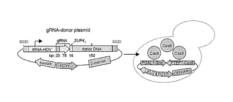
FIGS. 1A-1C show a dual CRISPR/Cas9 editing and barcoding system. FIG.
1A shows guide RNA (gRNA)-donor DNA sequences cloned into a high-copy vector,
with the tRNA-HDV ribozyme promoter driving gRNA expression. The guide-donor
plasmids were then transformed into cells with Cas9 pre-expressed from a
strong
constitutive promoter. FIG. 1B shows target locus editing. Cas9-gRNA-induced
dsDNA breaks are resolved through either donor DNA directed HR, NHEJ, or cell
death. FIG. 1C shows REDI locus barcoding. Induction of SceI with galactose
enables
replacement of counter-selectable FCY1 with the guide RNA-donor DNA segment,
allowing for (1) PCR-based phenotyping of competitively grown pools, and (2)
REDI-based identification of individual variants.
FIGS. 2A-2C show proof-of-concept for high-efficiency Cas9 editing and
SceI-mediated barcoding. FIG. 2A shows a gRNA targeting ADE2 cloned into the
high copy vector backbone shown in FIG. 1 without (left column) or with (right
column) donor DNA. The gRNA vectors were transformed into cells with pre-
expressed Cas9 (top row), or with PTEF1-Cas9 encoded on the gRNA vector (not
pre-
expressed; bottom row). FIG. 2B shows the ADE2 locus in select clones.
Sequencing
confirmed the desired loss-of-function edit. FIG. 2C shows results for pooled
cells
from the plate, which were either shifted to galactose or glucose media.
Individual
colonies were isolated and screened for the integration of the guide-donor
cassette at
the REDI locus, which was confirmed by Sanger sequencing.
FIG. 3 shows selected guide-donor plasmids for editing a heterologous ORF
(mCherry). A library of guide-donor oligonucleotides was purchased from
Agilent
Technologies, and cloned into the high-copy guide expression vector (FIG. 1).
A few
bacterial clones were sequenced for correct incorporation of the guide-donor
insert,
and subsequently transformed into yeast cells harboring pre-expressed Cas9 and
the
mCherry ORF. Shown are the transformation plates after 2 days of growth.
FIGS. 4A-4F show that active donor recruitment enables high frequencies of
donor directed repair. FIG. 4A shows a two plasmid-based system for high-
throughput editing in the absence of donor recruitment. FIG. 4B shows that
random
diffusion of donor DNA results in inefficient homologous recombination repair,
and
the majority of transformants with effective gRNAs undergo cell death. FIG. 4C
shows a modified two plasmid-based system with LexA binding sites on the guide-
-15-
CA 03075532 2020-03-10
WO 2019/055878
PCT/US2018/051240
donor plasmid and a LexA DNA-binding domain (DBD) fused to the Fkhl protein
for
ultra-high efficiency high-throughput editing. FIG. 4D shows dsDNA breaks
trigger
the phosphorylation of threonine residues on endogenous cellular proteins in
the
vicinity of the break. This results in the recruitment of Fkhl via its
forkhead-
.. associated (FHA) phosphothreonine-binding domain, resulting in a high local
concentration of donor DNA to facilitate the search for homologous DNA during
DNA repair. This leads to precision editing being the predominant outcome over
cell
death. FIG. 4E shows a LexA DNA binding domain fused to Cas9 instead of Fkhl.
FIG. 4F shows that the Cas9-LexA DBD enables pre-recruitment of the guide-
donor
plasmid to the gRNA target site, facilitating HDR after DNA cleavage by Cas9.
FIGS. 5A and 5B show that active donor recruitment enables high frequencies
of donor directed repair. FIG. 5A shows cells pre-expressing Cas9 (upper left
panel,
as in FIG.4A) or Cas9 and LexA DBD-Fkhl (upper right panel, as in FIG.4C) were
transformed with a plasmid pool harboring 85% sequence-verified guide-donor
targeting a null mutation in ADE2, and 15% of the same plasmid with a mutated
ADE2 guide RNA. FIG. 5B shows cells pre-expressing Cas9, transformed with the
sequence-perfect guide-donor (lower left panel). Cells pre-expressing the high-
copy
guide-donor plasmid were transformed with the Cas9 plasmid (lower right
panel).
FIGS. 6A and 6B show a dual editing and barcoding system combined with
Recombinase Directed Indexing (REDI). FIG. 6A shows steps 1-4 of the process.
Step 1: a complex library of plasmids harboring guide RNA (gRNA) and donor DNA
sequences is transformed into recipient strains modified to contain a barcode
locus
with a counter-selectable marker (FCY1) flanked by sites for the mega-nuclease
SceI.
Transformed cells are plated onto ¨HIS to select for plasmids containing the
correct
internal cloning event, and colonies pooled and grown to mid-log phase in rich
medium containing G418 to maintain selection for the guide-donor plasmids. The
cells are then transformed with the Cas9/SceI plasmid and plated onto ¨LEU-HIS
Step 2: The chromosomal target will be cut with the Cas9-gRNA and repaired
using
homologous recombination (HR) with the donor DNA encoded on the plasmid. The
colonies are recovered and grown for several generations in rich medium with
galactose to induce SceI. dsDNA breaks at the chromosomal barcode locus
promote
integration of the guide-HI53-donor cassette, and linearize the plasmids. Step
3:
Successful integration of guide-donor barcodes and loss of plasmids is
selected for by
-16-
CA 03075532 2020-03-10
WO 2019/055878
PCT/US2018/051240
plating on synthetic medium containing 5-fluoro-cytosine (5-FC). Transformants
are
arrayed on agar plates at a density of 1536, to allow subsequent mating to
barcoder
strains. At this stage, it is possible that transformants contain successful
intended
edits, unintended mutations due to oligo synthesis-derived errors, or no edit.
Step 4:
The arrayed strain variants are mated to barcoder strains, which contain a
LoxP site,
followed by a unique positional barcode that specifies the colony coordinates
on the
plate, and the rest of the URA3 gene. Induction of Cre results in LoxP-
mediated
reconstitution of the split URA3, which physically links the guide-donor
sequence
with the positional barcode for high-throughput paired-end sequencing (HTS) of
the
.. guide-donor-barcode combinations (Step 5). Two different P5 primers allow
linkage
of the guide and donor sequence to the specific colony coordinates by virtue
of the
shared positional barcode. FIG. 6B shows schematics of the Mat a variant
chromosome and the Mat a barcoded chromosome and the results of Step 4 and
Step
5.
FIGS. 7A-7C show REDI-mediated massively parallel strain validation. FIG.
7A shows clones isolated from multiplexed precision editing experiments can
contain
successfully edited target loci (dark gray), an unintended mutation at the
target locus
due to a synthesis-derived error or error during homologous recombination (HR)
(light gray), or an unsuccessful edit due to an ineffective guide RNA (light
gray). FIG.
7B shows independent clones for each designed variant (as indicated by light
gray,
dark gray, and medium gray). Step 1: These replicates are re-arrayed onto
separate
plates such that each plate contains mutations targeted within a designated
chromosomal window or gene, and only one colony is present per designed
variant
per plate. Colonies are pooled and genomic DNA extracted for each plate. Step
2:
PCR of the targeted chromosomal locus and deep amplicon sequencing are
performed. Successfully edited variants are expected to be present at a
frequency of
1/1536. FIG. 7C depicts rearraying of desired clones for pooled (top) or
spatially-
separated phenotyping assays.
FIG. 8 shows one potential workflow for editing, barcoding, validating and
.. phenotyping strains.
FIG. 9 shows a library cloning strategy to minimize non-functional vector
background. Step 1: Oligo pools are amplified with primers containing 5"-
extensions
to facilitate Gibson- or sticky end-mediated cloning into vector backbones.
Step 2:
-17-
CA 03075532 2020-03-10
WO 2019/055878
PCT/US2018/051240
The amplified oligos contain an internal Type IIS restriction site. The cloned
vector is
treated with the Type IIS enzyme and a phosphatase. This enables scarless
internal
cloning of the structural guide RNA component, a Pol III terminator, and a
selectable
marker (e.g. HIS3). Step 3: The constant insert is treated with BspQI only to
retain 5'-
phosphates. Step 4: The insert is ligated into the vector backbone, and can
then be
transformed into recipient yeast with selection on ¨HIS medium.
FIGS. 10A and 10B show a synonymous codon spreading strategy to enable
amino acid mutations outside of a guide recognition region. Saturation
mutagenesis of
open reading frames is enabled through engineering synonymous codon mutations
(dark gray) between the nonsynonymous variant (light gray) and the protospacer-
adjacent motif (PAM, box, NGG in this depiction). A pseudo-WT control is
established by including only the synonymous variants (dark gray). Also shown
are
donor DNA and guide RNA sequences to engineer a nonsynonymous variant falling
within (FIG. 10A) or outside of (FIG. 10B) the guide recognition sequence.
FIG. 11 shows repair directly with a genome integrated editing cassette.
FIG. 12 shows library cloning to link guide-donors with unique DNA
barcodes. (1) Oligonucleotides encoding guide-donors are synthesized in high-
density
array format and cleaved off of the array surface to generate a complex pool.
(2) Each
oligo contains common amplification sequences flanking the guide-donor
cassette to
enable amplification of specific subpools. The forward primer harbors a
restriction
site (AscI) at its 3'-end and the reverse primer encodes a distinct
restriction site (NotI)
at its 5'-end followed by a degenerate barcode (bc) encoding a pseudo-random
sequence (either NNNVHTGNNNVHTGNNNVHTGNNNVHTGNNN or
NNNTGVHNNNTGVHNNNTGVHNNNTGVHNNN) that excludes illegal
restriction sites (NotI, AscI, and BspQI). The degenerate barcode is flanked
by a 50
bp downstream homology sequence (DH). NotI and AscI sites enable sticky end
cloning into a multi-copy recipient vector, with the AscI site at the 3 '-end
of the guide
RNA promoter. The guide and donor sequences are separated by a type IIS
restriction
site (BspQI) that enables cloning with an arbitrary overhang, in this case the
GTTT
directly 3' of the guide sequence, to enable cloning in the constant
structural
component of the guide RNA.
(3) High-throughput sequencing (HTS) of the first-step cloning products
enables
linking the guide-BspQI-donor sequences with unique barcodes (bc). Paired-end
-18-
CA 03075532 2020-03-10
WO 2019/055878
PCT/US2018/051240
sequencing can be used to increase confidence of base calls following quality-
based
merging of read 1 and read 2. (4) (a) The structural guide RNA component along
with
yeast-specific (e.g. URA3) and bacterial-specific (e.g. kanR) selection
markers are
amplified with primers harboring BspQI sequences at their 5'-ends. The reverse
primer includes an additional barcode (bc*; either NNNNNN or
NNNNNNHVVN}{BBHBHD) situated 3' of the Illumina read 2 priming sequence,
modified to contain a G-to-A SNP at the first position of the BspQI site. (b)
Cleavage
of the first step cloning products with BspQI followed by phosphatase
treatment
enables scarless cloning of the structural gRNA insert. These second-step
libraries
are selected with kanamycin to enable enrichment of vectors harboring the
insert.
Paired-end HTS of bc*-donor and bc enables mapping the barcodes to unique
guide-
donor combinations.
FIG. 13 shows simultaneous editing and barcode integration via self-
destructing plasmids. (1) The guide-donor vectors after the second-step
cloning are
transformed into yeast and selected with the insert-specific marker (URA3).
The
recipient strain is modified to harbor a barcode integration locus with a
counter-
selectable marker (FCY1). In addition to the guide sequences from the library,
the
guide-donor plasmids also harbor a guide X expression unit to promote barcode
integration, as guide X cleavage sites flank FCY1. Following transformation,
the
guide-donor plasmids accumulate to high copy number through outgrowth. At the
5"-
end of the downstream homology (DH) sequence on the guide-donor plasmid
resides
a guide X cleavage site, which enables later linearization of the plasmid to
accelerate
plasmid loss after editing. (2) Induction of Cas9 results in guide X cleaving
the
plasmid and genomic barcode locus, and in the library-derived guide cleavage
elsewhere in the genome. (a) Guide X cleavage results in genomic integration
of the
entire guide RNA-bc*-donor DNA-bc cassette via upstream homology (UH) sequence
present on both the guide-donor plasmid and in the chromosomal barcode site.
(b) The
edit-directing guide cleavage is followed by donor DNA-directed homologous
recombination to generate the intended genomic edit.
FIGS. 14A and 14B show that Cpfl guide-donor system results in highly
efficient (>99%) editing and editing with Cpfl is enhanced ¨10-fold with donor
recruitment to a similar extent as Cas9. FIG. 14A: A Cpfl guide-donor plasmid
(the
guide has the Cpfl scaffold) targeting the ADE2 gene was transformed into
cells pre-
-19-
CA 03075532 2020-03-10
WO 2019/055878
PCT/US2018/051240
expressing Cpfl. The donor DNA encodes a frameshift-causing deletion. FIG.
14B:
The Cpfl guide-donor was mixed with a non-editing plasmid at a ratio of 17:3
and
transformed into cells expressing Cpfl without (left) or with (right) LexA-
FHA. The
ratio of red:white colonies is shown on the y-axis.
FIG. 15 shows a modified version of the multiplex genome editing system,
where Cpfl and/or Cas9 or other RNA-guided nuclease (RGN) or site-specific
nuclease (e.g. SceI, other meganucleases, ZFNs, or TALENs) are expressed from
the
REDI locus, optionally along with other multiplex editing components, e.g.
TetR and
LexA-FHA and markers for forward and counter selection (URA3 and hphMX). In
this arrangement, the self-destructing guide-donor vector integrates into the
REDI
barcoding locus with simultaneous removal of Cas9, Cpfl, and all genes in
between.
The genetic removal of Cas9 at the DNA level followed by sufficient outgrowth
to
dilute out Cas9 mRNA and protein ensures that subsequent fitness assays are
not
confounded by effects of Cas9: :guide-edit binding to chromatin. The editing
guide
can be paired with either Cas9 or Cpfl, and likewise the barcoding guide X can
be
paired with either Cas9 or Cpfl. The advantage of having dedicated nucleases
for
RGNs is that there is no competition between editing and barcoding guides for
associating with the RGN. This arrangement also increases the flexibility of
the
multiplex system with regards to allowing targeting more genomic regions by
utilizing RNA-guided nucleases with different PAM requirements.
FIG. 16 shows plasmid spike-in experiments demonstrating that both LexA-
FHA and linearized vectors enhance HDR efficiency and editing survival. Note
that
LexA-FHA with a circular plasmid results in the overall highest transformation
survival.
FIG. 17 shows the efficiency of HDR in human cells with or without LexA
sites in the presence of the donor recruitment protein, dn53BP1-LexA. Two
independent genes were targeted (CACNA1D (CAC) and PPP1R12C (PPP)). The
first panel shows the rate of NHEJ at the cut site. The second panel shows the
percentage total HDR at the cut site, and the third panel shows the ratio of
HDR to
NHEJ in the cells.
-20-
CA 03075532 2020-03-10
WO 2019/055878
PCT/US2018/051240
DETAILED DESCRIPTION
The practice of the present disclosure will employ, unless otherwise
indicated,
conventional methods of genome editing, biochemistry, chemistry, immunology,
molecular biology and recombinant DNA techniques within the skill of the art.
Such
techniques are explained fully in the literature. See, e.g., Targeted Genome
Editing
Using Site-Specific Nucleases: ZFNs, TALENs, and the CRISPR/Cas9 System (T.
Yamamoto ed., Springer, 2015); Genome Editing: The Next Step in Gene Therapy
(Advances in Experimental Medicine and Biology, T. Cathomen, M. Hirsch, and M.
Porteus eds., Springer, 2016); Aachen Press Genome Editing (CreateSpace
Independent Publishing Platform, 2015); Handbook of Experimental Immunology,
V ols. I-TV (D.M. Weir and C.C. Blackwell eds., Blackwell Scientific
Publications);
A.L. Lehninger, Biochemistry (Worth Publishers, Inc., current addition);
Sambrook, et
al., Molecular Cloning: A Laboratory Manual (3rd Edition, 2001); Methods In
Enzymology (S. Colowick and N. Kaplan eds., Academic Press, Inc.).
All publications, patents and patent applications cited herein, whether supra
or
infra, are hereby incorporated by reference in their entireties.
I. DEFINITIONS
In describing the present disclosure, the following terms will be employed,
and are intended to be defined as indicated below.
It must be noted that, as used in this specification and the appended claims,
the
singular forms "a," "an" and "the" include plural referents unless the content
clearly
dictates otherwise. Thus, for example, reference to "a cell" includes a
mixture of two
or more cells, and the like.
The term "about," particularly in reference to a given quantity, is meant to
encompass deviations of plus or minus five percent.
A "barcode" refers to one or more nucleotide sequences that are used to
identify a nucleic acid or cell with which the barcode is associated. Barcodes
can be
3-1000 or more nucleotides in length, preferably 10-250 nucleotides in length,
and
more preferably 10-30 nucleotides in length, including any length within these
ranges,
such as 3, 4, 5, 6, 7, 8, 9, 10, 11, 12, 13, 14, 15, 16, 17, 18, 19, 20, 21,
22, 23, 24, 25,
26, 27, 28, 29, 30, 40, 50, 60, 70, 80, 90, 100, 200, 300, 400, 500, 600, 700,
800, 900,
-21-
CA 03075532 2020-03-10
WO 2019/055878
PCT/US2018/051240
or 1000 nucleotides in length. Barcodes may be used, for example, to identify
a
single cell, subpopulation of cells, colony, or sample from which a nucleic
acid
originated. Barcodes may also be used to identify the position (i.e.,
positional
barcode) of a cell, colony, or sample from which a nucleic acid originated,
such as the
position of a colony in a cellular array, the position of a well in a multi-
well plate, or
the position of a tube, flask, or other container in a rack. In particular, a
barcode may
be used to identify a genetically modified cell from which a nucleic acid
originated.
In some embodiments, a barcode is used to identify a particular type of genome
edit.
For example, a guide RNA-donor polynucleotide cassette itself can be used as a
barcode to identify a genetically modified cell from which a nucleic acid
originated.
Alternatively, a unique barcode may be used to identify each guide-RNA-donor
polynucleotide cassette used in multiplex genome editing. Furthermore,
multiple
barcodes can be used in combination to identify different features of a
nucleic acid.
For example, positional barcoding (e.g., to identify the position of a cell,
colony,
culture, or sample in an array, multi-well plate, or rack) can be combined
with
barcodes identifying guide-RNA-donor polynucleotide cassettes used in genome
editing. In some embodiments, barcodes are inserted into a nucleic acid (e.g.,
at a
"barcode locus") at each round of genome editing to identify the guide-RNAs
and/or
donor polynucleotides used in genetic modification of a cell.
The term "barcoder cell" refers to a cell comprising a nucleic acid comprising
a barcode sequence. In one embodiment, the barcode identifies the position of
a
colony comprising the barcoder cells.
The terms "polypeptide" and "protein" refer to a polymer of amino acid
residues and are not limited to a minimum length. Thus, peptides,
oligopeptides,
dimers, multimers, and the like, are included within the definition. Both full
length
proteins and fragments thereof are encompassed by the definition. The terms
also
include post expression modifications of the polypeptide, for example,
glycosylation,
acetylation, phosphorylation, hydroxylation, and the like. Furthermore, for
purposes
of the present disclosure, a "polypeptide" refers to a protein which includes
modifications, such as deletions, additions and substitutions to the native
sequence, so
long as the protein maintains the desired activity. These modifications may be
deliberate, as through site directed mutagenesis, or may be accidental, such
as through
mutations of hosts which produce the proteins or errors due to PCR
amplification.
-22-
CA 03075532 2020-03-10
WO 2019/055878
PCT/US2018/051240
The term "Cas9" as used herein encompasses type II clustered regularly
interspaced short palindromic repeats (CRISPR) system Cas9 endonucleases from
any
species, and also includes biologically active fragments, variants, analogs,
and
derivatives thereof that retain Cas9 endonuclease activity (i.e., catalyze
site-directed
cleavage of DNA to generate double-strand breaks). A Cas9 endonuclease binds
to
and cleaves DNA at a site comprising a sequence complementary to its bound
guide
RNA (gRNA).
A Cas9 polynucleotide, nucleic acid, oligonucleotide, protein, polypeptide, or
peptide refers to a molecule derived from any source. The molecule need not be
physically derived from an organism, but may be synthetically or recombinantly
produced. Cas9 sequences from a number of bacterial species are well known in
the
art and listed in the National Center for Biotechnology Information (NCBI)
database.
See, for example, NCBI entries for Cas9 from: Streptococcus pyogenes
(WP 002989955, WP 038434062, WP 011528583); Campylobacter jejuni
(WP 022552435, YP 002344900), Campylobacter coil (WP 060786116);
Campylobacter fetus (WP 059434633); Corynebacterium ulcerans (NC 015683,
NCO17317); Corynebacterium diphtheria (NC 016782, NCO16786); Enterococcus
faecalis (WP 033919308); Spiroplasma syrphidicola (NC 021284); Prevotella
intermedia (NC 017861); Spiroplasma taiwanense (NC 021846); Streptococcus
in/ac (NC 021314); Belliella bait/ca (NC 018010); Psychroflexus torquisl
(NC 018721); Streptococcus thermophilus (YP 820832), Streptococcus mutans
(WP 061046374, WP 024786433); Listeria innocua (NP 472073); Listeria
monocytogenes (WP 061665472); Legionella pneumophila (WP 062726656);
Staphylococcus aureus (WP 001573634); Francisella tularensis (WP 032729892,
WPO14548420), Enterococcus faecalis (WP 033919308); Lactobacillus rhamnosus
(WP 048482595, WP 032965177); and Neisseria meningitidis (WP 061704949,
YP 002342100); all of which sequences (as entered by the date of filing of
this
application) are herein incorporated by reference. Any of these sequences or a
variant
thereof comprising a sequence having at least about 70-100% sequence identity
thereto, including any percent identity within this range, such as 70, 71, 72,
73, 74,
75, 76, 77, 78, 79, 80, 81, 82, 83, 84, 85, 86, 87, 88, 89, 90, 91, 92, 93,
94, 95, 96, 97,
98, or 99% sequence identity thereto, can be used for genome editing, as
described
herein, wherein the variant retains biological activity, such as Cas9 site-
directed
-23-
CA 03075532 2020-03-10
WO 2019/055878
PCT/US2018/051240
endonuclease activity. See also Fonfara et al. (2014) Nucleic Acids Res.
42(4):2577-
90; Kapitonov et al. (2015) J. Bacteriol. 198(5):797-807, Shmakov et al.
(2015) Mol.
Cell. 60(3):385-397, and Chylinski et al. (2014) Nucleic Acids Res.
42(10):6091-
6105); for sequence comparisons and a discussion of genetic diversity and
phylogenetic analysis of Cas9.
By "derivative" is intended any suitable modification of the native
polypeptide
of interest, of a fragment of the native polypeptide, or of their respective
analogs, such
as glycosylation, phosphorylation, polymer conjugation (such as with
polyethylene
glycol), or other addition of foreign moieties, as long as the desired
biological activity
of the native polypeptide is retained. Methods for making polypeptide
fragments,
analogs, and derivatives are generally available in the art.
By "fragment" is intended a molecule consisting of only a part of the intact
full-length sequence and structure. The fragment can include a C-terminal
deletion an
N- terminal deletion, and/or an internal deletion of the polypeptide. Active
fragments
of a particular protein or polypeptide will generally include at least about 5-
10
contiguous amino acid residues of the full length molecule, preferably at
least about
15-25 contiguous amino acid residues of the full length molecule, and most
preferably
at least about 20-50 or more contiguous amino acid residues of the full length
molecule, or any integer between 5 amino acids and the full length sequence,
provided that the fragment in question retains biological activity, such as
Cas9 site-
directed endonuclease activity.
"Substantially purified" generally refers to isolation of a substance
(compound, polynucleotide, nucleic acid, protein, polypeptide, polypeptide
composition) such that the substance comprises the majority percent of the
sample in
which it resides. Typically in a sample, a substantially purified component
comprises
50%, preferably 80%-85%, more preferably 90-95% of the sample. Techniques for
purifying polynucleotides and polypeptides of interest are well-known in the
art and
include, for example, ion-exchange chromatography, affinity chromatography and
sedimentation according to density.
By "isolated" is meant, when referring to a polypeptide, that the indicated
molecule is separate and discrete from the whole organism with which the
molecule is
found in nature or is present in the substantial absence of other biological
macro
molecules of the same type. The term "isolated" with respect to a
polynucleotide is a
-24-
CA 03075532 2020-03-10
WO 2019/055878
PCT/US2018/051240
nucleic acid molecule devoid, in whole or part, of sequences normally
associated with
it in nature; or a sequence, as it exists in nature, but having heterologous
sequences in
association therewith; or a molecule disassociated from the chromosome.
As used herein, the phrase "heterogeneous population of cells" refers to a
mixture of at least two types of cells, one type being the cells of interest
(e.g., having
a genomic modification of interest). The heterogeneous population of cells may
be
derived from any organism.
The terms "isolating" and "isolation," as used herein in the context of
selecting
a cell or population of cells having a genomic modification of interest, refer
to
separating a cell or population of cells having the genomic modification of
interest
from a heterogeneous population of cells, such as by positive or negative
selection.
The term "selection marker" refers to a marker which can be used for
identification or enrichment of a cell population from a heterogeneous
population of
cells, either by positive selection (selecting cells expressing the marker) or
by
negative selection (excluding cells expressing the marker).
The terms "polynucleotide," "oligonucleotide," "nucleic acid" and "nucleic
acid molecule" are used herein to include a polymeric form of nucleotides of
any
length, either ribonucleotides or deoxyribonucleotides. This term refers only
to the
primary structure of the molecule. Thus, the term includes triple-, double-
and single-
stranded DNA, as well as triple-, double- and single-stranded RNA. It also
includes
modifications, such as by methylation and/or by capping, and unmodified forms
of the
polynucleotide. More particularly, the terms "polynucleotide,"
"oligonucleotide,"
"nucleic acid" and "nucleic acid molecule" include polydeoxyribonucleotides
(containing 2-deoxy-D-ribose), polyribonucleotides (containing D-ribose), any
other
type of polynucleotide which is an N- or C-glycoside of a purine or pyrimidine
base,
and other polymers containing nonnucleotidic backbones, for example, polyamide
(e.g., peptide nucleic acids (PNAs)) and polymorpholino (commercially
available
from the Anti-Virals, Inc., Corvallis, Oreg., as Neugene) polymers, and other
synthetic sequence-specific nucleic acid polymers providing that the polymers
contain
nucleobases in a configuration which allows for base pairing and base
stacking, such
as is found in DNA and RNA. There is no intended distinction in length between
the
terms "polynucleotide," "oligonucleotide," "nucleic acid" and "nucleic acid
molecule," and these terms will be used interchangeably. Thus, these terms
include,
-25-
CA 03075532 2020-03-10
WO 2019/055878
PCT/US2018/051240
for example, 3'-deoxy-2',5'-DNA, oligodeoxyribonucleotide N3' P5'
phosphoramidates, 2'-0-alkyl-substituted RNA, double- and single-stranded DNA,
as
well as double- and single-stranded RNA, microRNA, DNA:RNA hybrids, and
hybrids between PNAs and DNA or RNA, and also include known types of
modifications, for example, labels which are known in the art, methylation,
"caps,"
substitution of one or more of the naturally occurring nucleotides with an
analog (e.g.,
2-aminoadenosine, 2-thiothymidine, inosine, pyrrolo-pyrimidine, 3-methyl
adenosine,
C5-propynylcytidine, C5-propynyluridine, C5-bromouridine, C5-fluorouridine, C5-
iodouridine, C5-methylcytidine, 7-deazaadenosine, 7-deazaguanosine, 8-
oxoadenosine, 8-oxoguanosine, 0(6)-methylguanine, and 2-thiocytidine),
internucleotide modifications such as, for example, those with uncharged
linkages
(e.g., methyl phosphonates, phosphotriesters, phosphoramidates, carbamates,
etc.),
with negatively charged linkages (e.g., phosphorothioates,
phosphorodithioates, etc.),
and with positively charged linkages (e.g., aminoalklyphosphoramidates,
aminoalkylphosphotriesters), those containing pendant moieties, such as, for
example,
proteins (including nucleases, toxins, antibodies, signal peptides, poly-L-
lysine, etc.),
those with intercalators (e.g., acridine, psoralen, etc.), those containing
chelators (e.g.,
metals, radioactive metals, boron, oxidative metals, etc.), those containing
alkylators,
those with modified linkages (e.g., alpha anomeric nucleic acids, etc.), as
well as
unmodified forms of the polynucleotide or oligonucleotide. The term also
includes
locked nucleic acids (e.g., comprising a ribonucleotide that has a methylene
bridge
between the 2'-oxygen atom and the 4'-carbon atom). See, for example, Kurreck
et al.
(2002) Nucleic Acids Res. 30: 1911-1918; Elayadi et al. (2001) Curr. Opinion
Invest.
Drugs 2: 558-561; Orum et al. (2001) Curr. Opinion Mol. Ther. 3: 239-243;
Koshkin
et al. (1998) Tetrahedron 54: 3607-3630; Obika et al. (1998) Tetrahedron Lett.
39:
5401-5404.
The terms "hybridize" and "hybridization" refer to the formation of complexes
between nucleotide sequences which are sufficiently complementary to form
complexes via Watson-Crick base pairing.
The term "homologous region" refers to a region of a nucleic acid with
homology to another nucleic acid region. Thus, whether a "homologous region"
is
present in a nucleic acid molecule is determined with reference to another
nucleic acid
region in the same or a different molecule. Further, since a nucleic acid is
often
-26-
CA 03075532 2020-03-10
WO 2019/055878
PCT/US2018/051240
double-stranded, the term "homologous, region," as used herein, refers to the
ability
of nucleic acid molecules to hybridize to each other. For example, a single-
stranded
nucleic acid molecule can have two homologous regions which are capable of
hybridizing to each other. Thus, the term "homologous region" includes nucleic
acid
segments with complementary sequences. Homologous regions may vary in length,
but will typically be between 4 and 500 nucleotides (e.g., from about 4 to
about 40,
from about 40 to about 80, from about 80 to about 120, from about 120 to about
160,
from about 160 to about 200, from about 200 to about 240, from about 240 to
about
280, from about 280 to about 320, from about 320 to about 360, from about 360
to
about 400, from about 400 to about 440, etc.).
As used herein, the terms "complementary" or "complementarity" refers to
polynucleotides that are able to form base pairs with one another. Base pairs
are
typically formed by hydrogen bonds between nucleotide units in an anti-
parallel
orientation between polynucleotide strands. Complementary polynucleotide
strands
can base pair in a Watson-Crick manner (e.g., A to T, A to U, C to G), or in
any other
manner that allows for the formation of duplexes. As persons skilled in the
art are
aware, when using RNA as opposed to DNA, uracil (U) rather than thymine (T) is
the
base that is considered to be complementary to adenosine. However, when a
uracil is
denoted in the context of the present disclosure, the ability to substitute a
thymine is
implied, unless otherwise stated. "Complementarity" may exist between two RNA
strands, two DNA strands, or between a RNA strand and a DNA strand. It is
generally
understood that two or more polynucleotides may be "complementary" and able to
form a duplex despite having less than perfect or less than 100%
complementarity.
Two sequences are "perfectly complementary" or "100% complementary" if at
least a
contiguous portion of each polynucleotide sequence, comprising a region of
complementarity, perfectly base pairs with the other polynucleotide without
any
mismatches or interruptions within such region. Two or more sequences are
considered "perfectly complementary" or "100% complementary" even if either or
both polynucleotides contain additional non-complementary sequences as long as
the
contiguous region of complementarity within each polynucleotide is able to
perfectly
hybridize with the other. "Less than perfect" complementarity refers to
situations
where less than all of the contiguous nucleotides within such region of
complementarity are able to base pair with each other. Determining the
percentage of
-27-
CA 03075532 2020-03-10
WO 2019/055878
PCT/US2018/051240
complementarity between two polynucleotide sequences is a matter of ordinary
skill
in the art. For purposes of Cas9 targeting, a gRNA may comprise a sequence
"complementary" to a target sequence (e.g., major or minor allele), capable of
sufficient base-pairing to form a duplex (i.e., the gRNA hybridizes with the
target
sequence). Additionally, the gRNA may comprise a sequence complementary to a
PAM sequence, wherein the gRNA also hybridizes with the PAM sequence in a
target
DNA.
A "target site" or "target sequence" is the nucleic acid sequence recognized
(i.e., sufficiently complementary for hybridization) by a guide RNA (gRNA) or
a
homology arm of a donor polynucleotide. The target site may be allele-specific
(e.g.,
a major or minor allele).
The term "donor polynucleotide" refers to a polynucleotide that provides a
sequence of an intended edit to be integrated into the genome at a target
locus by
HDR.
By "homology arm" is meant a portion of a donor polynucleotide that is
responsible for targeting the donor polynucleotide to the genomic sequence to
be
edited in a cell. The donor polynucleotide typically comprises a 5' homology
arm that
hybridizes to a 5' genomic target sequence and a 3' homology arm that
hybridizes to a
3' genomic target sequence flanking a nucleotide sequence comprising the
intended
edit to the genomic DNA. The homology arms are referred to herein as 5' and 3'
(i.e.,
upstream and downstream) homology arms, which relates to the relative position
of
the homology arms to the nucleotide sequence comprising the intended edit
within the
donor polynucleotide. The 5' and 3' homology arms hybridize to regions within
the
target locus in the genomic DNA to be modified, which are referred to herein
as the
"5' target sequence" and "3' target sequence," respectively. The nucleotide
sequence
comprising the intended edit is integrated into the genomic DNA by HDR at the
genomic target locus recognized (i.e., sufficiently complementary for
hybridization)
by the 5' and 3' homology arms.
"Administering" a nucleic acid, such as a donor polynucleotide, guide RNA,
or Cas9 expression system to a cell comprises transducing, transfecting,
electroporating, translocating, fusing, phagocytosing, shooting or ballistic
methods,
etc., i.e., any means by which a nucleic acid can be transported across a cell
membrane.
-28-
CA 03075532 2020-03-10
WO 2019/055878
PCT/US2018/051240
By "selectively binds" with reference to a guide RNA is meant that the guide
RNA binds preferentially to a target sequence of interest or binds with
greater affinity
to the target sequence than to other genomic sequences. For example, a gRNA
will
bind to a substantially complementary sequence and not to unrelated sequences.
A
gRNA that "selectively binds" to a particular allele, such as a particular
mutant allele
(e.g., allele comprising a substitution, insertion, or deletion), denotes a
gRNA that
binds preferentially to the particular target allele, but to a lesser extent
to a wild-type
allele or other sequences. A gRNA that selectively binds to a particular
target DNA
sequence will selectively direct binding of an RNA-guided nuclease (e.g.,
Cas9) to a
substantially complementary sequence at the target site and not to unrelated
sequences.
As used herein, the term "recombination target site" denotes a region of a
nucleic acid molecule comprising a binding site or sequence-specific motif
recognized
by a site-specific recombinase that binds at the target site and catalyzes
recombination
.. of specific sequences of DNA at the target site. Site-specific recombinases
catalyze
recombination between two such target sites. The relative orientation of the
target
sites determines the outcome of recombination. For example, translocation
occurs if
the recombination target sites are on separate DNA molecules.
As used herein, the terms "label" and "detectable label" refer to a molecule
capable of detection, including, but not limited to, radioactive isotopes,
fluorescers,
chemiluminescers, chromophores, enzymes, enzyme substrates, enzyme cofactors,
enzyme inhibitors, semiconductor nanoparticles, dyes, metal ions, metal sols,
ligands
(e.g., biotin, streptavidin or haptens) and the like. The term "fluorescer"
refers to a
substance or a portion thereof which is capable of exhibiting fluorescence in
the
detectable range. Particular examples of labels which may be used in the
practice of
the present disclosure include, but are not limited to, SYBR green, SYBR gold,
a
CAL Fluor dye such as CAL Fluor Gold 540, CAL Fluor Orange 560, CAL Fluor Red
590, CAL Fluor Red 610, and CAL Fluor Red 635, a Quasar dye such as Quasar
570,
Quasar 670, and Quasar 705, an Alexa Fluor such as Alexa Fluor 350, Alexa
Fluor
488, Alexa Fluor 546, Alexa Fluor 555, Alexa Fluor 594, Alexa Fluor 647,and
Alexa
Fluor 784, a cyanine dye such as Cy 3, Cy3.5, Cy5, Cy5.5, and Cy7,
fluorescein, 2',
4', 5', 7'-tetrachloro-4-7-dichlorofluorescein (TET), carboxyfluorescein
(FAM), 6-
carboxy-4',5'-dichloro-2',7'-dimethoxyfluorescein (JOE), hexachlorofluorescein
-29-
CA 03075532 2020-03-10
WO 2019/055878
PCT/US2018/051240
(HEX), rhodamine, carboxy-X-rhodamine (ROX), tetramethyl rhodamine (TAMRA),
FITC, dansyl, umbelliferone, dimethyl acridinium ester (DMAE), Texas red,
luminol,
NADPH, horseradish peroxidase (HRP), and a-P-galactosidase.
"Homology" refers to the percent identity between two polynucleotide or two
polypeptide moieties. Two nucleic acid, or two polypeptide sequences are
"substantially homologous" to each other when the sequences exhibit at least
about
50% sequence identity, preferably at least about 75% sequence identity, more
preferably at least about 80% 85% sequence identity, more preferably at least
about
90% sequence identity, and most preferably at least about 95% 98% sequence
identity
.. over a defined length of the molecules. As used herein, substantially
homologous
also refers to sequences showing complete identity to the specified sequence.
In general, "identity" refers to an exact nucleotide to nucleotide or amino
acid
to amino acid correspondence of two polynucleotides or polypeptide sequences,
respectively. Percent identity can be determined by a direct comparison of the
sequence information between two molecules by aligning the sequences, counting
the
exact number of matches between the two aligned sequences, dividing by the
length
of the shorter sequence, and multiplying the result by 100. Readily available
computer programs can be used to aid in the analysis, such as ALIGN, Dayhoff,
M.O.
in Atlas of Protein Sequence and Structure M.O. Dayhoff ed., 5 Suppl. 3:353
358,
National biomedical Research Foundation, Washington, DC, which adapts the
local
homology algorithm of Smith and Waterman Advances in Appl. Math. 2:482 489,
1981 for peptide analysis. Programs for determining nucleotide sequence
identity are
available in the Wisconsin Sequence Analysis Package, Version 8 (available
from
Genetics Computer Group, Madison, WI) for example, the BESTFIT, FASTA and
GAP programs, which also rely on the Smith and Waterman algorithm. These
programs are readily utilized with the default parameters recommended by the
manufacturer and described in the Wisconsin Sequence Analysis Package referred
to
above. For example, percent identity of a particular nucleotide sequence to a
reference sequence can be determined using the homology algorithm of Smith and
Waterman with a default scoring table and a gap penalty of six nucleotide
positions.
Another method of establishing percent identity in the context of the present
disclosure is to use the MPSRCH package of programs copyrighted by the
University
of Edinburgh, developed by John F. Collins and Shane S. Sturrok, and
distributed by
-30-
CA 03075532 2020-03-10
WO 2019/055878
PCT/US2018/051240
IntelliGenetics, Inc. (Mountain View, CA). From this suite of packages, the
Smith
Waterman algorithm can be employed where default parameters are used for the
scoring table (for example, gap open penalty of 12, gap extension penalty of
one, and
a gap of six). From the data generated the "Match" value reflects "sequence
identity."
Other suitable programs for calculating the percent identity or similarity
between
sequences are generally known in the art, for example, another alignment
program is
BLAST, used with default parameters. For example, BLASTN and BLASTP can be
used using the following default parameters: genetic code = standard; filter =
none;
strand = both; cutoff= 60; expect = 10; Matrix = BLOSUM62; Descriptions = 50
sequences; sort by = HIGH SCORE; Databases = non-redundant, GenBank + EMBL
+ DDBJ + PDB + GenBank CDS translations + Swiss protein + Spupdate + PIR.
Details of these programs are readily available.
Alternatively, homology can be determined by hybridization of
polynucleotides under conditions which form stable duplexes between homologous
regions, followed by digestion with single stranded specific nuclease(s), and
size
determination of the digested fragments. DNA sequences that are substantially
homologous can be identified in a Southern hybridization experiment under, for
example, stringent conditions, as defined for that particular system. Defining
appropriate hybridization conditions is within the skill of the art. See,
e.g., Sambrook
et al., supra; DNA Cloning, supra; Nucleic Acid Hybridization, supra.
"Recombinant" as used herein to describe a nucleic acid molecule means a
polynucleotide of genomic, cDNA, viral, semisynthetic, or synthetic origin
which, by
virtue of its origin or manipulation, is not associated with all or a portion
of the
polynucleotide with which it is associated in nature. The term "recombinant"
as used
with respect to a protein or polypeptide means a polypeptide produced by
expression
of a recombinant polynucleotide. In general, the gene of interest is cloned
and then
expressed in transformed organisms, as described further below. The host
organism
expresses the foreign gene to produce the protein under expression conditions.
The term "transformation" refers to the insertion of an exogenous
polynucleotide into a host cell, irrespective of the method used for the
insertion. For
example, direct uptake, transduction or f-mating are included. The exogenous
polynucleotide may be maintained as a non-integrated vector, for example, a
plasmid,
or alternatively, may be integrated into the host genome.
-31-
CA 03075532 2020-03-10
WO 2019/055878
PCT/US2018/051240
"Recombinant host cells", "host cells," "cells", "cell lines," "cell
cultures," and
other such terms denoting microorganisms or higher eukaryotic cell lines
cultured as
unicellular entities refer to cells which can be, or have been, used as
recipients for
recombinant vector or other transferred DNA, and include the original progeny
of the
original cell which has been transfected.
A "coding sequence" or a sequence which "encodes" a selected polypeptide, is
a nucleic acid molecule which is transcribed (in the case of DNA) and
translated (in
the case of mRNA) into a polypeptide in vivo when placed under the control of
appropriate regulatory sequences (or "control elements"). The boundaries of
the
coding sequence can be determined by a start codon at the 5' (amino) terminus
and a
translation stop codon at the 3' (carboxy) terminus. A coding sequence can
include,
but is not limited to, cDNA from viral, prokaryotic or eukaryotic mRNA,
genomic
DNA sequences from viral or prokaryotic DNA, and even synthetic DNA sequences.
A transcription termination sequence may be located 3' to the coding sequence.
Typical "control elements," include, but are not limited to, transcription
promoters, transcription enhancer elements, transcription termination signals,
polyadenylation sequences (located 3' to the translation stop codon),
sequences for
optimization of initiation of translation (located 5' to the coding sequence),
and
translation termination sequences.
"Operably linked" refers to an arrangement of elements wherein the
components so described are configured so as to perform their usual function.
Thus, a
given promoter operably linked to a coding sequence is capable of effecting
the
expression of the coding sequence when the proper enzymes are present. The
promoter need not be contiguous with the coding sequence, so long as it
functions to
direct the expression thereof. Thus, for example, intervening untranslated yet
transcribed sequences can be present between the promoter sequence and the
coding
sequence and the promoter sequence can still be considered "operably linked"
to the
coding sequence.
"Expression cassette" or "expression construct" refers to an assembly which is
capable of directing the expression of the sequence(s) or gene(s) of interest.
An
expression cassette generally includes control elements, as described above,
such as a
promoter which is operably linked to (so as to direct transcription of) the
sequence(s)
or gene(s) of interest, and often includes a polyadenylation sequence as well.
Within
-32-
CA 03075532 2020-03-10
WO 2019/055878
PCT/US2018/051240
certain embodiments of the present disclosure, the expression cassette
described
herein may be contained within a plasmid or viral vector construct (e.g., a
vector for
genome modification comprising a genome editing cassette comprising a promoter
operably linked to a polynucleotide encoding a guide RNA and a donor
.. polynucleotide). In addition to the components of the expression cassette,
the
construct may also include, one or more selectable markers, a signal which
allows the
construct to exist as single stranded DNA (e.g., a M13 origin of replication),
at least
one multiple cloning site, and a "mammalian" origin of replication (e.g., a
SV40 or
adenovirus origin of replication).
"Purified polynucleotide" refers to a polynucleotide of interest or fragment
thereof which is essentially free, e.g., contains less than about 50%,
preferably less
than about 70%, and more preferably less than about at least 90%, of the
protein with
which the polynucleotide is naturally associated. Techniques for purifying
polynucleotides of interest are well-known in the art and include, for
example,
disruption of the cell containing the polynucleotide with a chaotropic agent
and
separation of the polynucleotide(s) and proteins by ion-exchange
chromatography,
affinity chromatography and sedimentation according to density.
The term "transfection" is used to refer to the uptake of foreign DNA by a
cell.
A cell has been "transfected" when exogenous DNA has been introduced inside
the
cell membrane. A number of transfection techniques are generally known in the
art.
See, e.g., Graham et al. (1973) Virology, 52:456, Sambrook et al. (2001)
Molecular
Cloning, a laboratory manual, 3rd edition, Cold Spring Harbor Laboratories,
New
York, Davis et al. (1995) Basic Methods in Molecular Biology, 2nd edition,
McGraw-
Hill, and Chu et al. (1981) Gene 13:197. Such techniques can be used to
introduce
one or more exogenous DNA moieties into suitable host cells. The term refers
to both
stable and transient uptake of the genetic material, and includes uptake of
peptide- or
antibody-linked DNAs.
A "vector" is capable of transferring nucleic acid sequences to target cells
(e.g., viral vectors, non-viral vectors, particulate carriers, and liposomes).
Typically,
"vector construct," "expression vector," and "gene transfer vector," mean any
nucleic
acid construct capable of directing the expression of a nucleic acid of
interest and
which can transfer nucleic acid sequences to target cells. Thus, the term
includes
cloning and expression vehicles, as well as plasmid and viral vectors.
-33-
CA 03075532 2020-03-10
WO 2019/055878
PCT/US2018/051240
The terms "variant, ""analog" and "mutein" refer to biologically active
derivatives of the reference molecule that retain desired activity, such as
site-directed
Cas9 endonuclease activity. In general, the terms "variant" and "analog" refer
to
compounds having a native polypeptide sequence and structure with one or more
amino acid additions, substitutions (generally conservative in nature) and/or
deletions,
relative to the native molecule, so long as the modifications do not destroy
biological
activity and which are "substantially homologous" to the reference molecule as
defined below. In general, the amino acid sequences of such analogs will have
a high
degree of sequence homology to the reference sequence, e.g., amino acid
sequence
homology of more than 50%, generally more than 60%-70%, even more particularly
80%-85% or more, such as at least 90%-95% or more, when the two sequences are
aligned. Often, the analogs will include the same number of amino acids but
will
include substitutions, as explained herein. The term "mutein" further includes
polypeptides having one or more amino acid-like molecules including but not
limited
.. to compounds comprising only amino and/or imino molecules, polypeptides
containing one or more analogs of an amino acid (including, for example,
unnatural
amino acids, etc.), polypeptides with substituted linkages, as well as other
modifications known in the art, both naturally occurring and non-naturally
occurring
(e.g., synthetic), cyclized, branched molecules and the like. The term also
includes
molecules comprising one or more N-substituted glycine residues (a "peptoid")
and
other synthetic amino acids or peptides. (See, e.g., U.S. Patent Nos.
5,831,005;
5,877,278; and 5,977,301; Nguyen et al., Chem. Biol. (2000) 7:463-473; and
Simon et
al., Proc. Natl. Acad. Sci. USA (1992) 89:9367-9371 for descriptions of
peptoids).
Methods for making polypeptide analogs and muteins are known in the art and
are
described further below.
As explained above, analogs generally include substitutions that are
conservative in nature, i.e., those substitutions that take place within a
family of
amino acids that are related in their side chains. Specifically, amino acids
are
generally divided into four families: (1) acidic -- aspartate and glutamate;
(2) basic --
lysine, arginine, histidine; (3) non-polar -- alanine, valine, leucine,
isoleucine, proline,
phenylalanine, methionine, tryptophan; and (4) uncharged polar -- glycine,
asparagine, glutamine, cysteine, serine threonine, and tyrosine.
Phenylalanine,
tryptophan, and tyrosine are sometimes classified as aromatic amino acids. For
-34-
CA 03075532 2020-03-10
WO 2019/055878
PCT/US2018/051240
example, it is reasonably predictable that an isolated replacement of leucine
with
isoleucine or valine, an aspartate with a glutamate, a threonine with a
serine, or a
similar conservative replacement of an amino acid with a structurally related
amino
acid, will not have a major effect on the biological activity. For example,
the
polypeptide of interest may include up to about 5-10 conservative or non-
conservative
amino acid substitutions, or even up to about 15-25 conservative or non-
conservative
amino acid substitutions, or any integer between 5-25, so long as the desired
function
of the molecule remains intact. One of skill in the art may readily determine
regions
of the molecule of interest that can tolerate change by reference to
Hopp/Woods and
Kyte-Doolittle plots, well known in the art.
"Gene transfer" or "gene delivery" refers to methods or systems for reliably
inserting DNA or RNA of interest into a host cell. Such methods can result in
transient expression of non-integrated transferred DNA, extrachromosomal
replication
and expression of transferred replicons (e.g., episomes), or integration of
transferred
genetic material into the genomic DNA of host cells. Gene delivery expression
vectors include, but are not limited to, vectors derived from bacterial
plasmid vectors,
viral vectors, non-viral vectors, adenoviruses, retroviruses, alphaviruses,
pox viruses,
and vaccinia viruses.
The term "derived from" is used herein to identify the original source of a
molecule but is not meant to limit the method by which the molecule is made
which
can be, for example, by chemical synthesis or recombinant means.
A polynucleotide "derived from" a designated sequence refers to a
polynucleotide sequence which comprises a contiguous sequence of approximately
at
least about 6 nucleotides, preferably at least about 8 nucleotides, more
preferably at
least about 10-12 nucleotides, and even more preferably at least about 15-20
nucleotides corresponding, i.e., identical or complementary to, a region of
the
designated nucleotide sequence. The derived polynucleotide will not
necessarily be
derived physically from the nucleotide sequence of interest, but may be
generated in
any manner, including, but not limited to, chemical synthesis, replication,
reverse
transcription or transcription, which is based on the information provided by
the
sequence of bases in the region(s) from which the polynucleotide is derived.
As such,
it may represent either a sense or an antisense orientation of the original
polynucleotide.
-35-
CA 03075532 2020-03-10
WO 2019/055878
PCT/US2018/051240
The term "subject" includes both vertebrates and invertebrates, including,
without limitation, mammals, including human and non-human mammals such as
non-human primates, including chimpanzees and other apes and monkey species;
laboratory animals such as mice, rats, rabbits, hamsters, guinea pigs, and
chinchillas;
domestic animals such as dogs and cats; farm animals such as sheep, goats,
pigs,
horses and cows; and birds such as domestic, wild and game birds, including
chickens, turkeys and other gallinaceous birds, ducks, geese, and the like. In
some
cases, the methods of the present disclosure find use in experimental animals,
in
veterinary application, and in the development of animal models for disease,
including, but not limited to, rodents including mice, rats, and hamsters;
primates, and
transgenic animals.
Modes of Carrying Out the Invention
Before describing the present disclosure in detail, it is to be understood
that
this disclosure is not limited to particular formulations or process
parameters as such
may, of course, vary. It is also to be understood that the terminology used
herein is
for the purpose of describing particular embodiments of the disclosure only,
and is not
intended to be limiting.
Although a number of methods and materials similar or equivalent to those
described herein can be used in the practice of the present disclosure, the
preferred
materials and methods are described herein.
The present disclosure is based on the development of methods for massively
parallel production of genetically engineered clones using RNA-guided
nucleases and
genomic barcoding. In particular, high-throughput multiplex genome editing is
achieved utilizing a system that facilitates precise genome editing at desired
target
chromosomal loci by homology directed repair. Integration of guide RNA and
donor
DNA sequences as a genomic barcode at a separate chromosomal locus allows
identification, isolation, and massively-parallel validation of individual
variants from
a pool of transformants. Strains can be arrayed according to their precise
genetic
modifications, as specified by donor DNA incorporation in heterologous or
native
genes. The inventors have demonstrated that their system provides high editing
efficiency in yeast cells and enables simultaneous editing at more than one
genomic
location (Example 1). The inventors have further developed a method of editing
-36-
CA 03075532 2020-03-10
WO 2019/055878
PCT/US2018/051240
codons outside of canonical guide RNA recognition regions, which enables
complete
saturation mutagenesis of protein-coding genes, and a marker-based internal
cloning
method, which removes background due to oligonucleotide synthesis errors and
incomplete vector backbone cleavage. In addition, homology directed repair
(HDR)
in metazoan cells may be enhanced by using CRISPR-interference (CRISPRi), RNA
interference (RNAi), or chemical-based inhibition of non-homologous end
joining
(NHEJ) in combination with active donor recruitment. Genome-modified strain
collections produced by the methods described herein may be arrayed according
to
their precise genetic modifications, as specified by barcoded donor DNA
incorporation in heterologous or native genes.
In order to further an understanding of the present disclosure, a more
detailed
discussion is provided below regarding multiplex genome editing with barcoding
and
strain validation using these methods.
A. Multiplex Genome Editing
As explained above, the methods of the present disclosure provide multiplex
genome editing with barcoding of guide RNA-donor DNA expression cassettes used
in genome modification of cells to facilitate validation of individual
variants from a
pool of transformants. Multiplex editing is accomplished by transfecting cells
with a
plurality of recombinant polynucleotides, each comprising a genome editing
cassette
comprising a polynucleotide encoding a guide RNA capable of hybridizing at a
genomic target locus to be modified and a donor polynucleotide comprising an
intended edit sequence to be integrated into the genomic target locus by
homology
directed repair (HDR). Each genome editing cassette comprises a different
guide
RNA-donor polynucleotide combination, such that the plurality of recombinant
polynucleotides containing them is capable of producing a plurality of
different
intended edits at one or more genomic target loci. After transfecting the
cells with the
recombinant polynucleotides, the cells are cultured under conditions suitable
for
transcription of the guide RNAs from each genome editing cassette. An RNA-
guided
nuclease is introduced into the cells that is capable of forming complexes
with the
transcribed guide RNAs, wherein the guide RNAs direct the complexes to one or
more genomic target loci in the cells, where the RNA-guided nuclease creates
double-
stranded breaks in the genomic DNA resulting in integration of the donor
-37-
CA 03075532 2020-03-10
WO 2019/055878
PCT/US2018/051240
polynucleotides at the genomic target loci by HDR to produce a plurality of
genetically modified cells. In certain embodiments, the method further
comprises
performing additional rounds of genome editing on the genetically modified
cells by
repeating the steps using different genome editing cassettes. The genetically
modified
cells can be plated in an ordered array on media suitable for their growth to
produce
arrayed colonies of clones.
A set of genome editing cassettes can be designed to produce mutations at
multiple sites within a single gene or at multiple sites in different genes,
or anywhere
in the genome, including non-coding regions. Such mutations may include
insertions,
deletions, or substitutions. Each donor polynucleotide contains a sequence
comprising
a different intended edit to the genome, which can be used to modify a
particular
target locus in a cell, wherein the donor polynucleotide is integrated into
the genome
at the target locus by site-directed homologous recombination. A donor
polynucleotide can be used, for example, to introduce an intended edit into
the
genome for the purpose of repairing, modifying, replacing, deleting,
attenuating, or
inactivating a target gene.
In a donor polynucleotide, the sequence comprising the intended edit is
flanked by a pair of homology arms responsible for targeting the donor
polynucleotide
to the target locus to be edited in a cell. The donor polynucleotide typically
comprises
a 5' homology arm that hybridizes to a 5' genomic target sequence and a 3'
homology
arm that hybridizes to a 3' genomic target sequence. The homology arms are
referred
to herein as 5' and 3' (i.e., upstream and downstream) homology arms, which
relates
to the relative position of the homology arms to the nucleotide sequence
comprising
the intended edit within the donor polynucleotide. The 5' and 3' homology arms
hybridize to regions within the target locus in the genomic DNA to be
modified,
which are referred to herein as the "5' target sequence" and "3' target
sequence,"
respectively.
The homology arm must be sufficiently complementary for hybridization to
the target sequence to mediate homologous recombination between the donor
.. polynucleotide and genomic DNA at the target locus. For example, a homology
arm
may comprise a nucleotide sequence having at least about 80-100% sequence
identity
to the corresponding genomic target sequence, including any percent identity
within
this range, such as at least 80%, 81%, 82%, 83%, 84%, 85%, 86%, 87%, 88%, 89%,
-38-
CA 03075532 2020-03-10
WO 2019/055878
PCT/US2018/051240
90%, 91%, 92%, 93%, 94%, 95%, 96%, 97%, 98%, 99% or 100% sequence identity
thereto, wherein the nucleotide sequence comprising the intended edit is
integrated
into the genomic DNA by HDR at the genomic target locus recognized (i.e.,
sufficiently complementary for hybridization) by the 5' and 3' homology arms.
In certain embodiments, the corresponding homologous nucleotide sequences
in the genomic target sequence (i.e., the "5' target sequence" and "3' target
sequence")
flank a specific site for cleavage and/or a specific site for introducing the
intended
edit. The distance between the specific cleavage site and the homologous
nucleotide
sequences (e.g., each homology arm) can be several hundred nucleotides. In
some
.. embodiments, the distance between a homology arm and the cleavage site is
200
nucleotides or less (e.g., 0, 10, 20, 30, 50, 75, 100, 125, 150, 175, and 200
nucleotides). In most cases, a smaller distance may give rise to a higher gene
targeting
rate. In a preferred embodiment, the donor polynucleotide is substantially
identical to
the target genomic sequence, across its entire length except for the sequence
changes
.. to be introduced to a portion of the genome that encompasses both the
specific
cleavage site and the portions of the genomic target sequence to be altered.
A homology arm can be of any length, e.g. 10 nucleotides or more, 50
nucleotides or more, 100 nucleotides or more, 250 nucleotides or more, 300
nucleotides or more, 350 nucleotides or more, 400 nucleotides or more, 450
nucleotides or more, 500 nucleotides or more, 1000 nucleotides (1 kb) or more,
5000
nucleotides (5 kb) or more, 10000 nucleotides (10 kb) or more, etc. In some
instances, the 5' and 3' homology arms are substantially equal in length to
one
another, e.g. one may be 30% shorter or less than the other homology arm, 20%
shorter or less than the other homology arm, 10% shorter or less than the
other
homology arm, 5% shorter or less than the other homology arm, 2% shorter or
less
than the other homology arm, or only a few nucleotides less than the other
homology
arm. In other instances, the 5' and 3' homology arms are substantially
different in
length from one another, e.g. one may be 40% shorter or more, 50% shorter or
more,
sometimes 60% shorter or more, 70% shorter or more, 80% shorter or more, 90%
shorter or more, or 95% shorter or more than the other homology arm.
In certain embodiments, cells containing modified genomes are identified in
vitro or in vivo by including a selection marker expression cassette in the
vectors.
Selection markers confer an identifiable change to a cell permitting positive
selection
-39-
CA 03075532 2020-03-10
WO 2019/055878
PCT/US2018/051240
of genetically modified cells having the donor polynucleotide integrated into
the
genome. For example, nutritional markers (i.e., genes that confer the ability
to grow
in a nutrient deficient medium), such as cytosine deaminase (Fcyl), which in
Saccharomyces cerevisiae confers the ability to grow on media containing
cytosine as
the sole nitrogen source (5-fluorocytosine is toxic to cytosine deaminase-
producing
cells and can be used for counter-selection), imidazoleglycerol-phosphate
dehydratase
(HIS3), which in Saccharomyces cerevisiae confers the ability to grow on
medium
lacking histidine, phosphoribosyl-anthranilate isomerase (TRP1), which in
Saccharomyces cerevisiae confers the ability to grow on medium lacking
tryptophan
.. (5-fluoroanthranilic acid is toxic to phosphoribosyl-anthranilate isomerase-
producing
cells and can be used for counter-selection), and orotidine 5'-phosphate
decarboxylase
(URA3) which in Saccharomyces cerevisiae confers the ability to grow on media
lacking uracil or uridine (5-fluoroorotic acid is toxic to orotidine 5'-
phosphate
decarboxylase-producing cells and can be used for counter-selection);
fluorescent or
bioluminescent markers (e.g., mCherry, Dronpa, mOrange, mPlum, Venus, YPet,
green fluorescent protein (GFP), enhanced green fluorescent protein (EGFP),
phycoerythrin, or luciferase); cell surface markers; expression of a reporter
gene (e.g.,
GFP, dsRed, GUS, lacZ, CAT); or drug selection markers such as genes that
confer
resistance to neomycin, puromycin, hygromycin, DHFR, GPT, zeocin, or
histidinol
may be used to identify cells. Alternatively, enzymes such as herpes simplex
virus
thymidine kinase (tk) or chloramphenicol acetyltransferase (CAT) may be
employed.
Any selectable marker may be used as long as it is capable of being expressed
after
integration of the donor polynucleotide by HDR to allow identification of
genetically
modified cells. Further examples of selectable markers are well known to one
of skill
in the art.
In certain embodiments, the selection marker expression cassette encodes two
or more selection markers. Selection markers may be used in combination, for
example, a nutritional marker or cell surface marker may be used with a
fluorescent
marker, or a drug resistance gene may be used with a suicide gene. In certain
embodiments, the donor polynucleotide is provided by a multicistronic vector
to allow
expression of multiple selection markers in combination. The multicistronic
vector
may include an IRES or viral 2A peptide to allow expression of more than one
selection marker from a single vector as described further below.
-40-
CA 03075532 2020-03-10
WO 2019/055878
PCT/US2018/051240
In diploid cells, genome editing, as described herein, may result in either
one
allele or two alleles being modified in the genomic DNA. In certain
embodiments, at
least one of the selection markers used for positive selection is a
fluorescent marker,
wherein florescence intensity can be measured to determine if the genetically
modified cell comprises a mono-allelic edit or a bi-allelic edit.
In other embodiments, a negative selection marker is used to identify cells
not
having a selection marker expression cassette (i.e. having sequences encoding
positive
selection markers disrupted or deleted). For example, integration of a genome
editing
cassette into a vector may be detected by the disruption of a selection marker
gene. A
suicide marker may be included as a negative selection marker to facilitate
negative
selection of cells. Suicide genes can be used to selectively kill cells by
inducing
apoptosis or converting a nontoxic drug to a toxic compound in genetically
modified
cells. Examples include suicide genes encoding thymidine kinases, cytosine
deaminases, intracellular antibodies, telomeraseses, caspases, and DNases. In
certain
.. embodiments, a suicide gene is used in combination with one or more other
selection
markers, such as those described above for use in positive selection of cells.
In
addition, a suicide gene may be used in genetically modified cells, for
example, to
improve their safety by allowing their destruction at will. See, e.g., Jones
et al. (2014)
Front. Pharmacol. 5:254, Mitsui et al. (2017) Mol. Ther. Methods Clin. Dev.
5:51-58,
Greco et al. (2015) Front. Pharmacol. 6:95; herein incorporated by reference.
Genome editing may be performed on a single cell or a population of cells of
interest and can be performed on any type of cell, including any cell from a
prokaryotic, eukaryotic, or archaeon organism, including bacteria, archaea,
fungi,
protists, plants, and animals. Cells from tissues, organs, and biopsies, as
well as
recombinant cells, genetically modified cells, cells from cell lines cultured
in vitro,
and artificial cells (e.g., nanoparticles, liposomes, polymersomes, or
microcapsules
encapsulating nucleic acids) may all be used in the practice of the present
disclosure.
The methods of the disclosure are also applicable to editing of nucleic acids
in cellular
fragments, cell components, or organelles comprising nucleic acids (e.g.,
mitochondria in animal and plant cells, plastids (e.g., chloroplasts) in plant
cells and
algae). Cells may be cultured or expanded prior to or after performing genome
editing as described herein. In one embodiment, the cells are yeast cells.
-41-
CA 03075532 2020-03-10
WO 2019/055878
PCT/US2018/051240
An RNA-guided nuclease can be targeted to a particular genomic sequence
(i.e., genomic target sequence to be modified) by altering its guide RNA
sequence. A
target-specific guide RNA comprises a nucleotide sequence that is
complementary to
a genomic target sequence, and thereby mediates binding of the nuclease-gRNA
complex by hybridization at the target site. For example, the gRNA can be
designed
with a sequence complementary to the sequence of a minor allele to target the
nuclease-gRNA complex to the site of a mutation. The mutation may comprise an
insertion, a deletion, or a substitution. For example, the mutation may
include a
single nucleotide variation, gene fusion, translocation, inversion,
duplication,
frameshift, missense, nonsense, or other mutation associated with a phenotype
or
disease of interest. The targeted minor allele may be a common genetic variant
or a
rare genetic variant. In certain embodiments, the gRNA is designed to
selectively
bind to a minor allele with single base-pair discrimination, for example, to
allow
binding of the nuclease-gRNA complex to a single nucleotide polymorphism
(SNP).
In particular, the gRNA may be designed to target disease-relevant mutations
of
interest for the purpose of genome editing to remove the mutation from a gene.
Alternatively, the gRNA can be designed with a sequence complementary to the
sequence of a major or wild-type allele to target the nuclease-gRNA complex to
the
allele for the purpose of genome editing to introduces a mutation into a gene
in the
genomic DNA of the cell, such as an insertion, deletion, or substitution. Such
genetically modified cells can be used, for example, to alter phenotype,
confer new
properties, or produce disease models for drug screening.
In certain embodiments, the RNA-guided nuclease used for genome
modification is a clustered regularly interspersed short palindromic repeats
(CRISPR)
system Cas nuclease. Any RNA-guided Cas nuclease capable of catalyzing site-
directed cleavage of DNA to allow integration of donor polynucleotides by the
HDR
mechanism can be used in genome editing, including CRISPR system type I, type
II,
or type III Cas nucleases. Examples of Cas proteins include Casl, Cas1B, Cas2,
Cas3, Cas4, Cas5, Cas5e (CasD), Cas6, Cas6e, Cas6f, Cas7, Cas8a1, Cas8a2,
Cas8b,
.. Cas8c, Cas9 (Csnl or Csx12), Cas10, CaslOd, CasF, CasG, CasH, Csyl, Csy2,
Csy3,
Csel (CasA), Cse2 (CasB), Cse3 (CasE), Cse4 (CasC), Cscl, Csc2, Csa5, Csn2,
Csm2, Csm3, Csm4, Csm5, Csm6, Cmrl, Cmr3, Cmr4, Cmr5, Cmr6, Csbl, Csb2,
-42-
CA 03075532 2020-03-10
WO 2019/055878
PCT/US2018/051240
Csb3, Csx17, Csx14, Csx10, Csx16, CsaX, Csx3, Csxl, Csx15, Csfl, Csf2, Csf3,
Csf4, and Cu1966, and homologs or modified versions thereof.
In certain embodiments, a type II CRISPR system Cas9 endonuclease is used.
Cas9 nucleases from any species, or biologically active fragments, variants,
analogs,
or derivatives thereof that retain Cas9 endonuclease activity (i.e., catalyze
site-
directed cleavage of DNA to generate double-strand breaks) may be used to
perform
genome modification as described herein. The Cas9 need not be physically
derived
from an organism, but may be synthetically or recombinantly produced. Cas9
sequences from a number of bacterial species are well known in the art and
listed in
the National Center for Biotechnology Information (NCBI) database. See, for
example, NCBI entries for Cas9 from: Streptococcus pyogenes (WP 002989955,
WP 038434062, WP 011528583); Campylobacter jejuni (WP 022552435,
YP 002344900), Campylobacter coil (WP 060786116); Campylobacter fetus
(WP 059434633); Corynebacterium ulcerans (NC 015683, NCO17317);
Corynebacterium diphtheria (NC 016782, NCO16786); Enterococcus faecalis
(WP 033919308); Spiroplasma syrphidicola (NC 021284); Prevotella intermedia
(NC 017861); Spiroplasma taiwanense (NC 021846); Streptococcus in/ac
(NC 021314); Belliella bait/ca (NC 018010); Psychroflexus torquisl (NC
018721);
Streptococcus thermophilus (YP 820832), Streptococcus mutans (WP 061046374,
WP 024786433); Listeria innocua (NP 472073); Listeria monocytogenes
(WP 061665472); Legionella pneumophila (WP 062726656); Staphylococcus aureus
(WP 001573634); Francisella tularensis (WP 032729892, WPO14548420),
Enterococcus faecalis (WP 033919308); Lactobacillus rhamnosus (WP 048482595,
WP 032965177); and Neisseria meningitidis (WP 061704949, YP 002342100); all
of which sequences (as entered by the date of filing of this application) are
herein
incorporated by reference. Any of these sequences or a variant thereof
comprising a
sequence having at least about 70-100% sequence identity thereto, including
any
percent identity within this range, such as 70, 71, 72, 73, 74, 75, 76, 77,
78, 79, 80,
81, 82, 83, 84, 85, 86, 87, 88, 89, 90, 91, 92, 93, 94, 95, 96, 97, 98, or 99%
sequence
identity thereto, can be used for genome editing, as described herein. See
also Fonfara
et al. (2014) Nucleic Acids Res. 42(4):2577-90; Kapitonov et al. (2015) J.
Bacteriol.
198(5):797-807, Shmakov et al. (2015) Mol. Cell. 60(3):385-397, and Chylinski
et al.
-43-
CA 03075532 2020-03-10
WO 2019/055878
PCT/US2018/051240
(2014) Nucleic Acids Res. 42(10):6091-6105); for sequence comparisons and a
discussion of genetic diversity and phylogenetic analysis of Cas9.
The CRISPR-Cas system naturally occurs in bacteria and archaea where it
plays a role in RNA-mediated adaptive immunity against foreign DNA. The
bacterial
type II CRISPR system uses the endonuclease, Cas9, which forms a complex with
a
guide RNA (gRNA) that specifically hybridizes to a complementary genomic
target
sequence, where the Cas9 endonuclease catalyzes cleavage to produce a double-
stranded break. Targeting of Cas9 typically further relies on the presence of
a 5'
protospacer-adjacent motif (PAM) in the DNA at or near the gRNA-binding site.
The genomic target site will typically comprise a nucleotide sequence that is
complementary to the gRNA, and may further comprise a protospacer adjacent
motif
(PAM). In certain embodiments, the target site comprises 20-30 base pairs in
addition
to a 3 base pair PAM. Typically, the first nucleotide of a PAM can be any
nucleotide,
while the two other nucleotides will depend on the specific Cas9 protein that
is
chosen. Exemplary PAM sequences are known to those of skill in the art and
include,
without limitation, NNG, NGN, NAG, and NGG, wherein N represents any
nucleotide. In certain embodiments, the allele targeted by a gRNA comprises a
mutation that creates a PAM within the allele, wherein the PAM promotes
binding of
the Cas9-gRNA complex to the allele.
In certain embodiments, the gRNA is 5-50 nucleotides, 10-30 nucleotides, 15-
nucleotides, 18-22 nucleotides, or 19-21 nucleotides in length, or any length
between the stated ranges, including, for example, 10, 11, 12, 13, 14, 15, 16,
17, 18,
19, 20, 21, 22, 23, 24, 25, 26, 27, 28, 29, 30, 31, 32, 33, 34, or 35
nucleotides in
length. The guide RNA may be a single guide RNA comprising crRNA and tracrRNA
25 sequences in a single RNA molecule, or the guide RNA may comprise two
RNA
molecules with crRNA and tracrRNA sequences residing in separate RNA
molecules.
In another embodiment, the CRISPR nuclease from Prevotella and Francisella
1 (Cpfl) may be used. Cpfl is another class II CRISPR/Cas system RNA-guided
nuclease with similarities to Cas9 and may be used analogously. Unlike Cas9,
Cpfl
does not require a tracrRNA and only depends on a crRNA in its guide RNA,
which
provides the advantage that shorter guide RNAs can be used with Cpfl for
targeting
than Cas9. Cpfl is capable of cleaving either DNA or RNA. The PAM sites
recognized by Cpfl have the sequences 5'-YTN-3' (where "Y" is a pyrimidine and
-44-
CA 03075532 2020-03-10
WO 2019/055878
PCT/US2018/051240
"N" is any nucleobase) or 5'-TTN-3', in contrast to the G-rich PAM site
recognized by
Cas9. Cpfl cleavage of DNA produces double-stranded breaks with a sticky-ends
having a 4 or 5 nucleotide overhang. For a discussion of Cpfl, see, e.g.,
Ledford et al.
(2015) Nature. 526 (7571):17-17, Zetsche et al. (2015) Cell. 163 (3):759-771,
Murovec et al. (2017) Plant Biotechnol. J. 15(8):917-926, Zhang et al. (2017)
Front.
Plant Sci. 8:177, Fernandes et al. (2016) Postepy Biochem. 62(3):315-326;
herein
incorporated by reference.
C2clis another class II CRISPR/Cas system RNA-guided nuclease that may
be used. C2c1, similarly to Cas9, depends on both a crRNA and tracrRNA for
guidance to target sites. For a description of C2c1, see, e.g., Shmakov et al.
(2015)
Mol Cell. 60(3):385-397, Zhang et al. (2017) Front Plant Sci. 8:177; herein
incorporated by reference.
In yet another embodiment, an engineered RNA-guided FokI nuclease may be
used. RNA-guided FokI nucleases comprise fusions of inactive Cas9 (dCas9) and
the
FokI endonuclease (FokI-dCas9), wherein the dCas9 portion confers guide RNA-
dependent targeting on FokI. For a description of engineered RNA-guided FokI
nucleases, see, e.g., Havlicek et al. (2017) Mol. Ther. 25(2):342-355, Pan et
al. (2016)
Sci Rep. 6:35794, Tsai et al. (2014) Nat Biotechnol. 32(6):569-576; herein
incorporated by reference.
The RNA-guided nuclease can be provided in the form of a protein, such as
the nuclease complexed with a gRNA, or provided by a nucleic acid encoding the
RNA-guided nuclease, such as an RNA (e.g., messenger RNA) or DNA (expression
vector). Codon usage may be optimized to improve production of an RNA-guided
nuclease in a particular cell or organism. For example, a nucleic acid
encoding an
RNA-guided nuclease can be modified to substitute codons having a higher
frequency
of usage in a yeast cell, a bacterial cell, a human cell, a non-human cell, a
mammalian
cell, a rodent cell, a mouse cell, a rat cell, or any other host cell of
interest, as
compared to the naturally occurring polynucleotide sequence. When a nucleic
acid
encoding the RNA-guided nuclease is introduced into cells, the protein can be
transiently, conditionally, or constitutively expressed in the cell.
Donor polynucleotides and gRNAs are readily synthesized by standard
techniques, e.g., solid phase synthesis via phosphoramidite chemistry, as
disclosed in
U.S. Patent Nos. 4,458,066 and 4,415,732, incorporated herein by reference;
-45-
CA 03075532 2020-03-10
WO 2019/055878
PCT/US2018/051240
Beaucage et al., Tetrahedron (1992) 48:2223-2311; and Applied Biosystems User
Bulletin No. 13 (1 April 1987). Other chemical synthesis methods include, for
example, the phosphotriester method described by Narang et al., Meth. Enzymol.
(1979) 68:90 and the phosphodiester method disclosed by Brown et al., Meth.
Enzymol. (1979) 68:109. In view of the short lengths of gRNAs (typically about
20
nucleotides in length) and donor polynucleotides (typically about 100-150
nucleotides), gRNA-donor polynucleotide cassettes can be produced by standard
oligonucleotide synthesis techniques and subsequently ligated into vectors.
Moreover, libraries of gRNA-donor polynucleotide cassettes directed against
thousands of genomic targets can be readily created using highly parallel
array-based
oligonucleotide library synthesis methods (see, e.g., Cleary et al. (2004)
Nature
Methods 1:241-248, Svensen et al. (2011) PLoS One 6(9):e24906).
In addition, adapter sequences can be added to oligonucleotides to facilitate
high-throughput amplification or sequencing. For example, a pair of adapter
sequences can be added at the 5' and 3' ends of an oligonucleotide to allow
amplification or sequencing of multiple oligonucleotides simultaneously by the
same
set of primers. Additionally, restriction sites can be incorporated into
oligonucleotides
to facilitate cloning of oligonucleotides into vectors. For example,
oligonucleotides
comprising gRNA-donor polynucleotide cassettes can be designed with a common
5'
restriction site and a common 3' restriction site to facilitate ligation into
the genome
modification vectors. A restriction digest that selectively cleaves each
oligonucleotide
at the common 5' restriction site and the common 3' restriction site is
performed to
produce restriction fragments that can be cloned into vectors (e.g., plasmids
or viral
vectors), followed by transformation of cells with the vectors comprising the
gRNA-
donor polynucleotide cassettes.
Amplification of polynucleotides encoding gRNA-donor polynucleotide
cassettes may be performed, for example, before ligation into genome
modification
vectors or before sequencing after barcoding. Any method for amplifying
oligonucleotides may be used, including, but not limited to polymerase chain
reaction
(PCR), isothermal amplification, nucleic acid sequence-based amplification
(NASBA), transcription mediated amplification (TMA), strand displacement
amplification (SDA), and ligase chain reaction (LCR). In one embodiment, the
genome editing cassettes comprise common 5' and 3' priming sites to allow
-46-
CA 03075532 2020-03-10
WO 2019/055878
PCT/US2018/051240
amplification of the gRNA-donor polynucleotide sequences in parallel with a
set of
universal primers. In another embodiment, a set of selective primers is used
to
selectively amplify a subset of the gRNA-donor polynucleotides from a pooled
mixture.
Cells that are transformed with recombinant polynucleotides comprising the
genome editing cassettes may be prokaryotic cells or eukaryotic cells, and are
preferably designed for high-efficiency incorporation of gRNA-donor
polynucleotide
libraries by transformation. Methods of introducing nucleic acids into a host
cell are
well known in the art. Commonly used methods of transformation include
chemically
induced transformation, typically using divalent cations (e.g., CaCl2), and
electroporation. See, e.g., Sambrook et al. (2001) Molecular Cloning, a
laboratory
manual, 3rd edition, Cold Spring Harbor Laboratories, New York, Davis et al.
(1995)
Basic Methods in Molecular Biology, 2nd edition, McGraw-Hill, and Chu et al.
(1981)
Gene 13:197; herein incorporated by reference in their entireties.
Normally random diffusion of donor DNA to a DNA break is rate-limiting for
homologous repair. Active donor recruitment may be used to increase the
frequency
of cells genetically modified by HDR. The method for active donor recruitment
comprises: a) introducing into a cell a fusion protein comprising a protein
that
selectively binds to the DNA break connected to a polypeptide comprising a
nucleic
acid binding domain; and b) introducing into the cell a donor polynucleotide
comprising i) a nucleotide sequence sufficiently complementary to hybridize to
a
sequence adjacent to the DNA break, and ii) a nucleotide sequence comprising a
binding site recognized by the nucleic acid binding domain of the fusion
protein,
wherein the nucleic acid binding domain selectively binds to the binding site
on the
donor polynucleotide to produce a complex between the donor polynucleotide and
the
fusion protein, thereby recruiting the donor polynucleotide to the DNA break
and
promoting HDR.
The DNA break may be created by a site-specific nuclease, such as, but not
limited to, a Cas nuclease (e.g., Cas9, Cpfl, or C2c1), an engineered RNA-
guided FokI nuclease, a zinc finger nuclease (ZFN), a transcription activator-
like
effector-based nuclease (TALEN), a restriction endonuclease, a meganuclease, a
homing endonuclease, and the like. Any site-specific nuclease that selectively
cleaves
a sequence at the target integration site for the donor polynucleotide may be
used.
-47-
CA 03075532 2020-03-10
WO 2019/055878
PCT/US2018/051240
The DNA break may be a single-stranded (nick) or double-stranded DNA
break. If the DNA break is a single-stranded DNA break, the fusion protein
used
comprises a protein that selectively binds to the single-stranded DNA break,
whereas
if the DNA break is a double-stranded DNA break, the fusion protein used
comprises
a protein that selectively binds to the double-stranded DNA break.
In the fusion, the protein that selectively binds to the DNA break can be, for
example, an RNA-guided nuclease, such as a Cas nuclease (e.g., Cas9 or Cpfl)
or an
engineered RNA-guided FokI nuclease.
Donor polynucleotides may be single-stranded or double-stranded, and may be
composed of RNA or DNA. A donor polynucleotide comprising DNA can be
produced from a donor polynucleotide comprising RNA, if desired, by reverse
transcription using reverse transcriptase. Depending on the type of nucleic
acid
binding domain in the fusion protein, the donor polynucleotide may comprise,
for
example, a corresponding binding site comprising an RNA sequence recognized by
an
RNA binding domain or a DNA sequence recognized by a DNA binding domain. For
example, the fusion protein can be constructed with a LexA DNA binding domain
to
be matched with a corresponding LexA binding site in the donor polynucleotide.
In
another example, the fusion protein can be constructed with a FKH1 DNA binding
domain to be matched with a corresponding FKH1 binding site in the donor
polynucleotide.
The DNA binding domain may be any protein or domain from a protein that
binds a known DNA sequence. Nonlimiting examples include LexA, Ga14, Zinc
Finger protein, TALE, or a transcription factor. Additionally, a streptavidin-
biotin
complex may be used, such that the DNA binding domain is streptavidin or a
portion
thereof that binds biotin, and the donor DNA has biotin conjugated thereto.
Examples
of each of these proteins, including their sequences, are well known in the
art.
In another embodiment, the fusion protein may further comprise a FHA
phosphothreonine-binding domain, wherein the donor polynucleotide is
selectively
recruited to a DNA break having a protein comprising a phosphorylated
threonine
residue located sufficiently close to the DNA break for the FHA
phosphothreonine-
binding domain to bind to the phosphorylated threonine residue. The FHA
phosphothreonine-binding domain may be combined with any DNA binding domain
(e.g., fusion with FHK1-LexA) for donor recruitment.
-48-
CA 03075532 2020-03-10
WO 2019/055878
PCT/US2018/051240
Without being bound by theory, it is contemplated that the donor recruitment
protein may comprise a polypeptide domain from any protein that is recruited
to a
DNA break, e.g., a double-stranded DNA break. Non-limiting examples include
proteins that bind to areas of DNA damage and/or DNA repair proteins. Phospho-
Ser/Thr-binding domains have emerged as crucial regulators of cell cycle
progression
and DNA damage signaling. Such domains include 14-3-3 proteins, WW domains,
Polo-box domains (in PLK1), WD40 repeats (including those in the E3 ligase
SCFI3TrcP), BRCT domains (including those in BRCA1) and FHA domains (such as
in
CHK2 and MDC1). These domains all have the potential to be used in donor
recruitment systems. FHA domains are conserved as far back as bacteria and
thus
would also have utility in bacteria as well as eukaryotes for donor
recruitment.
Examples of proteins or genes encoding such proteins are provided, without
limitation, in Tables 1-5. Additional genes/proteins are known in the art and
can be
found, for example, by searching public gene or protein databases for genes or
proteins known to have a role in DNA repair or binding of DNA damage (e.g.,
gene
ontology term analysis). It is contemplated that proteins from any species can
be used
(e.g., eukaryotic proteins, proteins from yeast, mammalian cells, including
human
proteins, and/or from fungus). In embodiments, the donor recruitment protein
comprises a polypeptide sequence from a DNA break-recruiting protein from the
same kingdom, phylum or division, class, order, family, genus, and/or species
as the
cell to be genetically modified.
Table 1. Human Proteins for Recruitment to DNA Break
KU70 RAD9 PARP1 BLM PolV
KU80 HUS1 PARP2 WRN RECQL5
DNA PK RAD1 ATRIP EX01 FANCJ
DNA LIG IV RAD18 RAD50 53BP1 FANCM
RPA TOPBP1 NBS1 BRCA1 RNF8
RAD51 ATM MIRE 11 RIF1 RINN1
RAD52 ATR CtIP PTIP RINN2
BRCA2 H2AX DNA2 PART RINN3
Table 2. Mammalian FOX Genes
Foxa 1 Foxd 2 Foxg2 Foxj3 Foxn 3 Foxp3
Foxa 2 Foxd 3 Foxgl Foxkl Foxn4 Foxp4
Foxa 3 Foxd4 Foxh 1 Foxk2 Foxol Foxql
-49-
CA 03075532 2020-03-10
WO 2019/055878 PCT/US2018/051240
Foxb1 Foxe1 Foxi1 Fox11 Foxo3 Foxr1
Foxb2 Foxe3 Foxi2 Fox12 Foxo4 Foxr2
Foxc1 Foxf1 Foxi3 Foxm1 Foxo6 Foxs1
Foxc2 Foxf2 Foxj1 Foxn1 Foxp1
Foxd 1 Foxg1 Foxj2 Foxn2 Foxp2
Table 3. Human DNA Damage-Binding Genes
MUTYH MSH3 ERC C4 P CNA XRC C6 REV1 HMGB2 RAD1
APEX1 saga human ERC C2 DDB1 BRCA1 NBN DCLRE1B ERCC3
ddb 1 -
RPA1 ddb2 human XRCC5 BLM TDG POLK POLB FANCG
POT1 tftc human WRN NEIL1 XRCC1 GTF2H3 RBBP8 RPA4
msh2-
CREBBP msh6 human EP300 POLQ DCLRE1A XPA H2AFX AUNIP
OGG1 RPS3 RAD18 MSH6 RPA3 APTX CUL4B ERCC1
Q6ZNB5 UNG MSH5 RPA2 DDB2 TP53BP1 RAD23A RAD23B
FEN1 POLD1 MOR2N6 MPG CRY2 HMGB1 POLI PNKP
NEIL3 MSH2 POLH E9PQ18 RECQL4 NEIL2 MSH4 DCLRE1C
XPC
Table 4: Human DNA Repair
MCMDC
FAN1 2 RRM2B UHRF1 UBE2V2 FOXM1 RAD51 ESCO2
TNKS1BP
KDM2A UBE2L6 REV3L TERF2IP CDK1 1 TIGAR PRKDC
HIST3H2 SMARCA
ZBTB1 TP53BP1 PRKCG MMS19 A FTO D1 SHLD2
BIVM-
USP10 FIGNL2 HMGN1 PNKP SIX60S1 PIF1 ERCC5 K1 F22
RECQL4 HMGB1 USP7 FANCA DTX3L PPIE RAD21L1 MSH3
NCOA6 SLF1 RAD23A TP73 TTC5 IN080B FANCF POLR2J
SMC1A MUS81 POLR2C ETAA1 UVSSA PRMT6 MCM8 POLR2H
MDC1 ZRANB3 CDK9 RAD51B EID3 EME1 UBE2U DMAP1
GADD45
TRIP12 POLE2 PO LI A CDC5L NEIL1 HMGA2 ZFYVE26
URS0000
170CF4
TONSL NEIL2 REC8 TRIM28 RAD52 UVRAG EME2 9606
ASCC2 BACH1 UBR5 BABAM2 PARP9 CDCA5 EGFR SHPRH
RNF169 PSMD14 TRIM25 IN080C PARK7 DDX11 ZSWI M7 ube2n-
-50-
CA 03075532 2020-03-10
WO 2019/055878 PCT/US2018/051240
ube2v2_
human
inip-
ints3-
RAD51AP nabp2_h
REV1 RNF168 ATR PPP4R2 XRCC5 PTTG1 1 uman
ercc1-
xpf_hurn
ASF1A UFD1 RECQL5 DCLRE1C EMSY POLG2 ABL1 an
eme1-
MORF4 mus81 h
Li COPSE MEIOB XPC MGMT HUS1B RFC1 uman
msh2-
msh3_hu
POLR2G UBE2B CDKN2D ERCC8 SFPQ MCRS1 RFC2 man
SAMHD btr hum
1 USP3 RPS3 SMC6 FANCC SPATA22 RFC4 an
rrm1-
MAD2L A0A1W2 rrm2b h
2 EYA3 BRCC3 P0.90 ASTE1 MLH3 RPA3 uman
ku70_80
OTUB1 SHLD1 POLN SYCP1 MUM1 CDC45 DDB2 _human
nua4_hu
TDP2 HMGB2 UBC PPP5C USP43 POLO MC1R man
ddb1-
ddb2_hu
MSH5 DCLRE1B HSF1 POLR2I USP45 RAD51C WDR70 man
L0C107 pp4_hu
984078 FANCM UNG KAT5 USP51 MSH6 POLE man-2b
eme2-
mus81_h
UBE2W NSD2 GTF2H2 COPS5 ERCC5 EPC2 PAGR1 uman
inip-
ints3-
nabp1_h
RFC3 ACTL6A APLF UPF1 SWI5 RBM17 IN080D uman
RNASEH2
RFC5 CHEK2 TP53 MEIOC VCP POLH FANCE A
MOR2N6 NONO TOPBP1 MRNIP CHRNA4 POLR2B MUTYH POLK
hCG 203 TIMELES
NABP1 9718 PARG FANCD2 S RUVBL1 APEX1 GINS2
TMEM1
89-
UBE2V1 FAAP20 GTF2H5 TEX12 WDR33 COPS7A RPA1 RUVBL2
AXIN2 PALB2 MAGEF1 TEX15 YY1 ALKBH5 SPRTN 0.6ZNB5
-51-
CA 03075532 2020-03-10
WO 2019/055878 PCT/US2018/051240
TENT4A CHD1L CLSPN PAXIP1 CDK7 APEX2 RHNO1 RNF111
IN080E CIB1 DDX1 POLA1 WRN PCNA RCHY1 FEN1
DMC1 ZNF365 SWSAP1 PARP1 GTF2H2C DDB1 TDP1 WDHD1
TAOK3 TERF2 GINS4 ERCC6L2 SPIDR XRCC3 RBM14 RBX1
NFRKB RNF138 ACTR5 SLX4 KIN XRCC2 NHEJ1 POLR2L
BOD1L1 POLB DHX9 GTF2H4 EP300 BLM COPS7B NUDT16
RPA2 PMS2P5 RMI1 SMC5 DEK UBE2A AP5S1 RMI2
NUDT16L
SSRP1 ATP23 1 SFR1 RAD18 NUCKS1 USP1 POLG
HIST1H4
EYA4 NPAS2 GEN1 HELQ RPS27A AUNIP POLD4 A
HMGA1 MNAT1 ACTR8 TNP1 COPS8 HINFP MEN1 FBH1
AP5Z1 CCNH SEM1 E9PQ18 RNF8 POLR2E XAB2 PARP3
GTF2H3 NSMCE1 COPS3 GPS1 ATRIP TREX1 RAD54L UCHL5
CENPS RBBP8 ERCC3 XRCC6 SMUG1 ERCC1 NUDT1 SP011
POLR2K ORA0V1 MGME1 PRPF19 SUPT16H EX01 APBB1 PSME4
MARF1 SLF2 SMC3 FANCL RECQL CEP164 NPM1 FIGN
PIAS4 BRCA2 PDS5A FANCI CHEK1 FBX06 CEBPG POLD2
TICRR H2AFX XRCC4 EXD2 COPS4 SHLD3 TCEA1 SIRT1
IGHMBP
SETX DNA2 N0P53 RIF1 BARD1 ZNF830 2 UBA52
FAM168
A POLD3 UBE2V1 ALKBH3 BRCA1 TREX2 ASCC3 HELB
MORF4L
PMS2P1 2 PMS2P3 CDC14B TWIST1 RAD23B EEPD1 POLL
XPA PCLAF DTL INTS3 TRIP13 PML OGG1 PARP2
SMARCA
CBX8 UBB L1 XRCC1 SETMAR NABP2 FGF10 PPP4C
SUM01 EYA2 POLR2F RTEL1 TDG RFWD3 POLR2D TFPT
APTX CETN2 COPS2 CYREN USP47 EX05 RADX CENPX
TAOK1 SLC30A9 MSH4 DCLRE1A GTF2H1 INIP WRAP53 RAD54B
ASCC1 WDR48 FIGNL1 SIRT6 ISY1 HERC2 POLM ALKBH2
RTEL1-
TNFRSF
6B ERCC6 MTA1 HUWE1 FZR1 PMS2P2 ATRX FANCB
PARPBP RPAIN ATM BRIP1 CSNK1E HIST3H3 UBE2N MBD4
TMEM1
61A CDK2 CINP CCDC155 IN080 PMS2 NIPBL MLH1
BABAM
1 LIG1 PAXX POLD1 PDS5B PMS1 UBE2D3 LIG3
JMY ALKBH1 FANCG EYA1 NSMCE3 ATXN3 NBN ANKLE1
WRNIP1 POLR2A UIMC1 MSH2 NSMCE2 PARP4 HUS1
SMCHD PMS2P1
1 1 TFIP11 RAD50 MRE11 RAD51D USP28
ENDOV CUL4B RPA4 ERCC4 LIG4 MPG NPLOC4
-52-
CA 03075532 2020-03-10
WO 2019/055878
PCT/US2018/051240
MCM9 CUL4A CHAF1B ERCC2 KLHL15 STUB1 NEIL3
NSMCE
4A SETD2 CHAF1A CDC7 KDM1A RAD21 GGN
KDM4D UBE2T MMS22L FAAP100 PRIMPOL SLX1A NTHL1
ABRAXAS
BCCIP ISG15 FAAP24 1 AQR UBE2NL BTG2
Table 5: Yeast DNA Repair
SGS1 RTT101 SIR3 MREll DPB2 IRC7 MHF1 LYS20
SMC5 IRC4 SEN1 RAD14 MMS1 RSC8 TFB5 CDC9
SIN3 LCD1 CDC73 RFC 1 CTF4 ECO1 MSH3 NUP133
DDR48 CDC45 EAF6 ULS1 RPT4 SOH1 DNA2 MSH5
MLH1 MLH2 RFC2 TEL1 HNT3 NUP84 RAD18 RP021
TPP1 RADS RAD7 MCM2 ESA1 NSE4 POL4 MPH1
PS02 RAD2 TAH11 APN2 XRS2 POL3 RTT107 RAD51
YKU80 SLD5 P0L32 TFB4 ESC2 SUB2 WS S1 RRD1
RF A2 TFB3 BDF1 NHP6A EAF1 BRE1 MSH1 MCM10
MCK1 ADA2 C ST9 EAF3 HIM1 BDF2 TRA1 SSL2
RFC3 ECM11 SMC6 MCM4 RAD34 DOA1 YNG2 REV7
TOF 1 DOT 1 PSY3 REC8 TFB1 RAD59 MRC1 TPA1
CTF18 LRS4 RS C2 ELG1 SPT4 SIT4 SEMI YEN1
PD S5 RAD30 RAD26 ARP8 RSC1 SIR2 RSC30 KRE29
POL2 MUS81 SFH1 LE01 SWC4 RPN4 RRM3 CSM2
CSM3 YRA1 MM S22 HTA2 HRQ1 CDC 7 SLD3 MET18
SUB1 RAD 1 0 HRR25 RFA1 NSE3 SLX5 PAN2 STH1
HED1 POB3 DDC1 NUP60 RTF1 MCD1 NUP145 MHF2
RAD50 PIF1 EX01 SAW1 DIN7 NHP10 LIF1 RAD23
VP S75 OGG1 HST3 FUN30 SIR4 NHP6B MMS2 MCM3
MGS1 NSE1 AHC1 NT G1 HTA1 APN1 RPB9 HUG1
IES2 SSL1 DNL4 ELC1 HTB1 RAD27 RAD6 MMS21
PSY2 PRP19 PSF3 RAD1 RAD9 ABF 1 UBC13 EAF5
P0L31 RTT109 CTR9 CHL1 SPT16 SLD2 RPT6 SNE5
RF A3 MGM101 HHT2 HAT1 MCM6 YNK1 SHU2 SNF6
MEC3 TH02 REV3 PHR1 S AE2 NUP120 RAD55 SP 011
MCM5 EAF 7 MLH3 EX05 RAD54 DEF 1 PPH3 PAF1
NEJ1 FYV6 RAD53 CDC28 HST4 PRI2 RAD28 DPB3
IRC20 YAF 9 TFB2 MEC1 RVB1 SHU1 PSF1 RIF 1
SPT10 POL1 MEI5 RAD16 CDC1 Dal MSH4 HSM3
SRS2 MKT1 HRT1 MMS4 IN080 DMC 1 RADS 7 SLX1
DPB11 PM S1 PAP2 RAD17 S C C2 PAN3 BLM10 TDP1
ARP4 RAD52 HMI1 HET 1 S AC3 RAD24 SMC 1 MCM7
S MC3 RNH201 RF C 4 REV1 HPR1 RAD3 BUD27 RP S3
PSF2 NSE5 MSH2 PDR10 DPB4 RAD4 EPL1 RVB2
DLS1 UNG1 THP1 SNF2 PD S1 SCC4 ACT1
PCD1 RAD33 YKU70 P OL30 MSH6 MAGI MLP1
SLX4 SPT5 NTG2 RDH54 SAE3 SLX8 MGT1
-53-
CA 03075532 2020-03-10
WO 2019/055878
PCT/US2018/051240
In embodiments, the donor recruitment protein comprises a polypeptide
sequence derived from a protein that is recruited to a DNA break, e.g., a
double-
stranded DNA break. In embodiments, the polypeptide sequence is a portion of
the
protein that is recruited to a DNA break, in particular the portion of the
protein that is
responsible for recruitment to the DNA break. In embodiments, the donor
recruitment
protein comprises a phospho-Ser/Thr-binding domain. In embodiments, the
phospho-
Ser/Thr-binding domain is a 14-3-3 domain, WW domain, Polo-box domain (in
PLK1), WD40 repeat (including those in the E3 ligase SCF13T"), BRCT domain
(including those in BRCA1) or FHA domain (such as in CHK2 and MDC1). In
embodiments, the donor recruitment protein comprises a polypeptide sequence
derived from a protein listed in any of Tables 1-5.
In certain embodiments, an inhibitor of the non-homologous end joining
(NHEJ) pathway is used to further increase the frequency of cells genetically
modified by HDR. Examples of inhibitors of the NHEJ pathway include any
compound (agent) that inhibits or blocks either expression or activity of any
protein
component in the NHEJ pathway. Protein components of the NHEJ pathway include,
but are not limited to, Ku70, Ku86, DNA protein kinase (DNA-PK), Rad50, MRE11,
NBS1, DNA ligase W, and XRCC4. An exemplary inhibitor is wortmannin which
inhibits at least one protein component (e.g., DNA-PK) of the NHEJ
pathway. Another exemplary inhibitor is Scr7 (5,6-bis((E)-benzylideneamino)-2-
mercaptopyrimidin-4-ol), which inhibits joining of DSBs (Maruyama et al.
(2015)
Nat. Biotechnol. 33(5):538-542, Lin et al. (2016) Sci. Rep. 6:34531). RNA
interference or CRISPR-interference may also be used to block expression of a
protein component of the NHEJ pathway (e.g., DNA-PK or DNA ligase IV). For
example, small interfering RNAs (siRNAs), hairpin RNAs, and other RNA or
RNA:DNA species which can be cleaved or dissociated in vivo to form siRNAs may
be used to inhibit the NHEJ pathway by RNA interference. Alternatively,
deactivated
Cas9 (dCas9) together with single guide RNAs (sgRNAs) complementary to the
promoter or exonic sequences of genes of the NHEJ pathway can be used in
transcriptional repression by CRISPR-interference. Alternatively, an HDR
enhancer
such as RS-1 may be used to increase the frequency of HDR in cells (Song et
al.
(2016) Nat. Commun. 7:10548).
-54-
CA 03075532 2020-03-10
WO 2019/055878
PCT/US2018/051240
Barcoding is accomplished by integrating the genome editing cassette in each
transfected cell at a separate chromosomal locus (i.e., the chromosomal
barcode
locus) from the target locus being edited. The genome editing cassette itself
may
serve as a barcode to identify genome edits to the cell. Integration at a
chromosomal
barcode locus avoids problems associated with plasmid instability in retaining
the
barcode.
In certain embodiments, integration of the genome editing cassette at the
chromosomal barcode locus is performed using HDR. A recombinant polynucleotide
can be designed with a pair of universal homology arms flanking the genome
editing
.. cassette, which are capable of hybridizing to complementary sequences at
the
chromosomal barcode locus. In addition, each recombinant polynucleotide
further
comprises a second guide RNA capable of hybridizing at the chromosomal barcode
locus. Formation of a complex between this second guide RNA and an RNA-guided
nuclease directs the RNA-guided nuclease to the chromosomal barcode locus,
wherein the RNA-guided nuclease creates a double-stranded break at the
chromosomal barcode locus, and the genome editing cassette is integrated into
the
chromosomal barcode locus by HDR.
In other embodiments, integration of a genome editing cassette at the
chromosomal barcode locus is performed using a site-specific recombinase
system.
Exemplary site-specific recombinase systems that can be used for this purpose
include
a Cre-loxP site-specific recombinase system, a Flp-FRT site-specific
recombinase
system, a PhiC31-att site-specific recombinase system, and a Dre-rox site-
specific
recombinase system. For a description of these and other site-specific
recombinase
systems that can be used in the practice of the present disclosure, see, e.g.,
Wirth et al.
(2007) Curr. Opin. Biotechnol. 18(5):411-419; Branda et al. (2004) Dev. Cell
6(1):7-
28; Birling et al. (2009) Methods Mol. Biol. 561:245-263; Bucholtz et al.
(2008) J.
Vis. Exp. May 29 (15) pii: 718; Nem et al. (2011) Proc. Natl. Acad. Sci.
U.S.A.
108(34):14198-14203; Smith et al. (2010) Biochem. Soc. Trans. 38(2):388-394;
Turan et al. (2011) FASEB J. 25(12):4088-4107; Garcia-Otin et al. (2006)
Front.
.. Biosci. 11:1108-1136; Gaj et al. (2014) Biotechnol Bioeng. 111(1):1-15;
Krappmann
(2014) Appl. Microbiol. Biotechnol. 98(5):1971-1982; Kolb et al. (2002)
Cloning
Stem Cells 4(1):65-80; and Lopatniuk et al. (2015) J. Appl. Genet. 56(4):547-
550;
herein incorporated by reference in their entireties.
-55-
CA 03075532 2020-03-10
WO 2019/055878
PCT/US2018/051240
A recombination target site for a site-specific recombinase is added to the
chromosomal barcode locus to allow integration by site-specific recombination.
In
addition, the recombinant polynucleotide is designed with a matching
recombination
target site for the site-specific recombinase such that site-specific
recombination
.. between the recombination target site on the recombinant polynucleotide and
the
recombination target site at the chromosomal barcode locus results in
integration of
the genome editing cassette at the chromosomal barcode locus.
Alternatively or additionally, a unique barcode may be used to identify each
guide-RNA-donor polynucleotide pair used in multiplex genome editing. Such
barcodes may be inserted into the chromosomal barcode locus at each round of
genome editing to identify the number of rounds of genome editing and the
guide-
RNAs and/or donor polynucleotides used in genetic modification of a cell.
Barcodes may comprise one or more nucleotide sequences that are used to
identify a nucleic acid or cell with which the barcode is associated. Barcodes
can be
3-1000 or more nucleotides in length, preferably 10-250 nucleotides in length,
and
more preferably 10-30 nucleotides in length, including any length within these
ranges,
such as 3, 4, 5, 6, 7, 8, 9, 10, 11, 12, 13, 14, 15, 16, 17, 18, 19, 20, 21,
22, 23, 24, 25,
26, 27, 28, 29, 30, 40, 50, 60, 70, 80, 90, 100, 200, 300, 400, 500, 600, 700,
800, 900,
or 1000 nucleotides in length.
In some embodiments, barcodes are also used to identify the position (i.e.,
positional barcode) of a cell, colony, or sample from which a nucleic acid
originated,
such as the position of a colony in a cellular array or the position of a well
in a multi-
well plate. In particular, a barcode may be used to identify the position of a
genetically modified cell in a cellular array.
In certain embodiments, barcoder cells are used for high-throughput positional
barcoding of genetically modified cells, wherein the barcode sequences are
used to
identify the colonies from which each gRNA and donor polynucleotide
originated.
The use of such barcodes allows gRNAs and donor polynucleotides from different
cells to be pooled in a single reaction mixture for sequencing while still
being able to
trace back a particular gRNA-donor polynucleotide combination to the colony
from
which it originated. Exemplary yeast barcoder cells are described in Smith et
al.
(2017) Mol. Syst. Biol. 13(2):913, herein incorporated by reference in its
entirety.
-56-
CA 03075532 2020-03-10
WO 2019/055878
PCT/US2018/051240
In certain embodiments, genetically modified cells comprising a gRNA-donor
polynucleotide cassette library are initially plated at separate locations in
an ordered
array. Barcoder cells are plated in a matching array, and gRNA-donor
polynucleotide
cassettes from each genetically modified cell are introduced into each
corresponding
barcoder cell. This can be accomplished for example, by mating genetically
modified
cells with the barcoder cells.
Example 1 describes using the yeast, Saccharomyces cerevisiae, for this
purpose. Saccharomyces cerevisiae exists in both diploid and haploid forms.
Mating
only occurs between haploid forms of yeast of different mating types, which
can be
either the a or a mating type. The allele at the MAT locus (either MATa or
MATa)
determines mating type. Diploid cells result from the mating of MATa and MATa
yeast strains. Thus, a haploid genetically modified yeast cell comprising a
gRNA-
donor polynucleotide cassette can be mated with a haploid barcoder yeast cell
to
produce a diploid yeast cell comprising both the gRNA-donor polynucleotide
cassette
and the barcode sequence on separate nucleic acids. For example, genetically
modified yeast cells of strain MATa can be mated with barcoder yeast cells of
strain
MATa. Alternatively, genetically modified yeast cells of strain MATa can be
mated
with barcoder yeast cells of strain MATa.
Translocation of the gRNA-donor polynucleotide cassette to a position
adjacent to a barcode sequence in order to tag the cassette with the barcode
may be
accomplished with any suitable site-specific recombinase system. Site-specific
recombinases catalyze DNA exchange reactions between two recombination target
sites. A "recombination target site" is a region of a nucleic acid molecule,
typically
30-50 nucleotides in length, comprising a binding site or sequence-specific
motif
recognized by the site-specific recombinase. Upon binding to the target site,
the site-
specific recombinase catalyzes recombination of specific sequences of DNA at
the
target site. The relative orientation of the target sites determines the
outcome of
recombination, which can result in excision, insertion, inversion,
translocation or
cassette exchange. Translocation occurs if the recombination target sites are
on
separate DNA molecules. Site-specific recombinase systems often include
tyrosine
recombinases or serine recombinases, but other types of site-specific
recombinases
may also be used along with their specific recombination target sites.
Exemplary site-
-57-
CA 03075532 2020-03-10
WO 2019/055878
PCT/US2018/051240
specific recombinase systems include Cre-loxP, Flp-FRT, PhiC31-att, and Dre-
rox
site-specific recombinase systems.
A recombination target site for a site-specific recombinase can be added to a
gRNA-donor polynucleotide cassette in a number of ways. For example, a
polynucleotide comprising the gRNA-donor cassette can be amplified with a
primer
comprising a recombination target site capable of undergoing recombination
with the
recombination target site of a barcoder cell. Alternatively, a gRNA-donor
polynucleotide cassette can be integrated into the genome or a plasmid of a
host cell
at a locus adjacent to a recombination target site capable of undergoing
recombination
with the recombination target site of a barcoder cell to produce a barcode-
gRNA-
donor polynucleotide fusion sequence. In addition, a selectable marker may be
used
that selects for clones that have undergone successful site-specific
recombination.
In some instances, a population of cells may be enriched for those comprising
a genetic modification by separating the genetically modified cells from the
remaining
population. Separation of genetically modified cells typically relies upon the
expression of a selectable marker co-integrated with the intended edit at the
target
locus. After integration of a donor polynucleotide by HDR, positive selection
is
performed to isolate cells from a population, e.g. to create an enriched
population of
cells comprising the genetic modification.
Cell separation may be accomplished by any convenient separation technique
appropriate for the selection marker used, including, but not limited to flow
cytometry, fluorescence activated cell sorting (FACS), magnetic-activated cell
sorting
(MACS), elutriation, immunopurification, and affinity chromatography. For
example,
if a fluorescent marker is used, cells may be separated by fluorescence
activated cell
sorting (FACS), whereas if a cell surface marker is used, cells may be
separated from
the heterogeneous population by affinity separation techniques, e.g. MACS,
affinity
chromatography, "panning" with an affinity reagent attached to a solid matrix,
immunopurification with an antibody specific for the cell surface marker, or
other
convenient technique.
In certain embodiments, positive or negative selection of genetically modified
cells is performed using a binding agent that specifically binds to a
selection marker
on a cell (e.g., such as produced from a selection marker expression cassette
included
in a donor polynucleotide). Examples of binding agents include, without
limitation,
-58-
CA 03075532 2020-03-10
WO 2019/055878
PCT/US2018/051240
antibodies, antibody fragments, antibody mimetics, and aptamers. In some
embodiments, the binding agent binds to the selection marker with high
affinity. The
binding agent may be immobilized on a solid support to facilitate isolation of
genetically modified cells from a liquid culture. Exemplary solid supports
include a
magnetic bead, a non-magnetic bead, a slide, a gel, a membrane, and a
microtiter plate
well.
In certain embodiments, the binding agent comprises an antibody that
specifically binds to the selection marker on a cell. Any type of antibody may
be
used, including polyclonal and monoclonal antibodies, hybrid antibodies,
altered
antibodies, chimeric antibodies and, humanized antibodies, as well as: hybrid
(chimeric) antibody molecules (see, for example, Winter et al. (1991) Nature
349:293-299; and U.S. Pat. No. 4,816,567); F(ab1)2 and F(ab) fragments; F,
molecules
(noncovalent heterodimers, see, for example, Inbar et al. (1972) Proc Natl
Acad Sci
USA 69:2659-2662; and Ehrlich et al. (1980) Biochem 19:4091-4096); single-
chain
Fv molecules (sFv) (see, e.g., Huston et al. (1988) Proc Natl Acad Sci USA
85:5879-
5883); nanobodies or single-domain antibodies (sdAb) (see, e.g., Wang et al.
(2016)
Int JNanomedicine 11:3287-3303, Vincke et al. (2012) Methods Mol Blot 911:15-
26;
dimeric and trimeric antibody fragment constructs; minibodies (see, e.g., Pack
et al.
(1992) Biochem 31:1579-1584; Cumber et al. (1992) J Immunology 149B:120-126);
humanized antibody molecules (see, e.g., Riechmann et al. (1988) Nature
332:323-
327; Verhoeyan et al. (1988) Science 239:1534-1536; and U.K. Patent
Publication
No. GB 2,276,169, published 21 Sep. 1994); and, any functional fragments
obtained
from such molecules, wherein such fragments retain specific-binding properties
of the
parent antibody molecule (i.e., specifically binds to a selection marker on a
cell).
In other embodiments, the binding agent comprises an aptamer that
specifically binds to the selection marker on a cell. Any type of aptamer may
be used,
including a DNA, RNA, xeno-nucleic acid (XNA), or peptide aptamer that
specifically binds to the target antibody isotype. Such aptamers can be
identified, for
example, by screening a combinatorial library. Nucleic acid aptamers (e.g.,
DNA or
RNA aptamers) that bind selectively to a target antibody isotype can be
produced by
carrying out repeated rounds of in vitro selection or systematic evolution of
ligands by
exponential enrichment (SELEX). Peptide aptamers that bind to a selection
marker on
a cell may be isolated from a combinatorial library and improved by directed
mutation
-59-
CA 03075532 2020-03-10
WO 2019/055878
PCT/US2018/051240
or repeated rounds of mutagenesis and selection. For a description of methods
of
producing aptamers, see, e.g., Aptamers: Tools for Nanotherapy and Molecular
Imaging (R.N. Veedu ed., Pan Stanford, 2016), Nucleic Acid and Peptide
Aptamers:
Methods and Protocols (Methods in Molecular Biology, G. Mayer ed., Humana
Press,
2009), Nucleic Acid Aptamers: Selection, Characterization, and Application
(Methods in Molecular Biology, G. Mayer ed., Humana Press, 2016), Aptamers
Selected by Cell-SELEX for Theranostics (W. Tan, X. Fang eds., Springer,
2015), Cox
et al. (2001) Bioorg. Med. Chem. 9(10):2525-2531; Cox et al. (2002) Nucleic
Acids
Res. 30(20): e108, Kenan et al. (1999) Methods Mol Biol. 118:217-231; Platella
et al.
(2016) Biochim. Biophys. Acta Nov 16 pii: S0304-4165(16)30447-0, and Lyu et
al.
(2016) Theranostics 6(9):1440-1452; herein incorporated by reference in their
entireties.
In yet other embodiments, the binding agent comprises an antibody mimetic.
Any type of antibody mimetic may be used, including, but not limited to,
affibody
molecules (Nygren (2008) FEBS J. 275 (11):2668-2676), affilins (Ebersbach et
al.
(2007) J. Mol. Biol. 372 (1):172-185), affimers (Johnson et al. (2012) Anal.
Chem. 84
(15):6553-6560), affitins (Krehenbrink et al. (2008) J. Mol. Biol. 383
(5):1058-1068),
alphabodies (Desmet et al. (2014) Nature Communications 5:5237), anticalins
(Skerra
(2008) FEBS J. 275 (11):2677-2683), avimers (Silverman et al. (2005) Nat.
Biotechnol. 23 (12):1556-1561), darpins (Stumpp et al. (2008) Drug Discov.
Today
13 (15-16):695-701), fynomers (Grabulovski et al. (2007) J. Biol. Chem. 282
(5):3196-3204), and monobodies (Koide et al. (2007) Methods Mol. Biol. 352:95-
109).
In positive selection, cells carrying a selection marker are collected,
whereas
.. in negative selection, cells carrying a selection marker are removed from a
cell
population. For example, in positive selection, a binding agent specific for a
surface
marker can be immobilized on a solid support (e.g., column or magnetic bead)
and
used to collect cells of interest on the solid support. Cells that are not of
interest do
not bind to the solid support (e.g., flow through the column or do not attach
to the
magnetic beads). In negative selection, the binding agent is used to deplete a
cell
population of cells that are not of interest. The cells of interest are those
that do not
bind to the binding agent (e.g., flow through the column or remain after the
magnetic
beads are removed).
-60-
CA 03075532 2020-03-10
WO 2019/055878
PCT/US2018/051240
Dead cells may be selected against by employing dyes that preferentially stain
dead cells (e.g. propidium iodide). Any technique may be employed which is not
unduly detrimental to the viability of the genetically modified cells.
Compositions that are highly enriched for cells having a desired genetic
.. modification can be produced in this manner. By "highly enriched" is meant
that the
genetically modified cells are 70% or more, 75% or more, 80% or more, 85% or
more, 90% or more, or 95% or more, or 98% or more of the cell composition. In
other words, the composition may be a substantially pure composition of
genetically
modified cells.
Genetically modified cells produced by the methods described herein may be
used immediately. Alternatively, the cells may be frozen at liquid nitrogen
temperatures and stored for long periods of time before being thawed and used.
In
such cases, cells may be frozen in 10% DMSO, 50% serum, 40% buffered medium,
or
some other such solution as is commonly used in the art to preserve cells at
such
freezing temperatures, and thawed in a manner as commonly known in the art for
thawing frozen cultured cells.
The method steps using an RNA-guided nuclease, genome modification
vectors comprising expression cassettes encoding guide RNAs and donor
polynucleotides, and barcoding with barcoder cells, as described herein, can
be
repeated to provide any desired number of DNA modifications with barcoding.
Provided herein is a method for multiplex production of genetically
engineered cells, the method including: (a) transfecting a plurality of cells
with
plurality of different recombinant polynucleotides, each recombinant
polynucleotide
including a genome editing cassette including a first nucleic acid sequence
encoding a
.. first guide RNA (gRNA) capable of hybridizing at a genomic target locus to
be
modified and a donor polynucleotide thereby forming a gRNA-donor
polynucleotide
combination, where each recombinant polynucleotide includes a different genome
editing cassette including a different gRNA-donor polynucleotide combination,
and
allowing each of the cells to express the first nucleic acid sequence thereby
forming
the gRNA; and (b) introducing an RNA-guided nuclease into each of the
plurality of
cells, where the RNA-guided nuclease in each cell forms a complex with the
gRNA
thereby forming a gRNA-RNA-guided nuclease complex, and allowing the gRNA-
RNA-guided nuclease complex to modify the genomic target locus by integrating
the
-61-
CA 03075532 2020-03-10
WO 2019/055878
PCT/US2018/051240
donor polynucleotide into the genomic target locus, thereby producing a
plurality of
genetically engineered cells.
In another aspect is provided a method for multiplex production of genetically
engineered cells, the method including: (a) transfecting a plurality of cells
with
plurality of different recombinant polynucleotides, each recombinant
polynucleotide
including a unique polynucleotide barcode and a genome editing cassette
including a
first nucleic acid sequence encoding a first guide RNA (gRNA) capable of
hybridizing at a genomic target locus to be modified and a donor
polynucleotide
thereby forming a gRNA-donor polynucleotide combination, where each
recombinant
polynucleotide includes a different genome editing cassette including a
different
gRNA-donor polynucleotide combination, and allowing each of the cells to
express
the first nucleic acid sequence thereby forming the gRNA; and (b) introducing
an
RNA-guided nuclease into each of the plurality of cells, where the RNA-guided
nuclease in each cell forms a complex with the gRNA thereby forming a gRNA-
RNA-guided nuclease complex, and allowing the gRNA-RNA-guided nuclease
complex to modify the genomic target locus by integrating the donor
polynucleotide
into the genomic target locus, thereby producing a plurality of genetically
engineered
cells.
In embodiments, the method further includes sequence verification and
arraying of the plurality of genetically modified cells, the method including:
(c)
plating the plurality of genetically modified cells in an ordered array on
media
suitable for growth of the genetically modified cells; (d) culturing the
plurality of
genetically modified cells under conditions whereby each genetically modified
cell
produces a colony of clones in the ordered array; (e) introducing a genome
editing
cassette from a colony in the ordered array into a barcoder cell, where the
barcoder
cell includes a nucleic acid including a recombination target site for a site-
specific
recombinase and a barcode sequence that identifies the position of the colony
in the
ordered array to which the genome editing cassette corresponds; (f)
translocating the
genome editing cassette to a position adjacent to the barcode sequence of the
barcoder
cell using a site-specific recombinase system, where site-specific
recombination with
the recombination target site of the barcoder cell generates a nucleic acid
including
the barcode sequence linked to the genome editing cassette; (g) sequencing the
nucleic acid including the barcode sequence of the barcoder cell linked to the
genome
-62-
CA 03075532 2020-03-10
WO 2019/055878
PCT/US2018/051240
editing cassette to identify the sequences of the guide RNA and the donor
polynucleotide of the genome editing cassette from the colony, where the
barcode
sequence of the barcoder cell is used to identify the position of the colony
in the
ordered array from which the genome editing cassette originated; and (h)
picking a
clone including the genome editing cassette from the colony in the ordered
array
identified by the barcode of the barcoder cell. In embodiments, the method
further
includes repeating (e) through (h) with all the colonies in the ordered array
to identify
the sequences of the guide RNAs and the donor polynucleotides of the genome
editing cassettes for every colony in the ordered array.
In embodiments, each recombinant polynucleotide further comprises a second
nucleic acid sequence encoding the RNA-guided nuclease. In embodiments, the
RNA-guided nuclease is provided by a vector or a second nucleic acid sequence
integrated into the genome of the cells. In embodiments, the genome editing
cassette
and the RNA-guided nuclease are provided by a single vector or separate
vectors.
In embodiments, the method further includes identifying the presence of the
donor polynucleotide in at least one of the plurality of genetically
engineered cells. In
embodiments, identifying the presence of the donor polynucleotide includes
identifying the barcode.
In embodiments, the barcodes are inserted into the genomes of the plurality of
.. genetically engineered cells at a chromosomal barcode locus.
In embodiments, the RNA-guided nuclease is provided by a second nucleic
acid sequence integrated into a chromosomal barcode locus, and further wherein
the
insertion of the barcodes at the chromosomal barcode locus removes the second
nucleic acid sequence from the chromosomal barcode locus.
In embodiments, the chromosomal barcode locus further comprises a promoter
that becomes operably linked to the first nucleic acid sequence of any genome
editing
cassette that integrates at the chromosomal barcode locus.
In embodiments, each recombinant polynucleotide is provided by a vector. In
embodiments, the vector comprises a promoter that is operably linked to the
polynucleotide encoding the gRNA. In embodiments, the promoter is a
constitutive or
inducible promoter. In embodiments, the vector is a plasmid or viral vector.
In
embodiments, the vector is a high copy number vector.
-63-
CA 03075532 2020-03-10
WO 2019/055878
PCT/US2018/051240
In embodiments, the RNA-guided nuclease is a Cas nuclease or an engineered
RNA-guided FokI-nuclease. In embodiments, the Cas nuclease is Cas9 or Cpfl.
In embodiments, each recombinant polynucleotide further comprises a second
nucleic acid sequence encoding a second guide RNA (guide X) capable of
hybridizing
with the recombinant polynucleotide, wherein the guide X forms a complex with
a
nuclease in each cell such that the guide X-nuclease complex cleaves the
recombinant
polynucleotide. In embodiments, the recombinant polynucleotide is a plasmid
vector
and the guide X-nuclease complex linearizes the plasmid vector. In
embodiments, the
guide X-nuclease complex integrates at least a portion of the recombinant
polynucleotide into the chromosomal barcode locus. In embodiments, the
nuclease is
the RNA-guided nuclease. In embodiments, the nuclease is a second RNA-guided
nuclease that is introduced into the cell. In embodiments, the second RNA-
guided
nuclease is a Cas nuclease or an engineered RNA-guided FokI-nuclease. In
embodiments, wherein the nuclease is selected from a meganuclease, a FokI-
nuclease,
a CRISPR-associated nuclease, a zinc finger nuclease (ZFN), and a
transcription
activator-like effector-based nuclease (TALEN).
In embodiments, wherein the donor polynucleotide is a donor DNA.
In embodiments, each recombinant polynucleotide further comprises a DNA
binding sequence known to bind a DNA binding domain.
In embodiments, the method further includes introducing into the cells a donor
recruitment protein comprising the DNA binding domain and a DNA break site
localizing domain that selectively recruits the donor recruitment protein to a
DNA
break.
In embodiments, the chromosomal barcode locus comprises a polynucleotide
encoding the RNA-guided nuclease, the nuclease, and/or the donor recruitment
protein; and further wherein the insertion of the barcode at the chromosomal
barcode
locus removes the polynucleotide encoding the RNA-guided nuclease, the
nuclease,
and/or the donor recruitment protein from the chromosomal barcode locus.
In embodiments, each donor polynucleotide introduces a different mutation
into the genomic DNA. In embodiments, the mutation is selected from the group
consisting of an insertion, deletion, and substitution.
In embodiments, at least one donor polynucleotide introduces a mutation that
inactivates a gene in the genomic DNA.
-64-
CA 03075532 2020-03-10
WO 2019/055878
PCT/US2018/051240
In embodiments, at least one donor polynucleotide removes a mutation from a
gene in the genomic DNA.
In embodiments, the plurality of recombinant polynucleotides is capable of
producing mutations at multiple sites within a single gene or non-coding
region. In
embodiments, the plurality of recombinant polynucleotides is capable of
producing
mutations at multiple sites in different genes or non-coding regions.
In embodiments, the method further includes using a selectable marker that
selects for clones that have undergone successful integration of the donor
polynucleotide at the genomic target locus or successful integration of the
genome
editing cassette at the chromosomal barcode locus.
In embodiments, the cells are yeast cells. In embodiments, the yeast cells are
haploid yeast cells. In embodiments, the yeast cells are diploid yeast cells.
In embodiments, the method further includes inhibiting non-homologous end
joining (NHEJ).
In embodiments, the genetically modified cells are haploid yeast cells and the
barcoder cells are haploid yeast cells capable of mating with the genetically
modified
cells.
In embodiments, introducing a genome editing cassette from a colony in the
ordered array into a barcoder cell comprises mating the clone from the colony
with
the barcoder cell to produce a diploid yeast cell. In embodiments, the
genetically
modified cells are of strain MATa and the barcoder yeast cells are of strain
MATa. In
embodiments, the genetically modified cells are of strain MATa and the
barcoder
yeast cells are of strain MATa.
In embodiments, the genome editing cassette is flanked by restriction sites
recognized by a meganuclease. In embodiments, the recombinase system in the
barcoder cell uses the meganuclease to generate a DNA double-strand break.
In embodiments, the recombinase system in the barcoder cell is a Cre-loxP
site-specific recombinase system, a Flp-FRT site-specific recombinase system,
a
PhiC31-att site-specific recombinase system, or a Dre-rox site-specific
recombinase
system.
In another aspect is provided an ordered array of colonies comprising clones
of the genetically modified cells produced by the method described herein,
wherein
-65-
CA 03075532 2020-03-10
WO 2019/055878
PCT/US2018/051240
the colonies are indexed according to the verified sequences of their guide
RNAs and
donor polynucleotides.
In another aspect is provided a method for localizing a donor polynucleotide
to a genomic target locus in a cell, the method including: (a) transfecting a
cell with a
recombinant polynucleotide, the recombinant polynucleotide including a genome
editing cassette including a donor polynucleotide and a DNA binding sequence
known to bind a DNA binding domain; (b) introducing a nuclease into the cell,
where
the nuclease recognizes and causes a DNA break at the genomic target locus;
(c)
introducing a donor recruitment protein into the cell, the donor recruitment
protein
including the DNA binding domain and a DNA break site localizing domain and
allowing the donor recruitment protein to selectively recruit the DNA break,
thereby
localizing the donor polynucleotide to the genomic target locus. In
embodiments, the
DNA break is a double-strand break.
In embodiments, the donor polynucleotide is localized to the genomic target
locus by loading of DNA repair enzymes onto the donor DNA. In embodiments, the
donor polynucleotide is localized to the genomic target locus by interaction
of the
donor recruitment protein with one or more agents (e.g., DNA repair enzymes,
DNA
break binding proteins, and/or agents that are produced at or recuited to a
DNA break)
at the genomic target locus.
In embodiments, the donor recruitment protein is a fusion protein.
In embodiments, the DNA binding domain comprises a polypeptide sequence
from a DNA binding protein. In embodiments, the DNA binding protein is
selected
from LexA, Gal4 DBD, Zinc Finger protein, TALE, and a transcription factor. In
embodiments, the DNA binding protein is streptavidin, and wherein biotin is
conjugated to the donor polynucleotide. The DNA binding protein may include
any
protein known to bind DNA at a known DNA sequence.
In embodiments, the DNA break site localizing domain comprises a
polypeptide sequence from a protein that binds to a DNA break site, e.g.,
double-
strand DNA break site, or a region near a DNA break site as a result of the
DNA
break. In embodiments, the protein that binds to a DNA break site or a region
near a
DNA break site as a result of the DNA break is a protein that is involved in
DNA
repair. In embodiments, the protein that is involved in DNA repair is selected
from a
-66-
CA 03075532 2020-03-10
WO 2019/055878
PCT/US2018/051240
DNA break binding protein, a FOX transcription factor, and a protein from
Table 1,
Table 2, Table 3, Table 4, or Table 5.
In embodiments, the nuclease is selected from a meganuclease, a FokI-
nuclease, a CRISPR-associated nuclease, a zinc finger nuclease (ZFN), and a
transcription activator-like effector-based nuclease (TALEN).
In embodiments, the nuclease is a RNA-guided nuclease.
In embodiments, the nuclease modifies the genomic target locus by integrating
the donor polynucleotide into the genomic target locus, thereby producing a
genetically engineered cell.
In embodiments, the genetically engineered cell is a genetically engineered
therapeutic cell. In embodiments, the genetically engineered therapeutic cell
is a
genetically engineered immune cell. In embodiments, the genetically engineered
immune cell is a T cell or a natural killer cell that targets a cancer.
In another aspect is provided a gene editing vector, including a genome
editing
cassette including (i) a barcode, (ii) a first nucleic acid sequence encoding
a first
guide RNA (gRNA) capable of hybridizing at a genomic target locus to be
modified,
and (iii) a donor polynucleotide, thereby forming a barcode-gRNA-donor
polynucleotide combination.
In another aspect is provided a gene editing vector, including a genome
editing
cassette including (i) a first nucleic acid sequence encoding a first guide
RNA
(gRNA) capable of hybridizing at a genomic target locus to be modified, and
(ii) a
donor polynucleotide, thereby forming a gRNA-donor polynucleotide combination.
In another aspect is provided a library of gene editing vectors, each gene
editing vector including a genome editing cassette including (i) a barcode,
(ii) a first
nucleic acid sequence encoding a first guide RNA (gRNA) capable of hybridizing
at a
genomic target locus to be modified, and (iii) a donor polynucleotide, thereby
forming
a barcode-gRNA-donor polynucleotide combination; where each recombinant
polynucleotide includes a different genome editing cassette including a
different
barcode-gRNA-donor polynucleotide combination.
In another aspect is provided a library of gene editing vectors, each gene
editing vector including a genome editing cassette including (i) a first
nucleic acid
sequence encoding a first guide RNA (gRNA) capable of hybridizing at a genomic
target locus to be modified, and (ii) a donor polynucleotide, thereby forming
a gRNA-
-67-
CA 03075532 2020-03-10
WO 2019/055878
PCT/US2018/051240
donor polynucleotide combination; where each recombinant polynucleotide
includes a
different genome editing cassette including a different gRNA-donor
polynucleotide
combination.
In embodiments, each vector further includes a polynucleotide encoding a
second guide RNA (guide X) capable of hybridizing with the vector. In
embodiments,
the guide Xis capable of hybridizing with the chromosomal barcode locus.
In embodiments, each vector further includes a DNA binding sequence known
to bind a DNA binding moiety.
In embodiments, each vector further includes a polynucleotide encoding a
RNA-guided nuclease.
In another aspect is provided a gene editing vector including a donor
polynucleotide and a first nucleic acid sequence encoding a first guide RNA
(guide X)
capable of hybridizing with the vector at a target site such that when the
guide X is
expressed by a cell, the guide X hybridizes to the vector and creates a DNA
break at
the target site. In embodiments, the vector includes a second nucleic acid
sequence
encoding a second guide RNA (gRNA) capable of hybridizing at a genomic target
locus to be modified. In embodiments, the vector includes a DNA binding
sequence
known to bind a DNA binding domain. In embodiments, the vector includes a
polynucleotide encoding a nuclease. In embodiments, the nuclease is selected
from a
meganuclease, a FokI-nuclease, a CRISPR-associated nuclease, a zinc finger
nuclease
(ZFN), and a transcription activator-like effector-based nuclease (TALEN).
In another aspect is provided a kit including: (a) a gene editing vector as
described herein including embodiments thereof; and (b) a nuclease or a
polynucleotide encoding a nuclease.
In another aspect is provided a kit including: (a) a gene editing vector as
described herein including embodiments thereof; and (b) a reagent for
genetically
modifying a cell.
In embodiments, each recombinant polynucleotide further comprises a second
nucleic acid sequence encoding the RNA-guided nuclease.
In another aspect is provided a composition comprising a target cell, a
nuclease, and a gene editing vector as described herein. In embodiments, the
composition includes a donor recruitment protein, the donor recruitment
protein
comprising a DNA binding moiety and a DNA break site localizing moiety that
-68-
CA 03075532 2020-03-10
WO 2019/055878
PCT/US2018/051240
selectively recruits the donor recruitment protein to a DNA break site. In
embodiments, the target cell is a cell from a subject. In embodiments, the
subject has
cancer.
In embodiments, the target cell is an immune cell. In embodiments, the
immune cell is a T cell.
In embodiments, the donor polynucleotide encodes a therapeutic agent. In
embodiments, the therapeutic agent is a chimeric antigen receptor or a T cell
receptor.
In embodiments, the subject has a disease that can be treated by incorporating
the donor DNA into the genome of the cell.
In embodiments, the cell is a human cell. In embodiments, the subject is
human.
B. Nucleic Acids Encoding Donor Polynucleotides, Guide RNAs, and
RNA-Guided Nucleases
In certain embodiments, the gRNA-donor polynucleotide cassettes and/or
RNA-guided nucleases are expressed in vivo from a vector. A "vector" is a
composition of matter which can be used to deliver a nucleic acid of interest
to the
interior of a cell. The gRNA-donor polynucleotide cassettes and the RNA-guided
nuclease can be introduced into a cell with a single vector or in separate
vectors. The
ability of constructs to produce the donor polynucleotides, guide RNAs, and
the
RNA-guided nuclease (e.g., Cas9) and genetically modify cells can be
empirically
determined (e.g., see Example 1 describing the use nutritional markers such as
FCY1
and HIS3 for detection of genetically modified yeast cells).
Numerous vectors are known in the art including, but not limited to, linear
polynucleotides, polynucleotides associated with ionic or amphiphilic
compounds,
plasmids, and viruses. Thus, the term "vector" includes an autonomously
replicating
plasmid or a virus. Examples of viral vectors include, but are not limited to,
adenoviral vectors, adeno-associated virus vectors, retroviral vectors,
lentiviral
vectors, and the like. An expression construct can be replicated in a living
cell, or it
can be made synthetically. For purposes of this application, the terms
"expression
construct," "expression vector," and "vector," are used interchangeably to
demonstrate
the application of the present disclosure in a general, illustrative sense,
and are not
intended to limit the disclosure.
-69-
CA 03075532 2020-03-10
WO 2019/055878
PCT/US2018/051240
In certain embodiments, the nucleic acid encoding a polynucleotide of interest
is under transcriptional control of a promoter. A "promoter" refers to a DNA
sequence recognized by the synthetic machinery of the cell, or introduced
synthetic
machinery, required to initiate the specific transcription of a gene. The term
promoter
will be used here to refer to a group of transcriptional control modules that
are
clustered around the initiation site for RNA polymerase I, II, or III. Typical
promoters for mammalian cell expression include the SV40 early promoter, a CMV
promoter such as the CMV immediate early promoter (see, U.S. Patent Nos.
5,168,062 and 5,385,839, incorporated herein by reference in their
entireties), the
mouse mammary tumor virus LTR promoter, the adenovirus major late promoter (Ad
MLP), and the herpes simplex virus promoter, among others. Other nonviral
promoters, such as a promoter derived from the murine metallothionein gene,
will
also find use for mammalian expression. These and other promoters can be
obtained
from commercially available plasmids, using techniques well known in the art.
See,
e.g., Sambrook et al., supra. Enhancer elements may be used in association
with the
promoter to increase expression levels of the constructs. Examples include the
5V40
early gene enhancer, as described in Dijkema et al., EMBO 1 (1985) 4:761, the
enhancer/promoter derived from the long terminal repeat (LTR) of the Rous
Sarcoma
Virus, as described in Gorman et al., Proc. Natl. Acad. Sci. USA (1982b)
79:6777 and
elements derived from human CMV, as described in Boshart et al., Cell (1985)
41:521, such as elements included in the CMV intron A sequence.
In one embodiment, an expression vector for expressing a donor
polynucleotide, gRNA, or an RNA-guided nuclease (e.g., Cas9) comprises a
promoter
"operably linked" to a polynucleotide encoding the donor polynucleotide, gRNA,
or
RNA-guided nuclease. The phrase "operably linked" or "under transcriptional
control" as used herein means that the promoter is in the correct location and
orientation in relation to a polynucleotide to control the initiation of
transcription by
RNA polymerase and expression of the donor polynucleotide, gRNA, or RNA-guided
nuclease.
Typically, transcription terminator/polyadenylation signals will also be
present
in the expression construct. Examples of such sequences include, but are not
limited
to, those derived from 5V40, as described in Sambrook et al., supra, as well
as a
bovine growth hormone terminator sequence (see, e.g., U.S. Patent No.
5,122,458).
-70-
CA 03075532 2020-03-10
WO 2019/055878
PCT/US2018/051240
Additionally, 5'- UTR sequences can be placed adjacent to the coding sequence
in
order to enhance expression of the same. Such sequences may include UTRs
comprising an internal ribosome entry site (IRES).
Inclusion of an IRES permits the translation of one or more open reading
frames from a vector. The IRES element attracts a eukaryotic ribosomal
translation
initiation complex and promotes translation initiation. See, e.g., Kaufman et
al., Nuc.
Acids Res. (1991) 19:4485-4490; Gurtu et al., Biochem. Biophys. Res. Comm.
(1996)
229:295-298; Rees et al., BioTechniques (1996) 20:102-110; Kobayashi et al.,
BioTechniques (1996) 21:399-402; and Mosser et al., BioTechniques (1997 22 150-
161. A multitude of IRES sequences are known and include sequences derived
from a
wide variety of viruses, such as from leader sequences of picornaviruses such
as the
encephalomyocarditis virus (EMCV) UTR (Jang et al. I Virol. (1989) 63:1651-
1660),
the polio leader sequence, the hepatitis A virus leader, the hepatitis C virus
IRES,
human rhinovirus type 2 IRES (Dobrikova et al., Proc. Natl. Acad. Sci. (2003)
100(25):15125-15130), an IRES element from the foot and mouth disease virus
(Ramesh et al., Nucl. Acid Res. (1996) 24:2697-2700), a giardiavirus IRES
(Garlapati
et al., I Biol. Chem. (2004) 279(5):3389-3397), and the like. A variety of
nonviral
IRES sequences will also find use herein, including, but not limited to IRES
sequences from yeast, as well as the human angiotensin II type 1 receptor IRES
(Martinet al., Mol. Cell Endocrinol. (2003) 212:51-61), fibroblast growth
factor
IRESs (FGF-1 IRES and FGF-2 IRES, Martineau et al. (2004) Mol. Cell. Biol.
24(17):7622-7635), vascular endothelial growth factor IRES (Baranick et al.
(2008)
Proc. Natl. Acad. Sci. U.S.A. 105(12):4733-4738, Stein et al. (1998)Mol. Cell.
Biol.
18(6):3112-3119, Bert et al. (2006) RNA 12(6):1074-1083), and insulin-like
growth
factor 2 IRES (Pedersen et al. (2002) Biochem. I 363(Pt 1):37-44). These
elements
are readily commercially available in plasmids sold, e.g., by Clontech
(Mountain
View, CA), Invivogen (San Diego, CA), Addgene (Cambridge, MA) and
GeneCopoeia (Rockville, MD). See also IRESite: The database of experimentally
verified IRES structures (iresite.org). An IRES sequence may be included in a
vector,
for example, to express multiple selection markers or an RNA-guided nuclease
(e.g.,
Cas9) in combination with one or more selection markers from an expression
cassette.
Alternatively, a polynucleotide encoding a viral T2A peptide can be used to
allow production of multiple protein products (e.g., Cas9, one or more
selection
-71-
CA 03075532 2020-03-10
WO 2019/055878
PCT/US2018/051240
markers) from a single vector. 2A linker peptides are inserted between the
coding
sequences in the multicistronic construct. The 2A peptide, which is self-
cleaving,
allows co-expressed proteins from the multicistronic construct to be produced
at
equimolar levels. 2A peptides from various viruses may be used, including, but
not
limited to 2A peptides derived from the foot-and-mouth disease virus, equine
rhinitis
A virus, Thosea asigna virus and porcine teschovirus-1. See, e.g., Kim et al.
(2011)
PLoS One 6(4):e18556, Trichas et al. (2008) BMC Biol. 6:40, Provost et al.
(2007)
Genesis 45(10):625-629, Furler et al. (2001) Gene Ther. 8(11):864-873; herein
incorporated by reference in their entireties.
In certain embodiments, the expression construct comprises a plasmid suitable
for transforming a yeast cell. Yeast expression plasmids typically contain a
yeast-
specific origin of replication (ORI) and nutritional selection markers (e.g.,
HI53,
URA3, LYS2, LEU2, TRP1, MET15, ura4+, leul+, ade6+), antibiotic selection
markers (e.g., kanamycin resistance), fluorescent markers (e.g., mCherry), or
other
markers for selection of transformed yeast cells. The yeast plasmid may
further
contain components to allow shuttling between a bacterial host (e.g., E. colt)
and
yeast cells. A number of different types of yeast plasmids are available
including
yeast integrating plasmids (YIp), which lack an ORI and are integrated into
host
chromosomes by homologous recombination; yeast replicating plasmids (YRp),
which contain an autonomously replicating sequence (ARS) and can replicate
independently; yeast centromere plasmids (YCp), which are low copy vectors
containing a part of an ARS and part of a centromere sequence (CEN); and yeast
episomal plasmids (YEp), which are high copy number plasmids comprising a
fragment from a 2 micron circle (a natural yeast plasmid) that allows for 50
or more
copies to be stably propagated per cell.
Alternatively, a bacterial plasmid vector may be used to transform a bacterial
host. Numerous bacterial expression vectors are known to those of skill in the
art, and
the selection of an appropriate vector is a matter of choice. Bacterial
expression
vectors include, but are not limited to, pACYC177, pASK75, pBAD, pBADM,
pBAT, pCal, pET, pETM, pGAT, pGEX, pHAT, pKK223, pMal, pProEx, pQE, and
pZA31 vectors. See, e.g., Sambrook et al., supra.
In other embodiments, the expression construct comprises a virus or
engineered construct derived from a viral genome. A number of viral based
systems
-72-
CA 03075532 2020-03-10
WO 2019/055878
PCT/US2018/051240
have been developed for gene transfer into mammalian cells. These include
adenoviruses, retroviruses (y-retroviruses and lentiviruses), poxviruses,
adeno-
associated viruses, baculoviruses, and herpes simplex viruses (see e.g.,
Warnock et al.
(2011) Methods Mol. Biol. 737:1-25; Walther et al. (2000) Drugs 60(2):249-271;
and
Lundstrom (2003) Trends Biotechnol. 21(3):117-122; herein incorporated by
reference in their entireties). The ability of certain viruses to enter cells
via receptor-
mediated endocytosis, to integrate into host cell genomes and express viral
genes
stably and efficiently have made them attractive candidates for the transfer
of foreign
genes into mammalian cells.
For example, retroviruses provide a convenient platform for gene delivery
systems. Selected sequences can be inserted into a vector and packaged in
retroviral
particles using techniques known in the art. The recombinant virus can then be
isolated and delivered to cells of the subject either in vivo or ex vivo. A
number of
retroviral systems have been described (U.S. Pat. No. 5,219,740; Miller and
Rosman
(1989) BioTechniques 7:980-990; Miller, A. D. (1990) Human Gene Therapy 1:5-
14;
Scarpa et al. (1991) Virology 180:849-852; Burns et al. (1993) Proc. Natl.
Acad. Sci.
USA 90:8033-8037; Boris-Lawrie and Temin (1993) Cur. Opin. Genet. Develop.
3:102-109; and Ferry et al. (2011) Curr. Pharm. Des. 17(24):2516-2527).
Lentiviruses are a class of retroviruses that are particularly useful for
delivering
polynucleotides to mammalian cells because they are able to infect both
dividing and
nondividing cells (see e.g., Lois et al (2002) Science 295:868-872; Durand et
al.
(2011) Viruses 3(2):132-159; herein incorporated by reference).
A number of adenovirus vectors have also been described. Unlike retroviruses
which integrate into the host genome, adenoviruses persist extrachromosomally
thus
minimizing the risks associated with insertional mutagenesis (Haj-Ahmad and
Graham, J. Virol. (1986) 57:267-274; Bett et al., J. Virol. (1993) 67:5911-
5921;
Mittereder et al., Human Gene Therapy (1994) 5:717-729; Seth et al., J. Virol.
(1994)
68:933-940; Barr et al., Gene Therapy (1994) 1:51-58; Berkner, K. L.
BioTechniques
(1988) 6:616-629; and Rich et al., Human Gene Therapy (1993) 4:461-476).
Additionally, various adeno-associated virus (AAV) vector systems have been
developed for gene delivery. AAV vectors can be readily constructed using
techniques well known in the art. See, e.g., U.S. Pat. Nos. 5,173,414 and
5,139,941;
International Publication Nos. WO 92/01070 (published 23 January 1992) and WO
-73-
CA 03075532 2020-03-10
WO 2019/055878
PCT/US2018/051240
93/03769 (published 4 March 1993); Lebkowski et al., Molec. Cell. Biol. (1988)
8:3988-3996; Vincent et al., Vaccines 90 (1990) (Cold Spring Harbor Laboratory
Press); Carter, B. J. Current Opinion in Biotechnology (1992) 3:533-539;
Muzyczka,
N. Current Topics in Microbiol. and Immunol. (1992) 158:97-129; Kotin, R. M.
Human Gene Therapy (1994) 5:793-801; Shelling and Smith, Gene Therapy (1994)
1:165-169; and Zhou et al., J. Exp. Med. (1994) 179:1867-1875.
Another vector system useful for delivering the polynucleotides of the present
disclosure is the enterically administered recombinant poxvirus vaccines
described by
Small, Jr., P. A., et al. (U.S. Pat. No. 5,676,950, issued Oct. 14, 1997,
herein
incorporated by reference).
Additional viral vectors which will find use for delivering the nucleic acid
molecules of interest include those derived from the pox family of viruses,
including
vaccinia virus and avian poxvirus. By way of example, vaccinia virus
recombinants
expressing a nucleic acid molecule of interest (e.g., a donor polynucleotide,
gRNA, or
RNA-guided nuclease) can be constructed as follows. The DNA encoding the
particular nucleic acid sequence is first inserted into an appropriate vector
so that it is
adjacent to a vaccinia promoter and flanking vaccinia DNA sequences, such as
the
sequence encoding thymidine kinase (TK). This vector is then used to transfect
cells
which are simultaneously infected with vaccinia. Homologous recombination
serves
to insert the vaccinia promoter plus the gene encoding the sequences of
interest into
the viral genome. The resulting TK-recombinant can be selected by culturing
the cells
in the presence of 5-bromodeoxyuridine and picking viral plaques resistant
thereto.
Alternatively, avipoxviruses, such as the fowlpox and canarypox viruses, can
also be used to deliver the nucleic acid molecules of interest. The use of an
avipox
vector is particularly desirable in human and other mammalian species since
members
of the avipox genus can only productively replicate in susceptible avian
species and
therefore are not infective in mammalian cells. Methods for producing
recombinant
avipoxviruses are known in the art and employ genetic recombination, as
described
above with respect to the production of vaccinia viruses. See, e.g., WO
91/12882; WO
89/03429; and WO 92/03545.
Molecular conjugate vectors, such as the adenovirus chimeric vectors
described in Michael et al., J. Biol. Chem. (1993) 268:6866-6869 and Wagner et
al.,
Proc. Natl. Acad. Sci. USA (1992) 89:6099-6103, can also be used for gene
delivery.
-74-
CA 03075532 2020-03-10
WO 2019/055878
PCT/US2018/051240
Members of the alphavirus genus, such as, but not limited to, vectors derived
from the Sindbis virus (SIN), Semliki Forest virus (SFV), and Venezuelan
Equine
Encephalitis virus (VEE), will also find use as viral vectors for delivering
the
polynucleotides of the present disclosure. For a description of Sindbis-virus
derived
vectors useful for the practice of the instant methods, see, Dubensky et al.
(1996) J.
Virol. 70:508-519; and International Publication Nos. WO 95/07995, WO
96/17072;
as well as, Dubensky, Jr., T. W., et al., U.S. Pat. No. 5,843,723, issued Dec.
1, 1998,
and Dubensky, Jr., T. W., U.S. Patent No. 5,789,245, issued Aug. 4, 1998, both
herein
incorporated by reference. Particularly preferred are chimeric alphavirus
vectors
comprised of sequences derived from Sindbis virus and Venezuelan equine
encephalitis virus. See, e.g., Perri et al. (2003) J. Virol. 77: 10394-10403
and
International Publication Nos. WO 02/099035, WO 02/080982, WO 01/81609, and
WO 00/61772; herein incorporated by reference in their entireties.
A vaccinia based infection/transfection system can be conveniently used to
provide for inducible, transient expression of the polynucleotides of interest
(e.g.,
gRNAs-donor polynucleotide cassettes, polynucleotides encoding RNA guided
nucleases) in a host cell. In this system, cells are first infected in vitro
with a vaccinia
virus recombinant that encodes the bacteriophage T7 RNA polymerase. This
polymerase displays exquisite specificity in that it only transcribes
templates bearing
T7 promoters. Following infection, cells are transfected with the
polynucleotide of
interest, driven by a T7 promoter. The polymerase expressed in the cytoplasm
from
the vaccinia virus recombinant transcribes the transfected DNA into RNA. The
method provides for high level, transient, cytoplasmic production of large
quantities
of RNA. See, e.g., Elroy-Stein and Moss, Proc. Natl. Acad. Sci. USA (1990)
87:6743-
6747; Fuerst et al., Proc. Natl. Acad. Sci. USA (1986) 83:8122-8126.
As an alternative approach to infection with vaccinia or avipox virus
recombinants, or to the delivery of nucleic acids using other viral vectors,
an
amplification system can be used that will lead to high level expression
following
introduction into host cells. Specifically, a T7 RNA polymerase promoter
preceding
the coding region for T7 RNA polymerase can be engineered. Translation of RNA
derived from this template will generate T7 RNA polymerase which in turn will
transcribe more templates. Concomitantly, there will be a cDNA whose
expression is
under the control of the T7 promoter. Thus, some of the T7 RNA polymerase
-75-
CA 03075532 2020-03-10
WO 2019/055878
PCT/US2018/051240
generated from translation of the amplification template RNA will lead to
transcription of the desired gene. Because some T7 RNA polymerase is required
to
initiate the amplification, T7 RNA polymerase can be introduced into cells
along with
the template(s) to prime the transcription reaction. The polymerase can be
introduced
as a protein or on a plasmid encoding the RNA polymerase. For a further
discussion
of T7 systems and their use for transforming cells, see, e.g., International
Publication
No. WO 94/26911; Studier and Moffatt, J. Mol. Biol. (1986) 189:113-130; Deng
and
Wolff, Gene (1994) 143:245-249; Gao et al., Biochem. Biophys. Res. Commun.
(1994) 200:1201-1206; Gao and Huang, Nuc. Acids Res. (1993) 21:2867-2872; Chen
et al., Nuc. Acids Res. (1994) 22:2114-2120; and U.S. Pat. No. 5,135,855.
Insect cell expression systems, such as baculovirus systems, can also be used
and are known to those of skill in the art and described in, e.g., Baculovirus
and Insect
Cell Expression Protocols (Methods in Molecular Biology, D.W. Murhammer ed.,
Humana Press, 2nd edition, 2007) and L. King The Baculovirus Expression
System: A
laboratory guide (Springer, 1992). Materials and methods for
baculovirus/insect cell
expression systems are commercially available in kit form from, inter alia,
Thermo
Fisher Scientific (Waltham, MA) and Clontech (Mountain View, CA).
Plant expression systems can also be used for transforming plant cells.
Generally, such systems use virus-based vectors to transfect plant cells with
.. heterologous genes. For a description of such systems see, e.g., Porta et
al., Mol.
Biotech. (1996) 5:209-221; and Hackland et al., Arch. Virol. (1994) 139:1-22.
In order to effect expression of sense or antisense gene constructs, the
expression construct must be delivered into a cell. This delivery may be
accomplished in vitro, as in laboratory procedures for transforming cells
lines, or in
vivo or ex vivo, as in the treatment of certain disease states. One mechanism
for
delivery is via viral infection where the expression construct is encapsulated
in an
infectious viral particle.
Several non-viral methods for the transfer of expression constructs into
cultured mammalian cells also are contemplated by the present disclosure.
These
include the use of calcium phosphate precipitation, DEAE-dextran,
electroporation,
direct microinjection, DNA-loaded liposomes, lipofectamine-DNA complexes, cell
sonication, gene bombardment using high velocity microprojectiles, and
receptor-
mediated transfection (see, e.g., Graham and Van Der Eb (1973) Virology 52:456-
-76-
CA 03075532 2020-03-10
WO 2019/055878
PCT/US2018/051240
467; Chen and Okayama (1987) Mol. Cell Biol. 7:2745-2752; Rippe et al. (1990)
Mol. Cell Biol. 10:689-695; Gopal (1985) Mol. Cell Biol. 5:1188-1190; Tur-
Kaspa et
al. (1986) Mol. Cell. Biol. 6:716-718; Potter et al. (1984) Proc. Natl. Acad.
Sci. USA
81:7161-7165); Harland and Weintraub (1985) J. Cell Biol. 101:1094-1099);
Nicolau
and Sene (1982) Biochim. Biophys. Acta 721:185-190; Fraley etal. (1979) Proc.
Natl.
Acad. Sci. USA 76:3348-3352; Fechheimer et al. (1987) Proc Natl. Acad. Sci.
USA
84:8463-8467; Yang et al. (1990) Proc. Natl. Acad. Sci. USA 87:9568-9572; Wu
and
Wu (1987) J. Biol. Chem. 262:4429-4432; Wu and Wu (1988) Biochemistry 27:887-
892; herein incorporated by reference). Some of these techniques may be
successfully adapted for in vivo or ex vivo use.
Once the expression construct has been delivered into the cell the nucleic
acid
encoding the gene of interest may be positioned and expressed at different
sites. In
certain embodiments, the nucleic acid encoding the gene may be stably
integrated into
the genome of the cell. This integration may be in the cognate location and
orientation
via homologous recombination (gene replacement) or it may be integrated in a
random, non-specific location (gene augmentation). In yet further embodiments,
the
nucleic acid may be stably maintained in the cell as a separate, episomal
segment of
DNA. Such nucleic acid segments or "episomes" encode sequences sufficient to
permit maintenance and replication independent of or in synchronization with
the host
cell cycle. How the expression construct is delivered to a cell and where in
the cell
the nucleic acid remains is dependent on the type of expression construct
employed.
In yet another embodiment of the present disclosure, the expression construct
may simply consist of naked recombinant DNA or plasmids. Transfer of the
construct
may be performed by any of the methods mentioned above which physically or
chemically permeabilize the cell membrane. This is particularly applicable for
transfer in vitro but it may be applied to in vivo use as well. Dub ensky et
al. (Proc.
Natl. Acad. Sci. USA (1984) 81:7529-7533) successfully injected polyomavirus
DNA
in the form of calcium phosphate precipitates into liver and spleen of adult
and
newborn mice demonstrating active viral replication and acute infection.
Benvenisty
and Neshif (Proc. Natl. Acad. Sci. USA (1986) 83:9551-9555) also demonstrated
that
direct intraperitoneal injection of calcium phosphate-precipitated plasmids
results in
expression of the transfected genes. It is envisioned that DNA encoding a gene
of
-77-
CA 03075532 2020-03-10
WO 2019/055878
PCT/US2018/051240
interest may also be transferred in a similar manner in vivo and express the
gene
product.
In still another embodiment, a naked DNA expression construct may be
transferred into cells by particle bombardment. This method depends on the
ability to
accelerate DNA-coated microprojectiles to a high velocity allowing them to
pierce
cell membranes and enter cells without killing them (Klein et al. (1987)
Nature
327:70-73). Several devices for accelerating small particles have been
developed.
One such device relies on a high voltage discharge to generate an electrical
current,
which in turn provides the motive force (Yang et al. (1990) Proc. Natl. Acad.
Sci.
USA 87:9568-9572). The microprojectiles may consist of biologically inert
substances, such as tungsten or gold beads.
In a further embodiment, the expression construct may be delivered using
liposomes. Liposomes are vesicular structures characterized by a phospholipid
bilayer membrane and an inner aqueous medium. Multilamellar liposomes have
multiple lipid layers separated by aqueous medium. They form spontaneously
when
phospholipids are suspended in an excess of aqueous solution. The lipid
components
undergo self-rearrangement before the formation of closed structures and
entrap water
and dissolved solutes between the lipid bilayers (Ghosh and Bachhawat (1991)
Liver
Diseases, Targeted Diagnosis and Therapy Using Specific Receptors and Ligands,
Wu
.. et al. (Eds.), Marcel Dekker, NY, 87-104). Also contemplated is the use of
lipofectamine-DNA complexes.
In certain embodiments of the present disclosure, the liposome may be
complexed with a hemagglutinating virus (HVJ). This has been shown to
facilitate
fusion with the cell membrane and promote cell entry of liposome-encapsulated
DNA
(Kaneda et al. (1989) Science 243:375-378). In other embodiments, the liposome
may be complexed or employed in conjunction with nuclear non-histone
chromosomal proteins (HMG-I) (Kato et al. (1991) J. Biol. Chem. 266(6):3361-
3364).
In yet further embodiments, the liposome may be complexed or employed in
conjunction with both HVJ and HMG-I. In that such expression constructs have
been
.. successfully employed in transfer and expression of nucleic acid in vitro
and in vivo,
then they are applicable for the present disclosure. Where a bacterial
promoter is
employed in the DNA construct, it also will be desirable to include within the
liposome an appropriate bacterial polymerase.
-78-
CA 03075532 2020-03-10
WO 2019/055878
PCT/US2018/051240
Other expression constructs which can be employed to deliver a nucleic acid
into cells are receptor-mediated delivery vehicles. These take advantage of
the
selective uptake of macromolecules by receptor-mediated endocytosis in almost
all
eukaryotic cells. Because of the cell type-specific distribution of various
receptors, the
delivery can be highly specific (Wu and Wu (1993) Adv. Drug Delivery Rev.
12:159-
167).
Receptor-mediated gene targeting vehicles generally consist of two
components: a cell receptor-specific ligand and a DNA-binding agent. Several
ligands have been used for receptor-mediated gene transfer. The most
extensively
characterized ligands are asialoorosomucoid (ASOR) and transferrin (see, e.g.,
Wu
and Wu (1987), supra; Wagner et al. (1990) Proc. Natl. Acad. Sci. USA
87(9):3410-
3414). Recently, a synthetic neoglycoprotein, which recognizes the same
receptor as
ASOR, has been used as a gene delivery vehicle (Ferkol et al. (1993) FASEB J.
7:1081-1091; Perales et al. (1994) Proc. Natl. Acad. Sci. USA 91(9):4086-
4090), and
epidermal growth factor (EGF) has also been used to deliver genes to squamous
carcinoma cells (Myers, EPO 0273085).
In other embodiments, the delivery vehicle may comprise a ligand and a
liposome. For example, Nicolau et al. (Methods Enzymol. (1987) 149:157-176)
employed lactosyl-ceramide, a galactose-terminal asialgangliosi de,
incorporated into
liposomes and observed an increase in the uptake of the insulin gene by
hepatocytes.
Thus, it is feasible that a nucleic acid encoding a particular gene also may
be
specifically delivered into a cell by any number of receptor-ligand systems
with or
without liposomes. Also, antibodies to surface antigens on cells can similarly
be used
as targeting moieties.
In a particular example, a recombinant polynucleotide encoding a gRNA-
donor polynucleotide cassette or RNA-guided nuclease may be administered in
combination with a cationic lipid. Examples of cationic lipids include, but
are not
limited to, lipofectin, DOTMA, DOPE, and DOTAP. The publication of
WO/0071096, which is specifically incorporated by reference, describes
different
formulations, such as a DOTAP:cholesterol or cholesterol derivative
formulation that
can effectively be used for gene therapy. Other disclosures also discuss
different lipid
or liposomal formulations including nanoparticles and methods of
administration;
these include, but are not limited to, U.S. Patent Publication 20030203865,
-79-
CA 03075532 2020-03-10
WO 2019/055878
PCT/US2018/051240
20020150626, 20030032615, and 20040048787, which are specifically incorporated
by reference to the extent they disclose formulations and other related
aspects of
administration and delivery of nucleic acids. Methods used for forming
particles are
also disclosed in U.S. Pat. Nos. 5,844,107, 5,877,302, 6,008,336, 6,077,835,
5,972,901, 6,200,801, and 5,972,900, which are incorporated by reference for
those
aspects.
In certain embodiments, gene transfer may more easily be performed under ex
vivo conditions. Ex vivo gene therapy refers to the isolation of cells from a
subject,
the delivery of a nucleic acid into cells in vitro, and then the return of the
modified
.. cells back into the subject. This may involve the collection of a
biological sample
comprising cells from the subject. For example, blood can be obtained by
venipuncture, and solid tissue samples can be obtained by surgical techniques
according to methods well known in the art.
Usually, but not always, the subject who receives the cells (i.e., the
recipient)
is also the subject from whom the cells are harvested or obtained, which
provides the
advantage that the donated cells are autologous. However, cells can be
obtained from
another subject (i.e., donor), a culture of cells from a donor, or from
established cell
culture lines. Cells may be obtained from the same or a different species than
the
subject to be treated, but preferably are of the same species, and more
preferably of
the same immunological profile as the subject. Such cells can be obtained, for
example, from a biological sample comprising cells from a close relative or
matched
donor, then transfected with nucleic acids (e.g., encoding a donor
polynucleotide,
gRNA, or RNA-guided nuclease), and administered to a subject in need of genome
modification, for example, for treatment of a disease or condition.
C. Sequencing of Barcoded gRNA-Donor Polynucleotide Cassettes
Any high-throughput technique for sequencing can be used in the practice of
the present disclosure. DNA sequencing techniques include dideoxy sequencing
reactions (Sanger method) using labeled terminators or primers and gel
separation in
slab or capillary, sequencing by synthesis using reversibly terminated labeled
nucleotides, pyrosequencing, 454 sequencing, sequencing by synthesis using
allele
specific hybridization to a library of labeled clones followed by ligation,
real time
-80-
CA 03075532 2020-03-10
WO 2019/055878
PCT/US2018/051240
monitoring of the incorporation of labeled nucleotides during a polymerization
step,
polony sequencing, SOLID sequencing, and the like.
Certain high-throughput methods of sequencing comprise a step in which
individual molecules are spatially isolated on a solid surface where they are
sequenced in parallel. Such solid surfaces may include nonporous surfaces
(such as in
Solexa sequencing, e.g. Bentley et al, Nature, 456: 53-59 (2008) or Complete
Genomics sequencing, e.g. Drmanac et al, Science, 327: 78-81 (2010)), arrays
of
wells, which may include bead- or particle-bound templates (such as with 454,
e.g.
Margulies et al, Nature, 437: 376-380 (2005) or Ion Torrent sequencing, U.S.
patent
publication 2010/0137143 or 2010/0304982), micromachined membranes (such as
with SMRT sequencing, e.g. Eid et al, Science, 323: 133-138 (2009)), or bead
arrays
(as with SOLiD sequencing or polony sequencing, e.g. Kim et al, Science, 316:
1481-
1414 (2007)). Such methods may comprise amplifying the isolated molecules
either
before or after they are spatially isolated on a solid surface. Prior
amplification may
comprise emulsion-based amplification, such as emulsion PCR, or rolling circle
amplification.
Of particular interest is sequencing on the Illumina MiSeq, NextSeq, and
HiSeq platforms, which use reversible-terminator sequencing by synthesis
technology
(see, e.g., Shen et al. (2012) BMC Bioinformatics 13:160; Junemann et al.
(2013) Nat.
Biotechnol. 31(4):294-296; Glenn (2011) Mol. Ecol. Resour. 11(5):759-769;
Thudi et
al. (2012) Brief Funct. Genomics 11(1):3-11; herein incorporated by
reference).
These sequencing approaches can thus be used to sequence the barcoded
gRNA-donor polynucleotide cassettes to associate their sequences with adjacent
(shorter) barcodes, and to identify their corresponding colonies in an ordered
array.
Short DNA barcodes can also be used in multiplex sequencing of ordered array
samples. Accordingly, a clone comprising any desired gRNA-donor polynucleotide
cassette can then be picked out of an ordered array of cells (e.g., with an
automated
robotic device or manually).
D. Kits
The above-described reagents including recombinant polynucleotides
encoding gRNA-donor polynucleotide cassettes, RNA-guided nucleases, barcoder
cells, media suitable for growing cells, and site-specific recombinase systems
can be
-81-
CA 03075532 2020-03-10
WO 2019/055878
PCT/US2018/051240
provided in kits, with suitable instructions and other necessary reagents for
genome
modification and barcoding as described herein. The kit may also contain cells
for
genome modification, agents for positive and negative selection of cells, and
transfection agents. The kit will normally contain in separate containers the
gRNA-
donor polynucleotide cassettes, RNA-guided nucleases, barcoder cells, media
suitable
for growing cells, and site-specific recombinase systems, and other reagents
that are
required. Instructions (e.g., written, CD-ROM, DVD, Blu-ray, flash drive,
digital
download, etc.) for carrying out genome editing and barcoding as described
herein
usually will be included in the kit. The kit can also contain, depending on
the
particular assay used, other packaged reagents and materials (i.e., wash
buffers, and
the like). Genome editing and barcoding such as described herein, can be
conducted
using these kits.
E. Applications
The genome editing and barcoding methods of the present disclosure will find
numerous applications in basic research and development and regenerative
medicine.
The methods can be used to introduce a mutation (e.g., insertion, deletion, or
substitution) into any gene in the genomic DNA of a cell. For example, the
methods
described herein can be used for inactivation of a gene in a cell to determine
the
effects of a gene knockout or to study the effects of a known disease-causing
mutation. Such genetically modified cells may be useful as disease models for
drug
screening. Alternatively, the methods described herein can be used for removal
of a
mutation, such as a disease-causing mutation, from a gene in the genomic DNA
of a
cell. In particular, genome editing as described herein can be used for
developing cell
lines with desired characteristics, such as adding reporter genes to cells at
desired
sites, or improving efficacy, controllability safety, and/or survival.
In particular, the methods of the present disclosure are useful for creating
arrayed strain collections with known genetic modifications for various
purposes,
including, but not limited to, protein engineering, DNA variant production,
strain
engineering, metabolic engineering, or drug screening. Strains with mutations
can be
ordered in an array according to their known gRNA and donor polynucleotide
sequences with positioning dependent, for example, on the targeted chromosomal
locus or gene that was modified. In addition, strains can be phenotyped to
determine
-82-
CA 03075532 2020-03-10
WO 2019/055878
PCT/US2018/051240
the effects of particular mutations. Arrayed strains can be grown on media
plates or in
liquid cultures. For example, strains can be parsed into an array comprising
media
plates or separate tubes containing media. Subsequently, any colony or
combination
of colonies with genetic modifications of interest from the arrayed strains
can be
selected to inoculate a liquid culture and grown in bulk. Subsequent rounds of
genome
modification may be performed to optimize a desired property, such as
increasing
biomass, improving growth under different conditions, or optimizing metabolic
production of different compounds.
In certain embodiments, the methods described herein are used to create an
arrayed collection of genetically modified yeast strains. Such arrayed
collections of
yeast strains can be used, for example, in optimizing production of bread,
beer, wine,
biofuel, animal-free production of antibodies, enzymes, and other proteins,
and other
yeast-based technology. Genetically modified yeast strains will also find use
in drug
screening, metabolic production of compounds, vaccine production, pathogen
detection, and production of DNA and protein variants.
III. Experimental
Below are examples of specific embodiments for carrying out the present
disclosure. The examples are offered for illustrative purposes only, and are
not
intended to limit the scope of the present disclosure in any way.
Efforts have been made to ensure accuracy with respect to numbers used (e.g.,
amounts, temperatures, etc.), but some experimental error and deviation
should, of
course, be allowed for.
Example 1
Scarless Genome Editing through Two-Step Homology Directed Repair
Introduction
We have previously described a cost-effective method called Recombinase
Directed Indexing (RED!), which involves integration of a complex library into
yeast, site-specific recombination to index library DNA, and next-generation
-83-
CA 03075532 2020-03-10
WO 2019/055878
PCT/US2018/051240
sequencing to identify desired clones. REDI was initially developed to produce
high-
quality DNA libraries, circumventing the high synthesis error rates, uneven
representation, and lack of access to individual oligonucleotides associated
with
array-synthesized oligonucleotides. It was also used to rapidly create a
CRISPRi
collection for transcriptional repression of the essential yeast open reading
frames
(ORFs).
Here we extend this technology for massively parallel production of
genetically engineered clones. Our method involves large-scale highly-
efficient
genome editing with a plasmid system that facilitates integration of the gRNA
and
donor sequence as a genomic barcode, allowing identification, isolation, and
massively-parallel validation of individual variants from a pool of
transformants.
Importantly, we also outline key strategies to enhance HR in metazoan cells,
including CRISPR-interference (CRISPRi), RNA interference (RNAi), or chemical-
based inhibition of NHEJ in combination with active donor recruitment.
Results
We have previously described an inexpensive, high-throughput, yeast-based
method for parsing verified sequences from complex mixtures, which we call
Recombinase Directed Indexing, or REDI17. Building on the REDI system, we now
describe a dual editing-barcoding system that involves CRISPR/Cas9-mediated
editing of a target genomic locus with high-copy plasmid-borne donor DNA,
followed
by SceI-mediated capture of the guide-donor cassette into the REDI locus. The
integrating gRNA and donor DNA sequences act as a barcode that enables both
(1)
strain isolation via REDI and (2) robust phenotyping following competitive
growth.
The high copy (2-micron) nature of the guide-donor plasmid enables efficient
repair.
Integration of the guide RNA-donor DNA cassette into the REDI locus results in
precisely one barcode molecule per cell, thereby circumventing noise that
could arise
from copy number variation and plasmid loss which are characteristic of vector-
based
barcodes and which confound phenotyping accuracy.
To permit parallel production of many genetically engineered variants, we
used gRNA/donor DNA pairs synthesized on the same oligonucleotide molecule.
Therefore, an internal cloning step was used to maintain oligo lengths below
the limits
of array-based synthesis, and avoid the inclusion of DNA synthesis errors in
the
-84-
CA 03075532 2020-03-10
WO 2019/055878
PCT/US2018/051240
constant part of the guide RNA sequence (FIG.S1). The internal cloning step
inserts
this DNA as a sequence-perfect insert (see FIG. 9, Methods).
Effective isolation of edited clones following transformation with libraries
encoding thousands to millions of genome modifications requires a system with
optimal editing efficiencies. Therefore, numerous parameters of the
CRISPR/Cas9
editing system in yeast, including promoters for Cas9 and guide RNA
expression,
were systematically assessed. A tRNA-HDV promoter for guide RNA expression was
found to produce optimal editing efficiency. We also examined the importance
of
Cas9 expression level, as this parameter was found to be important in previous
studies
in yeast using linear donor DNA'. We created a construct targeting the yeast
ADE2
locus, which produces yeast with a characteristic red color when mutated.
Donor
DNA was designed to incorporate a frame-shift mutation in the ADE2 locus and
be
resistant to recognition by its partner guide RNA (see Methods). This
construct was
co-transformed into yeast with a Cas9-expressing construct, or transformed
into yeast
pre-expressing Cas9. When transformed into yeast pre-expressing Cas9, nearly
all
clones incorporated the desired change encoded by the donor DNA, as
illustrated by
the dominance of red colonies (FIG. 2A, top right). Sequencing at the ADE2
locus
further verified the desired change had been incorporated in six independent
clones
(FIG. 2B). Importantly, these experiments revealed that cell death occurs in
the
absence of donor DNA, rather than survival through the error-prone NHEJ
pathway
that predominates in most other systems (FIG. 2A, top left). Thus, the
expression of
Cas9 under a strong, constitutive promoter results in a strict dependence of
cell
survival on the plasmid-borne donor DNA, and the only clones surviving the
transformation faithfully incorporated the donor DNA-directed changes.
We have demonstrated that transforming plasmids harboring Cas9 into cells
pre-expressing guide RNA results in similarly high-levels of editing
efficiency and
survival rate. The enhanced survival may be due to providing guide-donor
plasmids
enough time to accumulate in high copy numbers, resulting in enhanced repair
of
DNA breaks. In addition, we tested an inducible promoter (Gall promoter) for
Cas9
and found it to provide equally efficient editing.
We next sought to demonstrate that genomic barcode integration at the REDI
locus can be readily achieved after target editing. This was achieved in two
different
ways. ADE2-edited cells were shifted to galactose medium to induce expression
of
-85-
CA 03075532 2020-03-10
WO 2019/055878
PCT/US2018/051240
SceI and cutting of SceI sites flanking the FCY 1 counter-selectable marker at
the
REDI locus. This method of high throughput genome integration is an extension
of
the method we previously described for integration of transforming
oligonucleotides17. The guide RNA-donor DNA cassette from the plasmid was
efficiently incorporated in all clones tested (FIG. 2C). Alternatively, we
have used a
gRNA that targets the SceI sites or the counter-selectable FCY1 gene and used
CRISPR cutting at the same time as CRISPR was used to edit the genome to
integrate
the editing cassette into the REDI locus. Thus, mating of these clones to REDI
barcoder strains followed by paired-end Illumina sequencing could be used to
identify
and isolate these clones from a highly complex pool of variants.
To establish the scalability of our method, we designed and ordered (Agilent
Technologies) a library of gRNA-donor DNAs to saturate a heterologous ORF
(mCherry) with single amino acid mutations. To enable complete saturation of
the
ORF and ensure that the cells incorporated the intended changes, we devised a
novel
synonymous codon spreading strategy to enable editing at sites outside of
guide
recognition regions (FIG. 10). We selected a few guide-donors isolated from
the pool
to verify their functionality. Unexpectedly, one of the guide-donor plasmids
resulted
in high toxicity and low survival (FIG. 3, right panel). This guide RNA
targeted the
initiating methionine codon (ATG) and adjacent TPI1 promoter sequence. The
same
guide RNA target sequence is also present at the native yeast TPI1 gene. Thus,
it is
expected that the construct will induce double strand breaks at two locations
in the
yeast genome. While containing homology to the TPI1 promoter, the donor DNA
lacked any homology to the beginning of the TPI1 ORF, suggesting that repair
of
target cut sites requires sufficient homology on both sides of the dsDNA
break.
Together with our data demonstrating the toxicity of pre-expressed Cas9 and
gRNA in
the absence of donor DNA (FIG. 2A), this result suggests that gRNAs with
strong
off-target effects are likely to cause cell death after transformation if
there is no donor
DNA to repair these breaks. Importantly, we expect that these guide-donor
sequences
will not be captured by our REDI isolation protocol, and therefore will not
lead to
false positives or negatives. This indicates that the editing system we have
described
has extremely high fidelity, underscoring its utility for exploring the genome-
wide
impact of natural and artificial variants.
-86-
CA 03075532 2020-03-10
WO 2019/055878
PCT/US2018/051240
We noticed that relative to plasmids containing non-functional guide RNAs,
transforming cells with plasmids having functional gRNAs and pre-expressing
Cas9
led to significantly fewer (-10-fold) colonies, suggesting that ¨90% of cells
transformed with the guide-donor are unable to complete homologous repair
despite
the presence of the donor on the plasmid in the nucleus. We reasoned that
Rad51-
mediated homology searching for the donor DNA might be rate limiting for cell
survival in our system.
To test this hypothesis, we developed a system for actively recruiting the
donor to the site of the dsDNA break (FIG. 4). We noticed that less than
¨0.01% of
transformants survived the Cas9-gRNA expression in the absence of donor DNA,
and
less than ¨10% survived in the presence of donor DNA. All survivors
incorporated
the sequence change specified by the donor DNA, indicating that the vast
majority of
survivors utilized homologous recombination to repair the dsDNA break.
Furthermore, the presence of non-functional gRNA sequences in a pooled editing
experiment resulted in significant bottleneck for the edited cells, and
enrichment of
the pool for gRNAs that did not yield any genome modification. This is an
important
issue to address because typical array synthesis errors of 1 in 200 are
expected to
result in ¨10% of the 20-mer guide sequences containing at least one error [(1
-
1/200)1\20 ¨ 0.1].
To increase the fraction of cells surviving the editing process and reduce
bottleneck effects, we implemented a system for active donor recruitment,
reasoning
that random diffusion of donor DNA to the cut site is rate-limiting for
homologous
repair. We co-expressed a LexA DNA Binding Domain (DBD) fused to Fkhl (Fkhl
binds HML recombination enhancer, regulates donor preference during mating-
type
switching by recruiting DNA with Fkhl bound to be the donor DNA [Saccharomyces
Genome Database, Li et al. (2012) PLoS Genet. 8(4):e1002630) with Cas9, and
transformed guide-donor plasmids containing LexA binding sites (FIG. 4). We
also
designed a system for direct fusion of LexA to Cas9 to ensure the presence of
donor
concurrent with dsDNA cleavage. This resulted in a dramatic increase in the
survival
rate and efficiency of homologous recombination-directed precision editing
(FIG.
5A). We are currently testing a Cas9-LexA DBD fusion that we expect to yield a
similar increase in editing efficiency, and should be generally applicable to
all model
systems in which RGNs can be introduced.
-87-
CA 03075532 2020-03-10
WO 2019/055878
PCT/US2018/051240
Multiple cell generations are required for 2-micron plasmids to accumulate to
their highest levels within the nucleus. Therefore, we tested whether pre-
expressed
guide-donor would have the same effect as pre-expressed Cas9 when the opposite
plasmid was transformed. Under identical transformation conditions, we were
surprised to find that transforming Cas9 plasmid into cells harboring guide-
donors led
to significantly higher number of edited colonies, with similar or superior
editing
efficiency (FIG. 5B). Using an inducible Cas9 lead to a similar improved
outcome.
Additionally, we learned if we included a cleavage site on the guide-donor
plasmid in
addition to in the genome we dramatically improved editing efficiency to the
point
where we had very high survival with editing at both the Ade2 locus and the
REDI
locus concurrently.
Finally, we are currently testing repair directly of genome integrated
cassettes
using the SceI meganuclease to cut a chromosomal barcode locus with a
counterselecteable marker flanked by SceI sites, which is inside a region
containing a
promoter and terminator for the expression of the guide RNA which is then
flanked
by LexA-Fkhl binding sites (FIG. 11). We suspect that this may lead to a
similar
level of editing efficiency and allow for direct genome integration from an
amplified
oligonucleotide library followed by induction of expression of Cas9. It
further holds
the advantage that this would guarantee only one edit per cell.
A major advantage of our system is that it leverages both our REDI strain
parsing technology with our platform for high-throughput precision editing
with
guide-donor integration and active donor recruitment to the RGN dsDNA break
site.
This technology was previously applied for purifying oligos (United States
Patent
Application Publication No. 20160122748, which is incorporated herein by
reference
in its entirety), but we have here adapted the technology to allow for the
creation of
functional strain collections. Specifically, this enables us to parse
individual edited
strains, and verify both gRNA and donor sequences, allowing isolation of
sequence
perfect guide-donors and equimolar pooling of variant strains (FIG. 6).
Another key
aspect of our technology is that REDI-mediated strain parsing and re-arraying
allows
unambiguous confirmation of the edited locus (FIG. 7). This is not possible
with any
of the available strategies employing multiplexed editing, and enables
assaying
validated strains in separate wells for non-growth based phenotyping, which is
of
particular importance in numerous functional genomics applications (e.g.
improving
-88-
CA 03075532 2020-03-10
WO 2019/055878
PCT/US2018/051240
strains for production of a compound, protein, or enzymatic activity,
analyzing protein
localization, and for validating edited strains by multiplexed whole genome
sequencing). Our platform promises to revolutionize high-throughput genome
editing, enabling more efficient, accurate, and validated editing than any
currently
available technology or model system. The entire workflow for our platform is
detailed in FIG. 8.
For improved phenotyping in bulk culture, we have also developed a system to
barcode our editing cassette. In this system, each editing cassette is
associated with a
random barcode (FIG. 12). These associations are then determined by paired end
sequencing of the guide and the donor with the barcode. The small barcode can
then
be sequenced for phenotyping experiments as a proxy for the editing cassette,
reducing the cost of phenotyping and enabling internal editing replicates
(FIG. 13).
Discussion
High-throughput genetic engineering using RGNs in combination with array-
synthesized oligonucleotides encoding guide RNAs and donor DNAs has tremendous
potential for a variety of applications 1. Current progress in this area has
been rapid,
but limited to generating large mutant libraries in pools, which are not
amenable to
many phenotyping methods. By combining REDI with a novel high-throughput Cas9-
based genome editing system using array-derived oligonucleotides, we have
addressed this key limitation. Our method provides a simple mechanism to
rapidly
create arrayed libraries of yeast variants. It can be applied to generate
mutations
anywhere in the yeast genome, or in heterologous genes and pathways expressed
in a
yeast host and may be particularly valuable for engineering strains for high
value
chemical synthesis.
Methods
Oligonucleotide libraries are ordered from Agilent or Twist Biosciences. The
basic oligo design is a sequence containing a -20 nt specificity sequence for
a
CRISPR nuclease such as Cas9 or Cpfl as well as a donor sequence that includes
a
desired mutation (FIG. 1). Additionally, we can add additional synonymous
mutations that allow us to obtain amino acid changes outside of the cut site
without
-89-
CA 03075532 2020-03-10
WO 2019/055878
PCT/US2018/051240
requiring a PAM mutation (FIG. 10). On either side of these modification
sequences
is a homology sequence of ¨30-90 nt that matches the genomic target.
The oligos are PCR amplified with primers that can add additional sequence
and then are either ligated or assembled via Gibson Assembly into a plasmid
that
contains a promotor to express the gRNA, and is flanked by homology for the
REDI
integration locus (FIG. 9). Additionally, we have developed a method that
allows for
internal cloning of constant parts of the gRNA as well as a selectable marker
such as
His3 or KanR2, which allows us to select only cassettes that have successfully
incorporated the constant part of the gRNA and cut down on background due to
synthesis errors or cloning errors (FIGS. 10 and 12).
The cassettes encode two edits, one to modify the genome, and one to
integrate the cassette into the REDI locus (FIGS. 1 and 13). Cas9 and the gRNA
are
expressed from different plasmids (FIG. 1). In different iterations of the
method,
either the gRNA plasmid or the Cas9 plasmid are transformed into the host
(yeast)
first followed by a second transformation of the other plasmid (FIGS. 1 and
5). Both
are expressed under a constitutive promoter. Alternatively, we can express
either
under and inducible promoter such as a galactose inducible promoter or a
tetracycline
inducible promoter. One of the two plasmids will also contain the SceI or
other site-
specific nuclease gene either under an inducible or constitutive promoter. By
.. selecting for both plasmids, we ensure that the Cas9 edit is made to the
genome. We
can then induce the SceI gene to integrate the gRNA-donor cassette at the REDI
locus
(FIG. 6), in the process deleting a counter selectable marker such as Fcyl.
Alternatively, we can achieve this using a second constant guide RNA to
recruit Cas9
to cut the REDI barcoding locus deleting Fcyl and integrating the gRNA-donor-
barcode cassette (FIG. 13) in a manner similar to SceI meganuclease cleavage.
We
can then select for successful integration of this cassette, which will serve
as a
barcode for the edit it encodes. These barcodes allow for parsing of edited
strains
with REDI and for pooled competitive growth experiments. When we perform REDI
we can select only for cassettes that perfectly encode the desired edit and no
other
undesired edits, making our method highly specific.
In addition to Cas9, our plasmids can contain enzymes such as Fkhl -LexA or
Cas9-LexA to bring the donor DNA to the site of DNA double strand breaks (FIG.
4).
This can dramatically increase the survival rate of editing as well as the
efficiency of
-90-
CA 03075532 2020-03-10
WO 2019/055878
PCT/US2018/051240
homologous recombination. After editing, the resulting editing cells can be
parsed in
a method akin to our previously reported REDI method (FIG. 6). This
additionally
allows us to parse edited cells into subpools and validate that we did indeed
make the
intended edit by sequencing a specific region where we expect all the edits
for a
single plate to occur (FIG. 7). If an edit is absent from that location, we
assume the
strain representing that edit did not successfully make the edit and remove it
from the
collection.
Either before or after REDI and edit confirmation, our integrated gRNA-donor
cassettes and/or their associated barcodes can be used to track edited cells.
This
allows high-throughput bulk culture phenotyping. Additionally, we are able to
phenotype strains on arrayed plates through methods such as microscopy.
Example 2
Gene Editing Using a Cpfl-Donor System Results in Highly Efficient Editing
When a Cpfl guide-donor system was used in a method similar to that
described in Example 1, the Cpfl guide-donor system resulted in highly
efficient
(>99%) editing and editing with Cpfl was enhanced ¨10-fold with donor
recruitment
to a similar extent as Cas9.
Data are provided in FIGs. 14A and 14B. FIG. 14A shows colonies of cells
pre-expressing Cpfl, transformed with a Cpfl guide-donor plasmid (the guide
has the
Cpfl scaffold) targeting the ADE2 gene. The donor DNA encodes a frameshift-
causing deletion. FIG. 14B shows % red colonies (ratio of red:white colonies)
when
Cpfl guide-donor was mixed with a non-editing plasmid at a ratio of 17:3 and
transformed into cells expressing Cpfl without (left) or with (right) LexA-
FHA.
Example 3
Plasmid Spike-In Experiments Demonstrate that Both LexA-FHA and
Linearized Vectors Enhance HDR Efficiency and Editing Survival.
-91-
CA 03075532 2020-03-10
WO 2019/055878
PCT/US2018/051240
A plasmid editing the ADE2 ORF was mixed at 85% (17:3) with a non-editing
plasmid and transformed into a strain harboring either Cas9 (FIG. 16, top
panel) or
both Cas9 and LexA-FHA (FIG. 16, bottom panel). The use of the same strain for
each transformation enables direct comparison of total colonies for each row.
Data are provided in FIG. 16. The y-axis indicates the total number of
colonies
observed in each transformation, while the x-axis indicates the percentage of
colonies
which are red, which is a proxy for survival of the process of editing ADE2.
The
shape of each point corresponds to the restriction enzyme used to linearize
the
plasmid in vitro, prior to transformation. The 5 different columns correspond
to
different versions of spike-in mixes. The first number corresponds to the
number of
genomic loci cut by the ADE2 editing plasmid (2 indicates cutting at ADE2 and
the
chromosomal barcode locus, while 1 indicates cutting only at ADE2), and the
second
number corresponds to the number of genomic loci cut by the non-editing
plasmid (1
indicates cutting at the guide X recognition site (in this case the SceI site)
at the
chromosomal barcode locus, while 0 indicates no guide RNAs on the non-editing
plasmids). For example, 2v1 corresponds to a mix where the ADE2 editing
plasmid is
cutting the genome at both the ADE2 ORF and the chromosomal barcode locus, and
the non-editing plasmid only cuts the chromosomal barcode locus. Additionally
the
plasmids either contain the SceI site, in which case they are cleaved by the
SceI guide
RNA which also targets the chromosomal barcode locus, or they do not contain
the
SceI site, in which case the plasmids remain intact even if the SceI guide is
expressed.
Plasmids cut with the SceI gRNA become linearized in vivo. As these different
mixes are prepared separately (although quantified at 85% by mass), the most
valid
comparisons for % red colonies can be made in each column separately. The
toxicity
of editing in the absence of LexA-FHA or plasmid linearization results in
little
survival (samples in dashed circles, no enzyme - None reps 1 and 2). The
greatest
transformation survival occurs with LexA-FHA without plasmid linearization
(samples in dotted circles, no enzyme - None reps 1 and 2).
These data show either plasmid linearization prior to transformation or the
use
.. of a targeting fusion protein (e.g., LexA-FHA) can greatly boost editing
efficiency
relative to non-editing plasmids. Additionally, this method does not require
plasmids
to be transformed, it is also compatible with linear donor molecules because
the
barcode is captured in the barcoding locus. Furthermore, plasmid linearization
in vivo
-92-
CA 03075532 2020-03-10
WO 2019/055878
PCT/US2018/051240
increases the ratio of properly edited cells to those with a non-editing
guide. The
amplification of non-editing guide vectors is reduced either with linear
donor, self
cleaving donor plasmid, or LexA-FHA. The overall number of colonies that can
be
obtained, which is important for making complex libraries, is highest in the
presence
of a donor recruitment protein, such as LexA-FHA.
Example 4
Donor DNA Recruitment in Human Cells
The donor recruitment technology described herein can also be used in
mammalian cells. Applying the same concepts that worked for yeast, a protein
was
selected that is recruited to DNA double strand breaks, TP53BP1. The normal
role of
TP53BP1 in the cell is to bind to double strand breaks and promoter non-
homologous
end joining (NHEJ). A subdomain of this protein, amino acids 1221 to 1718, has
been shown to act in a dominant negative fashion to NHEJ (dn53BP1)(Xie et al.,
2007). We hypothesized that this protein would be recruited to breaks, and
when
fused to LexA DNA binding domain, could be used to bring donor DNA to the site
of
breaks when the donor DNA contained LexA sites. Additionally, because it could
inhibit NHEJ, it might increase the rate of homology directed repair (HDR)
regardless
of whether or not LexA sites were provided.
To test this, two versions of a plasmid expressing an NLS, dn53BP1, with a C-
terminal LexA DNA binding domain fused to it were created. One version
expresses
a gRNA to CACNA1D and the other version expresses a gRNA to the gene
PPP1R12C. The gRNAs for these sites were previously characterized (Wang et
al.,
2018). A second plasmid expressing Cas9 and a third plasmid containing a donor
sequence (-300 nt of homology flanking either side of an XbaI site that would
be
introduced, deleting a small section of DNA including the gRNA PAM sequence)
to
either CACNA1D or PPP 1R12C were used. There were two version of each donor
plasmid, one with 4 LexA sites and one with no LexA sites. The plasmids were
built
with Gibson Assembly. Cas9 and the dn53BP1-LexA were both expressed from the
EFlalpha promoter.
-93-
CA 03075532 2020-03-10
WO 2019/055878
PCT/US2018/051240
Each of the three plasmids (25 ng) were transiently transfected into Hek293
cells that were plated the day before at a density of 10,000 cells per well in
96 well
plates, using the X-tremeGENE 9 transfection reagent (Sigma Aldrich). Each set
of
conditions was tested in triplicate. The cells were grown for 72 hours post
transfection and then harvested by removing the media and washing the cells
with
water. Half the cells were transferred to a 96 well PCR plate, pelleted, and
then the
DNA was extracted using 100 ul per sample of Lucigen QuickExtract DNA
extraction solution.
The QuickExtract solution was then diluted 1/5 by adding 5 ul of
QuickExtract to 20 ul of water. From this 2 ul was used to inoculate PCRs. 14
cycles
of PCR were performed in 25 ul of Q5 PCR mix with inner primers that bound to
the
gene target of interest and also add the Readl and Read2 TruSeq primers
(Illumina).
One primer bound far enough away from the edit site as to not be found in the
provided homology region of the donor DNA, such that only genomic DNA would be
amplified (not donor DNA). The other primer bound 32 or 33 nt away from the
DNA
sequence to be introduced by homology directed repair (HDR). This primer was
used
with Read 1. After the first 14 cycles, and additional 25 ul of Q5 PCR mix
with
primers that added P5 and P7 adapter (Illumina) as well as sequencing indices.
The samples were sequenced on an Illumina MiSeq to look at the distribution
of edits at the cut site. As the Readl primer was much closer to the cut site,
Readl
was analyzed to determine the rates of HDR and NHEJ. NHEJ was defined as
sequences that that contained insertions or deletions within the gRNA
recognition
sequence or PAM sequence of the target gene. HDR was defined as sequences that
mapped to the donor sequence.
Results
FIG. 17 shows the efficiency of HDR with or without LexA sites in the
presence of the donor recruitment protein, dn53BP1-LexA. Two independent genes
were targeted (CACNA1D (CAC) and PPP1R12C (PPP)). The first panel shows the
rate of NHEJ at the cut site. The second panel shows the percentage total HDR
at the
cut site, and the third panel shows the ratio of HDR to NHEJ in the cells.
It was found that dn53BP1-LexA fusions could increase the rate of HDR at the
gRNA cut site in the presence of LexA DNA sites on the donor plasmid DNA. When
LexA sites are absent on the donor plasmid, no increase in HDR was observed,
but
-94-
CA 03075532 2020-03-10
WO 2019/055878
PCT/US2018/051240
there was a similar rate of NHEJ. This suggests DNA repair can generally be
improved through the use of fusion proteins that are recruited to breaks and
contain a
domain that binds to the donor DNA and brings it to the site of breaks.
REFERENCES
1. Garst AD, Bassalo MC, Pines G, Lynch SA, Halweg-Edwards AL, Liu R, et
al. Genome-wide mapping of mutations at single-nucleotide resolution for
protein,
metabolic and genome engineering. Nat Biotechnol [Internet]. 2016 Dec 12;
Available from: http://www.ncbi.nlm.nih.gov/pubmed/27941803
2. Jinek M, Chylinski K, Fonfara I, Hauer M, Doudna JA, Charpentier E. A
programmable dual-RNA-guided DNA endonuclease in adaptive bacterial immunity.
Science. 2012 Aug;337(6096):816-21.
3. Koike-Yusa H, Li Y, Tan E-P, Velasco-Herrera MDC, Yusa K. Genome-wide
recessive genetic screening in mammalian cells with a lentiviral CRISPR-guide
RNA
library. Nat Biotechnol. 2014 Mar;32(3):267-73.
4. Shalem 0, Sanjana NE, Hartenian E, Shi X, Scott DA, Mikkelsen TS, et al.
Genome-scale CRISPR-Cas9 knockout screening in human cells. Science. 2014
Jan;343(6166):84-7.
5. Wang T, Wei JJ, Sabatini DM, Lander ES. Genetic screens in human
cells
using the CRISPR-Cas9 system. Science. 2014 Jan;343(6166):80-4.
6. Zhou Y, Zhu S, Cai C, Yuan P, Li C, Huang Y, et al. High-throughput
screening of a CRISPR/Cas9 library for functional genomics in human cells.
Nature.
2014 May;509(7501):487-91.
7. Gilbert LA, Horlbeck MA, Adamson B, Villalta JE, Chen Y, Whitehead EH,
et al. Genome-Scale CRISPR-Mediated Control of Gene Repression and Activation.
Cell. 2014 Oct;159(3):647-61.
8. Konermann S, Brigham MD, Trevino AE, Joung J, Abudayyeh 00, Barcena
C, et al. Genome-scale transcriptional activation by an engineered CRISPR-Cas9
complex. Nature. 2015 Jan;517(7536):583-8.
9. Ronda C, Maury J, Jakoeiunas T, Jacobsen SAB, Germann SM, Harrison SJ,
.. et al. CrEdit: CRISPR mediated multi-loci gene integration in Saccharomyces
cerevisiae. Microb Cell Fact. 2015;14:97.
10. Ryan OW, Skerker JM, Maurer MJ, Li X, Tsai JC, Poddar S, et al.
Selection
of chromosomal DNA libraries using a multiplex CRISPR system. Elife. 2014;3.
-95-
CA 03075532 2020-03-10
WO 2019/055878
PCT/US2018/051240
11. Jakoeianas T, Bonde I, Herrgard M, Harrison SJ, Kristensen M, Pedersen
LE,
et al. Multiplex metabolic pathway engineering using CRISPR/Cas9 in
Saccharomyces cerevisiae. Metab Eng. 2015 Mar;28:213-22.
12. Bao Z, Xiao H, Liang J, Zhang L, Xiong X, Sun N, et al. Homology-
integrated
CRISPR-Cas (HI-CRISPR) system for one-step multigene disruption in
Saccharomyces cerevisiae. ACS Synth Biol. 2015 May;4(5):585-94.
13. DiCarlo JE, Norville JE, Mali P, Rios X, Aach J, Church GM. Genome
engineering in Saccharomyces cerevisiae using CRISPR-Cas systems. Nucleic
Acids
Res. 2013 Apr;41(7):4336-43.
14. Ryan OW, Cate JHD. Multiplex engineering of industrial yeast genomes
using
CRISPRm. Methods Enzymol. 2014;546:473-89.
15. Richardson CD, Ray GJ, DeWitt MA, Curie GL, Corn JE. Enhancing
homology-directed genome editing by catalytically active and inactive CRISPR-
Cas9
using asymmetric donor DNA. Nat Biotechnol. 2016 Jan;
16. Chu VT, Weber T, Wefers B, Wurst W, Sander S, Raj ewsky K, et al.
Increasing the efficiency of homology-directed repair for CRISPR-Cas9-induced
precise gene editing in mammalian cells. Nat Biotechnol. 2015 May;33(5):543-8.
17. Justin D. Smith, Ulrich Schlecht, Weihong Xu, Sundari Suresh, Joe
Horecka,
Michael J. Proctor, Raeka S. Aiyar, Richard A.O. Bennett, Angela Chu, Yong
Fuga
Li, Kevin Roy, Ronald W. Davis, Lars M. Steinmetz, Richard W. Hyman, Sasha F.
Levy RPSO. High-throughput Parsing of Complex DNA Libraries for Isolation and
Functional Characterization of Clonal, Sequence-verified DNA. Revis Mol Syst
Biol.
18. Wang, Y., Liu, K. I., Sutrisnoh, N.-A. B., Srinivasan, H., Zhang, J.,
Li, J., ...
Tan, M. H. (2018). Systematic evaluation of CRISPR-Cas systems reveals design
principles for genome editing in human cells. Genome Biology, 19(1), 62.
https://doi.org/10.1186/s13059-018-1445-x
19. Xie, A., Hartlerode, A., Stucki, M., Odate, S., Puget, N., Kwok, A.,
... Scully,
R. (2007). Distinct roles of chromatin-associated proteins MDC1 and 53BP1 in
mammalian double-strand break repair. Molecular Cell, 28(6), 1045-1057.
https://doi.org/10.1016/j.molce1.2007.12.005
-96-
CA 03075532 2020-03-10
WO 2019/055878
PCT/US2018/051240
Embodiments
Embodiment 1. A method for multiplex genetic modification and barcoding of
cells, the method comprising: a) providing a plurality of recombinant
polynucleotides,
wherein each recombinant polynucleotide comprises a genome editing cassette
.. comprising a polynucleotide encoding a guide RNA (gRNA) capable of
hybridizing at
a genomic target locus to be modified and a donor polynucleotide comprising a
5'
homology arm that hybridizes to a 5' genomic target sequence and a 3' homology
arm
that hybridizes to a 3' genomic target sequence flanking a nucleotide sequence
comprising an intended edit to be integrated into the genomic target locus,
wherein
each recombinant polynucleotide comprises a different genome editing cassette
comprising a different guide RNA-donor polynucleotide combination, such that
the
plurality of recombinant polynucleotides is capable of producing a plurality
of
different intended edits at one or more genomic target loci; and b)
transfecting the
cells with the plurality of recombinant polynucleotides; c) culturing the
transfected
.. cells under conditions suitable for transcription, wherein guide RNAs are
produced
from each genome editing cassette; d) introducing an RNA-guided nuclease into
the
cells, wherein the RNA-guided nuclease forms complexes with the guide RNAs
produced in the cells, said guide RNAs directing the complexes to the one or
more
genomic target loci, wherein the RNA-guided nuclease creates double-stranded
breaks
in the genomic DNA of the cells at the one or more genomic target loci, and
the donor
polynucleotide present in each cell is integrated at the genomic target locus
recognized by its 5' homology arm and 3' homology arm by homology directed
repair
(HDR) such that a plurality of genetically modified cells are produced; and e)
barcoding the plurality of genetically modified cells by integrating the
genome editing
cassette present in each genetically modified cell at a chromosomal barcode
locus.
Embodiment 2. The method of embodiment 1, wherein each genome editing
cassette further comprises a promoter operably linked to the polynucleotide
encoding
the guide RNA.
Embodiment 3. The method of embodiment 1, wherein the chromosomal
.. barcode locus further comprises a promoter that becomes operably linked to
the
polynucleotide encoding the guide RNA of any genome editing cassette that
integrates at the chromosomal barcode locus.
-97-
CA 03075532 2020-03-10
WO 2019/055878
PCT/US2018/051240
Embodiment 4. The method of embodiment 1, wherein each recombinant
polynucleotide is provided by a vector.
Embodiment 5. The method of embodiment 4, wherein the vector comprises a
promoter that is operably linked to the polynucleotide encoding the guide RNA.
Embodiment 6. The method of embodiment 5, wherein the promoter is a
constitutive or inducible promoter.
Embodiment 7. The method of embodiment 4, further comprising replication
of the vector inside the transfected cells.
Embodiment 8. The method of embodiment 4, wherein the vector is a plasmid
or viral vector.
Embodiment 9. The method of embodiment 4, wherein the vector is a high
copy number vector.
Embodiment 10. The method of embodiment 1, wherein the RNA-guided
nuclease is provided by a vector or a recombinant polynucleotide integrated
into the
genome of the cells.
Embodiment 11. The method of embodiment 10, wherein the genome editing
cassette and the RNA-guided nuclease are provided by a single vector or
separate
vectors.
Embodiment 12. The method of embodiment 1, wherein the genome editing
cassette further comprises a tRNA sequence at the 5' end of the nucleotide
sequence
encoding the guide RNA.
Embodiment 13. The method of embodiment 1, wherein the genome editing
cassette further comprises a nucleotide sequence encoding a hepatitis delta
virus
(HDV) ribozyme at the 5' end of the nucleotide sequence encoding the guide
RNA.
Embodiment 14. The method of embodiment 1, wherein the RNA-guided
nuclease is a Cas nuclease or an engineered RNA-guided FokI-nuclease.
Embodiment 15. The method of embodiment 14, wherein the Cas nuclease is
Cas9 or Cpfl.
Embodiment 16. The method of embodiment 1, wherein each donor
polynucleotide introduces a different mutation into the genomic DNA.
Embodiment 17. The method of embodiment 16, wherein the mutation is
selected from the group consisting of an insertion, deletion, and
substitution.
-98-
CA 03075532 2020-03-10
WO 2019/055878
PCT/US2018/051240
Embodiment 18. The method of embodiment 16, wherein at least one donor
polynucleotide introduces a mutation that inactivates a gene in the genomic
DNA.
Embodiment 19. The method of embodiment 1, wherein at least one donor
polynucleotide removes a mutation from a gene in the genomic DNA.
Embodiment 20. The method of embodiment 1, wherein the plurality of
recombinant polynucleotides is capable of producing mutations at multiple
sites
within a single gene or non-coding region.
Embodiment 21. The method of embodiment 1, wherein the plurality of
recombinant polynucleotides is capable of producing mutations at multiple
sites in
different genes or non-coding regions.
Embodiment 22. The method of embodiment 1, wherein said integrating the
genome editing cassette present in each genetically modified cell at the
chromosomal
barcode locus is performed using HDR.
Embodiment 23. The method of embodiment 22, wherein each recombinant
.. polynucleotide further comprises a pair of universal homology arms flanking
the
genome editing cassette that are capable of hybridizing to complementary
sequences
at the chromosomal barcode locus to allow said integration of the genome
editing
cassette at the chromosomal barcode locus by the HDR.
Embodiment 24. The method of embodiment 23, wherein each recombinant
polynucleotide further comprises a second guide RNA capable of hybridizing at
the
chromosomal barcode locus.
Embodiment 25. The method of embodiment 24, wherein the RNA-guided
nuclease further forms a complex with the second guide RNA, said second guide
RNA directing said complex to the chromosomal barcode locus, wherein the RNA-
guided nuclease creates a double-stranded break at the chromosomal barcode
locus,
and the genome editing cassette is integrated into the chromosomal barcode
locus by
the HDR.
Embodiment 26. The method of embodiment 1, wherein said integrating the
genome editing cassette present in each genetically modified cell at the
chromosomal
barcode locus is performed using a site-specific recombinase system.
Embodiment 27. The method of embodiment 26, wherein the site-specific
recombinase system is a Cre-loxP site-specific recombinase system, a Flp-FRT
site-
-99-
CA 03075532 2020-03-10
WO 2019/055878
PCT/US2018/051240
specific recombinase system, a PhiC31-att site-specific recombinase system, or
a Dre-
rox site-specific recombinase system.
Embodiment 28. The method of embodiment 27, wherein the chromosomal
barcode locus further comprises a first recombination target site for a site-
specific
recombinase and the recombinant polynucleotide further comprises a second
recombination target site for the site-specific recombinase, and site-specific
recombination between the first recombination target site and the second site-
specific
recombination site results in said integrating of the genome editing cassette
at the
chromosomal barcode locus.
Embodiment 29. The method of embodiment 1, further comprising using a
selectable marker that selects for clones that have undergone successful
integration of
the donor polynucleotide at the genomic target locus or successful integration
of the
genome editing cassette at the chromosomal barcode locus.
Embodiment 30. The method of embodiment 1, wherein the cells are yeast
cells.
Embodiment 31. The method of embodiment 1, wherein the yeast cells are
haploid yeast cells.
Embodiment 32. The method of embodiment 1, wherein each recombinant
polynucleotide further comprises a pair of restriction sites flanking the
genome
editing cassette.
Embodiment 33. The method of embodiment 32, wherein the restriction sites
are recognized by a meganuclease that generates a DNA double-strand break.
Embodiment 34. The method of embodiment 33, wherein expression of the
meganuclease is controlled by an inducible promoter.
Embodiment 35. The method of embodiment 34, wherein the meganuclease is
SceI.
Embodiment 36. The method of embodiment 1, further comprising performing
additional rounds of genetic modification and genomic barcoding on the
genetically
modified cells by repeating (a)-(e) using different genome editing cassettes.
Embodiment 37. The method of embodiment 1, wherein each genome editing
cassette further comprises a unique barcode sequence for identifying the guide
RNA
and the donor polynucleotide encoded by each genome editing cassette.
-100-
CA 03075532 2020-03-10
WO 2019/055878
PCT/US2018/051240
Embodiment 38. The method of embodiment 37, further comprising
sequencing each genome editing cassette.
Embodiment 39. The method of embodiment 38, wherein said sequencing is
performed prior to transfecting the cells.
Embodiment 40. The method of embodiment 37, further comprising deleting
the polynucleotide encoding the guide RNA and the donor polynucleotide at the
chromosomal barcode locus where each genome editing cassette integrates while
retaining the unique barcode at said chromosomal barcode locus.
Embodiment 41. The method of embodiment 40, further comprising
sequencing the barcode at the chromosomal barcode locus of at least one
genetically
modified cell to identify the genome editing cassette used in genetically
modifying
said cell.
Embodiment 42. The method of embodiment 1, further comprising inhibiting
non-homologous end joining (NHEJ).
Embodiment 43. The method of embodiment 42, wherein said inhibiting
comprises contacting cells with a small molecule inhibitor selected from the
group
consisting of wortmannin and Scr7.
Embodiment 44. The method of embodiment 42, wherein said inhibiting
comprises using RNA interference or CRISPR-interference to inhibit expression
of a
protein component of the NHEJ pathway.
Embodiment 45. The method of embodiment 1, further comprising using an
HDR enhancer or active donor recruitment to increase the frequency of HDR in
the
cells.
Embodiment 46. The method of embodiment 1, further comprising using a
selectable marker that selects for clones that have undergone successful
integration of
the donor polynucleotides at the one or more genomic target loci by HDR.
Embodiment 47. The method of embodiment 1, further comprising
phenotyping at least one genetically modified cell.
Embodiment 48. The method of embodiment 1, further comprising sequencing
an entire genome of at least one genetically modified cell.
Embodiment 49. The method of embodiment 1, further comprising sequence
verification and arraying of the plurality of genetically modified cells, the
method
comprising: a) plating the plurality of genetically modified cells in an
ordered array
-101-
CA 03075532 2020-03-10
WO 2019/055878
PCT/US2018/051240
on media suitable for growth of the genetically modified cells; b) culturing
the
plurality of genetically modified cells under conditions whereby each
genetically
modified cell produces a colony of clones in the ordered array; c) introducing
a
genome editing cassette from a colony in the ordered array into a barcoder
cell,
wherein the barcoder cell comprises a nucleic acid comprising a recombination
target
site for a site-specific recombinase and a barcode sequence that identifies
the position
of the colony in the ordered array to which the genome editing cassette
corresponds;
d) translocating the genome editing cassette to a position adjacent to the
barcode
sequence of the barcoder cell using a site-specific recombinase system,
wherein site-
specific recombination with the recombination target site of the barcoder cell
generates a nucleic acid comprising the barcode sequence linked to the genome
editing cassette; e) sequencing the nucleic acid comprising the barcode
sequence of
the barcoder cell linked to the genome editing cassette to identify the
sequences of the
guide RNA and the donor polynucleotide of the genome editing cassette from the
colony, wherein the barcode sequence of the barcoder cell is used to identify
the
position of the colony in the ordered array from which the genome editing
cassette
originated; and f) picking a clone comprising the genome editing cassette from
the
colony in the ordered array identified by the barcode of the barcoder cell.
Embodiment 50. The method of embodiment 49, wherein the genetically
modified cells are haploid yeast cells and the barcoder cells are haploid
yeast cells
capable of mating with the genetically modified cells.
Embodiment 51. The method of embodiment 50, wherein said introducing a
genome editing cassette from a colony in the ordered array into a barcoder
cell
comprises mating the clone from the colony with the barcoder cell to produce a
diploid yeast cell.
Embodiment 52. The method of embodiment 51, wherein the genetically
modified cells are of strain MATa and the barcoder yeast cells are of strain
MATa.
Embodiment 53. The method of embodiment 51, wherein the genetically
modified cells are of strain MATa and the barcoder yeast cells are of strain
MATa.
Embodiment 54. The method of embodiment 49, wherein the genome editing
cassette is flanked by restriction sites recognized by a meganuclease.
-102-
CA 03075532 2020-03-10
WO 2019/055878
PCT/US2018/051240
Embodiment 55. The method of embodiment 54, wherein the recombinase
system in the barcoder cell uses the meganuclease to generate a DNA double-
strand
break.
Embodiment 56. The method of embodiment 49, wherein the recombinase
system in the barcoder cell is a Cre-loxP site-specific recombinase system, a
Flp-FRT
site-specific recombinase system, a PhiC31-att site-specific recombinase
system, or a
Dre-rox site-specific recombinase system.
Embodiment 57. The method of embodiment 49, further comprising repeating
c) through f) with all the colonies in the ordered array to identify the
sequences of the
guide RNAs and the donor polynucleotides of the genome editing cassettes for
every
colony in the ordered array.
Embodiment 58. An ordered array of colonies comprising clones of the
genetically modified cells produced by the method of embodiment 49, wherein
the
colonies are indexed according to the verified sequences of their guide RNAs
and
donor polynucleotides.
Embodiment 59. A method of promoting homology directed repair (HDR) by
active donor recruitment to a DNA break, the method comprising: a) introducing
into
a cell a fusion protein comprising a protein that selectively binds to the DNA
break
connected to a polypeptide comprising a nucleic acid binding domain; and b)
introducing into the cell a donor polynucleotide comprising i) a nucleotide
sequence
sufficiently complementary to hybridize to a sequence adjacent to the DNA
break,
and ii) a nucleotide sequence comprising a binding site recognized by the
nucleic acid
binding domain of the fusion protein, wherein the nucleic acid binding domain
selectively binds to the binding site on the donor polynucleotide to produce a
complex
between the donor polynucleotide and the fusion protein, thereby recruiting
the donor
polynucleotide to the DNA break and promoting HDR.
Embodiment 60. The method of embodiment 59, wherein the protein that is
recruited to the DNA break is an RNA-guided nuclease.
Embodiment 61. The method of embodiment 59, wherein the RNA-guided
nuclease is a Cas nuclease or an engineered RNA-guided FokI-nuclease.
Embodiment 62. The method of embodiment 61, wherein the Cas nuclease is
Cas9 or Cpfl.
-103-
CA 03075532 2020-03-10
WO 2019/055878
PCT/US2018/051240
Embodiment 63. The method of embodiment 59, wherein the DNA break is a
single-stranded or double-stranded DNA break.
Embodiment 64. The method of embodiment 63, wherein the fusion protein
comprises a protein that selectively binds to the single-stranded DNA break or
the
double-stranded DNA break.
Embodiment 65. The method of embodiment 59, wherein the donor
polynucleotide is single-stranded or double-stranded.
Embodiment 66. The method of embodiment 59, wherein the nucleic acid
binding domain is an RNA-binding domain and the binding site comprises an RNA
sequence recognized by the RNA binding domain.
Embodiment 67. The method of embodiment 59, wherein the nucleic acid
binding domain is a DNA-binding domain and the binding site comprises an DNA
sequence recognized by the DNA binding domain.
Embodiment 68. The method of embodiment 67, wherein the DNA binding
domain is a LexA DNA binding domain and the binding site is a LexA binding
site.
Embodiment 69. The method of embodiment 67, wherein the DNA binding
domain is a forkhead homolog 1 (FKH1) DNA binding domain and the binding site
is
a FKH1 binding site.
Embodiment 70. The method of embodiment 59, wherein the polypeptide
comprising the nucleic acid binding domain further comprises a forkhead-
associated
(FHA) phosphothreonine-binding domain, wherein the donor polynucleotide is
selectively recruited to a DNA break having a protein comprising a
phosphorylated
threonine residue located sufficiently close to the DNA break for the FHA
phosphothreonine-binding domain to bind to the phosphorylated threonine
residue.
Embodiment 71. The method of embodiment 59, wherein the polypeptide
comprising the nucleic acid binding domain comprises a LexA DNA binding domain
linked to a FHA phosphothreonine-binding domain.
Embodiment 72. The method of embodiment 59, wherein the donor
polynucleotide is provided by a recombinant polynucleotide comprising a
promoter
operably linked to the donor polynucleotide.
Embodiment 73. The method of embodiment 59, wherein the fusion protein is
provided by a recombinant polynucleotide comprising a promoter operably linked
to a
polynucleotide encoding the fusion protein.
-104-
CA 03075532 2020-03-10
WO 2019/055878
PCT/US2018/051240
Embodiment 74. The method of embodiment 59, wherein the donor
polynucleotide and the fusion protein are provided by a single vector or
multiple
vectors.
Embodiment 75. The method of embodiment 74, wherein at least one vector is
a viral vector or a plasmid.
Embodiment 76. The method of embodiment 50, wherein the donor
polynucleotide is RNA or DNA.
Embodiment 77. The method of embodiment 76, further comprising reverse
transcribing the donor polynucleotide comprising RNA with reverse
transcriptase to
produce a donor polynucleotide comprising DNA.
Embodiment 78. The method of embodiment 59, wherein the DNA break is
created by a site-specific nuclease.
Embodiment 79. The method of embodiment 78, wherein the site-specific
nuclease is selected from the group consisting of a Cas nuclease, an
engineered RNA-
guided FokI-nuclease, a meganuclease, a zinc finger nuclease (ZFN), and a
transcription activator-like effector-based nuclease (TALEN).
Embodiment 80. A kit for multiplex genetic modification and barcoding of
cells, the kit comprising: a) a plurality of recombinant polynucleotides,
wherein each
recombinant polynucleotide comprises a genome editing cassette comprising a
polynucleotide encoding a guide RNA (gRNA) capable of hybridizing at a genomic
target locus to be modified and a donor polynucleotide comprising a 5'
homology arm
that hybridizes to a 5' genomic target sequence and a 3' homology arm that
hybridizes
to a 3' genomic target sequence flanking a nucleotide sequence comprising an
intended edit to be integrated into the genomic target locus, wherein each
recombinant
polynucleotide comprises a different genome editing cassette comprising a
different
guide RNA-donor polynucleotide combination, such that the plurality of
recombinant
polynucleotides is capable of producing a plurality of different intended
edits at one
or more genomic target loci; and b) an RNA-guided nuclease; and c) cells
comprising
a chromosomal barcode locus, wherein the barcode locus comprises a site for
integration of the genome editing cassette of at least one recombinant
polynucleotide.
Embodiment 81. The kit of embodiment 80, wherein each recombinant
polynucleotide further comprises a pair of universal homology arms flanking
the
genome editing cassette that are capable of hybridizing to complementary
sequences
-105-
CA 03075532 2020-03-10
WO 2019/055878
PCT/US2018/051240
at the site for integration at the chromosomal barcode locus to allow said
integration
of the genome editing cassette at the chromosomal barcode locus by homology
directed repair (HDR).
Embodiment 82. The kit of embodiment 81, wherein each recombinant
polynucleotide further comprises a second guide RNA capable of hybridizing at
the
chromosomal barcode locus.
Embodiment 83. The kit of embodiment 80, further comprising a site-specific
recombinase system.
Embodiment 84. The kit of embodiment 83, wherein the site-specific
recombinase system is a Cre-loxP site-specific recombinase system, a Flp-FRT
site-
specific recombinase system, a PhiC31-att site-specific recombinase system, or
a Dre-
rox site-specific recombinase system.
Embodiment 85. The kit of embodiment 83, wherein the chromosomal
barcode locus further comprises a first recombination target site for a site-
specific
recombinase and the recombinant polynucleotide further comprises a second
recombination target site for the site-specific recombinase, such that site-
specific
recombination can occur between the first recombination target site and the
second
site-specific recombination site to allow said integration of the genome
editing
cassette at the chromosomal barcode locus.
Embodiment 86. The kit of embodiment 80, wherein the RNA-guided
nuclease is a Cas nuclease or an engineered RNA-guided FokI-nuclease.
Embodiment 87. The kit of embodiment 86, wherein the Cas nuclease is Cas9
or Cpfl.
Embodiment 88. The kit of embodiment 80, further comprising a fusion
protein comprising a polypeptide comprising a nucleic acid binding domain
connected
to a protein that selectively binds to a DNA break generated by the RNA-guided
nuclease.
Embodiment 89. The kit of embodiment 88, wherein the donor polynucleotide
further comprises a nucleotide sequence sufficiently complementary to
hybridize to a
sequence adjacent to the DNA break, and a nucleotide sequence comprising a
binding
site recognized by the nucleic acid binding domain of the fusion protein.
Embodiment 90. The kit of embodiment 89, wherein the nucleic acid binding
domain is a LexA DNA binding domain and the binding site is a LexA binding
site or
-106-
CA 03075532 2020-03-10
WO 2019/055878
PCT/US2018/051240
nucleic acid binding domain is a forkhead homolog 1 (FKH1) DNA binding domain
and the binding site is a FKH1 binding site.
Embodiment 91. The kit of embodiment 90, wherein the polypeptide
comprising the nucleic acid binding domain further comprises a forkhead-
associated
(FHA) phosphothreonine-binding domain.
Embodiment 92. The kit of embodiment 91, wherein the polypeptide
comprising the nucleic acid binding domain comprises a LexA DNA binding domain
linked to a FHA phosphothreonine-binding domain.
While the preferred embodiments of the present disclosure have been
illustrated and described, it will be appreciated that various changes can be
made
therein without departing from the spirit and scope of the disclosure.
-107-