Note: Descriptions are shown in the official language in which they were submitted.
CA 03075648 2020-03-12
WO 2018/049479 PCT/AU2017/051004
ORIFICE INSPECTION SYSTEM
TECHNICAL FIELD
[0001] The present invention relates to a system for inspecting a patient's
orifice, especially
the patient's throat, ear or nose, to components of the system, and to methods
of inspecting a
patient's orifice.
BACKGROUND ART
[0002] It will be clearly understood that, if a prior art publication is
referred to herein, this
reference does not constitute an admission that the publication forms part of
the common general
knowledge in the art in Australia or in any other country.
[0003] The discussion below relates to inspection systems for ears and
throats. However,
for the avoidance of doubt, the specification is not limited to systems for
inspecting ears and
throats.
[0004] Typically a patient suffering from discomfort in their ear or throat
has the choice of
waiting to see if the discomfort goes away, or visiting a medical
professional. In some cases,
neither option is ideal. For example, if a patient waits to see if the
discomfort goes away, the
discomfort may become worse over time or may be unchanged. In the meantime,
the patient has
had to suffer through prolonged discomfort. Alternatively, it can be difficult
for the patient to
visit a medical professional due to cost or availability. For example, the
patient may have other
commitments making it difficult to find time to see the medical professional,
the medical
professional may be heavily booked preventing an immediate visit, or the
patient may need to
travel some distance to see the medical professional.
SUMMARY OF INVENTION
[0005] The present invention is directed to an orifice inspection system or
components
thereof, which may at least partially overcome at least one of the
abovementioned disadvantages
or provide the consumer with a useful or commercial choice. In one aspect, the
present
invention is directed to an orifice inspection system, or components thereof,
which may allow a
user to perform improved self-assessment of a potential condition affecting a
patient's orifice, or
to allow a medical professional to perform a diagnosis of a condition
affecting a patient's orifice
remote to the patient.
[0006] With the foregoing in view, the present invention in one form,
resides broadly in an
1
CA 03075648 2020-03-12
WO 2018/049479 PCT/AU2017/051004
orifice inspection system.
[0007] In a first aspect, the present invention relates to an orifice
inspection system
including:
- An image capture device for capturing a photograph of a patient's
orifice; and
- A graphical user interface including an orifice positioning guide for
positioning a
patient's orifice in the photograph captured by the image capturing device.
[0008] Advantageously, a graphical user interface including an orifice
positioning guide for
positioning a patient's orifice in the photograph captured by the image
capturing device may
allow a user to capture improved photographs of the patient's orifice. This
may allow the user or
a medical professional inspecting the photograph to better evaluate the
condition affecting the
patient's orifice.
[0009] In one embodiment of the first aspect, the system further includes a
data interface for
transmitting the photograph to the patient's medical service provider. In a
further embodiment
of the first aspect, the graphical user interface is configured to display the
captured photograph
and at least one photograph of an orifice having a known condition for
comparison, to assist in
identifying a condition affecting the patient's orifice. In yet another
embodiment of the first
aspect, the system further includes an orifice inspection device for
illuminating a patient's
orifice.
[0010] In a second aspect, the present invention relates to an orifice
inspection system
including:
- An image capture device for capturing a photograph of a patient's
orifice, and
- A data interface for transmitting the photograph to the patient's medical
service
provider.
[0011] Advantageously, a data interface for transmitting the photograph to
the patient's
medical service provider may allow a user to easily transmit a photograph to
the medical service
provider. In turn, this may allow the medical service provider to remotely
diagnose a condition
affecting the patient's orifice, or may allow the medical service provider to
assess whether or not
they need to see the patient in person.
[0012] In one embodiment of the second aspect, the system further includes
a graphical user
interface including an orifice positioning guide for positioning a patient's
orifice in the
2
CA 03075648 2020-03-12
WO 2018/049479 PCT/AU2017/051004
photograph captured by the image capturing device. In a further embodiment of
the second
aspect, the system further includes a graphical user interface configured to
display the captured
photograph and at least one photograph of an orifice having a known condition
for comparison,
to assist in identifying a condition affecting the patient's orifice. In yet
another embodiment of
the second aspect, the system further includes an orifice inspection device
for illuminating a
patient's orifice.
[0013] In a third aspect, the present invention relates to an orifice
inspection system
including:
- An image capture device for capturing a photograph of a patient's
orifice; and
- A graphical user interface configured to display the captured photograph
and at
least one photograph of an orifice having a known condition for comparison, to
assist in identifying a condition affecting the patient's orifice.
[0014] Advantageously, a graphical user interface configured to display the
captured
photograph and at least one photograph of an orifice having a known condition
for comparison,
to assist in identifying a condition affecting the patient's orifice may allow
a user to better self-
evaluate a condition possibly affecting the patient's orifice. In turn, this
may allow the user to
make a more informed decision as to whether or not it is necessary to visit a
medical service
provider to obtain medical advice.
[0015] In one embodiment of the third aspect, the system further includes a
data interface
for transmitting the photograph to the patient's medical service provider. In
a further
embodiment of the third aspect, the graphical user interface further includes
an orifice
positioning guide for positioning a patient's orifice in the photograph
captured by the image
capturing device. In yet another embodiment of the third aspect, the system
further includes an
orifice inspection device for illuminating a patient's orifice.
[0016] In a fourth aspect, the present invention relates to an orifice
inspection system,
including:
- An orifice inspection device for illuminating a patient's orifice; and
- An image capture device for capturing a photograph of the patient's
orifice as
illuminated by the device.
[0017] Advantageously, an orifice inspection device for illuminating a
patient's orifice may
3
CA 03075648 2020-03-12
WO 2018/049479 PCT/AU2017/051004
allow a user to provide improved illumination of a patient's orifice which in
turn allows for a
clearer photograph to be captured by the image capture device. The orifice
inspection device
may be an illuminated orifice inspection device.
[0018] In one embodiment of the fourth aspect, there is provided an orifice
inspection
system, including:
- An orifice inspection device for illuminating a patient's orifice; and
- An image capture device for capturing a photograph of the patient's
orifice as
illuminated by the orifice inspection device;
wherein the orifice inspection device is mountable relative to the image
capture device, or
wherein the image capture device is mounted on the orifice inspection device.
Advantageously,
the system of this embodiment may allow for a user to capture a photograph of
the patient's
orifice with one hand. Advantageously, the system of this embodiment may allow
for a user to
more easily capture a photograph of their own orifice.
[0019] In one embodiment of the fourth aspect, the system further includes
a graphical user
interface including an orifice positioning guide for positioning a patient's
orifice in the
photograph captured by the image capturing device. In another embodiment of
the fourth aspect,
the system further includes a data interface for transmitting the photograph
to the patient's
medical service provider. In a further embodiment of the fourth aspect, the
system further
includes a graphical user interface configured to display the captured
photograph and at least one
photograph of an orifice having a known condition for comparison, to assist in
identifying a
condition affecting the patient's orifice.
[0020] Features of the systems of the first to fourth aspects of the
present invention may be
as further described below.
[0021] As used herein, the term "orifice" includes any cavity or opening to
a passage in the
patient's body. In one embodiment, the orifice inspection device includes a
light source, and the
orifice inspection device is for inspecting an oral cavity, an outer ear or a
nostril. In one
embodiment, the orifice includes an oral cavity, at least one nostril (or
nasal passage or nares), or
an outer ear. The oral cavity may include at least one of the group consisting
of: at least one
cheek (or buccal region), tongue (or lingual region), beneath the tongue (or
sub-lingual region),
at least one tonsil (including palatine tonsils, nasopharyngeal tonsil (or
adenoid) or lingual
tonsils), uvula, pharynx and palate (including soft palate (or velum) or hard
palate). The oral
4
CA 03075648 2020-03-12
WO 2018/049479 PCT/AU2017/051004
cavity may especially include at least one of (or all of) the group consisting
of: palatine tonsils,
adenoid, uvula and pharynx. The patient may be an animal, especially a human.
However, the
orifice inspection system may also be for veterinary use.
[0022] The outer ear may include one or more of the tragus, the anti
tragus, the intertragic
notch, the concha cavum, the eardrum (the tympanic membrane) and the outer ear
canal (the
external acoustic meatus). The outer ear may especially include the eardrum
and/or the outer ear
canal (which may be all of the outer ear canal or at least a portion of the
outer ear canal, as
inspected without moving an otoscope in the ear).
[0023] The orifice inspection system of the first to fourth aspects of the
present invention
includes an image capture device for capturing a photograph of a patient's
orifice. The
photograph may be an image captured in the visible spectrum. The image capture
device may be
any device capable of taking a photograph. In one embodiment, the image
capture device is a
camera, camera module, webcam or film.
[0024] The orifice inspection system may include a user device. The user
device may be,
for example, a computer (including a desktop, laptop or tablet computer), a
smartphone, or any
other suitable computing device. The user device may especially be a
smartphone.
[0025] The user device may include the image capture device. For example,
the image
capture device may be a camera or camera module of a smartphone. The user
device may also
include the graphical user interface. The user device may also include the
data interface. In one
embodiment, the user device is a smartphone including a camera, a graphical
user interface and a
data interface.
[0026] In another embodiment, the image capture device may be a webcam,
especially a
webcam connectable to a computer. In this embodiment, the user device may be a
computer.
The computer may include a data interface, and a graphical user interface.
[0027] In a further embodiment, the image capture device may be a film. The
film may be
mounted on the orifice inspection device, for example on an orifice inserter
(as discussed below).
[0028] In one embodiment, the orifice inspection system may disable a flash
associated with
an image capture device. Sufficient light for the photograph may be provided
by the orifice
inspection device.
[0029] The graphical user interface may include the orifice positioning
guide. The orifice
CA 03075648 2020-03-12
WO 2018/049479 PCT/AU2017/051004
positioning guide may be instructions to guide a user to correctly position
the patient's orifice in
the photograph to be captured by the image capturing device. In another
embodiment, the
graphical user interface includes an image to be captured by the image
capturing device, and the
orifice positioning guide is a graphical guide (or diagrammatic guide)
overlaid on said image. In
this way, when the orifice is aligned with the orifice positioning guide the
user can capture the
photograph. In another embodiment, the graphical user interface includes an
image to be
captured by the image capturing device.
[0030] In one embodiment, the orifice positioning guide is a line overlaid
on an image to be
captured by the image capturing device. In another embodiment, the orifice
positioning guide is
a semi-transparent image of all or a portion of a correctly positioned
orifice, which is overlaid on
an image to be captured by the image capturing device. For example, if the
orifice is the oral
cavity, the orifice positioning guide may be a line illustrating the opening
to the pharynx. In
another example, if the orifice is the oral cavity, the orifice positioning
guide may be a line
illustrating the position of one or more of the palatine tonsils and the uvula
(especially all of the
palatine tonsils and the uvula). In a further example, if the orifice is the
outer ear, the orifice
positioning guide may be a line illustrating the position of the tympanic
membrane.
[0031] In one embodiment, the orifice inspection system is configured to
detect the presence
of the orifice inspection device, and to automatically adjust the image
magnification of the image
capture device based on the location of the orifice inspection device. In one
embodiment, the
orifice inspection system is configured to detect the presence of the orifice
inspection device
based on user input. For example, a graphical user interface may include a
menu querying if an
orifice inspection device is mounted relative to or mounted to an image
capture device or a user
device including an image capture device. If the user confirms that the
orifice inspection device
is mounted relative to or mounted to an image capture device or a user device
including an image
capture device, then the image magnification of the image capture device may
be automatically
adjusted. The image magnification may be automatically adjusted in any
suitable way. In one
embodiment, the orifice inspection system may detect or locate the position of
the orifice inserter
and automatically adjust the image magnification so the image includes a
defined portion (or
none) of the orifice inserter. In another embodiment, the orifice inspection
system may set the
image magnification based on a defined distance between the image capture
device and the
orifice inserter. The orifice inspection system may include a graphical user
interface which is
configured to manually adjust the image magnification. The orifice inspection
system may, for
example, adjust the focal length of a lens in the image capture device, or
digitally zoom (or
enlarge) the image to be captured by the image capture device, with or without
user input.
6
CA 03075648 2020-03-12
WO 2018/049479 PCT/AU2017/051004
[0032] In one embodiment, the graphical user interface may include a menu
querying what
type of orifice inspection device is present. Selection of the type of orifice
inspection device
may accordingly change the graphical user interface. For example, if the
orifice inspection
device is a tongue depressor, then the database may include images of an oral
cavity.
Alternatively, if the orifice inspection device is an otoscope, then the
database may include
images of an outer ear. In one embodiment, the user registers the orifice
inspection device
and/or the orifice inserter, to confirm the type of orifice inspection device.
In an exemplary
embodiment, the orifice inspection system may automatically register the
orifice inspection
device and/or the orifice inserter as the orifice inspection device and/or the
orifice inserter is
placed in proximity to the image capture device. For example, the orifice
inspect device and/or
the orifice inserter may include a radio frequency identification tag.
[0033] The orifice inspection system may include a graphical user interface
for modifying a
captured image (or photograph). For example, the graphical user interface may
allow a user to
highlight (or circle) a portion of an image. Advantageously, in this way a
user may be able to
highlight regions of the photograph of particular concern to a medical service
provider. The
graphical user interface for modifying a captured image may digitally zoom or
enlarge a
captured image (or photograph).
[0034] The data interface may transmit the photograph to the patient's
medical service
provider in any suitable way. For example, the data interface may operate via
data
communications network. The data communications network may include a wireless
protocol
for exchanging data over a short distance personal area network (for example
BluetoothTM or
Wi-Fi, which may be especially appropriate in a doctor's rooms (for example))
or may include
coupling to the internet or to a telephone system (including via a mobile
telephony network such
as 3G or 4G data connection). In one example, the data interface may include a
modem. The
data interface may transmit the photograph by sending an email to the medical
service provider.
The data interface may transmit the photograph and also one or more of: the
patient's name, the
patient's address, the patient's telephone number, the patient's healthcare
(or Medicare) number,
an email address, the medical service provider's name, the medical service
provider's email
address, the medical service provider's address, symptoms experienced by the
patient, and
comments from the patient.
[0035] The medical service provider may have a database into which the
captured
photograph of the patient's orifice may be uploaded. In one embodiment, a user
profile may be
associated with a medical service provider profile. The user may choose to add
a captured image
7
CA 03075648 2020-03-12
WO 2018/049479 PCT/AU2017/051004
to their profile for viewing by the medical service provider. The medical
service provider's
database may assist the provider to inspect the patient's orifice and/or to
provide a record of how
the patient's orifice appeared. The captured photograph of the patient's
orifice may be
automatically uploaded into the database.
[0036] As used herein, the term "medical service provider" may include any
medical
personnel who may need to inspect a photograph of a patient's orifice.
Examples include
doctors, nurses or audiologists. The medical service provider may be a
telehealth or
telemedicine service provider.
[0037] The graphical user interface may be configured to display a captured
photograph and
at least one photograph of an orifice having a known condition for comparison,
to assist in
identifying a condition affecting the patient's orifice. The graphical user
interface may be so
configured in any suitable way. For example, the graphical user interface may
simultaneously
display a captured photograph of the patient's orifice and at least one
(especially one)
photograph of an orifice having a known condition. The user may be able to
replace (or cycle
through) the photograph of an orifice having a known condition with a second
photograph (or a
plurality of photographs) of an orifice having a different known condition.
The orifice
inspection system may include a database including said at least one
photograph of an orifice
having a known condition. The at least one photograph of an orifice having a
known condition
may be a plurality of said photographs. The database may also include
information on said
conditions (as discussed further below).
[0038] In one embodiment, the orifice inspection system may include an
image comparator
for comparing the photograph of the patient's orifice with a database of
photographs of orifices
having known conditions. The image comparator may identify photographs
displaying orifices
having known conditions that are most likely to match (or correspond to) the
photograph of the
patient's orifice. The image comparator may display photographs of orifices
having known
conditions filtered by the likelihood of such a match. The image comparator
may diagnose a
condition affecting a patient's orifice.
[0039] The photographs of orifices having a known condition may include any
condition
affecting the orifice. The term "condition" includes diseases and disorders.
For example, if the
orifice is a patient's oral cavity, conditions may include one or more
selected from the group
consisting of: tonsillitis (especially acute tonsillitis), mononucleosis,
pharyngitis, streptococcal
pharyngitis (or strep throat), angina tonsillaris, candidiasis (or thrush) and
measles. In another
example, if the orifice is the ear, conditions may include one or more
selected from the group
8
CA 03075648 2020-03-12
WO 2018/049479 PCT/AU2017/051004
consisting of: otitis externa, wax impaction, keratosis obturans, foreign
bodies in the ear canal,
osteoma and exostoses of the external ear canal. Otitis externa may include
dermatological
conditions (such as seborrhoeic dermatitis and atopic dermatitis), viral
conditions (including
Herpes zoster oticus), fungal conditions (otomycosis), acute localised otitis
externa
(furunculosis) and acute diffuse otitis externa.
[0040] The user may be able to obtain further information on any condition.
For example, a
user may be able to select (or click on) a photograph of an orifice having the
condition to obtain
further information. In one embodiment, such information includes one or more
of: a summary
of the condition, symptoms of the condition, the contagiousness of the
condition, and
recommendations of when to contact a medical service provider.
[0041] The orifice inspection system may include a data store (or database)
of conditions
affecting an orifice. The data store may include photographs of orifices
having known
conditions, as discussed above. The data store may also include information on
known
conditions, such as discussed in the preceding paragraph. The data store may
be searchable in
any suitable way. For example, the data store may be searchable by condition,
or by symptoms.
The data store may compile images captured by users. Images captured in the
data store may be
used for research and/or training purposes.
[0042] The orifice inspection system may include an orifice inspection
device. The orifice
inspection system may also include a light source for illuminating the
orifice. The orifice
inspection device may include a tongue depressor or an otoscope. The orifice
inspection device
may include a light source for illuminating the patient's orifice. The orifice
inspection device
may be an illuminated orifice inspection device. An exemplary illuminated
tongue depressor is
described in W02012/021937. An exemplary otoscope is described in Australian
Provisional
Application No. 2016903740 (and the Patent Cooperation Treaty application
derived from that
application), the contents of which are incorporated herein by reference in
their entirety.
[0043] The orifice inspection device may be mountable relative to the image
capture device,
or the image capture device may be mounted on the orifice inspection device.
In one
embodiment, the orifice inspection system includes a user device including the
image capture
device, and wherein the orifice inspection device is mountable to the user
device. In another
embodiment, the orifice inspection device includes:
- a handle; and
- a coupler for coupling the handle to the user device;
wherein the coupler is releasably engageable with the handle; and wherein the
coupler is
9
CA 03075648 2020-03-12
WO 2018/049479 PCT/AU2017/051004
fastenable to the user device. In one embodiment, the handle is slideable onto
the coupler.
[0044] In another embodiment, the image capture device may be mounted on
the orifice
inspection device. The image capture device may be mounted on the orifice
inspection device in
any suitable way. In one embodiment, the image capture device may be integral
with the orifice
inspection device. For example, the handle of the orifice inspection device
may have an inbuilt
camera. In another embodiment, the image capture device may be a film, for
example a film
mounted to an orifice inserter of the orifice inspection device. In this
embodiment, the image
capture device and/or the orifice inserter may be for a single use.
[0045] In one embodiment, the orifice inspection system includes a handle
for an orifice
inspection device. The handle may be a handle for an illuminated tongue
depressor or a handle
for an otoscope. In another embodiment, the orifice inspection system includes
an orifice inserter
for insertion into a patient's orifice. The orifice inserter may be coupled to
the handle. The
orifice inserter may be adapted to direct light into the patient's orifice, or
to a distal end of the
orifice inserter. The orifice inserter may be the blade of a tongue depressor.
The orifice inserter
may be a speculum of an otoscope or a speculum or a device for illuminating a
patient's nostril.
[0046] Accordingly, the orifice inspection device may include a handle and
an orifice
inserter. The orifice inserter may be configured to removably couple to the
handle. The handle
may include at least one light source. The orifice inspection device may be
configured so that
light emitted from the light source is transmitted by the orifice inserter
into the patient's orifice.
The light source in the handle may be activated when the handle and orifice
inserter are coupled
together.
[0047] When the orifice inspection device is used by a user, it may be held
by the handle,
and the orifice inserter may be inserted into the orifice. For example, if the
orifice inserter is the
blade of a tongue depressor, the term "blade" relates to the portion of the
tongue depressor which
may be used to depress the tongue of the patient. As used herein, the term
"speculum", when
used in relation to an otoscope or a device for inspecting a patient's
nostril, refers to a portion of
the orifice inspection device that is intended to contact a patient's ear or
nostril. For example, at
least part of the speculum is intended to contact the outer ear of the patient
when the otoscope is
in use. At least part of the speculum may be insertable into a patient's outer
ear canal or nostril
(for example). When the speculum is removable from the remainder of the
otoscope or device
for inspecting a patient's nostril, the term "speculum" refers to the entire
removable portion, and
not just to the portion of the device that is intended to contact the
patient's orifice.
CA 03075648 2020-03-12
WO 2018/049479 PCT/AU2017/051004
[0048] The shape of the orifice inserter will depend upon the orifice to be
inspected. For
example, if the orifice inspection device is a tongue depressor, the tongue
depressor blade may
be substantially laminar. The surface of the tongue depressor blade for
depressing the tongue of
the subject may be, for example, curved or flat.
[0049] The orifice inserter may be detachable. The orifice inserter may be
sterilisable or
disposable.
[0050] The orifice inserter may include a distal end portion (the portion
at the end furthest
from the handle), which may include a terminal end. The orifice inserter may
include a
proximate end portion (closest to the handle). The orifice inserter may be
adapted to direct light
from the light source to illuminate the orifice. In one embodiment, the
orifice inserter is for (or
is adapted to) focussing light from the light source to illuminate the
orifice. In one embodiment,
light from the light source may pass through the orifice inserter to
illuminate the orifice.
[0051] The orifice inserter may direct light from the light source to
illuminate the orifice in
any suitable way. The orifice inserter may be configured (or adapted) to emit
light from the
terminal end. The orifice inserter may be made of a light-reflective material.
The orifice inserter
may include fibre optics extending from the terminal end to the proximate end.
The fibre optics
may be configured to transmit light from the light source to the distal end
portion.
[0052] However, in a preferred embodiment the orifice inserter acts as an
optical waveguide
for light emitted from the light source. The orifice inserter may be
relatively thin, which
provides a shallow angle of incidence for light impinging on an inner surface
of the upper and
lower faces of the orifice inserter. This advantageously results in light
travelling along the
orifice inserter to undergo a minimal number of internal reflections within
the orifice inserter so
that light emitted from the light source, and especially substantially all
light emitted from the
light source, is emitted at the terminal end of the orifice inserter.
[0053] In some embodiments, the surface of the orifice inserter may be
configured to control
the passage of light. For example, the surface of the orifice inserter may be
smooth. This may
improve internal reflection for light passing through the orifice inserter,
resulting in most, if not
substantially all light passing through to the terminal end. Alternatively,
the surface of the
orifice inserter may be roughened where emission of light is desired. The
thickness of the orifice
inserter may also be used to control the passage of light, as the thinner the
orifice inserter the
more light is expected to pass through to the terminal end.
[0054] The orifice inserter may be configured to control where light is
emitted. For
11
CA 03075648 2020-03-12
WO 2018/049479 PCT/AU2017/051004
example, it may be advantageous for substantially all light travelling through
the orifice inserter
to pass to the terminal end. Alternatively, it may be advantageous for some
light to be emitted
before the terminal end which may illuminate a greater portion of the orifice.
[0055] The orifice inserter may be a solid body extending from adjacent the
light source to
the terminal end. This may provide a waveguide for light emitted from the
light source. The
orifice inserter may include a reflective surface coating. Where light is to
be emitted the orifice
inserter may not include the reflective surface coating. Alternatively, the
orifice inserter may
include an internal cavity.
[0056] The orifice inserter may be transparent or clear. The orifice
inserter may be made of
any suitable materials which are capable of transmitting light. For example,
the orifice inserter
may be made of a plastic, especially poly(methyl methacrylate), polycarbonate
or polystyrene
(especially general purpose polystyrene); especially polycarbonate or a
polystyrene.
Advantageously, polycarbonate or polystyrene may be recyclable, optically
transparent and not
be brittle. The orifice inserter may especially be made from an injection
moulded plastic.
[0057] The orifice inserter may be integral with the handle. The orifice
inserter may be
removable from the handle. The orifice inserter may be releasably engageable
with the handle.
When the orifice inserter is removable from the handle, the term "orifice
inserter" refers to the
entire removable portion, and not just to the portion of the orifice inserter
that is insertable into
the patient's orifice. At least a portion of the orifice inserter may be
intended to contact the
orifice of the patient when the orifice inspection device is in use.
[0058] The orifice inserter may be disposable. The orifice inserter may be
sterilisable. In
either case, the orifice inspection device may be handled hygienically. If the
orifice inserter is
sterilisable then a sterilisation method may be selected so as not to impair
the function of the
orifice inserter.
[0059] If the orifice inserter is removable from the handle, the proximate
end portion of the
orifice inserter may be engageable with an inserter-coupling portion of the
handle. The
proximate end portion may be coupled to the handle using a clip-fit, friction-
fit, interference-fit
or the like. The inserter-coupling portion of the handle may include a cavity
into which the
proximate end portion may be located, encapsulated or enveloped.
Alternatively, the proximate
end portion may include a cavity into which the inserter-coupling portion of
the handle may be
located, encapsulated or enveloped. The proximate end portion may include one
or more
projections or depressions which cooperate with corresponding depressions or
projections on the
12
CA 03075648 2020-03-12
WO 2018/049479 PCT/AU2017/051004
handle inserter-coupling portion.
[0060] The handle may include a switch to connect a power supply to the
light source. The
switch may be multi-positionable, and may especially allow variable
intensities of light to be
emitted from the light source.
[0061] Coupling the orifice inserter to the handle may actuate the switch
so that light
emitted from the light source is transmitted by the orifice inserter to the
orifice when the handle
and orifice inserter are coupled together. Advantageously, this arrangement
permits simple
operation by a user, such as medical personnel or a member of the general
public, by allowing
the user to activate the light source by simply coupling the handle and
orifice inserter together,
thereby avoiding the need to actuate a separate switch and making the orifice
inspection device
easier to use.
[0062] In one embodiment, the orifice inserter proximate end portion
includes a cavity and
includes a projection within the cavity for actuating the switch when the
handle and orifice
inserter are coupled together. The switch may be positioned within a recess in
the inserter-
coupling portion of the handle, so that the orifice inserter cavity projection
enters the recess and
engages the switch when the handle and orifice inserter are coupled together.
In this way the
light source may be activated.
[0063] In another embodiment, the inserter-coupling portion of the handle
includes a switch.
The proximate end portion of the orifice inserter may include a cavity, and
the inserter-coupling
portion may be slideable inside the cavity. Sliding the inserter-coupling
portion inside the cavity
may actuate the switch. In this way the light source may be activated. The
inserter-coupling
portion may include an outer wall having a flap, and the flap may be
positioned over the switch,
so that depression of the flap (by coupling the inserter-coupling portion and
the cavity) actuates
the switch.
[0064] In a further embodiment, the handle inserter-coupling portion may
include a cavity,
and the proximate end portion of the orifice inserter may be slideable inside
the cavity. Sliding
the proximate end portion inside the cavity may actuate the switch. In this
way the light source
may be activated. The switch may be inside the cavity.
[0065] Alternatively, the switch may be located on the exterior of the
handle, so that the
switch may be actuated by the user after the handle and orifice inserter are
coupled together.
[0066] The orifice inserter and handle may include at least one depression
or projection to
13
CA 03075648 2020-03-12
WO 2018/049479 PCT/AU2017/051004
assist a user in handling the orifice inspection device, and in particular in
decoupling the orifice
inserter from the handle. For example, the orifice inserter and/or handle may
include a plurality
of projections or depressions, most especially a plurality of ridges. In one
embodiment, the
proximate end portion of the orifice inserter includes a plurality of ridges
on at least one side
wall opposite to the inner side wall of the cavity.
[0067] Advantageously, the presence of at least one depression or
projection adjacent to the
handle may allow a user to hygienically detach the orifice inserter from the
handle without
touching the portion of the orifice inserter that has come into contact with
the patient's orifice.
For example, the at least one depression or projection may be pushed or
"flicked" by the user to
detach the orifice inserter. This arrangement may also be relatively simple to
manufacture.
[0068] The light source may be located within, or entirely within the
handle. The handle
may include a handle body, and the light source may be located within, or
entirely within, the
handle body. The light source may be located within, or entirely within, the
inserter-coupling
portion of the handle. The light source may be positioned adjacent to an
opening in the inserter-
coupling portion of the handle, so that visible radiation from the light
source passes through the
opening and into the orifice inserter, thereby maximising transmission of
light into the orifice
inserter.
[0069] The handle may include a plurality of light sources. In one
embodiment, the handle
includes 1, 2, 3, 4 or 5 light sources; especially three light sources. The
light sources may be
positioned so as to extend laterally across the handle. Including a plurality
of spaced apart light
sources may be advantageous, as this advantageously ensures that light enters
the orifice inserter
at a number of different locations, which can provide improved illumination
and contrast when
the orifice inspection device is used to illuminate the orifice of a patient.
[0070] Any suitable light source may be used in the orifice inspection
device. Exemplary
light sources include an incandescent bulb (including a halogen bulb), a
fluorescent lamp, a high-
intensity discharge lamp, a low-pressure sodium lamp, a light-emitting diode,
a gas-discharge
lamp and a monatomic gas bulb such as krypton or xenon. However, typically the
light source or
plurality of light sources in the handle is light-emitting diodes (LEDs), such
as surface mount
LEDs. LEDs typically use less energy than other forms of light source, thereby
maximising
battery life. Additionally, LEDs typically generate less heat than other forms
of light source,
thereby preventing the handle from overheating. Overheating may occur, for
example, if
incandescent or other light sources are used, and overheating may result in
distortion of the
handle and/or the orifice inserter, or patient discomfort when the orifice
inspection device is
14
CA 03075648 2020-03-12
WO 2018/049479 PCT/AU2017/051004
used. The LEDs may be of about 3mm in size.
[0071] The light source may be connected to a power supply through the
switch, especially
on a circuit board. The circuit board may include other components, such as
resistors and the
like, although in one example, the circuit board consists of a light source, a
switch and a
connection for a power supply. The circuit may consist of, for example, three
surface mounted
LEDs in parallel, a switch in series and a battery.
[0072] The handle may include a power supply, such as a battery. Suitable
batteries for use
in the orifice inspection device may include a battery selected from: zinc-
carbon, zinc-chloride,
lithium, alkaline, nickel-cadmium, nickel-metal hydride, lead-acid and lithium
ion, although any
suitable power supply may be used. The battery may be especially a lithium-ion
battery, more
especially a CR2032 battery. The battery may be a rechargeable lithium
battery. The handle may
also include a removable cover for allowing the battery to be removed and
replaced as required.
The handle may also include a fastener for the cover, such as a screw to
secure the cover closed.
The use of such a fastener may be advantageous for child safety, for example.
[0073] In one example, the battery may be located within the portion of the
handle opposite
to the orifice inserter, however this is not essential and any suitable
position may be used. The
battery may be replaceable and/or rechargeable. For example, the battery may
be charged by
coupling the orifice inspection device to a power cord. In another example,
the battery may be
charged by wireless or inductive charging. In some examples, the battery in
the orifice
inspection device is not replaceable. For example, the handle may not include
removable cover
to access the battery, so that the handle is replaced when the battery is
depleted. In this example,
the outer shell of handle may be formed from a single piece of plastic,
especially a single piece
of injection moulded plastic.
[0074] In another example, the orifice inspection device may not include a
battery. For
example, the light source in the orifice inspection device may be powered by
electricity from an
external power supply when in use.
[0075] The handle may include a magnet. Advantageously this may allow the
handle to be
mounted on a metal surface such as a refrigerator or filing cabinet.
[0076] The handle may be made of a material such as a moulded plastic,
especially a
thermosetting plastic or a thermoplastic. The handle especially may be made
from a material
selected from: polystyrene, acrylonitrile butadiene styrene (ABS),
polypropylene, polycarbonate
and polymethylmethacrylate, especially acrylonitrile butadiene styrene (ABS).
The handle may
CA 03075648 2020-03-12
WO 2018/049479 PCT/AU2017/051004
be especially made using an injection moulded plastic.
[0077] The
handle may include a coupler for releaseably coupling the handle to the image
capture device or user device. In one embodiment, the coupler is integral with
the handle. The
coupler may couple the handle to the image capture device or user device in
any suitable way.
For example, the coupler may couple to the image capture device or user device
by virtue of
suction, adhesive, hook and loop fasteners (such as VelcroTM) or mechanical
fasteners (including
couplers having arms biased to a closed position, clips, screws and the like).
In one
embodiment, the coupler includes a suction cap for affixing the handle to the
user device or the
image capture device. In another embodiment, the coupler includes a portion of
a hook and loop
fastener (the other portion of the hook and loop fastener may be adhered to a
user device, for
example). In another embodiment, the coupler may be configured to mechanically
clamp the
orifice inspection device, and/or to mechanically clamp the image capture
device or user device
including the image capture device. In a further embodiment, the coupler may
include at least a
pair of oppositely positioned arms biased to a closed position. The at least a
pair of oppositely
positioned arms may be biased to a closed position by a spring. The at least a
pair of oppositely
positioned arms may be adapted to engage with at least one side wall of a user
device.
[0078] In
another embodiment, the coupler is independent to the handle. The coupler may
be releaseably coupleable to a handle of the orifice inspection device and/or
releaseably
coupleable to a user device or to an image capture device. The coupler may
releaseably couple
to the handle and/or to the user device or image capture device in any
suitable way. For example,
the coupler may couple to the image capture device or user device as outlined
in the preceding
paragraph. The coupler may couple to the handle by virtue of suction,
adhesive, hook and loop
fasteners (such as VelcroTm), mechanical fasteners (including couplers having
arms biased to a
closed position, clips, screws and the like) or a docking port or sleeve. In
one embodiment, the
coupler includes a handle engager for releaseably engaging the handle. The
handle engager may
include a cavity. An end of the handle may be insertable into the handle
engager. The handle
engager may be releaseably engageable to the handle by clip-fit, friction-fit,
interference-fit or
the like. The handle engager may be planar. The handle engager may be
slideably engageable
with the handle. The handle may include a groove for engaging the handle. The
handle engager
may be fastenable to an image capture device or to a user device. For example,
the handle
engager may include an adhesive layer for fastening the handle engager. An
exemplary adhesive
is VHB Tape PX5011, manufactured by 3M. The
adhesive may be compatible with
hydrophobic, hydrophilic or amphiphilic surfaces. The adhesive layer may be of
any suitable
thickness. In one embodiment, the adhesive layer is of less than 1.5 mm
thickness, especially
16
CA 03075648 2020-03-12
WO 2018/049479 PCT/AU2017/051004
about 1.1 or 0.8 mm thickness.
[0079] The handle, orifice inserter and/or coupler may be manufactured
and/or sold
separately. In either case, the orifice inserter may be sold in a sterile
form, for example provided
sterilised in packaging, allowing the orifice inserter to be used as a single
use disposable item.
The orifice inserter may alternatively be sterilisable for repeated use. The
handle and/or coupler
may also be sterilisable, although in a preferred form the handle and coupler
do not require
sterilisation and may be repeatedly used. If the handle is sterilisable, and
the handle includes a
power supply and a light source, then the power supply and the light source
should be selected
and located so that these components will continue to function after repeated
sterilisation
(whether chemical or thermal sterilisation, for example). For example, it may
be advantageous to
employ inductive charging if the orifice inspection device is sealed for
sterilisation.
[0080] In a further example, the orifice inserter is integrally formed with
handle. In this
example, the orifice inserter is not removably coupled to the handle, and the
outer shell of orifice
inserter and handle may be formed from a single piece of plastic, especially a
single piece of
injection moulded plastic. In this example, the orifice inspection device may
be sterilisable for
repeated use, or may be sold in a sterile form, for example provided
sterilised in packaging,
allowing the orifice inspection device to be used as a single use disposable
item.
[0081] In some examples, the handle includes a keyring attachment, allowing
the handle to
be easily carried by a user. The handle may then be easily attached to a
orifice inserter when the
orifice inspection device is to be used.
[0082] The orifice inserter and handle may be of any suitable length or
shape. In one
embodiment, the length of handle alone may be, for example, 15 to 65 mm,
especially from 20
mm to 60 mm, or from 25 to 55 mm, more especially from 30 to 50 mm, or from 35
mm to 45
mm, most especially about 40 mm. When present, the length of the orifice
inserter-coupling
portion of the handle may be, for example, from 5 to 35 mm, especially from 10
to 30 mm, more
especially from 15 mm to 25 mm, most especially about 20 mm.
[0083] In another example, the width of the orifice inspection device
(including the orifice
inserter and/or the handle) is from 5 mm to 40 mm, more especially from 7 mm
to 35 mm, more
especially from 10 mm to 30 mm, more especially from 15 mm to 25 mm, most
especially about
20 mm.
[0084] In another example, the height of the handle 4 is from 0.5 mm to 70
mm, especially
from 3 mm to 35 mm, more especially from 5 mm to 20 mm, more especially from 6
mm to 12
17
CA 03075648 2020-03-12
WO 2018/049479 PCT/AU2017/051004
mm, more especially about 9 mm.
[0085] In one embodiment, the orifice inspection device includes:
a. A handle which includes:
(i). A light source; and
(ii).A switch for connecting a power supply to the light source; and
b. A orifice inserter for illuminating the orifice, the orifice inserter being
removably coupled
to the handle and having a cavity at one end for receiving the handle;
wherein coupling the orifice inserter to the handle actuates the switch so
that light emitted
from the light source is transmitted by the orifice inserter to the orifice.
[0086] In another embodiment, the orifice inspection device includes:
a. A handle having a handle body, wherein the handle includes:
i. A light source entirely within the handle body; and
ii. An inserter-coupling portion including a switch for connecting a power
supply to the light source; and
b. A orifice inserter for illuminating the orifice, the orifice inserter being
removably
coupled to the handle and including:
i. A cavity at one end for receiving the inserter-coupling portion of the
handle, and
ii. A solid body extending from the cavity to the end distal the cavity,
wherein the solid body is configured to act as an optical waveguide for
light emitted from the light source;
wherein coupling the orifice inserter to the handle actuates the switch so
that light
emitted from the light source is transmitted by the orifice inserter into the
orifice.
[0087] The orifice inspection device may include a lens for magnifying the
view of the
patient's orifice. The handle may include the lens, or the orifice inserter
may include the lens.
The orifice inserter may include a lens or a lens engager for engaging the
lens. The orifice
inserter may be integral with the lens or lens inserter.
[0088] In one embodiment, the orifice inspection device may be adapted to
inspect more
than one type of orifice (for example, the outer ear and the oral cavity). In
this embodiment, a
plurality of orifice inserters for inspecting different orifices may be
coupleable to a handle. In
this way, a user may use the same orifice inspection system to inspect two,
three or more of a
18
CA 03075648 2020-03-12
WO 2018/049479 PCT/AU2017/051004
patient's orifices (for example the outer ear, nostril and the oral cavity) by
simply attaching a
different orifice inserter to the orifice inspection device handle.
[0089] In a fifth aspect the present invention relates to an orifice
inspection device. The
orifice inspection device may be as described above. The orifice inspection
device may include
a handle and an orifice inserter. The handle may include a light source. The
orifice inspection
device may be an illuminated orifice inspection device. The orifice inspection
device may be for
illuminating the orifice of a patient. The orifice inspection device may be
configurable to
inspect the patient's outer ear and oral cavity.
[0090] In an embodiment of the fifth aspect, there is provided an orifice
inspection device
including:
- A handle including a light source;
- An orifice inserter for illuminating a patient's orifice, wherein the
orifice inserter
extends from the handle; and
- A coupler fastenable to a user device, wherein the user device includes
an image
capture device, and wherein the coupler is releasably engageable with the
handle.
[0091] In a sixth aspect, the present invention relates to a handle of an
orifice inspection
device. The handle of the orifice inspection device may be as described above.
[0092] In a seventh aspect, the present invention relates to an orifice
inserter for an orifice
inspection device. The orifice inserter may be as described above.
[0093] In embodiments of the first to fourth aspects of the present
invention, the orifice
inspection system further includes a positioner for positioning the image
capture device relative
to the orifice inspection device. In one embodiment, the positioner is a
coupler for coupling the
image capture device relative to the orifice inspection device (especially for
coupling the image
capture device to the orifice inspection device). In an embodiment of the
fifth aspect of the
present invention, the orifice inspection device further includes a coupler
for coupling the image
capture device relative to the orifice inspection device (especially for
coupling the image capture
device to the orifice inspection device). In one embodiment, the coupler is
releaseably
engageable with the handle of the orifice inspection device. In another
embodiment, the coupler
is integral with the handle of the orifice inspection device. Accordingly, in
an embodiment of
the sixth aspect, the handle includes a coupler for coupling the image capture
device to the
handle. In one embodiment, the coupler is releasably engageable with an image
capture device.
[0094] In an eighth aspect, the present invention relates to a coupler for
coupling the image
19
CA 03075648 2020-03-12
WO 2018/049479 PCT/AU2017/051004
capture device or user device relative to the orifice inspection device. The
coupler may be for
coupling the image capture device or user device to the orifice inspection
device. The coupler
may be releaseably engageable with a handle of an orifice inspection device.
The coupler may
be releaseably engageable with an image capture device.
[0095] Features of the fifth, sixth, seventh and eighth aspects of the
present invention may
be as described for the first to fourth aspects of the present invention.
[0096] In a ninth aspect, the present invention relates to a patient
orifice inspection method,
including:
- Positioning, using an orifice positioning guide, an image capture device
in
relation to an orifice of a patient; and
- Capturing, using the image capture device, a photograph of the orifice of
the
patient.
[0097] In one embodiment, a graphical user interface may include the
orifice positioning
guide. In one embodiment, the method may include the step of transmitting,
using a data
interface, the photograph to the patient's medical service provider. The
method may further
include the step of comparing the captured photograph and at least one
photograph of an orifice
having a known condition, to thereby assist in identifying a condition
affecting the patient's
orifice. The method may further include the step of retrieving information of
at least one known
condition affecting an orifice.
[0098] Features of the ninth aspect of the present invention may be as
described for the first
to fourth aspects of the present invention.
[0099] In a tenth aspect, the present invention relates to a patient
orifice inspection method,
including:
- Capturing, using an image capture device, a photograph of an orifice of a
patient;
and
- Transmitting, using a data interface, the photograph to the patient's
medical
service provider.
[00100] In one embodiment, the method may include the step of positioning,
using an orifice
positioning guide, an image capture device in relation to an orifice of a
patient. A graphical user
interface may include the orifice positioning guide. The method may further
include the step of
comparing the captured photograph and at least one photograph of an orifice
having a known
CA 03075648 2020-03-12
WO 2018/049479 PCT/AU2017/051004
condition, to thereby assist in identifying a condition affecting the
patient's orifice. The method
may further include the step of retrieving information of at least one known
condition affecting
an orifice.
[00101] Features of the tenth aspect of the present invention may be as
described for the first
to fourth aspects of the present invention.
[00102] In an eleventh aspect, the present invention relates to a patient
orifice inspection
method, including:
- Capturing, using an image capture device, a photograph of an orifice of a
patient;
and
- Comparing the captured photograph and at least one photograph of an
orifice
having a known condition, to thereby assist in identifying a condition
affecting
the patient's orifice
[00103] In one embodiment, the method may include the step of positioning,
using an orifice
positioning guide, an image capture device in relation to an orifice of a
patient. A graphical user
interface may include the orifice positioning guide. The method may further
include the step of
transmitting, using a data interface, the photograph to the patient's medical
service provider.
The method may further include the step of retrieving information of at least
one known
condition affecting an orifice.
[00104] Features of the eleventh aspect of the present invention may be as
described for the
first to fourth aspects of the present invention.
[00105] In a twelfth aspect, the present invention relates to a method of
illuminating the
orifice of a patient, the method including the step of inserting at least a
portion of an orifice
inspection device into a patient's orifice, or contacting at least a portion
of an orifice inspection
device with a patient's orifice. Features of the orifice inspection device of
the twelfth aspect of
the present invention may be as described for the fifth aspect of the present
invention.
[00106] In a thirteenth aspect, the present invention relates to a method
of illuminating the
orifice of a patient, the method including the step of coupling the orifice
inserter to the handle to
form the orifice inspection device of the fifth aspect of the present
invention. This step may
thereby actuate the switch and activate the light source. In one embodiment,
the method further
includes the step of inserting at least a portion of an orifice inspection
device into a patient's
orifice, or contacting at least a portion of an orifice inspection device with
a patient's orifice.
[00107] In a fourteenth aspect, the present invention relates to a method
of coupling an image
21
CA 03075648 2020-03-12
WO 2018/049479 PCT/AU2017/051004
capture device or user device to a orifice inspection device, the method
including the step of
coupling an image capture device or user device to the coupler. The method may
also include
the step of coupling an orifice inspection device to the coupler. Features of
the fourteenth aspect
of the present invention may be as described for the first to thirteenth
aspects of the present
invention.
[00108] In a fifteenth aspect, the present invention relates to an orifice
inspection system
including an image capture device for capturing a photograph of a patient's
orifice;
wherein the orifice inspection system is configured to detect the presence of
an orifice inspection
device, and to automatically adjust the image magnification of the image
capture device based on
the location of the orifice inspection device. Features of the fifteenth
aspect of the present
invention may be as described for the first to fourth aspects of the present
invention.
[00109] In a sixteenth aspect, the present invention relates to a patient
orifice inspection
method including:
- Detecting the presence of an orifice inspection device;
- Automatically adjusting the image magnification of the image capture
device
based on the location of the orifice inspection device; and
- Capturing a photograph of the patient's orifice
[00110] Features of the sixteenth aspect of the present invention may be as
described for the
first to fourteenth aspects of the present invention.
[00111] In a seventeenth aspect, the present invention relates to a patient
orifice inspection
method including:
- Mounting an orifice inspection device relative to an image capture
device;
- Illuminating a patient's orifice with the orifice inspection device; and
- Capturing a photograph of the patient's orifice as illuminated by the
orifice
inspection device.
[00112] In one embodiment of the seventeenth aspect, the step of mounting
the orifice
inspection device relative to an image capture device is a step of mounting
the orifice inspection
device to a user device, and the user device includes the image capture
device. In a further
embodiment, the orifice inspection device includes:
- a handle; and
22
CA 03075648 2020-03-12
WO 2018/049479 PCT/AU2017/051004
- a coupler for coupling the handle to the user device;
wherein the step of mounting the orifice inspection device to the user device
is a step
of coupling the handle to the coupler, wherein the coupler is fastened to the
user
device.
In another embodiment, the step of coupling the handle to the coupler is a
step of sliding the
handle onto the coupler.
[00113] In one embodiment of the seventeenth aspect, before the photograph
is captured the
method further includes the step of detecting the presence of the orifice
inspection device, and
automatically adjusting the image magnification of the image capture device
based on the
location of the orifice inspection device. In one embodiment, the step of
detecting the presence
of the orifice inspection device is a step of a user confirming that the
orifice inspection device is
present.
[00114] In a further embodiment of the seventeenth aspect, the method
further includes the
step of using an orifice positioning guide to position the image capture
device in relation to the
patient's orifice. In another embodiment, the method further includes the step
of transmitting the
captured photograph to the patient's medical service provider. In a further
embodiment, the
method further includes the step of comparing the captured photograph and at
least one
photograph of an orifice having a known condition, to thereby assist in
identifying a condition
affecting the patient's orifice. In another embodiment, the orifice is the
patient's oral cavity,
outer ear or nostril.
[00115] Features of the seventeenth aspect of the present invention may be
as described for
the first to sixteenth aspects of the present invention.
[00116] Any of the features described herein can be combined in any
combination with any
one or more of the other features described herein within the scope of the
invention.
BRIEF DESCRIPTION OF DRAWINGS
[00117] Examples of the invention will now be described by way of example
with reference
to the accompanying Figures, in which:
[00118] Figure 1 illustrates an orifice inspection system, according to one
embodiment of the
present invention;
[00119] Figure 2 shows a screenshot of a main menu screen of an application
of the system of
23
CA 03075648 2020-03-12
WO 2018/049479 PCT/AU2017/051004
Figure 1, according to an embodiment of the present invention;
[00120] Figure 3 is a screenshot of the system of Figure 2, illustrating a
graphical user
interface showing an image to be captured by an image capture device, with an
orifice
positioning guide overlaid on the image;
[00121] Figure 4 is a screenshot of the system of Figure 2, illustrating a
photograph captured
by the image capture device;
[00122] Figure 5 is a screenshot of the system of Figure 2, illustrating a
graphical user
interface displaying the captured photograph and a photograph of an orifice
having a known
condition for comparison;
[00123] Figure 6 is a screenshot of the system of Figure 2, illustrating
further information
concerning an orifice having a known condition;
[00124] Figure 7 is a screenshot of the system of Figure 2, illustrating an
image to be
transmitted to a medical service provider;
[00125] Figure 8 is a screenshot of the system of Figure 2, illustrating a
database of known
conditions affecting an orifice, browseable by photograph;
[00126] Figure 9 is a screenshot of the system of Figure 2, illustrating a
database of known
conditions affecting an orifice, browseable by symptom;
[00127] Figure 10 illustrates a patient orifice inspection method according
to an embodiment
of the present invention;
[00128] Figure 11 shows a perspective view of a handle of an example
orifice inspection
device according to an embodiment of the present invention;
[00129] Figure 12 shows a side view of the handle of Figure 11;
[00130] Figure 13 shows a top view of the handle of Figure 11;
[00131] Figure 14 shows a perspective view of a tongue depressor according
to an
embodiment of the present invention;
[00132] Figure 15 shows a perspective view of the tongue depressor of
Figure 14, with the
blade decoupled from the handle;
24
CA 03075648 2020-03-12
WO 2018/049479 PCT/AU2017/051004
[00133] Figure 16 shows a side view of the tongue depressor of Figure 14;
[00134] Figure 17 shows a side view of the tongue depressor of Figure 15;
[00135] Figure 18 shows a top view of the tongue depressor of Figure 14;
[00136] Figure 19 shows a top view of the tongue depressor of Figure 15;
[00137] Figure 20 shows a perspective view of the tongue depressor of
Figure 14, a coupler
and a user device coupled together;
[00138] Figure 21 shows a side view of the tongue depressor, coupler and
user device of
Figure 20;
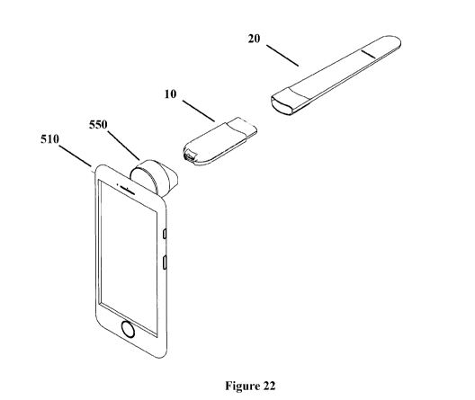
[00139] Figure 22 shows an exploded perspective view of the tongue
depressor, coupler and
user device of Figure 20;
[00140] Figure 23 shows an exploded side view of the tongue depressor,
coupler and user
device of Figure 20;
[00141] Figure 24 shows a perspective view of a tongue depressor coupled to
a user device
according to an embodiment of the present invention;
[00142] Figure 25 shows a side view of the tongue depressor and user device
of claim 24;
[00143] Figure 26 shows an exploded perspective view of the tongue
depressor and user
device of claim 24;
[00144] Figure 27 shows an exploded side view of the tongue depressor and
user device of
claim 24;
[00145] Figure 28 shows an exploded perspective view of an otoscope coupled
to a user
device according to an embodiment of the present invention;
[00146] Figure 29 shows a top view of an otoscope according to an
embodiment of the
present invention;
[00147] Figure 30 shows a side view of the otoscope of Figure 29;
[00148] Figure 31 shows a bottom view of the otoscope of Figure 29;
[00149] Figure 32 shows an exploded side view of the otoscope of Figure 29;
CA 03075648 2020-03-12
WO 2018/049479 PCT/AU2017/051004
[00150] Figure 33 shows an exploded perspective view of the otoscope of
Figure 29;
[00151] Figure 34 shows an exploded perspective view of a tongue depressor
according to an
embodiment of the present invention;
[00152] Figure 35 shows a front view of the tongue depressor of Figure 34;
[00153] Figure 36 shows a cross-sectional view through section P-P of
Figure 39;
[00154] Figure 37 shows a cross sectional view through section 0-0 of
Figure 35;
[00155] Figure 38 shows a cross sectional view through section M-M of
Figure 35;
[00156] Figure 39 shows a cross sectional view through section L-L of
Figure 35;
[00157] Figure 40 shows an detailed view of section N of Figure 39;
[00158] Figure 41 shows an exploded perspective view of a tongue depressor
according to an
embodiment of the present invention;
[00159] Figure 42 shows a front view of the tongue depressor of Figure 34;
[00160] Figure 43 shows a cross-sectional view through section E-E of
Figure 42;
[00161] Figure 44 shows a cross sectional view through section C-C of
Figure 42;
[00162] Figure 45 shows a cross sectional view through section F-F of
Figure 44;
[00163] Figure 46 shows a cross sectional view through section D-D of
Figure 44;
[00164] Figure 47 shows a left side view of a coupler fastened to a user
device according to
an embodiment of the present invention;
[00165] Figure 48 shows a front view of the coupler fastened to a user
device of Figure 47;
[00166] Figure 49 shows a right side view of the coupler fastened to a user
device of Figure
47;
[00167] Figure 50 shows a perspective view of the coupler fastened to a
user device of Figure
47;
[00168] Figure 51 shows a left side view of a handle mounted to a user
device according to
an embodiment of the present invention;
26
CA 03075648 2020-03-12
WO 2018/049479 PCT/AU2017/051004
[00169] Figure 52 shows a front view of the handle mounted to a user device
of Figure 51;
[00170] Figure 53 shows a right side view of the handle mounted to a user
device of Figure
51;
[00171] Figure 54 shows a perspective view of the handle mounted to a user
device of Figure
51;
[00172] Figure 55 illustrates an orifice inspection system, according to an
embodiment of the
present invention;
[00173] Figure 56 shows a screenshot of an application of the system of
Figure 55,
illustrating a photograph captured by the image capture device;
[00174] Figure 57 shows a screenshot of the system of Figure 56,
illustrating the photograph
taken in Figure 56 as modified by a user;
[00175] Figure 58 shows a screenshot of the system of Figure 56,
illustrating a screenshot
showing the photograph being assigned to a user profile;
[00176] Figure 59 shows a screenshot of the system of Figure 56,
illustrating a menu of
actions that can be taken after the image capture is complete;
[00177] Figure 60 shows a screenshot of the system of Figure 56,
illustrating a graphical user
interface displaying the captured photograph and a photograph of an orifice
having a known
condition for comparison; and
[00178] Figure 61 illustrates a patient orifice inspection method according
to an embodiment
of the present invention.
[00179] Preferred features, embodiments and variations of the invention may
be discerned
from the following Description which provides sufficient information for those
skilled in the art
to perform the invention. The following Description is not to be regarded as
limiting the scope of
the preceding Summary of the Invention in any way.
DESCRIPTION OF EMBODIMENTS
[00180] Embodiments of the present invention will now be described with
reference to
Figures 1 to 61. In the figures, like reference numerals refer to like
features.
27
CA 03075648 2020-03-12
WO 2018/049479 PCT/AU2017/051004
[00181] Figure 1 illustrates an example orifice inspection system 100,
according to an
embodiment of the present invention. The orifice inspection system 100 may be
useable
domestically (for example, by a patient or a family member of the patient), or
by a medical
professional (for example in a doctor's surgery or hospital).
[00182] As an example, a user 101 may have an application on their computer
or smartphone
110. The application uses an image capture device 120 (such as a camera or
webcam) to capture
a photograph 130 of a patient's orifice. The application includes a graphical
user interface which
includes a positioning guide 140 for positioning a patient's orifice in the
photograph 130
captured by the image capture device 120. A database 150 of at least one
photograph of an
orifice having a known condition may be accessible via the graphical user
interface. The
graphical user interface may be used to compare the photograph 130 with images
from the
database at 160. For example, the graphical user interface may be configured
to display the
captured photograph 130 and at least one photograph of an orifice having a
known condition for
comparison, to assist in identifying a condition affecting the patient's
orifice.
[00183] The user 101 may use a data interface on the computer or smartphone
110 in order to
transmit the photograph to the patient's medical service provider. For
example, the data
interface may transmit the photograph 130 by sending an email to the medical
service provider.
The data interface may also transmit the photograph 130 by uploading the
photograph onto a
server 170, from which one or more of the user 101 or the medical service
provider (such as a
doctor 102, or other health care provider 103) may view the photograph 130.
[00184] An exemplary orifice inspection system for the oral cavity will now
be described
with reference to Figures 2 to 9. The orifice inspection system exemplified in
Figures 2 to 9
includes an application for a smartphone.
[00185] Figure 2 illustrates a main menu screen 200 for the application.
From this screen the
user can choose to take a photograph of an oral cavity 202, or browse a
database showing at least
one oral cavity having a known condition 204.
[00186] If the user choses to take a photograph 202, a graphical user
interface is provided
showing an image 240 to be captured by an image capture device (see Figure 3).
Overlaid on
image 240 is an orifice positioning guide 250. The orifice positioning guide
250 is a broken line
illustrating the correct position of the palatine tonsils 260 and the uvula
270. When the image
240 is in the correct position, the user can capture the photograph 290, as
shown in Figure 4.
[00187] The user then has the option of capturing another photograph 300,
transmitting the
28
CA 03075648 2020-03-12
WO 2018/049479 PCT/AU2017/051004
captured photograph to a medical service provider 310, or comparing the
captured photograph
with photographs of oral cavities having known conditions 320.
[00188] If the user chooses to compare the photograph, the graphical user
interface displays
the captured photograph 290, and at least one photograph of an oral cavity
having a known
condition 330 (see Figure 5). In Figure 5, the user is able to change the
photograph of the oral
cavity having a known condition 330 to compare photographs of acute
tonsillitis, mononucleosis,
pharyngitis, and streptococcal pharyngitis. The user may click on the image of
the oral cavity
having a known condition 330 to obtain further information on the condition.
[00189] Figure 6 illustrates an example of further information which may be
obtained by the
user by clicking on the image of the oral cavity having a known condition. The
further
information includes: a summary of the condition, symptoms of the condition,
the
contagiousness of the condition, and recommendations of when to contact a
medical service
provider.
[00190] If the user chooses to send the captured photograph 290 to a
medical service
provider, they can do so either prior to or after comparing the captured
photograph 290 with
photographs of oral cavities having known conditions (see Figure 4 at 310 and
Figure 5 at 340).
If this option is chosen, the user may be directed to a form into which
information may be
entered concerning the patient's name, the patient's address, the patient's
phone number, the
patient's healthcare (or Medicare) number, an email address, the medical
service provider's
name, the medical service provider's email address, the medical service
provider's address,
symptoms experienced by the patient, and comments from the patient. An
exemplary form is
illustrated in Figure 7.
[00191] Alternatively, the user can browse oral cavities having known
conditions via 204 at
Figure 2. Choosing this option allows the user to access a database of oral
cavities having
known conditions, and this database is browseable by condition (Figure 8) or
by symptom
(Figure 9). Again, selecting a particular condition allows the user to access
further information
on that condition, as illustrated in Figure 6.
[00192] The orifice inspection system may include the application of
Figures 2-9 and an
orifice inspection device 1 (see Figures 11 to 19 and 28-54). The orifice
inspection device 1 may
be for illuminating the patient's orifice which in turn allows for a clearer
photo to be captured by
the image capture device.
[00193] The orifice inspection device 1 of the embodiment of Figures 11-19
is a tongue
29
CA 03075648 2020-03-12
WO 2018/049479 PCT/AU2017/051004
depressor. Features of the exemplary tongue depressor of Figures 11-19 are as
outlined below.
[00194] The orifice inspection device 1 includes a handle 10 (see Figures
11-19) and a blade
20 (see Figures 14-19). The tongue depressor 1 is for illuminating the oral
cavity, especially the
pharynx, at least one tonsil and the uvula.
[00195] The blade 20 extends from the handle 10 and is for directing light
from the light
source 12 (see Figure 11 and as discussed below) to illuminate the oral
cavity.
[00196] The handle 10 of the tongue depressor is illustrated in Figures 11-
13. As shown in
Figure 11, the handle 10 includes three light sources 12. Light from the light
source 12 passes
through the blade 20 to illuminate the oral cavity.
[00197] The blade 20 in Figures 14-19 is releasably engageable from the
handle 10. The
blade 20 acts as an optical waveguide for light emitted from the light source
12. The blade 20 is
a solid body extending from adjacent the light source 12 to its terminal end
32. The blade 20 of
Figures 14-19 is adapted to emit light from the terminal end 32. The blade 20
may be made from
injection moulded plastic. The blade 20 may be disposable.
[00198] The blade 20 includes a distal end portion 30 having a terminal end
32. The distal
end portion 30 is substantially flat in a longitudinal and lateral direction.
[00199] At least a portion of the distal end portion 30 is intended to be
inserted into the
patient's oral cavity. The blade 20 also includes a proximate end portion 50
(see Figures 15, 17
and 19). The proximate end portion 50 of the blade 20 is slideably engageable
with the insert-
coupling portion 13 of the handle 10 (see Figures 11 and 13). The proximate
end portion 50
includes a cavity 52 into which the insert-coupling portion 13 of the handle
is located (see Figure
17). The internal walls of the cavity 52 are smooth.
[00200] The handle 10 includes a switch to connect a power supply (not
shown) to the light
source 12. Coupling the blade 20 to the handle 10 actuates the switch so that
light emitted from
the light source 12 is transmitted by the blade 20 to the oral cavity when the
handle 10 and blade
20 are coupled together.
[00201] The insert-coupling portion 13 includes an outer wall having a flap
14, and the flap
14 is positioned over the switch, so that depression of the flap 14 by sliding
the insert-coupling
portion 13 into cavity 52 actuates the switch and activates the light source
12.
[00202] The light source 12 is located entirely within the handle 10. The
light source is
CA 03075648 2020-03-12
WO 2018/049479 PCT/AU2017/051004
positioned adjacent to an opening 15 in the insert-coupling portion 13 so that
light from the light
source 12 passes through the opening 15 and into the blade 20. The handle 10
includes three
light sources 12 which are positioned to extend laterally across the handle
10. The light sources
12 are especially light-emitting diodes (LEDs).
[00203] The power source in the handle 10 is in the form of a battery. The
battery is
especially a lithium-ion battery. The battery is connected to the light source
12 and the switch
via a circuit board (not shown). The handle 10 includes a removable cover 16
for allowing the
battery to be removed and replaced, as required. The removable cover 16
includes an aperture
18 providing an attachment for a keyring. The handle 10 may be made from
injection moulded
plastic.
[00204] The handle 10 especially does not require sterilisation and is
intended for multiple
uses. The blade 20 is either sterilisable (e.g. via heat or chemical
sterilisation), or is disposable.
The blade 20 and the handle 10 may be sold separately.
[00205] The orifice inspection device lb of the embodiment of Figures 29-33
is an otoscope.
Features of the exemplary otoscope lb of Figures 29-33 are as outlined below.
[00206] The handle 10 for otoscope lb is as described above. Speculum 20b
is slideably
engageable with the attachment-coupling portion 13 of the handle 10.
[00207] Speculum 20b acts as an optical waveguide for light emitted from
the light source 12,
and is a solid body extending from adjacent the light source 12 to its
terminal end 32b.
Speculum 20b is made from clear injection moulded plastic and is disposable.
[00208] Speculum 20b includes a distal end portion 30b having a terminal
end 32b and a non-
terminal end 34b (see Figure 30). At least a portion of the distal end portion
30b is intended to
be inserted into the patient's outer ear when the otoscope lb is used. The
distal end portion 30b
includes a side wall having a substantially conical portion 38b extending from
the non-terminal
end 34b, and a substantially cylindrical portion 39b extending from the
terminal end 32b. The
side wall 38b, 39b defines a visualisation passageway through the distal end
portion 30b. The
distal end portion 30b defines an aperture at the terminal end 32b and
includes a lens engager
24b at the non-terminal end 34b. The lens 22b is releaseably engageable with
the lens engager
24b. Once fitted, the lens 22b is positioned proximate to the non-terminal end
34b.
[00209] Speculum 20b also includes a medial portion 40b and a proximate end
portion 50b.
The medial portion 40b is substantially flat in a lateral and longitudinal
direction. The
31
CA 03075648 2020-03-12
WO 2018/049479 PCT/AU2017/051004
longitudinal axis of the distal end portion 30b is at about 75 relative to
the longitudinal axis of
the handle 10.
[00210] The proximate end portion 50b of the speculum is slideably
engageable with the
attachment-coupling portion 13 of the handle 10. The proximate end portion 50b
includes a
cavity 52b into which the attachment-coupling portion 13 of the handle is
located. The internal
walls of the cavity 52b are smooth.
[00211] Figures 20-23 and 28 illustrate orifice inspection systems 500,
500b. The orifice
inspection system 500 includes the tongue depressor 1 of Figures 14-19,
including handle 10 and
blade 20. The orifice inspection system 500b includes an otoscope lb,
including handle 10 and
speculum 20a (or orifice inserter). A similar otoscope which could be used in
the orifice
inspection system 500b is shown in Figures 29-33. The proximate end portion
50a of the
speculum 20a includes a cavity into which the attachment-coupling portion 13
of the handle is
located.
[00212] The system 500, 500b also includes a user device 510 in the form of
a smartphone,
and the user device 510 includes an image capture device 515, in the form of a
camera. The user
device includes the application as described in Figures 1-10 or Figures 55-61.
The system 500,
500b also includes a coupler 550, for releaseably coupling the handle 10 to
the user device 510.
[00213] The coupler 550 is releaseably coupleable to the user device 510 by
virtue of hook
and loop fasteners (for example, VelcroTm) One strip of a hook and loop
fastener is affixed to a
surface of the user device 510, and another strip of a hook and loop fastener
is affixed to a rear
surface 552 of the coupler 550. The coupler also includes a handle engager
560, which includes
a cavity. One end of the handle 10 is insertable into the handle engager 560,
to thereby
releaseably engage the handle 10 and the handle engager 560.
[00214] Figures 24-27 illustrate a further orifice inspection system 500a.
The system 500a
includes a user device 510 in the form of a smartphone, and the user device
510 includes an
image capture device 515, in the form of a camera. The user device includes
the application as
described in Figures 1-10 or 55 to 61.
[00215] The orifice inspection system 500a also includes a tongue depressor
la. The tongue
depressor includes a handle 10a and a blade 20. The blade 20 is as described
for the tongue
depressor of Figures 14-19. The handle 10a includes three light sources. Light
from the light
source passes through the blade 20 to illuminate the oral cavity.
32
CA 03075648 2020-03-12
WO 2018/049479 PCT/AU2017/051004
[00216] The proximate end portion 50 of the blade 20 is slideably
engageable with the insert-
coupling portion 13a of the handle 10a (see Figures 26-27). The proximate end
portion 50
includes a cavity 52 into which the insert-coupling portion 13a of the handle
is located (see
Figures 26-27). The internal walls of the cavity 52 are smooth.
[00217] The handle 10a includes a circuit board 80 including a switch to
connect a power
supply 85 (in the form of a battery) to the light source. Coupling the blade
20 to the handle 10a
actuates the switch so that light emitted from the light source is transmitted
by the blade 20 to the
oral cavity when the handle 10a and blade 20 are coupled together.
[00218] The insert-coupling portion 13a includes an outer wall having a
flap (not shown), and
the flap is positioned over the switch, so that depression of the flap by
sliding the insert-coupling
portion 13a into cavity 52 actuates the switch and activates the light source.
[00219] The light source is located entirely within the handle 10a. The
light source is
positioned adjacent to an opening in the insert-coupling portion 13a so that
light from the light
source passes through the opening and into the blade 20. The handle 10a
includes three light
sources which are positioned to extend laterally across the handle 10a. The
light sources are
especially light-emitting diodes (LEDs).
[00220] The power source in the handle 10 is in the form of a battery 85.
The battery 85 is
especially a lithium-ion battery. The battery 85 is connected to the light
source and the switch
via circuit board 80. The handle 10a may include a suction cap portion 90 for
affixing the handle
10a to the user device 510. Separation of the suction cap portion 90 from the
remainder of the
handle 10a allows a user to access the battery 85, for example.
[00221] The handle 10a especially does not require sterilisation and is
intended for multiple
uses. The blade 20 is either sterilisable (e.g. via heat or chemical
sterilisation), or is disposable.
The blade 20 and the handle 10a may be sold separately.
[00222] To use the oral inspection device 1, the insert-coupling portion
13, 13a is slideably
engaged into the cavity 52 of the blade 20. This actuates the switch and
activates the light source
12, which transmits light into the blade 20. The blade 20 acts as an optical
waveguide to direct
the light to the terminal end 32 of the blade 20 for illuminating the
patient's oral cavity. At least
a portion of the distal end portion 30 of the blade 20 is then inserted into
the patient's oral cavity
to depress the tongue and illuminate the oral cavity for inspection.
[00223] Further orifice inspection devices lc, id are illustrated in
Figures 34-46. While the
33
CA 03075648 2020-03-12
WO 2018/049479 PCT/AU2017/051004
orifice inspection devices lc, id are illustrated in the form of a tongue
depressor, the speculum
20a, 20b of Figures 28-33 could be used with the handle 10c, 10d of the device
lc, id, instead of
the tongue depressor blade 20c. Tongue depressor blade 20c is identical to the
tongue depressor
blade 20 discussed above in Figures 14-19.
[00224] The orifice inspection devices lc, id include a coupler 550c in the
form of a handle
engager. The handle engager 550c is planar and is slideably engageable with
the handle 10c,
10d. The handle engager includes an adhesive layer for fastening the handle
engager 10c, 10d
with a user device such as a smart phone 510c (see Figures 47-54). The user
device includes
image capture device 515c. The adhesive layer is VHB Tape PX5011, manufactured
by 3M.
[00225] Handle 10c includes 3 3mm LED lights 12c, and handle 10d includes
one surface
mount LED 12d. Light from lights 12c, 12d pass through the blade 20c to
illuminate the oral
cavity. The blade 20c is releaseably engageable from the handle 10c, 10d. The
blade 20c acts as
an optical waveguide for light emitted from the light source 12c, 12d. The
blade 20c is a solid
body extending from adjacent the light source 12c, 12d to its terminal end
32c. The blade 20c is
adapted to emit light from the terminal end 32c. The blade 20c may be made
from injection
moulded plastic and may be disposable.
[00226] The blade 20c includes a distal end portion 30c having a terminal
end 32c. The distal
end portion 30c is substantially flat in a longitudinal and lateral direction.
[00227] At least a portion of the distal end portion 30c is intended to be
inserted into the
patient's oral cavity. The blade 20c also includes a proximate end portion
50c. The proximate
end portion 50c of the blade 20c is slideably engageable with the insert-
coupling portion 13c,
13d of the handle 10c, 10d. The proximate end portion 50c includes a cavity
(not shown) into
which the insert-coupling portion 13c, 13d of the handle 10c, 10d is located.
The internal walls
of the cavity are smooth.
[00228] The handle 10c, 10d includes a switch 27c, 27d to connect a power
source 31c, 31d
to the light source 12c, 12d. Coupling the blade 20c to the handle 10c, 10d
actuates the switch
27c, 27d so that light emitted from the light source 12c, 12d is transmitted
by the blade 20c to the
oral cavity when the handle 10c, 10d and blade 20c are coupled together.
[00229] The insert-coupling portion 13c, 13d includes an outer wall having
a flap 14c, 14d,
and the flap 14c, 14d is positioned over the switch 27c, 27d, so that
depression of the flap 14c,
14d by sliding the insert-coupling portion 13c, 13d into cavity actuates the
switch 27c, 27d and
activates the light source 12c, 12d.
34
CA 03075648 2020-03-12
WO 2018/049479 PCT/AU2017/051004
[00230] The light source 12c, 12d is located entirely within the handle
10c, 10d. The light
source is positioned adjacent to an opening 15c, 15d in the insert-coupling
portion 13c, 13d so
that light from the light source 12c, 12d passes through the opening 15c, 15d
and into the blade
20c.
[00231] The power source 31c, 31d in the handle 10c, 10d is in the form of
a lithium ion
battery. The battery is connected to the light source 12c, 12d and the switch
27c, 27d via a circuit
board. The handle 10c, 10d may be made from injection moulded plastic.
[00232] In use, the coupler 550c is fastened to the smart phone 510c. The
handle 10c, 10d is
then slideably engaged with the coupler. Advantageously, this engagement
provides a rigid
connection between the handle 10c, 10d and the smart phone 510c.
[00233] Figure 55 illustrates a further example orifice inspection system
900, according to an
embodiment of the present invention. The orifice inspection system 900 may be
useable
domestically (for example, by a patient or a family member of the patient), or
by a medical
professional (for example in a doctor's surgery or hospital) The system 900 is
similar to the
system 100 shown in Figure 1.
[00234] As an example, a user 901 may have an application on their computer
or smartphone
910. The application uses an image capture device 920 (such as a camera or
webcam) to capture
a photograph 930 of a patient's orifice. The image capture device may be
associated with an
orifice inspective device (as previously described), and the system 900 may
detect the presence
of the orifice inspection device and automatically adjust the image
magnification of the image
capture device 920 based on the location of the orifice inspection device. A
database 950 of at
least one photograph of an orifice having a known condition may be accessible
via the graphical
user interface. The graphical user interface may be used to compare the
photograph 930 with
images from the database at 960. For example, the graphical user interface may
be configured to
display the captured photograph 930 and at least one photograph of an orifice
having a known
condition for comparison, to assist in identifying a condition affecting the
patient's orifice.
[00235] The user 901 may use a data interface on the computer or smartphone
910 in order to
transmit the photograph to the patient's medical service provider. For
example, the data
interface may transmit the photograph 930 by sending an email to the medical
service provider.
The data interface may also transmit the photograph 930 by uploading the
photograph onto a
server 970, from which one or more of the user 901 or the medical service
provider (such as a
doctor 902, or other health care provider 903) may view the photograph 930.
The photograph
CA 03075648 2020-03-12
WO 2018/049479 PCT/AU2017/051004
930 may also be uploaded onto a database 975 for use in research/training 977.
[00236] An exemplary orifice inspection system for the oral cavity will now
be described
with reference to Figures 2 and 56 to 60. The orifice inspection system
exemplified in Figures
56 to 60 includes an application for a smartphone.
[00237] First, Figure 2 illustrates a main menu screen 200 for the
application. From this
screen the user can choose to take a photograph of an oral cavity 202, or
browse a database
showing at least one oral cavity having a known condition 204.
[00238] If the user choses to take a photograph 202, a graphical user
interface is provided
showing an image to be captured by an image capture device. The orifice
positioning device is
mounted to a smart phone (for example), and the system automatically adjusts
the image
magnification of the image capture device based on the location of the orifice
inspection device.
This is accomplished based on knowledge of the length that the orifice
inspection device extends
from the smart phone. When the image is in the correct position, the user can
capture the
photograph 1090, as shown in Figure 56.
[00239] The user then can modify the photograph 1090, as shown in Figure
57. In Figure 57
the user has highlighted a portion of the photograph 1095 for closer
examination by a medical
service provider.
[00240] The user then can assign the photograph 1090 to their user profile,
as shown in
Figure 58. In this process, the user's profile may be associated with a
medical service provider's
profile and the photograph can be assigned to the medical service provider.
The user can also
seek professional advice, as shown in Figure 59.
[00241] If the user chooses to compare the photograph, the graphical user
interface displays
the captured photograph 1090, and at least one photograph of an oral cavity
having a known
condition 1130 (see Figure 60). In Figure 60, the user is able to change the
photograph of the
oral cavity having a known condition 1130 to compare photographs of acute
tonsillitis,
mononucleosis, pharyngitis, and streptococcal pharyngitis. The user may click
on the image of
the oral cavity having a known condition 1130 to obtain further information on
the condition (as
shown in Figure 6).
[00242] Figure 10 illustrates an orifice inspection method 800. The method
may include the
step of coupling an orifice inserter (such as blade 20) to a handle 10, to
thereby form an orifice
inspection device 1. A light source 12 in the orifice inspection device may be
activated by
36
CA 03075648 2020-03-12
WO 2018/049479 PCT/AU2017/051004
coupling the orifice inserter to the handle 10. A coupler may be releaseably
engaged with the
handle 10, and the coupler may also be releaseably engaged with a smartphone.
[00243] Using a graphical user interface, the patient's orifice is
positioned using the orifice
positioning guide 810. At step 820, after the patient's orifice has been
positioned, a photograph
of the orifice is captured using the image capture device. The user may then
choose to transmit
the photograph to a medical service provider 830, or to compare the captured
photograph to
photographs of orifices having known conditions 840. The user may retrieve
information
concerning the known conditions at step 850. Following steps 840 or 850, the
user may also
choose to transmit the photograph to a medical service provider 830.
[00244] Figure 61 illustrates a further orifice inspection method 1300. The
method may
include the step of mounting an orifice inspection device relative to an image
capture device
1310. For example, a coupler may be fastened to a user device such as a smart
phone, and a
handle 10-10d may be releasably engaged with the coupler. The method may
include the step of
coupling an orifice inserter (such as blade 20-20c) to a handle 10-10d, to
thereby form an orifice
inspection device 1-1d. A light source 12-12d in the orifice inspection device
may be activated
by coupling the orifice inserter to the handle 10-10d.
[00245] The patient's orifice is then illuminated 1320, and using a
graphical user interface an
image is captured 1330. The user has the option of modifying the captured
image 1340, for
example by highlighting a portion. The photograph is then assigned to a user
profile 1350, and
then the photograph can be transmitted to a medical service provider 1360 or
the photograph can
be compared with photographs of orifices having known conditions 1370.
Information
concerning one or more of the orifice conditions may also be retrieved 1380.
[00246] Reference throughout this specification to 'one embodiment' or 'an
embodiment'
means that a particular feature, structure, or characteristic described in
connection with the
embodiment is included in at least one embodiment of the present invention.
Thus, the
appearance of the phrases 'in one embodiment' or 'in an embodiment' in various
places
throughout this specification are not necessarily all referring to the same
embodiment.
Furthermore, the particular features, structures, or characteristics may be
combined in any
suitable manner in one or more combinations.
[00247] In compliance with the statute, the invention has been described in
language more or
less specific to structural or methodical features. It is to be understood
that the invention is not
limited to specific features shown or described since the means herein
described includes
37
CA 03075648 2020-03-12
WO 2018/049479 PCT/AU2017/051004
preferred forms of putting the invention into effect. The invention is,
therefore, claimed in any
of its forms or modifications within the proper scope of the appended claims
appropriately
interpreted by those skilled in the art.
38