Note: Descriptions are shown in the official language in which they were submitted.
CA 03076611 2020-03-20
WO 2019/060750 PCT/US2018/052253
A33 ANTIBODY COMPOSITIONS AND METHODS OF USING THE SAME IN
RADIOIMMUNOTHERAPY
CROSS-REFERENCE TO RELATED APPLICATIONS
[0001] This application claims the benefit of and priority to U.S.
Provisional Patent
Application No. 62/562,373, filed September 23, 2017, and also to U.S.
Provisional Patent
Application No. 62/562,374, filed September 23, 2017, the contents of which
are incorporated
herein by reference in their entireties.
TECHNICAL FIELD
[0002] The present technology relates generally to the preparation of
immunoglobulin-
related compositions (e.g., antibodies or antigen binding fragments thereof)
that specifically bind
A33 protein and uses of the same. In particular, the present technology
relates to the preparation
of A33 neutralizing antibodies and their use in detecting and treating A33
associated cancers.
BACKGROUND
[0003] The following description of the background of the present
technology is provided
simply as an aid in understanding the present technology and is not admitted
to describe or
constitute prior art to the present technology.
[0004] Colorectal cancers (CRCs) constitute the 3rd leading cause of cancer
death in the
United States (Siegel RL, Miller KD, Jemal A, CA: A Cancer Journal for
Clinicians 67:7-30
(2017)) and account for 10% of all cancers in men and 9.2% in women worldwide
(Ferlay J,
Soerjomataram I, Dikshit R, et al., International Journal of Cancer 136:E359-
E386 (2015)).
Although there has been steady yearly 3% decline in the incidence from 2004 to
2013, 135,000
new cases are expected in 2017 in the United States alone. Although localized
and regional
diseases can be curable, the prognosis of metastatic CRCs (mCRCs) is poor,
with a 5-year
survival rate of only 14% (Siegel RL, Miller KD, Jemal A, CA: A Cancer Journal
for Clinicians
67:7-30 (2017)).
[0005] Standard treatment for mCRC consists of chemotherapy in combination
with
monoclonal antibodies that block tumor signaling or angiogenesis. Currently
four monoclonal
antibody drugs have been FDA approved for CRC treatment: bevacizumab and
ramucirumab
target the VEGF-VEGFR angiogenesis pathway, while cetuximab and panitumumab
target the
EGFR pathway. However, cetuximab and panitumumab do not provide clinical
benefits for
mCRC with RAS mutations (Van Cutsem E, Cervantes A, Adam R, et al., Annals of
Oncology
27:1386-1422 (2016)), which are found in around 40% of all mCRC (Bencsikova B,
Bortlicek Z,
Halamkova J, et al., BMC Gastroenterology 15:37 (2015)). Moreover, improvement
in survival
1
CA 03076611 2020-03-20
WO 2019/060750 PCT/US2018/052253
with these antibodies is generally modest (Peeters M et at., Journal of
Clinical Oncology
28:4706-4713 (2010); Cutsem EV et al., Journal of Clinical Oncology 29:2011-
2019 (2011);
Saltz LB et al., Journal of Clinical Oncology 26:2013-2019 (2008)) and can be
associated with
severe side effects. The benefits of immune checkpoint inhibitors (ICI) have
been restricted to
subsets of CRC patients with microsatellite instability (MSI). However, the
majority of patients
with mCRC are microsatellite stable (MSS) and are thus not expected to benefit
from ICI
monotherapy. Thus far, the use of antibodies to target toxins to tumors, e.g.,
radioimmunotherapy (RIT) with directly conjugated antibodies, has been met
with limited
success due in part to suboptimal tumor dose and therapeutic index (TI).
Further, because of
normal tissue bystander toxicity, dose escalation is not feasible and
therefore such therapy results
in limited anti-tumor effect.
SUMMARY OF THE PRESENT TECHNOLOGY
[0006] In one aspect, the present disclosure provides an antibody or
antigen binding
fragment thereof comprising a heavy chain immunoglobulin variable domain (VH)
and a light
chain immunoglobulin variable domain (VIA wherein (a) the VH comprises a VH-
CDR1
sequence of FTFSTYDMS (SEQ ID NO: 37), a VH-CDR2 sequence of
TISSGGSYTYYLDSVKG (SEQ ID NO: 38), and a VH-CDR3 sequence of TTVVPFAY (SEQ
ID NO: 39); and/or (b) the VL comprises a VL-CDR1 sequence, a VL-CDR2
sequence, and a \IL'
CDR3 sequence selected from the group consisting of: KASQNVRTVVA (SEQ ID NO:
40),
LASNRHT (SEQ ID NO: 41), and QYWSYPLT (SEQ ID NO: 42); KASQNVRTVVA (SEQ ID
NO: 40), LASDRHT (SEQ ID NO: 43), and QYWSYPLT (SEQ ID NO: 42); KASQNVRTLVA
(SEQ ID NO: 44), LASNRHT (SEQ ID NO: 41), and QHWSYPLT (SEQ ID NO: 45); and
KASQNVRTLVA (SEQ ID NO: 44), LASNRHT (SEQ ID NO: 41), and QYWSYPLT (SEQ ID
NO: 42).
[0007] The antibody may further comprise an Fc domain of an isotype
selected from the
group consisting of IgGl, IgG2, IgG3, IgG4, IgAl, IgA2, IgM, IgD, and IgE. In
some
embodiments, the antibody comprises an IgG1 constant region comprising one or
more amino
acid substitutions selected from the group consisting of N297A and K322A.
Additionally or
alternatively, in some embodiments, the antibody comprises an IgG4 constant
region comprising
a 5228P mutation. In certain embodiments, the antigen binding fragment is
selected from the
group consisting of Fab, F(ab')2, Fab', scF,, and F. In some embodiments, the
antibody is a
monoclonal antibody, chimeric antibody, humanized antibody, or a bispecific
antibody. In
certain embodiments, the antibody or antigen binding fragment binds to an
epitope of A33
2
CA 03076611 2020-03-20
WO 2019/060750 PCT/US2018/052253
protein comprising at least five to eight consecutive amino acid residues of
SEQ ID NO: 57. In
some embodiments, the epitope is a conformational epitope.
[0008] In another aspect, the present disclosure provides an antibody
comprising a heavy
chain (HC) amino acid sequence comprising SEQ ID NO: 5, SEQ ID NO: 6, SEQ ID
NO: 15,
SEQ ID NO: 19, SEQ ID NO: 23, SEQ ID NO: 25, SEQ ID NO: 27, SEQ ID NO: 29, SEQ
ID
NO: 31, SEQ ID NO: 33, SEQ ID NO: 35, SEQ ID NO: 58, SEQ ID NO: 62, or a
variant thereof
having one or more conservative amino acid substitutions, and/or a light chain
(LC) amino acid
sequence comprising SEQ ID NO: 9, SEQ ID NO: 10, SEQ ID NO: 17, SEQ ID NO: 21,
SEQ
ID NO: 24, SEQ ID NO: 26, SEQ ID NO: 28, SEQ ID NO: 30, SEQ ID NO: 32, SEQ ID
NO:
34, SEQ ID NO: 36, SEQ ID NO: 60, SEQ ID NO: 63, or a variant thereof having
one or more
conservative amino acid substitutions. In certain embodiments, the antibody
comprises a HC
amino acid sequence and a LC amino acid sequence selected from the group
consisting of: SEQ
ID NO: 5 and SEQ ID NO: 9 (3A3-H1/L1); SEQ ID NO: 5 and SEQ ID NO: 10 (3A3-
H1/L2);
SEQ ID NO: 6 and SEQ ID NO: 9 (3A3-H2/L1); SEQ ID NO: 6 and SEQ ID NO: 10 (3A3-
H2/L2); SEQ ID NO: 15 and SEQ ID NO: 17 (huA33-IgG1 (H2L2)); SEQ ID NO: 19 and
SEQ
ID NO: 21 (huA33-BsAb); SEQ ID NO: 23 and SEQ ID NO: 24 (clone 31); SEQ ID NO:
25 and
SEQ ID NO: 26 (clone 32); SEQ ID NO: 27 and SEQ ID NO: 28 (clone 48); SEQ ID
NO: 29
and SEQ ID NO: 30 (clone 49); SEQ ID NO: 31 and SEQ ID NO: 32 (clone 53); SEQ
ID NO:
33 and SEQ ID NO: 34 (clone 56); SEQ ID NO: 35 and SEQ ID NO: 36 (clone 57);
SEQ ID
NO: 58 and SEQ ID NO: 60 (huA33-huC825); and SEQ ID NO: 62 and SEQ ID NO: 63
(huA33-mC825), respectively.
[0009] In one aspect, the present disclosure provides an antibody
comprising (a) a light chain
immunoglobulin variable domain sequence that is at least 80%, at least 85%, at
least 90%, at
least 95%, or at least 99% identical to the light chain immunoglobulin
variable domain sequence
present in any one of SEQ ID NOs: 9. 10, 17, 21, 24, 26, 28, 30, 32, 34, 36,
60, or 63; and/or (b)
a heavy chain immunoglobulin variable domain sequence that is at least 80%, at
least 85%, at
least 90%, at least 95%, or at least 99% identical to the heavy chain
immunoglobulin variable
domain sequence present in any one of SEQ ID NOs: 5, 6, 15, 19, 23, 25, 27,
29, 31, 33, 35, 58
or 62.
[0010] In another aspect, the present disclosure provides an antibody
comprising (a) a LC
sequence that is at least 80%, at least 85%, at least 90%, at least 95%, or at
least 99% identical to
the LC sequence present in any one of SEQ ID NOs: 9. 10, 17, 21, 24, 26, 28,
30, 32, 34, 36, 60,
or 63; and/or (b) a HC sequence that is at least 80%, at least 85%, at least
90%, at least 95%, or
3
CA 03076611 2020-03-20
WO 2019/060750 PCT/US2018/052253
at least 99% identical to the HC sequence present in any one of SEQ ID NOs: 5,
6, 15, 19, 23,
25, 27, 29, 31, 33, 35, 58 or 62.
[0011] In any of the above embodiments, the antibody is a chimeric
antibody, a humanized
antibody, or a bispecific antibody. Additionally or alternatively, in some
embodiments, the
antibody comprises an IgG1 constant region comprising one or more amino acid
substitutions
selected from the group consisting of N297A and K322A. In certain embodiments,
the antibody
of the present technology comprises an IgG4 constant region comprising a 5228P
mutation. In
any of the above embodiments, the antibody binds to an epitope of A33 protein
comprising at
least five to eight consecutive amino acid residues of SEQ ID NO: 57. In some
embodiments,
the epitope is a conformational epitope. Additionally or alternatively, in
some embodiments, the
antibody of the present technology lacks a-1,6-fucose modifications.
[0012] In one aspect, the present disclosure provides a recombinant nucleic
acid sequence
encoding any of the antibodies described herein. In some embodiments, the
recombinant nucleic
acid sequence selected from the group consisting of: SEQ ID NOs: 7, 8, 11, 12,
16, 18, 20, 22,
59 and 61.
[0013] In another aspect, the present disclosure provides a host cell or
vector comprising any
of the recombinant nucleic acid sequences disclosed herein.
[0014] In one aspect, the present disclosure provides a composition
comprising an antibody
or antigen binding fragment of the present technology and a pharmaceutically-
acceptable carrier,
wherein the antibody or antigen binding fragment is optionally conjugated to
an agent selected
from the group consisting of isotopes, dyes, chromagens, contrast agents,
drugs, toxins,
cytokines, enzymes, enzyme inhibitors, hormones, hormone antagonists, growth
factors,
radionuclides, metals, liposomes, nanoparticles, RNA, DNA or any combination
thereof
[0015] In some embodiments of the bispecific antibody of the present
technology, the
bispecific antibody binds to T cells, B-cells, myeloid cells, plasma cells, or
mast-cells.
Additionally or alternatively, in some embodiments, the bispecific antibody
binds to CD3, CD4,
CD8, CD20, CD19, CD21, CD23, CD46, CD80, HLA-DR, CD74, CD22, CD14, CD15, CD16,
CD123, TCR gamma/delta, NKp46, KIR, or a small molecule DOTA hapten. The small
molecule DOTA hapten may be selected from the group consisting of DOTA, DOTA-
Bn,
DOTA-desferrioxamine, DOTA-Phe-Lys(HSG)-D-Tyr-Lys(HSG)-NH2, Ac-Lys(HSG)D-Tyr-
Lys(HSG)-Lys(Tscg-Cys)-NH2, DOTA-D-Asp-D-Lys(HSG)-D-Asp-D-Lys(HSG)-NH2; DOTA-
D-Glu-D-Lys(HSG)-D-Glu-D-Lys(HSG)-NH2, DOTA-D-Tyr-D-Lys(HSG)-D-Glu-D-
Lys(HSG)-NH2, DOTA-D-Ala-D-Lys(HSG)-D-Glu-D-Lys(HSG)-NH2, DOTA-D-Phe-D-
Lys(HSG)-D-Tyr-D-Lys(HSG)-NH2, Ac-D-Phe-D-Lys(DOTA)-D-Tyr-D-Lys(DOTA)-NH2, Ac-
D-Phe-D-Lys(DTPA)-D-Tyr-D-Lys(DTPA)-NH2, Ac-D-Phe-D-Lys(Bz-DTPA)-D-Tyr-D-
4
CA 03076611 2020-03-20
WO 2019/060750 PCT/US2018/052253
Lys(Bz-DTPA)-NH2, Ac-D-Lys(HSG)-D-Tyr-D-Lys(HSG)-D-Lys(Tscg-Cys)-NH2, DOTA-D-
Phe-D-Lys(HSG)-D-Tyr-D-Lys(HSG)-D-Lys(Tscg-Cys)-NH2, (Tscg-Cys)-D-Phe-D-
Lys(HSG)-
D-Tyr-D-Lys(HSG)-D-Lys(DOTA)-NH2, Tscg-D-Cys-D-Glu-D-Lys(HSG)-D-Glu-D-
Lys(HSG)-NH2, (Tscg-Cys)-D-Glu-D-Lys(HSG)-D-Glu-D-Lys(HSG)-NH2, Ac-D-Cys-D-
Lys(DOTA)-D-Tyr-D-Ala-D-Lys(DOTA)-D-Cys-NH2, Ac-D-Cys-D-Lys(DTPA)-D-Tyr-D-
Lys(DTPA)-NH2, Ac-D-Lys(DTPA)-D-Tyr-D-Lys(DTPA)-D-Lys(Tscg-Cys)-NH2, and Ac-D-
Lys(DOTA)-D-Tyr-D-Lys(DOTA)-D-Lys(Tscg-Cys)-NH2.
[0016] In another aspect, the present disclosure provides a method for
treating an A33
associated cancer in a subject in need thereof, comprising administering to
the subject an
effective amount of any one of the antibodies disclosed herein. In certain
embodiments, the
antibody comprises a HC amino acid sequence and a LC amino acid sequence
selected from the
group consisting of: SEQ ID NO: 5 and SEQ ID NO: 9 (3A3-H1/L1); SEQ ID NO: 5
and SEQ
ID NO: 10 (3A3-H1/L2); SEQ ID NO: 6 and SEQ ID NO: 9 (3A3-H2/L1); SEQ ID NO: 6
and
SEQ ID NO: 10 (3A3-H2/L2); SEQ ID NO: 15 and SEQ ID NO: 17 (huA33-IgG1
(H2L2));
SEQ ID NO: 19 and SEQ ID NO: 21 (huA33-BsAb); SEQ ID NO: 23 and SEQ ID NO: 24
(clone 31); SEQ ID NO: 25 and SEQ ID NO: 26 (clone 32); SEQ ID NO: 27 and SEQ
ID NO:
28 (clone 48); SEQ ID NO: 29 and SEQ ID NO: 30 (clone 49); SEQ ID NO: 31 and
SEQ ID
NO: 32 (clone 53); SEQ ID NO: 33 and SEQ ID NO: 34 (clone 56); SEQ ID NO: 35
and SEQ
ID NO: 36 (clone 57), SEQ ID NO: 58 and SEQ ID NO: 60 (huA33-huC825); and SEQ
ID NO:
62 and SEQ ID NO: 63 (huA33-mC825), respectively, wherein the antibody
specifically binds to
and neutralizes A33 activity.
[0017] In some embodiments, the A33 associated cancer is colorectal cancer,
Pseudomyxoma peritonei, appendiceal cancer, pancreatic cancer, or gastric
cancer. The A33
associated cancer may be colorectal cancer with a MSI genotype or a MSS
genotype.
Additionally or alternatively, in some embodiments, the colorectal cancer is
associated with a
KRAS G12D mutation or a p53 mutation.
[0018] Additionally or alternatively, in some embodiments of the method,
the antibody is
administered to the subject separately, sequentially or simultaneously with an
additional
therapeutic agent. Examples of additional therapeutic agents include one or
more of alkylating
agents, platinum agents, taxanes, vinca agents, anti-estrogen drugs, aromatase
inhibitors, ovarian
suppression agents, VEGF/VEGFR inhibitors, EGF/EGFR inhibitors, PARP
inhibitors,
cytostatic alkaloids, cytotoxic antibiotics, antimetabolites,
endocrine/hormonal agents,
bisphosphonate therapy agents.
[0019] In another aspect, the present disclosure provides a method for
detecting a tumor in a
subject in vivo comprising (a) administering to the subject an effective
amount of an antibody of
CA 03076611 2020-03-20
WO 2019/060750 PCT/US2018/052253
the present technology, wherein the antibody is configured to localize to a
tumor expressing A33
and is labeled with a radioisotope; and (b) detecting the presence of a tumor
in the subject by
detecting radioactive levels emitted by the antibody that are higher than a
reference value. In
some embodiments, the subject is diagnosed with or is suspected of having
cancer. Radioactive
levels emitted by the antibody may be detected using positron emission
tomography or single
photon emission computed tomography.
[0020] Additionally or alternatively, in some embodiments, the method
further comprises
administering to the subject an effective amount of an immunoconjugate
comprising an antibody
of the present technology conjugated to a radionuclide. In some embodiments,
the radionuclide
is an alpha particle-emitting isotope, a beta particle-emitting isotope, an
Auger-emitter, or any
combination thereof. Examples of beta particle-emitting isotopes include 86Y,
90Y, 89Sr, 165Dy,
186Re, 188Re, 177Lu, and 67Cu. In some embodiments of the method, nonspecific
FcR-dependent
binding in normal tissues is eliminated or reduced (e.g., via N297A mutation
in Fc region,
which results in aglycosylation).
[0021] Also disclosed herein are kits for the detection and/or treatment of
A33 associated
cancers, comprising at least one immunoglobulin-related composition of the
present technology
(e.g., any antibody or antigen binding fragment described herein), or a
functional variant (e.g.,
substitutional variant) thereof and instructions for use. In certain
embodiments, the
immunoglobulin-related composition is coupled to one or more detectable
labels. In one
embodiment, the one or more detectable labels comprise a radioactive label, a
fluorescent label,
or a chromogenic label.
[0022] Additionally or alternatively, in some embodiments, the kit further
comprises a
secondary antibody that specifically binds to an anti-A33 immunoglobulin-
related composition
described herein. In some embodiments, the secondary antibody is coupled to at
least one
detectable label selected from the group consisting of a radioactive label, a
fluorescent label, or a
chromogenic label.
[0023] In one aspect, the present disclosure provides a method for
detecting solid tumors in a
subject in need thereof comprising (a) administering to the subject an
effective amount of a
complex comprising a radiolabeled DOTA hapten and a bispecific antibody of the
present
technology that binds to the radiolabeled DOTA hapten and an A33 antigen,
wherein the
complex is configured to localize to a solid tumor expressing the A33 antigen
recognized by the
bispecific antibody of the complex; and (b) detecting the presence of solid
tumors in the subject
by detecting radioactive levels emitted by the complex that are higher than a
reference value.
6
CA 03076611 2020-03-20
WO 2019/060750 PCT/US2018/052253
[0024] In another aspect, the present disclosure provides a method for
selecting a subject for
pretargeted radioimmunotherapy comprising (a) administering to the subject an
effective amount
of a complex comprising a radiolabeled DOTA hapten and a bispecific antibody
of the present
technology that binds to the radiolabeled DOTA hapten and an A33 antigen,
wherein the
complex is configured to localize to a solid tumor expressing the A33 antigen
recognized by the
bispecific antibody of the complex; (b) detecting radioactive levels emitted
by the complex; and
(c) selecting the subject for pretargeted radioimmunotherapy when the
radioactive levels emitted
by the complex are higher than a reference value.
[0025] In one aspect, the present disclosure provides a method for
increasing tumor
sensitivity to radiation therapy in a subject diagnosed with an A33-positive
cancer comprising
administering to the subject an effective amount of a complex comprising a
radiolabeled-DOTA
hapten and a bispecific antibody of the present technology that recognizes and
binds to the
radiolabeled-DOTA hapten and an A33 antigen target, wherein the complex is
configured to
localize to a tumor expressing the A33 antigen target recognized by the
bispecific antibody of the
complex.
[0026] In another aspect, the present disclosure provides a method for
treating cancer in a
subject in need thereof comprising administering to the subject an effective
amount of a complex
comprising a radiolabeled-DOTA hapten and a bispecific antibody of the present
technology that
recognizes and binds to the radiolabeled-DOTA hapten and an A33 antigen
target, wherein the
complex is configured to localize to a tumor expressing the A33 antigen target
recognized by the
bispecific antibody of the complex.
[0027] In any of the above embodiments of the methods disclosed herein, the
complex is
administered intravenously, intramuscularly, intraarterially, intrathecally,
intracapsularly,
intraorbitally, intradermally, intraperitoneally, transtracheally,
subcutaneously,
intracerebroventricularly, orally or intranasally. In some embodiments of the
methods disclosed
herein, the subject is human. Additionally or alternatively, in any of the
above embodiments of
the methods disclosed herein, the radiolabeled-DOTA hapten comprises 213Bi,
211At, 225Ac,
152 212 223 219 215 211 221 217 255 86 90 89
165 186 188
Dy, Bi, Ra, Rn, Po, Bi, Fr, At, Fm, Y, Y, Sr, Dy, Re, Re,
177Lu, 67Cu, 67Ga, 51
U 58Co, 99mTc, M3mR11, 195mpt, 119sb, 161Ho, 189m05, 1921r, 201T1, 203pb,
68Ga, 227Th, or 64Cu, and optionally comprises an alpha particle-emitting
isotope, a beta particle-
emitting isotope, or an Auger-emitter.
[0028] In one aspect, the present disclosure provides a method for
increasing tumor
sensitivity to radiation therapy in a subject diagnosed with an A33-positive
cancer comprising (a)
administering an effective amount of an anti-DOTA bispecific antibody of the
present
7
CA 03076611 2020-03-20
WO 2019/060750 PCT/US2018/052253
technology to the subject, wherein the anti-DOTA bispecific antibody is
configured to localize to
a tumor expressing an A33 antigen target; and (b) administering an effective
amount of a
radiolabeled-DOTA hapten to the subject, wherein the radiolabeled-DOTA hapten
is configured
to bind to the anti-DOTA bispecific antibody. In another aspect, the present
disclosure provides
a method for treating cancer in a subject in need thereof comprising (a)
administering an
effective amount of an anti-DOTA bispecific antibody of the present technology
to the subject,
wherein the anti-DOTA bispecific antibody is configured to localize to a tumor
expressing an
A33 antigen target; and (b) administering an effective amount of a
radiolabeled-DOTA hapten to
the subject, wherein the radiolabeled-DOTA hapten is configured to bind to the
anti-DOTA
bispecific antibody. In some embodiments, the methods of the present
technology further
comprise administering an effective amount of a clearing agent to the subject
prior to
administration of the radiolabeled-DOTA hapten.
[0029] Additionally or alternatively, in any of the above embodiments of
the methods
disclosed herein, the radiolabeled-DOTA hapten comprises 213Bi, 211At, 225Ac,
152Dy, 212Bi,
223Ra, 219Rn, 215po, 211Bi, 221Fr, 217At, 255Fin, 86y, 90y, 89Sr, 165Dy,
186Re, 188Re, 177Ln, 67cn,
67Ga, 51Cr, 58CO, 99MTC, 103MR11
, 195n13t, 119sh, 161Ho, 189M05, 1921r, 201T1, 203ph, 68Ga, 227Th, or
64
Cu, and optionally comprises an alpha particle-emitting isotope, a beta
particle-emitting
isotope, or an Auger-emitter. In any of the above embodiments of the methods
disclosed herein,
the subject is human.
BRIEF DESCRIPTION OF THE DRAWINGS
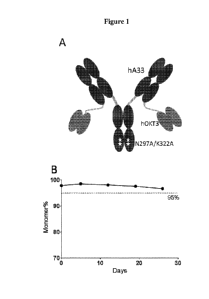
[0030] Figure 1(A) shows the design and construction of humanized A33-
bispecific antibody
(huA33-BsAb). Figure 1(B) shows the accelerated stability test of purified
huA33-BsAb at 37
C over 4 weeks. Figure 1(C) shows the Surface Plasmon Resonance (SPR) analysis
of huA33-
BsAb at 25 C and 37 C. Data were fit to 1:1 binding model. Figure 1(D) shows
the FACS
staining of different tumor cell lines and activated T cells. Mean
fluorescence intensity (MFI)
values were geometric means.
[0031] Figure 2(A) shows the activation of CD25 and CD69 markers in T cells
at 24 hours
post incubation with Colo205 cells and different antibodies. Figure 2(B) shows
the
quantification of dividing cells based on CFSE dye dilution at 96 hours post
incubation with
Colo205 cells and different antibodies. Figure 2(C) shows representative
images for Figure
2(B). Figure 2(D) shows the staining of CD45R0 at 96 hours post incubation
with Colo205 cells
and huA33-BsAb in an independent experiment. Figure 2(E) shows the in vivo
activation and
proliferation of T cells by huA33-BsAb. Briefly, CF SE-labeled peripheral
blood mononuclear
cells (PBMCs) were mixed with Colo205 cells and the mixture was implanted
subcutaneously
8
CA 03076611 2020-03-20
WO 2019/060750 PCT/US2018/052253
onto DKO mice. HuA33-BsAb was injected intravenously the next day and the
tumors were
isolated after another 4 days and analyzed by FACS.
[0032] Figure 3 shows the profile of secreted cytokines and cytotoxic
components by
huA33-BsAb activated T cells in the presence of a target tumor. The kinetics
of cytokine and
cytolytic molecule production by T cells in the presence of huA33-BsAb and
target cell Co10205
or negative control cell SKMEL5 were determined over 4 days. Because SKMEL5
secreted
copious amounts of IL-6, the supernatant from T cells incubated with Co10205
cells in the
absence of antibody was used as a negative control in the IL-6 kinetics
experiment.
[0033] Figure 4(A) shows the cytotoxicity elicited by huA33-BsAb against
different target
tumor cell lines and control cells. Activated T cells were used as effector
cells at an effector to
target ratio (E:T) of 10:1. Cells were incubated for 16 hours before obtaining
readings. Figure
4(B) shows the cytotoxicity induced by huA33-BsAb against Co10205 cells.
Sorted fresh T cell
subsets were used as effector cells (E:T=5:1). Cells were incubated for 48
hours before
obtaining readings. EC50 for CD4 memory T cells was 25 pM.
[0034] Figure 5(A) shows a summary of the affinity maturation of huA33
(H2L2) by yeast
screening. Figure 5(B) shows a summary of KD of parental and affinity matured
huA33-BsAb
(top) and the on-off rate map of different huA33-BsAbs derived from SPR
analysis (bottom).
Figure 5(C) shows the results of the T-cell dependent cytotoxicity (TDCC)
assay of the parental
and affinity matured huA33-BsAbs.
[0035] Figure 6(A) shows the growth of s.c. L5174T tumors in treatment
group and control
groups. Tumor sizes were assessed by volume (p=0.0133 for tumor+scPBMC versus
tumor+scPBMC+huA33-BsAb; p=0.006 for tumor only versus tumor+scPBMC+huA33-
BsAb).
Figure 6(B) shows the i.p. L5174T tumor growth in treatment group and control
groups (top)
(p=0.0125 for tumor+ATC versus tumor+ATC+huA33-BsAb; p=0.0026 for tumor only
versus
tumor+ATC+huA33-BsAb) and survival of mice in treatment group and control
groups
(bottom). Figure 6(C) shows luminescence images of abdominal LS174T tumors in
different
groups. Tumor only group had one mouse (#4) that did not take the tumor after
21 days and was
excluded from analysis.
[0036] Figure 7(A) shows luminescence images showing growth of s.c. Colo205
tumors in
different groups. Figure 7(B) shows the quantification of signals from Figure
7(A) (top) and the
survival of mice from different groups in Figure 7(A) (bottom). Figure 7(C)
shows
luminescence images of i.p. 5W1222 tumor; mouse #1 from tumor only group and
mouse #3
from tumor+ATC group which did not take tumor after 21 days were excluded from
imaging at
9
CA 03076611 2020-03-20
WO 2019/060750 PCT/US2018/052253
later time points and were not included in survival analysis in Figure 7(D).
Figure 7(D) shows
the survival of mice with i.p. SW1222 tumor.
[0037] Figure 8(A) shows the growth of s.c. SNU16 tumors in different
groups. Figure 8(B)
shows the engraftment of human cells from mice blood in Figure 8(A).
[0038] Figure 9 shows the SPR analysis of 4 versions of humanized A33. All
antibodies
comprised a IgG1 constant domain. 3A3-H1L1, 3A3-H1L2, 3A3-H2L1 and 3A3-H2L2
were 4
versions of humanized 3A3. 3A3-chA33 was chimeric 3A3.
[0039] Figure 10 shows the results of FACS analysis of various cell lines
stained with
huA33-BsAb.
[0040] Figure 11(A) shows the upregulation of PD-1 on T cells activated by
huA33-BsAb in
the presence of Colo205 cells after 24 hours (left) and 96 hours (right).
Figure 11(B) shows the
absence of T cell division after incubating with SKMEL5 in the presence of
huA33-BsAb after
96 hours. Figure 11(C) shows the activation of T cell division by huA33-BsAb
in the presence
of LS174T cells. Figures 11(B) and 11(C) used the same preparation of T cells.
[0041] Figures 12(A) and 12(B) show the staining of CD45R0 and CD25 markers
on T cells
48 hours post incubation with Colo205 cells in the presence of different
antibodies. Cells were
obtained from the TDCC assay after the supernatant was used for LDH
measurement. Figure
12(A): gated on CD4(+) T cells; Figure 12(B): gated on CD8(+) T cells.
[0042] Figure 13 shows the strategy for rapid reformation of scFv to huA33-
BsAb format.
Expression vector (1) was linearized with HindIII/ApaI. Promoter fragment (3)
was prepared
from SapI digested promoter-containing vector. Both vector and promoter
fragment could be
prepared in large amounts for higher throughput cloning. VH (4) and VL (2)
were amplified
directly from yeast with two 5' primers to add the leader sequences and
digested with
HindIII/SapI (VL) or ApaI/SapI (VH). The 4 fragments were ligated in a one-
step reaction.
[0043] Figure 14 shows the amino acid sequences of the VH and VL domains of
the murine
A33 antibody and their corresponding homologous human sequences (SEQ ID NOs: 1-
4). The
CDR1, CDR2, and CDR3 regions of the VH and VL domains of the murine A33
antibody are
indicated by the underlined boldface font.
[0044] Figure 15 shows the amino acid sequences of the humanized heavy
chains of huA33-
H1 (3A3-H1) (SEQ ID NO: 5) and huA33-H2 (3A3-H2) (SEQ ID NO: 6). The CDR1,
CDR2,
and CDR3 regions of 3A3-H1 and 3A3-H2 are indicated by the underlined boldface
font.
CA 03076611 2020-03-20
WO 2019/060750 PCT/US2018/052253
[0045] Figure 16 shows the cDNA sequences of the humanized heavy chains of
huA33-H1
(3A3-H1) (SEQ ID NO: 7) and huA33-H2 (3A3-H2) (SEQ ID NO: 8).
[0046] Figure 17 shows the amino acid sequences of the humanized light
chains of huA33-
Li (3A3-L1) (SEQ ID NO: 9) and huA33-L2 (3A3-L2) (SEQ ID NO: 10). The CDR1,
CDR2,
and CDR3 regions of 3A3-L1 and 3A3-L2 are indicated by the underlined boldface
font.
[0047] Figure 18 shows the cDNA sequences of the humanized light chains of
huA33-L1
(3A3-L1) (SEQ ID NO: 11) and huA33-L2 (3A3-L2) (SEQ ID NO: 12).
[0048] Figure 19 shows the alignment of the original humanized sequences
hA33 from King
et at. (1995) supra, versus the newly rehumanized huA33 (3A3) sequences.
[0049] Figure 20 shows the binding kinetics of the humanized IgG variants
of huA33
assayed on GPA33 recombinant protein using SPR (Biacore T100). All four
versions retained
the high binding affinity of chimeric A33 (chA33).
[0050] Figure 21 shows the binding kinetics of the original humanized hA33
(in hA33-
mC825 bispecific format as described in Cheal et at, Eur. I Nucl. Med. Mot.
Imaging, 43:925-
937 (2016) vs. huA33 (3A3-H2L2) assayed on GPA33 recombinant protein using SPR
(Biacore
T100). The original humanized hA33 lost considerable affinity compared to
huA33.
[0051] Figure 22 shows the binding kinetics of the original humanized hA33
(in hA33-
mC825 bispecific format as described in Cheal et at, Eur. I Nucl. Med. Mot.
Imaging, 43:925-
937 (2016) vs. huA33 (3A3-H2L2) assayed on GPA33 recombinant protein using SPR
(Biacore
T100). The original humanized hA33 lost considerable affinity compared to
huA33.
[0052] Figure 23 shows the humanness analysis of huA33 heavy and light
chain sequences
(3A3-H1, 3A3-H2, 3A3-L1, 3A3-L2) and the original hA33 sequences. Since all
four versions
of rehumanization retained high binding affinity of original chA33, H2L2
version was chosen for
further development based on its higher humanness T20 score.
[0053] Figure 24 shows the amino acid sequences of the light chain and
heavy chain of the
chimeric chA33-IgGl, which correspond to SEQ ID NO: 13 and SEQ ID NO: 14
respectively.
[0054] Figure 25 shows the amino acid and cDNA sequences of the heavy chain
of huA33-
IgG1 (H2L2), which correspond to SEQ ID NO: 15 and SEQ ID NO: 16 respectively.
[0055] Figure 26 shows the amino acid and cDNA sequences of the light chain
of huA33-
IgG1 (H2L2), which correspond to SEQ ID NO: 17 and SEQ ID NO: 18 respectively.
11
CA 03076611 2020-03-20
WO 2019/060750 PCT/US2018/052253
[0056] Figure 27 shows the amino acid and cDNA sequences of the heavy chain
of T-cell
engaging huA33-BsAb bispecific antibodies, which correspond to SEQ ID NO: 19
and SEQ ID
NO: 20 respectively.
[0057] Figure 28 shows the amino acid and cDNA sequences of the light chain
of T-cell
engaging huA33-BsAb bispecific antibodies, which correspond to SEQ ID NO: 21
and SEQ ID
NO: 22 respectively. The underlined sequences correspond to GS linker
sequences.
[0058] Figure 29 shows a summary of potential modifications to the T-cell
engaging huA33-
BsAb bispecific antibodies disclosed herein.
[0059] Figure 30 shows the amino acid sequences of the heavy chain and
light chain of the
affinity-matured clone 31 in huA33-BsAb format, corresponding to SEQ ID NO: 23
and SEQ ID
NO: 24 respectively. The underlined sequences correspond to GS linker
sequences.
[0060] Figure 31 shows the amino acid sequences of the heavy chain and
light chain of the
affinity-matured clone 32 in huA33-BsAb format, corresponding to SEQ ID NO: 25
and SEQ ID
NO: 26 respectively. The underlined sequences correspond to GS linker
sequences.
[0061] Figure 32 shows the amino acid sequences of the heavy chain and
light chain of the
affinity-matured clone 48 in huA33-BsAb format, corresponding to SEQ ID NO: 27
and SEQ ID
NO: 28 respectively. The underlined sequences correspond to GS linker
sequences.
[0062] Figure 33 shows the amino acid sequences of the heavy chain and
light chain of the
affinity-matured clone 49 in huA33-BsAb format, corresponding to SEQ ID NO: 29
and SEQ ID
NO: 30 respectively. The underlined sequences correspond to GS linker
sequences.
[0063] Figure 34 shows the amino acid sequences of the heavy chain and
light chain of the
affinity-matured clone 53 in huA33-BsAb format, corresponding to SEQ ID NO: 31
and SEQ ID
NO: 32 respectively. The underlined sequences correspond to GS linker
sequences.
[0064] Figure 35 shows the amino acid sequences of the heavy chain and
light chain of the
affinity-matured clone 56 in huA33-BsAb format, corresponding to SEQ ID NO: 33
and SEQ ID
NO: 34 respectively. The underlined sequences correspond to GS linker
sequences.
[0065] Figure 36 shows the amino acid sequences of the heavy chain and
light chain of the
affinity-matured clone 57 in huA33-BsAb format, corresponding to SEQ ID NO: 35
and SEQ ID
NO: 36 respectively. The underlined sequences correspond to GS linker
sequences.
[0066] Figure 37 shows the amino acid and cDNA sequences of the heavy chain
of bispecific
antibodies huA33-huC825 (H2L2), which correspond to SEQ ID NO: 58 and SEQ ID
NO: 59,
respectively.
12
CA 03076611 2020-03-20
WO 2019/060750 PCT/US2018/052253
[0067] Figure 38 shows the amino acid and cDNA sequences of the light chain
of bispecific
antibodies huA33-huC825 (H2L2), which correspond to SEQ ID NO: 60 and SEQ ID
NO: 61,
respectively. The underlined sequences correspond to GS linker sequences.
[0068] Figure 39 shows the amino acid sequence of the heavy chain and light
chain of the
bispecific antibodies huA33-mC825 (H2L2), which correspond to SEQ ID NO: 62
and SEQ ID
NO: 63, respectively. The underlined sequences correspond to GS linker
sequences.
[0069] Figure 40 shows ex vivo biodistribution results for GPA33-positive
(GPA33(+))
human colorectal tumor xenograft targeting in mice bearing subcutaneous
GPA33(+) SW1222
human colorectal xenografts that were treated with the rehumanized huA33-DOTA
bispecific
antibody disclosed herein and tracer doses of 177Lu-DOTA-biotin.
DETAILED DESCRIPTION
[0070] It is to be appreciated that certain aspects, modes, embodiments,
variations and
features of the present methods are described below in various levels of
detail in order to provide
a substantial understanding of the present technology.
[0071] The present disclosure generally provides immunoglobulin-related
compositions (e.g.,
antibodies or antigen binding fragments thereof), which can specifically bind
to and neutralize
the biological activity of A33 polypeptides. The immunoglobulin-related
compositions of the
present technology are useful in methods for detecting or treating A33
associated cancers in a
subject in need thereof. Accordingly, the various aspects of the present
methods relate to the
preparation, characterization, and manipulation of anti-A33 antibodies. The
immunoglobulin-
related compositions of the present technology are useful alone or in
combination with additional
therapeutic agents for treating cancer. In some embodiments, the
immunoglobulin-related
composition is a humanized antibody, a chimeric antibody, or a bispecific
antibody.
[0072] In practicing the present methods, many conventional techniques in
molecular
biology, protein biochemistry, cell biology, immunology, microbiology and
recombinant DNA
are used. See, e.g., Sambrook and Russell eds. (2001) Molecular Cloning: A
Laboratory
Manual, 3rd edition; the series Ausubel et al. eds. (2007) Current Protocols
in Molecular
Biology; the series Methods in Enzymology (Academic Press, Inc., N.Y.);
MacPherson et al.
(1991) PCR 1: A Practical Approach (IRL Press at Oxford University Press);
MacPherson et al.
(1995) PCR 2: A Practical Approach; Harlow and Lane eds. (1999) Antibodies, A
Laboratory
Manual; Freshney (2005) Culture of Animal Cells: A Manual of Basic Technique,
5th edition;
Gait ed. (1984) Oligonucleotide Synthesis; U.S. Patent No. 4,683,195; Hames
and Higgins eds.
(1984) Nucleic Acid Hybridization; Anderson (1999) Nucleic Acid Hybridization;
Hames and
Higgins eds. (1984) Transcription and Translation; Immobilized Cells and
Enzymes (IRL Press
13
CA 03076611 2020-03-20
WO 2019/060750 PCT/US2018/052253
(1986)); Perbal (1984)A Practical Guide to Molecular Cloning; Miller and Cabs
eds. (1987)
Gene Transfer Vectors for Mammalian Cells (Cold Spring Harbor Laboratory);
Makrides ed.
(2003) Gene Transfer and Expression in Mammalian Cells; Mayer and Walker eds.
(1987)
Immunochemical Methods in Cell and Molecular Biology (Academic Press, London);
and
Herzenberg et al. eds (1996) Weir 's Handbook of Experimental Immunology.
Methods to detect
and measure levels of polypeptide gene expression products (i.e., gene
translation level) are well-
known in the art and include the use of polypeptide detection methods such as
antibody detection
and quantification techniques. (See also, Strachan & Read, Human Molecular
Genetics, Second
Edition. (John Wiley and Sons, Inc., NY, 1999)).
Definitions
[0073] Unless defined otherwise, all technical and scientific terms used
herein generally have
the same meaning as commonly understood by one of ordinary skill in the art to
which this
technology belongs. As used in this specification and the appended claims, the
singular forms
"a", "an" and "the" include plural referents unless the content clearly
dictates otherwise. For
example, reference to "a cell" includes a combination of two or more cells,
and the like.
Generally, the nomenclature used herein and the laboratory procedures in cell
culture, molecular
genetics, organic chemistry, analytical chemistry and nucleic acid chemistry
and hybridization
described below are those well-known and commonly employed in the art.
[0074] As used herein, the term "about" in reference to a number is
generally taken to
include numbers that fall within a range of 1%, 5%, or 10% in either direction
(greater than or
less than) of the number unless otherwise stated or otherwise evident from the
context (except
where such number would be less than 0% or exceed 100% of a possible value).
[0075] As used herein, the "administration" of an agent or drug to a
subject includes any
route of introducing or delivering to a subject a compound to perform its
intended function.
Administration can be carried out by any suitable route, including but not
limited to, orally,
intranasally, parenterally (intravenously, intramuscularly, intraperitoneally,
or subcutaneously),
rectally, intrathecally, intratumorally or topically. Administration includes
self-administration
and the administration by another.
[0076] An "adjuvant" refers to one or more substances that cause
stimulation of the immune
system. In this context, an adjuvant is used to enhance an immune response to
one or more
vaccine antigens or antibodies. An adjuvant may be administered to a subject
before, in
combination with, or after administration of the vaccine. Examples of chemical
compounds used
as adjuvants include aluminum compounds, oils, block polymers, immune
stimulating
complexes, vitamins and minerals (e.g., vitamin E, vitamin A, selenium, and
vitamin B12), Quil
14
CA 03076611 2020-03-20
WO 2019/060750 PCT/US2018/052253
A (saponins), bacterial and fungal cell wall components (e.g.,
lipopolysaccarides, lipoproteins,
and glycoproteins), hormones, cytokines, and co-stimulatory factors.
[0077] As used herein, the term "antibody" collectively refers to
immunoglobulins or
immunoglobulin-like molecules including by way of example and without
limitation, IgA, IgD,
IgE, IgG and IgM, combinations thereof, and similar molecules produced during
an immune
response in any vertebrate, for example, in mammals such as humans, goats,
rabbits and mice, as
well as non-mammalian species, such as shark immunoglobulins. As used herein,
"antibodies"
(includes intact immunoglobulins) and "antigen binding fragments" specifically
bind to a
molecule of interest (or a group of highly similar molecules of interest) to
the substantial
exclusion of binding to other molecules (for example, antibodies and antibody
fragments that
have a binding constant for the molecule of interest that is at least 103 M1
greater, at least 104M-
1
greater or at least 105 M-1 greater than a binding constant for other
molecules in a biological
sample). The term "antibody" also includes genetically engineered forms such
as chimeric
antibodies (for example, humanized murine antibodies), heteroconjugate
antibodies (such as,
bispecific antibodies). See also, Pierce Catalog and Handbook, 1994-1995
(Pierce Chemical
Co., Rockford, Ill.); Kuby, J., Immunology, 3rd Ed., W.H. Freeman & Co., New
York, 1997.
[0078] More particularly, antibody refers to a polypeptide ligand
comprising at least a light
chain immunoglobulin variable region or heavy chain immunoglobulin variable
region which
specifically recognizes and binds an epitope of an antigen. Antibodies are
composed of a heavy
and a light chain, each of which has a variable region, termed the variable
heavy (VH) region and
the variable light (VI) region. Together, the VH region and the VL region are
responsible for
binding the antigen recognized by the antibody. Typically, an immunoglobulin
has heavy (H)
chains and light (L) chains interconnected by disulfide bonds. There are two
types of light chain,
lambda (X) and kappa (x). There are five main heavy chain classes (or
isotypes) which
determine the functional activity of an antibody molecule: IgM, IgD, IgG, IgA
and IgE. Each
heavy and light chain contains a constant region and a variable region, (the
regions are also
known as "domains"). In combination, the heavy and the light chain variable
regions
specifically bind the antigen. Light and heavy chain variable regions contain
a "framework"
region interrupted by three hypervariable regions, also called
"complementarity-determining
regions" or "CDRs". The extent of the framework region and CDRs have been
defined (see,
Kabat et at., Sequences of Proteins of Immunological Interest,U U.S.
Department of Health and
Human Services, 1991, which is hereby incorporated by reference). The Kabat
database is now
maintained online. The sequences of the framework regions of different light
or heavy chains
are relatively conserved within a species. The framework region of an
antibody, that is the
CA 03076611 2020-03-20
WO 2019/060750 PCT/US2018/052253
combined framework regions of the constituent light and heavy chains, largely
adopt a 13-sheet
conformation and the CDRs form loops which connect, and in some cases form
part of, the 13-
sheet structure. Thus, framework regions act to form a scaffold that provides
for positioning the
CDRs in correct orientation by inter-chain, non-covalent interactions.
[0079] The CDRs are primarily responsible for binding to an epitope of an
antigen. The
CDRs of each chain are typically referred to as CDR1, CDR2, and CDR3, numbered
sequentially
starting from the N-terminus, and are also typically identified by the chain
in which the
particular CDR is located. Thus, a VH CDR3 is located in the variable domain
of the heavy chain
of the antibody in which it is found, whereas a VL CDR1 is the CDR1 from the
variable domain
of the light chain of the antibody in which it is found. An antibody that
binds A33 protein will
have a specific VH region and the VL region sequence, and thus specific CDR
sequences.
Antibodies with different specificities (i.e. different combining sites for
different antigens) have
different CDRs. Although it is the CDRs that vary from antibody to antibody,
only a limited
number of amino acid positions within the CDRs are directly involved in
antigen binding. These
positions within the CDRs are called specificity determining residues (SDRs).
"Immunoglobulin-related compositions" as used herein, refers to antibodies
(including
monoclonal antibodies, polyclonal antibodies, humanized antibodies, chimeric
antibodies,
recombinant antibodies, multispecific antibodies, bispecific antibodies,
etc.,) as well as antibody
fragments. An antibody or antigen binding fragment thereof specifically binds
to an antigen.
[0080] As used herein, the term "antibody-related polypeptide" means
antigen-binding
antibody fragments, including single-chain antibodies, that can comprise the
variable region(s)
alone, or in combination, with all or part of the following polypeptide
elements: hinge region,
CHi, CH2, and CH3 domains of an antibody molecule. Also included in the
technology are any
combinations of variable region(s) and hinge region, CHi, CH2, and CH3
domains. Antibody-
related molecules useful in the present methods, e.g., but are not limited to,
Fab, Fab' and F(a1302,
Fd, single-chain Fvs (scFv), single-chain antibodies, disulfide-linked Fvs
(sdFv) and fragments
comprising either a VL or VH domain. Examples include: (i) a Fab fragment, a
monovalent
fragment consisting of the VL, VH, CL and CHi domains; (ii) a F(a1302
fragment, a bivalent
fragment comprising two Fab fragments linked by a disulfide bridge at the
hinge region; (iii) a
Fd fragment consisting of the VH and CHi domains; (iv) a Fv fragment
consisting of the VL and
VH domains of a single arm of an antibody, (v) a dAb fragment (Ward et al.,
Nature 341: 544-
546, 1989), which consists of a VH domain; and (vi) an isolated
complementarity determining
region (CDR). As such "antibody fragments" or "antigen binding fragments" can
comprise a
portion of a full length antibody, generally the antigen binding or variable
region thereof.
16
CA 03076611 2020-03-20
WO 2019/060750 PCT/US2018/052253
Examples of antibody fragments or antigen binding fragments include Fab, Fab',
F(ab')2, and Fv
fragments; diabodies; linear antibodies; single-chain antibody molecules; and
multi specific
antibodies formed from antibody fragments.
[0081] "Bispecific antibody" or "BsAb", as used herein, refers to an
antibody that can bind
simultaneously to two targets that have a distinct structure, e.g., two
different target antigens,
two different epitopes on the same target antigen, or a hapten and a target
antigen or epitope on a
target antigen. A variety of different bispecific antibody structures are
known in the art. In some
embodiments, each antigen binding moiety in a bispecific antibody includes VH
and/or VL
regions; in some such embodiments, the VH and/or VL regions are those found in
a particular
monoclonal antibody. In some embodiments, the bispecific antibody contains two
antigen
binding moieties, each including VH and/or VL regions from different
monoclonal antibodies. In
some embodiments, the bispecific antibody contains two antigen binding
moieties, wherein one
of the two antigen binding moieties includes an immunoglobulin molecule having
VH and/or VL
regions that contain CDRs from a first monoclonal antibody, and the other
antigen binding
moiety includes an antibody fragment (e.g., Fab, F(ab'), F(ab')2, Fd, Fv, dAB,
scFv, etc.) having
VH and/or VL regions that contain CDRs from a second monoclonal antibody.
[0082] As used herein, a "clearing agent" is an agent that binds to excess
bispecific antibody
that is present in the blood compartment of a subject to facilitate rapid
clearance via kidneys.
The use of the clearing agent prior to hapten administration (e.g., DOTA)
facilitates better
tumor-to-background ratios in pretargeted radioimmunotherapy (PRIT) systems.
Examples of
clearing agents include 500 kD-dextran-DOTA-Bn(Y) (Orcutt et at., Mot Cancer
Ther. 11(6):
1365-1372 (2012)), 500 kD aminodextran-DOTA conjugate, antibodies against the
pretargeting
antibody, etc.
[0083] As used herein, the term "conjugated" refers to the association of
two molecules by
any method known to those in the art. Suitable types of associations include
chemical bonds and
physical bonds. Chemical bonds include, for example, covalent bonds and
coordinate bonds.
Physical bonds include, for instance, hydrogen bonds, dipolar interactions,
van der Waal forces,
electrostatic interactions, hydrophobic interactions and aromatic stacking.
[0084] As used herein, the term "diabodies" refers to small antibody
fragments with two
antigen-binding sites, which fragments comprise a heavy-chain variable domain
(VH) connected
to a light-chain variable domain (VI) in the same polypeptide chain (VH VIA By
using a linker
that is too short to allow pairing between the two domains on the same chain,
the domains are
forced to pair with the complementary domains of another chain and create two
antigen binding
17
CA 03076611 2020-03-20
WO 2019/060750 PCT/US2018/052253
sites. Diabodies are described more fully in, e.g., EP 404,097; WO 93/11161;
and 30 Hollinger
et al., Proc. Natl. Acad. Sci. USA, 90: 6444-6448 (1993).
[0085] As used herein, the terms "single-chain antibodies" or "single-chain
Fv (scFv)" refer
to an antibody fusion molecule of the two domains of the Fv fragment, VL and
VH. Single-chain
antibody molecules may comprise a polymer with a number of individual
molecules, for
example, dimer, trimer or other polymers. Furthermore, although the two
domains of the F,
fragment, VL and VH, are coded for by separate genes, they can be joined,
using recombinant
methods, by a synthetic linker that enables them to be made as a single
protein chain in which
the VL and VH regions pair to form monovalent molecules (known as single-chain
F, (scF,)).
Bird et at. (1988) Science 242:423-426 and Huston et at. (1988) Proc. Natl.
Acad Sci. USA
85:5879-5883. Such single-chain antibodies can be prepared by recombinant
techniques or
enzymatic or chemical cleavage of intact antibodies.
[0086] Any of the above-noted antibody fragments are obtained using
conventional
techniques known to those of skill in the art, and the fragments are screened
for binding
specificity and neutralization activity in the same manner as are intact
antibodies.
[0087] As used herein, an "antigen" refers to a molecule to which an
antibody (or antigen
binding fragment thereof) can selectively bind. The target antigen may be a
protein,
carbohydrate, nucleic acid, lipid, hapten, or other naturally occurring or
synthetic compound. In
some embodiments, the target antigen may be a polypeptide (e.g., an A33
polypeptide). An
antigen may also be administered to an animal to generate an immune response
in the animal.
[0088] The term "antigen binding fragment" refers to a fragment of the
whole
immunoglobulin structure which possesses a part of a polypeptide responsible
for binding to
antigen. Examples of the antigen binding fragment useful in the present
technology include
scFv, (scFv)2, scFvFc, Fab, Fab' and F(ab1)2, but are not limited thereto.
[0089] By "binding affinity" is meant the strength of the total noncovalent
interactions
between a single binding site of a molecule (e.g., an antibody) and its
binding partner (e.g., an
antigen or antigenic peptide). The affinity of a molecule X for its partner Y
can generally be
represented by the dissociation constant (KD). Affinity can be measured by
standard methods
known in the art, including those described herein. A low-affinity complex
contains an antibody
that generally tends to dissociate readily from the antigen, whereas a high-
affinity complex
contains an antibody that generally tends to remain bound to the antigen for a
longer duration.
[0090] As used herein, the term "biological sample" means sample material
derived from
living cells. Biological samples may include tissues, cells, protein or
membrane extracts of cells,
18
CA 03076611 2020-03-20
WO 2019/060750 PCT/US2018/052253
and biological fluids (e.g., ascites fluid or cerebrospinal fluid (C SF))
isolated from a subject, as
well as tissues, cells and fluids present within a subject. Biological samples
of the present
technology include, but are not limited to, samples taken from breast tissue,
renal tissue, the
uterine cervix, the endometrium, the head or neck, the gallbladder, parotid
tissue, the prostate,
the brain, the pituitary gland, kidney tissue, muscle, the esophagus, the
stomach, the small
intestine, the colon, the liver, the spleen, the pancreas, thyroid tissue,
heart tissue, lung tissue, the
bladder, adipose tissue, lymph node tissue, the uterus, ovarian tissue,
adrenal tissue, testis tissue,
the tonsils, thymus, blood, hair, buccal, skin, serum, plasma, CSF, semen,
prostate fluid, seminal
fluid, urine, feces, sweat, saliva, sputum, mucus, bone marrow, lymph, and
tears. Biological
samples can also be obtained from biopsies of internal organs or from cancers.
Biological
samples can be obtained from subjects for diagnosis or research or can be
obtained from non-
diseased individuals, as controls or for basic research. Samples may be
obtained by standard
methods including, e.g., venous puncture and surgical biopsy. In certain
embodiments, the
biological sample is a breast, lung, colon, or prostate tissue sample obtained
by needle biopsy.
[0091] As used herein, the term "CDR-grafted antibody" means an antibody in
which at least
one CDR of an "acceptor" antibody is replaced by a CDR "graft" from a "donor"
antibody
possessing a desirable antigen specificity.
[0092] As used herein, the term "chimeric antibody" means an antibody in
which the Fc
constant region of a monoclonal antibody from one species (e.g., a mouse Fc
constant region) is
replaced, using recombinant DNA techniques, with an Fc constant region from an
antibody of
another species (e.g., a human Fc constant region). See generally, Robinson et
at.,
PCT/U586/02269; Akira et at., European Patent Application 184,187; Taniguchi,
European
Patent Application 171,496; Morrison et at., European Patent Application
173,494; Neuberger et
at., WO 86/01533; Cabilly et al. U.S. Patent No. 4,816,567; Cabilly et al.,
European Patent
Application 0125,023; Better et at., Science 240: 1041-1043, 1988; Liu et at.,
Proc. Natl. Acad.
Sci. USA 84: 3439-3443, 1987; Liu et al., I Immunol 139: 3521-3526, 1987; Sun
et al., Proc.
Natl. Acad. Sci. USA 84: 214-218, 1987; Nishimura et al., Cancer Res 47: 999-
1005, 1987;
Wood et al., Nature 314: 446-449, 1885; and Shaw et al.,I Natl. Cancer Inst.
80: 1553-1559,
1988.
[0093] As used herein, the term "consensus FR" means a framework (FR)
antibody region in
a consensus immunoglobulin sequence. The FR regions of an antibody do not
contact the
antigen.
[0094] As used herein, a "control" is an alternative sample used in an
experiment for
comparison purpose. A control can be "positive" or "negative." For example,
where the purpose
19
CA 03076611 2020-03-20
WO 2019/060750 PCT/US2018/052253
of the experiment is to determine a correlation of the efficacy of a
therapeutic agent for the
treatment for a particular type of disease, a positive control (a compound or
composition known
to exhibit the desired therapeutic effect) and a negative control (a subject
or a sample that does
not receive the therapy or receives a placebo) are typically employed.
[0095] As used herein, the term "effective amount" refers to a quantity
sufficient to achieve a
desired therapeutic and/or prophylactic effect, e.g., an amount which results
in the prevention of,
or a decrease in a disease or condition described herein or one or more signs
or symptoms
associated with a disease or condition described herein. In the context of
therapeutic or
prophylactic applications, the amount of a composition administered to the
subject will vary
depending on the composition, the degree, type, and severity of the disease
and on the
characteristics of the individual, such as general health, age, sex, body
weight and tolerance to
drugs. The skilled artisan will be able to determine appropriate dosages
depending on these and
other factors. The compositions can also be administered in combination with
one or more
additional therapeutic compounds. In the methods described herein, the
therapeutic
compositions may be administered to a subject having one or more signs or
symptoms of a
disease or condition described herein. As used herein, a "therapeutically
effective amount" of a
composition refers to composition levels in which the physiological effects of
a disease or
condition are ameliorated or eliminated. A therapeutically effective amount
can be given in one
or more administrations.
[0096] As used herein, the term "effector cell" means an immune cell which
is involved in
the effector phase of an immune response, as opposed to the cognitive and
activation phases of
an immune response. Exemplary immune cells include a cell of a myeloid or
lymphoid origin,
e.g., lymphocytes (e.g., B cells and T cells including cytolytic T cells
(CTLs)), killer cells,
natural killer cells, macrophages, monocytes, eosinophils, neutrophils,
polymorphonuclear cells,
granulocytes, mast cells, and basophils. Effector cells express specific Fc
receptors and carry out
specific immune functions. An effector cell can induce antibody-dependent cell-
mediated
cytotoxicity (ADCC), e.g., a neutrophil capable of inducing ADCC. For example,
monocytes,
macrophages, neutrophils, eosinophils, and lymphocytes which express FcaR are
involved in
specific killing of target cells and presenting antigens to other components
of the immune
system, or binding to cells that present antigens.
[0097] As used herein, the term "epitope" means a protein determinant
capable of specific
binding to an antibody. Epitopes usually consist of chemically active surface
groupings of
molecules such as amino acids or sugar side chains and usually have specific
three dimensional
structural characteristics, as well as specific charge characteristics.
Conformational and non-
CA 03076611 2020-03-20
WO 2019/060750 PCT/US2018/052253
conformational epitopes are distinguished in that the binding to the former
but not the latter is
lost in the presence of denaturing solvents. In some embodiments, an "epitope"
of the A33
protein is a region of the protein to which the anti- A33 antibodies of the
present technology
specifically bind. In some embodiments, the epitope is a conformational
epitope. To screen for
anti-A33 antibodies which bind to an epitope, a routine cross-blocking assay
such as that
described in Antibodies, A Laboratory Manual, Cold Spring Harbor Laboratory,
Ed Harlow and
David Lane (1988), can be performed. This assay can be used to determine if an
anti-A33
antibody binds the same site or epitope as an anti-A33 antibody of the present
technology.
Alternatively, or additionally, epitope mapping can be performed by methods
known in the art.
For example, the antibody sequence can be mutagenized such as by alanine
scanning, to identify
contact residues. In a different method, peptides corresponding to different
regions of A33
protein can be used in competition assays with the test antibodies or with a
test antibody and an
antibody with a characterized or known epitope.
[0098] As used herein, "expression" includes one or more of the following:
transcription of
the gene into precursor mRNA; splicing and other processing of the precursor
mRNA to produce
mature mRNA; mRNA stability; translation of the mature mRNA into protein
(including codon
usage and tRNA availability); and glycosylation and/or other modifications of
the translation
product, if required for proper expression and function.
[0099] As used herein, the term "gene" means a segment of DNA that contains
all the
information for the regulated biosynthesis of an RNA product, including
promoters, exons,
introns, and other untranslated regions that control expression.
[00100] "Homology" or "identity" or "similarity" refers to sequence similarity
between two
peptides or between two nucleic acid molecules. Homology can be determined by
comparing a
position in each sequence which may be aligned for purposes of comparison.
When a position in
the compared sequence is occupied by the same base or amino acid, then the
molecules are
homologous at that position. A degree of homology between sequences is a
function of the
number of matching or homologous positions shared by the sequences. A
polynucleotide or
polynucleotide region (or a polypeptide or polypeptide region) has a certain
percentage (for
example, at least 60%, 65%, 70%, 75%, 80%, 85%, 90%, 95%, 98% or 99%) of
"sequence
identity" to another sequence means that, when aligned, that percentage of
bases (or amino acids)
are the same in comparing the two sequences. This alignment and the percent
homology or
sequence identity can be determined using software programs known in the art.
In some
embodiments, default parameters are used for alignment. One alignment program
is BLAST,
using default parameters. In particular, programs are BLASTN and BLASTP, using
the
21
CA 03076611 2020-03-20
WO 2019/060750 PCT/US2018/052253
following default parameters: Genetic code=standard; filter=none; strand=both;
cutoff=60;
expect=10; Matrix=BLOSUM62; Descriptions=50 sequences; sort by =HIGH SCORE;
Databases=non-redundant, GenBank+EMBL+DDBJ+PDB+GenBank CDS
translations+SwissProtein+SPupdate+PIR. Details of these programs can be found
at the
National Center for Biotechnology Information. Biologically equivalent
polynucleotides are
those having the specified percent homology and encoding a polypeptide having
the same or
similar biological activity. Two sequences are deemed "unrelated" or "non-
homologous" if they
share less than 40% identity, or less than 25% identity, with each other.
[00101] As used herein, the term "humanized" forms of non-human (e.g., murine)
antibodies
are chimeric antibodies which contain minimal sequence derived from non-human
immunoglobulin. For the most part, humanized antibodies are human
immunoglobulins in
which hypervariable region residues of the recipient are replaced by
hypervariable region
residues from a non-human species (donor antibody) such as mouse, rat, rabbit
or nonhuman
primate having the desired specificity, affinity, and capacity. In some
embodiments, Fv
framework region (FR) residues of the human immunoglobulin are replaced by
corresponding
non-human residues. Furthermore, humanized antibodies may comprise residues
which are not
found in the recipient antibody or in the donor antibody. These modifications
are made to
further refine antibody performance such as binding affinity. Generally, the
humanized antibody
will comprise substantially all of at least one, and typically two, variable
domains (e.g., Fab,
Fab', F(ab1)2, or Fv), in which all or substantially all of the hypervariable
loops correspond to
those of a non-human immunoglobulin and all or substantially all of the FR
regions are those of
a human immunoglobulin consensus FR sequence although the FR regions may
include one or
more amino acid substitutions that improve binding affinity. The number of
these amino acid
substitutions in the FR are typically no more than 6 in the H chain, and in
the L chain, no more
than 3. The humanized antibody optionally may also comprise at least a portion
of an
immunoglobulin constant region (Fc), typically that of a human immunoglobulin.
For further
details, see Jones et al., Nature 321:522-525 (1986); Reichmann et al., Nature
332:323-329
(1988); and Presta, Curr. Op. Struct. Biol. 2:593-596 (1992). See e.g., Ahmed
& Cheung, FEBS
Letters 588(2):288-297 (2014).
[00102] As used herein, the term "hypervariable region" refers to the amino
acid residues of
an antibody which are responsible for antigen-binding. The hypervariable
region generally
comprises amino acid residues from a "complementarity determining region" or
"CDR" (e.g.,
around about residues 24-34 (L1), 50-56 (L2) and 89-97 (L3) in the VL, and
around about 31-
35B (H1), 50-65 (H2) and 95-102 (H3) in the VH (Kabat et al., Sequences of
Proteins of
22
CA 03076611 2020-03-20
WO 2019/060750 PCT/US2018/052253
Immunological Interest, 5th Ed. Public Health Service, National Institutes of
Health, Bethesda,
MD. (1991)) and/or those residues from a "hypervariable loop" (e.g., residues
26-32 (L1), 50-52
(L2) and 91-96 (L3) in the VL, and 26-32 (H1), 52A-55 (H2) and 96-101 (H3) in
the VH (Chothia
and Leski Mol. Biol. 196:901-917 (1987)).
[00103] As used herein, the terms "identical" or percent "identity", when used
in the context
of two or more nucleic acids or polypeptide sequences, refer to two or more
sequences or
subsequences that are the same or have a specified percentage of amino acid
residues or
nucleotides that are the same (i.e., about 60%, 65%, 70%, 75%, 80%, 85%, 90%,
91%, 92%,
93%, 94%, 95%, 96%, 97%, 98%, 99%, or higher identity over a specified region
(e.g.,
nucleotide sequence encoding an antibody described herein or amino acid
sequence of an
antibody described herein)), when compared and aligned for maximum
correspondence over a
comparison window or designated region) as measured using a BLAST or BLAST 2.0
sequence
comparison algorithms with default parameters described below, or by manual
alignment and
visual inspection, e.g., NCBI web site). Such sequences are then said to be
"substantially
identical." This term also refers to, or can be applied to, the complement of
a test sequence. The
term also includes sequences that have deletions and/or additions, as well as
those that have
substitutions. In some embodiments, identity exists over a region that is at
least about 25 amino
acids or nucleotides in length, or 50-100 amino acids or nucleotides in
length.
[00104] As used herein, the term "intact antibody" or "intact immunoglobulin"
means an
antibody that has at least two heavy (H) chain polypeptides and two light (L)
chain polypeptides
interconnected by disulfide bonds. Each heavy chain is comprised of a heavy
chain variable
region (abbreviated herein as HCVR or VH) and a heavy chain constant region.
The heavy chain
constant region is comprised of three domains, CHi, CH2 and CH3. Each light
chain is
comprised of a light chain variable region (abbreviated herein as LCVR or VL)
and a light chain
constant region. The light chain constant region is comprised of one domain,
CL. The VH and
VL regions can be further subdivided into regions of hypervariability, termed
complementarity
determining regions (CDR), interspersed with regions that are more conserved,
termed
framework regions (FR). Each VH and VL is composed of three CDRs and four FRs,
arranged
from amino-terminus to carboxyl-terminus in the following order: FRi, CDRi,
FR2, CDR2, FR3,
CDR3, FR4. The variable regions of the heavy and light chains contain a
binding domain that
interacts with an antigen. The constant regions of the antibodies can mediate
the binding of the
immunoglobulin to host tissues or factors, including various cells of the
immune system (e.g.,
effector cells) and the first component (Clq) of the classical complement
system.
23
CA 03076611 2020-03-20
WO 2019/060750 PCT/US2018/052253
[00105] As used herein, the terms "individual", "patient", or "subject" can
be an individual
organism, a vertebrate, a mammal, or a human. In some embodiments, the
individual, patient or
subject is a human.
[00106] The term "monoclonal antibody" as used herein refers to an antibody
obtained from a
population of substantially homogeneous antibodies, i.e., the individual
antibodies comprising
the population are identical except for possible naturally occurring mutations
that may be present
in minor amounts. For example, a monoclonal antibody can be an antibody that
is derived from
a single clone, including any eukaryotic, prokaryotic, or phage clone, and not
the method by
which it is produced. A monoclonal antibody composition displays a single
binding specificity
and affinity for a particular epitope. Monoclonal antibodies are highly
specific, being directed
against a single antigenic site. Furthermore, in contrast to conventional
(polyclonal) antibody
preparations which typically include different antibodies directed against
different determinants
(epitopes), each monoclonal antibody is directed against a single determinant
on the antigen.
The modifier "monoclonal" indicates the character of the antibody as being
obtained from a
substantially homogeneous population of antibodies, and is not to be construed
as requiring
production of the antibody by any particular method. Monoclonal antibodies can
be prepared
using a wide variety of techniques known in the art including, e.g., but not
limited to, hybridoma,
recombinant, and phage display technologies. For example, the monoclonal
antibodies to be
used in accordance with the present methods may be made by the hybridoma
method first
described by Kohler et at., Nature 256:495 (1975), or may be made by
recombinant DNA
methods (See, e.g.,U U.S. Patent No. 4,816,567). The "monoclonal antibodies"
may also be
isolated from phage antibody libraries using the techniques described in
Clackson et at., Nature
352:624-628 (1991) and Marks et al., I Mot. Biol. 222:581-597 (1991), for
example.
[00107] As used herein, the term "pharmaceutically-acceptable carrier" is
intended to include
any and all solvents, dispersion media, coatings, antibacterial and antifungal
compounds,
isotonic and absorption delaying compounds, and the like, compatible with
pharmaceutical
administration. Pharmaceutically-acceptable carriers and their formulations
are known to one
skilled in the art and are described, for example, in Remington's
Pharmaceutical Sciences
(20th edition, ed. A. Gennaro, 2000, Lippincott, Williams & Wilkins,
Philadelphia, Pa.).
[00108] As used herein, the term "polyclonal antibody" means a preparation of
antibodies
derived from at least two (2) different antibody-producing cell lines. The use
of this term
includes preparations of at least two (2) antibodies that contain antibodies
that specifically bind
to different epitopes or regions of an antigen.
24
CA 03076611 2020-03-20
WO 2019/060750 PCT/US2018/052253
[00109] As used herein, the term "polynucleotide" or "nucleic acid" means any
RNA or DNA,
which may be unmodified or modified RNA or DNA. Polynucleotides include,
without
limitation, single- and double-stranded DNA, DNA that is a mixture of single-
and double-
stranded regions, single- and double-stranded RNA, RNA that is mixture of
single- and double-
stranded regions, and hybrid molecules comprising DNA and RNA that may be
single-stranded
or, more typically, double-stranded or a mixture of single- and double-
stranded regions. In
addition, polynucleotide refers to triple-stranded regions comprising RNA or
DNA or both RNA
and DNA. The term polynucleotide also includes DNAs or RNAs containing one or
more
modified bases and DNAs or RNAs with backbones modified for stability or for
other reasons.
[00110] As used herein, the terms "polypeptide", "peptide" and "protein" are
used
interchangeably herein to mean a polymer comprising two or more amino acids
joined to each
other by peptide bonds or modified peptide bonds, i.e., peptide isosteres.
Polypeptide refers to
both short chains, commonly referred to as peptides, glycopeptides or
oligomers, and to longer
chains, generally referred to as proteins. Polypeptides may contain amino
acids other than the 20
gene-encoded amino acids. Polypeptides include amino acid sequences modified
either by
natural processes, such as post-translational processing, or by chemical
modification techniques
that are well known in the art. Such modifications are well described in basic
texts and in more
detailed monographs, as well as in a voluminous research literature.
[00111] As used herein, "PRIT" or "pretargeted radioimmunotherapy" refers to a
multistep
process that resolves the slow blood clearance of tumor targeting antibodies,
which contributes
to undesirable toxicity to normal tissues such as bone marrow. In pre-
targeting, a radionuclide or
other diagnostic or therapeutic agent is attached to a small hapten. A pre-
targeting bispecific
antibody, which has binding sites for the hapten as well as a target antigen,
is administered first.
Unbound antibody is then allowed to clear from circulation and the hapten is
subsequently
administered.
[00112] As used herein, the term "recombinant" when used with reference, e.g.,
to a cell, or
nucleic acid, protein, or vector, indicates that the cell, nucleic acid,
protein or vector, has been
modified by the introduction of a heterologous nucleic acid or protein or the
alteration of a native
nucleic acid or protein, or that the material is derived from a cell so
modified. Thus, for
example, recombinant cells express genes that are not found within the native
(non-recombinant)
form of the cell or express native genes that are otherwise abnormally
expressed, under
expressed or not expressed at all.
[00113] As used herein, the term "separate" therapeutic use refers to an
administration of at
least two active ingredients at the same time or at substantially the same
time by different routes.
CA 03076611 2020-03-20
WO 2019/060750 PCT/US2018/052253
[00114] As used herein, the term "sequential" therapeutic use refers to
administration of at
least two active ingredients at different times, the administration route
being identical or
different. More particularly, sequential use refers to the whole
administration of one of the
active ingredients before administration of the other or others commences. It
is thus possible to
administer one of the active ingredients over several minutes, hours, or days
before
administering the other active ingredient or ingredients. There is no
simultaneous treatment in
this case.
[00115] As used herein, "specifically binds" refers to a molecule (e.g., an
antibody or antigen
binding fragment thereof) which recognizes and binds another molecule (e.g.,
an antigen), but
that does not substantially recognize and bind other molecules. The terms
"specific binding,"
"specifically binds to," or is "specific for" a particular molecule (e.g., a
polypeptide, or an
epitope on a polypeptide), as used herein, can be exhibited, for example, by a
molecule having a
KD for the molecule to which it binds to of about 10-4M, 10-5M, 10-6M, 10-7M,
10-8M,
10-9M, 10-1 M, 10-11M, or 10-12M. The term "specifically binds" may also refer
to binding
where a molecule (e.g., an antibody or antigen binding fragment thereof) binds
to a particular
polypeptide (e.g., an A33 polypeptide), or an epitope on a particular
polypeptide, without
substantially binding to any other polypeptide, or polypeptide epitope.
[00116] As used herein, the term "simultaneous" therapeutic use refers to the
administration
of at least two active ingredients by the same route and at the same time or
at substantially the
same time.
[00117] As used herein, the term "therapeutic agent" is intended to mean a
compound that,
when present in an effective amount, produces a desired therapeutic effect on
a subject in need
thereof.
[00118] "Treating" or "treatment" as used herein covers the treatment of a
disease or disorder
described herein, in a subject, such as a human, and includes: (i) inhibiting
a disease or disorder,
i.e., arresting its development; (ii) relieving a disease or disorder, i.e.,
causing regression of the
disorder; (iii) slowing progression of the disorder; and/or (iv) inhibiting,
relieving, or slowing
progression of one or more symptoms of the disease or disorder. In some
embodiments,
treatment means that the symptoms associated with the disease are, e.g.,
alleviated, reduced,
cured, or placed in a state of remission.
[00119] It is also to be appreciated that the various modes of treatment of
disorders as
described herein are intended to mean "substantial," which includes total but
also less than total
treatment, and wherein some biologically or medically relevant result is
achieved. The treatment
26
CA 03076611 2020-03-20
WO 2019/060750 PCT/US2018/052253
may be a continuous prolonged treatment for a chronic disease or a single, or
few time
administrations for the treatment of an acute condition.
[00120] Amino acid sequence modification(s) of the anti- A33 antibodies
described herein are
contemplated. For example, it may be desirable to improve the binding affinity
and/or other
biological properties of the antibody. Amino acid sequence variants of an anti-
A33 antibody are
prepared by introducing appropriate nucleotide changes into the antibody
nucleic acid, or by
peptide synthesis. Such modifications include, for example, deletions from,
and/or insertions
into and/or substitutions of, residues within the amino acid sequences of the
antibody. Any
combination of deletion, insertion, and substitution is made to obtain the
antibody of interest, as
long as the obtained antibody possesses the desired properties. The
modification also includes
the change of the pattern of glycosylation of the protein. The sites of
greatest interest for
substitutional mutagenesis include the hypervariable regions, but FR
alterations are also
contemplated. "Conservative substitutions" are shown in the Table below.
Table 1. Amino Acid Substitutions
Conservative
Original Residue Exemplary Substitutions
Substitutions
Ala (A) val; leu; ile val
Arg (R) lys; gln; asn lys
Asn (N) gln; his; asp, lys; arg gln
Asp (D) glu; asn glu
Cys (C) ser; ala ser
Gln (Q) asn; glu asn
Glu (E) asp; gln asp
Gly (G) ala ala
His (H) asn; gln; lys; arg arg
leu; val; met; ala; phe;
Ile (I) leu
norleucine
norleucine; ile; val; met; ala;
Leu (L) ile
phe
27
CA 03076611 2020-03-20
WO 2019/060750 PCT/US2018/052253
Table 1. Amino Acid Substitutions
Conservative
Original Residue Exemplary Substitutions
Substitutions
Lys (K) arg; gin; asn arg
Met (M) leu; phe; ile leu
Phe (F) leu; val; ile; ala; tyr tyr
Pro (P) ala ala
Ser (S) thr thr
Thr (T) ser ser
Trp (W) tyr; phe tyr
Tyr (Y) trp; phe; thr; ser phe
ile; leu; met; phe; ala;
Val (V) leu
norleucine
[00121] One type of substitutional variant involves substituting one or more
hypervariable
region residues of a parent antibody. A convenient way for generating such
substitutional
variants involves affinity maturation using phage display. Specifically,
several hypervariable
region sites (e.g., 6-7 sites) are mutated to generate all possible amino acid
substitutions at each
site. The antibody variants thus generated are displayed in a monovalent
fashion from
filamentous phage particles as fusions to the gene III product of M13 packaged
within each
particle. The phage-displayed variants are then screened for their biological
activity (e.g.,
binding affinity) as herein disclosed. In order to identify candidate
hypervariable region sites for
modification, alanine scanning mutagenesis can be performed to identify
hypervariable region
residues contributing significantly to antigen binding. Alternatively, or
additionally, it may be
beneficial to analyze a crystal structure of the antigen-antibody complex to
identify contact
points between the antibody and the antigen. Such contact residues and
neighboring residues are
candidates for substitution according to the techniques elaborated herein.
Once such variants are
generated, the panel of variants is subjected to screening as described herein
and antibodies with
similar or superior properties in one or more relevant assays may be selected
for further
development.
28
CA 03076611 2020-03-20
WO 2019/060750 PCT/US2018/052253
CRC and A33
[00122] CRC is a heterogeneous disease and can be subdivided into 4 consensus
molecular
subtypes (CMS) based on gene expression analysis, i.e., CMS1-4. MSI tumors
mainly belong to
CMS1 (14% of all CRC patients) and are characterized by genome hypermutation
and
microsatellite instability, due to deficiencies in DNA repair pathways.
Presumably,
hypermutation creates a plethora of neoantigens that are presented on the cell
surface and attract
T cells into tumors. Indeed, CMS1 has a strong molecular signature of immune
system
activation and evasion. ICIs function by reinvigorating repressed tumor
infiltrating lymphocytes
(TILs) to regain tumoricidal capabilities and therefore, MSI tumors are the
most responsive CRC
tumors to ICIs. However, MSI tumors account for <5% of mCRC; for the majority
of mCRC,
the efficacy of ICIs so far has been disappointing. Peritoneal carcinomatosis
is typically the
terminal phase of incurable CRC. Unlike metastasis to liver and lung, it is
usually unresectable,
unresponsive to chemotherapy and radiation, causing significant morbidity.
Current treatment is
mostly palliative, consisting of cytoreduction surgery (CRS) and hyperthemic
chemotherapy
(HIPEC), which are effective in only a small percentage of patients with small
volume disease.
[00123] Human Glycoprotein A33 (GPA33 or A33) is a single-pass type I membrane
protein
that belongs to the CTX family of cell adhesion molecular within the
immunoglobulin family.
A33 is expressed in 95% of CRC tissues with very restricted expression in
normal tissues. A33
comprises one Ig-like C2-type domain and one Ig-like V-type domain. The
predicted mature
protein includes a single transmembrane domain, an extracellular region and an
intracellular tail.
A33 plays a role in intracellular traffic, cell-cell recognition/signaling and
recycling to the cell
surface. The amino acid sequence of the ectodomain of A33 (11e22-Va1235) is
provided below:
ISVETPQDVLRASQGKSVTLPCTYHTSTSSREGLIQWDKLLLTHTERVVIWPFSNKNYIH
GELYKNRVSISNNAEQSDASITIDQLTMADNGTYEC SVSLMSDLEGNTKSRVRLLVLVPP
SKPECGIEGETIIGNNIQLTCQSKEGSPTPQYSWKRYNILNQEQPLAQPASGQPVSLKNIST
DTSGYYICTSSNEEGTQFCNITVAVRSPSMNV (SEQ ID NO: 57).
Immunoglobulin-related Compositions of the Present Technology
[00124] Existing humanized A33 IgG1 antibodies have been found to be
immunogenic in
colon cancer patients. See Ritter G et at., Cancer Res 61:6851-9 (2001). The
present technology
describes methods and compositions for the generation and use of anti-A33
immunoglobulin-
related compositions (e.g., anti-A33 antibodies or antigen binding fragments
thereof). The anti-
A33 immunoglobulin-related compositions of the present disclosure may be
useful in the
diagnosis, or treatment of A33-positive cancers. Anti-A33 immunoglobulin-
related
compositions within the scope of the present technology include, e.g., but are
not limited to,
29
CA 03076611 2020-03-20
WO 2019/060750 PCT/US2018/052253
monoclonal, chimeric, humanized, and diabodies that specifically bind the
target polypeptide, a
homolog, derivative or a fragment thereof. The present disclosure also
provides antigen binding
fragments of any of the anti-A33 antibodies disclosed herein, wherein the
antigen binding
fragment is selected from the group consisting of Fab, F(ab)'2, Fab', scF,,
and F.
[00125] In one aspect, the present technology provides an antibody or antigen
binding
fragment thereof comprising a heavy chain immunoglobulin variable domain (VH)
and a light
chain immunoglobulin variable domain (VL), wherein (a) the VH comprises a VH-
CDR1
sequence of FTFSTYDMS (SEQ ID NO: 37), a VH-CDR2 sequence of
TISSGGSYTYYLDSVKG (SEQ ID NO: 38), and a VH-CDR3 sequence of TTVVPFAY (SEQ
ID NO: 39); and/or (b) the VL comprises a VL-CDR1 sequence, a VL-CDR2
sequence, and a VL-
CDR3 sequence selected from the group consisting of: KASQNVRTVVA (SEQ ID NO:
40),
LASNRHT (SEQ ID NO: 41), and QYWSYPLT (SEQ ID NO: 42); KASQNVRTVVA (SEQ ID
NO: 40), LASDRHT (SEQ ID NO: 43), and QYWSYPLT (SEQ ID NO: 42); KASQNVRTLVA
(SEQ ID NO: 44), LASNRHT (SEQ ID NO: 41), and QHWSYPLT (SEQ ID NO: 45); and
KASQNVRTLVA (SEQ ID NO: 44), LASNRHT (SEQ ID NO: 41), and QYWSYPLT (SEQ ID
NO: 42). In some embodiments, the antibody further comprises a Fc domain of
any isotype, e.g.,
but are not limited to, IgG (including IgGl, IgG2, IgG3, and IgG4), IgA
(including IgAi and
IgA2), IgD, IgE, or IgM, and IgY. Non-limiting examples of constant region
sequences include:
[00126] Human IgD constant region, Uniprot: P01880 (SEQ ID NO: 46)
APTKAPDVFPIISGCRHPKDNSPVVLACLITGYHPT SVTVTWYMGTQ SQPQRTFPEIQRR
DSYYMTS SQL STPLQQWRQGEYKCVVQHTASK SKKEIFRWPE SPKAQAS SVPTAQPQA
EGSLAKATTAPATTRNTGRGGEEKKKEKEKEEQEERETKTPECP SHTQPLGVYLLTPAV
QDLWLRDKATFTCFVVGSDLKDAHLTWEVAGKVPTGGVEEGLLERHSNGSQ SQHSRL
TLPRSLWNAGT S VT C TLNHP SLPPQRLMALREPAAQAPVKL SLNLLAS SDPPEAASWLL
CEVSGF SPPNILLMWLEDQREVNT S GF APARPPP QP GS T TFWAW S VLRVPAPP SPQPATY
TCVV SHED SRTLLNA SRSLEV S YVTDHGPMK
[00127] Human IgG1 constant region, Uniprot: P01857 (SEQ ID NO: 47)
ASTKGP SVFPLAP SSKST SGGTAALGCLVKDYFPEPVTVSWNSGALTSGVHTFPAVLQ S S
GLYSL SSVVTVP S SSLGTQTYICNVNHKP SNTKVDKKVEPKSCDKTHTCPPCPAPELLGG
PSVFLFPPKPKDTLMISRTPEVTCVVVDVSHEDPEVKFNWYVDGVEVHNAKTKPREEQY
NS TYRVV SVL TVLHQDWLNGKEYKCKV SNKALPAPIEKTI SKAKGQPREPQVYTLPP SR
DEL TKNQV SL TCLVKGF YP SDIAVEWESNGQPENNYKTTPPVLD SD GSFFLY SKL TVDK
SRWQQGNVF SC SVMHEALHNHYTQK SL SL SPGK
CA 03076611 2020-03-20
WO 2019/060750 PCT/US2018/052253
[00128] Human IgG2 constant region, Uniprot: P01859 (SEQ ID NO: 48)
ASTKGP SVFPLAPC SRS T SES TAALGCLVKDYFPEPVTVSWNS GALT SGVHTFPAVLQ S S
GLYSL SSVVTVPS SNFGTQTYTCNVDHKP SNTKVDKTVERKCCVECPPCPAPPVAGP SV
FLFPPKPKDTLMISRTPEVTCVVVDVSHEDPEVQFNWYVDGVEVHNAKTKPREEQFNST
FRVVSVLTVVHQDWLNGKEYKCKVSNKGLPAPIEKTISKTKGQPREPQVYTLPP SREEM
TKNQVSLTCLVKGFYP SDI SVEWESNGQPENNYKTTPPMLD SDGSFFLYSKLTVDKSRW
QQGNVF SCSVMHEALHNHYTQKSL SL SP GK
[00129] Human IgG3 constant region, Uniprot: P01860 (SEQ ID NO: 49)
ASTKGP SVFPLAPCSRST SGGTAALGCL VKDYFPEPVTVSWNS GALT SGVHTFPAVLQ SS
GLYSL SSVVTVPS S SLGTQTYTCNVNHKP SNTKVDKRVELKTPLGDTTHTCPRCPEPKSC
D TPPPCPRCPEPK S CD TPPPCPRCPEPK S CD TPPP CPRCPAPELL GGP SVFLFPPKPKDTLMI
SRTPEVTCVVVDVSHEDPEVQFKWYVDGVEVHNAKTKPREEQYNSTFRVVSVLTVLHQ
DWLNGKEYKCKVSNKALPAPIEKTISKTKGQPREPQVYTLPP SREEMTKNQVSLTCLVK
GFYP SDIAVEWES SGQPENNYNTTPPMLD SD GSFFLY SKLTVDK SRWQ Q GNIF SC SVMH
EALHNRFTQKSLSLSPGK
[00130] Human IgM constant region, Uniprot: P01871 (SEQ ID NO: 50)
GSA S AP TLFPLV S CENSP SD T S SVAVGCLAQDFLPDSITLSWKYKNNSDIS STRGFPSVLR
GGKYAAT SQVLLP SKDVMQGTDEHVVCKVQHPNGNKEKNVPLPVIAELPPKVSVFVPP
RDGFFGNPRKSKLICQATGF SPRQIQVSWLREGKQVGSGVTTDQVQAEAKESGPTTYKV
TSTLTIKESDWLGQ SMFTCRVDHRGLTFQQNAS SMCVPDQDTAIRVFAIPP SFASIFLTKS
TKLTCLVTDLTTYDSVTISWTRQNGEAVKTHTNISESHPNATFSAVGEASICEDDWNSGE
RFTCTVTHTDLP SPLKQTISRPKGVALHRPDVYLLPPAREQLNLRESATITCLVTGF SP AD
VFVQWMQRGQPL SPEK YVT S APNIPEPQ APGRYF AH SIL TV SEEEWNT GETYT CVAHEA
LPNRVTERTVDK S T GKP TLYNV SLVM SD TAGTC Y
[00131] Human IgG4 constant region, Uniprot: P01861 (SEQ ID NO: 51)
ASTKGP SVFPLAPC SRS T SES TAALGCLVKDYFPEPVTVSWNS GALT SGVHTFPAVLQ S S
GLYSL SSVVTVP S SSLGTKTYTCNVDHKP SNTKVDKRVESKYGPP CP S CP APEFLGGP SV
FLFPPKPKDTLMISRTPEVTCVVVDVSQEDPEVQFNWYVDGVEVHNAKTKPREEQFNST
YRVVSVLTVLHQDWLNGKEYKCKVSNKGLPSSIEKTISKAKGQPREPQVYTLPPSQEEM
TKNQVSLTCLVKGFYP SDIAVEWESNGQPENNYKTTPPVLD SDGSFFLYSRLTVDKSRW
QEGNVF SCSVMHEALHNHYTQKSLSLSLGK
[00132] Human IgAl constant region, Uniprot: P01876 (SEQ ID NO: 52)
31
CA 03076611 2020-03-20
WO 2019/060750 PCT/US2018/052253
ASPTSPKVFPLSLCSTQPDGNVVIACLVQGFFPQEPLSVTWSESGQGVTARNFPPSQDAS
GDLYTTSSQLTLPATQCLAGKSVTCHVKHYTNPSQDVTVPCPVPSTPPTPSPSTPPTPSPS
CCHPRLSLHRPALEDLLLGSEANLTCTLTGLRDASGVTFTWTPSSGKSAVQGPPERDLC
GCYSVSSVLPGCAEPWNHGKTFTCTAAYPESKTPLTATLSKSGNTFRPEVHLLPPPSEEL
ALNELVTLTCLARGFSPKDVLVRWLQGSQELPREKYLTWASRQEPSQGTTTFAVTSILR
VAAEDWKKGDTFSCMVGHEALPLAFTQKTIDRLAGKPTHVNVSVVMAEVDGTCY
[00133] Human IgA2 constant region, Uniprot: P01877 (SEQ ID NO: 53)
ASPTSPKVFPLSLDSTPQDGNVVVACLVQGFFPQEPLSVTWSESGQNVTARNFPPSQDAS
GDLYTTSSQLTLPATQCPDGKSVTCHVKHYTNPSQDVTVPCPVPPPPPCCHPRLSLHRPA
LEDLLLGSEANLTCTLTGLRDASGATFTWTPSSGKSAVQGPPERDLCGCYSVSSVLPGC
AQPWNHGETFTCTAAHPELKTPLTANITKSGNTFRPEVHLLPPPSEELALNELVTLTCLA
RGFSPKDVLVRWLQGSQELPREKYLTWASRQEPSQGTTTFAVTSILRVAAEDWKKGDT
FSCMVGHEALPLAFTQKTIDRMAGKPTHVNVSVVMAEVDGTCY
[00134] Human Ig kappa constant region, Uniprot: P01834 (SEQ ID NO: 54)
TVAAPSVFIFPPSDEQLKSGTASVVCLLNNFYPREAKVQWKVDNALQSGNSQESVTEQD
SKDSTYSLSSTLTLSKADYEKHKVYACEVTHQGLSSPVTKSFNRGEC
[00135] In some embodiments, the immunoglobulin-related compositions of the
present
technology comprise a heavy chain constant region that is at least 80%, at
least 85%, at least
90%, at least 95%, at least 99%, or is 100% identical to SEQ ID NOS: 46-53.
Additionally or
alternatively, in some embodiments, the immunoglobulin-related compositions of
the present
technology comprise a light chain constant region that is at least 80%, at
least 85%, at least 90%,
at least 95%, at least 99%, or is 100% identical to SEQ ID NO: 54. In some
embodiments, the
immunoglobulin-related compositions of the present technology bind to an
epitope of an A33
polypeptide comprising at least five to eight consecutive amino acid residues
of SEQ ID NO: 57.
In some embodiments, the epitope is a conformational epitope.
[00136] In another aspect, the present disclosure provides an isolated
immunoglobulin-related
composition (e.g., an antibody or antigen binding fragment thereof) comprising
a heavy chain
(HC) amino acid sequence comprising SEQ ID NO: 5, SEQ ID NO: 6, SEQ ID NO: 15,
SEQ ID
NO: 19, SEQ ID NO: 23, SEQ ID NO: 25, SEQ ID NO: 27, SEQ ID NO: 29, SEQ ID NO:
31,
SEQ ID NO: 33, SEQ ID NO: 35, SEQ ID NO: 58, SEQ ID NO: 62, or a variant
thereof having
one or more conservative amino acid substitutions.
[00137] Additionally or alternatively, in some embodiments, the immunoglobulin-
related
compositions of the present technology comprise a light chain (LC) amino acid
sequence
32
CA 03076611 2020-03-20
WO 2019/060750 PCT/US2018/052253
comprising SEQ ID NO: 9, SEQ ID NO: 10, SEQ ID NO: 17, SEQ ID NO: 21, SEQ ID
NO: 24,
SEQ ID NO: 26, SEQ ID NO: 28, SEQ ID NO: 30, SEQ ID NO: 32, SEQ ID NO: 34, SEQ
ID
NO: 36, SEQ ID NO: 60, SEQ ID NO: 63, or a variant thereof having one or more
conservative
amino acid substitutions.
[00138] In some embodiments, the immunoglobulin-related compositions of the
present
technology comprise a HC amino acid sequence and a LC amino acid sequence
selected from the
group consisting of: SEQ ID NO: 5 and SEQ ID NO: 9 (3A3-H1/L1); SEQ ID NO: 5
and SEQ
ID NO: 10 (3A3-H1/L2); SEQ ID NO: 6 and SEQ ID NO: 9 (3A3-H2/L1); SEQ ID NO: 6
and
SEQ ID NO: 10 (3A3-H2/L2); SEQ ID NO: 15 and SEQ ID NO: 17 (huA33-IgG1
(H2L2));
SEQ ID NO: 19 and SEQ ID NO: 21 (huA33-BsAb); SEQ ID NO: 23 and SEQ ID NO: 24
(clone 31); SEQ ID NO: 25 and SEQ ID NO: 26 (clone 32); SEQ ID NO: 27 and SEQ
ID NO:
28 (clone 48); SEQ ID NO: 29 and SEQ ID NO: 30 (clone 49); SEQ ID NO: 31 and
SEQ ID
NO: 32 (clone 53); SEQ ID NO: 33 and SEQ ID NO: 34 (clone 56); SEQ ID NO: 35
and SEQ
ID NO: 36 (clone 57), SEQ ID NO: 58 and SEQ ID NO: 60 (huA33-huC825); and SEQ
ID NO:
62 and SEQ ID NO: 63 (huA33-mC825), respectively.
[00139] In any of the above embodiments of the immunoglobulin-related
compositions, the
HC and LC immunoglobulin variable domain sequences form an antigen binding
site that binds
to an epitope of an A33 polypeptide comprising at least five to eight
consecutive amino acid
residues of the ectodomain of A33 (SEQ ID NO: 57). In some embodiments, the
epitope is a
conformational epitope.
[00140] In some embodiments, the HC and LC immunoglobulin variable domain
sequences
are components of the same polypeptide chain. In other embodiments, the HC and
LC
immunoglobulin variable domain sequences are components of different
polypeptide chains. In
certain embodiments, the antibody is a full-length antibody.
[00141] In some embodiments, the immunoglobulin-related compositions of the
present
technology bind specifically to at least one A33 polypeptide. In some
embodiments, the
immunoglobulin-related compositions of the present technology bind at least
one A33
polypeptide with a dissociation constant (KD) of about 10 3 M, 10' M, 10 5 M,
106 M, 10 7 M,
108M, 109M, 10' M, 10"M, or --12
M. In certain embodiments, the immunoglobulin-
related compositions are monoclonal antibodies, chimeric antibodies, humanized
antibodies, or
bispecific antibodies. In some embodiments, the antibodies comprise a human
antibody
framework region.
[00142] In certain embodiments, the immunoglobulin-related composition
includes one or
more of the following characteristics: (a) the light chain immunoglobulin
variable domain
33
CA 03076611 2020-03-20
WO 2019/060750 PCT/US2018/052253
sequence is at least 80%, at least 85%, at least 90%, at least 95%, or at
least 99% identical to the
light chain immunoglobulin variable domain sequence present in any one of SEQ
ID NOs: 9. 10,
17, 21, 24, 26, 28, 30, 32, 34, 36, 60, or 63; and/or (b) a heavy chain
immunoglobulin variable
domain sequence that is at least 80%, at least 85%, at least 90%, at least
95%, or at least 99%
identical to the heavy chain immunoglobulin variable domain sequence present
in any one of
SEQ ID NOs: 5,6, 15, 19, 23, 25, 27, 29, 31, 33, 35, 58 or 62. In another
aspect, one or more
amino acid residues in the immunoglobulin-related compositions provided herein
are substituted
with another amino acid. The substitution may be a "conservative substitution"
as defined
herein.
[00143] In some embodiments, the immunoglobulin-related composition comprises
(a) a LC
sequence that is at least 80%, at least 85%, at least 90%, at least 95%, or at
least 99% identical to
the LC sequence present in any one of SEQ ID NOs: 9. 10, 17, 21, 24, 26, 28,
30, 32, 34, 36, 60,
or 63; and/or (b) a HC sequence that is at least 80%, at least 85%, at least
90%, at least 95%, or
at least 99% identical to the HC sequence present in any one of SEQ ID NOs: 5,
6, 15, 19, 23,
25, 27, 29, 31, 33, 35, 58 or 62.
[00144] In certain embodiments, the immunoglobulin-related compositions
contain an IgG1
constant region comprising one or more amino acid substitutions selected from
the group
consisting of N297A and K322A. Additionally or alternatively, in some
embodiments, the
immunoglobulin-related compositions contain an IgG4 constant region comprising
a 5228P
mutation.
[00145] In some aspects, the anti-A33 immunoglobulin-related compositions
described herein
contain structural modifications to facilitate rapid binding and cell uptake
and/or slow release. In
some aspects, the anti-A33 immunoglobulin-related composition of the present
technology (e.g.,
an antibody) may contain a deletion in the CH2 constant heavy chain region to
facilitate rapid
binding and cell uptake and/or slow release. In some aspects, a Fab fragment
is used to facilitate
rapid binding and cell uptake and/or slow release. In some aspects, a F(ab)'2
fragment is used to
facilitate rapid binding and cell uptake and/or slow release.
[00146] In one aspect, the present technology provides a nucleic acid sequence
encoding a
heavy chain or a light chain of an immunoglobulin-related composition
described herein. Also
disclosed herein are recombinant nucleic acid sequences encoding any of the
antibodies
described herein. In some embodiments, the nucleic acid sequence is selected
from the group
consisting of SEQ ID NOs: 7, 8, 11, 12, 16, 18, 20, 22, 59 and 61. In another
aspect, the present
technology provides a host cell expressing any nucleic acid sequence encoding
a heavy chain or
a light chain of an immunoglobulin-related composition described herein.
34
CA 03076611 2020-03-20
WO 2019/060750 PCT/US2018/052253
[00147] The immunoglobulin-related compositions of the present technology
(e.g., an anti-
A33 antibody) can be monospecific, bispecific, trispecific or of greater
multispecificity.
Multispecific antibodies can be specific for different epitopes of one or more
A33 polypeptides
or can be specific for both the A33 polypeptide(s) as well as for heterologous
compositions, such
as a heterologous polypeptide or solid support material. See, e.g., WO
93/17715; WO 92/08802;
WO 91/00360; WO 92/05793; Tutt et al., I Immunol. 147: 60-69 (1991); U.S. Pat.
Nos.
5,573,920, 4,474,893, 5,601,819, 4,714,681, 4,925,648; 6,106,835; Kostelny et
al., I Immunol.
148: 1547-1553 (1992). In some embodiments, the immunoglobulin-related
compositions are
chimeric. In certain embodiments, the immunoglobulin-related compositions are
humanized.
[00148] The immunoglobulin-related compositions of the present technology can
further be
recombinantly fused to a heterologous polypeptide at the N- or C-terminus or
chemically
conjugated (including covalently and non-covalently conjugations) to
polypeptides or other
compositions. For example, the immunoglobulin-related compositions of the
present technology
can be recombinantly fused or conjugated to molecules useful as labels in
detection assays and
effector molecules such as heterologous polypeptides, drugs, or toxins. See,
e.g., WO 92/08495;
WO 91/14438; WO 89/12624; U.S. Pat. No. 5,314,995; and EP 0 396 387.
[00149] In any of the above embodiments of the immunoglobulin-related
compositions of the
present technology, the antibody or antigen binding fragment may be optionally
conjugated to an
agent selected from the group consisting of isotopes, dyes, chromagens,
contrast agents, drugs,
toxins, cytokines, enzymes, enzyme inhibitors, hormones, hormone antagonists,
growth factors,
radionuclides, metals, liposomes, nanoparticles, RNA, DNA or any combination
thereof For a
chemical bond or physical bond, a functional group on the immunoglobulin-
related composition
typically associates with a functional group on the agent. Alternatively, a
functional group on
the agent associates with a functional group on the immunoglobulin-related
composition.
[00150] The functional groups on the agent and immunoglobulin-related
composition can
associate directly. For example, a functional group (e.g., a sulfhydryl group)
on an agent can
associate with a functional group (e.g., sulfhydryl group) on an
immunoglobulin-related
composition to form a disulfide. Alternatively, the functional groups can
associate through a
cross-linking agent (i.e., linker). Some examples of cross-linking agents are
described below.
The cross-linker can be attached to either the agent or the immunoglobulin-
related composition.
The number of agents or immunoglobulin-related compositions in a conjugate is
also limited by
the number of functional groups present on the other. For example, the maximum
number of
agents associated with a conjugate depends on the number of functional groups
present on the
immunoglobulin-related composition. Alternatively, the maximum number of
immunoglobulin-
CA 03076611 2020-03-20
WO 2019/060750 PCT/US2018/052253
related compositions associated with an agent depends on the number of
functional groups
present on the agent.
[00151] In yet another embodiment, the conjugate comprises one immunoglobulin-
related
composition associated to one agent. In one embodiment, a conjugate comprises
at least one
agent chemically bonded (e.g., conjugated) to at least one immunoglobulin-
related composition.
The agent can be chemically bonded to an immunoglobulin-related composition by
any method
known to those in the art. For example, a functional group on the agent may be
directly attached
to a functional group on the immunoglobulin-related composition. Some examples
of suitable
functional groups include, for example, amino, carboxyl, sulfhydryl,
maleimide, isocyanate,
isothiocyanate and hydroxyl.
[00152] The agent may also be chemically bonded to the immunoglobulin-related
composition by means of cross-linking agents, such as dialdehydes,
carbodiimides,
dimaleimides, and the like. Cross-linking agents can, for example, be obtained
from Pierce
Biotechnology, Inc., Rockford, Ill. The Pierce Biotechnology, Inc. web-site
can provide
assistance. Additional cross-linking agents include the platinum cross-linking
agents described
in U.S. Pat. Nos. 5,580,990; 5,985,566; and 6,133,038 of Kreatech
Biotechnology, B.V.,
Amsterdam, The Netherlands.
[00153] Alternatively, the functional group on the agent and immunoglobulin-
related
composition can be the same. Homobifunctional cross-linkers are typically used
to cross-link
identical functional groups. Examples of homobifunctional cross-linkers
include EGS (i.e.,
ethylene glycol bis[succinimidylsuccinate]), DSS (i.e., disuccinimidyl
suberate), DMA (i.e.,
dimethyl adipimidate.2HC1), DTSSP (i.e., 3,3'-
dithiobis[sulfosuccinimidylpropionate])), DPDPB
(i.e., 1,4-di-[3'-(2'-pyridyldithio)-propionamido]butane), and BMH (i.e., bis-
maleimidohexane).
Such homobifunctional cross-linkers are also available from Pierce
Biotechnology, Inc.
[00154] In other instances, it may be beneficial to cleave the agent from the
immunoglobulin-
related composition. The web-site of Pierce Biotechnology, Inc. described
above can also
provide assistance to one skilled in the art in choosing suitable cross-
linkers which can be
cleaved by, for example, enzymes in the cell. Thus the agent can be separated
from the
immunoglobulin-related composition. Examples of cleavable linkers include SMPT
(i.e., 4-
succinimidyloxycarbonyl-methyl-a-[2-pyridyldithio]toluene), Sulfo-LC-SPDP
(i.e.,
sulfosuccinimidyl 6-(342-pyridyldithio]-propionamido)hexanoate), LC-SPDP
(i.e., succinimidyl
6-(3-[2-pyridyldithio]-propionamido)hexanoate), Sulfo-LC-SPDP (i.e.,
sulfosuccinimidyl 6-(3-
[2-pyridyldithio]-propionamido)hexanoate), SPDP (i.e., N-succinimidyl 3-[2-
pyridyldithio]-
propionamidohexanoate), and AEDP (i.e., 3-[(2-aminoethyl)dithio]propionic acid
HC1).
36
CA 03076611 2020-03-20
WO 2019/060750 PCT/US2018/052253
[00155] In another embodiment, a conjugate comprises at least one agent
physically bonded
with at least one immunoglobulin-related composition. Any method known to
those in the art
can be employed to physically bond the agents with the immunoglobulin-related
compositions.
For example, the immunoglobulin-related compositions and agents can be mixed
together by any
method known to those in the art. The order of mixing is not important. For
instance, agents can
be physically mixed with immunoglobulin-related compositions by any method
known to those
in the art. For example, the immunoglobulin-related compositions and agents
can be placed in a
container and agitated, by for example, shaking the container, to mix the
immunoglobulin-related
compositions and agents.
[00156] The immunoglobulin-related compositions can be modified by any method
known to
those in the art. For instance, the immunoglobulin-related composition may be
modified by
means of cross-linking agents or functional groups, as described above.
A. Methods of Preparing Anti-A33 Antibodies of the Present Technology
[00157] General Overview. Initially, a target polypeptide is chosen to which
an antibody of
the present technology can be raised. For example, an antibody may be raised
against the full-
length A33 protein, or to a portion of the extracellular domain of the A33
protein. Techniques
for generating antibodies directed to such target polypeptides are well known
to those skilled in
the art. Examples of such techniques include, for example, but are not limited
to, those
involving display libraries, xeno or human mice, hybridomas, and the like.
Target polypeptides
within the scope of the present technology include any polypeptide derived
from A33 protein
containing the extracellular domain which is capable of eliciting an immune
response. The
preparation of antibodies specific for A33 protein is illustrated in Examples
1, 2, 3, and 5.
[00158] It should be understood that recombinantly engineered antibodies and
antibody
fragments, e.g., antibody-related polypeptides, which are directed to A33
protein and fragments
thereof are suitable for use in accordance with the present disclosure.
[00159] Anti-A33 antibodies that can be subjected to the techniques set forth
herein include
monoclonal and polyclonal antibodies, and antibody fragments such as Fab,
Fab', F(ab1)2, Fd,
scFv, diabodies, antibody light chains, antibody heavy chains and/or antibody
fragments.
Methods useful for the high yield production of antibody Fv-containing
polypeptides, e.g., Fab'
and F(ab1)2 antibody fragments have been described. See U .S . Pat. No.
5,648,237.
[00160] Generally, an antibody is obtained from an originating species.
More particularly, the
nucleic acid or amino acid sequence of the variable portion of the light
chain, heavy chain or
both, of an originating species antibody having specificity for a target
polypeptide antigen is
obtained. An originating species is any species which was useful to generate
the antibody of the
37
CA 03076611 2020-03-20
WO 2019/060750 PCT/US2018/052253
present technology or library of antibodies, e.g., rat, mouse, rabbit,
chicken, monkey, human, and
the like.
[00161] Phage or phagemid display technologies are useful techniques to derive
the antibodies
of the present technology. Techniques for generating and cloning monoclonal
antibodies are
well known to those skilled in the art. Expression of sequences encoding
antibodies of the
present technology, can be carried out in E. coil.
[00162] Due to the degeneracy of nucleic acid coding sequences, other
sequences which
encode substantially the same amino acid sequences as those of the naturally
occurring proteins
may be used in the practice of the present technology These include, but are
not limited to,
nucleic acid sequences including all or portions of the nucleic acid sequences
encoding the above
polypeptides, which are altered by the substitution of different codons that
encode a functionally
equivalent amino acid residue within the sequence, thus producing a silent
change. It is
appreciated that the nucleotide sequence of an immunoglobulin according to the
present
technology tolerates sequence homology variations of up to 25% as calculated
by standard
methods ("Current Methods in Sequence Comparison and Analysis," Macromolecule
Sequencing and Synthesis, Selected Methods and Applications, pp. 127-149,
1998, Alan R. Liss,
Inc.) so long as such a variant forms an operative antibody which recognizes
A33 proteins. For
example, one or more amino acid residues within a polypeptide sequence can be
substituted by
another amino acid of a similar polarity which acts as a functional
equivalent, resulting in a silent
alteration. Substitutes for an amino acid within the sequence may be selected
from other
members of the class to which the amino acid belongs. For example, the
nonpolar (hydrophobic)
amino acids include alanine, leucine, isoleucine, valine, proline,
phenylalanine, tryptophan and
methionine. The polar neutral amino acids include glycine, serine, threonine,
cysteine, tyrosine,
asparagine, and glutamine. The positively charged (basic) amino acids include
arginine, lysine
and histidine. The negatively charged (acidic) amino acids include aspartic
acid and glutamic
acid. Also included within the scope of the present technology are proteins or
fragments or
derivatives thereof which are differentially modified during or after
translation, e.g., by
glycosylation, proteolytic cleavage, linkage to an antibody molecule or other
cellular ligands, etc.
Additionally, an immunoglobulin encoding nucleic acid sequence can be mutated
in vitro or in
vivo to create and/or destroy translation, initiation, and/or termination
sequences or to create
variations in coding regions and/or form new restriction endonuclease sites or
destroy pre-
existing ones, to facilitate further in vitro modification. Any technique for
mutagenesis known
in the art can be used, including but not limited to in vitro site directed
mutagenesis, I Biol.
Chem. 253:6551, use of Tab linkers (Pharmacia), and the like.
38
CA 03076611 2020-03-20
WO 2019/060750 PCT/US2018/052253
[00163] Preparation of Polyclonal Antisera and Immunogens. Methods of
generating
antibodies or antibody fragments of the present technology typically include
immunizing a
subject (generally a non-human subject such as a mouse or rabbit) with a
purified A33 protein or
fragment thereof or with a cell expressing the A33 protein or fragment
thereof. An appropriate
immunogenic preparation can contain, e.g., a recombinantly-expressed A33
protein or a
chemically-synthesized A33 peptide. The ECM of A33 protein, or a portion or
fragment thereof,
can be used as an immunogen to generate an anti-A33 antibody that binds to the
A33 protein, or
a portion or fragment thereof using standard techniques for polyclonal and
monoclonal antibody
preparation.
[00164] The full-length A33 protein or fragments thereof, are useful as
fragments as
immunogens. In some embodiments, an A33 fragment comprises at least five to
eight
consecutive amino acid residues of the amino acid sequence of SEQ ID NO: 57,
and
encompasses an epitope of the A33 protein such that an antibody raised against
the peptide forms
a specific immune complex with A33 protein.
[00165] In some embodiments, the antigenic A33 peptide overlapping with the
A33
ectodomain (11e22-Va1235) comprises at least 5, 8, 10, 15, 20, or 30 amino
acid residues. Longer
antigenic peptides are sometimes desirable over shorter antigenic peptides,
depending on use and
according to methods well known to those skilled in the art. Multimers of a
given epitope are
sometimes more effective than a monomer.
[00166] If needed, the immunogenicity of the A33 protein (or fragment thereof)
can be
increased by fusion or conjugation to a hapten such as keyhole limpet
hemocyanin (KLH) or
ovalbumin (OVA). Many such haptens are known in the art. One can also combine
the A33
protein with a conventional adjuvant such as Freund's complete or incomplete
adjuvant to
increase the subject's immune reaction to the polypeptide. Various adjuvants
used to increase
the immunological response include, but are not limited to, Freund's (complete
and incomplete),
mineral gels (e.g., aluminum hydroxide), surface active substances (e.g.,
lysolecithin, pluronic
polyols, polyanions, peptides, oil emulsions, dinitrophenol, etc.), human
adjuvants such as
Bacille Calmette-Guerin and Corynebacterium parvum, or similar
immunostimulatory
compounds. These techniques are standard in the art.
[00167] In describing the present technology, immune responses may be
described as either
"primary" or "secondary" immune responses. A primary immune response, which is
also
described as a "protective" immune response, refers to an immune response
produced in an
individual as a result of some initial exposure (e.g., the initial
"immunization") to a particular
antigen, e.g., A33 protein. In some embodiments, the immunization can occur as
a result of
39
CA 03076611 2020-03-20
WO 2019/060750 PCT/US2018/052253
vaccinating the individual with a vaccine containing the antigen. For example,
the vaccine can
be an A33 vaccine comprising one or more A33 protein-derived antigens. A
primary immune
response can become weakened or attenuated over time and can even disappear or
at least
become so attenuated that it cannot be detected. Accordingly, the present
technology also relates
to a "secondary" immune response, which is also described here as a "memory
immune
response." The term secondary immune response refers to an immune response
elicited in an
individual after a primary immune response has already been produced.
[00168] Thus, a secondary immune response can be elicited, e.g., to enhance an
existing
immune response that has become weakened or attenuated, or to recreate a
previous immune
response that has either disappeared or can no longer be detected. The
secondary or memory
immune response can be either a humoral (antibody) response or a cellular
response. A
secondary or memory humoral response occurs upon stimulation of memory B cells
that were
generated at the first presentation of the antigen. Delayed type
hypersensitivity (DTH) reactions
are a type of cellular secondary or memory immune response that are mediated
by CD4+ T cells.
A first exposure to an antigen primes the immune system and additional
exposure(s) results in a
DTH.
[00169] Following appropriate immunization, the anti-A33 antibody can be
prepared from the
subject's serum. If desired, the antibody molecules directed against the A33
protein can be
isolated from the mammal (e.g., from the blood) and further purified by well-
known techniques,
such as polypeptide A chromatography to obtain the IgG fraction.
[00170] Monoclonal Antibody. In one embodiment of the present technology, the
antibody is
an anti- A33 monoclonal antibody. For example, in some embodiments, the anti-
A33
monoclonal antibody may be a human or a mouse anti-A33 monoclonal antibody.
For
preparation of monoclonal antibodies directed towards the A33 protein, or
derivatives,
fragments, analogs or homologs thereof, any technique that provides for the
production of
antibody molecules by continuous cell line culture can be utilized. Such
techniques include, but
are not limited to, the hybridoma technique (See, e.g., Kohler & Milstein,
1975. Nature 256:
495-497); the trioma technique; the human B-cell hybridoma technique (See,
e.g., Kozbor, et at.,
1983. Immunol. Today 4: 72) and the EBV hybridoma technique to produce human
monoclonal
antibodies (See, e.g., Cole, et al., 1985. In: MONOCLONAL ANTIBODIES AND
CANCER
THERAPY, Alan R. Liss, Inc., pp. 77-96). Human monoclonal antibodies can be
utilized in the
practice of the present technology and can be produced by using human
hybridomas (See, e.g.,
Cote, et at., 1983. Proc. Natl. Acad. Sci. USA 80: 2026-2030) or by
transforming human B-cells
with Epstein Barr Virus in vitro (See, e.g., Cole, et at., 1985. In:
MONOCLONAL
CA 03076611 2020-03-20
WO 2019/060750 PCT/US2018/052253
ANTIBODIES AND CANCER THERAPY, Alan R. Liss, Inc., pp. 77-96). For example, a
population of nucleic acids that encode regions of antibodies can be isolated.
PCR utilizing
primers derived from sequences encoding conserved regions of antibodies is
used to amplify
sequences encoding portions of antibodies from the population and then DNAs
encoding
antibodies or fragments thereof, such as variable domains, are reconstructed
from the amplified
sequences. Such amplified sequences also can be fused to DNAs encoding other
proteins ¨ e.g.,
a bacteriophage coat, or a bacterial cell surface protein ¨ for expression and
display of the fusion
polypeptides on phage or bacteria. Amplified sequences can then be expressed
and further
selected or isolated based, e.g., on the affinity of the expressed antibody or
fragment thereof for
an antigen or epitope present on the A33 protein. Alternatively, hybridomas
expressing anti-A33
monoclonal antibodies can be prepared by immunizing a subject and then
isolating hybridomas
from the subject's spleen using routine methods. See, e.g., Milstein et at.,
(Galfre and Milstein,
Methods Enzymol (1981) 73: 3-46). Screening the hybridomas using standard
methods will
produce monoclonal antibodies of varying specificity (i.e., for different
epitopes) and affinity. A
selected monoclonal antibody with the desired properties, e.g., A33 binding,
can be used as
expressed by the hybridoma, it can be bound to a molecule such as polyethylene
glycol (PEG) to
alter its properties, or a cDNA encoding it can be isolated, sequenced and
manipulated in various
ways. Synthetic dendromeric trees can be added to reactive amino acid side
chains, e.g., lysine,
to enhance the immunogenic properties of A33 protein. Also, CPG-dinucleotide
techniques can
be used to enhance the immunogenic properties of the A33 protein. Other
manipulations include
substituting or deleting particular amino acyl residues that contribute to
instability of the
antibody during storage or after administration to a subject, and affinity
maturation techniques to
improve affinity of the antibody of the A33 protein.
[00171] Hybridoma Technique. In some embodiments, the antibody of the present
technology
is an anti-A33 monoclonal antibody produced by a hybridoma which includes a B
cell obtained
from a transgenic non-human animal, e.g., a transgenic mouse, having a genome
comprising a
human heavy chain transgene and a light chain transgene fused to an
immortalized cell.
Hybridoma techniques include those known in the art and taught in Harlow et
at., Antibodies: A
Laboratory Manual Cold Spring Harbor Laboratory, Cold Spring Harbor, NY, 349
(1988);
Hammerling et at., Monoclonal Antibodies And T-Cell Hybridomas, 563-681(1981).
Other
methods for producing hybridomas and monoclonal antibodies are well known to
those of skill
in the art.
[00172] Phage Display Technique. As noted above, the antibodies of the present
technology
can be produced through the application of recombinant DNA and phage display
technology.
41
CA 03076611 2020-03-20
WO 2019/060750 PCT/US2018/052253
For example, anti-A33 antibodies, can be prepared using various phage display
methods known
in the art. In phage display methods, functional antibody domains are
displayed on the surface
of a phage particle which carries polynucleotide sequences encoding them.
Phages with a
desired binding property are selected from a repertoire or combinatorial
antibody library (e.g.,
human or murine) by selecting directly with an antigen, typically an antigen
bound or captured to
a solid surface or bead. Phages used in these methods are typically
filamentous phage including
fd and M13 with Fab, Fv or disulfide stabilized Fv antibody domains that are
recombinantly
fused to either the phage gene III or gene VIII protein. In addition, methods
can be adapted for
the construction of Fab expression libraries (See, e.g., Huse, et at.,.
Science 246: 1275-1281,
1989) to allow rapid and effective identification of monoclonal Fab fragments
with the desired
specificity for an A33 polypeptide, e.g., a polypeptide or derivatives,
fragments, analogs or
homologs thereof. Other examples of phage display methods that can be used to
make the
antibodies of the present technology include those disclosed in Huston et at.,
Proc. Natl. Acad.
Sci U.S.A., 85: 5879-5883, 1988; Chaudhary et al., Proc. Natl. Acad. Sci
U.S.A., 87: 1066-1070,
1990; Brinkman et al., I Immunol. Methods 182: 41-50, 1995; Ames et al., I
Immunol. Methods
184: 177-186, 1995; Kettleborough et at., Eur. I Immunol. 24: 952-958, 1994;
Persic et at.,
Gene 187: 9-18, 1997; Burton et al., Advances in Immunology 57: 191-280, 1994;
PCT/GB91/01134; WO 90/02809; WO 91/10737; WO 92/01047; WO 92/18619; WO
93/11236;
WO 95/15982; WO 95/20401; WO 96/06213; WO 92/01047 (Medical Research Council
et al.);
WO 97/08320 (Morphosys); WO 92/01047 (CAT/MRC); WO 91/17271 (Affymax); and
U.S.
Pat. Nos. 5,698,426, 5,223,409, 5,403,484, 5,580,717, 5,427,908, 5,750,753,
5,821,047,
5,571,698, 5,427,908, 5,516,637, 5,780,225, 5,658,727 and 5,733,743. Methods
useful for
displaying polypeptides on the surface of bacteriophage particles by attaching
the polypeptides
via disulfide bonds have been described by Lohning, U.S. Pat. No. 6,753,136.
As described in
the above references, after phage selection, the antibody coding regions from
the phage can be
isolated and used to generate whole antibodies, including human antibodies, or
any other desired
antigen binding fragment, and expressed in any desired host including
mammalian cells, insect
cells, plant cells, yeast, and bacteria. For example, techniques to
recombinantly produce Fab,
Fab' and F(ab1)2 fragments can also be employed using methods known in the art
such as those
disclosed in WO 92/22324; Mullinax et al., BioTechniques 12: 864-869, 1992;
and Sawai et al.,
AIRI 34: 26-34, 1995; and Better et at., Science 240: 1041-1043, 1988.
[00173] Generally, hybrid antibodies or hybrid antibody fragments that are
cloned into a
display vector can be selected against the appropriate antigen in order to
identify variants that
maintain good binding activity, because the antibody or antibody fragment will
be present on the
surface of the phage or phagemid particle. See, e.g., Barbas III et at., Phage
Display, A
42
CA 03076611 2020-03-20
WO 2019/060750 PCT/US2018/052253
Laboratory Manual (Cold Spring Harbor Laboratory Press, Cold Spring Harbor,
N.Y., 2001).
However, other vector formats could be used for this process, such as cloning
the antibody
fragment library into a lytic phage vector (modified T7 or Lambda Zap systems)
for selection
and/or screening.
[00174] Expression of Recombinant Anti-A33 Antibodies. As noted above, the
antibodies of
the present technology can be produced through the application of recombinant
DNA
technology. Recombinant polynucleotide constructs encoding an anti-A33
antibody of the
present technology typically include an expression control sequence operably-
linked to the
coding sequences of anti-A33 antibody chains, including naturally-associated
or heterologous
promoter regions. As such, another aspect of the technology includes vectors
containing one or
more nucleic acid sequences encoding an anti-A33 antibody of the present
technology. For
recombinant expression of one or more of the polypeptides of the present
technology, the nucleic
acid containing all or a portion of the nucleotide sequence encoding the anti-
A33 antibody is
inserted into an appropriate cloning vector, or an expression vector (i.e., a
vector that contains
the necessary elements for the transcription and translation of the inserted
polypeptide coding
sequence) by recombinant DNA techniques well known in the art and as detailed
below.
Methods for producing diverse populations of vectors have been described by
Lerner et al.,U U.S.
Pat. Nos. 6,291,160 and 6,680,192.
[00175] In general, expression vectors useful in recombinant DNA techniques
are often in the
form of plasmids. In the present disclosure, "plasmid" and "vector" can be
used interchangeably
as the plasmid is the most commonly used form of vector. However, the present
technology is
intended to include such other forms of expression vectors that are not
technically plasmids, such
as viral vectors (e.g., replication defective retroviruses, adenoviruses and
adeno-associated
viruses), which serve equivalent functions. Such viral vectors permit
infection of a subject and
expression of a construct in that subject. In some embodiments, the expression
control
sequences are eukaryotic promoter systems in vectors capable of transforming
or transfecting
eukaryotic host cells. Once the vector has been incorporated into the
appropriate host, the host is
maintained under conditions suitable for high level expression of the
nucleotide sequences
encoding the anti-A33 antibody, and the collection and purification of the
anti-A33 antibody,
e.g., cross-reacting anti-A33 antibodies. See generally,U U.S. 2002/0199213.
These expression
vectors are typically replicable in the host organisms either as episomes or
as an integral part of
the host chromosomal DNA. Commonly, expression vectors contain selection
markers, e.g.,
ampicillin-resistance or hygromycin-resistance, to permit detection of those
cells transformed
43
CA 03076611 2020-03-20
WO 2019/060750 PCT/US2018/052253
with the desired DNA sequences. Vectors can also encode signal peptide, e.g.,
pectate lyase,
useful to direct the secretion of extracellular antibody fragments. See U.S.
Pat. No. 5,576,195.
[00176] The recombinant expression vectors of the present technology comprise
a nucleic
acid encoding a protein with A33 binding properties in a form suitable for
expression of the
nucleic acid in a host cell, which means that the recombinant expression
vectors include one or
more regulatory sequences, selected on the basis of the host cells to be used
for expression that is
operably-linked to the nucleic acid sequence to be expressed. Within a
recombinant expression
vector, "operably-linked" is intended to mean that the nucleotide sequence of
interest is linked to
the regulatory sequence(s) in a manner that allows for expression of the
nucleotide sequence
(e.g., in an in vitro transcription/translation system or in a host cell when
the vector is introduced
into the host cell). The term "regulatory sequence" is intended to include
promoters, enhancers
and other expression control elements (e.g., polyadenylation signals). Such
regulatory sequences
are described, e.g., in Goeddel, GENE EXPRESSION TECHNOLOGY: METHODS IN
ENZYMOLOGY 185, Academic Press, San Diego, Calif (1990). Regulatory sequences
include
those that direct constitutive expression of a nucleotide sequence in many
types of host cell and
those that direct expression of the nucleotide sequence only in certain host
cells (e.g., tissue-
specific regulatory sequences). It will be appreciated by those skilled in the
art that the design of
the expression vector can depend on such factors as the choice of the host
cell to be transformed,
the level of expression of polypeptide desired, etc. Typical regulatory
sequences useful as
promoters of recombinant polypeptide expression (e.g., anti-A33 antibody),
include, e.g., but are
not limited to, promoters of 3-phosphoglycerate kinase and other glycolytic
enzymes. Inducible
yeast promoters include, among others, promoters from alcohol dehydrogenase,
isocytochrome
C, and enzymes responsible for maltose and galactose utilization. In one
embodiment, a
polynucleotide encoding an anti-A33 antibody of the present technology is
operably-linked to an
ara B promoter and expressible in a host cell. See U.S. Pat. 5,028,530. The
expression vectors of
the present technology can be introduced into host cells to thereby produce
polypeptides or
peptides, including fusion polypeptides, encoded by nucleic acids as described
herein (e.g., anti-
A33 antibody, etc.).
[00177] Another aspect of the present technology pertains to anti-A33 antibody-
expressing
host cells, which contain a nucleic acid encoding one or more anti-A33
antibodies. The
recombinant expression vectors of the present technology can be designed for
expression of an
anti-A33 antibody in prokaryotic or eukaryotic cells. For example, an anti-A33
antibody can be
expressed in bacterial cells such as Escherichia coli, insect cells (using
baculovirus expression
vectors), fungal cells, e.g., yeast, yeast cells or mammalian cells. Suitable
host cells are
44
CA 03076611 2020-03-20
WO 2019/060750 PCT/US2018/052253
discussed further in Goeddel, GENE EXPRESSION TECHNOLOGY: METHODS IN
ENZYMOLOGY 185, Academic Press, San Diego, Calif (1990). Alternatively, the
recombinant expression vector can be transcribed and translated in vitro,
e.g., using T7 promoter
regulatory sequences and T7 polymerase. Methods useful for the preparation and
screening of
polypeptides having a predetermined property, e.g., anti-A33 antibody, via
expression of
stochastically generated polynucleotide sequences has been previously
described. See U.S. Pat.
Nos. 5,763,192; 5,723,323; 5,814,476; 5,817,483; 5,824,514; 5,976,862;
6,492,107; 6,569,641.
[00178] Expression of polypeptides in prokaryotes is most often carried out in
E. coil with
vectors containing constitutive or inducible promoters directing the
expression of either fusion or
non-fusion polypeptides. Fusion vectors add a number of amino acids to a
polypeptide encoded
therein, usually to the amino terminus of the recombinant polypeptide. Such
fusion vectors
typically serve three purposes: (i) to increase expression of recombinant
polypeptide; (ii) to
increase the solubility of the recombinant polypeptide; and (iii) to aid in
the purification of the
recombinant polypeptide by acting as a ligand in affinity purification. Often,
in fusion
expression vectors, a proteolytic cleavage site is introduced at the junction
of the fusion moiety
and the recombinant polypeptide to enable separation of the recombinant
polypeptide from the
fusion moiety subsequent to purification of the fusion polypeptide. Such
enzymes, and their
cognate recognition sequences, include Factor Xa, thrombin and enterokinase.
Typical fusion
expression vectors include pGEX (Pharmacia Biotech Inc; Smith and Johnson,
1988. Gene 67:
31-40), pMAL (New England Biolabs, Beverly, Mass.) and pRIT5 (Pharmacia,
Piscataway, N.J.)
that fuse glutathione S-transferase (GST), maltose E binding polypeptide, or
polypeptide A,
respectively, to the target recombinant polypeptide.
[00179] Examples of suitable inducible non-fusion E. coil expression vectors
include pTrc
(Amrann et al., (1988) Gene 69: 301-315) and pET lid (Studier et al., GENE
EXPRESSION
TECHNOLOGY: METHODS IN ENZYMOLOGY 185, Academic Press, San Diego, Calif.
(1990) 60-89). Methods for targeted assembly of distinct active peptide or
protein domains to
yield multifunctional polypeptides via polypeptide fusion has been described
by Pack et al.,U U.S.
Pat. Nos. 6,294,353; 6,692,935. One strategy to maximize recombinant
polypeptide expression,
e.g., an anti-A33 antibody, in E. coil is to express the polypeptide in host
bacteria with an
impaired capacity to proteolytically cleave the recombinant polypeptide. See,
e.g., Gottesman,
GENE EXPRESSION TECHNOLOGY: METHODS IN ENZYMOLOGY 185, Academic Press,
San Diego, Calif. (1990) 119-128. Another strategy is to alter the nucleic
acid sequence of the
nucleic acid to be inserted into an expression vector so that the individual
codons for each amino
acid are those preferentially utilized in the expression host, e.g., E. coil
(See, e.g., Wada, et al.,
CA 03076611 2020-03-20
WO 2019/060750 PCT/US2018/052253
1992. Nucl. Acids Res. 20: 2111-2118). Such alteration of nucleic acid
sequences of the present
technology can be carried out by standard DNA synthesis techniques.
[00180] In another embodiment, the anti-A33 antibody expression vector is a
yeast expression
vector. Examples of vectors for expression in yeast Saccharomyces cerevisiae
include
pYepSecl (Baldari, et al., 1987. EMBO J. 6: 229-234), pMFa (Kurj an and
Herskowitz, Cell 30:
933-943, 1982), pJRY88 (Schultz et al., Gene 54: 113-123, 1987), pYES2
(Invitrogen
Corporation, San Diego, Calif.), and picZ (Invitrogen Corp, San Diego,
Calif.). Alternatively, an
anti-A33 antibody can be expressed in insect cells using baculovirus
expression vectors.
Baculovirus vectors available for expression of polypeptides, e.g., anti-A33
antibody, in cultured
insect cells (e.g., SF9 cells) include the pAc series (Smith, et al., Mol.
Cell. Biol. 3: 2156-2165,
1983) and the pVL series (Lucklow and Summers, 1989. Virology 170: 31-39).
[00181] In yet another embodiment, a nucleic acid encoding an anti-A33
antibody of the
present technology is expressed in mammalian cells using a mammalian
expression vector.
Examples of mammalian expression vectors include, e.g., but are not limited
to, pCDM8 (Seed,
Nature 329: 840, 1987) and pMT2PC (Kaufman, et al., EMBO 6: 187-195, 1987).
When used
in mammalian cells, the expression vector's control functions are often
provided by viral
regulatory elements. For example, commonly used promoters are derived from
polyoma,
adenovirus 2, cytomegalovirus, and simian virus 40. For other suitable
expression systems for
both prokaryotic and eukaryotic cells that are useful for expression of the
anti-A33 antibody of
the present technology, see, e.g., Chapters 16 and 17 of Sambrook, et al.,
MOLECULAR
CLONING: A LABORATORY MANUAL. 2nd ed., Cold Spring Harbor Laboratory, Cold
Spring Harbor Laboratory Press, Cold Spring Harbor, N.Y., 1989.
[00182] In another embodiment, the recombinant mammalian expression vector is
capable of
directing expression of the nucleic acid in a particular cell type (e.g.,
tissue-specific regulatory
elements). Tissue-specific regulatory elements are known in the art. Non-
limiting examples of
suitable tissue-specific promoters include the albumin promoter (liver-
specific; Pinkert, et al.,
Genes Dev. 1: 268-277, 1987), lymphoid-specific promoters (Calame and Eaton,
Adv. Immunol.
43: 235-275, 1988), promoters of T cell receptors (Winoto and Baltimore, EMBO
J. 8: 729-733,
1989) and immunoglobulins (Banerji, et al., 1983. Cell 33: 729-740; Queen and
Baltimore, Cell
33: 741-748, 1983.), neuron-specific promoters (e.g., the neurofilament
promoter; Byrne and
Ruddle, Proc. Natl. Acad. Sci. USA 86: 5473-5477, 1989), pancreas-specific
promoters (Edlund,
et al., 1985. Science 230: 912-916), and mammary gland-specific promoters
(e.g., milk whey
promoter; U.S. Pat. No. 4,873,316 and European Application Publication No.
264,166).
Developmentally-regulated promoters are also encompassed, e.g., the murine hox
promoters
46
CA 03076611 2020-03-20
WO 2019/060750 PCT/US2018/052253
(Kessel and Gruss, Science 249: 374-379, 1990) and the a-fetoprotein promoter
(Campes and
Tilghman, Genes Dev. 3: 537-546, 1989).
[00183] Another aspect of the present methods pertains to host cells into
which a recombinant
expression vector of the present technology has been introduced. The terms
"host cell" and
"recombinant host cell" are used interchangeably herein. It is understood that
such terms refer
not only to the particular subject cell but also to the progeny or potential
progeny of such a cell.
Because certain modifications may occur in succeeding generations due to
either mutation or
environmental influences, such progeny may not, in fact, be identical to the
parent cell, but are
still included within the scope of the term as used herein.
[00184] A host cell can be any prokaryotic or eukaryotic cell. For example, an
anti-A33
antibody can be expressed in bacterial cells such as E. coil, insect cells,
yeast or mammalian
cells. Mammalian cells are a suitable host for expressing nucleotide segments
encoding
immunoglobulins or fragments thereof. See Winnacker, From Genes To Clones,
(VCH
Publishers, NY, 1987). A number of suitable host cell lines capable of
secreting intact
heterologous proteins have been developed in the art, and include Chinese
hamster ovary (CHO)
cell lines, various COS cell lines, HeLa cells, L cells and myeloma cell
lines. In some
embodiments, the cells are non-human. Expression vectors for these cells can
include expression
control sequences, such as an origin of replication, a promoter, an enhancer,
and necessary
processing information sites, such as ribosome binding sites, RNA splice
sites, polyadenylation
sites, and transcriptional terminator sequences. Queen et al., Immunol. Rev.
89: 49, 1986.
Illustrative expression control sequences are promoters derived from
endogenous genes,
cytomegalovirus, SV40, adenovirus, bovine papillomavirus, and the like. Co et
al., J Immunol.
148: 1149, 1992. Other suitable host cells are known to those skilled in the
art.
[00185] Vector DNA can be introduced into prokaryotic or eukaryotic cells via
conventional
transformation or transfection techniques. As used herein, the terms
"transformation" and
"transfection" are intended to refer to a variety of art-recognized techniques
for introducing
foreign nucleic acid (e.g., DNA) into a host cell, including calcium phosphate
or calcium
chloride co-precipitation, DEAE-dextran-mediated transfection, lipofection,
electroporation,
biolistics or viral-based transfection. Other methods used to transform
mammalian cells include
the use of polybrene, protoplast fusion, liposomes, electroporation, and
microinjection (See
generally, Sambrook et al., Molecular Cloning). Suitable methods for
transforming or
transfecting host cells can be found in Sambrook, et al. (MOLECULAR CLONING: A
LABORATORY MANUAL. 2nd ed., Cold Spring Harbor Laboratory, Cold Spring Harbor
Laboratory Press, Cold Spring Harbor, N.Y., 1989), and other laboratory
manuals. The vectors
47
CA 03076611 2020-03-20
WO 2019/060750 PCT/US2018/052253
containing the DNA segments of interest can be transferred into the host cell
by well-known
methods, depending on the type of cellular host.
[00186] For stable transfection of mammalian cells, it is known that,
depending upon the
expression vector and transfection technique used, only a small fraction of
cells may integrate
the foreign DNA into their genome. In order to identify and select these
integrants, a gene that
encodes a selectable marker (e.g., resistance to antibiotics) is generally
introduced into the host
cells along with the gene of interest. Various selectable markers include
those that confer
resistance to drugs, such as G418, hygromycin and methotrexate. Nucleic acid
encoding a
selectable marker can be introduced into a host cell on the same vector as
that encoding the anti-
A33 antibody or can be introduced on a separate vector. Cells stably
transfected with the
introduced nucleic acid can be identified by drug selection (e.g., cells that
have incorporated the
selectable marker gene will survive, while the other cells die).
[00187] A host cell that includes an anti-A33 antibody of the present
technology, such as a
prokaryotic or eukaryotic host cell in culture, can be used to produce (i.e.,
express) recombinant
anti-A33 antibody. In one embodiment, the method comprises culturing the host
cell (into which
a recombinant expression vector encoding the anti-A33 antibody has been
introduced) in a
suitable medium such that the anti-A33 antibody is produced. In another
embodiment, the
method further comprises the step of isolating the anti-A33 antibody from the
medium or the
host cell. Once expressed, collections of the anti-A33 antibody, e.g., the
anti-A33 antibodies or
the anti-A33 antibody-related polypeptides are purified from culture media and
host cells. The
anti-A33 antibody can be purified according to standard procedures of the art,
including HPLC
purification, column chromatography, gel electrophoresis and the like. In one
embodiment, the
anti-A33 antibody is produced in a host organism by the method of Boss et
al.,U U.S. Pat. No.
4,816,397. Usually, anti-A33 antibody chains are expressed with signal
sequences and are thus
released to the culture media. However, if the anti-A33 antibody chains are
not naturally
secreted by host cells, the anti-A33 antibody chains can be released by
treatment with mild
detergent. Purification of recombinant polypeptides is well known in the art
and includes
ammonium sulfate precipitation, affinity chromatography purification
technique, column
chromatography, ion exchange purification technique, gel electrophoresis and
the like (See
generally Scopes, Protein Purification (Springer-Verlag, N.Y., 1982).
[00188] Polynucleotides encoding anti-A33 antibodies, e.g., the anti-A33
antibody coding
sequences, can be incorporated in transgenes for introduction into the genome
of a transgenic
animal and subsequent expression in the milk of the transgenic animal. See,
e.g.,U U.S. Pat. Nos.
5,741,957, 5,304,489, and 5,849,992. Suitable transgenes include coding
sequences for light
48
CA 03076611 2020-03-20
WO 2019/060750 PCT/US2018/052253
and/or heavy chains in operable linkage with a promoter and enhancer from a
mammary gland
specific gene, such as casein or P-lactoglobulin. For production of transgenic
animals,
transgenes can be microinjected into fertilized oocytes, or can be
incorporated into the genome
of embryonic stem cells, and the nuclei of such cells transferred into
enucleated oocytes.
[00189] Single-Chain Antibodies. In one embodiment, the anti-A33 antibody of
the present
technology is a single-chain anti-A33 antibody. According to the present
technology, techniques
can be adapted for the production of single-chain antibodies specific to an
A33 protein (See, e.g.,
U.S. Pat. No. 4,946,778). Examples of techniques which can be used to produce
single-chain
Fvs and antibodies of the present technology include those described in U.S.
Pat. Nos. 4,946,778
and 5,258,498; Huston et al., Methods in Enzymology, 203: 46-88, 1991; Shu, L.
et al., Proc.
Natl. Acad. Sci. USA, 90: 7995-7999, 1993; and Skerra et al., Science 240:
1038-1040, 1988.
[00190] Chimeric and Humanized Antibodies. In one embodiment, the anti-A33
antibody of
the present technology is a chimeric anti-A33 antibody. In one embodiment, the
anti-A33
antibody of the present technology is a humanized anti-A33 antibody. In one
embodiment of the
present technology, the donor and acceptor antibodies are monoclonal
antibodies from different
species. For example, the acceptor antibody is a human antibody (to minimize
its antigenicity in
a human), in which case the resulting CDR-grafted antibody is termed a
"humanized" antibody.
[00191] Recombinant anti-A33 antibodies, such as chimeric and humanized
monoclonal
antibodies, comprising both human and non-human portions, can be made using
standard
recombinant DNA techniques, and are within the scope of the present
technology. For some
uses, including in vivo use of the anti-A33 antibody of the present technology
in humans as well
as use of these agents in in vitro detection assays, it is possible to use
chimeric or humanized
anti-A33 antibodies. Such chimeric and humanized monoclonal antibodies can be
produced by
recombinant DNA techniques known in the art. Such useful methods include,
e.g., but are not
limited to, methods described in International Application No. PCT/U586/02269;
U.S. Pat. No.
5,225,539; European Patent No. 184187; European Patent No. 171496; European
Patent No.
173494; PCT International Publication No. WO 86/01533; U.S. Pat. Nos.
4,816,567; 5,225,539;
European Patent No. 125023; Better, et al., 1988. Science 240: 1041-1043; Liu,
et al., 1987.
Proc. Natl. Acad. Sci. USA 84: 3439-3443; Liu, et al., 1987. 1 Immunol. 139:
3521-3526; Sun,
et al., 1987. Proc. Natl. Acad. Sci. USA 84: 214-218; Nishimura, et al., 1987.
Cancer Res. 47:
999-1005; Wood, et al., 1985. Nature 314: 446-449; Shaw, et al., 1988.1 Natl.
Cancer Inst. 80:
1553-1559; Morrison (1985) Science 229: 1202-1207; 0i, et al. (1986)
BioTechniques 4: 214;
Jones, et al., 1986. Nature 321: 552-525; Verhoeyan, et al., 1988. Science
239: 1534; Morrison,
Science 229: 1202, 1985; Oi et al., BioTechniques 4:214, 1986; Gillies et
al.,' Immunol.
49
CA 03076611 2020-03-20
WO 2019/060750 PCT/US2018/052253
Methods, 125: 191-202, 1989; U.S. Pat. No. 5,807,715; and Beidler, et al.,
1988.1 Immunol.
141: 4053-4060. For example, antibodies can be humanized using a variety of
techniques
including CDR-grafting (EP 0 239 400; WO 91/09967; U.S. Pat. No. 5,530,101;
5,585,089;
5,859,205; 6,248,516; EP460167), veneering or resurfacing (EP 0 592 106; EP 0
519 596;
Padlan E. A., Molecular Immunology, 28: 489-498, 1991; Studnicka et al.,
Protein Engineering
7: 805-814, 1994; Roguska et al., PNAS 91: 969-973, 1994), and chain shuffling
(U.S. Pat. No.
5,565,332). In one embodiment, a cDNA encoding a murine anti-A33 monoclonal
antibody is
digested with a restriction enzyme selected specifically to remove the
sequence encoding the Fc
constant region, and the equivalent portion of a cDNA encoding a human Fc
constant region is
substituted (See Robinson et al., PCT/U586/02269; Akira et at., European
Patent Application
184,187; Taniguchi, European Patent Application 171,496; Morrison et al.,
European Patent
Application 173,494; Neuberger et al., WO 86/01533; Cabilly et al. U.S. Patent
No. 4,816,567;
Cabilly et al., European Patent Application 125,023; Better et al. (1988)
Science 240: 1041-
1043; Liu et al. (1987) Proc. Natl. Acad. Sci. USA 84: 3439-3443; Liu et al.
(1987) J Immunol
139: 3521-3526; Sun et al. (1987) Proc. Natl. Acad. Sci. USA 84: 214-218;
Nishimura et al.
(1987) Cancer Res 47: 999-1005; Wood et al. (1985) Nature 314: 446-449; and
Shaw et al.
(1988)1 Natl. Cancer Inst. 80: 1553-1559; U.S. Pat. No. 6,180,370; U.S. Pat.
Nos. 6,300,064;
6,696,248; 6,706,484; 6,828,422.
[00192] In one embodiment, the present technology provides the construction of
humanized
anti-A33 antibodies that are unlikely to induce a human anti-mouse antibody
(hereinafter
referred to as "HAMA") response, while still having an effective antibody
effector function. As
used herein, the terms "human" and "humanized", in relation to antibodies,
relate to any
antibody which is expected to elicit a therapeutically tolerable weak
immunogenic response in a
human subject. In one embodiment, the present technology provides for a
humanized anti-A33
antibodies, heavy and light chain immunoglobulins.
[00193] CDR Antibodies. In some embodiments, the anti-A33 antibody of the
present
technology is an anti-A33 CDR antibody. Generally the donor and acceptor
antibodies used to
generate the anti-A33 CDR antibody are monoclonal antibodies from different
species; typically
the acceptor antibody is a human antibody (to minimize its antigenicity in a
human), in which
case the resulting CDR-grafted antibody is termed a "humanized" antibody. The
graft may be of
a single CDR (or even a portion of a single CDR) within a single VH or VL of
the acceptor
antibody, or can be of multiple CDRs (or portions thereof) within one or both
of the VH and VL.
Frequently, all three CDRs in all variable domains of the acceptor antibody
will be replaced with
the corresponding donor CDRs, though one need replace only as many as
necessary to permit
CA 03076611 2020-03-20
WO 2019/060750 PCT/US2018/052253
adequate binding of the resulting CDR-grafted antibody to A33 protein. Methods
for generating
CDR-grafted and humanized antibodies are taught by Queen et at. U.S. Pat. No.
5,585,089; U.S.
Pat. No. 5,693,761; U.S. Pat. No. 5,693,762; and Winter U.S. 5,225,539; and EP
0682040.
Methods useful to prepare VH and VL polypeptides are taught by Winter et al.,U
U.S. Pat. Nos.
4,816,397; 6,291,158; 6,291,159; 6,291,161; 6,545,142; EP 0368684; EP0451216;
and
EP0120694.
[00194] After selecting suitable framework region candidates from the same
family and/or the
same family member, either or both the heavy and light chain variable regions
are produced by
grafting the CDRs from the originating species into the hybrid framework
regions. Assembly of
hybrid antibodies or hybrid antibody fragments having hybrid variable chain
regions with regard
to either of the above aspects can be accomplished using conventional methods
known to those
skilled in the art. For example, DNA sequences encoding the hybrid variable
domains described
herein (i.e., frameworks based on the target species and CDRs from the
originating species) can
be produced by oligonucleotide synthesis and/or PCR. The nucleic acid encoding
CDR regions
can also be isolated from the originating species antibodies using suitable
restriction enzymes
and ligated into the target species framework by ligating with suitable
ligation enzymes.
Alternatively, the framework regions of the variable chains of the originating
species antibody
can be changed by site-directed mutagenesis.
[00195] Since the hybrids are constructed from choices among multiple
candidates
corresponding to each framework region, there exist many combinations of
sequences which are
amenable to construction in accordance with the principles described herein.
Accordingly,
libraries of hybrids can be assembled having members with different
combinations of individual
framework regions. Such libraries can be electronic database collections of
sequences or
physical collections of hybrids.
[00196] This process typically does not alter the acceptor antibody's FRs
flanking the grafted
CDRs. However, one skilled in the art can sometimes improve antigen binding
affinity of the
resulting anti-A33 CDR-grafted antibody by replacing certain residues of a
given FR to make the
FR more similar to the corresponding FR of the donor antibody. Suitable
locations of the
substitutions include amino acid residues adjacent to the CDR, or which are
capable of
interacting with a CDR (See, e.g., US 5,585,089, especially columns 12-16). Or
one skilled in
the art can start with the donor FR and modify it to be more similar to the
acceptor FR or a
human consensus FR. Techniques for making these modifications are known in the
art.
Particularly if the resulting FR fits a human consensus FR for that position,
or is at least 90% or
more identical to such a consensus FR, doing so may not increase the
antigenicity of the
51
CA 03076611 2020-03-20
WO 2019/060750 PCT/US2018/052253
resulting modified anti-A33 CDR-grafted antibody significantly compared to the
same antibody
with a fully human FR.
[00197] Bispecific Antibodies (BsAbs). A bispecific antibody is an antibody
that can bind
simultaneously to two targets that have a distinct structure, e.g., two
different target antigens,
two different epitopes on the same target antigen, or a hapten and a target
antigen or epitope on a
target antigen. BsAbs can be made, for example, by combining heavy chains
and/or light chains
that recognize different epitopes of the same or different antigen. In some
embodiments, by
molecular function, a bispecific binding agent binds one antigen (or epitope)
on one of its two
binding arms (one VH/VL pair), and binds a different antigen (or epitope) on
its second arm (a
different VH/VL pair). By this definition, a bispecific binding agent has two
distinct antigen
binding arms (in both specificity and CDR sequences), and is monovalent for
each antigen to
which it binds.
[00198] Bispecific antibodies (BsAb) and bispecific antibody fragments (BsFab)
of the
present technology have at least one arm that specifically binds to, for
example, A33 and at least
one other arm that specifically binds to a second target antigen. In some
embodiments, the
second target antigen is an antigen or epitope of a B-cell, a T-cell, a
myeloid cell, a plasma cell,
or a mast-cell. Additionally or alternatively, in certain embodiments, the
second target antigen is
selected from the group consisting of CD3, CD4, CD8, CD20, CD19, CD21, CD23,
CD46,
CD80, HLA-DR, CD74, CD22, CD14, CD15, CD16, CD123, TCR gamma/delta, NKp46 and
KIR. In certain embodiments, the BsAbs are capable of binding to tumor cells
that express A33
antigen on the cell surface. In some embodiments, the BsAbs have been
engineered to facilitate
killing of tumor cells by directing (or recruiting) cytotoxic T cells to a
tumor site. Other
exemplary BsAbs include those with a first antigen binding site specific for
A33 and a second
antigen binding site specific for a small molecule hapten (e.g., DTP A,
IMP288, DOTA, DOTA-
Bn, DOTA-desferrioxamine, other DOTA-chelates described herein, Biotin,
fluorescein, or those
disclosed in Goodwin, D A. et al, 1994, Cancer Res. 54(22):5937-5946).
[00199] A variety of bispecific fusion proteins can be produced using
molecular engineering.
For example, BsAbs have been constructed that either utilize the full
immunoglobulin
framework (e.g., IgG), single chain variable fragment (scFv), or combinations
thereof In some
embodiments, the bispecific fusion protein is divalent, comprising, for
example, a scFv with a
single binding site for one antigen and a Fab fragment with a single binding
site for a second
antigen. In other embodiments, the bispecific fusion protein is tetravalent,
comprising, for
example, an immunoglobulin (e.g., IgG) with two binding sites for one antigen
and two identical
scFv for a second antigen. BsAbs composed of two scFv units in tandem have
been shown to be
52
CA 03076611 2020-03-20
WO 2019/060750 PCT/US2018/052253
a clinically successful bispecific antibody format. In some embodiments, BsAbs
comprise two
single chain variable fragments (scFvs) in tandem have been designed such that
an scFv that
binds a tumor antigen (e.g., A33) is linked with an scFv that engages T cells
(e.g., by binding
CD3). In this way, T cells are recruited to a tumor site such that they can
mediate cytotoxic
killing of the tumor cells. See e.g., Dreier et at., I Immunol. 170:4397-4402
(2003); Bargou et
at., Science 321 :974- 977 (2008)).
[00200] Recent methods for producing BsAbs include engineered recombinant
monoclonal
antibodies which have additional cysteine residues so that they crosslink more
strongly than the
more common immunoglobulin isotypes. See, e.g., FitzGerald et al., Protein
Eng. 10(10):1221-
1225 (1997). Another approach is to engineer recombinant fusion proteins
linking two or more
different single-chain antibody or antibody fragment segments with the needed
dual specificities.
See, e.g., Coloma et al., Nature Biotech. 15:159-163 (1997). A variety of
bispecific fusion
proteins can be produced using molecular engineering.
[00201] Bispecific fusion proteins linking two or more different single-chain
antibodies or
antibody fragments are produced in similar manner. Recombinant methods can be
used to
produce a variety of fusion proteins. In some certain embodiments, a BsAb
according to the
present technology comprises an immunoglobulin, which immunoglobulin comprises
a heavy
chain and a light chain, and an scFv. In some certain embodiments, the scFv is
linked to the C-
terminal end of the heavy chain of any A33 immunoglobulin disclosed herein. In
some certain
embodiments, scFvs are linked to the C-terminal end of the light chain of any
A33
immunoglobulin disclosed herein. In various embodiments, scFvs are linked to
heavy or light
chains via a linker sequence. Appropriate linker sequences necessary for the
in-frame
connection of the heavy chain Fd to the scFv are introduced into the VL and
Vkappa domains
through PCR reactions. The DNA fragment encoding the scFv is then ligated into
a staging
vector containing a DNA sequence encoding the CH1 domain. The resulting scFv-
CH1
construct is excised and ligated into a vector containing a DNA sequence
encoding the VH region
of an A33 antibody. The resulting vector can be used to transfect an
appropriate host cell, such
as a mammalian cell for the expression of the bispecific fusion protein.
[00202] In some embodiments, a linker is at least 2, 3, 4, 5, 6, 7, 8, 9,
10, 11 , 12, 13, 14, 15,
16, 17, 18, 19, 20, 21, 22, 23, 24, 25, 26, 27, 28, 29, 30, 35, 40, 45, 50,
55, 60, 65, 70, 75, 80, 85,
90, 95, 100 or more amino acids in length. In some embodiments, a linker is
characterized in
that it tends not to adopt a rigid three-dimensional structure, but rather
provides flexibility to the
polypeptide (e.g., first and/or second antigen binding sites). In some
embodiments, a linker is
employed in a BsAb described herein based on specific properties imparted to
the BsAb such as,
53
CA 03076611 2020-03-20
WO 2019/060750 PCT/US2018/052253
for example, an increase in stability. In some embodiments, a BsAb of the
present technology
comprises a G4S linker. In some certain embodiments, a BsAb of the present
technology
comprises a (G4S)õ linker, wherein n is 1, 2, 3, 4, 5, 6, 7, 8, 9, 10, 11, 12,
13, 14, 15 or more.
[00203] Fc Modifications. In some embodiments, the anti-A33 antibodies of the
present
technology comprise a variant Fc region, wherein said variant Fc region
comprises at least one
amino acid modification relative to a wild-type Fc region (or the parental Fc
region), such that
said molecule has an altered affinity for an Fc receptor (e.g., an Fc7R),
provided that said variant
Fc region does not have a substitution at positions that make a direct contact
with Fc receptor
based on crystallographic and structural analysis of Fc-Fc receptor
interactions such as those
disclosed by Sondermann et al., Nature, 406:267-273 (2000). Examples of
positions within the
Fc region that make a direct contact with an Fc receptor such as an Fc7R,
include amino acids
234-239 (hinge region), amino acids 265-269 (B/C loop), amino acids 297-299
(C7E loop), and
amino acids 327-332 (F/G) loop.
[00204] In some embodiments, an anti-A33 antibody of the present technology
has an altered
affinity for activating and/or inhibitory receptors, having a variant Fc
region with one or more
amino acid modifications, wherein said one or more amino acid modification is
a N297
substitution with alanine, or a K322 substitution with alanine.
[00205] Glycosylation Modifications. In some embodiments, anti-A33 antibodies
of the
present technology have an Fc region with variant glycosylation as compared to
a parent Fc
region. In some embodiments, variant glycosylation includes the absence of
fucose; in some
embodiments, variant glycosylation results from expression in GnT1 -deficient
CHO cells.
[00206] In some embodiments, the antibodies of the present technology, may
have a modified
glycosylation site relative to an appropriate reference antibody that binds to
an antigen of interest
(e.g., A33), without altering the functionality of the antibody, e.g., binding
activity to the
antigen. As used herein, "glycosylation sites" include any specific amino acid
sequence in an
antibody to which an oligosaccharide (i.e., carbohydrates containing two or
more simple sugars
linked together) will specifically and covalently attach.
[00207] Oligosaccharide side chains are typically linked to the backbone of an
antibody via
either N-or 0-linkages. N-linked glycosylation refers to the attachment of an
oligosaccharide
moiety to the side chain of an asparagine residue. 0-linked glycosylation
refers to the
attachment of an oligosaccharide moiety to a hydroxyamino acid, e.g., serine,
threonine. For
example, an Fc- glycoform (huA33-IgGln) that lacks certain oligosaccharides
including fucose
54
CA 03076611 2020-03-20
WO 2019/060750 PCT/US2018/052253
and terminal N- acetylglucosamine may be produced in special CHO cells and
exhibit enhanced
ADCC effector function.
[00208] In some embodiments, the carbohydrate content of an immunoglobulin-
related
composition disclosed herein is modified by adding or deleting a glycosylation
site. Methods for
modifying the carbohydrate content of antibodies are well known in the art and
are included
within the present technology, see, e.g.,U U.S. Patent No. 6,218,149; EP
0359096B1 ; U.S. Patent
Publication No. US 2002/0028486; International Patent Application Publication
WO 03/035835;
U.S. Patent Publication No. 2003/0115614; U.S. Patent No. 6,218,149; U.S.
Patent No.
6,472,511; all of which are incorporated herein by reference in their
entirety. In some
embodiments, the carbohydrate content of an antibody (or relevant portion or
component
thereof) is modified by deleting one or more endogenous carbohydrate moieties
of the antibody.
In some certain embodiments, the present technology includes deleting the
glycosylation site of
the Fc region of an antibody, by modifying position 297 from asparagine to
alanine.
[00209] Engineered glycoforms may be useful for a variety of purposes,
including but not
limited to enhancing or reducing effector function. Engineered glycoforms may
be generated by
any method known to one skilled in the art, for example by using engineered or
variant
expression strains, by co-expression with one or more enzymes, for example DI
N-
acetylglucosaminyltransferase III (GnTIII), by expressing a molecule
comprising an Fc region in
various organisms or cell lines from various organisms, or by modifying
carbohydrate(s) after
the molecule comprising Fc region has been expressed. Methods for generating
engineered
glycoforms are known in the art, and include but are not limited to those
described in Umana et
at., 1999, Nat. Biotechnol. 17: 176-180; Davies et al., 2001, Biotechnol.
Bioeng. 74:288-294;
Shields et at., 2002, 1 Biol. Chem. 277:26733-26740; Shinkawa et at., 2003, 1
Biol. Chem.
278:3466-3473; U.S. Patent No. 6,602,684; U.S. Patent Application Serial No.
10/277,370; U.S.
Patent Application Serial No. 10/113,929; International Patent Application
Publications WO
00/61739A1 ; WO 01/292246A1; WO 02/311140A1; WO 02/30954A1; POTILLEGENTTm
technology (Biowa, Inc. Princeton, N.J.); GLYCOMAB TM glycosylation
engineering technology
(GLYCART biotechnology AG, Zurich, Switzerland); each of which is incorporated
herein by
reference in its entirety. See, e.g., International Patent Application
Publication WO 00/061739;
U.S. Patent Application Publication No. 2003/0115614; Okazaki et at., 2004,
,IMB, 336: 1239-
49.
[00210] Fusion Proteins. In one embodiment, the anti-A33 antibody of the
present
technology is a fusion protein. The anti-A33 antibodies of the present
technology, when fused to
a second protein, can be used as an antigenic tag. Examples of domains that
can be fused to
CA 03076611 2020-03-20
WO 2019/060750 PCT/US2018/052253
polypeptides include not only heterologous signal sequences, but also other
heterologous
functional regions. The fusion does not necessarily need to be direct, but can
occur through
linker sequences. Moreover, fusion proteins of the present technology can also
be engineered to
improve characteristics of the anti-A33 antibodies. For instance, a region of
additional amino
acids, particularly charged amino acids, can be added to the N-terminus of the
anti-A33 antibody
to improve stability and persistence during purification from the host cell or
subsequent handling
and storage. Also, peptide moieties can be added to an anti-A33 antibody to
facilitate
purification. Such regions can be removed prior to final preparation of the
anti-A33 antibody.
The addition of peptide moieties to facilitate handling of polypeptides are
familiar and routine
techniques in the art. The anti-A33 antibody of the present technology can be
fused to marker
sequences, such as a peptide which facilitates purification of the fused
polypeptide. In select
embodiments, the marker amino acid sequence is a hexa-histidine peptide, such
as the tag
provided in a pQE vector (QIAGEN, Inc., Chatsworth, Calif), among others, many
of which are
commercially available. As described in Gentz et al., Proc. Natl. Acad. Sci.
USA 86: 821-824,
1989, for instance, hexa-histidine provides for convenient purification of the
fusion protein.
Another peptide tag useful for purification, the "HA" tag, corresponds to an
epitope derived from
the influenza hemagglutinin protein. Wilson et at., Cell 37: 767, 1984.
[00211] Thus, any of these above fusion proteins can be engineered using the
polynucleotides
or the polypeptides of the present technology. Also, in some embodiments, the
fusion proteins
described herein show an increased half-life in vivo.
[00212] Fusion proteins having disulfide-linked dimeric structures (due to the
IgG) can be
more efficient in binding and neutralizing other molecules compared to the
monomeric secreted
protein or protein fragment alone. Fountoulakis et al., I Biochem. 270: 3958-
3964, 1995.
[00213] Similarly, EP-A-0 464 533 (Canadian counterpart 2045869) discloses
fusion proteins
comprising various portions of constant region of immunoglobulin molecules
together with
another human protein or a fragment thereof In many cases, the Fc part in a
fusion protein is
beneficial in therapy and diagnosis, and thus can result in, e.g., improved
pharmacokinetic
properties. See EP-A 0232 262. Alternatively, deleting or modifying the Fc
part after the fusion
protein has been expressed, detected, and purified, may be desired. For
example, the Fc portion
can hinder therapy and diagnosis if the fusion protein is used as an antigen
for immunizations.
In drug discovery, e.g., human proteins, such as hIL-5, have been fused with
Fc portions for the
purpose of high-throughput screening assays to identify antagonists of hIL-5.
Bennett et at.,
Molecular Recognition 8: 52-58, 1995; Johanson et al., I Biol. Chem., 270:
9459-9471, 1995.
56
CA 03076611 2020-03-20
WO 2019/060750 PCT/US2018/052253
[00214] Labeled Anti-A33 antibodies. In one embodiment, the anti-A33 antibody
of the
present technology is coupled with a label moiety, i.e., detectable group. The
particular label or
detectable group conjugated to the anti-A33 antibody is not a critical aspect
of the technology, so
long as it does not significantly interfere with the specific binding of the
anti-A33 antibody of
the present technology to the A33 protein. The detectable group can be any
material having a
detectable physical or chemical property. Such detectable labels have been
well-developed in
the field of immunoassays and imaging. In general, almost any label useful in
such methods can
be applied to the present technology. Thus, a label is any composition
detectable by
spectroscopic, photochemical, biochemical, immunochemical, electrical, optical
or chemical
means. Labels useful in the practice of the present technology include
magnetic beads (e.g.,
DynabeadsTm), fluorescent dyes (e.g., fluorescein isothiocyanate, Texas red,
rhodamine, and the
like), radiolabels (e.g., 3H, 14C, 35s, 1251, 1211, 1311, 112-.-1n, 99
mTc), other imaging agents such as
microbubbles (for ultrasound imaging), 18F, 11,,, 15
0, (for Positron emission tomography), 99mTC,
"In (for Single photon emission tomography), enzymes (e.g., horse radish
peroxidase, alkaline
phosphatase and others commonly used in an ELISA), and calorimetric labels
such as colloidal
gold or colored glass or plastic (e.g., polystyrene, polypropylene, latex, and
the like) beads.
Patents that describe the use of such labels include U.S. Pat. Nos. 3,817,837;
3,850,752;
3,939,350; 3,996,345; 4,277,437; 4,275,149; and 4,366,241, each incorporated
herein by
reference in their entirety and for all purposes. See also Handbook of
Fluorescent Probes and
Research Chemicals (6th Ed., Molecular Probes, Inc., Eugene OR.).
[00215] The label can be coupled directly or indirectly to the desired
component of an assay
according to methods well known in the art. As indicated above, a wide variety
of labels can be
used, with the choice of label depending on factors such as required
sensitivity, ease of
conjugation with the compound, stability requirements, available
instrumentation, and disposal
provisions.
[00216] Non-radioactive labels are often attached by indirect means.
Generally, a ligand
molecule (e.g., biotin) is covalently bound to the molecule. The ligand then
binds to an anti-
ligand (e.g., streptavidin) molecule which is either inherently detectable or
covalently bound to a
signal system, such as a detectable enzyme, a fluorescent compound, or a
chemiluminescent
compound. A number of ligands and anti-ligands can be used. Where a ligand has
a natural
anti-ligand, e.g., biotin, thyroxine, and cortisol, it can be used in
conjunction with the labeled,
naturally-occurring anti-ligands. Alternatively, any haptenic or antigenic
compound can be used
in combination with an antibody, e.g., an anti-A33 antibody.
57
CA 03076611 2020-03-20
WO 2019/060750
PCT/US2018/052253
[00217] The molecules can also be conjugated directly to signal generating
compounds, e.g.,
by conjugation with an enzyme or fluorophore. Enzymes of interest as labels
will primarily be
hydrolases, particularly phosphatases, esterases and glycosidases, or
oxidoreductases,
particularly peroxidases. Fluorescent compounds useful as labeling moieties,
include, but are not
limited to, e.g., fluorescein and its derivatives, rhodamine and its
derivatives, dansyl,
umbelliferone, and the like. Chemiluminescent compounds useful as labeling
moieties, include,
but are not limited to, e.g., luciferin, and 2,3-dihydrophthalazinediones,
e.g., luminol. For a
review of various labeling or signal-producing systems which can be used, see
U.S. Pat. No.
4,391,904.
[00218] Means of detecting labels are well known to those of skill in the art.
Thus, for
example, where the label is a radioactive label, means for detection include a
scintillation counter
or photographic film as in autoradiography. Where the label is a fluorescent
label, it can be
detected by exciting the fluorochrome with the appropriate wavelength of light
and detecting the
resulting fluorescence. The fluorescence can be detected visually, by means of
photographic
film, by the use of electronic detectors such as charge coupled devices (CCDs)
or
photomultipliers and the like. Similarly, enzymatic labels can be detected by
providing the
appropriate substrates for the enzyme and detecting the resulting reaction
product. Finally
simple colorimetric labels can be detected simply by observing the color
associated with the
label. Thus, in various dipstick assays, conjugated gold often appears pink,
while various
conjugated beads appear the color of the bead.
[00219] Some
assay formats do not require the use of labeled components. For instance,
agglutination assays can be used to detect the presence of the target
antibodies, e.g., the anti-A33
antibodies. In this case, antigen-coated particles are agglutinated by samples
comprising the
target antibodies. In this format, none of the components need be labeled and
the presence of the
target antibody is detected by simple visual inspection.
B. Identifying and Characterizing the Anti-A33 Antibodies of the Present
Technology
[00220] Methods for identifting and/or screening the anti-A33 antibodies of
the present
technology. Methods useful to identify and screen antibodies against A33
polypeptides for those
that possess the desired specificity to A33 protein include any
immunologically-mediated
techniques known within the art. Components of an immune response can be
detected in vitro
by various methods that are well known to those of ordinary skill in the art.
For example,
(1) cytotoxic T lymphocytes can be incubated with radioactively labeled target
cells and the lysis
of these target cells detected by the release of radioactivity; (2) helper T
lymphocytes can be
incubated with antigens and antigen presenting cells and the synthesis and
secretion of cytokines
58
CA 03076611 2020-03-20
WO 2019/060750 PCT/US2018/052253
measured by standard methods (Windhagen A et at., Immunity, 2: 373-80, 1995);
(3) antigen
presenting cells can be incubated with whole protein antigen and the
presentation of that antigen
on MHC detected by either T lymphocyte activation assays or biophysical
methods (Harding et
at., Proc. Natl. Acad. Sc., 86: 4230-4, 1989); (4) mast cells can be incubated
with reagents that
cross-link their Fc-epsilon receptors and histamine release measured by enzyme
immunoassay
(Siraganian et al., TIPS, 4: 432-437, 1983); and (5) enzyme-linked
immunosorbent assay
(ELISA).
[00221] Similarly, products of an immune response in either a model
organism (e.g., mouse)
or a human subject can also be detected by various methods that are well known
to those of
ordinary skill in the art. For example, (1) the production of antibodies in
response to vaccination
can be readily detected by standard methods currently used in clinical
laboratories, e.g., an
ELISA; (2) the migration of immune cells to sites of inflammation can be
detected by scratching
the surface of skin and placing a sterile container to capture the migrating
cells over scratch site
(Peters et al., Blood, 72: 1310-5, 1988); (3) the proliferation of peripheral
blood mononuclear
cells (PBMCs) in response to mitogens or mixed lymphocyte reaction can be
measured using 3H-
thymidine; (4) the phagocytic capacity of granulocytes, macrophages, and other
phagocytes in
PBMCs can be measured by placing PBMCs in wells together with labeled
particles (Peters et
at., Blood, 72: 1310-5, 1988); and (5) the differentiation of immune system
cells can be
measured by labeling PBMCs with antibodies to CD molecules such as CD4 and CD8
and
measuring the fraction of the PBMCs expressing these markers.
[00222] In one embodiment, anti-A33 antibodies of the present technology are
selected using
display of A33 peptides on the surface of replicable genetic packages. See,
e.g.,U U.S. Pat. Nos.
5,514,548; 5,837,500; 5,871,907; 5,885,793; 5,969,108; 6,225,447; 6,291,650;
6,492,160;
EP 585 287; EP 605522; EP 616640; EP 1024191; EP 589 877; EP 774 511; EP 844
306.
Methods useful for producing/selecting a filamentous bacteriophage particle
containing a
phagemid genome encoding for a binding molecule with a desired specificity has
been described.
See, e.g., EP 774 511; US 5871907; US 5969108; US 6225447; US 6291650; US
6492160.
[00223] In some embodiments, anti-A33 antibodies of the present technology are
selected
using display of A33 peptides on the surface of a yeast host cell. Methods
useful for the
isolation of scFv polypeptides by yeast surface display have been described by
Kieke et at.,
Protein Eng. 1997 Nov; 10(11): 1303-10.
[00224] In some embodiments, anti-A33 antibodies of the present technology are
selected
using ribosome display. Methods useful for identifying ligands in peptide
libraries using
59
CA 03076611 2020-03-20
WO 2019/060750 PCT/US2018/052253
ribosome display have been described by Mattheakis et at., Proc. Natl. Acad.
Sci. USA 91: 9022-
26, 1994; and Hanes et at., Proc. Natl. Acad. Sci. USA 94: 4937-42, 1997.
[00225] In certain embodiments, anti-A33 antibodies of the present technology
are selected
using tRNA display of A33 peptides. Methods useful for in vitro selection of
ligands using
tRNA display have been described by Merryman et al., Chem. Biol., 9: 741-46,
2002.
[00226] In one embodiment, anti-A33 antibodies of the present technology are
selected using
RNA display. Methods useful for selecting peptides and proteins using RNA
display libraries
have been described by Roberts et at. Proc. Natl. Acad. Sci. USA, 94: 12297-
302, 1997; and
Nemoto et at., FEBS Lett., 414: 405-8, 1997. Methods useful for selecting
peptides and proteins
using unnatural RNA display libraries have been described by Frankel et at.,
Curr. Op/n. Struct.
Biol., 13: 506-12, 2003.
[00227] In some embodiments, anti-A33 antibodies of the present technology are
expressed in
the periplasm of gram negative bacteria and mixed with labeled A33 protein.
See WO 02/34886.
In clones expressing recombinant polypeptides with affinity for A33 protein,
the concentration of
the labeled A33 protein bound to the anti-A33 antibodies is increased and
allows the cells to be
isolated from the rest of the library as described in Harvey et at., Proc.
Natl. Acad. Sci. 22: 9193-
98 2004 and U.S. Pat. Publication No. 2004/0058403.
[00228] After selection of the desired anti-A33 antibodies, it is
contemplated that said
antibodies can be produced in large volume by any technique known to those
skilled in the art,
e.g., prokaryotic or eukaryotic cell expression and the like. The anti-A33
antibodies which are,
e.g., but not limited to, anti-A33 hybrid antibodies or fragments can be
produced by using
conventional techniques to construct an expression vector that encodes an
antibody heavy chain
in which the CDRs and, if necessary, a minimal portion of the variable region
framework, that
are required to retain original species antibody binding specificity (as
engineered according to
the techniques described herein) are derived from the originating species
antibody and the
remainder of the antibody is derived from a target species immunoglobulin
which can be
manipulated as described herein, thereby producing a vector for the expression
of a hybrid
antibody heavy chain.
[00229] Measurement of A33 Binding. In some embodiments, an A33 binding assay
refers to
an assay format wherein A33 protein and an anti-A33 antibody are mixed under
conditions
suitable for binding between the A33 protein and the anti-A33 antibody and
assessing the
amount of binding between the A33 protein and the anti-A33 antibody. The
amount of binding
is compared with a suitable control, which can be the amount of binding in the
absence of the
A33 protein, the amount of the binding in the presence of a non-specific
immunoglobulin
CA 03076611 2020-03-20
WO 2019/060750 PCT/US2018/052253
composition, or both. The amount of binding can be assessed by any suitable
method. Binding
assay methods include, e.g., ELISA, radioimmunoassays, scintillation proximity
assays,
fluorescence energy transfer assays, liquid chromatography, membrane
filtration assays, and the
like. Biophysical assays for the direct measurement of A33 protein binding to
anti-A33 antibody
are, e.g., nuclear magnetic resonance, fluorescence, fluorescence
polarization, surface plasmon
resonance (BIACORE chips) and the like. Specific binding is determined by
standard assays
known in the art, e.g., radioligand binding assays, ELISA, FRET,
immunoprecipitation, SPR,
NMR (2D-NMR), mass spectroscopy and the like. If the specific binding of a
candidate anti-
A33 antibody is at least 1 percent greater than the binding observed in the
absence of the
candidate anti-A33 antibody, the candidate anti-A33 antibody is useful as an
anti-A33 antibody
of the present technology.
[00230] Measurement of A33 Neutralization. As used here, "A33 neutralization"
refers to
reduction of the activity and/or expression of A33 protein through the binding
of an anti-A33
antibody. The capacity of anti-A33 antibodies of the present technology to
neutralize A33
activity/expression may be assessed in vitro or in vivo using methods known in
the art.
Uses of the Anti-A33 Antibodies of the Present Technology
[00231] General. The anti-A33 antibodies of the present technology are useful
in methods
known in the art relating to the localization and/or quantitation of an A33
protein (e.g., for use in
measuring levels of the A33 protein within appropriate physiological samples,
for use in
diagnostic methods, for use in imaging the polypeptide, and the like).
Antibodies of the present
technology are useful to isolate an A33 protein by standard techniques, such
as affinity
chromatography or immunoprecipitation. An anti-A33 antibody of the present
technology can
facilitate the purification of natural immunoreactive A33 proteins from
biological samples, e.g.,
mammalian sera or cells as well as recombinantly-produced immunoreactive A33
proteins
expressed in a host system. Moreover, anti-A33 antibodies can be used to
detect an
immunoreactive A33 protein (e.g., in plasma, a cellular lysate or cell
supernatant) in order to
evaluate the abundance and pattern of expression of the immunoreactive
polypeptide. The anti-
A33 antibodies of the present technology can be used diagnostically to monitor
immunoreactive
A33 protein levels in tissue as part of a clinical testing procedure, e.g., to
determine the efficacy
of a given treatment regimen. As noted above, the detection can be facilitated
by coupling (i.e.,
physically linking) the anti-A33 antibodies of the present technology to a
detectable substance.
[00232] Detection of A33 protein. An exemplary method for detecting the
presence or
absence of an immunoreactive A33 protein in a biological sample involves
obtaining a biological
sample from a test subject and contacting the biological sample with an anti-
A33 antibody of the
61
CA 03076611 2020-03-20
WO 2019/060750 PCT/US2018/052253
present technology capable of detecting an immunoreactive A33 protein such
that the presence
of an immunoreactive A33 protein is detected in the biological sample.
Detection may be
accomplished by means of a detectable label attached to the antibody.
[00233] The term "labeled" with regard to the anti-A33 antibody is intended to
encompass
direct labeling of the antibody by coupling (i.e., physically linking) a
detectable substance to the
antibody, as well as indirect labeling of the antibody by reactivity with
another compound that is
directly labeled, such as a secondary antibody. Examples of indirect labeling
include detection
of a primary antibody using a fluorescently-labeled secondary antibody and end-
labeling of a
DNA probe with biotin such that it can be detected with fluorescently-labeled
streptavidin.
[00234] In some embodiments, the anti-A33 antibodies disclosed herein are
conjugated to one
or more detectable labels. For such uses, anti-A33 antibodies may be
detectably labeled by
covalent or non-covalent attachment of a chromogenic, enzymatic,
radioisotopic, isotopic,
fluorescent, toxic, chemiluminescent, nuclear magnetic resonance contrast
agent or other label.
[00235] Examples of suitable chromogenic labels include diaminobenzidine and 4-
hydroxyazo-benzene-2-carboxylic acid. Examples of suitable enzyme labels
include malate
dehydrogenase, staphylococcal nuclease, A-5-steroid isomerase, yeast-alcohol
dehydrogenase, a-
glycerol phosphate dehydrogenase, triose phosphate isomerase, peroxidase,
alkaline
phosphatase, asparaginase, glucose oxidase, 0-galactosidase, ribonuclease,
urease, catalase,
glucose-6-phosphate dehydrogenase, glucoamylase, and acetylcholine esterase.
[00236] Examples of suitable radioisotopic labels include 3H, "In, 1251,
1311, 32p, 35s, 14C,
51Cr, 57TO, 58CO, 59Fe, 75Se, 152Eu, 90y, 67cti, 2170, 211At, 212pb, 47se,
109pd, etc. "In is an
exemplary isotope where in vivo imaging is used since its avoids the problem
of dehalogenation
of the 125I or 131I-labeled A33-binding antibodies by the liver. In addition,
this isotope has a
more favorable gamma emission energy for imaging (Perkins et at, Eur. I Nucl.
Med. 70:296-
301 (1985); Carasquillo et al., I Nucl. Med. 25:281-287 (1987)). For example,
"In coupled to
monoclonal antibodies with 1-(P-isothiocyanatobenzy1)-DPTA exhibits little
uptake in non-
tumorous tissues, particularly the liver, and enhances specificity of tumor
localization (Esteban
et al., I Nucl. Med. 28:861-870 (1987)). Examples of suitable non-radioactive
isotopic labels
include 157Gd, 55Mn, 162D¨ 37, 52Tr, and 56Fe.
[00237] Examples of suitable fluorescent labels include an 152Eu label, a
fluorescein label, an
isothiocyanate label, a rhodamine label, a phycoerythrin label, a phycocyanin
label, an
allophycocyanin label, a Green Fluorescent Protein (GFP) label, an o-
phthaldehyde label, and a
fluorescamine label. Examples of suitable toxin labels include diphtheria
toxin, ricin, and
cholera toxin.
62
CA 03076611 2020-03-20
WO 2019/060750 PCT/US2018/052253
[00238] Examples of chemiluminescent labels include a luminol label, an
isoluminol label, an
aromatic acridinium ester label, an imidazole label, an acridinium salt label,
an oxalate ester
label, a luciferin label, a luciferase label, and an aequorin label. Examples
of nuclear magnetic
resonance contrasting agents include heavy metal nuclei such as Gd, Mn, and
iron.
[00239] The detection method of the present technology can be used to detect
an
immunoreactive A33 protein in a biological sample in vitro as well as in vivo.
In vitro
techniques for detection of an immunoreactive A33 protein include enzyme
linked
immunosorbent assays (ELISAs), Western blots, immunoprecipitations,
radioimmunoassay, and
immunofluorescence. Furthermore, in vivo techniques for detection of an
immunoreactive A33
protein include introducing into a subject a labeled anti-A33 antibody. For
example, the anti-
A33 antibody can be labeled with a radioactive marker whose presence and
location in a subject
can be detected by standard imaging techniques. In one embodiment, the
biological sample
contains A33 protein molecules from the test subject.
[00240] Immunoassay and Imaging. An anti-A33 antibody of the present
technology can be
used to assay immunoreactive A33 protein levels in a biological sample (e.g.,
human plasma)
using antibody-based techniques. For example, protein expression in tissues
can be studied with
classical immunohistological methods. Jalkanen, M. et al., I Cell. Biol. 101:
976-985, 1985;
Jalkanen, M. et al., I Cell. Biol. 105: 3087-3096, 1987. Other antibody-based
methods useful
for detecting protein gene expression include immunoassays, such as the enzyme
linked
immunosorbent assay (ELISA) and the radioimmunoassay (MA). Suitable antibody
assay labels
are known in the art and include enzyme labels, such as, glucose oxidase, and
radioisotopes or
other radioactive agent, such as iodine (1251, 1211, 131,,1),
carbon (14C), sulfur (35S), tritium (3H),
indium ("2In), and technetium (99mTc), and fluorescent labels, such as
fluorescein, rhodamine,
and green fluorescent protein (GFP), as well as biotin.
[00241] In addition to assaying immunoreactive A33 protein levels in a
biological sample,
anti-A33 antibodies of the present technology may be used for in vivo imaging
of A33.
Antibodies useful for this method include those detectable by X-radiography,
NMR or ESR. For
X-radiography, suitable labels include radioisotopes such as barium or cesium,
which emit
detectable radiation but are not overtly harmful to the subject. Suitable
markers for NMR and
ESR include those with a detectable characteristic spin, such as deuterium,
which can be
incorporated into the anti-A33 antibodies by labeling of nutrients for the
relevant scFv clone.
[00242] An anti-A33 antibody which has been labeled with an appropriate
detectable imaging
moiety, such as a radioisotope (e.g., 1311, 112-.-1n,
99mTc), a radio-opaque substance, or a material
detectable by nuclear magnetic resonance, is introduced (e.g., parenterally,
subcutaneously, or
63
CA 03076611 2020-03-20
WO 2019/060750 PCT/US2018/052253
intraperitoneally) into the subject. It will be understood in the art that the
size of the subject and
the imaging system used will determine the quantity of imaging moiety needed
to produce
diagnostic images. In the case of a radioisotope moiety, for a human subject,
the quantity of
radioactivity injected will normally range from about 5 to 20 millicuries of
"mTc. The labeled
anti-A33 antibody will then accumulate at the location of cells which contain
the specific target
polypeptide. For example, labeled anti-A33 antibodies of the present
technology will
accumulate within the subject in cells and tissues in which the A33 protein
has localized.
[00243] Thus, the present technology provides a diagnostic method of a medical
condition,
which involves: (a) assaying the expression of immunoreactive A33 protein by
measuring
binding of an anti-A33 antibody of the present technology in cells or body
fluid of an individual;
(b) comparing the amount of immunoreactive A33 protein present in the sample
with a standard
reference, wherein an increase or decrease in immunoreactive A33 protein
levels compared to
the standard is indicative of a medical condition.
[00244] Affinity Purification. The anti-A33 antibodies of the present
technology may be used
to purify immunoreactive A33 protein from a sample. In some embodiments, the
antibodies are
immobilized on a solid support. Examples of such solid supports include
plastics such as
polycarbonate, complex carbohydrates such as agarose and sepharose, acrylic
resins and such as
polyacrylamide and latex beads. Techniques for coupling antibodies to such
solid supports are
well known in the art (Weir et at., "Handbook of Experimental Immunology" 4th
Ed., Blackwell
Scientific Publications, Oxford, England, Chapter 10 (1986); Jacoby et at.,
Meth. Enzym. 34
Academic Press, N.Y. (1974)).
[00245] The simplest method to bind the antigen to the antibody-support matrix
is to collect
the beads in a column and pass the antigen solution down the column. The
efficiency of this
method depends on the contact time between the immobilized antibody and the
antigen, which
can be extended by using low flow rates. The immobilized antibody captures the
antigen as it
flows past. Alternatively, an antigen can be contacted with the antibody-
support matrix by
mixing the antigen solution with the support (e.g., beads) and rotating or
rocking the slurry,
allowing maximum contact between the antigen and the immobilized antibody.
After the
binding reaction has been completed, the slurry is passed into a column for
collection of the
beads. The beads are washed using a suitable washing buffer and then the pure
or substantially
pure antigen is eluted.
[00246] An antibody or polypeptide of interest can be conjugated to a solid
support, such as a
bead. In addition, a first solid support such as a bead can also be
conjugated, if desired, to a
second solid support, which can be a second bead or other support, by any
suitable means,
64
CA 03076611 2020-03-20
WO 2019/060750
PCT/US2018/052253
including those disclosed herein for conjugation of a polypeptide to a
support. Accordingly, any
of the conjugation methods and means disclosed herein with reference to
conjugation of a
polypeptide to a solid support can also be applied for conjugation of a first
support to a second
support, where the first and second solid support can be the same or
different.
[00247] Appropriate linkers, which can be cross-linking agents, for use for
conjugating a
polypeptide to a solid support include a variety of agents that can react with
a functional group
present on a surface of the support, or with the polypeptide, or both.
Reagents useful as cross-
linking agents include homo-bi-functional and, in particular, hetero-bi-
functional reagents.
Useful bi-functional cross-linking agents include, but are not limited to, N-
STAB, dimaleimide,
DTNB, N-SATA, N-SPDP, SMCC and 6-HYNIC. A cross-linking agent can be selected
to
provide a selectively cleavable bond between a polypeptide and the solid
support. For example,
a photolabile cross-linker, such as 3-amino-(2-nitrophenyl)propionic acid can
be employed as a
means for cleaving a polypeptide from a solid support. (Brown et al., Mol.
Divers, pp, 4-12
(1995); Rothschild et al., Nucl. Acids Res., 24:351-66 (1996); and US. Pat.
No. 5,643,722).
Other cross-linking reagents are well-known in the art. (See, e.g., Wong
(1991), supra; and
Hermanson (1996), supra).
[00248] An antibody or polypeptide can be immobilized on a solid support, such
as a bead,
through a covalent amide bond formed between a carboxyl group functionalized
bead and the
amino terminus of the polypeptide or, conversely, through a covalent amide
bond formed
between an amino group functionalized bead and the carboxyl terminus of the
polypeptide. In
addition, a bi-functional trityl linker can be attached to the support, e.g.,
to the 4-nitrophenyl
active ester on a resin, such as a Wang resin, through an amino group or a
carboxyl group on the
resin via an amino resin. Using a bi-functional trityl approach, the solid
support can require
treatment with a volatile acid, such as formic acid or trifluoroacetic acid to
ensure that the
polypeptide is cleaved and can be removed. In such a case, the polypeptide can
be deposited as a
beadless patch at the bottom of a well of a solid support or on the flat
surface of a solid support.
After addition of a matrix solution, the polypeptide can be desorbed into a
MS.
[00249]
Hydrophobic trityl linkers can also be exploited as acid-labile linkers by
using a
volatile acid or an appropriate matrix solution, e.g., a matrix solution
containing 3-HPA, to
cleave an amino linked trityl group from the polypeptide. Acid lability can
also be changed. For
example, trityl, monomethoxytrityl, dimethoxytrityl or trimethoxytrityl can be
changed to the
appropriate p-substituted, or more acid-labile tritylamine derivatives, of the
polypeptide, i.e.,
trityl ether and tritylamine bonds can be made to the polypeptide.
Accordingly, a polypeptide
can be removed from a hydrophobic linker, e.g., by disrupting the hydrophobic
attraction or by
CA 03076611 2020-03-20
WO 2019/060750 PCT/US2018/052253
cleaving tritylether or tritylamine bonds under acidic conditions, including,
if desired, under
typical MS conditions, where a matrix, such as 3-HPA acts as an acid.
[00250] Orthogonally cleavable linkers can also be useful for binding a
first solid support,
e.g., a bead to a second solid support, or for binding a polypeptide of
interest to a solid support.
Using such linkers, a first solid support, e.g., a bead, can be selectively
cleaved from a second
solid support, without cleaving the polypeptide from the support; the
polypeptide then can be
cleaved from the bead at a later time. For example, a disulfide linker, which
can be cleaved
using a reducing agent, such as DTT, can be employed to bind a bead to a
second solid support,
and an acid cleavable bi-functional trityl group could be used to immobilize a
polypeptide to the
support. As desired, the linkage of the polypeptide to the solid support can
be cleaved first, e.g.,
leaving the linkage between the first and second support intact. Trityl
linkers can provide a
covalent or hydrophobic conjugation and, regardless of the nature of the
conjugation, the trityl
group is readily cleaved in acidic conditions.
[00251] For example, a bead can be bound to a second support through a linking
group which
can be selected to have a length and a chemical nature such that high density
binding of the
beads to the solid support, or high density binding of the polypeptides to the
beads, is promoted.
Such a linking group can have, e.g., "tree-like" structure, thereby providing
a multiplicity of
functional groups per attachment site on a solid support. Examples of such
linking group;
include polylysine, polyglutamic acid, penta-erythrole and tris-hydroxy-
aminomethane.
[00252] Noncovalent Binding Association. An antibody or polypeptide can be
conjugated to a
solid support, or a first solid support can also be conjugated to a second
solid support, through a
noncovalent interaction. For example, a magnetic bead made of a ferromagnetic
material, which
is capable of being magnetized, can be attracted to a magnetic solid support,
and can be released
from the support by removal of the magnetic field. Alternatively, the solid
support can be
provided with an ionic or hydrophobic moiety, which can allow the interaction
of an ionic or
hydrophobic moiety, respectively, with a polypeptide, e.g., a polypeptide
containing an attached
trityl group or with a second solid support having hydrophobic character.
[00253] A solid support can also be provided with a member of a specific
binding pair and,
therefore, can be conjugated to a polypeptide or a second solid support
containing a
complementary binding moiety. For example, a bead coated with avidin or with
streptavidin can
be bound to a polypeptide having a biotin moiety incorporated therein, or to a
second solid
support coated with biotin or derivative of biotin, such as iminobiotin.
[00254] It should be recognized that any of the binding members disclosed
herein or
otherwise known in the art can be reversed. Thus, biotin, e.g., can be
incorporated into either a
66
CA 03076611 2020-03-20
WO 2019/060750 PCT/US2018/052253
polypeptide or a solid support and, conversely, avidin or other biotin binding
moiety would be
incorporated into the support or the polypeptide, respectively. Other specific
binding pairs
contemplated for use herein include, but are not limited to, hormones and
their receptors,
enzyme, and their substrates, a nucleotide sequence and its complementary
sequence, an
antibody and the antigen to which it interacts specifically, and other such
pairs knows to those
skilled in the art.
A. Diagnostic Uses of Anti-A33 Antibodies of the Present Technology
[00255] General. The anti-A33 antibodies of the present technology are useful
in diagnostic
methods. As such, the present technology provides methods using the antibodies
in the
diagnosis of A33 activity in a subject. Anti-A33 antibodies of the present
technology may be
selected such that they have any level of epitope binding specificity and very
high binding
affinity to an A33 protein. In general, the higher the binding affinity of an
antibody the more
stringent wash conditions can be performed in an immunoassay to remove
nonspecifically bound
material without removing target polypeptide. Accordingly, anti-A33 antibodies
of the present
technology useful in diagnostic assays usually have binding affinities of
about 108M-1, 109 M-1,
1010 M-1, 1011 M-1 or 1012M-1. Further, it is desirable that anti-A33
antibodies used as diagnostic
reagents have a sufficient kinetic on-rate to reach equilibrium under standard
conditions in at
least 12 h, at least five (5) h, or at least one (1) hour.
[00256] Anti-A33 antibodies can be used to detect an immunoreactive A33
protein in a
variety of standard assay formats. Such formats include immunoprecipitation,
Western blotting,
ELISA, radioimmunoassay, and immunometric assays. See Harlow & Lane,
Antibodies, A
Laboratory Manual (Cold Spring Harbor Publications, New York, 1988); U.S. Pat.
Nos.
3,791,932; 3,839,153; 3,850,752; 3,879,262; 4,034,074, 3,791,932; 3,817,837;
3,839,153;
3,850,752; 3,850,578; 3,853,987; 3,867,517; 3,879,262; 3,901,654; 3,935,074;
3,984,533;
3,996,345; 4,034,074; and 4,098,876. Biological samples can be obtained from
any tissue or
body fluid of a subject. In certain embodiments, the subject is at an early
stage of cancer. In one
embodiment, the early stage of cancer is determined by the level or expression
pattern of A33
protein in a sample obtained from the subject. In certain embodiments, the
sample is selected
from the group consisting of urine, blood, serum, plasma, saliva, amniotic
fluid, cerebrospinal
fluid (CSF), and biopsied body tissue.
[00257] Immunometric or sandwich assays are one format for the diagnostic
methods of the
present technology. See U .S . Pat. No. 4,376,110, 4,486,530, 5,914,241, and
5,965,375. Such
assays use one antibody, e.g., an anti-A33 antibody or a population of anti-
A33 antibodies
immobilized to a solid phase, and another anti-A33 antibody or a population of
anti-A33
67
CA 03076611 2020-03-20
WO 2019/060750 PCT/US2018/052253
antibodies in solution. Typically, the solution anti-A33 antibody or
population of anti-A33
antibodies is labeled. If an antibody population is used, the population can
contain antibodies
binding to different epitope specificities within the target polypeptide.
Accordingly, the same
population can be used for both solid phase and solution antibody. If anti-A33
monoclonal
antibodies are used, first and second A33 monoclonal antibodies having
different binding
specificities are used for the solid and solution phase. Solid phase (also
referred to as "capture")
and solution (also referred to as "detection") antibodies can be contacted
with target antigen in
either order or simultaneously. If the solid phase antibody is contacted
first, the assay is referred
to as being a forward assay. Conversely, if the solution antibody is contacted
first, the assay is
referred to as being a reverse assay. If the target is contacted with both
antibodies
simultaneously, the assay is referred to as a simultaneous assay. After
contacting the A33
protein with the anti-A33 antibody, a sample is incubated for a period that
usually varies from
about 10 min to about 24 hr and is usually about 1 hr. A wash step is then
performed to remove
components of the sample not specifically bound to the anti-A33 antibody being
used as a
diagnostic reagent. When solid phase and solution antibodies are bound in
separate steps, a wash
can be performed after either or both binding steps. After washing, binding is
quantified,
typically by detecting a label linked to the solid phase through binding of
labeled solution
antibody. Usually for a given pair of antibodies or populations of antibodies
and given reaction
conditions, a calibration curve is prepared from samples containing known
concentrations of
target antigen. Concentrations of the immunoreactive A33 protein in samples
being tested are
then read by interpolation from the calibration curve (i.e., standard curve).
Analyte can be
measured either from the amount of labeled solution antibody bound at
equilibrium or by kinetic
measurements of bound labeled solution antibody at a series of time points
before equilibrium is
reached. The slope of such a curve is a measure of the concentration of the
A33 protein in a
sample.
[00258] Suitable supports for use in the above methods include, e.g.,
nitrocellulose
membranes, nylon membranes, and derivatized nylon membranes, and also
particles, such as
agarose, a dextran-based gel, dipsticks, particulates, microspheres, magnetic
particles, test tubes,
microtiter wells, SEPHADEXTM (Amersham Pharmacia Biotech, Piscataway N.J.),
and the like.
Immobilization can be by absorption or by covalent attachment. Optionally,
anti-A33 antibodies
can be joined to a linker molecule, such as biotin for attachment to a surface
bound linker, such
as avidin.
[00259] In some embodiments, the present disclosure provides an anti-A33
antibody of the
present technology conjugated to a diagnostic agent. The diagnostic agent may
comprise a
68
CA 03076611 2020-03-20
WO 2019/060750 PCT/US2018/052253
radioactive or non-radioactive label, a contrast agent (such as for magnetic
resonance imaging,
computed tomography or ultrasound), and the radioactive label can be a gamma-,
beta-, alpha-,
Auger electron-, or positron-emitting isotope. A diagnostic agent is a
molecule which is
administered conjugated to an antibody moiety, i.e., antibody or antibody
fragment, or
subfragment, and is useful in diagnosing or detecting a disease by locating
the cells containing
the antigen.
[00260] Useful diagnostic agents include, but are not limited to,
radioisotopes, dyes (such as
with the biotin-streptavidin complex), contrast agents, fluorescent compounds
or molecules and
enhancing agents (e.g., paramagnetic ions) for magnetic resonance imaging
(MRI). U.S. Pat.
No. 6,331,175 describes MRI technique and the preparation of antibodies
conjugated to a MRI
enhancing agent and is incorporated in its entirety by reference. In some
embodiments, the
diagnostic agents are selected from the group consisting of radioisotopes,
enhancing agents for
use in magnetic resonance imaging, and fluorescent compounds. In order to load
an antibody
component with radioactive metals or paramagnetic ions, it may be necessary to
react it with a
reagent having a long tail to which are attached a multiplicity of chelating
groups for binding the
ions. Such a tail can be a polymer such as a polylysine, polysaccharide, or
other derivatized or
derivatizable chain having pendant groups to which can be bound chelating
groups such as, e.g.,
ethylenediaminetetraacetic acid (EDTA), diethylenetriaminepentaacetic acid
(DTPA),
porphyrins, polyamines, crown ethers, bis-thiosemicarbazones, polyoximes, and
like groups
known to be useful for this purpose. Chelates may be coupled to the antibodies
of the present
technology using standard chemistries. The chelate is normally linked to the
antibody by a
group which enables formation of a bond to the molecule with minimal loss of
immunoreactivity
and minimal aggregation and/or internal cross-linking. Other methods and
reagents for
conjugating chelates to antibodies are disclosed in U.S. Pat. No. 4,824,659.
Particularly useful
metal-chelate combinations include 2-benzyl-DTPA and its monomethyl and
cyclohexyl
analogs, used with diagnostic isotopes for radio-imaging. The same chelates,
when complexed
with non-radioactive metals, such as manganese, iron and gadolinium are useful
for MM, when
used along with the A33 antibodies of the present technology.
[00261] Macrocyclic chelates such as NOTA (1,4,7-triaza-cyclononane-N,N',N"-
triacetic
acid), DOTA, and TETA (p-bromoacetamido-benzyl-tetraethylaminetetraacetic
acid) are of use
with a variety of metals and radiometals, such as radionuclides of gallium,
yttrium and copper,
respectively. Such metal-chelate complexes can be stabilized by tailoring the
ring size to the
metal of interest. Examples of other DOTA chelates include (i) DOTA-Phe-
Lys(HSG)-D-Tyr-
Lys(HSG)-NH2; (ii) Ac-Lys(HSG)D-Tyr-Lys(HSG)-Lys(Tscg-Cys)-NH2; (iii) DOTA-D-
Asp-D-
69
CA 03076611 2020-03-20
WO 2019/060750 PCT/US2018/052253
Lys(HSG)-D-Asp-D-Lys(HSG)-NH2; (iv) DOTA-D-Glu-D-Lys(HSG)-D-Glu-D-Lys(HSG)-
NH2; (v) DOTA-D-Tyr-D-Lys(HSG)-D-Glu-D-Lys(HSG)-NH2; (vi) DOTA-D-Ala-D-
Lys(HSG)-D-Glu-D-Lys(HSG)-NH2; (vii) DOTA-D-Phe-D-Lys(HSG)-D-Tyr-D-Lys(HSG)-
NH2; (viii) Ac-D-Phe-D-Lys(DOTA)-D-Tyr-D-Lys(DOTA)-NH2; (ix) Ac-D-Phe-D-
Lys(DTPA)-D-Tyr-D-Lys(DTPA)-NH2; (x) Ac-D-Phe-D-Lys(Bz-DTPA)-D-Tyr-D-Lys(Bz-
DTPA)-NH2; (xi) Ac-D-Lys(HSG)-D-Tyr-D-Lys(HSG)-D-Lys(Tscg-Cys)-NH2; (xii) DOTA-
D-
Phe-D-Lys(HSG)-D-Tyr-D-Lys(HSG)-D-Lys(Tscg-Cys)-NH2; (xiii) (Tscg-Cys)-D-Phe-D-
Lys(HSG)-D-Tyr-D-Lys(HSG)-D-Lys(DOTA)-NH2; (xiv) Tscg-D-Cys-D-Glu-D-Lys(HSG)-D-
Glu-D-Lys(HSG)-NH2; (xv) (Tscg-Cys)-D-Glu-D-Lys(HSG)-D-Glu-D-Lys(HSG)-NH2;
(xvi)
Ac-D-Cys-D-Lys(DOTA)-D-Tyr-D-Ala-D-Lys(DOTA)-D-Cys-NH2; (xvii) Ac-D-Cys-D-
Lys(DTPA)-D-Tyr-D-Lys(DTPA)-NH2; (xviii) Ac-D-Lys(DTPA)-D-Tyr-D-Lys(DTPA)-D-
Lys(Tscg-Cys)-NH2; and (xix) Ac-D-Lys(DOTA)-D-Tyr-D-Lys(DOTA)-D-Lys(Tscg-Cys)-
NH2.
[00262] Other ring-type chelates such as macrocyclic polyethers, which are of
interest for
stably binding nuclides, such as 223Ra for RAIT are also contemplated.
B. Therapeutic Use of Anti-A33 Antibodies of the Present Technology
[00263] The immunoglobulin-related compositions (e.g., antibodies or antigen
binding
fragments thereof) of the present technology are useful for the treatment of
A33 associated
cancers. Such treatment can be used in patients identified as having
pathologically high levels of
the A33 (e.g., those diagnosed by the methods described herein) or in patients
diagnosed with a
disease known to be associated with such pathological levels. In one aspect,
the present
disclosure provides a method for treating an A33 associated cancer in a
subject in need thereof,
comprising administering to the subject an effective amount of an antibody (or
antigen binding
fragment thereof) of the present technology. Examples of cancers that can be
treated by the
antibodies of the present technology include, but are not limited to:
colorectal cancer,
Pseudomyxoma peritonei, appendiceal cancer, pancreatic cancer, and gastric
cancer. The A33
associated cancer may be colorectal cancer with a MSI genotype or a MSS
genotype.
Additionally or alternatively, in some embodiments, the colorectal cancer is
associated with a
KRAS G12D mutation or a p53 mutation.
[00264] The compositions of the present technology may be employed in
conjunction with
other therapeutic agents useful in the treatment of A33 associated cancers.
For example, the
antibodies of the present technology may be separately, sequentially or
simultaneously
administered with at least one additional therapeutic agent-selected from the
group consisting of
alkylating agents, platinum agents, taxanes, vinca agents, anti-estrogen
drugs, aromatase
CA 03076611 2020-03-20
WO 2019/060750 PCT/US2018/052253
inhibitors, ovarian suppression agents, VEGF/VEGFR inhibitors, EGF/EGFR
inhibitors, PARP
inhibitors, cytostatic alkaloids, cytotoxic antibiotics, antimetabolites,
endocrine/hormonal agents,
bisphosphonate therapy agents and targeted biological therapy agents (e.g.,
therapeutic peptides
described in US 6306832, WO 2012007137, WO 2005000889, WO 2010096603 etc.). In
some
embodiments, the at least one additional therapeutic agent is a
chemotherapeutic agent. Specific
chemotherapeutic agents include, but are not limited to, cyclophosphamide,
fluorouracil (or 5-
fluorouracil or 5-FU), methotrexate, edatrexate (10-ethyl-10-deaza-
aminopterin), thiotepa,
carboplatin, cisplatin, taxanes, paclitaxel, protein-bound paclitaxel,
docetaxel, vinorelbine,
tamoxifen, raloxifene, toremifene, fulvestrant, gemcitabine, irinotecan,
ixabepilone,
temozolmide, topotecan, vincristine, vinblastine, eribulin, mutamycin,
capecitabine, anastrozole,
exemestane, letrozole, leuprolide, abarelix, buserlin, goserelin, megestrol
acetate, risedronate,
pamidronate, ibandronate, alendronate, denosumab, zoledronate, trastuzumab,
tykerb,
anthracyclines (e.g., daunorubicin and doxorubicin), bevacizumab, oxaliplatin,
melphalan,
etoposide, mechlorethamine, bleomycin, microtubule poisons, annonaceous
acetogenins, or
combinations thereof.
[00265] The compositions of the present technology may optionally be
administered as a
single bolus to a subject in need thereof. Alternatively, the dosing regimen
may comprise
multiple administrations performed at various times after the appearance of
tumors.
[00266] Administration can be carried out by any suitable route, including
orally, intranasally,
parenterally (intravenously, intramuscularly, intraperitoneally, or
subcutaneously), rectally,
intracranially, intrathecally, or topically. Administration includes self-
administration and the
administration by another. It is also to be appreciated that the various modes
of treatment of
medical conditions as described are intended to mean "substantial", which
includes total but also
less than total treatment, and wherein some biologically or medically relevant
result is achieved.
[00267] In some embodiments, the antibodies of the present technology comprise
pharmaceutical formulations which may be administered to subjects in need
thereof in one or
more doses. Dosage regimens can be adjusted to provide the desired response
(e.g., a therapeutic
response).
[00268] Typically, an effective amount of the antibody compositions of the
present
technology, sufficient for achieving a therapeutic effect, range from about
0.000001 mg per
kilogram body weight per day to about 10,000 mg per kilogram body weight per
day. Typically,
the dosage ranges are from about 0.0001 mg per kilogram body weight per day to
about 100 mg
per kilogram body weight per day. For administration of anti-A33 antibodies,
the dosage ranges
from about 0.0001 to 100 mg/kg, and more usually 0.01 to 5 mg/kg every week,
every two
71
CA 03076611 2020-03-20
WO 2019/060750 PCT/US2018/052253
weeks or every three weeks, of the subject body weight. For example, dosages
can be 1 mg/kg
body weight or 10 mg/kg body weight every week, every two weeks or every three
weeks or
within the range of 1-10 mg/kg every week, every two weeks or every three
weeks. In one
embodiment, a single dosage of antibody ranges from 0.1-10,000 micrograms per
kg body
weight. In one embodiment, antibody concentrations in a carrier range from 0.2
to 2000
micrograms per delivered milliliter. An exemplary treatment regime entails
administration once
per every two weeks or once a month or once every 3 to 6 months. Anti-A33
antibodies may be
administered on multiple occasions. Intervals between single dosages can be
hourly, daily,
weekly, monthly or yearly. Intervals can also be irregular as indicated by
measuring blood levels
of the antibody in the subject. In some methods, dosage is adjusted to achieve
a serum antibody
concentration in the subject of from about 75 [tg/mL to about 125 [tg/mL, 100
[tg/mL to about
150 [tg/mL, from about 125 [tg/mL to about 175 [tg/mL, or from about 150
[tg/mL to about 200
[tg/mL. Alternatively, anti-A33 antibodies can be administered as a sustained
release
formulation, in which case less frequent administration is required. Dosage
and frequency vary
depending on the half-life of the antibody in the subject. The dosage and
frequency of
administration can vary depending on whether the treatment is prophylactic or
therapeutic. In
prophylactic applications, a relatively low dosage is administered at
relatively infrequent
intervals over a long period of time. In therapeutic applications, a
relatively high dosage at
relatively short intervals is sometimes required until progression of the
disease is reduced or
terminated, or until the subject shows partial or complete amelioration of
symptoms of disease.
Thereafter, the patient can be administered a prophylactic regime.
[00269] In another aspect, the present disclosure provides a method for
detecting a tumor in a
subject in vivo comprising (a) administering to the subject an effective
amount of an antibody (or
antigen binding fragment thereof) of the present technology, wherein the
antibody is configured
to localize to a tumor expressing A33 and is labeled with a radioisotope; and
(b) detecting the
presence of a tumor in the subject by detecting radioactive levels emitted by
the antibody that are
higher than a reference value. In some embodiments, the reference value is
expressed as injected
dose per gram (%ID/g). The reference value may be calculated by measuring the
radioactive
levels present in non-tumor (normal) tissues, and computing the average
radioactive levels
present in non-tumor (normal) tissues standard deviation. In some
embodiments, the ratio of
radioactive levels between a tumor and normal tissue is about 2:1, 3:1, 4:1,
5:1, 6:1, 7:1, 8:1, 9:1,
10:1, 15:1, 20:1, 25:1, 30:1, 35:1, 40:1, 45:1, 50:1, 55:1, 60:1, 65:1, 70:1,
75:1, 80:1, 85:1, 90:1,
95:1 or 100:1.
72
CA 03076611 2020-03-20
WO 2019/060750 PCT/US2018/052253
[00270] In some embodiments, the subject is diagnosed with or is suspected of
having cancer.
Radioactive levels emitted by the antibody may be detected using positron
emission tomography
or single photon emission computed tomography.
[00271] Additionally or alternatively, in some embodiments, the method further
comprises
administering to the subject an effective amount of an immunoconjugate
comprising an antibody
of the present technology conjugated to a radionuclide. In some embodiments,
the radionuclide
is an alpha particle-emitting isotope, a beta particle-emitting isotope, an
Auger-emitter, or any
combination thereof. Examples of beta particle-emitting isotopes include "Y,
90Y, "Sr, 165Dy,
211
186Re, 188Re, 177Lu, and 67Cu. Examples of alpha particle-emitting isotopes
include 213 Bi, At,
225 152 212 223 219 215 211 221 217
Ac, Dy, Bi, Ra, Rn, Po, Bi, Fr, At, and 255Fm. Examples of Auger-
emitters include 67Ga, 51cr,58Co,99-Te, 103m1h, 195mpt, 119sb, 161Ho,
189mos, 192fr, 201n,
and 203Pb. In some embodiments of the method, nonspecific FcR-dependent
binding in normal
tissues is eliminated or reduced (e.g., via N297A mutation in Fc region, which
results in
aglycosylation). The therapeutic effectiveness of such an immunoconjugate may
be determined
by computing the area under the curve (AUC) tumor: AUC normal tissue ratio. In
some
embodiments, the immunoconjugate has a AUC tumor: AUC normal tissue ratio of
about 2:1,
3:1, 4:1, 5:1, 6:1, 7:1, 8:1, 9:1, 10:1, 15:1, 20:1, 25:1, 30:1, 35:1, 40:1,
45:1, 50:1, 55:1, 60:1,
65:1, 70:1, 75:1, 80:1, 85:1, 90:1, 95:1 or 100:1.
[00272] PRIT. In one aspect, the present disclosure provides a method for
detecting solid
tumors in a subject in need thereof comprising (a) administering to the
subject an effective
amount of a complex comprising a radiolabeled DOTA hapten and a bispecific
antibody of the
present technology that binds to the radiolabeled DOTA hapten and an A33
antigen, wherein the
complex is configured to localize to a solid tumor expressing the A33 antigen
recognized by the
bispecific antibody of the complex; and (b) detecting the presence of solid
tumors in the subject
by detecting radioactive levels emitted by the complex that are higher than a
reference value. In
some embodiments, the subject is human.
[00273] In another aspect, the present disclosure provides a method for
selecting a subject for
pretargeted radioimmunotherapy comprising (a) administering to the subject an
effective amount
of a complex comprising a radiolabeled DOTA hapten and a bispecific antibody
of the present
technology that binds to the radiolabeled DOTA hapten and an A33 antigen,
wherein the
complex is configured to localize to a solid tumor expressing the A33 antigen
recognized by the
bispecific antibody of the complex; (b) detecting radioactive levels emitted
by the complex; and
(c) selecting the subject for pretargeted radioimmunotherapy when the
radioactive levels emitted
by the complex are higher than a reference value. In some embodiments, the
subject is human.
73
CA 03076611 2020-03-20
WO 2019/060750 PCT/US2018/052253
[00274] Examples of DOTA haptens include (i) DOTA-Phe-Lys(HSG)-D-Tyr-Lys(HSG)-
NH2; (ii) Ac-Lys(HSG)D-Tyr-Lys(HSG)-Lys(Tscg-Cys)-NH2; (iii) DOTA-D-Asp-D-
Lys(HSG)-
D-Asp-D-Lys(HSG)-NH2; (iv) DOTA-D-Glu-D-Lys(HSG)-D-Glu-D-Lys(HSG)-NH2;
(v) DOTA-D-Tyr-D-Lys(HSG)-D-Glu-D-Lys(HSG)-NH2; (vi) DOTA-D-Ala-D-Lys(HSG)-D-
Glu-D-Lys(HSG)-NH2; (vii) DOTA-D-Phe-D-Lys(HSG)-D-Tyr-D-Lys(HSG)-NH2; (viii)
Ac-D-
Phe-D-Lys(DOTA)-D-Tyr-D-Lys(DOTA)-NH2; (ix) Ac-D-Phe-D-Lys(DTPA)-D-Tyr-D-
Lys(DTPA)-NH2; (x) Ac-D-Phe-D-Lys(Bz-DTPA)-D-Tyr-D-Lys(Bz-DTPA)-NH2; (xi) Ac-D-
Lys(HSG)-D-Tyr-D-Lys(HSG)-D-Lys(Tscg-Cys)-NH2; (xii) DOTA-D-Phe-D-Lys(HSG)-D-
Tyr-
D-Lys(HSG)-D-Lys(Tscg-Cys)-NH2; (xiii) (Tscg-Cys)-D-Phe-D-Lys(HSG)-D-Tyr-D-
Lys(HSG)-D-Lys(DOTA)-NH2; (xiv) Tscg-D-Cys-D-Glu-D-Lys(HSG)-D-Glu-D-Lys(HSG)-
NH2; (xv) (Tscg-Cys)-D-Glu-D-Lys(HSG)-D-Glu-D-Lys(HSG)-NH2; (xvi) Ac-D-Cys-D-
Lys(DOTA)-D-Tyr-D-Ala-D-Lys(DOTA)-D-Cys-NH2; (xvii) Ac-D-Cys-D-Lys(DTPA)-D-Tyr-
D-Lys(DTPA)-NH2; (xviii) Ac-D-Lys(DTPA)-D-Tyr-D-Lys(DTPA)-D-Lys(Tscg-Cys)-NH2;
(xix) Ac-D-Lys(DOTA)-D-Tyr-D-Lys(DOTA)-D-Lys(Tscg-Cys)-NH2 and (xx) DOTA. The
radiolabel may be an alpha particle-emitting isotope, a beta particle-emitting
isotope, or an
Auger-emitter. Examples of radiolabels include 213Bi, 211At, 225Ac, 152Dy,
212Bi, 223Ra, 219Rn,
215 211 221 217 255 86 90 89 165 186 188 177
67 111 67 51
Po, Bi, Fr, At, Fm, Y, Y, Sr, Dy, Re, Re, Lu, Cu, In, Ga, Cr,
58Co, 99mTc, 103mRh, 195nT,t, 119sh, 161Ho, 189m0s, 1921r, 201n, 203ph, 68Ga,
227Th, or 64cu.
[00275] In some embodiments of the methods disclosed herein, the radioactive
levels emitted
by the complex are detected using positron emission tomography or single
photon emission
computed tomography. Additionally or alternatively, in some embodiments of the
methods
disclosed herein, the subject is diagnosed with, or is suspected of having an
A33-positive cancer
such as colorectal cancer, Pseudomyxoma peritonei, appendiceal cancer,
pancreatic cancer, and
gastric cancer.
[00276] Additionally or alternatively, in some embodiments of the methods
disclosed herein,
the complex is administered intravenously, intramuscularly, intraarterially,
intrathecally,
intracapsularly, intraorbitally, intradermally, intraperitoneally,
transtracheally, subcutaneously,
intracerebroventricularly, orally or intranasally. In certain embodiments, the
complex is
administered into the cerebral spinal fluid or blood of the subject.
[00277] In some embodiments of the methods disclosed herein, the radioactive
levels emitted
by the complex are detected between 2 to 120 hours after the complex is
administered. In certain
embodiments of the methods disclosed herein, the radioactive levels emitted by
the complex are
expressed as the percentage injected dose per gram tissue (%ID/g). The
reference value may be
calculated by measuring the radioactive levels present in non-tumor (normal)
tissues, and
computing the average radioactive levels present in non-tumor (normal) tissues
standard
74
CA 03076611 2020-03-20
WO 2019/060750 PCT/US2018/052253
deviation. In some embodiments, the reference value is the standard uptake
value (SUV). See
Thie JA, JNucl Med. 45(9):1431-4 (2004). In some embodiments, the ratio of
radioactive levels
between a tumor and normal tissue is about 2:1, 3:1, 4:1, 5:1, 6:1, 7:1, 8:1,
9:1, 10:1, 15:1,20:1,
25:1, 30:1, 35:1, 40:1, 45:1, 50:1, 55:1, 60:1, 65:1, 70:1, 75:1, 80:1, 85:1,
90:1, 95:1 or 100:1.
[00278] In another aspect, the present disclosure provides a method for
increasing tumor
sensitivity to radiation therapy in a subject diagnosed with an A33-positive
cancer comprising (a)
administering an effective amount of an anti-DOTA bispecific antibody of the
present
technology to the subject, wherein the anti-DOTA bispecific antibody is
configured to localize to
a tumor expressing an A33 antigen target; and (b) administering an effective
amount of a
radiolabeled-DOTA hapten to the subject, wherein the radiolabeled-DOTA hapten
is configured
to bind to the anti-DOTA bispecific antibody. In some embodiments, the subject
is human.
[00279] The anti-DOTA bispecific antibody is administered under conditions and
for a period
of time (e.g., according to a dosing regimen) sufficient for it to saturate
tumor cells. In some
embodiments, unbound anti-DOTA bispecific antibody is removed from the blood
stream after
administration of the anti-DOTA bispecific antibody. In some embodiments, the
radiolabeled-
DOTA hapten is administered after a time period that may be sufficient to
permit clearance of
unbound anti-DOTA bispecific antibody.
[00280] The radiolabeled-DOTA hapten may be administered at any time between 1
minute to
4 or more days following administration of the anti-DOTA bispecific antibody.
For example, in
some embodiments, the radiolabeled-DOTA hapten is administered 1 minute, 2
minutes, 3
minutes, 4 minutes, 5 minutes, 10 minutes, 15 minutes, 20 minutes, 25 minutes,
30 minutes, 35
minutes, 40 minutes, 45 minutes, 50 minutes, 55 minutes, 1 hour, 1.25 hours,
1.5 hours, 1.75
hours, 2 hours, 2.5 hours, 3 hours, 3.5 hours, 4 hours, 4.5 hours, 5 hours,
5.5 hours, 6 hours, 6.5
hours, 7 hours, 7.5 hours, 8 hours, 8.5 hours, 9 hours, 9.5 hours, 10 hours,
11 hours, 12 hours, 13
hours, 14 hours, 15 hours, 16 hours, 17 hours, 18 hours, 19 hours, 20 hours,
21 hours, 22 hours,
23 hours, 24 hours, 48 hours, 72 hours, 96 hours, or any range therein,
following administration
of the anti-DOTA bispecific antibody. Alternatively, the radiolabeled-DOTA
hapten may be
administered at any time after 4 or more days following administration of the
anti-DOTA
bispecific antibody.
[00281] Additionally or alternatively, in some embodiments, the method further
comprises
administering an effective amount of a clearing agent to the subject prior to
administration of the
radiolabeled-DOTA hapten. A clearing agent can be any molecule (dextran or
dendrimer or
polymer) that can be conjugated with C825-hapten. In some embodiments, the
clearing agent is
no more than 2000 kD, 1500 kD, 1000 kD, 900 kD, 800 kD, 700 kD, 600 kD, 500
kD, 400 kD,
300 kD, 200 kD, 100 kD, 90 kD, 80 kD, 70 kD, 60 kD, 50 kD, 40 kD, 30 kD, 20
kD, 10 kD, or
CA 03076611 2020-03-20
WO 2019/060750 PCT/US2018/052253
5kD. In some embodiments, the clearing agent is a 500 kD aminodextran-DOTA
conjugate
(e.g., 500 kD dextran-DOTA-Bn (Y), 500 kD dextran-DOTA-Bn (Lu), or 500 kD
dextran-
DOTA-Bn (In) etc.).
[00282] In some embodiments, the clearing agent and the radiolabeled-DOTA
hapten are
administered without further administration of the anti-DOTA bispecific
antibody of the present
technology. For example, in some embodiments, an anti-DOTA bispecific antibody
of the
present technology is administered according to a regimen that includes at
least one cycle of: (i)
administration of the anti-DOTA bispecific antibody of the present technology
(optionally so
that relevant tumor cells are saturated); (ii) administration of a
radiolabeled-DOTA hapten and,
optionally a clearing agent; (iii) optional additional administration of the
radiolabeled-DOTA
hapten and/or the clearing agent, without additional administration of the
anti-DOTA bispecific
antibody. In some embodiments, the method may comprise multiple such cycles
(e.g., 1, 2, 3, 4,
5, 6, 7, 8, 9, 10 or more cycles).
[00283] Additionally or alternatively, in some embodiments of the method, the
anti-DOTA
bispecific antibody and/or the radiolabeled-DOTA hapten is administered
intravenously,
intramuscularly, intraarterially, intrathecally, intracapsularly,
intraorbitally, intradermally,
intraperitoneally, transtracheally, subcutaneously, intracerebroventricularly,
orally or
intrana sally.
[00284] In one aspect, the present disclosure provides a method for increasing
tumor
sensitivity to radiation therapy in a subject diagnosed with an A33-positive
cancer comprising
administering to the subject an effective amount of a complex comprising a
radiolabeled-DOTA
hapten and a bispecific antibody of the present technology that recognizes and
binds to the
radiolabeled-DOTA hapten and an A33 antigen target, wherein the complex is
configured to
localize to a tumor expressing the A33 antigen target recognized by the
bispecific antibody of the
complex. The complex may be administered intravenously, intramuscularly,
intraarterially,
intrathecally, intracapsularly, intraorbitally, intradermally,
intraperitoneally, transtracheally,
subcutaneously, intracerebroventricularly, orally or intranasally. In some
embodiments, the
subject is human.
[00285] In another aspect, the present disclosure provides a method for
treating cancer in a
subject in need thereof comprising (a) administering an effective amount of an
anti-DOTA
bispecific antibody of the present technology to the subject, wherein the anti-
DOTA bispecific
antibody is configured to localize to a tumor expressing an A33 antigen
target; and (b)
administering an effective amount of a radiolabeled-DOTA hapten to the
subject, wherein the
radiolabeled-DOTA hapten is configured to bind to the anti-DOTA bispecific
antibody. The
anti-DOTA bispecific antibody is administered under conditions and for a
period of time (e.g.,
76
CA 03076611 2020-03-20
WO 2019/060750 PCT/US2018/052253
according to a dosing regimen) sufficient for it to saturate tumor cells. In
some embodiments,
unbound anti-DOTA bispecific antibody is removed from the blood stream after
administration
of the anti-DOTA bispecific antibody. In some embodiments, the radiolabeled-
DOTA hapten is
administered after a time period that may be sufficient to permit clearance of
unbound anti-
DOTA bispecific antibody. In some embodiments, the subject is human.
[00286] Accordingly, in some embodiments, the method further comprises
administering an
effective amount of a clearing agent to the subject prior to administration of
the radiolabeled-
DOTA hapten. The radiolabeled-DOTA hapten may be administered at any time
between 1
minute to 4 or more days following administration of the anti-DOTA bispecific
antibody. For
example, in some embodiments, the radiolabeled-DOTA hapten is administered 1
minute, 2
minutes, 3 minutes, 4 minutes, 5 minutes, 10 minutes, 15 minutes, 20 minutes,
25 minutes, 30
minutes, 35 minutes, 40 minutes, 45 minutes, 50 minutes, 55 minutes, 1 hour,
1.25 hours, 1.5
hours, 1.75 hours, 2 hours, 2.5 hours, 3 hours, 3.5 hours, 4 hours, 4.5 hours,
5 hours, 5.5 hours, 6
hours, 6.5 hours, 7 hours, 7.5 hours, 8 hours, 8.5 hours, 9 hours, 9.5 hours,
10 hours, 11 hours, 12
hours, 13 hours, 14 hours, 15 hours, 16 hours, 17 hours, 18 hours, 19 hours,
20 hours, 21 hours,
22 hours, 23 hours, 24 hours, 48 hours, 72 hours, 96 hours, or any range
therein, following
administration of the anti-DOTA bispecific antibody. Alternatively, the
radiolabeled-DOTA
hapten may be administered at any time after 4 or more days following
administration of the
anti-DOTA bispecific antibody.
[00287] The clearing agent may be a 500 kD aminodextran-DOTA conjugate (e.g.,
500 kD
dextran-DOTA-Bn (Y), 500 kD dextran-DOTA-Bn (Lu), or 500 kD dextran-DOTA-Bn
(In)
etc.). In some embodiments, the clearing agent and the radiolabeled-DOTA
hapten are
administered without further administration of the anti-DOTA bispecific
antibody. For example,
in some embodiments, an anti-DOTA bispecific antibody is administered
according to a regimen
that includes at least one cycle of: (i) administration of the an anti-DOTA
bispecific antibody of
the present technology (optionally so that relevant tumor cells are
saturated); (ii) administration
of a radiolabeled-DOTA hapten and, optionally a clearing agent; (iii) optional
additional
administration of the radiolabeled-DOTA hapten and/or the clearing agent,
without additional
administration of the anti-DOTA bispecific antibody. In some embodiments, the
method may
comprise multiple such cycles (e.g., 1, 2, 3, 4, 5, 6, 7, 8, 9, 10 or more
cycles).
[00288] Also provided herein are methods for treating cancer in a subject in
need thereof
comprising administering to the subject an effective amount of a complex
comprising a
radiolabeled-DOTA hapten and a bispecific antibody of the present technology
that recognizes
and binds to the radiolabeled-DOTA hapten and an A33 antigen target, wherein
the complex is
configured to localize to a tumor expressing the A33 antigen target recognized
by the bispecific
77
CA 03076611 2020-03-20
WO 2019/060750 PCT/US2018/052253
antibody of the complex. The therapeutic effectiveness of such a complex may
be determined by
computing the area under the curve (AUC) tumor: AUC normal tissue ratio. In
some
embodiments, the complex has a AUC tumor: AUC normal tissue ratio of about
2:1, 3:1, 4:1,
5:1, 6:1, 7:1, 8:1, 9:1, 10:1, 15:1, 20:1, 25:1, 30:1, 35:1, 40:1, 45:1, 50:1,
55:1, 60:1, 65:1, 70:1,
75:1, 80:1, 85:1, 90:1, 95:1 or 100:1.
[00289] Toxicity. Optimally, an effective amount (e.g., dose) of anti-A33
antibody described
herein will provide therapeutic benefit without causing substantial toxicity
to the subject.
Toxicity of the anti-A33 antibody described herein can be determined by
standard
pharmaceutical procedures in cell cultures or experimental animals, e.g., by
determining the
LD50 (the dose lethal to 50% of the population) or the LDioo (the dose lethal
to 100% of the
population). The dose ratio between toxic and therapeutic effect is the
therapeutic index. The
data obtained from these cell culture assays and animal studies can be used in
formulating a
dosage range that is not toxic for use in human. The dosage of the anti-A33
antibody described
herein lies within a range of circulating concentrations that include the
effective dose with little
or no toxicity. The dosage can vary within this range depending upon the
dosage form employed
and the route of administration utilized. The exact formulation, route of
administration and
dosage can be chosen by the individual physician in view of the subject's
condition. See, e.g.,
Fingl et al., In: The Pharmacological Basis of Therapeutics, Ch. 1 (1975).
[00290] Formulations of Pharmaceutical Compositions. According to the methods
of the
present technology, the anti-A33 antibody can be incorporated into
pharmaceutical compositions
suitable for administration. The pharmaceutical compositions generally
comprise recombinant
or substantially purified antibody and a pharmaceutically-acceptable carrier
in a form suitable for
administration to a subject. Pharmaceutically-acceptable carriers are
determined in part by the
particular composition being administered, as well as by the particular method
used to administer
the composition. Accordingly, there is a wide variety of suitable formulations
of pharmaceutical
compositions for administering the antibody compositions (See, e.g.,
Remington' s
Pharmaceutical Sciences, Mack Publishing Co., Easton, PA 18th ed., 1990). The
pharmaceutical
compositions are generally formulated as sterile, substantially isotonic and
in full compliance
with all Good Manufacturing Practice (GMP) regulations of the U.S. Food and
Drug
Administration.
[00291] The terms "pharmaceutically-acceptable," "physiologically-
tolerable," and
grammatical variations thereof, as they refer to compositions, carriers,
diluents and reagents, are
used interchangeably and represent that the materials are capable of
administration to or upon a
subject without the production of undesirable physiological effects to a
degree that would
78
CA 03076611 2020-03-20
WO 2019/060750 PCT/US2018/052253
prohibit administration of the composition. For example, "pharmaceutically-
acceptable
excipient" means an excipient that is useful in preparing a pharmaceutical
composition that is
generally safe, non-toxic, and desirable, and includes excipients that are
acceptable for veterinary
use as well as for human pharmaceutical use. Such excipients can be solid,
liquid, semisolid, or,
in the case of an aerosol composition, gaseous. "Pharmaceutically-acceptable
salts and esters"
means salts and esters that are pharmaceutically-acceptable and have the
desired
pharmacological properties. Such salts include salts that can be formed where
acidic protons
present in the composition are capable of reacting with inorganic or organic
bases. Suitable
inorganic salts include those formed with the alkali metals, e.g., sodium and
potassium,
magnesium, calcium, and aluminum. Suitable organic salts include those formed
with organic
bases such as the amine bases, e.g., ethanolamine, diethanolamine,
triethanolamine,
tromethamine, N-methylglucamine, and the like. Such salts also include acid
addition salts
formed with inorganic acids (e.g., hydrochloric and hydrobromic acids) and
organic acids (e.g.,
acetic acid, citric acid, maleic acid, and the alkane- and arene-sulfonic
acids such as
methanesulfonic acid and benzenesulfonic acid). Pharmaceutically-acceptable
esters include
esters formed from carboxy, sulfonyloxy, and phosphonoxy groups present in the
anti-A33
antibody, e.g., C1-6 alkyl esters. When there are two acidic groups present, a
pharmaceutically-
acceptable salt or ester can be a mono-acid-mono-salt or ester or a di-salt or
ester; and similarly
where there are more than two acidic groups present, some or all of such
groups can be salified
or esterified. An anti-A33 antibody named in this technology can be present in
unsalified or
unesterified form, or in salified and/or esterified form, and the naming of
such anti-A33 antibody
is intended to include both the original (unsalified and unesterified)
compound and its
pharmaceutically-acceptable salts and esters. Also, certain embodiments of the
present
technology can be present in more than one stereoisomeric form, and the naming
of such anti-
A33 antibody is intended to include all single stereoisomers and all mixtures
(whether racemic or
otherwise) of such stereoisomers. A person of ordinary skill in the art, would
have no difficulty
determining the appropriate timing, sequence and dosages of administration for
particular drugs
and compositions of the present technology.
[00292] Examples of such carriers or diluents include, but are not limited
to, water, saline,
Ringer's solutions, dextrose solution, and 5% human serum albumin. Liposomes
and non-
aqueous vehicles such as fixed oils may also be used. The use of such media
and compounds for
pharmaceutically active substances is well known in the art. Except insofar as
any conventional
media or compound is incompatible with the anti-A33 antibody, use thereof in
the compositions
is contemplated. Supplementary active compounds can also be incorporated into
the
compositions.
79
CA 03076611 2020-03-20
WO 2019/060750 PCT/US2018/052253
[00293] A pharmaceutical composition of the present technology is formulated
to be
compatible with its intended route of administration. The anti-A33 antibody
compositions of the
present technology can be administered by parenteral, topical, intravenous,
oral, subcutaneous,
intraarterial, intradermal, transdermal, rectal, intracranial, intrathecal,
intraperitoneal, intranasal;
or intramuscular routes, or as inhalants. The anti-A33 antibody can optionally
be administered
in combination with other agents that are at least partly effective in
treating various A33
associated cancers.
[00294] Solutions or suspensions used for parenteral, intradermal, or
subcutaneous application
can include the following components: a sterile diluent such as water for
injection, saline
solution, fixed oils, polyethylene glycols, glycerine, propylene glycol or
other synthetic solvents;
antibacterial compounds such as benzyl alcohol or methyl parabens;
antioxidants such as
ascorbic acid or sodium bisulfite; chelating compounds such as
ethylenediaminetetraacetic acid
(EDTA); buffers such as acetates, citrates or phosphates, and compounds for
the adjustment of
tonicity such as sodium chloride or dextrose. The pH can be adjusted with
acids or bases, such
as hydrochloric acid or sodium hydroxide. The parenteral preparation can be
enclosed in
ampoules, disposable syringes or multiple dose vials made of glass or plastic.
[00295] Pharmaceutical compositions suitable for injectable use include
sterile aqueous
solutions (where water soluble) or dispersions and sterile powders for the
extemporaneous
preparation of sterile injectable solutions or dispersion. For intravenous
administration, suitable
carriers include physiological saline, bacteriostatic water, Cremophor ELTM
(BASF, Parsippany,
N.J.) or phosphate buffered saline (PBS). In all cases, the composition must
be sterile and
should be fluid to the extent that easy syringeability exists. It must be
stable under the
conditions of manufacture and storage and must be preserved against the
contaminating action of
microorganisms such as bacteria and fungi. The carrier can be a solvent or
dispersion medium
containing, e.g., water, ethanol, polyol (e.g., glycerol, propylene glycol,
and liquid polyethylene
glycol, and the like), and suitable mixtures thereof. The proper fluidity can
be maintained, e.g.,
by the use of a coating such as lecithin, by the maintenance of the required
particle size in the
case of dispersion and by the use of surfactants. Prevention of the action of
microorganisms can
be achieved by various antibacterial and antifungal compounds, e.g., parabens,
chlorobutanol,
phenol, ascorbic acid, thimerosal, and the like. In many cases, it will be
desirable to include
isotonic compounds, e.g., sugars, polyalcohols such as manitol, sorbitol,
sodium chloride in the
composition. Prolonged absorption of the injectable compositions can be
brought about by
including in the composition a compound which delays absorption, e.g.,
aluminum monostearate
and gelatin.
CA 03076611 2020-03-20
WO 2019/060750 PCT/US2018/052253
[00296] Sterile injectable solutions can be prepared by incorporating an
anti-A33 antibody of
the present technology in the required amount in an appropriate solvent with
one or a
combination of ingredients enumerated above, as required, followed by filtered
sterilization.
Generally, dispersions are prepared by incorporating the anti-A33 antibody
into a sterile vehicle
that contains a basic dispersion medium and the required other ingredients
from those
enumerated above. In the case of sterile powders for the preparation of
sterile injectable
solutions, methods of preparation are vacuum drying and freeze-drying that
yields a powder of
the active ingredient plus any additional desired ingredient from a previously
sterile-filtered
solution thereof. The antibodies of the present technology can be administered
in the form of a
depot injection or implant preparation which can be formulated in such a
manner as to permit a
sustained or pulsatile release of the active ingredient.
[00297] Oral compositions generally include an inert diluent or an edible
carrier. They can be
enclosed in gelatin capsules or compressed into tablets. For the purpose of
oral therapeutic
administration, the anti-A33 antibody can be incorporated with excipients and
used in the form
of tablets, troches, or capsules. Oral compositions can also be prepared using
a fluid carrier for
use as a mouthwash, wherein the compound in the fluid carrier is applied
orally and swished and
expectorated or swallowed. Pharmaceutically compatible binding compounds,
and/or adjuvant
materials can be included as part of the composition. The tablets, pills,
capsules, troches and the
like can contain any of the following ingredients, or compounds of a similar
nature: a binder
such as microcrystalline cellulose, gum tragacanth or gelatin; an excipient
such as starch or
lactose, a disintegrating compound such as alginic acid, Primogel, or corn
starch; a lubricant
such as magnesium stearate or Sterotes; a glidant such as colloidal silicon
dioxide; a sweetening
compound such as sucrose or saccharin; or a flavoring compound such as
peppermint, methyl
salicylate, or orange flavoring.
[00298] For administration by inhalation, the anti-A33 antibody is delivered
in the form of an
aerosol spray from pressured container or dispenser which contains a suitable
propellant, e.g., a
gas such as carbon dioxide, or a nebulizer.
[00299] Systemic administration can also be by transmucosal or transdermal
means. For
transmucosal or transdermal administration, penetrants appropriate to the
barrier to be permeated
are used in the formulation. Such penetrants are generally known in the art,
and include, e.g., for
transmucosal administration, detergents, bile salts, and fusidic acid
derivatives. Transmucosal
administration can be accomplished through the use of nasal sprays or
suppositories. For
transdermal administration, the anti-A33 antibody is formulated into
ointments, salves, gels, or
creams as generally known in the art.
81
CA 03076611 2020-03-20
WO 2019/060750 PCT/US2018/052253
[00300] The anti-A33 antibody can also be prepared as pharmaceutical
compositions in the
form of suppositories (e.g., with conventional suppository bases such as cocoa
butter and other
glycerides) or retention enemas for rectal delivery.
[00301] In one embodiment, the anti-A33 antibody is prepared with carriers
that will protect
the anti-A33 antibody against rapid elimination from the body, such as a
controlled release
formulation, including implants and microencapsulated delivery systems.
Biodegradable,
biocompatible polymers can be used, such as ethylene vinyl acetate,
polyanhydrides,
polyglycolic acid, collagen, polyorthoesters, and polylactic acid. Methods for
preparation of
such formulations will be apparent to those skilled in the art. The materials
can also be obtained
commercially from Alza Corporation and Nova Pharmaceuticals, Inc. Liposomal
suspensions
(including liposomes targeted to infected cells with monoclonal antibodies to
viral antigens) can
also be used as pharmaceutically-acceptable carriers. These can be prepared
according to
methods known to those skilled in the art, e.g., as described in U.S. Pat. No.
4,522,811.
C. Kits
[00302] The present technology provides kits for the detection and/or
treatment of A33
associated cancers, comprising at least one immunoglobulin-related composition
of the present
technology (e.g., any antibody or antigen binding fragment described herein),
or a functional
variant (e.g., substitutional variant) thereof. Optionally, the above
described components of the
kits of the present technology are packed in suitable containers and labeled
for diagnosis and/or
treatment of A33 associated cancers. The above-mentioned components may be
stored in unit or
multi-dose containers, for example, sealed ampoules, vials, bottles, syringes,
and test tubes, as an
aqueous, preferably sterile, solution or as a lyophilized, preferably sterile,
formulation for
reconstitution. The kit may further comprise a second container which holds a
diluent suitable
for diluting the pharmaceutical composition towards a higher volume. Suitable
diluents include,
but are not limited to, the pharmaceutically acceptable excipient of the
pharmaceutical
composition and a saline solution. Furthermore, the kit may comprise
instructions for diluting
the pharmaceutical composition and/or instructions for administering the
pharmaceutical
composition, whether diluted or not. The containers may be formed from a
variety of materials
such as glass or plastic and may have a sterile access port (for example, the
container may be an
intravenous solution bag or a vial having a stopper which may be pierced by a
hypodermic
injection needle). The kit may further comprise more containers comprising a
pharmaceutically
acceptable buffer, such as phosphate-buffered saline, Ringer's solution and
dextrose solution. It
may further include other materials desirable from a commercial and user
standpoint, including
other buffers, diluents, filters, needles, syringes, culture medium for one or
more of the suitable
82
CA 03076611 2020-03-20
WO 2019/060750 PCT/US2018/052253
hosts. The kits may optionally include instructions customarily included in
commercial
packages of therapeutic or diagnostic products, that contain information
about, for example, the
indications, usage, dosage, manufacture, administration, contraindications
and/or warnings
concerning the use of such therapeutic or diagnostic products.
[00303] The kits are useful for detecting the presence of an immunoreactive
A33 protein in a
biological sample, e.g., any body fluid including, but not limited to, e.g.,
serum, plasma, lymph,
cystic fluid, urine, stool, cerebrospinal fluid, ascitic fluid or blood and
including biopsy samples
of body tissue. For example, the kit can comprise: one or more humanized,
chimeric, or
bispecific anti-A33 antibodies of the present technology (or antigen binding
fragments thereof)
capable of binding an A33 protein in a biological sample; means for
determining the amount of
the A33 protein in the sample; and means for comparing the amount of the
immunoreactive A33
protein in the sample with a standard. One or more of the anti-A33 antibodies
may be labeled.
The kit components, (e.g., reagents) can be packaged in a suitable container.
The kit can further
comprise instructions for using the kit to detect the immunoreactive A33
protein.
[00304] For antibody-based kits, the kit can comprise, e.g., 1) a first
antibody, e.g. a
humanized, chimeric or bispecific A33 antibody of the present technology (or
an antigen binding
fragment thereof), attached to a solid support, which binds to an A33 protein;
and, optionally; 2)
a second, different antibody which binds to either the A33 protein or to the
first antibody, and is
conjugated to a detectable label.
[00305] The kit can also comprise, e.g., a buffering agent, a preservative
or a protein-
stabilizing agent. The kit can further comprise components necessary for
detecting the
detectable-label, e.g., an enzyme or a substrate. The kit can also contain a
control sample or a
series of control samples, which can be assayed and compared to the test
sample. Each
component of the kit can be enclosed within an individual container and all of
the various
containers can be within a single package, along with instructions for
interpreting the results of
the assays performed using the kit. The kits of the present technology may
contain a written
product on or in the kit container. The written product describes how to use
the reagents
contained in the kit, e.g., for detection of an A33 protein in vitro or in
vivo, or for treatment of
A33 associated cancers in a subject in need thereof. In certain embodiments,
the use of the
reagents can be according to the methods of the present technology.
EXAMPLES
[00306] The present technology is further illustrated by the following
Examples, which should
not be construed as limiting in any way. The following Examples demonstrate
the preparation,
characterization, and use of illustrative A33 antibodies of the present
technology. The following
83
CA 03076611 2020-03-20
WO 2019/060750 PCT/US2018/052253
Examples demonstrate the production of chimeric, humanized, and bispecific
antibodies of the
present technology, and characterization of their binding specificities and in
vivo biological
activities.
Example 1: Materials and Methods for Generating and Characterizing the Anti-
A33 Antibodies
of the Present Technology
[00307] Cell lines and human white cells. Cell lines LS174T, Colo205, SNU-16
and 293T
cells were purchased from ATCC (Manassas, Virginia); SW1222 was ECACC
(Salisbury,
United Kingdom). All cells were authenticated by STR typing. Cells were
maintained in RPMI
medium supplemented with 10% FBS (Sigma, St. Louis, MO), 0.03% L-Glutamine
(Gibco
Laboratories, Gaithersburg, MD) and Pen/Strep (Gibco Laboratories,
Gaithersburg, MD). Buffy
coats from healthy donors were purchased from New York Blood Center (New York
City, NY)
and human PBMCs were isolated by Ficoll gradient of Buffy coats.
[00308] Establishment of luciferase expressing cell lines. 293T cells were
first transfected
with a retroviral construct containing luciferase and GFP genes using PolyJet
transfection
reagent (SignaGen, Rockville, MD ) according to the manufacturer's
instructions. Thirty-six
hours later virus-containing supernatant was collected and filtered with a
0.45[tm filter. Two
milliliter filtered supernatant was aliquoted into each well of a 12 well
plate pre-coated with
Retronectin (Clontech Laboratories, Mountain View, CA). The plate was spun at
3500 rpm at
4 C for 45 min. The process was repeated 2 to 3 times. After spinning, the
plate was washed
once with PBS, after which 0.2x10⁶ tumor cells were plated and incubated at 37
C.
Spinnoculation was repeated once more after 24 hours. Cells were then further
incubated for at
least 48 hours before sorting for GFP expressing cells. For cell sorting,
transduced cells were
sorted into at least 4 plates of 96-well plate at 1 cell/well density and
incubated for 2 weeks
before colony picking. Picked colonies were assessed and selected based on
luciferase, GFP and
GPA33 expression in comparison to parental cell lines.
[00309] SEC-HPLC analysis. Size and purity of huA33-BsAb was analyzed using
HPLC
system (Shimadzu Scientific Instruments Inc., Columbia, MD). Monomeric species
were
identified using a molecular weight standard (Bio-Rad Laboratories, Hercules,
CA) and percent
monomer was calculated based on the relative area under curve (AUC) of
different non-buffer
peaks.
[00310] Humanization of murine A33. Using CDR grafting, mouse A33 was
humanized as
IgGi. Two different VH and VL sequences were combined to generate 4 different
humanized
A33 antibodies. Binding kinetics was compared with that of chimeric antibody
chA33 using
84
CA 03076611 2020-03-20
WO 2019/060750 PCT/US2018/052253
Surface Plasmon resonance (SPR) analysis. The heavy chain sequence is SEQ ID
NO: 6 (3A3-
H2) and the light chain sequence is SEQ ID NO: 10 (3A3-L2).
[00311] The heavy chain sequence including the leader sequence is
MGWSCIILFLVATATGEVQLVESGGGLVKPGGSLRLSCAASGFAF STYDMSWVRQAPG
KRLEWVATISSGGSYTYYLDSVKGRFTISRDNAKNSLYLQMNSLRAEDTAVYYCAPTT
VVPFAYWGQGTLVTVSS (SEQ ID NO: 55) and the light chain sequence including the
leader
sequence is
MGWSCIILFLVATATGDIQMTQSQSSLSTSVGDRVTITCKASQNVRTVVAWYQQKPGKS
PKTLIYLASNRHTGVPSRF SGSGSGTEFTLTISNVQPEDFADYFCLQHWSYPLTFGSGTKL
EIKR (SEQ ID NO: 56). The underlined sequence corresponds to the leader
sequence.
[00312] SPR analysis. Human GPA33 (Novoprotein, Summit, NJ) was immobilized on
CM5
chips. Five concentrations of 2-fold serially diluted huA33 IgG1 or huA33-
BsAbs (starting at 20
nM) were flowed over the chip using a BiacoreTm T100 system (GE Healthcare,
Chicago, IL).
Binding kinetics of huA33 were measured at 25 C and binding kinetics of huA33-
BsAbs were
measured at both 25 C and 37 C. The sensorgrams were fitted with 1:1 binding
model for both
to derive kinetic parameters.
[00313] Generation of huA33-BsAb bispecific antibody. HuA33-BsAb was
constructed by
fusing the humanized OKT3 scFv onto the C-terminus of the light chain of huA33
antibody via a
(G45)3 linker as previously described in Xu H et al., Cancer Immunology
Research 3:266-277
(2015) and Lopez-Albaitero A et al., OncoImmunology 6:e1267891 (2017). N297A
and K322A
mutations were introduced in the Fc region of the antibody to eliminate FcR
and complement
binding activities, respectively (Shields RL et al., Journal of Biological
Chemistry 276:6591-
6604 (2001); Idusogie EE et al., Journal of Immunology 164:4178-4184 (2000)).
The DNA
construct was then transfected into CHO-S cells and stable clones were
selected for high levels
of antibody production. For larger-scale antibody purification, the selected
stable clone was
expanded in shaker flasks. Bispecific antibody was purified from supernatant
using one-step
protein A affinity chromatography.
[00314] T-cell dependent cytotoxicity (TDCC) assays. Cytotoxicity assays were
performed
using both 51Cr-release assay and Pierce LDH-release assay (Thermo Fisher
Scientific,
Cambridge, MA). For both assays, T cells activated by exposure to anti-
CD3/anti-CD28
Dynabeads for 14 days were subsequently used as effector cells, excepted for
sorted cells from
PBMCs, which were used for TDCC assay without prior stimulation. 51Cr assay
was performed
as previously described in Cheng M et al., International Journal of Cancer
136:476-486 (2015).
LDH assay was conducted according to the manufacturer's instructions with the
following
CA 03076611 2020-03-20
WO 2019/060750 PCT/US2018/052253
modifications. Briefly, for each assay well of a 96-well round-bottom plate,
1.5x10⁴ target cells
were incubated with variable number of effector cells, depending on the
intended E:T ratios.
Antibodies were then added at different dilutions and the plates were
incubated at 37 C for 16
hours. Each condition was done in triplicates. Supernatant was then
transferred to a flat bottom
plate with reaction substrate and incubated for 30 min before reading at 490
nm, with 680 nm as
a reference wavelength. EC50 values were calculated by fitting the curves to a
4-parameter
nonlinear regression model using GraphPad Prism.
[00315] Cytokines and cytolytic molecules release assay. T cells were purified
from PBMCs
using pan T cell isolation kit (Miltenyi Biotec, Cambridge, MA). Target and
control tumor cells
were incubated with 2x10⁶ cells/well at an effector to target ratio of 5:1 in
24-well plates in
triplicates, with 2 ml total volume per well. Culture supernatants were
collected at 24 hours, 48
hours, 72 hours and 96 hours and cytokine levels were measured with flow
cytometry using
LEGENDplexTm human CD8/NK Panel (Biolegend, San Diego, CA) according to the
manufacturer's protocol.
[00316] T cell proliferation assay. Fresh PBMCs from healthy donors were
labeled with 2.5
nM CF SE (Life Technologies Corp., Carlsbad, CA) for 5 min at room
temperature, followed by
neutralization using PBS containing 5% FBS. Target cells were then incubated
with labeled
PBMCs under different conditions at 37 C before analyzing the expression of
surface activation
markers and T cell proliferation at 24 hours and 96 hours.
[00317] In vivo tumor therapy. For subcutaneous (s.c.) tumor models, L5174T,
Colo205 or
SNU16 cells were combined with fresh PBMCs at a 1:1 ratio, mixed with Matrigel
(volume of
celige1=1:2), and implanted (100 1/mouse) at the flank of Balb/c Rag2-/-IL2Ry-
/- (DKO) mice
(now commercially available from Taconic as CIEA BRG mice). Treatment started
after
confirmation of tumor presence with a BIW schedule at 100 g/mouse and
continued for 3-4
weeks. Tumor growth was monitored by weekly measurement of tumor volume using
a caliper
or a digital device Peira TM900 Scanner (Peira Scientific Instruments,
Turnhout, Belgium).
[00318] For intraperitoneal (i.p.) tumor models, luciferase expressing
L5174T-luc or
5W1222-luc cells were resuspended in RPMI medium and injected
intraperitoneally into DKO
mice. Human effector cells were supplied as activated T cells and injected
intravenously. Mice
were treated with 2-3 weekly cycles of antibody-T cell-antibody regimen, with
each component
separated by 3-4 days. Growth of tumors was followed weekly by measuring
luminescence
signals on an IVIS Spectrum in vivo imaging system (PerkinElmer Inc., Waltham,
MA) after
injection of 3.3 mg/mouse of luciferin. Luminescence was analyzed and
quantified using Living
Image Software (PerkinElmer Inc., Waltham, MA).
86
CA 03076611 2020-03-20
WO 2019/060750 PCT/US2018/052253
[00319] Antibodies and flow cytometry. Antibodies anti-hCD25-PE, anti-CD69-PE,
anti-
hCD8-APC, anti-hCD45-PECy7, anti-hCD4-PE, anti-hCD4-BV421, anti-hCD62L-
PercpCy5.5,
anti-hPD1-BV421, anti-hCD45RO-PECy7, were purchased from Biolegend (San Diego,
CA).
Goat anti-human IgG-PE was purchased from SouthernBiotech (Birmingham, AL).
Streptavidin-PE, anti-hCD4-APC and anti-hCD25-APC were purchased from BD
Biosciences
(San Jose, CA). All FACS analysis was done using FACSCalibur or LSRII system
(BD
Biosciences, San Jose, CA) and analyzed using FlowJo (FLOWJO, Ashland, OR).
[00320] Affinity maturation using yeast display. Parental huA33 was converted
into scFv
format with a 20 amino acid (G45)4 linker and cloned into a yeast display
vector. HuA33 scFv
was randomly mutated using GeneMorph II mutagenesis kit (Agilent Technologies,
Santa Clara,
CA). PCR products were electroporated together with linearized vector into
yeast and the library
was subjected to 4 rounds of sorting using biotinylated GPA33. Individual
clones from the last
round were PCR amplified and sequenced to analyze the mutation pattern.
Conversion of
selected scFv clones into huA33-BsAb format was done using a one-step 4-
fragment ligation
method with 50 ng linearized vector and a 1:3 vector to insert molar ratio for
the other 3
components. Ligation was done with Rapid DNA ligation kit (Thermo Fisher
Scientific,
Cambridge, MA) at room temperature for 1 hour. Type II restriction enzyme SapI
(New
England Biolabs, Ipswich, MA) was used to ensure seamless linkage among the
different
components (Figure 13). Selected clones were transiently expressed using
Expi293 expression
system (Thermo Fisher Scientific, Cambridge, MA) according to the
manufacturer's instructions.
Supernatant from Expi293 cells after 4-5 days of culture in shaking flasks was
used to purify
antibodies using Mab Select SuRe (GE Healthcare, Chicago, IL) and dialyzed
against pH 8.0
citrate buffer in dialysis membrane (Spectrum Laboratories, Inc., Rancho
Dominguez, CA).
[00321] Statistical analysis. Significance (p <0.05) was tested using
Student's t-test.
Example 2: Structure and Binding Affinity of the Humanized Anti-A33 Antibodies
of the Present
Technology
[00322] Figure 14 shows the amino acid sequences of the VH and VL domains of
the murine
A33 antibody and their corresponding homologous human sequences (SEQ ID NOs: 1-
4). The
amino acid sequences of the VH and VL domains of 3A3-H1/L1, 3A3-H1/L2, 3A3-
H2/L1 and
3A3-H2/L2 humanized A33 antibodies of the present technology are shown in
Figure 15 and
Figure 17. The cDNA sequences of the VH and VL domains of 3A3-H1/L1, 3A3-
H1/L2, 3A3-
H2/L1 and 3A3-H2/L2 humanized A33 antibodies are shown in Figure 16 and Figure
18. Figure
19 shows the alignment of the original humanized amino acid sequences hA33
from King et al.
(1995) supra, versus the newly rehumanized huA33 (3A3) amino acid sequences.
87
CA 03076611 2020-03-20
WO 2019/060750 PCT/US2018/052253
[00323] Figure 24 shows the amino acid sequences of the light chain and heavy
chain of the
chimeric chA33-IgG1, which correspond to SEQ ID NO: 13 and SEQ ID NO: 14
respectively.
Figure 25 shows the amino acid and cDNA sequences of the heavy chain of huA33-
IgG1
(H2L2), which correspond to SEQ ID NO: 15 and SEQ ID NO: 16 respectively.
Figure 26
shows the amino acid and cDNA sequences of the light chain of huA33-IgG1
(H2L2), which
correspond to SEQ ID NO: 17 and SEQ ID NO: 18 respectively.
[00324] When compared to chimeric A33, all four newly humanized A33 antibodies
have
slightly improved /coif in binding to immobilized GPA33 in SPR analysis
(Figure 9 and Figure
20). Based on the KD, stability at 37 C and T20 humanness score, the H2L2
huA33 clone was
chosen for further development. See Figure 23. Figure 21 and Figure 22
demonstrate that the
original humanized hA33 (hA33-mC825 BsAb) described in Cheal et at. (2016)
supra exhibited
a considerable reduction in binding affinity compared to huA33-H2L2.
[00325] These results demonstrate that the antibodies of the present
technology or antigen
binding fragments thereof, specifically bind to A33 antigen with high binding
affinity.
Accordingly, the immunoglobulin-related compositions disclosed herein are
useful for detecting
A33 protein in a sample.
Example 3: Structure and Binding Affinity of T-cell engaging HuA33-BsAb
Antibodies of the
Present Technology
[00326] The huA33 antibody was reformatted into bispecific format by fusing
scFv of
humanized OKT3 to the C-terminus of light chain via a flexible GS linker
(Figure 1(A)). The
DNA construct was used to establish a CHO-S stable cell line and huA33-BsAb
was purified
from the supernatant. Protein yield using protein A chromatography was 50 mg/L
to 100 mg/L
without extensive optimization. Similar yields were observed using Expi293
transient
expression system (data not shown). One-step protein A purification routinely
produced protein
with purity above 90%, as measured by SEC-HPLC. Four cycles of freeze-thaw did
not cause
noticeable changes in SEC-HPLC profile (data not shown). After incubating the
molecule at
37 C for 4 weeks, there was only a minimal decrease in the percentage of
monomers, as shown
in Figure 1(B). These data suggest that huA33-BsAb had good solubility, purity
and thermal
stability, all of which are critical characteristics for further downstream
development.
[00327] Figure 27 shows the amino acid and cDNA sequences of the heavy chain
of T-cell
engaging huA33-BsAb bispecific antibodies, which correspond to SEQ ID NO: 19
and SEQ ID
NO: 20 respectively. Figure 28 shows the amino acid and cDNA sequences of the
light chain of
T-cell engaging huA33-BsAb bispecific antibodies, which correspond to SEQ ID
NO: 21 and
88
CA 03076611 2020-03-20
WO 2019/060750 PCT/US2018/052253
SEQ ID NO: 22 respectively. Figure 29 shows a summary of potential
modifications to the T-
cell engaging huA33-BsAb bispecific antibodies disclosed herein.
[00328] The avidities of huA33-BsAb towards GPA33 at both 25 C and 37 C were
measured
using GPA33 immobilized CM5 chips. As shown in Figure 1(C), huA33-BsAb bound
GPA33
with a high apparent affinity of around 0.2 nM, which is slightly lower than
the 0.13 nM value
observed for parental huA33. FACS analysis of a panel of cell lines derived
from different
cancers showed that huA33-BsAb stained colon cancer cell lines and one gastric
cancer cell line
but not GPA33(-) neuroblastoma cell line IMR32, osteosarcoma cell line TC32 or
melanoma cell
line SKMEL5 (Figure 1(D) and Figure 10), demonstrating that huA33-BsAb
retained the
specificity of parental antibody A33 in binding to target antigens on colon
cancer cells and a
subset of gastric cancer cells. Staining of activated T cells also showed that
huA33-BsAb bound
to CD3 on T cell surface. See Figure 1(D).
[00329] These results demonstrate that the antibodies of the present
technology or antigen
binding fragments thereof, specifically bind to A33 antigen with high binding
affinity.
Accordingly, the immunoglobulin-related compositions disclosed herein are
useful for detecting
A33 protein in a sample.
Example 4: Biological Activity of T-cell engaging HuA33-BsAb Antibodies of the
Present
Technology
[00330] HuA33-BsAb activated and induced cell cycle entry of fresh T cells. To
test the
ability of huA33-BsAb to activate unstimulated T cells, CF SE-labeled PBMCs
were mixed with
Colo205 cells at an E:T ratio of 5, and cultured in the presence of huA33-BsAb
(1 [tg/m1).
huA33-C825 that carried an irrelevant scFv (Cheal SM et al., Eur JNucl Med Mot
Imaging
43:925-37 (2016)) instead of the anti-CD3 scFv, as well as an irrelevant T
cell engaging-BsAb
antibody L1CAM-BsAb against the antigen L1CAM (L1CAM x CD3) that did not bind
to
L1CAM(-) Colo205 by FACS were used as negative controls.
[00331] After 24 and 96 hours, cells were stained with different T cell
activation markers to
assess T cell activation status and proliferation. As early as 24 hours, huA33-
BsAb caused
activation of both CD4(+) and CD8(+) T cells, as shown by the upregulation of
CD25 and CD69
markers on cell surface (Figure 2(A)). In contrast, huA33-C825 and L1CAM-BsAb
caused only
minimal upregulation of CD25. L1CAM-BsAb did increase the expression of CD69,
especially
in CD4(+) T cells, possibly due to the expression of L1CAM on T cells.
Similarly, PD-1
upregulation was observed after 24 hours and persisted until 96 hours (Figure
11(A)). Cell
division, as measured by CFSE dye dilution, was observed in both CD4(+) and
CD8(+) T cells
after 96 hours (Figure 2(B)). CD8(+) T cells showed higher cell division
cycles, suggesting that
89
CA 03076611 2020-03-20
WO 2019/060750 PCT/US2018/052253
CD8(+) T cells divided faster than CD4(+) T cells (Figure 2(C)). Control
antibodies huA33-
C825 did not stimulate significant amount of cell division in either T cell
subset, confirming the
requirement of CD3 activation for T cell division. A low level of cell
division was induced by
L1CAM-BsAb, consistent with the low level of activation observed above. When
GPA33(-)
SKMEL5 target cells were used, huA33-BsAb did not activate T cells (Figure
11(B)). These
results demonstrate that activation of T cells by huA33-BsAb is dependent on
the presence of
cognate antigens on tumor cells. We also observed that cell division was
associated with
expansion of CD45R0(+) effector/memory cells (Figure 2(D)), suggesting the
importance of this
subset in mediating T-BsAb activity which was confirmed below. These assays
were repeated
using a different colon cancer cell line LS174T with similar conclusions
(Figure 11(C)).
[00332] To determine if T cells can be activated in vivo, an in vivo
proliferation assay was
conducted by mixing CFSE-labeled PBMCs with Co10205 cells and the mixture was
implanted
subcutaneously onto DKO mice. huA33-BsAb was injected intravenously the next
day and the
tumors were isolated after another 4 days and analyzed by FACS. As shown in
Figure 2(E),
around 25% of CD4(+) and CD8(+) T cells upregulated CD25 expression while
undergoing cell
division (progressive halving of CF SE fluorescence), suggesting that huA33-
BsAb was also able
to stimulate T cell activation and proliferation in vivo.
[00333] HuA33-BsAb induced secretion of inflammatory cytokines and cytolytic
molecules.
The secreted cytokine profile of T cells activated by huA33-BsAb in the
presence of target
tumors was examined. Total T cells were purified from PBMCs and cultured in
the presence of
Colo205 tumor cells at an effector to target ratio of 5:1. To estimate
nonspecific activation,
SKMEL5 cells were used as a negative control. Cell culture supernatants were
collected daily
over 4 days and the levels of cytokines and cytotoxic molecules were measured
using a flow
cytometry based multiplex method. As shown in Figure 3, both Thl cytokines (IL-
2, IFNy,
TNFa) and Th2 cytokines (IL-4, IL-10) were secreted by activated T cells,
although IL-4 was
secreted at a much lower level than other cytokines. Similarly, a significant
amount of IL-6 was
secreted by activated T cells. Th17 cytokine IL-17a was also secreted at high
levels. Cytotoxic
components sFasL, sFas, Granzyme A, Granzyme B, Perforin and Granulysin were
all released
in the supernatant.
[00334] HuA33-BsAb redirected T cells to specifically kill colon cancer and
gastric cancer
cells. The ability of huA33-BsAb to redirect T cells to kill cancer cells was
tested. Based on a
survey of GPA33 expression on multiple human cancer cell lines (Figure 10), 3
colon cancer cell
lines (L5174T, 5W1222 and Colo205) and one gastric cancer cell line (SNU16)
were selected
for cytotoxicity studies. These cell lines were classified as MSI (L5174T)
subtype or MSS
CA 03076611 2020-03-20
WO 2019/060750 PCT/US2018/052253
(SW1222, Co10205, SNU16) subtype based on their microsatellite instability
profile (Williams
DS et al., PLOS ONE 5:e16012 (2011); Yoon K et al., Genome Research 23:1109-
1117 (2013);
Suter CM et at., Br J Cancer 88:413-419 (2003)). Moreover, L5174T cells
carried a KRAS
G12D mutation while the others carried p53 mutations or deletions (Ahmed D et
at.,
Oncogenesis 2:e71 (2013); Liu Y & Bodmer WF, Proceedings of the National
Academy of
Sciences of the United States of America 103:976-981 (2006); Ku J-L & Park J-
G: Cancer
Research and Treatment .= Official Journal of Korean Cancer Association 37:1-
19 (2005);
Ikediobi ON, Davies H, Bignell G, et at., Molecular cancer therapeutics 5:2606-
2612 (2006)).
[00335] Cancer cells were incubated with activated T cells at an effector
to target ratio of 10:1
in the presence of 10-fold serial dilutions of huA33-BsAb. GPA33(-) melanoma
cell line
SKMEL5 and osteosarcoma cell line TC32 were used as negative controls. As
shown in Figure
4(A), huA33-BsAb redirected T cells to specifically kill all GPA33-expressing
cancer cells
regardless of their genetic backgrounds, while sparing SKMEL5 and TC32,
confirming the
antigen specificity of huA33-BsAb mediated TDCC. Maximal level of cytotoxicity
seemed to
correlate with the level of FACS staining (Figure l(D) and Figure 4(A)).
Moreover, TDCC
induced by huA33-BsAb was potent, with EC50 values in the pM range. See Figure
4(A).
[00336] To determine which subsets of T cells were mobilized by huA33-BsAb,
CD45RA(+)CD62L(+) and CD45RA(-)CD45R0(+)CD62L(-) memory subsets from both
CD4(+) and CD8(+) T cells were sorted. Sorted cells were cultured in the
presence of Colo205
tumors cells at an E:T ratio of 5:1 for 48 hours before measuring cytotoxicity
using the LDH
assay. As shown in Figure 4(B), both CD4(+) and CD8(+) memory T cell subsets
were capable
of inducing cytotoxicity, with CD8(+) memory T cells mediating more efficient
killing at higher
concentration. CD45RA(+)CD62L(+) subsets of both CD4(+) and CD8(+)
populations, with the
majority being naive T cells, were capable of inducing cytotoxicity after 48
hours, although the
potency was less compared to memory T cells.
[00337] When T cells from the TDCC assay were stained with CD45R0 and CD25, it
was
found that CD25 expression was upregulated in the presence of huA33-BsAb, in
both CD45R0+
and CD45R0- fractions (Figure 12(A) and Figure 12(B)), confirming that both
naive and
memory T cells could be activated by huA33-BsAb. The majority of
CD45RA(+)CD62L(+) T
cells stayed CD45R0(-) after incubation, with a small but significant
population increasing their
CD45R0 expression, especially among the CD8(+) cells. See Figure 12(B).
However, this
population could have been derived from either the maturation of naive T cells
or from
expansion of rare CD45R0+ cells present in the initial culture. Taken
together, these data
demonstrated that huA33-BsAb could induce potent cytotoxicity against colon
and gastric cancer
91
CA 03076611 2020-03-20
WO 2019/060750 PCT/US2018/052253
cells in a GPA33 dependent manner, by mobilizing both CD4 and CD8 T cells,
especially those
of the memory phenotype.
[00338] These results demonstrate that the antibodies of the present
technology or antigen
binding fragments thereof, specifically bind to A33 antigen with high binding
affinity.
Accordingly, the immunoglobulin-related compositions disclosed herein are
useful for detecting
A33 protein in a sample.
Example 5: Affinity Maturation of huA33-BsAb by Yeast Display
[00339] In an attempt to further improve the potency of huA33-BsAb, yeast
display method
was used to affinity mature scFv derived from huA33. Since scFv tends to
aggregate easily, a
method to rapidly reformat scFv to T cell engaging huA33-BsAbs (which is a
more relevant
format and could be readily produced in high yield and purity) was developed
(Figure 13).
[00340] The method was based on a one-step 4-fragment ligation approach
that made use of
the type II enzyme SapI to allow seamless linkage of different fragments and
could be readily
adapted to other vectors or scaled up to a high-throughput workflow. Multi-
fragment In-
Fusion cloning (Clontech Laboratories, Mountain View, CA) was also tested
with success but
was less robust (data not shown), possibly due to the specific sequences of
the expression vector.
[00341] From sequence analysis of 60 single clones, 7 clones (Figure 5(A))
were selected for
further characterization by SPR and TDCC assay. All 7 clones showed increased
binding
affinity as compared to the parental antibody, ranging from 4.3 to 51-fold
(Figure 5(B)). The
amino acid sequences of the heavy chain and light chain of the seven affinity-
matured clones are
shown in Figures 30-36. Majority of improvement was attributable to the slower
off-rate. All
clones showed slight improvement in maximal killing in TDCC assays (Figure
5(C)).
[00342] These results demonstrate that the antibodies of the present
technology or antigen
binding fragments thereof, specifically bind to A33 antigen with high binding
affinity.
Accordingly, the immunoglobulin-related compositions disclosed herein are
useful for detecting
A33 protein in a sample.
Example 6: In vivo Therapy Studies Using huA33-BsAb
[00343] Efficacy of huA33-BsAb was tested in vivo in the following xenograft
models in
humanized DKO mice using two different tumor models: (1) s.c. tumor plus s.c.
effectors and (2)
i.p. tumor plus i.v. effectors.
[00344] HuA33-BsAb cured MSI tumor LS174T in a s.c. xenograft model and
suppressed
tumor growth in an i.p. model. LS174T cells were mixed with PBMCs at a 1:1
ratio and
implanted subcutaneously in DKO mice. As shown in Figure 6(A), without
antibody treatment,
92
CA 03076611 2020-03-20
WO 2019/060750 PCT/US2018/052253
both tumor only and tumor+PBMC groups exhibited rapid tumor growth. Mice from
tumor+PBMC group developed tumor ulceration and had to be euthanized. In
contrast, 6 doses
of huA33-BsAb over 3 weeks effectively cured the mice, which remained tumor
free for at least
120 days.
[00345] To simulate malignant ascites, a common occurrence in colon cancers,
luciferase-
expressing LS174T cells were planted intraperitoneally into DKO mice. When
tumor growth
was confirmed by luminescence, mice were randomized into different treatment
groups, treated
with no antibody group (tumor only), tumor + T cell only group (tumor +ATC),
or i.v. huA33-
BsAb (7 doses over 3 weeks), plus weekly T cell injections over 3 weeks.
Treatment was started
on day 5 after tumor implantation. All T cells were injected through retro-
orbital route.
[00346] As shown in Figure 6(B) and Figure 6(C), all control groups (no
antibody group, or
tumor + T cell only group) showed exponential growth of tumor in the abdomen.
By day 28, 3
out of 4 mice in the no antibody group, and 3 out of 5 mice from tumor+T cell
group,
respectively, succumbed to tumors. In contrast, huA33-BsAb significantly
suppressed metastatic
growth of LS174T tumor in the abdominal areas of treated mice. All mice
remained alive until
at least 60 days without further treatment. These results demonstrate that
huA33-BsAb was
effective against CRC tumors with an MSI genotype. However, as mentioned
before, MSI
tumors account for only a minority of CRC cancer patients. Majority of CRC
patients are MSS.
Therefore, efficacy of huA33-BsAb was tested further using MSS tumors.
[00347] HuA33-BsAb cured MSS tumor C0L0205 in a s.c. xenograft model and
suppressed
tumor growth of metastatic MSS tumor SW1222. Two MSS colon cancer cell lines
Colo205 and
SW1222 were tested. For Colo205 cells, tumors were implanted subcutaneously
with PBMCs as
effector cells. In this model, 4 doses of huA33-BsAb were enough to completely
eradicate
Colo205 tumors and the mice remained tumor free for at least 4 months after a
total of 6 doses of
antibody treatment (Figure 7(A) and Figure 7(B)).
[00348] For the SW1222 cell line, tumors were planted intraperitoneally and
treated with 6
doses of i.v. antibody over 3 weeks, and i.v. T cell weekly over 2 weeks. As
shown in Figure
7(C) and Figure 7(D), the two control groups had tumor growth spread
throughout the whole
abdominal area, whereas treatment with huA33-BsAb suppressed tumor growth and
significantly
prolonged mice survival (p=0.0125). These results demonstrate that the
efficacy of huA33-BsAb
in treating MSS tumors was comparable to that observed when treating MSI
tumors. It is
anticipated that additional treatment cycles will completely eradicate the
growth of tumors, as in
subcutaneous models.
93
CA 03076611 2020-03-20
WO 2019/060750 PCT/US2018/052253
[00349] HuA33-BsAb inhibited growth of gastric cancers in a s.c. xenograft
model. GPA33 is
expressed in a subset of gastric cancer cells. FACS analysis and in vitro TDCC
assays
demonstrated that SNU16 expressed GPA33 and was sensitive to huA33-BsAb
redirected T cell
killing. SNU16 cells were xenografted subcutaneously after mixing with human
PBMCs.
huA33-BsAb effectively cured the mice harboring s.c. tumors (Figure 8(A)) and
significantly
prolonged survival. Unlike NSG mice, DKO mice were more resistant to
engraftment of human
PBMCs. In s.c. SNU16 model on day 58 (Figure 8(B)), most mice showed low
levels of blood
chimerism with large variation.
[00350] As demonstrated above, the efficacy of huA33-BsAb was independent of
the genetic
background or mutational status of the CRC tumors, which is a critical
biological property if
applicability to the majority of CRC is desired. Unlike CAR modified T cells,
the huA33-BsAb-
redirected T cells did not undergo cellular exhaustion and exhibited continual
quantitative tumor-
homing properties >3 weeks. Tumors were cured despite the presence of PD1 and
PD-L1, and
the addition of checkpoint blockade at subtherapeutic doses of huA33-BsAb
further enhanced
these anti-tumor properties. The A33 immunoglobulin-related compositions
disclosed herein
offer both FcR-dependent and T cell-dependent immunotherapeutic strategies for
the diagnosis
and therapy of A33 associated cancers.
[00351] Taken together, these results demonstrate that the antibodies or
antigen binding
fragments of the present technology can detect tumors and inhibit the
progression of tumor
growth and/or metastasis. Accordingly, the immunoglobulin-related compositions
disclosed
herein are useful for detecting and treating an A33-positive cancer in a
subject in need thereof.
Example 7: Use of huA33-C825 Antibodies of the Present Technology in
Pretargeted
Radioimmunotherapy
[00352] Major drawbacks in the development of antibody agents for pretargeted
radioimmunotherapy (PRIT) are radiation overexposure in normal tissues,
immunogenicity,
suboptimal tumor dose and a low therapeutic index. This Example will
demonstrate that the
huA33-DOTA bispecific antibodies of the present technology are useful in PRIT
for treating
cancers expressing the human A33 antigen such as colorectal cancer.
[00353] The A33-DOTA bispecific antibodies of the present technology comprise
a first
antigen-binding site based on the humanized A33 antibodies described herein
and a second
antigen-binding site that binds to a small molecule hapten (e.g., benzyl-
1,4,7, 10-
tetraazacyclododecane- 1,4,7, 10- tetraacetic acid [DOTA-Bn]). An anti-DOTA-Bn
single chain
Fv fragment (ScFv) based on an affinity matured 2D 12.5 antibody will be
linked to the carboxyl
end of a humanized A33 light chain. Figure 37 shows the amino acid and cDNA
sequences of
94
CA 03076611 2020-03-20
WO 2019/060750 PCT/US2018/052253
the heavy chain of bispecific antibodies huA33-huC825 (H2L2), which correspond
to SEQ ID
NO: 58 and SEQ ID NO: 59, respectively. Figure 38 shows the amino acid and
cDNA
sequences of the light chain of bispecific antibodies huA33-huC825 (H2L2),
which correspond
to SEQ ID NO: 60 and SEQ ID NO: 61, respectively. Figure 39 shows the amino
acid sequence
of the heavy chain and light chain of the bispecific antibodies huA33-mC825
(H2L2), which
correspond to SEQ ID NO: 62 and SEQ ID NO: 63, respectively.
[00354] Tumor cell lines and cell culture reagents. The human colorectal
cancer cell line
SW1222 will be maintained by serial passage. The cells are cultured in Minimal
Essential
Medium supplemented with 10% heat inactivated fetal calf serum, 2.0 mM
glutamine, 100
units/mL penicillin, and 100 units/mL streptomycin in a 37 C environment
containing 5% CO2.
Cultures are established and cryopreserved in small aliquots to limit passages
to less than three
months, and periodically tested for mycoplasma according to manufacturer's
specifications using
a commercial kit (Lonza, Portsmouth, NH). For trypsinization during passage
and harvesting of
cells, a solution of 0.25% trypsin/0.53 mM EDTA in Hanks Buffered Salt
Solution without
calcium and magnesium is used. huA33-huC825 and huA33-mC825 will be produced
in CHO
cells in a mammalian expression vector and purified by protein A affinity
chromatography.
[00355] Surface plasmon resonance studies. Biacore T100 Biosensor, CMS sensor
chip, and
related reagents are purchased from GE Healthcare (Chicago, IL). Recombinant
human A33
protein will be purchased from Novoprotein Scientific, Inc. (Summit, NJ). A
BSA- (Y)-DOTA-
Bn conjugate will be prepared as described in Cheal et al,Mol. Cancer Ther.
13(7) (2014). A33
and DOTA antigens are immobilized using the Amino Coupling kit (GE Healthcare,
Chicago,
IL). Purified bispecific antibodies and control antibodies are analyzed, and
data will be fit to a
bivalent analyte model using the Biacore T100 evaluation software as described
in Cheal et al,
(2014).
[00356] PRIT reagents, protocol and xenograft studies. All animal experiments
will be
approved by the Institutional Animal Care and Use Committee of Memorial Sloan
Kettering
Cancer Center and institutional guidelines for the proper and humane use of
animals in research
will be followed. Athymic nu/nu female mice (6-8 weeks old; Harlan Sprague
Dawley) are
allowed to acclimate in the vivarium for at least one week. Groups of animals
are injected s.c.
with A33-positive 5W1222 in the left flank with 5x10⁶ tumor cells formulated
with Matrigel
(BD Biosciences, San Jose, CA) in a 1:1 ratio, and established tumors (100-900
mm2) will be
observed in 7-10 days using the formula for the volume of an ellipsoid (V =
4/371(length/ 2 x
width /2 x height/2). All reagents will be given intravenously (iv.) via the
lateral tail vein.
PRIT protocol includes injections of: huA33-huC825 or huA33-mC825 [t = -28 h],
followed by
CA 03076611 2020-03-20
WO 2019/060750 PCT/US2018/052253
clearing agent (a 500 KDa dextran-(Y)-DOTA-Bn conjugate, prepared according to
Orcutt et at.,
Nucl. Med. Biol. 38:223-233 (2011) and formulated in saline for injection 24 h
later. The
substitution ratio of moles of (Y)-DOTA-Bn per moles of dextran will be 61(Y)-
DOTA-
Bn/dextran)) [t = -4 h], and 177Lu-DOTA-Bn (prepared as previously described
by incubating
aminobenzyl-DOTA(p-NH2- Bn-DOTA) from Macrocyclics and 177LuC13 (specific
activity -30
Ci/mg; Perkin Elmer) and formulated in saline for injection) after 4 h [t = 0
h]. In addition,
huA33-C825 is trace radiolabeled with 1131 to estimate tumor uptake during
PRIT.
[00357] The IODOGEN method (Cheal, S. et al., Mol. Cancer Ther. 13(7): 1-10
(2014)) is
used to prepare 131I-huA33-mC825 or 131I-huA33-huC825 (final specific activity
95.5 MBq/mg,
with cold huA33-huC825 or huA33-mC825 added to achieve desired mg dose,
radiochemical
purity >98% using size-exclusion high pressure liquid chromatography), and the
in vitro cell
binding immunoreactivity will be evaluated using SW1222 cells essentially as
described by
Lindmo, T. et al., I Immunol. Meth. 126(2): 183-189 (1990)). For PRIT with non-
specific IgG-
C825, an equivalent mg dose of a GD2-targeted bispecific antibody (hu3F8-C825)
is used in
place of huA33-mC825 or huA33-huC825.
[00358] For ex vivo biodistribution analysis, mice are euthanized by CO2(g)
asphyxiation, and
tumor and selected organs are harvested, rinsed with water and allowed to air-
dry, weighed and
radioassayed by gamma scintillation counting (Perkin Elmer Wallac Wizard 3",
PerkinElmer
Inc., Waltham, MA). Count rates will be background and decay corrected,
converted to
activities using a system calibration factor, normalized to the administered
activity, and
expressed as percent injected dose per gram (%ID/g). Differences in 177Lu-
activity concentration
in tumor and various tissues will be analyzed by Student's unpaired t-test
when appropriate.
[00359] Estimation of absorbed doses. Groups of A33-positive 5W1222 tumor-
bearing mice
(n = 4-5) are given 0.25 mg of huA33-C825 (either huA33-huC825 or huA33-
mC825), Clearing
Agent (62.5 Ilg; 25% (w/w)), and 1.85-2.0 MBq (-10 pmol) of 177Lu-DOTA-Bn, and
are
sacrificed at 2, 24, and 120 h p.i. For each tissue, the non-decay-corrected
time-activity
concentration data are fit using Excel to a 1 -component, a 2-component, or a
more complex
exponential function as appropriate, and will be analytically integrated to
yield the cumulated
activity concentration per unit administered activity (MBq-h/g per MBq). The
177Lu equilibrium
dose constant for non-penetrating radiations (8.49 g-cGy/MBq-h) will be used
to estimate the
tumor-to- tumor and select organ-to-organ self-absorbed doses, assuming
complete local
absorption of the 177Lu beta rays only and ignoring the gamma ray and non-self
dose
contributions. To determine the effect of the 177Lu-DOTA-Bn dose on the
relative uptake of
177Lu-DOTA-Bn in tumor and select tissues with the highest absorbed doses
(i.e., blood, liver,
96
CA 03076611 2020-03-20
WO 2019/060750 PCT/US2018/052253
spleen, and kidneys), groups of SW1222 tumor-bearing female athymic nude mice
(n= 5/group)
will be given 0.25 mg (1.19 nmol) of huA33-C825 (either huA33-huC825 or huA33-
mC825) at t
= -28 h and 62.5 pg of Clearing Agent at t = -4 h, followed with either 11.1
MBq (11.14- 11.40
MBq), 55.5 MBq (54.61-55.06 MBq), or 111 MBq (109.52-112.5 MBq). All groups
are
sacrificed at 24 h p.i. of 177Lu-DOTA-Bn (i.e., time of maximum tumor uptake)
for
biodistribution analysis of 177Lu-activity.
[00360] PET imaging of PRIT + 86Y-DOTA-Bn. A single group of mice bearing A33-
positive
5W1222 tumors in the shoulder (n = 5) are given 0.25 mg of huA33-C825 (huA33-
huC825 or
huA33-mC825), Clearing Agent (62.511 25% (w/w)), and 8.6-8.8 MBq (-50 pmol) of
86Y-
DOTA-Bn, and are non-invasively imaged using a microPET Focus 120 (CTI
Molecular
Imaging, Inc. Knoxville, TN) at approximately 2 and 20 h p.i. The following
imaging
acquisition parameters will be used: energy window of 350-750 keV, coincidence
timing
window of 6 nsec, and an acquisition time of 20 min. The resulting list-mode
data are sorted
into 2D histograms by Fourier re-binning and transverse images reconstructed
by filtered back-
projection into a 128x 128x95 matrix (reconstructed spatial resolution is 2.6
mm full-width half
maximum (FWHM)). The image data are corrected for non-uniformity of response
of the
scanner, deadtime count losses, physical decay (to the time of injection), and
the 86Y positron
branching ratio. No attenuation, scatter, or partial-volume averaging
correction will be applied.
An empirically determined system calibration factor (i.e. [tCi/mL/cps/voxel)
for mice is used to
convert voxel count rates to activity concentrations. The resulting image data
are then
normalized to the administered activity to determine by region-of-interest
analysis the percent of
the injected dose per gram (%ID/g) of tissue corrected for radioactive decay
to the time of
injection. AsiPRO VM 5.0 software (Concorde Microsystems, Knoxville, TN) is
used to
perform imaging and region of interest (ROT) analyses (as ROT maximum, %ID/g).
The animals
are sacrificed at 24 h p.i. for ex vivo biodistribution analysis.
[00361] Autoradiography and immunohistochemistry. Frozen and OCT-embedded
tumor and
kidney from select mice administered huA33- C825 (huA33- huC825 or huA33-
mC825) PRIT
followed with either 11.1(11.14-11.40 MBq), 55.5 (54.61-55.06 MBq), or 111 MBq
(109.52-
112.5 MBq) of 177Lu-DOTA-Bn (time of sacrifice: 24 hours p.i.) are cut into 10
[tm sections
using a cryostat (Avantik, Springfield, NJ), and are immediately exposed to an
imaging plate
(Fuji Photo Film, Kanagawa, Japan) for 72 h and subsequently scanned using
Typhoon FLA
7000 scanner (GE, Pittsburg, PA). The same sections will be subjected to
hematoxylin and eosin
staining and will be scanned under Olympus BX60 microscope equipped with
controlled moving
97
CA 03076611 2020-03-20
WO 2019/060750 PCT/US2018/052253
stage (Olympus, Central Valley, PA). Both autoradiogram and microscope images
are processed
and analyzed using ImageJ (NIH, Bethesda, MD).
[00362] Therapy and scintigraphy studies. Groups of mice bearing established
s.c. A33-
positive SW1222 xenografts are injected with either huA33-C825 (huA33- huC825
or huA33-
mC825) or non-specific (n.s.) IgG-C825 PRIT (i.e., single-cycle treatment,
177Lu-DOTA-Bn
injection on day 7 post tumor-inoculation) or two cycles of PRIT (i.e., dual-
cycle treatment
study, 177Lu-DOTA-Bn injections given on day 10 and day 17 post tumor-
inoculation). For the
dual-cycle treatment study, the tumor volume on day 10-post tumor inoculation
(TV 10) is
described (i.e., day of first 177Lu-DOTA-Bn injection) and expressed when
appropriate as
average SD. The following definitions are used to describe treatment
response: a complete
response (CR) is defined as tumor shrinkage to <100 mm3. A durable response
(DR) is defined
as survival at 140 days post treatment. Excessive tumor burden is defined as
>2000 mm3. For
scintigraphy studies, select groups of A33-positive 5W1222 tumor- bearing mice
undergoing
treatment are placed under anesthesia by gas inhalation before scanning in a
nanoSPECT
(Bioscan, Washington D.C.) at 20 hours p.i. for 30 minutes (-105 counts per
image) using a low-
energy high-resolution collimator and a window set at 208 keV. Images are
reconstructed to a
256 x 256 matrix using Bioscan HiSPECT software and are uploaded into ASIPro
VM for
analysis.
[00363] Results. A dose of huA33-C825 (huA33-huC825 or huA33-mC825) is
selected based
on pilot biodistribution studies in SW1222-tumor bearing mice at 24 h p.i. of
177Lu-DOTA-Bn
using 0.1-0.6 mg of huA33-C825 (0.48-2.86 nmol), and fixed ratios of Clearing
Agent and 177Lu-
DOTA-Bn (5.6 MBq). Next, additional biodistribution experiments are performed
to optimize
the Clearing Agent dose during PRIT. Groups of tumor-bearing mice (n = 3 to 4
per group) are
injected with huA33-C825 (huA33-huC825 or huA33-mC825), followed 24 h later
with either:
saline (i.e., vehicle), 2.4% (w/w, with respect to a preselected huA33-C825
dose), 5% (w/w),
10% (w/w), or 25% (w/w) Clearing Agent doses (0-62.5 1.tg/mouse). After an
additional 4 h,
mice are injected with 5.6 MBq of 177Lu-DOTA-Bn, and sacrificed 24 h later for
biodistribution
analysis. It is expected that the Clearing Agent dose will have a significant
impact on the
circulating (i.e., blood) 177Lu-activity and may reduce the capacity for
subsequent 177Lu-DOTA-
Bn uptake at the tumor.
[00364] Next, 177Lu-DOTA-Bn dose titration studies are performed using the
optimized PRIT
doses for huA33-C825 (huA33-huC825 or huA33-mC825) and Clearing Agent. For Lu-
DOTA-
Bn dose titration studies, the 177Lu-activity biodistribution data for tumor
and critical select
tissues (blood, liver, spleen, and kidneys) is compared between 177Lu-DOTA-Bn
dose groups as
98
CA 03076611 2020-03-20
WO 2019/060750 PCT/US2018/052253
both %ID/g and absolute uptake (kBq/g). Finally, a single-time point
biodistribution experiment
at 24 h p.i. of 1-31I-trace labeled huA33-C825 (0.39-0.40 MBq with cold huA33-
C825 added to
1.19 nmol) is performed in SW1222-tumor bearing mice to estimate the absolute
antibody
uptake of huA33-C825 (huA33-huC825 or huA33-mC825) in tumor (as pmol/g) during
PRIT.
[00365] Biodistribution of radiolabeled DOTA-Bn and estimates of absorbed
doses in mice
implanted with SW1222 tumor cells are determined. PRIT is carried out in
groups of A33-
positive SW1222 tumor- bearing mice with the optimum doses of huA33-C825 and
Clearing
Agent, followed with 2.0 MBq (-10 pmol) of 177Lu-DOTA-Bn and biodistribution
studies are
carried out from 2-120 h p.i. of 177Lu- DOTA-Bn to determine the 177Lu-
activity residence time
in tumor and various normal tissues. Briefly, 177Lu-activity in tumor and
various normal tissues
are determined using a biodistribution assay following PRIT with optimum A33-
C825 (huA33-
huC825 or huA33-mC825) and dextran- clearing agent doses and 2.0 MBq (-10
pmol) of 177Lu-
DOTA-Bn. Groups of SW1222 tumor-bearing mice (n = 4 to 5) are given huA33-
C825,
followed 24 h later with dextran-clearing agent, and after an additional 4 h,
2.0 MBq (-10 pmol)
of 177Lu-DOTA-Bn. A single group of animals is sacrificed at 2, 24, and 120 h
p.i. of 177Lu-
DOTA-Bn for biodistribution analysis. These data will be used as described
herein to estimate
absorbed doses for radioimmunotherapy with 177Lu-DOTA-Bn. It is anticipated
that tumor
uptake of 177Lu will occur very rapidly following administration, and will
decrease over the next
96 h at 120 h p.i. For each target region, the absorbed dose is calculated as
the product of
the 177Lu equilibrium dose constant for non-penetrating radiations (i.e., beta
rays) and the target
regions 177Lu cumulated activity, assuming complete local absorption of the
177Lu beta rays and
ignoring the gamma ray and non- self dose contributions. It is anticipated
that a high tumor to
blood therapeutic index will be observed during a single-cycle treatment, and
that little to no
toxicity will be observed over prolonged periods in the subjects that exhibit
durable responses.
Similar results in tumor uptake after PRIT are expected when assessed using
PET imaging.
[00366] This Example demonstrates the efficacy of PRIT using the A33-DOTA
bispecific
antibodies of the present technology on A33-positive tumors. It is anticipated
that a progressive
increase in therapeutic index and a reduction in absolute tumor uptake will be
observed with
increasing doses. Accordingly, the huA33-DOTA bispecific antibodies of the
present
technology are useful in treating cancers expressing the human A33 antigen
such as colorectal
cancer.
Example 8: In vivo Therapeutic Effects of huA33-C825 Antibodies of the Present
Technology
[00367] This Example illustrates the in vivo efficacy of a huA33-C825
bispecific antibody in
PRIT to mediate a reduction in tumor burden in mice bearing A33- positive
cancer cells. In
99
CA 03076611 2020-03-20
WO 2019/060750 PCT/US2018/052253
particular, this Example describes effect of single- and dual-cycle therapy on
tumor burden in
SW1222 -tumor bearing mice.
[00368] During the single-cycle therapy study, 5 groups of tumor-bearing mice
(n = 6 to 8 per
group) are treated with either: vehicle (i.e., untreated, n = 8, TV7: 76 15
mm3), 33.3
MBq 177Lu- DOTA-Bn alone (vehicle given during bispecific antibody and
Clearing Agent
injections, n = 6, TV 7: 116 23 mm3), single-cycle IgG-C825 PRIT + 33.3 MBq
177Lu-DOTA-
Bn (n.s. IgG-C825 given in place of huA33-C825, n = 8, TV7: 100 10 mm3), or
single-cycle
huA33-C825 (huA33-huC825 or huA33-mC825) PRIT + either 11.1 MBq or 33.3 MBq
177Lu-
DOTA-Bn (both n = 8, TV7: 103 17 mm3 and TV7: 93 15 mm3, respectively). It
is expected
that the relative tumor uptake will decrease as the 177Lu-DOTA-Bn dose
increases during
treatment. It is anticipated that the groups of tumor-bearing mice receiving
either no treatment,
treatment consisting of either 33.3 MBq 177Lu-DOTA-Bn alone, or single-cycle
IgG-C825 PRIT
+ 33.3 MBq 177Lu- DOTA-Bn will show no tumor responses. Scintigraphy of the
two latter
groups given 177Lu- DOTA-Bn is expected to show minimal activity in the tumor
region. In
contrast, groups treated with single- cycle huA33-C825 PRIT (huA33-huC825 or
huA33-
mC825) + either 11.1 MBq or 33.3 MBq 177Lu-DOTA-Bn are expected to show a
delay in tumor
growth following treatment.
[00369] In a second therapy study, dual-cycle huA33-C825 PRIT (huA33-huC825 or
huA33-
mC825) treatment is investigated. When mice are given either no treatment (n =
5/TV10: 314
77 mm3), all mice will be sacrificed within 30 days due to excessive tumor
burden. It is
anticipated that treatment with two cycles of PRIT + 11.1 MBq 177Lu-DOTA-Bn
(total 177Lu-
DOTA-Bn dose 22.2 MBq) (n = 5/TV10: 462 179 mm3) will lead to a complete
tumor response
and/or delay tumor growth in the treated subjects.
[00370] Toxicity studies. Briefly, a total of six mice treated with either
two cycles of PRIT +
11.1 MBq of 177Lu-DOTA-Bn (n = 3) or two cycles of PRIT + 1.5 mCi of 177Lu-
DOTA-Bn (n =
3) are submitted for anatomic pathology assessment of kidney, bone marrow,
liver, and spleen up
to 9 weeks following treatment. It is anticipated that the kidney, bone
marrow, liver, and spleen
will be normal in the treated animals, and that the PRIT treatment will not be
associated with
radiation-induced toxicity.
[00371] Curative theranostic PRIT. Nude mice bearing established 5W1222 s.c.
xenografts
(n = 20; tumor volume = 102 40 mm3; average standard deviation (SD)) will
undergo
treatment (n = 5-10/group) with either: no treatment (n = 5), 177Lu-DOTA-Bn
only (n = 5), or a
three-cycle PRIT regimen consisting of huA33-C825 PRIT (huA33-huC825 or huA33-
mC825) +
55 MBq of 177Lu-DOTA-Bn (n = 10; total: 165 MBq). Serial nanoSPECT/CT imaging
is
100
CA 03076611 2020-03-20
WO 2019/060750 PCT/US2018/052253
conducted on five randomly selected mice undergoing DPRIT up to 160 hours post-
injection of
the first cycle of 177Lu-DOTA-Bn for dosimetry calculations. It is anticipated
that DPRIT will
induce complete tumor response in all treated animals and no obvious
toxicities in kidney, liver,
spleen, and bone/marrow. Lutetium-177 nanoSPECT/CT imaging of three-cycle PRIT
regimen
treated animals are expected to show high contrast with visible uptake in
tumors and minimal
tissue background. These results demonstrate that the huA33-DOTA bispecific
antibodies of the
present technology are useful in reducing tumor burden in vivo and that a PRIT-
based theranostic
may have curative effects and/or be useful in detecting tumors in a subject.
[00372] Theranostic "real-time" simultaneous treatment and image-guided
dosimetry.
NanoSPECT/CT is utilized for high-resolution quantitative imaging of mice
undergoing 177Lu-
DPRIT treatment for "real-time" dosimetry. A SW1222-tumor bearing nude mouse
(volume:
100 mm3 according to Vernier caliper measurement) is treated with a single
cycle of huA33-
C825 PRIT (huA33-huC825 or huA33-mC825) + 55 MBq of 177Lu- DOTA-Bn and is
imaged by
nanoSPECT/CT at three times following injection of 177Lu-DOTA- Bn: at 1, 24,
and 160 hours
post-injection. The images are decay corrected to the time of injection and
calibrated using
known activity standards. The activity concentration in tumor is determined
using region-of-
interest analysis of the calibrated images.
[00373] Accordingly, the huA33-DOTA bispecific antibodies of the present
technology are
useful in treating A33-positive cancers and are useful in detecting tumors in
a subject.
Example 9: Ex Vivo Biodistribution studies with the huA33-DOTA Bispecific
Antibodies of the
Present Technology
[00374] The GPA33(+) human colorectal cancer cell line SW1222 was obtained
from the
Ludwig Institute for Cancer Immunotherapy (New York, NY). 5W1222 cells were
cultured in
Minimal Essential Medium supplemented with 10% heat-inactivated fetal calf
serum, 2.0 mM
glutamine, 100 units/mL penicillin, and 100 ,ug/mL streptomycin. All cells
were maintained in a
37 C environment containing 5% CO2(g). Upon receipt of the cell line, cultures
were
established and cryopreserved in small aliquots to limit passages to less than
three months, and
were periodically tested for mycoplasma negativity using a commercial kit
(Lonza, Basel,
Switzerland). A solution of 0.25% trypsin/0.53 mM EDTA in Hanks Buffered Salt
Solution
without calcium and magnesium was used for trypsinization during cell
passaging and
harvesting. For establishment of 5W1222 tumors, groups of mice were inoculated
with 5.0 x 10⁶
cells in a 200 ,uL cell suspension of a 1:1 mixture of media with
reconstituted basement
membrane (BD Matrigel, Collaborative Biomedical Products Inc., Bedford, MA) on
lower flank
via subcutaneous (s.c.) injection, and established tumors (100-300 mm3) were
observed within
7- 10 d.
101
CA 03076611 2020-03-20
WO 2019/060750 PCT/US2018/052253
[00375] For all intraveneous injections, mice were gently warmed with a heat
lamp and placed
on a restrainer. Mice tails were sterilized with alcohol pads, and injections
were placed into the
lateral tail vein.
[00376] For biodistribution analysis following radiotracer administration,
mice were
euthanized by CO2(g) asphyxiation, and tumor and selected organs were
harvested, rinsed with
water and allowed to air-dry, weighed, and radioassayed by gamma scintillation
counting
(Wallac Wizard 3", Perkin Elmer, Waltham, MA). Count rates were corrected for
background
and decay, converted to activities using a system calibration factor specific
for the isotope,
normalized to the administered activity, and expressed as average SD %ID/g
unless otherwise
noted.
[00377] To determine the biodistribution of the huA33-DOTA BsAb, 5W1222 tumor
bearing
mice (group sizes: n = 2 for those given Clearing Agent (CA) step, and n = 1
for saline vehicle
control; tumor masses ex vivo ranged from 225-571 mg at time of
biodistribution) were
intravenously administered via lateral tail-vein, three separate reagents:
0.25 mg (1.19 nmol) of
huA33-C825 (BC155-3 Clone 2G7) at t = -28 hours, followed by 500 kD dextran-
DOTA(Y)
clearing agent at t = -4 hours, and [177Lu]Lu-DOTA-Biotin (50 [Xi) at t = 0
hours.
Biodistribution studies were conducted at 24 hours post-injection of the
[177Lu]Lu-DOTA-Biotin
tracer. 500 kD dextran-DOTA(Y) clearing agent and [177Lu]Lu-DOTA-Biotin was
prepared
using methods described in Orcutt, K. D. et at., Mot Cancer Ther 11:1365-1372
(2012); an
identical radiochemistry protocol described for [177Lu]Lu-DOTA-benzene was
used for
[177Lu]Lu-DOTA-Biotin. %ID/gm = percent injected dose per gram of tissue.
[00378] As shown in Figure 40, animals undergoing PRIT with the huA33-DOTA
bispecific
antibodies of the present technology exhibited a high degree of tumor
penetration with or
without the clearing agent administration step (approximately 20-25% ID/g
localizing to the
GPA33(+) tumors), thus demonstrating that the huA33-DOTA bispecific antibodies
can
effectively target tumors in vivo.
[00379] Accordingly, the huA33-DOTA bispecific antibodies of the present
technology are
useful in treating A33-positive cancers and are useful in detecting tumors in
a subject.
EQUIVALENTS
[00380] The present technology is not to be limited in terms of the particular
embodiments
described in this application, which are intended as single illustrations of
individual aspects of
the present technology. Many modifications and variations of this present
technology can be
made without departing from its spirit and scope, as will be apparent to those
skilled in the art.
Functionally equivalent methods and apparatuses within the scope of the
present technology, in
102
CA 03076611 2020-03-20
WO 2019/060750 PCT/US2018/052253
addition to those enumerated herein, will be apparent to those skilled in the
art from the
foregoing descriptions. Such modifications and variations are intended to fall
within the scope
of the present technology. It is to be understood that this present technology
is not limited to
particular methods, reagents, compounds compositions or biological systems,
which can, of
course, vary. It is also to be understood that the terminology used herein is
for the purpose of
describing particular embodiments only, and is not intended to be limiting.
[00381] In addition, where features or aspects of the disclosure are described
in terms of
Markush groups, those skilled in the art will recognize that the disclosure is
also thereby
described in terms of any individual member or subgroup of members of the
Markush group.
[00382] As will be understood by one skilled in the art, for any and all
purposes, particularly
in terms of providing a written description, all ranges disclosed herein also
encompass any and
all possible subranges and combinations of subranges thereof Any listed range
can be easily
recognized as sufficiently describing and enabling the same range being broken
down into at
least equal halves, thirds, quarters, fifths, tenths, etc. As a non-limiting
example, each range
discussed herein can be readily broken down into a lower third, middle third
and upper third, etc.
As will also be understood by one skilled in the art all language such as "up
to," "at least,"
"greater than," "less than," and the like, include the number recited and
refer to ranges which can
be subsequently broken down into subranges as discussed above. Finally, as
will be understood
by one skilled in the art, a range includes each individual member. Thus, for
example, a group
having 1-3 cells refers to groups having 1, 2, or 3 cells. Similarly, a group
having 1-5 cells refers
to groups having 1, 2, 3, 4, or 5 cells, and so forth.
[00383] All patents, patent applications, provisional applications, and
publications referred to
or cited herein are incorporated by reference in their entirety, including all
figures and tables, to
the extent they are not inconsistent with the explicit teachings of this
specification.
103