Note: Descriptions are shown in the official language in which they were submitted.
CA 03076876 2020-03-24
WO 2019/073037
PCT/EP2018/077880
1
ELECTROSURGICAL RESECTOR TOOL
FIELD OF THE INVENTION
The invention relates to an electrosurgical resector
tool, for cutting, coagulating and ablating biological tissue.
In particular the invention relates to an electrosurgical
resector tool capable of delivering radiofrequency (RF) energy
and/or microwave frequency energy for cutting biological
tissue, haemostasis (i.e. sealing broken blood vessels by
promoting coagulation of blood) and tissue ablation.
BACKGROUND TO THE INVENTION
Surgical resection is a means of removing sections of
organs from within the human or animal body. The organs may
be highly vascular. When tissue is cut (i.e. divided or
transected), small blood vessels may be damaged or ruptured.
Initial bleeding is followed by a coagulation cascade where
the blood is turned into a clot in an attempt to plug the
bleed. During an operation it is desirable for a patient to
lose as little blood as possible, so various devices have been
developed in an attempt to provide bleeding-free cutting. For
endoscopic procedures, it is also undesirable for a bleed to
occur and not to be dealt with expediently, since the flow of
blood may obscure the operator's vision. Instead of a sharp
blade, it is known to use RF energy to cut biological tissue.
The method of cutting using RF energy operates using the
principle that as an electric current passes through a tissue
matrix (aided by the ionic cell contents), the impedance to
electron flow across the tissue generates heat. When a pure
sine wave is applied to the tissue matrix, enough heat is
generated within the cells to vaporize the water content of
the tissue. There is thus a huge rise in the internal cell
pressure that cannot be controlled by the cell membrane,
resulting in rupture of the cell. When this occurs over a
large area, it can be seen that the tissue is transected.
The above procedure works elegantly in lean tissue, but
it is less efficient in fatty tissue because there are fewer
ionic constituents to aid the passage of electrons. This
CA 03076876 2020-03-24
WO 2019/073037
PCT/EP2018/077880
2
means that the energy required to vaporize the contents of the
cells is much greater, since the latent heat of vaporization
of fat is much greater than the latent heat of vaporization of
water. RF coagulation operates by applying a less efficient
waveform to the tissue, whereby instead of being vaporized,
the cell contents are heated to around 65 C, drying out the
tissue by desiccation and denaturing the proteins in the
vessel walls. This denaturing acts as a stimulus to the
coagulation cascade, so clotting is enhanced. At the same
time the collagen in the wall is denatured, turning from a
rod-shaped to a coil-shaped molecule, causing the vessel to
contract and reduce in size, giving the clot an anchor point,
and a smaller area to be plugged.
However, RF coagulation is less efficient when fatty
tissue is present because the electrical effect is diminished.
It can thus be very difficult to seal fatty bleeders. Instead
of having clean white margins, the tissue has a blackened
burned appearance.
SUMMARY OF THE INVENTION
At its most general the present invention provides an
electrosurgical resector tool having an energy delivery
structure that provides a plurality of operational modalities
that facilitate biological tissue cutting and sealing using
radiofrequency (RF) electromagnetic energy and/or microwave EM
energy. In particular, the invention relates to combined
actuation and energy delivery mechanisms that are compact
enough to enable the tool to be insertable through an
instrument channel of a surgical scoping device, such as an
endoscope, gastroscope or bronchoscope. The device could also
be used to perform laparoscopic or open surgery, i.e. the
bloodless resection of a liver lobe with the abdominal cavity
open.
In one example, the electrosurgical resector tool may
comprise a pair of blade elements that provide a scissor-like
mechanism that can provide three complimentary modalities: (i)
a gliding RF-based cut when the blade elements are closed,
(ii) a scissor-type cut performed on tissue grasped between
the blade elements using a combination of RF energy and
applied pressure, and (iii) a coagulation or vessel sealing
CA 03076876 2020-03-24
WO 2019/073037
PCT/EP2018/077880
3
operation performed on tissue grasped between the blade
elements using a combination of microwave energy and applied
pressure. Moreover, the RF and/or microwave energy may be
supplied in any of these modalities at a power level
sufficient to cause tissue ablation. By suitable
configuration of a pair of electrodes on the blade elements,
the supplied RF or microwave energy in each of these
operational modalities can be focussed in the region required.
The pair of electrodes may both on the same blade element, or
there may be an electrode on each blade element.
According to the present invention, there is provided an
electrosurgical resector tool comprising: a shaft defining a
lumen; an energy conveying structure for carrying
radiofrequency (RF) electromagnetic (EM) energy and microwave
EM energy through the lumen of the shaft, wherein the energy
conveying structure comprises a coaxial transmission line
extending in a longitudinal direction through the lumen, and
wherein the coaxial transmission line comprises an inner
conductor separated from an outer conductor by a dielectric
material; an instrument tip mounted at a distal end of the
shaft, wherein the instrument tip comprises: a static portion
comprising a first blade element, wherein the first blade
element; and a movable portion comprising a second blade
element, wherein the movable portion is movable relative to
the static portion between a closed position in which the
first blade element and second blade element lie alongside
each other to an open position in which the second blade
element is spaced from the first blade element by a gap for
receiving biological tissue, and wherein the first blade
element or the second blade element comprises a longitudinally
extending planar dielectric body having a first electrode on a
first laterally facing surface thereof; a second electrode
spaced away from the first electrode and electrically isolated
therefrom by at least the planar dielectric body; and an
actuator for controlling relative movement between the movable
portion and the static portion, wherein the second blade
element has a length commensurate with the first blade element
whereby, in the closed position, it lies adjacent to a second
laterally facing surface of the longitudinally extending
planar dielectric body opposite to the first laterally facing
surface thereof, and wherein the inner conductor is connected
CA 03076876 2020-03-24
WO 2019/073037
PCT/EP2018/077880
4
to one of the first electrode and the second electrode and the
outer conductor is connected to the other of the first
electrode and the second electrode, whereby the first
electrode and the second electrode are operable: as active and
return electrodes for delivering RF energy conveyed from the
energy conveying structure; and a microwave field emitting
structure for delivering microwave energy conveyed from the
energy conveying structure.
In this structure, the first and second blade elements
may resemble a scissors-type closure mechanism. Thus, the
second blade element may be arranged to slide past the first
blade element during movement between the open position and
closed position, e.g. to effect mechanical cutting through
application of a shearing force. The movable portion may be
movable relative to the static portion in a plane parallel to
a plane defined by the planar dielectric body. Herein the term
"static" may mean that fixed in relation to the distal end of
the shaft when in use (i.e. when the second blade element is
moved between the open and closed position).
The shaft may be flexible, e.g. suitable for bending or
other steering to reach the treatment site. A flexible shaft
may enable the device to be usable in a surgical scoping
device such as an endoscope. In other examples, the shaft may
be rigid, e.g. for use in open surgery or with a laparoscope.
The first electrode and second electrode may be disposed
at the cutting interface. In one example, both electrodes are
on the same blade element, which may be on either the movable
portion or the static portion. For example, the second
electrode may be located on the second laterally facing
surface of the longitudinally extending planar dielectric
body. This may assist in provide uniform energy delivery at
the cutting interface. Where both electrodes are on one blade
element, the other blade element may be electrically inert,
e.g. made of plastic or other insulator.
In another example, the first electrode may be on one of
the blade elements, and the second electrode on the other
blade element. For example, the longitudinally extending
planar dielectric body may be on the first blade element, and
the second electrode may extend along a side of the second
blade element.
CA 03076876 2020-03-24
WO 2019/073037
PCT/EP2018/077880
The first and second electrodes may thus be disposed
along each side of the cutting interface, with the planar
dielectric body in between. In this arrangement RF EM energy
applied to the electrodes flows preferentially between the
5 first and second blade elements across the cutting interface.
Similarly, if microwave EM energy is applied while the blade
elements are open, a microwave field emitted by the electrodes
has a much higher field strength within the gap between the
blade elements than elsewhere.
When in the closed position, the second electrode is
separated from the first electrode along much of its length by
the planar dielectric body. If RF EM energy is applied in
this position, the RF EM energy preferentially flows around a
distal tip and side edge of the closed blade elements, which
facilitates a RF-only gliding cut performed by sliding the
instrument tip through tissue.
The movable portion and thus the second blade element may
be formed from an insulator-coated conductive material. For
example, the movable portion may be a cast piece of stainless
steel having a ceramic (e.g. alumina spray), synthetic plastic
(e.g. Bakelite) or diamond-like carbon (DLC) coating. The
second electrode may be formed at a side portion of the second
blade element where the insulator coating is removed. The
second electrode may be the exposed conductive material of the
movable portion, or may comprise an additional conductive
layer (e.g. of gold or the like) deposited or otherwise
affixed to the exposed conductive material.
The second blade element may comprise a laterally
protruding flange along its side portion. The flange thus
protrudes towards the first blade element when in the closed
position. The second electrode may be formed on a laterally
facing edge of the laterally protruding flange.
The static portion may comprise a support arm on which
the movable portion is mounted. The support arm may form part
of an electrical connection between the energy conveying
structure and the second electrode. For example, the support
arm may be formed from an insulator-coated conductive
material, and may comprise a proximal contact portion at which
the insulator coating is removed and which is electrically
connected to the inner conductor or outer conductor of the
coaxial transmission line. The support arm may have a
CA 03076876 2020-03-24
WO 2019/073037
PCT/EP2018/077880
6
proximal recess for attachment to a distal end of the coaxial
transmission line. Other types of electrical connection may
also be used. For example, a flexible conductor may be
connected between the energy conveying structure (e.g. the
inner conductor or outer conductor of the coaxial transmission
line) and the first electrode or second electrode. Preferably
the length of any flexible conductor is equal to or less than
an eighth of a wavelength of the microwave energy, in order to
prevent it from affecting the emitted field.
The coaxial transmission line may be adapted to convey
both the RF EM energy and the microwave EM energy.
Alternatively, the energy conveying structure may comprise
different routes for the RF EM energy and microwave EM energy.
For example, the microwave EM energy may be delivered through
the coaxial transmission line, whereas the RF EM energy can be
delivered via twisted pair wires or the like. Where a
separate energy delivery route is provided, the first and
second electrodes may comprise separate RF electrode portions
and microwave electrode portions to enable the RF energy and
microwave energy to be delivered from different regions of the
instrument tip. For example, the microwave energy may be
delivered from one of the blade elements, whereas the RF
energy may be delivered between the blade elements.
The movable portion may be mounted to the support arm via
a pivot connection. For example, the support arm may provide
a clevis-type structure that supports a pivot axle on which
the movable portion is mounted. The electrical connection
between the energy conveying structure and the second
electrode may pass through the pivot connection. For example,
the pivot axle may be formed from a conductive material, and
the insulator coating of the movable portion and the support
arm may be removed where they respectively contact the pivot
axle.
The dielectric material and inner conductor of the
coaxial transmission line may extend beyond a distal end of
the outer conductor. The inner conductor may include an
exposed distal portion that is electrically connected to the
first electrode, e.g. by directly overlapping with and
contacting a proximal portion of the first electrode.
The movement between the movable portion and the static
portion may be rotational or translational or a combination of
CA 03076876 2020-03-24
WO 2019/073037
PCT/EP2018/077880
7
the two. In one example, the movable portion may be pivotable
relative to the static portion, whereby the second blade
element is angled relative to the first blade element in the
open position. This example may resemble a conventional
scissor-type closure. The second blade element may be movable
through an obtuse angle between the open position and the
closed position. This may be useful for obtaining purchase on
tissue to be grasped, especially tissue having a low surface
profile.
In another example, it may be beneficial for a gap
between the electrodes to be uniform once tissue is grasped
therebetween, e.g. to ensure that the energy supplied is
uniform along the length of the blade elements. In this
example, the movable portion may be occupy a position in which
the second blade element lies parallel to the first blade
element but spaced therefrom to define a gap therebetween.
The movable portion may by slidable from this position to the
closed position, e.g. under operation of the actuator. The
first blade element and the second blade element may then lie
parallel in the longitudinal direction when sliding past one
another. The spaced parallel position may be an intermediate
position, e.g. from which the movable portion is pivotable to
an angle with respect to the static portion.
The actuator may comprise a control rod slidably mounted
in the flexible shaft. The control rod may have an attachment
feature engaged with the movable portion, whereby longitudinal
movement of the control rod in the shaft causes movement of
the movable portion relative to the static portion. The
attachment feature may be a hook or any suitable engagement
for transmitting push and pull forces to the movable portion.
In one example, the movable portion comprises a cam
surface against which the control rod acts to drive movement
of the second blade element past the first blade element. The
cam surface may be engagable only during a final stage of the
closure operation, e.g. to provide an additional force boost
to complete the closure. In one example, the cam surface may
be provided by a slot in the movable portion. The attachment
feature comprises an engagement portion for locating in the
slot. A cam action may be provided by the engagement portion
sliding along the slot.
CA 03076876 2020-03-24
WO 2019/073037
PCT/EP2018/077880
8
The static portion may comprise a support arm that
provide a mounting based (e.g. a pivot base) for the movable
portion. The planar dielectric body may be a separate piece
of material mounted on, e.g. adhered or otherwise affixed to,
the support arm. The planar dielectric body may be formed
from ceramic (e.g. alumina). Herein, reference to "planar"
material may mean a flat piece of material having a thickness
that is substantially less that its width and length. The
planar dielectric body may have a length dimension aligned in
the longitudinal direction, a thickness dimension aligned in a
lateral direction, and a width dimension orthogonal to both
the length and thickness dimensions. A plane of the planar
dielectric body is that in which the length and width
dimensions lie, i.e. a plane orthogonal to the width
dimension.
The first electrode may be a conductive material (e.g.
gold) deposited or otherwise mounted on the first laterally-
facing surface of the planar dielectric body. The second
laterally-facing surface of the planar dielectric body that
faces in an opposite direction to the first laterally-facing
surface may be exposed at the cutting interface.
The instrument tip may comprise a shield mounted around
the static portion. The shield may comprise an insulting
covering mounted around the static portion. For example, the
insulating shield may cover the support arm of the static
portion. The insulating shield may also be using to partly
cover the first electrode, e.g. to ensure that an exposed
portion of the first electrode has a desired shape for
controlling the delivery or RF or microwave energy. The
insulating covering may have one or more field-shielding
conductive regions, e.g. patches of metallisation on its outer
surface. These conductive regions may provide shielding for
the electric fields, e.g. to prevent leakage of energy from
the instrument in unwanted locations. The shield may moulded
over the instrument tip following assembly. Alternatively,
the shield may be formed from a tube of insulating material
that can be cut (e.g. laser cut) to the desired shape and then
mounted over the blade elements. The shield may be formed
from a suitable insulating plastic, e.g. PEEK or the like.
The material for the shield may preferably be resistant to
high temperatures.
CA 03076876 2020-03-24
WO 2019/073037
PCT/EP2018/077880
9
The first blade element may be shaped as a longitudinally
extending finger having a upstanding tooth at its distalmost
end. The second blade element may be shaped in a
corresponding way, e.g. as an elongate finger having a
downwardly extending tooth at its distalmost end. The
distalmost teeth may assist in retaining tissue in the gap
between the jaws as they are closed.
A longitudinally extending insert may be mounted in the
lumen of the flexible shaft to prevent relative movement of
the actuator or coaxial cable with the shaft from resulting in
lost or jerky movement of the instrument tip. The insert may
comprise a tubular body having a plurality of longitudinal
sub-lumens formed therein, wherein each of the plurality of
longitudinal sub-lumens breaks the outer surface of the
tubular body. The tubular body is sized to fit snugly within
the lumen so that its broken circumferential surface defines a
plurality of feet that abut the inner surface of the shaft to
resist relative movement therebetween.
The coaxial transmission line may comprise a coaxial
cable mounted in a first sub-lumen of the tubular body. The
actuator may comprise a control rod slidably mounted in a
second sub-lumen of the tubular body. The control rod may
have a low friction coating (e.g. of PTFE or the like) to
facilitate longitudinal sliding relative to the insert.
Alternatively, the second sub-lumen may have a low friction
tube mounted therein, wherein the control rod can be slidably
mounted in the low friction tube.
The instrument tip may be dimensioned to fit within an
instrument channel of a surgical scoping device. Accordingly,
in another aspect the invention provides an electrosurgical
apparatus comprising: an electrosurgical generator for
supplying radiofrequency (RF) electromagnetic (EM) energy and
microwave EM energy; a surgical scoping device having an
instrument cord for insertion into a patient's body, the
instrument cord having an instrument channel extending
therethrough; and an electrosurgical resector tool as
described above inserted through the instrument channel of the
surgical scoping device.
The apparatus may comprise a handpiece for controlling
the electrosurgical resector tool. The handpiece may be
mounted at a proximal end of the flexible shaft, e.g. outside
CA 03076876 2020-03-24
WO 2019/073037
PCT/EP2018/077880
the surgical soaping device. The handpiece may comprise: a
body; an actuating element slidably mounted on the body; and a
rotator rotatably mounted on the body. The coaxial
transmission line and the flexible shaft of the
5 electrosurgical resection tool may be mounted to slide
relative to the body with the actuating element and rotate
relative to the body with the rotator. The actuator of the
electrosurgical resection tool may comprise a control rod
extending through the lumen of the flexible shaft, wherein the
10 control rod has a proximal portion that is mounted in a
longitudinally fixed position relative to the body. With this
arrangement, the actuating element is operable to control
movement of the movable portion relative to the static
portion, and the rotator is operable to control rotation of
the electrosurgical resector tool relative to the instrument
channel.
In use, the handpiece can deliver power to the
electrosurgical resector tool at the distal end of the
flexible shaft in combination with both a longitudinal (axial)
force (via the control rod) and rotational force (via the
flexible shaft). The longitudinal force may be used to
control an end effector on the instrument, e.g. the movable
portion discussed above, or a sliding blade or needle. The
rotational force may be used to control the orientation of the
instrument.
The connection between the components in the handpiece
are such that the flexible shaft and the coaxial cable are
slidably relative to the control rod. In other words, the
position of the control rod can change relative to the
flexible shaft, which can thus provide a physical movement at
the distal end thereof for operating the instrument.
The body may be a barrel-type housing that lies on a axis
that is aligned with the flexible shaft as it extends away
from the body. A rotation axis of the rotator may be aligned
with or coaxial within the axis of the body. The rotator may
be a collar or ring mounted on an outer surface of the body.
The rotator may be retained in a longitudinal (axial)
direction on the body. For example, the body may have a
circumferential recess in which the rotator is seated.
The control rod may be rotatable with respect to the
body. This means that all of flexible shaft, control rod and
CA 03076876 2020-03-24
WO 2019/073037
PCT/EP2018/077880
11
coaxial cable rotate relative to the body upon rotation of the
rotator. This can prevent twisting of components within the
flexible shaft. In one example, the proximal portion of the
control rod may be mounted on the rotator. If the rotator is
axially fixed relative to the body, this attachment means that
the control rod will rotate with the rotator but will not
slide relative to the body. The proximal portion may include
a radial extension that passes through the flexible shaft in
order to connect to the rotator.
The handpiece may comprise an internal shaft that housing
a proximal portion of the flexible shaft. The internal shaft
may be coupled to the rotator to rotate with it. The internal
shaft may be axially slidably along a track formed within the
rotator.
The actuating element may comprise a shaft mounted to
slide in a longitudinal direction (i.e. the axial direction
mentioned above) within the housing. The actuating element
and body may have grip elements, e.g. finger rings or the
like, for a user to hold while operating the device.
The handpiece may comprise a power input port on the
actuating element. The power input port may be a QMA
connector or the like. The power input port may be connected
to transfer power received therein to the coaxial cable.
Thus, a proximal end of the coaxial cable may be connected to
the actuating element to receive power from the power input
port. The proximal end of the coaxial cable may be connected
to the actuating element via a rotatable coupling to permit
relative rotation therebetween.
The power input port may connect to an external coaxial
cable e.g. from an electrosurgical generator. A connection
direction into the power input port may extend perpendicularly
to the direction in which the actuating element is slidable
relative to the body. For example, the power input port may
be at an underside of the actuating element.
The term "surgical scoping device" may be used herein to
mean any surgical device provided with an insertion tube that
is a rigid or flexible (e.g. steerable) conduit that is
introduced into a patient's body during an invasive procedure.
The insertion tube may include the instrument channel and an
optical channel (e.g. for transmitting light to illuminate
and/or capture images of a treatment site at the distal end of
CA 03076876 2020-03-24
WO 2019/073037
PCT/EP2018/077880
12
the insertion tube. The instrument channel may have a
diameter suitable for receiving invasive surgical tools. The
diameter of the instrument channel may be 5 mm or less.
Herein, the term "inner" means radially closer to the
centre (e.g. axis) of the instrument channel and/or coaxial
cable. The term "outer" means radially further from the centre
(axis) of the instrument channel and/or coaxial cable.
The term "conductive" is used herein to mean electrically
conductive, unless the context dictates otherwise.
Herein, the terms "proximal" and "distal" refer to the
ends of the elongate probe. In use the proximal end is closer
to a generator for providing the RF and/or microwave energy,
whereas the distal end is further from the generator.
In this specification "microwave" may be used broadly to
indicate a frequency range of 400 MHz to 100 GHz, but
preferably the range 1 GHz to 60 GHz. Specific frequencies
that have been considered are: 915 MHz, 2.45 GHz, 3.3 GHz, 5.8
GHz, 10 GHz, 14.5 GHz and 24 GHz. In contrast, this
specification uses "radiofrequency" or "RF" to indicate a
frequency range that is at least three orders of magnitude
lower, e.g. up to 300 MHz, preferably 10 kHz to 1 MHz, and
most preferably 400 kHz.
BRIEF DESCRIPTION OF THE DRAWINGS
Embodiments of the invention are discussed in detail with
reference to the accompanying drawings, in which:
Fig. 1 is a schematic diagram of an electrosurgical
system that is an embodiment of the invention;
Figs. 2A and 2B are perspective views of an instrument
tip of an electrosurgical resector instrument that is an
embodiment the invention in an open configuration and a closed
configuration respectively;
Figs. 3A, 3B, 3C and 3D are perspective views of an
instrument tip of an electrosurgical resector instrument
illustrating various stages in a closing operation;
Fig. 4 is a schematic partially cut-away side view of an
electrosurgical resector instrument that is an embodiment the
invention;
CA 03076876 2020-03-24
WO 2019/073037
PCT/EP2018/077880
13
Fig. 5 is a partially cut-away perspective view of an
electrosurgical resector instrument that is an embodiment the
invention;
Fig. 6A is a perspective view of a handpiece of an
electrosurgical apparatus that is an embodiment of the
invention;
Fig. 6B is a part cutaway view of the handpiece of Fig.
6A, revealing parts of the internal structure of the
handpiece;
Fig. 7A is a perspective view of the contents of an
instrument shaft that can be used with an electrosurgical
resector instrument that is an embodiment of the invention;
Fig. 7B is a cross-section of the instrument shaft shown
in Fig. 7A;
Figs. 8A, 8B and 8C are perspective views of an
instrument tip of an electrosurgical resector instrument that
is another embodiment of the invention; and
Figs. 9A and 9B are perspective views of an instrument
tip of an electrosurgical resector instrument that is yet
another embodiment of the invention.
DETAILED DESCRIPTION OF THE DRAWINGS
Fig. 1 is a schematic diagram of a complete
electrosurgical system 100 that is an embodiment of the
invention. The system is arranged to treat (e.g. cut or seal)
biological tissue using radiofrequency (RF) or microwave
electromagnetic (EM) energy from an instrument tip. The
system 100 comprises a generator 102 for controllably
supplying the RF and microwave EM energy. A suitable
generator for this purpose is described in WO 2012/076844,
which is incorporated herein by reference. The generator 102
is connected to a handpiece 106 by an interface cable 104. The
handpiece 106 may also be connected to receive a fluid supply
107 from a fluid delivery device 108, such as a syringe,
although this is not essential. If needed, the handpiece 106
may house an instrument actuation mechanism that is operable
by an actuator 109, e.g. a thumb operated slider or plunger.
For example the instrument actuation mechanism may be used to
operate a pivotable blade element of a resector instrument as
discussed herein. Other mechanisms may also be included in
CA 03076876 2020-03-24
WO 2019/073037
PCT/EP2018/077880
14
the handpiece. For example, a needle movement mechanism may
be provided (operable by a suitable trigger on the handpiece)
for deploying a needle at the instrument. A function of the
handpiece 106 is to combine the inputs from the generator 102,
fluid delivery device 108 and instrument actuation mechanism,
together with any other inputs which may be required, into a
single flexible shaft 112, which extends from the distal end
of the handpiece 106.
The flexible shaft 112 is insertable through the entire
length of an instrument (working) channel of a surgical
scoping device 114. The flexible shaft 112 has an instrument
tip 118 that is shaped to pass through the instrument channel
of the surgical scoping device 114 and protrude (e.g. inside
the patient) at the distal end of the endoscope's insertion
tube. The instrument tip 118 includes a pair of blade
elements for gripping biological tissue and an energy delivery
structure arranged to deliver RF or microwave EM energy
conveyed from the generator 102. Optionally the instrument tip
118 may also include a retractable hypodermic needle for
delivering fluid conveyed from the fluid delivery device 108.
As described in more detail below, the handpiece 106 includes
an actuation mechanism for opening and closing the blade
elements of the instrument tip 118. The handpiece 106 also
includes a rotation mechanism for rotating the instrument tip
118 relative to the instrument channel of the surgical scoping
device 114.
The structure of the instrument tip 118 may be arranged
to have a maximum outer diameter suitable for passing through
the working channel. Typically, the diameter of a working
channel in a surgical scoping device such as an endoscope is
less than 4.0 mm, e.g. any one of 2.8 mm, 3.2 mm, 3.7 mm, 3.8
mm. The flexible shaft 112 may have a maximum diameter less
than this, e.g. 2.65 mm. The length of the flexible shaft 112
can be equal to or greater than 1.2 m, e.g. 2 m or more. In
other examples, the instrument tip 118 may be mounted at the
distal end of the flexible shaft 112 after the shaft has been
inserted through the working channel (and before the
instrument cord is introduced into the patient).
Alternatively, the flexible shaft 112 can be inserted into the
working channel from the distal end before making its proximal
connections. In these arrangements, the distal end assembly
CA 03076876 2020-03-24
WO 2019/073037
PCT/EP2018/077880
118 can be permitted to have dimensions greater than the
working channel of the surgical scoping device 114. The system
described above is one way of introducing the instrument into
a patient. Other techniques are possible. For example, the
5 instrument may also be inserted using a catheter.
Although the examples herein are present in the context
of a surgical scoping device, it is to be understood that the
electrosurgical resector instrument may be embodiment in a
device suitable for open surgery or use with a laparoscope.
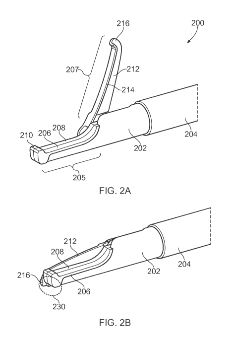
10 Fig. 2A is perspective view of an instrument tip 200 of
an electrosurgical resector instrument that is an embodiment
the invention. The instrument tip 200 is mounted at the
distal end of a flexible shaft 204, which may correspond to
the flexible shaft 112 discussed above. In this embodiment,
15 the instrument tip 200 comprises a static portion 202 that
carries a first electrode 206, and a movable portion 212 that
carries a second electrode 214. However, the invention need
not be limited to this configuration. In other examples both
electrodes may be provided on either the static portion 202 or
the movable portion 212.
The static portion 202 has a proximal region that is
secured to a distal end of the flexible shaft 204. The static
portion 202 extends in a longitudinal direction away from the
distal end of the flexible shaft 204. At its distal end, the
static portion 202 defines a first blade element 205, which is
a longitudinally extending finger having a upstanding tooth
210 at its distalmost end. The first electrode 206 extends
along an upper surface of the first blade element 205.
The movable portion 212 is pivotably mounted on the
static portion 202. In this embodiment, the movable portion
212 comprises a second blade element 207, which is an elongate
finger having a length commensurate with the first blade
element 205. The second blade element 207 has a downwardly
extending tooth 216 at its distalmost end.
The movable portion is pivotable about a pivot axis
located at a proximal end of the first blade element 205,
whereby the second blade element 207 can swing between an open
position (shown Fig. 2A) in which it is angled away from the
first blade element 205 and a closed position (shown in Fig.
2B) where is lies alongside (i.e. laterally adjacent) to the
first blade element 205. The range of movement of the movable
CA 03076876 2020-03-24
WO 2019/073037
PCT/EP2018/077880
16
portion may be such to allow the second blade element 207 to
adopt an obtuse angle relative to the first blade element 205.
This may be particular useful for grasping tissue that present
a low surface profile.
The first blade element 205 and second blade element 207
may thus define a scissor-type closure mechanism in which
tissue located in a gap between the blade elements 205, 207
when in the open position can have pressure applied to it as
the second blade element 207 is moved to the closed position.
The upstanding tooth 210 on the first blade element 205 and
the downwardly extending tooth 216 on the second blade element
207 act to retain tissue in the gap as second blade element
207 moves to the closed position.
The first blade element 205 comprises a planar dielectric
body 208, e.g. made from ceramic or other suitable
electrically insulating material. The planar dielectric body
208 defines a plane that is parallel to a plane through which
the second blade element 207 pivots. The planar dielectric
body 208 provide an insulating barrier between the first
electrode 206 and the second blade element 207. For example,
the second blade element 207 is arranged to slide past a first
surface of the planar dielectric body 208, and the first
electrode 206 is formed on a second surface of the planar
dielectric body 208, the second surface being on the opposite
side of the planar dielectric body 208 from the first surface.
The first electrode 206 may be made from a conductor exhibit
high conductivity, e.g. gold or the like.
The second electrode 214 extends along a side surface of
the second blade element 207 that slides past an adjacent side
surface of the first blade element 205 (i.e. the first surface
of the planar dielectric body 208 mentioned above) when the
second blade element 207 is moved into the closed position.
In this example, the second blade element 207 comprises a
laterally protruding flange along a bottom edge thereof. The
second electrode 214 extends along the laterally facing
surface of the flange. The second blade element may be formed
from an electrically conductive material that is coated with
an insulating material. For example, it may be made from
stainless steel with a ceramic or diamond-like carbon (DLC)
coating. The insulating coating may be removed, e.g. etched
away, from regions where it is not required. For example, the
CA 03076876 2020-03-24
WO 2019/073037
PCT/EP2018/077880
17
second electrode 214 may be formed by etching away the coating
from the side edge of the lateral flange. A gold layer may be
deposited over the etched surface to form the electrode.
Other portions of the coating may be removed to enable an
electrical connection to be made to the outer conductor of the
coaxial cable, as explained below.
The flexible shaft 204 defines a lumen through which
extends a coaxial cable (not shown) for conveying RF and
microwave EM energy, and a longitudinally slidable control rod
(shown in Figs. 3A to 3D) for controlling movement of the
movable portion 212.
As discussed in more detail with reference to Fig. 4, the
first electrode 206 is electrically connected to an inner
conductor of the coaxial cable and the second electrode 214 is
electrically connected to an outer conductor of the coaxial
cable. The instrument tip thus provides an energy delivery
structure that is operable to deliver RF energy along a
current path (e.g. through tissue) between the first electrode
and second electrode, or microwave energy through a microwave
field emitted by the first electrode and second electrode.
The instrument tip 200 may provide three operational
modalities. In a first modality, the instrument can be used
with the blade elements 205, 207 in the closed position to
deliver RF EM energy to cut through biological tissue. In
this first modality, the RF EM energy passes primarily between
the first electrode 206 and second electrode 214 in a distal
cutting zone 230 adjacent to the upstanding tooth 210 on the
first blade element 205 and the downwardly extending tooth 216
on the second blade element 207. The instrument may thus be
used to sweep or glide across or through tissue to effect
cutting.
In a second modality, the blade elements 205, 207 may be
used to perform a grasping cut, i.e. a cut through tissue
captured between the blade elements. In this modality cutting
is done by a combination of physical pressure applied by
closing the blade elements 205, 207 and RF EM energy applied
during the closing process.
In a third modality, the blade elements 205, 207 may be
used to grasp and seal tissue, such as a blood vessel or the
like. In this modality, microwave EM energy is delivered to
CA 03076876 2020-03-24
WO 2019/073037
PCT/EP2018/077880
18
the electrodes, which set up a microwave field that acts to
coagulate the tissue held within the blade elements.
The static portion 202 may have a dielectric shield
mounted over its outer surface. In this example, the
dielectric shield is a thermoplastic polymer, e.g. polyether
ether ketone (PEEK), or the like. The dielectric shield may
be moulded over the device, or may be a cover (e.g. formed by
laser cutting a suitably size tube) that can slide over the
instrument tip when the blade elements are in the closed
position. The dielectric shield can be used to control the
shape of the first electrode 206, e.g. to ensure that the
first electrode 206 is exposed substantially only at an upper
surface of the first blade element 205. In turn this can
ensure that the RF and microwave energy delivered from he
electrodes is focussed into the desired region.
Figs. 3A, 3B, 3C and 3D are perspective views of the
instrument tip 200 that illustrate the closing operation.
Figs. 3A to 3D show the opposite side of the instrument tip
200 from Figs. 2A and 2B. The dielectric shield is omitted in
Figs. 3A to 3D for clarity.
Fig. 3A illustrates the instrument tip 200 in an open
position, with the movable portion 212 disposed so that the
second blade element 207 is sits at an obtuse angle to the
first blade element 205. As shown in Fig. 3A, the static
portion 202 includes a longitudinally extending arm 218 that
provides a pivot base to which the movable portion 212 is
attached. The arm 218 has a pivot axle 226 rotatably mounted
therein. The pivot axle 226 defines a laterally extending
pivot axis (i.e. the pivot axis is orthogonal to the
longitudinal direction defined by the flexible shaft 204).
A slidable control rod 220 protrudes from the flexible
shaft 204. The static portion 202 has a guide channel 221
formed therein through which the control rod 220 passes. The
control rod 220 has a distal attachment feature 223 that is
engaged with the movable portion 212. In this example, the
distal attachment feature 223 is a hook that engages a slot
224 formed in an attachment plate 222 of the movable portion
212. Other types of engagement may be used. Longitudinal
sliding motion of the control rod 220 is transformed into
pivoting motion of the attachment plate 222. The attachment
CA 03076876 2020-03-24
WO 2019/073037
PCT/EP2018/077880
19
plate 222 may be integrally formed with or otherwise operably
coupled to the second blade element 207.
Fig. 3B shows the instrument tip 200 in a partly closed
configuration, where the control rod 220 has been partly
retracted into the flexible sleeve 204, and where there is an
acute angle between the first and second blade elements.
Fig. 3C shows the instrument tip 200 in another partly
closed configuration, where the control rod 220 is further
retracted into the flexible sleeve, and where the downwardly
extending tooth 216 on the second blade element 207 is about
to slide past the upstanding tooth 210 on the first blade
element 205. In reaching this position, it can be seen that
the distal attachment feature 223 of the control rod 220 has
remained at a first end of the slot 224. The slot 224 provide
a cam surface along which the control rod slides for the final
portion of the closing operation, where the first and second
blade elements slide past each other. Fig. 3D shows the final
closed position, where the distal attachment feature 223 of
the control rod 220 has moved to a second end of the slot 224.
The slot advantageously provides a cam surface against which
the distal attachment feature 223 acts in this final part of
the movement operation, e.g. to boost the closure force to
overcome resistance that can occur at the final stages of a
cut.
Fig. 4 is a schematic partly cut-away side view of an
instrument tip 300 for an electrosurgical resector instrument
that is an embodiment of the invention. The instrument tip
300 is located at the distal end of a flexible sleeve 302,
which conveys a coaxial cable 304 and a control rod 312. The
control rod 312 is for controlling pivoting motion of a
movable portion 322 relative to a static portion 318 in the
same way as discussed above. The static portion 318 has a
planar dielectric body 314 secured to it, e.g. by a suitable
adhesive, the planar dielectric body 314 extending in a
longitudinal direction away from the static portion 318 to
form a first blade element. A first electrode 316 is formed
on one side of the planar dielectric body 314.
The moveable portion 322 is pivotably mounted on the
static portion 318 via a pivot axle (not visible in Fig. 4) at
an opposite side of the planar dielectric body 314 to the
first electrode 316. The moveable portion 322 comprises a
CA 03076876 2020-03-24
WO 2019/073037
PCT/EP2018/077880
second blade element that is arrange to slide past the first
blade element in a similar manner to the first and second
blade elements 205, 207 discussed above. The moveable portion
322 includes a second electrode 324 thereon that lies adjacent
5 the opposite side of the planar dielectric body 314 when the
blade elements are in a closed position.
The coaxial cable 304 comprises an inner conductor 306
that is separated from an outer conductor 310 by a dielectric
material 308. The dielectric material 308 and inner conductor
10 306 extend beyond a distal end of the outer conductor 310. A
distal end of the dielectric material 308 abuts a proximal end
of the planar dielectric body 314. The inner conductor 306
extends distally from this junction to overlap with and
electrically contact a proximal portion of the first electrode
15 316. The invention need not be limited to this arrangement.
In other examples, the inner conductor may be electrically
connected to an electrode on the movable portion, for example.
The static body 318 includes a support arm on which the
movable portion is mounted. The planar dielectric body 314
20 may also be mounted on the support arm, e.g. using adhesive of
the like. The support arm is formed from an electrically
conductive material (e.g. stainless steel) with an
electrically insulating coating. The coating is removed at a
proximal contact portion 320 which is electrically connected
to the outer conductor 310 of the coaxial cable 304. The
movable portion 322 is also formed from an electrically
conductive material (e.g. stainless steel) with an
electrically insulating coating. The movable portion 322 is
physically engaged with the static portion 318 at the pivot
connection. An electrical connection between the second
electrode 324 and the outer conductor 310 of the coaxial cable
304 passes through the pivot connection. For example, the
pivot axle itself may be formed from an electrical conductive
material (e.g. stainless steel). The insulating coating of
the static portion 318 may be remove at a region of sliding
engagement (e.g. an aperture or recess for receiving the pivot
axle) between the static portion 318 and the movable portion
322. Similarly, the insulating coating of the movable portion
322 may be removed at this region. As the second electrode
324 may be or may be electrically connected to the
electrically conductive material of the movable portion 322, a
CA 03076876 2020-03-24
WO 2019/073037
PCT/EP2018/077880
21
complete electrical connection to the outer conductor can be
formed.
Fig. 5 is a partially cut-away perspective view of an
electrosurgical resector instrument that illustrates how the
schematic features of Fig. 4 may map on to a device similar to
that shown in Figs. 2A and 2B. Features in common with Fig. 4
are given the same reference numbers and are not described
again.
Fig. 6A is an illustration of a handpiece 600 which may
be used as part of an electrosurgical apparatus that is an
embodiment of the invention. The handpiece 600 includes a body
602 and an actuating portion 604. The body 602 includes a
hollow barrel 606 in which a shaft 608 of the actuating
portion 604 is slidably engaged. The body 602 also includes a
rotator 610 which is rotatably connected to the barrel 606.
The actuating portion 604 is connected to an internal shaft
628 which extends through the barrel 606 and rotator 610, and
which protrudes from a distal end of the rotator 610. The
internal shaft 628 moves longitudinally with the shaft 608,
but is rotatable relative to it. An instrument shaft 612
exits the handpiece 600 from a distal end of the internal
shaft 628. For example, the instrument shaft 612 may be
flexible shaft 204 described above, which is connected to an
instrument tip 200 at its distal end. The instrument shaft
612 is connected to rotate with the internal shaft 628.
The actuating portion 604 is slidable in a longitudinal
direction relative to the body 602 along its shaft 608 between
two positions: a closed position where a length of the shaft
608 is contained within the barrel 606, and an open position
where the length of the shaft 608 is outside the barrel 606.
Fig. 6A shows the handpiece 600 with the actuating portion 604
in the open position. The total range of motion of the
actuating portion 604 relative to the body 602 may be
approximately 35 mm. The longitudinal direction of motion of
the actuating portion 604 relative to the body 602 is aligned
with a longitudinal axis of the instrument shaft 612 as is
passes out of the internal shaft 628. The shaft 608 may
include one or more grooves 614 which engage with protrusions
(not shown) inside the barrel 606, in order to prevent the
actuating portion 604 from rotating relative to the body 602.
The body 602 includes a pair of finger rings 614, 616 and the
CA 03076876 2020-03-24
WO 2019/073037
PCT/EP2018/077880
22
actuating portion 604 includes a thumb ring 618, which may be
used to facilitate a user's grip when pushing and pulling the
barrel 606 relative to the actuating portion 604. The
actuating portion 604 further includes an input connector 620
for connecting an interface cable (e.g. interface cable 104)
which connects the handpiece 600 to a generator (e.g.
generator 102). The input connector 620 may for example be a
QMA connector or any other suitable connector for interfacing
with the generator.
Fig. 6B is a cut-away illustration of the handpiece 600,
where certain parts are not shown in order to reveal the
internal structure of the handpiece. Where features have
already been described above in reference to Fig. 6A,
identical reference numerals have been used.
The input connector 620 is electrically connected to a
circuit board 622 contained within the shaft 608 of the
actuating portion 604. The input connector 620 forms a
substantially right angle with the circuit board 622, such
that it is oriented along a direction which is substantially
perpendicular to the direction of relative motion between the
actuating portion and the body 602. In this manner, a cable
which is connected to the input connector 620 may not get in a
user's way. An output connector 624 is attached at an edge of
the circuit board 622. The output connector 624 is
electrically connected to a coaxial transmission line 626 via
a mating connector 627 on the coaxial transmission line 626.
The coaxial transmission 626 line runs through the handpiece
600 and enters the instrument shaft 612 at the distal end of
the handpiece 600. The coaxial transmission line 626 may for
example correspond to coaxial line 226 described above, which
serves to convey RF and microwave EM energy to the instrument
tip.
The electrical connection between the output connector
624 and the coaxial transmission line 626 is rotatable, i.e.
it allows the coaxial transmission line to rotate about its
axis relative to the output connector 624. Suitable connectors
which enable rotatable electrical connections include QMA
connectors, micro coaxial (MCX) connectors and micro-miniature
coaxial (MMCX) connectors.
CA 03076876 2020-03-24
WO 2019/073037
PCT/EP2018/077880
23
In other embodiments, the circuit board 622 may be
omitted, and replaced by a single QMA to MCX right-angle
connector.
As shown in Fig. 6B, the internal shaft 628 extends
through and is longitudinally slidable relative to both the
barrel 606 and the rotator 610 of the body 602. A distal end
of the internal shaft 628 protrudes from the rotator 610. The
length of the protruding portion depends on the position of
the shaft 608 of the actuating portion 604. The internal
shaft 628 is connected at a proximal end to the shaft 608 of
the actuating portion 604, by means of a circumferential
recess 630 around an outer surface of the internal shaft 628
which is engaged by a radial protrusion 632 on an inner
surface of the shaft 608. The connection between the shaft 608
and the internal shaft 628 prevents the internal shaft 628
from moving longitudinally relative to the shaft 608, but
allows the internal shaft 628 to rotate about its axis
relative to the shaft 608. The internal shaft 628 may
therefore be moved longitudinally backwards and forwards
relative to the body 602 by moving the actuating portion 604
relative to the body 602.
The internal shaft 628 may include a proximal portion 631
having a cavity for holding the connector 627 of the coaxial
transmission line 626 in position to ensure that it remains
securely connected to the output connector 624 on the circuit
board 622. Additionally, the connector 627 on the coaxial
transmission line 626 may include a protrusion 633 which is
configured to engage a slot in the proximal portion 630 of the
internal shaft 628, to prevent the connector 627 from moving
relative to the internal shaft 628. For example, the
protrusion 633 may be a nut which is part of or attached (e.g.
by soldering) to the connector 627. The protrusion 627 may
also be configured to rotationally lock the connector 627 to
the internal shaft 628, such that rotation of the internal
shaft 628 causes the connector 627 to rotate.
The coaxial transmission line 626 passes through the
internal shaft 628 where, at a distal end thereof, it enters
the instrument shaft 612. A length of the instrument shaft 612
is contained within a distal portion 634 of the internal shaft
628, where it is fixed to the internal shaft 628. In this
manner, both longitudinal and rotational motion of the
CA 03076876 2020-03-24
WO 2019/073037
PCT/EP2018/077880
24
internal shaft 628 may be transmitted to the instrument shaft
612. For example, the instrument shaft 612 may be glued using
epoxy to the distal portion 634 of the internal shaft 628.
Adhesion between the instrument shaft 612 and the internal
shaft 628 may be improved by roughing the surface of the
instrument shaft 612 before applying the epoxy. In some cases,
the length of instrument shaft 612 contained in the distal
portion 634 may be approximately 22 mm, to ensure good
adhesion.
The rotator 610 is connected to the barrel 606 such that
it is rotatable relative to the barrel about a longitudinal
axis of the handpiece 600. In the example shown, the rotator
610 has a proximal portion 642 with a circumferential recessed
channel 644 that receives a radially inwardly extending
protrusion 646 on the barrel 606.
The internal shaft 628 passes through the rotator 610 and
is engaged with the rotator 610 such that it is slidable
relative to the rotator 610 along its length, but it is not
rotatable relative to the rotator 610 (i.e. the rotator 610
and internal shaft 628 are rotationally locked relative to one
another). This may be achieved by any kind of interengagement
that transfers rotational movement. For example there may be
one or more longitudinally oriented cooperating engagement
elements (e.g. grooves and teeth) formed on an outer surface
of the internal shaft 628 and an inner surface of the rotator
610. The engagement elements may respectively engage with
each other to cause the internal shaft 628 to rotate as the
rotator 610 is turned on the barrel 606. This in turn causes
the instrument shaft 612, which is fixed to the internal shaft
628, to rotate such that an instrument tip connected at a
distal end of the instrument shaft 612 may also be caused to
rotate. However, as the internal shaft 628 is not rotationally
coupled to the actuating portion 604, the actuating portion
604 is not caused to rotate by rotation of the rotator 610.
The axis of rotation of the rotator 610 relative to the barrel
606 may be aligned with a longitudinal axis of the internal
shaft 628, such that rotation of the rotator 610 causes
rotation of the internal shaft 628 about its longitudinal
axis.
A length of a main control rod 636 is contained within
the internal shaft 628, and exits the handpiece through the
CA 03076876 2020-03-24
WO 2019/073037
PCT/EP2018/077880
instrument shaft 612. The main control rod 636 may be used to
operate a movable portion (e.g. a pivotable blade element) on
an instrument tip connected at a distal end of the instrument
shaft 612. For example, main control rod 636 may correspond to
5 main control rod 242 described above. A proximal end of the
main control rod 636 is held fixed relative to the body 602 of
the handpiece 600. Therefore, motion of the body 602 relative
to the actuating portion 604 may cause the main control rod
636 to move longitudinally along the instrument shaft 612.
10 This is because the longitudinal position of the instrument
shaft 612 is held fixed relative to the actuating portion 604
(by means of the internal shaft 628, which is connected at one
end to the actuating portion 604 and at another end to the
instrument shaft 612), whilst the main control rod 636 is
15 movable with the body 602 relative to the actuating portion
604, and thus the instrument shaft 612.
Thus, a user may move the actuating portion 604 relative
to the body 602 in order to move the main control rod 636
backwards and forwards relative to the instrument shaft 612
20 and control the opening and closing of a movable portion (e.g.
pivotable blade element) on an instrument tip connected at a
distal end of the instrument shaft 612.
There are several possible ways for holding the proximal
end of the main control rod 636 fixed relative to the body 602
25 of the handpiece 600. In the example shown, a block 638 is
attached to the proximal end of the main control rod 636. The
block 638 may for example be a piece of metal which is
soldered or welded to the proximal end of the main control rod
638. The block 638 may be configured to fit in a holder (not
shown) which is rigidly connected to the rotator 610, such
that longitudinal motion of the body 602 relative to the
actuating portion 604 is transmitted to the block 638 (and
hence the main control rod 636) via the holder. The holder may
be connected to the rotator 610 through an opening in a side
wall of the internal shaft 628.
A portion of the main control rod 636 in the internal
shaft 628 may be contained in a protective tube 640. The
protective tube may be made of any suitable material (e.g.
PTFE), and may serve to prevent the main control rod 636 from
bending when the handpiece 600 is opened. Alternatively, a
CA 03076876 2020-03-24
WO 2019/073037
PCT/EP2018/077880
26
metal tube may be soldered or welded to the main control rod
636 to achieve the same effect.
The relative linear motion between the actuating portion
604 and the body 602 directly controls linear motion of the
main control rod 636 relative to the instrument shaft 612.
This may enable a user to accurately control the opening and
closing of a pivotable blade element on an instrument tip at
the distal end of the instrument shaft 612. Furthermore, the
configuration of the handpiece 600 enables a user to
comfortably hold the handpiece 600 in one hand and control the
opening and closing of the blade element with one hand (by
placing fingers of one hand in the finger rings 614, 616,
618). The user may also simultaneously rotate the rotator 610
with the other hand, in order to rotate the instrument tip.
The orientation of the input connector 620 may ensure that any
cable connected to the input connector 620 does not interfere
with a user's operation of the handpiece 600. In this manner,
the user isn't forced to hold the handpiece 600 in an awkward
position in order to accommodate a cable, which might cause
stress on the user's wrist.
In one example, a heat shrink or other stiffening
material may be applied around a proximal portion of the
instrument shaft 612. The length of this stiffening portion
is selected to occupy a part of the shaft that will always be
outside the insertion tube of the endoscope, even when the
shaft is fully inserted. The stiffening portion may assist in
translating torque from this part of shaft into the part that
is within the insertion tube of the scoping device. It can
also prevent the instrument shaft from rippling under
actuation as well as under rotation. Moreover, it can give
the clinician (i.e. scoping device operator) something larger
in diameter to grip onto for both rotation and push/pull
without having to communicate to the assistant.
The fact that the handpiece 600 has a free moving
rotating joint in it permits the clinician to rotate without
the assistant who will be holding the hand-piece, but also
enables the assistant to apply rotation through the hand-piece
if necessary.
Fig. 7A is a cut-away perspective view of the instrument
shaft 612 as it travels towards the instrument tip. The
instrument shaft 612 comprises a outer sleeve 648 that defines
CA 03076876 2020-03-24
WO 2019/073037
PCT/EP2018/077880
27
a lumen for conveying the coaxial cable 626 and control rod
636. In this example, the coaxial cable 626 and control rod
636 are retained in a longitudinally extending insert 650.
The insert 650 is an extrusion, e.g. formed from a deformable
polymer such as PEEK or other plastic with similar mechanical
properties. As shown more clearly in Fig. 7B, the insert 650
is a cylindrical element having a series of sub-lumens 664 cut
away around its outer surface. The sub-lumens 664 break
through the outer surface of the insert 650 to define a
plurality of discrete feet 662 around the circumference
thereof. The sub-lumens 664 can be sized to convey components
such as the coaxial cable 626 or control rod 636, or may be
for the purpose of allowing fluid flow along the lumen of the
sleeve 648.
It may be beneficial for the insert not to include any
enclosed sub-lumens. Fully enclosed sub-lumens can be prone
to retaining deformations if stored in a bent condition. Such
deformations can lead to jerky motion in use.
The insert 650 may comprise a sub-lumen for receiving the
coaxial cable 626. In this example, the coaxial cable 626
comprises an inner conductor 658 separated from an outer
conductor 654 by a dielectric material 656. The outer
conductor 654 may in turn have a protective cover or sheath
652, e.g. formed from PTFE or other suitably low friction
material to permit relative longitudinal movement between the
insert and coaxial cable as the shaft with flexing of the
shaft.
Another sub-lumen may be arranged to receive a standard
PFTE tube 660 through which the control rod 636 extends. In
an alternative embodiment, the control rod 636 may be provided
with a low-friction (e.g. PFTE) coating before use, so that a
separate PFTE tube is not required.
The insert is arranged to fill, i.e. fit snugly within,
the lumen of the sleeve 648 when mounted with the coaxial
cable 626 and control rod 636. This means that the insert
functions to restrict relative movement between the coaxial
cable, control rod and sleeve during bending and rotation of
the shaft 612. Moreover, by filling the sleeve 648, the
insert helps to prevent the sleeve from collapsing and losing
rotation if rotated excessively. The insert is preferably
CA 03076876 2020-03-24
WO 2019/073037
PCT/EP2018/077880
28
made from a material that exhibits rigidity to resist such
movement.
The presence of the insert may furthermore prevent "lost"
travel of the control rod caused by deformation of the
instrument shaft 612. Such lost travel can occur in the
absence of the insert for two reasons.
Firstly, the control rod 636 can move from side to side
in the sleeve 648 so that when the sleeve follows a curved
path it is able to go round the outside of bends which is a
longer path than the length of the centre-line which is also
the length of the sleeve when straight. For
example, if an
inside diameter of the sleeve was 2.15 mm, and a diameter of
the control rod 0.4 mm, the centre-line of the control rod may
be as much as 0.875 mm away from the centre-line of the
sleeve. In each 180 degrees of bend, if the control rod goes
to the outer limit of its possible travel within the sleeve,
the path of the control rod would be 2.75 mm longer than the
length along the centre-line of the sleeve. Thus, five 180
degree bends could yield 13.75 mm 'lost' travel.
Secondly, the control rod 636 may follow a sinuous path
inside the sleeve 648, even if the sleeve is straight, which
is longer than the length of the sleeve. Thus, in any
location where the control rod is unsupported, it may bow
sideways. The bowed shape would be like a sine wave. If it
was stopped from going very far sideways, then it might have
multiple bows down its length. Within the sleeve, the control
rod cannot bow sideways but has to wrap round the inside of
the sleeve, with its centre at a radius of 0.875 mm from the
sleeve centre. Each wrap round the tube is equivalent to 5.5
mm of bowing. The length increase of a sinusoidal path over a
direct path is calculated with an elliptic integral. For
small ratios of a bow (a) to the straight length (p) of two
bows, the change in length is close to that for the arc of a
circle, and for this the ratio of the lengths is 8ah3sin(3a/p),
and the difference (lost travel) is approximately
8a/psin(8a/p) ¨1 (8a/p)2/6. For instance, if the actuator rod
had 6 loops (3 in each direction) down a 2.3 m length, and
each one went twice round the centre-line, then p = 2300/3 =
766.666 mm, and a = 11 mm, and the lost travel is 0.22%, or 5
mm.
CA 03076876 2020-03-24
WO 2019/073037
PCT/EP2018/077880
29
The extruded insert discussed above provides cam-like
feet that jam on the inside of the sleeve and impede the
wrapping of the control rod around the axis of the sleeve.
This will reduce the lost travel discussed above.
Figs. 8A, 8B and 8C are perspective views of an
instrument tip 700 of an electrosurgical resector instrument
that is another embodiment of the invention. In this
arrangement, the instrument tip is modified to provide a
parallel closing action between the blade elements, as opposed
to the pivoting scissor-type action discussed above.
Similarly to the instrument tip 200 discussed above, the
instrument tip 700 comprises a static portion 706 that carries
a first electrode 708, and a movable portion 710 that carries
a second electrode 712. The instrument tip 700 is mounted at
the distal end of a flexible shaft 702. A shielding element
704 is mounted over a junction between a coaxial cable
conveyed by the shaft 702 and a proximal end of the first
electrode 706.
The static portion 706 has a proximal region that is
secured to a distal end of the flexible shaft 702. The static
portion 706 extends in a longitudinal direction away from the
distal end of the flexible shaft 702 and defines a first blade
element in a distal region. The first blade element is a
longitudinally extending finger having a upstanding tooth at
its distalmost end. The first electrode 708 extends along an
upper and side surfaces of the first blade element.
The movable portion 710 is pivotably mounted on the
static portion 706. In this embodiment, the movable portion
710 comprises a second blade element, which is an elongate
finger having a length commensurate with the first blade
element. The second blade element has a downwardly extending
tooth at its distalmost end.
In this example, the movable portion 710 is pivotable
about a pivot axis 711 that is itself movable relative to the
static portion 706. Similar to the structure discussed above,
the instrument tip 700 comprises a control rod 714 that is
slidable mounted in the shaft 702 and which engages a slot 716
on a proximal part of the movable portion 710. The movable
portion 710 is connected to the static portion 706 by a
connector rod 718. A first end of the connector rod 718 is
pivotably connected to the movable portion 710 at the pivot
CA 03076876 2020-03-24
WO 2019/073037
PCT/EP2018/077880
axis 711, and a second end of the connector rod 718 is
slidably mounted to the static portion 706 in a channel (not
shown) formed therein.
Fig. 8A shows the instrument tip 700 in an open
5 configuration, where the control rod 714 is extended out of
the shaft 702 to push connector rod 718 into a deployed
position where the pivot axis 711 is moved away from the
static portion and the movable portion is pivoted around the
pivot axis 711 so that the second blade element is at an angle
10 to the first blade element.
Fig. 8B shows the instrument tip 700 in an intermediate
configuration, wherein the control rod 714 is partly retracted
so that the connector rod 718 remains in the deployed position
where the pivot axis 711 is spaced away from the static
15 portion 706, but where the movable portion has pivoted around
the pivot axis so that the second blade element is parallel to
the first blade element.
Fig. 8C shows the instrument tip 700 in a closed
configuration, wherein the control rod 714 is fully retracted
20 to cause the connector rod 718 to move to a withdrawn position
where the pivot axis 711 is drawn into the static portion 706
so that the second blade element passes alongside the first
blade element while remaining parallel therewith.
Figs. 9A and 9B are perspective views of an instrument
25 tip 800 of an electrosurgical resector instrument that is
another embodiment of the invention. In this arrangement, the
instrument tip is modified to provide a wider base to create
better tissue sealing capabilities by confining or
concentrating the microwave field set up between the
30 electrodes.
Similarly to the instrument tip 200 discussed above, the
instrument tip 800 comprises a static portion 804 that
comprises a planar dielectric body 806 having a first
electrode 810 thereon, and a movable portion 808 that carries
a second electrode 812. The instrument tip 800 is mounted at
the distal end of a flexible shaft 802. A shielding element
803 is mounted over a junction between a coaxial cable
conveyed by the shaft 802 and a proximal end of the first
electrode 810.
The static portion 804 has a proximal region that is
secured to a distal end of the flexible shaft 802. The planar
CA 03076876 2020-03-24
WO 2019/073037
PCT/EP2018/077880
31
dielectric body 806 extends in a longitudinal direction away
from the distal end of the flexible shaft 802 and defines a
first blade element in a distal region. The first blade
element is a longitudinally extending finger having a
upstanding tooth at its distalmost end. The first electrode
810 extends along an upper and side surfaces of the first
blade element.
The movable portion 808 is pivotably mounted on the
static portion 804. In this embodiment, the movable portion
808 comprises a second blade element, which is an elongate
finger having a length commensurate with the first blade
element. The second blade element has a downwardly extending
tooth at its distalmost end. The second electrode 812 extends
along a side edge of the second blade element.
In this example, the static portion 804 comprise a third
electrode 814. The third electrode 814 is formed from a
conductive material and takes the form of a third blade
element having the same shape as the first blade element, but
spaced laterally from it on the opposite side of the planar
dielectric body 806 to the first electrode 810. The third
electrode 814 is spaced from the first blade element by a gap
that is sized to receive the second blade element as is
pivoted from an open position to a closed position in the same
way as described above with reference to Figs. 2A and 2B.
Fig. 9B shows the opposite side of the instrument tip
800. The static portion 804 comprises a longitudinal arm 816
that supports a pivot axle 818 on which the movable portion
808 is mounted. A channel 822 is cut or otherwise formed in
the static portion 804 to receive the movable portion 808 as
it moves into the closed position.
Pivoting of the movable portion is controlled by a
longitudinally retractable control rod (not shown in Fig. 9B)
that extends from the shaft 802 via guide channel 824 to
engage with a slot 820 formed in the movable portion 808, in a
similar manner as described above with reference to Figs. 3A
to 3D.
The third electrode 814 may be electrically connected to
the first electrode 810 by a laterally extending conductive
portion, e.g. pin or rod, (not shown) that passes across the
gap between the third electrode 814 and the first blade
element beneath the second blade element.
CA 03076876 2020-03-24
WO 2019/073037
PCT/EP2018/077880
32
With the structure shown in Figs. 9A and 9B, the
instrument tip can support a wider extent of tissue to be
gripped between the blade elements. Moreover, the microwave
field created between the second electrode 812 and the first
and third electrodes 810, 814 may exhibit a more consistent
effect on gripped tissue on both sides of the second blade
element. In this example it may be desirable for the inner
conductor of the coaxial transmission line to be connected to
the second electrode and the outer conductor to the first and
third electrodes, whereby the first and third electrodes
perform a field shielding function as the second blade element
is moved to the closed position.