Note: Descriptions are shown in the official language in which they were submitted.
CA 03076918 2020-03-24
WO 2019/070769
PCT/US2018/054042
METHOD OF PREDICTION OF TUMOR-DERIVED NEO-PEPTIDE ANTIGENICITY
AND/OR IMMUNOGENICITY USING MUTATIONAL SIGNATURE PATTERNS
CROSS-REFERENCE TO RELATED APPLICATIONS
[0001] This application claims priority from U.S. Provisional Application
Serial No.
62/567,096 filed on October 2, 2017, which is incorporated herein by reference
in its entirety.
STATEMENT REGARDING FEDERALLY SPONSORED RESEARCH OR
DEVELOPMENT
[0002] Not Applicable.
FIELD
[0003] The field of the invention relates to proliferative diseases and to
biomarkers
of response to immunotherapy, including pharmaceutical agents and antibodies
used for the
prevention and the treatment of cancer.
INTRODUCTION
[0004] Definition of suitable biomarkers of response to treatment can highly
impact
disease outcome and progression. In oncology particularly, there is a need for
highly specific
and sensitive prognostic and predictive markers. The information relative to
these biomarkers
can be obtained from a tumor biopsy, later analyzed using molecular methods
including but
not limited to genomics and sequencing, transcriptomics, proteomics.
[0005] Immunotherapy agents are drugs that harness and enhance the capacity of
the innate immune system to fight proliferative diseases. Indeed, cancer
immunotherapy has
been proven efficient even for tumors resistant to chemotherapy and radiation
therapy, and
thus offer the possibility for a long-term cancer remission. Multiple
biomarkers of response to
immunotherapies have been developed, but there is yet no comparison,
standardization or
prospective validation of these companion assays. Expression of proteins
directly targeted by
such agents on tumor cells and/or tumor-infiltrating lymphocytes (e.g.
Programmed-cell
Death Ligand 1 (PD-L1) protein staining for PD-1/PD-L1 axis inhibitors) only
constitutes a
part of the predictive model for the response to the drugs, and additional
biomarkers are
needed. Various embodiments of the invention described below meet this need as
well as
other needs existing in the field of diagnosing and treating cancer.
- 1 -
CA 03076918 2020-03-24
WO 2019/070769
PCT/US2018/054042
[0006] The immune system exhibits ubiquitous properties, such as context-
dependent response to pathogens and non-self-elements, continuous learning,
and memory.
The overall mechanisms behind these features seem rather common between
individuals and
populations. Recently, it has been hypothesized that the variability of
response to cancer
immunotherapy mostly depends on the intrinsic heterogeneity of the tumor,
largely
highlighted by the uniqueness of one's tumor mutation profile. This molecular
'fingerprint'
may be reflected by a unique neo-antigens catalog, presenting a certain level
of "non-
selfness", further eliciting or repressing the immune response.
[0007] The present invention provides a method to estimate the antigenicity
(i.e. the
probability for a peptide to be presented by the major histocompatibility
complex (MHC) to
the immune system) and/or immunogenicity (i.e. the probability for a peptide
to be
recognized by the immune system) of the set of neo-peptides presented by one
tumor, given
its specific mutation description. This method comprises: (i) describing the
unique set of
DNA or RNA mutations presented by a tumor sample; (ii) determining the set of
all possible
8- to 10-mers neo-epitopes encoded by the nucleic acid or protein sequences
encompassing
the mutations observed; (iii) defining the physicochemical properties of the
set of neo-
epitopes produced by the tumor cell, particularly their overall hydrophobicity
and specific
amino-acid content; (iv) assessing the antigenicity and immunogenicity of the
set of neo-
epitopes; and (v) estimating the further patient's response to
immunotherapies, based on the
set of neo-epitopes actually presented by the tumor cells to the immune
system.
SUMMARY
[0008] The present teachings include methods for prediction of response to
immunotherapy for patients diagnosed with a proliferative, degenerative or
inflammatory
disease, by analysis of physicochemical properties of the set of neo-antigens
produced by the
injured tissue, comprising description of genomic or/and protein alterations
in a sample. The
set of alterations described may be obtained by a validated assay that
involves: a) contacting
the sample with one or more agents that detect genomic and/or protein
variations in at least
one molecular marker; b) comparing the sequence(s) of at least one genomic or
protein
marker detected in the sample with this of a reference genome or a reference
proteome; and
c) defining a list of genomic or protein alterations specific to the sample;
elucidation of all
possible peptides encompassing the genomic and/or protein alterations observed
in the tumor;
description of the physicochemical properties of the set of neo-epitopes
possibly produced by
the tumor cell, as compared to the epitopes normally presented by a
healthy/non-mutated cell;
- 2 -
CA 03076918 2020-03-24
WO 2019/070769
PCT/US2018/054042
estimation of the antigenicity and immunogenicity of the set of neo-epitopes,
based on the
physicochemical properties of these antigens; use of the antigenicity and
immunogenicity
estimates as biomarkers for prediction of the patient's response to
immunotherapy.
[0009] In an aspect, the sample is obtained from a cancer patient. The sample
may
be a tumor biopsy, or a body fluid containing tumor biomolecules. In various
aspects, the
molecular alterations are missense, non-sense, non-stop, small deletions,
small insertions, or
frameshift mutations. The alterations observed can be specifically related to
an endogenous
mutagenesis mechanism. In another aspect, the endogenous mechanism underlying
the
mutations observed in the tumor sample can be caused by the cytidine-deaminase
AID/APOBEC family of enzymes.
[0010] In various embodiments, the endogenous mechanism underlying the
mutations observed in the tumor sample respect the nucleotide patterns TCW¨TKW
or
WGA¨WMA where T represents a thymine, C represents a cytosine, G represents a
guanine,
A represents an adenine, W represents an A or a T, K represents a G or T, and
M represents
an A or C. In various embodiments, the alterations observed are specifically
related to an
exogenous mutagenesis mechanism. In various embodiments, the exogenous
mechanism
underlying the mutations observed in the tumor sample is caused by exposure to
ultra-violet
(UV) radiation.
[0011] In various embodiments, the exogenous mechanism underlying the
mutations observed in the tumor sample respect the nucleotide pattern TCC¨TIC
or
GGA¨GAA where T represents a thymine, C represents a cytosine, G represents a
guanine,
A represents an adenine. In various embodiments, the size of the peptides
allow their
presentation by the major histocompatibility complex (MHC) class I. In various
embodiments, the definition of peptides includes the retrieval of all 8 amino-
acids contiguous
from both sides to the alterations detected. In various embodiments, the
alterations detected
can be located at position 1 to 8 within said peptides.
[0012] In yet other aspects of the present invention, peptides are provided
having
the formula XiX,X,X,X,X,X,Xm, XiX,X,XXXXmXm, XXXX,X,XmXmXm,
XiX,X,X,XmXmXmXm, XiX,X,XmXmXmXmXm, XiX,XmXmXmXmXmXm,
XiXmXmXmXmXmXmXm or XmXmXmXmXmXmXmXm wherein Xi corresponds to the amino-
acid(s) considered conserved (i.e. not different from the reference); and Xm
corresponds to the
amino-acid(s) altered or potentially altered by the mutation observed in the
marker of interest.
[0013] In various embodiments, the definition of peptides includes the
retrieval of
all 9 amino-acids contiguous to the alterations detected. In various
embodiments, the
- 3 -
CA 03076918 2020-03-24
WO 2019/070769
PCT/US2018/054042
alterations detected can be located at position 1 to 9 within said peptides.
[0014] In yet other aspects of the present invention, peptides are provided
having
the formula XiX,X,X,X,X,X,X,Xm, XiX,XXXXXXmXm, XiX,X,X,X,X,XmXmXm,
XiX,X,X,X,XmXmXmXm, XXX,X,XmXmXmXmXm, XXX,XmXmXmXmXmXm,
XiX,XmXmXmXmXmXmXm, XiXmXmXmXmXmXmXmXm or XmXmXmXmXmXmXmXmXm
wherein Xi corresponds to the amino-acid(s) considered conserved (i.e. not
different from the
reference); and Xm corresponds to the amino-acid(s) altered or potentially
altered by the
mutation observed in the marker of interest.
[0015] In various embodiments, the definition of peptides includes the
retrieval of
all 10 amino-acids contiguous from both sides to the alterations detected. In
various
embodiments, the alterations detected can be located at position 1 to 10
within said peptides.
[0016] In yet other aspects of the present invention, peptides are provided
having
the formula XiX,X,X,X,X,X,X,X,Xm, XiXiXiXiXiXiXiXiXmXm, XiX,X,X,X,X,X,XmXmXm,
XiX,X,X,X,X,XmXmXmXm, XiX,X,X,X,XmXmXmXmXm, XiX,X,X,XmXmXmXmXmXm,
XiX,X,XmXmXmXmXmXmXm, XiX,XmXmXmXmXmXmXmXm, XiXmXmXmXmXmXmXmXmXm or
XmXmXmXmXmXmXmXmXmXm wherein Xi corresponds to the amino-acid(s) considered
conserved (i.e. not different from the reference); and Xm corresponds to the
amino-acid(s)
altered or potentially altered by the mutation observed in the marker of
interest. In various
embodiments, the physicochemical properties of each epitope include
hydrophobicity, amino-
acid content, size, charge, polarity, amino-acid side-chain bonds, tertiary
conformation and
steric parameters. In various embodiments, the neo-epitopes produced by the
tumor cell
present an increase of hydrophobicity compared to the non-mutated epitopes. In
various
embodiments, the neo-epitopes produced by the tumor cell present an increase
of valine (V,
Val) or/and isoleucine (Ile, I) or/and leucine (Leu, L), methionine (Met, M)
or/and
phenylalanine (Phe, F) or/and alanine (Ala, A) or/and cysteine (Cys, C) amino-
acid content
compared to the non-mutated epitopes. In various embodiments, the antigenicity
of one neo-
epitope is dependent of its binding to the MHC class I moieties.
[0017] In various embodiments, one neo-epitope may be presented by the MHC
class I isotypes HLA-A, HLA-B, HLA-C, HLA-E, HLA-F, HLA-G, HLA-K or HLA-L. In
various embodiments, the binding to the MHC class I moieties is proportional
to the neo-
epitope hydrophobicity. In various embodiments, the hydrophobicity of one neo-
epitope is
determined by summing the hydrophobicity of each amino-acid included in said
peptide. In
various embodiments, the hydrophobicity of the complete set of tumor neo-
epitopes is
determined by summing the hydrophobicity corresponding to each peptide
observed. In
- 4 -
CA 03076918 2020-03-24
WO 2019/070769
PCT/US2018/054042
various embodiments, the immunogenicity of one neo-epitope is dependent of its
recognition
by a specific immune-cell receptor. In various embodiments, the immune-cell
receptor is the
T-cell receptor (TCR) located at the surface of the cytotoxic T lymphocytes.
In various
embodiments, the recognition by the immune-cell receptor is predicted to be
proportional to
the neo-epitope hydrophobicity. In various embodiments, the hydrophobicity of
one neo-
epitope is determined by summing the hydrophobicity of each amino-acid
included in said
peptide. In various embodiments, the hydrophobicity of the complete set of
tumor neo-
epitopes is determined by summing the hydrophobicity corresponding to each
peptide
observed.
[0018] In various embodiments, the patient is treated by checkpoint inhibitor.
In
various embodiments, the patient's response to immunotherapy is directly
proportional to the
mutational pattern retrieved from the teachings herein. In various
embodiments, the patient's
response to immunotherapy is directly proportional to the mutational pattern
caused by the
AID/APOBEC family of enzymes. In various embodiments, the patient's response
to
immunotherapy is directly proportional to the mutational pattern caused by an
exposure to
UV radiation. In various embodiments, the tumor-specific expression of immune
checkpoints
is proportional to the mutational pattern retrieved from the teachings herein.
In various
embodiments, the immune checkpoints considered are PD-L1, PD-L2, PD-1, CTLA-4
or
BTLA.
[0019] In various embodiments, the immune checkpoint expression is
proportional
to the mutational pattern caused by the AID/APOBEC family of enzymes. In
various
embodiments, the immune checkpoint expression is proportional to the
mutational pattern
caused by an exposure to UV radiation. In various embodiments, the patient's
predicted
response to immunotherapy is directly proportional to the neo-epitope
physicochemical
properties retrieved from the teachings herein. In various embodiments, the
patient's
predicted response to immunotherapy is directly proportional to the increase
of
hydrophobicity of the neo-epitopes produced by the tumor, compared to the non-
mutated
epitopes. In various embodiments, the patient's predicted response to
immunotherapy is
directly proportional to the increase of valine (V, Val) or/and isoleucine
(Ile, I), or/and
leucine (Leu, L) or/and methionine (Met, M) or/and phenylalanine (Phe, F)
or/and alanine
(Ala, A) or/and cysteine (Cys, C) amino-acid content of the neo-epitopes
produced by the
tumor, compared to the non-mutated epitopes. In various embodiments, the tumor-
specific
expression of immune checkpoints is predicted to be proportional to the neo-
epitope
physicochemical properties retrieved from the teachings herein.
- 5 -
CA 03076918 2020-03-24
WO 2019/070769
PCT/US2018/054042
[0020] In various embodiments, the immune checkpoints considered are PD-L1,
PD-L2, PD-1, CTLA-4 or BTLA. In various embodiments, the immune checkpoint
expression is predicted to be proportional to the increase of hydrophobicity
of the neo-
epitopes produced by the tumor, compared to the non-mutated epitopes. In
various
embodiments, the immune checkpoint expression is predicted to be proportional
to the
increase of valine (V, Val) or/and isoleucine (Ile, I) or/and leucine (Leu, L)
or/and
methionine (Met, M) or/and phenylalanine (Phe, F) or/and alanine (Ala, A)
or/and cysteine
(Cys, C) amino-acid content of the neo-epitopes produced by the tumor,
compared to the non-
mutated epitopes.
[0021] These and other features, aspects and advantages of the present
teachings
will become better understood with reference to the following description,
examples and
appended claims.
DRAWINGS
[0022] Those of skill in the art will understand that the drawings, described
below,
are for illustrative purposes only. The drawings are not intended to limit the
scope of the
present teachings in any way.
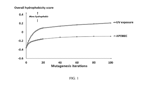
[0023] Figure 1. Overall hydrophobicity change of the human coding genome,
after
multiple iterations of kataegis or UV exposure (computed in silico ¨ N = 1 to
100 iterations).
[0024] Figure 2. Cumulative change in hydrophobicity of 8- to 10-mer neo-
antigens
in human tumor samples and correlation with APOBEC-related mutation burden.
DETAILED DESCRIPTION
[0025] EXAMPLES
[0026] EXAMPLE 1 ¨ AID/APOBEC mutational signature is associated with an
increase of neo-peptide hydrophobicity and PD-Li mRNA expression in a large
collection of
human tumor samples.
[0027] To illustrate the methods described above, we downloaded the molecular
profile (point mutations and small insertions/deletions and mRNA expression
data obtained
by next-generation sequencing (NGS) methods) of 469 highly-mutated pan-cancer
human
tumors, available without restriction of use from the community resource
project The Cancer
Genome Atlas (TCGA) (Broad GDAC Firehose website:
https://gdac.broadinstitute.org -
standardized data run release 2016 01 28. All samples were published and
available on the
date of March, 1st 2017), and for which the presence of an AID/APOBEC
mutational
- 6 -
CA 03076918 2020-03-24
WO 2019/070769
PCT/US2018/054042
signature was previously determined by the P-MACD (Pattern of Mutagenesis by
APOBEC
Cytidine Deaminases analysis) computation method (Roberts, S.A. et al., An
APOBEC
cytidine deaminase mutagenesis pattern is widespread in human cancers, Nature
Genetics
45:970-976 (2013)).
[0028] Using the mutation description available for these tumors, we generated
all
possible 8-mer to 10-mer neo-peptides encompassing each mutation (n =
2,660,232 epitopes
located in 15,163 different gene products). The differences in total
hydrophobicity (i.e. the
sum of hydrophobicity of all residues) of the neo-peptides after versus before
mutagenesis
was then considered. The results obtained were computed in two ways -- either
not weighted
by mRNA expression levels or weighted by these levels (in order to take into
consideration
whether the neo-antigens were actually transcribed and their respective levels
of expression).
Finally, the hydrophobicity and expression of immune markers of tumors
harboring an
AID/APOBEC mutational signature were compared to those without, using a
Wilcoxon-
Mann-Whitney rank-sum test and a Fisher's exact test, respectively.
[0029] Here, we showed that highly mutated tumors (top 30% tumor mutation
burden in the TCGA database) presenting an AID/APOBEC mutational signature
presented a
significant increase in terms of overall change in hydrophobicity in
comparison to tumors not
altered by the AID/APOBEC enzymes (mean [confidence interval 95% (CI95%)1 =
8,702
[7,506 - 9,8981 versus 3,374 [2,987 - 3,7611 arbitrary units (AU) ¨ p-value
<0.0001 ¨ Table
1). This difference remained significant when the change in hydrophobicity
score was
weighted by the expression level of each transcript (mean [CI95%1 = 22.2 [17.7
¨ 26.6] x108
versus 2.6 [-8.9 ¨ 14.21 x10⁸ AU ¨ p-value <0.0001 ¨ Table 1).
- 7 -
CA 03076918 2020-03-24
WO 2019/070769
PCT/US2018/054042
[0030] Table 1: Comparison of change in hydrophobicity score of the neo-
peptide
library (8- to 10-mer peptides) of TCGA tumors with and without AID/APOBEC
mutagenesis.
Top 30% of tumors by mutatiornil burden (ii ¨469)
AID/APOBEC 1gnatur AID/APOBEC
(ii 239) sIgnature
mmm=,:gm-0--=-0no%angmommg
iiMilMMEMMEMMEMMinin
Change in hydrophobicity score by tumor
Mean [CI, 8,702 [7,506 -
3,374 [2,987 - 3,761]
956/9] 9,8981
<0.0001
Median
2,763 [-1,692 -224281 .. 5,587 [765 - 70,4441
[range]
Weighted change in hydrophobicity score, by tumor
Mean [CI, 22.2 [17.7 ¨ 26.6]
2.6 [-8.9 ¨ 14.2] x108
95%] x108
<0.0001
Median 11.5 [-63 ¨ 291]
5.1 [-1,344 ¨ 215] x108
[range] x108
Abbreviation: CI, 95% = 95% confidence interval
[0031] Interestingly, an extended analysis of the expression of common
lymphocyte
and monocyte markers between tumors presenting an AID/APOBEC mutational
signature
versus tumors not impacted by APOBEC hyper-activity (excluding melanoma) also
revealed
an association with the overexpression of the PD-Li and/or PD-L2 ligands (Odds
Ratio (OR)
= 4.20, p-value = 0.0023). The expression of interferon gamma (IFNy), a marker
of
lymphocyte activation, was found significantly and similarly associated with
the presence of
an AID/APOBEC mutational signature (p-value = 0.0023). Additionally, T-cell
specific
markers, such as CD4 (associated with the presence of CD4+ helper T cells) and
CD8A
(associated with the presence of CD8+ cytotoxic T cells), were significantly
and positively
associated with the AID/APOBEC mutational signature (OR = 3.4 and 4.3
respectively , p-
values <0.0095) (Table 2).
- 8 -
CA 03076918 2020-03-24
WO 2019/070769
PCT/US2018/054042
[0032] Table 2: Immune response markers associated with the presence of an
AID/APOBEC mutational signature in a set of human pan-cancer tumors.*
Tumors presenting
AID/APOBEC Odd Ratio
signature p-value 1CI95%1
Yes (%) No
Imptoottoollpootoktopioloomploolooloolooloolion
Presence of lymphocyte 76.4% 73.8% 0.6942 1.15 [0.69-1.92]
infiltrate
Presence of monocyte 30.0% 42.0% 0.0413 0.59 [0.37-0.96]
infiltrate
CD3G overexpression 7.9% 3.9% 0.1281 2.10 [0.89-4.96]
CD8A overexpression 11.8% 3.0% 0.0006 4.26 [1.77-10.27]
CD4 overexpression 9.6% 3.0% 0.0095 3.36 [1.36-8.30]
MS4A1 overexpression 4.5% 2.2% 0.2562 2.12 [0.68-6.59]
CD14 overexpression 7.3% 4.8% 0.2967 1.57 [0.69-3.59]
CD33 overexpression 6.7% 2.6% 0.0527 2.70 [0.99-7.34]
IL3RA overexpression 6.2% 5.2% 0.6725 1.20 [0.52-2.78]
NCAM1 overexpression 0.6% 1.7% 0.3923 0.32 [0.04-2.88]
IFNG overexpression 10.1% 2.6% 0.0023 4.20 [1.63-10.82]
PD-L1/2 overexpression 10.1% 2.6% 0.0023 4.20 [1.63-10.82]
[0033] EXAMPLE 2 - AID/APOBEC mutational signature is associated with a
better outcome following treatment by PD-1/PD-L1 blockade.
[0034] In this example, we aimed at studying if, whether or not, the tumor
AID/APOBEC mutational signature is associated with a higher response to
immunotherapy.
A cohort of 99 patients (including 36 with non-small cell lung cancers and 63
with diverse
advanced cancers other than melanoma) previously treated by immunotherapy
revealed that
the response to immunotherapy is associated with the `AID/APOBEC high mutation
status';
patients with a high APOBEC status were more likely to have a complete (CR) or
partial
(PR) response (OR = 9.69, p-value 0.0106). Additionally, patients with a high
APOBEC
status had a median PFS of 3.1 months while those with low APOBEC had a median
PFS of
only 2.1 months (p-value =0.0239) (Table 3).
- 9 -
CA 03076918 2020-03-24
WO 2019/070769 PCT/US2018/054042
[0035] Table 3: APOBEC mutational status of 99 pan-cancer tumors and
response to immunotherapy.
postwe negative
HimimaimmammiNaimmia
ignatur* signature
UMMWMWMWMWMMA
..................................................................
ni.i1NW:21(2#%).N
Clinical CR/PR 18 (26%) 1 (3%) 0.0106
response SD or PD 52 (74%) 28 (97%) OR = 9.69 (95% CI
1.46-104.8)
PFS (range) Median 3.1 (0.2-22.4+) 2.1 (0.4-15.9) 0.0239
(months) HR = 0.60 (95% CI
0.37-0.99)
Abbreviations: CI = confidence interval; CR = complete response; HR = hazard
ratio; OR= odds
ratio; PD-1 = programmed death receptor-1; PD = progressive disease: PFS =
progression free
survival; PR = partial response; SD = stable disease.
[0036] EXAMPLE 3 ¨ AID/APOBEC and UV mutational signatures induce an
increase of neo-peptide hydrophobicity, as revealed by an in silico
computation and analysis
of repository pan-cancer human samples.
[0037] All possible 6-nucleotides stretches (n=4,096) observed in the human
coding
genome were used as a template for in silico mutagenesis analysis. The
nucleotide pattern
description of AID/APOBEC signature described by Alexandrov etal. (Alexandrov
LB, Nik-
Zainal S, Wedge DC, Aparicio SAJR, Behjati S, Biankin AV, et al. Signatures of
mutational
processes in human cancer. Nature. 22 aofit 2013;500(7463):415-21) was applied
on this set
of virtual stretches. Overall, 192 virtual single-nucleotide substitutions
caused by
AID/APOBEC enzymes were applied in silico on the set of stretches, resulting
in a total of
786,432 possible changes. The difference in total hydrophobicity corresponding
to each
nucleotide stretch (i.e. the hydrophobicity of possible peptides resulting
from these virtual
stretches) before and after single-round of APOBEC mutagenesis was then
evaluated using a
Wilcoxon signed-rank test (Table 4). Application of our in silico mutagenesis
method
resulted in a significant difference of hydrophobicity ranks for stretches
presenting a kataegis
mutation (n=3,744 (91.4% of existing stretches) ¨ p-value <0.0001). The median
hydrophobicity change per stretch was positive (median= +1.0x10⁻⁷ arbitrary
unit (AU)), and
the sum of all hydrophobicity changes -- corresponding to the hydrophobicity
change
observed after creation of a single APOBEC alteration in the complete human
coding
genome, weighted by the probability that the mutation occurs within a given
stretch, was
equal to +0.0235 AU.
- 10 -
CA 03076918 2020-03-24
WO 2019/070769 PCT/US2018/054042
[0038] Table 4: Consequences of a single iteration of APOBEC mutagenesis on
the overall hydrophobicity of the human coding genome (per in silico
computation).
Hmm=onmmmmmmmmmmommmmumona
iininininAlleIlfROPHOBICITYSCOREMEMA
IfOntilgatia0a41111,(0114-4000I111111111111111PKO.On0
Number of mutated
3744
stretches
+1.0 x 10-
Median 0 -0.000003384
7
25% percentile -0.000486 -0.0004733 -1.3 x 10-
7
75% percentile 0.0004366 0.000448 1.3 x 10⁻⁶
+6.3 x 10-
Mean -0.000009969 -0.000003683
6
Standard deviation 0.001202 0.001199 2.2 x 10⁻⁵
Standard error 0.00001964 0.00001959 3.5 x 10⁻⁷
Lower 95% CI -0.00004847 -0.00004209 5.6 x 10⁻⁶
Upper 95% CI 0.00002853 0.00003472 7.0 x 10'
Sum -0.03732 -0.01379
P-value <0.0001
Wilcoxon signed rank test
Abbreviations: CI = confidence interval.
[0039] With the intention to mimic the effect of the APOBEC hyper-activity
observed in human tumors (TCGA samples present an average of 60 kataegis-
related
mutations per tumor) or the regular exposure to UV, we evaluated the impact of
repeated in
silico mutagenesis over the estimated hydrophobicity of the complete human
coding genome
(the mutated stretches being used as template for additional rounds of
mutagenesis). As
shown in Figure 1, the reference (baseline) coding genome tends to be
hydrophilic, with a
score of -0.36 AU (calculated by summing the scores of all 6-nucleotide
stretches, weighted
by frequencies of observation within the genome). After 100 rounds of
kataegis, the overall
hydrophobicity was estimated at -0.09 AU, which corresponds to an increase of
hydrophobicity of +75% (+0.27 AU). After 100 rounds of UV-related mutagenesis,
the
- 11 -
CA 03076918 2020-03-24
WO 2019/070769
PCT/US2018/054042
overall hydrophobicity was estimated at +0.21 AU, which corresponds to an
increase of
hydrophobicity of +158% (+0.57 AU) (Figure 1).
[0040] These results were later confirmed on a set of highly-mutated tumors
(TCGA
database, n=469 tumor samples). Mutation descriptions for each tumor were used
to generate
8- to 10-mers neo-antigens pools. A total of 2,660,232 neo-antigens was
computed. The
change in hydrophobicity of these neo-antigens before and after mutagenesis
(as compared to
the human reference genome GRCh37) was then summed by tumor, and plotted
against the
number of APOBEC-related mutations within each associated tumor (Figure 2).
The
correlation between the overall neo-antigen hydrophobicity and the number of
APOBEC-
related mutation was significant (p<0.0001), but with a low coefficient
(R2=0.2741) and the
graph presented in a 'fish-tail' shape. This shape allowed discrimination of 2
groups of
tumors: melanoma (n=52) and non-melanoma (n=178) samples. Correlation
coefficients for
these groups considered separately were R2=0.9034 for melanoma and R2=0.6976
for non-
melanoma tumors. Both correlations presented a p-value <0.0001 (Figure 2).
Interestingly,
prevalence of melanoma tumors is highly associated with UV exposure, and
therefore the 2
groups presented in the graph separate tumors presenting an UV-mutation
signature from
tumors presenting an APOBEC-mutation signature.
[0041] Other Embodiments
[0042] The detailed description set-forth above is provided to aid those
skilled in the
art in practicing the present invention. However, the invention described and
claimed herein
is not to be limited in scope by the specific embodiments herein disclosed
because these
embodiments are intended as illustration of several aspects of the invention.
Any equivalent
embodiments are intended to be within the scope of this invention. Indeed,
various
modifications of the invention in addition to those shown and described herein
will become
apparent to those skilled in the art from the foregoing description which do
not depart from
the spirit or scope of the present inventive discovery. Such modifications are
also intended to
fall within the scope of the appended claims.
[0043] References Cited
[0044] All publications, patents, patent applications and other references
cited in
this application are incorporated herein by reference in their entirety for
all purposes to the
same extent as if each individual publication, patent, patent application or
other reference was
specifically and individually indicated to be incorporated by reference in its
entirety for all
- 12 -
CA 03076918 2020-03-24
WO 2019/070769
PCT/US2018/054042
purposes. Citation of a reference herein shall not be construed as an
admission that such is
prior art to the present invention.
- 13 -