Note: Descriptions are shown in the official language in which they were submitted.
CA 03077085 2020-03-25
WO 2019/071164
PCT/US2018/054664
SCREENING OF T LYMPHOCYTES FOR CANCER-SPECIFIC ANTIGENS
CROSS-REFERENCE TO RELATED APPLICATIONS
The present invention claims the priority benefit of U.S. Provisional Patent
Application 62/569,215, filed October 6, 2017, which is incorporated by
reference in its
entirety.
FIELD
Provided herein are methods to identify T-cell-receptor-recognizing cancer-
specific
antigens, and T-cell-receptor-engineered T cells having antigen-specific
cytotoxic activity.
BACKGROUND
Cancer immunotherapies treat cancer by boosting the patient's own anti-tumor
immune responses. In particular, the success of immune checkpoint inhibitors
has highlighted
the importance of anti-cancer immune activity in cancer patients. However, a
minority of
patients exhibit clinical benefits from anti-immune checkpoint treatments, and
70-80% of
cancer patients have no or minimum benefit by this type of treatment.
Therefore, it is
important and urgent to identify mechanisms of resistance to immunotherapies
and to develop
methods to further enhance and improve immune responses (Ref 1; incorporated
by
reference in its entirety). Cytotoxic T lymphocytes (CTLs) play critical roles
in cancer
immunotherapy, but identification of T cell receptors (TCRs) of CTLs as well
as their targets,
cancer-specific antigens, is difficult and time-consuming.
SUMMARY
Provided herein are methods to identify TCR-recognizing cancer-specific
antigens,
and TCR-engineered T cells having antigen-specific cytotoxic activity.
In some embodiments, provided herein are methods comprising: (a) stimulating
target
lymphocytes (e.g., CD8+ cytotoxic T lymphocytes) with a stimulation peptide
comprising
candidate antigen sequence; (b) capturing immune-active lymphocytes (e.g.,
CD8+ cytotoxic
T lymphocytes) with T-cell receptor (TCR) that binds to the candidate peptide,
wherein said
capturing comprises contacting the immune-active T lymphocytes with a capture
reagent that
displays major histocompatibility complex (MHC) bound to a capture peptide
comprising the
candidate antigen sequence; and (c) sequencing the all or a portion of the TCR
of the
captured immune-active T lymphocytes.
1
CA 03077085 2020-03-25
WO 2019/071164
PCT/US2018/054664
In some embodiments, the target lymphocytes are obtained from a healthy donor.
In
some embodiments, the target lymphocytes are CD8+ cytotoxic T lymphocytes. In
some
embodiments, the stimulating is performed in vitro (e.g., in cell culture).
In some embodiments, the peptide comprising a candidate antigen sequence is
all or a
fragment of an oncoantigen and neoantigen. In some embodiments, a candidate
antigen
sequence is all or a fragment of an oncoantigen and neoantigen.
In some embodiments, the capture reagent is an MHC multimer. In some
embodiments, the MHC multimer is an MHC dextramer.
In some embodiments, the sequencing comprises a next-generation sequencing
technique. In some embodiments, the portion of the TCR sequenced comprises the
TCR-a
and/or TCR-r3 chains. In some embodiments, the portion of the TCR sequenced
comprises
one or more complementarity determining regions (CDRs) of the TCR-a and/or TCR-
r3
chains. In some embodiments, the portion of the TCR sequenced comprises the
CDR3 of the
TCR-a and/or TCR-r3 chains.
In some embodiments, the target lymphocytes (e.g., CD8+ cytotoxic T
lymphocytes)
are a population of target lymphocytes, wherein the stimulation peptide is one
of a population
of stimulation peptides comprising different candidate antigen sequences; and
wherein said
capturing comprises contacting the population of immune-active T lymphocytes
(e.g., CD8+
cytotoxic T lymphocytes) with a capture reagents that displays major
histocompatibility
complex (MHC) bound to a population of capture peptides comprising the
candidate antigen
sequences.
In some embodiments, provided herein are TCR-recognizing cancer-specific
antigens
identified by the methods described herein (e.g., SEQ ID NO: 1, SEQ ID NO: 2,
SEQ ID NO:
3, SEQ ID NO: 4, SEQ ID NO: 5, SEQ ID NO: 6, SEQ ID NO: 7, SEQ ID NO: 8, SEQ
ID
NO: 9, SEQ ID NO: 10, SEQ ID NO: 11, SEQ ID NO: 12, SEQ ID NO: 13, SEQ ID NO:
14,
SEQ ID NO: 15, SEQ ID NO: 16, SEQ ID NO: 17, SEQ ID NO: 18, SEQ ID NO: 19, SEQ
ID NO: 20, SEQ ID NO: 21, SEQ ID NO: 22, SEQ ID NO: 23, SEQ ID NO: 24, SEQ ID
NO: 25, SEQ ID NO: 26, SEQ ID NO: 27, SEQ ID NO: 28, SEQ ID NO: 29, SEQ ID NO:
30, SEQ ID NO: 31, SEQ ID NO: 32, SEQ ID NO: 33, SEQ ID NO: 34, SEQ ID NO: 35,
SEQ ID NO: 36, SEQ ID NO: 37, SEQ ID NO: 38, SEQ ID NO: 39, SEQ ID NO: 40, SEQ
ID NO: 41, SEQ ID NO: 42, SEQ ID NO: 43, SEQ ID NO: 44, etc.).
In some embodiments, provided herein are methods comprising: (a) stimulating
target
lymphocytes (e.g., CD8+ cytotoxic T lymphocytes) with a stimulation peptide
comprising
candidate antigen sequence; (b) capturing immune-active T lymphocytes (e.g.,
CD8+
2
CA 03077085 2020-03-25
WO 2019/071164
PCT/US2018/054664
cytotoxic T lymphocytes) with T-cell receptor (TCR) that binds to the
candidate peptide,
wherein said capturing comprises contacting the immune-active T lymphocytes
with a
capture reagent that displays major histocompatibility complex (MHC) bound to
a capture
peptide comprising the candidate antigen sequence; (c) sequencing the all or a
portion of the
TCR of the captured immune-active T lymphocytes; and further comprising: (d)
generating
engineered T lymphocytes (e.g., CD8+ cytotoxic T lymphocytes) displaying all
or a portion of
the TCR of the captured immune-active T lymphocytes, wherein the engineered T
lymphocytes recognize antigen presenting cells displaying MHC bound to the
peptide
comprising the candidate antigen sequence.
In some embodiments, the engineered T lymphocytes are CD8+ cytotoxic T
lymphocytes.
In some embodiments, generating engineered T lymphocytes (e.g., CD8+ cytotoxic
T
lymphocytes) displaying all or a portion of the TCR of the captured immune-
active T
lymphocytes comprising: (i) cloning a nucleic acid sequence encoding the
portion of the TCR
of the captured immune-active T lymphocytes into a vector; (ii)introducing the
vector into
host T lymphocytes (e.g., CD8+ cytotoxic T lymphocytes); and (iii) culturing
under
conditions such that the portion of the TCR of the captured immune-active T
lymphocytes is
expressed and displayed on the engineered T lymphocytes. In some embodiments,
the
portion of the TCR comprises the TCR-a and/or TCR-r3 chains. In some
embodiments, the
portion of the TCR comprises one or more complementarity determining regions
(CDRs) of
the TCR-a and/or TCR-r3 chains. In some embodiments, the portion of the TCR
sequenced
comprises the CDR3 of the TCR-a and/or TCR-r3 chains. In some embodiments, the
portion
of the TCR sequenced comprises an amino acid sequence selected from the group
consisting
of SEQ ID NOS: 45-132. In some embodiments, the engineered T lymphocytes
display a
TCR comprising a and 13 chains (e.g., CDR3s) comprising the amino acid
sequence pairs
selected from the group consisting of SEQ ID NOS: 45 and 46,47 and 48,49 and
50,51 and
52, 53 and 54, 55 and 56, 57 and 58, 59 and 60, 61 and 62, 63 and 64, 65 and
66, 67 and 68,
69 and 70, 71 and 72, 73 and 74, 75 and 76, 77 and 78, 79 and 80, 81 and 82,
83 and 84, 85
and 86, 87 and 88, 89 and 90, 91 and 92, 93 and 94, 95 and 96, 97 and 98, 99
and 100, 101
and 102, 103 and 104, 105 and 106, 107 and 108, 109 and 110, 111 and 112, 113
and 114,
115 and 116, 117 and 118, 119 and 120, 121 and 122, 123 and 124, 125 and 126,
127 and
128, 129 and 130, and 131 and 132. In some embodiments, the vector is
introduced into host
T lymphocytes from a healthy donor host. In some embodiments, the vector is
introduced
3
CA 03077085 2020-03-25
WO 2019/071164
PCT/US2018/054664
into host T lymphocytes from a cancer patient to be treated with the
engineered T
lymphocytes.
In some embodiments, provided herein are engineered T lymphocytes (e.g., CD8+
cytotoxic T lymphocytes) produced by the methods described herein. In some
embodiments,
the engineered T lymphocytes are CD8+ cytotoxic T lymphocytes.
In some embodiments, provided herein are methods of treating cancer in a
subject
comprising administering the engineered T lymphocytes described herein (e.g.,
CD8+
cytotoxic T lymphocytes) to a subject.
In some embodiments, provided herein are methods comprising: (a) stimulating
target
lymphocytes (e.g., CD8+ cytotoxic T lymphocytes) with a stimulation peptide
comprising
candidate antigen sequence; (b) capturing immune-active T lymphocytes (e.g.,
CD8+
cytotoxic T lymphocytes) with T-cell receptor (TCR) that binds to the
candidate peptide,
wherein said capturing comprises contacting the immune-active T lymphocytes
with a
capture reagent that displays major histocompatibility complex (MHC) bound to
a capture
peptide comprising the candidate antigen sequence; (c) sequencing the all or a
portion of the
TCR of the captured immune-active T lymphocytes; and further comprising: (d)
generating
therapeutic antibodies comprising all or a portion of the sequence of the TCR
of the captured
immune-active T lymphocytes. In some embodiments, the portion of the TCR
comprises the
TCR-a and/or TCR-r3 chains. In some embodiments, the portion of the TCR
comprises one
or more complementarity determining regions (CDRs) of the TCR-a and/or TCR-r3
chains.
In some embodiments, the portion of the TCR sequenced comprises the CDR3 of
the TCR-a
and/or TCR-r3 chains. In some embodiments, the portion of the TCR sequenced
comprises an
amino acid sequence selected from the group consisting of SEQ ID NOS: 45-132.
In some
embodiments, the therapeutic antibodies comprise a CDR3s comprising the amino
acid
sequence pairs selected from the group consisting of SEQ ID NOS: 45 and 46, 47
and 48, 49
and 50, 51 and 52, 53 and 54, 55 and 56, 57 and 58, 59 and 60, 61 and 62, 63
and 64, 65 and
66, 67 and 68, 69 and 70, 71 and 72, 73 and 74, 75 and 76, 77 and 78, 79 and
80, 81 and 82,
83 and 84, 85 and 86, 87 and 88, 89 and 90, 91 and 92, 93 and 94, 95 and 96,
97 and 98, 99
and 100, 101 and 102, 103 and 104, 105 and 106, 107 and 108, 109 and 110, 111
and 112,
113 and 114, 115 and 116, 117 and 118, 119 and 120, 121 and 122, 123 and 124,
125 and
126, 127 and 128, 129 and 130, and 131 and 132. In some embodiments, the
antibodies are
antibody fragments.
In some embodiments, provided herein are antibodies produced by the methods
described herein. In some embodiments, the antibodies are antibody fragments.
4
CA 03077085 2020-03-25
WO 2019/071164
PCT/US2018/054664
In some embodiments, provided herein are methods of treating cancer in a
subject
comprising administering the antibodies described herein to a subject.
Embodiments herein are described as utilizing CD8+ cyttooxic lymphocytes as
target
cells and/or for generating engineered CD8+ cytotoxic lymphocytes. However, in
other
embodiments within the scope herein, the target cells and/or engineered
lymphocytes
described herein may instead comprise CD4+ helper lymphocytes, NK cells, NKT
cells, B
cells, dendritic cells as target cells.
BRIEF DESCRIPTION OF THE DRAWINGS
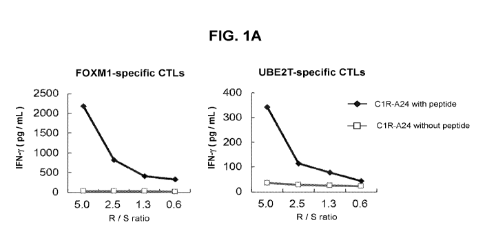
Figures 1A-C. Induction of FOXM1- and UBE2T-derived peptides-specific CTLs and
cytotoxic activity of established CTLs. (A) IFN-y production by FOXM1- and
UBE2T-
specific CTLs was confirmed only when exposed with C1R-A24 cells stimulated
with
FOXM1- or UBE2T-specific peptides. R/S ratio indicates responder cells
(CTLs)/stimulators
(C1R-A24 cells) ratio. (B, C) FOXM1- (B) and UBE2T-specific CTLs (C) exerted
significant
cell killing effect against HLA-A*24:02 positive SW480 cells, but not against
HLA-A*24:02
negative HCC1143 cells or BT549 cells. Both CTLs (2 x 10⁵ cells/well) were
coincubated
with cancer cells (2 x 10⁴ cells/well) for 5h.
Figures 2A-B. Generation of TCR-engineered T cells for FOXM1 and UBE2T. (A)
The distribution of TCRA and TCRB CDR3 clonotypes of FOXM1- and UBE2T-specific
CTLs is presented in pie chart with CDR3 sequences. Black color indicates
portion of CDR3
clonotypes below the read frequency of 1%. This population contained only one
dominant
clonotype for TCRA and TCRB. (B) The transduced efficiency was examined by
staining for
CD8 and TCRA38 (FOXM1 TCR-engineered T cells) or TCRA313 (UBE2T TCR-engineered
T cells). Flow cytometry figures are representative of FOXM1- or UBE2T-TCR
engineered T
cells.
Figure 3A-H. Cytotoxic activity of TCR-engineered T cells for FOXM1 and UBE2T.
(A, B) TCR-engineered T cells for FOXM1 (A) and UBE2T (B) exerted significant
cell
killing effect against HLA-A*24:02 positive SW480 cells, but not against HLA-
A*24:02
negative HCC1143 cells. (C, D) The time course of cancer cells viability
cocultured with
FOXM1 (C) and UBE2T TCR-engineered T cells (D). Both sorted TCR-engineered T
cells (4
x 105 cells/well) were coincubated with cancer cells (2 x 10⁴ cells/well) for
20h. (E, F)
Recognition of TCR-engineered T cells for FOXM1 and UBE2T stimulated with C1R-
A24
cells when pulsed with or without FOXM1- (E) or UBE2T-specific peptide (F) in
ELISPOT
assay. Sorted TCR-engineered T cells (5 x 10⁴ cells/well) were coincubated
with peptide-
5
CA 03077085 2020-03-25
WO 2019/071164
PCT/US2018/054664
pulsed stimulator cells (2 x 10⁴ cells/well) at 37 C for 20 h in 96-well
plates. (G, H) The
secreted protein levels of granzyme B and perforin from original specific CTLs
or TCR-
engineered T cells after cocultured with cancer cells at Oh, 2.5 h and 5 h.
Figure 4. FOXM1 and UBE2T protein expression in cancer cells. Expression of
endogenous FOXM1 and UBE2T protein in HLA-A*24:02 positive or negative cancer
cell
lines examined by western blot analysis.
DEFINITIONS
The terminology used herein is for the purpose of describing the particular
embodiments only, and is not intended to limit the scope of the embodiments
described
herein. Unless otherwise defined, all technical and scientific terms used
herein have the same
meaning as commonly understood by one of ordinary skill in the art to which
this invention
belongs. However, in case of conflict, the present specification, including
definitions, will
control. Accordingly, in the context of the embodiments described herein, the
following
definitions apply.
As used herein and in the appended claims, the singular forms "a", "an" and
"the"
include plural reference unless the context clearly dictates otherwise. Thus,
for example,
reference to "an engineered lymphocyte" is a reference to one or more
engineered
lymphocytes and equivalents thereof known to those skilled in the art, and so
forth.
As used herein, the term "comprise" and linguistic variations thereof denote
the
presence of recited feature(s), element(s), method step(s), etc. without the
exclusion of the
presence of additional feature(s), element(s), method step(s), etc.
Conversely, the term
"consisting of" and linguistic variations thereof, denotes the presence of
recited feature(s),
element(s), method step(s), etc. and excludes any unrecited feature(s),
element(s), method
step(s), etc., except for ordinarily-associated impurities. The phrase
"consisting essentially
of" denotes the recited feature(s), element(s), method step(s), etc. and any
additional
feature(s), element(s), method step(s), etc. that do not materially affect the
basic nature of the
composition, system, or method. Many embodiments herein are described using
open
"comprising" language. Such embodiments encompass multiple closed "consisting
of"
and/or "consisting essentially of" embodiments, which may alternatively be
claimed or
described using such language.
As used herein, an "immune response" refers to the action of a cell of the
immune
system (e.g., T lymphocytes, B lymphocytes, natural killer (NK) cells,
macrophages,
eosinophils, mast cells, dendritic cells, neutrophils, etc.) and soluble
macromolecules
6
CA 03077085 2020-03-25
WO 2019/071164
PCT/US2018/054664
produced by any of these cells or the liver (including antibodies, cytokines,
and complement)
that results in selective targeting, binding to, damage to, destruction of,
and/or elimination
from a subject of invading pathogens, cells or tissues infected with
pathogens, or cancerous
or other abnormal cells. Some embodiments herein comprise generating an immune
response
in a subject to treat cancer.
As used herein, the term "immunotherapy" refers to the treatment or prevention
of a
disease or condition by a method comprising inducing, enhancing, suppressing
or otherwise
modifying an immune response. Some embodiments herein comprise
immunotherapies.
As used herein, the terms "adoptive immunotherapy" and "adoptive cell
transfer"
refer to the transfer of immunocompetent cells (e.g., TCR-engineered T cells)
for the
treatment of cancer or infectious diseases (June, C. H., ed., 2001, In: Cancer
Chemotherapy
and Biotherapy: Principles and Practice, Lippincott Williams & Wilkins,
Baltimore;
Vonderheide et al., 2003, Immun. Research 27:1-15; incorporated by reference
in its
entirety). Some embodiments herein comprise adoptive immunotherapy.
As used herein, the term "cancer vaccine" refers to a composition (e.g., a
tumor
antigen) that elicits a specific immune response. The response is elicited
from the subject's
own immune system by administering the cancer vaccine.
As used herein, the term "native immune cell" refers to an immune cell that
naturally
occurs in the immune system of a subject. Illustrative examples include, but
are not limited
to, T-cells, NK cells, NKT cells, B cells, and dendritic cells. Some
embodiments herein
comprise eliciting a response in a subject to the subject native immune cells.
As used herein, the term "engineered immune cell" refers to an immune cell
(e.g., T-
cell, NK cell, NKT cell, B cell, dendritic cell, etc.) that is genetically
modified. Some
embodiments herein comprise generating and/or administering engineered immune
cells.
As used herein, the term "T-cell receptor" ("TCR") refers to a molecular
complex
found on the surface of T cells (T lymphocytes) that is responsible for
recognizing antigen
fragments bound to major histocompatibility complex (MHC) of antigen
presenting cells. The
binding between TCR and antigen peptides is of relatively low affinity and is
degenerate: that
is, many TCRs recognize the same antigen peptide and many antigen peptides are
recognized
by the same TCR. The TCR is a heterodimer composed of two different protein
chains. In
95% of human T cells, the TCR is made up of an alpha (a) chain and a beta (0)
chain
(encoded by TRA and TRB, respectively), whereas in 5% of T cells the TCR is
made up of
gamma and delta (y/.5) chains (encoded by TRG and TRD, respectively). When the
TCR
engages with an antigenic peptide and the MHC, the T lymphocyte is activated
through signal
7
CA 03077085 2020-03-25
WO 2019/071164
PCT/US2018/054664
transduction. Some embodiments herein comprise generating engineered TCR,
preparing
cells displaying engineered TCR, and/or administering cells displaying
engineered TCR to a
subject for the treatment of cancer.
As used herein, the term "human leukocyte antigen" ("HLA") refers to the major
histocompatibility complex (MHC) proteins in humans or the gene complex
encoding the
human MHC proteins.
As used herein, the term "antibody" refers to a whole antibody molecule or a
fragment thereof (e.g., fragments such as Fab, Fab', and F(ab1)2), it may be a
polyclonal or
monoclonal antibody, a chimeric antibody, a humanized antibody, a human
antibody, etc. As
used herein, when an antibody or other entity "specifically recognizes" or
"specifically
binds" an antigen or epitope, it preferentially recognizes the antigen in a
complex mixture of
proteins and/or macromolecules, and binds the antigen or epitope with affinity
which is
substantially higher than to other entities not displaying the antigen or
epitope. In this regard,
"affinity which is substantially higher" means affinity that is high enough to
enable detection
of an antigen or epitope which is distinguished from entities using a desired
assay or
measurement apparatus. Typically, it means binding affinity having a binding
constant (Ka)
of at least 107 M-1 (e.g., >107 M-1, >108 M-1, >109M-1, >1010 M-1, >1011 M-1,
>1012M-1, >1013
M-1, etc.). In certain such embodiments, an antibody is capable of binding
different antigens
so long as the different antigens comprise that particular epitope. In certain
instances, for
example, homologous proteins from different species may comprise the same
epitope. Some
embodiments herein comprise generating and/or administering antibodies that
bind
oncoantigens and/or neoantigens.
As used herein, the term "antibody fragment" refers to a portion of a full-
length
antibody, including at least a portion antigen binding region or a variable
region. Antibody
fragments include, but are not limited to, Fab, Fab', F(ab1)2, Fv, scFv, Fd,
diabodies, and other
antibody fragments that retain at least a portion of the variable region of an
intact antibody.
See, e.g., Hudson et al. (2003) Nat. Med. 9:129-134; herein incorporated by
reference in its
entirety. In certain embodiments, antibody fragments are produced by enzymatic
or chemical
cleavage of intact antibodies (e.g., papain digestion and pepsin digestion of
antibody)
produced by recombinant DNA techniques, or chemical polypeptide synthesis. For
example,
a "Fab" fragment comprises one light chain and the CHi and variable region of
one heavy
chain. The heavy chain of a Fab molecule cannot form a disulfide bond with
another heavy
chain molecule. A "Fab" fragment comprises one light chain and one heavy chain
that
comprises additional constant region, extending between the CHi and CH2
domains. An
8
CA 03077085 2020-03-25
WO 2019/071164
PCT/US2018/054664
interchain disulfide bond can be formed between two heavy chains of a Fab'
fragment to form
a "F(ab1)2" molecule. An "Fv" fragment comprises the variable regions from
both the heavy
and light chains, but lacks the constant regions. A single-chain Fv (scFv)
fragment comprises
heavy and light chain variable regions connected by a flexible linker to form
a single
polypeptide chain with an antigen-binding region. Exemplary single chain
antibodies are
discussed in detail in WO 88/01649 and U.S. Pat. Nos. 4,946,778 and 5,260,203;
herein
incorporated by reference in their entireties. In certain instances, a single
variable region
(e.g., a heavy chain variable region or a light chain variable region) may
have the ability to
recognize and bind antigen. Other antibody fragments will be understood by
skilled artisans.
Some embodiments herein comprise generating and/or administering antibody
fragments that
bind oncoantigens and/or neoantigens.
As used herein, the term "monoclonal antibody" refers to an antibody which is
a
member of a substantially homogeneous population of antibodies that
specifically bind to the
same epitope. In certain embodiments, a monoclonal antibody is secreted by a
hybridoma. In
certain such embodiments, a hybridoma is produced according to certain methods
known to
those skilled in the art. See, e.g., Kohler and Milstein (1975) Nature 256:
495-499; herein
incorporated by reference in its entirety. In certain embodiments, a
monoclonal antibody is
produced using recombinant DNA methods (see, e.g., U.S. Pat. No. 4,816,567).
In certain
embodiments, a monoclonal antibody refers to an antibody fragment isolated
from a phage
display library. See, e.g., Clackson et al. (1991) Nature 352: 624-628; and
Marks et al.
(1991) J. Mol. Biol. 222: 581-597; herein incorporated by reference in their
entireties. The
modifying word "monoclonal" indicates properties of antibodies obtained from a
substantially-homogeneous population of antibodies, and does not limit a
method of
producing antibodies to a specific method. For various other monoclonal
antibody production
techniques, see, e.g., Harlow and Lane (1988) Antibodies: A Laboratory Manual
(Cold
Spring Harbor Laboratory, Cold Spring Harbor, N.Y.); herein incorporated by
reference in its
entirety. Some embodiments herein comprise generating and/or administering
monoclonal
antibodies that bind oncoantigens and/or neoantigens.
The term "antigen-binding site" refers to a portion of an antibody capable of
specifically binding an antigen. In certain embodiments, an antigen-binding
site is provided
by one or more antibody variable regions.
The term "epitope" refers to any polypeptide determinant capable of
specifically
binding to an immunoglobulin or a T-cell or B-cell receptor. In certain
embodiments, an
epitope is a region of an antigen that is specifically bound by an antibody.
In certain
9
CA 03077085 2020-03-25
WO 2019/071164
PCT/US2018/054664
embodiments, an epitope may include chemically active surface groupings of
molecules such
as amino acids, sugar side chains, phosphoryl, or sulfonyl groups. In certain
embodiments, an
epitope may have specific three dimensional structural characteristics (e.g.,
a
"conformational" epitope) and/or specific charge characteristics.
An epitope is defined as "the same" as another epitope if a particular
antibody
specifically binds to both epitopes. In certain embodiments, polypeptides
having different
primary amino acid sequences may comprise epitopes that are the same. In
certain
embodiments, epitopes that are the same may have different primary amino acid
sequences.
Different antibodies are said to bind to the same epitope if they compete for
specific binding
to that epitope.
As used herein, the term "sequence identity" refers to the degree to which two
polymer sequences (e.g., peptide, polypeptide, nucleic acid, etc.) have the
same sequential
composition of monomer subunits. The term "sequence similarity" refers to the
degree with
which two polymer sequences (e.g., peptide, polypeptide, nucleic acid, etc.)
have similar
polymer sequences. For example, similar amino acids are those that share the
same
biophysical characteristics and can be grouped into the families (see above).
The "percent
sequence identity" (or "percent sequence similarity") is calculated by: (1)
comparing two
optimally aligned sequences over a window of comparison (e.g., the length of
the longer
sequence, the length of the shorter sequence, a specified window, etc.), (2)
determining the
number of positions containing identical (or similar) monomers (e.g., same
amino acids
occurs in both sequences, similar amino acid occurs in both sequences) to
yield the number of
matched positions, (3) dividing the number of matched positions by the total
number of
positions in the comparison window (e.g., the length of the longer sequence,
the length of the
shorter sequence, a specified window), and (4) multiplying the result by 100
to yield the
percent sequence identity or percent sequence similarity. For example, if
peptides A and B
are both 20 amino acids in length and have identical amino acids at all but 1
position, then
peptide A and peptide B have 95% sequence identity. If the amino acids at the
non-identical
position shared the same biophysical characteristics (e.g., both were acidic),
then peptide A
and peptide B would have 100% sequence similarity. As another example, if
peptide C is 20
amino acids in length and peptide D is 15 amino acids in length, and 14 out of
15 amino acids
in peptide D are identical to those of a portion of peptide C, then peptides C
and D have 70%
sequence identity, but peptide D has 93.3% sequence identity to an optimal
comparison
window of peptide C. For the purpose of calculating "percent sequence
identity" (or "percent
sequence similarity") herein, any gaps in aligned sequences are treated as
mismatches at that
CA 03077085 2020-03-25
WO 2019/071164
PCT/US2018/054664
position. In some embodiments, peptides or polypeptides herein comprise a
minimum
sequence identity to a base sequence.
The term "effective dose" or "effective amount" refers to an amount of an
agent, e.g.,
an antibody, that results in the reduction of symptoms in a patient or results
in a desired
biological outcome. In certain embodiments, an effective dose or effective
amount is
sufficient to treat or reduce symptoms of a disease or condition.
As used herein, the terms "administration" and "administering" refer to the
act of
giving a drug, prodrug, or other agent, or therapeutic to a subject or in
vivo, in vitro, or ex
vivo cells, tissues, and organs. Exemplary routes of administration to the
human body can be
through space under the arachnoid membrane of the brain or spinal cord
(intrathecal), the
eyes (ophthalmic), mouth (oral), skin (topical or transdermal), nose (nasal),
lungs (inhalant),
oral mucosa (buccal), ear, rectal, vaginal, by injection (e.g., intravenously,
subcutaneously,
intratumorally, intraperitoneally, etc.) and the like.
The term "treatment" encompasses both therapeutic and
prophylactic/preventative
measures unless otherwise indicated. Those in need of treatment include, but
are not limited
to, individuals already having a particular condition as well as individuals
who are at risk of
acquiring a particular condition or disorder (e.g., those having a genetic or
epigenetic
predisposition; based on age, gender, lifestyle, etc.). The term "treating"
refers to
administering an agent to a subject for therapeutic and/or
prophylactic/preventative purposes.
A "therapeutic agent" refers to an agent that may be administered in vivo to
bring
about a therapeutic and/or prophylactic/preventative effect.
A "therapeutic antibody" refers to an antibody that may be administered in
vivo to
bring about a therapeutic and/or prophylactic/preventative effect.
As used herein, the terms "co-administration" and "co-administering" refer to
the
administration of at least two agent(s) or therapies to a subject. In some
embodiments, the co-
administration of two or more agents or therapies is concurrent. In other
embodiments, a first
agent/therapy is administered prior to a second agent/therapy. Those of skill
in the art
understand that the formulations and/or routes of administration of the
various agents or
therapies used may vary. The appropriate dosage for co-administration can be
readily
determined by one skilled in the art. In some embodiments, when agents or
therapies are co-
administered, the respective agents or therapies are administered at lower
dosages than
appropriate for their administration alone. Thus, co-administration is
especially desirable in
embodiments where the co-administration of the agents or therapies lowers the
requisite
dosage of a potentially harmful (e.g., toxic) agent(s), and/or when co-
administration of two or
11
CA 03077085 2020-03-25
WO 2019/071164
PCT/US2018/054664
more agents results in sensitization of a subject to beneficial effects of one
of the agents via
co-administration of the other agent.
As used herein, the term "pharmaceutical composition" refers to the
combination of
an active agent (e.g., binding agent) with a carrier, inert or active, making
the composition
especially suitable for diagnostic or therapeutic use in vitro, in vivo or ex
vivo.
The terms "pharmaceutically acceptable" or "pharmacologically acceptable," as
used herein,
refer to compositions that do not substantially produce adverse reactions,
e.g., toxic, allergic,
or immunological reactions, when administered to a subject.
As used herein, the term "pharmaceutically acceptable carrier" refers to any
of the
standard pharmaceutical carriers including, but not limited to, phosphate
buffered saline
solution, water, emulsions (e.g., such as an oil/water or water/oil
emulsions), and various
types of wetting agents, any and all solvents, dispersion media, coatings,
sodium lauryl
sulfate, isotonic and absorption delaying agents, disintegrants (e.g., potato
starch or sodium
starch glycolate), and the like. The compositions also can include stabilizers
and
preservatives. For examples of carriers, stabilizers and adjuvants, see, e.g.,
Martin,
Remington's Pharmaceutical Sciences, 15th Ed., Mack Publ. Co., Easton, Pa.
(1975),
incorporated herein by reference in its entirety.
As used herein, the term "healthy donor" refers to a mammal, such a human, who
does not suffer from any form of cancer and/or whose cells/tissues that are
used in
embodiments herein do not show any signs of cancer (e.g., cancer morphology,
cancer
biomarkers, etc.).
DETAILED DESCRIPTION
Provided herein are methods to identify TCR-recognizing cancer-specific
antigens,
and TCR-engineered T cells having antigen-specific cytotoxic activity.
Pre-existing cytotoxic T lymphocytes (CTLs) recognizing cancer-specific
antigens
(oncoantigens and neoantigens) in tumor or blood circulation play critical
roles to achieve a
beneficial clinical response to cancer immunotherapy. For instance, a higher
number of
somatic mutations may increase a chance of generating a larger number of
immunogenic
neoantigens that could be recognized by lymphocytes with high cytolytic
activity, which may
be further unleashed by immune checkpoint inhibitors (Refs. 2-5; incorporated
by reference
in their entireties). In addition, higher expression levels of programmed
death-ligand 1 (PD-
L1), that interacts with programmed death-1 (PD-1) in T cells, in cancer cells
was
upregulated and is a biomarker for good clinical response (Refs. 4, 6-8;
incorporated by
12
CA 03077085 2020-03-25
WO 2019/071164
PCT/US2018/054664
reference in their entireties). Furthermore, tumor-infiltrating lymphocytes
(TILs) in patients
who responded to adoptive TILs transfer therapy include CTLs targeting both
neoantigens
and oncoantigens (shared antigens) (Ref 9; incorporated by reference in its
entirety).
To enhance CTL-mediated anti-tumor immune responses for further improvement in
clinical outcomes of cancer immunotherapy, embodiments herein utilize cancer-
specific
antigens, oncoantigens and neoantigens, as vaccines to activate antigen-
specific CTLs in
cancer patients. Oncoantigens are immunogenic peptides derived from oncogenic
proteins
that are highly expressed in cancer cells but not expressed in normal organs,
except testis or
fetal organs (Ref 10; incorporated by reference in its entirety). It has been
contemplated that
immunogenic peptide epitopes derived from oncoantigens induce oncoantigen-
specific CTLs
and improve the prognosis of cancer patients (Refs. 11-13; incorporated by
reference in their
entireties). Neoantigens are immunogenic peptides derived from non-synonymous
mutations
in cancer cells (Ref 10; incorporated by reference in its entirety).
Considering some evidence
that neoantigen-specific T cells showed good clinical outcome (Refs. 14-15;
incorporated by
reference in their entireties), neoantigen vaccine provides an option to
further activate anti-
cancer immune responses in patients. However, since induction of a sufficient
number of
anti-tumor T cells with vaccine therapy often occur very gradually and needs
several months,
this vaccine approach does not work for patients with a large tumor burden.
Hence,
identification of cancer antigen-specific T cell receptor (TCR), generation of
TCR-engineered
T cells using autologous T lymphocytes, and infusion of such genetically
engineered T cells
with/without anti-immune checkpoint antibodies provide attractive options for
patients with
advanced tumors where the host immune system was usually suppressed
significantly.
Preclinical studies and recent clinical trials have showed encouraging results
that
oncoantigen/neoantigen-specific TCR-engineered T cells are even effective for
a large size of
solid tumors (Refs. 16-18; incorporated by reference in their entireties).
Provided herein are
rapid screening methods to identify TCR sequences that recognize neoantigens
and the rapid
preparation of personalized TCR-engineered T cell therapies therewith.
Experiments were conducted during development of embodiments herein to
establish
a rapid screening method to detect oncoantigen/neoantigen-specific TCRs. After
in vitro
stimulation of CD8+ T lymphocytes from healthy donors with candidate peptides,
CD8+ T
cells were sorted using an HLA class I dextramer with each peptide, and TCR
sequences for
these cells were determined. Mono- or oligo-clonal expansion of unique T cells
was achieved
by stimulation of the epitope peptides. The TCR cDNAs were cloned and TCR-
engineered T
cells were generated. Through this approach, two antigen-specific CD8+ T cell
clones were
13
CA 03077085 2020-03-25
WO 2019/071164
PCT/US2018/054664
generated; two of the T-cell clones, which recognize oncoantigens derived from
FOXM1 and
UBE2T, revealed strong cytotoxic activity against HLA-matched cancer cells
expressing
target proteins, but not against HLA-unmatched cancer cells. The methods
described herein
allow for the rapid identification of TCR-recognizing cancer-specific antigens
after obtaining
antigen peptides. The approach allows for the rapid development of
personalized T-cell
immunotherapies for treating cancer. Provided herein is a pipeline to identify
TCR-
recognizing cancer-specific antigens by integrating the in vitro neoantigen
stimulation of T
cells, dextramer sorting, and TCR sequencing using next-generation sequencers
as well as to
establish TCR-engineered T cells having antigen-specific cytotoxic activity.
Experiments conducted during development of embodiments herein to develop a
pipeline from screening of putative oncoantigen/neoantigen-derived peptides to
induction of
specific T cells from peripheral blood mononuclear cells (PBMCs) of healthy
donors, and
also established antigen-specific TCR-engineered T cells. Throughout this
pipeline,
immunogenic oncoantigens/neoantigens-derived peptides are identified as useful
in cancer
.. vaccines, and oncoantigen/neoantigen-specific TCRs are identified which
lead to the
establishment of antigen-specific TCR-engineered T cells to observe cytotoxic
activity
against HLA-matched cancer cells.
As a source of PBMCs, PBMCs from healthy donors allow detection of candidates
for
oncoantigens/neoantigens-specific CTLs, because they have different T cell
repertoires from
that of cancer patients. T cells obtained from healthy donors broaden
neoantigen-specific T
cell reactivity and enable targeting of neoantigens that have not been
recognized by the
patients' own immune system (Ref 30; incorporated by reference in its
entirety). In some
embodiments, after identification of TCR recognizing cancer-specific antigens,
TCR-
engineered T cells are established from autologous T cells from patients and
infused as an
adoptive cell transfer therapy.
TCR-engineered T cells, generated using the methods described herein, using
PBMCs
from HLA-A*24:02-positive healthy donors, recognized only HLA-A*24:02
restricted
peptides and showed significant cytotoxic activity against the HLA-A*24:02
matched cancer
cells. Considering that TCR-engineered T cells targeting HLA-A*02:01
restricted NY-ESO-
1-derived peptide using autologous PBMCs showed encouraging clinical responses
in
myeloma patients (Ref 21; incorporated by reference in its entirety), it is
noteworthy that the
TCR-engineered T cells from healthy donors herein also exerted cytotoxic
activity against
HLA-A matched cancer cells. These results demonstrate the feasibility of
preparing TCR-
14
CA 03077085 2020-03-25
WO 2019/071164
PCT/US2018/054664
engineered T cells from healthy donors, for example, in situations in which
obtaining
autologous T cells from patients in unfeasible.
The pipeline described herein provides personalized immunotherapies responding
to
both oncoantigens and neoantigens. Given that some oncoantigens are frequently
overexpressed in many types of cancer, TCR-engineered T cell therapy targeting
oncoantigens is reasonable because identified TCRs recognizing specific
oncoantigens have
broad utility for patients having the same HLA genotype. For instance,
elevated FOXM1 or
UBE2T expression in tumor tissues was correlated with poor survival of
patients with breast
cancer, colon cancer, and prostate cancer (Refs. 31-34; incorporated by
reference in its
entirety). Therefore, in some embodiments, the FOXM1- and UBE2T-specific TCR
engineered T cells describe herein find use in adoptive transfer therapies.
Given that clinical
benefit of chimeric antigen receptor (CAR) T cell therapy and TCR-engineered T
cell therapy
are currently limited to hematological malignancies (Refs. 35-36; incorporated
by reference
in their entireties), the pipeline presented herein for oncoantigen-specific
TCR-engineered T
cells provide another adoptive cell transfer therapy for solid tumors. In
contrast, neoantigens
are more specific to cancer cells and regarded as attractive immune targets,
although their
presentation is dependent on somatic mutations of cancer cell. Considering
that the transfer
therapy of neoantigen-specific TILs already showed encouraging clinical
results against not
only melanoma but solid tumors (Refs. 14-15; incorporated by reference in
their entireties),
the TCR-engineered T cells for neoantigens described herein provide a therapy
in the clinical
settings.
In some embodiments, provided herein are methods for identifying sequences of
immune active TCR comprising stimulating target lymphocytes (e.g., CD8+
cytotoxic T
lymphocytes) with a stimulation peptide comprising candidate antigen sequence.
In some
embodiments, stimulation peptides are fragments of proteins that are expressed
on cancer
and/or tumor cells. In some embodiments, stimulation peptides are fragments of
cancer-
specific antigens and/or tumor-specific antigens. In some embodiments, the
target T
lymphocytes are obtained from any suitable source (e.g., a donor, cell
culture, etc.). In some
embodiments, the target T lymphocytes are obtained from a healthy donor. In
some
embodiments, the target T lymphocytes are CD8+ cytotoxic T lymphocytes. In
some
embodiments, the stimulating is performed in vitro (e.g., in cell culture). In
some
embodiments, the type of cell culture is determined by the type of target T
lymphocytes.
Suitable conditions and methods for culturing T lymphocytes and stimulating T
lymphocytes
with a stimulation peptide are understood in the field.
CA 03077085 2020-03-25
WO 2019/071164
PCT/US2018/054664
In some embodiments, the target T lymphocytes (e.g., CD8+ cytotoxic T
lymphocytes) are a population of target T lymphocytes, and the stimulation
peptide is one of
a population of stimulation peptides comprising different candidate antigen
sequences; and
wherein said capturing comprises contacting the population of immune-active T
lymphocytes
(e.g., CD8+ cytotoxic T lymphocytes) with a capture reagents that displays
major
histocompatibility complex (MHC) bound to a population of capture peptides
comprising the
candidate antigen sequences.
In some embodiments, the target lymphocytes are CD8+ cytotoxic lymphocytes,
CD4+
helper lymphocytes, NK cells, NKT cells, B cells, dendritic cells, etc.
In some embodiments, the stimulation peptide comprising a candidate antigen
sequence is all or a fragment of an oncoantigen and neoantigen. In some
embodiments, a
candidate antigen sequence is all or a fragment of an oncoantigen and
neoantigen. In some
embodiments, the stimulation peptide comprises a random amino acid sequence
and methods
herein allow for identification of peptides capable of eliciting an immune
response. In some
embodiments, the stimulation peptide comprises an amino acid sequence selected
from the
group consisting of SEQ ID NO: 1, SEQ ID NO: 2, SEQ ID NO: 3, SEQ ID NO: 4,
SEQ ID
NO: 5, SEQ ID NO: 6, SEQ ID NO: 7, SEQ ID NO: 8, SEQ ID NO: 9, SEQ ID NO: 10,
SEQ
ID NO: 11, SEQ ID NO: 12, SEQ ID NO: 13, SEQ ID NO: 14, SEQ ID NO: 15, SEQ ID
NO: 16, SEQ ID NO: 17, SEQ ID NO: 18, SEQ ID NO: 19, SEQ ID NO: 20, SEQ ID NO:
21, SEQ ID NO: 22, SEQ ID NO: 23, SEQ ID NO: 24, SEQ ID NO: 25, SEQ ID NO: 26,
SEQ ID NO: 27, SEQ ID NO: 28, SEQ ID NO: 29, SEQ ID NO: 30, SEQ ID NO: 31, SEQ
ID NO: 32, SEQ ID NO: 33, SEQ ID NO: 34, SEQ ID NO: 35, SEQ ID NO: 36, SEQ ID
NO: 37, SEQ ID NO: 38, SEQ ID NO: 39, SEQ ID NO: 40, SEQ ID NO: 41, SEQ ID NO:
42, SEQ ID NO: 43, and SEQ ID NO: 44.
In some embodiments, after stimulating the target T lymphocytes with a
stimulation
peptide, immune-active T lymphocytes (e.g., CD8+ cytotoxic T lymphocytes) with
T-cell
receptor (TCR) that binds to the stimulation peptide are captured. In some
embodiments,
capturing comprises contacting the immune-active T lymphocytes with a capture
reagent that
displays major histocompatibility complex (MHC) bound to a capture peptide
comprising the
.. candidate antigen sequence. In some embodiments, the capture reagent
displays a peptide
comprising the sequence of one or more of the stimulation peptides. In some
embodiments,
the peptide is added to the T lymphocytes in a form bound to a MHC I complex.
In some
embodiments, the capture reagent is an MHC multimer. In some embodiments, the
MHC
16
CA 03077085 2020-03-25
WO 2019/071164
PCT/US2018/054664
multimer is an MHC dextramer. For example, the peptide may be presented to the
T
lymphocytes bound to MHC dextramers. In some embodiments, MHC dextramers are
fluorescently-labeled MHC multimers bound to a dextrose backbone. The use of
multimeric
MHC structures has the advantage that multiple copies of the peptide are
presented thereby
increasing the capture potential.
In some embodiments, after capture of the immune-active T lymphocytes, all or
a
portion of the TCR of the captured immune-active T lymphocytes is sequenced.
In some
embodiments, the sequencing comprises a next-generation sequencing technique.
Next-
generation sequencing techniques are described in more detail below. In some
embodiments,
the portion of the TCR sequenced comprises the TCR-a and/or TCR-r3 chains. In
some
embodiments, the portion of the TCR sequenced comprises one or more
complementarity
determining regions (CDRs) of the TCR-a and/or TCR-r3 chains. In some
embodiments, the
portion of the TCR sequenced comprises the CDR3 of the TCR-a and/or TCR-r3
chains.
In some embodiments, provided herein are TCR-recognizing cancer-specific
antigens
identified by the methods described herein (e.g., SEQ ID NO: 1, SEQ ID NO: 2,
etc.). In
some embodiments, the cancer-specific antigens identified by the methods
described herein
are employed as therapeutics, such as cancer vaccines. Delivery systems for
cancer vaccines
may include, for example, liposomes, systems made of cholesterol, cholesterol
hemisuccinate
or alpha-tochoferol (e.g., vitamin E), or other amphipathic molecules in which
modified or
synthesized neoantigens can attach or insert. In some embodiments, a cancer
vaccine
comprises a cancer-specific antigen identified by the methods herein of
variants thereof In
some embodiments, a cancer-specific antigen is provided as fusion peptide. In
some
embodiments, incorporates multiple sequences identified in the methods herein.
In some
embodiments, the peptide used in a cancer vaccine is 10-80 amino acids in
length (e.g., 10,
20, 30, 40, 50, 60, 70, 80, or ranges therebetween).
In some embodiments, provided herein are therapeutic antibodies that binds to
the
TCR-recognizing cancer-specific antigens described herein. In some
embodiments, a
therapeutic antibody herein is an antibody fragment. Antibodies and antibody
fragments for
use in treatment of cancer are well understood in the field. In some
embodiments, antibodies
are monoclonal antibodies. In some embodiments, antibodies are humanized
antibodies. In
some embodiments, the therapeutic antibodies bind to an antigen comprising an
amino acid
sequence selected from the group consisting of SEQ ID NOS: 1-44. In some
embodiments,
the therapeutic antibodies comprise CDR3 sequences comprising pairs of amino
acid
sequences selected from the group consisting of SEQ ID NOS: 45 and 46, 47 and
48, 49 and
17
CA 03077085 2020-03-25
WO 2019/071164
PCT/US2018/054664
50, 51 and 52, 53 and 54, 55 and 56, 57 and 58, 59 and 60, 61 and 62, 63 and
64, 65 and 66,
67 and 68, 69 and 70, 71 and 72, 73 and 74, 75 and 76, 77 and 78, 79 and 80,
81 and 82, 83
and 84, 85 and 86, 87 and 88, 89 and 90, 91 and 92, 93 and 94, 95 and 96, 97
and 98, 99 and
100, 101 and 102, 103 and 104, 105 and 106, 107 and 108, 109 and 110, 111 and
112, 113
and 114, 115 and 116, 117 and 118, 119 and 120, 121 and 122, 123 and 124, 125
and 126,
127 and 128, 129 and 130, and 131 and 132.
In some embodiments, provided herein are methods for generating engineered T
lymphocytes (e.g., CD8+ cytotoxic T lymphocytes) displaying all or a portion
of a TCR of
captured immune-active T lymphocytes, wherein the engineered T lymphocytes
recognize
.. antigen presenting cells displaying MHC bound to the peptide comprising the
candidate
antigen sequence. In some embodiments, the engineered lymphocytes are CD8+
cytotoxic
lymphocytes, CD4+ helper lymphocytes, NK cells, NKT cells, B cells, dendritic
cells, etc. In
some embodiments, sequences TCR of immune-active T lymphocytes are used to
prepare
nucleic acids and/or vectors encoding TCRs that will recognize target
oncoantigens or
neoantigens. In some embodiments, such nucleic acids and/or vectors are
transformed,
transfected, and/or otherwise placed into T lymphocytes to generate engineered
T
lymphocytes. Nucleic acids, vectors, and methods for such purposes are known
in the field
and described herein. In some embodiments, the engineered T lymphocytes are
CD8+
cytotoxic T lymphocytes. In some embodiments, generating engineered T
lymphocytes
(e.g., CD8+ cytotoxic T lymphocytes) displaying all or a portion of the TCR of
the captured
immune-active T lymphocytes comprising: (i) cloning a nucleic acid sequence
encoding the
portion of the TCR of the captured immune-active T lymphocytes into a vector;
(ii)
introducing the vector into host T lymphocytes (e.g., CD8+ cytotoxic T
lymphocytes); and
(iii) culturing under conditions such that the portion of the TCR of the
captured immune-
active T lymphocytes is expressed and displayed on the engineered T
lymphocytes. In some
embodiments, the portion of the TCR comprises the TCR-a and/or TCR-r3 chains.
In some
embodiments, the portion of the TCR comprises one or more complementarity
determining
regions (CDRs) of the TCR-a and/or TCR-r3 chains. In some embodiments, the
portion of the
TCR sequenced comprises the CDR3 of the TCR-a and/or TCR-r3 chains. In some
embodiments, the portion of the TCR sequenced comprises an amino acid sequence
selected
from the group consisting of SEQ ID NOS: 45-132. In some embodiments, the
engineered T
lymphocytes display a TCR comprising a and 13 chains comprising the amino acid
sequence
pairs selected from the group consisting of SEQ ID NOS: 45 and 46,47 and 48,49
and 50, 51
and 52, 53 and 54, 55 and 56, 57 and 58, 59 and 60, 61 and 62, 63 and 64, 65
and 66, 67 and
18
CA 03077085 2020-03-25
WO 2019/071164
PCT/US2018/054664
68, 69 and 70, 71 and 72, 73 and 74, 75 and 76, 77 and 78, 79 and 80, 81 and
82, 83 and 84,
85 and 86, 87 and 88, 89 and 90, 91 and 92, 93 and 94, 95 and 96, 97 and 98,
99 and 100, 101
and 102, 103 and 104, 105 and 106, 107 and 108, 109 and 110, 111 and 112, 113
and 114,
115 and 116, 117 and 118, 119 and 120, 121 and 122, 123 and 124, 125 and 126,
127 and
128, 129 and 130, and 131 and 132. In some embodiments, the vector is
introduced into host
T lymphocytes from a healthy donor host. In some embodiments, the vector is
introduced
into host T lymphocytes from a cancer patient to be treated with the
engineered T
lymphocytes.
In some embodiments, provided herein are methods for generating engineered T
lymphocytes (e.g., CD8+ cytotoxic T lymphocytes) comprising chimeric antigen
receptors
(CARs), wherein the CARs recognize antigen presenting cells displaying MHC
bound to the
peptide comprising the candidate antigen sequence. In certain embodiments, the
antigen-binding
domain is a single-chain variable fragment (scFv) containing heavy and light
chain variable regions
that bind with specificity to the desired antigen (e.g., variable regions
identified by the methods
herein). In some embodiments, the CAR further comprises a transmembrane domain
(e.g., a T cell
transmembrane domain (e.g., a CD28 transmembrane domain)) and a signaling
domain comprising
one or more immunoreceptor tyrosine-based activation motifs (ITAMs)(e.g., a T
cell receptor
signaling domain (e.g., TCR zeta chain). In some embodiments, the CAR
comprises one or more co-
stimulatory domains (e.g., domains that provide a second signal to stimulate T
cell activation). The
invention is not limited by the type of co-stimulatory domain. In some
embodiments, the
engineered lymphocytes are CD8+ cytotoxic lymphocytes, CD4+ helper
lymphocytes, NK
cells, NKT cells, B cells, dendritic cells, etc. In some embodiments, TCR
sequences of
immune-active T lymphocytes are used to prepare CARs that will recognize
target
oncoantigens or neoantigens. In some embodiments, nucleic acids and/or vectors
encoding
such CARs are transformed or transfected into T cells, and/or the CARs are
otherwise placed
into T lymphocytes to generate engineered T lymphocytes. Nucleic acids,
vectors, and
methods for such purposes are known in the field and described herein. In some
embodiments, the engineered T lymphocytes are CD8+ cytotoxic T lymphocytes. In
some
embodiments, the CAR comprises an antigen binding region comprising sequences
of the
TCR-a and/or TCR-r3 chains identified in the methods herein. In some
embodiments, the
CAR comprises one or more complementarity determining regions (CDRs) of the
TCR-a
and/or TCR-r3 chains. In some embodiments, the portion of the TCR sequenced
comprises
the CDR3 of the TCR-a and/or TCR-r3 chains. In some embodiments, the portion
of the TCR
sequenced comprises an amino acid sequence selected from the group consisting
of SEQ ID
19
CA 03077085 2020-03-25
WO 2019/071164
PCT/US2018/054664
NOS: 45-132. In some embodiments, the engineered T lymphocytes display a TCR
comprising a and 13 chains comprising the amino acid sequence pairs selected
from the group
consisting of SEQ ID NOS: 45 and 46, 47 and 48, 49 and 50, 51 and 52, 53 and
54, 55 and
56, 57 and 58, 59 and 60, 61 and 62, 63 and 64, 65 and 66, 67 and 68, 69 and
70, 71 and 72,
.. 73 and 74, 75 and 76, 77 and 78, 79 and 80, 81 and 82, 83 and 84, 85 and
86, 87 and 88, 89
and 90, 91 and 92, 93 and 94, 95 and 96, 97 and 98, 99 and 100, 101 and 102,
103 and 104,
105 and 106, 107 and 108, 109 and 110, 111 and 112, 113 and 114, 115 and 116,
117 and
118, 119 and 120, 121 and 122, 123 and 124, 125 and 126, 127 and 128, 129 and
130, and
131 and 132. In some embodiments, the vector is introduced into host T
lymphocytes from a
healthy donor host. In some embodiments, the vector is introduced into host T
lymphocytes
from a cancer patient to be treated with the engineered T lymphocytes.
In some embodiments, the methods herein are applicable to generating
engineered
lymphocytes, such as CD4+ helper lymphocytes, NK cells, NKT cells, B cells,
dendritic cells,
etc.
In some embodiments, nucleic acids (e.g., TCR cDNAs) are sequenced. Nucleic
acid
molecules may be sequence analyzed by any number of techniques. The analysis
may
identify the sequence of all or a part of a nucleic acid. Illustrative non-
limiting examples of
nucleic acid sequencing techniques include, but are not limited to, chain
terminator (Sanger)
sequencing and dye terminator sequencing, as well as "next generation"
sequencing
techniques. In some embodiments, RNA is reverse transcribed to cDNA before
sequencing.
A number of DNA sequencing techniques are known in the art, including
fluorescence-based sequencing methodologies (See, e.g., Birren et al., Genome
Analysis:
Analyzing DNA, 1, Cold Spring Harbor, N.Y.; herein incorporated by reference
in its
entirety). In some embodiments, automated sequencing techniques understood in
that art are
utilized. In some embodiments, the systems, devices, and methods employ
parallel
sequencing of partitioned amplicons (PCT Publication No: W02006084132 to Kevin
McKernan et al., herein incorporated by reference in its entirety). In some
embodiments,
DNA sequencing is achieved by parallel oligonucleotide extension (See, e.g.,
U.S. Pat. No.
5,750,341 to Macevicz et al., and U.S. Pat. No. 6,306,597 to Macevicz et al.,
both of which
are herein incorporated by reference in their entireties). Additional examples
of sequencing
techniques include the Church polony technology (Mitra et al., 2003,
Analytical
Biochemistry 320, 55-65; Shendure et al., 2005 Science 309, 1728-1732; U.S.
Pat. No.
CA 03077085 2020-03-25
WO 2019/071164
PCT/US2018/054664
6,432,360, U.S. Pat. No. 6,485,944, U.S. Pat. No. 6,511,803; herein
incorporated by reference
in their entireties) the 454 picotiter pyrosequencing technology (Margulies et
al., 2005 Nature
437, 376-380; US 20050130173; herein incorporated by reference in their
entireties), the
Solexa single base addition technology (Bennett et al., 2005,
Pharmacogenomics, 6, 373-382;
U.S. Pat. No. 6,787,308; U.S. Pat. No. 6,833,246; herein incorporated by
reference in their
entireties), the Lynx massively parallel signature sequencing technology
(Brenner et al.
(2000). Nat. Biotechnol. 18:630-634; U.S. Pat. No. 5,695,934; U.S. Pat. No.
5,714,330;
herein incorporated by reference in their entireties), the Adessi PCR colony
technology
(Adessi et al. (2000). Nucleic Acid Res. 28, E87; WO 00018957; herein
incorporated by
reference in its entirety), and suitable combinations or alternative thereof
A set of methods referred to as "next-generation sequencing" techniques have
emerged as alternatives to Sanger and dye-terminator sequencing methods
(Voelkerding et
al., Clinical Chem., 55: 641-658, 2009; MacLean et al., Nature Rev.
Microbiol., 7: 287-296;
each herein incorporated by reference in their entirety). Next-generation
sequencing (NGS)
methods share the common feature of massively parallel, high-throughput
strategies, with the
goal of lower costs and higher speeds in comparison to older sequencing
methods. NGS
methods can be broadly divided into those that require template amplification
and those that
do not.
Sequencing techniques that finds use in embodiments herein include, for
example,
Helicos True Single Molecule Sequencing (tSMS) (Harris T. D. et al. (2008)
Science
320:106-109). In the tSMS technique, a DNA sample is cleaved into strands of
approximately
100 to 200 nucleotides, and a polyA sequence is added to the 3' end of each
DNA strand.
Each strand is labeled by the addition of a fluorescently labeled adenosine
nucleotide. The
DNA strands are then hybridized to a flow cell, which contains millions of
oligo-T capture
sites that are immobilized to the flow cell surface. The templates can be at a
density of about
100 million templates/cm2. The flow cell is then loaded into a sequencer,
and a laser
illuminates the surface of the flow cell, revealing the position of each
template. A CCD
camera can map the position of the templates on the flow cell surface. The
template
fluorescent label is then cleaved and washed away. The sequencing reaction
begins by
introducing a DNA polymerase and a fluorescently labeled nucleotide. The oligo-
T nucleic
acid serves as a primer. The polymerase incorporates the labeled nucleotides
to the primer in
a template directed manner. The polymerase and unincorporated nucleotides are
removed.
The templates that have directed incorporation of the fluorescently labeled
nucleotide are
detected by imaging the flow cell surface. After imaging, a cleavage step
removes the
21
CA 03077085 2020-03-25
WO 2019/071164
PCT/US2018/054664
fluorescent label, and the process is repeated with other fluorescently
labeled nucleotides
until the desired read length is achieved. Sequence information is collected
with each
nucleotide addition step. Further description of tSMS is shown for example in
Lapidus et al.
(U.S. Pat. No. 7,169,560), Lapidus et al. (U.S. patent application number
2009/0191565),
Quake et al. (U.S. Pat. No. 6,818,395), Harris (U.S. Pat. No. 7,282,337),
Quake et al. (U.S.
patent application number 2002/0164629), and Braslaysky, et al., PNAS (USA),
100: 3960-
3964 (2003), each of which is incorporated by reference in their entireties.
Another example of a DNA sequencing technique that finds use in embodiments
herein is 454 sequencing (Roche) (Margulies, M et al. 2005, Nature, 437, 376-
380;
incorporated by reference in its entirety). 454 sequencing involves two steps.
In the first step,
DNA is sheared into fragments of approximately 300-800 base pairs, and the
fragments are
blunt ended. Oligonucleotide adaptors are then ligated to the ends of the
fragments. The
adaptors serve as primers for amplification and sequencing of the fragments.
The fragments
are attached to DNA capture beads, e.g., streptavidin-coated beads using,
e.g., Adaptor B,
which contains 5'-biotin tag. The fragments attached to the beads are PCR
amplified within
droplets of an oil-water emulsion. The result is multiple copies of clonally
amplified DNA
fragments on each bead. In the second step, the beads are captured in wells
(pico-liter
sized). Pyrosequencing is performed on each DNA fragment in parallel. Addition
of one or
more nucleotides generates a light signal that is recorded by a CCD camera in
a sequencing
instrument. The signal strength is proportional to the number of nucleotides
incorporated. Pyrosequencing makes use of pyrophosphate (PPi) which is
released upon
nucleotide addition. PPi is converted to ATP by ATP sulfurylase in the
presence of adenosine
5' phosphosulfate. Luciferase uses ATP to convert luciferin to oxyluciferin,
and this reaction
generates light that is detected and analyzed.
Another example of a DNA sequencing technique that finds use in embodiments
herein is SOLiD technology (Applied Biosystems). In SOLiD sequencing, genomic
DNA is
sheared into fragments, and adaptors are attached to the 5' and 3' ends of the
fragments to
generate a fragment library. Alternatively, internal adaptors can be
introduced by ligating
adaptors to the 5' and 3' ends of the fragments, circularizing the fragments,
digesting the
circularized fragment to generate an internal adaptor, and attaching adaptors
to the 5' and 3'
ends of the resulting fragments to generate a mate-paired library. Next,
clonal bead
populations are prepared in microreactors containing beads, primers, template,
and PCR
components. Following PCR, the templates are denatured and beads are enriched
to separate
the beads with extended templates. Templates on the selected beads are
subjected to a 3'
22
CA 03077085 2020-03-25
WO 2019/071164
PCT/US2018/054664
modification that permits bonding to a glass slide. The sequence can be
determined by
sequential hybridization and ligation of partially random oligonucleotides
with a central
determined base (or pair of bases) that is identified by a specific
fluorophore. After a color is
recorded, the ligated oligonucleotide is cleaved and removed and the process
is then
repeated.
Another example of a DNA sequencing technique that finds use in embodiments
herein is Ion Torrent sequencing (U.S. patent application numbers
2009/0026082,
2009/0127589, 2010/0035252, 2010/0137143, 2010/0188073, 2010/0197507,
2010/0282617,
2010/0300559), 2010/0300895, 2010/0301398, and 2010/0304982; incorporated by
reference
in their entireties). In Ion Torrent sequencing, DNA is sheared into fragments
of
approximately 300-800 base pairs, and the fragments are blunt ended.
Oligonucleotide
adaptors are then ligated to the ends of the fragments. The adaptors serve as
primers for
amplification and sequencing of the fragments. The fragments can be attached
to a surface
and is attached at a resolution such that the fragments are individually
resolvable. Addition of
one or more nucleotides releases a proton (RE), which signal detected and
recorded in a
sequencing instrument. The signal strength is proportional to the number of
nucleotides
incorporated.
Another example of a DNA sequencing technique that finds use in embodiments
herein is Illumina sequencing. Illumina sequencing is based on the
amplification of DNA on
a solid surface using fold-back PCR and anchored primers. Genomic DNA is
fragmented, and
adapters are added to the 5' and 3' ends of the fragments. DNA fragments that
are attached to
the surface of flow cell channels are extended and bridge amplified. The
fragments become
double stranded, and the double stranded molecules are denatured. Multiple
cycles of the
solid-phase amplification followed by denaturation can create several million
clusters of
approximately 1,000 copies of single-stranded DNA molecules of the same
template in each
channel of the flow cell. Primers, DNA polymerase and four fluorophore-
labeled, reversibly
terminating nucleotides are used to perform sequential sequencing. After
nucleotide
incorporation, a laser is used to excite the fluorophores, and an image is
captured and the
identity of the first base is recorded. The 3' terminators and fluorophores
from each
incorporated base are removed and the incorporation, detection and
identification steps are
repeated.
Another example of a DNA sequencing technique that finds use in embodiments
herein is the single molecule, real-time (SMRT) technology of Pacific
Biosciences. In SMRT,
each of the four DNA bases is attached to one of four different fluorescent
dyes. These dyes
23
CA 03077085 2020-03-25
WO 2019/071164
PCT/US2018/054664
are phospholinked. A single DNA polymerase is immobilized with a single
molecule of
template single stranded DNA at the bottom of a zero-mode waveguide (ZMW). A
ZMW is a
confinement structure which enables observation of incorporation of a single
nucleotide by
DNA polymerase against the background of fluorescent nucleotides that rapidly
diffuse in an
out of the ZMW (in microseconds). It takes several milliseconds to incorporate
a nucleotide
into a growing strand. During this time, the fluorescent label is excited and
produces a
fluorescent signal, and the fluorescent tag is cleaved off Detection of the
corresponding
fluorescence of the dye indicates which base was incorporated. The process is
repeated.
Another example of a DNA sequencing technique that finds use in embodiments
herein involves nanopore sequencing (Soni G V and Meller A. (2007) Clin Chem
53: 1996-
2001; incorporated by reference in its entirety). A nanopore is a small hole,
of the order of 1
nanometer in diameter. Immersion of a nanopore in a conducting fluid and
application of a
potential across it results in a slight electrical current due to conduction
of ions through the
nanopore. The amount of current which flows is sensitive to the size of the
nanopore. As a
DNA molecule passes through a nanopore, each nucleotide on the DNA molecule
obstructs
the nanopore to a different degree. Thus, the change in the current passing
through the
nanopore as the DNA molecule passes through the nanopore represents a reading
of the DNA
sequence.
Another example of a DNA sequencing technique that finds use in embodiments
herein involves using a chemical-sensitive field effect transistor (chemFET)
array to
sequence DNA (for example, as described in US Patent Application Publication
No.
20090026082; incorporated by reference in its entirety). In one example of the
technique,
DNA molecules can be placed into reaction chambers, and the template molecules
can be
hybridized to a sequencing primer bound to a polymerase. Incorporation of one
or more
triphosphates into a new nucleic acid strand at the 3' end of the sequencing
primer can be
detected by a change in current by a chemFET. An array can have multiple
chemFET
sensors. In another example, single nucleic acids can be attached to beads,
and the nucleic
acids can be amplified on the bead, and the individual beads can be
transferred to individual
reaction chambers on a chemFET array, with each chamber having a chemFET
sensor, and
the nucleic acids can be sequenced.
In some embodiments, other sequencing techniques (e.g., NGS techniques)
understood in the field, or alternatives or combinations of the above
techniques find use in
embodiments herein.
24
CA 03077085 2020-03-25
WO 2019/071164
PCT/US2018/054664
Certain embodiments herein comprise the detection of one or more biomarkers
(e.g.,
detection of cytokines (e.g., IFN-y) to detect and/or quantify immune
response). In some
embodiments of the methods, the method further comprises isolating one or more
biomarkers
(e.g., detection of cytokines (e.g., IFN-y) to detect and/or quantify immune
response) from a
biological sample or in vitro culture. In some embodiments, reagents are
provided that bind
to biomarkers. Such reagents are selected from antibodies, antibody fragments,
aptamers, etc.
In some embodiments, the detection method includes an enzyme/substrate
combination that generates a detectable signal that corresponds to the
biomarker level (e.g.,
using the techniques of ELISA, Western blotting, isoelectric focusing).
Generally, the
enzyme catalyzes a chemical alteration of the chromogenic substrate which can
be measured
using various techniques, including spectrophotometry, fluorescence, and
chemiluminescence. Suitable enzymes include, for example, luciferases,
luciferin, malate
dehydrogenase, urease, horseradish peroxidase (HRPO), alkaline phosphatase,
beta-
galactosidase, glucoamylase, lysozyme, glucose oxidase, galactose oxidase, and
glucose-6-
phosphate dehydrogenase, uricase, xanthine oxidase, lactoperoxidase,
microperoxidase, and
the like. In some embodiments, the detection method is a combination of
fluorescence,
chemiluminescence, radionuclide or enzyme/substrate combinations that generate
a
measurable signal. In some embodiments, multimodal signaling has unique and
advantageous characteristics in biomarker assay formats.
In some embodiments, the biomarker presence/levels is detected using any
analytical
methods including, singleplex aptamer assays, multiplexed aptamer assays,
singleplex or
multiplexed immunoassays, expression profiling, mass spectrometric analysis,
histological/cytological methods, etc. as discussed below.
In some embodiments, biomarkers (e.g., detection of cytokines (e.g., IFN-y) to
detect
and/or quantify immune response) are detected/quantified using a suitable
immunoassay.
Immunoassay methods are based on the reaction of an antibody to its
corresponding target or
analyte and can detect the analyte in a sample depending on the specific assay
format. To
improve specificity and sensitivity of an assay method based on immuno-
reactivity,
monoclonal antibodies and fragments thereof are often used because of their
specific epitope
recognition. Polyclonal antibodies have also been successfully used in various
immunoassays because of their increased affinity for the target as compared to
monoclonal
antibodies. Immunoassays have been designed for use with a wide range of
biological
sample matrices. Immunoassay formats have been designed to provide
qualitative, semi-
quantitative, and quantitative results.
CA 03077085 2020-03-25
WO 2019/071164
PCT/US2018/054664
Numerous immunoassay formats have been designed. ELISA or ETA can be
quantitative for the detection of an analyte. This method relies on attachment
of a label to
either the analyte or the antibody and the label component includes, either
directly or
indirectly, an enzyme. ELISA tests may be formatted for direct, indirect,
competitive, or
sandwich detection of the analyte. Other methods rely on labels such as, for
example,
radioisotopes (I125) or fluorescence. Additional techniques include, for
example,
agglutination, nephelometry, turbidimetry, Western blot, immunoprecipitation,
immunocytochemistry, immunohistochemistry, flow cytometry, Luminex assay, and
others
(see ImmunoAssay: A Practical Guide, edited by Brian Law, published by Taylor
& Francis,
Ltd., 2005 edition; herein incorporated by reference in its entirety).
Exemplary assay formats include enzyme-linked immunosorbent assay (ELISA),
radioimmunoassay, fluorescent, chemiluminescence, and fluorescence resonance
energy
transfer (FRET) or time resolved-FRET (TR-FRET) immunoassays. Examples of
procedures
for detecting biomarkers include biomarker immunoprecipitation followed by
quantitative
methods that allow size and peptide level discrimination, such as gel
electrophoresis,
capillary electrophoresis, planar electrochromatography, and the like.
Methods of detecting and/or for quantifying a detectable label or signal
generating
material depend on the nature of the label. The products of reactions
catalyzed by
appropriate enzymes (where the detectable label is an enzyme; see above) can
be, without
limitation, fluorescent, luminescent, or radioactive or they may absorb
visible or ultraviolet
light. Examples of detectors suitable for detecting such detectable labels
include, without
limitation, x-ray film, radioactivity counters, scintillation counters,
spectrophotometers,
colorimeters, fluorometers, luminometers, and densitometers.
Any of the methods for detection can be performed in any format that allows
for any
suitable preparation, processing, and analysis of the reactions. This can be,
for example, in
multi-well assay plates (e.g., 96 wells or 384 wells) or using any suitable
array or microarray.
Stock solutions for various agents can be made manually or robotically, and
all subsequent
pipetting, diluting, mixing, distribution, washing, incubating, sample
readout, data collection
and analysis can be done robotically using commercially available analysis
software,
robotics, and detection instrumentation capable of detecting a detectable
label.
In some embodiments, antigenic peptides and sequences thereof for use in
embodiments herein are derived from cancer or tumor cell markers. Such markers
may be
selected from the group including but not limited to, epidermal growth factor
receptor
(EGFR, EGFR1, ErbB-1, HER1). ErbB-2 (HER2/neu), ErbB-3/HER3, ErbB-4/HER4, EGFR
26
CA 03077085 2020-03-25
WO 2019/071164
PCT/US2018/054664
ligand family; insulin-like growth factor receptor (IGFR) family, IGF-binding
proteins
(IGFBPs), IGFR ligand family (IGF-1R); platelet derived growth factor receptor
(PDGFR)
family, PDGFR ligand family; fibroblast growth factor receptor (FGFR) family,
FGFR ligand
family, vascular endothelial growth factor receptor (VEGFR) family, VEGF
family; HGF
receptor family: TRK receptor family; ephrin (EPH) receptor family: AXL
receptor family;
leukocyte tyrosine kinase (LTK) receptor family; TIE receptor family,
angiopoietin 1, 2;
receptor tyrosine kinase-like orphan receptor (ROR) receptor family; discoidin
domain
receptor (DDR) family; RET receptor family; KLG receptor family; RYK receptor
family;
MuSK receptor family; Transforming growth factor alpha (TGF-a), TGF-a
receptor;
Transforming growth factor-beta (TGF-(3), TGF-r3 receptor; Interleukin (3
receptor a1pha2
chain (IL13Ralpha2), Interleukin-6 (IL-6), 1L-6 receptor, interleukin-4, IL-4
receptor,
Cytokine receptors, Class I (hematopoietin family) and Class II (interferon/1L-
10 family)
receptors, tumor necrosis factor (TNF) family, TNF-a, tumor necrosis factor
(TNF) receptor
superfamily (TNTRSF), death receptor family, TRAIL-receptor; cancer-testis
(CT) antigens,
lineage-specific antigens, differentiation antigens, alpha-actinin-4, ARTC1,
breakpoint
cluster region-Abelson (Bcr-abl) fusion products, B-RAF, caspase-5 (CASP-5),
caspase-8
(CASP-8), beta-catenin (CTNNB1), cell division cycle 27 (CDC27), cyclin-
dependent kinase
4 (CDK4), CDKN2A, COA-1, dek-can fusion protein, EFTUD-2, Elongation factor 2
(ELF2), Ets variant gene 6/acute myeloid leukemia 1 gene ETS (ETC6-AML1)
fusion
protein, fibronectin (FN), GPNMB, low density lipid receptor/GDP-L fucose:
beta-
Dgalactose 2-alpha-Lfucosyltraosferase (LDLR/FUT) fusion protein, HLA-A2, MLA-
All,
heat shock protein 70-2 mutated (HSP70-2M), KIAA0205, MART2, melanoma
ubiquitous
mutated 1, 2, 3 (MUM-1, 2, 3), prostatic acid phosphatase (PAP), neo-PAP,
Myosin class 1,
NFYC, OGT, 0S-9, pml-RARalpha fusion protein, PRDX5, PTPRK, K-ras (KRAS2), N-
ras
(NRAS), HRAS, RBAF600, SIRT12, SNRPD1, SYT-SSX1 or -SSX2 fusion protein,
Triosephosphate Isomerase, BAGE, BAGE-1, BAGE-2, 3, 4, 5, GAGE-1, 2, 3, 4, 5,
6, 7, 8,
GnT-V (aberrant N-acetyl glucosaminyl transferase V, MGAT5), HERV-K MEL, KK-
LC,
KM-FIN-1, LAGE, LAGE-1, CTL-recognized antigen on melanoma (CAMEL), MAGE-Al
(MAGE-1). MAGE-A2, MAGE-A3, MAGE-A4, MAGE-AS, MAGE-A6, MAGE-A8,
MAGE-A9, MAGE-A10. MAGE-All, MAGE-Al2, MAGE-3, MAGE-B1, MAGE-B2,
MAGE-B5. MAGE-B6, MAGE-C1, MAGE-C2, mucin 1 (MUC1), MART-1/Melan-A
(MLANA), gp100, gp100/Pme117 (S1LV), tyrosinase (TYR), TRP-1, HAGE, NA-88, NY-
ESO-1, NY-ES0-1/LAGE-2, SAGE, Sp17. SSX-1, 2, 3, 4, TRP2-1NT2, carcino-
embryonic
antigen (CEA), Kallikrein 4, mammaglobin-A, 0A1, prostate specific antigen
(PSA), prostate
27
CA 03077085 2020-03-25
WO 2019/071164
PCT/US2018/054664
specific membrane antigen, TRP-1/, 75. TRP-2 adipophilin, interferon inducible
protein
absent in melanoma 2 (AIM-2). BING-4, CPSF, cyclin D1, epithelial cell
adhesion molecule
(Ep-CAM), EpbA3, fibroblast growth factor-5 (FGF-5), glycoprotein 250
(gp250intestinal
carboxyl esterase (iCE), alpha-feto protein (AFP), M-CSF, mdm-2, MUCI, p53
(TP53), PBF,
PRAME, PSMA, RAGE-1, RNF43, RU2AS, SOX10, STEAP1, suryivin (BIRCS), human
telomerase reverse transcriptase (hTERT), telomerase, Wilms' tumor gene (WT1),
SYCP1,
BRDT, SPANX, XAGE, ADAM2, PAGE-5, LIP1, CTAGE-1, CSAGE, MMA1, CAGE,
BORIS, HOM-TES-85, AF15q14, HCA66I, LDHC, MORC, SGY-1, SPO1 1, TPX1, NY-
SAR-35, FTHLI7, NXF2 TDRD1, TEX 15, FATE, TPTE, immunoglobulin idiotypes,
Bence-
Jones protein, estrogen receptors (ER), androgen receptors (AR), CD40, CD30,
CD20, CD19,
CD33, CD4, CD25, CD3, cancer antigen 72-4 (CA 72-4), cancer antigen 15-3 (CA
15-3),
cancer antigen 27-29 (CA 27-29), cancer antigen 125 (CA 125), cancer antigen
19-9 (CA 19-
9), beta-human chorionic gonadotropin, 1-2 microglobulin, squamous cell
carcinoma antigen,
neuron-specific enolase, heat shock protein gp96. GM2, sargramostim, CTLA-4,
707 alanine
proline (707-AP), adenocarcinoma antigen recognized by T cells 4 (ART-4),
carcinoembryogenic antigen peptide-1 (CAP-1), calcium-activated chloride
channel-2
(CLCA2), cyclophilin B (Cyp-B), human signet ring tumor-2 (HST-2), etc. In
some
embodiments, antigenic peptides and sequences thereof for use in embodiments
herein are
derived from cell surface markers that are specific to, or predominantly
displayed upon (e.g.,
recognizable by antibodies and/or immune cells) cancer/tumor cells.
In some embodiments, provided herein are T lymphocytes engineered to express
immune-active TCR. In some embodiments, provided herein are T lymphocytes
engineered
to express immune-active CAR. Engineered cells may be generated by any
suitable method
in the art. In some embodiments, T lymphocytes are engineered to
express/display immune-
active TCR obtained by the methods described herein (e.g., stimulation,
capture, sequencing).
In some embodiments, T lymphocytes are engineered to express/display immune-
active CAR
obtained by the methods described herein (e.g., stimulation, capture,
sequencing).
In some embodiments, provided herein are nucleic acids and nucleic acid
sequences
encoding immune-active TCR (or immune active CAR), as described above, and
cells
harboring such nucleic acids. In some embodiments, nucleic acid molecules are
recombinant
nucleic acid molecules. In some embodiments, nucleic acid molecules are
synthetic. Nucleic
acids encoding immune-active TCR and portions thereof may comprise DNA, RNA,
PNA
(peptide nucleic acid), and hybrids thereof
28
CA 03077085 2020-03-25
WO 2019/071164
PCT/US2018/054664
In some embodiments, a nucleic acid encoding an immune-active TCR and portions
thereof comprises one or more regulatory sequences. For example, promoters,
transcriptional
enhancers and/or sequences that allow for induced expression of the
polynucleotide of the
disclosure may be employed. In some embodiments, nucleic acid molecules are
transcribed
by an appropriate vector comprising a chimeric gene that allows for the
transcription of the
nucleic acid molecule in the cell.
In some embodiments, a nucleic acid molecule is a recombinantly-produced
chimeric
nucleic acid molecule comprising any of the aforementioned nucleic acid
molecules either
alone or in combination. In some embodiments, the nucleic acid molecule is
part of a vector.
In some embodiments, provided herein are vectors comprising the nucleic acid
molecule described herein (e.g., encoding immune-active TCR and portions
thereof). Many
suitable vectors are known to those skilled in molecular biology, the choice
of which would
depend on the function desired and include plasmids, cosmids, viruses,
bacteriophages and
other vectors used conventionally in genetic engineering. Methods that are
well known to
those skilled in the art can be used to construct various plasmids and
vectors; see, for
example, the techniques described in Sambrook et al. (1989) and Ausubel,
Current Protocols
in Molecular Biology, Green Publishing Associates and Wiley Interscience, N.Y.
(1989),
(1994); incorporated by reference in its entirety. Alternatively, the
polynucleotides and
vectors of the disclosure are reconstituted into liposomes for delivery to
target cells. A
cloning vector may be used to isolate individual sequences of DNA. Relevant
sequences can
be transferred into expression vectors where expression of a particular
polypeptide is
required. Typical cloning vectors include pBluescript SK, pGEM, pUC9, pBR322
and
pGBT9. Typical expression vectors include pTRE, pCAL-n-EK, pESP-1, p0P13CAT.
In some embodiments, a vector comprises a nucleic acid sequence that is a
regulatory
sequence operably linked to the nucleic acid sequence encoding immune-active
TCR and
portions thereof Such regulatory sequences (control elements) are known to the
artisan and
may include a promoter, a splice cassette, translation initiation codon, and
insertion site for
introducing an insert into the vector. In specific embodiments, the nucleic
acid molecule is
operatively linked to said expression control sequences allowing expression in
eukaryotic or
prokaryotic cells.
In some embodiments, the vector is a viral vector, such as a lentiviral vector
or
adenovirus associate vector.
In some embodiments, nucleic acids and/or vectors are used in a cell to
express
encoded polypeptides (e.g., immune-active TCR and portions thereof, etc.) in
the cells. The
29
CA 03077085 2020-03-25
WO 2019/071164
PCT/US2018/054664
nucleic acid molecules or vectors containing the DNA sequence(s) encoding any
of the
immune-active TCR described herein are introduced into the cells that in turn
produce the
polypeptide(s). The recited nucleic acid molecules and vectors may be designed
for direct
introduction or for introduction via liposomes, or viral vectors (e.g.,
adenoviral, retroviral)
into a cell.
In accordance with the above, provided herein are methods to derive vectors,
particularly plasmids, cosmids, viruses and bacteriophages used conventionally
in genetic
engineering that comprise a nucleic acid molecule encoding a polypeptide
sequence (e.g., an
immune-active TCR and portions thereof) described herein. In some embodiments,
a vector
is an expression vector and/or a gene transfer or targeting vector. Expression
vectors derived
from viruses such as retroviruses, vaccinia virus, adeno-associated virus,
herpes viruses, or
bovine papilloma virus, may be used for delivery of polynucleotides and/or
vectors into
targeted cell populations. Methods which are well known to those skilled in
the art can be
used to construct recombinant vectors. Vectors are transferred into the host
cells by well-
known methods, which vary depending on the type of cellular host.
In some embodiments, provided herein are cells comprising a host cell
transformed or
transfected with a vector defined herein above (e.g., encoding immune-active
TCR described
herein). The host cell may be produced by introducing at least one of the
above described
vectors or at least one of the above described nucleic acid molecules into the
host cell. The
presence of the at least one vector or at least one nucleic acid molecule in
the host may
mediate the expression of a gene encoding the above described immune-active
TCRs and
portions thereof The nucleic acid molecule or vector that is introduced in the
host cell may
either integrate into the genome of the host or it may be maintained
extrachromosomally.
In some embodiments, provided herein are methods comprising culturing a host
cell
defined herein above under conditions allowing the introduction of the nucleic
acid and/or
vector. In some embodiments, provided herein are methods comprising culturing
a host cell
defined herein above under conditions allowing expression of a construct
(e.g., comprising an
immune-active TCR or portion thereof). In particular embodiments, the cultured
cells are
provided to a subject (e.g., from which the original cells were obtained, a
second subject,
etc.). Conditions for the culturing of cells harboring an expression construct
are known in the
art.
In some embodiments, lymphocytes for engineering according to embodiments
herein
are from any suitable source. For example, a source of lymphocytes is a
subject (e.g., the
subject to be treated, a healthy subject, etc.). Lymphocytes can be obtained
from a number of
CA 03077085 2020-03-25
WO 2019/071164
PCT/US2018/054664
sources, including peripheral blood mononuclear cells, bone marrow, lymph node
tissue, cord
blood, thymus tissue, tissue from a site of infection, ascites, pleural
effusion, spleen tissue,
and tumors. In some embodiments, a specific type of lymphocyte (e.g., a
cytotoxic T cell)
desired for an embodiment described herein is obtained by appropriate methods.
In some
embodiments, lymphocytes expressing a particular marker are obtained by known
methods
(e.g., cell sorting). In some embodiments, cells are cultured following
isolation. In some
embodiments, cells are engineered using methods described herein.
In some embodiments, compositions herein (e.g., engineered lymphocytes,
antibodies,
vaccines, nucleic acid molecules, vectors, etc.) are administered either alone
or in any
combination using standard delivery systems and methods, and in at least some
aspects,
together with a pharmaceutically acceptable carrier or excipient. In the case
of nucleic acid
molecules or vectors, they may be stably integrated into the genome of the
subject.
In some embodiments, methods and compositions are provided relating to the
prevention, treatment or amelioration of a cancer comprising the step of
administering to a
subject in the need thereof an effective amount of compositions herein (e.g.,
engineered
lymphocytes, antibodies, vaccines, nucleic acid molecules, vectors, etc.), as
contemplated
herein and/or produced by a process as contemplated herein. When cells are
administered, the
engineered cells are either administered to a site of treatment or may
localize at a site of
treatment (e.g., cell type, tissue type, etc.).
Non-limiting examples of cancers that may be treated with the compositions
(e.g.,
engineered lymphocytes, antibodies, vaccines, etc.) and methods described
herein include,
but are not limited to: cancer cells from the bladder, blood, bone, bone
marrow, brain, breast,
colon, esophagus, gastrointestine, gum, head, kidney, liver, lung,
nasopharynx, neck, ovary,
prostate, skin, stomach, testis, tongue, or uterus. In addition, the cancer
may specifically be of
the following histological type, though it is not limited to these: neoplasm,
malignant;
carcinoma; carcinoma, undifferentiated; giant and spindle cell carcinoma;
small cell
carcinoma; papillary carcinoma; squamous cell carcinoma; lymphoepithelial
carcinoma; basal
cell carcinoma; pilomatrix carcinoma; transitional cell carcinoma; papillary
transitional cell
carcinoma; adenocarcinoma; gastrinoma, malignant; cholangiocarcinoma;
hepatocellular
carcinoma; combined hepatocellular carcinoma and cholangiocarcinoma;
trabecular
adenocarcinoma; adenoid cystic carcinoma; adenocarcinoma in adenomatous polyp;
adenocarcinoma, familial polyposis coli; solid carcinoma; carcinoid tumor,
malignant;
branchiolo-alveolar adenocarcinoma; papillary adenocarcinoma; chromophobe
carcinoma;
acidophil carcinoma; oxyphilic adenocarcinoma; basophil carcinoma; clear cell
31
CA 03077085 2020-03-25
WO 2019/071164
PCT/US2018/054664
adenocarcinoma; granular cell carcinoma; follicular adenocarcinoma; papillary
and follicular
adenocarcinoma; nonencapsulating sclerosing carcinoma; adrenal cortical
carcinoma;
endometroid carcinoma; skin appendage carcinoma; apocrine adenocarcinoma;
sebaceous
adenocarcinoma; ceruminous adenocarcinoma; mucoepidermoid carcinoma;
cystadenocarcinoma; papillary cystadenocarcinoma; papillary serous
cystadenocarcinoma;
mucinous cystadenocarcinoma; mucinous adenocarcinoma; signet ring cell
carcinoma;
infiltrating duct carcinoma; medullary carcinoma; lobular carcinoma;
inflammatory
carcinoma; paget's disease, mammary; acinar cell carcinoma; adenosquamous
carcinoma;
adenocarcinoma w/squamous metaplasia; thymoma, malignant; ovarian stromal
tumor,
malignant; thecoma, malignant; granulosa cell tumor, malignant; and
roblastoma, malignant;
sertoli cell carcinoma; leydig cell tumor, malignant; lipid cell tumor,
malignant;
paraganglioma, malignant; extra-mammary paraganglioma, malignant;
pheochromocytoma;
glomangiosarcoma; malignant melanoma; amelanotic melanoma; superficial
spreading
melanoma; malig melanoma in giant pigmented nevus; epithelioid cell melanoma;
blue
.. nevus, malignant; sarcoma; fibrosarcoma; fibrous histiocytoma, malignant;
myxosarcoma;
liposarcoma; leiomyosarcoma; rhabdomyosarcoma; embryonal rhabdomyosarcoma;
alveolar
rhabdomyosarcoma; stromal sarcoma; mixed tumor, malignant; mullerian mixed
tumor;
nephroblastoma; hepatoblastoma; carcinosarcoma; mesenchymoma, malignant;
brenner
tumor, malignant; phyllodes tumor, malignant; synovial sarcoma; mesothelioma,
malignant;
dysgerminoma; embryonal carcinoma; teratoma, malignant; struma ovarii,
malignant;
choriocarcinoma; mesonephroma, malignant; hemangiosarcoma;
hemangioendothelioma,
malignant; kaposi's sarcoma; hemangiopericytoma, malignant; lymphangiosarcoma;
osteosarcoma; juxtacortical osteosarcoma; chondrosarcoma; chondroblastoma,
malignant;
mesenchymal chondrosarcoma; giant cell tumor of bone; ewing's sarcoma;
odontogenic
tumor, malignant; ameloblastic odontosarcoma; ameloblastoma, malignant;
ameloblastic
fibrosarcoma; pinealoma, malignant; chordoma; glioma, malignant; ependymoma;
astrocytoma; protoplasmic astrocytoma; fibrillary astrocytoma; astroblastoma;
glioblastoma;
oligodendroglioma; oligodendroblastoma; primitive neuroectodermal; cerebellar
sarcoma;
ganglioneuroblastoma; neuroblastoma; retinoblastoma; olfactory neurogenic
tumor;
meningioma, malignant; neurofibrosarcoma; neurilemmoma, malignant; granular
cell tumor,
malignant; malignant lymphoma; Hodgkin's disease; Hodgkin's lymphoma;
paragranuloma;
malignant lymphoma, small lymphocytic; malignant lymphoma, large cell,
diffuse; malignant
lymphoma, follicular; mycosis fungoides; other specified non-Hodgkin's
lymphomas;
malignant histiocytosis; multiple myeloma; mast cell sarcoma;
immunoproliferative small
32
CA 03077085 2020-03-25
WO 2019/071164
PCT/US2018/054664
intestinal disease; leukemia; lymphoid leukemia; plasma cell leukemia;
erythroleukemia;
lymphosarcoma cell leukemia; myeloid leukemia; basophilic leukemia;
eosinophilic
leukemia; monocytic leukemia; mast cell leukemia; megakaryoblastic leukemia;
myeloid
sarcoma; and hairy cell leukemia. In some embodiments, the cancer is a
melanoma (e.g.,
metastatic malignant melanoma), renal cancer (e.g. clear cell carcinoma),
prostate cancer
(e.g. hormone refractory prostate adenocarcinoma), pancreatic cancer (e.g.,
adenocarcinoma),
breast cancer, colon cancer, gallbladder cancer, lung cancer (e.g. non-small
cell lung cancer),
esophageal cancer, squamous cell carcinoma of the head and neck, liver cancer,
ovarian
cancer, cervical cancer, thyroid cancer, glioblastoma, glioma, leukemia,
lymphoma, and other
neoplastic malignancies. In some embodiments, the cancer is a solid tumor
cancer.
In some embodiments, the therapeutic compositions (e.g., engineered
lymphocytes,
antibodies, vaccines, etc.) and methods herein are employed with one or more
co-therapies
for the treatment of cancer. In some embodiments, one or more chemotherapies
and/or
immunotherapies are co-administered with the therapeutic compositions (e.g.,
engineered
lymphocytes, antibodies, vaccines, etc.) and methods herein. In some
embodiments, one or
more chemotherapeutics and/or immunotherapies are provided as co-therapies,
with or
without (known) synergism.
The disclosure further encompasses co-administration protocols with other
compounds, e.g., targeted toxins or other blocking or functional antibodies or
compounds,
which act via immune cells. The clinical regimen for co-administration may
encompass co-
administration at the same time, before or after the administration of the
other component.
Particular combination therapies include chemotherapy, radiation, surgery,
hormone therapy,
or other types of immunotherapy. Many chemotherapeutics are presently known in
the art
and can be used in combination with the compounds of the invention. In some
embodiments,
the chemotherapeutic is selected from the group consisting of mitotic
inhibitors, alkylating
agents, anti-metabolites, intercalating antibiotics, growth factor inhibitors,
cell cycle
inhibitors, enzymes, topoisomerase inhibitors, biological response modifiers,
anti-hormones,
angiogenesis inhibitors, and anti-androgens.
In some embodiments, the therapeutic compositions (e.g., engineered
lymphocytes,
antibodies, vaccines, etc.) and methods herein are co-administered with one or
more
chemotherapeutics. Chemotherapies for co-administration herein include all
classes of
chemotherapeutic agents, such as, alkylating agents, antimetabalites, plant
alkaloids,
antibiotics, hormonal agents, and miscellaneous anticancer drugs. Specific
agents include, for
example, abraxane, altretamine, docetaxel, herceptin, methotrexate,
novantrone, zoladex,
33
CA 03077085 2020-03-25
WO 2019/071164
PCT/US2018/054664
cisplatin (CDDP), carboplatin, procarbazine, mechlorethamine,
cyclophosphamide,
camptothecin, ifosfamide, melphalan, chlorambucil, busulfan, nitrosurea,
dactinomycin,
daunorubicin, doxorubicin, bleomycin, plicomycin, mitomycin, etoposide (VP16),
tamoxifen,
raloxifene, estrogen receptor binding agents, taxol, gemcitabine, fuldarabine,
navelbine,
farnesyl-protein tansferase inhibitors, transplatinum, 5-fluorouracil,
vincristin, and vinblastin,
or any analog or derivative variant of the foregoing and also combinations
thereof In some
embodiments, chemotherapy is employed before, during and/or after
administration of the
therapeutic compositions (e.g., engineered lymphocytes, antibodies, vaccines,
etc.) and
methods herein.
In some embodiments, the therapeutic compositions (e.g., engineered
lymphocytes,
antibodies, vaccines, etc.) and methods herein are co-administered with
radiotherapy,
methods of which are understood in the field. In some embodiments,
radiotherapy is
employed before, during and/or after administration of the therapeutic
compositions (e.g.,
engineered lymphocytes, antibodies, vaccines, etc.) and methods herein.
In some embodiments, the therapeutic compositions (e.g., engineered
lymphocytes,
antibodies, vaccines, etc.) and methods herein are co-administered with non-
immune based
targeted therapies, such as, agents that inhibit signaling pathways such WNT,
p53, and/or
RB-signaling pathways. Other examples include agents that inhibit tyrosine
kinases, BRAF,
STAT3, c-met, regulate gene expression, induce cell death or block blood
vessel formation.
Examples of specific agents include imatinib mesylate, dasatinib, nilotinib,
bosutinib,
lapatinib, gefinitib, erlotinib, tensirolimus, everolimus, vemurafenib,
crizotinib, vorinostat,
romidepsin, bexarotene, alitrionin, tretionin, bortezomib, carfilzomib,
pralatrexate, sorafenib,
sunitinib, pazopanib, regorafenib, or cabozantinib. In some embodiments, non-
immune based
targeted therapy is employed before, during and/or after administration of
engineered
lymphocytes.
In some embodiments, the therapeutic compositions (e.g., engineered
lymphocytes,
antibodies, vaccines, etc.) and methods herein are co-administered with an
immunotherapy.
Immunotherapeutics generally rely on the use of immune effector cells and
molecules to
target and destroy cancer cells. The immune effector may be, for example, an
antibody
specific for some marker on the surface of a tumor cell. The antibody alone
may serve as an
effector of therapy or it may recruit other cells to actually effect-cell
killing. The antibody
may also prevent cancer immunoevasion or immunosuppression. The antibody also
may be
conjugated to a drug or toxin (chemotherapeutic, radionuclide, ricin A chain,
cholera toxin,
pertussis toxin, etc.) and serve merely as a targeting agent. Alternatively,
the effector may be
34
CA 03077085 2020-03-25
WO 2019/071164
PCT/US2018/054664
a lymphocyte carrying a surface molecule that interacts, either directly or
indirectly, with a
tumor cell target. In some embodiments, immunotherapy is employed before,
during and/or
after administration of engineered lymphocytes.
In some embodiments, the therapeutic compositions (e.g., engineered
lymphocytes,
antibodies, vaccines, etc.) and methods herein are co-administered with a gene
therapy in
which a therapeutic polynucleotide is administered before, after, or at the
same time as the
engineered lymphocytes described herein. A variety of expression products are
encompassed,
including inducers of cellular proliferation, inhibitors of cellular
proliferation, or regulators of
programmed cell death.
In some embodiments, the therapeutic compositions (e.g., engineered
lymphocytes,
antibodies, vaccines, etc.) and methods herein are administered before,
during, and/or after
surgery. Surgeries include resection in which all or part of cancerous tissue
is physically
removed, excised, and/or destroyed. Tumor resection refers to physical removal
of at least
part of a tumor. In addition to tumor resection, treatment by surgery includes
laser surgery,
cryosurgery, electrosurgery, and miscopically controlled surgery (Mohs'
surgery). It is further
contemplated that embodiments herein may be used in conjunction with removal
of
superficial cancers, precancers, or incidental amounts of normal tissue.
In some embodiments, the therapeutic compositions (e.g., engineered
lymphocytes,
antibodies, vaccines, etc.) and methods herein are co-administered with other
agents to
improve the therapeutic efficacy of treatment.
In some embodiments, the co-administered agents are formulated into a single
dose
and/or composition. In some embodiments, the co-administered agents are in
separate doses
and/or compositions. In some embodiments in which separate doses and/or
compositions are
administered, the doses and/or compositions are administered simultaneously,
consecutively,
or spaced over a time span (e.g., <30 minutes, 1 hour, 2 hours, 4 hours, 6
hours, 12 hours, 1
day, 2 days, 3 days, 4 days, 5 days, 6 days, 1 week, or more, or any suitable
ranges
therebetween).
In some embodiments, the therapeutic compositions (e.g., engineered
lymphocytes,
antibodies, vaccines, etc.) and methods herein are provided as part of a kit
or system along
with one or more additional components, such as instructions, devices for
administration,
additional therapeutic agents, diagnostic agents, research agents, etc.
EXPERIMENTAL
Materials and methods
CA 03077085 2020-03-25
WO 2019/071164
PCT/US2018/054664
Peptides
9-mer and 10-mer peptides were synthesized by using a standard solid phase
synthesis
method and purified by reversed phase high performance liquid chromatography
(HPLC)
(See Table la-e). The purity (>90%) and the identity of the peptides were
determined by
analytical HPLC and mass spectrometry analysis, respectively. Peptides were
dissolved in
dimethylsulfoxide at 20 mg/ml and stored at -80 C.
Table 1. Peptide amino acid sequences for the establishment of peptide
specific CTLs.
Table la. List of HLA-A*24:02 restricted peptides used for establishing
peptide specific CTLs
Peptide Name Amino Acid Sequence SEQ ID NO
CDCA5-A24-10-232 EWAAAMNAEF 1
CDH3-A24-10-807 DYLNEWGSRF 2
FOXM1-A24-9-262 IYTWIEDHF 3
HJURP-A24-9-408 KWLISPVKI 4
INHBB-A24-9-180 LYLKLLPYV 5
KIF20A-A24-10-66 KVYLRVRPLL 6
MELK-A24-9-87 7N EYCPGGNLF 7
NEIL3-A24-9-545 EWADLSFPF 8
RNF43-A24-9-721 NSQPVWLCL 9
SEMA5B-A24-10-290 AYDIGLFAYF 10
SMYD3-A24-9-197 QYCFECDCF 11
TOPK-A24-10-289 SYQKVIELFS 12
UBE2T-A24-9-60 RYPFEPPQI 13
VANGL1-A24-9-443 RYLSAGPTL 14
VEGFR1-A24-9-1084 SYGVLLWEI 15
VEGFR2-A24-9-169 RFVPDGNRI 16
WDHD1-A24-9-844 GYSNTATEW 17
WDRPUH-A24-9-314 IYRVSFTDF 18
36
CA 03077085 2020-03-25
WO 2019/071164
PCT/US2018/054664
Table lb. List of HLA-A*02:01 restricted peptides used for establishing
peptide specific CTLs
Peptide Name Amino Acid Sequence SEQ ID NO
Cl2orf48-A02-10-193 SIAGGQILSV 19
Cl8orf54-A02-9-507 SLQKALHHL 20
C6orf167-A02-10-622 TLLSIYIDGV 21
CDCA5-A02-9-183 VVCSKLTEV 22
DEPDC1v1-A02-10-302 ILVVCGYITV 23
ECT2-A02-9-34 LLIGSTSYV 24
KNTC2-A02-9-184 ALVWLIDCI 25
MELK-A02-9-138 LLFDEYHKL 26
MPHOSPH1-A02-10-282 YIYDLFVPVS 27
MYBL2-A02-9-144 RIICEAHKV 28
NEIL3-A02-9-416 FQNSPPASV 29
SMYD3-A02-9-335 RLAFDIMRV 30
TMEM22-A02-10-195 TTMWRATTTV 31
TOMM34-A02-9-30 ALYGRALRV 32
TTK-A02-9-593 ITDQYIYMV 33
TTLL4-A02-9-66 GL GP GLLGV 34
VANGL1 -A02-9-484 KCLDFSLVV 35
Table lc. List of HLA-A*11:01 restricted peptides used for establishing
peptide specific CTLs
Peptide Name Amino Acid Sequence SEQ ID NO
CDCA1-A11-9-219 KTKRLNELK 36
DEPDC 1 vl-Al 1-9-627 MS QNVDMPK 37
KIF20A-Al 1-9-45 VVSTSLEDK 38
MPHOSPH1-Al 1-10-1546 STSFEISRNK 39
Table ld. List of HLA-A*33:03 restricted peptides used for establishing
peptide specific CTLs
37
CA 03077085 2020-03-25
WO 2019/071164
PCT/US2018/054664
Peptide Name Amino Acid Sequence SEQ ID NO
CDCA1-A33-9-43 EVLHMIYMR 40
FOXM1-A33-9-308 WTIHPSANR 41
MPHOSPH1-A33-9-608 EFTQYWAQR 42
VEGFR2-A33-9-114 IYVYVQDYR 43
Table le. List of HLA-A*03:01 restricted peptide used for establishing peptide
specific CTLs
Peptide Name Amino Acid Sequence SEQ ID NO
KOC1-A03-10-120 AVVNVTYSSK 44
Cell lines
TISI (HLA-A*24:02, B-lymphoblastoid cell line) was purchased from
International
Histocompatibility Working Group. T2 (HLA-A*02:01, B-lymphoblastoid cell
line), EB-3
(HLA-A3/Aw32, B-lymphoblastoid cell line), Jiyoye (HLA-A32, B-lymphoblastoid
cell
line), SW480 (HLA-A*24:02, colorectal adenocarcinoma), HCC1143 (HLA-A*31:01,
breast
cancer), BT549 (HLA-A*02:01, breast cancer) and C1R (lacking HLA-A and HLA-B,
B
lymphoblast) were purchased from American Type Culture Collection (Rockville,
MD). All
cells were cultured under the recommendations of their respective depositors.
Generation of C1R cells stably expressing HLA class I
In addition to TISI and T2, human leukocyte antigen (HLA)-transfected C1R
cells
were used as stimulator cells. The cDNA encoding an open reading frame of HLA
class I
(A*24:02, A*02:01, A*11:01, A*33:03 or A*03:01) was amplified by PCR and
inserted into
an expression vector. C1R cells were transfected with HLA class I expression
vector and
cultured in presence of G418 (Invitrogen, Carlsbad, CA) for 14 days. G418-
resistant single
cell and feeder cells were plated into 96 well cell culture plate (Corning,
Inc., Corning, NY)
containing culture medium supplemented with G418 and further cultured for 30
days. The
expression of transfected HLA class I on the C1R cells was confirmed by flow
cytometry
analysis.
In vitro CTL Induction
38
CA 03077085 2020-03-25
WO 2019/071164
PCT/US2018/054664
Monocyte-derived dendritic cells (DCs) were used as antigen-presenting cells
to
induce cytotoxic T lymphocyte (CTL) which respond against peptide presented on
HLA class
I. DCs were generated in vitro (Ref. 37; incorporated by reference in its
entirety). Peripheral
blood mononuclear cells (PBMCs) were isolated from blood of healthy volunteer
by Ficoll-
Paque PLUS (GE Healthcare). Monocytes (adherent cells in PBMCs) were cultured
to induce
into DCs in the presence of 1000 IU/ml of granulocyte-macrophage colony-
stimulating factor
(R&D Systems, Minneapolis, MN) and 1000 IU/ml of interleukin (IL)-4 (R&D
System) in
AIM-V Medium (Invitrogen) containing 2 % heat-inactivated human serum (AIM-V/2
% HS
medium). After seven days of culture, monocyte-derived DCs were pulsed with 20
micro
g/m1 of the synthesized peptide in the presence of 3 micro g/m1 of beta-2-
microglobulin for 3
hr at 37 C in AIM-V Medium. These peptide-pulsed DCs were inactivated by X
ray-
irradiation (20 Gy) and mixed at a 1:20 ratio with autologous CD8+ T cells
obtained from
PBMCs by using CD8 Positive Isolation Kit (Thermo Fisher Scientific, Carlsbad,
CA). These
cultures were set up in 48 well cell culture plate (Corning). Each well
contained 1.5 x 10⁴
peptide-pulsed DCs, 3 x 10⁵ CD8+ T cells and 10 ng/ml of IL-7 (R&D System) in
0.5 ml of
AIM-V/2 % HS medium. The day after next (day 2), IL-2 (Novartis) was added to
the culture
at final concentration of 20 IU/ml. On day 7 and day 14, CD8+ T cells were
further
stimulated with autologous peptide-pulsed DCs. DCs were prepared each time in
the same
way as described above. Peptide specific IFN-y production of CD8+ T cells was
tested by
ELISPOT assay on day 21 (Refs. 38-39; incorporated by reference in their
entireties).
Expansion culture
After limiting dilution, CD8+ T cells were expanded using Rapid Expansion
Method
(Ref 40; incorporated by reference in its entirety). EB-3 and Jiyoye were
treated with
Mitomycin C and used as feeder cells. CD8+ T cells were cultured with feeder
cells (5 x 10⁶
cells each) and 40 ng/ml of anti-CD3 antibody in 25 ml of AIM-V/5 % HS medium.
Next day
(day 1), 3000 IU of IL-2 were added to the culture. The half volume of culture
medium were
exchanged with fresh AIM-V/5 % HS medium containing 60 IU/ml of IL-2 on day 5,
8 and
11. Peptide specific IFN-y production of CD8+ T cells was tested by ELISA
between day 14
and day 16 (Refs. 38-39; incorporated by reference in their entireties).
Detection of Peptide specific IFN-y
To examine peptide specific IFN-gamma production of CD8+ T cells, ELISPOT
assay or ELISA were performed. Peptide-pulsed T2, TISI or HLA class I
expressing C1R
39
CA 03077085 2020-03-25
WO 2019/071164
PCT/US2018/054664
cells (1 x 10⁴ cells) were prepared as stimulator cells. CD8+ T cells were
used as responder
cells. IFN-gamma ELISPOT assay and IFN-gamma ELISA were performed under the
manufacturer's procedure (BD Biosciences, San Jose, CA).
Evaluation of cytotoxic activities of CTLs against cancer cells by time-lapse
recording
CTLs and TCR-engineered T cells were pre-treated with IL-2 (100 U/mL) for 16h.
Target cells were pre-treated with IFN-y (100 U/mL) for 48 h before
experiments. The cells
were incubated with 1 ug/mL of Calcein AM (Dojindo, Kumamoto, Japan) for 30
min. After
3-time washing by PBS, 2 x 10⁴ of target cells were mixed with 2 x 10⁵
FOXM1/UBE2T-
.. specific CTLs or 4 x 10⁵ TCR-engineered T cells into Lab-Tek Chamber Slide
Cover Glass
Slide Sterile 16 Well (Thermo Scientific). Time-lapse recording was performed
by an
inverted microscope Axio Vert.A1 TL (Zeiss, Oberkochen, Germany). The live and
dead
cells were quantified using ImageJ program (National Institutes of Health,
Bethesda, MD).
T cell receptor sequencing
TCR sequences were determined (Ref 41; incorporated by reference in its
entirety).
Total RNA was extracted from expanded or dextramer-positive T cells. cDNAs
with
common 5'-RACE adapter were synthesized using SMART library construction kit
(Clontech, Mountain View, CA). The fusion PCR was performed to amplify TCRA or
TCRB cDNAs using a forward primer corresponding to the SMART adapter sequence
and
reverse primers corresponding to the constant region of each of TCRA or TCRB.
After
adding the Illumina index sequences with barcode using the Nextera Index kit
(Illumina, San
Diego, CA), the prepared libraries were sequenced by 300-bp paired-end reads
on the MiSeq
(Illumina). Obtained sequence reads were analyzed using Tcrip software (Ref
41;
incorporated by reference in its entirety). The sequence was also confirmed by
Sanger
sequence using fusion PCR products as a template (Thermo Scientific).
TCR-Engineered T cells
Both TCRA and TCRB sequences were codon-optimized and cloned into pMP71-
PRE (Refs. 18, 42; incorporated by reference in their entireties). To maximize
TCR
expression, modified murine TCRA and TCRB constant domains were used.
Transient
retrovival supernatants were generated and PBMCs from donors were transduced
(Ref 18;
incorporated by reference in its entirety). The expression of the TCR was
evaluated with
anti-human TCROV antibodies. Only transduced TCR-engineered T cells were
transduced
CA 03077085 2020-03-25
WO 2019/071164 PCT/US2018/054664
using the staining with APC-conjugated anti-mouse TCR beta monoclonal antibody
(H57-
597, eBioscience, San Diego, CA) at a proper condition for TCR-engineered T
cells for
FOXM1 and UBE2T followed by the incubation with anti-APC microbeads (Miltenyi
Biotec,
Bergisch Gladbach, Germany) according to the manufacturer's instructions. To
increase the
number of T cells transduced with desired TCRs which were not occupied with
antibodies,
conditions were based on the peak fluorescence intensity and the number of
sorted cells by
comparing the five different conditions of antibody dilution. It was
determined that 1:2000
(0.1 ug/mL) and 1:4000 (0.05 ug/mL) ratios of antibody staining were proper
for sorting of
TCR-engineered T cells for FOXM1 and UBE2T, respectively.
Results
Peptides-specific CTLs
CTL clones were induced that are specific to HLA-A*24:02, HLA-A*02:01, HLA-
A*11:01, HLA-A*33:03, or HLA-A*03:01 restricted peptides from Tables la-e. The
CTLs
.. were captured with HLA dextramer with each peptide, and TCR sequences for
these cells
were determined (See Tables 2a-e for CDR3 amino acid sequences of TCRs in the
peptide
specific CTLs). Using the HLA expressing cells with or without peptide, all of
44 CTL
clones were evaluated for peptide specific IFN-y production by ELISA assays.
Table 2. CDR3 amino acid sequences of TCRs in the peptide specific CTLs.
Table 2a. List of predominant CDR3 sequences of CTL clones specific to the HLA-
A*24:02
restricted peptides
Peptide Name TCR Amino Acid Sequence SEQ ID NO
alpha CAALDSNYQLIW 45
CDCA5-A24-10-232
beta CASSKNGGSYKNEQFF 46
alpha CAMREVLSGGGADGLTF 47
CDH3-A24-10-807
beta CASSPLIDTNQPQHF 48
alpha CACPIMWGSNYKLTF 49
FOXM1 -A24-9-262
beta CASSLRVHEQYF 50
alpha CAMREALSYNTDKLIF 51
HJURP-A24-9-408
beta CASREYKNEQFF 52
41
CA 03077085 2020-03-25
WO 2019/071164
PCT/US2018/054664
alpha CAP S GS GAGSYQLTF 53
INHBB-A24-9-180
beta CAS SFSIDTQYF 54
alpha CAVIGGGSNYQLIW 55
KIF20A-A24-10-66
beta CAS SP S PLDWETQYF 56
alpha CAGRNSGTYKYIF 57
MELK-A24-9-87 7N
beta CAS SLGTPKETQYF 58
alpha CAARGYSGAGSYQLTF 59
NEIL3-A24-9-545
beta CASRQGGTPLHF 60
alpha CAVRRGNQFYF 61
RNF43-A24-9-721
beta CAS SLALQGMVSTEAFF 62
alpha CAVDMWSQGNLIF 63
SEMA5B-A24-10-290
beta CAS SLGTGDYEQYF 64
alpha CAVRDIEAGGSYIPTF 65
SMYD3-A24-9-197
beta CAS SVGWTS SYEQYF 66
alpha CAVEAGYSTLTF 67
TOPK-A24-10-289
beta CASGAFF 68
alpha CAMREGRNFNKFYF 69
UBE2T-A24-9-60
beta CAS SL SGGPNEQFF 70
alpha CAMREVTGNQFYF 71
VANGL1-A24-9-443
beta CAS SQKSGPLKRQPQHF 72
alpha CAVRAGAGNMLTF 73
VEGFR1-A24-9-1084
beta CAS SIDGLAGEQYF 74
alpha CAMS QYGNKLVF 75
VEGFR2-A24-9-169
beta C AS SEIRNAYEQYF 76
alpha CAVRGGSNYQLIW 77
WDHD1-A24-9-844
beta CAS S S SSGTPWNEQFF 78
alpha C ATVNDYKL SF 79
WDRPUH-A24-9-314
beta CAS SLVLGRNTEAFF 80
42
CA 03077085 2020-03-25
WO 2019/071164 PCT/US2018/054664
Table 2b. List of predominant CDR3 sequences of CTL clones specific to the HLA-
A*02:01
restricted peptides
Peptide Name TCR Amino Acid Sequence SEQ ID NO
alpha CLVGDRQAGTALIF 81
C12orf48-A02-10-193
beta CSVEGSLGGRDEQFF 82
alpha CAMRERS GGSYIPTF 83
Cl8orf54-A02-9-507
beta CASKGTGQKETQYF 84
alpha CAETDTTSGTYKYIF 85
C6orf167-A02-10-622
beta CAS SLFAQS SYKNEQFF 86
alpha CAASAEGAGGTSYGKLTF 87
CDCA5 -A02-9-183
beta CASSLLKNTEAFF 88
alpha CAVHDNYGQNFVF 89
DEPDC lvl-A02-10-302
beta CASSLGTGNEQYF 90
alpha CATIRKLTGNQFYF 91
ECT2-A02-9-34
beta CASSRWKGQGLHTGELFF 92
alpha CAMREGQAGTALIF 93
KNTC2-A02-9-184
beta CASSLRQGRDTQYF 94
alpha CAASAGNYGQNFVF 95
MELK-A02-9-138
beta CAS S SDRTAFF 96
alpha CAVNEPYKLSF 97
MPHOSPH1-A02-10-282
beta CASSFTKNEQYF 98
alpha CAMRTGGKLIF 99
MYBL2-A02-9-144
beta CAWSVGQGVRETQYF 100
alpha CAENLARGGNKLTF 101
NEIL3-A02-9-416
beta CATSRDLFGDEQFF 102
alpha CAGCPFRDDKIIF 103
SMYD3-A02-9-335
beta CASSLAGEETQYF 104
43
CA 03077085 2020-03-25
WO 2019/071164 PCT/US2018/054664
alpha CALNNAGNMLTF 105
TMEM22-A02-10-195
beta CASTLRGWSTGELFF 106
alpha CIVRAYYGGATNKLIF 107
TOMM34-A02-9-30
beta CASSQARMGNGELFF 108
alpha CAESGYTGANNLFF 109
TTK-A02-9-593
beta CAS S SARQGTDTQYF 110
alpha CATDFNAGNMLTF 111
TTLL4-A02-9-66
beta CASSPDREITDTQYF 112
alpha CAASGRAGANNLFF 113
VANGL1 -A02-9-484
beta CSAGVAGGRPDTQYF 114
Table 2c. List of predominant CDR3 sequences of CTL clones specific to the HLA-
A*11:01
restricted peptides
Peptide Name TCR Amino Acid Sequence SEQ ID NO
alpha CAVSESDSGYALNF 115
CDCA1-A11-9-219
beta CASSLGIDSGYGYTF 116
alpha CADVSRDDKIIF 117
DEPDC 1 vl-A11-9-627
beta CSALAGGDPYEQYF 118
alpha CAMREGRSEVIF 119
KIF20A-A11 -9-45
beta CAS S SYNEQFF 120
alpha CAENQKGGKLIF 121
MPHOSPH1-A 1 1-10-1546
beta CASSYSRGTNTGELFF 122
Table 2d. List of predominant CDR3 sequences of CTL clones specific to the HLA-
A*33:03
restricted peptides
Peptide Name TCR Amino Acid Sequence SEQ ID NO
alpha CAGQDNNDMRF 123
CDCA1-A33-9-43
beta CASTAWGANTEAFF 124
FOXM1-A33-9-308 alpha CAVNANTDKLIF 125
44
CA 03077085 2020-03-25
WO 2019/071164
PCT/US2018/054664
beta CSAWERTSLFEQYF 126
alpha CLVGRDNAGNMLTF 127
MPHOSPH1-A33-9-608
beta CASGTDTDTQYF 128
alpha CAGDPDSGNTPLVF 129
VEGFR2-A33-9-114
beta CASSVGLTVTNTEAFF 130
Table 2e. List of predominant CDR3 sequences of CTL clones specific to the HLA-
A*03:01
restricted peptides
Peptide Name TCR Amino Acid Sequence SEQ ID NO
alpha CAMSATEGRDNYGQNFVF 131
KOC1-A03-10-120
beta CASGFYTGVSTEAFF 132
Induction of and
UBE2T-derived peptides-specific CTLs with cytotoxic activity
against cancer cells
CTL clones were induced that are specific to peptides derived from FOXM1- and
UBE2T (Refs. 19-20; incorporated by reference in their entireties). Highly
immunogenic
FOXM1- and UBE2T-derived short peptides were identified (e.g., IYTWIEDHF (SEQ
ID
NO: 3) and RYPFEPPQI (SEQ ID NO: 13), respectively) that can induce HLA-
A*24:02-
restricted CTLs from PBMCs of healthy donors by interferon (IFN)-y Enzyme-
Linked
ImmunoSpot (ELISPOT) assay. After obtaining CTLs clones by limiting dilution,
it was
confirmed that these FOXM1- and UBE2T-specific CTLs produced IFN-y when they
were
exposed to antigen-presenting C1R cells expressing HLA-A*24:02 (C1R-A24 cells)
stimulated with specific peptide-pulsed, while no or low IFN-y production was
detected
without peptide-stimulation to C1R-A24 cells (Fig. 1A), indicating that
established FOXM1-
and UBE2T-specific CTLs specifically recognized HLA-A*24:02- restricted
peptides.
After examination of FOXM1 and UBE2T protein levels in cancer cell lines by
western blot analysis (Fig. 4), cytotoxic activity of FOXM1- and UBE2Tspecific
CTLs was
examined against several cancer cell lines by a time-lapse recording system.
FOXM1- and
UBE2T-specific CTLs showed very strong cytotoxic activity against 5W480 cells
which
expressed a high level of HLA-A24 as well as both FOXM1 and UBE2T proteins.
Little
cytotoxicity was observed against HLA-A24-negative cancer cell lines, HCC1143
and BT549
CA 03077085 2020-03-25
WO 2019/071164
PCT/US2018/054664
cells (Fig 1B and 1C). Results clearly indicated the HLA-restricted cytotoxic
activity of
antigen-specific T cells against cancer cells.
Generation of FOXM1- and UBE2T-specific TCR-engineered T cells
Subsequently, TCRA and TCRB chains of these FOXM1- and UBE2T-specific CTLs
were sequenced by TCR repertoire analysis with next generation sequencing (Fig
2A). Both
of these CTL clones showed monoclonal TCR repertoire (Fig 2A). Through DNA
sequencing, dominant TCRA and TCRB CDR3 clonotypes were identified for FOXM1-
CTLs
(CACPIMWGSNYKLTF (SEQ ID NO: 49) and CASSLRVHEQYF (SEQ ID NO: 50)) as
well as in UBE2T-CTLs (CAMREGRNFNKFYF (SEQ ID NO: 69) and
CASSLSGGPNEQFF (SEQ ID NO: 70)). TCR-expressing vector was constructed using
cDNA information, cloned into the lenti virus vector, and generated TCR-
engineered T cells
recognizing FOXM1 and UBE2T. Transduction efficiency was measured by TCRA3-
specific
antibodies (representative staining data was shown in Fig 2B). For assays,
only TCR-
transduced cells were transduced.
Cytotoxic activity of FOXM1 and UBE2T-specific TCR-engineered T cells
It was then assessed whether TCR-engineered T cells kill cancer cells as the
original
CTL clones as shown in Fig 1B and 1C. TCR-engineered T cells for FOXM1 and
UBE2T
exerted significant killing effects against HLA-A24-positive 5W480 cells with
the reduction
of cell viability of 47.5% and 39.3% during first five hours, respectively,
but not against
HLA-A24-negative HCC1143 cells (Fig 3A-3D). TCR-engineered T cells showed
peptide-
specific IFN-y production in ELISPOT assay when co-cultured with C1R-A24 cells
pulsed
with respective peptides (Fig 3E and 3F).
REFERENCES
The following references, some of which are cited above by number, are
incorporated
by reference in their entireties.
1. Postow MA, Callahan MK, Wolchok JD. Immune Checkpoint Blockade in Cancer
Therapy. J Clin Oncol. 2015;33:1974-82.
2. Yarchoan M, Johnson BA 3rd, Lutz ER, Laheru DA, Jaffee EM. Targeting
neoantigens to augment antitumour immunity. Nat Rev Cancer. 2017;17:209-222.
46
CA 03077085 2020-03-25
WO 2019/071164
PCT/US2018/054664
3. Rizvi NA, Hellmann MD, Snyder A, Kvistborg P, Makarov V, Havel JJ,
Lee W,
Yuan J, Wong P, Ho TS, Miller ML, Rekhtman N, Moreira AL, Ibrahim F,
Bruggeman C, Gasmi B, Zappasodi R, Maeda Y, Sander C, Garon EB, Merghoub T,
Wolchok JD, Schumacher TN, Chan TA. Cancer immunology. Mutational landscape
determines sensitivity to PD-1 blockade in non-small cell lung cancer.
Science.
2015;348:124-8.
4. Topalian SL, Hodi FS, Brahmer JR, Gettinger SN, Smith DC, McDermott
DF,
Powderly JD, Carvajal RD, Sosman JA, Atkins MB, Leming PD, Spigel DR, Antonia
SJ, Horn L, Drake CG, Pardo!! DM, Chen L, Sharfman WH, Anders RA, Taube JM,
McMiller TL, Xu H, Korman AJ, Jure-Kunkel M, Agrawal S, McDonald D, Kollia
GD, Gupta A, Wigginton JM, Sznol M. Safety, activity, and immune correlates of
anti-PD-1 antibody in cancer. N Engl J Med. 2012;366:2443-54.
5. Rooney MS, Shukla SA, Wu CJ, Getz G, Hacohen N. Molecular and
genetic
properties of tumors associated with local immune cytolytic activity. Cell.
2015;160:48-61.
6. Tumeh PC, Harview CL, Yearley JH, Shintaku IP, Taylor EJ, Robert L,
Chmielowski
B, Spasic M, Henry G, Ciobanu V, West AN, Carmona M, Kivork C, Seja E, Cherry
G, Gutierrez AJ, Grogan TR, Mateus C, Tomasic G, Glaspy JA, Emerson RO, Robins
H, Pierce RH, Elashoff DA, Robert C, Ribas A. PD-1 blockade induces responses
by
inhibiting adaptive immune resistance. Nature. 2014;515:568-71.
7. Schalper KA, Velcheti V, Carvajal D, Wimberly H, Brown J, Pusztai L,
Rimm DL. In
situ tumor PD-Li mRNA expression is associated with increased TILs and better
outcome in breast carcinomas. Clin Cancer Res. 2014;20:2773-82.
8. Pardo!! DM. The blockade of immune checkpoints in cancer
immunotherapy. Nat Rev
Cancer. 2012;12:252-64.
9. Stevanovie S, Pasetto A, Helman SR, Gartner JJ, Prickett TD, Howie
B, Robins HS,
Robbins PF, Klebanoff CA, Rosenberg SA, Hinrichs CS. Landscape of immunogenic
tumor antigens in successful immunotherapy of virally induced epithelial
cancer.
Science. 2017;356:200-205.
10. Coulie PG, Van den Eynde BJ, van der Bruggen P, Boon T. Tumour antigens
recognized by T lymphocytes: at the core of cancer immunotherapy. Nat Rev
Cancer.
2014;14:135-46.
11. Yoshitake Y, Fukuma D, Yuno A, Hirayama M, Nakayama H, Tanaka T, Nagata
M,
Takamune Y, Kawahara K, Nakagawa Y, Yoshida R, Hirosue A, Ogi H, Hiraki A,
Jono H, Hamada A, Yoshida K, Nishimura Y, Nakamura Y, Shinohara M. Phase II
clinical trial of multiple peptide vaccination for advanced head and neck
cancer
patients revealed induction of immune responses and improved OS. Clin Cancer
Res.
2015;21:312-21.
12. Hazama S, Nakamura Y, Tanaka H, Hirakawa K, Tahara K, Shimizu R, Ozasa
H,
Etoh R, Sugiura F, Okuno K, Furuya T, Nishimura T, Sakata K, Yoshimatsu K,
Takenouchi H, Tsunedomi R, Inoue Y, Kanekiyo S, Shindo Y, Suzuki N, Yoshino S,
Shinozaki H, Kamiya A, Furukawa H, Yamanaka T, Fujita T, Kawakami Y, Oka M.
A phase II study of five peptides combination with oxaliplatin-based
chemotherapy as
47
CA 03077085 2020-03-25
WO 2019/071164
PCT/US2018/054664
a first-line therapy for advanced colorectal cancer (FXV study). J Transl Med.
2014;12:108.
13. Kono K, Iinuma H, Akutsu Y, Tanaka H, Hayashi N, Uchikado Y, Noguchi T,
Fujii
H, Okinaka K, Fukushima R, Matsubara H, Ohira M, Baba H, Natsugoe S, Kitano S,
Takeda K, Yoshida K, Tsunoda T, Nakamura Y. Multicenter, phase II clinical
trial of
cancer vaccination for advanced esophageal cancer with three peptides derived
from
novel cancer-testis antigens. J Transl Med. 2012;10:141.
14. Tran E, Turcotte S, Gros A, Robbins PF, Lu YC, Dudley ME, Wunderlich
JR,
Somerville RP, Hogan K, Hinrichs CS, Parkhurst MR, Yang JC, Rosenberg SA.
Cancer immunotherapy based on mutation-specific CD4+ T cells in a patient with
epithelial cancer. Science. 2014;344:641-5.
15. Tran E Robbins PF, Lu YC, Prickett TD, Gartner JJ, Jia L, Pasetto A,
Zheng Z, Ray
S, Groh EM, Kriley IR, Rosenberg SA. T-Cell Transfer Therapy Targeting Mutant
KRAS in Cancer. N Engl J Med. 2016;375:2255-2262.
16. Johnson LA, Morgan RA, Dudley ME, Cassard L, Yang JC, Hughes MS,
Kammula
US, Royal RE, Sherry RM, Wunderlich JR, Lee CC, Restifo NP, Schwarz SL,
Cogdill
AP, Bishop RJ, Kim H, Brewer CC, Rudy SF, VanWaes C, Davis JL, Mathur A,
Ripley RT, Nathan DA, Laurencot CM, Rosenberg SA. Gene therapy with human and
mouse T-cell receptors mediates cancer regression and targets normal tissues
expressing cognate antigen. Blood. 2009;114:535-46.
17. Robbins PF, Morgan RA, Feldman SA, Yang JC, Sherry RM, Dudley ME,
Wunderlich JR, Nahvi AV, Helman LJ, Mackall CL, Kammula US, Hughes MS,
Restifo NP, Raffeld M, Lee CC, Levy CL, Li YF, El-Gamil M, Schwarz SL,
Laurencot C, Rosenberg SA. Tumor regression in patients with metastatic
synovial
cell sarcoma and melanoma using genetically engineered lymphocytes reactive
with
NY-ESO-1. J Clin Oncol. 2011;29:917-24.
18. Leisegang M, Engels B, Schreiber K, Yew PY, Kiyotani K, Idel C, Anna A,
Duraiswamy J, Weichselbaum RR, Uckert W, Nakamura Y, Schreiber H. Eradication
of Large Solid Tumors by Gene Therapy with a T-Cell Receptor Targeting a
Single
Cancer-Specific Point Mutation. Clin Cancer Res. 2016;22:2734-43.
19. Osawa R, Tsunoda T, Yoshimura S, Watanabe T, Miyazawa M, Tani M, Takeda
K,
Nakagawa H, Nakamura Y, Yamaue H. Identification of HLA-A24-restricted novel T
Cell epitope peptides derived from P-cadherin and kinesin family member 20A. J
Biomed Biotechnol. 2012;2012:848042.
20. Yoshimura S, Tsunoda T, Osawa R, Harada M, Watanabe T, Hikichi T,
Katsuda M,
Miyazawa M, Tani M, Iwahashi M, Takeda K, Katagiri T, Nakamura Y, Yamaue H.
Identification of an HLA-A2-restricted epitope peptide derived from hypoxia-
inducible protein 2 (HIG2). PLoS One. 2014;9:e85267.
21. Rapoport AP, Stadtmauer EA, Binder-Scholl GK, Goloubeva 0, Vogl DT,
Lacey SF,
Badros AZ, Garfall A, Weiss B, Finklestein J, Kulikovskaya I, Sinha SK,
Kronsberg
S, Gupta M, Bond S, Melchiori L, Brewer JE, Bennett AD, Gerry AB, Pumphrey NJ,
Williams D, Tayton-Martin HK, Ribeiro L, Holdich T, Yanovich S, Hardy N, Yared
J, Kerr N, Philip S, Westphal S, Siegel DL, Levine BL, Jakobsen BK, Kalos M,
June
48
CA 03077085 2020-03-25
WO 2019/071164
PCT/US2018/054664
CH. NY-ES0-1-specific TCR-engineered T cells mediate sustained antigen-
specific
antitumor effects in myeloma. Nat Med. 2015;21:914-921.
22. van der Bruggen P, Traversari C, Chomez P, Lurquin C, De Plaen E, Van
den Eynde
B, Knuth A, Boon T. A gene encoding an antigen recognized by cytolytic T
lymphocytes on a human melanoma. Science. 1991;254:1643-7.
23. Ishida Y, Agata Y, Shibahara K, Honjo T. Induced expression of PD-1, a
novel
member of the immunoglobulin gene superfamily, upon programmed cell death.
EMBO J. 1992;11:3887-95.
24. Krummel MF, Allison JP. CD28 and CTLA-4 have opposing effects on the
response
of T cells to stimulation. J Exp Med. 1995;182:459-65.
25. Guo C, Manjili MH, Subjeck JR, Sarkar D, Fisher PB, Wang XY.
Therapeutic cancer
vaccines: past, present, and future. Adv Cancer Res. 2013;119:421-75.
26. Hinrichs CS, Rosenberg SA. Exploiting the curative potential of
adoptive T-cell
therapy for cancer. Immunol Rev. 2014;257:56-71.
27. Dudley ME, Wunderlich JR, Robbins PF, Yang JC, Hwu P, Schwartzentruber
DJ,
Topalian SL, Sherry R, Restifo NP, Hubicki AM, Robinson MR, Raffeld M, Duray
P,
Seipp CA, Rogers-Freezer L, Morton KE, Mavroukakis SA, White DE, Rosenberg
SA. Cancer regression and autoimmunity in patients after clonal repopulation
with
antitumor lymphocytes. Science. 2002;298:850-4.
28. Rosenberg SA, Yang JC, Sherry RM, Kammula US, Hughes MS, Phan GQ,
Citrin
DE, Restifo NP, Robbins PF, Wunderlich JR, Morton KE, Laurencot CM, Steinberg
SM, White DE, Dudley ME. Durable complete responses in heavily pretreated
patients with metastatic melanoma using T-cell transfer immunotherapy. Clin
Cancer
Res. 2011;17:4550-7.
29. Hodi FS, O'Day SJ, McDermott DF, Weber RW, Sosman JA, Haanen JB,
Gonzalez
R, Robert C, Schadendorf D, Hassel JC, Akerley W, van den Eertwegh AJ, Lutzky
J,
Lorigan P, Vaubel JM, Linette GP, Hogg D, Ottensmeier CH, Lebbe C, Peschel C,
Quirt I, Clark JI, Wolchok JD, Weber JS, Tian J, Yellin MJ, Nichol GM, Hoos A,
Urba WJ. Improved survival with ipilimumab in patients with metastatic
melanoma.
N Engl J Med. 2010;363:711-23.
30. Stronen E, Toebes M, Kelderman S, van Buuren MM, Yang W, van Rooij
N, Donia
M, Boschen ML, Lund-Johansen F, Olweus J, Schumacher TN. Targeting of cancer
neoantigens with donor-derived T cell receptor repertoires. Science.
2016;352:1337-
41.
31. Bektas N, Haaf At, Veeck J, Wild PJ, Ltischer-FirzlaffJ, Hartmann A,
Kntichel R,
Dahl E. Tight correlation between expression of the Forkhead transcription
factor
FOXM1 and HER2 in human breast cancer. BMC Cancer. 2008;8:42.
32. Weng W, Okugawa Y, Toden S, Toiyama Y, Kusunoki M, Goel A. FOXM1 and
FOXQ1 Are Promising Prognostic Biomarkers and Novel Targets of Tumor-
Suppressive miR-342 in Human Colorectal Cancer. Clin Cancer Res. 2016;22:4947-
4957.
49
CA 03077085 2020-03-25
WO 2019/071164
PCT/US2018/054664
33. Wen M, Kwon Y, Wang Y, Mao JH, Wei G. Elevated expression of UBE2T
exhibits
oncogenic properties in human prostate cancer. Oncotarget. 2015;6:25226-39.
34. Yu H, Xiang P, Pan Q, Huang Y, Xie N, Zhu W. Ubiquitin-Conjugating
Enzyme E2T
is an Independent Prognostic Factor and Promotes Gastric Cancer Progression.
Tumour Biol. 2016;37:11723-11732.
35. Maude SL, Frey N, Shaw PA, Aplenc R, Barrett DM, Bunin NJ, Chew A,
Gonzalez
VE, Zheng Z, Lacey SF, Mahnke YD, Melenhorst JJ, Rheingold SR, Shen A, Teachey
DT, Levine BL, June CH, Porter DL, Grupp SA. Chimeric antigen receptor T cells
for
sustained remissions in leukemia. N Engl J Med. 2014;371:1507-17.
36. Jackson HJ, Rafiq S, Brentj ens RJ. Driving CAR T-cells forward. Nat
Rev Clin
Oncol. 2016;13:370-83.
37. Uchida N, Tsunoda T, Wada S, Furukawa Y, Nakamura Y, Tahara H. Ring
finger
protein 43 as a new target for cancer immunotherapy. Clin Cancer Res.
2004;10:8577-
86.
38. Watanabe T, Suda T, Tsunoda T, Uchida N, Ura K, Kato T, Hasegawa S,
Satoh S,
Ohgi S, Tahara H, Furukawa Y, Nakamura Y. Identification of immunoglobulin
superfamily 11 (IGSF11) as a novel target for cancer immunotherapy of
gastrointestinal and hepatocellular carcinomas. Cancer Sci. 2005;96:498-506.
39. Suda T, Tsunoda T, Uchida N, Watanabe T, Hasegawa S, Satoh S, Ohgi S,
Furukawa
Y, Nakamura Y, Tahara H. Identification of secernin 1 as a novel immunotherapy
target for gastric cancer using the expression profiles of cDNA microarray.
Cancer
Sci. 2006;97:411-9.
40. Riddell SR, Elliott M, Lewinsohn DA, Gilbert MJ, Wilson L, Manley SA,
Lupton SD,
Overell RW, Reynolds TC, Corey L, Greenberg PD. T-cell mediated rejection of
gene-modified HIV-specific cytotoxic T lymphocytes in HIV-infected patients.
Nat
Med. 1996;2:216-23.
41. Fang H, Yamaguchi R, Liu X, Daigo Y, Yew PY, Tanikawa C, Matsuda K,
Imoto S,
Miyano S, Nakamura Y. Quantitative T cell repertoire analysis by deep cDNA
sequencing of T cell receptor a and 13 chains using next-generation sequencing
(NGS).
Oncoimmunology. 2015;3:e968467.
42. Engels B, Chervin AS, Sant AJ, Kranz DM, Schreiber H. Long-term
persistence of
CD4(+) but rapid disappearance of CD8(+) T cells expressing an MHC class I-
restricted TCR of nanomolar affinity. Mol Ther. 2012;20:652-60.