Note: Descriptions are shown in the official language in which they were submitted.
COVERT SURVEILLANCE USING MULTI-MODALITY SENSING
The present application is a divisional of Canadian Patent Application Number
2,863,363, filed June 14, 2012.
FIELD
The present specification generally relates to the field of covert
surveillance for detecting
threat items and contraband, either in a vehicle or on a person, and more
specifically to a covert
mobile inspection vehicle which combines a plurality of detection and
prevention components
that may be deployed rapidly to a threat zone to aid detection and prevention
of subversive
activities. More specifically, the present specification relates to an
inspection system and method
for simultaneous active backscatter and passive radiation detection.
BACKGROUND
To counter the threat of terrorism, there is a requirement for systems to be
put in place to
detect and address subversive activity. Some of such systems known in the art
are purely
designed to detect subversive activity; others are designed to prevent
subversive activity; while
still other known systems are designed purely as a deterrent. For example,
some systems are
primarily physical (such as barriers and security agents), some rely on
networks of sensors (such
as CCTV systems) while others involve dedicated installations (such as radio
jamming mast or
X-ray scanning machines).
What is needed, however, are covert surveillance systems that are highly
mobile, can be
rapidly deployed and allow the use of a plurality of surveillance data to
enable more informed,
robust and intelligent threat detection and prevention.
Accordingly, there is need for a covert mobile inspection vehicle that uses a
plurality of
prevention and detection components or sensors.
There is also need for a system that intelligently integrates and/or
correlates surveillance
information from the plurality of multi-modality sensors to detect and prevent
subversive
activities.
1
Date Recue/Date Received 2020-04-17
Further, among detection systems that provide for efficient non-invasive
inspection, X-
ray imaging systems are the most commonly used. Transmission based X-ray
imaging systems
are traditionally used to inspect trucks and cargo containers for contraband.
Inspection of a
certain larger structures, such as complete aircraft, however, can be
challenging with a
transmission-based geometry wherein, typically, the source is located on one
side of the aircraft
and detectors are located on the other side of the aircraft. This geometry has
many challenges,
and in particular, when scanning around the landing gear and engines there is
difficulty in
placing detectors and thus, in producing radiographic images.
In backscatter-based inspection systems, X-rays are used for irradiating a
vehicle or
object being inspected, and rays that are scattered back by the object are
collected by one or
more detectors. The resultant data is appropriately processed to provide
images which help
identify the presence of contraband. Since aircraft are typically made of
lighter materials, a
backscatter-based detection system would provide adequate penetration in most
cases and thus
would only require equipment to be placed on one side of the aircraft.
However, backscatter technology may not be suitable when all areas of the
aircraft have
to be penetrated with a high detection probability, such as is the case with
nuclear materials
detection. Areas of high attenuation as measured by the backscattered
radiation include fuel
tanks, transformers, counterweights, among other aircraft components. In
addition, backscatter
technology cannot effectively discriminate between typical metals and special
nuclear materials.
Aircraft inspection calls for unique requirements such as the capability of
inspecting large
aircraft from more than one side. In addition, varying aircraft sizes would
require the inspection
head to scan at different heights, and several sections of the aircraft, such
as the wings and tails,
would require different head and detector scanning configurations.
Conventional X-ray
backscatter and transmission systems, however, do not have adequate scanning
robustness,
ability to work in various orientations, scanning range, or field of view for
aircraft inspection
applications.
There is also a need to detect partially shielded or un-shielded special and
radiological
materials using passive detection technology.
2
Date Recue/Date Received 2020-04-17
There is an even greater need to perform active and passive measurements
simultaneously to prevent re-scanning the object or to avoid having two
separate screening
systems.
In passive radiation-based detection systems, radiation emitted from special
and
radiological materials is measured without active interrogation. It is
challenging, however, to
combine both active backscatter inspection and passive radiation detection
while still ensuring
that the backscatter beam signals do not interfere with passive detection
techniques, because the
high backscatter radiation will impinge upon passive detectors at the same
time the low-intensity
passive signals are measured.
Therefore, what is needed is a method and system for detection of both active
backscatter
and passive radiation, and in particular, simultaneous inspection.
What is also needed is an active and passive detection system that is easily
transportable,
mobile, and non-intrusive, that is capable of operating even in rugged outdoor
conditions such as
airport environments.
SUMMARY
In one embodiment, the present specification discloses a covert mobile
inspection vehicle
comprising: a backscatter X-ray scanning system comprising an X-ray source and
a plurality of
detectors for obtaining a radiographic image of an object outside the vehicle;
at least one sensor
for determining a distance from at least one of the plurality of detectors to
points on the surface
of the object; a processor for processing the obtained radiographic image by
using the
determined distance of the object to obtain an atomic number of each material
contained in the
object; and one or more sensors to obtain surveillance data from a predefined
area surrounding
the vehicle. In an embodiment, the sensor is a scanning laser range finder
causing a beam of
infra-red light to be scattered from the surface of the object wherein a time
taken for the beam of
infra-red light to return to the sensor is indicative of the distance to the
surface of the object.
In one embodiment, the present invention is an inspection system and method
for
simultaneous active backscatter and passive radiation detection.
3
Date Recue/Date Received 2020-04-17
In one embodiment, the present invention is a simultaneous low energy
backscatter (100-
600 kV) and passive radiation (gamma rays and neutrons) detection system and
method.
In one embodiment, the present invention is a non-intrusive inspection system
that
includes an inspection head having an x-ray source, a scanning wheel, a dual-
purpose detector
and associated electronics. The dual purpose detector can detect both
backscatter x-rays and
passive radiation. In one embodiment, the x-ray and gamma ray detectors are
combined in the
same module. In another embodiment, the x-ray detector is different from the
gamma-ray
detector.
In one embodiment, the x-ray source of the present invention is constantly on,
producing
x-rays in a fan beam. In one embodiment, a spinning wheel having a plurality
of pinholes therein
is employed to produce a pencil beam of radiation through at least one
pinhole. In one
embodiment, the spinning wheel is employed to "block" the x-ray fan beam (and
resultant pencil
beam) from exiting, by blocking the slits in the spinning wheel, during which
time passive
radiation detection is active.
In another embodiment, a beam chopping mechanism is employed, wherein the beam
chopping mechanism is designed to present a helical profile shutter
(aperture), formed on a
cylinder, for X-ray beam scanners. In one embodiment, a radiation shield is
provided on a
radiation source such that only a fan beam of radiation is produced from the
source. The fan
beam of radiation emits X-rays and then passes through the spin-roll chopper,
which acts as an
active shutter. Thus, when the spin-roll chopper and therefore, helical
aperture(s) is rotating,
there is only a small opening for the X-ray fan beam to pass through, which
provides the moving
flying spot beam. In this embodiment, at least one gap between the spin-roll
slits is used to block
the exiting radiation to allow for passive measurements.
In yet another embodiment, a scanning pencil beam is generated by any one of
the
approaches described above or any other approach as is known to those of
ordinary skill in the
art and deactivated by turning off the X-ray source (in contrast with previous
embodiments,
where the source is "blocked" by use of the spinning wheel or spin-roll
chopper). Examples of
suitable x-ray sources include, but are not limited to gridded sources, field
emission electron
4
Date Recue/Date Received 2020-04-17
sources (e.g. carbon nanotubes) or any other source that can switch the beam
on-off within a few
microseconds.
In one embodiment, the present invention is a system for detecting concealed
threats in
an object by simultaneously performing active and passive radiation detection,
the system
comprising: an X-ray source with a modulating device to produce a pencil beam
of radiation for
scanning the object, the modulating device capable of blocking the pencil beam
at regular
intervals; a detector module for detecting both radiation backscattered by the
object when
scanned with the pencil beam of radiation and passive radiation emitted from
threats within the
object when the pencil beam of radiation is blocked, wherein the detector
module comprises at
least one detector; and a controller to measure backscattered radiation only
when the x-ray pencil
beam is on, and to measure only passive radiation when the x-ray pencil beam
is blocked.
In another embodiment, the present invention is a system for detecting
concealed threats
in an object by simultaneously performing active and passive radiation
detection, the system
comprising: an X-ray source with a modulating device to produce a pencil beam
of radiation for
scanning the object; a controller for switching the X-ray source on and off at
regular intervals;
and a detector module comprising an X-ray detector for detecting radiation
backscattered by the
object when scanned with the pencil beam, and a passive radiation detector for
detecting
radiation emitted from threats inside the object when the pencil beam is
switched off. The system
further comprises control electronics to measure backscattered radiation only
when the beam is
on, and to measure only passive radiation when the x-ray pencil beam is off.
In one embodiment, the detector module comprises a detector array, wherein the
detector
array is capable of detecting both backscattered x-rays and passive radiation.
In one embodiment,
the passive radiation detector is at least one of a gamma ray detector, a
neutron detector, or a
gamma-neutron detector. In one embodiment, the neutron detector is used to
passively measure
neutrons simultaneously with backscatter radiation and passive gamma rays.
In one embodiment, the modulating device comprises a disc with at least one
pinhole. In
another embodiment, the modulating device comprises a cylindrical chopper with
at least one
helical slit. In one embodiment, the modulating device is rotated to produce a
pencil beam that is
Date Recue/Date Received 2020-04-17
blocked at regular intervals and the system does not illuminate the object
with radiation when the
pencil beam is blocked.
In one embodiment, the X-ray source is switched on and off at least once in a
time period
determined by a rotational frequency of the X-ray source, on the order of less
than 1% of the
rotational time.
In another embodiment, the present invention is a method for detecting
concealed threats
in an object by simultaneously performing active and passive radiation
detection, the method
comprising: modulating an X-ray source to produce a pencil beam of radiation
for scanning the
object, such that the pencil beam is blocked at regular intervals; and
detecting radiation
backscattered by the object when scanned with the pencil beam, and detecting
passive radiation
emitted from threats inside the object when the pencil beam is blocked. In one
embodiment,
radiation is detected by using a dual-purpose detector adapted to detect both
backscattered x-rays
and passive radiation. In another embodiment, passive radiation is detected
using a separate
passive radiation detector that is at least one of a gamma ray detector, a
neutron detector, or a
combined gamma-neutron detector. In one embodiment, the neutron detector
passively measures
neutrons simultaneously with backscatter radiation and passive gamma rays.
In one embodiment, backscattered radiation is measured when the x-ray pencil
beam is
on, and only passive radiation is measured when the beam is blocked. In one
embodiment, the X-
ray beam is modulated using a modulating device that comprises a disc with at
least one pinhole.
In another embodiment, the beam is modulated using a modulating device that
comprises a
cylindrical chopper with helical slits. In one embodiment, the modulating
device is rotated to
produce a pencil beam and is adapted to block the pencil beam at regular
intervals. In one
embodiment, the measured backscatter radiation and passive radiation data is
combined to
determine the presence of threats.
In yet another embodiment, the present invention is a system for detecting
concealed
threats in an object by simultaneously performing active and passive radiation
detection, the
system comprising: an X-ray source with a modulating device to produce a
pencil beam of
radiation for scanning the object; a detector module comprising a detector for
detecting radiation
backscattered by the object when scanned with the pencil beam and radiation
emitted from
6
Date Recue/Date Received 2020-04-17
threats inside the object; and control electronics to measure a resultant
backscatter signal having
energies less than a first threshold and to measure passive gamma rays above a
second threshold
that is set at approximately the first threshold. In one embodiment, the
system further comprises
a processor, wherein the processor is programmed to subtract background noise
produced by the
high-energy gamma rays from the backscatter signal. In one embodiment, the
system comprises
a neutron detector to passively measure neutrons simultaneously with the
backscatter radiation
and passive gamma rays. In one embodiment, a processor is employed to analyze
both the x-ray
image and the passive gamma and neutron information for potential threats.
In yet another embodiment, the present invention is system for detecting
concealed
threats in an object by simultaneously performing active and passive radiation
detection, the
system comprising: an X-ray source with a modulating device to produce a
pencil beam of
radiation for scanning the object, the modulating device configured to block
the pencil beam at
regular intervals; a distance sensor adapted to emit a beam of light and
adapted to determine a
plurality of times for the light to scatter off a plurality of surfaces of the
object and return to the
sensor, wherein the plurality of times are indicative of a plurality of
distances from the distance
sensor to the plurality of surfaces; a detector module for generating signals
indicative of radiation
backscattered by the object when the object is scanned with the pencil beam of
radiation and for
generating signals indicative of passive radiation that is emitted from
threats within the object
when the pencil beam of radiation is blocked, wherein the detector module
comprises at least one
detector; and a controller configured to cause the system to measure
backscattered radiation only
when the x-ray pencil beam is on, and to measure only passive radiation when
the x-ray pencil
beam is blocked, wherein the controller further uses the plurality of
distances to implement an
adaptive region based averaging method such that a size of each region is a
function of a distance
to the X-ray source thereby resulting in signals from regions far from the X-
ray source being
averaged over a larger region.
In one embodiment, the detector module comprises a detector array, wherein the
detector
array is capable of detecting both backscattered x-rays and passive radiation.
In one embodiment, the passive radiation detector is at least one of a gamma
ray detector,
a neutron detector, or a gamma-neutron detector.
7
Date Recue/Date Received 2020-04-17
In one embodiment, the neutron detector is used to passively measure neutrons
simultaneously with backscatter radiation and passive gamma rays.
In one embodiment, the modulating device comprises a disc with at least one
pinhole.
In one embodiment, the modulating device comprises a cylindrical chopper with
at least
one helical slit.
In one embodiment, the modulating device is rotated to produce a pencil beam
that is
blocked at regular intervals and wherein the system does not illuminate the
object with radiation
when the pencil beam is blocked.
In yet another embodiment, the present invention is system for detecting
concealed
threats in an object by simultaneously performing active and passive radiation
detection, the
system comprising: an X-ray source with a modulating device to produce a
pencil beam of
radiation for scanning the object; a distance sensor adapted to generate a
plurality of distances
from the distance sensor to portions of the object; a detector module
comprising an X-ray
detector for detecting radiation backscattered by the object when scanned with
the pencil beam
and generating backscatter signals indicative thereof; a passive radiation
detector for detecting
radiation emitted from threats inside the object when the pencil beam is
switched off and
generating passive radiation threat signals indicative thereof; and a
controller for switching the
X-ray source on and off at regular intervals and, wherein the controller
further uses the plurality
of distances to implement an adaptive region based averaging method such that
a size of each
region is a function of a distance to the X-ray source thereby resulting in
signals from regions
further from the X-ray source being averaged over a larger region.
In one embodiment, the system further compres control electronics to measure
backscattered radiation only when the beam is on, and to measure only passive
radiation when
the x-ray pencil beam is off.
In one embodiment, the detector module comprises a detector capable of
detecting both
backscattered x-rays and passive radiation.
8
Date Recue/Date Received 2020-04-17
In one embodiment, the passive radiation detector is at least one of a gamma
ray detector,
a neutron detector, or a gamma-neutron detector.
In one embodiment, the controller is further adapted to use the plurality of
distances to
geometrically correct an image of the object.
In yet another embodiment, the present invention is a method for detecting
concealed
threats in an object by simultaneously performing active and passive radiation
detection, the
method comprising: modulating an X-ray source to produce a pencil beam of
radiation for
scanning the object, such that the pencil beam is blocked at regular
intervals; measuring
distances to a plurality of surfaces of the object; detecting radiation
backscattered by the object
when scanned with the pencil beam; detecting passive radiation emitted from
threats inside the
object when the pencil beam is blocked; and using a filter to implement an
adaptive region based
averaging method using the distances such that a size of each region is a
function of a distance to
the X-ray source thereby resulting in signals from regions far from the X-ray
source being
averaged over a larger region.
In one embodiment, radiation is detected by using a dual-purpose detector
adapted to
detect both backscattered x-rays and passive radiation.
In one embodiment, the passive radiation is detected using a separate passive
radiation
detector that is at least one of a gamma ray detector, a neutron detector, or
a combined gamma-
neutron detector.
In one embodiment, the neutron detector passively measures neutrons
simultaneously
with backscatter radiation and passive gamma rays.
In one embodiment, only backscattered radiation is measured when the x-ray
pencil beam
is on, and only passive radiation is measured when the beam is blocked.
In one embodiment, the beam is modulated using a modulating device that
comprises a
disc with at least one pinhole.
In one embodiment, the beam is modulated using a modulating device that
comprises a
cylindrical chopper with helical slits.
9
Date Recue/Date Received 2020-04-17
In one embodiment, the modulating device is rotated to produce a pencil beam
and is
adapted to block the pencil beam at regular intervals.
The aforementioned and other embodiments of the present shall be described in
greater
depth in the drawings and detailed description provided below.
BRIEF DESCRIPTION OF THE DRAWINGS
These and other features and advantages of the present invention will be
further
appreciated, as they become better understood by reference to the detailed
description when
considered in connection with the accompanying drawings:
FIG. lA is an illustration of a covert mobile inspection vehicle, in
accordance with an
embodiment of the present invention;
FIG. 1B is a schematic representation of one embodiment of a four-sided X-ray
imaging
system that may be employed in accordance with the present invention;
FIG. 2 is an illustration of an embodiment of the X-ray scanning system on-
board the
surveillance vehicle of FIG. lA in accordance with one embodiment of the
present invention;
FIG. 2A depicts a representation, as a step function, of an X-ray source being
switched
rapidly from its beam-off condition to its beam-on condition, that may be
employed in
accordance with the present invention;
FIG. 2B diagrammatically illustrates an operation of time of flight
backscatter imaging,
that may be employed in accordance with the present invention;
FIG. 3A depicts a backscatter radiographic image without using intensity or
effective
atomic number scaling;
FIG. 3B depicts a backscatter radiographic image where intensity of object
images has
been scaled for distance, in accordance with an embodiment of the present
invention;
FIG. 3C depicts a backscatter radiographic quantitative image scaled by
effective atomic
number, in accordance with an embodiment of the present invention;
Date Recue/Date Received 2020-04-17
FIG. 4 is a graphical representation of a Bremsstrahlung spectrum with a
typical tungsten
anode X-ray tube;
FIG. 5 is a graphical representation of a high mean energy spectrum for high Z
materials
and a low mean energy spectrum for lower Z materials, in accordance with an
embodiment of the
present invention;
FIG. 6 is a graphical representation of a gamma ray spectrum with higher
energies as
compared with X-rays, in accordance with an embodiment of the present
invention;
FIG. 7 is a flowchart illustrating a method of obtaining an atomic number of
each
material contained in an object being scanned by the covert mobile inspection
vehicle of the
present invention;
FIG. 8 is a cross-sectional view of a backscatter head of the present
invention comprising
a backscatter module;
FIG. 9 is a flowchart illustrating serial X-ray backscatter and passive gamma
ray
detection;
FIG. 10 is a flowchart illustrating interleaved X-ray backscatter and passive
gamma ray
detection;
FIG. 11 is an illustration of one embodiment of a spinning wheel as used in
the system of
the present invention, showing the pencil beam in an "on" position, wherein a
backscatter
measurement is taken;
FIG. 12 is an illustration of one embodiment of a spinning wheel as used in
the system of
the present invention, showing the pencil beam in an "off' position, wherein a
passive
measurement is taken;
FIG. 13A is a mechanical illustration of an exemplary design of one embodiment
of a
spin-roll chopper as used in the present invention;
11
Date Recue/Date Received 2020-04-17
FIG. 13B illustrates the spin-roll chopper mechanism employed in one
embodiment of
the present invention with an X-ray source;
FIG. 14 is a block diagram showing signal processing with two different sets
of
electronics when the backscatter x-ray detector and passive gamma ray detector
are the same;
FIG. 15 illustrates the basic functional design of the backscatter-based
aircraft inspection
system of the present invention;
FIG. 16 illustrates an exemplary vehicle that can be used with the mobile
aircraft
inspection system of the present invention;
FIG. 17 illustrates an exemplary manipulator arm used for mounting the
inspection head
or radiation source of the system of present invention;
FIG. 18 is an illustration of another embodiment of the covert mobile
inspection vehicle,
shown in FIG. 1A, further illustrating an on-board X-ray scanning system;
FIG. 19 is a schematic representation of components of a scanning system that
may be
employed in accordance with the present invention;
FIG. 20 is a schematic representation of components of a scanning system that
may be
employed in accordance with the present invention;
FIG. 21 is a schematic representation of components of a scanning system that
may be
employed in accordance with the present invention;
FIG. 22 shows a schematic view of a detector element that may be employed in
accordance with the present invention; and
FIG. 23 is a schematic representation of a radiation imaging system that may
be
employed in accordance with the present invention.
DETAILED DESCRIPTION
12
Date Recue/Date Received 2020-04-17
The present specification is directed towards a covert mobile inspection
system,
comprising a vehicle, which is equipped with a plurality of multi-modality
sensors. Surveillance
information from the plurality of sensors is utilized to detect and prevent
subversive activities.
Thus, the present specification describes a system and method for providing
covert and mobile
surveillance/inspection of subversive activities using a plurality of multi-
modality surveillance
sensors.
In addition, the present specification is directed toward using a backscatter
X-ray
scanning system that has improved threat detection capabilities as at least
one of the plurality of
surveillance sensors utilized.
Accordingly, in one embodiment, the present specification describes a covert
mobile
inspection vehicle having an improved on-board backscatter X-ray scanning
system and further
equipped with a plurality of prevention and inspection components or devices.
In one embodiment, the backscatter X-ray scanning system includes a sensor,
such as a
scanning laser range finder, that measures the distance of the detectors from
the surface of the
object under inspection.
Because it is possible to map the equivalent distance between the X-ray beam
at any
angle and the surface of the object by determining the relative positions of
the X-ray source and
the laser sensor, in one embodiment, the present specification describes an
improved method of
generating a radiographic image of the object under inspection, using this
known distance to
generate an intensity-corrected image at a given equivalent distance. The
corrected image is then
used to map an effective atomic number of all materials in the radiographic
image. Additionally,
this distance data is also used to provide an accurate geometric correction in
the image to
produce a true likeness of the shape of the object under inspection.
In another aspect of the improved method of generating a radiographic image of
the
object under inspection, adaptive region based averaging is applied (such as
by using a statistical
filter and/or median filter). This results in an image which has equivalent
statistical properties
useful in determining an accurate effective atomic number for all regions in
the object under
13
Date Recue/Date Received 2020-04-17
investigation. Optionally, the knowledge of effective atomic numbers and their
ranges or
variations is used to colour code the radiographic image.
In another embodiment, the present specification describes a method for
measuring
individual X-ray energies as they interact within at least one detector in
order to form an analysis
of the spectral content of the scattered X-ray beam.
In another embodiment, the backscatter X-ray scanning system additionally uses
a multi-
element scatter collimator to allow use of fan-beam X-ray irradiation to
generate the backscatter
image. Therefore, scattered X-rays which lie within an acceptance angle of,
for example, the
collimator element are detected and associated to the appropriate
corresponding part of the
generated radiographic X-ray image.
Apart from the X-ray scanner/sensor, the plurality of multi-modality
surveillance sensors
comprise any or all combinations of components such as GPS receivers, scanning
lasers, CCTV
cameras, infra-red cameras, audio microphones, directional RF antennas, wide-
band antennas,
chemical sensors, jamming devices.
In accordance with another embodiment, the present specification describes an
automated
detection processor for integrating and analysing all surveillance information
from the plurality
of sensors, in real-time, to highlight threat items for review by an operator
seated inside the
covert vehicle and/or remotely through a secured wireless network.
The present specification discloses multiple embodiments. The following
disclosure is
provided in order to enable a person having ordinary skill in the art to
practice the invention.
Language used in this specification should not be interpreted as a general
disavowal of any one
specific embodiment or used to limit the claims beyond the meaning of the
terms used therein.
The general principles defined herein may be applied to other embodiments and
applications
without departing from the scope of the present specification. Also, the
terminology and
phraseology used is for the purpose of describing exemplary embodiments and
should not be
considered limiting. Thus, the present specification is to be accorded the
widest scope
encompassing numerous alternatives, modifications and equivalents consistent
with the
principles and features disclosed. For purpose of clarity, details relating to
technical material that
14
Date Recue/Date Received 2020-04-17
is known in the technical fields related to the invention have not been
described in detail so as
not to unnecessarily obscure the present invention.
FIG. lA shows a covert mobile inspection system 100 in accordance with an
embodiment
of the present invention. The system 100 comprises a relatively small vehicle
102, such as a van,
which is equipped with a plurality of detection and prevention sensors 104
such as scanning,
listening and broadcasting devices. In an embodiment, the vehicle is a 3.5 ton
chassis having a
height less than 3 m above road level, length ranging from 4 m to 6 m and
width ranging from
2.2 m to 2.5 m. In other embodiments, the vehicle may comprise small vans
having a weight
ranging from 1.5 T to 3.5 T. One aspect of the embodiments disclosed herein is
the use of
surveillance data from these multi-modality sensors in correlation and/or
aggregation with data
from an on-board X-ray scanning sensor. In one embodiment of the present
invention, the X-ray
scanning system on-board the surveillance vehicle of FIG. lA also comprises a
sensor in order to
measure its distance to the scattering object, material or point.
In one embodiment, the X-ray sensor generates a backscatter radiographic image
of an
object from a single side utilizing Compton scattering. This allows the
vehicle 105 to collect scan
data, in a covert fashion, at a low dose to allow scanning of individuals,
small as well as large
vehicles/cargo for detection of threat devices, materials and individuals.
In another embodiment, the X-ray scanning system allows for scanning of
several sides
of a vehicle under inspection. For example, United States Patent Application
Number 12/834,890
and Patent Cooperation Treaty (PCT) Application Number PCT/US2010/041757, both
entitled
"Four-Sided Imaging" and filed on July 12, 2010 by the Applicant of the
present specification,
describe "[a] scanning system for the inspection of cargo, comprising: a
portal defining an
inspection area, the portal comprising a first vertical side, a second
vertical side, a top horizontal
side, and a horizontal base defined by a ramp adapted to be driven over by a
vehicle; a first X-ray
source disposed on at least one of the first vertical side, second vertical
side or top horizontal
side for generating an X-ray beam into the inspection area toward the vehicle;
a first set of
transmission detectors disposed within the portal for receiving the X-rays
transmitted through the
vehicle; a second X-ray source disposed within the ramp of the portal for
generating an X-ray
Date Recue/Date Received 2020-04-17
beam towards the underside of the vehicle; and a second set of detectors
disposed within the
ramp of the portal for receiving X-rays that are backscattered from the
vehicle.
FIG. 1B is a schematic representation of one embodiment of the four-sided X-
ray
imaging system 100B disclosed in United States Patent Application Number
12/834,890 and
Patent Cooperation Treaty (PCT) Application Number PCT/US2010/041757. As shown
in FIG.
1B, vehicle 105 drives over a ramp 110 and underneath an archway 115, which
defines an
inspection portal. Specifically, the portal is defined by a first (left) side,
a second (right) side, a
top side and a bottom platform, which is a portion of the ramp 110. In one
embodiment, ramp
110 comprises a base, a first angled surface leading upward to a flat
transition point defining the
highest part of the ramp, which also functions as the bottom platform, and a
second angled
surface leading back down to the ground. The highest part of the ramp is
typically between 50
and 150 mm in height. In one embodiment, archway 115 houses multiple X-ray
transmission
detectors 117 and at least one X-ray source 119, housed within an enclosure,
shown as 220 in
FIG. 2.
While FIG. 1B depicts the X-ray source 119 as being on the left side of the
portal, one of
ordinary skill in the art would appreciate that it could be on the right side,
with an appropriate
reconfiguration of the detectors 117. Preferably, the enclosure housing the X-
ray is physically
attached to the exterior face of the first side and is approximately 1 meter
tall. The position of the
enclosure depends upon the size of the inspection portal. In one embodiment,
the enclosure
occupies 20% to 50% of the total height of the first side. In one embodiment,
a slit or opening is
provided on first side, through which X-rays are emitted. Slit or opening
extends substantially up
first side to approximately 100% of the height. In one embodiment, slit or
opening is covered
with a thin coating that is substantially transparent to an X-ray. In one
embodiment, the thin
coating is comprises of a material such as aluminum or plastic and further
provides an
environmental shield.
In one embodiment, the enclosure and X-ray unit further comprise a first
collimator close
to the source of X-rays and a second collimator close to the exit, described
in greater detail
below. Where the X-ray source enclosure is so positioned, detectors 117 are
positioned on the
16
Date Recue/Date Received 2020-04-17
interior face of the second side and the interior face of top side and occupy
the full height of
second side and the full length of top side, proximate to second side.
In another embodiment, the enclosure housing the X-ray is physically attached
to the
exterior face of the second side and is approximately 1 meter tall. The
position of the enclosure
depends upon the size of the inspection portal. In one embodiment, the
enclosure occupies 20%
to 50% of the total height of the first side. As described above with respect
to first side, if the
enclosure housing the X-ray is on second side, a slit or opening is similarly
provided on second
side. The detectors are also similarly positioned on the interior faces of top
side and first side
when the enclosure is on second side. In one embodiment, with a dual-view
system, an enclosure
housing an X-ray source can be provided on both the first side and second
side.
As shown in FIG. 2, the X-ray scanning system 200 comprises an X-ray source
205
collimated by a rotating disk with a small aperture which allows X-rays to
scan in at least one
pencil beam 206, and preferably a series of "moving" pencil beams, within a
substantially
vertical plane from the X-ray source 205 to the object 210. X-rays 207 scatter
back from the
object 210 under inspection and some of these reach at least one detector
array 215 located
adjacent to the X-ray source 205 but outside the plane described by the moving
X-ray beam 206.
The intensity of the backscatter signal 207 is representative of the product
of distance to the
object and atomic number of the object.
Persons of ordinary skill in the art would appreciate that the signal size due
to Compton
scattering from objects varies as the inverse fourth power of distance between
the X-ray source
and the scattering object. It is also known to persons of ordinary skill in
the art that low atomic
number materials are less efficient at scattering X-rays than high atomic
number materials while
high atomic number materials are more efficient at absorbing X-rays of a given
energy than low
atomic number materials. Therefore, the net result is that more X-rays having
a greater intensity
are scattered from low atomic number materials than from high atomic number
materials.
However, this effect varies approximately linearly with atomic number while
the X-ray signal
varies as the inverse fourth power of distance from the source to the
scattering object. This also
implies that known Compton scatter based radiographic images are essentially
binary in nature
(scattering or not scattering) since the small but quantitative variation of
the signal size due to
17
Date Recue/Date Received 2020-04-17
variation in atomic number is lost in the gross variation in signal intensity
caused due to varying
distances from X-ray source to scattering points.
To correct for distance, a sensor 220 is provided (adjacent to the X-ray
source and
detectors) which is capable of detecting the distance to each point at the
surface of the object
210. In one embodiment, the sensor 220 is advantageously a scanning laser
range finder in which
a beam of infra-red light 221 is scattered from the surface of the object 210
and the time taken
for the pulsed beam to return to the sensor 220 is indicative of the distance
to the surface of the
object 210. For example, United States Patent Application Number 12/959,356
and Patent
Cooperation Treaty Application Number PCT/U52010/058809, also by the Applicant
of the
present specification, entitled "Time of Flight Backscatter Imaging System"
and filed on
December 22, 2010, describes a method in which the time of flight of the X-ray
beam to and
from the surface of the object under inspection is used to determine the
distance between the
source and scattering object.
One of ordinary skill in the art would note that the distances between the
surface of the
object and the planar detector arrays are variable, since the object is not
straight sided. Further,
since the distance from the X-ray source to the object under inspection is not
known in general,
an assumption is generally made that the object is planar and at a fixed
distance from the source.
Thus, if the object is closer than assumed, then the object will appear
smaller in the image and
conversely, if the object is further away then it will appear to be larger.
The result is an image
which is representative of the object under inspection but not with correct
geometry. This makes
it difficult to identify the precise location of a threat or illicit object
within the object under
inspection.
United States Patent Application Number 12/959,356 and Patent Cooperation
Treaty
Application Number PCT/U52010/058809 address the above problem by integrating
time of
flight processing into conventional backscatter imaging. X-rays travel at a
constant speed which
is equal to the speed of light (3 x 10⁸ m/s). An X-ray will therefore travel a
distance of 1 m in 3.3
ns or equivalently, in 1 ns (le s) an X-ray will travel 0.3 m. Thus, if the
distance between a
backscatter source and the object under inspection is on the order of 1 m, it
corresponds to
around 3 ns of transit time. Similarly, if the backscatter X-ray detector is
also located around 1 m
18
Date Recue/Date Received 2020-04-17
from the surface of the object, it corresponds to an additional 3 ns of
transit time. Thus, the
signal received at the detector should be received, in this example, 6 ns
after the X-ray beam
started its transit from the X-ray tube. In sum, the X-ray's transit time is
directly related to the
detectors' distance to or from the object. Such times, although quite short,
can be measured using
detection circuits known to those of ordinary skill in the art.
The minimum distance is practically associated with the time resolution of the
system.
Objects can be proximate to the source, but one will not see much scattered
signal since the
scatter will generally be directed back to the X-ray source rather than to a
detector. A practical
lower limit, or the minimum distance between the plane of the system and the
nearest part of the
object to be inspected, is 100 mm. The further away the object is from the
detector, the smaller
the signal size and thus a practical upper limit for distance is of the order
of 5 m.
In the systems of the present application, as shown diagrammatically in FIGS.
2A and
2B, the distance between the X-ray source and the object under inspection is
determined
precisely by recording the time taken for an X-ray to leave the source and
reach the detector.
FIG. 2A depicts a representation, as a step function, of an X-ray source being
switched rapidly
from its beam-off condition to its beam-on condition. While 201 represents the
step function at
the source, 202 represents the detector's response. Thus, as can be seen from
201 and 202, after
the beam is switched on from its off state at the source, the detector
responds with a step-
function like response after a time delay At 203. Referring to FIG. 2B, as the
source 209 emits a
pencil beam 211 of X-rays towards the object 212, some of the X-rays 213
transmit into the
object 212, while some X-rays 214 backscatter towards the detectors 217.
It may be noted that there are different path lengths from the X-ray
interaction point (with
the object) to the X-ray detector array. Therefore if a large detector is
used, there will be a
blurring to the start of the step pulse at the detector, where the leading
edge of the start of the
pulse will be due to signal from the part of the detector which is nearest to
the interaction spot,
and the trailing edge of the start of the pulse will be due to signal from
parts of the detector
which are further away from the interaction spot. A practical system can
mitigate such temporal
blurring effects by segmenting the detector such that each detector sees only
a small blurring and
the changes in response time each provide further enhancement in localisation
of the precise
19
Date Recue/Date Received 2020-04-17
interaction position, hence improving the determination of the surface profile
of the object under
inspection.
The detector size (minimum and/or maximum) that would avoid such bluffing
effects
described above is commensurate with the time resolution of the system. Thus,
a system with 0.1
ns time resolution has detectors of the order of 50 mm in size. A system with
1 ns time resolution
has detectors of the order of 500 mm in size. Of course, smaller detectors can
be used to improve
statistical accuracy in the time measurement, but at the expense of reduced
numbers of X-ray
photons in the intensity signal, so there is a trade-off in a practical system
design which is
generally constrained by the product of source brightness and scanning
collimator diameter.
Referring to FIG. 2, it should be appreciated that knowing the relative
positions of the X-
ray source 205 and the laser sensor 220 the equivalent distance between the X-
ray beam 206 at
any angle and the surface of the object 210 is mapped using a geometric look
up table (for
computational efficiency). This known distance is then used to apply an
intensity correction to
the measured X-ray scatter data to produce a radiographic image at a given
equivalent distance
of, say, 1 m. Thus, objects that are closer than 1 m will have their intensity
reduced by a factor of
1/(1-distance)4 while objects farther away than 1 m will have their intensity
increased by a factor
of 1/(1-distance)4. The quantitatively corrected image so produced is then
used to map an
effective atomic number of all materials in the radiographic image, as shown
in FIGS. 3A
through 3C.
As shown in FIG. 3A, radiographic image 305 represents an image of two objects
obtained using an X-ray scanning system without intensity or effective atomic
number scaling,
the lower one 302 being close to the X-ray source and the upper one 304 being
farther away from
the source. The lower object 302 is shown to be bright while the upper image
304 is seen to be
faint.
Referring now to FIG. 3B, image 310 shows the result of scaling intensity for
distance
where the lower object 307 is now lighter than in image 305 while the upper
object 308 is now
brighter than the lower object 307. This suggests that the upper object 308 is
of lower atomic
number than the lower object 307. This is in contrast to the original image
305, wherein the
relative atomic numbers are typically prone to misrepresentation.
Date Recue/Date Received 2020-04-17
In accordance with another aspect of the present application, it is recognized
that signal
scattered due to objects farther from the X-ray source have poorer signal-to-
noise ratio than
signal from scattering objects closer to the source. This implies that the
distance measurement
can be further utilized to implement an adaptive region based averaging method
whereby signal
from regions far from the source are averaged over a larger region, such that
the linear dimension
of these regions is scaled as the square of the distance from source to
object. This effect is shown
in image 315 of FIG. 3C. In FIG. 3C, the upper object 313 has been averaged
over larger regions
than the lower object 312 thereby resulting in equivalent statistical
properties useful in
determining an accurate effective atomic number for all regions in the object
under investigation.
In a preferred embodiment, the adaptive region averaging method is implemented
using a
statistical filter to determine if a given pixel is likely to be a part of the
main scattering object, or
part of an adjacent object in which this value should not be used to compute
the region average.
In one embodiment, a suitable statistical filter lists all pixel values within
a region (for
example a 7x7 block), ranks them in order and then determines the mean value
and standard
deviation of the central range of values. Any pixel within the whole block
whose intensity is
more than 2 standard deviations from the mean value within that block is
considered to be part of
an adjacent object. A range of statistical filters can be developed which may
use higher order
statistical attributes, such as skewness, to refine the analysis. Alternate
methods, such as median
filtering, which can mitigate against boundary effects between image features
are well known to
persons of ordinary skill and all such methods can be suitably applied within
the scope of the
present invention.
In accordance with yet another aspect described in the present specification,
in one
embodiment, the individual pixels in image 310 are colored according to the
values in the
quantitative image 315 scaled by effective atomic number. Here, the distance
normalized pixels
are colored on an individual basis (to ensure a sharp looking image) based on
results from the
region averaged image 315 with improved statistics. Alternative schemes can
also be used for
pixel coloring. For example, pixels with effective atomic number below 10 are
colored orange
(corresponding to organic materials such as explosives), pixels with effective
atomic numbers
between 10 and 20 are colored green (corresponding to low atomic number
inorganic materials
such as narcotics) while materials with effective atomic numbers greater than
20, such as steel,
21
Date Recue/Date Received 2020-04-17
are colored blue. Still alternatively, a rainbow spectrum can be used in which
pixel colored
changes from red through yellow, green and blue as effective atomic number
increases. Many
other color tables can be selected depending on preference and application.
In accordance with further aspect of the present specification, it is
recognized that the
beam from the X-ray source is diverging from a point which is generally
located at least one
meter from ground level. This implies that the raw image 305 is actually
distorted¨with regions
at the centre of the image being unnaturally wide compared to regions at the
top and bottom of
the image which are unnaturally narrow. In conventional methods, a geometric
correction is
applied according to a cosine-like function which makes the assumption of a
flat sided object at a
fixed distance from the source. In contrast, in an embodiment of the present
invention, the
distance data from the scanning laser sensor 220 of FIG. 2 is used to provide
an accurate
geometric correction to produce a true likeness of the shape of the object
under inspection.
The present invention also lays focus on spectral composition of the X-ray
beam that is
incident on the object under inspection. Accordingly, in one embodiment it is
advantageous to
create the X-ray beam using an X-ray tube with cathode-anode potential
difference in the range
160 kV to 320 kV with tube current in the range of 1 mA to 50 mA depending on
allowable dose
to the object under inspection and weight and power budget for the final
system configuration.
Regardless of tube voltage and current, a broad spectrum of X-ray energies is
produced as shown
in FIG. 4. Here, a broad Bremsstrahlung spectrum 405 is visible complimented
by fluorescence
peaks 410 at 60 keV with a typical tungsten anode tube.
It should be noted that as a result of Compton scattering, the X-rays
backscattered
towards the detectors are generally of lower energy than those interacting in
the object itself, and
so the scattered beam has a lower mean energy than the incident beam. Further,
the impact of the
scattering object is to preferentially filter the X-ray beam--removing more
and more of the lower
energy components of the beam the higher the effective atomic number of the
scattering object.
This phenomenon is shown in FIG. 5 where a high atomic number (Z) material
represents higher
mean energy spectrum 505 while a lower atomic number (Z) material is
represented by the
relatively lower mean energy spectrum 510, thereby enabling discerning of low
Z items from
relatively high Z items.
22
Date Recue/Date Received 2020-04-17
Referring back to FIG. 2, the detectors 215 measure the energy of the X-rays
207 that
arrive at the detectors 215 after being scattered by the object 210. In one
embodiment, each
detector 215 comprises an inorganic scintillation detector such as NaI(T1) or
an organic
scintillator such as polyvinyl toluene coupled directly to one or more light
sensitive readout
devices such as a photomultiplier tube or a photodiode. In an alternate
embodiment, the detectors
comprise semiconductor sensors such as semiconductors having a wide bandgap
including, but
not limited to, CdTe, CdZnTe or HgI which can operate at room temperature; or
semiconductors
having a narrow bandgap such as, but not limited to, HPGe which needs to be
operated at low
temperatures. Regardless of the detector configuration chosen, the objective
is to measure
individual X-ray energies as they interact in the detector in order to form an
analysis of the
spectral content of the scattered X-ray beam 207.
Persons of ordinary skill in the art would appreciate that the data
acquisition module
(typically comprising detectors, photomultipliers/photodiodes and analog-to-
digital converter
circuitry and well known to persons skilled in the art) will be synchronized
to the position of the
primary X-ray beam 206 in order to collect one spectrum for each interacting X-
ray source point.
For example, the X-ray system 200 may be configured to collect 300 lines per
second with 600
pixels per image line. In this case, the equivalent dwell time of the primary
X-ray beam at each
source point is 1/180000 sec=5.5 s per point and the detectors need to be
capable of recording
several hundred X-rays during this time. To achieve the necessary count rates,
one embodiment
uses a small number of fast responding detectors (such as polyvinyl toluene
plastic scintillators
with photomultiplier readout) or a larger number of slow responding detectors
(such as NaI
scintillators with photomultiplier readout), depending upon factors such as
cost and complexity.
Given the acquisition of the X-ray spectrum at each sample point and the
phenomena
described with reference to FIGS. 4 and 5, it would be evident to those of
ordinary skill in the art
that the statistical properties of the X-ray spectrum can provide additional
information on the
effective atomic number of the scattering material at each primary beam
interaction site. Using
the known distance information, the area of the spectrum may be corrected to
yield an improved
quantitative result (as discussed earlier), while properties such as mean
energy, peak energy and
skewness of the spectrum provide the quantitative parameters that are required
for accurate
materials analysis.
23
Date Recue/Date Received 2020-04-17
As an example, a scattering object far from the detector will produce a
naturally faint
signal, with the displayed brightness of this object being corrected through
the use of known
distance information, such as that provided by a scanning laser. Given that
the signal for the
region is formed from a limited number of scattered X-ray photons, the
properties of the signal
can be described using Gaussian statistics. Gain correction to account for
distance from the
source is applied in a linear fashion, and so the region still maintains its
original statistical
properties even though its mean value has been scaled to a larger value.
As identified in FIG. 5, the spectral composition of the scattered beam is
dependent on
effective atomic number of the scattering material. FIG. 7 is a flowchart
illustrating a method of
obtaining an atomic number of each material contained in an object being
scanned by the covert
mobile inspection vehicle of the present invention. At step 702, a true extent
of each region of
the radiographic image is obtained by using a suitable statistical filter as
described earlier. A true
extent of a region enables determining a boundary of each constituent
material. Thus, the true
extent refers to the physical area over which the object extends. It is
desirable to find the point at
which one object finishes and at which the next object begins so that only
pixels for the current
object are used in quantitative imaging, without the effects of contamination
from adjacent
objects. At step 704, a mean energy of each detected signal is calculated
along with a standard
deviation and skewness of energies of pixels present in each region. At step
706, a product of the
calculated standard deviation and a mean energy of the pixels energies of
pixels present in each
region is calculated. At step 708, the calculated product is compared with a
pre-determined scale
where a low value of the product corresponds to a low atomic number material
and a high value
of the product corresponds to a high atomic number material.
In one embodiment, the present invention is directed towards a combination of
active
low-energy backscatter radiation (100-600 kV) detection and passive radiation
(gamma rays and
neutrons) detection for non-intrusive inspection of vehicles, trucks,
containers, railcars, aircraft
and other objects for nuclear, radiological and other contraband materials.
It should be appreciated that the X-ray scatter data is generally at low
energy and often
below 100 keV in magnitude. In contrast, gamma-rays from radioactive sources,
that may be
present in the object under inspection, will typically be at much higher
energy (for example Co-
24
Date Recue/Date Received 2020-04-17
60 has gamma-rays at 1.1 and 1.3 MeV while Cs-137 emits gamma rays at 662
keV). As shown
in FIG. 6, it is therefore possible to discriminate these high energy gamma
rays, represented by
spectrums 605 and 606, from the low energy scattered X-rays 610 thereby
allowing simultaneous
acquisition of active X-ray backscatter signals along with passive gamma-ray
detection in
accordance with an aspect of the present invention.
In one embodiment, control electronics are employed to measure the resultant
backscatter
signal 610 having an upper threshold 611 set at or near the highest
backscatter energy and to
measure passive gamma rays 606, 605 above a threshold level 608 that is at or
around the upper
backscatter threshold 607.
It should be noted that the low-energy backscatter spectrum is contaminated
with the
Compton background produced in the detector from incomplete energy deposition.
In general,
this background is very low compared to the backscatter signal. However, if
needed, this
background can be subtracted based on the signals measured at high energy.
In one embodiment, the non-intrusive inspection system includes an inspection
head
having an x-ray source, a mechanism for producing a scanning pencil beam, a
dual-purpose
detector and associated electronics. The dual purpose detector can detect both
backscatter x-rays
and passive radiation.
In one embodiment, the x-ray source of the present specification is constantly
on,
producing x-rays in a fan beam. In one embodiment, a spinning wheel having a
plurality of
"slits" or "pinholes" therein is employed to "block" the x-ray fan beam (and
resultant pencil
beam) from exiting, during which time passive radiation detection is active.
In another embodiment, a beam chopping mechanism, such as a spin-roll chopper,
is
employed, wherein the beam chopping mechanism is designed to present a helical
profile shutter
(aperture), formed on a cylinder, for X-ray beam scanners. In this embodiment,
the slits are
configured in such a way that there is at least one gap where no pencil beam
is produced and the
beam is effectively turned "off".
In one embodiment, the present invention employs X-ray backscatter imaging,
although
one of ordinary skill in the art would appreciate that screening of the object
may be performed
Date Recue/Date Received 2020-04-17
using any available radiation imaging technique. For the purpose of inspection
based on
backscatter technology, in one embodiment the X-ray energy delivered by the
source is
optimized to be in the range of 150 kV to 600 kV. This range allows adequate
penetration of the
object under inspection. For better quality of imaging and to allow for
shorter inspection times,
the beam current is maximized, especially since the dose of radiation
delivered to the object
under inspection is less of a concern.
In one embodiment, the beam scanning mechanism further comprises a beam
chopper,
and is designed to include shielding material as well. In one embodiment, the
angle of the X-ray
beam with respect to the normal to the front of the detector head is kept
preferentially at about 10
degrees. This angle avoids the beam having to travel through the full length
of an object which is
commonly vertical, and provides some depth information to the screener. It
should be
appreciated that other ranges of energy levels may be used and other forms of
radiation or energy
can be used, including gamma, millimeter wave, radar or other energy sources.
Any imaging
system that has the potential for displaying object detail may be employed in
the system and
methods of the present invention.
FIG. 8 is a cross-sectional view of an inspection head used in one embodiment
of the
present invention. In one embodiment, backscatter module 800 comprises X-ray
source 801, a
mechanism for producing a scanning pencil beam 802, and detectors 803. A front
panel 804 of
backscatter module 800 employs a scintillator material 805, which detects the
backscattered X-
rays resultant from a pencil beam of X-rays 806 that is scanned over the
surface of the object
(and in this example, aircraft) 807 being inspected.
In one embodiment, detector 803 is a dual-purpose detector capable of
detecting both
backscatter x-rays and passive radiation. In a preferred embodiment, the x-ray
and gamma-ray
detectors are combined in the same module, and therefore, the same detector is
employed for
detecting both the backscatter x-rays and passive gamma rays. In another
embodiment, the x-ray
detector is different from the gamma-ray detector, especially in cases when
the preferred gamma-
ray detector has a response slower than few microseconds such that the
detector is not
appropriate for backscatter inspection.
26
Date Recue/Date Received 2020-04-17
Gamma-ray detectors and neutron detectors are also employed for passive
measurements
along with x-ray inspection. The passive detector consists of at least one
gamma-ray detector and
an optional moderated 3He or other neutron detectors. In one embodiment of
operation, the
system scans the object employing the inspection module. The object, or part
of the object, is
then rescanned using a passive detector.
United States Patent Application Number 12/976,861, also by the Applicant of
the
present invention, entitled "Composite Gamma Neutron Detection System" and
filed on
December 22, 2010, describes a method for simultaneous detection of gamma-rays
and neutrons
with pulse shape discrimination to discriminate between the two effects. This
method is also
applicable to the current invention.
As described in United States Patent Application Number 12/976,861, several
nuclei
have a high cross-section for detection of thermal neutrons. These nuclei
include He, Gd, Cd and
two particularly high cross-section nuclei: Li-6 and B-10. In each case, after
the interaction of a
high cross-section nucleus with a thermal neutron, the result is an energetic
ion and a secondary
energetic charged particle.
For example, the interaction of a neutron with a B-10 nucleus can be
characterized by the
following equation:
Equation 1: n + B-10 ¨> Li-7 + He-4 (945 barns, Q = 4.79 MeV)
Here, the cross section and the Q value, which is the energy released by the
reaction, are
shown in parenthesis.
Similarly, the interaction of a neutron with a Li-6 nucleus is characterized
by the
following equation:
Equation 2: n + Li-6 ¨> H-3 + He-4 (3840 barns, Q = 2.79 MeV)
It is known that charged particles and heavy ions have a short range in
condensed matter,
generally travelling only a few microns from the point of interaction.
Therefore, there is a high
27
Date Recue/Date Received 2020-04-17
rate of energy deposition around the point of interaction. In the present
invention, molecules
containing nuclei with a high neutron cross section are mixed with molecules
that provide a
scintillation response when excited by the deposition of energy. Thus, neutron
interaction with
Li-6 or B-10, for example, results in the emission of a flash of light when
intermixed with a
scintillation material. If this light is transported via a medium to a
photodetector, it is then
possible to convert the optical signal to an electronic signal, where that
electronic signal is
representative of the amount of energy deposited during the neutron
interaction.
Further, materials such as Cd, Gd and other materials having a high thermal
capture
cross section with no emission of heavy particles produce low energy internal
conversion
electrons, Auger electrons, X-rays, and gamma rays ranging in energy from a
few keV to several
MeV emitted at substantially the same time. Therefore, a layer of these
materials, either when
mixed in a scintillator base or when manufactured in a scintillator, such as
Gadolinium
Oxysulfide (GOS) or Cadmium Tungstate (CWO) will produce light (probably less
than heavier
particles). GOS typically comes with two activators, resulting in slow (on the
order of 1 ms) and
fast (on the order of 5 s) decays. CWO has a relatively fast decay constant.
Depending on the
overall energy, a significant portion of the energy will be deposited in the
layer, while some of
the electrons will deposit the energy in the surrounding scintillator. In
addition, the copious X-
rays and gamma rays produced following thermal capture will interact in the
surrounding
scintillator. Thus, neutron interactions will result in events with both slow
and fast decay
constants. In many cases, neutron signals will consist of a signal with both
slow and fast
components (referred to as "coincidence") due to electron interlacing in the
layer and gamma
rays interacting in the surrounding scintillator.
The scintillation response of the material that surrounds the Li-6 or B-10
nuclei can be
tuned such that this light can be transported through a second scintillator,
such as a plastic
scintillator in one embodiment, with a characteristic which is selected to
respond to gamma
radiation only. In another embodiment, the material that surrounds the Li-6 or
B-10 is not a
scintillator, but a transparent non-scintillating plastic resulting in a
detector that is only sensitive
to neutrons.
28
Date Recue/Date Received 2020-04-17
Thus, the plastic scintillator is both neutron and gamma sensitive. When a
neutron is
thermalized and subsequently captured by the H in the detector, a 2.22 MeV
gamma ray is also
emitted and often detected. In this manner, the invention disclosed in United
States Patent
Application Number No. 12/976,861 achieves a composite gamma-neutron detector
capable of
detecting neutrons as well as gamma radiation with high sensitivity. Further,
the composite
detector also provides an excellent separation of the gamma and neutron
signatures. It should be
noted herein that in addition to charged particles, B-10 produces gamma rays.
Therefore, in using
materials that produce gamma rays following neutron capture, the result may be
a detection that
looks like gamma rays. Most applications, however, want to detect neutrons;
thus, the disclosed
detector is advantageous in that it also detects the neutrons.
FIG. 9 is a flowchart illustrating serial X-ray backscatter and passive gamma
ray
detection. Referring to FIG. 9, in the first step 901, the X-ray source is
turned on and the beam
chopping mechanism is started. In the next step 902, the system is moved to
the location where
scan is to be started. Thereafter, the backscatter passive inspection module
is moved relative to
the object for scanning, as shown in step 903. In the next step 904, the
object is scanned and
backscatter data is received. The X-ray source is then turned off, as shown in
step 905. The area
is then rescanned with passive detectors, as shown in step 906. After this,
image generated from
backscatter data and passive measurement results are displayed, as shown in
step 907. The
system then checks if the scan is complete, as shown in step 908. In cases
where the scan is not
complete, the system moves to the next scanning location, as shown in step
909. The X-ray
source is then turned back on, as shown in step 910, and the scan process is
repeated until
complete.
In another embodiment, the backscatter and passive detector works in an
interleaved
mode, in such a way that there is no need to rescan the object. In this mode,
the backscatter
measurement is performed when the beam of radiation impinges on the object.
During the time the pencil-beam impinges unto the object, the X-ray system
(via the
inspection head) collects data to produce images. When the pencil beam is
blocked and there is
no radiation beam exiting from the beam chopping mechanism, the passive
detectors are enabled
to collect gamma-rays and neutrons. The main advantage of simultaneous
inspection is the
29
Date Recue/Date Received 2020-04-17
reduced logistic complexity and shorter scan time compared with performing X-
ray and passive
detection separately.
FIG. 10 is a flowchart illustrating interleaved X-ray backscatter and passive
gamma ray
detection. Referring to FIG. 10, in the first step 1001, X-ray is turned on
and the beam chopping
mechanism is started. The beam chopping mechanism comprises, in one
embodiment, a spinning
wheel that can be rotated to periodically block the beam. In the next step
1002, the backscatter
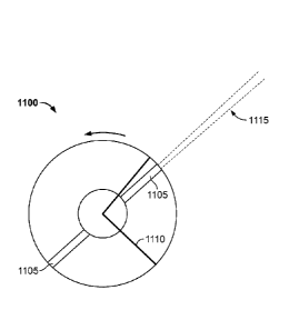
passive inspection module is moved relative to the object for scanning. Next,
neutron data is
collected passively, as shown in step 1003. Thereafter, the system checks if X-
rays are being
emitted, in step 1004. Thus, if X-ray beam is being emitted, and is not
blocked, the system
collects backscatter data, as shown in step 1005. However, if the beam
chopping mechanism is
currently blocking the X-ray beam, the system collects data pertaining to
passive gamma rays
emitted from the object. This is shown in step 1006. In the end, image
generated from
backscatter data and passive measurement results are displayed, as shown in
step 1007.
The results of the passive detection measurements and the X-ray images are
data fused to
improve detection of nuclear and radioactive materials. For example, dark
areas in the
backscatter image may indicate the presence of partially shielded nuclear or
radioactive
materials. If higher levels of radiation occur in these dark areas, there is a
stronger indication of
the presence of these threat materials.
In one embodiment, a spinning wheel having a plurality of pinholes therein is
employed
to produce a pencil beam of radiation through at least one pinhole, during
which time backscatter
radiation detection is active. In one embodiment, the spinning wheel
effectively "blocks" the x-
ray fan beam (and resultant pencil beam) from exiting, due to the position of
the pinholes in the
spinning wheel, during which time passive radiation detection is active. Thus,
passive radiation
measurement proceeds when the beam is "off' or blocked by the spinning wheel
geometry,
where there is no pinhole for the radiation to exit.
FIG. 11 is an illustration of an embodiment of a spinning wheel as used in the
system of
the present invention, showing the pencil beam in an "on" position, wherein a
backscatter
measurement is taken. As shown in FIG. 11, spinning wheel 1100 comprises a
disc fabricated
from shielding material defining at least one pinhole 1105 through which a fan
beam 1110
Date Recue/Date Received 2020-04-17
"exits" through the spinning wheel as pencil beam 1115. In one embodiment,
spinning wheel
1100 comprises two pinholes 1105. The pencil beam radiation, and thus
backscatter
measurement capability, is "on" when the fan beam 1110 exits the spinning
wheel as a pencil
beam 1115.
FIG. 12 is an illustration of one embodiment of a spinning wheel as used in
the system of
the present invention, showing the pencil beam in an "off' position, wherein a
passive
measurement is taken. As shown in FIG. 12, as spinning wheel 1200 is rotated,
there are times
when the fan beam 1210 does not coincide with at least one slit 1205. During
this time, the fan
beam 1210 is shielded by the spinning wheel 1200, and therefore, no radiation
exits the system.
It is during these times when the fan beam 1210 is "off' that a passive
radiation measurement is
taken.
It should be noted herein that employing a spinning wheel having two pinholes
is only
exemplary and that the basic approach can use any number of pinholes in the
spinning wheel
geometry as long as a passive measurement is performed when the pencil beam is
off.
In another embodiment, a beam chopping mechanism is employed, wherein the beam
chopping mechanism is designed to present a helical profile shutter
(aperture), formed on a
cylinder, for X-ray beam scanners. In one embodiment, a radiation shield is
provided on a
radiation source such that only a fan beam of radiation is produced from the
source whereby the
fan beam of radiation emits X-rays which then pass through the spin-roll
chopper, which acts as
an active shutter. Thus, when the spin-roll chopper and therefore, helical
aperture(s) is rotating,
there is only a small opening for the X-ray fan beam to pass through, which
provides the moving
flying spot beam. In this embodiment, the slits are configured in such a way
that there is at least
one gap where no pencil beam is produced. United States Patent Application
Number
13/047,657, entitled "Beam Forming Apparatus" and assigned to the Applicant of
the present
invention.
FIG. 13A illustrates an exemplary design for one embodiment of the spin-roll
chopper, as
used in various embodiments of the present invention. Beam chopper 1302 is, in
one
embodiment, fabricated in the form of a hollow cylinder having helical slits
1304 for "chopping"
the X-ray fan beam. The cylindrical shape enables the beam chopper 1302 to
rotate about the Z-
31
Date Recue/Date Received 2020-04-17
axis and along with the helical apertures 1304, create a spin-roll motion,
which provides
effective scanning and therefore good image resolution, as described below,
while at the same
time keeping the chopper lightweight and having less moment of inertia as the
spin-roll mass is
proximate to the axis of rotation. Stated differently, the radius of the spin-
roll chopper is small
compared to spinning wheel or disc beam chopping mechanisms, and is
advantageous in some
cases.
It should be noted that the helical twist angle 1325 represents the angle of
motion of the
helical aperture from the y-axis (center line) when the cylinder is spun about
the z-axis a total of
90 degrees.
Thus, an X-ray beam scanner employing the spin-roll chopper as in one
embodiment of
the present invention effectuates beam chopping by rotating the hollow
cylinder 1302 machined
with at least two helical slits 1304, enabling X-ray beam scanning with both
constant and
variable linear scan beam velocity and scan beam spot size. The spin-roll
chopper enables both
constant and variable linear scan beam velocity by manipulating the geometry
of the helical
apertures. In one embodiment, the velocity is varied or kept constant by
manipulating the pitch
and roll of the helical apertures along the length of the spin-roll chopper.
Thus, it is possible to
have a constant speed or to slow the scan down towards areas where more
resolution is desired.
The spin-roll chopper as described with respect to the present invention also
enables
variable and constant beam spot size by manipulating the geometry of the
helical apertures, thus
varying the resultant beam power. In one embodiment, the actual width of the
aperture is
manipulated to alter the beam spot size. In one embodiment, the width of the
helical aperture
varies along the length of the spin-roll chopper cylinder to compensate for
the varying distance
of the aperture from the center of the source and allow for uniform beam spot
projection along
the scan line. Thus, in one embodiment, the farther the aperture is away from
the source, the
narrower the width of the helical aperture to create a smaller beam spot size.
In one embodiment,
closer the aperture is to the source, wider the helical aperture to create a
larger beam spot size.
Helical slits 1304 are fabricated to ensure that the projection of the X-ray
beam is not
limited by dual collimation of the two slits. Dual collimation refers to the
concept whereby the
X-ray beam will pass through two helical slits at any given point in time. The
resultant X-ray
32
Date Recue/Date Received 2020-04-17
beam trajectory 1330 is also shown in FIG. 13A. In one embodiment, a pair of
helices will
produce one travelling beam. In another embodiment, additional pairs of
helices may optionally
be added to produce additional travelling or flying spot beams depending upon
scanning
requirements.
In an embodiment of the present invention a plurality of viewing angles
ranging from
sixty degrees to ninety degrees can be obtained through the helical slits in
the spin-roll chopper.
FIG. 13B illustrates a beam chopping mechanism using the spin-roll chopper
described with
respect to FIG. 13A. Referring to FIG. 13B, the cylindrical spin-roll chopper
1352 is placed in
front of a radiation source 1354, which, in one embodiment, comprises an X-ray
tube. In one
embodiment, rotation of the chopper 1352 is facilitated by including a
suitable motor 1358, such
as an electromagnetic motor. The speed or RPM of rotation of the spin-roll
chopper system is
dynamically controlled to optimize the scan velocity. In one embodiment, the
spin-roll chopper
system is capable of achieving speeds up to 80,000 RPM.
In yet another embodiment, a scanning pencil beam is generated by any one of
the
approaches described above or any other approach as is known to those of
ordinary skill in the
art and deactivated by turning off the X-ray source (in contrast with previous
embodiments,
where the source is "blocked" by use of the spinning wheel or spin-roll
chopper). Examples of
suitable x-ray sources include, but are not limited to gridded sources, field
emission electron
sources (e.g. carbon nanotubes) or any other source that can switch the beam
on-off within a few
microseconds. However, it should be noted that if the wheel or spin-roll
chopper is spinning
slower, then the time between switching the X-ray source on and off can be
longer. Therefore, it
can be stated that the time it takes for the X-ray source to be switched on
and off is relative to the
rotational frequency of the spinning wheel, on the order of a fraction of the
rotational time of the
source, which is in the range of less than 1%. By way of example, if the
rotational frequency if
2400 rpm (rotations per minute) and there are four pinholes, the time would be
6.25 ms ON and
6.25 ms OFF. If the spinning wheel is rotating at 240 rpm, then the times
would be 62.5 ms ON
and 62.5 ms OFF. Thus, the expression for the preferred time is as follows:
Equation 3: Time [ms]=((60/frequency [rpm])/number of pinholes) x 1000
33
Date Recue/Date Received 2020-04-17
FIG. 14 is a block diagram 1400 showing signal processing with two different
sets of
electronics when the backscatter x-ray detector and passive gamma-ray detector
are the same.
That is, the detector is dual-purpose, capable of detecting both backscattered
X-rays and passive
radiation. The backscatter system uses integrating electronics 1405, while the
passive detector
uses spectroscopic electronics 1410. Both set of electronics 1405, 1410 are
gated with a gating
signal 1415 from the spinning wheel control 1417. This produces a high signal
when the system
emits a pencil beam of radiation.
The backscatter integrating electronics 1405 employs an AND gate 1420 to
measure
backscatter radiation only when the beam is on, as described above with
respect to FIG. 3A. The
passive detector 1425 uses a NAND gate 1430 to measure only gamma rays when
the x-ray
pencil beam is off, as described above with respect to FIG. 13B. The optional
neutron detector
(not shown) need not be gated and can measure neutrons at all times.
The resultant backscatter image and results of the passive gamma-ray and
neutron
measurements are then shown on the screen (separately or combined).
The inspection system refers to any backscatter and passive radiation
detection system
that can be deployed in a scanning vehicle, portal, gantry, trailer, mobile
platform or other
scanning configurations. The system is also designed such that it can be moved
relative to the
object or such that the object can be moved relative to the system.
Reference will now be made to a specific embodiment of an aircraft inspection
system
that employs the active and passive radiation techniques as described in the
present specification.
It should be noted herein that such embodiment is exemplary only and that any
system can be
designed such that it takes advantage of the methods described above.
United States Patent Application Number 12/916,371, entitled "Mobile Aircraft
Inspection System" and filed on Oct. 29, 2010 is also relevant to the present
application.
FIG. 15 illustrates the overall system design of one embodiment of the present
invention.
Referring to FIG. 15, aircraft inspection system 1500, in one embodiment,
comprises inspection
head 1501, vehicle or transport cart 1502, and manipulator arm 1503. In one
embodiment,
inspection head 1501 comprises an inspection module, further comprising an X-
ray source, a
34
Date Recue/Date Received 2020-04-17
beam scanning mechanism and X-ray detectors. The inspection module is
described in greater
detail above with respect to FIG. 8. In one embodiment, vehicle or transport
cart 1502 is any
standard vehicle suitable for movement about an aircraft 1505.
In one embodiment, vehicle 1502 is movably connected to first, proximal end
1609a of
manipulator arm 1503 and inspection head 1501 is movably connected to second,
distal end
1509b of manipulator arm 1503 via a customized attachment 1504. Manipulator
arm 1503 is
described in greater detail below. In one embodiment, customized attachment
1504 is designed
for use with the system of the present invention. In another embodiment,
customized attachment
1504 may be available as an off-shelf component, as long as it achieves the
objectives of the
present invention, as described below.
In one embodiment, the inspection head 1501 is mounted on manipulator arm 1503
in
such a manner that it allows for scanning of a variety of aircraft sizes,
shapes and configurations.
The manipulator arm 1503 is also capable of rotating and moving the inspection
head 1501 in all
directions. In one embodiment, customized attachment 1504 is movably attached
to manipulator
arm 1503 at a first joint 1504a and movably attached to inspection head 1501
at a second joint
1504b. Thus customized attachment 1504 allows for the inspection head 1501 to
be moved and
rotated about first joint 1504a and second joint 1504b. In one embodiment,
first joint 1504a
and/or second joint 1504b is a ball and socket type joint that allows for at
least one movement,
such as but not limited to tilt, swivel and/or rotation at the joint, and in
one embodiment, full
motion. The ability to move and rotate the source at both the first attachment
joint 1504a and at
the second attachment joint 1504b allow for the system to follow the contour
of the aircraft and
thus, adjust to its shape using several degrees of movement freedom.
In addition, manipulator arm 1503 has multiple articulation or pivot joints
1507 that
allow for complex motions.
In one embodiment, in order to avoid damage to the aircraft 1505 being
inspected, the
inspection head 1501 includes at least one proximity sensor 1506. In one
embodiment, the
sensors are redundant, so if one fails to operate, another sensor will still
alert when the system is
too close to the aircraft. The at least one proximity sensor 1506 is
configured to avoid collision
and keep the inspection head 1501 at a safe distance from the aircraft 1505.
Therefore, once the
Date Recue/Date Received 2020-04-17
at least one proximity sensor 1506 is triggered, the inspection system 1500
will cease operation.
When inspection system 1500 ceases operation, the scanning head is retracted
and the system
cannot be operated until the sensor alarm is cleared.
In one embodiment, the at least one proximity sensor 1506 is connected and
controlled
via hardware.
In one embodiment, manipulator arm 1503 includes at least one proximity
sensor. In one
embodiment, vehicle 1502 also includes at least one proximity sensor.
To select appropriate design specifications for the vehicle and the
manipulator arm, the
critical areas of focus are: a) the distance from the source/detector to the
aircraft, b) the
controlled motion of the source/detector, and c) collision avoidance for both
the vehicle and the
manipulator with the aircraft. In one embodiment, an optimal distance from the
source/detector
arrangement to the aircraft rages from 1/2 meter up to two meters. In one
embodiment, the
distance is chosen to provide optimal image resolution, inspection coverage
and signal strength.
The weight of the source/detector in conjunction with the maximum height and
maximum reach
that the manipulator arm must obtain further determines the dimensions of the
vehicle platform.
It should be understood by those of ordinary skill in the art that the weight
of the source is
largely dependent on source type, and that source type is chosen based on the
object under
inspection and scanning requirements. Scanning sequence, motion speed, and
tolerances for
position and vibration also direct the specifications for the manipulator arm
and/or any special
attachments or tooling. As mentioned earlier, in order to minimize development
time and costs in
one embodiment, any suitable off-the-shelf vehicle and/or manipulator arm may
be employed
and modified as per the design requirements of the present invention. In one
embodiment, the
height and reach of the manipulator arm and weight and/or dimensions of the
inspection head are
a function of the size of the airplane or large cargo containing entity being
scanned.
FIG. 16 illustrates an exemplary vehicle 1600 that is connected to a
backscatter module
(not shown), via manipulator arm 1601, for the aircraft inspection system of
the present
invention. In one embodiment, for example, the vehicle 1600 may be a wheeled
excavator or a
similar vehicle.
36
Date Recue/Date Received 2020-04-17
FIG. 17 illustrates an exemplary manipulator arm 1700 that is used for
mounting a
backscatter module (not shown) for the aircraft inspection system of the
present invention. In one
embodiment, the manipulator arm 1700 comprises a multi-purpose hydraulic boom.
The boom
design allows for the flexibility of attaching the vehicle (not shown) to a
first, proximal end
1709a while attaching standard or custom tools at its second, distal end
1709b. Second, distal end
1709b, in one embodiment, is modified to allow for attachment of a backscatter
inspection
module at joint 1703.
In one embodiment, manipulator arm 1700 is operated using computer-controlled
motion
and has at least five degrees of freedom for positioning in all directions,
including up-down, left-
right, in/out and rotation. In one embodiment, the system further comprises a
controller unit,
which can be remote from the system or located within the vehicle, for
communicating motion
instructions to controllers located in the scanning head or gantry unit which,
in turn, directs
motors to move the scanning head and/or gantry unit in the requisite
direction. One method of
controlling motion of the vehicle and the manipulator arm using a computer
involves referring to
a database of airplane models, stored in a memory on the computing system.
Each entry in the
database corresponds to a plane contour. This database enables the motion-
control program to
generate a scan plan, which is used to control the motion of the arm and the
head to scan the
airplane according to the plan. Further, for some planes, it may not be
possible to scan the entire
plane from one vehicle position. Therefore, the motion control program
analyzes the various
positions required and the system scans the plane accordingly.
In one embodiment, the arm is capable of full 360 degree rotation. The
manipulator 1700
is linearly extensible and contractible, and the extension and contraction can
be achieved with a
complex motion of the various parts of the manipulator arm. The system scans
the aircraft by
moving the arm at a nearly constant distance from the surface of the aircraft.
The manipulator arm 1700 is also equipped with the capability of source
rotation at the
joint 1703, as described above. The ability to rotate and move the source
through several degrees
of freedom at attachment joint 1703, allow for the system to follow the
contour of the aircraft
and thus, adjust to its shape. The manipulator arm of the present invention
has multiple
37
Date Recue/Date Received 2020-04-17
articulation or pivot points 1705 that allow for complex motions, including
but not limited to
extension and contraction.
In one embodiment, the aircraft inspection system of the present invention is
capable of
producing high-resolution images that enable the operator to easily identify
concealed threat and
contraband items. In one embodiment, a database or threat library containing
standard images of
airplanes is employed to compare resultant scans of the aircraft under
inspection with images
collected from planes of the same model to determine anomalies.
In one embodiment, depending on the size of the airplane, the images of parts
of the
planes are collected separately. These images can then be displayed
separately, or they could be
"stitched" together show a combined image.
The aircraft inspection system of the present invention is capable of
accurately detecting
both organic materials, such as solid and liquid explosives, narcotics,
ceramic weapons, as well
as inorganic materials, such as metal. In one embodiment, the aircraft imaging
system uses
automated threat software to alert an operator to the presence of potential
inorganic and organic
threat items. In one embodiment, the system is capable of transmitting
backscatter and
photographic images to an operator or remote inspector wirelessly.
The aircraft inspection system of the present invention is designed to be
modular to
enhance transportability and ease of assembly. In one embodiment, the
individual modules--the
vehicle, the manipulator arm, the scanning head, and optionally detector cart
can be assembled
on site and/or customized per application. In addition, in another embodiment,
the system is
ready to deploy and requires no assembly.
The system is also designed to be rugged so that it can withstand harsh
environments for
outdoor deployments even in inclement conditions. In one embodiment, the power
required to
run the system is provided on-board allowing the system to operate anywhere on
the airfield. In
one embodiment, the aircraft inspection system of the present invention is
scalable for inspecting
any aircraft size from executive jets to Airbus 380. Thus, the size of the
vehicle and arm can be
scaled to the size of the aircraft.
38
Date Recue/Date Received 2020-04-17
FIG. 18 shows another embodiment of the X-ray scanning system 1800 of the
present
invention that additionally uses a multi-element scatter collimator 1816 to
allow use of fan-beam
X-ray irradiation to generate the backscatter image. Here, the X-ray source
1805 emits a fan
beam 1806 of radiation towards the object 1810. A segmented detector array
1815 is located
behind a multi-element collimator 1816, one detector element per collimator
section. The
collimator 1816 is designed to permit X-rays to enter from a narrow angular
range, typically less
than +/-2 degrees to the perpendicular to the detector array 1815. X-rays 1807
scattering from
various points in the object 810 which lie within the acceptance angle of, for
example, the
collimator element 1816 are detected and associated to the appropriate
corresponding part of the
generated radiographic X-ray image. Again, a sensor 1820 is provided to
measure distance to the
surface of the object 1810 in order to correct the X-ray backscatter signal
and produce a
quantitative image scaled by effective atomic number. United States Patent
Application Number
12/993,831, also by Applicant of the present invention, entitled "High-Energy
X-Ray Inspection
System Using A Fan-Shaped Beam and Collimated Backscatter Detectors", and
filed on
November 19, 2010, discloses use of such a multi-element scatter collimator.
A system configuration according to an embodiment of the invention disclosed
in United
States Patent Application Number 12/993,831 is outlined in FIGS. 19 to 21.
Here, an X-ray
linear accelerator 20 is used to fire a collimated fan-beam of high energy (at
least 900 keV) X-
radiation through an object 22 under inspection and to a set of X-ray
detectors 24 which can be
used to form a high resolution transmission X-ray imaging of the item under
inspection. The X-
ray linear accelerator beam is pulsed, so that as the object under inspection
moves through the
beam, the set of one-dimensional projections can be acquired and subsequently
stacked together
to form a two-dimensional image.
In this embodiment, an X-ray backscatter detector 26 is placed close to the
edge of the
inspection region on the same side as the X-ray linear accelerator 20 but
offset to one side of the
X-ray beam so that it does not attenuate the transmission X-ray beam itself.
As shown in FIG.
10, it is advantageous to use two backscatter imaging detectors 26, one on
either side of the
primary beam. In some embodiments the backscatter detectors may be arranged
differently. In
some embodiments there may be only one backscatter detector. In other
embodiments there may
be more than two such detectors.
39
Date Recue/Date Received 2020-04-17
In contrast to known backscatter imaging detectors which use the localisation
of the
incident X-ray beam to define the scattering region, the backscatter imaging
detector described,
is able to spatially correlate the intensity of backscattered X-ray signals
with their point of origin
regardless of the extended fan-beam shape of the X-ray beam.
In the backscatter imaging detector 26, this spatial mapping is performed
using a
segmented collimator 28 in zone plate configuration as shown schematically in
FIG. 21.
Normally, a zone plate will comprise a series of sharply defined patterns
whose impulse response
function is well known in the plane of a two-dimensional imaging sensor that
is located behind
the sensor. In the present case, the energy of the X-ray beam to be detected
is typically in the
range 10 keV to 250 keV and so the edges of the zone plate pattern will not be
sharp. For
example, a zone plate fabricated using lead will require material of thickness
typically 2 mm to 5
mm. Further, it is expensive to fabricate a high resolution two-dimensional
imaging sensor of the
size that is required in this application.
However, it is noted that the radiation beam is well collimated in one
direction (the width
of the radiation fan beam) and therefore the imaging problem is reduced to a
one-dimensional
rather than a two-dimensional problem. Therefore a backscatter detector in the
form of an
effectively one dimensional imaging sensor 30 is provided behind the zone
plate 28. To address
this problem an elemental backscatter detector is used in this embodiment. As
shown in FIG. 21,
the detector 30 comprises a plurality of detector elements 32. FIG. 22
illustrates a detector
element 32 suitable for use in this example. Here, the detector element 32
comprises a bar of
scintillation material (about 100 mm long in this example) and is supplied
with a photo-detector
34 at either end. The photo-detector 34 may advantageously be a semiconductor
photodiode or a
photomultiplier tube. X-ray photons that interact in the scintillation
material emit light photons
and these will travel to the two photo-detectors where they may be detected.
It may be shown
that the intensity of the light reaching each photo-detector is in proportion
to the distance of the
point of interaction from the face of the photo-detector. Therefore, by
measuring the relative
intensity at the two photo detectors, the point of interaction of the X-ray
photon with the detector
can be resolved.
Date Recue/Date Received 2020-04-17
Referring back to FIG. 1A, the covert surveillance vehicle 105 is equipped
with a
plurality of other sensors 110, apart from the X-ray scanning system, in
accordance with an
aspect of the present invention. In one embodiment, the vehicle 105 is
equipped with a GPS
receiver the output of which is integrated with the on-board X-ray scanning
system to provide
the absolute location at which each scan line is conducted. Again, output from
a scanning laser is
reconstructed into a 2D image to provide a quantitative analysis of the scene
around the vehicle.
This 2D image is archived for subsequent analysis and review.
The 2D laser scanner image may also be used to determine when the overall scan
of a
particular object should start and when the scan for that object is complete.
Also, optical wavelength colour CCTV images are collected at the front and
sides of the
vehicle, ideally using pan-tilt-zoom capability, to allow clear review of all
locations around the
vehicle. In one embodiment, images from the CCTV cameras are analysed to read
license plate
and container codes and this data is also archived along with the X-ray, GPS
and all other
surveillance data. Similarly, infra-red cameras can also be used to monitor
the scene around the
vehicle to look for unexpectedly warm or cold personnel as indication of
stress or presence of
improvised explosive devices. This data is also archived along with X-ray and
all other
surveillance data.
In one embodiment, audio microphones are also installed around the vehicle to
listen for
sounds that are being produced in the vicinity of the vehicle. Specialist
microphones with pan-tilt
capability are installed to listen to sounds from specific points at some
distance from the vehicle,
this direction being analysed from the CCTV and IR image data.
Directional RF (Radio Frequency) antennas are installed in the skin of the
vehicle to
listen for the presence of electronic devices in the vicinity of the vehicle.
This data is integrated
with the rest of the surveillance data. Similarly, wide band antennas are
installed with receiving
devices that monitor communications channels that may be used by law
enforcement, military
and emergency services. Again, RF antennas are installed to monitor mobile
phone
communications including text messaging from the local region around the
vehicle.
41
Date Recue/Date Received 2020-04-17
In one embodiment, chemical sensors are also installed to monitor composition
of the air
around the vehicle to detect trace quantities of explosives, narcotics and
other relevant
compounds with this data being integrated with that generated by the imaging
and other sensors.
In accordance with another aspect of the present invention, an automated
detection
processor integrates and analyses all surveillance information from the
plurality of sensors 110,
in real-time, to highlight threat items for review by an operator seated
inside the vehicle 105
and/or remotely through a secured wireless network. In one embodiment, data
from the
individual sensors is analysed for key signatures. For example, the X-ray data
is analysed for
detection of improvised explosive devices or for the presence of organic
materials in unexpected
places (such as the tires of a car). CCTV data is analysed for license plates
with cross-checking
against a law enforcement database. Audio information is analysed for key
words such as
"bomb" or "drugs", for unexpectedly fast or deliberate phrasing which may
indicate stress, or for
a non-native language in the presence of a native language background for
example. Once a
piece of information has been analysed to comprise a threat or risk, this is
escalated up a decision
tree and is then compared against automated risk analysis from other sensors.
If correlated risks
are detected, a significant threat alarm is raised for immediate action by a
human operator. If no
correlated risk is detected, a moderate threat alarm is raised for review by
the operator. The result
is a managed flow of information where all sensor surveillance information is
analysed at all
times, and only significant threat information is passed up the decision tree
to reach the final
level of an alert to a system operator. The detection processor, in one
embodiment, is a
microprocessor computer running relevant code programmed for managing
information and
decision flow based on correlation and aggregation of the plurality of
surveillance information.
Great Britain Provisional Patent Application Number 1001736.6, entitled "Image
Driven
Optimization", and filed on Feb. 3, 2010, and Patent Cooperation Treaty (PCT)
Application
Number PCT/GB2011/050182 entitled "Scanning Systems", and filed on February 3,
2011 by
the Applicant of the present specification, disclose a scanner system
comprising a radiation
generator arranged to generate radiation to irradiate an object, and detection
means arranged to
detect the radiation after it has interacted with the object and generate a
sequence of detector data
sets. Referring to FIG. 23, a scanner system comprises an X-ray beam
generation system which
includes a shielded radiation source 10, a primary collimator set 12A and a
secondary collimator
42
Date Recue/Date Received 2020-04-17
set 12B, and a set of radiation detectors 14 configured into a folded L-shaped
array 16, are
disclosed.
The primary collimator set 12 A acts to constrain the radiation emitted by the
source 10
into a substantially fan-shaped beam 18. The beam 18 will typically have a fan
angle in the range
+/-20 degrees to +/-45 degrees with a width at the detector elements 14 in the
range 0.5 mm to
50 mm. The second collimator set 12B is adjustably mounted and the position of
the two second
collimators 12B can be adjusted by means of actuators 20, under the control of
a decision
processor 22. The detectors 14 output detector signals indicative of the
radiation intensity they
detect and these form, after conversion and processing described in more
detail below, basic
image data that is input to the decision processor 22. The decision processor
22 is arranged to
analyse the image data and to control the actuators 20 to control the position
of the second
collimator set 12B in response to the results of that analysis. The decision
processor 22 is also
connected to a control input of the radiation source 10 and arranged to
generate and vary a
control signal it provides to the control input to control the energy and
timing of X-ray pulses
generated by the radiation source 10. The decision processor 22 is also
connected to a display 24
on which an image of the imaged object, generated from the image data, can be
displayed.
By way of example, the radiation source 10 may comprise a high energy linear
accelerator with a suitable target material (such as tungsten) which produces
a broad X-ray
spectrum with a typical beam quality in the range from 0.8 MV to 15 MV from a
relatively small
focal spot typically in the range 1 mm to 10 mm diameter. The radiation source
10 in this case
would be pulsed with a pulse repetition frequency generally in the range 5 Hz
to 1 kHz where the
actual rate of pulsing is determined by the decision processor 22.
The detectors 14 in this case are advantageously fabricated from a set of
scintillation
crystals (generally high density scintillator such as Csl, CdW04, ZnW04, LSO,
GS0 and similar
are preferred) which are optically coupled to a suitable light detector, such
as a photodiode or
photomultiplier tube. Signals from these detectors 14 converted to digital
values by a suitable
electronic circuit (such as a current integrator or trans impedance amplifier
with bandwidth
filtering followed by an analogue to digital converter) and these digital
values of the sampled
intensity measurements are transferred to the decision processor 22 for
analysis. The primary 12
43
Date Recue/Date Received 2020-04-17
A and secondary 12B collimators in this case are advantageously fabricated
from high density
materials such as lead and tungsten.
A plurality of active devices are installed on the vehicle 105 to help
mitigate against
threats that may be present proximate to the covert inspection vehicle itself.
For example, a
jamming device can be installed to block mobile phone communication. This
device may be
turned on automatically in certain situations based on results from the
automated decision
processor. For example, should an improvised explosive device be detected in
the vicinity of the
vehicle the jamming device is turned on automatically to block spoken commands
to a
subversive or to prevent direct communication to the trigger of the explosive
device. A jamming
device can also be installed to block satellite communications required in
order to prevent
satellite phone communications that may result in subversive activity.
In one embodiment the covert inspection vehicle 105 is operated by a single
person with
the primary responsibility for driving the vehicle. Surveillance data can be
broadcast back to a
central intelligence location in real time, as required, with download of the
full archived
surveillance data once the vehicle returns to its home location. The automated
decision processor
can action or trigger appropriate events, depending upon the decision steps
programmed therein,
without operator intervention to avoid the driver loosing focus on their
primary task. In another
embodiment, the covert inspection vehicle 105 is also provided with space for
another security
operative whose task is to monitor the surveillance data stream as it arrives
from the plurality of
sensors either in parallel with the automated decision processor or as a
consequence of
information from the automated decision processor. This operator is provided
with two way
secure wireless communication back to a central intelligence location in order
to transact
instructions and actions as required.
The above examples are merely illustrative of the many applications of the
system of
present invention. Although only a few embodiments have been described herein,
it should be
understood that other embodiments are possible and that the present invention
might be
embodied in many other specific forms without departing from the scope of the
present
specification. Therefore, the present examples and embodiments are to be
considered as
illustrative and not restrictive. The scope of protection being sought is
defined by the following
44
Date Recue/Date Received 2020-04-17
claims rather than the described embodiments in the foregoing description. The
scope of the
claims should not be limited by the described embodiments set forth in the
examples but should
be given the broadest interpretation consistent with the description as a
whole.
Date Recue/Date Received 2020-04-17