Note: Descriptions are shown in the official language in which they were submitted.
CA 03078249 2020-04-01
WO 2019/075119
PCT/US2018/055290
HUMERAL FIXATION PLATE GUIDES
INCORPORATION BY REFERENCE TO ANY PRIORITY APPLICATIONS
[0001] Any and
all applications for which a foreign or domestic priority claim
is identified in the Application Data Sheet as filed with the present
application are hereby
incorporated by reference under 37 C.F.R. 1.57.
BACKGROUND OF THE INVENTION
Field of the Invention
[0002] This
application is directed to guides for controlled insertion of
fasteners into bone fracture plates, e.g., for the humerus, and for fracture
repair and
methods of using the same.
Description of the Related Art
[0003] Humeral
fractures arise from serious injuries and other causes. One
approach to repairing fracture involves attaching a fracture plate to an
outside surface of
the humerus to fix exposed sides of the facture onto or adjacent to one
another to facilitate
the process of fusing these exposed sides together. The fracture plate assures
that the
multiple pieces of the fractured bone remain in a prescribed position or
orientation to each
other and do not move relative to each other so that the fusion process is not
continually
disrupted and so that the bones do not migrate to and fuse in misaligned
positions.
[0004] Humeral
bone has several layers. An outer layer called the cortical
layer is a relatively dense portion of the humerus that is most capable of
bearing loads
absent other bone considerations. Inward of the cortical layer is cancellous
bone matter.
This bone matter is less dense and is not as capable of bearing loads. A
number of screws
can be used to secure a fracture plate to the humerus. These screws should be
lodged in
the cortical bone or in a transition between cancellous and cortical bone to
provide good
fixation of the fracture plate to the bone, and thereby of the multiple pieces
of the
fractured humerus to each other.
[0005] Because
fracture plates are typically coupled with the lateral side of the
humerus and the screws are typically directed into the humeral head, it is
important to
control the final location of the tips of the screws.
-1-
CA 03078249 2020-04-01
WO 2019/075119
PCT/US2018/055290
SUMMARY OF THE INVENTION
[0006] It would
be desirable to provide improved apparatuses and methods for
securing a fracture plate to a humerus. It would be advantageous to provide a
guide that
can be used to direct fasteners into the humeral bone in a way that provides
robust
connection of the fasteners to strong bone, e.g., to cortical bone, of the
humerus. In some
applications the guides can be configured to provide a prescribed pattern of
fasteners
projecting from a fracture plate. The prescribed pattern can be controlled by
a guide that
is appropriate for the humerus, e.g., appropriate for the size and location
(left, right) of the
humerus. The prescribed pattern could be unique to a specific patient based on
pre- or
intra-operative imaging. The guides can enable a non-patient specific fracture
plate to be
used with humerus bones of different size and in some cases in patient
specific method,
e.g., resulting in proper placement of the fracture plate and of a plurality
of fasteners.
[0007] In one
embodiment, an anchor trajectory guide is provided that
includes a body that has a medial side. The medial side is configured to be
placed over a
lateral side of a fixation plate. The anchor trajectory guide also includes a
locator and a
plurality of guide apertures. The locator is disposed on or through the medial
side of the
body. The locator is configured to mate with the fixation plate. The guide
apertures are
disposed through the body at positions corresponding to define anchor
locations and
orientations to provide good fixation in bone around a medial portion of a
humerus.
[0008] In
another embodiment an anatomical guide is provided. The
anatomical guide is configured to be coupled with a sleeve and a visual guide
member.
The sleeve is configured to mate with a guide aperture of an anchor trajectory
guide or
with a fixation plate. The sleeve can have a lumen that is disposed
therethrough. The
lumen can be used to advance a K-wire or other pin through the anatomical
guide. The
visual guide member is configured to extend from the anchor trajectory guide
or the
fixation plate to an anterior side or a posterior side of the humerus.
[0009] In
another embodiment a method is provided. In the method, a medial
side of a fixation plate is placed on a lateral surface of a humerus. The
fixation plate is
placed in contact with the lateral surface. The fixation plate is coupled with
the humerus
spanning a fracture. A medial side of an anchor trajectory guide is coupled
with a lateral
side of the fixation plate. An anchor channel is formed in the humerus from
the lateral
side of the humerus toward an opposing cortical bone region. The anchor
channel can be
formed, through a guide aperture in the anchor trajectory guide and an anchor
aperture in
-2-
CA 03078249 2020-04-01
WO 2019/075119
PCT/US2018/055290
the fixation plate. An anchor is advanced through the anchor aperture and the
anchor
channel, which is formed in the bone, to secure the anchor and the fixation
plate to the
humerus. The anchor follows a trajectory defined by the anchor trajectory
guide. A
medial end of the anchor is embedded in or adjacent to the opposing cortical
bone while a
proximal end of the anchor is embedded in lateral cortex of the humerus. When
the
medial end is embedded adjacent to opposing cortical bone the medial end can
be lodged
in cancellous bone. When the medial end is embedded adjacent to opposing
cortical bone
the medial end can be lodged in a transitional bone matter between the
cancellous bone
and the cortical bone.
[0010] In
another embodiment, a slot anchor guide is provided that includes a
medial portion and a lateral portion. The medial portion has a medial
projection
configured to span a slot of a fixation plate. The slot anchor guide is
configured to guide
an anchor through a predetermined position of a slot of the fixation plate.
The slot anchor
guide is configured to mate with the fixation plate to allow a surgeon to
place the fixation
plate on a bone face by manipulating the lateral portion.
[0011] In
another embodiment, a tuberosity fracture plate is provided. The
tuberosity fracture plate has a tuberosity end, a distal portion, a first
screw hole and a
second screw hole. The tuberosity end has a first portion configured to
overlay a first
tuberosity and a second portion opposite the first portion. The distal portion
is coupled
with and extends away from the tuberosity portion. The first screw hole is
disposed in the
tuberosity end. The second screw hole is disposed in the distal portion. The
tuberosity
fracture plate has a bend zone disposed between the first screw hole and the
second screw
hole. The bend zone is configured to locate a bend in the tuberosity fracture
plate
between the first screw hole and the second screw hole upon application of a
load to the
tuberosity end, to the distal portion or to both the tuberosity end and the
distal portion.
BRIEF DESCRIPTION OF THE DRAWINGS
[0012] These
and other features, aspects and advantages are described below
with reference to the drawings, which are intended for illustrative purposes
and should in
no way be interpreted as limiting the scope of the embodiments. Furthermore,
various
features of different disclosed embodiments can be combined to form additional
embodiments, which are part of this disclosure. In the drawings, like
reference characters
-3-
CA 03078249 2020-04-01
WO 2019/075119
PCT/US2018/055290
denote corresponding features consistently throughout similar embodiments. The
following is a brief description of each of the drawings.
[0013] FIG. 1 is a schematic view of anatomy around the shoulder
joint;
[0014] FIG. 1A is a schematic view of a proximal humerus with a two-
piece
fracture;
[0015] FIG. 1B is an anterior view of a proximal portion of a humerus
with a
tuberosity fracture;
[0016] FIG. 1C is an antero-lateral view of the proximal portion of
the
humerus of FIG. 1B;
[0017] FIG. 2 is a perspective view of a fixation plate coupled to a
proximal
humerus by a plurality of screw anchors;
[0018] FIG. 2A is a view of a lateral side of another embodiment of a
fixation
plate;
[0019] FIG. 2B show a part of a method of connecting another
embodiment of
a fixation plate to the proximal humerus;
[0020] FIG. 2C is a cross-sectional view of the slot and screw of
FIG. 2B
taken through section plane 2C-2C in FIG. 2B;
[0021] FIG. 3 is a perspective view of a lateral side of an anchor
trajectory
guide;
[0022] FIG. 3A is a perspective view of a lateral side of another
example of an
anchor trajectory guide;
[0023] FIG. 4 is a view of a lateral side of the anchor trajectory
guide of FIG.
3;
[0024] FIG. 5 is a view of a superior side of the anchor trajectory
guide of
FIG. 3;
[0025] FIG. 6 is a view of an anterior side of the anchor trajectory
guide of
FIG. 3;
[0026] FIG. 7 is an anterior view of a humerus with a fixation plate
and an
anchor trajectory guide coupled thereto illustrating part of a method of
connecting the
fixation plate to the humerus;
[0027] FIG. 7A is a perspective view of a slot anchor guide assembly
according to one example;
-4-
CA 03078249 2020-04-01
WO 2019/075119
PCT/US2018/055290
[0028] FIG. 7B is a medial side perspective view of a slot anchor
guide of the
slot anchor guide assembly of FIG. 7A;
[0029] FIG. 7C is a superior or inferior side view of the slot anchor
guide of
FIG. 7B;
[0030] FIG. 7D is a cross-sectional view of the slot anchor guide of
FIG. 7B
taken through section plane 7D-7D in FIG. 7C;
[0031] FIG. 7E is a medial side view of the slot anchor guide of FIG.
7B;
[0032] FIG. 7F is a perspective view of a slot anchor guide assembly
according to another example;
[0033] FIG. 8 shows a part of a method of connecting the fixation
plate to the
humerus following the part of the method illustrated in FIG. 7 in which K-
wires are
placed through anchor sleeves and K-wire sleeves mated with medial calcar and
anterior
superior access apertures;
[0034] FIG. 9 shows a part of a method of connecting the fixation
plate to the
humerus following the part of the method illustrated in FIG. 8 in which the
fixation plate
and the guide are secured in a selected position;
[0035] FIG. 10 is a perspective view of one example of a medial
calcar guide;
[0036] FIGS. 11 and 12 are examples of another medial calcar guide;
[0037] FIGS. 13 and 14 show variations methods of confirming the
position of
the fixation plate using the medial calcar guide of FIG. 10;
[0038] FIGS. 15, 16, and 16A show a part of a method of connecting
the
fixation plate to the humerus following the part of the method illustrated in
FIGS. 13 and
14, the fixation plate and the guide are secured in a selected position and in
which access
is provided for an anchor to be advanced to the humerus through the medial
calcar guide;
[0039] FIGS. 17 and 17A show a part of a method of connecting the
fixation
plate to the humerus following the parts of the method illustrated in FIGS. 9
and 13 in
which an access opening is formed through the lateral cortex of the humerus;
[0040] FIGS. 18-19 show a part of a method of connecting the fixation
plate to
the humerus following the part of the method illustrated in FIGS. 17 and 17A
in which an
anchor length is determined through the lateral cortex access opening;
[0041] FIG. 20 shows an example of a humeral anchor length gauge that
can
be used to determine an anchor length in the method part illustrated in FIGS.
18-19;
-5-
CA 03078249 2020-04-01
WO 2019/075119
PCT/US2018/055290
[0042] FIG. 21 illustrates one example of visual indicia that can be
provided at
the segment 21-21 shown in FIG. 20 to determine an anchor length in the method
part
illustrated in FIGS. 18-19;
[0043] FIG. 22 shows a part of a method of connecting the fixation
plate to the
humerus following the part of the methods illustrated in FIGS. 18-19 in which
an anchor
of selected length is advanced through the lateral cortex access opening;
[0044] FIG. 23 shows a part of a method of connecting the fixation
plate to the
humerus following the part of the methods illustrated in FIG. 22 in which an
anterior-
superior lateral cortex access opening is formed;
[0045] FIGS. 24 and 25 show lateral and perspective views of an
assembly
including the fixation plate and a plurality of screw anchors disposed through
the fixation
plate into the humerus with a medial side of the guide coupled with a lateral
side of the
fixation plate;
[0046] FIGS. 26 and 27 are posterior and medial side views that
illustrates a
part of a method of connecting the fixation plate to the humerus following the
part of the
method illustrated in FIGS. 24 and 25;
[0047] FIG. 28 illustrates a kit comprising a guide, a fixation plate
and a
plurality of anchors as well as instruments facilitating connection of the
fixation plate to
the humerus;
[0048] FIG. 29 is a perspective view of a tuberosity fracture plate
coupled to a
greater tuberosity by a plurality of screw anchors;
[0049] FIG. 30A is a perspective view of a tuberosity fracture plate.
[0050] FIG. 30B is a view of an anterior side of the tuberosity
fracture plate of
FIG. 30A;
[0051] FIG. 30C is a view of a superior side of the tuberosity
fracture plate of
FIG. 30A; and
[0052] FIG. 30D is a cross sectional view of the tuberosity fracture
plate of
FIG. 30A.
DETAILED DESCRIPTION OF THE PREFERRED EMBODIMENT
[0053] This application is directed to a guide for attaching a
fixation plate to a
humerus of a patient and to plates that can be so attached. The guide could be
used
following a fracture. The fracture can be between the metaphysis and the
diaphysis of the
-6-
CA 03078249 2020-04-01
WO 2019/075119
PCT/US2018/055290
humerus or along a prominence of a proximal portion of a humerus, such as a
tuberosity.
Although the guides and methods are described in connection with the humerus
the
guides and methods can be used for other bones, such as any long bone fracture
or for
other orthopedic plate fixation procedures.
[0054] FIG. 1
shows anatomy of a glenohumeral joint. The joint is formed in
part by a head 10 of a humerus H and a glenoid 18 of a scapula 14. The head 10
is a
convex structure that is generally spherical. The glenoid 18 includes a
concave articular
surface upon which the head 10 moves. FIG. 1A shows that the humerus H has a
medial
side (right side in the view) and a lateral side (left side in the view). The
medial calcar
MC is located at the inferior edge of the head 10 on the medial side of the
humerus. A
lateral cortex LC extends along the lateral side of the bone generally
opposite to the
medial calcar MC. An anterior- superior region AS of the humerus H is located
on the
lateral side and superior to the distal-proximal location of the medial calcar
MC.
[0055] As
discussed above, the humerus has a proximal portion that is the
portion of the humerus adjacent to the glenoid 18 and forming part of the
shoulder joint.
The proximal humerus is sometimes referred to herein as the superior humerus.
Proximal
and distal in this sense are shown on FIG. 1A with reference to the humerus.
In this
application a location that is distal to another location refers to being
closer to an inferior
or elbow-adjacent end of the humerus. A distal portion of the humerus is
sometimes
referred to herein as an inferior portion of the humerus.
[0056] FIG. 1A
shows a fracture F which is one simple form of fracture that
can be treated by the apparatuses and methods discussed below. In many cases
the
fracture F is accompanied by additional fractures around the humeral head 10.
These
additional fractures can be treated as well, as discussed further below.
[0057] FIGS. 1B
and 1C show an example of a tuberosity fracture TF that can
be treated as discussed below with a plate that is suitable for repairing the
tuberosity
fracture TF. The tuberosity fracture TF is of a greater tuberosity GT but
could be of the
lesser tuberosity LT or another prominence of a long bone.
I. FIXATION PLATE ASSEMBLIES
[0058] FIG. 2
shows how a fracture F in a humerus H can be treated using a
fixation plate assembly 90. The fixation plate assembly 90 includes a fixation
plate 100
-7-
CA 03078249 2020-04-01
WO 2019/075119
PCT/US2018/055290
and a plurality of polyaxial anchors 130 in one embodiment. The fixation plate
100 can
have a lateral side 108 configured to face away from the humerus H and a
medial side 112
(see FIG. 7) configured to face the humerus H. The medial side 112 can be in
direct
contact with a lateral surface LS of the humerus H in some applications. The
fixation
plate 100 preferably is configured to work well for an entire population of
patients. The
proximal-distal dimensions enable the fixation plate 100 to span a wide range
of neck
fractures. The anterior-posterior dimensions allow the fixation plate 100 to
be placed on
the lateral surface LS of a wide range of bone sizes.
[0059] The
fixation plate 100 can include a distal portion 116 and a proximal
portion 118. In some methods, the distal portion 116 is disposed between the
humeral
neck and the end of the humerus H forming a portion of the elbow joint. In
some
methods, the distal portion 116 is disposed between the fracture F and the end
of the
humerus H forming a portion of the elbow joint. In some methods the proximal
portion
118 is positioned proximal of the humeral neck or of the fracture F. The
proximal portion
118 can be configured to be secured to the lateral surface LS of the humerus H
in the
region of the head of the humerus H. For example, the proximal portion 118 can
include
an array of anchor apertures 134. The anchor apertures 134 can be disposed
about the
periphery of the proximal portion 118 of the fixation plate 100. In one
embodiment, there
are four anchor apertures 134 on an anterior side of the proximal portion 118
and there are
an additional four anchor apertures 134 on a posterior side of the proximal
portion 118 of
the fixation plate 100. In some variations there can be more than four anchor
apertures
134 on the anterior and posterior sides. In some variations there can be more
anchor
apertures 134 on the anterior than on the posterior side. In some variations
there can be
more anchor apertures 134 on the posterior than on the anterior side.
[0060] One or
more or all of the anchor apertures 134 can be suited to mate
with polyaxial anchors 130. The engagement between the polyaxial anchor 130
and the
anchor aperture 134 allow the anchor to be directed along a range of
directions rather than
just being directed along a single axis as is provided with a more simple
thread
arrangement. As discussed further below, the fixation plate 100 enables a
medial end 142
of the polyaxial anchors 130 to span across cancellous bone of the humerus H
to engage
an opposing cortical bone region CB. A lateral end 146 of the polyaxial
anchors 130 is
configured to engage cortical bone at or adjacent to the lateral surface LS of
the humerus
-8-
CA 03078249 2020-04-01
WO 2019/075119
PCT/US2018/055290
H. The lateral end 146 also has a head portion 134A that is configured to
engage a
corresponding one of the anchor apertures 134.
[0061] FIG. 2
and FIG. 27 show that when the fixation plate 100 is coupled
with a humerus H the polyaxial anchors 130 are generally splayed out. In this
specification two anchors are splayed when they are disposed in space along
longitudinal
axes that are not parallel to each other. FIGS. 2 and 27 show that the anchors
130 are
generally splayed out with respect to a medial-lateral and proximal-distal
plane PL of the
humerus H. That is, a plurality of, e.g., four, polyaxial anchors 130 can be
secured
through an anterior portion of the fixation plate 100 to the lateral surface
LS of the
humerus H. Another plurality of, e.g., four, polyaxial anchors 130 can be
secured to a
posterior portion of the fixation plate 100 to the lateral surface LS of the
humerus H. The
polyaxial anchors 130 can be oriented such that medial ends 142 thereof are
more anterior
or more posterior than are lateral ends 146 thereof. Described another way,
the lateral
ends 146 can be located closer to the medial-lateral and proximal-distal plane
PL than are
the medial ends 142 thereof. FIG. 2 and FIG. 27 also show that the fixation
plate 100 can
be configured to engage some anchors along the medial-lateral and proximal-
distal plane
PL. For example, one polyaxial anchor 130 can be disposed on the medial-
lateral and
proximal-distal plane PL in a central portion of the fixation plate 100. Also,
one or a
plurality of, e.g., two, polyaxial anchors 130 can be secured to anchor
apertures 134 in the
distal portion 116 of the fixation plate 100 and therethrough to a portion of
the humerus H
distal to the head 10 or distal to the fracture F.
[0062] Some
advantageous methods discussed herein aid in initial placement
of the fixation plate 100 such that the initial placement normally does not
require
repositioning and thus is normally the final placement. The fixation plate 100
includes a
slot 160 in the distal portion 116 that facilitates some of these methods. The
slot 160 can
extend along a length of the distal portion 116. In some embodiments the slot
160 is
aligned with a longitudinal axis of the fixation plate 100 and so can be
positioned to
symmetrically straddle the medial-lateral and proximal-distal plane PL. The
slot 160 can
have a smooth inner surface 161 to engage with a non-locking anchor 132 (see
FIG. 2).
The slot 160 allows the fixation plate 100 to move in a proximal-distal
direction and also
to rotate about the non-locking anchor 132 prior to placement of other anchors
through
the fixation plate 100.
-9-
CA 03078249 2020-04-01
WO 2019/075119
PCT/US2018/055290
[0063] The
fixation plate 100 can have one or a plurality of suture apertures
164 disposed about the periphery thereof. The suture apertures 164 enable a
surgeon to
secure fracture portions to the fixation plate 100. In some cases fractured
portions of the
head can include the greater and/or the lesser tuberosities. These bone
portions are
usually attachment points for soft tissue, e.g., rotator cuff portions. The
soft tissue tends
to pull these fractured pieces medially. The suture apertures 164 can be used
to pull these
fracture pieces back laterally to engagement with the rest of the head 10 of
the humerus H
such that the humerus can heal properly. In the illustrated embodiment there
are four
suture apertures 164 on each of the anterior and posterior side of the
fixation plate 100.
Also, there can be one or two suture apertures 164 on the proximal end of the
fixation
plate 100. In some embodiments, the suture apertures 164 on the anterior side
of the
fixation plate 100 are oriented anteriorly. In some embodiments, the suture
apertures 164
on the posterior side of the fixation plate 100 are oriented posteriorly. The
suture
apertures 164 can be oriented away from the center of the fixation plate 100.
[0064] Certain
embodiments are configured to keep the fixation plate 100 on a
small surface area. Accordingly, the fixation plate 100 can be located on the
lateral
surface LS and not extend around to the anterior surface or the posterior
surface of the
humerus H. In some cases, the fixation plate 100 includes scallops 168 that
are located
between the suture apertures 164. The scallops 168 reduce the anterior and
posterior
extent of the fixation plate 100, keeping the plate as low profile in the
anterior and
posterior directions.
[0065] The
fixation plate 100 can be configured to mate with an anchor
trajectory guide 200, which is discussed below in FIGS. 3-6. For example, the
fixation
plate 100 can have a locating aperture 120. The locating aperture 120 can
extend from the
lateral side 108 toward the medial side 112. The locating aperture 120 can
extend from
the lateral side 108 to an end portion within the thickness of the fixation
plate 100, e.g., as
a blind hole. The locating aperture 120 can extend entirely through the
thickness of the
fixation plate 100 from the lateral side 108 to the medial side 112. Other
approaches can
be provided to mate the anchor trajectory guide 200 with the fixation plate
100 can
include providing a protrusion on the lateral side 108 of the fixation plate
100 that extends
laterally toward and into the anchor trajectory guide 200. In some cases, the
lateral side
108 has contours that mate in a positive-negative manner with a medial side
208 of the
anchor trajectory guide 200. In this sense positive-negative manner refers to
a concavity
-10-
CA 03078249 2020-04-01
WO 2019/075119
PCT/US2018/055290
in one of the lateral and medial sides 108 208 being configured to be received
in a
convexity formed at a corresponding location of the other of the lateral and
medial sides
108, 208.
[0066] The
fixation plate 100 can also include a coupling aperture 172 that can
used to further secure the fixation plate 100 to the anchor trajectory guide
200. The
coupling aperture 172 can also be seen in FIG. 7F. The coupling aperture 172
can be a
through-hole or a blind recess. The coupling aperture 172 can include threads
to engage a
screw that is advanced through the anchor trajectory guide 200 as discussed
further below.
[0067] In some
cases, it may be beneficial to form the fixation plate 100 as a
patient specific device. For example the medial side 112 of the fixation plate
100 in the
proximal portion 118 can be formed with a curvature matching the curvature of
the head
of the humerus H of the specific patient being treated. Also, the location of
a change
in curvature or profile from the proximal portion 118 to the distal portion
116 can be
selected to match the location of the transition from the long shaft portion
of the humerus
H to the head 10 thereof. Also, although the anterior-posterior coverage of
the fixation
plate 100 is generally kept as small as possible, the curvature in this
direction on the
medial side 112 of the fixation plate 100 can be configured to match that of
the lateral
surface LS of the humerus H. An anterior-posterior curvature of the medial
side 112 in
the proximal portion 118 can be different from, e.g., larger than, that of the
distal portion
116 of the fixation plate 100.
[0068] A
process for forming a patient specific version of the fixation plate
100 can include obtaining imaging of (e.g., pre- or intra-operative imaging) a
humerus.
The imaging can be that of the humerus H that is affected and to be treated.
In certain
fractures portions of the lateral surface LS are not altered by the fracture.
For example,
the fracture illustrated in FIG. 3 does not affect the lateral surface LS of
the head 10 of the
humerus H. So, the curvature thereof can be obtained even from an image of the
fractured
humerus H. Similarly the curvature of the humerus H distal the fracture F may
be
unaffected by the fracture. In other cases the fracture is such that the form
of the lateral
surface LS of the humerus H in the fracture state does not provide good
information about
the proper shape of the fixation plate 100. In such cases, imaging (either pre-
operative or
intra-operative) of the contralateral humerus H can provide a good
approximation of
patient specific features discussed above. Once the form of the fixation plate
100 is
-11-
CA 03078249 2020-04-01
WO 2019/075119
PCT/US2018/055290
determined from the imaging the fixation plate 100 can be formed using
additive
manufacturing techniques, such as 3D printing, DMLS, and other similar
techniques.
[0069] FIG. 2A
illustrates a humeral fixation plate 100A that is similar to the
humeral fixation plate 100 except as described differently above or elsewhere
herein. The
disclosure of the humeral fixation plate 100 can supplement the disclosure of
the humeral
fixation plate 100A. The disclosure of the humeral fixation plate 100A can
supplement
the disclosure of the humeral fixation plate 100
[0070] The
humeral fixation plate 100A includes a lateral side 108 and a
medial side 112. The lateral side 108 is the portion of the humeral fixation
plate 100A
that faces away from the humerus H when the humeral fixation plate 100A is
applied
thereto. The medial side 112 is the portion of the humeral fixation plate 100A
that
contacts the humerus H when the humeral fixation plate 100A is applied
thereto. The
humeral fixation plate 100A has a thickness between the lateral side 108 and
medial side
112 that can be uniform such that the plate is generally uniformly stiff along
a
longitudinal axis 159 thereof. For example, the humeral fixation plate 100A
can respond
to typical load in surgery by not preferentially bending at any particular
location there. In
a modified embodiment, the humeral fixation plate 100A can have a bend zone,
e.g., a
thinner region as discussed below in connection with the tuberosity fracture
plate 600
such that the humeral fixation plate 100A can be shaped intra-operatively.
[0071] The
humeral fixation plate 100A can have a distal portion 116 and a
proximal portion 118. The proximal portion 118 generally can be configured to
overlay a
proximal portion of the humerus H when applied thereto. The distal portion 116
can be
configured to extend distal of the metaphysis of the humerus H and can overlay
a portion
of a diaphysis of the humerus H when applied thereto. In some applications the
distal
portion 116 can be disposed across a fraction of the humerus H, e.g., as shown
in FIGS.
1A and 2. The proximal portion 118 can be configured to be connected to a head
portion
of the humerus H. The proximal portion 118 can be wider in a direction
transverse to the
longitudinal axis 159 and to the thickness of the humeral fixation plate 100A
than is the
distal portion 116.
[0072] The
humeral fixation plate 100A can have a slot 160A that is similar to
the slot 160 except as described differently. The slot 160A can be disposed
through the
distal portion 116 from the lateral side 108 to the medial side 112 of the
humeral fixation
plate 100A. The slot 160A can extend along the longitudinal axis 159. The slot
160A
-12-
CA 03078249 2020-04-01
WO 2019/075119
PCT/US2018/055290
can extend from a first end 162 to a second end 163 along the longitudinal
axis 159. The
first end 162 can be disposed adjacent to the proximal portion 118. The second
end 163
can be disposed adjacent to the distal end of the humeral fixation plate 100A.
The slot
160A enables the surgeon to adjust the position of the humeral fixation plate
100A
relative to the humerus H along a proximal-distal (or inferior-superior)
direction.
[0073] The slot
160A can have a plurality of discrete position sites 167 that
assist in the process of placing the humeral fixation plate 100A. The
plurality of discrete
position sites 167 are useful when the humeral fixation plate 100A is
repositioned during
the use thereof, as discussed further below. The plurality of discrete
position sites 167
can include a plurality of concavities 167A. The concavities 167A can include
scallop
disposed along the length of the slot 160A. A non-locking anchor 132 can be
placed in the
slot 160A (see FIGs. 2B-C).
[0074] The slot
160A also can include a visual spacing indicator 169 disposed
along the slot 160A. The visual spacing indicator 169 can include one or a
plurality of
lines 171. The lines 171 can be formed transverse to the longitudinal axis
159. The lines
171 can extend away from the slot 160A toward a perimeter of the distal
portion 116. In
one embodiment, each of the lines 171 extends from a central portion of one of
the
concavities 167A. The lines 171 can be provided on one side of the slot 160A
or on both
sides of the slot 160A.
[0075] The
spacing between the lines 171 can be provided to assure that
repositioning of the humeral fixation plate 100A is successful. For example,
the spacing
between the lines 171 can assure that a K-wire 296 placed through a
positioning channel
296A of the humeral fixation plate 100A will not be in a same bone location
after
repositioning the plate 100A as when the K-wire 296 was initially placed
through the
positioning channel 296A of the plate 100A.
II. ANCHOR TRAJECTORY GUIDES AND METHODS
[0076] As noted
above it is desired to have the polyaxial anchors 130 extend
through the humerus H such that the medial ends 142 thereof extend to and are
lodged in
opposing cortical bone region CB. The cortical bone region CB of the head 10
of the
humerus H is an outer shell of the head. It is desired that the contact
surface between the
medial side 112 of the fixation plate 100 and the lateral surface LS of the
humerus H be
-13-
CA 03078249 2020-04-01
WO 2019/075119
PCT/US2018/055290
bounded by a smaller area than an area bounding all of the medial ends 142 of
the
polyaxial anchors 130. As noted above, the polyaxial anchors 130 generally are
implanted in a splayed orientation to achieve this. Because the bone of the
humerus H is
irregular it is not a simple task to assure that the medial ends 142 of the
polyaxial anchors
130 reach the opposing cortical bone region CB through the anchor apertures
134 of the
fixation plate 100 while, in some applications, at the same time achieving a
high degree of
splaying. Furthermore, because patients are of different sizes, a proper
splayed
arrangement for a large patient may result in exposed screw tips on the medial
side of the
humerus which could even be exposed in the articular surface. This result
would be
disadvantageous as potentially resulting in scoring of or otherwise damaging
the articular
surface of the glenoid. The anchor trajectory guide 200 helps to solve these
problems.
[0077] The
anchor trajectory guide 200 includes a body 204 that has a medial
side 208 and lateral side 212. The medial side 208 is a first side and the
lateral side 212 is
a second side. The medial side 208 is configured to mate with, e.g., to be in
direct contact
with, the lateral side 108 of the fixation plate 100 as discussed above and
further below.
The lateral side 212 is exposed when the anchor trajectory guide 200 is
coupled with the
fixation plate 100 such that access can be provided to a plurality apertures,
including a
plurality of guide apertures 232, a pin aperture 236, and a fastener aperture
237 (see FIG.
4). In one embodiment a plurality of, e.g., six, guide apertures 232 are
provided in a
proximal portion 218 of the anchor trajectory guide 200 and a plurality of,
e.g., three,
guide apertures 232 are provided in a distal portion 216 of the anchor
trajectory guide
200. The guide apertures 232 can extend from a first opening on the medial
side 208 to a
second opening on the lateral side 212.
[0078] The
proximal portion 218 of the anchor trajectory guide 200 is
configured to be disposed over the proximal portion 118 of the fixation plate
100 when
the fixation plate 100 and the anchor trajectory guide 200 are coupled
together. At least
the medial side 112 and in some cases both the medial side 112 and the lateral
side 108 of
the proximal portion 118 are arcuate in form. The fixation plate 100
preferably has a
concavity on the medial side 112 such that the convexity of the humerus H can
be
received in or accommodated in the proximal portion 118 of the fixation plate
100. The
concavity on the medial side 112 may be generic or patient specific. The
distal portion
116 of the fixation plate 100 generally extends along the neck region and
distal of the
-14-
CA 03078249 2020-04-01
WO 2019/075119
PCT/US2018/055290
neck region of the humerus H and thus has less or no concavity in the proximal-
distal
direction. The distal portion 116 extends from an end of the proximal portion
118.
[0079] Due to
the shape of the fixation plate 100 and the configuration of the
anchor trajectory guide 200 to nest in or on the fixation plate 100, the
proximal portion
218 is gradually thinner in the medial-lateral direction toward the proximal
terminal end
of the anchor trajectory guide 200. The proximal portion 218 is gradually
thinner in the
medial-lateral direction toward the distal terminal end of proximal portion
218. The
reduction in thickness is due to the configuration of the anchor trajectory
guide 200 to
accommodate the arcuate shape of the lateral side 108 of the proximal portion
118 of the
fixation plate 100. The thickness of distal portion 216 of the anchor
trajectory guide 200
in the medial-lateral direction is less variable. The distal portion 216 can
have a generally
constant thickness in the medial-lateral direction between the distal end of
proximal
portion 218 and the distal terminal end of the anchor trajectory guide 200.
[0080] As is
discussed in greater detail below, the guide apertures 232 are
arranged to provide anchorage to cortical bone portions dispersed around the
head 10 of
the humerus H. For example, one or more, e.g., two, superior guide apertures
232S can
be provided to direct creation of probe channel PC and thereby anchor channels
toward a
superior portion of the head 10. A plurality of, e.g., four, central guide
apertures 232C
can be provided in a central portion of the proximal portion 218 of the anchor
trajectory
guide 200. The central guide apertures 232C can be used to form probe channel
PC and
thereby anchor channels for directing anchors into cortical bone regions in a
central
portion of the head 10. Finally, a plurality of, e.g., three, inferior guide
apertures 2321 can
be provided to enable formation of probe channel PC and thereby anchor
channels that are
directed form the lateral surface LS of the humerus H to the medial calcar MC
thereof.
[0081] FIG. 6
shows that the pin aperture 236 can extend from the lateral side
212 to the medial side 208 along a longitudinal axis 238. The longitudinal
axis 238
preferably is non perpendicular to the lateral side 212 but rather is disposed
at an acute
angle to the lateral side 212. For example, an angle of between 30 and 60
degrees, e.g.,
about 50 degrees can be provided between a longitudinal axis 213 of the
lateral side 212
and the longitudinal axis 238.
[0082] The
anchor trajectory guide 200 also includes a locator 220 provided
on the medial side 208 that can be used to couple the anchor trajectory guide
200 to the
fixation plate 100. The locator 220 can be configured as a protrusion with a
fixed end
-15-
CA 03078249 2020-04-01
WO 2019/075119
PCT/US2018/055290
disposed at or coupled with the medial side 208 and a free end disposed away
from the
medial side 208. The free end of the locator 220 can be disposed medially of
the medial
side 208. The free end of the locator 220 can be disposed along a longitudinal
axis 222 of
the locator 220 that extends through the free end of the locator and that
intersects the
lateral side 212. The longitudinal axis 222 of the locator 220 can be disposed
perpendicular to the lateral side 212 in one embodiment. The longitudinal axis
222 of the
locator 220 can be disposed non-parallel to the longitudinal axis 238. An
angle of
between 5 degrees and about 60 degrees, e.g., about 15 degrees, about 25
degrees or about
35 degrees can be provided between the longitudinal axis 222 of the locator
220 and the
longitudinal axis 238 of the pin aperture 236.
[0083] The
locator 220 and the fastener aperture 237 can work together to
secure the anchor trajectory guide 200 to the fixation plate 100 as discussed
further below.
For example, after the locator 220 is received in the locating aperture 120 a
screw or other
fastener can be advanced through the fastener aperture 237 and into the
coupling aperture
172. The coupling aperture 172 can be threaded to engage threads of the screw.
A
friction or interference fit could be used to couple the anchor trajectory
guide 200 to the
fixation plate 100 via the fastener aperture 237 and the coupling aperture
172.
[0084] FIG. 3A
illustrates a anchor trajectory guide 200A that is a modified
example or embodiment of the anchor trajectory guide 200. The anchor
trajectory guide
200A can include any of the features of the anchor trajectory guide 200 and
such
descriptions will not be repeated here. Also, structurally compatible features
of the
anchor trajectory guide 200A can be incorporated into the anchor trajectory
guide 200.
The anchor trajectory guide 200A includes an perimeter along which a number of
concavities are provided. The concavities include suture slots 233 that are
disposed along
an anterior side and a posterior side of the anchor trajectory guide 200A. In
the image, the
anterior side of the anchor trajectory guide 200A is generally to the left and
the posterior
side is generally to the right. The anchor trajectory guide 200A can also
include a
superior suture slot 233 disposed at a superior location of the anchor
trajectory guide
200A. In one embodiment, the suture slots 233 align with the suture apertures
164 on the
fixation plate 100. This allows the surgeon to perform any soft tissue or bone
fragment
suture anchoring to the fixation plate 100 without interference from the
anchor trajectory
guide 200A.
-16-
CA 03078249 2020-04-01
WO 2019/075119
PCT/US2018/055290
[0085] The
anchor trajectory guide 200A can include the guide apertures 232
disposed in inferior, central and superior locations as discussed above in
connection with
the anchor trajectory guide 200. The guide apertures 232 can include an
anterior superior
guide aperture 232A and a posterior superior guide aperture 232B. The guide
apertures
232 can include a plurality of, e.g., two, anterior central guide apertures
232D, a plurality
of, e.g., two, posterior central guide apertures 232E. The guide apertures 232
can include
an inferior guide aperture 232H. The inferior terminal end of the anchor
trajectory guide
200 can be configured to receive a portion of another guide. For example, the
anchor
trajectory guide 200A can include a guide groove 234 disposed in the inferior
terminal
end.
[0086] One or
more, e.g. all of the guide apertures 232 can include structures
for mating with guide sleeves, which are discussed below. The guide apertures
232 of the
anchor trajectory guide 200A can include internal threads 235 disposed through
the length
of the guide apertures 232. The threads 235 are configured such that the
direction of
advancing an anchor 130 therethrough is fixed and the threading axis is
suitable for the
size of the humerus being repaired. In contrast the axis of advancing the poly
axial
anchors 130 through the fixation plate 100 can vary. This can be made possible
by any
suitable structure in the polyaxial apertures 134 of the plate 100. The
threads 235 can
retain their configuration as the anchors 130 are being advanced therethrough.
The
apertures 134 can allow the anchors 130 to be advanced in a range of
directions
therethrough. For example, the apertures 134 can have a limited number of
thread
features (e.g., three or less, two or less, or just one arcuate thread) from
the medial to the
lateral side of the plate 100. Threads through the apertures 134 can be soft
enough to
allow cross-threading when the anchors 130 are advanced to modify an initial
trajectory
defined by the threads. The threads through the apertures 134 can comprise
helical or
annular arc segments that can be threaded in different directions or axes.
Threads through
the apertures 134 could also be eliminated by providing an inner surface of
the apertures
134 that can yield as the anchor 130 is being advanced along a selected
trajectory. These
polyaxial apertures features can be imposed on the apertures 134 of the
fixation plate 100
by the configuration of the threads 235 of the anchor trajectory guide 200 or
the guide
200A or of other variants disclosed herein. The guide apertures 232 can have
tapered
configurations, slots, or other structures for mating with the sleeves, as
discussed further
below.
-17-
CA 03078249 2020-04-01
WO 2019/075119
PCT/US2018/055290
III. FIXATION PLATES, METHODS, AND KITS
[0087] FIGS. 7-
26 illustrate various fixation plate methods. FIG. 7 shows that
in one technique the fixation plate 100 is initially placed in contact with
the lateral surface
LS of the humerus H. A medial side 112 of the fixation plate 100 can be placed
on the
lateral surface LS of the humerus H. In so placing the fixation plate 100, the
distal
portion 116 can be aligned with the lateral surface LS distal of the fracture
F, which will
usually be distal of the neck of the humerus H. Thereafter a non-locking
anchor 132 can
be placed in the slot 160 (see FIG. 2) of the fixation plate 100 in the distal
portion 116.
The non-locking anchor 132 can be placed approximately in the center of the
slot 160 or
can be guided to the center (or another initial position) by a slot anchor
guide assembly
239 including a slot anchor guide 240 as shown in one embodiment in FIGS. 7A-
7E. The
slot anchor guide assembly 239 also includes a drill sleeve 260. The drill
sleeve 260 can
be configured to control advancement of a drill but also can be used to couple
the slot
anchor guide 240 to the fixation plate 100 as discussed further below.
[0088] The slot
anchor guide 240 includes a lateral portion 242 and a medial
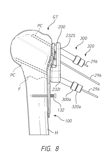
portion 244. The lateral portion 242 is the portion that is farther away from
the lateral
surface LS of the humerus H when the slot anchor guide assembly 239 is coupled
with the
humerus. The medial portion 244 is the portion that is closer to the lateral
surface LS of
the humerus H when the slot anchor guide assembly 239 is coupled with the
humerus.
The lateral portion 242 includes an elongate cylinder 246 that projects
between the
terminal lateral end of the slot anchor guide 240 and the medial portion 244.
The
elongate cylinder 246 can be configured with a ribbed outer surface along a
portion
thereof to help the surgeon grasp the slot anchor guide 240. FIG. 7F shows
that in another
embodiment a slot anchor guide 280 can be configured with a handle 282 as
discussed
further below. The lateral portion 242 also can include a lumen 248 (see
FIG.7D)
disposed therethrough. The lumen 248 can be accessed at a terminal lateral end
of the slot
anchor guide 240. The lumen 248 can extend through the lateral portion 242
adjacent to
the medial portion 244.
[0089] The
medial portion 244 can include a medial projection 250. The
medial projection 250 can be configured to mate with the slot 160. For
example, if the
slot is oval shaped the medial projection 250 can have the same oval shape.
The inferior-
superior extent 250L of the medial projection 250 can be slightly smaller than
the
-18-
CA 03078249 2020-04-01
WO 2019/075119
PCT/US2018/055290
inferior-superior extent of the slot 160. As a result, the medial projection
250 can slip
into the slot 160. The slot anchor guide 240 is coupled with the fixation
plate 100 using a
threaded interface of the drill sleeve 260, as discussed further below. In
other
embodiments the slot anchor guide 240 can be configured for positioning the
fixation
plate 100. The medial projection 250 can be made to have a small amount of
interference
fit with the slot 160 so that the fixation plate 100 can be held on the slot
anchor guide 240
as the surgeon moves the slot anchor guide 240 around. This can reduce the
amount of
direct handling of the fixation plate 100 that is needed during the procedure.
[0090] The
medial portion 244 can also include one or more anterior-posterior
projections 252. The anterior-posterior projections 252 are configured to nest
over the
portions of the fixation plate 100 that are disposed anterior and posterior of
the slot 160.
Although the drill sleeve 260 can be used to engage the slot anchor guide 240
to the
fixation plate 100 by a threaded interface, in one embodiment the anterior-
posterior
projections 252 have a smaller radius of curvature than does the slot 160 in
the anterior
and posterior directions on the anterior and posterior sides of the slot 160
and/or mating
ridges on these surfaces can be provided for the anterior-posterior projection
252 to grip
the fixation plate 100. The anterior-posterior projections 252 can flex to
grip the fixation
plate 100 in the area anterior and posterior of the slot 160.
[0091] The slot
anchor guide 240 also can include one or more cleats 254.
The cleats 254 provide for at least a temporary footing or connection to the
lateral surface
LS of the humerus H. The cleats 254 can be configured as short spikes that
project
medially of the medial projection 250. The cleats 254 can be configured to
project
medially of the medial side 112 of the fixation plate 100 when the slot anchor
guide
assembly 239 is assembled. When the combination of the fixation plate 100 and
the slot
anchor guide 240 are brought into initial contact with the lateral surface LS
of the
humerus H the cleats 254 can be pressed into the cortical bone on the lateral
surface LS
which will hold the fixation plate 100 in place as the surgeon holds the slot
anchor guide
240.
[0092] The
lumen 248 can be configured to couple with a guide sleeve, such
as any of those disclosed herein. For example, the lumen 248 can have threads
249
disposed adjacent to the terminal lateral end of the lateral portion 242. When
a guide
sleeve is disposed in the lumen 248 and mated with the threads 249 a medial
end of the
-19-
CA 03078249 2020-04-01
WO 2019/075119
PCT/US2018/055290
sleeve can be disposed adjacent to the lateral surface LS of the humerus H to
provide
access for a non-locking anchor 132 advanced through the sleeve.
[0093] As
discussed above, the slot anchor guide assembly 239 can be coupled
together using the drill sleeve 260. The drill sleeve 260 can have a lateral
portion 262, a
medial portion 264, and a lumen 268 disposed through the lateral and medial
portions
262, 264 through an elongate cylinder 266. The medial portion 264 can include
a
transverse projection 270. The transverse projection 270 can include a short
cylindrical
shoulder that is configured to mate with a superior-inferior projection 257.
The transverse
projection 270 can have a planar side that contacts a planar lateral side of
the superior-
inferior projection 257. FIG. 7A shows the superior-inferior projection 257 in
a superior
orientation relative to the fixation plate 100. As discussed above, the anchor
trajectory
guide 200 and the anchor trajectory guide 200A can be configured with a guide
groove
234. The guide groove 234 can be configured to receive the curved free end of
the
superior-inferior projection 257 so that the slot anchor guide 240 and the
anchor trajectory
guides 200, 200A can nest together in an assembly. The slot anchor guide 240
can also be
oriented 180 degrees from the orientation in FIG. 7A with the superior-
inferior projection
257 aligned with and coupled to the inferior-most anchor aperture 134. In
either
orientation a medial end of the drill sleeve 260 can be coupled to one of the
anchor
aperture 134 in the fixation plate 100 by way of threads disposed on an
outside surface of
the drill sleeve 260. The medial end of the drill sleeve 260 can be advanced
through an
anchor hole 258 in the slot anchor guide 240 until threads thereon mate with
threads in the
anchor aperture 134. Thereafter the lateral portion 242 and/or the lateral
portion 262 can
be used to manipulate any or all of the slot anchor guide assembly 239.
[0094] FIG. 7F
illustrate a slot anchor guide assembly 279 that is similar to the
slot anchor guide assembly 239 except as described differently below. The slot
anchor
guide assembly 279 includes a slot anchor guide 280 and the drill sleeve 260.
The slot
anchor guide 280 includes a handle 282. The handle 282 has a fixed end 284
that is
coupled with and extends from one side of a cylindrical body in a lateral
portion of the
slot anchor guide 280. The handle 282 extends longitudinally between a free
end 286 and
the fixed end 284. The handle 282 is configured to enable the surgeon to move
the slot
anchor guide 280 and thereby the fixation plate 100.
[0095] FIG. 7F
shows the slot anchor guide assembly 279 coupled with the
fixation plate 100 in an opposite orientation to that of FIG. 7A. The superior-
inferior
-20-
CA 03078249 2020-04-01
WO 2019/075119
PCT/US2018/055290
projection 257 of the slot anchor guide 280 is oriented inferiorly. The anchor
hole 258 in
the superior-inferior projection 257 is aligned with the distal or inferior-
most anchor
aperture 134 in the fixation plate 100. Thereafter, the drill sleeve 260 is
aligned with and
advanced through the superior-anchor hole 258 and into the anchor aperture
134. Threads
on the medial portion 264 of the drill sleeve 260 are advanced into the
threads in the
anchor aperture 134 until the transverse projection 270 comes into contact
with the lateral
side of the superior-inferior projection 257.
[0096] Both the
superior and the inferior orientations of the slot anchor guides
240, 280 allow the anchor trajectory guide 200 to be coupled with the fixation
plate 100 at
the same time as the guides 240, 280. The orientation of FIG. 2F
advantageously
provides more clearance between the inferior end of the anchor trajectory
guide 200 and
the slot anchor guides 240, 280.
[0097] In one
method the anchor trajectory guide 200 is coupled with the
fixation plate 100. For example, the medial side 208 of the anchor trajectory
guide 200
can be placed up against the lateral side 108 of the fixation plate 100. The
locator 220
anchor trajectory guide 200 can be aligned with the corresponding locating
aperture 120
of the fixation plate 100 and inserted into the aperture. FIG. 7 shows that
the profile of
the medial side 208 is matched to the profile of the lateral side 108 of the
fixation plate
100. As such the fixation plate 100 can nest into the concavity of the medial
side 208 in
the proximal portion 218 of the anchor trajectory guide 200.
[0098] Although
the locator 220 holds the position of the anchor trajectory
guide 200 on the fixation plate 100 a more complete coupling of the anchor
trajectory
guide 200 can be provided. A screw can be advanced through the fastener
aperture 237
and into the coupling aperture 172 as discussed above to provide a secure
connection that
will persist through the procedure. In another approach, the fixation plate
100 can be
secured by advancing a K-wire 296 through the pin aperture 236. Because the
pin
aperture 236 and the locator 220 converge toward the bone and are on diverging
axes
away from the bone, e.g., the longitudinal axis 222 and the longitudinal axis
238 are
converging toward other another toward the bone, the anchor trajectory guide
200 is held
in place relative to the fixation plate 100. The fixed position of the anchor
trajectory
guide 200 relative to the fixation plate 100 allows probe channel PC and
corresponding
anchor channels to be reliably formed in the correct locations. Prior to
forming such
-21-
CA 03078249 2020-04-01
WO 2019/075119
PCT/US2018/055290
channels, however, the location of the fixation plate 100 and the size of the
anchor
trajectory guide 200 can be confirmed.
[0099] FIG. 8
shows that once the anchor trajectory guide 200 is secured to the
fixation plate 100 and the non-locking anchor 132 is advanced into the bone
through the
slot 160. The slot 160 advantageously allows distal-proximal motion of the
fixation plate
100 after the non-locking anchor 132 is placed but before the plate is fully
fixed to the
humerus H. If either of the slot anchor guides 240, 280 is used to place the
fixation plate
100 initially such guides 240, 280 can be removed allowing for inferior-
superior
adjustment of the fixation plate 100 relative to the lateral surface LS of the
humerus H.
The humeral fixation plate 100A facilitates a convenient method of confirming
the
inferior-superior position of the humeral fixation plate 100A. The K-wire 296
is
advanced through an aperture in a guide 200, 200A and further through the
positioning
channel 296A at a first position of the non-locking anchor 132 along the slot
160A. As
shown in FIG. 2B the non-locking anchor 132 can be advanced until a head
portion
thereof is in contact with one of the plurality of discrete position sites
167, e.g., with one
of the concavities 167A. The K-wire 296 can be removed from the humerus H. The
position of the humeral fixation plate 100A can be evaluated. If the position
is not as
desired, the non-locking anchor 132 can be retracted sufficiently to out of
engagement
with the discrete position site 167 in which it was initially positioned. The
humeral
fixation plate 100A can be shifted relative to the non-locking anchor 132 to a
plurality of
discrete position sites 167 proximal or distal of the initial site, e.g., to a
concavity 167A
proximal or distal to the initial concavity. The non-locking anchor 132 can be
advanced
into the concavity 167A at the new position. Then, the K-wire 296 can be
advanced
through the guide 200, 200A into the humerus H through the positioning channel
296A.
The spacing between two adjacent concavities of the concavities 167A can be
enough to
assure that the K-wire 296 is not in the same position, e.g., in the channel
that was formed
in the humerus H in the first insertion. For example, the spacing between
plurality of
discrete position sites 167 can be one-half the diameter of the K-wire 296,
e.g., about 1
mm in one embodiment. In other embodiment, the spacing is less or greater. For
example, the spacing can be about 2mm, about 3mm or about 4mm in various
modified
embodiments. The visual spacing indicator 169, e.g., the lines 171, can be
positioned at
each or at alternating concavities 167A. The spacing indicator 169, e.g., the
lines 171,
can be at greater intervals, e.g., every third or fourth concavity. In one
method, after
-22-
CA 03078249 2020-04-01
WO 2019/075119
PCT/US2018/055290
advancing the K-wire 296 through the aperture positioning channel 296A at a
first
position of the slot 160A relative to the anchor non-locking anchor 132, the K-
wire is
removed from the humerus H. The position of the humeral fixation plate 100A is
shifted
proximally or distally (e.g., inferiorly or superiorly) to a second position
of the slot
relative to the anchor 132. The second position is spaced from the first
position by an
amount greater than the one-half of or the diameter of the K-wire 296. The
second
position is provided by advancing the non-locking anchor 132 into a discrete
position site
of the plurality of discrete position sites 167 spaced along the slot 160A
from the initial
site of the plurality of discrete position sites 167. The slot 160A enables
the non-locking
anchor 132 to be retracted out to the concavities 167A without having to
remove the non-
locking anchor 132 from the humerus H when shifting among these and other
positions
that are defined along the slot 160A.
[0100] The
plurality of discrete position sites 167 and the visual spacing
indicator 169 enhance the usefulness of the humeral fixation plate 100A by
allowing the
surgeon to be certain that second and subsequent positions are spaced apart
from an initial
position. By providing this guidance, the surgeon can more quickly, accurately
and
confidently proceed through the stages of methods involving the humeral
fixation plate
100A.
[0101] If the
inferior-superior position is initially confirmed, K-wires can be
advanced into the head 10 of the humerus H. A first K-wire 296 can be advanced
toward
the medial calcar MC region. Thereafter the appropriate size anchor trajectory
guide 200
can be confirmed by advancing a second K-wire 296 into a superior anterior
guide
aperture 232S. With these K-wires 296 placed, the size of the anchor
trajectory guide 200
can be confirmed. For example, if the proximal end of the anchor trajectory
guide 200 is
a prescribed distance, e.g., 10 mm, from a proximal aspect of the head 10 then
the size of
the anchor trajectory guide 200 is appropriate. The proximal aspect from which
the
distance to the anchor trajectory guide 200 is measured can be the greater
tuberosity GT.
If the proximal end of the anchor trajectory guide 200 is less than about 10
mm from the
proximal aspect of the head 10 then a smaller anchor trajectory guide 200 can
be selected.
If the proximal end of the anchor trajectory guide 200 is more than about 10
mm from the
proximal aspect of the head 10 then a larger anchor trajectory guide 200 can
be selected.
[0102] In
another embodiment, the size of the head 10 of the humerus H can
be estimated by providing a scale on a K-wire 296 to be inserted into the pin
aperture 236
-23-
CA 03078249 2020-04-01
WO 2019/075119
PCT/US2018/055290
in the center of the anchor trajectory guide 200 after the non-locking anchor
132 has
secured the fixation plate 100 to the humerus H. The trajectory of the K-wire
296 is
aligned with the center of the articular surface of the head 10. The scale can
include
markings that indicate the length of the K-wire 296 inserted into pin aperture
236, across
the cancellous portion of the head 10 into contact with the cortical bone
region CB. From
this dimension, the size of the anchor trajectory guide 200 to be used can be
determined.
In some cases the K-wire 296 can indicate a size (small, medium, large, etc.)
of the anchor
trajectory guide 200 to be used. The K-wire 296 can indicate both a dimension
and a size
in some embodiments.
[0103] After
the size and placement of the anchor trajectory guide 200 are
confirmed the fixation plate 100 can be secured to the humerus H to prevent
movement
therebetween. FIG. 9 shows that securing the fixation plate 100 to the humerus
H can be
achieved by inserting a polyaxial anchor 130 into an anchor aperture 134 in
the distal
portion 116 of the fixation plate 100. The method can then proceed to forming
probe
channels PC and thereafter to implanting anchors as discussed further below.
[0104] FIGS. 8-
18 show that guide sleeves can be used in the formation of an
inferior probe channel PC. A sleeve 300 can be mated to the anchor trajectory
guide 200.
In one technique, a medial end of the sleeve 300 can be inserted into one of
the inferior
guide apertures 2321 and advanced from the lateral side 212 toward the medial
side 208 of
the anchor trajectory guide 200. The sleeve 300 can be seated in the anchor
trajectory
guide 200 by engaging threads on the outside surface of the sleeve 300 with
internal
threads in the inferior guide aperture 2321. For example, the anchor
trajectory guide 200
can have, as illustrated above in FIG. 3A in connection with the anchor
trajectory guide
200A, internal threads 235 disposed in an anterior inferior guide aperture
232F and in a
posterior inferior guide aperture 232G that can be engaged by threads on the
exterior
surface of the sleeve 300. In other embodiments the sleeve 300 can be mated
with a slip
fit inside the inferior guide aperture 2321 or any of the other guide
apertures 232. Once the
sleeve 300 is seated it can be an outer sleeve to receive other structures and
devices. An
inner sleeve 320 can be inserted into the sleeve 300. The inner sleeve 320 can
be inserted
by advancing a medial end of the inner sleeve 320 into a lateral end of the
sleeve 300 until
flanges or hubs 300a, 320a at the lateral ends of the sleeve 300 and of the
inner sleeve 320
are coupled, e.g., are adjacent to or abutting each other. The inner sleeve
320 can be
seated in or mated to the sleeve 300 by engaging threads on an outside surface
of the inner
-24-
CA 03078249 2020-04-01
WO 2019/075119
PCT/US2018/055290
sleeve 320 with internal thread disposed in the inside of the sleeve 300. The
inner sleeve
320 can be configured to direct a K-wire 296 through the anchor trajectory
guide 200
along a guide axis that is pre-defined to an axis in the humerus H to begin
the formation
of the probe channel PC in a desired location as shown in FIG. 8. The inner
sleeve 320
can have an inner diameter that is closely matched to an outer diameter of the
K-wire 296
such that the trajectory of the K-wire 296 is controlled by the location and
orientation of
the inferior guide aperture 2321 indirectly, which controls the position and
orientation of
the sleeves 300, 320.
[0105] After
the inner sleeve 320 has been mated with the sleeve 300 and the
sleeve 300 has been mated with the anchor trajectory guide 200 the K-wire 296
can be
advanced into the humerus H through the cortical bone at the lateral surface
LS and into
the cancellous bone within the cortical bone. The trajectory of the K-wire 296
is pre-
defined by the location and orientation of the mating of the fixation plate
100 with the
humerus H and by the orientation of the guide apertures 232 as described
above.
Advancing the K-wire 296 into the humerus H defines the direction along with
the probe
channel PC will be formed.
[0106] FIG. 8
also shows a K-wire 296 can be inserted into a superior portion
of the head 10 of the humerus H through the superior guide apertures 232S.
Inserting the
K-wire 296 through the superior guide apertures 232S can include mating sleeve
300 with
the superior guide apertures 232S and inserting the inner sleeve 320 into the
sleeve 300.
For example, the anchor trajectory guide 200 can have, as illustrated above in
FIG. 3A in
connection with the anchor trajectory guide 200A, internal threads 235
disposed in an
anterior superior guide aperture 232A and a posterior superior guide aperture
232B that
can be engaged by threads on the exterior surface of the sleeve 300. As noted
above the
position of the fixation plate 100 and the anchor trajectory guide 200 are
confirmed
following insertion of the inferior and superior guide apertures. If the
position is
confirmed insertion of the K-wires 296 through the superior guide apertures
232S defines
the trajectory of the probe channel PC to be formed later the procedure.
[0107] FIG. 9
shows that in some techniques the fixation plate 100 is secured
to the humerus H by inserting the polyaxial anchor 130 through the fixation
plate 100 as
discussed above. The distal portion 116 can include a polyaxial aperture 134
disposed
distally of the slot 160. The polyaxial aperture 104 at this location is
advantageous for
providing enhanced security of the fixation plate 100 because it is farthest
from the
-25-
CA 03078249 2020-04-01
WO 2019/075119
PCT/US2018/055290
proximal portion 118 through which one or a plurality, e.g., two as
illustrated in FIG. 9,
K-wires 296 can be advanced. FIGS. 2 and 9 shows a configuration in which the
non-
locking anchor 132 is fully advanced in the slot 160 (See FIG. 2) and the
polyaxial anchor
130 is fully advanced into the distal polyaxial aperture 134 to immobilize the
fixation
plate 100 against the lateral surface LS of the humerus H.
[0108] FIGS. 10-
16A illustrate additional approaches to orient the anchor
trajectory guide 200 and the fixation plate 100 on the humerus H. FIG. 10
shows an
anatomical alignment guide 400 that can be mated to the anchor trajectory
guide 200. The
anatomical alignment guide 400 can include a sleeve 404 that is configured to
mate with
one of the guide apertures 232, e.g., one of the inferior guide apertures
2321. The sleeve
404 includes a lumen 408 that extends from a lateral side of the sleeve 404,
e.g., from a
flange or hub 406, to a medial side thereof. The lumen 408 is configured to
slideably
receive a K-wire 296 as discussed below. The anatomical alignment guide 400
also
includes a visual alignment member 412 disposed away from the sleeve 404. The
visual
alignment member 412 can include an arcuate member 418 that is coupled at a
first end
with a lateral end of the sleeve 404. The arcuate member 418 can extend in a
first portion
418A away from the sleeve 404, e.g., in a direction substantially
perpendicular to the
orientation of the lumen 408. The first portion 418A can extend sufficiently
from the
sleeve 404 to provide clearance between the head 10 of the humerus H and a
second
portion 418B of the arcuate member 418 that extends from the first portion
418B when
the anchor trajectory guide 200 is coupled with the fixation plate 100 and
thereby with the
lateral surface LS of the humerus H. The second portion 418B can be
substantially
parallel to the sleeve 404 in one embodiment. The arcuate member 418 is
configured in
the second portion 418B to extend sufficiently to be positioned or aligned
with, e.g., in
front of, pre-defined anatomy to which the K-wire 296 disposed through the
sleeve 404 is
to be aligned. As discussed further below, the arcuate member 418 is
configured to
extend away from the sleeve 404 to a position in front of (either on the
anterior or
posterior side of the humerus H) so as to be between the surgeon's eye and the
an
anatomical landmark. Thus the arcuate member 418 can provide a visual
alignment
device for visually aligning the anatomical alignment guide 400 with the
landmark.
When so aligned the surgeon knows that a K-wire 296 placed through the lumen
408 and
through the inferior guide apertures 2321 extends in a direction toward the
landmark, e.g.,
toward the medial calcar. FIGS. 13-16A illustrate this method with the
anatomic
-26-
CA 03078249 2020-04-01
WO 2019/075119
PCT/US2018/055290
alignment guide 400 and with an anatomic alignment guide 400A discussed
further
below. The guide 400A is similar to the guide 400 except as described
differently below.
[0109] In the
flow of the methods discussed above, FIGS. 13 and 14 are
alterative techniques to that shown in FIGS. 7-9. FIG. 13 shows the fixation
plate 100
mated with the lateral surface LS of the humerus H. The anchor trajectory
guide 200A is
mated with the lateral side 108 of the fixation plate 100. The anchor
trajectory guide 200
could be used, as is illustrated in some of the figures. One K-wire 296 is
placed through
the pin aperture 236 into the head 10 of the humerus H. The anatomical
alignment guide
400A is mated with the anchor trajectory guide 200A. For example, the sleeve
404 can be
inserted into the inferior guide aperture 232H. In one example, the connection
between
the sleeve 404 and the inferior guide aperture 232H can be a slip fit,
although other
connections are possible as well. The fixation plate 100 can be mated to the
humerus H
by the non-locking anchor 132 inserted into the slot 160, which permits the
fixation plate
100 to be moved proximally and distally on the humerus H. The movement of the
fixation plate 100 can be until the anatomical alignment guide 400A is aligned
with a
predefined anatomical landmark, such as the medial calcar MC. Aligning the
anatomical
alignment guide 400A with the medial calcar MC can include viewing the
anterior surface
of the head 10 of the humerus H head on and moving the assembled fixation
plate 100,
anchor trajectory guide 200A, and anatomical alignment guide 400 proximally or
distally
until the second portion 418B of the arcuate member 418 is aligned with the
medial calcar
MC. In the case of the guide 400A, the guide 400A can be moved proximally and
distally
until a K-wire 422 is aligned to the medial calcar MC
[0110]
Following alignment of the arcuate member 418 or the K-wire 422
with the medial calcar MC one or both of the K-wires 296 can be placed through
the
anchor trajectory guide 200 or the guide 200A. A superior K-wire 296 can be
placed
through the pin aperture 236 in the anchor trajectory guide 200 or the guide
200A. An
inferior K-wire 296 can be placed through the inferior guide aperture 2321 to
which the
anatomical alignment guide 400 or the guide 400A is mated. The K-wire 296 can
be
inserted through the sleeve 404 or through the inner sleeve 320 disposed in
the sleeve
404. As illustrated above in FIG. 3A the anchor trajectory guide 200A shows
that anterior
inferior guide aperture 232F and the posterior inferior guide aperture 232G
have internal
threads 235 disposed therein that can be engaged by threads on the exterior
surface of the
inner sleeve 320. In some embodiments, the size of the anchor trajectory guide
200 or the
-27-
CA 03078249 2020-04-01
WO 2019/075119
PCT/US2018/055290
guide 200A is confirmed by measuring a distance from a proximal landmark such
as the
greater tuberosity to the proximal edge of the anchor trajectory guide 200 or
the guide
200A. A distance of about lOmm indicates proper sizing of the anchor
trajectory guide
200 or the guide 200A. A smaller distance between the proximal landmark and
the
proximal edge of the anchor trajectory guide 200 or the guide 200A suggests a
smaller
humerus for which a smaller anchor trajectory guide 200 or guide 200A is
appropriate. A
larger distance between the proximal landmark and the proximal edge of the
anchor
trajectory guide 200 or the guide 200A suggests a larger humerus for which a
larger
anchor trajectory guide 200 or guide 200A is appropriate.
[0111] Other
methods of sizing the anchor trajectory guide 200 or guide 200A
can include placing K-wires 296 in an inferior guide aperture 2321 and in a
superior guide
aperture 232S. When the K-wire 296 is properly placed using the anatomical
alignment
guide 400 or the guide 400A, e.g., extending to the medial calcar MC the
position of the
K-wire 296 in the inferior guide aperture 2321 can be confirmed to not be too
far
proximally suggesting that the anchor trajectory guide 200 or the guide 200A
is too large
and not too far distally suggesting that the anchor trajectory guide 200 or
the guide 200A
is too small. As noted above, the size of the humerus H can also be assessed
by the depth
into the head 10 that a K-wire 296 inserted into the pin aperture 236 extends.
Other
methods for sizing the anchor trajectory guide 200 or guide 200A can include
measuring
an external dimension of the head 10 of the humerus H or analyzing imaging
data (pre-
operative or intra-operative).
[0112] FIG. 16
shows an alternate method in which after the K-wires 296 have
been placed a polyaxial anchor 130 can be advanced through the fixation plate
100
through the guide aperture 232 disposed in the distal portion 216 of the
anchor trajectory
guide 200 or guide 200A. The combination of the polyaxial anchor 130 and the
non-
locking anchor 132 in the slot 160 secures the fixation plate 100 to the
lateral surface LS
of the humerus H. This provides a fully secure position for the remaining
portions of the
procedure. After the fixation plate 100 is fixed to the lateral surface LS
distal of the
fracture F, a first polyaxial anchor 130 can be secured to the head 10 of the
humerus H
through the anatomical alignment guide 400 as shown in FIG. 16A or through the
guide
400A. Placing the polyaxial anchor 130 through the anatomical alignment guide
400 or
the guide 400A can be directly into the small channel formed by the K-wire 296
or can be
following further bone preparation discussed below. If a polyaxial anchor 130
is placed
-28-
CA 03078249 2020-04-01
WO 2019/075119
PCT/US2018/055290
through the anatomical alignment guide 400 or the guide 400A, following such
placement
the anatomical alignment guide 400 or guide 400A can be removed from the
anchor
trajectory guide 200 or the guide 200A.
[0113] FIGS. 10
and 16A show that one variation of the anatomical alignment
guide 400 provides a secondary sleeve 426 in addition to the arcuate member
418. The
secondary sleeve 426 is disposed between the sleeve 404 and the arcuate member
418.
Like the arcuate member 418, the secondary sleeve 426 is disposed anteriorly
of the
anterior face of the humerus H when the sleeve 404 is mated with the inferior
guide
aperture 2321. In being so placed, the secondary sleeve 426 can receive a K-
wire 296
which can slide medially until the K-wire 296 is in front of a relevant
landmark, e.g., the
medial calcar. The secondary sleeve 426 can be used when the arcuate member
418 is not
in a good position for visualizing the landmark of interest. In some
variations, the
secondary sleeve 426 and the arcuate member 418 extend along planes or
longitudinal
axes that are not aligned. This enables the arcuate member 418 to be
configured to
visually align with one anatomical landmark, e.g., the medial calcar MC, and
the
secondary sleeve 426 to be configured to visually align with a different
anatomical
landmark.
[0114] FIGS. 11
and 12 illustrate the anatomical alignment guide 400A in
more detail. The anatomical alignment guide 400A is a streamlined form of
visual
alignment aid. The anatomical alignment guide 400A has a sleeve 404 as is
described
above. Coupled with and disposed to the side of the sleeve 404 is a secondary
sleeve 426.
The secondary sleeve 426 is located at a free end of a transverse projection
428 of the
anatomical alignment guide 400A. The transverse projection 428 can have a
first end
couple with the sleeve 404 and a second end disposed away from the sleeve 404
and
coupled with the secondary sleeve 426. The secondary sleeve 426 can be
disposed to the
side of (and in use generally anteriorly of) the sleeve 404 and at the end of
the free end of
the transverse projection 428. FIG. 12 shows that the secondary sleeve 426 of
the
anatomical alignment guide 400A is disposed entirely to the side of the medial
side 208 of
the anchor trajectory guide 200. This allows the secondary sleeve 426 to be
medial of the
fixation plate 100 and of the lateral surface LS of the humerus H when applied
to the
patient. This allows the anatomical alignment guide 400A to be out of the
field of view of
the head 10 of the humerus H during portions of the method, as illustrated by
FIG. 13.
The anatomical alignment guide 400A is thus lower profile than the anatomical
alignment
-29-
CA 03078249 2020-04-01
WO 2019/075119
PCT/US2018/055290
guide 400 so that the anatomical alignment guide 400A is less obstructive when
mated
with the lateral surface LS of the humerus H by way of the anchor trajectory
guide 200
and the fixation plate 100.
[0115] The
secondary sleeve 426 of the anatomical alignment guide 400A is
configured to be coupled with a wire 422. The wire 422 can be inserted through
a lumen
in the secondary sleeve 426. Preferably the transverse projection 428 is long
enough that
when the sleeve 404 is mated with the inferior guide apertures 2321 the wire
422 can be
inserted through the secondary sleeve 426 at a location spaced anterior of an
anterior side
or posterior of a posterior side of the head 10 of the humerus H. Thus, the
wire 422 can
be advanced through the secondary sleeve 426 without entering the humerus H
but rather
being disposed away from and along-side the bone.
[0116] In use,
the wire 422 can be inserted into the secondary sleeve 426
along-side the head 10 of the humerus H, as shown in FIGS. 13-16. The fixation
plate
100 and the anchor trajectory guide 200 can be aligned to the bone (e.g., by
sliding slot
160 along the non-locking anchor 132) until the wire 422 is aligned with a
selected
anatomical landmark or locations, such as with the medial calcar MC (see FIGS.
13-16).
The transverse projection 428 is configured such that the sleeve 404 and the
secondary
sleeve 426 are aligned and the longitudinal axes of the lumens formed
therethrough also
are aligned. As such, the K-wire 296 can be inserted into the sleeve 404 and
into the
lateral surface LS of the humerus H with confidence that the K-wire 296 will
be directed
to the medial calcar MC when the wire 422 is directed along the side of the
head 10 of the
humerus H toward the medial calcar MC.
[0117] The
anatomical alignment guide 400A also provides a simple visual
confirmation at least on the anterior side thereof. A lateral length of the
wire 422 should
cover up and prevent a viewer from seeing a lateral length of the K-wire 296
disposed
through the lumen 408 of the sleeve 404 when the alignment of the fixation
plate 100 and
anchor trajectory guide 200A or 200 is proper, as seen in comparing FIGS. 13
and 14. In
other words, a viewer seeing the wire 422 head-on should only see one wire and
should
not see the K-wire 296 disposed immediately posterior or behind the wire 422.
[0118] The
anatomical alignment guides 400, 400A provide convenient way to
confirm the fixation plate 100 is properly aligned to the lateral surface LS
of the humerus
H can be fully secured by inserting the polyaxial anchor 130 into the distal
guide aperture
232.
-30-
CA 03078249 2020-04-01
WO 2019/075119
PCT/US2018/055290
[0119] FIGS. 17
and 17A show a portion of a method that can follow any of
the alignment techniques discussed above to form the probe channels PC through
the
properly sized anchor trajectory guide 200 or guide 200A. In one step, the
inner sleeve
320 disposed in the inferior guide aperture 2321 can be exchanged for an inner
sleeve 340
that is configured to receive a drill bit 344. In some cases, the drill bit
344 can be
configured with the same diameter as the K-wire 296 so this exchange is not
necessary in
all cases. In many circumstances, the drill bit 344 has a larger diameter than
the diameter
of the K-wire 296. In some instances, the drill bit 344 can be directly
inserted into the
sleeve 300 such that the inner sleeve 320 configured for the K-wire 296 can
simply be
removed and the drill bit 344 can be advanced directly through the sleeve 300.
Where the
inner sleeve 340 configured for the drill bit 344 is needed, the inner sleeve
340 can be
advanced such that the medial end thereof is advanced into an aperture at the
lateral end
of the sleeve 300. The inner sleeve 340 can be docked with the sleeve 300 in
any suitable
manner, e.g., using a threaded connection, slip fit or friction fit.
Thereafter, the drill bit
344 can be advanced into the humerus H through the lateral surface LS thereof
as shown
in FIG. 17A. The drill bit 344 can have a smooth and tapered tip that can be
configured to
initially enter the lateral surface LS enlarging the hole therethrough formed
by the K-wire
296. A length of the drill bit 344 between the tapered tip and the opposite
end of the drill
bit 344 can be threaded to enlarge the small passage formed by the K-wire 296
between
the lateral surface LS and the cortical bone region CB opposite the lateral
surface LS on
the medial side of the head 10 in some techniques. In other embodiments the
drill bit 344
is used to enlarge the opening through the lateral surface LS of the humerus H
but not to
enlarge the passage formed by the K-wire 296 all the way to the cortical bone
region CB.
[0120] FIGS. 18
and 19 show that after the drill bit 344 is removed the inner
sleeve 340 can be exchanged for a probe 360. The probe 360 can be configured
to
perform multiple functions. The probe 360 can provides a basis for choosing an
appropriate polyaxial anchor 130 from a plurality of anchors, e.g., for
choosing the
polyaxial anchor 130 that has a length that will result in a medial portion of
the polyaxial
anchor 130 being embedded in the cortical bone region CB and a lateral portion
of the
polyaxial anchor 130 engaging one of the locating apertures 120 of the
fixation plate 100.
FIG. 18 shows that the probe 360 can be inserted into the lateral end of the
sleeve 300 and
the probe 360 can dock with the sleeve 300. When the probe 360 is docked with
the
sleeve 300, an inner member of the probe 360 can be inserted all the way
across the head
-31-
CA 03078249 2020-04-01
WO 2019/075119
PCT/US2018/055290
to the cortical bone region CB. At that point, the indicia 362 at the lateral
end of the
probe 360 indicate which of the polyaxial anchor 130 is suitable for the
particular locating
aperture 120.
[0121] Another
function of the probe 360 can be to enlarge the channel
formed by the K-wire 296 from the lateral surface LS of the humerus H to the
cortical
bone region CB to fully form the probe channel PC. FIG. 19 shows that a blunt
tip 361
can be provided that is well suited for pushing through the cancellous bone
matter within
the head 10 of the humerus H to formed the probe channel PC. The blunt tip 361
preferably is rounded to displace but not attach to tissue that is engaged
thereby.
[0122] FIGS. 20-
21 show one embodiment of the probe 360 in more detail.
The probe 360 includes an outer member 364 and an inner member 365. The outer
member 364 includes a medial portion 366 and a lateral portion 368. The medial
portion
366 includes a slender cylindrical portion with a lumen formed therein. The
lateral
portion 368 includes an outer surface that can have ribs for gripping by the
surgeon and an
inner lumen formed therein. A shoulder 370 can be formed between the medial
portion
366 and the lateral portion 368. The shoulder 370 can be used in docking the
probe 360
to the sleeve 300. For example, the probe 360 can be inserted into the sleeve
300 until the
shoulder 370 abuts a lateral surface of the probe 360. The lateral portion 368
can have an
aperture 372 at a lateral end thereof.
[0123] The
inner member 365 can have a cylindrical medial portion 374 and a
graduated lateral portion 376. The cylindrical medial portion 374 can be
configured to
slide within the lumen of the medial portion 366. The cylindrical medial
portion 374 can
be rigid to push cancellous matter aside to form the probe channel PC in some
embodiments and can be configured with the blunt (e.g. rounded) tip 361 that
can be
atraumatic (e.g., configured not to hook or otherwise attach to bone matter).
The tip 361 is
configured to be inserted into the bone to enlarge the channel formed by the K-
wire 296 to
form the probe channel PC. When the tip 361 contacts the cortical bone region
CB
opposite the lateral surface LS the probe channel PC has been formed. The
graduated
lateral portion 376 can be configured to move into or out of the lateral
portion 368 of the
probe 360. The graduated lateral portion 376 can include the indicia 362
formed along a
side surface thereof. The indicia 362 can include numbers indicating the
length of the
medial portion 374 spanning the cancellous bone to the cortical bone region
CB. The
same length can inform the choice of a polyaxial anchor 130 to be used in the
particular
-32-
CA 03078249 2020-04-01
WO 2019/075119
PCT/US2018/055290
probe channel PC. The indicia 362 can include any other marking that conveys
to the
surgeon which of a plurality of different length polyaxial anchor 130 to
select (e.g., small,
medium, and large).
[0124] FIG. 22
shows that in a technique after the size of the polyaxial anchor
130 to be used is selected, the appropriately sized polyaxial anchor 130 can
be inserted
into the sleeve 300 and advanced into the probe channel PC through one of the
inferior
guide apertures 2321. After the polyaxial anchor 130 is fully inserted the
medial tip of the
polyaxial anchor 130 is lodged in the opposing cortical bone region CB and the
lateral end
is engaged with one of the distal locating apertures 120 of the fixation plate
100, as shown
in FIG. 23. Using the probe 360 ensures that the medial end of the polyaxial
anchor 130
when fully advanced does not breach the cortical bone on the medial side of
the humerus
H, e.g., is not exposed on the articular surface of the humerus H. This is
important
because it prevents the polyaxial anchor 130 from contacting the articular
surface at or on
the glenoid of the scapula as discussed above.
[0125] FIG. 23
also shows that a technique can continue with using the drill
bit 344 to facilitate enlarging an opening through the lateral surface LS of
the humerus H
formed by a K-wire 296 to enable the probe 360 to be used to aid in selection
of an
appropriately sized polyaxial anchor 130 and/or to form a probe channel PC
through the
head 10 of the humerus H. The drill bit 344 is advanced through the superior
guide
apertures 232S guided by the sleeve 300 and the inner sleeve 320.
[0126] FIGS. 24
and 25 show that the processes of forming and probing the
probe channels PC and selecting appropriate sized polyaxial anchors 130 and
thereafter
inserting the polyaxial anchor 130 into the humerus H can be continued until a
sufficient
number of polyaxial anchor 130 are implanted, e.g., more than half of the
anchor aperture
134 and some cases all of the anchor aperture 134 have a polyaxial anchor 130
secured
therein. Thereafter the anchor trajectory guide 200 can be undocked form the
lateral side
108 of the fixation plate 100. In one embodiment, a screw is removed from
coupling
aperture 172 and the fastener aperture 237 and the locator 220 can be removed
from the
locating aperture 120 to permit the anchor trajectory guide 200 to be removed
from the
fixation plate 100.
[0127] FIG. 26
shows that in some techniques at least one of the polyaxial
anchors 130 can be implanted after the anchor trajectory guide 200 is removed.
In one
technique a polyaxial anchor 130 can be implanted on the distal side of the
neck of the
-33-
CA 03078249 2020-04-01
WO 2019/075119
PCT/US2018/055290
humerus H, e.g., distal of the fracture F without any guidance from the anchor
trajectory
guide 200.
[0128] FIG. 27
shows that in some techniques a great deal of splaying is
provided among the polyaxial anchor 130. For example, some, more or all of the
polyaxial anchor(s) 130 on an anterior side of the medial-lateral and proximal-
distal plane
PL can be oriented to diverge from the medial-lateral and proximal-distal
plane PL at
their medial tips. Some, more or all of the polyaxial anchor 130 on a
posterior side of the
medial-lateral and proximal-distal plane PL can be oriented to diverge from
the medial-
lateral and proximal-distal plane PL at their medial tips. Providing at least
some splaying
creates excellent security and force spreading to assure strong connection of
the fixation
plate 100 to bone portions located proximal and distal of the fracture F.
Greater splaying
also assures that the medial end 142 of the non-locking anchor 132 will be
outside of or
around the periphery of the articular surface of the head 10. This peripheral
positioning of
the medial end 142 provides an enhanced degree of safety for the patient.
[0129] As
discussed above the anchor trajectory guide 200 can be provided in
a range of sizes. For example, the anchor trajectory guide 200 can have a
small size, a
medium size, and a large size. The large size anchor trajectory guide 200 can
provide a
greater amount of splaying than illustrated in FIG. 27 such that the polyaxial
anchor tips
are secured in the cortical bone. In the larger size the medial ends 142 can
be disposed
outside of a boundary 500 that is more anterior on one of the medial-lateral
and proximal-
distal plane PL and more posterior on the opposite side of the medial-lateral
and
proximal-distal plane PL than is the illustrated by the medial ends 142 shown
in FIG. 27.
The smaller size anchor trajectory guide 200 can provide a lesser amount of
splaying than
illustrated in FIG. 27. In the smaller size, the medial ends 142 will be
disposed inside a
boundary 504 that is less anterior on one of the medial-lateral and proximal-
distal plane
PL and less posterior on the opposite side of the medial-lateral and proximal-
distal plane
PL than is the illustrated by the medial ends 142 shown in FIG. 27. Of course
the bone
will also be smaller for patient for which the boundary 504 is appropriate. As
such the
location of the medial ends 142 will generally stay peripheral of the
articular surface of
the humerus H regardless of the size of the anchor trajectory guide 200. The
polyaxial
anchor 130 enable the same fixation plate 100 to be used with a range of
sizes, including
enabling the medial end 142 to reach the position shown or either one of the
boundaries
500, 504. This advantageously simplifies the number of kits and the inventory
needed to
-34-
CA 03078249 2020-04-01
WO 2019/075119
PCT/US2018/055290
provide great outcomes for a wide range, if not the entire population, of
patients. As
discussed above the anchor trajectory guide 200 can be patient specific to
provide the
maximal amount of splaying possible in the bone of the specific patient or to
target
maximal amount of splaying into good bone and minimize splaying into weakened
bone.
In that case, the amount of splaying is specific to the patient and can be
different for each
anchor aperture 134 and for each polyaxial anchor 130.
[0130] FIG. 28
shows an example of a kit 550 that can be provided in some
embodiments. The kit 550 includes a plurality of anchor trajectory guides 200.
Each of
the anchor trajectory guide 200 can include a different size. For example, one
of the
anchor trajectory guide 200 can be suitable for an average size humerus, one
of the anchor
trajectory guide 200 can be suitable for larger humerus sizes, and one can be
suitable for
smaller humerus sizes. The periphery of the anchor trajectory guide 200 are
generally not
different, but the locations, positions, orientations, and/or trajectories of
the guide
apertures 232 through the anchor trajectory guide 200 will be different to
assure the
medial end 142 are splayed to an appropriate degree. The kit 550 can also
include the
fixation plate 100. The kit 550 also can include the humeral fixation plate
100A. The kit
550 also can include a tuberosity fracture plate 600 discussed further below.
Further, the
kit 550 can include a plurality of polyaxial anchor 130. The polyaxial anchor
130 will be
provided in a range of sizes to facilitate the above methods which provide the
distal
(medial) tips of the anchors to be lodged in opposing cortical bone but not
breaching the
opposite (medial) side of the humerus while the proximal (lateral) ends of the
polyaxial
anchor 130 are engaged with the lateral side 108 of the fixation plate 100.
This requires a
range of sizes for the polyaxial anchor 130 disposed in the neck region and in
various
regions of the head 10 portion of the humerus H, as indicated by the probe
360. The kit
550 can also include one or a plurality of non-locking anchor 132 configured
for engaging
the slot 160 of the fixation plate 100. The kit 550 can also include a
plurality of surgical
tools, including K-wires 296, outer sleeves 300, inner K-wire sleeves 320,
inner probe
and screw sleeves 340, and probe 360. The kit 550 can comprise a kit of
implantable
components and a kit of surgical components.
-35-
CA 03078249 2020-04-01
WO 2019/075119
PCT/US2018/055290
IV. TUBEROSITY PLATES AND METHODS
[0131] FIGS.
1B, 1C, and 29-30D illustrate a tuberosity fracture plate 600 and
the use thereof to repair a tuberosity fracture TF in the humerus. The
tuberosity fracture
plate 600 includes a tuberosity end 604 and a distal portion 608. A distance
between the
tuberosity end 604 and the opposite end of the distal portion 608 can be
between about
40mm and about 70mm, e.g., between 45mm and 65mm. The distance between the
tuberosity end 604 and the opposite end of the distal portion 608 can be
between 50mm
and 60mm, e.g., about 53mm. The tuberosity end 604 includes a first portion
612 and a
second portion 616. The first portion 612 can be a portion that will overlay a
first
tuberosity, e.g., a greater tuberosity GT. The first portion 612 can be used
to secure one
side of a fractured first tuberosity GT to the rest of the humerus H. The
second portion
616 can be used to secure the other side of a fractured first tuberosity GT to
the rest of the
humerus H.
[0132] The
first portion 612 can be used to secure one side of a second
tuberosity, e.g., a lesser tuberosity LT to the rest of the humerus H. The
second portion
616 can be used to secure the other side of a fractured second tuberosity LT
to the rest of
the humerus H. A distance between a portion of the first portion 612 disposed
opposite
the longitudinal axis 648 and a portion of the second portion 616 disposed
opposite the
longitudinal axis 648 can be between about 15mm and about 45mm, e.g., between
about
20mm and about 40mm. In some cases, a distance between a portion of the first
portion
612 disposed opposite the longitudinal axis 648 and a portion of the second
portion 616
disposed opposite the longitudinal axis 648 can be between about 25mm and
about
35mm, e.g., about 29mm.
[0133] The
first portion 612 and the second portion 616 can both be used to
secure a greater tuberosity, e.g., the first portion 612 can be used on a left
humerus H to
secure the greater tuberosity GT and the second portion 616 can be used on a
right
humerus H to secure the greater tuberosity GT of the right arm.
[0134] The
tuberosity fracture plate 600 can have a plurality of screw holes.
For instance, a first screw hole 624 can be provided on the tuberosity end
604. The first
screw hole 624 can be disposed in the first portion 612 to hold a polyaxial
screw 130 to
secure a first tuberosity GT. A second screw hole 628 can be provided on the
tuberosity
fracture plate 600. For example the second screw hole 628 can be provided on
the distal
-36-
CA 03078249 2020-04-01
WO 2019/075119
PCT/US2018/055290
portion 608. The second screw hole 628 also can engage a polyaxial screw 130
to secure
the tuberosity fracture plate 600 to the humerus H, e.g., to a portion of the
humerus H not
on fractured fragment, e.g., the fragment including the first tuberosity GT.
The tuberosity
fracture plate 600 can include additional screw holes, e.g., a screw hole 640
can be
provided in the distal portion 608. A plurality of screw holes 640 can be
provided in the
distal portion 608. A screw hole 644 can be provided in the tuberosity end
604. A
plurality of screw holes 644 can be provided in the tuberosity end 604. The
screw hole or
holes 644 can be disposed away from the first screw hole 624. The screw hole
644 can
secure a portion of the humeral head when the first screw hole 624 is secured
to the
fractured and fragmented first tuberosity GT. Usually all of the screw holes
624, 644, will
be engaged with a screw 130. FIG. 299 shows that screws 130 can be placed in
each of
the screw holes of the plate 600. FIG. 30A shows that the plate 600 can have a
relief 625
formed in the screw holes 624, 644 that are off-set from the longitudinal axis
648. The
relief 625 is formed to allow the screw 130 be inserted into the plate 600
along a
trajectory that is perpendicular to a plane tangent to the lateral side of the
plate 600 at the
longitudinal axis 648. The relief 625 can enable a screw 139 to be disposed
along a
trajectory that is parallel to the trajectory of one or more screws 130
disposed through the
screw holes that are on the longitudinal axis 648, e.g., extending
perpendicular to the axis
648 through the holes 640. In other techniques the first screw hole 624 is
used to secure
the fragmented portion of the humerus H, e.g., the first tuberosity GT and the
screw hole
or holes 644 is left without any screws in it to minimize the invasiveness of
the procedure
in the humerus H.
[0135] The
tuberosity fracture plate 600 can include a bend zone 632 disposed
along the length there. The bend zone 632 can be a portion that is configured
to
preferentially bend. For example, when a load is applied to the ends of the
tuberosity
fracture plate 600 the plate bend in the bend zone 632. In one embodiment a
bend zone
632 is provided between the tuberosity end 604 and the distal portion 608. In
one
embodiment, a plurality of bend zones 632 are provided along the length of the
tuberosity
fracture plate 600. A bend zone 632 can be provided between each pair of a
plurality of
adjacent screw holes 640. The bend zone 632 enable a plate that initially
disposed along a
straight longitudinal axis 648 to be shaped to match the shape of the lateral
side of the
humerus H. The shaped form of the tuberosity fracture plate 600 can be
adjusted from
-37-
CA 03078249 2020-04-01
WO 2019/075119
PCT/US2018/055290
straight along the longitudinal axis 648 to one or a plurality of curves along
the length
from the proximal to the distal end of the tuberosity fracture plate 600.
[0136] The bend
zone 632 can be formed in any suitable way. For example,
the tuberosity fracture plate 600 can have a portion with a first thickness
654 at one or all
of the screw hole 640 and a second thickness 656 in the bend zone 632. The
second
thickness 656 can be less than the first thickness 654. The second thickness
656 can be
less than 50% of the thickness of the tuberosity fracture plate 600 outside
the bend zone
632. The thickness 656 can be between 0.5mm and 1.5mm, e.g., about lmm in
various
embodiments. The thickness 656 can be between 0.5mm and 1.5mm, e.g., about
lmm.
Without the bend zone 632 the location of the screw hole 640 would be the
location most
likely to localize bending. The location of the screw hole 640 would
correspond to a
bending location. However, because the bend zone 632 has a second thickness
656 that is
less than the first thickness 654 the bending of the tuberosity fracture plate
600 is focused
at the bend zone 632. The second thickness 656 can be one-half or less than
the first
thickness 654. The second thickness 656 can be about one-half the first
thickness 654.
The second thickness 656 can be about one-third the first thickness 654. The
second
thickness 656 can be about one-quarter the first thickness 654.
[0137] FIG. 30C
shows that the tuberosity fracture plate 600 can be pre-
formed with transverse curvature. The tuberosity fracture plate 600 can be
curved in the
tuberosity end 604 transverse to the longitudinal axis 648. The tuberosity
fracture plate
600 can be curved in the distal portion 608 transverse to the longitudinal
axis 648.
[0138] FIG. 30D
shows that one or more of the first screw hole 624, the
second screw hole 628, the screw holes 640, and the screw hole 644 can be
threaded. The
threaded holes 624, 628, 640, 644 can be configured to receive polyaxial screw
130. The
threaded holes 624, 628, 640, 644 can thus enable the polyaxial screw 130 to
be targeted
to safe anatomy of the humerus H, e.g., away from the articular surface
thereof.
[0139] FIG. 29
illustrates the method of securing a fracture of a first, e.g., a
greater tuberosity GT. The first tuberosity GT is brought into place on the
remainder of
the humerus H. The tuberosity fracture plate 600 is placed over the humerus H
and the
first tuberosity GT. A polyaxial screw 130 is advanced through the first screw
hole 624
and into the first tuberosity GT. One or more, e.g., all of the screw hole 640
receive a
polyaxial screw 130. Prior to placing some or all of the polyaxial screw 130
the
tuberosity fracture plate 600 can be shaped to match the shape of the humerus
H. The
-38-
CA 03078249 2020-04-01
WO 2019/075119
PCT/US2018/055290
tuberosity fracture plate 600 can be bent along one or more of the bend zones
632 to
create curvature along the longitudinal axis 648 that matches that of the
particular
patient's humerus H. The tuberosity fracture plate 600 can be fully secured to
the one
once shaped. In some methods, gross shaping is performed by the surgeon. The
very act
of advancing the polyaxial screw 130 into the tuberosity fracture plate 600
can cause
additional bending to further match the tuberosity fracture plate 600 to the
shape of the
humerus H of the patient.
[0140] The
tuberosity fracture plate 600 can have one or a plurality of suture
apertures 652. The plurality of suture apertures 652 can receive a suture to
secure one or
more bone fragment to the tuberosity fracture plate 600. For example, in some
cases the
first tuberosity GT is too fragmented to allow the polyaxial screw 130 to
secure the first
tuberosity GT to the tuberosity fracture plate 600. In that case, one or more
sutures can
secure the fragment(s) of the first tuberosity GT to the tuberosity fracture
plate 600 and/or
to the rest of the humerus H.
Terminology
[0141] Although
certain embodiments have been described herein, the
implants and methods described herein can interchangeably use any articular
component,
as the context may dictate.
[0142] As used
herein, the relative terms "proximal" and "distal" shall be
defined from the perspective of the humerus. Thus, proximal refers to the
direction of the
end of the humerus adjacent to the scapula and forming part of the shoulder
joint, which
may be referred to herein as the superior direction, end or portion, and
distal refers to the
direction away from proximal, which can be the end of the humerus forming part
of the
elbow joint and which may be referred to herein as the inferior direction, end
or portion of
the humerus.
[0143]
Conditional language, such as "can," "could," "might," or "may,"
unless specifically stated otherwise, or otherwise understood within the
context as used, is
generally intended to convey that certain embodiments include, while other
embodiments
do not include, certain features, elements, and/or steps. Thus, such
conditional language
is not generally intended to imply that features, elements, and/or steps are
in any way
required for one or more embodiments.
-39-
CA 03078249 2020-04-01
WO 2019/075119
PCT/US2018/055290
[0144] The
terms "comprising," "including," "having," and the like are
synonymous and are used inclusively, in an open-ended fashion, and do not
exclude
additional elements, features, acts, operations, and so forth. Also, the term
"or" is used in
its inclusive sense (and not in its exclusive sense) so that when used, for
example, to
connect a list of elements, the term "or" means one, some, or all of the
elements in the
list. In addition, the articles "a," "an," and "the" as used in this
application and the
appended claims are to be construed to mean "one or more" or "at least one"
unless
specified otherwise.
[0145] The
ranges disclosed herein also encompass any and all overlap, sub-
ranges, and combinations thereof. Language such as "up to," "at least,"
"greater than,"
"less than," "between," and the like includes the number recited. Numbers
preceded by a
term such as "about" or "approximately" include the recited numbers and should
be
interpreted based on the circumstances (e.g., as accurate as reasonably
possible under the
circumstances, for example 5%, 10%, 15%, etc.). For example, "about 1"
includes
"1." Phrases preceded by a term such as "substantially," "generally," and the
like include
the recited phrase and should be interpreted based on the circumstances (e.g.,
as much as
reasonably possible under the circumstances). For example, "substantially
spherical"
includes "spherical." Unless stated otherwise, all measurements are at
standard
conditions including temperature and pressure.
[0146] As used
herein, a phrase referring to "at least one of' a list of items
refers to any combination of those items, including single members. As an
example, "at
least one of: A, B, or C" is intended to cover: A, B, C, A and B, A and C, B
and C, and A,
B, and C. Conjunctive language such as the phrase "at least one of X, Y and
Z," unless
specifically stated otherwise, is otherwise understood with the context as
used in general
to convey that an item, term, etc. may be at least one of X, Y or Z. Thus,
such
conjunctive language is not generally intended to imply that certain
embodiments require
at least one of X, at least one of Y and at least one of Z to each be present.
[0147] Although
certain embodiments and examples have been described
herein, it should be emphasized that many variations and modifications may be
made to
the humeral head assembly shown and described in the present disclosure, the
elements of
which are to be understood as being differently combined and/or modified to
form still
further embodiments or acceptable examples. All such modifications and
variations are
intended to be included herein within the scope of this disclosure. A wide
variety of
-40-
CA 03078249 2020-04-01
WO 2019/075119
PCT/US2018/055290
designs and approaches are possible. No feature, structure, or step disclosed
herein is
essential or indispensable.
[0148] Some
embodiments have been described in connection with the
accompanying drawings. However, it should be understood that the figures are
not drawn
to scale. Distances, angles, etc. are merely illustrative and do not
necessarily bear an
exact relationship to actual dimensions and layout of the devices illustrated.
Components
can be added, removed, and/or rearranged. Further, the disclosure herein of
any particular
feature, aspect, method, property, characteristic, quality, attribute,
element, or the like in
connection with various embodiments can be used in all other embodiments set
forth
herein. Additionally, it will be recognized that any methods described herein
may be
practiced using any device suitable for performing the recited steps.
[0149] For
purposes of this disclosure, certain aspects, advantages, and novel
features are described herein. It is to be understood that not necessarily all
such
advantages may be achieved in accordance with any particular embodiment. Thus,
for
example, those skilled in the art will recognize that the disclosure may be
embodied or
carried out in a manner that achieves one advantage or a group of advantages
as taught
herein without necessarily achieving other advantages as may be taught or
suggested
herein.
[0150]
Moreover, while illustrative embodiments have been described herein,
it will be understood by those skilled in the art that the scope of the
inventions extends
beyond the specifically disclosed embodiments to any and all embodiments
having
equivalent elements, modifications, omissions, combinations or sub-
combinations of the
specific features and aspects of the embodiments (e.g., of aspects across
various
embodiments), adaptations and/or alterations, and uses of the inventions as
would be
appreciated by those in the art based on the present disclosure. The
limitations in the
claims are to be interpreted broadly based on the language employed in the
claims and not
limited to the examples described in the present specification or during the
prosecution of
the application, which examples are to be construed as non-exclusive. Further,
the actions
of the disclosed processes and methods may be modified in any manner,
including by
reordering actions and/or inserting additional actions and/or deleting
actions. It is
intended, therefore, that the specification and examples be considered as
illustrative only,
with a true scope and spirit being indicated by the claims and their full
scope of
equivalents.
-41-
CA 03078249 2020-04-01
WO 2019/075119
PCT/US2018/055290
[0151] Any
methods disclosed herein need not be performed in the order
recited. The methods disclosed herein include certain actions taken by a
practitioner;
however, they can also include any third-party instruction of those actions,
either
expressly or by implication. For example, actions such as "inserting a humeral
stem into
a humerus" include "instructing insertion of a humeral head into a humerus."
-42-