Note: Descriptions are shown in the official language in which they were submitted.
CA 03078744 2020-04-07
WO 2019/075112
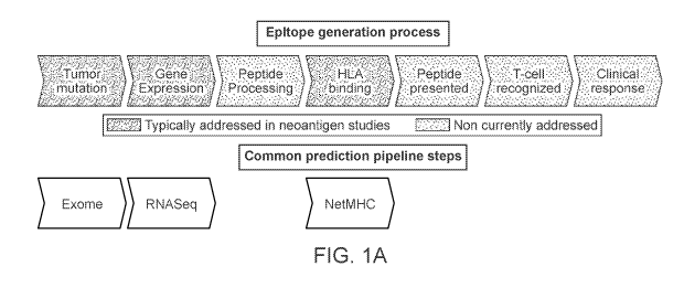
PCT/US2018/055283
TITLE
Neoantigen Identification using Hotspots
BACKGROUND
[0001] Therapeutic vaccines and T-cell therapy based on tumor-specific
neoantigens hold
great promise as a next-generation of personalized cancer immunotherapy. 1-8
Cancers with a
high mutational burden, such as non-small cell lung cancer (NSCLC) and
melanoma, are
particularly attractive targets of such therapy given the relatively greater
likelihood of
neoantigen generation. 4' 5 Early evidence shows that neoantigen-based
vaccination can elicit T-
cell responses' and that neoantigen targeted T-cell therapy can cause tumor
regression under
certain circumstances in selected patients.' Both MHC class I and MHC class II
have an impact
on T-cell responses'''.
[0002] However identification of neoantigens and neoantigen-recognizing T-
cells has
become a central challenge in assessing tumor re5pon5e577 , examining tumor
evolution"
and designing the next generation of personalized therapies112. Current
neoantigen
identification techniques are either time-consuming and laborious84,96, or
insufficiently
preci5e87,91-93. Although it has recently been demonstrated that neoantigen-
recognizing T-cells
are a major component of TIL84,96,13,"4 and circulate in the peripheral blood
of cancer
patientsi 7, current methods for identifying neoantigen-reactive T-cells have
some combination
of the following three limitations: (1) they rely on difficult-to-obtain
clinical specimens such as
TIL97,98 or leukapheresesl 7 (2) they require screening impractically large
libraries of peptides95
or (3) they rely on MHC multimers, which may practically be available for only
a small
number of MHC alleles.
[0003] Furthermore, initial methods have been proposed incorporating
mutation-based
analysis using next-generation sequencing, RNA gene expression, and prediction
of MHC
binding affinity of candidate neoantigen peptides 8. However, these proposed
methods can fail
to model the entirety of the epitope generation process, which contains many
steps (e.g., TAP
transport, proteasomal cleavage, MHC binding, transport of the peptide-MHC
complex to the
cell surface, and/or TCR recognition for MHC-I; endocytosis or autophagy,
cleavage via
extracellular or lysosomal proteases (e.g., cathepsins), competition with the
CLIP peptide for
HLA-DM-catalyzed HLA binding, transport of the peptide-MI-IC complex to the
cell surface
and/or TCR recognition for MI1C-II) in addition to gene expression and MHC
binding'.
1
CA 03078744 2020-04-07
WO 2019/075112
PCT/US2018/055283
Consequently, existing methods are likely to suffer from reduced low positive
predictive value
(PPV). (FIG. 1A)
[0004] Indeed, analyses of peptides presented by tumor cells performed by
multiple groups
have shown that <5% of peptides that are predicted to be presented using gene
expression and
MHC binding affinity can be found on the tumor surface MHC1Q11 (FIG. 1B). This
low
correlation between binding prediction and MHC presentation was further
reinforced by recent
observations of the lack of predictive accuracy improvement of binding-
restricted neoantigens
for checkpoint inhibitor response over the number of mutations alone."
[0005] This low positive predictive value (PPV) of existing methods for
predicting
presentation presents a problem for neoantigen-based vaccine design and for
neoantigen-based
T-cell therapy. If vaccines are designed using predictions with a low PPV,
most patients are
unlikely to receive a therapeutic neoantigen and fewer still are likely to
receive more than one
(even assuming all presented peptides are immunogenic). Similarly, if
therapeutic T-cells are
designed based on predictions with a low PPV, most patients are unlikely to
receive T-cells that
are reactive to tumor neoantigens and the time and physical resource cost of
identifying
predictive neoantigens using downstream laboratory techniques post-prediction
may be unduly
high. Thus, neoantigen vaccination and T-cell therapy with current methods is
unlikely to
succeed in a substantial number of subjects having tumors. (FIG. 1C)
[0006] Additionally, previous approaches generated candidate neoantigens
using only cis-
acting mutations, and largely neglected to consider additional sources of neo-
ORFs, including
mutations in splicing factors, which occur in multiple tumor types and lead to
aberrant splicing
of many genes", and mutations that create or remove protease cleavage sites.
[0007] Finally, standard approaches to tumor genome and transcriptome
analysis can miss
somatic mutations that give rise to candidate neoantigens due to suboptimal
conditions in
library construction, exome and transcriptome capture, sequencing, or data
analysis. Likewise,
standard tumor analysis approaches can inadvertently promote sequence
artifacts or germline
polymorphisms as neoantigens, leading to inefficient use of vaccine capacity
or auto-immunity
risk, respectively.
SUMMARY
[0008] Disclosed herein is an optimized approach for identifying and
selecting neoantigens
for personalized cancer vaccines, for T-cell therapy, or both. First,
optimized tumor exome and
transcriptome analysis approaches for neoantigen candidate identification
using next-
2
CA 03078744 2020-04-07
WO 2019/075112
PCT/US2018/055283
generation sequencing (NGS) are addressed. These methods build on standard
approaches for
NGS tumor analysis to ensure that the highest sensitivity and specificity
neoantigen candidates
are advanced, across all classes of genomic alteration. Second, novel
approaches for high-PPV
neoantigen selection are presented to overcome the specificity problem and
ensure that
neoantigens advanced for vaccine inclusion and/or as targets for T-cell
therapy are more likely
to elicit anti-tumor immunity. These approaches include, depending on the
embodiment,
trained statistical regression or nonlinear deep learning models that jointly
model peptide-allele
mappings as well as the per-allele motifs for peptides of multiple lengths,
sharing statistical
strength across peptides of different lengths. These deep learning models also
utilize
parameters describing the presence or absence of presentation hotspots in k-
mer blocks
associated with peptide sequences in determining presentation likelihood of
the peptides. The
nonlinear deep learning models particularly can be designed and trained to
treat different MHC
alleles in the same cell as independent, thereby addressing problems with
linear models that
would have them interfere with each other. Finally, additional considerations
for personalized
vaccine design and manufacturing based on neoantigens, and for production of
personalized
neoantigen-specific T-cells for T-cell therapy, are addressed.
[0009] The model disclosed herein outperforms state-of-the-art predictors
trained on
binding affinity and early predictors based on MS peptide data by up to an
order of magnitude.
By more reliably predicting presentation of peptides, the model enables more
time- and cost-
effective identification of neoantigen-specific or tumor antigen-specific T-
cells for personlized
therapy using a clinically practical process that uses limited volumes of
patient peripheral
blood, screens few peptides per patient, and does not necessarily rely on MHC
multimers.
However, in another embodiment, the model disclosed herein can be used to
enable more time-
and cost-effective identification of tumor antigen-specific T-cells using MHC
multimers, by
decreasing the number of peptides bound to MHC multimers that need to be
screened in order
to identify neoantigen- or tumor antigen-specific T-cells
[0010] The predictive performance of the model disclosed herein on the TIL
neoepitope
dataset and the prospective neoantigen-reactive T-cell identification task
demonstrate that it
is now possible to obtain therapeutically-useful neoepitope predictions by
modeling HLA
processing and presentation. In summary, this work offers practical in silico
antigen
identification for antigen-targeted immunotherapy, thereby accelerating
progress towards cures
for patients.
3
CA 03078744 2020-04-07
WO 2019/075112
PCT/US2018/055283
BRIEF DESCRIPTION OF THE SEVERAL VIEWS OF THE DRAWINGS
[0011] These and other features, aspects, and advantages of the present
invention will
become better understood with regard to the following description, and
accompanying
drawings, where:
[0012] FIG. lA shows current clinical approaches to neoantigen
identification.
[0013] FIG. 1B shows that <5% of predicted bound peptides are presented on
tumor cells.
[0014] FIG. 1C shows the impact of the neoantigen prediction specificity
problem.
[0015] FIG. 1D shows that binding prediction is not sufficient for
neoantigen identification.
[0016] FIG. lE shows probability of MIIC-I presentation as a function of
peptide length.
[0017] FIG. 1F shows an example peptide spectrum generated from Promega's
dynamic
range standard.
[0018] FIG. 1G shows how the addition of features increases the model
positive predictive
value.
[0019] FIG. 2A is an overview of an environment for identifying likelihoods
of peptide
presentation in patients, in accordance with an embodiment.
[0020] FIGS. 2B and 2C illustrate a method of obtaining presentation
information, in
accordance with an embodiment.
[0021] FIG. 3 is a high-level block diagram illustrating the computer logic
components of
the presentation identification system, according to one embodiment.
[0022] FIG. 4 illustrates an example set of training data, according to one
embodiment.
[0023] FIG. 5 illustrates an example network model in association with an
MHC allele.
[0024] FIG. 6A illustrates an example network model NNHO shared by MHC
alleles,
according to one embodiment.
[0025] FIG. 6B illustrates an example network model NNHO shared by MHC
alleles,
according to another embodiment.
[0026] FIG. 7 illustrates generating a presentation likelihood for a
peptide in association
with an MHC allele using an example network model.
[0027] FIG. 8 illustrates generating a presentation likelihood for a
peptide in association
with a MHC allele using example network models.
[0028] FIG. 9 illustrates generating a presentation likelihood for a
peptide in association
with MHC alleles using example network models.
[0029] FIG. 10 illustrates generating a presentation likelihood for a
peptide in association
with MHC alleles using example network models.
4
CA 03078744 2020-04-07
WO 2019/075112
PCT/US2018/055283
[0030] FIG. 11 illustrates generating a presentation likelihood for a
peptide in association
with MHC alleles using example network models.
[0031] FIG. 12 illustrates generating a presentation likelihood for a
peptide in association
with MHC alleles using example network models.
[0032] FIG. 13A illustrates a sample frequency distribution of mutation
burden in NSCLC
patients.
[0033] FIG. 13B illustrates the number of presented neoantigens in
simulated vaccines for
patients selected based on an inclusion criteria of whether the patients
satisfy a minimum
mutation burden, in accordance with an embodiment.
[0034] FIG. 13C compares the number of presented neoantigens in simulated
vaccines
between selected patients associated with vaccines including treatment subsets
identified based
on presentation models and selected patients associated with vaccines
including treatment
subsets identified through current state-of-the-art models, in accordance with
an embodiment.
[0035] FIG. 13D compares the number of presented neoantigens in simulated
vaccines
between selected patients associated with vaccines including treatment subsets
identified based
on a single per-allele presentation model for HLA-A*02:01 and selected
patients associated
with vaccines including treatment subsets identified based on both per-allele
presentation
models for HLA-A*02:01 and HLA-B*07:02. The vaccine capacity is set as v=20
epitopes, in
accordance with an embodiment.
[0036] FIG. 13E compares the number of presented neoantigens in simulated
vaccines
between patients selected based on mutation burden and patients selected by
expectation utility
score, in accordance with an embodiment.
[0037] FIG. 14 compares the positive predictive values (PPV) at 40% recall
of different
versions of the MS Model and earlier approaches to modeling HLA presented
peptides29 in
human tumors, when each model is tested on the test set comprising five
different held-out test
samples, each test sample comprising a held-out tumor sample with a 1:2500
ratio of presented
to non-presented peptides.
[0038] FIG. 15A compares the average positive predictive values (PPVs)
across recall of a
presentation model that uses presentation hotspot parameters and a
presentation model that
does not use presentation hotspot parameters, when the models are tested on
five held-out test
samples.
CA 03078744 2020-04-07
WO 2019/075112
PCT/US2018/055283
[0039] FIG. 15B compares precision and recall curves for a presentation
model that uses
presentation hotspot parameters and a presentation model that does not use
presentation hotspot
parameters, when the models are tested on a held-out test sample 0.
[0040] FIG. 15C compares precision and recall curves for a presentation
model that uses
presentation hotspot parameters and a presentation model that does not use
presentation hotspot
parameters, when the models are tested on a held-out test sample 1.
[0041] FIG. 15D compares precision and recall curves for a presentation
model that uses
presentation hotspot parameters and a presentation model that does not use
presentation hotspot
parameters, when the models are tested on a held-out test sample 2.
[0042] FIG. 15E compares precision and recall curves for a presentation
model that uses
presentation hotspot parameters and a presentation model that does not use
presentation hotspot
parameters, when the models are tested on a held-out test sample 3.
[0043] FIG. 15F compares precision and recall curves for a presentation
model that uses
presentation hotspot parameters and a presentation model that does not use
presentation hotspot
parameters, when the models are tested on a held-out test sample 4.
[0044] FIG. 16 compares the proportion of peptides that span somatic
mutations recognized
by T-cells for the top 5, 10, 20, and 30-ranked peptides identified by a
presentation model that
uses presentation hotspot parameters and by a presentation model that does not
use presentation
hotspot parameters, for a test set comprising test samples taken from patients
with at least one
pre-existing T-cell response.
[0045] FIG. 17A depicts detection of T-cell responses to patient-specific
neoantigen
peptide pools for nine patients.
[0046] FIG. 17B depicts detection of T-cell responses to individual patient-
specific
neoantigen peptides for four patients.
[0047] FIG. 17C depicts example images of ELISpot wells for patient CU04.
[0048] FIG. 18A depicts results from control experiments with neoantigens
in HLA-
matched healthy donors.
[0049] FIG. 18B depicts results from control experiments with neoantigens
in HLA-
matched healthy donors.
[0050] FIG. 19 depicts detection of T-cell responses to PHA positive
control for each donor
and each in vitro expansion depicted in FIG. 17A.
[0051] FIG. 20A depicts detection of T-cell responses to each individual
patient-specific
neoantigen peptide in pool #2 for patient CU04.
6
CA 03078744 2020-04-07
WO 2019/075112
PCT/US2018/055283
[0052] FIG. 20B depicts detection of T-cell responses to individual patient-
specific
neoantigen peptides for each of three visits of patient CUO4 and for each of
two visits of patient
1-024-002, each visit occurring at a different time point.
[0053] FIG. 20C depicts detection of T-cell responses to individual patient-
specific
neoantigen peptides and to patient-specific neoagntigen peptide pools for each
of two visits of
patient CUO4 and for each of two visits of patient 1-024-002, each visit
occurring at a different
time point.
[0054] FIG. 21 depicts detection of T-cell responses to the two patient-
specific neoantigen
peptide pools and to DMSO negative controls for the patients of FIG. 17A.
[0055] FIG. 22 compares the predictive performance of a presentation model
that uses
presentation hotspot parameters with a presentation model that does not use
presentation
hotspot parameters, when predicting presentation of neoepitopes by MHC class
II molecules.
[0056] FIG. 23 depicts a method for sequencing TCRs of neoantigen-specific
memory T-
cells from the peripheral blood of a NSCLC patient.
[0057] FIG. 24 depicts exemplary embodiments of TCR constructs for
introducing a TCR
into recipient cells.
[0058] FIG. 25 depicts an exemplary P526 construct backbone nucleotide
sequence for
cloning TCRs into expression systems for therapy development.
[0059] FIG. 26 depicts an exemplary construct sequence for cloning patient
neoantigen-
specific TCR, clonotype 1 TCR into expression systems for therapy development.
[0060] FIG. 27 depicts an exemplary construct sequence for cloning patient
neoantigen-
specific TCR, clonotype 3 into expression systems for therapy development.
[0061] FIG. 28 is a flow chart of a method for providing a customized,
neoantigen-specific
treatment to a patient, in accordance with an embodiment.
[0062] FIG. 29 illustrates an example computer for implementing the
entities shown in
FIGS. 1 and 3.
DETAILED DESCRIPTION
I. Definitions
[0063] In general, terms used in the claims and the specification are
intended to be
construed as having the plain meaning understood by a person of ordinary skill
in the art.
Certain terms are defined below to provide additional clarity. In case of
conflict between the
plain meaning and the provided definitions, the provided definitions are to be
used.
7
CA 03078744 2020-04-07
WO 2019/075112
PCT/US2018/055283
[0064] As used herein the term "antigen" is a substance that induces an
immune response.
[0065] As used herein the term "neoantigen" is an antigen that has at least
one alteration
that makes it distinct from the corresponding wild-type, parental antigen,
e.g., via mutation in a
tumor cell or post-translational modification specific to a tumor cell. A
neoantigen can include
a polypeptide sequence or a nucleotide sequence. A mutation can include a
frameshift or
nonframeshift indel, missense or nonsense substitution, splice site
alteration, genomic
rearrangement or gene fusion, or any genomic or expression alteration giving
rise to a neo0RF.
A mutations can also include a splice variant. Post-translational
modifications specific to a
tumor cell can include aberrant phosphorylation. Post-translational
modifications specific to a
tumor cell can also include a proteasome-generated spliced antigen. See Liepe
et al., A large
fraction of HLA class I ligands are proteasome-generated spliced peptides;
Science. 2016 Oct
21;354(6310):354-358.
[0066] As used herein the term "tumor neoantigen" is a neoantigen present
in a subject's
tumor cell or tissue but not in the subject's corresponding normal cell or
tissue.
[0067] As used herein the term "neoantigen-based vaccine" is a vaccine
construct based on
one or more neoantigens, e.g., a plurality of neoantigens.
[0068] As used herein the term "candidate neoantigen" is a mutation or
other aberration
giving rise to a new sequence that may represent a neoantigen.
[0069] As used herein the term "coding region" is the portion(s) of a gene
that encode
protein.
[0070] As used herein the term "coding mutation" is a mutation occurring in
a coding
region.
[0071] As used herein the term "ORF" means open reading frame.
[0072] As used herein the term "NEO-ORF" is a tumor-specific ORF arising
from a
mutation or other aberration such as splicing.
[0073] As used herein the term "missense mutation" is a mutation causing a
substitution
from one amino acid to another.
[0074] As used herein the term "nonsense mutation" is a mutation causing a
substitution
from an amino acid to a stop codon.
[0075] As used herein the term "frameshift mutation" is a mutation causing
a change in the
frame of the protein.
[0076] As used herein the term "indel" is an insertion or deletion of one
or more nucleic
acids.
8
CA 03078744 2020-04-07
WO 2019/075112
PCT/US2018/055283
[0077] As used herein, the term percent "identity," in the context of two
or more nucleic
acid or polypeptide sequences, refer to two or more sequences or subsequences
that have a
specified percentage of nucleotides or amino acid residues that are the same,
when compared
and aligned for maximum correspondence, as measured using one of the sequence
comparison
algorithms described below (e.g., BLASTP and BLASTN or other algorithms
available to
persons of skill) or by visual inspection. Depending on the application, the
percent "identity"
can exist over a region of the sequence being compared, e.g., over a
functional domain, or,
alternatively, exist over the full length of the two sequences to be compared.
[0078] For sequence comparison, typically one sequence acts as a reference
sequence to
which test sequences are compared. When using a sequence comparison algorithm,
test and
reference sequences are input into a computer, subsequence coordinates are
designated, if
necessary, and sequence algorithm program parameters are designated. The
sequence
comparison algorithm then calculates the percent sequence identity for the
test sequence(s)
relative to the reference sequence, based on the designated program
parameters. Alternatively,
sequence similarity or dissimilarity can be established by the combined
presence or absence of
particular nucleotides, or, for translated sequences, amino acids at selected
sequence positions
(e.g., sequence motifs).
[0079] Optimal alignment of sequences for comparison can be conducted,
e.g., by the local
homology algorithm of Smith & Waterman, Adv. Appl. Math. 2:482 (1981), by the
homology
alignment algorithm of Needleman & Wunsch, J. Mol. Biol. 48:443 (1970), by the
search for
similarity method of Pearson & Lipman, Proc. Nat'l. Acad. Sci. USA 85:2444
(1988), by
computerized implementations of these algorithms (GAP, BESTFIT, FASTA, and
TFASTA in
the Wisconsin Genetics Software Package, Genetics Computer Group, 575 Science
Dr.,
Madison, Wis.), or by visual inspection (see generally Ausubel et al., infra).
[0080] One example of an algorithm that is suitable for determining percent
sequence
identity and sequence similarity is the BLAST algorithm, which is described in
Altschul et al.,
J. Mol. Biol. 215:403-410 (1990). Software for performing BLAST analyses is
publicly
available through the National Center for Biotechnology Information.
[0081] As used herein the term "non-stop or read-through" is a mutation
causing the
removal of the natural stop codon.
[0082] As used herein the term "epitope" is the specific portion of an
antigen typically
bound by an antibody or T-cell receptor.
9
CA 03078744 2020-04-07
WO 2019/075112
PCT/US2018/055283
[0083] As used herein the term "immunogenic" is the ability to elicit an
immune response,
e.g., via T-cells, B cells, or both.
[0084] As used herein the term "HLA binding affinity" "MHC binding
affinity" means
affinity of binding between a specific antigen and a specific MHC allele.
[0085] As used herein the term "bait" is a nucleic acid probe used to
enrich a specific
sequence of DNA or RNA from a sample.
[0086] As used herein the term "variant" is a difference between a
subject's nucleic acids
and the reference human genome used as a control.
[0087] As used herein the term "variant call" is an algorithmic
determination of the
presence of a variant, typically from sequencing.
[0088] As used herein the term "polymorphism" is a germline variant, i.e.,
a variant found
in all DNA-bearing cells of an individual.
[0089] As used herein the term "somatic variant" is a variant arising in
non-germline cells
of an individual.
[0090] As used herein the term "allele" is a version of a gene or a version
of a genetic
sequence or a version of a protein.
[0091] As used herein the term "HLA type" is the complement of HLA gene
alleles.
[0092] As used herein the term "nonsense-mediated decay" or "NMD" is a
degradation of
an mRNA by a cell due to a premature stop codon.
[0093] As used herein the term "truncal mutation" is a mutation originating
early in the
development of a tumor and present in a substantial portion of the tumor's
cells.
[0094] As used herein the term "subclonal mutation" is a mutation
originating later in the
development of a tumor and present in only a subset of the tumor's cells.
[0095] As used herein the term "exome" is a subset of the genome that codes
for proteins.
An exome can be the collective exons of a genome.
[0096] As used herein the term "logistic regression" is a regression model
for binary data
from statistics where the logit of the probability that the dependent variable
is equal to one is
modeled as a linear function of the dependent variables.
[0097] As used herein the term "neural network" is a machine learning model
for
classification or regression consisting of multiple layers of linear
transformations followed by
element-wise nonlinearities typically trained via stochastic gradient descent
and back-
propagation.
CA 03078744 2020-04-07
WO 2019/075112
PCT/US2018/055283
[0098] As used herein the term "proteome" is the set of all proteins
expressed and/or
translated by a cell, group of cells, or individual.
[0099] As used herein the term "peptidome" is the set of all peptides
presented by MHC-I
or MI-IC-IT on the cell surface. The peptidome may refer to a property of a
cell or a collection
of cells (e.g., the tumor peptidome, meaning the union of the peptidomes of
all cells that
comprise the tumor).
[00100] As used herein the term "ELISPOT" means Enzyme-linked immunosorbent
spot
assay ¨ which is a common method for monitoring immune responses in humans and
animals.
[00101] As used herein the term "dextramers" is a dextran-based peptide-MHC
multimers
used for antigen-specific T-cell staining in flow cytometry.
[00102] As used herein the term "MHC multimers" is a peptide-MHC complex
comprising
multiple peptide- MHC monomer units.
[00103] As used herein the term "MHC tetramers" is a peptide-MI-IC complex
comprising
four peptide- MHC monomer units.
[00104] As used herein the term "tolerance or immune tolerance" is a state of
immune non-
responsiveness to one or more antigens, e.g. self-antigens.
[00105] As used herein the term "central tolerance" is a tolerance affected in
the thymus,
either by deleting self-reactive T-cell clones or by promoting self-reactive T-
cell clones to
differentiate into immunosuppressive regulatory T-cells (Tregs).
[00106] As used herein the term "peripheral tolerance" is a tolerance affected
in the
periphery by downregulating or anergizing self-reactive T-cells that survive
central tolerance or
promoting these T-cells to differentiate into Tregs.
[00107] The term "sample" can include a single cell or multiple cells or
fragments of cells or
an aliquot of body fluid, taken from a subject, by means including
venipuncture, excretion,
ejaculation, massage, biopsy, needle aspirate, lavage sample, scraping,
surgical incision, or
intervention or other means known in the art.
[00108] The term "subject" encompasses a cell, tissue, or organism, human or
non-human,
whether in vivo, ex vivo, or in vitro, male or female. The term subject is
inclusive of mammals
including humans.
[00109] The term "mammal" encompasses both humans and non-humans and includes
but is
not limited to humans, non-human primates, canines, felines, murines, bovines,
equines, and
porcine s.
11
CA 03078744 2020-04-07
WO 2019/075112
PCT/US2018/055283
[00110] The term "clinical factor" refers to a measure of a condition of a
subject, e.g.,
disease activity or severity. "Clinical factor" encompasses all markers of a
subject's health
status, including non-sample markers, and/or other characteristics of a
subject, such as, without
limitation, age and gender. A clinical factor can be a score, a value, or a
set of values that can
be obtained from evaluation of a sample (or population of samples) from a
subject or a subject
under a determined condition. A clinical factor can also be predicted by
markers and/or other
parameters such as gene expression surrogates. Clinical factors can include
tumor type, tumor
sub-type, and smoking history.
[00111] Abbreviations: MI-IC: major histocompatibility complex; HLA: human
leukocyte
antigen, or the human MHC gene locus; NGS: next-generation sequencing; PPV:
positive
predictive value; TSNA: tumor-specific neoantigen; FFPE: formalin-fixed,
paraffin-embedded;
NMD: nonsense-mediated decay; NSCLC: non-small-cell lung cancer; DC: dendritic
cell.
[00112] It should be noted that, as used in the specification and the appended
claims, the
singular forms "a," "an," and "the" include plural referents unless the
context clearly dictates
otherwise.
[00113] Any terms not directly defined herein shall be understood to have the
meanings
commonly associated with them as understood within the art of the invention.
Certain terms
are discussed herein to provide additional guidance to the practitioner in
describing the
compositions, devices, methods and the like of aspects of the invention, and
how to make or
use them. It will be appreciated that the same thing may be said in more than
one way.
Consequently, alternative language and synonyms may be used for any one or
more of the
terms discussed herein. No significance is to be placed upon whether or not a
term is
elaborated or discussed herein. Some synonyms or substitutable methods,
materials and the
like are provided. Recital of one or a few synonyms or equivalents does not
exclude use of
other synonyms or equivalents, unless it is explicitly stated. Use of
examples, including
examples of terms, is for illustrative purposes only and does not limit the
scope and meaning of
the aspects of the invention herein.
[00114] All references, issued patents and patent applications cited within
the body of the
specification are hereby incorporated by reference in their entirety, for all
purposes.
II. Methods of Identifyin2 Neoanti2ens
[00115] Disclosed herein are methods for identifying neoantigens from tumor
cells of a
subject that are likely to be presented on a surface of the tumor cells. The
method includes
obtaining exome, transcriptome, and/or whole genome nucleotide sequencing data
from the
12
CA 03078744 2020-04-07
WO 2019/075112
PCT/US2018/055283
tumor cells as well as normal cells of the subject. This nucleotide sequencing
data is used to
obtain a peptide sequence of each neoantigen in a set of neoantigens. The set
of neoantigens is
identified by comparing the nucleotide sequencing data from the tumor cells
and the nucleotide
sequencing data from the normal cells. Specifically, the peptide sequence of
each neoantigen in
the set of neoantigens comprises at least one alteration that makes it
distinct from the
corresponding wild-type peptide sequence identified from the normal cells of
the subject. The
method further includes encoding the peptide sequence of each neoantigen in
the set of
neoantigens into a corresponding numerical vector. Each numerical vector
includes information
describing the amino acids that make up the peptide sequence and the positions
of the amino
acids in the peptide sequence. The method further comprises associating the
peptide sequence
of each of the neoantigens with one or more k-mer blocks of a plurality of k-
mer blocks of the
nucleotide sequencing data of the subject,. The method further comprises
inputting the
numerical vectors and the associated k-mer blocks into a machine-learned
presentation model
to generate a presentation likelihood for each neoantigen in the set of
neoantigens. Each
presentation likelihood represents the likelihood that the corresponding
neoantigen is presented
by MHC alleles on the surface of the tumor cells of the subject. The machine-
learned
presentation model comprises a plurality of parameters and a function. The
plurality of
parameters are identified based on a training data set. The training data set
comprises, for each
sample in a plurality of samples, a label obtained by mass spectrometry
measuring presence of
peptides bound to at least one MHC allele in a set of MHC alleles identified
as present in the
sample, training peptide sequences encoded as numerical vectors that include
information
describing the amino acids that make up the peptides and the positions of the
amino acids in the
peptides, and, for each of the training peptide sequences of the sample,
associations between
the training peptide sequence and one or more k-mer blocks of a plurality of k-
mer blocks of
the nucleotide sequencing data of the training peptide sequences. The function
represents a
relation between the numerical vector and the associated k-mer blocks received
as input by the
machine-learned presentation model and the presentation likelihood generated
as output by the
machine-learned presentation model based on the numerical vector, the
associated k-mer
blocks, and the plurality of parameters. The method further includes selecting
a subset of the
set of neoantigens, based on the presentation likelihoods, to generate a set
of selected
neoantigens, and returning the set of selected neoantigens.
[00116] In some embodiments, inputting the numerical vector into the machine-
learned
presentation model comprises applying the machine-learned presentation model
to the peptide
13
CA 03078744 2020-04-07
WO 2019/075112
PCT/US2018/055283
sequence of the neoantigen to generate a dependency score for each of the MHC
alleles. The
dependency score for an MHC allele indicates whether the MHC allele will
present the
neoantigen, based on the particular amino acids at the particular positions of
the peptide
sequence. In further embodiments, inputting the numerical vector into the
machine-learned
presentation model further comprises transforming the dependency scores to
generate a
corresponding per-allele likelihood for each MHC allele indicating a
likelihood that the
corresponding MHC allele will present the corresponding neoantigen, and
combining the per-
allele likelihoods to generate the presentation likelihood of the neoantigen.
In some
embodiments, transforming the dependency scores models the presentation of the
neoantigen as
mutually exclusive across the MHC alleles. In alternative embodiments,
inputting the
numerical vector into the machine-learned presentation model further comprises
transforming a
combination of the dependency scores to generate the presentation likelihood.
In such
embodiments, transforming the combination of the dependency scores models the
presentation
of the neoantigen as interfering between the MHC alleles.
[00117] In some embodiments, the set of presentation likelihoods are further
identified by
one or more allele noninteracting features. In such embodiments, the method
further comprises
applying the machine-learned presentation model to the allele noninteracting
features to
generate a dependency score for the allele noninteracting features. The
dependency score
indicates whether the peptide sequence of the corresponding neoantigen will be
presented
based on the allele noninteracting features. In some embodiments, the one or
more allele
noninteracting features comprises the values indicating one of presence or
absence of a
presentation hotspot for each k-mer block of the peptide sequence of each
neoantigen.
[00118] In some embodiments, the method further comprises combining the
dependency
score for each MHC allele with the dependency score for the allele
noninteracting features,
transforming the combined dependency score for each MHC allele to generate a
per-allele
likelihood for each MHC allele, and combining the per-allele likelihoods to
generate the
presentation likelihood. The per-allele likelihood for a MHC allele indicates
a likelihood that
the MHC allele will present the corresponding neoantigen. In alternative
embodiments, the
method further comprises combining the dependency scores for the MHC alleles
and the
dependency score for the allele noninteracting features, and transforming the
combined
dependency scores to generate the presentation likelihood.
[00119] In some embodiments, the MHC alleles include two or more different MHC
alleles.
14
CA 03078744 2020-04-07
WO 2019/075112
PCT/US2018/055283
[00120] In some embodiments, the peptide sequences comprise peptide sequences
having
lengths other than 9 amino acids.
[00121] In some embodiments, encoding the peptide sequence comprises encoding
the
peptide sequence using a one-hot encoding scheme.
[00122] In some embodiments, the plurality of samples comprise at least one of
cell lines
engineered to express a single MHC allele, cell lines engineered to express a
plurality of MHC
alleles, human cell lines obtained or derived from a plurality of patients,
fresh or frozen tumor
samples obtained from a plurality of patients, and fresh or frozen tissue
samples obtained from
a plurality of patients.
[00123] In some embodiments, the training data set further comprises at least
one of data
associated with peptide-MI-IC binding affinity measurements for at least one
of the peptides,
and data associated with peptide-MI-IC binding stability measurements for at
least one of the
peptides.
[00124] In some embodiments, the set of presentation likelihoods are further
identified by
expression levels of the MHC alleles in the subject, as measured by RNA-seq or
mass
spectrometry.
[00125] In some embodiments, the set of presentation likelihoods are further
identified by
features comprising at least one of predicted affinity between a neoantigen in
the set of
neoantigens and the MHC alleles, and predicted stability of the neoantigen
encoded peptide-
MHC complex.
[00126] In some embodiments, the set of numerical likelihoods are further
identified by
features comprising at least one of the C-terminal sequences flanking the
neoantigen encoded
peptide sequence within its source protein sequence, and the N-terminal
sequences flanking the
neoantigen encoded peptide sequence within its source protein sequence.
[00127] In some embodiments, selecting the set of selected neoantigens
comprises selecting
neoantigens that have an increased likelihood of being presented on the tumor
cell surface
relative to unselected neoantigens, based on the machine-learned presentation
model.
[00128] In some embodiments, selecting the set of selected neoantigens
comprises selecting
neoantigens that have an increased likelihood of being capable of inducing a
tumor-specific
immune response in the subject relative to unselected neoantigens, based on
the machine-
learned presentation model.
[00129] In some embodiments, selecting the set of selected neoantigens
comprises selecting
neoantigens that have an increased likelihood of being capable of being
presented to naive T-
CA 03078744 2020-04-07
WO 2019/075112
PCT/US2018/055283
cells by professional antigen presenting cells (APCs) relative to unselected
neoantigens, based
on the presentation model. In such embodiments, the APC is optionally a
dendritic cell (DC).
[00130] In some embodiments, selecting the set of selected neoantigens
comprises selecting
neoantigens that have a decreased likelihood of being subject to inhibition
via central or
peripheral tolerance relative to unselected neoantigens, based on the machine-
learned
presentation model.
[00131] In some embodiments, selecting the set of selected neoantigens
comprises selecting
neoantigens that have a decreased likelihood of being capable of inducing an
autoimmune
response to normal tissue in the subject relative to unselected neoantigens,
based on the
machine-learned presentation model.
[00132] In some embodiments, the one or more tumor cells are selected from the
group
consisting of: lung cancer, melanoma, breast cancer, ovarian cancer, prostate
cancer, kidney
cancer, gastric cancer, colon cancer, testicular cancer, head and neck cancer,
pancreatic cancer,
brain cancer, B-cell lymphoma, acute myelogenous leukemia, chronic myelogenous
leukemia,
chronic lymphocytic leukemia, and T-cell lymphocytic leukemia, non-small cell
lung cancer,
and small cell lung cancer.
[00133] In some embodiments, the method further comprises generating an output
for
constructing a personalized cancer vaccine from the set of selected
neoantigens. In such
embodiments, the output for the personalized cancer vaccine may comprise at
least one peptide
sequence or at least one nucleotide sequence encoding the set of selected
neoantigens.
[00134] In some embodiments, the machine-learned presentation model is a
neural network
model. In such embodiments, the neural network model may include a plurality
of network
models for the MHC alleles, each network model assigned to a corresponding MHC
allele of
the MHC alleles and including a series of nodes arranged in one or more
layers. In such
embodiments, the neural network model may be trained by updating the
parameters of the
neural network model, the parameters of at least two network models being
jointly updated for
at least one training iteration. In some embodiments, the machine-learned
presentation model
may be a deep learning model that includes one or more layers of nodes.
[00135] In some embodiments, the MHC alleles are class I MHC alleles.
[00136] Also disclosed herein are computer systems comprising a computer
processor and a
memory storing computer program instructions. When the computer program
instructions are
executed by the computer processor, the instructions cause the computer
processor to carry out
any of the methods discussed above..
16
CA 03078744 2020-04-07
WO 2019/075112
PCT/US2018/055283
III. Identification of Tumor Specific Mutations in Neoanti2ens
[00137] Also disclosed herein are methods for the identification of certain
mutations (e.g.,
the variants or alleles that are present in cancer cells). In particular,
these mutations can be
present in the genome, transcriptome, proteome, or exome of cancer cells of a
subject having
cancer but not in normal tissue from the subject.
[00138] Genetic mutations in tumors can be considered useful for the
immunological
targeting of tumors if they lead to changes in the amino acid sequence of a
protein exclusively
in the tumor. Useful mutations include: (1) non-synonymous mutations leading
to different
amino acids in the protein; (2) read-through mutations in which a stop codon
is modified or
deleted, leading to translation of a longer protein with a novel tumor-
specific sequence at the
C-terminus; (3) splice site mutations that lead to the inclusion of an intron
in the mature mRNA
and thus a unique tumor-specific protein sequence; (4) chromosomal
rearrangements that give
rise to a chimeric protein with tumor-specific sequences at the junction of 2
proteins (i.e., gene
fusion); (5) frameshift mutations or deletions that lead to a new open reading
frame with a
novel tumor-specific protein sequence. Mutations can also include one or more
of
nonframeshift indel, missense or nonsense substitution, splice site
alteration, genomic
rearrangement or gene fusion, or any genomic or expression alteration giving
rise to a neo0RF.
[00139] Peptides with mutations or mutated polypeptides arising from for
example, splice-
site, frameshift, readthrough, or gene fusion mutations in tumor cells can be
identified by
sequencing DNA, RNA or protein in tumor versus normal cells.
[00140] Also mutations can include previously identified tumor specific
mutations. Known
tumor mutations can be found at the Catalogue of Somatic Mutations in Cancer
(COSMIC)
database.
[00141] A variety of methods are available for detecting the presence of a
particular
mutation or allele in an individual's DNA or RNA. Advancements in this field
have provided
accurate, easy, and inexpensive large-scale SNP genotyping. For example,
several techniques
have been described including dynamic allele-specific hybridization (DASH),
microplate array
diagonal gel electrophoresis (MADGE), pyrosequencing, oligonucleotide-specific
ligation, the
TaqMan system as well as various DNA "chip" technologies such as the
Affymetrix SNP chips.
These methods utilize amplification of a target genetic region, typically by
PCR. Still other
methods, based on the generation of small signal molecules by invasive
cleavage followed by
17
CA 03078744 2020-04-07
WO 2019/075112
PCT/US2018/055283
mass spectrometry or immobilized padlock probes and rolling-circle
amplification. Several of
the methods known in the art for detecting specific mutations are summarized
below.
[00142] PCR based detection means can include multiplex amplification of a
plurality of
markers simultaneously. For example, it is well known in the art to select PCR
primers to
generate PCR products that do not overlap in size and can be analyzed
simultaneously.
Alternatively, it is possible to amplify different markers with primers that
are differentially
labeled and thus can each be differentially detected. Of course, hybridization
based detection
means allow the differential detection of multiple PCR products in a sample.
Other techniques
are known in the art to allow multiplex analyses of a plurality of markers.
[00143] Several methods have been developed to facilitate analysis of single
nucleotide
polymorphisms in genomic DNA or cellular RNA. For example, a single base
polymorphism
can be detected by using a specialized exonuclease-resistant nucleotide, as
disclosed, e.g., in
Mundy, C. R. (U.S. Pat. No. 4,656,127). According to the method, a primer
complementary to
the allelic sequence immediately 3' to the polymorphic site is permitted to
hybridize to a target
molecule obtained from a particular animal or human. If the polymorphic site
on the target
molecule contains a nucleotide that is complementary to the particular
exonuclease-resistant
nucleotide derivative present, then that derivative will be incorporated onto
the end of the
hybridized primer. Such incorporation renders the primer resistant to
exonuclease, and thereby
permits its detection. Since the identity of the exonuclease-resistant
derivative of the sample is
known, a finding that the primer has become resistant to exonucleases reveals
that the
nucleotide(s) present in the polymorphic site of the target molecule is
complementary to that of
the nucleotide derivative used in the reaction. This method has the advantage
that it does not
require the determination of large amounts of extraneous sequence data.
[00144] A solution-based method can be used for determining the identity of a
nucleotide of
a polymorphic site. Cohen, D. et al. (French Patent 2,650,840; PCT Appin. No.
W091/02087).
As in the Mundy method of U.S. Pat. No. 4,656,127, a primer is employed that
is
complementary to allelic sequences immediately 3' to a polymorphic site. The
method
determines the identity of the nucleotide of that site using labeled
dideoxynucleotide
derivatives, which, if complementary to the nucleotide of the polymorphic site
will become
incorporated onto the terminus of the primer. An alternative method, known as
Genetic Bit
Analysis or GBA is described by Goelet, P. et al. (PCT Appin. No. 92/15712).
The method of
Goelet, P. et al. uses mixtures of labeled terminators and a primer that is
complementary to the
sequence 3' to a polymorphic site. The labeled terminator that is incorporated
is thus
18
CA 03078744 2020-04-07
WO 2019/075112
PCT/US2018/055283
determined by, and complementary to, the nucleotide present in the polymorphic
site of the
target molecule being evaluated. In contrast to the method of Cohen et al.
(French Patent
2,650,840; PCT Appin. No. W091/02087) the method of Goelet, P. et al. can be a
heterogeneous phase assay, in which the primer or the target molecule is
immobilized to a solid
phase.
[00145] Several primer-guided nucleotide incorporation procedures for assaying
polymorphic sites in DNA have been described (Komher, J. S. et al., Nucl.
Acids. Res.
17:7779-7784 (1989); Sokolov, B. P., Nucl. Acids Res. 18:3671 (1990); Syvanen,
A.-C., et al.,
Genomics 8:684-692 (1990); Kuppuswamy, M. N. et al., Proc. Natl. Acad. Sci.
(U.S.A.)
88:1143-1147 (1991); Prezant, T. R. et al., Hum. Mutat. 1:159-164 (1992);
Ugozzoli, L. et al.,
GATA 9:107-112 (1992); Nyren, P. et al., Anal. Biochem. 208:171-175 (1993)).
These
methods differ from GBA in that they utilize incorporation of labeled
deoxynucleotides to
discriminate between bases at a polymorphic site. In such a format, since the
signal is
proportional to the number of deoxynucleotides incorporated, polymorphisms
that occur in runs
of the same nucleotide can result in signals that are proportional to the
length of the run
(Syvanen, A.-C., et al., Amer. J. Hum. Genet. 52:46-59 (1993)).
[00146] A number of initiatives obtain sequence information directly from
millions of
individual molecules of DNA or RNA in parallel. Real-time single molecule
sequencing-by-
synthesis technologies rely on the detection of fluorescent nucleotides as
they are incorporated
into a nascent strand of DNA that is complementary to the template being
sequenced. In one
method, oligonucleotides 30-50 bases in length are covalently anchored at the
5' end to glass
cover slips. These anchored strands perform two functions. First, they act as
capture sites for
the target template strands if the templates are configured with capture tails
complementary to
the surface-bound oligonucleotides. They also act as primers for the template
directed primer
extension that forms the basis of the sequence reading. The capture primers
function as a fixed
position site for sequence determination using multiple cycles of synthesis,
detection, and
chemical cleavage of the dye-linker to remove the dye. Each cycle consists of
adding the
polymerase/labeled nucleotide mixture, rinsing, imaging and cleavage of dye.
In an alternative
method, polymerase is modified with a fluorescent donor molecule and
immobilized on a glass
slide, while each nucleotide is color-coded with an acceptor fluorescent
moiety attached to a
gamma-phosphate. The system detects the interaction between a fluorescently-
tagged
polymerase and a fluorescently modified nucleotide as the nucleotide becomes
incorporated
into the de novo chain. Other sequencing-by-synthesis technologies also exist.
19
CA 03078744 2020-04-07
WO 2019/075112
PCT/US2018/055283
[00147] Any suitable sequencing-by-synthesis platform can be used to identify
mutations.
As described above, four major sequencing-by-synthesis platforms are currently
available: the
Genome Sequencers from Roche/454 Life Sciences, the 1G Analyzer from
Illumina/Solexa, the
SOLiD system from Applied BioSystems, and the Heliscope system from Helicos
Biosciences.
Sequencing-by-synthesis platforms have also been described by Pacific
BioSciences and
VisiGen Biotechnologies. In some embodiments, a plurality of nucleic acid
molecules being
sequenced is bound to a support (e.g., solid support). To immobilize the
nucleic acid on a
support, a capture sequence/universal priming site can be added at the 3'
and/or 5' end of the
template. The nucleic acids can be bound to the support by hybridizing the
capture sequence to
a complementary sequence covalently attached to the support. The capture
sequence (also
referred to as a universal capture sequence) is a nucleic acid sequence
complementary to a
sequence attached to a support that may dually serve as a universal primer.
[00148] As an alternative to a capture sequence, a member of a coupling pair
(such as, e.g.,
antibody/antigen, receptor/ligand, or the avidin-biotin pair as described in,
e.g., US Patent
Application No. 2006/0252077) can be linked to each fragment to be captured on
a surface
coated with a respective second member of that coupling pair.
[00149] Subsequent to the capture, the sequence can be analyzed, for example,
by single
molecule detection/sequencing, e.g., as described in the Examples and in U.S.
Pat. No.
7,283,337, including template-dependent sequencing-by-synthesis. In sequencing-
by-synthesis,
the surface-bound molecule is exposed to a plurality of labeled nucleotide
triphosphates in the
presence of polymerase. The sequence of the template is determined by the
order of labeled
nucleotides incorporated into the 3' end of the growing chain. This can be
done in real time or
can be done in a step-and-repeat mode. For real-time analysis, different
optical labels to each
nucleotide can be incorporated and multiple lasers can be utilized for
stimulation of
incorporated nucleotides.
[00150] Sequencing can also include other massively parallel sequencing or
next generation
sequencing (NGS) techniques and platforms. Additional examples of massively
parallel
sequencing techniques and platforms are the Illumina HiSeq or MiSeq, Thermo
PGM or
Proton, the Poe Bio RS II or Sequel, Qiagen's Gene Reader, and the Oxford
Nanopore
MinION. Additional similar current massively parallel sequencing technologies
can be used, as
well as future generations of these technologies.
[00151] Any cell type or tissue can be utilized to obtain nucleic acid samples
for use in
methods described herein. For example, a DNA or RNA sample can be obtained
from a tumor
CA 03078744 2020-04-07
WO 2019/075112
PCT/US2018/055283
or a bodily fluid, e.g., blood, obtained by known techniques (e.g.
venipuncture) or saliva.
Alternatively, nucleic acid tests can be performed on dry samples (e.g. hair
or skin). In
addition, a sample can be obtained for sequencing from a tumor and another
sample can be
obtained from normal tissue for sequencing where the normal tissue is of the
same tissue type
as the tumor. A sample can be obtained for sequencing from a tumor and another
sample can
be obtained from normal tissue for sequencing where the normal tissue is of a
distinct tissue
type relative to the tumor.
[00152] Tumors can include one or more of lung cancer, melanoma, breast
cancer, ovarian
cancer, prostate cancer, kidney cancer, gastric cancer, colon cancer,
testicular cancer, head and
neck cancer, pancreatic cancer, brain cancer, B-cell lymphoma, acute
myelogenous leukemia,
chronic myelogenous leukemia, chronic lymphocytic leukemia, and T-cell
lymphocytic
leukemia, non-small cell lung cancer, and small cell lung cancer.
[00153] Alternatively, protein mass spectrometry can be used to identify or
validate the
presence of mutated peptides bound to MHC proteins on tumor cells. Peptides
can be acid-
eluted from tumor cells or from HLA molecules that are immunoprecipitated from
tumor, and
then identified using mass spectrometry.
IV. Neoanti2ens
[00154] Neoantigens can include nucleotides or polypeptides. For example, a
neoantigen
can be an RNA sequence that encodes for a polypeptide sequence. Neoantigens
useful in
vaccines can therefore include nucleotide sequences or polypeptide sequences.
[00155] Disclosed herein are isolated peptides that comprise tumor specific
mutations
identified by the methods disclosed herein, peptides that comprise known tumor
specific
mutations, and mutant polypeptides or fragments thereof identified by methods
disclosed
herein. Neoantigen peptides can be described in the context of their coding
sequence where a
neoantigen includes the nucleotide sequence (e.g., DNA or RNA) that codes for
the related
polypeptide sequence.
[00156] One or more polypeptides encoded by a neoantigen nucleotide sequence
can
comprise at least one of: a binding affinity with MHC with an IC50 value of
less than 1000nM,
for MHC Class I peptides a length of 8-15, 8, 9, 10, 11, 12, 13, 14, or 15
amino acids, presence
of sequence motifs within or near the peptide promoting proteasome cleavage,
and presence or
sequence motifs promoting TAP transport. For MHC Class II peptides a length 6-
30, 6, 7, 8, 9,
10, 11, 12, 13, 14, 15, 16, 17, 18,19, 20, 21, 22, 23, 24, 25, 26, 27, 28, 29,
or 30 amino acids,
21
CA 03078744 2020-04-07
WO 2019/075112
PCT/US2018/055283
presence of sequence motifs within or near the peptide promoting cleavage by
extracellular or
lysosomal proteases (e.g., cathepsins) or HLA-DM catalyzed HLA binding.
[00157] One or more neoantigens can be presented on the surface of a tumor.
[00158] One or more neoantigens can be is immunogenic in a subject having a
tumor, e.g.,
capable of eliciting a T-cell response or a B cell response in the subject.
[00159] One or more neoantigens that induce an autoimmune response in a
subject can be
excluded from consideration in the context of vaccine generation for a subject
having a tumor.
[00160] The size of at least one neoantigenic peptide molecule can comprise,
but is not
limited to, about 5, about 6, about 7, about 8, about 9, about 10, about 11,
about 12, about 13,
about 14, about 15, about 16, about 17, about 18, about 19, about 20, about
21, about 22, about
23, about 24, about 25, about 26, about 27, about 28, about 29, about 30,
about 31, about 32,
about 33, about 34, about 35, about 36, about 37, about 38, about 39, about
40, about 41, about
42, about 43, about 44, about 45, about 46, about 47, about 48, about 49,
about 50, about 60,
about 70, about 80, about 90, about 100, about 110, about 120 or greater amino
molecule
residues, and any range derivable therein. In specific embodiments the
neoantigenic peptide
molecules are equal to or less than 50 amino acids.
[00161] Neoantigenic peptides and polypeptides can be: for MHC Class 115
residues or less
in length and usually consist of between about 8 and about 11 residues,
particularly 9 or 10
residues; for MHC Class II, 6-30 residues, inclusive.
[00162] If desirable, a longer peptide can be designed in several ways. In one
case, when
presentation likelihoods of peptides on HLA alleles are predicted or known, a
longer peptide
could consist of either: (1) individual presented peptides with an extensions
of 2-5 amino acids
toward the N- and C-terminus of each corresponding gene product; (2) a
concatenation of some
or all of the presented peptides with extended sequences for each. In another
case, when
sequencing reveals a long (>10 residues) neoepitope sequence present in the
tumor (e.g. due to
a frameshift, read-through or intron inclusion that leads to a novel peptide
sequence), a longer
peptide would consist of: (3) the entire stretch of novel tumor-specific amino
acids--thus
bypassing the need for computational or in vitro test-based selection of the
strongest HLA-
presented shorter peptide. In both cases, use of a longer peptide allows
endogenous processing
by patient-cells and may lead to more effective antigen presentation and
induction of T-cell
responses.
[00163] Neoantigenic peptides and polypeptides can be presented on an HLA
protein. In
some aspects neoantigenic peptides and polypeptides are presented on an HLA
protein with
22
CA 03078744 2020-04-07
WO 2019/075112
PCT/US2018/055283
greater affinity than a wild-type peptide. In some aspects, a neoantigenic
peptide or polypeptide
can have an IC50 of at least less than 5000 nM, at least less than 1000 nM, at
least less than
500 nM, at least less than 250 nM, at least less than 200 nM, at least less
than 150 nM, at least
less than 100 nM, at least less than 50 nM or less.
[00164] In some aspects, neoantigenic peptides and polypeptides do not induce
an
autoimmune response and/or invoke immunological tolerance when administered to
a subject.
[00165] Also provided are compositions comprising at least two or more
neoantigenic
peptides. In some embodiments the composition contains at least two distinct
peptides. At least
two distinct peptides can be derived from the same polypeptide. By distinct
polypeptides is
meant that the peptide vary by length, amino acid sequence, or both. The
peptides are derived
from any polypeptide known to or have been found to contain a tumor specific
mutation.
Suitable polypeptides from which the neoantigenic peptides can be derived can
be found for
example in the COSMIC database. COSMIC curates comprehensive information on
somatic
mutations in human cancer. The peptide contains the tumor specific mutation.
In some aspects
the tumor specific mutation is a driver mutation for a particular cancer type.
[00166] Neoantigenic peptides and polypeptides having a desired activity or
property can be
modified to provide certain desired attributes, e.g., improved pharmacological
characteristics,
while increasing or at least retaining substantially all of the biological
activity of the
unmodified peptide to bind the desired MHC molecule and activate the
appropriate T-cell. For
instance, neoantigenic peptide and polypeptides can be subject to various
changes, such as
substitutions, either conservative or non-conservative, where such changes
might provide for
certain advantages in their use, such as improved MHC binding, stability or
presentation. By
conservative substitutions is meant replacing an amino acid residue with
another which is
biologically and/or chemically similar, e.g., one hydrophobic residue for
another, or one polar
residue for another. The substitutions include combinations such as Gly, Ala;
Val, Ile, Leu,
Met; Asp, Glu; Asn, Gln; Ser, Thr; Lys, Arg; and Phe, Tyr. The effect of
single amino acid
substitutions may also be probed using D-amino acids. Such modifications can
be made using
well known peptide synthesis procedures, as described in e.g., Merrifield,
Science 232:341-347
(1986), Barany & Merrifield, The Peptides, Gross & Meienhofer, eds. (N.Y.,
Academic Press),
pp. 1-284 (1979); and Stewart & Young, Solid Phase Peptide Synthesis,
(Rockford, Ill.,
Pierce), 2d Ed. (1984).
[00167] Modifications of peptides and polypeptides with various amino acid
mimetics or
unnatural amino acids can be particularly useful in increasing the stability
of the peptide and
23
CA 03078744 2020-04-07
WO 2019/075112
PCT/US2018/055283
polypeptide in vivo. Stability can be assayed in a number of ways. For
instance, peptidases and
various biological media, such as human plasma and serum, have been used to
test stability.
See, e.g., Verhoef et al., Eur. J. Drug Metab Pharmacokin. 11:291-302 (1986).
Half-life of the
peptides can be conveniently determined using a 25% human serum (v/v) assay.
The protocol
is generally as follows. Pooled human serum (Type AB, non-heat inactivated) is
delipidated by
centrifugation before use. The serum is then diluted to 25% with RPMI tissue
culture media
and used to test peptide stability. At predetermined time intervals a small
amount of reaction
solution is removed and added to either 6% aqueous trichloracetic acid or
ethanol. The cloudy
reaction sample is cooled (4 degrees C) for 15 minutes and then spun to pellet
the precipitated
serum proteins. The presence of the peptides is then determined by reversed-
phase HPLC using
stability-specific chromatography conditions.
[00168] The peptides and polypeptides can be modified to provide desired
attributes other
than improved serum half-life. For instance, the ability of the peptides to
induce CTL activity
can be enhanced by linkage to a sequence which contains at least one epitope
that is capable of
inducing a T helper cell response. Immunogenic peptides/T helper conjugates
can be linked by
a spacer molecule. The spacer is typically comprised of relatively small,
neutral molecules,
such as amino acids or amino acid mimetics, which are substantially uncharged
under
physiological conditions. The spacers are typically selected from, e.g., Ala,
Gly, or other
neutral spacers of nonpolar amino acids or neutral polar amino acids. It will
be understood that
the optionally present spacer need not be comprised of the same residues and
thus can be a
hetero- or homo-oligomer. When present, the spacer will usually be at least
one or two
residues, more usually three to six residues. Alternatively, the peptide can
be linked to the T
helper peptide without a spacer.
[00169] A neoantigenic peptide can be linked to the T helper peptide either
directly or via a
spacer either at the amino or carboxy terminus of the peptide. The amino
terminus of either the
neoantigenic peptide or the T helper peptide can be acylated. Exemplary T
helper peptides
include tetanus toxoid 830-843, influenza 307-319, malaria circumsporozoite
382-398 and 378-
389.
[00170] Proteins or peptides can be made by any technique known to those of
skill in the art,
including the expression of proteins, polypeptides or peptides through
standard molecular
biological techniques, the isolation of proteins or peptides from natural
sources, or the chemical
synthesis of proteins or peptides. The nucleotide and protein, polypeptide and
peptide
sequences corresponding to various genes have been previously disclosed, and
can be found at
24
CA 03078744 2020-04-07
WO 2019/075112
PCT/US2018/055283
computerized databases known to those of ordinary skill in the art. One such
database is the
National Center for Biotechnology Information's Genbank and GenPept databases
located at
the National Institutes of Health website. The coding regions for known genes
can be amplified
and/or expressed using the techniques disclosed herein or as would be known to
those of
ordinary skill in the art. Alternatively, various commercial preparations of
proteins,
polypeptides and peptides are known to those of skill in the art.
[00171] In a further aspect a neoantigen includes a nucleic acid (e.g.
polynucleotide) that
encodes a neoantigenic peptide or portion thereof The polynucleotide can be,
e.g., DNA,
cDNA, PNA, CNA, RNA (e.g., mRNA), either single- and/or double-stranded, or
native or
stabilized forms of polynucleotides, such as, e.g., polynucleotides with a
phosphorothiate
backbone, or combinations thereof and it may or may not contain introns. A
still further aspect
provides an expression vector capable of expressing a polypeptide or portion
thereof
Expression vectors for different-cell types are well known in the art and can
be selected
without undue experimentation. Generally, DNA is inserted into an expression
vector, such as a
plasmid, in proper orientation and correct reading frame for expression. If
necessary, DNA can
be linked to the appropriate transcriptional and translational regulatory
control nucleotide
sequences recognized by the desired host, although such controls are generally
available in the
expression vector. The vector is then introduced into the host through
standard techniques.
Guidance can be found e.g. in Sambrook et al. (1989) Molecular Cloning, A
Laboratory
Manual, Cold Spring Harbor Laboratory, Cold Spring Harbor, N.Y.
IV. Vaccine Compositions
[00172] Also disclosed herein is an immunogenic composition, e.g., a vaccine
composition,
capable of raising a specific immune response, e.g., a tumor-specific immune
response.
Vaccine compositions typically comprise a plurality of neoantigens, e.g.,
selected using a
method described herein. Vaccine compositions can also be referred to as
vaccines.
[00173] A vaccine can contain between 1 and 30 peptides, 2, 3, 4, 5, 6, 7,
8, 9, 10, 11, 12,
13, 14, 15, 16, 17, 18, 19, 20, 21, 22, 23, 24, 25, 26, 27, 28, 29, or 30
different peptides, 6, 7, 8,
9, 10 11, 12, 13, or 14 different peptides, or 12, 13 or 14 different
peptides. Peptides can
include post-translational modifications. A vaccine can contain between 1 and
100 or more
nucleotide sequences, 2, 3, 4, 5, 6, 7, 8, 9, 10, 11, 12, 13, 14, 15, 16, 17,
18, 19, 20, 21, 22, 23,
24, 25, 26, 27, 28, 29, 30, 31, 32, 33, 34, 35, 36, 37, 38, 39, 40, 41, 42,
43, 44, 45, 46, 47, 48,
49, 50, 51, 52, 53, 54, 55, 56, 57, 58, 59, 60, 61, 62, 63, 64, 65, 66, 67,
68, 69, 70, 71, 72, 73,
74, 75, 76, 77, 78, 79, 80, 81, 82, 83, 84, 85, 86, 87, 88, 89, 90, 91, 92,
93, 94,95, 96, 97, 98,
CA 03078744 2020-04-07
WO 2019/075112
PCT/US2018/055283
99, 100 or more different nucleotide sequences, 6,7, 8,9, 10 11, 12, 13, or 14
different
nucleotide sequences, or 12, 13 or 14 different nucleotide sequences. A
vaccine can contain
between 1 and 30 neoantigen sequences, 2, 3, 4, 5, 6, 7, 8, 9, 10, 11, 12, 13,
14, 15, 16, 17, 18,
19, 20, 21, 22, 23, 24, 25, 26, 27, 28, 29, 30, 31, 32, 33, 34, 35, 36, 37,
38, 39, 40, 41, 42, 43,
44, 45, 46, 47, 48, 49, 50, 51, 52, 53, 54, 55, 56, 57, 58, 59, 60, 61, 62,
63, 64, 65, 66, 67, 68,
69, 70, 71, 72, 73, 74, 75, 76, 77, 78, 79, 80, 81, 82, 83, 84, 85, 86, 87,
88, 89, 90, 91, 92, 93,
94,95, 96, 97, 98, 99, 100 or more different neoantigen sequences, 6, 7, 8, 9,
10 11, 12, 13, or
14 different neoantigen sequences, or 12, 13 or 14 different neoantigen
sequences.
[00174] In one embodiment, different peptides and/or polypeptides or
nucleotide sequences
encoding them are selected so that the peptides and/or polypeptides capable of
associating with
different MHC molecules, such as different MHC class I molecules and/or
different MHC class
II molecules. In some aspects, one vaccine composition comprises coding
sequence for
peptides and/or polypeptides capable of associating with the most frequently
occurring MHC
class I molecules and/or MHC class II molecules. Hence, vaccine compositions
can comprise
different fragments capable of associating with at least 2 preferred, at least
3 preferred, or at
least 4 preferred MHC class I molecules and/or MHC class II molecules.
[00175] The vaccine composition can be capable of raising a specific cytotoxic
T-cells
response and/or a specific helper T-cell response.
[00176] A vaccine composition can further comprise an adjuvant and/or a
carrier. Examples
of useful adjuvants and carriers are given herein below. A composition can be
associated with a
carrier such as e.g. a protein or an antigen-presenting cell such as e.g. a
dendritic cell (DC)
capable of presenting the peptide to a T-cell.
[00177] Adjuvants are any substance whose admixture into a vaccine composition
increases
or otherwise modifies the immune response to a neoantigen. Carriers can be
scaffold structures,
for example a polypeptide or a polysaccharide, to which a neoantigen, is
capable of being
associated. Optionally, adjuvants are conjugated covalently or non-covalently.
[00178] The ability of an adjuvant to increase an immune response to an
antigen is typically
manifested by a significant or substantial increase in an immune-mediated
reaction, or
reduction in disease symptoms. For example, an increase in humoral immunity is
typically
manifested by a significant increase in the titer of antibodies raised to the
antigen, and an
increase in T-cell activity is typically manifested in increased cell
proliferation, or cellular
cytotoxicity, or cytokine secretion. An adjuvant may also alter an immune
response, for
26
CA 03078744 2020-04-07
WO 2019/075112
PCT/US2018/055283
example, by changing a primarily humoral or Th response into a primarily
cellular, or Th
response.
[00179] Suitable adjuvants include, but are not limited to 1018 ISS, alum,
aluminium salts,
Amplivax, A515, BCG, CP-870,893, CpG7909, CyaA, dSLIM, GM-CSF, IC30, IC31,
Imiquimod, ImuFact IMP321, IS Patch, ISS, ISCOMATRIX, JuvImmune, LipoVac,
MF59,
monophosphoryl lipid A, Montanide IMS 1312, Montanide ISA 206, Montanide ISA
50V,
Montanide ISA-51, OK-432, 0M-174, 0M-197-MP-EC, ONTAK, PepTel vector system,
PLG
microparticles, resiquimod, 5RL172, Virosomes and other Virus-like particles,
YF-17D, VEGF
trap, R848, beta-glucan, Pam3Cys, Aquila's Q521 stimulon (Aquila Biotech,
Worcester, Mass.,
USA) which is derived from saponin, mycobacterial extracts and synthetic
bacterial cell wall
mimics, and other proprietary adjuvants such as Ribi's Detox. Quil or
Superfos. Adjuvants such
as incomplete Freund's or GM-CSF are useful. Several immunological adjuvants
(e.g., MF59)
specific for dendritic cells and their preparation have been described
previously (Dupuis M, et
al., Cell Immunol. 1998; 186(1):18-27; Allison A C; Dev Biol Stand. 1998; 92:3-
11). Also
cytokines can be used. Several cytokines have been directly linked to
influencing dendritic cell
migration to lymphoid tissues (e.g., TNF-alpha), accelerating the maturation
of dendritic cells
into efficient antigen-presenting cells for T-lymphocytes (e.g., GM-CSF, IL-1
and IL-4) (U.S.
Pat. No. 5,849,589, specifically incorporated herein by reference in its
entirety) and acting as
immunoadjuvants (e.g., IL-12) (Gabrilovich D I, et al., J Immunother Emphasis
Tumor
Immunol. 1996 (6):414-418).
[00180] CpG immunostimulatory oligonucleotides have also been reported to
enhance the
effects of adjuvants in a vaccine setting. Other TLR binding molecules such as
RNA binding
TLR 7, TLR 8 and/or TLR 9 may also be used.
[00181] Other examples of useful adjuvants include, but are not limited to,
chemically
modified CpGs (e.g. CpR, Idera), Poly(I:C)(e.g. polyi:Cl2U), non-CpG bacterial
DNA or RNA
as well as immunoactive small molecules and antibodies such as
cyclophosphamide, sunitinib,
bevacizumab, celebrex, NCX-4016, sildenafil, tadalafil, vardenafil, sorafinib,
XL-999, CP-
547632, pazopanib, ZD2171, AZD2171, ipilimumab, tremelimumab, and SC58175,
which may
act therapeutically and/or as an adjuvant. The amounts and concentrations of
adjuvants and
additives can readily be determined by the skilled artisan without undue
experimentation.
Additional adjuvants include colony-stimulating factors, such as Granulocyte
Macrophage
Colony Stimulating Factor (GM-CSF, sargramostim).
27
CA 03078744 2020-04-07
WO 2019/075112
PCT/US2018/055283
[00182] A vaccine composition can comprise more than one different adjuvant.
Furthermore, a therapeutic composition can comprise any adjuvant substance
including any of
the above or combinations thereof. It is also contemplated that a vaccine and
an adjuvant can
be administered together or separately in any appropriate sequence.
[00183] A carrier (or excipient) can be present independently of an adjuvant.
The function of
a carrier can for example be to increase the molecular weight of in particular
mutant to increase
activity or immunogenicity, to confer stability, to increase the biological
activity, or to increase
serum half-life. Furthermore, a carrier can aid presenting peptides to T-
cells. A carrier can be
any suitable carrier known to the person skilled in the art, for example a
protein or an antigen
presenting cell. A carrier protein could be but is not limited to keyhole
limpet hemocyanin,
serum proteins such as transferrin, bovine serum albumin, human serum albumin,
thyroglobulin
or ovalbumin, immunoglobulins, or hormones, such as insulin or palmitic acid.
For
immunization of humans, the carrier is generally a physiologically acceptable
carrier
acceptable to humans and safe. However, tetanus toxoid and/or diptheria toxoid
are suitable
carriers. Alternatively, the carrier can be dextrans for example sepharose.
[00184] Cytotoxic T-cells (CTLs) recognize an antigen in the form of a peptide
bound to an
MHC molecule rather than the intact foreign antigen itself The MHC molecule
itself is located
at the cell surface of an antigen presenting cell. Thus, an activation of CTLs
is possible if a
trimeric complex of peptide antigen, MHC molecule, and APC is present.
Correspondingly, it
may enhance the immune response if not only the peptide is used for activation
of CTLs, but if
additionally APCs with the respective MHC molecule are added. Therefore, in
some
embodiments a vaccine composition additionally contains at least one antigen
presenting cell.
[00185] Neoantigens can also be included in viral vector-based vaccine
platforms, such as
vaccinia, fowlpox, self-replicating alphavirus, marabavirus, adenovirus (See,
e.g., Tatsis et al.,
Adenoviruses, Molecular Therapy (2004) 10, 616-629), or lentivirus, including
but not
limited to second, third or hybrid second/third generation lentivirus and
recombinant lentivirus
of any generation designed to target specific cell types or receptors (See,
e.g., Hu et al.,
Immunization Delivered by Lentiviral Vectors for Cancer and Infectious
Diseases, Immunol
Rev. (2011) 239(1): 45-61, Sakuma et al., Lentiviral vectors: basic to
translational, Biochem I
(2012) 443(3):603-18, Cooper et al., Rescue of splicing-mediated intron loss
maximizes
expression in lentiviral vectors containing the human ubiquitin C promoter,
Nucl. Acids Res.
(2015) 43 (1): 682-690, Zufferey et al., Self-Inactivating Lentivirus Vector
for Safe and
Efficient In Vivo Gene Delivery, I Virol. (1998) 72 (12): 9873-9880).
Dependent on the
28
CA 03078744 2020-04-07
WO 2019/075112
PCT/US2018/055283
packaging capacity of the above mentioned viral vector-based vaccine
platforms, this approach
can deliver one or more nucleotide sequences that encode one or more
neoantigen peptides.
The sequences may be flanked by non-mutated sequences, may be separated by
linkers or may
be preceded with one or more sequences targeting a subcellular compartment
(See, e.g., Gros et
al., Prospective identification of neoantigen-specific lymphocytes in the
peripheral blood of
melanoma patients, Nat Med. (2016) 22 (4):433-8, Stronen et al., Targeting of
cancer
neoantigens with donor-derived T-cell receptor repertoires, Science. (2016)
352 (6291):1337-
41, Lu et al., Efficient identification of mutated cancer antigens recognized
by T-cells
associated with durable tumor regressions, Clin Cancer Res. (2014) 20(
13):3401-10). Upon
introduction into a host, infected cells express the neoantigens, and thereby
elicit a host
immune (e.g., CTL) response against the peptide(s). Vaccinia vectors and
methods useful in
immunization protocols are described in, e.g., U.S. Pat. No. 4,722,848.
Another vector is BCG
(Bacille Calmette Guerin). BCG vectors are described in Stover et al. (Nature
351:456-460
(1991)). A wide variety of other vaccine vectors useful for therapeutic
administration or
immunization of neoantigens, e.g., Salmonella typhi vectors, and the like will
be apparent to
those skilled in the art from the description herein.
IV.A. Additional Considerations for Vaccine Desi2n and Manufacture
IV.A.1. Determination of a set of peptides that cover all tumor
subclones
[00186] Truncal peptides, meaning those presented by all or most tumor
subclones, will be
prioritized for inclusion into the vaccine.53 Optionally, if there are no
truncal peptides predicted
to be presented and immunogenic with high probability, or if the number of
truncal peptides
predicted to be presented and immunogenic with high probability is small
enough that
additional non-truncal peptides can be included in the vaccine, then further
peptides can be
prioritized by estimating the number and identity of tumor subclones and
choosing peptides so
as to maximize the number of tumor subclones covered by the vaccine.54
IV.A.2. Neoanti2en prioritization
[00187] After all of the above neoantigen filters are applied, more candidate
neoantigens
may still be available for vaccine inclusion than the vaccine technology can
support.
Additionally, uncertainty about various aspects of the neoantigen analysis may
remain and
tradeoffs may exist between different properties of candidate vaccine
neoantigens. Thus, in
place of predetermined filters at each step of the selection process, an
integrated multi-
29
CA 03078744 2020-04-07
WO 2019/075112
PCT/US2018/055283
dimensional model can be considered that places candidate neoantigens in a
space with at least
the following axes and optimizes selection using an integrative approach.
1. Risk of auto-immunity or tolerance (risk of germline) (lower risk of
auto-immunity is
typically preferred)
2. Probability of sequencing artifact (lower probability of artifact is
typically preferred)
3. Probability of immunogenicity (higher probability of immunogenicity is
typically
preferred)
4. Probability of presentation (higher probability of presentation is
typically preferred)
5. Gene expression (higher expression is typically preferred)
6. Coverage of HLA genes (larger number of HLA molecules involved in the
presentation
of a set of neoantigens may lower the probability that a tumor will escape
immune
attack via downregulation or mutation of HLA molecules)
7. Coverage of HLA classes (covering both HLA-I and HLA-II may increase the
probability of therapeutic response and decrease the probability of tumor
escape)
V. Therapeutic and Manufacturin2 Methods
[00188] Also provided is a method of inducing a tumor specific immune response
in a
subject, vaccinating against a tumor, treating and or alleviating a symptom of
cancer in a
subject by administering to the subject one or more neoantigens such as a
plurality of
neoantigens identified using methods disclosed herein.
[00189] In some aspects, a subject has been diagnosed with cancer or is at
risk of developing
cancer. A subject can be a human, dog, cat, horse or any animal in which a
tumor specific
immune response is desired. A tumor can be any solid tumor such as breast,
ovarian, prostate,
lung, kidney, gastric, colon, testicular, head and neck, pancreas, brain,
melanoma, and other
tumors of tissue organs and hematological tumors, such as lymphomas and
leukemias,
including acute myelogenous leukemia, chronic myelogenous leukemia, chronic
lymphocytic
leukemia, T-cell lymphocytic leukemia, and B cell lymphomas.
[00190] A neoantigen can be administered in an amount sufficient to induce a
CTL
response.
[00191] A neoantigen can be administered alone or in combination with other
therapeutic
agents. The therapeutic agent is for example, a chemotherapeutic agent,
radiation, or
immunotherapy. Any suitable therapeutic treatment for a particular cancer can
be administered.
[00192] In addition, a subject can be further administered an anti-
immunosuppressive/immunostimulatory agent such as a checkpoint inhibitor. For
example, the
subject can be further administered an anti-CTLA antibody or anti-PD-1 or anti-
PD-Li.
Blockade of CTLA-4 or PD-Li by antibodies can enhance the immune response to
cancerous
CA 03078744 2020-04-07
WO 2019/075112
PCT/US2018/055283
cells in the patient. In particular, CTLA-4 blockade has been shown effective
when following a
vaccination protocol.
[00193] The optimum amount of each neoantigen to be included in a vaccine
composition
and the optimum dosing regimen can be determined. For example, a neoantigen or
its variant
can be prepared for intravenous (i.v.) injection, sub-cutaneous (s.c.)
injection, intradermal (i.d.)
injection, intraperitoneal (i.p.) injection, intramuscular (i.m.) injection.
Methods of injection
include s.c., i.d., i.p., i.m., and i.v. Methods of DNA or RNA injection
include i.d., i.m., s.c.,
i.p. and i.v. Other methods of administration of the vaccine composition are
known to those
skilled in the art.
[00194] A vaccine can be compiled so that the selection, number and/or amount
of
neoantigens present in the composition is/are tissue, cancer, and/or patient-
specific. For
instance, the exact selection of peptides can be guided by expression patterns
of the parent
proteins in a given tissue. The selection can be dependent on the specific
type of cancer, the
status of the disease, earlier treatment regimens, the immune status of the
patient, and, of
course, the HLA-haplotype of the patient. Furthermore, a vaccine can contain
individualized
components, according to personal needs of the particular patient. Examples
include varying
the selection of neoantigens according to the expression of the neoantigen in
the particular
patient or adjustments for secondary treatments following a first round or
scheme of treatment.
[00195] For a composition to be used as a vaccine for cancer, neoantigens with
similar
normal self-peptides that are expressed in high amounts in normal tissues can
be avoided or be
present in low amounts in a composition described herein. On the other hand,
if it is known that
the tumor of a patient expresses high amounts of a certain neoantigen, the
respective
pharmaceutical composition for treatment of this cancer can be present in high
amounts and/or
more than one neoantigen specific for this particularly neoantigen or pathway
of this
neoantigen can be included.
[00196] Compositions comprising a neoantigen can be administered to an
individual already
suffering from cancer. In therapeutic applications, compositions are
administered to a patient in
an amount sufficient to elicit an effective CTL response to the tumor antigen
and to cure or at
least partially arrest symptoms and/or complications. An amount adequate to
accomplish this is
defined as "therapeutically effective dose." Amounts effective for this use
will depend on, e.g.,
the composition, the manner of administration, the stage and severity of the
disease being
treated, the weight and general state of health of the patient, and the
judgment of the
prescribing physician. It should be kept in mind that compositions can
generally be employed
31
CA 03078744 2020-04-07
WO 2019/075112
PCT/US2018/055283
in serious disease states, that is, life-threatening or potentially life
threatening situations,
especially when the cancer has metastasized. In such cases, in view of the
minimization of
extraneous substances and the relative nontoxic nature of a neoantigen, it is
possible and can be
felt desirable by the treating physician to administer substantial excesses of
these
compositions.
[00197] For therapeutic use, administration can begin at the detection or
surgical removal of
tumors. This is followed by boosting doses until at least symptoms are
substantially abated and
for a period thereafter.
[00198] The pharmaceutical compositions (e.g., vaccine compositions) for
therapeutic
treatment are intended for parenteral, topical, nasal, oral or local
administration. A
pharmaceutical compositions can be administered parenterally, e.g.,
intravenously,
subcutaneously, intradermally, or intramuscularly. The compositions can be
administered at the
site of surgical excision to induce a local immune response to the tumor.
Disclosed herein are
compositions for parenteral administration which comprise a solution of the
neoantigen and
vaccine compositions are dissolved or suspended in an acceptable carrier,
e.g., an aqueous
carrier. A variety of aqueous carriers can be used, e.g., water, buffered
water, 0.9% saline,
0.3% glycine, hyaluronic acid and the like. These compositions can be
sterilized by
conventional, well known sterilization techniques, or can be sterile filtered.
The resulting
aqueous solutions can be packaged for use as is, or lyophilized, the
lyophilized preparation
being combined with a sterile solution prior to administration. The
compositions may contain
pharmaceutically acceptable auxiliary substances as required to approximate
physiological
conditions, such as pH adjusting and buffering agents, tonicity adjusting
agents, wetting agents
and the like, for example, sodium acetate, sodium lactate, sodium chloride,
potassium chloride,
calcium chloride, sorbitan monolaurate, triethanolamine oleate, etc.
[00199] Neoantigens can also be administered via liposomes, which target them
to a
particular cells tissue, such as lymphoid tissue. Liposomes are also useful in
increasing half-
life. Liposomes include emulsions, foams, micelles, insoluble monolayers,
liquid crystals,
phospholipid dispersions, lamellar layers and the like. In these preparations
the neoantigen to
be delivered is incorporated as part of a liposome, alone or in conjunction
with a molecule
which binds to, e.g., a receptor prevalent among lymphoid cells, such as
monoclonal antibodies
which bind to the CD45 antigen, or with other therapeutic or immunogenic
compositions. Thus,
liposomes filled with a desired neoantigen can be directed to the site of
lymphoid cells, where
the liposomes then deliver the selected therapeutic/immunogenic compositions.
Liposomes can
32
CA 03078744 2020-04-07
WO 2019/075112
PCT/US2018/055283
be formed from standard vesicle-forming lipids, which generally include
neutral and negatively
charged phospholipids and a sterol, such as cholesterol. The selection of
lipids is generally
guided by consideration of, e.g., liposome size, acid lability and stability
of the liposomes in
the blood stream. A variety of methods are available for preparing liposomes,
as described in,
e.g., Szoka et al., Ann. Rev. Biophys. Bioeng. 9; 467 (1980), U.S. Pat. Nos.
4,235,871,
4,501,728, 4,501,728, 4,837,028, and 5,019,369.
[00200] For targeting to the immune cells, a ligand to be incorporated into
the liposome can
include, e.g., antibodies or fragments thereof specific for cell surface
determinants of the
desired immune system cells. A liposome suspension can be administered
intravenously,
locally, topically, etc. in a dose which varies according to, inter alia, the
manner of
administration, the peptide being delivered, and the stage of the disease
being treated.
[00201] For therapeutic or immunization purposes, nucleic acids encoding a
peptide and
optionally one or more of the peptides described herein can also be
administered to the patient.
A number of methods are conveniently used to deliver the nucleic acids to the
patient. For
instance, the nucleic acid can be delivered directly, as "naked DNA". This
approach is
described, for instance, in Wolff et al., Science 247: 1465-1468 (1990) as
well as U.S. Pat. Nos.
5,580,859 and 5,589,466. The nucleic acids can also be administered using
ballistic delivery as
described, for instance, in U.S. Pat. No. 5,204,253. Particles comprised
solely of DNA can be
administered. Alternatively, DNA can be adhered to particles, such as gold
particles. Approaches for delivering nucleic acid sequences can include viral
vectors, mRNA
vectors, and DNA vectors with or without electroporation.
[00202] The nucleic acids can also be delivered complexed to cationic
compounds, such as
cationic lipids. Lipid-mediated gene delivery methods are described, for
instance, in
9618372W0AWO 96/18372; 9324640W0AW0 93/24640; Mannino & Gould-Fogerite,
BioTechniques 6(7): 682-691 (1988); U.S. Pat. No. 5,279,833 Rose U.S. Pat. No.
5,279,833;
9106309W0AW0 91/06309; and Felgner et al., Proc. Natl. Acad. Sci. USA 84: 7413-
7414
(1987).
[00203] Neoantigens can also be included in viral vector-based vaccine
platforms, such as
vaccinia, fowlpox, self-replicating alphavirus, marabavirus, adenovirus (See,
e.g., Tatsis et al.,
Adenoviruses, Molecular Therapy (2004) 10, 616-629), or lentivirus, including
but not
limited to second, third or hybrid second/third generation lentivirus and
recombinant lentivirus
of any generation designed to target specific cell types or receptors (See,
e.g., Hu et al.,
Immunization Delivered by Lentiviral Vectors for Cancer and Infectious
Diseases, Immunol
33
CA 03078744 2020-04-07
WO 2019/075112
PCT/US2018/055283
Rev. (2011) 239(1): 45-61, Sakuma etal., Lentiviral vectors: basic to
translational, Biochem
(2012) 443(3):603-18, Cooper etal., Rescue of splicing-mediated intron loss
maximizes
expression in lentiviral vectors containing the human ubiquitin C promoter,
Nucl. Acids Res.
(2015) 43 (1): 682-690, Zufferey etal., Self-Inactivating Lentivirus Vector
for Safe and
Efficient In Vivo Gene Delivery, I Virol. (1998) 72 (12): 9873-9880).
Dependent on the
packaging capacity of the above mentioned viral vector-based vaccine
platforms, this approach
can deliver one or more nucleotide sequences that encode one or more
neoantigen peptides.
The sequences may be flanked by non-mutated sequences, may be separated by
linkers or may
be preceded with one or more sequences targeting a subcellular compartment
(See, e.g., Gros et
al., Prospective identification of neoantigen-specific lymphocytes in the
peripheral blood of
melanoma patients, Nat Med. (2016) 22 (4):433-8, Stronen etal., Targeting of
cancer
neoantigens with donor-derived T-cell receptor repertoires, Science. (2016)
352 (6291):1337-
41, Lu et al., Efficient identification of mutated cancer antigens recognized
by T-cells
associated with durable tumor regressions, Clin Cancer Res. (2014) 20(
13):3401-10). Upon
introduction into a host, infected cells express the neoantigens, and thereby
elicit a host
immune (e.g., CTL) response against the peptide(s). Vaccinia vectors and
methods useful in
immunization protocols are described in, e.g., U.S. Pat. No. 4,722,848.
Another vector is BCG
(Bacille Calmette Guerin). BCG vectors are described in Stover et al. (Nature
351:456-460
(1991)). A wide variety of other vaccine vectors useful for therapeutic
administration or
immunization of neoantigens, e.g., Salmonella typhi vectors, and the like will
be apparent to
those skilled in the art from the description herein.
[00204] A means of administering nucleic acids uses minigene constructs
encoding one or
multiple epitopes. To create a DNA sequence encoding the selected CTL epitopes
(minigene)
for expression in human cells, the amino acid sequences of the epitopes are
reverse translated.
A human codon usage table is used to guide the codon choice for each amino
acid. These
epitope-encoding DNA sequences are directly adjoined, creating a continuous
polypeptide
sequence. To optimize expression and/or immunogenicity, additional elements
can be
incorporated into the minigene design. Examples of amino acid sequence that
could be reverse
translated and included in the minigene sequence include: helper T lymphocyte,
epitopes, a
leader (signal) sequence, and an endoplasmic reticulum retention signal. In
addition, MHC
presentation of CTL epitopes can be improved by including synthetic (e.g. poly-
alanine) or
naturally-occurring flanking sequences adjacent to the CTL epitopes. The
minigene sequence is
converted to DNA by assembling oligonucleotides that encode the plus and minus
strands of
34
CA 03078744 2020-04-07
WO 2019/075112
PCT/US2018/055283
the minigene. Overlapping oligonucleotides (30-100 bases long) are
synthesized,
phosphorylated, purified and annealed under appropriate conditions using well
known
techniques. The ends of the oligonucleotides are joined using T4 DNA ligase.
This synthetic
minigene, encoding the CTL epitope polypeptide, can then cloned into a desired
expression
vector.
[00205] Purified plasmid DNA can be prepared for injection using a variety of
formulations.
The simplest of these is reconstitution of lyophilized DNA in sterile
phosphate-buffer saline
(PBS). A variety of methods have been described, and new techniques can become
available.
As noted above, nucleic acids are conveniently formulated with cationic
lipids. In addition,
glycolipids, fusogenic liposomes, peptides and compounds referred to
collectively as
protective, interactive, non-condensing (PINC) could also be complexed to
purified plasmid
DNA to influence variables such as stability, intramuscular dispersion, or
trafficking to specific
organs or cell types.
[00206] Also disclosed is a method of manufacturing a tumor vaccine,
comprising
performing the steps of a method disclosed herein; and producing a tumor
vaccine comprising a
plurality of neoantigens or a subset of the plurality of neoantigens.
[00207] Neoantigens disclosed herein can be manufactured using methods known
in the art.
For example, a method of producing a neoantigen or a vector (e.g., a vector
including at least
one sequence encoding one or more neoantigens) disclosed herein can include
culturing a host
cell under conditions suitable for expressing the neoantigen or vector wherein
the host cell
comprises at least one polynucleotide encoding the neoantigen or vector, and
purifying the
neoantigen or vector. Standard purification methods include chromatographic
techniques,
electrophoretic, immunological, precipitation, dialysis, filtration,
concentration, and
chromatofocusing techniques.
[00208] Host cells can include a Chinese Hamster Ovary (CHO) cell, NSO cell,
yeast, or a
HEK293 cell. Host cells can be transformed with one or more polynucleotides
comprising at
least one nucleic acid sequence that encodes a neoantigen or vector disclosed
herein, optionally
wherein the isolated polynucleotide further comprises a promoter sequence
operably linked to
the at least one nucleic acid sequence that encodes the neoantigen or vector.
In certain
embodiments the isolated polynucleotide can be cDNA.
CA 03078744 2020-04-07
WO 2019/075112
PCT/US2018/055283
VI. Neoanti2en Identification
VI.A. Neoanti2en Candidate Identification.
[00209] Research methods for NGS analysis of tumor and normal exome and
transcriptomes
have been described and applied in the neoantigen identification space.
6,14,15 The example
below considers certain optimizations for greater sensitivity and specificity
for neoantigen
identification in the clinical setting. These optimizations can be grouped
into two areas, those
related to laboratory processes and those related to the NGS data analysis.
VI.A.1. Laboratory process optimizations
[00210] The process improvements presented here address challenges in high-
accuracy
neoantigen discovery from clinical specimens with low tumor content and small
volumes by
extending concepts developed for reliable cancer driver gene assessment in
targeted cancer
panels' to the whole- exome and -transcriptome setting necessary for
neoantigen
identification. Specifically, these improvements include:
1. Targeting deep (>500x) unique average coverage across the tumor exome to
detect
mutations present at low mutant allele frequency due to either low tumor
content or
subclonal state.
2. Targeting uniform coverage across the tumor exome, with <5% of bases
covered at
<100x, so that the fewest possible neoantigens are missed, by, for instance:
a. Employing DNA-based capture probes with individual probe QC17
b. Including additional baits for poorly covered regions
3. Targeting uniform coverage across the normal exome, where <5% of bases
are covered
at <20x so that the fewest neoantigens possible remain unclassified for
somatic/germline status (and thus not usable as TSNAs)
4. To minimize the total amount of sequencing required, sequence capture
probes will be
designed for coding regions of genes only, as non-coding RNA cannot give rise
to
neoantigens. Additional optimizations include:
a. supplementary probes for HLA genes, which are GC-rich and poorly captured
by standard exome sequencing18
b. exclusion of genes predicted to generate few or no candidate
neoantigens, due to
factors such as insufficient expression, suboptimal digestion by the
proteasome,
or unusual sequence features.
36
CA 03078744 2020-04-07
WO 2019/075112
PCT/US2018/055283
5. Tumor RNA will likewise be sequenced at high depth (>100M reads) in order
to enable
variant detection, quantification of gene and splice-variant ("isoform")
expression, and
fusion detection. RNA from FFPE samples will be extracted using probe-based
enrichment19, with the same or similar probes used to capture exomes in DNA.
VI.A.2. NGS data analysis optimizations
[00211] Improvements in analysis methods address the suboptimal sensitivity
and specificity
of common research mutation calling approaches, and specifically consider
customizations
relevant for neoantigen identification in the clinical setting. These include:
1. Using the HG38 reference human genome or a later version for alignment, as
it contains
multiple MHC regions assemblies better reflective of population polymorphism,
in
contrast to previous genome releases.
2. Overcoming the limitations of single variant callers 20 by merging
results from different
programs 5
a. Single-nucleotide variants and indels will be detected from tumor DNA,
tumor
RNA and normal DNA with a suite of tools including: programs based on
comparisons of tumor and normal DNA, such as Strelka 21 and Mutect 22; and
programs that incorporate tumor DNA, tumor RNA and normal DNA, such as
UNCeqR, which is particularly advantageous in low-purity samples 23.
b. Indels will be determined with programs that perform local re-assembly,
such as
Strelka and ABRA 24.
c. Structural rearrangements will be determined using dedicated tools such as
Pindel 25 or Breakseq 26.
3. In order to detect and prevent sample swaps, variant calls from samples for
the same
patient will be compared at a chosen number of polymorphic sites.
4. Extensive filtering of artefactual calls will be performed, for
instance, by:
a. Removal of variants found in normal DNA, potentially with relaxed
detection
parameters in cases of low coverage, and with a permissive proximity criterion
in case of indels
b. Removal of variants due to low mapping quality or low base quality27.
37
CA 03078744 2020-04-07
WO 2019/075112
PCT/US2018/055283
c. Removal of variants stemming from recurrent sequencing artifacts, even
if not
observed in the corresponding norma127. Examples include variants primarily
detected on one strand.
d. Removal of variants detected in an unrelated set of controls27
5. Accurate HLA calling from normal exome using one of seq2HLA 28, ATHLATES 29
or
Optitype and also combining exome and RNA sequencing data 28. Additional
potential
optimizations include the adoption of a dedicated assay for HLA typing such as
long-
read DNA sequencine, or the adaptation of a method for joining RNA fragments
to
retain continuity 31.
6. Robust detection of neo-ORFs arising from tumor-specific splice variants
will be
performed by assembling transcripts from RNA-seq data using CLASS 32,
Bayesembler
StringTie 34 or a similar program in its reference-guided mode (i.e., using
known
transcript structures rather than attempting to recreate transcripts in their
entirety from
each experiment). While Cufflinks 35 is commonly used for this purpose, it
frequently
produces implausibly large numbers of splice variants, many of them far
shorter than
the full-length gene, and can fail to recover simple positive controls. Coding
sequences
and nonsense-mediated decay potential will be determined with tools such as
SpliceR36
and MAMBA37, with mutant sequences re-introduced. Gene expression will be
determined with a tool such as Cufflinks35 or Express (Roberts and Pachter,
2013).
Wild-type and mutant-specific expression counts and/or relative levels will be
determined with tools developed for these purposes, such as ASE38 or HTSeq39.
Potential filtering steps include:
a. Removal of candidate neo-ORFs deemed to be insufficiently expressed.
b. Removal of candidate neo-ORFs predicted to trigger non-sense mediated decay
(NMD).
7. Candidate neoantigens observed only in RNA (e.g., neo0RFs) that cannot
directly be
verified as tumor-specific will be categorized as likely tumor-specific
according to
additional parameters, for instance by considering:
a. Presence of supporting tumor DNA-only cis-acting frameshift or splice-
site
mutations
b. Presence of corroborating tumor DNA-only trans-acting mutation in a
splicing
factor. For instance, in three independently published experiments with R625-
mutant SF3B1, the genes exhibiting the most differentially splicing were
38
CA 03078744 2020-04-07
WO 2019/075112
PCT/US2018/055283
concordant even though one experiment examined uveal melanoma patients 40,
the second a uveal melanoma cell line 41, and the third breast cancer patients
42.
c. For novel splicing isoforms, presence of corroborating "novel" splice-
junction
reads in the RNASeq data.
d. For novel re-arrangements, presence of corroborating juxta-exon reads in
tumor
DNA that are absent from normal DNA
e. Absence from gene expression compendium such as GTEx43 (i.e. making
germline origin less likely)
8. Complementing the reference genome alignment-based analysis by comparing
assembled DNA tumor and normal reads (or k-mers from such reads) directly to
avoid
alignment and annotation based errors and artifacts. (e.g. for somatic
variants arising
near germline variants or repeat-context indels)
[00212] In samples with poly-adenylated RNA, the presence of viral and
microbial RNA in
the RNA-seq data will be assessed using RNA CoMPASS44 or a similar method,
toward the
identification of additional factors that may predict patient response.
VI.B. Isolation and Detection of HLA Peptides
[00213] Isolation of HLA-peptide molecules was performed using classic
immunoprecipitation (IP) methods after lysis and solubilization of the tissue
sample'''. A
clarified lysate was used for HLA specific IP.
[00214] Immunoprecipitation was performed using antibodies coupled to beads
where the
antibody is specific for HLA molecules. For a pan-Class I HLA
immunoprecipitation, a pan-
Class I CR antibody is used, for Class II HLA ¨ DR, an HLA-DR antibody is
used. Antibody
is covalently attached to NHS-sepharose beads during overnight incubation.
After covalent
attachment, the beads were washed and aliquoted for IP.59, 6
Immunoprecipitations can also be
performed with antibodies that are not covalently attached to beads. Typically
this is done
using sepharose or magnetic beads coated with Protein A and/or Protein G to
hold the antibody
to the column. Some antibodies that can be used to selectively enrich
MHC/peptide complex
are listed below.
Antibody Name Specificity
W6/32 Class I HLA-A, B, C
L243 Class II¨ HLA-DR
Tu36 Class II¨ HLA-DR
39
CA 03078744 2020-04-07
WO 2019/075112
PCT/US2018/055283
LN3 Class II¨ HLA-DR
Tu39 Class II¨ HLA-DR, DP, DQ
[00215] The clarified tissue lysate is added to the antibody beads for the
immunoprecipitation. After immunoprecipitation, the beads are removed from the
lysate and
the lysate stored for additional experiments, including additional IPs. The IP
beads are washed
to remove non-specific binding and the HLA/peptide complex is eluted from the
beads using
standard techniques. The protein components are removed from the peptides
using a molecular
weight spin column or C18 fractionation. The resultant peptides are taken to
dryness by
SpeedVac evaporation and in some instances are stored at -20C prior to MS
analysis.
[00216] Dried peptides are reconstituted in an HPLC buffer suitable for
reverse phase
chromatography and loaded onto a C-18 microcapillary HPLC column for gradient
elution in a
Fusion Lumos mass spectrometer (Thermo). MS1 spectra of peptide mass/charge
(m/z) were
collected in the Orbitrap detector at high resolution followed by M52 low
resolution scans
collected in the ion trap detector after HCD fragmentation of the selected
ion. Additionally,
M52 spectra can be obtained using either CID or ETD fragmentation methods or
any
combination of the three techniques to attain greater amino acid coverage of
the peptide. M52
spectra can also be measured with high resolution mass accuracy in the
Orbitrap detector.
[00217] M52 spectra from each analysis are searched against a protein database
using
comet61, 62 and the peptide identification are scored using Percolator63-65.
Additional
sequencing is performed using PEAKS studio (Bioinformatics Solutions Inc.) and
other search
engines or sequencing methods can be used including spectral matching and de
novo
sequencing75.
VI.B.1. MS limit of detection studies in support of comprehensive
HLA peptide sequencin2.
[00218] Using the peptide YVYVADVAAK it was determined what the limits of
detection
are using different amounts of peptide loaded onto the LC column. The amounts
of peptide
tested were 1 pmol, 100fmol, 10 fmol, 1 fmol, and 100amo1. (Table 1) The
results are shown in
FIG. 1F. These results indicate that the lowest limit of detection (LoD) is in
the attomol range
(108), that the dynamic range spans five orders of magnitude, and that the
signal to noise
appears sufficient for sequencing at low femtomol ranges (10-15).
CA 03078744 2020-04-07
WO 2019/075112
PCT/US2018/055283
Peptide m/z Loaded on Column Copies/Cell in le9cells
566.830 1 pmol 600
562.823 100 fmol 60
559.816 10 fmol 6
556.810 1 film' 0.6
553.802 100 amol 0.06
VII. Presentation Model
VII.A. System Overview
[00219] FIG. 2A is an overview of an environment 100 for identifying
likelihoods of peptide
presentation in patients, in accordance with an embodiment. The environment
100 provides
context in order to introduce a presentation identification system 160, itself
including a
presentation information store 165.
[00220] The presentation identification system 160 is one or computer models,
embodied in
a computing system as discussed below with respect to FIG. 29, that receives
peptide
sequences associated with a set of MHC alleles and determines likelihoods that
the peptide
sequences will be presented by one or more of the set of associated MHC
alleles. The
presentation identification system 160 may be applied to both class I and
class II MHC alleles.
This is useful in a variety of contexts. One specific use case for the
presentation identification
system 160 is that it is able to receive nucleotide sequences of candidate
neoantigens associated
with a set of MHC alleles from tumor cells of a patient 110 and determine
likelihoods that the
candidate neoantigens will be presented by one or more of the associated MHC
alleles of the
tumor and/or induce immunogenic responses in the immune system of the patient
110. Those
candidate neoantigens with high likelihoods as determined by system 160 can be
selected for
inclusion in a vaccine 118, such an anti-tumor immune response can be elicited
from the
immune system of the patient 110 providing the tumor cells. Additionally, T-
cells with TCRs
that are responsive to candidate neoantigens with high presentation
likelihoods can be produced
for use in T-cell therapy, thereby also eliciting an anti-tumor immune
response from the
immune system of the patient 110.
41
CA 03078744 2020-04-07
WO 2019/075112
PCT/US2018/055283
[00221] The presentation identification system 160 determines presentation
likelihoods
through one or more presentation models. Specifically, the presentation models
generate
likelihoods of whether given peptide sequences will be presented for a set of
associated MHC
alleles, and are generated based on presentation information stored in store
165. For example,
the presentation models may generate likelihoods of whether a peptide sequence
"YVYVADVAAK" will be presented for the set of alleles HLA-A*02:01, HLA-
A*03:01,
HLA-B*07:02, HLA-B*08:03, HLA-C*01:04 on the cell surface of the sample. The
presentation information 165 contains information on whether peptides bind to
different types
of MHC alleles such that those peptides are presented by MHC alleles, which in
the models is
determined depending on positions of amino acids in the peptide sequences. The
presentation
model can predict whether an unrecognized peptide sequence will be presented
in association
with an associated set of MHC alleles based on the presentation information
165. As
previously mentioned, the presentation models may be applied to both class I
and class II MHC
alleles.
VII.B. Presentation Information
[00222] FIG. 2 illustrates a method of obtaining presentation information, in
accordance
with an embodiment. The presentation information 165 includes two general
categories of
information: allele-interacting information and allele-noninteracting
information. Allele-
interacting information includes information that influence presentation of
peptide sequences
that are dependent on the type of MHC allele. Allele-noninteracting
information includes
information that influence presentation of peptide sequences that are
independent on the type of
MHC allele.
VII.B.1. Allele-interactin2 Information
[00223] Allele-interacting information primarily includes identified peptide
sequences that
are known to have been presented by one or more identified MHC molecules from
humans,
mice, etc. Notably, this may or may not include data obtained from tumor
samples. The
presented peptide sequences may be identified from cells that express a single
MHC allele. In
this case the presented peptide sequences are generally collected from single-
allele cell lines
that are engineered to express a predetermined MHC allele and that are
subsequently exposed
to synthetic protein. Peptides presented on the MHC allele are isolated by
techniques such as
acid-elution and identified through mass spectrometry. FIG. 2B shows an
example of this,
where the example peptide YEMFNDKSQRAPDDKMF, presented on the predetermined
42
CA 03078744 2020-04-07
WO 2019/075112
PCT/US2018/055283
MHC allele HLA-DRB1*12:01, is isolated and identified through mass
spectrometry. Since in
this situation peptides are identified through cells engineered to express a
single predetermined
MHC protein, the direct association between a presented peptide and the MHC
protein to
which it was bound to is definitively known.
[00224] The presented peptide sequences may also be collected from cells that
express
multiple MHC alleles. Typically in humans, 6 different types of MIIC-I and up
to 12 different
types of MHC-II molecules are expressed for a cell. Such presented peptide
sequences may be
identified from multiple-allele cell lines that are engineered to express
multiple predetermined
MHC alleles. Such presented peptide sequences may also be identified from
tissue samples,
either from normal tissue samples or tumor tissue samples. In this case
particularly, the MHC
molecules can be immunoprecipitated from normal or tumor tissue. Peptides
presented on the
multiple MHC alleles can similarly be isolated by techniques such as acid-
elution and
identified through mass spectrometry. FIG. 2C shows an example of this, where
the six
example peptides, YEMFNDKSF, HROEIFSHDFJ, FJIEJFOESS, NEIOREIREI,
JFKSIFEMMSJDSSUIFLKSJFIEIFJ, and KNFLENFIESOFI, are presented on identified
class
I MHC alleles HLA-A*01:01, HLA-A*02:01, HLA-B*07:02, HLA-B*08:01, and class II
MHC alleles HLA-DRB1*10:01, HLA-DRB1:11:0 land are isolated and identified
through
mass spectrometry. In contrast to single-allele cell lines, the direct
association between a
presented peptide and the MHC protein to which it was bound to may be unknown
since the
bound peptides are isolated from the MHC molecules before being identified.
[00225] Allele-interacting information can also include mass spectrometry ion
current which
depends on both the concentration of peptide-MHC molecule complexes, and the
ionization
efficiency of peptides. The ionization efficiency varies from peptide to
peptide in a sequence-
dependent manner. Generally, ionization efficiency varies from peptide to
peptide over
approximately two orders of magnitude, while the concentration of peptide-MHC
complexes
varies over a larger range than that.
[00226] Allele-interacting information can also include measurements or
predictions of
binding affinity between a given MHC allele and a given peptide. (72, 73, 74)
One or more
affinity models can generate such predictions. For example, going back to the
example shown
in FIG. 1D, presentation information 165 may include a binding affinity
prediction of 1000nM
between the peptide YEMFNDKSF and the class I allele HLA-A*01:01. Few peptides
with
IC50 > 1000nm are presented by the MHC, and lower IC50 values increase the
probability of
43
CA 03078744 2020-04-07
WO 2019/075112
PCT/US2018/055283
presentation. Presentation information 165 may include a binding affinity
prediction between
the peptide KNFLENFIESOFI and the class II allele HLA-DRB 1:11:01.
[00227] Allele-interacting information can also include measurements or
predictions of
stability of the MHC complex. One or more stability models that can generate
such predictions.
More stable peptide-MHC complexes (i.e., complexes with longer half-lives) are
more likely to
be presented at high copy number on tumor cells and on antigen-presenting
cells that encounter
vaccine antigen. For example, going back to the example shown in FIG. 2C,
presentation
information 165 may include a stability prediction of a half-life of lh for
the class I molecule
HLA-A*01:01. Presentation information 165 may also include a stability
prediction of a half-
life for the class II molecule HLA-DRB1:11:01.
[00228] Allele-interacting information can also include the measured or
predicted rate of the
formation reaction for the peptide-MHC complex. Complexes that form at a
higher rate are
more likely to be presented on the cell surface at high concentration.
[00229] Allele-interacting information can also include the sequence and
length of the
peptide. MHC class I molecules typically prefer to present peptides with
lengths between 8
and 15 peptides. 60-80% of presented peptides have length 9. MHC class II
molecules
typically prefer to present peptides with lengths between 6-30 peptides.
[00230] Allele-interacting information can also include the presence of kinase
sequence
motifs on the neoantigen encoded peptide, and the absence or presence of
specific post-
translational modifications on the neoantigen encoded peptide. The presence of
kinase motifs
affects the probability of post-translational modification, which may enhance
or interfere with
MHC binding.
[00231] Allele-interacting information can also include the expression or
activity levels of
proteins involved in the process of post-translational modification, e.g.,
kinases (as measured or
predicted from RNA seq, mass spectrometry, or other methods).
[00232] Allele-interacting information can also include the probability of
presentation of
peptides with similar sequence in cells from other individuals expressing the
particular MHC
allele as assessed by mass-spectrometry proteomics or other means.
[00233] Allele-interacting information can also include the expression levels
of the
particular MHC allele in the individual in question (e.g. as measured by RNA-
seq or mass
spectrometry). Peptides that bind most strongly to an MHC allele that is
expressed at high
levels are more likely to be presented than peptides that bind most strongly
to an MHC allele
that is expressed at a low level.
44
CA 03078744 2020-04-07
WO 2019/075112
PCT/US2018/055283
[00234] Allele-interacting information can also include the overall neoantigen
encoded
peptide-sequence-independent probability of presentation by the particular MHC
allele in other
individuals who express the particular MHC allele.
[00235] Allele-interacting information can also include the overall peptide-
sequence-
independent probability of presentation by MHC alleles in the same family of
molecules (e.g.,
HLA-A, HLA-B, HLA-C, HLA-DQ, HLA-DR, HLA-DP) in other individuals. For
example,
HLA-C molecules are typically expressed at lower levels than HLA-A or HLA-B
molecules,
and consequently, presentation of a peptide by HLA-C is a priori less probable
than
presentation by HLA-A or HLA-B. For another example, HLA-DP is typically
expressed at
lower levels than HLA-DR or HLA-DQ; consequently, presentation of a peptide by
HLA-DP is
a prior less probable than presentation by HLA-DR or HLA-DQ.
[00236] Allele-interacting information can also include the protein sequence
of the particular
MHC allele.
[00237] Any MHC allele-noninteracting information listed in the below section
can also be
modeled as an MHC allele-interacting information.
VII.B.2. Allele-noninteractin2 Information
[00238] Allele-noninteracting information can include C-terminal sequences
flanking the
neoantigen encoded peptide within its source protein sequence. For ME1C-I, C-
terminal
flanking sequences may impact proteasomal processing of peptides. However, the
C-terminal
flanking sequence is cleaved from the peptide by the proteasome before the
peptide is
transported to the endoplasmic reticulum and encounters MHC alleles on the
surfaces of cells.
Consequently, MHC molecules receive no information about the C-terminal
flanking sequence,
and thus, the effect of the C-terminal flanking sequence cannot vary depending
on MHC allele
type. For example, going back to the example shown in FIG. 2C, presentation
information 165
may include the C-terminal flanking sequence FOEIFNDKSLDKFJI of the presented
peptide
FJIEJFOESS identified from the source protein of the peptide.
[00239] Allele-noninteracting information can also include mRNA quantification
measurements. For example, mRNA quantification data can be obtained for the
same samples
that provide the mass spectrometry training data. As later described in
reference to FIG. 13H,
RNA expression was identified to be a strong predictor of peptide
presentation. In one
embodiment, the mRNA quantification measurements are identified from software
tool RSEM.
Detailed implementation of the RSEM software tool can be found at Bo Li and
Cohn N.
Dewey. RSEM: accurate transcript quantification from RNA -Seq data with or
without a
CA 03078744 2020-04-07
WO 2019/075112
PCT/US2018/055283
reference genome. BMC Bioinformatics, 12:323, August 2011. In one embodiment,
the
mRNA quantification is measured in units of fragments per kilobase of
transcript per Million
mapped reads (FPKM).
[00240] Allele-noninteracting information can also include the N-terminal
sequences
flanking the peptide within its source protein sequence.
[00241] Allele-noninteracting information can also include the source gene of
the peptide
sequence. The source gene may be defined as the Ensembl protein family of the
peptide
sequence. In other examples, the source gene may be defined as the source DNA
or the source
RNA of the peptide sequence. The source gene can, for example, be represented
as a string of
nucleotides that encode for a protein, or alternatively be more categorically
represented based
on a named set of known DNA or RNA sequences that are known to encode specific
proteins.
In another example, allele-noninteracting information can also include the
source transcript or
isoform or set of potential source transcripts or isoforms of the peptide
sequence drawn from a
database such as Ensembl or RefSeq.
[00242] Allele-noninteracting information can also include the tissue type,
cell type or tumor
type of cells of origin of the peptide sequence.
[00243] Allele-noninteracting information can also include the presence of
protease
cleavage motifs in the peptide, optionally weighted according to the
expression of
corresponding proteases in the tumor cells (as measured by RNA-seq or mass
spectrometry).
Peptides that contain protease cleavage motifs are less likely to be
presented, because they will
be more readily degraded by proteases, and will therefore be less stable
within the cell.
[00244] Allele-noninteracting information can also include the turnover rate
of the source
protein as measured in the appropriate cell type. Faster turnover rate (i.e.,
lower half-life)
increases the probability of presentation; however, the predictive power of
this feature is low if
measured in a dissimilar cell type.
[00245] Allele-noninteracting information can also include the length of the
source protein,
optionally considering the specific splice variants ("isoforms") most highly
expressed in the
tumor cells as measured by RNA-seq or proteome mass spectrometry, or as
predicted from the
annotation of germline or somatic splicing mutations detected in DNA or RNA
sequence data.
[00246] Allele-noninteracting information can also include the level of
expression of the
proteasome, immunoproteasome, thymoproteasome, or other proteases in the tumor
cells
(which may be measured by RNA-seq, proteome mass spectrometry, or
immunohistochemistry). Different proteasomes have different cleavage site
preferences. More
46
CA 03078744 2020-04-07
WO 2019/075112
PCT/US2018/055283
weight will be given to the cleavage preferences of each type of proteasome in
proportion to its
expression level.
[00247] Allele-noninteracting information can also include the expression of
the source gene
of the peptide (e.g., as measured by RNA-seq or mass spectrometry). Possible
optimizations
include adjusting the measured expression to account for the presence of
stromal cells and
tumor-infiltrating lymphocytes within the tumor sample. Peptides from more
highly expressed
genes are more likely to be presented. Peptides from genes with undetectable
levels of
expression can be excluded from consideration.
[00248] Allele-noninteracting information can also include the probability
that the source
mRNA of the neoantigen encoded peptide will be subject to nonsense-mediated
decay as
predicted by a model of nonsense-mediated decay, for example, the model from
Rivas et al,
Science 2015.
[00249] Allele-noninteracting information can also include the typical tissue-
specific
expression of the source gene of the peptide during various stages of the cell
cycle. Genes that
are expressed at a low level overall (as measured by RNA-seq or mass
spectrometry
proteomics) but that are known to be expressed at a high level during specific
stages of the cell
cycle are likely to produce more presented peptides than genes that are stably
expressed at very
low levels.
[00250] Allele-noninteracting information can also include a comprehensive
catalog of
features of the source protein as given in e.g. uniProt or PDB
http://www.rcsb.org/pdb/home/home.do. These features may include, among
others: the
secondary and tertiary structures of the protein, subcellular localization 11,
Gene ontology
(GO) terms. Specifically, this information may contain annotations that act at
the level of the
protein, e.g., 5' UTR length, and annotations that act at the level of
specific residues, e.g., helix
motif between residues 300 and 310. These features can also include turn
motifs, sheet motifs,
and disordered residues.
[00251] Allele-noninteracting information can also include features describing
the properties
of the domain of the source protein containing the peptide, for example:
secondary or tertiary
structure (e.g., alpha helix vs beta sheet); Alternative splicing.
[00252] Allele-noninteracting information can also include associations
between a peptide
sequence of the neoantigen and one or more k-mer blocks of a plurality of k-
mer blocks of a
source gene of the neoantigen (as present in the nucleotide sequencing data of
the subject).
During training of the presentation model, these associations between the
peptide sequence of
47
CA 03078744 2020-04-07
WO 2019/075112
PCT/US2018/055283
the neoantigen and the k-mer blocks of the nucleotide sequencing data of the
neoantigen are
input into the model, and are used in part by the model to learn model
parameters that represent
presence or absence of a presentation hotspot for the k-mer blocks associated
with the training
peptide sequences. Then, during use of the model subsequent to training,
associations between
a test peptide sequence and one or more k-mer blocks of a source gene of test
peptide sequence
are input into the model, and the parameters learned by the model during
training enable the
presentation model to make more accurate predictions regarding the
presentation likelihood of
the test peptide sequence.
[00253] In general, the parameters of the model that represent presence or
absence of a
presentation hotspot for a k-mer block represent the residual propensity that
the k-mer block
will give rise to presented peptides, after controlling for all other
variables (e.g., peptide
sequence, RNA expression, amino acids commonly found in HLA-binding peptides,
etc.). The
parameters representing presence or absence of a presentation hotspot for a k-
mer block may be
a binary coefficient (e.g., 0 or 1), or an analog coefficient along a scale
(e.g., between 0 and 1,
inclusive). In either case, a greater coefficient (e.g., closer to 1 or 1)
represents a greater
likelihood that the k-mer block will give rise to presented peptides
controlling for other factors,
whereas lower coefficient (e.g., closer to 0 or 0) represents a lower
likelihood that the k-mer
block will give rise to presented peptides. For example, a k-mer block with a
low hotspot
coefficient might be a k-mer block from a gene with high RNA expression, with
amino acids
commonly found in HLA-binding peptides, where the source gene gives rise to
lots of other
presented peptides, but where presented peptides are rarely seen in the k-mer
block. Since other
sources of peptide presence may already be accounted for by other parameters
(e.g., RNA
expression on a k-mer block or larger basis, commonly found in HLA-binding
peptides), these
hotspot parameters provide new, separate information that does not "double
count" information
captured by other parameters.
[00254] Allele-noninteracting information can also include the probability of
presentation of
peptides from the source protein of the peptide in question in other
individuals (after adjusting
for the expression level of the source protein in those individuals and the
influence of the
different HLA types of those individuals).
[00255] Allele-noninteracting information can also include the probability
that the peptide
will not be detected or over-represented by mass spectrometry due to technical
biases.
[00256] The expression of various gene modules/pathways as measured by a gene
expression assay such as RNASeq, microarray(s), targeted panel(s) such as
Nanostring, or
48
CA 03078744 2020-04-07
WO 2019/075112
PCT/US2018/055283
single/multi- gene representatives of gene modules measured by assays such as
RT-PCR
(which need not contain the source protein of the peptide) that are
informative about the state
of the tumor cells, stroma, or tumor-infiltrating lymphocytes (TILs).
[00257] Allele-noninteracting information can also include the copy number of
the source
gene of the peptide in the tumor cells. For example, peptides from genes that
are subject to
homozygous deletion in tumor cells can be assigned a probability of
presentation of zero.
[00258] Allele-noninteracting information can also include the probability
that the peptide
binds to the TAP or the measured or predicted binding affinity of the peptide
to the TAP.
Peptides that are more likely to bind to the TAP, or peptides that bind the
TAP with higher
affinity are more likely to be presented by MHC-I.
[00259] Allele-noninteracting information can also include the expression
level of TAP in
the tumor cells (which may be measured by RNA-seq, proteome mass spectrometry,
immunohistochemistry). For MHC-I, higher TAP expression levels increase the
probability of
presentation of all peptides.
[00260] Allele-noninteracting information can also include the presence or
absence of tumor
mutations, including, but not limited to:
i. Driver mutations in known cancer driver genes such as EGFR, KRAS,
ALK, RET,
ROS1, TP53, CDKN2A, CDKN2B, NTRK1, NTRK2, NTRK3
In genes encoding the proteins involved in the antigen presentation machinery
(e.g., B2M, HLA-A, HLA-B, HLA-C, TAP-1, TAP-2, TAPBP, CALR, CNX,
ERP57, HLA-DM, HLA-DMA, HLA-DMB, HLA-DO, HLA-DOA, HLA-
DOBHLA-DP, HLA-DPA1, HLA-DPB1, HLA-DQ, HLA-DQA1, HLA-DQA2,
HLA-DQB1, HLA-DQB2, HLA-DR, HLA-DRA, HLA-DRB1, HLA-DRB3, HLA-
DRB4, HLA-DRB5 or any of the genes coding for components of the proteasome or
immunoproteasome). Peptides whose presentation relies on a component of the
antigen-presentation machinery that is subject to loss-of-function mutation in
the
tumor have reduced probability of presentation.
[00261] Presence or absence of functional germline polymorphisms, including,
but not
limited to:
1. In genes encoding the proteins involved in the antigen presentation
machinery (e.g.,
B2M, HLA-A, HLA-B, HLA-C, TAP-1, TAP-2, TAPBP, CALR, CNX, ERP57, HLA-DM,
HLA-DMA, HLA-DMB, HLA-DO, HLA-DOA, HLA-DOBHLA-DP, HLA-DPA1, HLA-
DPB1, HLA-DQ, HLA-DQA1, HLA-DQA2, FILA-DQB1, HLA-DQB2, HLA-DR, HLA-
49
CA 03078744 2020-04-07
WO 2019/075112
PCT/US2018/055283
DRA, HLA-DRB1, HLA-DRB3, HLA-DRB4, HLA-DRB5 or any of the genes coding for
components of the proteasome or immunoproteasome)
[00262] Allele-noninteracting information can also include tumor type (e.g.,
NSCLC,
melanoma).
[00263] Allele-noninteracting information can also include known functionality
of HLA
alleles, as reflected by, for instance HLA allele suffixes. For example, the N
suffix in the allele
name HLA-A*24:09N indicates a null allele that is not expressed and is
therefore unlikely to
present epitopes; the full HLA allele suffix nomenclature is described at
https://www.ebi.ac.uk/ipd/imgt/h1a/nomenclature/suffixes.html.
[00264] Allele-noninteracting information can also include clinical tumor
subtype (e.g.,
squamous lung cancer vs. non-squamous).
[00265] Allele-noninteracting information can also include smoking history.
[00266] Allele-noninteracting information can also include history of sunburn,
sun exposure,
or exposure to other mutagens.
[00267] Allele-noninteracting information can also include the typical
expression of the
source gene of the peptide in the relevant tumor type or clinical subtype,
optionally stratified by
driver mutation. Genes that are typically expressed at high levels in the
relevant tumor type are
more likely to be presented.
[00268] Allele-noninteracting information can also include the frequency of
the mutation in
all tumors, or in tumors of the same type, or in tumors from individuals with
at least one shared
MHC allele, or in tumors of the same type in individuals with at least one
shared MHC allele.
[00269] In the
case of a mutated tumor-specific peptide, the list of features used to predict
a
probability of presentation may also include the annotation of the mutation
(e.g., missense,
read-through, frameshift, fusion, etc.) or whether the mutation is predicted
to result in
nonsense-mediated decay (NMD). For example, peptides from protein segments
that are not
translated in tumor cells due to homozygous early-stop mutations can be
assigned a probability
of presentation of zero. NMD results in decreased mRNA translation, which
decreases the
probability of presentation.
VII.C. Presentation Identification System
[00270] FIG. 3 is a high-level block diagram illustrating the computer logic
components of
the presentation identification system 160, according to one embodiment. In
this example
embodiment, the presentation identification system 160 includes a data
management module
312, an encoding module 314, a training module 316, and a prediction module
320. The
CA 03078744 2020-04-07
WO 2019/075112
PCT/US2018/055283
presentation identification system 160 is also comprised of a training data
store 170 and a
presentation models store 175. Some embodiments of the model management system
160 have
different modules than those described here. Similarly, the functions can be
distributed among
the modules in a different manner than is described here.
VII.C.1. Data Mana2ement Module
[00271] The data management module 312 generates sets of training data 170
from the
presentation information 165. Each set of training data contains a plurality
of data instances, in
which each data instance i contains a set of independent variables zi that
include at least a
presented or non-presented peptide sequence pi , one or more associated MHC
alleles d
associated with the peptide sequence pi, and a dependent variable y' that
represents information
that the presentation identification system 160 is interested in predicting
for new values of
independent variables.
[00272] In one particular implementation referred throughout the remainder of
the
specification, the dependent variable y' is a binary label indicating whether
peptide pi was
presented by the one or more associated MHC alleles d. However, it is
appreciated that in
other implementations, the dependent variable yi can represent any other kind
of information
that the presentation identification system 160 is interested in predicting
dependent on the
independent variables zi. For example, in another implementation, the
dependent variable yi
may also be a numerical value indicating the mass spectrometry ion current
identified for the
data instance.
[00273] The peptide sequence pi for data instance i is a sequence of k amino
acids, in which
may vary between data instances i within a range. For example, that range may
be 8-15 for
MHC class I or 6-30 for MHC class II. In one specific implementation of system
160, all
peptide sequences pi in a training data set may have the same length, e.g. 9.
The number of
amino acids in a peptide sequence may vary depending on the type of MHC
alleles (e.g., MHC
alleles in humans, etc.). The MHC alleles d for data instance i indicate which
MHC alleles
were present in association with the corresponding peptide sequence pi .
[00274] The data management module 312 may also include additional allele-
interacting
variables, such as binding affinity bi and stability si predictions in
conjunction with the peptide
sequences pi and associated MHC alleles ai contained in the training data 170.
For example,
the training data 170 may contain binding affinity predictions bi between a
peptide pi and each
of the associated MHC molecules indicated in d. As another example, the
training data 170
may contain stability predictions si for each of the MHC alleles indicated in
a'.
51
CA 03078744 2020-04-07
WO 2019/075112
PCT/US2018/055283
[00275] The data management module 312 may also include allele-noninteracting
variables
such as C-terminal flanking sequences and mRNA quantification measurements in
conjunction with the peptide sequences pi .
[00276] The data management module 312 also identifies peptide sequences that
are not
presented by MHC alleles to generate the training data 170. Generally, this
involves identifying
the "longer" sequences of source protein that include presented peptide
sequences prior to
presentation. When the presentation information contains engineered cell
lines, the data
management module 312 identifies a series of peptide sequences in the
synthetic protein to
which the cells were exposed to that were not presented on MHC alleles of the
cells. When the
presentation information contains tissue samples, the data management module
312 identifies
source proteins from which presented peptide sequences originated from, and
identifies a series
of peptide sequences in the source protein that were not presented on MHC
alleles of the tissue
sample cells.
[00277] The data management module 312 may also artificially generate peptides
with
random sequences of amino acids and identify the generated sequences as
peptides not
presented on MHC alleles. This can be accomplished by randomly generating
peptide
sequences allows the data management module 312 to easily generate large
amounts of
synthetic data for peptides not presented on MHC alleles. Since in reality, a
small percentage of
peptide sequences are presented by MHC alleles, the synthetically generated
peptide sequences
are highly likely not to have been presented by MHC alleles even if they were
included in
proteins processed by cells.
[00278] FIG. 4 illustrates an example set of training data 170A, according to
one
embodiment. Specifically, the first 3 data instances in the training data 170A
indicate peptide
presentation information from a single-allele cell line involving the allele
HLA-C*01:03 and 3
peptide sequences QCEIOWAREFLKEIGJ, FIEUHFWI, and FEWRHRJTRUJR. The fourth
data instance in the training data 170A indicates peptide information from a
multiple-allele cell
line involving the alleles HLA-B*07:02, HLA-C*01:03, HLA-A*01:0 land a peptide
sequence
QIEJOEIJE. The first data instance indicates that peptide sequence QCEIOWARE
was not
presented by the allele HLA-DRB3:01:01. As discussed in the prior two
paragraphs, the
negatively-labeled peptide sequences may be randomly generated by the data
management
module 312 or identified from source protein of presented peptides. The
training data 170A
also includes a binding affinity prediction of 1000nM and a stability
prediction of a half-life of
lh for the peptide sequence-allele pair. The training data 170A also includes
allele-
52
CA 03078744 2020-04-07
WO 2019/075112
PCT/US2018/055283
noninteracting variables, such as the C-terminal flanking sequence of the
peptide
FJELFISBOSJFIE, and a mRNA quantification measurement of 102 TPM. The fourth
data
instance indicates that peptide sequence QIEJOEIJE was presented by one of the
alleles HLA-
B*07:02, HLA-C*01:03, or HLA-A*01:01. The training data 170A also includes
binding
affinity predictions and stability predictions for each of the alleles, as
well as the C-terminal
flanking sequence of the peptide and the mRNA quantification measurement for
the peptide.
VII.C.2. Encoding Module
[00279] The encoding module 314 encodes information contained in the training
data 170
into a numerical representation that can be used to generate the one or more
presentation
models. In one implementation, the encoding module 314 one-hot encodes
sequences (e.g.,
peptide sequences or C-terminal flanking sequences) over a predetermined 20-
letter amino acid
alphabet. Specifically, a peptide sequence pi with ki amino acids is
represented as a row vector
of 20.k1 elements, where a single element among p'2o.q-1)+1, ilzoi that
corresponds
to the alphabet of the amino acid at the j-th position of the peptide sequence
has a value of 1.
Otherwise, the remaining elements have a value of 0. As an example, for a
given alphabet {A,
C, D, E, F, G, H, I, K, L, M, N, P, Q, R, S, T, V, W, Y}, the peptide sequence
EAF of 3 amino
acids for data instance i may be represented by the row vector of 60 elements
pi =[0 0 0 1 0 0 0
0 0 0 0 0 0 0 0 0 0 0 0 0 1 0 0 0 0 0 0 0 0 0 0 0 0 0 0 0 0 0 0 0 0 0 0 0 1 0
0 0 0 0 0 0 0 0 0 0 0 0
0 01. The C-terminal flanking sequence ci can be similarly encoded as
described above, as well
as the protein sequence dh for MHC alleles, and other sequence data in the
presentation
information.
[00280] When the training data 170 contains sequences of differing lengths of
amino acids,
the encoding module 314 may further encode the peptides into equal-length
vectors by adding a
PAD character to extend the predetermined alphabet. For example, this may be
performed by
left-padding the peptide sequences with the PAD character until the length of
the peptide
sequence reaches the peptide sequence with the greatest length in the training
data 170. Thus,
when the peptide sequence with the greatest length has kmax amino acids, the
encoding module
314 numerically represents each sequence as a row vector of (20+1)= kmax
elements. As an
example, for the extended alphabet {PAD, A, C, D, E, F, G, H, I, K, L, M, N,
P, Q, R, S, T, V,
W, Y} and a maximum amino acid length of kmax=5, the same example peptide
sequence EAF
of 3 amino acids may be represented by the row vector of 105 elements pi 11 0
0 0 0 0 0 0 0 0
0 0 0 0 0 0 0 0 0 0 0 1 0 0 0 0 0 0 0 0 0 0 0 0 0 0 0 0 0 0 0 0 0 0 0 0 1 0 0
0 0 0 0 0 0 0 0 0 0 0 0
0 0 0 1 0 0 0 0 0 0 0 0 0 0 0 0 0 0 0 0 0 0 0 0 0 0 0 0 1 0 0 0 0 0 0 0 0 0 0
0 0 0 0 01. The C-
53
CA 03078744 2020-04-07
WO 2019/075112
PCT/US2018/055283
terminal flanking sequence ci or other sequence data can be similarly encoded
as described
above. Thus, each independent variable or column in the peptide sequence pi or
ci represents
presence of a particular amino acid at a particular position of the sequence.
[00281] Although the above method of encoding sequence data was described in
reference
to sequences having amino acid sequences, the method can similarly be extended
to other types
of sequence data, such as DNA or RNA sequence data, and the like.
[00282] The encoding module 314 also encodes the one or more MHC alleles ai
for data
instance i as a row vector of m elements, in which each element h=1, 2, ... ,
m corresponds to a
unique identified MHC allele. The elements corresponding to the MHC alleles
identified for
the data instance i have a value of 1. Otherwise, the remaining elements have
a value of O. As
an example, the alleles HLA-B*07:02 and HLA-DRB1*10:01 for a data instance i
corresponding to a multiple-allele cell line among m=4 unique identified MHC
allele types
{HLA-A*01:01, HLA-C*01:08, HLA-B*07:02, HLA-DRB1*10:01 } may be represented by
the row vector of 4 elements ai 10 0 1 11, in which a.1=1 and a4'=1. Although
the example is
described herein with 4 identified MHC allele types, the number of MHC allele
types can be
hundreds or thousands in practice. As previously discussed, each data instance
i typically
contains at most 6 different MHC allele types in association with the peptide
sequence pi.
[00283] The encoding module 314 also encodes the label yi for each data
instance i as a
binary variable having values from the set of {0, 1}, in which a value of 1
indicates that peptide
xi was presented by one of the associated MHC alleles d, and a value of 0
indicates that peptide
xi was not presented by any of the associated MHC alleles d. When the
dependent variable yi
represents the mass spectrometry ion current, the encoding module 314 may
additionally scale
the values using various functions, such as the log function having a range of
(-00, 00) for ion
current values between [0, 00).
[00284] The encoding module 314 may represent a pair of allele-interacting
variables xi,' for
peptide pi and an associated MHC allele h as a row vector in which numerical
representations
of allele-interacting variables are concatenated one after the other. For
example, the encoding
module 314 may represent xhi as a row vector equal to [p9, [pi b [pi s
Id, or [pi bh' sh'], where
bh' is the binding affinity prediction for peptide pi and associated MHC
allele h, and similarly
for sh' for stability. Alternatively, one or more combination of allele-
interacting variables may
be stored individually (e.g., as individual vectors or matrices).
54
CA 03078744 2020-04-07
WO 2019/075112
PCT/US2018/055283
[00285] In one instance, the encoding module 314 represents binding affinity
information by
incorporating measured or predicted values for binding affinity in the allele-
interacting
variables xhi.
[00286] In one instance, the encoding module 314 represents binding stability
information
by incorporating measured or predicted values for binding stability in the
allele-interacting
variables xhi,
[00287] In one instance, the encoding module 314 represents binding on-rate
information by
incorporating measured or predicted values for binding on-rate in the allele-
interacting
variables xhi.
[00288] In one instance, for peptides presented by class I MHC molecules, the
encoding
module 314 represents peptide length as a vector TkI1l(Lk=8)1(Lk=9)1(Lk=10)
l(Lk=11)
11(Lk=12) 11(Lk=13)1(Lk=14)1(Lk=15)] where 1 is the indicator function, and Lk
denotes the
length of peptide". The vector Tk can be included in the allele-interacting
variables xhi. In
another instance, for peptides presented by class II MHC molecules, the
encoding module 314
represents peptide length as a vector Tkl 11(Lk=6) 1(Lk=7) 1(Lk=8)
1(Lk=9)1(Lk=10) 1(Lk=11)
11(Lk=12)1(Lk=13)1(Lk=14)1(Lk=15)1(Lk=16)1(Lk=17)1(Lk=18)1(Lk=19)1(Lk=20)1(Lk=2
1)
11(Lk=22)1(Lk=23)1(Lk=24)1(Lk=25)1(Lk=26)1(Lk=27)1(Lk=28)1(Lk=29)1(Lk=30)]
where 11
is the indicator function, and Lk denotes the length of peptide pk. The vector
Tk can be included
in the allele-interacting variables xhi.
[00289] In one instance, the encoding module 314 represents RNA expression
information
of MHC alleles by incorporating RNA-seq based expression levels of MHC alleles
in the
allele-interacting variables xhi.
[00290] Similarly, the encoding module 314 may represent the allele-
noninteracting
variables wi as a row vector in which numerical representations of allele-
noninteracting
variables are concatenated one after the other. For example, wi may be a row
vector equal to
Ici] or wi] in which wi is a row vector representing any other allele-
noninteracting
variables in addition to the C-terminal flanking sequence of peptide pi and
the mRNA
quantification measurement mi associated with the peptide. Alternatively, one
or more
combination of allele-noninteracting variables may be stored individually
(e.g., as individual
vectors or matrices).
[00291] In one instance, the encoding module 314 represents turnover rate of
source protein
for a peptide sequence by incorporating the turnover rate or half-life in the
allele-noninteracting
variables wi.
CA 03078744 2020-04-07
WO 2019/075112
PCT/US2018/055283
[00292] In one instance, the encoding module 314 represents length of source
protein or
isoform by incorporating the protein length in the allele-noninteracting
variables
[00293] In one instance, the encoding module 314 represents activation of
immunoproteasome by incorporating the mean expression of the immunoproteasome-
specific
proteasome subunits including the fili, ,82i, ,85i subunits in the allele-
noninteracting variables w.
[00294] In one instance, the encoding module 314 represents the RNA-seq
abundance of the
source protein of the peptide or gene or transcript of a peptide (quantified
in units of FPKM,
TPM by techniques such as RSEM) can be incorporating the abundance of the
source protein in
the allele-noninteracting variables w.
[00295] In one instance, the encoding module 314 represents the probability
that the
transcript of origin of a peptide will undergo nonsense-mediated decay (NMD)
as estimated by
the model in, for example, Rivas et. al. Science, 2015 by incorporating this
probability in the
allele-noninteracting variables w.
[00296] In one instance, the encoding module 314 represents the activation
status of a gene
module or pathway assessed via RNA-seq by, for example, quantifying expression
of the genes
in the pathway in units of TPM using e.g., RSEM for each of the genes in the
pathway then
computing a summary statistics, e.g., the mean, across genes in the pathway.
The mean can be
incorporated in the allele-noninteracting variables
[00297] In one instance, the encoding module 314 represents the copy number of
the source
gene by incorporating the copy number in the allele-noninteracting variables
[00298] In one instance, the encoding module 314 represents the TAP binding
affinity by
including the measured or predicted TAP binding affinity (e.g., in nanomolar
units) in the
allele-noninteracting variables w.
[00299] In one instance, the encoding module 314 represents TAP expression
levels by
including TAP expression levels measured by RNA-seq (and quantified in units
of TPM by
e.g., RSEM) in the allele-noninteracting variables w.
[00300] In one instance, the encoding module 314 represents tumor mutations as
a vector of
indicator variables (i.e., clk = 1 if peptide pk comes from a sample with a
KRAS G12D mutation
and 0 otherwise) in the allele-noninteracting variables
[00301] In one instance, the encoding module 314 represents germline
polymorphisms in
antigen presentation genes as a vector of indicator variables (i.e., clk = 1
if peptide pk comes
from a sample with a specific germline polymorphism in the TAP). These
indicator variables
can be included in the allele-noninteracting variables
56
CA 03078744 2020-04-07
WO 2019/075112
PCT/US2018/055283
[00302] In one instance, the encoding module 314 represents tumor type as a
length-one
one-hot encoded vector over the alphabet of tumor types (e.g., NSCLC,
melanoma, colorectal
cancer, etc). These one-hot-encoded variables can be included in the allele-
noninteracting
variables w.
[00303] In one instance, the encoding module 314 represents MHC allele
suffixes by
treating 4-digit HLA alleles with different suffixes. For example, HLA-
A*24:09N is
considered a different allele from HLA-A*24:09 for the purpose of the model.
Alternatively,
the probability of presentation by an N-suffixed MHC allele can be set to zero
for all peptides,
because HLA alleles ending in the N suffix are not expressed.
[00304] In one instance, the encoding module 314 represents tumor subtype as a
length-one
one-hot encoded vector over the alphabet of tumor subtypes (e.g., lung
adenocarcinoma, lung
squamous cell carcinoma, etc). These one-hot encoded variables can be included
in the allele-
noninteracting variables 1,vi.
[00305] In one instance, the encoding module 314 represents smoking history as
a binary
indicator variable (dk = 1 if the patient has a smoking history, and 0
otherwise), that can be
included in the allele-noninteracting variables 1,vi. Alternatively, smoking
history can be
encoded as a length-one one-hot encoded variable over an alphabet of smoking
severity. For
example, smoking status can be rated on a 1-5 scale, where 1 indicates
nonsmokers, and 5
indicates current heavy smokers. Because smoking history is primarily relevant
to lung tumors,
when training a model on multiple tumor types, this variable can also be
defined to be equal to
1 if the patient has a history of smoking and the tumor type is lung tumors
and zero otherwise.
[00306] In one instance, the encoding module 314 represents sunburn history as
a binary
indicator variable (dk = 1 if the patient has a history of severe sunburn, and
0 otherwise), which
can be included in the allele-noninteracting variables Because severe
sunburn is primarily
relevant to melanomas, when training a model on multiple tumor types, this
variable can also
be defined to be equal to 1 if the patient has a history of severe sunburn and
the tumor type is
melanoma and zero otherwise.
[00307] In one instance, the encoding module 314 represents distribution of
expression
levels of a particular gene or transcript for each gene or transcript in the
human genome as
summary statistics (e,g., mean, median) of distribution of expression levels
by using reference
databases such as TCGA. Specifically, for a peptide pk in a sample with tumor
type melanoma,
not only the measured gene or transcript expression level of the gene or
transcript of origin of
peptide pk in the allele-noninteracting variables 1,vi, but also the mean
and/or median gene or
57
CA 03078744 2020-04-07
WO 2019/075112
PCT/US2018/055283
transcript expression of the gene or transcript of origin of peptide pk in
melanomas as measured
by TCGA can be included.
[00308] In one instance, the encoding module 314 represents mutation type as a
length-one
one-hot-encoded variable over the alphabet of mutation types (e.g., missense,
frameshift,
NMD-inducing, etc). These onehot-encoded variables can be included in the
allele-
noninteracting variables wi.
[00309] In one instance, the encoding module 314 represents protein-level
features of
protein as the value of the annotation (e.g., 5' UTR length) of the source
protein in the allele-
noninteracting variables w. In another instance, the encoding module 314
represents residue-
level annotations of the source protein for peptide pi by including an
indicator variable, that is
equal to 1 if peptide pi overlaps with a helix motif and 0 otherwise, or that
is equal to 1 if
peptide pi is completely contained with within a helix motif in the allele-
noninteracting
variables w. In another instance, a feature representing proportion of
residues in peptide pi that
are contained within a helix motif annotation can be included in the allele-
noninteracting
variables w.
[00310] In one instance, the encoding module 314 represents type of proteins
or isoforms in
the human proteome as an indicator vector ok that has a length equal to the
number of proteins
or isoforms in the human proteome, and the corresponding element (A is 1 if
peptide pk comes
from protein i and 0 otherwise.
[00311] In one instance, the encoding module 314 represents the source gene
G=gene(pi) of
peptide pi as a categorical variable with L possible categories, where L
denotes the upper limit
of the number of indexed source genes 1, 2, ..., L.
[00312] In one instance, the encoding module 314 represents the tissue type,
cell type, tumor
type, or tumor histology type T¨tissue(pi) of peptide pi as a categorical
variable with M
possible categories, where M denotes the upper limit of the number of indexed
types 1, 2, ...,
M Types of tissue can include, for example, lung tissue, cardiac tissue,
intestine tissue, nerve
tissue, and the like. Types of cells can include dendritic cells, macrophages,
CD4 T cells, and
the like. Types of tumors can include lung adenocarcinoma, lung squamous cell
carcinoma,
melanoma, non-Hodgkin lymphoma, and the like.
[00313] The encoding module 314 may also represent the overall set of
variables zi for
peptide pi and an associated MHC allele h as a row vector in which numerical
representations
of the allele-interacting variables xi and the allele-noninteracting variables
wi are concatenated
58
CA 03078744 2020-04-07
WO 2019/075112
PCT/US2018/055283
one after the other. For example, the encoding module 314 may represent zhi as
a row vector
equal to Ixhi wi] or lw xi,'].
VIII. Trainin2 Module
[00314] The training module 316 constructs one or more presentation models
that generate
likelihoods of whether peptide sequences will be presented by MHC alleles
associated with the
peptide sequences. Specifically, given a peptide sequence p' and a set of MHC
alleles ak
associated with the peptide sequence p', each presentation model generates an
estimate Ilk
indicating a likelihood that the peptide sequence p' will be presented by one
or more of the
associated MHC alleles ak
VIII.A. Overview
[00315] The training module 316 constructs the one more presentation models
based on the
training data sets stored in store 170 generated from the presentation
information stored in 165.
Generally, regardless of the specific type of presentation model, all of the
presentation models
capture the dependence between independent variables and dependent variables
in the training
data 170 such that a loss function is minimized. Specifically, the loss
function f(yiEs ttiEs, 0)
represents discrepancies between values of dependent variables yiEs for one or
more data
instances Sin the training data 170 and the estimated likelihoods thEs for the
data instances S
generated by the presentation model. In one particular implementation referred
throughout the
remainder of the specification, the loss function (yiEs, ItiEs; 0) is the
negative log likelihood
function given by equation (la) as follows:
-e(yies,UiES; 6) = 1(yi logui + (1 ¨ yi) log(1 ¨ ui)). (1a)
iES
However, in practice, another loss function may be used. For example, when
predictions are
made for the mass spectrometry ion current, the loss function is the mean
squared loss given by
equation lb as follows:
-e(YiES, UiES; 0) = 1(11Y UilID = (lb)
iES
[00316] The presentation model may be a parametric model in which one or more
parameters 0 mathematically specify the dependence between the independent
variables and
dependent variables. Typically, various parameters of parametric-type
presentation models that
minimize the loss function (yiEs ttiEs, 0) are determined through gradient-
based numerical
optimization algorithms, such as batch gradient algorithms, stochastic
gradient algorithms, and
59
CA 03078744 2020-04-07
WO 2019/075112
PCT/US2018/055283
the like. Alternatively, the presentation model may be a non-parametric model
in which the
model structure is determined from the training data 170 and is not strictly
based on a fixed set
of parameters.
VIII.B. Per-Allele Models
[00317] The training module 316 may construct the presentation models to
predict
presentation likelihoods of peptides on a per-allele basis. In this case, the
training module 316
may train the presentation models based on data instances S in the training
data 170 generated
from cells expressing single MHC alleles.
[00318] In one implementation, the training module 316 models the estimated
presentation
likelihood uk for peptide pk for a specific allele h by:
utk' = Pr(pk presented; MHC allele = f (gh(x14; h)) , (2)
where peptide sequence xhk denotes the encoded allele-interacting variables
for peptide pk and
corresponding MHC allele hj() is any function, and is herein throughout is
referred to as a
transformation function for convenience of description. Further, gh() is any
function, is herein
throughout referred to as a dependency function for convenience of
description, and generates
dependency scores for the allele-interacting variables xhk based on a set of
parameters Oh
determined for MHC allele h. The values for the set of parameters Oh for each
MHC allele h
can be determined by minimizing the loss function with respect to Oh, where i
is each instance
in the subset S of training data 170 generated from cells expressing the
single MHC allele h.
[00319] The output of the dependency function gh(xhk;Oh) represents a
dependency score for
the MHC allele h indicating whether the MHC allele h will present the
corresponding
neoantigen based on at least the allele interacting features xhk, and in
particular, based on
positions of amino acids of the peptide sequence of peptide pk . For example,
the dependency
score for the MHC allele h may have a high value if the MHC allele h is likely
to present the
peptide pk, and may have a low value if presentation is not likely. The
transformation function
JO transforms the input, and more specifically, transforms the dependency
score generated by
gh(xhk;Oh) in this case, to an appropriate value to indicate the likelihood
that the peptide p' will
be presented by an MHC allele.
[00320] In one particular implementation referred throughout the remainder of
the
specification, JO is a function having the range within [0, 11 for an
appropriate domain range.
In one examplej() is the expit function given by:
CA 03078744 2020-04-07
WO 2019/075112
PCT/US2018/055283
exp (z)
f (z) = ____________________________________________________________ (4)
1+ exp (z)
As another example, JO can also be the hyperbolic tangent function given by:
f (z) = tanh(z) (5)
when the values for the domain z is equal to or greater than 0. Alternatively,
when predictions
are made for the mass spectrometry ion current that have values outside the
range [0, 1], JO
can be any function such as the identity function, the exponential function,
the log function,
and the like.
[00321] Thus, the per-allele likelihood that a peptide sequence p' will be
presented by a
MHC allele h can be generated by applying the dependency function gh() for the
MHC allele h
to the encoded version of the peptide sequence p' to generate the
corresponding dependency
score. The dependency score may be transformed by the transformation
function/0 to
generate a per-allele likelihood that the peptide sequence pk will be
presented by the MHC
allele h.
VIII.B.1 Dependency Functions for Allele Interactin2 Variables
[00322] In one particular implementation referred throughout the
specification, the
dependency function gh() is an affine function given by:
gh(xih; Oh) = x'h = Oh. (6)
that linearly combines each allele-interacting variable in xhk with a
corresponding parameter in
the set of parameters Oh determined for the associated MHC allele h.
[00323] In another particular implementation referred throughout the
specification, the
dependency function gh() is a network function given by:
gh(xih; Oh) = N Nh(xih; Oh). (7)
represented by a network model NNhO having a series of nodes arranged in one
or more layers.
A node may be connected to other nodes through connections each having an
associated
parameter in the set of parameters Oh. A value at one particular node may be
represented as a
sum of the values of nodes connected to the particular node weighted by the
associated
parameter mapped by an activation function associated with the particular
node. In contrast to
the affine function, network models are advantageous because the presentation
model can
incorporate non-linearity and process data having different lengths of amino
acid sequences.
Specifically, through non-linear modeling, network models can capture
interaction between
61
CA 03078744 2020-04-07
WO 2019/075112
PCT/US2018/055283
amino acids at different positions in a peptide sequence and how this
interaction affects peptide
presentation.
[00324] In general, network models NM() may be structured as feed-forward
networks,
such as artificial neural networks (ANN), convolutional neural networks (CNN),
deep neural
networks (DNN), and/or recurrent networks, such as long short-term memory
networks
(LSTM), bi-directional recurrent networks, deep bi-directional recurrent
networks, and the like.
[00325] In one instance referred throughout the remainder of the
specification, each MHC
allele in h=1,2,..., m is associated with a separate network model, and NA/h0
denotes the
output(s) from a network model associated with MHC allele h.
[00326] FIG. 5 illustrates an example network model NN30 in association with
an arbitrary
MHC allele h=3. As shown in FIG. 5, the network model NN30 for MHC allele h=3
includes
three input nodes at layer 1=1, four nodes at layer 1=2, two nodes at layer
1=3, and one output
node at layer 1=4. The network model NN30 is associated with a set of ten
parameters 03(1),
03(2), , 03(10). The network model NN30 receives input values (individual data
instances
including encoded polypeptide sequence data and any other training data used)
for three allele-
interacting variables x3k(1), x3k(2), and x3k(3) for MHC allele h=3 and
outputs the value
1VN3(x3k). The network function may also include one or more network models
each taking
different allele interacting variables as input.
[00327] In another instance, the identified MHC alleles h=1, 2, ... , m are
associated with a
single network model NNHO, and NM() denotes one or more outputs of the single
network
model associated with MHC allele h. In such an instance, the set of parameters
Oh may
correspond to a set of parameters for the single network model, and thus, the
set of parameters
Oh may be shared by all MHC alleles.
[00328] FIG. 6A illustrates an example network model NNHO shared by MHC
alleles
h=1,2, ...,m. As shown in FIG. 6A, the network model NNHOincludes m output
nodes each
corresponding to an MHC allele. The network model NN30 receives the allele-
interacting
variables .x3k for MHC allele h=3 and outputs m values including the value
1\7N3(x3k)
corresponding to the MHC allele h=3.
[00329] In yet another instance, the single network model NNHO may be a
network model
that outputs a dependency score given the allele interacting variables xi,k
and the encoded
protein sequence di, of an MHC allele h. In such an instance, the set of
parameters Oh may
again correspond to a set of parameters for the single network model, and
thus, the set of
parameters Oh may be shared by all MHC alleles. Thus, in such an instance,
NM() may denote
62
CA 03078744 2020-04-07
WO 2019/075112
PCT/US2018/055283
the output of the single network model NNHO given inputs [xhk dh] to the
single network
model. Such a network model is advantageous because peptide presentation
probabilities for
MHC alleles that were unknown in the training data can be predicted just by
identification of
their protein sequence.
[00330] FIG. 6B illustrates an example network model NNHO shared by MHC
alleles. As
shown in FIG. 6B, the network model NNHO receives the allele interacting
variables and
protein sequence of MHC allele h=3 as input, and outputs a dependency score
NN3(x3k)
corresponding to the MHC allele h=3.
[00331] In yet another instance, the dependency function gh() can be expressed
as:
gh(x14; h) = g' h(x14; 0' h) +iiO
where g'h(xhk;O'h) is the affine function with a set of parameters O'h, the
network function, or
the like, with a bias parameter Oh in the set of parameters for allele
interacting variables for
the MHC allele that represents a baseline probability of presentation for the
MHC allele h.
[00332] In another implementation, the bias parameter Oh may be shared
according to the
gene family of the MHC allele h. That is, the bias parameter Oh for MHC
allele h may be
equal to Ogehe(h) , where gene(h) is the gene family of MHC allele h. For
example, class I MHC
alleles HLA-A*02:01, HLA-A*02:02, and HLA-A*02:03 may be assigned to the gene
family
of "HLA-A," and the bias parameter Oh for each of these MHC alleles may be
shared. As
another example, class II MHC alleles HLA-DRB1:10:01, HLA-DRB1:11:01, and HLA-
DRB3:01:01 may be assigned to the gene family of "HLA-DRB," and the bias
parameter Oh
for each of these MHC alleles may be shared.
[00333] Returning to equation (2), as an example, the likelihood that peptide
pk will be
presented by MHC allele h=3, among m=4 different identified MHC alleles using
the affine
dependency function gh(), can be generated by:
= f(xI4 = 03),
where X3k are the identified allele-interacting variables for MHC allele h=3,
and 03 are the set
of parameters determined for MHC allele h=3 through loss function
minimization.
[00334] As another example, the likelihood that peptide pk will be presented
by MHC allele
h=3, among m=4 different identified MHC alleles using separate network
transformation
functions gh(), can be generated by:
= f (N N3 (x14; 03)),
where X3k are the identified allele-interacting variables for MHC allele h=3,
and 03 are the set
of parameters determined for the network model NN30 associated with MHC allele
h=3.
63
CA 03078744 2020-04-07
WO 2019/075112
PCT/US2018/055283
[00335] FIG. 7 illustrates generating a presentation likelihood for peptide pk
in association
with MHC allele h=3 using an example network model NN30. As shown in FIG. 7,
the
network model 1V7V30 receives the allele-interacting variables x3k for MHC
allele h=3 and
generates the output NN3(x31). The output is mapped by function f() to
generate the estimated
presentation likelihood uk.
VIII.B.2. Per-Allele with Allele-Noninteractin2 Variables
[00336] In one implementation, the training module 316 incorporates allele-
noninteracting
variables and models the estimated presentation likelihood Ilk for peptide p'
by:
utk' = Pr(pk presented) = f (g,(wk; Ow) + gh(xih; Oh)), (8)
where wk denotes the encoded allele-noninteracting variables for peptide pk,
gw() is a function
for the allele-noninteracting variables Wk based on a set of parameters Ow
determined for the
allele-noninteracting variables. Specifically, the values for the set of
parameters Oh for each
MHC allele h and the set of parameters Ow for allele-noninteracting variables
can be determined
by minimizing the loss function with respect to Oh and Ow, where i is each
instance in the subset
S of training data 170 generated from cells expressing single MHC alleles.
[00337] The output of the dependency function gw(wk;Ow) represents a
dependency score for
the allele noninteracting variables indicating whether the peptide pk will be
presented by one or
more MHC alleles based on the impact of allele noninteracting variables. For
example, the
dependency score for the allele noninteracting variables may have a high value
if the peptide pk
is associated with a C-terminal flanking sequence that is known to positively
impact
presentation of the peptide p', and may have a low value if the peptide p' is
associated with a
C-terminal flanking sequence that is known to negatively impact presentation
of the peptide p'.
[00338] According to equation (8), the per-allele likelihood that a peptide
sequence pk will
be presented by a MHC allele h can be generated by applying the function gh()
for the MHC
allele h to the encoded version of the peptide sequence p' to generate the
corresponding
dependency score for allele interacting variables. The function gw() for the
allele
noninteracting variables are also applied to the encoded version of the allele
noninteracting
variables to generate the dependency score for the allele noninteracting
variables. Both scores
are combined, and the combined score is transformed by the transformation
function/0 to
generate a per-allele likelihood that the peptide sequence pk will be
presented by the MHC
allele h.
64
CA 03078744 2020-04-07
WO 2019/075112
PCT/US2018/055283
[00339] Alternatively, the training module 316 may include allele-
noninteracting variables
wk in the prediction by adding the allele-noninteracting variables wk to the
allele-interacting
variables xhk in equation (2). Thus, the presentation likelihood can be given
by:
ttik' = Pr(pk presented; allele h) = f (gh([xlf, wk]; en)). (9)
VIII.B.3 Dependency Functions for Allele-Noninteractin2 Variables
[00340] Similarly to the dependency function gh() for allele-interacting
variables, the
dependency function gw() for allele noninteracting variables may be an affine
function or a
network function in which a separate network model is associated with allele-
noninteracting
variables wk.
[00341] Specifically, the dependency function gwOis an affine function given
by:
gw(wk; ow) = wk
u
that linearly combines the allele-noninteracting variables in wk with a
corresponding parameter
in the set of parameters Ow.
[00342] The dependency function gw() may also be a network function given by:
gw (Wk ; w) = NNw(wk; Ow).
represented by a network model /W\Tw() having an associated parameter in the
set of parameters
O. The network function may also include one or more network models each
taking different
allele noninteracting variables as input.
[00343] In another instance, the dependency function gw() for the allele-
noninteracting
variables can be given by:
gw(wk; ow) = g,w(wk; 0,w) + h(nk; 0,47); (10)
where g 'w(wk;0'w) is the affine function, the network function with the set
of allele
noninteracting parameters O'w, or the like, ink is the mRNA quantification
measurement for
peptide p', 1,70 is a function transforming the quantification measurement,
and Om is a
parameter in the set of parameters for allele noninteracting variables that is
combined with the
mRNA quantification measurement to generate a dependency score for the mRNA
quantification measurement. In one particular embodiment referred throughout
the remainder
of the specification, 1,70 is the log function, however in practice 1,70 may
be any one of a
variety of different functions.
[00344] In yet another instance, the dependency function gw() for the allele-
noninteracting
variables can be given by:
gw(wk; ow) = g,w(wk; 0,w) + owo ok; (11)
CA 03078744 2020-04-07
WO 2019/075112
PCT/US2018/055283
where g'.(wk;0'w) is the affine function, the network function with the set of
allele
noninteracting parameters O'w, or the like, ok is the indicator vector
described in Section
VII.C.2 representing proteins and isoforms in the human proteome for peptide
pk, and Ow is a
set of parameters in the set of parameters for allele noninteracting variables
that is combined
with the indicator vector. In one variation, when the dimensionality of ok and
the set of
parameters Ow are significantly high, a parameter regularization term, such
as A = II II where
11'11represents Li norm, L2 norm, a combination, or the like, can be added to
the loss function
when determining the value of the parameters. The optimal value of the
hyperparameter2,, can
be determined through appropriate methods.
[00345] In yet another instance, the dependency function gw() for the allele-
noninteracting
variables can be given by:
gw(wk; Ow) = g'w(wk; 0'w) + 1(gene(pk) = 1) = Owl , (12)
/=1
where g'.(wk;0'w) is the affine function, the network function with the set of
allele
noninteracting parameters 0', or the like, 1(gene(pk=i)) is the indicator
function that equals
to 1 if peptide pk is from source gene / as described above in reference to
allele noninteracting
variables, and Owl is a parameter indicating "antigenicity" of source gene 1.
In one variation,
when L is significantly high, and thus, the number of parameters Ow"' 2' L are
significantly
high, a parameter regularization term, such as A = 110,4,1II where
represents Li norm, L2
norm, a combination, or the like, can be added to the loss function when
determining the value
of the parameters. The optimal value of the hyperparameter2,, can be
determined through
appropriate methods.
[00346] In yet another instance, the dependency function gw() for the allele-
noninteracting
variables can be given by:
L
gw(wk; Ow) = g'w(wk; 0'w) + 1(gene(pk) = 1, tissue(pk) = m) = Owlm,
(12b)
m=1 1=1
where g '.(wk; 61 w) is the affine function, the network function with the set
of allele
noninteracting parameters w, or the like, 1(gene(pk)=/, tissue (p')= m) is
the indicator
function that equals to 1 if peptide pk is from source gene land if peptide pk
is from tissue type
m as described above in reference to allele noninteracting variables, and Ow'
is a parameter
indicating antigenicity of the combination of source gene land tissue type m.
Specifically, the
antigenicity of gene 1 for tissue type m may denote the residual propensity
for cells of tissue
66
CA 03078744 2020-04-07
WO 2019/075112
PCT/US2018/055283
type m to present peptides from gene 1 after controlling for RNA expression
and peptide
sequence context.
[00347] In one variation, when L or M is significantly high, and thus, the
number of
parameters Owin'' are significantly high, a parameter regularization term,
such as as A =
Ilewin 1, where 11'11 represents Li norm, L2 norm, a combination, or the like,
can be added to the
loss function when determining the value of the parameters. The optimal value
of the
hyperparameter 2,, can be determined through appropriate methods. In another
variation, a
parameter regularization term can be added to the loss function when
determining the value of
the parameters, such that the parameters for the same source gene do not
significantly differ
between tissue types. For example, a penalization term such as:
TM
A =1 ¨ ef,õ)2
1=1 m=1
where 0,14, is the average antigenicity across tissue types for source gene 1,
may penalize the
standard deviation of antigenicity across different tissue types in the loss
function.
[00348] In yet another instance, the dependency function gw() for the allele-
noninteracting
variables can be given by:
gw(wk; Ow) = g' w(wk; ') + Il(gene(pk) = 1) = Owl
1=1
+ 1(loc(pk) = m) = 0: (12c)
m=1
where g '.(wk;0'w) is the affine function, the network function with the set
of allele
noninteracting parameters O'w, or the like, 1(gene(pk=/)) is the indicator
function that equals
to 1 if peptide pk is from source gene / as described above in reference to
allele noninteracting
variables, and Owl is a parameter indicating "antigenicity" of source gene 1,
and 1(1oc(pk=m))
is the indicator function that equals to 1 if peptide pk is from proteomic
location m, and Omw is a
parameter indicating the extent to which proteomic location m is a
presentation "hotspot". In
one embodiment, a proteomic location can comprise a block of n adjacent
peptides from the
same protein, where n is a hyperparameter of the model determined via
appropriate methods
such as grid-search cross-validation.
[00349] In practice, the additional terms of any of equations (10), (11),
(12a), (12b) and
(12c) may be combined to generate the dependency function gw() for allele
noninteracting
67
CA 03078744 2020-04-07
WO 2019/075112
PCT/US2018/055283
variables. For example, the term h() indicating mRNA quantification
measurement in
equation (10) and the term indicating source gene antigenicity in equation
(12) may be summed
together along with any other affine or network function to generate the
dependency function
for allele noninteracting variables.
[00350] Returning to equation (8), as an example, the likelihood that peptide
p' will be
presented by MHC allele h=3, among m=4 different identified MHC alleles using
the affine
transformation functions gh(), gw(), can be generated by:
= f (wk = 0 w X14 = 03),
where wk are the identified allele-noninteracting variables for peptide pk,
and Ow are the set of
parameters determined for the allele-noninteracting variables.
[00351] As another example, the likelihood that peptide p' will be presented
by MHC allele
h=3, among m=4 different identified MHC alleles using the network
transformation functions
gh(), gw(), can be generated by:
= f (NA1,(wk; Ow) + N N3(x14; 03))
where wk are the identified allele-interacting variables for peptide p', and
Ow are the set of
parameters determined for allele-noninteracting variables.
[00352] FIG. 8 illustrates generating a presentation likelihood for peptide pk
in association
with MHC allele h=3 using example network models NN30 and NNw(). As shown in
FIG. 8,
the network model NN30 receives the allele-interacting variables X31 for MHC
allele h=3 and
generates the output NN3(x31). The network model NN() receives the allele-
noninteracting
variables wk for peptide pk and generates the output NNw(wk). The outputs are
combined and
mapped by function/0 to generate the estimated presentation likelihood uk.
VIII.C. Multiple-Allele Models
[00353] The training module 316 may also construct the presentation models to
predict
presentation likelihoods of peptides in a multiple-allele setting where two or
more MHC alleles
are present. In this case, the training module 316 may train the presentation
models based on
data instances Sin the training data 170 generated from cells expressing
single MHC alleles,
cells expressing multiple MHC alleles, or a combination thereof
VIII.C.1. Example 1: Maximum of Per-Allele Models
[00354] In one implementation, the training module 316 models the estimated
presentation
likelihood Ilk for peptide pk in association with a set of multiple MHC
alleles H as a function of
the presentation likelihoods ukhEH determined for each of the MHC alleles h in
the set H
68
CA 03078744 2020-04-07
WO 2019/075112
PCT/US2018/055283
determined based on cells expressing single-alleles, as described above in
conjunction with
equations (2)-(11). Specifically, the presentation likelihood uk can be any
function of ukhEH. In
one implementation, as shown in equation (12), the function is the maximum
function, and the
presentation likelihood uk can be determined as the maximum of the
presentation likelihoods
for each MHC allele h in the set H
uk = Pr(pk presented; alleles H) = max(ur).
VIII.C.2. Example 2.1: Function-of-Sums Models
[00355] In one implementation, the training module 316 models the estimated
presentation
likelihood uk for peptide pk by:
m
uk = Pr(pk presented) = f ( lull', = gh(x14; eh)) (13)
(13)
h=1
where elements ahk are 1 for the multiple MHC alleles H associated with
peptide sequence pk
and xhk denotes the encoded allele-interacting variables for peptide pk and
the corresponding
MHC alleles. The values for the set of parameters Oh for each MHC allele h can
be determined
by minimizing the loss function with respect to Oh, where i is each instance
in the subset S of
training data 170 generated from cells expressing single MHC alleles and/or
cells expressing
multiple MHC alleles. The dependency function gh may be in the form of any of
the
dependency functions gh introduced above in sections VIII.B.1.
[00356] According to equation (13), the presentation likelihood that a peptide
sequence pk
will be presented by one or more MHC alleles h can be generated by applying
the dependency
function gh() to the encoded version of the peptide sequence p' for each of
the MHC alleles H
to generate the corresponding score for the allele interacting variables. The
scores for each
MHC allele h are combined, and transformed by the transformation function/0 to
generate the
presentation likelihood that peptide sequence pk will be presented by the set
of MHC alleles H.
[00357] The presentation model of equation (13) is different from the per-
allele model of
equation (2), in that the number of associated alleles for each peptide pk can
be greater than 1.
In other words, more than one element in ahk can have values of 1 for the
multiple MHC alleles
H associated with peptide sequence p'.
[00358] As an example, the likelihood that peptide pk will be presented by MHC
alleles h= 2,
h=3, among m=4 different identified MHC alleles using the affine
transformation functions
gh(), can be generated by:
uk = f (4 = 0 2 + .xI4 = 0 3) ,
69
CA 03078744 2020-04-07
WO 2019/075112
PCT/US2018/055283
where X2k, X3k are the identified allele-interacting variables for MHC alleles
h=2, h=3, and 02,
03 are the set of parameters determined for MHC alleles h=2, h=3.
[00359] As another example, the likelihood that peptide pk will be presented
by MHC alleles
h=2, h=3, among m=4 different identified MHC alleles using the network
transformation
functions gh(), gw(), can be generated by:
uk = f (A 1 N2(4; 02) + N N3(x14; 03)),
where NN20, NN30 are the identified network models for MHC alleles h=2, h=3,
and 02, 03
are the set of parameters determined for MHC alleles h=2, h=3.
[00360] FIG. 9 illustrates generating a presentation likelihood for peptide pk
in association
with MHC alleles h=2, h=3 using example network models NN20 and NN30. As shown
in
FIG. 9, the network model NN20 receives the allele-interacting variables xi'
for MHC allele
h=2 and generates the output 1VN2(x2k) and the network model NN30 receives the
allele-
interacting variables xik for MHC allele h=3 and generates the output
1V1\/3(x31). The outputs are
combined and mapped by function/0 to generate the estimated presentation
likelihood uk.
VIII.C.3. Example 2.2: Function-of-Sums Models with Allele-
Noninteractin2 Variables
[00361] In one implementation, the training module 316 incorporates allele-
noninteracting
variables and models the estimated presentation likelihood uk for peptide p'
by:
m
uk = Pr(pk presented) = f ( gw(wk; Ow) +14, = gh(xlii; 6h)) (14)
(14)
h=1
where wk denotes the encoded allele-noninteracting variables for peptide p'.
Specifically, the
values for the set of parameters Oh for each MHC allele h and the set of
parameters Ow for
allele-noninteracting variables can be determined by minimizing the loss
function with respect
to Oh and Ow, where i is each instance in the subset S of training data 170
generated from cells
expressing single MHC alleles and/or cells expressing multiple MHC alleles.
The dependency
function gw may be in the form of any of the dependency functions gw
introduced above in
sections VIII.B.3.
[00362] Thus, according to equation (14), the presentation likelihood that a
peptide sequence
pk will be presented by one or more MHC alleles H can be generated by applying
the function
gh() to the encoded version of the peptide sequence p' for each of the MHC
alleles H to
generate the corresponding dependency score for allele interacting variables
for each MHC
allele h. The function gw() for the allele noninteracting variables is also
applied to the encoded
version of the allele noninteracting variables to generate the dependency
score for the allele
CA 03078744 2020-04-07
WO 2019/075112
PCT/US2018/055283
noninteracting variables. The scores are combined, and the combined score is
transformed by
the transformation function/0 to generate the presentation likelihood that
peptide sequence pk
will be presented by the MHC alleles H
[00363] In the presentation model of equation (14), the number of associated
alleles for each
peptide pk can be greater than 1. In other words, more than one element in ahk
can have values
of 1 for the multiple MHC alleles H associated with peptide sequence p'.
[00364] As an example, the likelihood that peptide pk will be presented by MHC
alleles h=2,
h=3, among m=4 different identified MHC alleles using the affine
transformation functions
gh(), gw(), can be generated by:
uk = f (wk = 0 w + 4 = 02 + .XI4 = 03),
where wk are the identified allele-noninteracting variables for peptide p',
and Ow are the set of
parameters determined for the allele-noninteracting variables.
[00365] As another example, the likelihood that peptide pk will be presented
by MHC alleles
h=2, h=3, among m=4 different identified MHC alleles using the network
transformation
functions gh(), gw(), can be generated by:
uk = f (N Ac(wk; 0w) + N N2(4; 02) + N N3(x14; 03))
where wk are the identified allele-interacting variables for peptide pk , and
Ow are the set of
parameters determined for allele-noninteracting variables.
[00366] FIG. 10
illustrates generating a presentation likelihood for peptide pk in association
with MHC alleles h=2, h=3 using example network models NN20, NN30, and NNw().
As
shown in FIG. 10, the network model 1V7V20 receives the allele-interacting
variables X21 for
MHC allele h=2 and generates the output N7\T2(x2k). The network model NN30
receives the
allele-interacting variables x3k for MHC allele h=3 and generates the output
1V7\/3(x31). The
network model 1V/V.0 receives the allele-noninteracting variables Wk for
peptide pk and
generates the output NNw(wk). The outputs are combined and mapped by
function/0 to
generate the estimated presentation likelihood Ilk.
[00367] Alternatively, the training module 316 may include allele-
noninteracting variables
wk in the prediction by adding the allele-noninteracting variables Wk to the
allele-interacting
variables xhk in equation (15). Thus, the presentation likelihood can be given
by:
uk = Pr(pk presented) = f ahk = gh([xl4wk]; Oh)). (15)
1.1=1
71
CA 03078744 2020-04-07
WO 2019/075112
PCT/US2018/055283
VIII.C.4. Example 3.1: Models Usin2 Implicit Per-Allele Likelihoods
[00368] In another implementation, the training module 316 models the
estimated
presentation likelihood uk for peptide pk by:
uk = Pr(pk presented) = r (s(v = [alic = u'lk(0) anik = le ink MD), (16)
where elements ahk are 1 for the multiple MHC alleles h EH associated with
peptide sequence
pk ,u 'kh is an implicit per-allele presentation likelihood for MHC allele h,
vector v is a vector in
which element vh corresponds to ahk = u 'kh , s() is a function mapping the
elements of v, and r()
is a clipping function that clips the value of the input into a given range.
As described below in
more detail, s() may be the summation function or the second-order function,
but it is
appreciated that in other embodiments, s() can be any function such as the
maximum function.
The values for the set of parameters 0 for the implicit per-allele likelihoods
can be determined
by minimizing the loss function with respect to 0, where i is each instance in
the subset S of
training data 170 generated from cells expressing single MHC alleles and/or
cells expressing
multiple MHC alleles.
[00369] The presentation likelihood in the presentation model of equation (17)
is modeled as
a function of implicit per-allele presentation likelihoods u 'kh that each
correspond to the
likelihood peptide pk will be presented by an individual MHC allele h. The
implicit per-allele
likelihood is distinct from the per-allele presentation likelihood of section
VIII.B in that the
parameters for implicit per-allele likelihoods can be learned from multiple
allele settings, in
which direct association between a presented peptide and the corresponding MHC
allele is
unknown, in addition to single-allele settings. Thus, in a multiple-allele
setting, the
presentation model can estimate not only whether peptide pk will be presented
by a set of MHC
alleles H as a whole, but can also provide individual likelihoods u'khEH that
indicate which
MHC allele h most likely presented peptide p'. An advantage of this is that
the presentation
model can generate the implicit likelihoods without training data for cells
expressing single
MHC alleles.
[00370] In one particular implementation referred throughout the remainder of
the
specification, r() is a function having the range [0, 11. For example, r() may
be the clip
function:
r(z) = min(max(z, 0), 1),
where the minimum value between z and 1 is chosen as the presentation
likelihood uk. In
another implementation, r() is the hyperbolic tangent function given by:
72
CA 03078744 2020-04-07
WO 2019/075112
PCT/US2018/055283
r(z) = tanh(z)
when the values for the domain z is equal to or greater than 0.
VIII.C.5. Example 3.2: Sum-of-Functions Model
[00371] In one particular implementation, s() is a summation function, and the
presentation
likelihood is given by summing the implicit per-allele presentation
likelihoods:
m
uk = Pr(pk presented) = r ( 1 alic, = u'hk (6)).
h=1 (17)
[00372] In one implementation, the implicit per-allele presentation likelihood
for MHC
allele h is generated by:
f h
ilk = f (g h(x14; 0 h)), (18)
such that the presentation likelihood is estimated by:
uk = Pr(pk presented) -- r ( in ahk = f (gh(x14; oh))).
h=1 (19)
[00373] According to equation (19), the presentation likelihood that a peptide
sequence pk
will be presented by one or more MHC alleles H can be generated by applying
the function
gh() to the encoded version of the peptide sequence p' for each of the MHC
alleles H to
generate the corresponding dependency score for allele interacting variables.
Each dependency
score is first transformed by the function/0 to generate implicit per-allele
presentation
likelihoods u'kh. The per-allele likelihoods u'e are combined, and the
clipping function may be
applied to the combined likelihoods to clip the values into a range [0, 1] to
generate the
presentation likelihood that peptide sequence pk will be presented by the set
of MHC alleles H.
The dependency function gh may be in the form of any of the dependency
functions gh
introduced above in sections VIII.B.1.
[00374] As an example, the likelihood that peptide pk will be presented by MHC
alleles h=2,
h=3, among m=4 different identified MHC alleles using the affine
transformation functions
gh(), can be generated by:
uk = r (f (4 = 02) + f (xI4 = 03)),
where xi, x3k are the identified allele-interacting variables for MHC alleles
h=2, h=3, and 02,
03 are the set of parameters determined for MHC alleles h=2, h=3.
73
CA 03078744 2020-04-07
WO 2019/075112
PCT/US2018/055283
[00375] As another example, the likelihood that peptide pk will be presented
by MHC alleles
h=2, h=3, among m=4 different identified MHC alleles using the network
transformation
functions gh(), gw(), can be generated by:
uk = r (f (NN2(x12'; 02)) + f (N N3 (x14 ; 03))),
where NN20, NN30 are the identified network models for MHC alleles h=2, h=3,
and 02, 03
are the set of parameters determined for MHC alleles h=2, h=3.
[00376] FIG. 11
illustrates generating a presentation likelihood for peptide pk in association
with MHC alleles h=2, h=3 using example network models NN20 and NN30. As shown
in
FIG. 9, the network model NN20 receives the allele-interacting variables X21
for MHC allele
h=2 and generates the output N7\T2(x2k) and the network model NN30 receives
the allele-
interacting variables x3k for MHC allele h=3 and generates the output
1V1\/3(x31). Each output is
mapped by function/0 and combined to generate the estimated presentation
likelihood Ilk.
[00377] In another implementation, when the predictions are made for the log
of mass
spectrometry ion currents, r() is the log function and/0 is the exponential
function.
VIII.C.6. Example 3.3: Sum-of-Functions Models with Allele-
noninteractin2 Variables
[00378] In one implementation, the implicit per-allele presentation likelihood
for MHC
allele h is generated by:
th (gh(X14; 0 h) + gw(wk
uk = f ;6)), (20)
such that the presentation likelihood is generated by:
uk = Pr(pk presented) = r ( in alh = f (gõ,(wk; Ow) + gh(x14; Oh)) ,
h=1 (21)
to incorporate the impact of allele noninteracting variables on peptide
presentation.
[00379] According to equation (21), the presentation likelihood that a peptide
sequence pk
will be presented by one or more MHC alleles H can be generated by applying
the function
gh() to the encoded version of the peptide sequence p' for each of the MHC
alleles H to
generate the corresponding dependency score for allele interacting variables
for each MHC
allele h. The function gw() for the allele noninteracting variables is also
applied to the encoded
version of the allele noninteracting variables to generate the dependency
score for the allele
noninteracting variables. The score for the allele noninteracting variables
are combined to each
of the dependency scores for the allele interacting variables. Each of the
combined scores are
transformed by the function/0 to generate the implicit per-allele presentation
likelihoods. The
74
CA 03078744 2020-04-07
WO 2019/075112
PCT/US2018/055283
implicit likelihoods are combined, and the clipping function may be applied to
the combined
outputs to clip the values into a range [0,1] to generate the presentation
likelihood that peptide
sequence pk will be presented by the MHC alleles H. The dependency function gw
may be in
the form of any of the dependency functions gw introduced above in sections
VIII.B.3.
[00380] As an example, the likelihood that peptide pk will be presented by MHC
alleles h=2,
h=3, among m=4 different identified MHC alleles using the affine
transformation functions
gh(), gw(), can be generated by:
uk = r (f (wk = Ow + 4 = 02) + f (wk = 0 w + X14 = 0 3)),
where wk are the identified allele-noninteracting variables for peptide p',
and Ow are the set of
parameters determined for the allele-noninteracting variables.
[00381] As another example, the likelihood that peptide p' will be presented
by MHC alleles
h=2, h=3, among m=4 different identified MHC alleles using the network
transformation
functions gh(), gw(), can be generated by:
uk = r (f (NN,(wk; Ow) + NN2(4; 02)) + f (NN,(wk; ew) + N N3 (x14; 03)))
where wk are the identified allele-interacting variables for peptide pk , and
Ow are the set of
parameters determined for allele-noninteracting variables.
[00382] FIG. 12
illustrates generating a presentation likelihood for peptide pk in association
with MHC alleles h=2, h=3 using example network models NN20,NN30, and NNw().
As
shown in FIG. 12, the network model 1V7V20 receives the allele-interacting
variables X21 for
MHC allele h=2 and generates the output N7\T2(x2k). The network model NATwO
receives the
allele-noninteracting variables wk for peptide pk and generates the output
NATiv(wk). The outputs
are combined and mapped by functionf(). The network model NN30 receives the
allele-
interacting variables x3k for MHC allele h=3 and generates the output
1VN3(x31), which is again
combined with the output NNw(wk) of the same network model NIVw() and mapped
by function
JO. Both outputs are combined to generate the estimated presentation
likelihood uk.
[00383] In another implementation, the implicit per-allele presentation
likelihood for MHC
allele h is generated by:
f h
ilk = f (gh([xl4wk]; Oh)). (22)
such that the presentation likelihood is generated by:
uk = Pr(pk presented) -- r (in ahk = f (gh([4wk]; 0 h)) .
h=i
CA 03078744 2020-04-07
WO 2019/075112
PCT/US2018/055283
VIII.C.7. Example 4: Second Order Models
[00384] In one implementationõ s() is a second-order function, and the
estimated
presentation likelihood uk for peptide pk is given by:
uk = Pr(pk presented) = alic, = u( 6) ¨ alic, = all = u( 6) = u( 6)
(23)
h=1 h=1 j<h
where elements u 'kh are the implicit per-allele presentation likelihood for
MHC allele h. The
values for the set of parameters 0 for the implicit per-allele likelihoods can
be determined by
minimizing the loss function with respect to 0, where i is each instance in
the subset S of
training data 170 generated from cells expressing single MHC alleles and/or
cells expressing
multiple MHC alleles. The implicit per-allele presentation likelihoods may be
in any form
shown in equations (18), (20), and (22) described above.
[00385] In one aspect, the model of equation (23) may imply that there exists
a possibility
peptide pk will be presented by two MHC alleles simultaneously, in which the
presentation by
two HLA alleles is statistically independent.
[00386] According to equation (23), the presentation likelihood that a peptide
sequence pk
will be presented by one or more MHC alleles H can be generated by combining
the implicit
per-allele presentation likelihoods and subtracting the likelihood that each
pair of MHC alleles
will simultaneously present the peptide pk from the summation to generate the
presentation
likelihood that peptide sequence pk will be presented by the MHC alleles H.
[00387] As an example, the likelihood that peptide p' will be presented by HLA
alleles h=2,
h=3, among m=4 different identified HLA alleles using the affine
transformation functions
gh(), can be generated by:
uk = f (4 = 0 2) + f (xI4 = 0 3) ¨ f (4 = 0 2) = f (xI4 = 03),
where X21, X3k are the identified allele-interacting variables for HLA alleles
h=2, h=3, and 02, 03
are the set of parameters determined for HLA alleles h=2, h=3.
[00388] As another example, the likelihood that peptide pk will be presented
by HLA alleles
h=2, h=3, among m=4 different identified HLA alleles using the network
transformation
functions gh(), gw(), can be generated by:
uk = f (N N 2 (x12µ ; 2)) + f (N N 3 (x13µ ; 03)) ¨ f (N N 2 (x12µ ; 2)) = f
(N N 3 (x13µ ; 03)),
where 1VN20, NN3(.) are the identified network models for HLA alleles h=2,
h=3, and 02, 03
are the set of parameters determined for HLA alleles h=2, h=3.
76
CA 03078744 2020-04-07
WO 2019/075112
PCT/US2018/055283
IX. Example 5: Prediction Module
[00389] The prediction module 320 receives sequence data and selects candidate
neoantigens in the sequence data using the presentation models. Specifically,
the sequence
data may be DNA sequences, RNA sequences, and/or protein sequences extracted
from tumor
tissue cells of patients. The prediction module 320 processes the sequence
data into a plurality
of peptide sequences p' having 8-15 amino acids for MHC-I or 6-30 amino acids
for MI1C-II.
For example, the prediction module 320 may process the given sequence
"IEFROEIFJEF into
three peptide sequences having 9 amino acids "IEFROEIFJ," "EFROEIFJE," and
"FROEIFJEF." In one embodiment, the prediction module 320 may identify
candidate
neoantigens that are mutated peptide sequences by comparing sequence data
extracted from
normal tissue cells of a patient with the sequence data extracted from tumor
tissue cells of the
patient to identify portions containing one or more mutations.
[00390] The prediction module 320 applies one or more of the presentation
models to the
processed peptide sequences to estimate presentation likelihoods of the
peptide sequences.
Specifically, the prediction module 320 may select one or more candidate
neoantigen peptide
sequences that are likely to be presented on tumor HLA molecules by applying
the presentation
models to the candidate neoantigens. In one implementation, the prediction
module 320 selects
candidate neoantigen sequences that have estimated presentation likelihoods
above a
predetermined threshold. In another implementation, the presentation model
selects the v
candidate neoantigen sequences that have the highest estimated presentation
likelihoods (where
v is generally the maximum number of epitopes that can be delivered in a
vaccine). A vaccine
including the selected candidate neoantigens for a given patient can be
injected into the patient
to induce immune responses.
X. Example 6: Patient Selection Module
[00391] The patient selection module 324 selects a subset of patients for
vaccine treatment
and/or T-cell therapy based on whether the patients satisfy inclusion
criteria. In one
embodiment, the inclusion criteria is determined based on the presentation
likelihoods of
patient neoantigen candidates as generated by the presentation models. By
adjusting the
inclusion criteria, the patient selection module 324 can adjust the number of
patients that will
receive the vaccine and/or T-cell therapy based on his or her presentation
likelihoods of
neoantigen candidates. Specifically, a stringent inclusion criteria results in
a fewer number of
patients that will be treated with the vaccine and/or T-cell therapy, but may
result in a higher
77
CA 03078744 2020-04-07
WO 2019/075112
PCT/US2018/055283
proportion of vaccine and/or T-cell therapy-treated patients that receive
effective treatment
(e.g., 1 or more tumor-specific neoantigens (TSNA) and/or 1 or more neoantigen-
responsive T-
cells). On the other hand, a lenient inclusion criteria results in a higher
number of patients that
will be treated with the vaccine and/or with T-cell therapy, but may result in
a lower proportion
of vaccine and/or T-cell therapy-treated patients that receive effective
treatment. The patient
selection module 324 modifies the inclusion criteria based on the desired
balance between
target proportion of patients that will receive treatment and proportion of
patients that receive
effective treatment.
[00392] In some embodiments, inclusion criteria for selection of patients to
receive vaccine
treatment are the same as inclusion criteria for selection of patients to
receive T-cell therapy.
However, in alternative embodiments, inclusion criteria for selection of
patients to receive
vaccine treatment may differ from inclusion criteria for selection of patients
to receive T-cell
therapy. The following Sections X.A and X.B discuss inclusion criteria for
selection of patients
to receive vaccine treatment and inclusion criteria for selection of patients
to receive T-cell
therapy, respectively.
X.A. Patient Selection for Vaccine Treatment
[00393] In one embodiment, patients are associated with a corresponding
treatment subset of
v neoantigen candidates that can potentially be included in customized
vaccines for the patients
with vaccine capacity v. In one embodiment, the treatment subset for a patient
are the
neoantigen candidates with the highest presentation likelihoods as determined
by the
presentation models. For example, if a vaccine can include v=20 epitopes, the
vaccine can
include the treatment subset of each patient that have the highest
presentation likelihoods as
determined by the presentation model. However, it is appreciated that in other
embodiments,
the treatment subset for a patient can be determined based on other methods.
For example, the
treatment subset for a patient may be randomly selected from the set of
neoantigen candidates
for the patient, or may be determined in part based on current state-of-the-
art models that
model binding affinity or stability of peptide sequences, or some combination
of factors that
include presentation likelihoods from the presentation models and affinity or
stability
information regarding those peptide sequences.
[00394] In one embodiment, the patient selection module 324 determines that a
patient
satisfies the inclusion criteria if the tumor mutation burden of the patient
is equal to or above a
minimum mutation burden. The tumor mutation burden (TMB) of a patient
indicates the total
number of nonsynonymous mutations in the tumor exome. In one implementation,
the patient
78
CA 03078744 2020-04-07
WO 2019/075112
PCT/US2018/055283
selection module 324 may select a patient for vaccine treatment if the
absolute number of TMB
of the patient is equal to or above a predetermined threshold. In another
implementation, the
patient selection module 324 may select a patient for vaccine treatment if the
TMB of the
patient is within a threshold percentile among the TMB's determined for the
set of patients.
[00395] In another embodiment, the patient selection module 324 determines
that a patient
satisfies the inclusion criteria if a utility score of the patient based on
the treatment subset of
the patient is equal to or above a minimum utility score. In one
implementation, the utility
score is a measure of the estimated number of presented neoantigens from the
treatment subset.
[00396] The estimated number of presented neoantigens may be predicted by
modeling
neoantigen presentation as a random variable of one or more probability
distributions. In one
implementation, the utility score for patient i is the expected number of
presented neoantigen
candidates from the treatment subset, or some function thereof As an example,
the
presentation of each neoantigen can be modeled as a Bernoulli random variable,
in which the
probability of presentation (success) is given by the presentation likelihood
of the neoantigen
candidate. Specifically, for a treatment subset S, of v neoantigen candidates
pi, i2pv each
having the highest presentation likelihoods u,/, u12, itiv,
presentation of neoantigen candidate
is given by random variable Ay, in which:
P(Aii = 1) = uii, P(Aii = 0) = 1 ¨ uii. (24)
The expected number of presented neoantigens is given by the summation of the
presentation
likelihoods for each neoantigen candidate. In other words, the utility score
for patient i can be
expressed as:
utili (Si) = E [1 A = uii (25)
The patient selection module 324 selects a subset of patients having utility
scores equal to or
above a minimum utility for vaccine treatment.
[00397] In another implementation, the utility score for patient i is the
probability that at
least a threshold number of neoantigens k will be presented. In one instance,
the number of
presented neoantigens in the treatment subset S, of neoantigen candidates is
modeled as a
Poisson Binomial random variable, in which the probabilities of presentation
(successes) are
given by the presentation likelihoods of each of the epitopes. Specifically,
the number of
presented neoantigens for patient i can be given by random variable N, in
which:
79
CA 03078744 2020-04-07
WO 2019/075112
PCT/US2018/055283
Ni =1Aii ¨PBD (uii, 1142, , (26)
where PBDO denotes the Poisson Binomial distribution. The probability that at
least a
threshold number of neoantigens k will be presented is given by the summation
of the
probabilities that the number of presented neoantigens Ni will be equal to or
above k. In other
words, the utility score for patient i can be expressed as:
Util(Si) = P[Ni = P[N = m]. (27)
m=1
The patient selection module 324 selects a subset of patients having the
utility score equal to or
above a minimum utility for vaccine treatment.
[00398] In another implementation, the utility score for patient i is the
number of
neoantigens in the treatment subset Si of neoantigen candidates having binding
affinity or
predicted binding affinity below a fixed threshold (e.g., 500nM) to one or
more of the patient's
HLA alleles. In one instance, the fixed threshold is a range from 1000nM to
lOnM.
Optionally, the utility score may count only those neoantigens detected as
expressed via RNA-
seq.
[00399] In another implementation, the utility score for patient i is the
number of
neoantigens in the treatment subset Si of neoantigen candidates having binding
affinity to one
or more of that patient's HLA alleles at or below a threshold percentile of
binding affinities for
random peptides to that HLA allele. In one instance, the threshold percentile
is a range from
the 10th percentile to the 0.11th percentile. Optionally, the utility score
may count only those
neoantigens detected as expressed via RNA-seq.
[00400] It is appreciated that the examples of generating utility scores
illustrated with
respect to equations (25) and (27) are merely illustrative, and the patient
selection module 324
may use other statistics or probability distributions to generate the utility
scores.
X.B. Patient Selection for T-Cell Therapy
[00401] In another embodiment, instead of or in addition to receiving vaccine
treatment,
patients can receive T-cell therapy. Like vaccine treatment, in embodiments in
which a patient
receives T-cell therapy, the patient may be associated with a corresponding
treatment subset of
v neoantigen candidates as described above. This treatment subset of v
neoantigen candidates
can be used for in vitro identification of T cells from the patient that are
responsive to one or
CA 03078744 2020-04-07
WO 2019/075112
PCT/US2018/055283
more of the v neoantigen candidates. These identified T cells can then be
expanded and infused
into the patient for customized T-cell therapy.
[00402] Patients may be selected to receive T-cell therapy at two different
time points. The
first point is after the treatment subset of v neoantigen candidates have been
predicted for a
patient using the models, but before in vitro screening for T cells that are
specific to the
predicted treatment subset of v neoantigen candidates. The second point is
after in vitro
screening for T cells that are specific to the predicted treatment subset of v
neoantigen
candidates.
[00403] First, patients may be selected to receive T-cell therapy after the
treatment subset of
v neoantigen candidates have been predicted for the patient, but before in
vitro identification of
T-cells from the patient that are specific to the predicted subset of v
neoantigen candidates.
Specifically, because in vitro screening for neoantigen-specific T-cells from
the patient can be
expensive, it may be desirable to only select patients to screen for
neoantigen-specific T-cells if
the patients are likely to have neoantigen-specific T-cells. To select
patients before the in vitro
T-cell screening step, the same criteria that are used to select patients for
vaccine treatment
may be used. Specifically, in some embodiments, the patient selection module
324 may select a
patient to receive T-cell therapy if the tumor mutation burden of the patient
is equal to or above
a minimum mutation burden as described above. In another embodiment, the
patient selection
module 324 may select a patient to receive T-cell therapy if a utility score
of the patient based
on the treatment subset of v neoantigen candidates for the patient is equal to
or above a
minimum utility score, as described above.
[00404] Second, in addition to or instead of selecting patients to receive
T-cell therapy
before in vitro identification of T-cells from the patient that are specific
to the predicted subset
of v neoantigen candidates, patients may also be selected to receive T-cell
therapy after in vitro
identification of T-cells that are specific to the predicted treatment subset
of v neoantigen
candidates. Specifically, a patient may be selected to receive T-cell therapy
if at least a
threshold quantity of neoantigen-specific TCRs are identified for the patient
during the in vitro
screening of the patient's T-cells for neoantigen recognition. For example, a
patient may be
selected to receive T-cell therapy only if at least two neoantigen-specific
TCRs are identified
for the patient, or only if neoantigen-specific TCRs are identified for two
distinct neoantigens.
[00405] In another embodiment, a patient may be selected to receive T-cell
therapy only if at
least a threshold quantity of neoantigens of the treatment subset of v
neoantigen candidates for
the patient are recognized by the patient's TCRs. For example, a patient may
be selected to
81
CA 03078744 2020-04-07
WO 2019/075112
PCT/US2018/055283
receive T-cell therapy only if at least one neoantigen of the treatment subset
of v neoantigen
candidates for the patient are recognized by the patient's TCRs. In further
embodiments, a
patient may be selected to receive T-cell therapy only if at least a threshold
quantity of TCRs
for the patient are identified as neoantigen-specific to neoantigen peptides
of a particular HLA
restriction class. For example, a patient may be selected to receive T-cell
therapy only if at least
one TCR for the patient is identified as neoantigen-specific HLA class I
restricted neoantigen
peptides.
[00406] In even further embodiments, a patient may be selected to receive T-
cell therapy
only if at least a threshold quantity of neoantigen peptides of a particular
HLA restriction class
are recognized by the patient's TCRs. For example, a patient may be selected
to receive T-cell
therapy only if at least one HLA class I restricted neoantigen peptide is
recognized by the
patient's TCRs. As another example, a patient may be selected to receive T-
cell therapy only if
at least two HLA class II restricted neoantigen peptides are recognized by the
patient's TCRs.
Any combination of the above criteria may also be used for selecting patients
to receive T-cell
therapy after in vitro identification of T-cells that are specific to the
predicted treatment subset
of v neoantigen candidates for the patient.
XI. Example 7: Experimentation Results Showin2 Example Patient Selection
Performance
[00407] The validity of patient selection methods described in Section X are
tested by
performing patient selection on a set of simulated patients each associated
with a test set of
simulated neoantigen candidates, in which a subset of simulated neoantigens is
known to be
presented in mass spectrometry data. Specifically, each simulated neoantigen
candidate in the
test set is associated with a label indicating whether the neoantigen was
presented in a multiple-
allele JY cell line HLA-A*02:01 and HLA-B*07:02 mass spectrometry data set
from the
Bassani-Sternberg data set (data set "Dl") (data can be found at
www.ebi.ac.uk/pride/archive/projects/PXD0000394). As described in more detail
below in
conjunction with FIG. 13A, a number of neoantigen candidates for the simulated
patients are
sampled from the human proteome based on the known frequency distribution of
mutation
burden in non-small cell lung cancer (NSCLC) patients.
[00408] Per-allele presentation models for the same HLA alleles are trained
using a training
set that is a subset of the single-allele HLA-A*02:01 and HLA-B*07:02 mass
spectrometry
data from the IEDB data set (data set "D2") (data can be found at
82
CA 03078744 2020-04-07
WO 2019/075112
PCT/US2018/055283
http://www.iedb.org/doc/mhc ligand full.zip). Specifically, the presentation
model for each
allele was the per-allele model shown in equation (8) that incorporated N-
terminal and C-
terminal flanking sequences as allele-noninteracting variables, with network
dependency
functions gh() and gw(), and the expit functionfo. The presentation model for
allele HLA-
A*02:01 generates a presentation likelihood that a given peptide will be
presented on allele
HLA-A*02:01, given the peptide sequence as an allele-interacting variable, and
the N-terminal
and C-terminal flanking sequences as allele-noninteracting variables. The
presentation model
for allele HLA-B*07:02 generates a presentation likelihood that a given
peptide will be
presented on allele HLA-B*07:02, given the peptide sequence as an allele-
interacting variable,
and the N-terminal and C-terminal flanking sequences as allele-noninteracting
variables.
[00409] As laid out in the following examples and with reference to FIGS. 13A-
13E, various
models, such as the trained presentation models and current state-of-the-art
models for peptide
binding prediction, are applied to the test set of neoantigen candidates for
each simulated
patient to identify different treatment subsets for patients based on the
predictions. Patients
that satisfy inclusion criteria are selected for vaccine treatment, and are
associated with
customized vaccines that include epitopes in the treatment subsets of the
patients. The size of
the treatment subsets are varied according to different vaccine capacities. No
overlap is
introduced between the training set used to train the presentation model and
the test set of
simulated neoantigen candidates.
[00410] In the following examples, the proportion of selected patients having
at least a
certain number of presented neoantigens among the epitopes included in the
vaccines are
analyzed. This statistic indicates the effectiveness of the simulated vaccines
to deliver potential
neoantigens that will elicit immune responses in patients. Specifically, a
simulated neoantigen
in a test set is presented if the neoantigen is presented in the mass
spectrometry data set D2. A
high proportion of patients with presented neoantigens indicate potential for
successful
treatment via neoantigen vaccines by inducing immune responses.
XI.A. Example 7A: Frequency Distribution of Mutation Burden for
NSCLC Cancer Patients
[00411] FIG. 13A illustrates a sample frequency distribution of mutation
burden in NSCLC
patients. Mutation burden and mutations in different tumor types, including
NSCLC, can be
found, for example, at the cancer genome atlas (TCGA)
(https://cancergenome.nih.gov). The
x-axis represents the number of non-synonymous mutations in each patient, and
the y-axis
represents the proportion of sample patients that have the given number of non-
synonymous
83
CA 03078744 2020-04-07
WO 2019/075112
PCT/US2018/055283
mutations. The sample frequency distribution in FIG. 13A shows a range of 3-
1786 mutations,
in which 30% of the patients have fewer than 100 mutations. Although not shown
in FIG. 13A,
research indicates that mutation burden is higher in smokers compared to that
of non-smokers,
and that mutation burden may be a strong indicator of neoantigen load in
patients.
[00412] As introduced at the beginning of Section XI above, each of a number
of simulated
patients are associated with a test set of neoantigen candidates. The test set
for each patient is
generated by sampling a mutation burden mi from the frequency distribution
shown in FIG.
13A for each patient. For each mutation, a 21-mer peptide sequence from the
human proteome
is randomly selected to represent a simulated mutated sequence. A test set of
neoantigen
candidate sequences are generated for patient i by identifying each (8, 9, 10,
11)-mer peptide
sequence spanning the mutation in the 21-mer. Each neoantigen candidate is
associated with a
label indicating whether the neoantigen candidate sequence was present in the
mass
spectrometry DI data set. For example, neoantigen candidate sequences present
in data set DI
may be associated with a label "1," while sequences not present in data set DI
may be
associated with a label "0." As described in more detail below, FIGS. 13B
through 13E
illustrate experimental results on patient selection based on presented
neoantigens of the
patients in the test set.
XI.B. Example 7B: Proportion of Selected Patients with Neoanti2en
Presentation based on Mutation Burden Inclusion Criteria
[00413] FIG. 13B illustrates the number of presented neoantigens in simulated
vaccines for
patients selected based on an inclusion criteria of whether the patients
satisfy a minimum
mutation burden. The proportion of selected patients that have at least a
certain number of
presented neoantigens in the corresponding test is identified.
[00414] In FIG. 13B, the x-axis indicates the proportion of patients excluded
from vaccine
treatment based on the minimum mutation burden, as indicated by the label
"minimum # of
mutations." For example, a data point at 200 "minimum # of mutations"
indicates that the
patient selection module 324 selected only the subset of simulated patients
having a mutation
burden of at least 200 mutations. As another example, a data point at 300
"minimum # of
mutations" indicates that the patient selection module 324 selected a lower
proportion of
simulated patients having at least 300 mutations. The y-axis indicates the
proportion of
selected patients that are associated with at least a certain number of
presented neoantigens in
the test set without any vaccine capacity v. Specifically, the top plot shows
the proportion of
selected patients that present at least 1 neoantigen, the middle plot shows
the proportion of
84
CA 03078744 2020-04-07
WO 2019/075112
PCT/US2018/055283
selected patients that present at least 2 neoantigens, and the bottom plot
shows the proportion
of selected patients that present at least 3 neoantigens.
[00415] As indicated in FIG. 13B, the proportion of selected patients with
presented
neoantigens increases significantly with higher mutation burden. This
indicates that mutation
burden as an inclusion criteria can be effective in selecting patients for
whom neoantigen
vaccines are more likely to induce successful immune responses.
XI.C. Example 7C: Comparison of Neoantigen Presentation for Vaccines
Identified by Presentation Models vs. State-of-the-Art Models
[00416] FIG. 13C compares the number of presented neoantigens in simulated
vaccines
between selected patients associated with vaccines including treatment subsets
identified based
on presentation models and selected patients associated with vaccines
including treatment
subsets identified through current state-of-the-art models. The left plot
assumes limited
vaccine capacity v=10, and the right plot assumes limited vaccine capacity
v=20. The patients
are selected based on utility scores indicating expected number of presented
neoantigens.
[00417] In FIG. 13C, the solid lines indicate patients associated with
vaccines including
treatment subsets identified based on presentation models for alleles HLA-
A*02:01 and HLA-
B*07:02. The treatment subset for each patient is identified by applying each
of the
presentation models to the sequences in the test set, and identifying the v
neoantigen candidates
that have the highest presentation likelihoods. The dotted lines indicate
patients associated
with vaccines including treatment subsets identified based on current state-of-
the-art models
NETMHCpan for the single allele HLA-A*02:01. Implementation details for
NETMHCpan is
provided in detail at http://www.cbs.dtu.dkiservices/NetMHCpan. The treatment
subset for
each patient is identified by applying the NETMHCpan model to the sequences in
the test set,
and identifying the v neoantigen candidates that have the highest estimated
binding affinities.
The x-axis of both plots indicates the proportion of patients excluded from
vaccine treatment
based on expectation utility scores indicating the expected number of
presented neoantigens in
treatment subsets identified based on presentation models. The expectation
utility score is
determined as described in reference to equation (25) in Section X. The y-axis
indicates the
proportion of selected patients that present at least a certain number of
neoantigens (1, 2, or 3
neoantigens) included in the vaccine.
[00418] As indicated in FIG. 13C, patients associated with vaccines including
treatment
subsets based on presentation models receive vaccines containing presented
neoantigens at a
significantly higher rate than patients associated with vaccines including
treatment subsets
CA 03078744 2020-04-07
WO 2019/075112
PCT/US2018/055283
based on state-of-the-art models. For example, as shown in the right plot, 80%
of selected
patients associated with vaccines based on presentation models receive at
least one presented
neoantigen in the vaccine, compared to only 40% of selected patients
associated with vaccines
based on current state-of-the-art models. The results indicate that
presentation models as
described herein are effective for selecting neoantigen candidates for
vaccines that are likely to
elicit immune responses for treating tumors.
XI.D. Example 7D: Effect of HLA Coverage on Neoantigen Presentation for
Vaccines Identified Throu2h Presentation Models
[00419] FIG. 13D compares the number of presented neoantigens in simulated
vaccines
between selected patients associated with vaccines including treatment subsets
identified based
on a single per-allele presentation model for HLA-A*02:01 and selected
patients associated
with vaccines including treatment subsets identified based on both per-allele
presentation
models for HLA-A*02:01 and HLA-B*07:02. The vaccine capacity is set as v=20
epitopes.
For each experiment, the patients are selected based on expectation utility
scores determined
based on the different treatment subsets.
[00420] In FIG. 13D, the solid lines indicate patients associated with
vaccines including
treatment subsets based on both presentation models for HLA alleles HLA-
A*02:01 and HLA-
B*07:02. The treatment subset for each patient is identified by applying each
of the
presentation models to the sequences in the test set, and identifying the v
neoantigen candidates
that have the highest presentation likelihoods. The dotted lines indicate
patients associated
with vaccines including treatment subsets based on a single presentation model
for HLA allele
HLA-A*02:01. The treatment subset for each patient is identified by applying
the presentation
model for only the single HLA allele to the sequences in the test set, and
identifying the v
neoantigen candidates that have the highest presentation likelihoods. For
solid line plots, the x-
axis indicates the proportion of patients excluded from vaccine treatment
based on expectation
utility scores for treatment subsets identified by both presentation models.
For dotted line
plots, the x-axis indicates the proportion of patients excluded from vaccine
treatment based on
expectation utility scores for treatment subsets identified by the single
presentation model. The
y-axis indicates the proportion of selected patients that present at least a
certain number of
neoantigens (1, 2, or 3 neoantigens).
[00421] As indicated in FIG. 13D, patients associated with vaccines including
treatment
subsets identified by presentation models for both HLA alleles present
neoantigens at a
significantly higher rate than patients associated with vaccines including
treatment subsets
86
CA 03078744 2020-04-07
WO 2019/075112
PCT/US2018/055283
identified by a single presentation model. The results indicate the importance
of establishing
presentation models with high HLA allele coverage.
XI.E. Example 7E: Comparison of Neoanti2en Presentation for Patients
Selected by Mutation Burden vs. Expected Number of Presented
Neoanti2ens
[00422] FIG. 13E compares the number of presented neoantigens in simulated
vaccines
between patients selected based on mutation burden and patients selected by
expectation utility
score. The expectation utility scores are determined based on treatment
subsets identified by
presentation models having a size of v=20 epitopes.
[00423] In FIG. 13E, the solid lines indicate patients selected based on
expectation utility
score associated with vaccines including treatment subsets identified by
presentation models.
The treatment subset for each patient is identified by applying the
presentation models to
sequences in the test set, and identifying the v=20 neoantigen candidates that
have the highest
presentation likelihoods. The expectation utility score is determined based on
the presentation
likelihoods of the identified treatment subset based on equation (25) in
section X. The dotted
lines indicate patients selected based on mutation burden associated with
vaccines also
including treatment subsets identified by presentation models. The x-axis
indicates the
proportion of patients excluded from vaccine treatment based on expectation
utility scores for
solid line plots, and proportion of patients excluded based on mutation burden
for dotted line
plots. The y-axis indicates the proportion of selected patients who receive a
vaccine containing
at least a certain number of presented neoantigens (1, 2, or 3 neoantigens).
As indicated in FIG. 13E, patients selected based on expectation utility
scores receive a vaccine
containing presented neoantigens at a higher rate than patients selected based
on mutation
burden. However, patients selected based on mutation burden receive a vaccine
containing
presented neoantigens at a higher rate than unselected patients. Thus,
mutation burden is an
effective patient selection criteria for successful neoantigen vaccine
treatment, though
expectation utility scores are more effective.
XII. Example 8: Evalution of Mass Spectrometry-Trained Model on Held-Out
Mass Spectrometry Data
[00424] As HLA peptide presentation by tumor cells is a key requirement for
anti-tumor
immunity91,96,97, a large (N=74 patients) integrated dataset of human tumor
and normal tissue
samples with paired class I HLA peptide sequences, HLA types and transcriptome
RNA-seq
87
CA 03078744 2020-04-07
WO 2019/075112
PCT/US2018/055283
(Methods) was generated with the aim of using these and publicly available
data92'98'99 to train a
novel deep learning model' to predict antigen presentation in human cancer.
Samples were
chosen among several tumor types of interest for immunotherapy development and
based on
tissue availability. Mass spectrometry identified an average of 3,704 peptides
per sample at
peptide-level FDR<0.1 (range 344-11,301). The peptides followed the
characteristic class I
HLA length distribution: lengths 8-15aa, with a modal length of 9 (56% of
peptides).
Consistent with previous reports, a majority of peptides (median 79%) were
predicted to bind
at least one patient HLA allele at the standard 500nM affinity threshold by
MHCflurry90, but
with substantial variability across samples (e.g., 33% of peptides in one
sample had predicted
affinities >500nM). The commonly usedl 1 "strong binder" threshold of 50nM
captured a
median of only 42% of presented peptides. Transcriptome sequencing yielded an
average of
131M unique reads per sample and 68% of genes were expressed at a level of at
least 1
transcript per million (TPM) in at least one sample, highlighting the value of
a large and
diverse sample set to observe expression of a maximal number of genes. Peptide
presentation
by the HLA was strongly correlated with mRNA expression. Striking and
reproducible gene-
to-gene differences in the rate of peptide presentation, beyond what could be
explained by
differences in RNA expression or sequence alone, were observed. The observed
HLA types
matched expectations for specimens from a predominantly European-ancestry
group of
patients.
[00425] Using these and publicly available HLA peptide data92'98'99, a neural
network (NN)
model was trained to predict HLA antigen presentation. To learn allele-
specific models from
tumor mass spectrometry data where each peptide could have been presented by
any one of six
HLA alleles, a novel network architecture capable ofjointly learning the
allele-peptide
mappings and allele-specific presentation motifs (Methods) was developed. For
each patient,
the positive-labeled data points were peptides detected via mass spectrometry,
and the
negative-labeled data points were peptides from the reference proteome
(SwissProt) that were
not detected via mass spectrometry in that sample. The data was split into
training, validation
and testing sets (Methods). The training set consisted of 142,844 HLA
presented peptides
(FDR<-0.02) from 101 samples (69 newly described in this study and 32
previously
published). The validation set (used for early stopping) consisted of 18,004
presented peptides
from the same 101 samples. Two mass spectrometry datasets were used for
testing: (1) A tumor
sample test set consisting of 571 presented peptides from 5 additional tumor
samples (2 lung, 2
colon, 1 ovarian) that were held out of the training data, and (2) a single-
allele cell line test set
88
CA 03078744 2020-04-07
WO 2019/075112
PCT/US2018/055283
consisting of 2,128 presented peptides from genomic location windows (blocks)
adjacent to,
but distinct from, the locations of single-allele peptides included in the
training data (see
Methods for additional details on the train/test splits).
[00426] The training data identified predictive models for 53 HLA alleles. In
contrast to
prior work92-1 4, these models captured the dependence of HLA presentation on
each sequence
position for peptides of multiple lengths. The model also correctly learned
the critical
dependencies on gene RNA expression and gene-specific presentation propensity,
with the
mRNA abundance and learned per-gene propensity of presentation combining
independently to
yield up to a ¨60-fold difference in rate of presentation between the lowest-
expressed, least
presentation-prone and the highest expressed, most presentation-prone genes.
It was further
observed that the model predicted the measured stability of HLA/peptide
complexes in IEDB88
(p<1 e-10 for 10 alleles), even after controlling for predicted binding
affinity (p<0.05 for 8/10
alleles tested). Collectively, these features form the basis for improved
prediction of
immunogenic HLA class I peptides.
[00427] Performance of this NN model as a predictor of HLA presentation on the
held-out
mass spectrometry test sets was evaluated. Specifically, FIG. 14 compares the
positive
predictive values (PPV) at 40% recall of different versions of the MS Model
and a recently
published approach to modeling eluted peptides from mass spectrometry
(MixMHCPred),
when each model is tested on the five different held-out test samples. FIG. 14
also depicts the
average PPV at 40% recall of the models for the five test samples.
[00428] The models tested in FIG. 14 are (from left to right): "Full MS
Model": the full NN
model described in the Methods; "MS Model, No Flanking Sequence": identical to
the full NN
model, except with the flanking sequence feature removed; "MS Model, No
Flanking Sequence
or Per-Gene Parameters": identical to the full NN model, except with the
flanking sequence
and per-gene parameter features removed; "Peptide-Only MS Model, all Lengths
Trained
Jointly": identical to the full NN model, except the only features used are
peptide sequence and
HLA type; "Peptide-Only MS Model, Each Length Trained Separately": for this
model, the
model structure was the same as the peptide-only MS model, except separate
models for 9 and
lOmers were trained; "Linear Peptide-Only MS Model (with Ensembling)":
identical to the
peptide-only MS model with each peptide length trained separately; except
instead of modeling
peptide sequence using neural networks, an ensemble of linear models trained
using the same
optimization procedure used for the full model and described in the Methods
was used;
89
CA 03078744 2020-04-07
WO 2019/075112
PCT/US2018/055283
"MixMHCPred 1.1" is MixMHCPred with default settings; "Binding affinity" is
MHCflurry
1.2Ø
[00429] The "Full MS Model," the "MS Model, No Flanking Sequence," the "MS
Model,
No Flanking Sequence or Per-Gene Parameter," the "Peptide-Only MS Model, all
Lengths
Trained Jointly," the "Peptide-Only MS Model, all Lengths Trained Separately,"
and the
"Linear Peptide-Only MS Model" are all neural network models trained on mass
spectrometry
data as described above. However, each model is trained and tested using
different features of a
sample. The "MixMHCPred 1.1" model and the "Binding Affinity" model are
earlier
approaches to modeling HLA presented peptidesi 4. Only 9 and lOmers were used
in the
comparison because MixMHCPred does not currently model peptides of lengths
other than 9
and 10. The last 5 models ("Peptide-Only MS Model, all Lengths Trained
Jointly" through
"Binding Affinity") have the same inputs: peptide sequence and HLA types,
only. In particular,
none of the last 5 models uses RNA abundance to make predictions.
[00430] The best performing peptide-only model ("Peptide-Only MS Model, all
Lengths
Trained Jointly") achieves an average PPV of 0.41 at 40% recall, while the
worst-performing
peptide-only model trained on the mass spectrometry data ("Linear Peptide-Only
MS Model")
achieves an average PPV of only 28% (only slightly higher than to the average
PPV of
MixMHCPred 1.1 at 18%), highlighting the value of improved NN modeling of
peptide
sequences. Note that MixMHCPred 1.1 is trained on different data than the
linear peptide-only
MS model, but has many of the same modeling characteristics (e.g., it is a
linear model, where
the models for each peptide length are trained separately).
[00431] Overall, the NN model achieved significantly improved prediction of
HLA peptide
presentation, with a PPV up to 9-fold higher than standard binding affinity +
gene expression
on the tumor test set. The large PPV advantage of the MS-based NN model
persisted across
various recall thresholds and was statistically significant (p<10' for all
tumor samples). The
positive predictive value of standard binding affinity + gene expression for
HLA peptide
presentation reached as low as 6%, in line with previous estimates87,93.
Notably, however, this
¨6% PPV still represents a >100-fold enrichment over baseline prevalence,
because only a
small proportion of peptides are detected as presented (e.g., ¨1 in 2500 in
the tumor MS test
dataset).
[00432] By comparing a reduced model trained on mass spectrometry data that
uses only
HLA type and peptide sequence as inputs to the full MS model, it was
determined that ¨30% of
the gain in PPV over binding affinity prediction came from modeling peptide-
extrinsic features
CA 03078744 2020-04-07
WO 2019/075112
PCT/US2018/055283
(RNA abundance, flanking sequence, per-gene parameters) that can be captured
with mass
spectrometry but not binding affinity assays. The other ¨70% of the gain came
from improved
modeling of peptide sequence. It was not just the nature of the training
dataset (HLA presented
peptides), but the overall model architecture that contributed to the improved
performance, as it
also outperformed earlier approaches to modeling HLA presented peptidesi 4 in
human tumors.
The new model architecture enabled learning of allele-specific models via an
end-to-end
training process that does not require ex ante assignment of peptides to
purported presenting
alleles using binding affinity predictions or hard-clustering approaches104-
106. Importantly, it
also avoided imposing accuracy-reducing restrictions on the allele-specific
sub-models as a
prerequisite to deconvolution, such as linearity, or separate consideration of
each peptide
lengthi 4. The full model outperforms several simplified models and previously
published
approaches that impose these restrictions.
XIII. Example 9: Experimentation Results Including Presentation Hotspot
Modeling
[00433] To specifically evaluate the benefit of using presentation hotspot
parameters in
modeling HLA presentation, the performance of a neural network presentation
model that
incorporates presentation hotspot parameters was compared with the performance
of a neural
network presentation model that does not incorporate presentation hotspot
parameters. The
base neural network architecture was the same for both models and was
identical to the
presentation model described above in Section VII. In brief, the models
included peptide and
flanking amino acid sequence parameters, RNA-sequencing transcription data
(TPM), protein
family data, per-sample identification, and HLA-A, B, C types. Ensembles of 5
networks were
used for each model. The model that included the presentation hotspot
parameters used
Equation 12c described above in Section VIII.B.3., with a per-gene proteomic
block size of 10,
and peptide lengths 8-12.
[00434] The two models were compared by performing experiments using the mass
spectrometry dataset described above in Section XII. Specifically, five
samples were held-out
from model training and validation for the purpose of fairly evaluating the
competing models.
The remaining samples were randomly divided 90% for model training and 10% for
validating
the training.
[00435] FIG. 15A compares the average positive predictive values (PPVs) across
recall of
the presentation model that used presentation hotspot parameters and the
presentation model
91
CA 03078744 2020-04-07
WO 2019/075112
PCT/US2018/055283
that did not use presentation hotspot parameters, when the models were tested
on the five held-
out test samples. The model that incorporated presentation hotspot parameters
outperformed
the model that did not incorporate presentation hotspot parameters on each of
the samples
individually, with a mean average precision of 0 .82 with presentation hotspot
parameters and
0.77 without presentation hotspot parameters.
[00436] FIGS. 15B-F compare precision and recall curves for the presentation
model that
used presentation hotspot parameters and the presentation model that did not
use presentation
hotspot parameters, when the models were tested on each of the five held-out
test samples.
XIV. Example 10: Evaluation of Presentation Hotspot Parameters for Identifying
T-Cell Epitopes
[00437] The benefit of using presentation hotspot parameters to model HLA
presentation to
identify human tumor CD8 T-cell epitopes (i.e., immunotherapy targets) was
also directly
tested. Defining an appropriate test dataset for this evaluation is
challenging, as the test dataset
should contain peptides that are both recognized by T-cells and presented by
the HLA on the
tumor cell surface. In addition, formal performance assessment calls for not
only positive-
labeled (i.e., T-cell recognized) peptides, but also a sufficient number of
negative-labeled (i.e.,
tested but not recognized) peptides. Mass spectrometry datasets address tumor
presentation but
not T-cell recognition; oppositely, priming or T-cell assays post-vaccination
address T-cell
recognition but not tumor presentation.
[00438] To obtain an appropriate dataset, we collected published CD8 T-cell
epitopes from
recent studies that met the required criteria: study A96 examined TIL in 9
patients with
gastrointestinal tumors and reported T-cell recognition of 12/1,053 somatic
SNV mutations
tested by IFN-y ELISPOT using the tandem minigene (TMG) method in autologous
DCs.
Study l384 also used TMGs and reported T-cell recognition of 6/574 SNVs by
CD8+PD-1+
circulating lymphocytes from 5 melanoma patients. Study C9.7 assessed TIL from
3 melanoma
patients using pulsed peptide stimulation and found responses to 5/381 tested
SNV mutations.
Study Dm assessed TIL from one breast cancer patient using a combination of
TMG assays
and pulsing with minimal epitope peptides and reported recognition of 2/62
SNVs. Study E'69
assessed TIL in 17 patients from the National Cancer Institute with 52 TSNA.
The combined
dataset included 4,843 assayed SNVs from 33 patients, including 75 TSNA with
pre-existing
T-cell responses. Importantly, because the dataset was comprised largely of
neoantigen
recognition by tumor-infiltrating lymphocytes, successful prediction on this
data set
92
CA 03078744 2020-04-07
WO 2019/075112
PCT/US2018/055283
demonstrates that the model has the ability to identify not just neoantigens
that are able to
prime T-cells as in the previous section, but also neoantigens presented to T
cells by tumors.
[00439] To simulate the selection of antigens for personalized immunotherapy,
somatic
mutations were ranked in order of probability of presentation using two
methods: (1) the MS
model including the hotspot feature (as described in equation 12c with block
size n=10), and
(2) the traditional MS model without the hotspot feature. As capacities of
antigen-specific
immunotherapies are limited in the number of specificities targeted (e.g.,
current personalized
vaccines encode ¨10-20 mutations6, 8 1-82), predictive methods were compared
by counting the
number of pre-existing T-cell responses in the top 5, 10, 20, or 30-ranked
peptides for each
patient. The results are depicted in FIG. 16.
[00440] Specifically, FIG. 16 compares the proportion of peptides that span
somatic
mutations recognized by T-cells for the top 5, 10, 20, and 30-ranked peptides
identified by a
presentation model that uses presentation hotspot parameters and by a
presentation model that
does not use presentation hotspot parameters, for a test set comprising test
samples taken from
patients with at least one pre-existing T-cell response. As illustrated in
FIG. 16, the model with
the hotspot feature performed comparably to the model without the feature,
where both models
predicted 45 and 31 T-cell responses in the top 20 and 10 ranked peptides,
respectively.
However, the hotspot model showed improvement when predicting the top 30 and
top 5
peptides, where the hotspot model included 6 and 4 more T-cell responses,
respectively.
XIII.A. Data
[00441] We obtained mutation calls, HLA types and T-cell recognition data from
the
supplementary information of Gros et al.84, Tran et al.140, Stronen et al.141,
Zacharakis et al.,
and Kop.loglu-Yalcm et al.160.
[00442] For the mutation-level analysis (FIG. 16), the positive-labeled
datapoints for Gros et
al., Tran et al., Zacharakis et all 8., and Kop.loglu-Yalcm et al.16 were
mutations recognized
by patient T-cells in both the TMG assay or the minimal epitope peptide-
pulsing assays. The
negative-labeled datapoints were all other mutations tested in TMG assays. For
Stronen et al,
the positive labeled mutations were mutations spanned by at least one
recognized peptide, and
the negative datapoints were all mutations tested but not recognized in the
tetramer assays. For
the Gros, Tran and Zacharakis data, mutations were ranked either by summing
probabilities of
presentation or taking the minimum binding affinity across all mutation-
spanning peptides, as
the mutated-25mer TMG assay tests the T-cell recognition of all peptides
spanning the
93
CA 03078744 2020-04-07
WO 2019/075112
PCT/US2018/055283
mutation. For the Stronen data, mutations were ranked either by summing
probabilities of
presentation or taking the minimum binding affinity across all mutation-
spanning peptides
tested in the tetramer assays. The full list of mutations and features is
available in
Supplementary Table 1.
[00443] For the epitope-level analysis, the positive-labeled datapoints were
all minimal
epitopes recognized by patient T-cells in peptide-pulsing or tetramer assays,
and the negative
datapoints were all minimal epitopes not recognized by T-cells in peptide-
pulsing or tetramer
assays and all mutation-spanning peptides from tested TMGs that were not
recognized by
patient T-cells. In the case of Gros et al, Tran et al and Zacharakis et al
minimal epitope
peptides spanning mutations recognized in the TMG analysis that were not
tested via peptide-
pulsing assays were removed from the analysis, as the T-cell recognition
status of these
peptides was not determined experimentally.
XV. Example 11: Identification of Neoanti2en-Reactive T-Cells in Cancer
Patients
[00444] This example demonstrates that improved prediction can enable
neoantigen
identification from routine patient samples. To do so, archival FFPE tumor
biopsies and 5-30m1
of peripheral blood were analyzed from 9 patients with metastatic NSCLC
undergoing anti-
PD(L)1 therapy (Supplementary Table 2: Patient demographics and treatment
information for
N=9 patients studied in FIGS. 17A-C. Key fields include tumor stage and
subtype, anti-PD1
therapy received, and summary of NGS results.). Tumor whole exome sequencing,
tumor
transcriptome sequencing, and matched normal exome sequencing resulted in an
average of
198 somatic mutations per patient (SNVs and short indel), of which an average
of 118 were
expressed (Methods, Supplementary Table 2). The full MS model was applied to
prioritize 20
neoepitopes per patient for testing against pre-existing anti-tumor T-cell
responses. To focus
the analysis on likely CD8 responses, the prioritized peptides were
synthesized as 8-11mer
minimal epitopes (Methods), and then peripheral blood mononuclear cells
(PBMCs) were
cultured with the synthesized peptides in short in vitro stimulation (IVS)
cultures to expand
neoantigen-reactive T-cells (Supplementary Table 3). After two weeks the
presence of antigen-
specific T-cells was assessed using IFN-gamma ELISpot against the prioritized
neoepitopes. In
seven patients for whom sufficient PBMCs were available, separate experiments
were also
performed to fully or partially deconvolve the specific antigens recognized.
The results are
depicted in FIGS. 17A-C and 18A-21.
[00445] FIG. 17A depicts detection of T-cell responses to patient-specific
neoantigen
peptide pools for nine patients. For each patient, predicted neoantigens were
combined into 2
94
CA 03078744 2020-04-07
WO 2019/075112
PCT/US2018/055283
pools of 10 peptides each according to model ranking and any sequence
homologies
(homologous peptides were separated into different pools). Then, for each
patient, the in vitro
expanded PBMCs for the patient were stimulated with the 2 patient-specific
neoantigen peptide
pools in IFN-gamma ELISpot. Data in FIG. 17A are presented as spot forming
units (SFU) per
105 plated cells with background (corresponding DMSO negative controls)
subtracted.
Background measurements (DMSO negative controls) are shown in FIG. 21.
Responses of
single wells (patients 1-038-001, CUO2, CUO3 and 1-050-001) or replicates with
mean and
standard deviation (all other patients) against cognate peptide pools #1 and
#2 are shown for
patients 1-038-001, 1-050-001, 1-001-002, CU04, 1-024-001, 1-024-002 and CU05.
For
patients CUO2 and CU03, cell numbers allowed testing against specific peptide
pool #1 only.
Samples with values >2-fold increase above background were considered positive
and are
designated with a star (responsive donors include patients 1-038-001, CU04, 1-
024-001, 1-024-
002, and CUO2). Unresponsive donors include patients 1-050-001, 1-001-002,
CU05, and
CU03. FIG. 17C depicts photographs of ELISpot wells with in vitro expanded
PBMCs from
patient CU04, stimulated in IFN-gamma ELISpot with DMSO negative control, PHA
positive
control, CU04-specific neoantigen peptide pool #1, CU04-specific peptide 1,
CU04-specific
peptide 6, and CU04-specific peptide 8.
[00446] FIGS. 18A-B depict results from control experiments with patient
neoantigens in
HLA-matched healthy donors. The results of these experiments verify that in
vitro culture
conditions expanded only pre-existing in vivo primed memory T-cells, rather
than enabling de
novo priming in vitro.
[00447] FIG. 19 depicts detection of T-cell responses to PHA positive control
for each donor
and each in vitro expansion depicted in FIG. 17A. For each donor and each in
vitro expansion
in FIG. 17A, the in vitro expanded patient PBMCs were stimulated with PHA for
maximal T-
cell activation. Data in FIG. 19 are presented as spot forming units (SFU) per
105 plated cells
with background (corresponding DMSO negative controls) subtracted. Responses
of single
wells or biological replicates are shown for patients 1-038-001, 1-050-001, 1-
001-002, CU04,
1-024-001, 1-024-002, CUO5 and CU03. Testing with PHA was not conducted for
patient
CUO2. Cells from patient CUO2 were included into analyses, as a positive
response against
peptide pool #1 (FIG. 17A) indicated viable and functional T-cells. As shown
in FIG. 17A,
donors that were responsive to peptide pools include patients 1-038-001, CU04,
1-024-001, and
1-024-002. As also shown in FIG. 17A, donors that were unresponsive to peptide
pools include
patients 1-050-001, 1-001-002, CU05, and CU03.
CA 03078744 2020-04-07
WO 2019/075112
PCT/US2018/055283
[00448] FIG. 20A depicts detection of T-cell responses to each individual
patient-specific
neoantigen peptide in pool #2 for patient CUO4. FIG. 20A also depicts
detection of T-cell
responses to PHA positive control for patient CUO4. (This is positive control
data is also shown
in FIG. 19.) For patient CUO4, the in vitro expanded PBMCs for the patient
were stimulated in
IFN-gamma ELISpot with patient-specific individual neoantigen peptides from
pool #2 for
patient CUO4. The in vitro expanded PBMCs for the patient were also stimulated
in IFN-
gamma ELISpot with PHA as a positive control. Data are presented as spot
forming units
(SFU) per 105 plated cells with background (corresponding DMSO negative
controls)
subtracted.
[00449] FIG. 20B depicts detection of T-cell responses to individual patient-
specific
neoantigen peptides for each of three visits of patient CUO4 and for each of
two visits of patient
1-024-002, each visit occurring at a different time point. For both patients,
the in vitro
expanded PBMCs for the patient were stimulated in IFN-gamma ELISpot with
patient-specific
individual neoantigen peptides. For each patient, data for each visit are
presented as cumulative
(added) spot forming units (SFU) per 105 plated cells with background
(corresponding DMSO
controls) subtracted. Data for patient CUO4 are shown as background subtracted
cumulative
SFU from 3 visits. For patient CUO4, background subtracted SFU are shown for
the initial visit
(TO) and subsequent visits 2 months (TO + 2 months) and 14 months (TO + 14
months) after the
initial visit (TO). Data for patient 1-024-002 are shown as background
subtracted cumulative
SFU from 2 visits. For patient 1-024-002, background subtracted SFU are shown
for the initial
visit (TO) and a subsequent visit 1 month (TO + 1 month) after the initial
visit (TO). Samples
with values >2-fold increase above background were considered positive and are
designated
with a star.
[00450] FIG. 20C depicts detection of T-cell responses to individual patient-
specific
neoantigen peptides and to patient-specific neoantigen peptide pools for each
of two visits of
patient CUO4 and for each of two visits of patient 1-024-002, each visit
occurring at a different
time point. For both patients, the in vitro expanded PBMCs for the patient
were stimulated in
IFN-gamma ELISpot with patient-specific individual neoantigen peptides as well
as with
patient-specific neoantigen peptide pools. Specifically, for patient CUO4, the
in vitro expanded
PBMCs for patient CUO4 were stimulated in IFN-gamma ELISpot with CUO4-specific
individual neoantigen peptides 6 and 8 as well as with CUO4-specific
neoantigen peptide pools,
and for patient 1-024-002, the in vitro expanded PBMCs for patient 1-024-002
were stimulated
in IFN-gamma ELISpot with 1-024-002-specific individual neoantigen peptide 16
as well as
96
CA 03078744 2020-04-07
WO 2019/075112
PCT/US2018/055283
with 1-024-002-specific neoantigen peptide pools. The data of FIG. 20C are
presented as spot
forming units (SFU) per 105 plated cells with background (corresponding DMSO
controls)
subtracted for each technical replicate with mean and range. Data for patient
CUO4 are shown
as background subtracted SFU from 2 visits. For patient CU04, background
subtracted SFU are
shown for the initial visit (TO; technical triplicates) and a subsequent visit
at 2 months (TO + 2
months; technical triplicates) after the initial visit (TO). Data for patient
1-024-002 are shown
as background subtracted SFU from 2 visits. For patient 1-024-002, background
subtracted
SFU are shown for the initial visit (TO; technical triplicates) and a
subsequent visit 1 month (TO
+ 1 month; technical duplicates, except for the sample stimulated with patient
1-024-002-
specific neoantigen peptide pools) after the initial visit (TO).
[00451] FIG. 21 depicts detection of T-cell responses to the two patient-
specific neoantigen
peptide pools and to DMSO negative controls for the patients of FIG. 17A. For
each patient,
the in vitro expanded PBMCs for the patient were stimulated with the two
patient-specific
neoantigen peptide pools in IFN-gamma ELISpot. For each donor and each in
vitro expansion,
the in vitro expanded patient PBMCs were also stimulated in IFN-gamma ELISpot
with DMSO
as a negative control. Data in FIG. 21 are presented as spot forming units
(SFU) per 105 plated
cells with background (corresponding DMSO negative controls) included for
patient-specific
neoantigen peptide pools and corresponding DMSO controls. Responses of single
wells (1-038-
001, CU02, CUO3 and 1-050-001) or average with standard deviation of
biological duplicates
(all other samples) against cognate peptide pools #1 and #2 are shown for
patients 1-038-001,
1-050-001, 1-001-002, CU04, 1-024-001, 1-024-002 and CU05. For patients CUO2
and CU03,
cell numbers allowed testing against specific peptide pool #1 only. Samples
with values >2-
fold increase above background were considered positive and are designated
with a star
(responsive donors include patients 1-038-001, CU04, 1-024-001, 1-024-002, and
CU02).
Unresponsive donors include patients 1-050-001, 1-001-002, CU05, and CU03.
[00452] As discussed briefly above with regard to FIGS. 18A-B, to verify that
the in vitro
culture conditions expanded only pre-existing in vivo primed memory T-cells,
rather than
enabling de novo priming in vitro, a series of control experiments were
performed with
neoantigens in HLA-matched healthy donors. The results of these experiments
are depicted in
FIGS. 18A-B and in Supplementary Table 5. The results of these experiments
confirmed the
absence of de novo priming and absence of a detectable neoantigen-specific T-
cell response in
healthy donors using IVS culture technique.
97
CA 03078744 2020-04-07
WO 2019/075112
PCT/US2018/055283
[00453] By contrast, pre-existing neoantigen-reactive T-cells were identified
in the majority
(5/9, 56%) of patients tested with patient-specific peptide pools (FIGS. 17A
and 19-21) using
IFN-gamma ELISpot. Of the 7 patients for whom cell numbers permitted complete
or partial
testing of individual neoantigen cognate peptides, 4 patients responded to at
least one of the
tested neoantigen peptides, and all of these patients had a corresponding pool
response (FIG.
17B). The remaining 3 patients tested with individual neoantigens (patients 1-
001-002, 1-050-
001 and CU05) had no detectable responses against single peptides (data not
shown),
confirming the lack of response seen for these patients against neoantigen
pools (FIG. 17A).
Among the 4 responsive patients, samples from a single visit were available
for 2 patients with
a response (patients 1-024-001 and 1-038-001), while samples from multiple
visits were
available for the other 2 patients with a response (CUO4 and 1-024-002). For
the 2 patients with
samples from multiple visits, the cumulative (added) spot forming units (SFU)
from 3 visits
(patient CU04) or 2 visits (patient 1-024-002) are shown in FIG. 17B and
broken down by visit
in FIG. 20B. Additional PBMC samples from the same visits were also available
for patients 1-
024-002 and CU04, and repeat IVS culture and ELISpot confirmed responses to
patient-
specific neoantigens (FIG. 20C).
[00454] Overall, among patients for whom at least one T-cell recognized
neoepitope was
identified as shown by a response to a pool of 10 peptides in FIG. 17A, the
number of
recognized neoepitopes averaged at least 2 per patient (minimum of 10 epitopes
identified in 5
patients, counting a recognized pool that could not be deconvolved as 1
recognized peptide). In
addition to testing for IFN-gamma response by ELISpot, culture supernatants
were also tested
for granzyme B by ELISA and for TNF-alpha, IL-2 and IL-5 by MSD cytokine
multiplex
assay. Cells from 4 of the 5 patients with positive ELISpots secreted 3 or
more analytes,
including granzyme B (Supplementary Table 4), indicating polyfunctionality of
neoantigen-
specific T-cells. Importantly, because the combined prediction and IVS method
did not rely on
a limited set of available MHC multimers, responses were tested broadly across
restricting
HLA alleles. Furthermore, this approach directly identifies the minimal
epitope, in contrast to
tandem minigene screening, which identifies recognized mutations, and requires
a separate
deconvolution step to identify minimal epitopes. Overall, the neoantigen
identification yield
was comparable to previous best methods96 testing TIL against all mutations
with apheresis
samples, while screening only 20 synthetic peptides with a routine 5-30mL of
whole blood.
98
CA 03078744 2020-04-07
WO 2019/075112
PCT/US2018/055283
XV.A. Peptides
[00455] Custom-made, recombinant lyophilized peptides were purchased from JPT
Peptide
Technologies (Berlin, Germany) or Genscript (Piscataway, NJ, USA) and
reconstituted at 10-
50 mM in sterile DMSO (VWR International, Pittsburgh, PA, USA), aliquoted and
stored at -
80 C.
XV.B. Human Peripheral Blood Mononuclear Cells (PBMCs)
[00456] Cryopreserved HLA-typed PBMCs from healthy donors (confirmed HIV, HCV
and
HBV seronegative) were purchased from Precision for Medicine (Gladstone, NJ,
USA) or
Cellular Technology, Ltd. (Cleveland, OH, USA) and stored in liquid nitrogen
until use. Fresh
blood samples were purchased from Research Blood Components (Boston, MA, USA),
leukopaks from AllCells (Boston, MA, USA) and PBMCs were isolated by Ficoll-
Paque
density gradient (GE Healthcare Bio, Marlborough, MA, USA) prior to
cryopreservation.
Patient PBMCs were processed at local clinical processing centers according to
local clinical
standard operating procedures (SOPs) and IRB approved protocols. Approving
IRBs were
Quorum Review IRB, Comitato Etico Interaziendale A.O.U. San Luigi Gonzaga di
Orbassano,
and Comite Etico de la Investigacion del Grupo Hospitalario Quiron en
Barcelona.
[00457] Briefly, PBMCs were isolated through density gradient centrifugation,
washed,
counted, and cryopreserved in CryoStor CS10 (STEMCELL Technologies, Vancouver,
BC,
V6A 1B6, Canada) at 5 x 10⁶ cells/ml. Cryopreserved cells were shipped in
cryoports and
transferred to storage in LN2 upon arrival. Patient demographics are listed in
Supplementary
Table 2. Cryopreserved cells were thawed and washed twice in OpTmizer T-cell
Expansion
Basal Medium (Gibco, Gaithersburg, MD, USA) with Benzonase (EMD Millipore,
Billerica,
MA, USA) and once without Benzonase. Cell counts and viability were assessed
using the
Guava ViaCount reagents and module on the Guava easyCyte HT cytometer (EMD
Millipore).
Cells were subsequently re-suspended at concentrations and in media
appropriate for
proceeding assays (see next sections).
XV.C. In vitro stimulation (IVS) cultures
[00458] Pre-existing T-cells from healthy donor or patient samples were
expanded in the
presence of cognate peptides and IL-2 in a similar approach to that applied by
Ott et al.8'
Briefly, thawed PBMCs were rested overnight and stimulated in the presence of
peptide pools
99
CA 03078744 2020-04-07
WO 2019/075112
PCT/US2018/055283
(10 M per peptide, 10 peptides per pool) in ImmunoCultTm-XF T-cell Expansion
Medium
(STEMCELL Technologies) with 10 IU/ml rhIL-2 (R&D Systems Inc., Minneapolis,
MN) for
14 days in 24-well tissue culture plates. Cells were seeded at 2 x 10⁶
cells/well and fed every 2-
3 days by replacing 2/3 of the culture media. One patient sample showed a
deviation from the
protocol and should be considered as a potential false negative: Patient CUO3
did not yield
sufficient numbers of cells post thawing and cells were seeded at 2 x 10⁵
cells per peptide pool
(10-fold fewer than per protocol).
XV.D. IFNy Enzyme Linked Immunospot (ELISpot) assay
[00459] Detection of IFNy-producing T-cells was performed by ELISpot assay142.
Briefly,
PBMCs (ex vivo or post in vitro expansion) were harvested, washed in serum
free RPMI (VWR
International) and cultured in the presence of controls or cognate peptides in
OpTmizer T-cell
Expansion Basal Medium (ex vivo) or in ImmunoCultTm-XF T-cell Expansion Medium
(expanded cultures) in ELISpot Multiscreen plates (EMD Millipore) coated with
anti-human
IFNy capture antibody (Mabtech, Cincinatti, OH, USA). Following 18h incubation
in a 5%
CO2, 37 C, humidified incubator, cells were removed from the plate and
membrane-bound
IFNy was detected using anti-human IFNy detection antibody (Mabtech),
Vectastain Avidin
peroxidase complex (Vector Labs, Burlingame, CA, USA) and AEC Substrate (BD
Biosciences, San Jose, CA, USA). ELISpot plates were allowed to dry, stored
protected from
light and sent to Zellnet Consulting, Inc., Fort Lee, NJ, USA) for
standardized evaluation143.
Data are presented as spot forming units (SFU) per plated number of cells.
XV.E. Granzyme B ELISA and MSD multiplex assay
[00460] Detection of secreted IL-2, IL-5 and TNF-alpha in ELISpot supernatants
was
performed using using a 3-plex assay MSD U-PLEX Biomarker assay (catalog
number
K15067L-2). Assays were performed according to the manufacturer's
instructions. Analyte
concentrations (pg/ml) were calculated using serial dilutions of known
standards for each
cytokine. For graphical data representation, values below the minimum range of
the standard
curve were represented equals zero. Detection of Granzyme B in ELISpot
supernatants was
performed using the Granzyme B DuoSet0 ELISA (R & D Systems, Minneapolis, MN)
according to the manufacturer's instructions. Briefly, ELISpot supernatants
were diluted 1:4 in
100
CA 03078744 2020-04-07
WO 2019/075112
PCT/US2018/055283
sample diluent and run alongside serial dilutions of Granzyme B standards to
calculate
concentrations (pg/ml). For graphical data representation, values below the
minimum range of
the standard curve were represented equals zero.
XV.F. Ne2ative Control Experiments for IVS Assay ¨ Neoanti2ens from
Tumor Cell Lines Tested in Healthy Donors
[00461] FIG. 18A illustrates negative control experiments for IVS assay for
neoantigens
from tumor cell lines tested in healthy donors. Healthy donor PBMCs were
stimulated in IVS
culture with peptide pools containing positive control peptides (previous
exposure to infectious
diseases), HLA-matched neoantigens originating from tumor cell lines
(unexposed), and
peptides derived from pathogens for which the donors were seronegative.
Expanded cells were
subsequently analyzed by IFNy ELISpot (105 cells/well) following stimulation
with DMSO
(negative controls, black circles), PHA and common infectious diseases
peptides (positive
controls, red circles), neoantigens (unexposed, light blue circles), or HIV
and HCV peptides
(donors were confirmed to be seronegative, navy blue, A and B). Data are shown
as spot
forming units (SFU) per 105 seeded cells. Biological replicates with mean and
SEM are shown.
No responses were observed to neoantigens or to peptides deriving from
pathogens to which
the donors have not been exposed (seronegative).
XV.G. Negative Control Experiments for IVS Assay ¨ Neoantigens from
Patients Tested in Healthy Donors
[00462] FIG. 18A illustrates negative control experiments for IVS assay for
neoantigens
from patients tested for reactivity in healthy donors. Assessment of T-cell
responses in healthy
donors to HLA-matched neoantigen peptide pools. Left panel: Healthy donor
PBMCs were
stimulated with controls (DMSO, CEF and PHA) or HLA-matched patient-derived
neoantigen
peptides in ex vivo IFN-gamma ELISpot. Data are presented as spot forming
units (SFU) per 2
x 105 plated cells for triplicate wells. Right panel: Healthy donor PBMCs post
IVS culture,
expanded in the presence of either neoantigen pool or CEF pool were stimulated
in IFN-gamma
ELISpot either with controls (DMSO, CEF and PHA) or HLA-matched patient-
derived
neoantigen peptide pool. Data are presented as SFU per 1 x 10⁵ plated cells
for triplicate wells.
No responses to neoantigens in healthy donors are seen.
101
CA 03078744 2020-04-07
WO 2019/075112
PCT/US2018/055283
XV.H. Supplementary Table 3: Peptides Tested for T-Cell Reco2nition in
NSCLC Patients
[00463] Details on neoantigen peptides tested for the N=9 patients studied in
FIGS. 17A-C
(Identification of Neoantigen-Reactive T-cells from NSCLC Patients). Key
fields include
source mutation, peptide sequence, and pool and individual peptide responses
observed. The
"most_probable_restriction" column indicates which allele the model predicted
was most likely
to present each peptide. The ranks of these peptides among all mutated
peptides
for each patient as computed with binding affinity prediction (Methods) are
also included.
[00464] There were four peptides highly ranked by the full MS model and
recognized by
CD8 T-cells that had low predicted binding affinities or were ranked low by
binding affinity
prediction.
[00465] For three of these peptides, this is caused by differences in HLA
coverage between
the model and MHCflurry 1.2Ø Peptide YEHEDVKEA is predicted to be presented
by HLA-
B*49:01, which is not covered by MHCflurry 1.2Ø Similarly, peptides
SSAAAPFPL and
FVSTSDIKSM are predicted to be presented by HLA-C*03:04, which is also not
covered by
MHCflurry 1.2Ø The online NetMHCpan 4.0 (BA) predictor, a pan-specific
binding affinity
predictor that in principle covers all alleles, ranks SSAAAPFPL as a strong
binder to HLA-
C*03:04 (23.2nM, ranked 2nd for patient 1-024-002), predicts weak binding of
FVSTSDIKSM
to HLA-C*03:04 (943.4nM, ranked 39th for patient 1-024-002) and weak binding
of
YEHEDVKEA to HLA-B*49:01 (3387.8nM), but stronger binding to HLA-B*41:01
(208.9nM, ranked 11th for patient 1-038-001), which is also present in this
patient but is not
covered by the model. Thus, of these three peptides, FVSTSDIKSM would have
been missed
by binding affinity prediction, SSAAAPFPL would have been captured, and the
HLA
restriction of YEHEDVKEA is uncertain.
[00466] The remaining five peptides for which a peptide-specific T-cell
response was
deconvolved came from patients where the most probable presenting allele as
determined by
the model was also covered by MHCflurry 1.2Ø Of these five peptides, 4/5 had
predicted
binding affinities stronger than the standard 500nM threshold and ranked in
the top 20, though
with somewhat lower ranks than the ranks from the model (peptides DENITTIQF,
QDVSVQVER, EVADAATLTM, DTVEYPYTSF were ranked 0, 4, 5, 7 by the model
respectively vs 2, 14, 7, and 9 by MHCflurry). Peptide GTKKDVDVLK was
recognized by
CD8 T-cells and ranked 1 by the model, but had rank 70 and predicted binding
affinity 2169
nM by MHCflurry.
102
CA 03078744 2020-04-07
WO 2019/075112
PCT/US2018/055283
[00467] Overall, 6/8 of the individually-recognized peptides that were ranked
highly by
the full MS model also ranked highly using binding affinity prediction and had
predicted
binding affinity <500nM, while 2/8 of the individually-recognized peptides
would have been
missed if binding affinity prediction had been used instead of the full MS
model.
XV.I. Supplementary Table 4: MSD Cvtokine Multiplex and ELISA Assays
on ELISpot Supernatants from NSCLC Neoanti2en Peptides
[00468] Analytes detected in supernatants from positive ELISpot (IFNgamma)
wells are
shown for granzyme B (ELISA), TNFalpha, IL-2 and IL-5 (MSD). Values are shown
as
average pg/ml from technical replicates. Positive values are shown in italics.
Granzyme B
ELISA: Values >1.5-fold over DMSO background were considered positive. U-Plex
MSD
assay: Values >1.5-fold over DMSO background were considered positive.
XV.J. Supplementary Table 5: Neoanti2en and Infectious Disease Epitopes
in !VS Control Experiments
[00469] Details on tumor cell line neoantigen and viral peptides tested in IVS
control
experiments shown in FIGS. 18A-B. Key fields include source cell line or
virus, peptide
sequence, and predicted presenting HLA allele.
XV.K. Data
[00470] The MS peptide dataset used to train and test the prediction model
(FIG. 16) is
available at the MassIVE Archive (massive.ucsd.edu), accession number
M5V000082648.
Neoantigen peptides tested by ELISpot (FIGS. 17A-C and 18A-B) are included
with the
manuscript (Supplementary Tables 3 and 5).
XVI. Methods of Examples 8-11
XVI.A. Mass Spectrometry
XVI.A.1. Specimens
[00471] Archived frozen tissue specimens for mass spectrometry analysis were
obtained
from commercial sources, including BioServe (Beltsville, MD), ProteoGenex
(Culver City,
CA), iSpecimen (Lexington, MA), and Indivumed (Hamburg, Germany). A subset of
specimens was also collected prospectively from patients at Hopital Marie
Lannelongue (Le
103
CA 03078744 2020-04-07
WO 2019/075112
PCT/US2018/055283
Plessis-Robinson, France) under a research protocol approved by the Comite de
Protection des
Personnes, Ile-de-France VII.
XVI.A.2. HLA Immunoprecipitation
[00472] Isolation of HLA-peptide molecules was performed using established
immunoprecipitation (IP) methods after lysis and solubilization of the tissue
sampie87,124-126.
Fresh frozen tissue was pulverized (CryoPrep; Covaris, Woburn, MA), lysis
buffer (1%
CHAPS, 20mM Tris-HC1, 150mM NaCl, protease and phosphatase inhibitors, pH=8)
was
added to solubilize the tissue and the resultant solution was centrifuged at
4C for 2 hrs to pellet
debris. The clarified lysate is used for the HLA specific IP.
Immunoprecipitation was
performed as previously described using the antibody W6/32'27. The lysate is
added to the
antibody beads and rotated at 4C overnight for the immunoprecipitation. After
immunoprecipitation, the beads were removed from the lysate. The IP beads were
washed to
remove non-specific binding and the HLA/peptide complex was eluted from the
beads with 2N
acetic acid. The protein components were removed from the peptides using a
molecular weight
spin column. The resultant peptides were taken to dryness by SpeedVac
evaporation and stored
at -20C prior to MS analysis.
XVI.A.3. Peptide Sequencing
[00473] Dried peptides were reconstituted in HPLC buffer A and loaded onto a C-
18
microcapillary HPLC column for gradient elution in to the mass spectrometer. A
gradient of 0-
40%B (solvent A ¨ 0.1% formic acid, solvent B- 0.1% formic acid in 80%
acetonitrile) in 180
minutes was used to elute the peptides into the Fusion Lumos mass spectrometer
(Thermo).
MS1 spectra of peptide mass/charge (m/z) were collected in the Orbitrap
detector with 120,000
resolution followed by 20 M52 low resolution scans collected in the either the
Orbitrap or ion
trap detector after HCD fragmentation of the selected ion. Selection of M52
ions was
performed using data dependent acquisition mode and dynamic exclusion of 30
seconds after
M52 selection of anion. Automatic gain control (AGC) for MS1 scans was set to
4x10⁵ and
for M52 scans was set to lx104. For sequencing HLA peptides, +1, +2 and +3
charge states
can be selected for M52 fragmentation.
[00474] M52 spectra from each analysis were searched against a protein
database using
Comet128,129 and the peptide identification were scored using Percolator"0-"2.
104
CA 03078744 2020-04-07
WO 2019/075112
PCT/US2018/055283
XVI.B. Machine Learnin2
XVI.B.1. Data Encoding
[00475] For each sample, the training data points were all 8-1 lmer
(inclusive) peptides from
the reference proteome that mapped to exactly one gene expressed in the
sample. The overall
training dataset was formed by concatenating the training datasets from each
training sample.
Lengths 8-11 were chosen as this length range captures -95% of all HLA class I
presented
peptides; however, adding lengths 12-15 to the model could be accomplished
using the same
methodology, at the cost of a modest increase in computational demands.
Peptides and flanking
sequence were vectorized using a one-hot encoding scheme. Peptides of multiple
lengths (8-11)
were represented as fixed-length vectors by augmenting the amino acid alphabet
with a pad
character and padding all peptides to the maximum length of 11. RNA abundance
of the source
protein of the training peptides was represented as the logarithm of the
isoform-level transcripts
per million (TPM) estimate obtained from RSEM133. For each peptide, the per-
peptide TPM
was computed as the sum of the per-isoform TPM estimates for each of the
isoforms that
contain the peptide. Peptides from genes expressed at 0 TPM were excluded from
the training
data, and at test time, peptides from non-expressed genes are assigned a
probability of
presentation of 0. Lastly, each peptide was assigned to an Ensembl protein
family ID, and each
unique Ensembl protein family ID corresponded to a per-gene presentation
propensity intercept
(see next section).
XVI.B.2. Specification of the Model Architecture
[00476] The full presentation model has the following functional form:
(Equation 1) Pr(peptide i presented) = Tkn_ =
Pr(peptide i presented by allele a),
where k indexes HLA alleles in the dataset, which run from 1 to m, and 4 is an
indicator
variable whose value is 1 if allele k is present in the sample from which
peptide i is derived and
0 otherwise. Note that for a given peptide i, all but at most 6 of the 4 (the
6 corresponding to
the HLA type of the sample of origin of peptide i) will be zero. The sum of
probabilities is
clipped at 1 - c, with c = 10-6 for instance.
105
CA 03078744 2020-04-07
WO 2019/075112
PCT/US2018/055283
[00477] The per-allele probabilities of presentation are modeled as below:
Pr(peptide i presented by allele a) = sigmoidtIVNa(peptidei) +
NAinanking(flankingi) + NNRNA(log(TPMi)) + asample(i) )3protein(i))
where the variables have the following meanings: sigmoid is the sigmoid (aka
expit) function,
peptide i is the onehot-encoded middle-padded amino acid sequence of peptide
i, NNõ is a
neural network with linear last-layer activation modeling the contribution of
the peptide
sequence to the probability of presentation, flanking i is the onehot-encoded
flanking sequence
of peptide i in its source protein, NN
flanking is a neural network with linear last-layer activation
modeling the contribution of the flanking sequence to the probability of
presentation, TPMi is
the expression of the source mRNAs of peptide i in TPM units, sample(i) is the
sample (i.e.,
patient) of origin of peptide i, asample(i) is a per-sample intercept,
protein(i) is the source
protein of peptide i, and 8protein(i) is a per-protein intercept (aka the per-
gene propensity of
v
presentation).
[00478] For the models described in the results section, the component neural
networks have
the following architectures:
= Each of the NNõ is one output node of a one-hidden-layer multi-layer-
perceptron
(MLP) with input dimension 231 (11 residues x 21 possible characters per
residue,
including the pad character), width 256, rectified linear unit (ReLU)
activations in the
hidden layer, linear activation in the output layer, and one output node per
HLA allele a
in the training dataset.
= NNflanking is a one- hidden-layer MLP with input dimension 210 (5
residues of N-
terminal flanking sequence + 5 residues of C-terminal flanking sequence x 21
possible
characters per residue, including the pad character), width 32, rectified
linear unit
(ReLU) activations in the hidden layer and linear activation in the output
layer.
= NNRNA is a one- hidden-layer MLP with input dimension 1, width 16,
rectified linear
unit (ReLU) activations in the hidden layer and linear activation in the
output layer.
[00479] Note that some components of the model (e.g., NN,t) depend on a
particular HLA
allele, but many components (NN
flanking, NNRNA, asample(i), Pprotein(0) do not. The former is
referred to as "allele-interacting" and the latter as "allele-noninteracting".
Features to model as
allele-interacting or noninteracting were chosen on the basis of biological
prior knowledge: the
HLA allele sees the peptide, so the peptide sequence should be modeled as
allele-interacting,
106
CA 03078744 2020-04-07
WO 2019/075112
PCT/US2018/055283
but no information about the source protein, RNA expression or flanking
sequence is conveyed
to the HLA molecule (as the peptide has been separated from its source protein
by the time it
encounters the HLA in the endoplasmic reticulum), so these features should be
modeled as
allele-noninteracting. The model was implemented in Keras v2Ø4'34 and Theano
v0.9.0135.
[00480] The peptide MS model used the same deconvolution procedure as the full
MS
model (Equation 1), but the per-allele probabilities of presentation were
generated using
reduced per-allele models that consider only peptide sequence and HLA allele:
Pr(peptide i presented by allele a) = sigmoidtAINa(peptidei)}.
[00481] The peptide MS model uses the same features as binding affinity
prediction, but the
weights of the model are trained on a different data type (i.e., mass
spectrometry data vs HLA-
peptide binding affinity data). Therefore, comparing the predictive
performance of the peptide
MS model to the full MS model reveals the contribution of non-peptide features
(i.e., RNA
abundance, flanking sequence, gene ID) to the overall predictive performance,
and comparing
the predictive performance of the peptide MS model to the binding affinity
models reveals the
importance of improved modeling of the peptide sequence to the overall
predictive
performance.
XVI.B.3. Train/ Validate/ Test Splits
[00482] We ensured that no peptides appeared in more than one of the training
/ validation /
testing sets using the following procedure: first by removing all peptides
from the reference
proteome that appear in more than one protein, then by partitioning the
proteome into blocks of
adjacent peptides. Each block was assigned uniquely to the training,
validation or testing
sets. In this way, no peptide appears in more than one of the training,
validation on testing sets.
The validation set was used only for early stopping. The tumor sample test
data in FIGS. 14-16
represent test set peptides (i.e., peptides from the blocks of adjacent
peptides assigned uniquely
to the test set) from five tumor samples that were held out of the training
and validation sets
entirely.
107
CA 03078744 2020-04-07
WO 2019/075112
PCT/US2018/055283
XVI.B.4. Model Trainin2
[00483] For model training, all peptides were modeled as independent where the
per-
peptides loss is the negative Bernoulli log-likelihood loss function (aka log
loss). Formally, the
contribution of peptide i to the overall loss is
Loss(i) = ¨ log (Bernoulli(y I Pr(peptide i presented))),
where yi is the label of peptide i; i.e., yi = 1 if peptide i is presented and
0 otherwise, and
Bernoulli(y I p) detnoes the Bernoulli likelihood of parameter p E [0, 1]
given i.i.d. binary
observation vector y. The model was trained by minimizing the loss function.
[00484] In order to reduce training time, the class balance was adjusted by
removing 90% of
the negative-labeled training data at random, yielding an overall training set
class balance of
one presented peptide per ¨2000 non-presented peptides. Model weights were
initialized using
the Glorot uniform procedure 61 and trained using the ADAM62 stochastic
optimizer with
standard parameters on Nvidia Maxwell TITAN X GPUs. A validation set
consisting of 10% of
the total data was used for early stopping. The model was evaluated on the
validation set every
quarter-epoch and model training was stopped after the first quarter-epoch
where the validation
loss (i.e., the negative Bernoulli log-likelihood on the validation set)
failed to decrease.
[00485] The full presentation model was an ensemble of 10 model replicates,
with each
replicate trained independently on a shuffled copy of the same training data
with a different
random initialization of the model weights for every model within the
ensemble. At test time,
predictions were generated by taking the mean of the probabilities output by
the model
replicates.
XVI.B.5. Motif Lo2os
[00486] Motif logos were generated using the weblogolib Python API v3.5.0'38.
To generate
binding affinity logos, the mhc_ligand_full.csv file was downloaded from the
Immune Epitope
Database (IEDB88) in July, 2017 and peptides meeting the following criteria
were retained:
measurement in nanomolar (nM) units, reference date after 2000, object type
equal to "linear
peptide" and all residues in the peptide drawn from the canonical 20-letter
amino acid alphabet.
Logos were generated using the subset of the filtered peptides with measured
binding affinity
below the conventional binding threshold of 500nM. For alleles pair with too
few binders in
IEDB, logos were not generated. To generate logos representing the learned
presentation
108
CA 03078744 2020-04-07
WO 2019/075112
PCT/US2018/055283
model, model predictions for 2,000,000 random peptides were predicted for each
allele and
each peptide length. For each allele and each length, the logos were generated
using the
peptides ranked in the top 1% (i.e., the top 20,000) by the learned
presentation model.
Importantly, this binding affinity data from IEDB was not used in model
training or testing, but
rather used only for the comparison of motifs learned.
XVI.B.6. Binding Affinity Prediction
[00487] We predicted peptide-MHC binding affinities using the binding affinity-
only
predictor from MHCflurry v1.2.0'39, an open-source, GPU-compatible HLA class I
binding
affinity predictor with performance comparable to the NetMHC family of models.
To combine
binding affinity predictions for a single peptide across multiple HLA alleles,
the minimum
binding affinity was selected. To combine binding affinities across multiple
peptides (i.e., in
order to rank mutations spanned by multiple mutated peptides as in FIG. 16),
the minimum
binding affinity across the peptides was selected. For RNA expression
thresholding on the T-
cell dataset, tumor-type matched RNA-seq data from TCGA to threshold at TPM>1
was used.
All of the original T-cell datasets were filtered on TPM>0 in the original
publications, so the
TCGA RNA-seq data to filter on TPM>0 was not used.
XVI.B.7. Presentation Prediction
[00488] To combine probabilities of presentation for a single peptide across
multiple HLA
alleles, the sum of the probabilities was identified, as in Equation 1. To
combine probabilities
of presentation across multiple peptides (i.e., in order to rank mutations
spanned by multiple
peptides as in FIG. 16), the sum of the probabilities of presentation was
identified.
Probabilistically, if presentation of the peptides is viewed as i.i.d.
Bernoulli random variables,
the sum of the probabilities corresponds to the expected number of presented
mutated peptides:
ni
E[# presented neoantigens spanning mutation i] = Pr[epitope j presented] ,
where Pr[epitope j presented] is obtained by applying the trained presentation
model to
epitope j, and ni denotes the number of mutated epitopes spanning mutation i.
For example, for
an SNV i distant from the termini of its source gene, there are 8 spanning 8-
mers, 9-spanning
109
CA 03078744 2020-04-07
WO 2019/075112
PCT/US2018/055283
9-mers, 10 spanning 10-mers and 11 spanning 11-mers, for a total of ni = 38
spanning
mutated epitopes.
XVI.C. Next Generation Sequencin2
XVI.C.1. Specimens
[00489] For transcriptome analysis of the frozen resected tumors, RNA was
obtained from
same tissue specimens (tumor or adjacent normal) as used for MS analyses. For
neoantigen
exome and transcriptome analysis in patients on anti-PD1 therapy, DNA and RNA
was
obtained from archival FFPE tumor biopsies. Adjacent normal, matched blood or
PBMCs were
used to obtain normal DNA for normal exome and HLA typing.
XVI.C.2. Nucleic Acid Extraction and Library Construction
[00490] Normal/germline DNA derived from blood were isolated using Qiagen
DNeasy
columns (Hilden, Germany) following manufacturer recommended procedures. DNA
and
RNA from tissue specimens were isolated using Qiagen Allprep DNA/RNA isolation
kits
following manufacturer recommended procedures. The DNA and RNA were
quantitated by
Picogreen and Ribogreen Fluorescence (Molecular Probes), respectively
specimens with >50ng
yield were advanced to library construction. DNA sequencing libraries were
generated by
acoustic shearing (Covaris, Woburn, MA) followed by DNA Ultra II (NEB,
Beverly, MA)
library preparation kit following the manufacturers recommended protocols.
Tumor RNA
sequencing libraries were generated by heat fragmentation and library
construction with RNA
Ultra II (NEB). The resulting libraries were quantitated by Picogreen
(Molecular Probes).
XVI.C.3. Whole Exome Capture
[00491] Exon enrichment for both DNA and RNA sequencing libraries was
performed using
xGEN Whole Exome Panel (Integrated DNA Technologies). One to 1.5 lag of normal
DNA or
tumor DNA or RNA-derived libraries were used as input and allowed to hybridize
for greater
than 12 hours followed by streptavidin purification. The captured libraries
were minimally
amplified by PCR and quantitated by NEBNext Library Quant Kit (NEB). Captured
libraries
were pooled at equimolar concentrations and clustered using the c-bot
(Illumina) and
110
CA 03078744 2020-04-07
WO 2019/075112
PCT/US2018/055283
sequenced at 75 base paired-end on a HiSeq4000 (IIlumina) to a target unique
average
coverage of >500x tumor exome, >100x normal exome, and >100M reads tumor
transcriptome.
XVI.C.4. Analysis
[00492] Exome reads (FFPE tumor and matched normals) were aligned to the
reference
human genome (hg38) using BWA-MEM144 (v. 0.7.13-r1126). RNA-seq reads (FFPE
and
frozen tumor tissue samples) were aligned to the genome and GENCODE
transcripts (v. 25)
using STAR (v. 2.5.1b). RNA expression was quantified using RSEM133 (v.
1.2.31) with the
same reference transcripts. Picard (v. 2.7.1) was used to mark duplicate
alignments and
calculate alignment metrics. For FFPE tumor samples following base quality
score
recalibration with GATK" (v. 3.5-0), substitution and short indel variants
were determined
using paired tumor-normal exomes with FreeBayes146 (1Ø2). Filters included
allele frequency
>4%; median base quality >25, minimum mapping quality of supporting reads 30,
and alternate
read count in normal <=2 with sufficient coverage obtained. Variants must also
be detected on
both strands. Somatic variants occurring in repetitive regions were excluded.
Translation and
annotation were performed with snpEff147 (v. 4.2) using RefSeq transcripts.
Non-synonymous,
non-stop variants verified in tumor RNA alignments were advanced to neoantigen
prediction.
Optitype' 1.3.1 was used to generate HLA types.
XVI.C.5. FIGS. 18A-B: Tumor Cell Lines and Matched Normals for
IVS Control Experiments
[00493] Tumor cell lines H128, H122, H2009, H2126, Colo829 and their normal
donor
matched control cell lines BL128, BL2122, BL2009, BL2126 and Colo829BL were
all
purchased from ATCC (Manassas, VA) were grown to 1083-1084 cells per seller's
instructions
then snap frozen for nucleic acid extraction and sequencing. NGS processing
was performed
generally as described above, except that MuTecti49 (3.1-0) was used for
substitution mutation
detection only. Peptides used in the IVS control assays are listed in
Supplementary Table 5.
XVI.D. Presentation Hotspot Modelin2 for MHC Class II Molecules
[00494] We also evaluated performance of the model disclosed herein for class
II HLA
peptide presentation when using presentation hotspot parameters and when not
using
presentation hotspot parameters. While class I complexes present cytosolic
proteins and are
111
CA 03078744 2020-04-07
WO 2019/075112
PCT/US2018/055283
found on the surface of all nucleated cells in humans, class II complexes are
found mostly on
antigen-presenting cells and are primarily responsible for presenting
extracelluar (exogenous)
proteins. There are also differences between classes I and II in their binding
mechanisms and
peptide lengths.
[00495] To evaluate performance of the model disclosed herein for class II HLA
peptide
presentation when using the presentation hotspot feature and when not using
the presentation
hotspot feature, published class II mass spectrometry data was obtained for
two cell lines, each
of which expressed a single HLA class I allele. One cell line expressed HLA-
DRB1*15:01 and
the other expressed HLA-DRB5*01:01150. These two cell lines were used for
training data. For
test data, class II mass spectrometry data was obtained from a separate cell
line expressing both
HLA-DRB1*15:01 and HLA-DRB5*01:01.151RNA sequencing data was not available
either
the training or testing cell lines, therefore RNA-sequencing data from a
different B-cell line,
B721.22192, was substituted.
[00496] The peptide sets were split into training, validation and testing sets
using the same
procedure as for the HLA class I data, except that for the class II data
peptides with lengths
between 9 and 20 were included. The training data included 330 peptides
presented by HLA-
DRB1*15:01, and 103 peptides presented by HLA-DRB5*01:01. The test dataset
included 223
peptides presented by either HLA-DRB1*15:01 or HLA-DRB5*01:01 along with 4708
non-
presented peptides.
[00497] The presentation model used to generate the results depicted in FIG.
22 is the MHC
class II presentation prediction model disclosed herein. The presentation
model was an
ensemble of 10 models trained on the training dataset to predict HLA class II
peptide
presentation. The architecture and training procedures for these models were
identical to those
used to predict class I presentation, with the exception that class II models
took as input
peptides sequences one hot-encoded and zero-padded to length 20 rather than
11.
FIG. 22 compares the predictive performance of the presentation model that
used presentation
hotspot parameters with the presentation model that did not use presentation
hotspot
parameters, when predicting presentation of neoepitopes by MHC class II
molecules.
Specifically, FIG. 22 depicts receiver operating characteristic (ROC) curves
for these two
version of the presentation model. The hotspots model yielded improved
performance,
attaining an the area under the ROC curve (ROC AUC) of 0.96, while the model
without
hotspots yielded a ROC AUC of just 0.93.
112
CA 03078744 2020-04-07
WO 2019/075112
PCT/US2018/055283
XVII. Example 12: Sequencin2 TCRs of Neoanti2en-Specific Memory T-Cells
from Peripheral Blood of a NSCLC Patient
[00498] FIG. 23 depicts a method for sequencing TCRs of neoantigen-specific
memory T-
cells from the peripheral blood of a NSCLC patient. Peripheral blood
mononuclear cells
(PBMCs) from NSCLC patient CUO4 (described above with regard to FIGS. 17A-21)
were
collected after ELISpot incubation. Specifically, as discussed above, the in
vitro expanded
PBMCs from 2 visits of patient CUO4 were stimulated in IFN-gamma ELISpot with
the CUO4-
specific individual neoantigen peptides (FIG. 20C), with the CUO4-specific
neoantigen peptide
pool (FIG. 20C), and with DMSO negative control (FIG. 21). Following
incubation and prior to
addition of detection antibody, the PBMCs were transferred to a new culture
plate and
maintained in an incubator during completion of the ELISpot assay. Positive
(responsive) wells
were identified based on ELISpot results. As shown in FIG. 20, the positive
wells identified
include the wells stimulated with CUO4-specific individual neoantigen peptide
8 and the wells
simulated with the CUO4-specific neoantigen peptide pool. Cells from these
positive wells and
negative control (DMSO) wells were combined and stained for CD137 with
magnetically-
labelled antibodies for enrichment using Miltenyi magnetic isolation columns.
[00499] CD137-enriched and -depleted T-cell fractions isolated and expanded as
described
above were sequenced using 10x Genomics single cell resolution paired immune
TCR profiling
approach. Specifically, live T cells were partitioned into single cell
emulsions for subsequent
single cell cDNA generation and full-length TCR profiling (5' UTR through
constant region ¨
ensuring alpha and beta pairing). One approach utilizes a molecularly barcoded
template
switching oligo at the 5' end of the transcript, a second approach utilizes a
molecularly
barcoded constant region oligo at the 3' end, and a third approach couples an
RNA polymerase
promoter to either the 5' or 3' end of a TCR. All of these approaches enable
the identification
and deconvolution of alpha and beta TCR pairs at the single-cell level. The
resulting barcoded
cDNA transcripts underwent an optimized enzymatic and library construction
workflow to
reduce bias and ensure accurate representation of clonotypes within the pool
of cells. Libraries
were sequenced on Illumina's MiSeq or HiSeq4000 instruments (paired-end 150
cycles) for a
target sequencing depth of about five to fifty thousand reads per cell. The
resulting TCR
nucleic acid sequences are depicted in Supplementary Table 6. The presence of
the TCRa and
TCRb chains described in Supplementary Table 6 were confirmed by an orthogonal
anchor-
PCR based TCR sequencing approach (Archer). This particular approach has the
advantage of
113
CA 03078744 2020-04-07
WO 2019/075112
PCT/US2018/055283
using limited cell numbers as input and fewer enzymatic manipulations when
compared to the
10x Genomics based TCR sequencing.
[00500] Sequencing outputs were analyzed using the 10x software and custom
bioinformatics
pipelines to identify T-cell receptor (TCR) alpha and beta chain pairs as also
shown in
Supplementary Table 6. Supplementary table 6 further lists the alpha and beta
variable (V),
joining (J), constant (C), and beta diversity (D) regions, and CDR3 amino acid
sequence of the
most prevalent TCR clonotypes. Clonotypes were defined as alpha, beta chain
pairs of unique
CDR3 amino acid sequences. Clonotypes were filtered for single alpha and
single beta chain
pairs present at frequency above 2 cells to yield the final list of clonotypes
per target peptide in
patient CUO4 (Supplementary Table 6).
[00501] In summary, using the method described above with regard to FIG. 23,
memory
CD8+ T-cells from the peripheral blood of patient CU04, that are neoantigen-
specific to patient
CUO4's tumor neoantigens identified as discussed above with regard to Example
10 in Section
XIV., were identified. The TCRs of these identified neoantigen-specific T-
cells were
sequenced. And furthermore, sequenced TCRs that are neoantigen-specific to
patient CUO4's
tumor neoantigens as identified by the above presentation models, were
identified.
XVIII. Example 13: Use of Neoanti2en-Specific Memory T-Cells for T-Cell
Therapy
[00502] After T-cells and/or TCRs that are neoantigen-specific to neoantigens
presented by a
patient's tumor are identified, these identified neoantigen-specific T-cells
and/or TCRs can be
used for T-cell therapy in the patient. Specifically, these identified
neoantigen-specific T-cells
and/or TCRs can be used to produce a therapeutic quantity of neoantigen-
specific T-cells for
infusion into a patient during T-cell therapy. Two methods for producing a
therapeutic quantity
of neoantigen specific T-cells for use in T-cell therapy in a patient are
discussed herein in
Sections XVII.A. and XVII.B. The first method comprises expanding the
identified
neoantigen-specific T-cells from a patient sample (Section XVII.A.). The
second method
comprises sequencing the TCRs of the identified neoantigen-specific T-cells
and cloning the
sequenced TCRs into new T-cells (Section XVII.B.). Alternative methods for
producing
neoantigen specific T-cells for use in T-cell therapy that are not explicitly
mentioned herein can
also be used to produce a therapeutic quantity of neoantigen specific T-cells
for use in T-cell
therapy. Once the neoantigen-specific T-cells are obtained via one or more of
these methods,
these neoantigen-specific T-cells may be infused into the patient for T-cell
therapy.
114
CA 03078744 2020-04-07
WO 2019/075112
PCT/US2018/055283
XVIII.A. Identification and Expansion of Neoanti2en-Specific Memory T-
Cells from a Patient Sample for T-Cell Therapy
[00503] A first method for producing a therapeutic quantity of neoantigen
specific T-cells for
use in T-cell therapy in a patient comprises expanding identified neoantigen-
specific T-cells
from a patient sample.
[00504] Specifically, to expand neoantigen-specific T-cells to a therapeutic
quantity for use in
T-cell therapy in a patient, a set of neoantigen peptides that are most likely
to be presented by a
patient's cancer cells are identified using the presentation models as
described above.
Additionally, a patient sample containing T-cells is obtained from the
patient. The patient
sample may comprise the patient's peripheral blood, tumor-infiltrating
lymphocytes (TIL), or
lymph node cells.
[00505] In embodiments in which the patient sample comprises the patient's
peripheral
blood, the following methods may be used to expand neoantigen-specific T-cells
to a
therapeutic quantity. In one embodiment, priming may be performed. In another
embodiment,
already-activated T-cells may be identified using one or more of the methods
described above.
In another embodiment, both priming and identification of already-activated T-
cells may be
performed. The advantage to both priming and identifying already-activated T-
cells is to
maximize the number of specificities represented. The disadvantage both
priming and
identifying already-activated T-cells is that this approach is difficult and
time-consuming. In
another embodiment, neoantigen-specific cells that are not necessarily
activated may be
isolated. In such embodiments, antigen-specific or non-specific expansion of
these neoantigen-
specific cells may also be performed. Following collection of these primed T-
cells, the primed
T-cells can be subjected to rapid expansion protocol. For example, in some
embodiments, the
primed T-cells can be subjected to the Rosenberg rapid expansion protocol
(https://www.ncbi.nlm.nih.gov/pmc/articles/PMC2978753/,
https://www.ncbi.nlm.nih.gov/pmc/articles/PMC230572 0153 154.
[00506] In embodiments in which the patient sample comprises the patient's
TIL, the
following methods may be used to expand neoantigen-specific T-cells to a
therapeutic quantity.
In one embodiment, neoantigen-specific TIL can be tetramer/multimer sorted ex
vivo, and then
the sorted TIL can be subjected to a rapid expansion protocol as described
above. In another
embodiment, neoantigen-nonspecific expansion of the TIL may be performed, then
neoantigen-
specific TIL may be tetramer sorted, and then the sorted TIL can be subjected
to a rapid
115
CA 03078744 2020-04-07
WO 2019/075112 PCT/US2018/055283
expansion protocol as described above. In another embodiment, antigen-specific
culturing may
be performed prior to subjecting the TIL to the rapid expansion protocol.
(https://www.ncbi.nlm.nih.gov/pmc/articles/PMC4607110/,
https://onlinelibrary.wiley.com/doi/pdf/10.1002/eji.201545849)155' 156.
[00507] In some embodiments, the Rosenberg rapid expansion protocol may be
modified.
For example, anti-PD1 and/or anti-41BB may be added to the TIL culture to
simulate more
rapid expansion. (https://jitc.biomedcentral.com/articles/10.1186/s40425-016-
0164-7)157.
XVIII.B. Identification of Neoanti2en-Specific T Cells, Sequencin2 TCRs of
Identified Neoanti2en-Specific T Cells, and Clonin2 of Sequenced TCRs
into new T-Cells
[00508] A second method for producing a therapeutic quantity of neoantigen
specific T-cells
for use in T-cell therapy in a patient comprises identifying neoantigen-
specific T-cells from a
patient sample, sequencing the TCRs of the identified neoantigen-specific T-
cells, and cloning
the sequenced TCRs into new T-cells.
[00509] First, neoantigen-specific T-cells are identified from a patient
sample, and the TCRs of
the identified neoantigen-specific T-cells are sequenced. The patient sample
from which T cells
can be isolated may comprise one or more of blood, lymph nodes, or tumors.
More specifically, the
patient sample from which T cells can be isolated may comprise one or more of
peripheral blood
mononuclear cells (PBMCs), tumor-infiltrating cells (TILs), dissociated tumor
cells (DTCs), in
vitro primed T cells, and/or cells isolated from lymph nodes. These cells may
be fresh and/or
frozen. The PBMCs and the in vitro primed T cells may be obtained from cancer
patients and/or
healthy subjects.
[00510] After the patient sample is obtained, the sample may be expanded
and/or primed.
Various methods may be implemented to expand and prime the patient sample. In
one
embodiment, fresh and/or frozen PBMCs may be simulated in the presence of
peptides or tandem
mini-genes. In another embodiment, fresh and/or frozen isolated T-cells may be
simulated and
primed with antigen-presenting cells (APCs) in the presence of peptides or
tandem mini-genes.
Examples of APCs include B-cells, monocytes, dendritic cells, macrophages or
artificial antigen
presenting cells (such as cells or beads presenting relevant HLA and co-
stimulatory molecules,
reviewed in hilps://www.ncbi.nlm.nih.gov/pmc/articles/PMC2929753). In another
embodiment,
PBMCs, TILs, and/or isolated T-cells may be stimulated in the presence of
cytokines (e.g., IL-2,
IL-7, and/or IL-15). In another embodiment, TILs and/or isolated T-cells can
be stimulated in the
116
CA 03078744 2020-04-07
WO 2019/075112 PCT/US2018/055283
presence of maximal stimulus, cytokine(s), and/or feeder cells. In such
embodiments, T cells can
be isolated by activation markers and/or multimers (e.g., tetramers). In
another embodiment, TILs
and/or isolated T cells can be stimulated with stimulatory and/or co-
stimulatory markers (e.g., CD3
antibodies, CD28 antibodies, and/or beads (e.g., DynaBeads). In another
embodiment, DTCs can
be expanded using a rapid expansion protocol on feeder cells at high dose of
IL-2 in rich media.
[00511] Then, neoantigen-specific T cells are identified and isolated. In some
embodiments, T
cells are isolated from a patient sample ex vivo without prior expansion. In
one embodiment,
the methods described above with regard to Section XVI. may be used to
identify neoantigen-
specific T cells from a patient sample. In an alternative embodiment,
isolation is carried out by
enrichment for a particular cell population by positive selection, or
depletion of a particular cell
population, by negative selection. In some embodiments, positive or negative
selection is
accomplished by incubating cells with one or more antibodies or other binding
agent that
specifically bind to one or more surface markers expressed or expressed
(marker+) at a
relatively higher level (markerhigh) on the positively or negatively selected
cells, respectively.
[00512] In some embodiments, T cells are separated from a PBMC sample by
negative selection
of markers expressed on non-T cells, such as B cells, monocytes, or other
white blood cells, such
as CD14. In some aspects, a CD4+ or CD8+ selection step is used to separate
CD4+ helper and
CD8+ cytotoxic T-cells. Such CD4+ and CD8+ populations can be further sorted
into sub-
populations by positive or negative selection for markers expressed or
expressed to a relatively
higher degree on one or more naive, memory, and/or effector T-cell
subpopulations.
[00513] In some embodiments, CD8+ cells are further enriched for or depleted
of naive, central
memory, effector memory, and/or central memory stem cells, such as by positive
or negative
selection based on surface antigens associated with the respective
subpopulation. In some
embodiments, enrichment for central memory T (TCM) cells is carried out to
increase efficacy,
such as to improve long-term survival, expansion, and/or engraftment following
administration,
which in some aspects is particularly robust in such sub-populations. See
Terakura et al. (2012)
Blood. 1:72-82; Wang et al. (2012) J Immunother. 35(9):689-701. In some
embodiments,
combining TCM-enriched CD8+ T-cells and CD4+ T-cells further enhances
efficacy.
[00514] In embodiments, memory T cells are present in both CD62L+ and CD62L-
subsets of
CD8+ peripheral blood lymphocytes. PBMC can be enriched for or depleted of
CD62L-CD8+
and/or CD62L+CD8+ fractions, such as using anti-CD8 and anti-CD62L antibodies.
[00515] In some embodiments, the enrichment for central memory T (TCM) cells
is based on
positive or high surface expression of CD45RO, CD62L, CCR7, CD28, CD3, and/or
CD 127; in
117
CA 03078744 2020-04-07
WO 2019/075112 PCT/US2018/055283
some aspects, it is based on negative selection for cells expressing or highly
expressing CD45RA
and/or granzyme B. In some aspects, isolation of a CD8+ population enriched
for TCM cells is
carried out by depletion of cells expressing CD4, CD14, CD45RA, and positive
selection or
enrichment for cells expressing CD62L. In one aspect, enrichment for central
memory T (TCM)
cells is carried out starting with a negative fraction of cells selected based
on CD4 expression,
which is subjected to a negative selection based on expression of CD14 and
CD45RA, and a
positive selection based on CD62L. Such selections in some aspects are carried
out simultaneously
and in other aspects are carried out sequentially, in either order. In some
aspects, the same CD4
expression-based selection step used in preparing the CD8+ cell population or
subpopulation, also
is used to generate the CD4+ cell population or sub-population, such that both
the positive and
negative fractions from the CD4-based separation are retained and used in
subsequent steps of the
methods, optionally following one or more further positive or negative
selection steps.
[00516] In a particular example, a sample of PBMCs or other white blood cell
sample is
subjected to selection of CD4+ cells, where both the negative and positive
fractions are retained.
The negative fraction then is subjected to negative selection based on
expression of CD14 and
CD45RA or ROR1, and positive selection based on a marker characteristic of
central memory T-
cells, such as CD62L or CCR7, where the positive and negative selections are
carried out in either
order.
[00517] CD4+ T helper cells are sorted into naive, central memory, and
effector cells by
identifying cell populations that have cell surface antigens. CD4+ lymphocytes
can be obtained by
standard methods. In some embodiments, naive CD4+ T lymphocytes are CD45R0-,
CD45RA+,
CD62L+, CD4+ T-cells. In some embodiments, central memory CD4+ cells are
CD62L+ and
CD45R0+. In some embodiments, effector CD4+ cells are CD62L- and CD45R0-.
[00518] In one example, to enrich for CD4+ cells by negative selection, a
monoclonal antibody
cocktail typically includes antibodies to CD14, CD20, CD11b, CD16, HLA-DR, and
CD8. In some
embodiments, the antibody or binding partner is bound to a solid support or
matrix, such as a
magnetic bead or paramagnetic bead, to allow for separation of cells for
positive and/or negative
selection. For example, in some embodiments, the cells and cell populations
are separated or
isolated using immune-magnetic (or affinity-magnetic) separation techniques
(reviewed in
Methods in Molecular Medicine, vol. 58: Metastasis Research Protocols, Vol. 2:
Cell Behavior In
Vitro and In Vivo, p 17-25 Edited by: S. A. Brooks and U. Schumacher Humana
Press Inc.,
Totowa, N.J.).
118
CA 03078744 2020-04-07
WO 2019/075112 PCT/US2018/055283
[00519] In some aspects, the sample or composition of cells to be separated is
incubated with
small, magnetizable or magnetically responsive material, such as magnetically
responsive particles
or microparticles, such as paramagnetic beads (e.g., such as Dynabeads or MACS
beads). The
magnetically responsive material, e.g., particle, generally is directly or
indirectly attached to a
binding partner, e.g., an antibody, that specifically binds to a molecule,
e.g., surface marker,
present on the cell, cells, or population of cells that it is desired to
separate, e.g., that it is desired to
negatively or positively select.
[00520] In some embodiments, the magnetic particle or bead comprises a
magnetically
responsive material bound to a specific binding member, such as an antibody or
other binding
partner. There are many well-known magnetically responsive materials used in
magnetic
separation methods. Suitable magnetic particles include those described in
Molday, U.S. Pat. No.
4,452,773, and in European Patent Specification EP 452342 B, which are hereby
incorporated by
reference. Colloidal sized particles, such as those described in Owen U.S.
Pat. No. 4,795,698, and
Liberti et al., U.S. Pat. No. 5,200,084 are other examples.
[00521] The incubation generally is carried out under conditions whereby the
antibodies or
binding partners, or molecules, such as secondary antibodies or other
reagents, which specifically
bind to such antibodies or binding partners, which are attached to the
magnetic particle or bead,
specifically bind to cell surface molecules if present on cells within the
sample.
[00522] In some aspects, the sample is placed in a magnetic field, and those
cells having
magnetically responsive or magnetizable particles attached thereto will be
attracted to the magnet
and separated from the unlabeled cells. For positive selection, cells that are
attracted to the magnet
are retained; for negative selection, cells that are not attracted (unlabeled
cells) are retained. In
some aspects, a combination of positive and negative selection is performed
during the same
selection step, where the positive and negative fractions are retained and
further processed or
subject to further separation steps.
[00523] In certain embodiments, the magnetically responsive particles are
coated in primary
antibodies or other binding partners, secondary antibodies, lectins, enzymes,
or streptavidin. In
certain embodiments, the magnetic particles are attached to cells via a
coating of primary
antibodies specific for one or more markers. In certain embodiments, the
cells, rather than the
beads, are labeled with a primary antibody or binding partner, and then cell-
type specific
secondary antibody- or other binding partner (e.g., streptavidin)-coated
magnetic particles, are
added. In certain embodiments, streptavidin-coated magnetic particles are used
in conjunction with
biotinylated primary or secondary antibodies.
119
CA 03078744 2020-04-07
WO 2019/075112 PCT/US2018/055283
[00524] In some embodiments, the magnetically responsive particles are left
attached to the
cells that are to be subsequently incubated, cultured and/or engineered; in
some aspects, the
particles are left attached to the cells for administration to a patient. In
some embodiments, the
magnetizable or magnetically responsive particles are removed from the cells.
Methods for
removing magnetizable particles from cells are known and include, e.g., the
use of competing non-
labeled antibodies, magnetizable particles or antibodies conjugated to
cleavable linkers, etc. In
some embodiments, the magnetizable particles are biodegradable.
[00525] In some embodiments, the affinity-based selection is via magnetic-
activated cell sorting
(MACS) (Miltenyi Biotech, Auburn, Calif.). Magnetic Activated Cell Sorting
(MACS) systems are
capable of high-purity selection of cells having magnetized particles attached
thereto. In certain
embodiments, MACS operates in a mode wherein the non-target and target species
are sequentially
eluted after the application of the external magnetic field. That is, the
cells attached to magnetized
particles are held in place while the unattached species are eluted. Then,
after this first elution step
is completed, the species that were trapped in the magnetic field and were
prevented from being
eluted are freed in some manner such that they can be eluted and recovered. In
certain
embodiments, the non-large T cells are labelled and depleted from the
heterogeneous population of
cells.
[00526] In certain embodiments, the isolation or separation is carried out
using a system,
device, or apparatus that carries out one or more of the isolation, cell
preparation, separation,
processing, incubation, culture, and/or formulation steps of the methods. In
some aspects, the
system is used to carry out each of these steps in a closed or sterile
environment, for example, to
minimize error, user handling and/or contamination. In one example, the system
is a system as
described in International Patent Application, Publication Number
W02009/072003, or US
20110003380 Al.
[00527] In some embodiments, the system or apparatus carries out one or more,
e.g., all, of the
isolation, processing, engineering, and formulation steps in an integrated or
self-contained system,
and/or in an automated or programmable fashion. In some aspects, the system or
apparatus
includes a computer and/or computer program in communication with the system
or apparatus,
which allows a user to program, control, assess the outcome of, and/or adjust
various aspects of the
processing, isolation, engineering, and formulation steps.
[00528] In some aspects, the separation and/or other steps is carried out
using CliniMACS
system (Miltenyi Biotic), for example, for automated separation of cells on a
clinical-scale level in
a closed and sterile system. Components can include an integrated
microcomputer, magnetic
120
CA 03078744 2020-04-07
WO 2019/075112 PCT/US2018/055283
separation unit, peristaltic pump, and various pinch valves. The integrated
computer in some
aspects controls all components of the instrument and directs the system to
perform repeated
procedures in a standardized sequence. The magnetic separation unit in some
aspects includes a
movable permanent magnet and a holder for the selection column. The
peristaltic pump controls
the flow rate throughout the tubing set and, together with the pinch valves,
ensures the controlled
flow of buffer through the system and continual suspension of cells.
[00529] The CliniMACS system in some aspects uses antibody-coupled
magnetizable particles
that are supplied in a sterile, non-pyrogenic solution. In some embodiments,
after labelling of cells
with magnetic particles the cells are washed to remove excess particles. A
cell preparation bag is
then connected to the tubing set, which in turn is connected to a bag
containing buffer and a cell
collection bag. The tubing set consists of pre-assembled sterile tubing,
including a pre-column and
a separation column, and are for single use only. After initiation of the
separation program, the
system automatically applies the cell sample onto the separation column.
Labelled cells are
retained within the column, while unlabeled cells are removed by a series of
washing steps. In
some embodiments, the cell populations for use with the methods described
herein are unlabeled
and are not retained in the column. In some embodiments, the cell populations
for use with the
methods described herein are labeled and are retained in the column. In some
embodiments, the
cell populations for use with the methods described herein are eluted from the
column after
removal of the magnetic field, and are collected within the cell collection
bag.
[00530] In certain embodiments, separation and/or other steps are carried out
using the
CliniMACS Prodigy system (Miltenyi Biotec). The CliniMACS Prodigy system in
some aspects is
equipped with a cell processing unity that permits automated washing and
fractionation of cells by
centrifugation. The CliniMACS Prodigy system can also include an onboard
camera and image
recognition software that determines the optimal cell fractionation endpoint
by discerning the
macroscopic layers of the source cell product. For example, peripheral blood
may be automatically
separated into erythrocytes, white blood cells and plasma layers. The
CliniMACS Prodigy system
can also include an integrated cell cultivation chamber which accomplishes
cell culture protocols
such as, e.g., cell differentiation and expansion, antigen loading, and long-
term cell culture. Input
ports can allow for the sterile removal and replenishment of media and cells
can be monitored
using an integrated microscope. See, e.g., Klebanoff et al. (2012) J
Immunother. 35(9): 651-660,
Terakura et al. (2012) Blood. 1:72-82, and Wang et al. (2012) J Immunother.
35(9):689-701.
[00531] In some embodiments, a cell population described herein is collected
and enriched (or
depleted) via flow cytometry, in which cells stained for multiple cell surface
markers are carried in
121
CA 03078744 2020-04-07
WO 2019/075112 PCT/US2018/055283
a fluidic stream. In some embodiments, a cell population described herein is
collected and enriched
(or depleted) via preparative scale (FACS)-sorting. In certain embodiments, a
cell population
described herein is collected and enriched (or depleted) by use of
microelectromechanical systems
(MEMS) chips in combination with a FACS-based detection system (see, e.g., WO
2010/033140,
Cho et al. (2010) Lab Chip 10, 1567-1573; and Godin et al. (2008) J Biophoton.
1(5):355-376. In
both cases, cells can be labeled with multiple markers, allowing for the
isolation of well-defined T-
cell subsets at high purity.
[00532] In some embodiments, the antibodies or binding partners are labeled
with one or more
detectable marker, to facilitate separation for positive and/or negative
selection. For example,
separation may be based on binding to fluorescently labeled antibodies. In
some examples,
separation of cells based on binding of antibodies or other binding partners
specific for one or
more cell surface markers are carried in a fluidic stream, such as by
fluorescence-activated cell
sorting (FACS), including preparative scale (FACS) and/or
microelectromechanical systems
(MEMS) chips, e.g., in combination with a flow-cytometric detection system.
Such methods allow
for positive and negative selection based on multiple markers simultaneously.
[00533] In some embodiments, the preparation methods include steps for
freezing, e.g.,
cryopreserving, the cells, either before or after isolation, incubation,
and/or engineering. In some
embodiments, the freeze and subsequent thaw step removes granulocytes and, to
some extent,
monocytes in the cell population. In some embodiments, the cells are suspended
in a freezing
solution, e.g., following a washing step to remove plasma and platelets. Any
of a variety of known
freezing solutions and parameters in some aspects may be used. One example
involves using PBS
containing 20% DMSO and 8% human serum albumin (HSA), or other suitable cell
freezing
media. This can then be diluted 1:1 with media so that the final concentration
of DMSO and HSA
are 10% and 4%, respectively. Other examples include Cryostor0, CTL-CryoTm ABC
freezing
media, and the like. The cells are then frozen to -80 degrees C at a rate of
ldegree per minute and
stored in the vapor phase of a liquid nitrogen storage tank.
[00534] In some embodiments, the provided methods include cultivation,
incubation, culture,
and/or genetic engineering steps. For example, in some embodiments, provided
are methods for
incubating and/or engineering the depleted cell populations and culture-
initiating compositions.
[00535] Thus, in some embodiments, the cell populations are incubated in a
culture-initiating
composition. The incubation and/or engineering may be carried out in a culture
vessel, such as a
unit, chamber, well, column, tube, tubing set, valve, vial, culture dish, bag,
or other container for
culture or cultivating cells.
122
CA 03078744 2020-04-07
WO 2019/075112 PCT/US2018/055283
[00536] In some embodiments, the cells are incubated and/or cultured prior to
or in connection
with genetic engineering. The incubation steps can include culture,
cultivation, stimulation,
activation, and/or propagation. In some embodiments, the compositions or cells
are incubated in
the presence of stimulating conditions or a stimulatory agent. Such conditions
include those
designed to induce proliferation, expansion, activation, and/or survival of
cells in the population,
to mimic antigen exposure, and/or to prime the cells for genetic engineering,
such as for the
introduction of a recombinant antigen receptor.
[00537] The conditions can include one or more of particular media,
temperature, oxygen
content, carbon dioxide content, time, agents, e.g., nutrients, amino acids,
antibiotics, ions, and/or
stimulatory factors, such as cytokines, chemokines, antigens, binding
partners, fusion proteins,
recombinant soluble receptors, and any other agents designed to activate the
cells.
[00538] In some embodiments, the stimulating conditions or agents include one
or more agent,
e.g., ligand, which is capable of activating an intracellular signaling domain
of a TCR complex. In
some aspects, the agent turns on or initiates TCR/CD3 intracellular signaling
cascade in a T-cell.
Such agents can include antibodies, such as those specific for a TCR component
and/or
costimulatory receptor, e.g., anti-CD3, anti-CD28, for example, bound to solid
support such as a
bead, and/or one or more cytokines. Optionally, the expansion method may
further comprise the
step of adding anti-CD3 and/or anti CD28 antibody to the culture medium (e.g.,
at a concentration
of at least about 0.5 ng/ml). In some embodiments, the stimulating agents
include IL-2 and/or IL-
15, for example, an IL-2 concentration of at least about 10 units/mL.
[00539] In some aspects, incubation is carried out in accordance with
techniques such as those
described in U.S. Pat. No. 6,040,177 to Riddell et al., Klebanoff et al.
(2012) J Immunother. 35(9):
651-660, Terakura et al. (2012) Blood. 1:72-82, and/or Wang et al. (2012) J
Immunother.
35(9):689-701.
[00540] In some embodiments, the T-cells are expanded by adding to the culture-
initiating
composition feeder cells, such as non-dividing peripheral blood mononuclear
cells (PBMC), (e.g.,
such that the resulting population of cells contains at least about 5, 10, 20,
or 40 or more PBMC
feeder cells for each T lymphocyte in the initial population to be expanded);
and incubating the
culture (e.g. for a time sufficient to expand the numbers of T-cells). In some
aspects, the non-
dividing feeder cells can comprise gamma-irradiated PBMC feeder cells. In some
embodiments,
the PBMC are irradiated with gamma rays in the range of about 3000 to 3600
rads to prevent cell
division. In some embodiments, the PBMC feeder cells are inactivated with
Mytomicin C. In
123
CA 03078744 2020-04-07
WO 2019/075112 PCT/US2018/055283
some aspects, the feeder cells are added to culture medium prior to the
addition of the populations
of T-cells.
[00541] In some embodiments, the stimulating conditions include temperature
suitable for the
growth of human T lymphocytes, for example, at least about 25 degrees Celsius,
generally at least
about 30 degrees, and generally at or about 37 degrees Celsius. Optionally,
the incubation may
further comprise adding non-dividing EBV-transformed lymphoblastoid cells
(LCL) as feeder
cells. LCL can be irradiated with gamma rays in the range of about 6000 to
10,000 rads. The LCL
feeder cells in some aspects is provided in any suitable amount, such as a
ratio of LCL feeder cells
to initial T lymphocytes of at least about 10:1.
[00542] In embodiments, antigen-specific T-cells, such as antigen-specific
CD4+ and/or CD8+
T-cells, are obtained by stimulating naive or antigen specific T lymphocytes
with antigen. For
example, antigen-specific T-cell lines or clones can be generated to
cytomegalovirus antigens
by isolating T-cells from infected subjects and stimulating the cells in vitro
with the same
antigen.
[00543] In some embodiments, neoantigen-specific T-cells are identified and/or
isolated
following stimulation with a functional assay (e.g., ELISpot). In some
embodiments,
neoantigen-specific T-cells are isolated by sorting polyfunctional cells by
intracellular cytokine
staining. In some embodiments, neoantigen-specific T-cells are identified
and/or isolated using
activation markers (e.g., CD137, CD38, CD38/HLA-DR double-positive, and/or
CD69). In
some embodiments, neoantigen-specific CD8+, natural killer T-cells, memory T-
cells, and/or
CD4+ T-cells are identified and/or isolated using class I or class II
multimers and/or activation
markers. In some embodiments, neoantigen-specific CD8+ and/or CD4+ T-cells are
identified
and/or isolated using memory markers (e.g., CD45RA, CD45RO, CCR7, CD27, and/or
CD62L). In some embodiments, proliferating cells are identified and/or
isolated. In some
embodiments, activated T-cells are identified and/or isolated.
[00544] After identification of neoantigen-specific T-cells from a patient
sample, the
neoantigen-specific TCRs of the identified neoantigen-specific T-cells are
sequenced. To sequence
a neoantigen-specific TCR, the TCR must first be identified. One method of
identifying a
neoantigen-specific TCR of a T-cell can include contacting the T-cell with an
HLA-multimer (e.g.,
a tetramer) comprising at least one neoantigen; and identifying the TCR via
binding between the
HLA-multimer and the TCR. Another method of identifying a neoantigen-specific
TCR can
include obtaining one or more T-cells comprising the TCR; activating the one
or more T-cells with
124
CA 03078744 2020-04-07
WO 2019/075112 PCT/US2018/055283
at least one neoantigen presented on at least one antigen presenting cell
(APC); and identifying the
TCR via selection of one or more cells activated by interaction with at least
one neoantigen.
[00545] After identification of the neoantigen-specific TCR, the TCR can be
sequenced. In
one embodiment, the methods described above with regard to Section XVI. may be
used to
sequence TCRs. In another embodiment, TCRa and TCRb of a TCR can be bulk-
sequenced
and then paired based on frequency. In another embodiment, TCRs can be
sequenced and
paired using the method of Howie et al., Science Translational Medicine 2015
(doi:
10.1126/scitranslmed.aac5624). In another embodiment, TCRs can be sequenced
and paired
using the method of Han et al., Nat Biotech 2014 (PMID 24952902, doi
10.1038/nbt.2938). In
another embodiment, paired TCR sequences can be obtained using the method
described by
https://www.biorxiv.org/content/early/2017/05/05/134841 and
https://patents.google.com/patent/US20160244825A1/.158' 159
[00546] In another embodiment, clonal populations of T cells can be produced
by limiting
dilution, and then the TCRa and TCRb of the clonal populations of T cells can
be sequenced. In
yet another embodiment, T-cells can be sorted onto a plate with wells such
that there is one T
cell per well, and then the TCRa and TCRb of each T cell in each well can be
sequenced and
paired.
[00547] Next, after neoantigen-specific T-cells are identified from a patient
sample and the
TCRs of the identified neoantigen-specific T-cells are sequenced, the
sequenced TCRs are
cloned into new T-cells. These cloned T-cells contain neoantigen-specific
receptors, e.g.,
contain extracellular domains including TCRs. Also provided are populations of
such cells, and
compositions containing such cells. In some embodiments, compositions or
populations are
enriched for such cells, such as in which cells expressing the TCRs make up at
least 1, 5, 10,
20, 30, 40, 50, 60, 70, 80, 90, 91, 92, 93, 94, 95, 96, 97, 98, 99, or more
than 99 percent of the
total cells in the composition or cells of a certain type such as T-cells or
CD8+ or CD4+
cells. In some embodiments, a composition comprises at least one cell
containing a TCR
disclosed herein. Among the compositions are pharmaceutical compositions and
formulations
for administration, such as for adoptive cell therapy. Also provided are
therapeutic methods for
administering the cells and compositions to subjects, e.g., patients.
[00548] Thus also provided are genetically engineered cells expressing TCR(s).
The cells
generally are eukaryotic cells, such as mammalian cells, and typically are
human cells. In some
embodiments, the cells are derived from the blood, bone marrow, lymph, or
lymphoid organs,
are cells of the immune system, such as cells of the innate or adaptive
immunity, e.g., myeloid
125
CA 03078744 2020-04-07
WO 2019/075112
PCT/US2018/055283
or lymphoid cells, including lymphocytes, typically T-cells and/or NK cells.
Other exemplary
cells include stem cells, such as multipotent and pluripotent stem cells,
including induced
pluripotent stem cells (iPSCs). The cells typically are primary cells, such as
those isolated
directly from a subject and/or isolated from a subject and frozen. In some
embodiments, the
cells include one or more subsets of T-cells or other cell types, such as
whole T-cell
populations, CD4+ cells, CD8+ cells, and subpopulations thereof, such as those
defined by
function, activation state, maturity, potential for differentiation,
expansion, recirculation,
localization, and/or persistence capacities, antigen-specificity, type of
antigen receptor,
presence in a particular organ or compartment, marker or cytokine secretion
profile, and/or
degree of differentiation. With reference to the subject to be treated, the
cells may be allogeneic
and/or autologous. Among the methods include off-the-shelf methods. In some
aspects, such as
for off-the-shelf technologies, the cells are pluripotent and/or multipotent,
such as stem cells,
such as induced pluripotent stem cells (iPSCs). In some embodiments, the
methods include
isolating cells from the subject, preparing, processing, culturing, and/or
engineering them, as
described herein, and re-introducing them into the same patient, before or
after
cryopreservation.
[00549] Among the sub-types and subpopulations of T-cells and/or of CD4+
and/or of CD8+
T-cells are naive T (TN) cells, effector T-cells (TEFF), memory T-cells and
sub-types thereof,
such as stem cell memory T (TSCM), central memory T (TCM), effector memory T
(TEM), or
terminally differentiated effector memory T-cells, tumor-infiltrating
lymphocytes (TIL),
immature T-cells, mature T-cells, helper T-cells, cytotoxic T-cells, mucosa-
associated invariant
T (MALT) cells, naturally occurring and adaptive regulatory T (Treg) cells,
helper T-cells,
such as TH1 cells, TH2 cells, TH3 cells, TH17 cells, TH9 cells, TH22 cells,
follicular helper T-
cells, alpha/beta T-cells, and delta/gamma T-cells.
[00550] In some embodiments, the cells are natural killer (NK) cells. In some
embodiments,
the cells are monocytes or granulocytes, e.g., myeloid cells, macrophages,
neutrophils,
dendritic cells, mast cells, eosinophils, and/or basophils.
[00551] The cells may be genetically modified to reduce expression or knock
out endogenous
TCRs. Such modifications are described in Mol Ther Nucleic Acids. 2012 Dec;
1(12): e63;
Blood. 2011 Aug 11;118(6):1495-503; Blood. 2012 Jun 14; 119(24): 5697-5705;
Torikai,
Hiroki et al "HLA and TCR Knockout by Zinc Finger Nucleases: Toward "off-the-
Shelf'
Allogeneic T-Cell Therapy for CD19+ Malignancies.." Blood 116.21(2010): 3766;
Blood.
126
CA 03078744 2020-04-07
WO 2019/075112
PCT/US2018/055283
2018 Jan 18;131(3):311-322. doi: 10.1182/blood-2017-05-787598; and
W02016069283, which
are incorporated by reference in their entirety.
[00552] The cells may be genetically modified to promote cytokine secretion.
Such
modifications are described in Hsu C, Hughes MS, Zheng Z, Bray RB, Rosenberg
SA, Morgan
RA. Primary human T lymphocytes engineered with a codon-optimized IL-15 gene
resist
cytokine withdrawal-induced apoptosis and persist long-term in the absence of
exogenous
cytokine. J Immunol. 2005;175:7226-34; Quintarelli C, Vera JF, Savoldo B,
Giordano
Attianese GM, Pule M, Foster AE, Co-expression of cytokine and suicide genes
to enhance the
activity and safety of tumor-specific cytotoxic T lymphocytes. Blood.
2007;110:2793-802; and
Hsu C, Jones SA, Cohen CJ, Zheng Z, Kerstann K, Zhou J, Cytokine-independent
growth and
clonal expansion of a primary human CD8+ T-cell clone following retroviral
transduction with
the IL-15 gene. Blood. 2007;109:5168-77.
[00553] Mismatching of chemokine receptors on T-cells and tumor-secreted
chemokines has
been shown to account for the suboptimal trafficking of T-cells into the tumor
microenvironment. To improve efficacy of therapy, the cells may be genetically
modified to
increase recognition of chemokines in tumor micro environment. Examples of
such
modifications are described in Moon, EKCarpenito, CSun, Mang, LCKapoor,
VPredina, J
Expression of a functional CCR2 receptor enhances tumor localization and tumor
eradication
by retargeted human T-cells expressing a mesothelin-specific chimeric antibody
receptor. Clin
Cancer Res. 2011; 17: 4719-4730; and.Craddock, JALu, ABear, APule, MBrenner,
MKRooney, CM et al. Enhanced tumor trafficking of GD2 chimeric antigen
receptor T-cells by
expression of the chemokine receptor CCR2b.J Immunother. 2010; 33: 780-788.
[00554] The cells may be genetically modified to enhance expression of
costimulatory/enhancing receptors, such as CD28 and 41BB.
[00555] Adverse effects of T-cell therapy can include cytokine release
syndrome and
prolonged B-cell depletion. Introduction of a suicide/safety switch in the
recipient cells may
improve the safety profile of a cell-based therapy. Accordingly, the cells may
be genetically
modified to include a suicide/safety switch. The suicide/safety switch may be
a gene that
confers sensitivity to an agent, e.g., a drug, upon the cell in which the gene
is expressed, and
which causes the cell to die when the cell is contacted with or exposed to the
agent. Exemplary
suicide/safety switches are described in Protein Cell. 2017 Aug; 8(8): 573-
589. The
suicide/safety switch may be HSV-TK. The suicide/safety switch may be cytosine
daminase,
purine nucleoside phosphorylase, or nitroreductase. The suicide/safety switch
may be
127
CA 03078744 2020-04-07
WO 2019/075112
PCT/US2018/055283
RapaCIDe TM, described in U.S. Patent Application Pub. No. US20170166877A1.
The
suicide/safety switch system may be CD2O/Rituximab, described in
Haematologica. 2009 Sep;
94(9): 1316-1320. These references are incorporated by reference in their
entirety.
[00556] The TCR may be introduced into the recipient cell as a split receptor
which assembles
only in the presence of a heterodimerizing small molecule. Such systems are
described in
Science. 2015 Oct 16; 350(6258): aab4077, and in U.S. Patent No. 9,587,020,
which are hereby
incorporated by reference.
[00557] In some embodiments, the cells include one or more nucleic acids,
e.g., a
polynucleotide encoding a TCR disclosed herein, wherein the polynucleotide is
introduced via
genetic engineering, and thereby express recombinant or genetically engineered
TCRs as
disclosed herein. In some embodiments, the nucleic acids are heterologous,
i.e., normally not
present in a cell or sample obtained from the cell, such as one obtained from
another organism
or cell, which for example, is not ordinarily found in the cell being
engineered and/or an
organism from which such cell is derived. In some embodiments, the nucleic
acids are not
naturally occurring, such as a nucleic acid not found in nature, including one
comprising
chimeric combinations of nucleic acids encoding various domains from multiple
different cell
types.
[00558] The nucleic acids may include a codon-optimized nucleotide sequence.
Without being
bound to a particular theory or mechanism, it is believed that codon
optimization of the
nucleotide sequence increases the translation efficiency of the mRNA
transcripts. Codon
optimization of the nucleotide sequence may involve substituting a native
codon for another
codon that encodes the same amino acid, but can be translated by tRNA that is
more readily
available within a cell, thus increasing translation efficiency. Optimization
of the nucleotide
sequence may also reduce secondary mRNA structures that would interfere with
translation,
thus increasing translation efficiency.
[00559] A construct or vector may be used to introduce the TCR into the
recipient
cell. Exemplary constructs are described herein. Polynucleotides encoding the
alpha and beta
chains of the TCR may in a single construct or in separate constructs. The
polynucleotides
encoding the alpha and beta chains may be operably linked to a promoter, e.g.,
a heterologous
promoter. The heterologous promoter may be a strong promoter, e.g., EF lalpha,
CMV, PGK1,
Ubc, beta actin, CAG promoter, and the like. The heterologous promoter may be
a weak
promoter. The heterologous promoter may be an inducible promoter. Exemplary
inducible
promoters include, but are not limited to TRE, NFAT, GAL4, LAC, and the like.
Other
128
CA 03078744 2020-04-07
WO 2019/075112
PCT/US2018/055283
exemplary inducible expression systems are described in U.S. Patent Nos.
5,514,578;
6,245,531; 7,091,038 and European Patent No. 0517805, which are incorporated
by reference
in their entirety.
[00560] The construct for introducing the TCR into the recipient cell may also
comprise a
polynucleotide encoding a signal peptide (signal peptide element). The signal
peptide may
promote surface trafficking of the introduced TCR. Exemplary signal peptides
include, but are
not limited to CD8 signal peptide, immunoglobulin signal peptides, where
specific examples
include GM-CSF and IgG kappa. Such signal peptides are described in Trends
Biochem Sci.
2006 Oct;31(10):563-71. Epub 2006 Aug 21; and An, et al. "Construction of a
New Anti-CD19
Chimeric Antigen Receptor and the Anti-Leukemia Function Study of the
Transduced T-cells."
Oncotarget 7.9 (2016): 10638-10649. PMC. Web. 16 Aug. 2018; which are hereby
incorporated by reference.
[00561] In some cases, e.g., cases where the alpha and beta chains are
expressed from a single
construct or open reading frame, or cases wherein a marker gene is included in
the construct,
the construct may comprise a ribosomal skip sequence. The ribosomal skip
sequence may be a
2A peptide, e.g., a P2A or T2A peptide. Exemplary P2A and T2A peptides are
described in
Scientific Reports volume 7, Article number: 2193 (2017), hereby incorporated
by reference in
its entirety. In some cases, a FURIN/PACE cleavage site is introduced upstream
of the 2A
element. FURIN/PACE cleavage sites are described in, e.g.,
http://www.nuolan.net/substrates.html. The cleavage peptide may also be a
factor Xa cleavage
site. In cases where the alpha and beta chains are expressed from a single
construct or open
reading frame, the construct may comprise an internal ribosome entry site
(IRES).
[00562] The construct may further comprise one or more marker genes. Exemplary
marker
genes include but are not limited to GFP, luciferase, HA, lacZ. The marker may
be a selectable
marker, such as an antibiotic resistance marker, a heavy metal resistance
marker, or a biocide
resistant marker, as is known to those of skill in the art. The marker may be
a complementation
marker for use in an auxotrophic host. Exemplary complementation markers and
auxotrophic
hosts are described in Gene. 2001 Jan 24;263(1-2):159-69. Such markers may be
expressed via
an IRES, a frameshift sequence, a 2A peptide linker, a fusion with the TCR, or
expressed
separately from a separate promoter.
[00563] Exemplary vectors or systems for introducing TCRs into recipient cells
include, but
are not limited to Adeno-associated virus, Adenovirus, Adenovirus + Modified
vaccinia,
Ankara virus (MVA), Adenovirus + Retrovirus, Adenovirus + Sendai virus,
Adenovirus +
129
CA 03078744 2020-04-07
WO 2019/075112
PCT/US2018/055283
Vaccinia virus, Alphavirus (VEE) Replicon Vaccine, Antisense oligonucleotide,
Bifidobacterium longum, CRISPR-Cas9, E. coli, Flavivirus, Gene gun,
Herpesviruses, Herpes
simplex virus, Lactococcus lactis, Electroporation, Lentivirus, Lipofection,
Listeria
monocytogenes, Measles virus, Modified Vaccinia Ankara virus (MVA), mRNA
Electroporation, Naked/Plasmid DNA, Naked/Plasmid DNA + Adenovirus,
Naked/Plasmid
DNA + Modified Vaccinia Ankara virus (MVA), Naked/Plasmid DNA + RNA transfer,
Naked/Plasmid DNA + Vaccinia virus, Naked/Plasmid DNA + Vesicular stomatitis
virus,
Newcastle disease virus, Non-viral, PiggyBacTM (PB) Transposon, nanoparticle-
based systems,
Poliovirus, Poxvirus, Poxvirus + Vaccinia virus, Retrovirus, RNA transfer, RNA
transfer +
Naked/Plasmid DNA, RNA virus, Saccharomyces cerevisiae, Salmonella
typhimurium,
Semliki forest virus, Sendai virus, Shigella dysenteriae, Simian virus, siRNA,
Sleeping Beauty
transposon, Streptococcus mutans, Vaccinia virus, Venezuelan equine
encephalitis virus
replicon, Vesicular stomatitis virus, and Vibrio cholera.
[00564] In preferred embodiments, the TCR is introduced into the recipient
cell via adeno
associated virus (AAV), adenovirus, CRISPR-CAS9, herpesvirus, lentivirus,
lipofection,
mRNA electroporation, PiggyBacTM (PB) Transposon, retrovirus, RNA transfer, or
Sleeping
Beauty transposon.
[00565] In some embodiments, a vector for introducing a TCR into a recipient
cell is a viral
vector. Exemplary viral vectors include adenoviral vectors, adeno-associated
viral (AAV)
vectors, lentiviral vectors, herpes viral vectors, retroviral vectors, and the
like. Such vectors
are described herein.
[00566] Exemplary embodiments of TCR constructs for introducing a TCR into
recipient cells
is shown in FIG. 24. In some embodiments, a TCR construct includes, from the
5'-3'
direction, the following polynucleotide sequences: a promoter sequence, a
signal peptide
sequence, a TCR 13 variable (TCROv) sequence, a TCR 13 constant (TCROc)
sequence, a
cleavage peptide (e.g., P2A), a signal peptide sequence, a TCR a variable
(TCRay) sequence,
and a TCR a constant (TCRac) sequence. In some embodiments, the TCROc and
TCRac
sequences of the construct include one or more murine regions, e.g., full
murine constant
sequences or human 4 murine amino acid exchanges as described herein. In some
embodiments, the construct further includes, 3' of the TCRac sequence, a
cleavage peptide
sequence (e.g., T2A) followed by a reporter gene. In an embodiment, the
construct includes,
from the 5'-3' direction, the following polynucleotide sequences: a promoter
sequence, a signal
peptide sequence, a TCR 13 variable (TCROv) sequence, a TCR 13 constant
((TCROc) sequence
130
CA 03078744 2020-04-07
WO 2019/075112
PCT/US2018/055283
containing one or more murine regions, a cleavage peptide (e.g., P2A), a
signal peptide
sequence, a TCR a variable (TCRav) sequence, and a TCR a constant (TCRac)
sequence
containing one or more murine regions, a cleavage peptide (e.g., T2A), and a
reporter gene.
[00567] FIG. 25 depicts an exemplary P526 construct backbone nucleotide
sequence for
cloning TCRs into expression systems for therapy development.
[00568] FIG. 26 depicts an exemplary construct sequence for cloning patient
neoantigen-
specific TCR, clonotype 1 into expression systems for therapy development.
[00569] FIG. 27 depicts an exemplary construct sequence for cloning patient
neoantigen-
specific TCR, clonotype 3 into expression systems for therapy development.
[00570] Also provided are isolated nucleic acids encoding TCRs, vectors
comprising the
nucleic acids, and host cells comprising the vectors and nucleic acids, as
well as recombinant
techniques for the production of the TCRs.
[00571] The nucleic acids may be recombinant. The recombinant nucleic acids
may be
constructed outside living cells by joining natural or synthetic nucleic acid
segments to nucleic
acid molecules that can replicate in a living cell, or replication products
thereof. For purposes
herein, the replication can be in vitro replication or in vivo replication.
[00572] For recombinant production of a TCR, the nucleic acid(s) encoding it
may be isolated
and inserted into a replicable vector for further cloning (i.e., amplification
of the DNA) or
expression. In some aspects, the nucleic acid may be produced by homologous
recombination,
for example as described in U.S. Patent No. 5,204,244, incorporated by
reference in its entirety.
[00573] Many different vectors are known in the art. The vector components
generally include
one or more of the following: a signal sequence, an origin of replication, one
or more marker
genes, an enhancer element, a promoter, and a transcription termination
sequence, for example
as described in U.S. Patent No. 5,534,615, incorporated by reference in its
entirety.
[00574] Exemplary vectors or constructs suitable for expressing a TCR,
antibody, or antigen
binding fragment thereof, include, e.g., the pUC series (Fermentas Life
Sciences), the
pBluescript series (Stratagene, LaJolla, CA), the pET series (Novagen,
Madison, WI), the
pGEX series (Pharmacia Biotech, Uppsala, Sweden), and the pEX series
(Clontech, Palo Alto,
CA). Bacteriophage vectors, such as AGT10, AGT1 1, AZapII (Stratagene),
AEMBL4, and
ANM1 149, are also suitable for expressing a TCR disclosed herein.
XIX. Treatment Overview Flow Chart
[00575] FIG. 28 is a flow chart of a method for providing a customized,
neoantigen-specific
treatment to a patient, in accordance with an embodiment. In other
embodiments, the method
131
CA 03078744 2020-04-07
WO 2019/075112
PCT/US2018/055283
may include different and/or additional steps than those shown in FIG. 28.
Additionally, steps
of the method may be performed in different orders than the order described in
conjunction
with FIG. 28 in various embodiments.
[00576] The presentation models are trained 2801 using mass spectrometry data
as described
above. A patient sample is obtained 2802. In some embodiments, the patient
sample comprises
a tumor biopsy and/or the patient's peripheral blood. The patient sample
obtained in step 2802
is sequenced to identify data to input into the presentation models to predict
the likelihoods that
tumor antigen peptides from the patient sample will be presented. Presentation
likelihoods of
tumor antigen peptides from the patient sample obtained in step 2802 are
predicted 2803 using
the trained presentation models. Treatment neoantigens are identified 2804 for
the patient
based on the predicted presentation likelihoods. Next, another patient sample
is obtained 2805.
The patient sample may comprise the patient's peripheral blood, tumor-
infiltrating
lymphocytes (TIL), lymph, lymph node cells, and/or any other source of T-
cells. The patient
sample obtained in step 2805 is screened 2806 in vivo for neoantigen-specific
T-cells.
[00577] At this point in the treatment process, the patient can either receive
T-cell therapy
and/or a vaccine treatment. To receive a vaccine treatment, the neoantigens to
which the
patient's T-cells are specific are identified 2814. Then, a vaccine including
the identified
neoantigens is created 2815. Finally, the vaccine is administered 2816 to the
patient.
[00578] To receive T-cell therapy, the neoantigen-specific T-cells undergo
expansion and/or
new neoantigen-specific T-cells are genetically engineered. To expand the
neoantigen-specific
T-cells for use in T-cell therapy, the cells are simply expanded 2807 and
infused 2808 into the
patient.
[00579] To genetically engineer new neoantigen-specific T-cells for T-cell
therapy, the
TCRs of the neoantigen-specific T-cells that were identified in vivo are
sequenced 2809. Next,
these TCR sequences are cloned 2810 into an expression vector. The expression
vector 2810 is
then transfected 2811 into new T-cells. The transfected T-cells are 2812
expanded. And finally,
the expanded T-cells are infused 2813 into the patient.
[00580] A
patient may receive both T-cell therapy and vaccine therapy. In one
embodiment,
the patient first receives vaccine therapy then receives T-cell therapy. One
advantage of this
approach is that the vaccine therapy may increase the number of tumor-specific
T-cells and the
number of neoantigens recognized by detectable levels of T-cells.
[00581] In another embodiment, a patient may receive T-cell therapy followed
by vaccine
therapy, wherein the set of epitopes included in the vaccine comprises one or
more of the
132
CA 03078744 2020-04-07
WO 2019/075112
PCT/US2018/055283
epitopes targeted by the T-cell therapy. One advantage of this approach is
that administration
of the vaccine may promote expansion and persistence of the therapeutic T-
cells.
XX. Example Computer
[00582] FIG. 29 illustrates an example computer 2900 for implementing the
entities shown
in FIGS. 1 and 3. The computer 2900 includes at least one processor 2902
coupled to a chipset
2904. The chipset 2904 includes a memory controller hub 2920 and an
input/output (I/O)
controller hub 2922. A memory 2906 and a graphics adapter 2912 are coupled to
the memory
controller hub 2920, and a display 2918 is coupled to the graphics adapter
2912. A storage
device 2908, an input device 2914, and network adapter 2916 are coupled to the
I/O controller
hub 2922. Other embodiments of the computer 2900 have different architectures.
[00583] The storage device 2908 is a non-transitory computer-readable storage
medium such
as a hard drive, compact disk read-only memory (CD-ROM), DVD, or a solid-state
memory
device. The memory 2906 holds instructions and data used by the processor
2902. The input
interface 2914 is a touch-screen interface, a mouse, track ball, or other type
of pointing device,
a keyboard, or some combination thereof, and is used to input data into the
computer 2900. In
some embodiments, the computer 2900 may be configured to receive input (e.g.,
commands)
from the input interface 2914 via gestures from the user. The graphics adapter
2912 displays
images and other information on the display 2918. The network adapter 2916
couples the
computer 2900 to one or more computer networks.
[00584] The computer 2900 is adapted to execute computer program modules for
providing
functionality described herein. As used herein, the term "module" refers to
computer program
logic used to provide the specified functionality. Thus, a module can be
implemented in
hardware, firmware, and/or software. In one embodiment, program modules are
stored on the
storage device 2908, loaded into the memory 2906, and executed by the
processor 2902.
[00585] The types of computers 2900 used by the entities of FIG. 1 can vary
depending
upon the embodiment and the processing power required by the entity. For
example, the
presentation identification system 160 can run in a single computer 2900 or
multiple computers
2900 communicating with each other through a network such as in a server farm.
The
computers 2900 can lack some of the components described above, such as
graphics adapters
2912, and displays 2918.
133
CA 03078744 2020-04-07
WO 2019/075112
PCT/US2018/055283
References
1. Desrichard, A., Snyder, A. & Chan, T. A. Cancer Neoantigens and
Applications for
Immunotherapy. Clin. Cancer Res. Off. J. Am. Assoc. Cancer Res. (2015).
doi:10.1158/1078-
0432. CCR-14-3175
2. Schumacher, T. N. & Schreiber, R. D. Neoantigens in cancer
immunotherapy. Science 348,69-74
(2015).
3. Gubin, M. M., Artyomov, M. N., Mardis, E. R. & Schreiber, R. D. Tumor
neoantigens: building a
framework for personalized cancer immunotherapy. J. Clin. Invest. 125,3413-
3421 (2015).
4. Rizvi, N. A. et al. Cancer immunology. Mutational landscape determines
sensitivity to PD-1
blockade in non-small cell lung cancer. Science 348,124-128 (2015).
5. Snyder, A. et al. Genetic basis for clinical response to CTLA-4 blockade
in melanoma. N. Engl. J.
Med. 371,2189-2199 (2014).
6. Carreno, B. M. et al. Cancer immunotherapy. A dendritic cell vaccine
increases the breadth and
diversity of melanoma neoantigen-specific T-cells. Science 348,803-808 (2015).
7. Tran, E. et al. Cancer immunotherapy based on mutation-specific CD4+ T-
cells in a patient with
epithelial cancer. Science 344,641-645 (2014).
8. Hacohen, N. & Wu, C. J.-Y. United States Patent Application: 0110293637 -
COMPOSITIONS
AND METHODS OF IDENTIFYING TUMOR SPECIFIC NEOANTIGENS. (Al). at
<http://appftl.uspto.gov/netacgi/nph-
Parser? Sect1=PTOl& 5ect2=HITOFF&d=PG0l&p=l&u=inetahtml/PTO/srchnum.
html&r=l&f=G&1=
50&s1=20110293637.PGNR.>
9. Lundegaard, C., Hoof, I., Lund, 0. & Nielsen, M. State of the art and
challenges in sequence
based T-cell epitope prediction. Immunome Res. 6 Suppl 2, S3 (2010).
10. Yadav, M. et al. Predicting immunogenic tumour mutations by combining
mass spectrometry and
exome sequencing. Nature 515,572-576 (2014).
11. Bassani-Sternberg, M., Pletscher-Frankild, S., Jensen, L. J. & Mann, M.
Mass spectrometry of
human leukocyte antigen class I peptidomes reveals strong effects of protein
abundance and turnover on
antigen presentation. Mol. Cell. Proteomics MCP 14,658-673 (2015).
12. Van Allen, E. M. et al. Genomic correlates of response to CTLA-4
blockade in metastatic
melanoma. Science 350,207-211 (2015).
13. Yoshida, K. & Ogawa, S. Splicing factor mutations and cancer. Wiley
Interdiscip. Rev. RNA 5,
445-459 (2014).
14. Cancer Genome Atlas Research Network. Comprehensive molecular profiling
of lung
adenocarcinoma. Nature 511,543-550 (2014).
15. Rajasagi, M. et al. Systematic identification of personal tumor-
specific neoantigens in chronic
lymphocytic leukemia. Blood 124,453-462 (2014).
16. Downing, S. R. et al. United States Patent Application: 0120208706 -
OPTIMIZATION OF
MULTIGENE ANALYSIS OF TUMOR SAMPLES. (Al). at
<http://appftl.uspto.gov/netacgi/nph-
Parser? Sect1=PTOl& 5ect2=HITOFF&d=PG0l&p=l&u=inetahtml/PTO/srchnum.
html&r=l&f=G&1=
50&s1=20120208706.PGNR.>
17. Target Capture for NextGen Sequencing - IDT. at
<http://www.idtdna.com/pages/products/nextgen/target-capture>
18. Shulda, S. A. et al. Comprehensive analysis of cancer-associated
somatic mutations in class I
HLA genes. Nat. Biotechnol. 33,1152-1158 (2015).
19. Cieslik, M. et al. The use of exome capture RNA-seq for highly degraded
RNA with application to
clinical cancer sequencing. Genome Res. 25,1372-1381 (2015).
20. Bodini, M. et al. The hidden genomic landscape of acute myeloid
leukemia: subclonal structure
revealed by undetected mutations. Blood 125,600-605 (2015).
134
CA 03078744 2020-04-07
WO 2019/075112
PCT/US2018/055283
21. Saunders, C. T. et al. Strelka: accurate somatic small-variant calling
from sequenced tumor-
normal sample pairs. Bioinforma. Oxf. Engl. 28, 1811-1817 (2012).
22. Cibulskis, K. et al. Sensitive detection of somatic point mutations in
impure and heterogeneous
cancer samples. Nat. Biotechnol. 31, 213-219 (2013).
23. Wilkerson, M. D. et al. Integrated RNA and DNA sequencing improves
mutation detection in low
purity tumors. Nucleic Acids Res. 42, e107 (2014).
24. Mose, L. E., Wilkerson, M. D., Hayes, D. N., Perou, C. M. & Parker, J.
S. ABRA: improved
coding indel detection via assembly-based realignment. Bioinforma. Oxf. Engl.
30, 2813-2815 (2014).
25. Ye, K., Schulz, M. H., Long, Q., Apweiler, R. & Ning, Z. Pindel: a
pattern growth approach to
detect break points of large deletions and medium sized insertions from paired-
end short reads.
Bioinforma. Oxf. Engl. 25, 2865-2871 (2009).
26. Lam, H. Y. K. et al. Nucleotide-resolution analysis of structural
variants using BreakSeq and a
breakpoint library. Nat. Biotechnol. 28, 47-55 (2010).
27. Frampton, G. M. et al. Development and validation of a clinical cancer
genomic profiling test
based on massively parallel DNA sequencing. Nat. Biotechnol. 31, 1023-
1031(2013).
28. Boegel, S. et al. HLA typing from RNA-Seq sequence reads. Genome Med.
4, 102 (2012).
29. Liu, C. et al. ATHLA1ES: accurate typing of human leukocyte antigen
through exome
sequencing. Nucleic Acids Res. 41, e142 (2013).
30. Mayor, N. P. et al. HLA Typing for the Next Generation. PloS One 10,
e0127153 (2015).
31. Roy, C. K., Olson, S., Graveley, B. R., Zamore, P. D. & Moore, M. J.
Assessing long-distance
RNA sequence connectivity via RNA-templated DNA-DNA ligation. eLife 4, (2015).
32. Song, L. & Florea, L. CLASS: constrained transcript assembly of RNA-seq
reads. BMC
Bioinformatics 14 Suppl 5, S14 (2013).
33. Maretty, L., Sibbesen, J. A. & Krogh, A. Bayesian transcriptome
assembly. Genome Biol. 15, 501
(2014).
34. Pertea, M. et al. StringTie enables improved reconstruction of a
tmnscriptome from RNA-seq
reads. Nat. Biotechnol. 33, 290-295 (2015).
35. Roberts, A., Pimentel, H., Trapnell, C. & Pachter, L. Identification of
novel transcripts in
annotated genomes using RNA-Seq. Bioinforma. Oxf. Engl. (2011).
doi:10.1093/bioinformatics/btr355
36. Vitting-Seerup, K., Porse, B. T., Sandelin, A. & Waage, J. spliceR: an
R package for classification
of alternative splicing and prediction of coding potential from RNA-seq data.
BMC Bioinformatics 15,
81 (2014).
37. Rivas, M. A. et al. Human genomics. Effect of predicted protein-
truncating genetic variants on the
human transcriptome. Science 348, 666-669 (2015).
38. Skelly, D. A., Johansson, M., Madeoy, J., Wakefield, J. & Akey, J. M. A
powerful and flexible
statistical framework for testing hypotheses of allele-specific gene
expression from RNA-seq data.
Genome Res. 21, 1728-1737 (2011).
39. Anders, S., Pyl, P. T. & Huber, W. HTSeq--a Python framework to work
with high-throughput
sequencing data. Bioinforma. Oxf. Engl. 31, 166-169 (2015).
40. Furney, S. J. et al. SF3B1 mutations are associated with alternative
splicing in uveal melanoma.
Cancer Discov. (2013). doi:10.1158/2159-8290.CD-13-0330
41. Zhou, Q. et al. A chemical genetics approach for the functional
assessment of novel cancer genes.
Cancer Res. (2015). doi:10.1158/0008-5472.CAN-14-2930
42. Maguire, S. L. et al. SF3B1 mutations constitute a novel therapeutic
target in breast cancer. J.
Pathol. 235, 571-580 (2015).
43. Carithers, L. J. et al. A Novel Approach to High-Quality Postmortem
Tissue Procurement: The
GlEx Project. Biopreservation Biobanking 13, 311-319 (2015).
135
CA 03078744 2020-04-07
WO 2019/075112
PCT/US2018/055283
44. Xu, G. et al. RNA CoMPASS: a dual approach for pathogen and host
transcriptome analysis of
RNA-seq datasets. PloS One 9, e89445 (2014).
45. Andreatta, M. & Nielsen, M. Gapped sequence alignment using artificial
neural networks:
application to the MHC class I system. Bioinforma. Oxf. Engl. (2015).
doi:10.1093/bioinformatics/btv639
46. Jorgensen, K. W., Rasmussen, M., Buus, S. & Nielsen, M. NetMHCstab -
predicting stability of
peptide-MHC-I complexes; impacts for cytotoxic T lymphocyte epitope discovery.
Immunology 141,
18-26 (2014).
47. Larsen, M. V. et al. An integrative approach to CTL epitope prediction:
a combined algorithm
integrating MHC class I binding, TAP transport efficiency, and proteasomal
cleavage predictions. Eur.
J. Immunol. 35, 2295-2303 (2005).
48. cytotoxic T-cell epitopes: insights obtained from improved predictions
of proteasomal cleavage.
Immunogenetics 57, 33-41 (2005).
49. Boisvert, F.-M. et al. A Quantitative Spatial Proteomics Analysis of
Proteome Turnover in Human
Cells. Mol. Cell. Proteomics 11, M111.011429-M111.011429 (2012).
50. Duan, F. et al. Genomic and bioinformatic profiling of mutational
neoepitopes reveals new rules
to predict anticancer immunogenicity. J. Exp. Med. 211, 2231-2248 (2014).
51. Janeway's Immunobiology: 9780815345312: Medicine & Health Science Books
@ Amazon.com.
at <http://www. amazon. com/Janeway s-Immunobiology-Kenneth-
Murphy/dp/0815345313>
52. Calis, J. J. A. et al. Properties of MHC Class I Presented Peptides
That Enhance Immunogenicity.
PLoS Comput. Biol. 9, e1003266 (2013).
53. Zhang, J. et al. Intratumor heterogeneity in localized lung
adenocarcinomas delineated by
multiregion sequencing. Science 346, 256-259 (2014)
54. Walter, M. J. et al. Clonal architecture of secondary acute myeloid
leukemia. N. Engl. J. Med.
366, 1090-1098 (2012).
55. Hunt DF, Henderson RA, Shabanowitz J, Sakaguchi K, Michel H, Sevilir N,
Cox AL, Appella E,
Engelhard VH. Characterization of peptides bound to the class I MHC molecule
HLA-A2.1 by mass
spectrometry. Science 1992. 255: 1261-1263.
56. Zarling AL, Polefrone JM, Evans AM, Mikesh LM, Shabanowitz J, Lewis ST,
Engelhard
VH, Hunt DF. Identification of class I MHC-associated phosphopeptides as
targets for cancer
immunotherapy. Proc Natl Acad Sci U S A. 2006 Oct 3;103(40):14889-94.
57. Bassani-Sternberg M, Pletscher-Frankild S, Jensen LJ, Mann M. Mass
spectrometry of human
leukocyte antigen class I peptidomes reveals strong effects of protein
abundance and turnover on
antigen presentation. Mob Cell Proteomics. 2015 Mar;14(3):658-73. doi:
10.1074/mcp.M114.042812.
58. Abelin JG, Trantham PD, Penny SA, Patterson AM, Ward ST, Hildebrand WH,
Cobbold M, Bai
DL, Shabanowitz J, Hunt DF. Complementary IMAC enrichment methods for HLA-
associated
phosphopeptide identification by mass spectrometry. Nat Protoc. 2015
Sep;10(9):1308-18. doi:
10.1038/nprot.2015.086. Epub 2015 Aug 6
59. Barnstable CJ, Bodmer WF, Brown G, Galfre G, Milstein C, Williams AF,
Ziegler A. Production
of monoclonal antibodies to group A erythrocytes, HLA and other human cell
surface antigens-new
tools for genetic analysis. Cell. 1978 May;14(1):9-20.
60. Goldman JM, Hibbin J, Kearney L, Orchard K, Th'ng KH. HLA-DR monoclonal
antibodies
inhibit the proliferation of normal and chronic granulocytic leukaemia myeloid
progenitor cells. Br J
Haematol. 1982 Nov;52(3):411-20.
61. Eng JK, Jahan TA, Hoopmann MR. Comet: an open-source MS/MS sequence
database search tool. Proteomics. 2013 Jan;13(1):22-4. doi:
10.1002/pmic.201200439. Epub 2012 Dec
4.
62. Eng JK, Hoopmann MR, Jahan TA, Egertson JD, Noble WS, MacCoss MJ. A
deeper look
into Comet--implementation and features. J Am Soc Mass Spectrom. 2015
Nov;26(11):1865-74. doi:
10.1007/s13361-015-1179-x. Epub 2015 Jun 27.
136
CA 03078744 2020-04-07
WO 2019/075112
PCT/US2018/055283
63. Lukas Kali, Jesse Canterbury, Jason Weston, William Stafford Noble and
Michael J. MacCoss.
Semi-supervised learning for peptide identification from shotgun proteomics
datasets. Nature Methods
4:923 ¨ 925, November 2007
64. Lukas Kali, John D. Storey, Michael J. MacCoss and William Stafford
Noble. Assigning
confidence measures to peptides identified by tandem mass spectrometry.
Journal of Proteome
Research, 7(1):29-34, January 2008
65. Lukas Kali, John D. Storey and William Stafford Noble. Nonparametric
estimation of posterior
error probabilities associated with peptides identified by tandem mass
spectrometry. Bioinformatics,
24(16):i42-i48, August 2008
66. Bo Li and C. olin N. Dewey. RSEM: accurate transcript quantification
from RNA-Seq data with
or without a referenfe genome. BMC Bioinformatics, 12:323, August 2011
67. Hillary Pearson, Tariq Daouda, Diana Paola Granados, Chantal Durette,
Eric Bonneil, Mathieu
Courcelles, Anja Rodenbrock, Jean-Philippe Laverdure, Caroline Cote, Sylvie
Mader, Sebastien
Lemieux, Pierre Thibault, and Claude Perreault. MHC class I-associated
peptides derive from selective
regions of the human genome. The Journal of Clinical Investigation, 2016,
68. Juliane Liepe, Fabio Marino, John Sidney, Anita Jeko, Daniel E.
Bunting, Alessandro Sette, Peter
M. Kloetzel, Michael P. H. Stumpf, Albert J. R. Heck, Michele Mishto. A large
fraction of HLA class I
ligands are proteasome-generated spliced peptides. Science, 21, October 2016.
69. Mommen GP., Marino, F., Meiring HD., Poelen, MC., van Gaans-van den
Brink, JA., Mohammed
S., Heck AJ., and van Els CA. Sampling From the Proteome to the Human
Leukocyte Antigen-DR
(HLA-DR) Ligandome Proceeds Via High Specificity. Mol Cell Proteomics 15(4):
1412-1423, April
2016.
70. Sebastian Kreiter, Mathias Vormehr, Niels van de Roemer, Mustafa Diken,
Martin Lower, Jan
Diekmann, Sebastian Boegel, Barbara Schrors, Fulvia Vascotto, John C. Castle,
Arbel D. Tadmor,
Stephen P. Schoenberger, Christoph Huber, Ozlem Tirreci, and Ugur Sahin.
Mutant MHC class II
epitopes drive therapeutic immune responses to caner. Nature 520, 692-696,
April 2015.
71. Tran E., Turcotte S., Gros A., Robbins P.F., Lu Y.C., Dudley ME.,
Wunderlich JR., Somerville
R.P., Hogan K., Hinrichs CS., Parkhurst MR., Yang J.C., Rosenberg S.A. Cancer
immunotherapy
based on mutation-specific CD4+ T-cells in a patient with epithelial cancer.
Science 344(6184) 641-
645, May 2014.
72. Andreatta M., Karosiene E., Rasmussen M., Stryhn A., Buus S., Nielsen
M. Accurate pan-specific
prediction of peptide-MHC class II binding affinity with improved binding core
identification.
Immunogenetics 67(11-12) 641-650, November 2015.
73. Nielsen, M., Lund, 0. NN-align. An artificial neuml network-based
alignment algorithm for
MHC class II peptide binding prediction. BMC Bioinformatics 10:296, September
2009.
74. Nielsen, M., Lundegaard, C., Lund, 0. Prediction of MHC class II
binding affinity using SMM-
align, a novel stabilization matrix alignment method. BMC Bioinformatics
8:238, July 2007.
75. Zhang, J., et al. PEAKS DB: de novo sequencing assisted database search
for sensitive and
accurate peptide identification. Molecular & Cellular Proteomics. 11(4):1-8.
1/2/2012.
76. Snyder, A. et al. Genetic basis for clinical response to CTLA-4
blockade in melanoma. N. Engl. J.
Med. 371, 2189-2199 (2014).
77. Rizvi, N. A. et al. Cancer immunology. Mutational landscape determines
sensitivity to PD-1
blockade in non-small cell lung cancer. Science 348, 124-128 (2015).
78. Gubin, M. M., Artyomov, M. N., Mardis, E. R. & Schreiber, R. D. Tumor
neoantigens: building a
framework for personalized cancer immunotherapy. J. Clin. Invest. 125, 3413-
3421 (2015).
79. Schumacher, T. N. & Schreiber, R. D. Neoantigens in cancer
immunotherapy. Science 348, 69-74
(2015).
80. Carreno, B. M. et al. Cancer immunotherapy. A dendritic cell vaccine
increases the breadth and
diversity of melanoma neoantigen-specific T-cells. Science 348, 803-808
(2015).
137
CA 03078744 2020-04-07
WO 2019/075112
PCT/US2018/055283
81. Ott, P. A. et al. An immunogenic personal neoantigen vaccine for
patients with melanoma. Nature
547, 217-221 (2017).
82. Sahin, U. et al. Personalized RNA mutanome vaccines mobilize poly-
specific therapeutic
immunity against cancer. Nature 547, 222-226 (2017).
83. Tran, E. et al. T-Cell Transfer Thempy Targeting Mutant KRAS in Cancer.
N. Engl. J. Med. 375,
2255-2262 (2016).
84. Gros, A. et al. Prospective identification of neoantigen-specific
lymphocytes in the peripheral
blood of melanoma patients. Nat. Med. 22, 433-438 (2016).
85. The problem with neoantigen prediction. Nat. Biotechnol. 35, 97-97
(2017).
86. Vitiello, A. & Zanetti, M. Neoantigen prediction and the need for
validation. Nat. Biotechnol. 35,
815-817 (2017).
87. Bassani-Sternberg, M., Pletscher-Frankild, S., Jensen, L. J. & Mann, M.
Mass spectrometry of
human leukocyte antigen class I peptidomes reveals strong effects of protein
abundance and turnover on
antigen presentation. Mol. Cell. Proteomics MCP 14, 658-673 (2015).
88. Vita, R. et al. The immune epitope database (IEDB) 3Ø Nucleic Acids
Res. 43, D405-412 (2015).
89. Andreatta, M. & Nielsen, M. Gapped sequence alignment using artificial
neural networks:
application to the MHC class I system. Bioinforma. Oxf. Engl. 32, 511-517
(2016).
90. O'Donnell, T. J. et al. MHCflurry: Open-Source Class I MHC Binding
Affinity Prediction. Cell
Syst. (2018). doi:10.1016/j.cels.2018.05.014
91. Bassani-Sternberg, M. et al. Direct identification of clinically
relevant neoepitopes presented on
native human melanoma tissue by mass spectrometry. Nat. Commun. 7, 13404
(2016).
92. Abelin, J. G. et al. Mass Spectrometry Profiling of HLA-Associated
Peptidomes in Mono-allelic
Cells Enables More Accurate Epitope Prediction. Immunity 46, 315-326 (2017).
93. Yadav, M. et al. Predicting immunogenic tumour mutations by combining
mass spectrometry and
exome sequencing. Nature 515, 572-576 (2014).
94. Stranzl, T., Larsen, M. V., Lundegaard, C. & Nielsen, M. NetCTLpan: pan-
specific MHC class I
pathway epitope predictions. Immunogenetics 62, 357-368 (2010).
95. Bentzen, A. K. et al. Large-scale detection of antigen-specific T-cells
using peptide-MHC-I
multimers labeled with DNA barcodes. Nat. Biotechnol. 34, 1037-1045 (2016).
96. Tran, E. et al. Immunogenicity of somatic mutations in human
gastrointestinal cancers. Science
350, 1387-1390 (2015).
97. Stronen, E. et al. Targeting of cancer neoantigens with donor-derived T-
cell receptor repertoires.
Science 352, 1337-1341 (2016).
98. Trolle, T. et al. The Length Distribution of Class I-Restricted T-cell
Epitopes Is Determined by
Both Peptide Supply and MHC Allele-Specific Binding Preference. J. Immunol.
Baltim. Md 1950 196,
1480-1487 (2016).
99. Di Marco, M. et al. Unveiling the Peptide Motifs of HLA-C and HLA-G
from Naturally Presented
Peptides and Generation of Binding Prediction Matrices. J. Immunol. Baltim. Md
1950 199, 2639-2651
(2017).
100. Goodfellow, I., Bengio, Y. & Courville, A. Deep Learning. (MIT Press,
2016).
101. Sette, A. et al. The relationship between class I binding affinity and
immunogenicity of potential
cytotoxic T-cell epitopes. J. Immunol. Baltim. Md 1950 153, 5586-5592 (1994).
102. Fortier, M.-H. et al. The MHC class I peptide repertoire is molded by the
transcriptome. J. Exp.
Med. 205, 595-610 (2008).
103. Pearson, H. et al. MHC class I-associated peptides derive from selective
regions of the human
genome. J. Clin. Invest. 126, 4690-4701 (2016).
138
CA 03078744 2020-04-07
WO 2019/075112
PCT/US2018/055283
104. Bassani-Sternberg, M. et al. Deciphering HLA-I motifs across HLA
peptidomes improves neo-
antigen predictions and identifies allostery regulating HLA specificity. PLoS
Comput. Biol. 13,
e1005725 (2017).
105. Andreatta, M., Lund, 0. & Nielsen, M. Simultaneous alignment and
clustering of peptide data
using a Gibbs sampling approach. Bioinforma. Oxf. Engl. 29, 8-14 (2013).
106. Andreatta, M., Alvarez, B. & Nielsen, M. GibbsCluster: unsupervised
clustering and alignment of
peptide sequences. Nucleic Acids Res. (2017). doi:10.1093/nar/gkx248
107. Gros, A. et al. Prospective identification of neoantigen-specific
lymphocytes in the peripheral
blood of melanoma patients. Nat. Med. 22, 433-438 (2016).
108. Zacharakis, N. et al. Immune recognition of somatic mutations leading to
complete durable
regression in metastatic breast cancer. Nat. Med. 24, 724-730 (2018).
109. Chudley, L. et al. Harmonisation of short-term in vitro culture for the
expansion of antigen-
specific CD8+ T-cells with detection by ELISPOT and HLA-multimer staining.
Cancer Immunol.
Immunother. 63, 1199-1211 (2014).
110. Van Allen, E. M. et al. Genomic correlates of response to CTLA-4 blockade
in metastatic
melanoma. Science 350, 207-211 (2015).
111. Anagnostou, V. et al. Evolution of Neoantigen Landscape during Immune
Checkpoint Blockade in
Non-Small Cell Lung Cancer. Cancer Discov. 7, 264-276 (2017).
112. Carreno, B. M. et al. Cancer immunotherapy. A dendritic cell vaccine
increases the breadth and
diversity of melanoma neoantigen-specific T-cells. Science 348, 803-808
(2015).
113. Stevanovie, S. et al. Landscape of immunogenic tumor antigens in
successful immunotherapy of
virally induced epithelial cancer. Science 356, 200-205 (2017).
114. Pasetto, A. et al. Tumor- and Neoantigen-Reactive T-cell Receptors Can Be
Identified Based on
Their Frequency in Fresh Tumor. Cancer Immunol. Res. 4, 734-743 (2016).
115. Gillette, M. A. & Can, S. A. Quantitative analysis of peptides and
proteins in biomedicine by
targeted mass spectrometry. Nat. Methods 10, 28-34 (2013).
116. Boegel, S., Lower, M., Bukur, T., Sahin, U. & Castle, J. C. A catalog of
HLA type, HLA
expression, and neo-epitope candidates in human cancer cell lines.
Oncoimmunology 3, e954893
(2014).
117. Johnson, D. B. et al. Melanoma-specific MHC-II expression represents a
tumour-autonomous
phenotype and predicts response to anti-PD-1/PD-L1 therapy. Nat. Commun. 7,
10582 (2016).
118. Robbins, P. F. et al. A Pilot Trial Using Lymphocytes Genetically
Engineered with an NY-E50-1-
Reactive T-cell Receptor: Long-term Follow-up and Correlates with Response.
Clin. Cancer Res. 21,
1019-1027 (2015).
119. Snyder, A. et al. Genetic basis for clinical response to CTLA-4 blockade
in melanoma. N. Engl. J.
Med. 371, 2189-2199 (2014).
120. Calis, J. J. A. et al. Properties of MHC class I presented peptides that
enhance immunogenicity.
PLoS Comput. Biol. 9, e1003266 (2013).
121. Duan, F. et al. Genomic and bioinformatic profiling of mutational
neoepitopes reveals new rules
to predict anticancer immunogenicity. J. Exp. Med. 211, 2231-2248 (2014).
122. Glanville, J. et al. Identifying specificity groups in the T-cell
receptor repertoire. Nature 547, 94-
98 (2017).
123. Dash, P. et al. Quantifiable predictive features define epitope-specific
T-cell receptor repertoires.
Nature 547, 89-93 (2017).
124. Hunt, D. F. et al. Pillars article: Characterization of peptides bound to
the class I MHC molecule
HLA-A2.1 by mass spectrometry. Science 1992. 255: 1261-1263. J. Immunol.
Baltim. Md 1950 179,
2669-2671 (2007).
125. Zarling, A. L. et al. Identification of class I MHC-associated
phosphopeptides as targets for cancer
immunotherapy. Proc. Natl. Acad. Sci. U. S. A. 103, 14889-14894 (2006).
139
CA 03078744 2020-04-07
WO 2019/075112
PCT/US2018/055283
126. Abelin, J. G. et al. Complementary IMAC enrichment methods for HLA-
associated
phosphopeptide identification by mass spectrometry. Nat. Protoc. 10,1308-1318
(2015).
127. Barnstable, C. J. etal. Production of monoclonal antibodies to group A
erythrocytes, HLA and
other human cell surface antigens-new tools for genetic analysis. Cell 14,9-20
(1978).
128. Eng, J. K., Jahan, T. A. & Hoopmann, M. R. Comet: an open-source MS/MS
sequence database
search tool. Proteomics 13,22-24 (2013).
129. Eng, J. K. et al. A deeper look into Comet--implementation and features.
J. Am. Soc. Mass
Spectrom. 26,1865-1874 (2015).
130. Kali, L., Storey, J. D., MacCoss, M. J. & Noble, W. S. Assigning
significance to peptides
identified by tandem mass spectrometry using decoy databases. J. Proteome Res.
7,29-34 (2008).
131. Kali, L., Storey, J. D. & Noble, W. S. Non-parametric estimation of
posterior error probabilities
associated with peptides identified by tandem mass spectrometry. Bioinforma.
Oxf. Engl. 24, i42-48
(2008).
132. Kali, L., Canterbury, J. D., Weston, J., Noble, W. S. & MacCoss, M. J.
Semi-supervised learning
for peptide identification from shotgun proteomics datasets. Nat. Methods
4,923-925 (2007).
133. Li, B. & Dewey, C. N. RSEM: accurate transcript quantification from RNA-
Seq data with or
without a reference genome. BMC Bioinformatics 12,323 (2011).
134. Chollet, F. & others. Keras. (2015).
135. Bastien, F. et al. Understanding the difficulty of training deep
fealforward neural networks. Proc.
Thirteen. Int. Conf. Artif. Intel!. Stat. 249-256 (2010).
136. Glorot, X. & Bengio, Y. Understanding the difficulty of training deep
feecIforward neural
networks. in Proceedings of the Thirteenth International Conference on
Artificial Intelligence and
Statistics 249-256 (2010).
137. Kingma, D. & Ba, J. Adam: A method for stochastic optimization. ArXiv
Prepr. ArXiv14126980
(2014).
138. Schneider, T. D. & Stephens, R. M. Sequence logos: a new way to display
consensus sequences.
Nucleic Acids Res. 18,6097-6100 (1990).
139. Rubinsteyn, A., O'Donnell, T., Damaraju, N. & Hammerbacher, J. Predicting
Peptide-MHC
Binding Affinities With Imputed Training Data. biorxiv (2016).
doi:https://doi.org/10.1101/054775
140. Tran, E. et al. Immunogenicity of somatic mutations in human
gastrointestinal cancers. Science
350,1387-1390 (2015).
141. Stronen, E. et al. Targeting of cancer neoantigens with donor-derived T-
cell receptor repertoires.
Science 352,1337-1341 (2016).
142. Janetzki, S., Cox, J. H., Oden, N. & Ferrari, G. Standardization and
validation issues of the
ELISPOT assay. Methods Mol. Biol. Clifton NJ 302,51-86 (2005).
143. Janetzki, S. et al. Guidelines for the automated evaluation of Elispot
assays. Nat. Protoc. 10,
1098-1115 (2015).
144. Li, H. & Durbin, R. Fast and accurate short read alignment with Burrows-
Wheeler transform.
Bioinforma. Oxf. Engl. 25,1754-1760 (2009).
145. DePristo, M. A. et al. A framework for variation discovery and genotyping
using next-generation
DNA sequencing data. Nat. Genet. 43,491-498 (2011).
146. Garrison, E. & Marth, G. Haplotype-based variant detection from short-
read sequencing. arXiv
(2012).
147. Cingolani, P. et al. A program for annotating and predicting the effects
of single nucleotide
polymorphisms, SnpEff: SNPs in the genome of Drosophila melanogaster strain
w1118; iso-2; iso-3.
Fly (Austin) 6,80-92 (2012).
148. Szolek, A. et al. OptiType: precision HLA typing from next-generation
sequencing data.
Bioinforma. Oxf. Engl. 30,3310-3316 (2014).
140
CA 03078744 2020-04-07
WO 2019/075112
PCT/US2018/055283
149. Cibulskis, K. et al. Sensitive detection of somatic point mutations in
impure and heterogeneous
cancer samples. Nat. Biotechnol. 31, 213-219 (2013).
150. Scholz, E. M. etal. Human Leukocyte Antigen (HLA)-DRB1*15:01 and HLA-
DRB5*01:01
Present Complementary Peptide Repertoires. Front. Immunol. 8, 984 (2017).
151. Ooi, J. D. et al. Dominant protection from HLA-linked autoimmunity by
antigen-specific
regulatory T-cells. Nature 545, 243-247 (2017).
152. Karosiene, E. et al. NetMHCIIpan-3.0, a common pan-specific MHC class II
prediction method
including all three human MHC class II isotypes, HLA-DR, HLA-DP and HLA-DQ.
Immunogenetics
65, 711-724 (2013).
153. Dudley ME, Gross CA, Langhan MM, et al. CD8+ enriched "young" tumor
infiltrating
lymphocytes can mediate regression of metastatic melanoma. Clinical cancer
research: an official
journal of the American Association for Cancer Research. 2010;16(24):6122-
6131. doi:10.1158/1078-
0432.CCR-10-1297.
154. Dudley ME, Wunderlich JR, Shelton TE, Even J, Rosenberg SA. Generation of
Tumor-Infiltrating
Lymphocyte Cultures for Use in Adoptive Transfer Therapy for Melanoma
Patients. Journal of
immunotherapy (Hagerstown, Ald : 1997). 2003;26(4):332-342.
155. Cohen CJ, Gartner JJ, Horovitz-Fried M, et al. Isolation of neoantigen-
specific T cells from tumor
and peripheral lymphocytes. The Journal of Clinical Investigation.
2015;125(10):3981-3991.
doi:10.1172/JCI82416.
156. Kelderman, S. , Heemskerk, B. , Fanchi, L. , Philips, D. , Toebes, M. ,
Kvistborg, P. , Buuren, M.
M., Rooij, N. , Michels, S. , Germeroth, L. , Haanen, J. B. and Schumacher, N.
M. (2016), Antigen-
specific TIL therapy for melanoma: A flexible platform for personalized cancer
immunotherapy. Eur. J.
Immunol., 46: 1351-1360. doi:10.1002/eji.201545849.
157. Hall M, Liu H, Malafa M, et al. Expansion of tumor-infiltrating
lymphocytes (TIL) from human
pancreatic tumors. Journal for Immunotherapy of Cancer. 2016;4:61.
doi:10.1186/s40425-016-0164-7.
158. Briggs A, Goldfless S, Timberlake S, et al. Tumor-infiltrating immune
repertoires captured by
single-cell barcoding in emulsion. bioRxiv. 2017. doi.org/10.1101/134841.
159. US Patent Application No. 20160244825A1.
160. Kosaloglu-Yalcm, Z. et al. Predicting T cell recognition of MHC class I
restricted neoepitopes. J.
OncoImmunology, 1-15 (2018).
141
Supplementary Table 1
0
Predicted Ranks of1Mutations with Pre-Existing Response
t..)
o
,-.
MHCFlurry, MHCFlurry), MHCFlurry), Peptide MS
O-
-4
u,
Mutation ID Patient ID TPM > 0 TPM > 1 TPM > 2
Model, TPM > 1 Fill/ MS Model
,-.
t..)
KARS D356H 3942 81 44 36
26 5
NUP98_A359D 3942 13 8 7
0 0
CASP8 F67V 3971 13 3 2
3 1
KRAS Gl2D 3995 36 21 18
2 2
RNF213 N1702S 3995 0 0 0
7 7
TUKCP2 P293L 3995 2 2 2
8 6
P
H3F3B A48T 4007 33 23 21
13 0 .
0
SKIV2L R653H 4007 2 1 1
15 17 ,
-
,-.
,
4.
.
t..) APIS R2430, 4032 52 31 27
10 1
0
PFILPP1_G566E 4032 54 33 29
72 67
0
RNF10 E572K 4032 43 23 22
46 46 ,
-
,
ZFYVE27 R611 4069 35 23 22
0 0
CADPS2 R1266H 4136 23 22 22
4 5
KIAA0368 S186F 4136 2 2 2
1 0
FLNA R2049C NC1-3784 91 85 81
31 5
oo
n
1-i
cp
t..)
o
,-.
oe
O-
u,
u,
t..)
oe
(...)
Supplementary Table 1
0
Predicted Ranks of Kflutations with Pre-Existing Response
t..)
o
,-.
,o
MFICFlurry, MFICFlurryõ MFICFlurryõ Peptide MS
O-
-4
u,
Mutation ID Patient ID TPM > 0 TPM > 1 TPM > 2
Model, TPM > 1 NH MS Model
,-,
t..)
KIF16B L1009P N0-3784 22 21 19
74 69
SON R1927C NC1-3784 37 35 32
105 83
K1F1BP P2465 NC1-3903 66 35 32
22 7
MAGEA6 E168K NC-3998 15 10 9
1 0
MED13 P16915 N0-3998 5 3 2
0 1
PDS5A_Y1000F NC1-3998 13 8 7
6 4
P
CDK4 R71L patient' 56 23 20
5 0 .
0
DNAH17 H8302Y patientl 42 80 59
112 77 ,
-
,-.
,
4.
.
(...) GCN1 J2330P patientl 59 25 22
..) 1
0
BRWD1R925W patient2 80 62 58
74 75
0
PARG Y427N patient2 88 69 65
51 49 ,
-
,
Median 353 23 213
9 5
oo
n
1-i
cp
t..)
o
,-,
oe
O-
u,
u,
t..)
oe
(...)
Supplementary Table 2
Demographics of NSCLS Patients
0
Age Range Year of initial (Lung Tumor Stage
Location of
Patient ID (Years) Gender Race Cancer) Diagnosis (At
Enrollment) Primary Tumor Histological Type
1-001-002 81-90 Male White 2010 111B
Lung Non-squamous
Sarcomatoid
pulmonary
1-024-001 81-90 Male White 2016V
Lung carcinoma
,õ
1-024-002 51-60 Female White 2016 IV
Lung Adenocarcinorna
1-038-001 61-70 Male White 2016 IV
Lung Aclenocarcinorna
oe
oe
Supplementary Table 2
Demographics of NSCLS Patients
0
Systemic NSCLC- Current Anti-
Expressed
Directed Therapy PD(L)4 Therapy HLA-A HLA-A HLA-B HLA-B HLA-C
HLA-C Mutations
=Carboplantin Nivolumab
A*01:01 A*01:01 B*08:01 B*51:01 C*01:02 C*07:01 122
Pernbrolizurnab A*32:01 A*03:01 B*27:05 B*27:05 C*02:02 C*02:02
83.
DOCEtaxel,
Bevacizumab,
Ramucirumab,
Pemetrexed
Disodium Nivoiumab A*68:01 A*68:01 B*40:02 B*40:27 C*03:04
C*03:04 38
premetexed,
Cisplatin Nivolumab A*69:01 A*01:02 B*41:01 B*49:01 C17:01
C*07:01 158
oe
oe
Supplementary Table 2
Demographics of NSCLS Patients
0
Nonsynonymous Normal DNA Median Tumor DNA Median RNA PF Unique
Median
Mutations Exon Coverage Exon Coverage Reads (M)
Known Drivers Likely Drivers VAF
KRAS Gl2D,
232 145 552 173
TP53_R213* STK11_G52fs 0,22
KRAS Gl2C, E43*,
143 165 508 131.9
1P53_R2801 NF2_R341* 0.093
KRAS Gl2S,
69 190 454
114.41P530331* 51-K11_E199* 0.182
265 158 983 311,8 KRAS
Gl2V KOM5C E303 0.19
oe
oe
Supplementary Table 2
Demographics of NSCLS Patients
0
Age Range Year of initial (Lung Tumor Stage
Location of
Patient ID (Years) Gender Race Cancer) Diagnosis (At
Enrollment) Primary Tumor Histological Type
1-050-001 71-80 Female White 2015 111B
Lung Adenocarcinoma
CUO5 71-80 Female White 2013 IV
Lung Lung Squamous
Hispanic or
CUO4 61-70 Female Latino 2013 I
Lung Adenocarcinoma
African
CUO3 61-70 Male American 2016 I
Lung Lung Squamous
CUO2 61-70 Male White 2016 I
Lung Lung Squamous
oe
oe
Supplementary Table 2
Demographics of NSCLS Patients
0
Systemic NSCLC- Current Anti-
Expressed
Directed Therapy PD(L)4 Therapy HLA-A HLA-A HLA-B HLA-B
HLA-C HLA-C Mutations
ETOPOSIDE,
.cisplatin Nivolumab A*29:02 A*26:01 B*44:03 B*07:05
C*16:01 C*15:05 53
carboplatin plus
pemetrexed Nivolurnab A*24:02 A*68:02 B*14:02 B*15:17
C*07:01 C*08:02 65
durvaiumab plus
tremelirnurnab A*24:26 A*26:01 B*1801 B*38:01
C*12:03 C*12:03 336
oe
,õ
nia A*23:01 A*01:01 B*08:01 B*15:03
C*01:02 C*12:03 105
carboplatin
2
gemcitabine /I/a A*02:01 A*03:01 B*07:02 B*57:01
C*07:02 C*06:02 102
oe
oe
Supplementary Table 2
Demographics of NSCLS Patients
0
Nonsynonymous Normal DNA Median Tumor DNA Median RNA PF Unique
Median
Mutations Exon Coverage Exon Coverage Reads (M) Known
Drivers Likely Drivers VAF
92 117 556 119
0.059
109 191 448 83.6
0.095
NFKB1E G4lfs,
CDH1_0346*,
NFl D2163fs,
511 213 552 240.4 TP53 R158G
IVIED12 R730* 0.224
187 114 830 182.1
0.242
174 105 738 1853 1P53_R175H ATR_Q195*
0.32
oe
oe
Supplementary Table 3
0
Peptides Tested for T-Cell Recognition in NSCLC Patients
Individual Individual Pool
Peptide Pepetide Response
Response Response (Any
Time
Patient Peptide (Any Time Point) Notes Pool ID Point)
Mutation
1-001-002 HSPFTATSL N 1-001-002...pool...1 N
chr15 28215653 C A
1-001-002 DPEEVLVTV N 1-001-002...pool...1 N
chr17 59680958 C T
1-001-002 ELDPDIOLEY N 1-001-002...pool...1 N
chr13 30210371 C A
,õ
1-001-002 .TPLIKDVIL N 1-001-002...pool...1 N
chr5 78100974 A T
1-001-002 DGVGKSAL N 1-001-002...pool...1 N
chr12 25245350 C T
1-001-002 YTIVRALTL N 1-001-002_pooL1 N
chr17 28339664 G -1
¨
1-001-002 IPSAAVKLI N 1-001-002_pool_1 N
chr15 81319417 I C
chr3 179025167 AAC
1-001-002 WPVLIINV N 1-001-002_pooL1 N
A
oe
1-001-002 ELNARRCSF N 1-001-002_pool_1 N
chr18 79943341 G A
oe
Supplementary Table 3
0
Peptides Tested for T-Cell Recognition in NSCLC Patients
t..)
o
,-.
Most Probable
O-
-4
u,
Most Probable Full MS
Restriction
,-,
t..)
Mutation Protein
Restriction covered Model IVIHCFlurry MHCFlurry covered
by
Type Gene Effect ,TPIVI by Full MS Model , Rank Rank
(nM) MHCFlurry ,
snp HERC2 ,A20605 41.9, HLA-C*01:02
0 95 5169.68205 FALSE
snp CLIC 5989L , 272.1 HLA-
B*51:01 1 61 3455.25069 TRUE ,
.
P
KATNAL
.
0
,-. snp 1 ,D407Y 12.81, HLA-A*01:01
2 1 24.2177849 TRUE ,
.3
,
,-.
,õ
0
snp AP3B1 5817T , 44.4 HLA-B*08:01 3
2 48.9740194 TRUE
0
.
.
,
0
,
snp KRAS ,G12D 40.75, HLA-B*08:01
4 89 4714.29522 TRUE
snp INFAIP1 R48L 45.62 HLA-B*08:01
5 26 973.417701 TRUE
snp STARD5 M108V 1.95 HLA-B*51:01
6 39 2030.48603 TRUE oo
n
1-i
delfs ZMAT3 V240fs 14.99 HLA-B*51:01
7 16 600.564752 TRUE
cp
t..)
o
,-.
oe
snp PCZLC1 R109C 33,89 HLA-V08:01
8 5 62.0439997 TRUE O-
u,
u,
t..)
oe
(...)
Supplementary Table 3
0
Peptides Tested for T-Cell Recognition in NSCLC Patients
Individual Individual Pool
Peptide Pepetide Response
Response Response (Any
Time
Patient Peptide (Any Time Point) . Notes Pool ID . Point)
Mutation
1-001-002 QMKNPILEL N 1-001-002...pool...1 N
chr9 127663287 G T
1-001-002 .LTEKVSLIK N 1-001-002_ pool...2 N
chr9 92719180 C T
1-001-002 SPFTATSL N 1-001-002...pool...2 N
chr15 28215653 C A
,õ
1-001-002 NVDMRTISF N 1-001-002...pool...2 N
chr9 121353262 T A
1-001-002 TSIVVSOTL N 1-001-002...pool...2 N
chr4 39205691 C T
1-001-002 HIKIEPVA1 N 1-001-002_pooL2 N
chr13 73062087 C T
1-001-002 DSPDGSNGL N 1-001-002_pool_2 N
chr20 44197575 C T
1-001-002 YTAVHYAASY N 1-001-002_pooL2 N
chr12 56248788 C A
VGADGVGKSA
oe
1-001-002 L N 1-001-002_pool_2 N
chr12 25245350 C I
oe
Supplementary Table 3
0
Peptides Tested for T-Cell Recognition in NSCLC Patients
t..)
o
,-.
Most Probable
O-
-4
u,
Most Probable Full MS
Restriction
,-,
t..)
Mutation Protein
Restriction covered Model IVIHCFlurry MHCFlurry covered
by
Type Gene Effect ,TPIVI by Full MS Model , Rank Rank
(nM) MHCFlurry ,
snp STXBP1 ,R171L 38.76, HLA-B*08:01
9 20 674.64733 TRUE
snp B1CD2 E489K , 42.66 HLA-
A*01:01 10 10 428.744925 TRUE ,
.
P
0
0
,-. snp HERC2 , A20605 41.9, HLA-B*08:01
11 4 59.1155419 TRUE ,
.3
,
(...)
,õ
0
snp STOM K93N , 360.6 HLA-B*08:01
12 30 1490.72261 TRUE
0
.
.
,
0
,
snp WDR19 ,A282V 18.12, HLA-B*08:01
13 176 9862.33009 TRUE
snp KLF5 T1631 25,77 HLA-B*08:01
14 27 112217455 TRUE
snp SERI 5119N 20.7 HLA-C*01:02
15 471 21598,414 FALSE oo
ANKRD5
n
1-i
snp 2 A5595 18,32 HLA-A*01:01
16 0 11,5906737 TRUE
cp
t..)
o
,-.
oe
snp KRAS G120 40,75 HLA-C*01:02
17 370 17985,3612 FALSE O-
u,
u,
t..)
oe
(...)
Supplementary Table 3
0
Peptides Tested for T-Cell Recognition in NSCLC Patients
Individual Individual Pool
Peptide Pepetide Response
Response Response (Any
Time
Patient Peptide (Any Time Point) Notes Pool ID Point)
Mutation
1-001-002 MIVIPPLPGI N 1-001-002...pool...2 N
chr17 32369404 A T
1-001-002 ,FPYPGIVITNQ N 1-001-002_ pool...2 N
chr5 109186272 G T
1-024-001 VINHAPLSW N 1-024-001...pool...1 V
chr3 125552370 C A
,õ
1-024-001 GTKKDVDVLK V 1-024-001...pool...1 V
chr20 56513366 G A
1-024-001 GLNVPVCISNK N 1-024-001...pool...1 V
chr4 88390868 G T
1-024-001 VVVGACGVGK N 1-024-001_pooL1 V
chr12 25245351 C A
1-024-001 ACtFAGKDOTY N 1-024-001_pool _1 V
chr9 89045819 C A
1-024-001 KVVLPSDVTSY N 1-024-001 _poop_ V
chr3 48591778 G T
¨
oe
1-024-001 MLIVIKN1STK N 1-024-001_pool _1 V
chr12 6959976 G A
oe
Supplementary Table 3
0
Peptides Tested for T-Cell Recognition in NSCLC Patients
t..)
o
,-.
Most Probable
O-
-4
u,
Most Probable Full MS
Restriction
,-,
t..)
Mutation Protein
Restriction covered Model IVIHCFlurry MHCFlurry covered
by
Type Gene Effect ,TPIVI by Full MS Model , Rank Rank
(nM) MHCFlurry ,
snp ZNF207 ,0409L 186, HLA-B*51:01
18 136 7609.76602 TRUE
snp FER C759F 67.36 HLA-B*51:01
19 38 1999,07208 TRUE ,
.
P
0S8PL1
.
0
,-. snp 1 ,G489W 24.12, HLA-
A*32:01 0 7 77.009026 TRUE ,
00
,
u,
.
u,
,õ
0
snp RTEDC1 E177K , 61.32 HLA-A*03:01 1
70 2168,51668 TRUE
0
.
.
,
0
,
snp HERC6 ,R218L 8.7,HLA-A*03:01
2 4 59.675168 TRUE
snp KRAS Gl2C 40.05 HLA-A*03:01
3 11 133.648023 TRUE
snp SHC3 E376D 8,88 HLA-A*32:01
4 91 3715.42819 TRUE oo
n
1-i
snp COL7A1 R4685 25.42 FILA-A*32:01
6 85 3234,15772 TRUE
cp
t..)
o
,-.
oe
snp PIPN6 E471K 105.4 HLA-A*03:01
7 0 12.2301919 TRUE O-
u,
u,
t..)
oe
(...)
Supplementary Table 3
0
Peptides Tested for T-Cell Recognition in NSCLC Patients
Individual Individual Pool
Peptide Pepetide Response
Response Response (Any
Time
Patient Peptide (Any Time Point) . Notes Pool ID . Point)
Mutation
1-024-001 DLAGGTFDV N 1-024-001...pool...1 V
chrll 123059991 C G
1-024-001 LIFDLAGGIF N 1-024-001...pool...I .Y
chrll 123059991 C G
1-024-001 NVLIFDLA N 1-024-001...pool...1 V
chrll 123059991 C G
,õ
1-024-001 VVGACGVGK N 1-024-001...pool...2 N
chr12 25245351 C A
1-024-001 VIMLNGTKK N 1-024-001...pool...2 N
chr20 56513366 G A
1-024-001 LAGGT FDV N 1-024-001_pool_2 N
chrll 123059991 C G
1-024-001 LRNSGGEVF N 1-024-001_pool_2 N
chr14 80906012 IC I
1-024-001 VVLPSDVTSY N 1-024-001_pool_2 N
chr3 48591778 G
¨
oe
1-024-001 IFDLAGGIF N 1-024-001_pool_2 N
chrll 123059991 C G
oe
Supplementary Table 3
0
Peptides Tested for T-Cell Recognition in NSCLC Patients
t..)
o
,-.
Most Probable
O-
-4
u,
Most Probable Full MS
Restriction
,-,
t..)
Mutation Protein
Restriction covered Model IVIHCFlurry MHCFlurry covered
by
Type Gene Effect ,TPIVI by Full MS Model , Rank Rank
(nM) MHCFlurry ,
snp HSPA8 ,G201A 736.6, HLA-B*27:05
9 353 18290,7955 TRUE
snp HSPA8 G201A , 736.6 HLA-C*02:02 11
57 171E74204 FALSE ,
.
P
0
0
,
,-. snp HSPA8 ,G201A 736.6, HLA-A*32:01
17 621 27984,1357 TRUE .3
,
-4
,õ
0
snp KRAS Gl2C , 40.05 HLA-A*03:01
5 19 197,846108 TRUE
0
.
.
,
0
,
snp RTEDC1 ,E177K 61.32, HLA-A*03:01
8 10 122350322 TRUE
snp HSPA8 G201A 736,6 HLA-C*02:02
10 632 28384.8834 FALSE
del Js CEP128 R102fs 11,31 HLA-V27:05
12 46 1020.95087 TRUE oo
n
1-i
snp COL7A1 R4685 25,42 HLA-A*32:01
13 62 1925,29397 TRUE
cp
t..)
o
,-.
oe
snp HSPA8 G201A 736,6 HLA-C*02:02
14 427 21255,2074 FALSE O-
u,
u,
t..)
oe
(...)
Supplementary Table 3
0
Peptides Tested for T-Cell Recognition in NSCLC Patients t..)
o
,-.
Individual Individual
Pool 'a
-4
u,
Peptide Pepetide
Response
,-,
t..)
Response Response
(Any Time
Patient , Peptide (Any Time Point) . Notes Pool ID
. Point) Mutation .
1-024-001 GLLDEAKRLLY N 1-024-
001...pool...2 N chr19 57575861 G 1
1-024-001 .SVLLPENYITK N 1-024-001_ pool...2
.N chril 122789248 G T . P
0
0
1-024-001 DLAGGTFDVS N 1-024-
001...pool...2 N chrli 123059991 C G ,
.3
,-.
,
oe
,õ
0
1-024-001 .1FDLAGGIFDV N 1-024-
001...pool...2 .N chrll 123059991 C G
0
.
.
,
AEWRNGSTSS
2
1-024-002 L N 1-024-
002...pool...1 V chr3 122703943 C G
1-024-002 YVSEKDV1SAK N 1-024-002_pooL1 Y
chr2 43889858 G A
1-024-002 EGSLGISKTR N 1-024-002_pool _1 V
chr18 62157782 C A oo
n
1-i
1-024-002 1PASVSAPK N 1-024-002 _poop_ Y
chr13 109784018 C A
cp
t..)
o
,-.
oe
1-024-002 CZDVSVOYER V 1-024-002_pool _1 V
chr9 64411223 T G O-
u,
u,
t..)
oe
(...)
Supplementary Table 3
0
Peptides Tested for T-Cell Recognition in NSCLC Patients
t..)
o
,-.
Most Probable
O-
-4
u,
Most Probable Full MS
Restriction
,-,
t..)
Mutation Protein
Restriction covered Model IVIHCFlurry MHCFlurry covered
by
Type Gene Effect ,TPIVI by Full MS Model , Rank Rank
(nM) MHCFlurry ,
snp ZNF416 , 049K 11.89, HLA-A*03:01
15 24 354.82068 TRUE
UBASH3
snp B G307V , 12.11 HLA-
A*03:01 , 16 23 228.127132 TRUE ,
P
0
0
,-. snp HSPA8 ,G201A 736.6, HLA-A*32:01
18 487 23357.3292 TRUE ,
.3
,
,õ
0
snp HSPA8 G201A , 736.6 HLA-C*02:02 , 19
563 25887.4267 FALSE
0
,
0
,
snp PARP14 ,P1095A 129.5, HLA-A*68:01 0
8 126.397714 TRUE
snp LRPPRC T13351 79.08 HLA-A*68:01
1 9 136.482978 TRUE
snp P1GN W83L 20,74 HLA-A*68:01
2 6 88.2623459 TRUE oo
n
1-i
snp 1RS2 S6791 63.55 HLA-A*68:01
3 16 224.278982 TRUE
cp
t..)
ANKRD2
o
,-.
oe
snp 0A4 M646R 8.92 HLA-A*68:01
4 14 193.974327 TRUE O-
u,
u,
t..)
oe
(...)
Supplementary Table 3
0
Peptides Tested for T-Cell Recognition in NSCLC Patients
Individual Individual Pool
Peptide Pepetide Response
Response Response (Any
Time
Patient Peptide (Any Time Point) Notes Pool ID Point)
Mutation
LVVVGASG VG
1-024-002 K N 1-024-002...pool....1 V
chr12 25245351 C T
1-024-002 ,RATIVPEL N 1-024-002....pool...1 Y
chr7 131463253 A T
1-024-002 SSAAAPFPL Y 1-024-002...pool....1 V
chr6 13711102 T A
,õ
1-024-002 GVSKI1GGNPK N 1-024-002....pool...1 Y
chr4 10116175 C T
not tested
2
1-024-002 EQNFVSTSD1K individually 1-024-002...pool....1 V
chr3 25791346 A C
RTODVSVOVE
1-024-002 R N 1-024-002_pooL2 Y
chr9 64411223 I G
1-024-002 EAGNNSRVPR N 1-024-002_pool_2 V
chr2 74046630 G I
1-024-002 RYVLHVVAA N 1-024-002_pooL2 Y
chr3 122703943 C G
oe
1-024-002 VSKI1GGNPK N 1-024-002_pool_2 V
chr4 10116175 C T
oe
Supplementary Table 3
0
Peptides Tested for T-Cell Recognition in NSCLC Patients
t..)
o
,-.
Most Probable
O-
-4
u,
Most Probable Full MS
Restriction
,-,
t..)
Mutation Protein
Restriction covered Model IVIHCFlurry MHCFlurry covered
by
Type Gene Effect ,TPIVI by Full MS Model , Rank Rank
(nM) MHCFlurry ,
snp KRAS ,G12S 72.77,HLA-A*68:01 6
41 1238.56407 TRUE
snp MKLN1 0521V , 84.08 HLA-C*03:04 7
266 16010.7063 FALSE ,
.
P
0
0
,-. snp RANBP9 , H135L 43.5, HLA-C*03:04
8 103 4565.97417 FALSE ,
.3
,
,-.
,õ
0
snp WDR1 026N , 134.5 HLA-A*68:01 9
125 6797,60699 TRUE
0
.
.
,
0
,
snp OXSM , K109T 12.82, HLA-A*68:01
17 156 9099,70986 TRUE
ANKR02.
snp 0A4 M646R 8,92 HLA-A*68:01
5 53 1847,42359 TRUE
snp TET3 G238V 56,35 HLA-A*68:01
10 13 161.242762 TRUE oo
n
1-i
snp PARP14 P1095A 129,5 HLA-A*68:01 11
176 10453,627 TRUE
cp
t..)
o
,-.
oe
snp WDR1 026N 134.5 HLA-A*68:01
12 38 954.724495 TRUE O-
u,
u,
t..)
oe
(...)
Supplementary Table 3
0
Peptides Tested for T-Cell Recognition in NSCLC Patients t..)
o
,-.
Individual Individual
Pool 'a
-4
u,
Peptide Pepetide
Response
,-,
t..)
Response Response
(Any Time
Patient , Peptide (Any Time Point) . Notes Pool ID
. Point) Mutation .
chr12_14478436 GG I
1-024-002 OPSGVPISL N 1-024-
002...pool...2 Y T
1-024-002 . DVSVQVER N 1-024-
002....pool...2 .Y chr9 64411223 T G . P
0
0
1-024-002 FVSTSD1KSM V 1-024-
002...pool....2 V chr3 25791346 A C ,
.3
,-.
,
t..)
,õ
0
1-024-002 . FPVVNSFISL N 1-024-
002....pool...2 .Y chrl 116062776 G C
0
.
.
,
0
,
1-024-002 APFPLGDSAL N 1-024-
002...pool....2 V chr6 13711102 T A
1-024-002 ATIVPELNEI N 1-024-002_pool_2 Y
chr7 131463253 A T .
see pool
1-038-001 QEFAPLGTV N results 1-038-001_pooli V
chr2 219501883 G T oo
n
not tested see pool
1-038-001 IVINQVLHAY individually results 1-038-001_pool_1 Y
chr14 100354547 C G
cp
.
t..)
o
not tested see pool
oe
1-038-001 FIEDVKEAI individually results 1-038-001_pool_1 V
chr8 96231911 C G O-
u,
u,
t..)
oe
(...)
Supplementary Table 3
0
Peptides Tested for T-Cell Recognition in NSCLC Patients
t..)
o
,-,
,o
Most Probable
O-
-4
u,
Most Probable Full MS
Restriction
,-.
t..)
Mutation Protein
Restriction covered Model IVIHCFlurry MHCFlurry covered
by
Type Gene Effect ,TPIVI by Full MS Model , Rank Rank
(nM) MHCFlurry ,
rnnp ATF71P ,G1021L 123.2, HLA-A*68:01 13
139 7795.97025 TRUE
ANKRD2
snp 0A4 M646R , 8.92 HLA-A*68:01
, 14 7 123.489687 TRUE ,
P
0
0
,-. snp OXSM , K109T 12.82, HLA-C*03:04
15 128 7025.56581 FALSE ,
.3
,
(...) SLC22A1
0
snp 5 A396P , 8.57 HLA-C*03:04
, 16 155 9082.40652 FALSE
0
,
0
,
snp RANBP9 , H135L 43.5, HLA-A*68:01 18
196 11590.601 TRUE
snp MKLN1 D521V 84.08 HLA-A*68:01
19 365 19785.1419 TRUE
snp GIVIPPA G92V 21.6 HLA-B*49:01
0 31 3481.07375 FALSE oo
n
1-i
snp WARS D148H 757.2 HLA-C*07:01
12 422 27180.1513 FALSE
cp
t..)
o
,-.
oe
snp UOCRB 041H 174.8 HLA-B*49:01
16 300 24830.2411 FALSE O-
u,
u,
t..)
oe
(...)
Supplementary Table 3
0
Peptides Tested for T-Cell Recognition in NSCLC Patients t..)
o
,-.
Individual Individual
Pool 'a
-4
u,
Peptide Pepetide
Response
,-,
t..)
Response Response
(Any Time
Patient , Peptide (Any Time Point) . Notes Pool ID
. Point) Mutation .
not tested see pool
1-038-001 GPYPFVQAV individually results 1-038-
001...pool....1 V chrl 111242326 C T
not tested see pool
1-038-001 ,YEHEDVKEA1 individually _results 1-038-
001...pool...I. =Y chr8 96231911 C G . P
not tested see pool
-
0
,
,-. 1-038-001 EESVMLLTV individually results 1-038-
001...pool....1 V chrl 15583354 CC AG .
,
4. not tested see pool
0
1-038-001 ,IEEDSAEK1 individually _results 1-038-
001....pool...1 =Y chr6 84215849 C A
.
.
,
not tested see pool
2
1-038-001 TEEDVKIKF individuallY results 1-038-
001...pool....1 V chr7 93105459 C A
not tested see pool
1-038-001 NEQSKLLKV individually results 1-038-001_pool_1 Y
chrX 70375298 C G .
not tested see pool
1-038-001 VDNIIIQSI individually results 1-038-001_pool_1 V
chr20 2654879 G I oo
n
1-i
1-038-001 YEHEDVKEA Y 1-038-001_pool_2 Y
chr8 96231911 C G
cp
.
t..)
o
not tested
oe
1-038-001 YVSEVPVSV individually 1-038-001_pooL2 V
chr17 2330604 G A O-
u,
u,
t..)
oe
(...)
Supplementary Table 3
0
Peptides Tested for T-Cell Recognition in NSCLC Patients
t..)
o
,-.
Most Probable
O-
-4
u,
Most Probable Full MS
Restriction
,-,
t..)
Mutation Protein
Restriction covered Model IVIHCFlurry MHCFlurry covered
by
Type Gene Effect ,TPIVI by Full MS Model , Rank Rank
(nM) MHCFlurry ,
snp CH1312 ,L379F 122.3, HLA-B*49:01
1 19 1176.97782 FALSE
snp UOCRB D41H , 174.8 HLA-B*49:01 2
212 22559.0306 FALSE ,
.
P
0
0
,-. rnnp AGMAT ,G105L 1.03, HLA-B*49:01
3 109 17185.8013 FALSE ,
.3
,
u,
,õ
0
snp CEP162 E820 , 15.62 HLA-B*49:01 4
171 20568.515 FALSE
0
.
.
,
0
,
snp SAMD9 , M2131 68.23, HLA-B*49:01
5 226 22894.2742 FALSE
snp K1F4A L625V 19,51 HLA-B*49:01
6 141 19054.8385 FALSE
snp N0P56 M1671 89.39 HLA-V49:01
7 119 17928,6022 FALSE oo
n
1-i
snp UOCRB D41H 174.8 HLA-B*49:01 9
250 23419,567 FALSE
cp
t..)
o
,-.
oe
snp TSR1 H561Y 48,21 HLA-C*17:01
10 0 6.07874308 FALSE O-
u,
u,
t..)
oe
(...)
Supplementary Table 3
0
Peptides Tested for T-Cell Recognition in NSCLC Patients
Individual Individual Pool
Peptide Pepetide Response
Response Response (Any
Time
Patient Peptide (Any Time Point) Notes Pool ID Point)
Mutation
not tested
1-038-001 SHIM:113i individually 1-038-001...pool....2 V
chr19 37564705 G C
not tested
1-038-001 ,VGVGKSAL individually 1-038-001.....pool...2 =Y
chr12 25245350 C A
not tested
1-038-001 DMNQVLHAY individually 1-038-001...pool....2 V
chr14 100354547 C G
not tested
1-038-001 NEKGKAL1Y individually 1-038-001....pool...2 =Y
chr17 51294040 G T
.-IENKLVVVGA not tested
2
1-038-001 V individually 1-038-001...pool....2 V
chr12 25245350 C A
not tested
1-038-001 QEFAPLGTVG individually 1-038-001_pooL2 Y
chr2 219501883 G -1
not tested
1-038-001 CZEVRNILLNV individually 1-038-001_pool_2 V
chr17 4085728 C A
not tested
1-038-001 VEMLGL1SC individually 1-038-001_pooL2 Y
chr4 168427109 C A
oe
1-050-001 LFHDMNVSY N 1-050-001_pool_1 N
chrl 193097666 I C
oe
Supplementary Table 3
0
Peptides Tested for T-Cell Recognition in NSCLC Patients
t..)
o
,-.
Most Probable
O-
-4
u,
Most Probable Full MS
Restriction
,-,
t..)
Mutation Protein
Restriction covered Model IVIHCFlurry MHCFlurry covered
by
Type Gene Effect ,TPIVI by Full MS Model , Rank Rank
(nM) MHCFlurry .. ,
snp ZNF571 ,L575V 19.07, FILA-B*49:01
11 159 19886,0407 FALSE
snp KRAS Gl2V , 91.89 HLA-C*17:01
13 388 26432.7668 .. FALSE .. ,
.
P
0
0
,-. snp WARS ,D148H 757.2, HLA-C*07:01
14 64 10286,4383 FALSE ,
.3
,
-4
,õ
0
snp UTP18 M547I , 63.21 HLA-C*07:01 15
339 25564.2874 FALSE
0
.
.
,
0
,
snp KRAS ,G12V 91.89, FILA-B*49:01
17 233 23113.572 FALSE
snp GIVIPPA G92V 21.6 HLA-B*49:01
18 338 25558.5468 FALSE
snp ZZEF1 G863V 63 HLA-V49:01 19
124 18359,7482 FALSE oo
n
1-i
snp DDX6OL A6315 44,71 HLA-B*49:01
8 267 23949.2398 FALSE
cp
t..)
o
,-.
oe
snp GLRX2 N945 17,92 HLA-A*29:02
0 1 44.54051 TRUE O-
u,
u,
t..)
oe
(...)
Supplementary Table 3
0
Peptides Tested for T-Cell Recognition in NSCLC Patients t..)
o
,-.
Individual Individual
Pool 'a
-4
u,
Peptide Pepetide
Response .
t..)
Response Response
(Any Time
Patient , Peptide (Any Time Point) . Notes Pool ID
. Point) Mutation .
not tested
1-050-001 ISTFROCAL individually 1-050-
001...pool....1 N chr17 80346815 G T
not tested
1-050-001 .YNTDD1EFY individually 1-050-
001...pool...I. .N chr15 26580447 G T . P
0
0
,
1-050-001 EETPPFSNY N 1-050-
001...pool....1 N chr21 31266125 T A .3
,
,-.
oe not tested
0
1-050-001 .0,ASGNFIHVW individually 1-050-
001....pool...1 .N chr22 30893501 T C
.
.
,
not tested
2
1-050-001 EEVIPILAI individuallY 1-050-
001...pool....1 N chr18 5419733 G A
not tested
1-050-001 IEHNIRNAKY individually 1-050-001_pooL1 N
chr3 52617347 I G .
not tested
1-050-001 AERLDVKAI individually 1-050-001_pool _1 N
chr14 103339252 G -1 oo
n
LFQQGKDLQQ, not tested
1-050-001 Y individually 1-050-001_pool_1 N
chr17 80346815 G -1
cp
.
t..)
o
not tested
oe
1-050-001 DISPVAVAL individually 1-050-001_pool_1 N
chr5 73074790 I C O-
u,
u,
t..)
oe
(...)
Supplementary Table 3
0
Peptides Tested for T-Cell Recognition in NSCLC Patients
t..)
o
,-.
Most Probable
O-
-4
u,
Most Probable Full MS
Restriction
,-,
t..)
Mutation Protein
Restriction covered Model IVIHCFlurry MHCFlurry covered
by
Type Gene Effect ,TPIVI by Full MS Model , Rank Rank
(nM) MHCFlurry ,
snp RNF213 , R28271.. 330,6, HLA-C*16:01 10
322 22721.4424 FALSE
snp GABRB3 T185N , 2.2 HLA-A*29:02
16 20 447.152559 -- TRUE -- ,
.
P
0
0
,-. snp TIAM1 ,Y283F 13.99, HLA-B*44:03 1
26 537.02592 TRUE ,
.3
,
,õ
0
snp 05BP2 Y677H , 7.86 HLA-B*44:03 19
109 7506.81856 TRUE
0
.
.
,
0
,
snp EPB41L3,5495L 51.69, HLA-B*44:03
2 17 390.306194 TRUE
snp PBRIVil D578A 65,68 HLA-B*44:03
3 10 186.953378 TRUE
snp ElF5 M275I 89,97 HLA-V44:03
5 34 1075.19965 TRUE oo
n
1-i
snp RNF213 R2827L 330.6 FILA-A*29:02 6
54 2855,46701 TRUE
cp
t..)
o
,-.
oe
snp FCH02 L5435 43.6 HLA-A*26:01
8 91 5750.39585 TRUE O-
u,
u,
t..)
oe
(...)
Supplementary Table 3
0
Peptides Tested for T-Cell Recognition in NSCLC Patients t..)
o
,-,
,o
Individual Individual
Pool O-
-4
u,
Peptide Pepetide
Response
,-.
t..)
Response Response
(Any Time
Patient . Peptide (Any Time Point) _Notes Pool ID
_Point) Mutation .
1-050-001 AEETPPFSNY N 1-050-
001...000l....2 N chr21 31266125 T A
not tested
1-050-001 .AAKAALEDF individually 1-050-
001....pool...2 .N chr3 47661451 C G . P
not tested
-
0
,
,-. 1-050-001 EVTPILAIR individuallY 1-050-
001...000l....2 N chr18 5419733 G
A .
,
o not tested
0
1-050-001 . DVKAIGPLV individually 1-050-
001....pool...2 .N chr14 103339252 G T
.
.
,
not tested
2
1-050-001 NETPVAVLTI individually 1-050-
001...000l....2 N chr7 79453094 C A
not tested
1-050-001 LFVVFQTVY individually 1-050-001_pool_2 N
chrl 159535913 A -1- .
not tested
1-050-001 AEAERLDVKAI individually 1-050-001_000l_2 N
chr14 103339252 G -1 oo
n
not tested
1-050-001 ASGNHHVW individually 1-050-001_pooL2 N
chr22 30893501 --1 C
cp
.
t..)
o
not tested
oe
1-050-001 KLFHDIVINVSY individually 1-050-001_pooL2 N
chrl 193097666 I C O-
u,
u,
t..)
oe
(...)
Supplementary Table 3
0
Peptides Tested for T-Cell Recognition in NSCLC Patients
t..)
o
,-.
Most Probable
O-
-4
u,
Most Probable Full MS
Restriction
,-,
t..)
Mutation Protein
Restriction covered Model IVIHCFlurry MHCFlurry covered
by
Type Gene Effect ,TPIVI by Full MS Model , Rank Rank
(nM) MHCFlurry ,
snp TIAM1 ,Y283E 13.99, HLA-B*44:03
9 16 364.187996 TRUE
SMARCC
snp 1 E721D , 39.53 HLA-
C*16:01 11 307 22125.437 FALSE ,
.
P
0
0
,-. snp EPB41L3,5495L 51.69, HLA-A*26:01
12 125 9269.11767 TRUE ,
.3
,
,-.
,õ
0
snp EIF5 M275I , 89.97 HLA-
A*26:01 13 90 5692.75283 TRUE
0
.
.
,
0
,
snp MAGI2 , G76V 2.29, HLA-B*44:03
14 13 253A31553 TRUE
snp OR10.15 L320, 0.9 HLA-A*29:02
15 9 139.510048 TRUE
snp ElF5 M275I 89,97 IAA-B*44:03
17 38 1465.22509 TRUE oo
n
1-i
snp OSBP2 Y677H 7.86 HLA-C*16:01
18 173 13216,9384 FALSE
cp
t..)
o
,-.
oe
snp GLRX2 N94S 17,92 HLA-A*29:02
4 21 453.621334 TRUE O-
u,
u,
t..)
oe
(...)
Supplementary Table 3
0
Peptides Tested for T-Cell Recognition in NSCLC Patients
t..)
o
,-.
Individual Individual
Pool 'a
-4
u,
Peptide Pepetide
Response
,-,
t..)
Response Response
(Any Time
Patient , Peptide (Any Time Point) . Notes Pool ID .
Point) Mutation .
not tested
1-050-001 ETPPFSNYNTL individually 1-050-
001...pool....2 N chr21 31266125 TA
CUO4 ,DENITTIQF Y CU04...pool...1.
=Y chr4 22413213 C A . P
0
0
,
CUO4 MELKVESF N CU04...pool...1
V chrl 37874128 G C .3
,
,-.
.
t..)
,õ
0
CUO4 ,EHIPESAGF N CU04...pool...1.
=Y chr3 9943508 G C
0
,
0
,
CUO4 YHGDPMPCL N CU04...pool...1
V chr12 7066530 C T
CUO4 DEER1PVL N CUO4isooL1
Y chr7 5752914 I C .
CUO4 EVADAPCILTM V CU04pooL1
V chrl 52268541 A C oo
n
1-i
CUM I EVEVN El N CUO4isooL1
V chr7 135598004 C G
cp
o
,-.
oe
CUO4 DIVEYPYTSF V CU04pooL1
V chr14 34713369 C A O-
u,
u,
t..)
oe
(...)
Supplementary Table 3
0
Peptides Tested for T-Cell Recognition in NSCLC Patients
t..)
o
,-.
Most Probable
O-
-4
u,
Most Probable Full MS
Restriction
,-,
t..)
Mutation Protein
Restriction covered Model IVIHCFlurry MHCFlurry covered
by
Type Gene Effect ,TPIVI by Full MS Model , Rank Rank
(nM) MHCFlurry ,
snp TIAMI ,Y283F 13.99, HLA-A*26:01
7 172 13162.6216 TRUE
snp ADGRA3 C734F , 20.67 HLA-B*18:01 0
2 8.27203164 TRUE ,
.
P
0
0
,-. snp INPP5B , 0606E 36.85, FHA-B*18:01
1 5 13.0510076 TRUE ,
.3
,
(...)
,õ
0
snp CREW' 0347H , 29.9 HLA-B*38:01 2
103 4218.0095 TRUE
0
.
.
,
0
,
snp CIS , P2951. 157.5, FHA-B*38:01
3 12 76.7416543 TRUE
snp RNF216 IVI45V 49.2 HLA-B*18:01
4 29 387.328968 TRUE
snp ZFYVE9 K8451 70,08 HLA-A*26:01
5 7 38,7340629 TRUE oo
n
1-i
snp NUP205 L691V 42,37 HLA-B*18:01
6 21 209.301169 TRUE
cp
t..)
o
,-.
oe
snp CFL2 066Y 16,65 HLA-A*26:01
7 9 42,7267485 TRUE O-
u,
u,
t..)
oe
(...)
Supplementary Table 3
0
Peptides Tested for T-Cell Recognition in NSCLC Patients
t..)
o
,-.
Individual Individual
Pool O-
-4
u,
Peptide Pepetide
Response
,-.
t..)
Response Response
(Any Time
Patient , Peptide (Any Time Point) . Notes Pool ID .
Point) Mutation .
CUO4 VE1EQLTY N CUM...pool...1
Y chrll 62827178 C G
CUO4 ,LELKAVHAY N CU04...pool...1.
. Y chr7 138762364 G T
P
0
0
,-. CUO4 EEADFLLAY N CUM...pool...2
N chr6 10556704 C T ,
.3
,
4.
.
,õ
0
CUO4 ,ENITTIQFY N CU04...pool...2
. N chr4 22413213 C A 0
,
0
.
,
0
,
CUO4 FHATNPLNL N CUM...pool...2
N chr14 75117203 C G
CUO4 VFKDLSVTL N CU04pool_2
N chrX 40597563 G A
CUO4 CIAVAAVOKL N al04pool_2
N chr17 42104792 I A oo
n
1-i
CUO4 IODOIONCI N CU04pool_2
N chr2 67404159 G C
cp
t..)
o
,-.
oe
CUO4 VAKGFISRM N al04pool_2
N chr2 853955 79 C -1 O-
u,
u,
t..)
oe
(...)
Supplementary Table 3
0
Peptides Tested for T-Cell Recognition in NSCLC Patients
t..)
o
,-.
Most Probable
O-
-4
u,
Most Probable Full MS
Restriction
,-,
t..)
Mutation Protein
Restriction covered Model IVIHCFlurry MHCFlurry covered
by
Type Gene Effect ,TPIVI by Full MS Model , Rank Rank
(nM) MHCFlurry ,
snp STX5 ,E1340 83.43, HLA-B*18:01
8 3 11.6727539 TRUE
ATP6V0
snp A4 P163H , 47.21 HLA-
B*18:01 9 0 3.63590379 TRUE ,
.
P
0
0
,-. snp GCNT2 ,P94L 25.19, HLA-B*18:01
10 1 6.48490966 TRUE ,
.3
,
u,
,õ
0
snp ADGRA3 C734F , 20.67 HLA-A*26:01 11
16 135.44155 TRUE
0
.
.
,
0
,
snp NEK9 ,D252H 20.29, HLA-B*38:01
12 8 39.1165673 TRUE
ATP6AP
snp 2 E145K 88,26 HLA-B*38:01
13 45 1080.8332 TRUE
snp DHX58 M513L 35,87 HLA-C*12:03
14 136 6872.44 TRUE oo
n
1-i
snp ETAA1 E4930 38,47 HLA-B*38:01
15 59 1665.0162 TRUE
cp
t..)
o
,-.
oe
snp CAPG E314K 151,7 HLA-C*12:03
16 107 5236.61406 TRUE O-
u,
u,
t..)
oe
(...)
Supplementary Table 3
0
Peptides Tested for T-Cell Recognition in NSCLC Patients
t..)
o
,-.
Individual Individual
Pool O-
-4
u,
Peptide Pepetide
Response
,-.
t..)
Response Response
(Any Time
Patient , Peptide (Any Time Point) . Notes Pool ID .
Point) Mutation .
CUO4 QTKPASLLY N CU04...pool...2
N chr2 32487684 AG A
CUO4 , DFIFETIIKY N CU04...pool...2
. N chrl 220024376 C G
P
0
0
,-. CUO4 VEYPYTSF N CU04...pool...2
N chr14 34713369 C A ,
.3
,
,õ
0
CUO5 , SVSDISEYRV N CU05...pool...1
. N chr12 15670870 G C 0
,
0
.
,
YfFEIOGVNG
,
CUO5 V N CM5...pool...1
N chrl 22865138 C G
CUO5 IYISSGOLOIF N CU05_pool_1
N chr10 73293336 T C .
OJOS FA-I-PHI-I-5V N al05pool_1
N chr17 80345147 A I oo
n
1-i
CUO5 AVSKPGLDYEL N CU05_pool_1
N chrl4 77026556 I A
cp
t..)
o
,-.
oe
OJOS KYINKTIRV N CU05pool_1
N chr19 2328426 C I O-
u,
u,
t..)
oe
(...)
Supplementary Table 3
0
Peptides Tested for T-Cell Recognition in NSCLC Patients
t..)
o
,-.
Most Probable
O-
-4
u,
Most Probable Full MS
Restriction
,-,
t..)
Mutation Protein
Restriction covered Model IVIHCFlurry MHCFlurry covered
by
Type Gene Effect ,TPIVI by Full MS Model , Rank Rank
(nM) MHCFlurry ,
del js B1RC6 ,G2619fs 111.7, HLA-A*26:01 17
47 1143.73481 TRUE
snp EPRS M277I , 76.64 HLA-
13*18:01 18 6 29.8996386 TRUE ,
.
P
0
0
,-. snp CFL2 ,D66Y 16.65, HLA-B*18:01
19 4 12.3783994 TRUE ,
.3
,
-4
,õ
0
snp EPS8 064E , 52.56 HLA-A*68:02
0 1 6.0399624 TRUE
0
.
.
,
0
,
snp EPHB2 ,A410G 74.99, HLA-A*68:02
1 22 132.877429 TRUE
snp CFAP70 E636G 30.45 HLA-A*24:02
2 17 46,3526841 TRUE
snp RNF213 02271V 735.3 HLA-A*68:02 4
16 43.8761927 TRUE oo
n
1-i
snp IRF2BPL M413L 58.51 HLA-A*68:02
5 274 13566.6012 TRUE
cp
t..)
o
,-.
oe
snp LSM7 020N 76,01 HLA-A*24:02
8 32 318.671051 TRUE O-
u,
u,
t..)
oe
(...)
Supplementary Table 3
0
Peptides Tested for T-Cell Recognition in NSCLC Patients
t..)
o
,-.
Individual Individual
Pool 'a
-4
u,
Peptide Pepetide
Response
,-,
t..)
Response Response
(Any Time
Patient , Peptide (Any Time Point) . Notes Pool ID .
Point) Mutation .
CUO5 ETTEEMKYVL N CM5...pool...1
N chr6 80040624 G A
CUO5 ,VVSFIPHLVYW N CU05....pool,...1
. N chr4 106232956 C G
P
0
0
,-. CUO5 DIFQVVKAI N CM5...pool...1
N chrl 198754369 C A ,
.3
,
oe
.
,õ
0
CUO5 ,FAFDAVSKPGL N CU05....pool,...1
. N chr14 77026556 T A 0
,
0
.
,
0
,
CUO5 SVSDISEYR N CM5...pool...2
N chr12 15670870 G C
CUO5 YTFEIQGV N CU05_pool_2
N chrl 22865138 C G .
OJOS ATPSLHTSV N CU05pool_2
N chr17 80345147 A I oo
n
1-i
CUO5 DFAIPSLFITSV N CU05_pool_2
N chr17 80345147 A I
cp
t..)
o
,-.
oe
OJOS KYINKTIRVKF N CLJ05pool_2
N chr19 2328426 C I O-
u,
u,
t..)
oe
(...)
Supplementary Table 3
0
Peptides Tested for T-Cell Recognition in NSCLC Patients
t..)
o
,-.
Most Probable
O-
-4
u,
Most Probable Full MS
Restriction
,-,
t..)
Mutation Protein
Restriction covered Model IVIHCFlurry MHCFlurry covered
by
Type Gene Effect ,TPIVI by Full MS Model , Rank Rank
(nM) MHCFlurry ,
snp TIK G804E 17.14 HLA-A*68:02
9 37 398.324158 TRUE
snp TBCK D478H , 71.17 HLA-
A*68:02 11 235 10875.8686 TRUE ,
.
P
0
0
,-. snp PTPRC , L12041 104.6, HLA-A*68:02
13 36 394.198029 TRUE ,
00
,
,õ
0
snp 1RF213PL M413L , 58.51 HLA-A*68:02 18
65 1067,11951 TRUE
0
.
.
,
0
,
snp EPS8 , 064E 52.56, HLA-A*68:02
3 94 2050.45825 TRUE
snp EPHB2 A410G 74,99 HLA-A*68:02
6 11 26,6362167 TRUE
snp RNF213 02271V 735.3 HLA-A*68:02 7
25 177.027506 TRUE oo
n
1-i
snp RNF213 D2271V 735.3 HLA-A*68:02 10
185 7619,02631 TRUE
cp
t..)
o
,-.
oe
snp LSM7 020N 76,01 HLA-A*24:02
12 42 538.209517 TRUE O-
u,
u,
t..)
oe
(...)
Supplementary Table 3
0
Peptides Tested for T-Cell Recognition in NSCLC Patients
t..)
o
,-.
Individual Individual
Pool 'a
-4
u,
Peptide Pepetide
Response
,-,
t..)
Response Response
(Any Time
Patient , Peptide (Any Time Point) . Notes Pool ID .
Point) Mutation .
CUO5 SVKPHLCSL N CU05...pool...2
N chr17 35363437 C T
CUO5 , DISEYRVEHL N CU05...pool...2
. N chr12 15670870 G C . P
0
0
CUO5 WVVSFIPHLV N CU05...pool...2
N chr4 106232956 C G ,
.3
,-.
,
oe
.
o ,õ
0
CUO5 ,KVFKLGNKV N CU05...pool...2
. N chrX 24810777 G A
0
,
0
,
CUO5 VSKPGLDYEL N CU05...pool...2
N chr14 77026556 T A
not tested see pool
CUO2 SPSKTSLTL individually results CUOLpool_l
Y chr12 132750694 G T
not tested see pool
CUO2 ASADGTVKLW individually results CU02pool_1
V chr16 1977246 A G oo
n
not tested see pool
CUO2 LVGPAQLSHW individually results CUOLpool_l
Y chr8 143930249 G A
cp
o
not tested see pool
oe
CUO2 QTAAAVGVLK individually results CU02pool_1
V chr7 77773271 A G O-
u,
u,
t..)
oe
(...)
Supplementary Table 3
0
Peptides Tested for T-Cell Recognition in NSCLC Patients
t..)
o
,-.
Most Probable
O-
-4
u,
Most Probable Full MS
Restriction
,-,
t..)
Mutation Protein
Restriction covered Model IVIHCFlurry MHCFlurry covered
by
Type Gene Effect ,TPIVI by Full MS Model , Rank Rank
(nM) MHCFlurry ,
snp SLFN11 , R1241-1 91.5, HLA-A*68:02
14 88 1897.58723 TRUE
snp EPS8 064E , 52.56 HLA-A*68:02
15 59 885.161001 TRUE ,
.
P
0
0
,-. snp TBCK , D478H 71.17, HLA-A*68:02
16 15 40.725305 TRUE ,
.3
,
oe
.
,-.
,õ
0
snp POLA1 E1017K , 19.31 HLA-A*68:02 17
61 954.869111 TRUE
0
.
.
,
0
,
snp 1RF2BPL ,M413L 58.51, HLA-A*68:02
19 258 12457.5646 TRUE
snp ANKLE2 P266-1 43.78 HLA-B*07:02
0 7 20,5140939 TRUE
snp TBL3 1545V 26,23 HLA-V57:01
1 20 77.5504026 TRUE oo
n
1-i
snp PLEC P863L 528.5 HLA-B*57:01
4 42 287.473059 TRUE
cp
t..)
o
,-.
oe
snp RSBN1L 1584A 25,89 HLA-A*03:01
5 19 76.1012011 TRUE O-
u,
u,
t..)
oe
(...)
Supplementary Table 3
0
Peptides Tested for T-Cell Recognition in NSCLC Patients
t..)
o
,-.
Individual Individual
Pool 'a
-4
u,
Peptide Pepetide
Response .
t..)
Response Response
(Any Time
Patient , Peptide (Any Time Point) . Notes Pool ID .
Point) Mutation .
not tested see pool
CUO2 EPSPSKTSLIL individually results CU02...pool...1
Y chr12 132750694 G T
not tested see pool
CUO2 ,SSTSNRSSTW individually _results CU02...pool...1
=Y chr10 96604023 G A . P
not tested see pool
-
0
,
CUO2 LVYGPLGAGK individually results CU02...pool...1
V chr13 33821175 C T .
,
,-.
.
oe
.
t..) not tested see pool
0
CUO2 ,HSYSELCTW individually _results CU02...pool...1
=Y chr8 119802006 C G
.
.
,
not tested see pool
2
CUO2 VILDVILER individually results CU02...pool...1
V chr9 108979413 T G
not tested see pool
CUO2 HSKPEDTDAW individually results CUOLpool_l
Y chr12 133057238 A G .
not tested
CUO3 IAASRSVVM individually CU03pool_1
N chrl 230868472 G A oo
n
not tested
CUO3 AAIAASRSV individually CUO3isool_1
N chr1_230868472 G A
cp
.
t..)
o
not tested
oe
CUO3 AASRSVVM individually CU03pool_1
N chrl 230868472 G A O-
u,
u,
t..)
oe
(...)
Supplementary Table 3
0
Peptides Tested for T-Cell Recognition in NSCLC Patients
t..)
o
,-.
Most Probable
O-
-4
u,
Most Probable Full MS
Restriction
,-,
t..)
Mutation Protein
Restriction covered Model IVIHCFlurry MHCFlurry covered
by
Type Gene Effect ,TPIVI by Full MS Model , Rank Rank
(nM) MHCFlurry ,
snp ANKLE2 , P2661 43.78, HLA-B*07:02
6 26 131.765585 TRUE
snp P1K3AP1 R733W , 9,84 HLA-B*57:01 7
30 162.029882 TRUE ,
.
P
0
0
,-. snp RFC3 ,S44L 9.76,HLA-A*03:01
8 2 8.21211585 TRUE ,
.3
,
oe
.
(...)
,õ
0
snp TAF2 D194H , 29.74 HLA-
B*57:01 9 3 10.120376 TRUE
0
.
.
,
CINNAL
,
snp 1 , E323D 32.44, HLA-B*57:01
10 136 2107.24068 TRUE
snp ZNF84 1175A 29,84 HLA-B*57:01
11 23 90,7546185 TRUE
Clorf19
snp 8 Al4V 36.47 HLA-C*12:03
0 19 146.699014 TRUE oo
Clorf19
n
1-i
snp 8 A14V 36,47 HLA-C*12:03
2 42 492.404622 TRUE
cp
t..)
Clorf19
o
,-.
oe
snp 8 A14V 36.47 HLA-C*12:03
6 116 3437.73836 TRUE O-
u,
u,
t..)
oe
(...)
Supplementary Table 3
0
Peptides Tested for T-Cell Recognition in NSCLC Patients
Individual Individual
Pool
Peptide Pepetide
Response
Response Response
(Any Time
Patient Peptide (Any Time Point) Notes Pool ID
Point) Mutation
not tested
CUO3 EMDMHLSDY ndMduaHy CU03...pool...1
N chr5 37180032 T A
not tested
CUO3 ,VENQKHSL individually CU03...pool...1
N chr12 30728769 C T
not tested
CUO3 QYMDSSLVK1 individually CU03...pool...1
N chr10 60788061 G T
oe
not tested
CUO3 ,SASLFIPATV individually CU03...pool...1
N chr2 25929006 C T
not tested
2
CUO3 VPDOKSKQL individually CU03...pool...1
N chr6 63685063 TG
not tested
CUO3 IVEIATSEF individually CU03isool_1
N chrll 65976483 A I
not tested
CUO3 YPAPQPPVL individually CU03pool
N chr20_44066022 C A
oe
oe
Supplementary Table 3
0
Peptides Tested for T-Cell Recognition in NSCLC Patients
Most Probable
Most Probable Full MS
Restriction
Mutation Protein
Restriction covered Model IVIHCFlurry MHCFlurry covered
by
Type Gene Effect ,TPIVI by Full MS Model Rank Rank
(nM) MHCFlurry
snp C5orf42 ,11908L 14.78, HLA-A*01:01
8 7 35.7275148 TRUE
snp CAPR1N2 S554N 6.69 HLA-B*08:01
10 124 3970.47602 TRUE
snp CDK1 ,51071 26.84, HLA-A*23:01
7 8 50.3301427 TRUE
oe
,õ
snp K1F3C R785H 17.29 HLA-C*12:03
9 30 260370195 TRUE
snp PHF3 ,N447K 47.53, HLA-B*08:01
13 130 4071.14261 TRUE
snp SART1 N5541 70.53 HLA-B*15:03
5 3 17,4168253 TRUE
snp T0X2 5382Y 11,56 HLA-8'08:01
11 101 2455.95947 TRUE
oe
oe
Supplementary Table 4
0
t..)
o
,-.
Donor ID
O-
-4
u,
Arialyte (average) Stimulus 1-038-001 CUO4 1-024-
001 1-024-002 CUO2
,-,
t..)
DIRASO 1786.73 1383.53 2639.03
854.78 1449,74
Granzyme B Peptide Pool 1 1672.60 4269,64
2449.23 1281.54 1132,49
(pgimir DN150 , 1874.02 3747.71 2382.01
626.20 nia
, Peptide Pool 2 . 3118.30 3191.90 .
2006.73 , 872.89 nia
DIV150 37,58 34,64 . 21,76
, 38.07 1,22
Peptide Pool 1 53,02 217,57 42.05
57,13 7.44 P
TNFalpha (pgimi)
DIVISO 16.58 80.81 24,98
24.77 nia 2
,
,-, Peptide Pool 2 61.54 75.70 33.70
48.84 nia .3
,
oe
.
o,
DIAS 1.78 3.86 4.24
0.23 6.67 " c,
õ eP ptide Pool 1 15.53 9,88 7.75 0.00 0.00
,
-
IL-2 (pgimI)
0'
DMSO 26.66 27.25 , 5.72
, 10.20 nia ,
Peptide Pool 2 , 0,00 19.15 11.48
0.00 nia
DM50 26.47 5,20 20.92
11,96 18,91
õ Peptide Pool 1 10.48 14.65 26.72 9.42
17,64
IL-5 (pg/m1)"
DINASO 27.31 19,65 11.01
29,93 nia
Peptide Pool 2 26.47 25,43 20.11
40,11 nia oo
n
1-i
Positive values are shown in italics. * Granzyme B ELEA: Values ?..1,5-fold
over DMSO background were
cp
considered positive, # U-Plex MSD assay: Values ?_1.5-fold over DMSO
background were considered positive t..)
o
,-.
oe
O-
u,
u,
t..)
oe
(...)
Supplementary Table 5
0
TSNA and Infectious Disease Epitopes in IVS Control Experiments
Origin (Cell Line, Predicted HLA Predicted
Mutation Mutation
Peptide Name Sequence Gene) Restriction Binding
Affinity Position Nucleotide
chr19-
Neoantigen_Al APKKKS1KL H2009 PPF1A3 B*07:02
125 49140014 C-to-T
chr16-
.Neoantigen_A2 LLLEVVWHL H128 FANCA A*02:01
6 89808348 C-to-T
chr6-
Neoantigen_A3 FTDEKVKAY H2122 PDE10A A*01:01
41 165543564 G-to-T
chr13-
Neoantigen_A6 RTAKONPLIK H2122 GPR183 A*03:01
138 99295446 G-to-A
oe
chr11-
Neoantigen_A7 FLAPTGVPV H128 NTM A*02:01
8 131911555 T-to-C
chr16-
.Neoantigen_A10 RLADAEKLFQL H128 PLEKHG4 A*02:01
201 67284435 G-to-A
chr13-
Neoantigen_All RTAKCINPLIKK H2122 GPR183 A*03:01
131 99295446 G-to-A
chr16-
.Neoantigen_82 IMYLIGMVNK H2009 GSPT1 A*03:01
33 11891120 G-to-A
chrll-
NeoantgenB3 TLOELSHAL H128 PRPF19 A*02:01
106 60902829 G-to-T
Colo829
chrl-
oe
Neoantigen_86 VSQPVAPSY KIAA0319L A*01:01
948 35479047 C-to-T
oe
Supplementary Table 5
0
TSNA and Infectious Disease Epitopes in IVS Control Experiments
t..)
o
,-.
,o
Origin (Cell Line, Predicted HLA Predicted
Mutation Mutation 'a
-4
u,
Peptide Name Sequence Gene) Restriction Binding
Affinity Position Nucleotide .
t..)
chr2-
Neoantigen_B7 RLFTPISAGY H2126 CYP26B1 A*03:01
157 72133060 G-to-C .
chr8-
Neoantigen_B8 ITEEPILMTY H2122 RP1L1 A*01:01
308 10611205 C-to-A
Neoantigen_610 KVTGHRWLK H2009 BSG A*03:01
51 chr19-579577 G-to-A . P
chrl-
c,
0
Neoantigen_812 KLSEQILKK H2009 TLR5 A*03:01
39 223110532 C-to-G ,
.3
,-.
,
oe
chr12- .
0
Neoantigen_C3 GTKPNPHVY H2126 OAS3 A*03:01
7336 112961105 G-to-T ,
.
.
,
chr12-
0
,
.Neoantigen_C4 QQQQVVINK H2126 LRP1 A*03:01
2361 57162861 G-to-T
chr5,-
Neoantigen_C5 KVLGKGSFAK H2126 PLIQ A*03:01
40 58459089 G-to-A .
chr17-
Neoantigen_C6 SVQAPVPPK H2009 ENGASE A*03:01
279 79084548 C-to-G
oo
'EBV RAKF RAKFKOLL EBV BZLF4 8*08:01
457 Nan Nan n
,-i
Hu CTEL CTELKLSDY Influenza NP A*01:01
39 Nan Nan
cp
.
t..)
Flu ELRS ELRSRYWAI Influenza A 6*08:01
12 Nan Nan ' ,-.
oe
CMV NLVP NLVPMVATV CMV pp65 A*02:01
45 Nan Nan O-
u,
u,
'Flu GILG GILGFVFIL Influenza MP A'02:01
20 Nan Nan t..)
oe
(...)
Supplementary Table 5
0
TSNA and Infectious Disease Epitopes in IVS Control Experiments
Origin (Cell Line, Predicted HLA Predicted
Mutation Mutation
Peptide Name Sequence Gene) Restriction Binding
Affinity Position Nucleotide
HCV KLVA KLVALGINAV HCV N53 A*02:01
49 Nan Nan
-HIV ILKE ILKEPVHGV HIV poi .A*02:01
144 Nan Nan
,RSV NPKA NPKASLLSL RSV NP B*07:02
60 Nan Nan
*Mutated
peptides in
neoantigen
sequences are
underlined, NaN
Nan Nan
oe
**Tumor cell
lines: Colo829,
H128, H2009,
H2122, H2126 NaN
Nan Nan
oe
oe
Supplementary Table 6
0
Cionotype Frequency Proportion TRAV TRAJ TRAC TRBV TRBD
TRW TRBC
cionotypel 386 0.49171975 TRAV8-4 TRAJ5 ,TRAC TRBV2
,TRBD2 TRBJ2-5 TRBC2
clonotype3 53 0.06751592 TRAV6
TRAJ31 TRAC TRBV64 TRBD2 TRBJ14 TRBC1
clonotype9
7 0.0089172 TRAV22 TRAJ33 TRAC TRBV204 TRBD1 TRBJ1-5 IRBC1
cionotypel0
5 0.00636943 TRAV17 1RAJ57 _TRAC TRBV7-6 TRBD1 TRBJ2-3 TRBC2
cionotype14 4 0.00509554 -FRAV134 TRAJ33 TRAC TRBV28
TRBD2 TRIBJ2-7 TRBC2
oe
oe
Supplementary Table 6
0
ALPHA CDR3 BETA CDR3 Full Length ALPHA Vi
MLLLLVPVLEVIFTLGGTRAQSVTQLGSFIVSVSEGALVLLRCNYSSSVPPYLFWYV
QYPNQGLQLLLKYTTGATLVKGINGFEAEFKKSETSFFILTKPSAHMSDAAEYFCAV
CAVTVTGRRALTF CASNPPDAARGQETQYF TVTGRRALTFGSGTRLQVG.
MAFWLRRLGLHFRPHLGRRIVIESFLGGVLLILWLQVDWVKSQKIEQNSEALNIQE
GKIATLICNYINYSPAYLQWYRQDPGRGPVFLLLIRENEKEKRKERLKVIFDTTLK
CALNARLMF CASSYREYNTEAFF QSLFH1TASQPADSATYLCALNARLMFGDGTQLVVK
MKRILGALLGLLSAQVCCVRGIONEQSPPDLILQEGANSTLRCNFSDSVNNLQWF
HONPWGOLINLFYIPSGTKONGRLSATIVATERYSLLYISSSQTTDSGVYFCAVVLD
CAVVLDSNYCILIW CSATRGHLSNOPQHF SNYQLIWGAGIKLIIK
0
MEILLGVSLVILWLCILARVNSQQGEEDPCIALSICZEGENATIVINCSYKISINNLOW
YRQNSGRGLVFILILIRSNEREKHSGRLRVILDTSKKSSSLLITASRAADTASYFCATA
CATASRQGGSEKLVF CASSRGGGTDTQYF SRQGGSEKLVFGKGTKLIVN
MISIRAVFIFLWLQLDLVNGENVEQHPSTLSVQEGDSAVIKCTYSDSASNYFPWYK
QELGKGPCILIIDIRSNVGEKKDORIAVILNKTAKHFSLHITEMPEDSAVYFCAASS
CAASSNYCILIW CASSLGLAYEQYF NYQLIWGAGTKLI1K
oe
oe
Supplementary Table 6
0
Full Length BETA V(D)J
MDTWLVCWAIFSLLKAGLTEPEVTQTPSHQVTQMGQEVILRCVPISNHLYFYWYR
Q1LGQKVEFLVSFYNNEISEKSEIFDDQFSVERPDGSNFTLKIRSTKLEDSAMYFCAS
NPPDAARGQETQYFGPGIRLIVL
MSIGLLCCVAFSLLWASPVNAGVTQTPKFQVLKTGQSMTLOCAQDMNHNSMY
WYRQDPGIVIGLRLIYYSASEGITDKGEVPNGYNVSRLNKREFSLRLESAAPSQTSVY
FCASSYREYNTEAFFGQGTRLTVV
MLLLLLLLGPGSGLGAVVSQHPSRVICKSGTSVKIECRSLDFQATTMFWYRQFPKQ
SLMLMATSNEGSKATYEQGVEKDKFLINHASLTLSTLIVISAHPEDSSFYICSATRG
HLSNQPQHFGDGTRLS1L
0
MGISLLCWVVLGFLGTDHIGAGVSQSPRYKVTKRGQDVALRCDPISGHVSLYWY
ROALGQGPEFLTYFNYEAQQDKSGLPNDRESAERPEGSISTLTIQRTEQRDSAMYR
CASSRGGGTDTQYFGPGIRLTVL
MGIRLLCRVAFCFLAVGLVDVKVTOSSRYLVKRTG EKVFLECVQDM DH EN M FWY
RQDPGLGLRLIYESYDVKMKEKGDIPEGYSVSREKKERFSLILESASINQISMYLCAS
SLGLAYEQYFGPGTRLTVT
oe
oe