Note: Descriptions are shown in the official language in which they were submitted.
CA 03078772 2020-04-08
WO 2019/073209
PCT/GB2018/052858
1
Stereo Microscope with Single Objective
Field of the Invention
The invention relates to stereoscopic microscopes. In particular, the
invention relates
to an assembly for a microscope, a stereoscopic microscope incorporating that
assembly, and a method of retrofitting a microscope with that assembly.
Background
In order to aid with the description of the background and the present
disclosure,
several terms will now be defined:
"mono" ¨ (of an image) appearing "flat" or 2D to an observer, or (of a device)
producing
a mono image;
"stereo" ¨ (of an image) appearing to have depth or 3D to an observer, or (of
a device)
producing a stereo image;
monocular ¨ viewed with only one eye;
binocular ¨ viewed with both eyes.
Figure 1 illustrates a typical monocular mono compound microscope (not to
scale ¨
length reduced for clarity). The microscope comprises an objective assembly
101
(generally a compound lens made up of several complex lenses) having an
aperture
stop 102. The objective assembly 101 is configured such that it produces an
image of
the object 111 at infinity. A tube lens 103 focusses light from the objective
assembly
101 to produce an intermediate image 112 within the microscope. An eyepiece
104
magnifies the intermediate image 112, producing a larger virtual image. This
virtual
image is viewed through an exit pupil 113, which is a reduced image of the
aperture
stop 102
A simple binocular stereo microscope can be provided by effectively placing
two of the
microscopes of Figure 1 side by side and angled with respect to each other to
provide
the parallax required for stereo. However, as the objective assemblies 101 are
bulky,
the working distance of the microscope (i.e. the distance between the
objective
assembly 101 and the object 111) must be large so that there is sufficient
space for the
CA 03078772 2020-04-08
WO 2019/073209
PCT/GB2018/052858
2
objective assemblies 101 to be placed side-by-side. The resolution of the
microscope
is inversely correlated with the aperture and depth of field, and so stereo
microscopes
with this structure cannot deliver the useful magnification of a monocular
mono
microscope.
Figure 2 illustrates a binocular mono microscope ¨ i.e. a microscope which
produces a
mono image that can be viewed with both eyes. The objective assembly 201,
aperture
202, and tube lens 203 are equivalent to those in the monocular mono
microscope. A
beamsplitter 221 is provided within the microscope tube, splitting the light
along two
paths. Each path comprises an eyepiece 204 and mirrors 222, arranged to direct
the
light to the observer and to ensure that the length of each path is the same.
A separate
intermediate image 212 is produced on each path, and each path has its own
exit pupil
214 ¨ located such that a viewer can place an eye at each pupil to view the
image.
The experience of using a binocular mono microscope is rather like looking at
a
photograph ¨ the viewer is able to see the image with both eyes, but there is
no
parallax and therefore no depth information and it can be difficult to
determine the
elevation of features of the image. As such, binocular mono systems may be
more
comfortable for the user, but they do not replicate the advantages of stereo
systems
with regards to depth perception. However, because only a single objective
assembly
is used, the aperture and magnification is not limited in the same way as for
a stereo
microscope.
Summary
According to a first aspect of the invention, there is provided an assembly
for use in a
microscope having an objective assembly including an aperture. The assembly
comprises a lens and a beamsplitter. The lens and the beamsplitter are
configured to
form a respective aperture image on each of two optical paths. The assembly
further
comprises, on each optical path, a stop structure. Each stop structure is
located on a
plane of the respective aperture image, so as to block a portion of the
respective
aperture image in order to provide an exit pupil, such that a stereoscopic
image of an
object viewed through the microscope is produceable by the combination of the
images
of the object visible through each exit pupil. The assembly further comprises
two image
sensors and a digital image processor. Each image sensor is configured to
capture an
CA 03078772 2020-04-08
WO 2019/073209
PCT/GB2018/052858
3
image visible through the respective exit pupil and to output a digital image.
The digital
image processor is configured to apply a correction to the respective digital
image
output by each image sensor, the correction being based on the position of the
respective stop structure.
According to a further aspect of the invention, there is provided a stereo
microscope
comprising an objective assembly having an aperture and an assembly according
to
the first aspect.
According to a yet further aspect of the invention, there is provided a method
of
retrofitting a microscope. The method comprises removing an eyepiece of the
microscope; and placing an assembly according to the first aspect such that
the lens is
in the location from which the eyepiece was removed.
Further embodiments are presented in claim 2 et seq.
Brief Description of the Drawings
Figure 1 is a schematic illustration of a monocular mono microscope;
Figure 2 is a schematic illustration of a binocular mono microscope;
Figures 3A is a schematic illustration of a stereo microscope having a split
aperture;
Figure 3B is a schematic front view of the split aperture of Figure 3;
Figure 4A and 4B show the ray paths through the objective assembly and
aperture of
the microscope of Figure 3;
Figure 5 is a schematic illustration of a stereo microscope; Figures 6A to C
show
several possibilities for the shape of the stop structure in the microscope of
Figure 5;
Figures 7A to C illustrate the effect of different stop structure positions in
the
microscope of Figure 5;
Figure 8 is a schematic illustration of a movable stop structure;
Figure 9 is a schematic illustration of a stereoscopic viewing apparatus;
Figure 10 is a schematic illustration of a microscope system.
Detailed Description
CA 03078772 2020-04-08
WO 2019/073209
PCT/GB2018/052858
4
In order to provide a stereo image, a microscope must provide an image to each
eye
from different perspectives (at an angle to mimic normal stereo vision). In
previous
designs, this has been done by providing separate objective assemblies, each
providing a separate image, one for each eye of the user. This results in the
reduced
magnification possible with stereo microscopes compared to mono microscopes.
An
alternative means of providing the different perspectives is shown in Figure
3A. The
objective assembly 301 comprises an aperture stop 302 which is illustrated in
Figure
3B. The aperture stop 302 is divided into two separate apertures 311 and 312.
The
light from each aperture 311, 312 is then routed via separate optics 303 to
the
eyepeices (not shown).
Figures 4A and B show the ray paths through each of apertures 311 and 312,
respectively. As can be seen, the perspective of each aperture is different ¨
meaning
that the images resulting from each aperture can be directed to different eyes
of a
viewer so that they are perceived as a stereo image.
However, providing a split aperture raises several challenges. Firstly, the
aperture
itself is small and generally embedded within the objective assembly, and so
the
manufacture required to produce such an aperture is complex. Secondly, the
apertures 311 and 312 are each smaller than the single aperture 102. This
results in
the resolution of the microscope being lower than can be achieved by a single
aperture
(though the achievable resolution is still greater than for a stereo
microscope with two
objectives), as well as dimming and distortion of the image compared to a
single
aperture. This also results in the exit pupils being smaller than they would
be with a
single aperture, making such a microscope impractical for actual use (as the
observer
must keep their head extremely still to avoid losing the image and the optical
performance of the eye is reduced when the iris aperture is not completely
filled ).
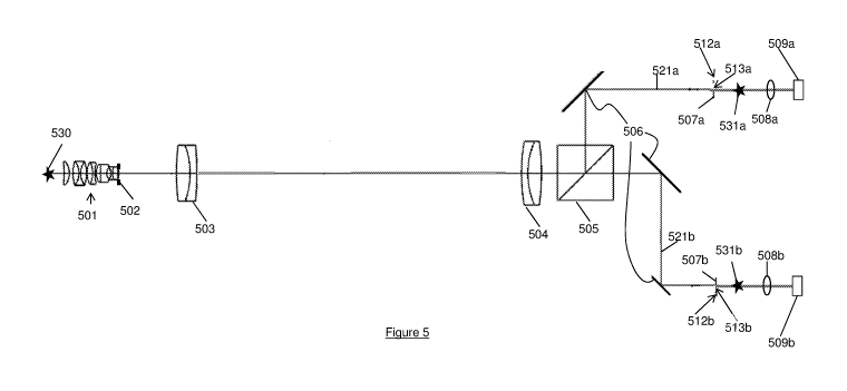
Figure 5 is a schematic diagram of a microscope configured to provide split
apertures.
The microscope comprises an objective assembly 501 having a (single) aperture
502,
and a tube lens 503, arranged to provide an image of an object 530. The
microscope
further comprises an intermediate lens 504, a beamsplitter 505, and mirrors
506. The
lens 504, beamsplitter 505, and mirrors 506 together form respective images
512a,
512b of the aperture 502 (hereinafter referred to as an aperture image) on
each of two
optical paths 521a, 521b. A respective stop structure 507a, 507b is provided
in the
CA 03078772 2020-04-08
WO 2019/073209
PCT/GB2018/052858
plane of each aperture image 512a, 512b, such that each stop structure 507a,
507b
blocks a different portion of the respective aperture image, defining an exit
pupil 513a,
513b. Eyepiece lenses 508a, 508b and image sensors 509a, 509b are located such
that the image sensors capture a real image of the object 520 through each of
the exit
5 pupils 513a, 513b. In practice, further optics (not shown) will be
required beyond the
aperture images to produce real images 531a, 531b of the object beyond the
exit pupils
513a, 513b which are then viewed through the eyepieces 508a, 508b. The stop
structures 507a, 507b are positioned such that the respective images captured
by the
image sensors 509a, 509b can be displayed on a stereoscopic viewer as a stereo
image of the object, i.e. with one image sensor providing the left eye view
and the other
providing the right eye view, due to the positioning of the respective stop
structures.
The aperture image 512a, 512b will be larger than the aperture itself, and
each stop
structure only needs to provide one of the perspectives. Therefore, the stop
structures
can be manufactured much more easily than a split aperture as shown in Figure
4,
while having the same effect of providing a stereo image.
The stop structure 507a, 507b may be any suitable shape. Several possibilities
are
shown in Figures 6A to 60. For example, the stop structure 507a, 507b may have
an
aperture which defines the exit pupil (61) or may be a "curtain" which blocks
only one
side of the aperture image 512a, 512b with a flat (62) or curved (63) edge. In
order to
provide a full, pure stereo image, the stop structures 507a, 507b must be
located so
that the exit pupil for each eye corresponds to a portion of the aperture that
is not in the
other exit pupil. A less pronounced stereo effect is produced if the exit
pupils overlap
slightly (i.e. each contains a portion of the aperture which is in the other
exit pupil, and
a portion which is not in the other exit pupil). If the exit pupils overlap
completely, then
the result is a binocular mono image.
Figures 7A, 7B, and 70 illustrate the effect of different stop structure
positions for
"curtain"-style stop structures. The same principles apply for other shapes of
stop
structure. The top part of each figure shows the aperture images and stop
structures,
the middle part shows the resulting exit pupils (overlaid so that the
differences can be
seen), and the bottom part shows a representation of the degree of stereoscopy
(as
much as can be presented in a 2D medium). As shown in Figure 7A, where there
is no
stop structure 507a, 507b occluding the aperture images 512a, 512b, the exit
pupils
CA 03078772 2020-04-08
WO 2019/073209
PCT/GB2018/052858
6
513a, 513b for each eye correspond exactly and a binocular mono image 71
results.
This can also occur for stop structures comprising a symmetric aperture,
positioned
such that the exit pupils correspond exactly. As shown in Figure 70, where
each stop
structure 507a, 507b occludes the respective aperture image 512a, 512b such
that the
exit pupils 513a, 513b are completely separate regions of the aperture, a full
stereo
image 73 results. As shown in Figure 7B, where each stop structure 507a, 507b
occludes a separate portion of each aperture image 512a, 512b, such that the
exit
pupils 51a, 513b are overlapping regions of the aperture but there is a
portion of each
exit pupil which does not correspond to a portion of the other exit pupil,
then a less
pronounced stereo image 72 results.
The resolution of the image is dependent on the dimensions of the effective
aperture
formed by the aperture 502 and the stop structure 507a, 507b (i.e. the
aperture which,
if located at the aperture 502, would form the exit pupil 513a, 513b), with
the resolution
being lower the smaller the effective aperture is (though the exact value
depends on
the shape of the effective aperture). As such, the positioning of the stop
structure
507a, 507b is a balance between resolution and stereo effect.
Other effects of the stop structures on the stereo image produced can be
compensated
for prior to display of the images. Figure 10 shows a schematic illustration
of the
microscope 1001. The microscope 1001 comprises stop structures 1011 and image
sensors 1012 as described above. Additionally, the microscope comprises a
digital
image processor 1013 which takes the output 1021 of the image sensors 1012 and
output of 1023 of the stop structure control, and adjusts it to compensate for
unwanted
changes to the image resulting from adjustments to the stop structures 1011,
with the
adjustments being performed on the basis of the shape and position 1023 of the
stop
structures. The digital image processor then provides the corrected image as
an
output 1022 from the microscope (e.g. to a stereoscopic display).
For example, the intensity of the image will vary with the position of the
stop structures
1011. This occurs both due to different amounts of area of the aperture 502
being
blocked, and due to intensity variations across the aperture 502 (meaning that
there
will be intensity variations even for stop structures such as that of Figure
6A that
always block the same amount of the aperture area). The intensity will depend
on both
the position and shape of the stop structures.
CA 03078772 2020-04-08
WO 2019/073209
PCT/GB2018/052858
7
The aperture 502 will have an intensity profile, which is a function
describing how much
each point on the aperture contributes to the intensity of the final image.
The reduction
in intensity resulting from the stop structures can be determined by comparing
the
integral of this intensity profile over the effective aperture formed by each
stop structure
with the integral of the intensity profile over the whole aperture 502. The
digital image
processor may then adjust the brightness of the output of each image sensor to
ensure
that the intensity is apparently constant for the user between different stop
structure
positions.
The intensity variation will also depend on the shape of the stop structures.
The
microscope may be provided with multiple different sets of stop structures
from which
the stop structures to be used are selected. The digital image processor
should be
configured to apply a different relationship between stop structure position
and image
brightness adjustment for each set of stop structures. The set of stop
structures may
be identified by the user in software of the digital image processor, or
automatic
identification may occur when the stop structures are installed in the
microscope (e.g.
by providing optical or electronic identifiers on the stop structures which
interface with
sensors on the microscope, or by other suitable means). Where the microscope
is
intended to work with only a single type of stop structure, the digital image
processor
only requires a single relationship between stop structure position and image
brightness.
Similarly, occluding different regions of the aperture 502 will affect the
distortion of the
image (due to lens aberrations and other optical effects). This distortion may
also be
corrected by the digital image processor, with the parameters of the
transformation
used being dependent on the stop structure shape and position.
The relationship between stop structure position (and shape, if multiple sets
of stop
structures may be used) and the digital image processing required may be
preconfigured, e.g. with a lookup table programmed into the digital image
processor, or
it may be calculated on-the-fly from the known parameters. The lookup table or
predetermined function may be obtained via a calibration step, e.g. measuring
intensity, image distortion, or other properties for a range of stop structure
positions,
and using this data (with suitable interpolation) to compute a lookup table.
CA 03078772 2020-04-08
WO 2019/073209
PCT/GB2018/052858
8
The stop structure may be adjustable to allow the user to transition from mono
to
stereo views, and control the degree of stereoscopy. A setup for achieving
this is
shown in Figure 8. Each stop structure includes a moveable curtain 801, which
is can
be introduced into the optical path in a controllable manner so as to occlude
a variable
amount of the aperture image 512a, 512b. The optical path for the other image
has an
equivalent system, and the curtains are coupled such that each occludes the
same
proportion of the respective aperture image 512a, 512b. The moveable curtain
801
can be adjusted from a position in which each occludes none of the aperture
image
(resulting in a binocular mono image) to a position in which the exit pupils
are non-
overlapping portions of the aperture image 512a, 512b (resulting in a pure
stereo
image). The moveable curtains 801 are configured to move such that each blocks
an
equal sized portion of the respective aperture image 512a, 512b, on opposite
sides of
the respective image.
The setup of Figure 8 allows a continuous and progressive transition between
stereo
and binocular mono modes of the microscope, without interruption of the image
viewed
by the observer. It has been surprisingly found through the use of this
apparatus that
when transitioning smoothly from a stereo image to a higher resolution mono
image,
the user experiences a sensation of depth with the mono image which is not
present if
the mono image is viewed without such a transition. This means that the system
described above allows much of the advantage of the stereoscopic image to be
retained, while also having the higher resolution of the mono image.
From the above description, it will be noted that the structure of the single
objective
stereo microscope from the objective assembly 501 up to but not including the
lens 504
is the same as that of the conventional mono microscope from the objective
assembly
101 up to but not including the eyepiece 104. The head and eyepiece assembly
of
many commercially available microscopes are removeable, and therefore it is
possible
to retrofit an existing mono microscope (whether binocular or monocular) with
a system
comprising the lens 504, beamsplitter 505, mirrors 506, and stop structures
507a,
507b, where the system is configured to attach in place of the head and
eyepiece
assembly of the mono microscope such that the lens 504 is in the light path of
the
microscope ¨ i.e. in the path which light from the object takes through the
microscope.
The original mono microscope may or may not apply optical corrections such as
field
CA 03078772 2020-04-08
WO 2019/073209
PCT/GB2018/052858
9
curvature, chromatic aberration etc using the eyepiece ¨ in systems for
retrofitting to
microscopes where these corrections are applied, the lens 504 and/or eyepieces
508a,
508b may be configured to apply equivalent corrections.
The image sensors may be CODs or other image sensors. A further advantage of
the
use of image sensors is that there is no requirement for the exit pupils to be
arranged
to precisely align with the viewers left and right eyes to view them, which
allows for
simplified structure of the microscope.
One example of a stereoscopic display is that described in GB2524609, and
shown in
Figure 9. The display comprises two projectors 20a and 20b, which display the
left eye
and right eye images respectively. Each projector comprises a display 21 and
an
optical arrangement 25 (comprising one or more lenses 29 and/or mirrors 31)
for
providing a focussed image of each of the left eye and right eye images on a
mirror 35.
The mirror 35 reflects the exit pupils of the projectors onto a viewing plane
(VP) for
viewing by an observer, optionally via a viewing lens 37. Optical components
other
than the mirror 35 and viewing lens 37 may be placed out of the direct line of
sight of
the observer, to give a clean viewing experience.
Other examples of stereoscopic displays include "virtual reality" headsets, 3D
displays
with active glasses (i.e. glasses which are synchronised to the refresh rate
of the TV,
and block each eye for alternate frames), and 3D displays with passive glasses
(e.g.
displays that present each of the left eye image and right eye image as a
different
polarisation, and are used with glasses that have a corresponding polarisation
filter for
each eye).
An advantage of using an image sensor coupled to a stereoscopic display rather
than
having the user directly observe the microscope through the exit pupils is
that the size
of the exit pupils available for the viewer is not limited by the microscope
optics, and is
not restricted by the stop structures 507a, 507b. Larger exit pupils give a
more
comfortable viewing experience. This is due to the fact that, where the exit
pupils are
small, the user must keep their head in a specific position to see the stereo
image.
Where the exit pupils are smaller than a certain size, as would likely be the
case where
stop structures are used, the user may have difficulty seeing the image at
all, as the
human eye does not function well when the exit pupil is smaller than the pupil
of the
CA 03078772 2020-04-08
WO 2019/073209
PCT/GB2018/052858
eye. In fact, with the optical systems as used in most existing microscopes,
the exit
pupil is already smaller than the entrance pupil of the user's eye, which
limits the
resolution, and causes any inhomogeneity in the eye (e.g. floaters) to have a
significantly greater effect on the user's vision.
5
It will be appreciated that the above disclosure is by way of example only,
and
variations are possible while still holding to the principle of the
disclosure. It will also be
appreciated that particular features are not dependent on each other unless
otherwise
stated.