Note: Descriptions are shown in the official language in which they were submitted.
CA 03079243 2020-04-16
IMAGE RECONSTRUCTION METHOD, DEVICE AND
MICROSCOPIC IMAGING DEVICE
TECHNICAL FIELD
[0001] The
present invention relates to an image processing technology, and in
particular, to an image reconstruction method, device, and microscopic imaging
device.
BACKGROUND
[0002]
Structured illumination-based microscopes have a sectioning imaging
function of suppressing out-of-focus noise, and have the advantages of simple
structure and fast imaging speed compared with confocal microscopes. In the
current
technology, they are often used as a conventional endoscope, and predict the
occurrence and evolution of tumors in advance by scanning internal organs such
as
the human digestive tract and observing changes in shapes of cells, having an
important guiding significance for cancer screening.
[0003] The structured illumination-based microscopes specifically, by an
exciter,
emit fluorescence that excites an organism's fluorescence, which then passes
through
a grating to form a sinusoidal light source with black and white stripes; and
then, by
moving 1/3 grating spacing of the grating each time, multiple images returned
after
scanning human cells are collected and acquired, for example, image i, image
'2,
.. and image 13; and then the image /, , image '2, and image /3 are
reconstructed
according to the root mean square
formula
/ = V(r, - 12)2 + (1, - 13)2 + (12 - I )2
3 to obtain a reconstructed image I.
However, this reconstruction method needs to calculate gray levels of all
pixels in the
image 1, image -12, and image-13 using the root mean square formula.
Therefore,
it takes a lot of calculation time, and the grating and fiber bundle cellular
grids in the
reconstructed image have obvious residues, and low imaging quality.
WSLEGAL\ 070171\ 00015\ 24522303v2
CA 03079243 2020-04-16
SUMMARY
[0004] In order to solve the technical problems in the prior art that
the structured
illumination-based microscope have a low image reconstruction efficiency,
obvious
residual gratings in the image, and low imaging quality, the present invention
provides
.. an image reconstruction method, device, and microscopic imaging device, so
as to
accelerate the speed of image reconstruction, remove the residual of the
grating in the
reconstructed image, and the improve the quality of the reconstructed image.
[0005] The present invention provides an image reconstruction method,
including:
[0006] calculating a gray value at each fiber center in a fiber bundle
in a
reconstructed image according to a gray value at a center position of each
fiber,
determined in one or more sample images;
[0007] performing a spatial interpolation using the gray value at the
each fiber
center to obtain gray values of other pixel points in the fiber bundle in the
reconstructed image to form the reconstructed image.
[0008] Optionally, the method further includes:
100091 acquiring an original image of a uniform fluorescent fiber
bundle; and
[0010] determining a target pixel point with a pixel value higher than
surrounding
pixel values in the original image, and determining the target pixel point as
the center
position of each fiber in the fiber bundle.
[0011] Optionally, the acquiring an original image of a uniform fluorescent
fiber
bundle includes:
[0012] collecting a plurality of fiber bundle images spaced at a preset
step size
within a grating interval; and
[0013] obtaining an average image of the plurality of fiber bundle
images to form
the original image of the uniform fluorescent fiber bundle.
[0014] Optionally, before performing the spatial interpolation, the
method further
includes:
[0015] determining an interpolation weight between each pixel point in
the fiber
bundle and the center position of each fiber according to the center position
of each
fiber.
[0016] Optionally, the method further includes determining the
interpolation
weight by using the following method:
[0017] forming a plurality of triangular structures using the center
position of
2
WSLEGAL\ 070171\ 00015\ 24522303v2
CA 03079243 2020-04-16
each fiber and center positions of adjacent fibers as vertices; and
[0018] determining the interpolation weight between a pixel point in
each triangle
structure and the center position of each fiber according to the triangle
structures.
[0019] Optionally, the method further includes acquiring the more sample
images
by using the following method:
[0020] according to a preset phase interval, moving N-1 times within a
grating
interval to obtain N sample images including an initial phase and moved by the
preset
phase interval with respect to the initial phase each time.
[0021] Optionally, the preset phase interval is 120 degrees; and N=3.
[0022] Optionally, after determining the gray value at the center position
of each
fiber in the one or more sample images, the method further includes:
[0023] performing a saturation judgment on the gray value at the center
position
of each fiber;
100241 if there is a fiber whose center position has a gray value of
exceeding a
preset saturation threshold in the sample images, determining that the fiber
exceeding
the preset saturation threshold is a fiber to be corrected;
[0025] correcting the gray value at the center position of the fiber to
be corrected
to the preset saturation threshold in the reconstructed image, and performing
the step
of calculating the gray value at each fiber center in the fiber bundle in the
reconstructed image according to the gray value at the center position of each
fiber,
determined in the sample images after correction; and
[0026] if there is no fiber whose center position has a gray value of
exceeding the
preset saturation threshold in the sample images, performing the step of
calculating
the gray value at each fiber center in the fiber bundle in the reconstructed
image
.. according to the gray value at the center position of each fiber,
determined in the
sample images.
[0027] Optionally, the calculating a gray value at each fiber center in
a fiber
bundle in a reconstructed image according to a gray value at a center position
of each
fiber, determined in more sample images includes:
[0028] subtracting the gray value at the center position of each fiber in
the more
sample images from each other to obtain difference values, and taking a sum of
squares of the obtained difference values and then taking a square root to
obtain the
gray value at each fiber center in the fiber bundle in the reconstructed
image.
3
WSLEGAL\ 070171\ 00015\ 24522303v2
[0029] The present invention also provides an image reconstruction device,
including:
[0030] a calculating module, configured to calculate a gray value at each
fiber
center in a fiber bundle in a reconstructed image according to a gray value at
a center
position of each fiber, determined in one or more sample images; and
[0031] a forming module, configured to perform a spatial interpolation
using the
gray value at the each fiber center to obtain gray values of other pixel
points in the
fiber bundle in the reconstructed image, so as to form the reconstructed
image.
[0032] Optional, the device further includes:
[0033] a first acquiring module, configured to acquire an original image of
a
uniform fluorescent fiber bundle; and
[0034] a third determining module, configured to determine a target pixel
point
with a pixel value higher than surrounding pixel values in the original image,
and
deteimine the target pixel point as the center position of each fiber in the
fiber bundle.
[0035] Optionally, the first acquiring module includes:
[0036] a collecting sub-module, configured to collect a plurality of fiber
bundle
images spaced at a preset step size within a grating interval; and
[0037] a forming sub-module, configured to obtain an average image of the
more
fiber bundle images to foim the original image of the uniform fluorescent
fiber
bundle.
[0038] Optionally, the device further includes:
[0039] a first determining module, configured to determine an interpolation
weight between each pixel point in the fiber bundle and the center position of
each
fiber according to the center position of each fiber.
[0040] Optionally, the device further includes:
[0041] a second determining module, configured to foul' a plurality of
triangular
structures using the center position of each fiber and center positions of
adjacent
fibers as vertices, and determine an interpolation weight between a pixel
point in each
triangle structure and the center position of each fiber according to the
triangle
structures.
[0042] Optionally, the device further includes:
[0043] a second acquiring module, configured to move N-1 times within a
grating
interval according to a preset phase interval to obtain N sample images
including an
initial phase and moved by the preset phase interval with respect to the
initial phase
4
WSLEGAL\070171k 00015 \31933238v1
Date Recue/Date Received 2022-08-04
CA 03079243 2020-04-16
each time.
[0044] Optionally, the preset phase interval is 120 degrees; and
[0045] N=3.
[0046] Optionally, the device further includes:
[0047] a judging module, configured to perform a saturation judgment on the
gray
value at the center position of each fiber;
[0048] a first processing module, configured to: when there is a fiber
whose
center position has a gray value of exceeding a preset saturation threshold in
the
sample images, determine that the fiber exceeding the preset saturation
threshold is a
fiber to be corrected; correct the gray value at the center position of the
fiber to be
corrected to the preset saturation threshold in the reconstructed image, and
perform
the step of calculating the gray value at each fiber center in the fiber
bundle in the
reconstructed image according to the gray value at the center position of each
fiber,
determined in the sample images after correction; and
[0049] a second processing module, configured to: when there is no fiber
whose
center position has a gray value of exceeding the preset saturation threshold
in the
sample images, perform the step of calculating the gray value at each fiber
center in
the fiber bundle in the reconstructed image according to the gray value at the
center
position of each fiber, determined in the sample images.
[0050] Optionally, the calculating module is specifically configured to
subtract the
gray value at the center position of each fiber in the more sample images from
each
other to obtain difference values, and take a sum of squares of the obtained
difference
values and then take a square root to obtain the gray value at each fiber
center in the
fiber bundle in the reconstructed image.
[0051] The invention also provides a microscopic imaging device, including:
[0052] a light emitting unit, a phase adjusting unit, steering unit, a
fiber bundle
containing a plurality of fibers, a detecting unit, and a processing unit,
where:
[0053] the light emitting unit is configured to emit an excitation
light;
[0054] the phase adjusting unit is provided at an exit of an optical
path of the
excitation light, and is connected to the processing unit, and is configured
to adjust a
phase of the excitation light according to a phase adjustment amount sent by
the
processing unit to obtain excitation lights in different phases;
[0055] the steering unit is configured to steer the excitation lights in
different
phases, so that the steered excitation lights are focused to a tissue to be
detected along
5
WSLEGAL\ 070171\ 00015\ 24522303v2
CA 03079243 2020-04-16
the fiber bundle and to steer fluorescence in different phases returned
through the
tissue to be detected;
[0056] the detecting unit is configured to collect fluorescence in
different phases
to form a plurality of sample images; and
[0057] the processing unit is connected to the detecting unit, and is
configured to
receive the plurality of sample images, and calculate a gray value at each
fiber center
in the fiber bundle in a reconstructed image according to a gray value at a
center
position of each fiber in the fiber bundle determined in the plurality of
sample images;
perform a spatial interpolation using the gray value at the each fiber center
to obtain
gray values of other pixel points in the fiber bundle in the reconstructed
image so as to
form the reconstructed image.
[0058] Optionally, the phase adjusting unit includes: a motor and a
grating;
[0059] the motor is connected to the processing unit and the grating,
respectively,
and is configured to drag the grating to move according to the phase
adjustment
amount sent by the processing unit, so that the excitation light is
transmitted through
the grating to obtain an excitation light corresponding to the phase
adjustment
amount.
[0060] Optionally, the motor includes: a direct current motor; and
[0061] correspondingly, the processing unit determines an equal-interval
phase
adjustment amount according to a preset phase interval; the direct current
motor
receives the equal-interval phase adjustment amount, and drags the grating to
move
by an equal interval distance within a grating spacing range to enable the
processing
unit to obtain a plurality of sample images corresponding to the preset phase
interval.
[0062] Optionally, the preset phase interval is 120 degrees; and the
phase
adjustment amount is 3.
[0063] Optionally, the light emitting unit includes: a laser, configured
to emit the
excitation light; and further includes a beam expander-focuser provided at an
exit of
the excitation light of the laser and is configured to expand the excitation
light and
one-dimensionally focus it into a line beam.
[0064] Optionally, the steering unit is a dichroic mirror.
[0065] Optionally, the device further includes: a filter; the filter is
disposed
between the phase adjusting unit and the steering unit, and is configured to
filter out
stray light.
[0066] Optionally, the detecting unit includes: a charge coupled device
CCD.
6
WSLEGAL\ 070171\ 00015\ 24522303v2
CA 03079243 2020-04-16
[0067] Optionally, the device further includes: an objective lens
including a
plurality of lenses; the objective lens is disposed between the steering unit
and the
fiber bundle, and is configured to perform a focusing processing on the
excitation
light steered by the steering unit.
[0068] The image reconstruction method, device and microscopic imaging
device
of the present invention are to form a reconstructed image by calculating a
gray value
at each fiber center in a fiber bundle in the reconstructed image according to
a gray
value at a center position of each fiber, determined in one or more sample
images; and
performing a spatial interpolation using the gray value at the each fiber
center to
obtain gray values of other pixel points in the fiber bundle in the
reconstructed image.
This image reconstruction method only calculates a gray value of a pixel point
at the
fiber center, and then obtains gray values of pixel points of the entire image
based on
the spatial interpolation, thereby reducing the calculation amount due to
calculating
the gray value of each pixel point, greatly accelerating the speed of image
reconstruction, and the method is helpful to remove the grating and fiber
bundle
cellular grid residues in the reconstructed image and improve the imaging
quality of
the reconstructed image.
BRIEF DESCRIPTION OF THE DRAWINGS
[0069] FIG. 1 is a flowchart of an image reconstruction method of the
present
invention according to an exemplary embodiment;
[0070] FIG. 2 is a schematic view of a structured light microendoscope
device
according to the embodiment shown in FIG. 1;
[0071] FIG. 3 is a flow chart of an image reconstruction method of the
present
invention according to another exemplary embodiment;
[0072] FIG. 4 is a schematic diagram of a triangular structure of a fiber
pixel
according to the embodiment shown in FIG. 3;
[0073] FIG. 5 is a schematic structural diagram of an image
reconstruction device
of the present invention according to an exemplary embodiment;
[0074] FIG. 6 is a schematic structural diagram of an image
reconstruction device
of the present invention according to another exemplary embodiment;
[0075] FIG. 7 is a schematic structural diagram of a microscopic imaging
device
of the present invention according to an exemplary embodiment; and
7
WSLEGAL\ 070171\ 00015\ 24522303v2
CA 03079243 2020-04-16
[0076] FIG. 8 is a schematic structural diagram of a microscopic imaging
device
of the present invention according to another exemplary embodiment.
[0077] Reference numerals: light emitting unit 01, laser 011, beam
expander-focuser 012, phase adjusting unit 02, motor 021, grating 022,
steering unit
03, fiber bundle 04, detecting unit 05, processing unit 06, filter 07,
objective lens 08.
DETAILED DESCRIPTION OF THE EMBODIMENTS
[0078] In order to make the objects, technical solutions, and advantages
of the
present invention more clear, technical solutions in embodiments of the
present
invention will be clearly and completely described below with reference to the
accompanying drawings in the embodiments of the present invention. Obviously,
the
described embodiments are only part of embodiments of the present invention,
not all
embodiments of the present invention. All other embodiments obtained by those
skilled in the art based on the embodiments of the present invention without
creative
efforts shall fall within the protection scope of the present invention.
[0079] FIG. 1 is a flowchart of an image reconstruction method of the
present
invention according to an exemplary embodiment. As shown in FIG. 1, the image
reconstruction method of the present invention is suitable for reconstructions
of all
optically imaged images, and is especially suitable for an image
reconstruction based
on structured light. First, in this embodiment, the principle of structured
light imaging
is briefly explained by taking a structured light-based endoscope as an
example:
100801 In the structured light-based microendoscope device shown in FIG.
2, a
light source emitted by the exciter is modulated by a grating to produce a
sinusoidal
light, and the sinusoidal light passes through a dichroic mirror (that is,
light with a
specific frequency is transmitted and light with a non-specific frequency is
reflected)
and an objective lens, and excites a stained tissue (for example, a cell
tissue in the
human body) along the fiber bundle, and fluorescence after excitation reaches
a
charge-coupled device (CCD) along the fiber bundle, the objective lens and the
dichroic mirror and performs an image collection. CCD, also called image
sensor or
image controller, is a semiconductor device that can convert an optical image
into
electrical signals. The modulated sinusoidal light source is focused on a
certain focal
plane of the tissue. By exciting fluorescence imaging in multiple phases (for
example,
three phases), the background fluorescence outside the focal plane are
filtered out
8
WSLEGAL\ 070171\ 00015\ 24522303v2
CA 03079243 2020-04-16
using the Neil formula, to realize a sectioning imaging. Sectioning imaging
technology is a geophysical prospecting inversion interpretation method that
inversely
calculates obtained information according to ray scanning that uses medical CT
as a
reference, to reconstruct an image of a distribution law of elastic wave and
electromagnetic wave parameters of a rock mass in a measured range and thereby
achieve delineating geological anomaly.
100811 The light source of the structured light modulated by the grating
can be
expressed as:
\ 1 r
) = - 1.1 + rn co4x + 0, A
2
[0082] In the above formula, m is a modulation contrast;
)82 v
[0083] NA is a normalized spatial frequency, the -1)- value can be
used to
achieve sectioning of images at different depth (axial depth); )8 is a
magnification
between a specimen plane and a grid plane, Ais a wavelength, V is an actual
spatial
frequency, and NA is a numerical aperture.
[0084] In this embodiment, the pixel information transmitted from each
fiber in
the fiber bundle in FIG. 2 needs to be determined in order to accurately
obtain the
fluorescence information returned after the structured light irradiates the
stained tissue,
and a clear and accurate image of the information is formed. The specific
implementation steps of the image reconstruction method in this embodiment
includes:
[0085] Step 101: calculating a gray value at each fiber center in a
fiber bundle in a
reconstructed image according to a gray value at a center position of each
fiber,
determined in one or more sample images.
[0086] Specifically, the structured light microendoscope device shown in
FIG. 2,
a direct current motor is driven to move the grating to acquire one or more
sample
images. The sample images contain the pixel information transmitted by each
fiber in
the fiber bundle. For the fiber bundle, one fiber bundle is usually including
nearly
30,000 fibers (the difference in the number can reach several thousand). The
pixel
information is transmitted in each fiber, so the fiber bundle can also be
called a
multi-sensor. The imaging of fibers generally shows an image in a hexagonal
cellular
shape, and the diameter of each fiber is preferably 5 to 6 pixels. In the more
sample
9
WSLEGAL\ 070171\ 00015\ 24522303v2
CA 03079243 2020-04-16
images, the center position of each fiber is determined, and the gray value of
the pixel
point at each center position is obtained. The method for determining the gray
value at
the center position can be obtained by using the root-mean-square formula
described
above, that is, a gray-value average value of the gray values at the same
center
position in the more sample images is obtained, and the calculated gray-value
average
value is used as the gray value at the fiber center in the reconstructed
image, and then
the gray value at each fiber center in the fiber bundle in the reconstructed
image is
obtained.
[0087] Step 102: performing a spatial interpolation using the gray value
at the
fiber center to obtain gray values of other pixel points in the fiber bundle
in the
reconstructed image so as to form the reconstructed image.
[0088] Specifically, the center position of each fiber is used as a
reference to find
a linear relationship between the other pixel points and the pixel point at
the center
position in each fiber, so that interpolation weights of all pixel points
relative to the
pixel point at the center position in each fiber can be determined, i.e.,
weights of the
other pixel points relative to the pixel point at the center position in each
fiber.
Therefore, based on the interpolation weight between each pixel point and the
fiber
center, the spatial interpolation is performed using the gray value at the
fiber center, to
obtain the gray values of the other pixel points in the fiber bundle in the
reconstructed
image, and the reconstructed image is formed.
[0089] The image reconstruction method of this embodiment form the
reconstructed image by calculating the gray value at each fiber center in the
fiber
bundle in the reconstructed image according to the gray value at the center
position of
each fiber, determined in one or more sample images; performing the spatial
interpolation using the gray value at the each fiber center to obtain the gray
values of
other pixel points in the fiber bundle in the reconstructed image. This image
reconstruction method only calculates the gray value of the pixel point at the
fiber
center position, and then obtains the gray values of the pixel points of the
entire image
based on the spatial interpolation, thereby reducing the calculation amount
due to
calculating the gray value of each pixel point, greatly accelerating the speed
of image
reconstruction, and the method is helpful to remove the grating and fiber
bundle
cellular grid residues in the reconstructed image and improve the imaging
quality of
the reconstructed image.
[0090] FIG. 3 is a flowchart of an image reconstruction method of the
present
WSLEGAL\ 070171\ 00015\ 24522303v2
CA 03079243 2020-04-16
invention according to another exemplary embodiment As shown in FIG. 3, the
image reconstruction method according to this embodiment includes:
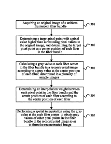
100911 Step 301: acquiring an original image of a uniform fluorescent
fiber
bundle.
100921 Step 302: determining a target pixel point with a pixel value higher
than
surrounding pixel values in the original image, and determining the target
pixel point
as a center position of each fiber in the fiber bundle.
100931 Specifically, before the image reconstruction, an image with
uniform
fluorescence may be taken, and the image with uniform fluorescence is used to
accurately locate fiber. For the fiber bundle, a fiber bundle is usually
including nearly
30,000 fibers (the difference in the number can reach several thousand). Pixel
information is transmitted in each fiber, so the fiber bundle can also be
called a
multi-sensor. The imaging of fibers generally shows an image in a hexagonal
cellular
shape, and the diameter of each fiber is preferably 5 to 6 pixels. In order to
reduce
mutual interference between fibers, the fibers are arranged irregularly in
space, rather
than aligned in rows or columns. The fiber center position in this embodiment
refers
to the brightest point at the fiber center. The so-called brightest point
means a target
pixel point having a pixel value determined to be higher than the surrounding
pixel
values in the original image, and the target pixel point is determined to be
the fiber
center of each fiber in the fiber bundle, other pixel points in each fiber is
located by
using the coordinates of the brightest point at the center as coordinates of
the fiber. In
order to remove the grid, that is, the hexagonal cell of the fiber, the gray
value at the
fiber center needs to be used for spatial interpolation to obtain the gray
values of other
pixel points in the entire fiber bundle. Generally, after the grating is
installed, the
captured locating image, i.e., the original image will have the grating.
Therefore, it is
possible to shoot after removing the grating, to obtain an original image of a
uniform
fluorescent fiber bundle; optionally, a plurality of fiber bundle images
spaced at a
preset step size can also be collected within a grating interval; an average
image of the
plurality of fiber bundle images is obtained to form the original image of the
uniform
fluorescent fiber bundle. That is to say, the direct current motor in FIG. 2
uniformly
moves several same displacements within a grating spacing range, and then the
collected average image is taken. Those skilled in the art can self-determine
a method
for obtaining an original image of the uniform fluorescent fiber bundle, which
is not
specifically limited in this embodiment.
11
WSLEGAL\ 070171\ 00015\ 24522303v2
CA 03079243 2020-04-16
[0094] Step 303: calculating a gray value at each fiber center in the
fiber bundle in
the reconstructed image according to a gray value at the center position of
each fiber,
determined in a plurality of sample images.
[0095] Where the sample image can be obtained by moving N-1 times within
a
grating interval according to a preset phase interval, to obtain N sample
images
including an initial phase, and moving by the preset phase interval with
respect to the
initial phase each time. For example, a grating is installed, and the grating
is dragged
by a motor to move so as to obtain N sample images of the fiber bundle. For
example,
when starting to collect the sample images, one sample image is taken at an
initial
position of the motor; then the motor is moved to another position and another
sample
image is taken; the motor is moved again and still another image is taken,
thereby
obtaining N sample images. In order to ensure the accuracy of the obtained
sample
images, the motor can be rotated clockwise to multiple positions to obtain the
above
N sample images. After waiting for a period of time, the motor is moved
counterclockwise to further obtain N sample images. This can reconstruct two
structured light images, and the accuracy of the reconstructed image can be
guaranteed by comparison. Preferably, the preset phase interval is 120
degrees, and
N=3; correspondingly, the motor drags the grating to move horizontally, and
the
preset phase interval threshold for each move is 1/3 of the grating spacing.
Before
collecting, the camera captures an image at the initial position where the
motor moves,
then the motor moves, the camera captures another image, the motor further
moves,
and the camera captures still another image, thereby obtaining sample images
in three
phases, then an image is reconstructed; then, for keeping a consistent period,
wait for
a period of time; continue to perform capturing in an opposite movement
direction ...
in this way, two structured light images can be reconstructed after the motor
moves
back and forth once. The three sample images can be 0-degree phase sample
image
/1 (initial phase), 120-degree phase sample image /2 (moving by one preset
phase
interval threshold), and 240-degree phase sample image 13 (moving by two
preset
phase interval thresholds). In the three phase sample images, according to
positions of
the fiber centers, gray values at the fiber centers of the three phase sample
images are
retrieved, then a gray value G1 of the fiber center of the 0-degree phase
sample
image /1, a gray value G2 of the fiber center of the 120-degree phase sample
image
12
WSLEGAL\ 070171\ 00015\ 24522303v2
CA 03079243 2020-04-16
12, and a gray value G3 of the fiber center of the 240-degree phase sample
image
13 are obtained. Optionally, calculation of a gray value at each fiber center
in the
fiber bundle in the reconstructed image can be realized by subtracting the
gray value
at a center position of each fiber in plurality of sample images from each
other to
obtain difference values, and taking a sum of squares of the obtained
difference values
and then taking a square root to obtain the gray value at each fiber center in
the fiber
bundle in the reconstructed image. For example, based on the Neil formula
G = G2 )2
( - G3)2 + (G2 ¨ G3)2 , differences between three center gray
values in the three sample images are taken, and then the difference values
are
squared, and the squared differences are added and then a square root is
taken, thereby
the gray value at the each fiber center in the reconstructed image is
obtained.
[0096]
However, for the above Neil formula, the disadvantage lies in that when
the sample images are oversaturated, the center gray values being subtracted
from
each other will cause the calculated gray value at the center point to be a
black point
with a very small gray level. This will result in a black area in the
reconstructed image,
making it impossible to image the cells clearly. In order to avoid the problem
of
unclear imaging caused by image saturation, a saturation correction can be
applied to
the gray value at the fiber center point. In this way, the reconstructed image
can have
a good sectioning effect.
[0097] Optionally, after determining the gray value at the center position
of each
fiber in one or more sample images, a step of performing saturation judgment
on the
gray value at the center position of each fiber may be added. That is, if
there is a fiber
whose central position has a gray value exceeding a preset saturation
threshold in the
sample images, determining that the fiber exceeding the preset saturation
threshold is
a fiber to be corrected; correcting the gray value at the center position of
the fiber to
be corrected to the preset saturation threshold in the reconstructed image,
and
according to the gray value at the center position of each fiber, determined
in the
sample images after correction, performing the step of calculating the gray
value at
each fiber center in the fiber bundle in the reconstructed image;
[0098] if there is no fiber whose center position has a gray value of
exceeding the
preset saturation threshold in the sample images, performing the step of
calculating a
gray value at each fiber center in the fiber bundle in the reconstructed image
13
WSLEGAL\ 070171\ 00015\24522303v2
CA 03079243 2020-04-16
according to the gray value at the center position of each fiber, determined
in the
sample images.
100991 The
preset saturation threshold can be determined according to the
performance of the CCD. For example, whether the three gray values, the gray
value
G1 of the fiber center of the 0-degree phase sample image /1, the gray value
G2 of
the fiber center of the 120-degree phase sample image /2, and the gray value
G3 of
the fiber center of the 240-degree phase sample image 13, are greater than
4095
(4095 corresponds to a maximum value of a 12-bit image, meaning that the CCD
is
saturated) is determined, and then instead of using the above Neil formula
_________________________________________________________________ G = \AG1-G2
)2 ( - G3 )2 ( G2 G3 )2 to calculate the gray value at the center
point in the reconstructed image, the preset saturation threshold of 4095 is
directly
used as the gray value at the center point. This processing avoids a
phenomenon that
black and white in the sample images and the reconstructed structured light
image are
visually opposite. However, this processing is a last resort remedy. For those
skilled in
the art, the problem of image saturation when collecting sample images should
be
avoided as far as possible. For example, measures, such as avoiding
excessively long
exposure times and excessive gain of camera parameters, avoiding too much
fluorescent staining on a sample, and avoiding excessively strong laser light
emitted
by the laser, may be adopted.
101001 Similarly, if there is a fiber whose central position has a gray
value of
exceeding a preset saturation threshold in the sample images, it is determined
that the
fiber that exceeds the preset saturation threshold is a fiber to be corrected;
the gray
value at the center position of the fiber to be corrected is corrected to the
preset
saturation threshold in the reconstructed image. That is to say, if a
calculated value of
_________________________________________________________________ G = \AG, -
G2 )2 ( - G3 )2 ( G2 - G3 )2 exceeds the preset saturation threshold,
the fiber is determined as the fiber to be corrected, and the preset
saturation threshold
is also used as the gray value at the center position of the fiber, thereby
achieving
saturation correction of the sample image.
101011 Step
304: determining an interpolation weight between each pixel point in
the fiber bundle and the center position of each fiber according to the center
position
of each fiber.
14
WSLEGAL\ 070171\ 00015\ 24522303v2
CA 03079243 2020-04-16
[0102] Specifically, as described above, both a sample image and an
original
image are optical imaging of a fiber bundle of the same structure. Therefore,
according to the center position of each fiber, determined in the original
image, a
center position of a corresponding fiber in the sample image can be found, and
the
gray value at this center point can be read. Each fiber in N sample images is
located
and its gray value is obtained. Therefore, each fiber corresponds to gray
values at N
center positions. Based on a preset algorithm (such as the Neil formula of
root mean
square, as described above), gray-value average value of the gray values at
the N
center positions is obtained, and the calculated gray-value average value is
used as the
gray value at the fiber center in the reconstructed image
[0103] For the interpolation weight between each pixel point in the
fiber bundle
and the center position of each fiber, it can be determined by forming a
plurality of
triangular structures using the center position of each fiber and center
positions of
adjacent fibers as vertices; and determining an interpolation weight between a
pixel
point in each triangle structure and the center position of the each fiber
according to
the triangle structure.
[0104] Specifically, fiber center coordinates can be obtained according
to a
regional maximum method, that is, as shown in FIG. 4, a center position of a
fiber A
is used as a vertex, and three center positions of the fiber A and adjacent
fibers B and
C form a triangle, so that a range of the entire fiber bundle is divided into
multiple
triangles. The interpolation relationship between pixels and fibers is
established
through these triangles. Because the fiber bundle is roughly hexagonal with an
irregular distribution, and adjacent fibers do not have an alignment
relationship in
horizontal or vertical coordinates, an intermediate pixel cannot be
interpolated by four
regular vertices, like a conventional bilinear interpolation. However, using
this
triangular structure, the interpolation weight between the pixel point in each
triangular
structure and the center position of each fiber can also be determined.
[0105] Step 305: performing a spatial interpolation using the gray value
at the
each fiber center to obtain gray values of other pixel points in the fiber
bundle in the
reconstructed image so as to form the reconstructed image.
[0106] Specifically, after acquiring the original image of the uniform
fluorescent
fiber bundle, center positions of all fibers contained in the fiber bundle are
determined
in the original image, that is, position coordinates of a brightest pixel
point in each
fiber. The center position of each fiber is used as a reference to find a
linear
WSLF,GAL\ 070171\ 00015\ 24522303v2
CA 03079243 2020-04-16
relationship between other pixel points in each fiber and the pixel point at
the center
position, so that interpolation weights of all pixel points in each fiber
relative to the
pixel point at the center position are determined, i.e., weights of other
pixel points in
each fiber relative to the pixel point at the center position. Subsequent
reconstruction
of the sample images obtained by irradiating a tissue with a structured light
can be
based on linear weights calculated in advance and multiplied by the gray
values of the
fibers during reconstruction to obtain the gray values of the pixels to be
interpolated
to form a reconstructed image.
[0107] The image reconstruction method of this embodiment obtains
reconstruction of structured light imaging by using fiber positioning in
triangle-based
pixel space, where only the pixels of the center points of the fibers are
calculated by
using the Neil formula, and then the entire structured light image is
reconstructed by
interpolation. The calculation time is greatly saved, and the cellular
structure of the
fibers can be removed. When phase differences between N sample images, for
example, three sample images, are exactly 120 degrees, traces of the grating
are also
absent. Therefore, the image reconstruction method of the present invention
can
greatly reduce the calculation amount due to calculating the gray value of
each pixel
point, greatly accelerate the speed of image reconstruction, and also the
method is
helpful to remove the grating and fiber bundle cellular grid residues in the
reconstructed image and improve the imaging quality of the reconstructed
image.
[0108] FIG. 5 is a schematic structural diagram of an image
reconstruction device
of the present invention according to an exemplary embodiment. As shown in
FIG. 5,
the image reconstruction device according to this embodiment includes:
[0109] a calculating module 1, configured to calculate a gray value at
each fiber
center in a fiber bundle in a reconstructed image according to a gray value at
a center
position of each fiber, determined in one or more sample images;
[0110] a forming module 2, configured to perform a spatial interpolation
using the
gray value at the each fiber center to obtain gray values of other pixel
points in the
fiber bundle in the reconstructed image to form the reconstructed image.
[0111] This embodiment may be used to implement the embodiment shown in
FIG. 1, and implementation principles of the two embodiments are similar, and
details
are not described herein again.
[0112] In the image reconstruction device of this embodiment, the
reconstructed
image is formed by calculating a gray value at each fiber center in a fiber
bundle in a
16
WSLEGAL\ 070171\ 00015\ 24522303v2
reconstructed image according to a gray value at a center position of each
fiber,
deteimined in a plurality of sample images; performing a spatial interpolation
using
the gray value at the each fiber center to obtain gray values of other pixel
points in the
fiber bundle in the reconstructed image. This image reconstruction method only
calculates gray values of pixel points of center positions of fibers, and then
obtains
gray value of pixel points of the entire image based on a spatial
interpolation, thereby
reducing the calculation amount due to calculating the gray value of each
pixel point,
greatly accelerating the speed of image reconstruction, and the method is
helpful to
remove the grating and fiber bundle cellular grid residues in the
reconstructed image
and improve the imaging quality of the reconstructed image.
[0113] FIG. 6 is a schematic structural diagram of an image reconstruction
device
of the present invention according to another exemplary embodiment. As shown
in
FIG. 6, based on the above embodiment, the image reconstruction device in this
embodiment further includes:
[0114] a first acquiring module 3, configured to acquire an original image
of a
uniform fluorescent fiber bundle;
[0115] a third determining module 4, configured to determine a target pixel
point
with a pixel value higher than surrounding pixel values in the original image,
and
detemiining the target pixel point to be the center position of each fiber in
the fiber
bundle.
[0116] Optionally, the first acquiring module 3 includes:
[0117] a collecting sub-module 31, configured to collect a plurality of
fiber
bundle images spaced at a preset step size within a grating interval;
[0118] a forming sub-module 32, configured to obtain an average image of
the
plurality of fiber bundle images to form an original image of the unifoim
fluorescent
fiber bundle.
[0119] Optionally, the device further includes:
[0120] a first deteimining module 5, configured to determine an
interpolation
weight between each pixel point in the fiber bundle and the center position of
each
fiber according to the center position of each fiber.
[0121] Optionally, the device further includes:
[0122] a second determining module 6, configured to form a plurality of
triangular structures using the center position of each fiber and center
positions of
adjacent fibers as vertices, and deteimine an interpolation weight between a
pixel in
each
17
WSLEGAL\070171k 00015 \31933238v1
Date Recue/Date Received 2022-08-04
CA 03079243 2020-04-16
triangle structure and the center position of each fiber according to the
triangle
structures.
[0123] Optionally, the device further includes:
[0124] a second acquiring module 7, configured to move N-1 times within
a
grating interval according to a preset phase interval to obtain N sample
images
including an initial phase, and moving by the preset phase interval with
respect to the
initial phase each time.
[0125] Optionally, the preset phase interval is 120 degrees; and N=3.
[0126] Optionally, the device further includes:
[0127] a judging module 8, configured to perform a saturation judgment on
the
gray value at the center position of each fiber;
[0128] a first processing module 9, configured to: when there is a fiber
whose
central position has a gray value of exceeding a preset saturation threshold
in the
sample images, determine that the fiber exceeding the preset saturation
threshold is a
fiber to be corrected; correct the gray value at the center position of the
fiber to be
corrected to the preset saturation threshold in the reconstructed image, and
according
to the gray value at the center position of each fiber, determined in the
sample images
after correction, perform the step of calculating the gray value at each fiber
center in
the fiber bundle in the reconstructed image; and
[0129] a second processing module 10, configured to: when there is no fiber
whose center position has a gray value of exceeding the preset saturation
threshold in
the sample images, perform the step of calculating the gray value at each
fiber center
in the fiber bundle in the reconstructed image according to the gray value at
the center
position of each fiber, determined in the sample images.
[0130] Optionally, the calculating module 1 is specifically configured to
subtract
the gray value at a center position of each fiber in a plurality of sample
images from
each other to obtain difference values, and take a sum of squares of the
obtained
difference values and then take a square root to obtain the gray value at each
fiber
center in the fiber bundle in the reconstructed image.
[0131] This embodiment can be used to implement the embodiment shown in
FIG.
3, and the implementation principles of the two embodiments are similar, and
details
are not described herein again.
[0132] FIG. 7 is a schematic structural diagram of a microscopic imaging
device
of the present invention according to an exemplary embodiment. As shown in
FIG. 7,
18
WSLEGAL\ 070171\ 00015\ 24522303v2
CA 03079243 2020-04-16
this embodiment provides a microscopic imaging device including a light
emitting
unit 01, a phase adjusting unit 02, a steering Unit 03, a fiber bundle 04
containing a
plurality of fibers, a detecting unit 05, and a processing unit 06, where:
[0133] the light emitting unit 01 is configured to emit an excitation
light;
[0134] the phase adjusting unit 02 is provided at an exit of an optical
path of the
excitation light, and is connected to the processing unit 06, and is
configured to adjust
the phase of the excitation light according to a phase adjustment amount sent
by the
processing unit 06 to obtain excitation lights in different phases;
[0135] the steering unit 03 is configured to steer the excitation lights
in different
phases, so that the steered excitation lights are focused to a tissue to be
detected along
the fiber bundle 04; and to steer fluorescence in different phases returned
through the
tissue to be detected;
[0136] the detecting unit 05 is configured to collect fluorescence in
different
phases to form a plurality of sample images; and
101371 the processing unit 06 is connected to the detecting unit 05, and is
configured to receive the plurality of sample images, and calculate a gray
value at
each fiber center in the fiber bundle in the reconstructed image according to
a gray
value at a center position of each fiber in the fiber bundle determined in the
plurality
of sample images; perform a spatial interpolation using the gray value at the
each
fiber center to obtain gray values of other pixel points in the fiber bundle
in the
reconstructed image to form the reconstructed image.
[0138] Specifically, the excitation light emitted by the light emitting
unit 01
passes through the steering unit 03 (that is, light with a specific frequency
is
transmitted and light with a non-specific frequency is reflected), and excites
a stained
tissue (for example, a cell tissue in the human body) along the fiber bundle
04, the
fluorescence after excitation reaches the detecting unit 05 along the fiber
bundle and
the steering unit 03, and performs an image collection. The detecting unit 05
may be a
charge-coupled device (CCD), also called image sensor or image controller, and
it is a
semiconductor device that can convert an optical image into electrical
signals. The
excitation light emitted by the light emitting unit 01 is focused on a certain
focal plane
of the tissue, and the phase adjusting unit 02 adjusts the phase of the
excitation light
according to the phase adjustment amount sent by the processing unit 06 to
obtain
excitation lights in different phases; the processing unit 06 excites
fluorescence for
imaging in multiple phases (for example, three phases), the Neil formula is
used to
19
WSLEGAL\ 070171\ 00015\ 24522303v2
CA 03079243 2020-04-16
filter out the background fluorescence outside the focal plane to realize the
sectioning
imaging. The sectioning imaging technology is a geophysical prospecting
inversion
interpretation method that inversely calculates obtained information according
to ray
scanning that uses medical CT as a reference, to reconstruct an image of a
distribution
law of elastic wave and electromagnetic wave parameters of a rock mass in a
measured range and thereby achieve delineating geological anomaly.
Specifically, the
processing unit 06 calculates a gray value at each fiber center in the fiber
bundle in
the reconstructed image according to a gray value at a center position of each
fiber in
the fiber bundle determined in a plurality of sample images in multiple
phases;
perform a spatial interpolation using the gray value at the each fiber center
to obtain
gray values of other pixel points in the fiber bundle in the reconstructed
image,
forming the reconstructed image.
101391 The microscopic imaging device of this embodiment includes a
light
emitting unit, a phase adjusting unit, a steering unit, a fiber bundle
including a
plurality of fibers, a detecting unit, and a processing unit, where the light
emitting unit
is configured to emit an excitation light; the phase adjusting unit is
provided at an exit
of an optical path of the excitation light, and is connected to the processing
unit, and
is configured to adjust the phase of the excitation light according to a phase
adjustment amount sent by the processing unit to obtain excitation lights in
different
phases; the steering unit is configured to steer the excitation lights in
different phases,
so that the steered excitation lights are focused to a tissue to be detected
along the
fiber bundle; and to steer fluorescence in different phases returned through
the tissue
to be detected; the detecting unit is configured to collect fluorescence in
different
phases to form a plurality of sample images; the processing unit is connected
to the
detecting unit, and is configured to receive the plurality of sample images,
and
calculate a gray value at each fiber center in the fiber bundle in the
reconstructed
image according to a gray value at a center position of each fiber the fiber
bundle
determined in the plurality of sample images; perform a spatial interpolation
using the
gray value at the fiber center to obtain gray values of other pixel points in
the fiber
bundle in the reconstructed image to form the reconstructed image. The phase
adjusting unit adjusts the phase of the excitation light according to the
phase
adjustment amount sent by the processing unit, so that the processing unit can
obtain
multiple sample images in required phases. Therefore the imaging quality of
the
reconstructed image obtained after processing the multiple sample images can
be
WSLEGAL\07017000015\24522303v2
CA 03079243 2020-04-16
improved. Further, use of the device can reduce calculation amount for the
gray
values of pixel points in the reconstructed image, increasing the rate of
image
reconstruction.
[0140] FIG. 8 is a schematic structural diagram of a microscopic imaging
device
of the present invention according to another exemplary embodiment. As shown
in
FIG. 8, based on the above embodiment, the phase adjusting unit 02 includes: a
motor
021 and a grating 022;
[0141] the motor 021 is connected to the processing unit 06 and the
grating 022,
respectively, and is configured to drag the grating 022 to move according to
the phase
adjustment amount sent by the processing unit 06, so that the excitation light
is
transmitted through the grating 022 to obtain an excitation light
corresponding to the
phase adjustment amount.
[0142] Optionally, the motor 021 includes: a direct current motor; the
processing
unit 06 determines an equal-interval phase adjustment amount according to a
preset
phase interval; a direct current motor receives the equal-interval phase
adjustment
amount, and drags the grating 022 to move by an equal interval distance within
a
grating spacing range to enable the processing unit 06 to obtain a plurality
of sample
images corresponding to the preset phase interval.
[0143] Specifically, the processing unit 06 drives the direct current
motor to drag
the grating 022 to move, so as to acquire a plurality of sample images. The
sample
images contain pixel information transmitted by each fiber in the fiber bundle
04. For
the fiber bundle 04, a fiber bundle 04 is usually including nearly 30,000
fibers (the
difference in the number can reach several thousand). The pixel information is
transmitted in each fiber, so the fiber bundle 04 can also be called a multi-
sensor. A
schematic diagram of the fiber imaging is shown in FIG. 4. The fiber imaging
generally shows an image in a hexagonal cellular shape, and the diameter of
each
fiber is preferably 5 to 6 pixels. In the plurality of sample images, the
center position
of each fiber is determined, and a gray value of a pixel point at each center
position is
obtained. A method for determining a gray value at a center position can be
using the
root-mean-square formula, that is, a gray-value average value of the gray
values at the
same center position in a plurality of sample images is obtained, and the
calculated
gray-value average value is used as the gray value at this fiber center in the
reconstructed image, and then the gray value at each fiber center in the fiber
bundle
04 in the reconstructed image is obtained.
21
WSLEGAL\ 070171\ 00015\ 24522303v2
CA 03079243 2020-04-16
101441 Optionally, the preset phase interval is 120 degrees; and the
phase
adjustment amount is 3.
101451 For example, a grating 022 is installed, and the grating 022 is
dragged by a
motor 021 to move so as to obtain N sample images of the fiber bundle. For
example,
before starting to collect the sample images, one sample image is taken at an
initial
position of the motor 021; then the motor 021 is moved to another position and
another sample image is taken; the motor 021 is moved again and still another
image
is taken, thereby obtaining N sample images. In order to ensure the accuracy
of the
obtained sample images, the motor 021 can be rotated clockwise to multiple
positions
to obtain the above N sample images. After waiting for a period of time, the
motor
021 is moved counterclockwise to further obtain N sample images. This can
reconstruct two structured light images, and the accuracy of the reconstructed
image
can be guaranteed by comparison. In the case where the preset phase interval
is 120
degrees, and the phase adjustment amount is 3 (i.e. N=3), the motor 021 drags
the
grating to move horizontally, and the preset phase interval threshold for each
move is
1/3 of the grating spacing 022. Before collecting, the camera captures an
image at the
initial position where the motor 021 moves, then the motor 021 moves, the
camera
captures another image, the motor 021 further moves, and the camera captures
still
another image, thereby obtaining sample images in three phases, then an image
is
reconstructed; then, for keeping a consistent period, wait for a period of
time;
continue to perform capturing in an opposite movement direction ... in this
way, two
structured light images can be reconstructed after the motor 21 moves back and
forth
once. The three sample images can be 0-degree phase sample image /1 (initial
phase), 120-degree phase sample image 12 (moving by one preset phase interval
threshold), and 240-degree phase sample image 13 (moving by two preset phase
interval thresholds). In the three phase sample images, according to positions
of the
fiber centers, the gray values at the fiber centers of the three phase sample
images are
retrieved, then a gray value G1 of the fiber center of the 0-degree phase
sample
image/1, a gray value G2 of the fiber center of the 120-degree phase sample
image
12, and a gray value G3 of the fiber center of the 240-degree phase sample
image
13 are obtained.
22
WSLEGAL\ 070171\ 00015\ 24522303v2
CA 03079243 2020-04-16
[0146] Optionally, the light emitting unit 01 includes: a laser 011,
configured to
emit an excitation light; and further includes a beam expander-focuser 012,
provided
at an exit of the excitation light of the laser 011 and is configured to
expand the
excitation light and one-dimensionally focus it into a line beam.
[0147] The laser 011 is configured to emit the excitation light. It may be
a laser
for emitting collimated laser light with a specific wavelength. The specific
wavelength may be in the range of 20 nm-2000 nm. Laser light in this
wavelength
range can excite a wide range of fluorophores. The laser 011 may be a quantum
well
laser, a solid-state laser, a gas laser (such as an argon ion laser), or a
laser diode. The
beam expander-focuser 012 is provided at the exit of the excitation light of
the laser
011, and is configured to expand the excitation light and one-dimensionally
focus it
into a line beam. It may include a beam expanding lens and a cylindrical lens.
The
beam expanding lens is configured to expand the collimated beam emitted from
the
laser 011 to change a diameter of the collimated beam; and the cylindrical
lens
one-dimensionally focuses the expanded beam into a linear beam and transmits
it to
the steering unit 03.
[0148] Optionally, the steering unit 03 is a dichroic mirror or a
dichroscope. It can
have a wavelength range of 40nm-2200nm, and can realize transmission of a
light at a
specific frequency and reflection of a light at a non-specific frequency.
[0149] Optionally, the device further includes: a filter 07; the filter 07
is disposed
between the phase adjusting unit 02 and the steering unit 03, and is
configured to filter
out stray light to improve the imaging quality of the sample images and then
improve
the imaging quality of the reconstructed image.
[0150] Optionally, the detecting unit 05 includes: a charge coupled
device CCD.
The detecting unit 05 may be a linear array detecting unit or a planar array
detecting
unit. For example, a CCD (Charge Coupled device) linear array camera or a CMOS
(Complementary Metal Oxide Semiconductor) linear array camera, etc. The
imaging
speed of the linear array detecting unit is in the range of tens of frames to
tens of
millions of frames.
[0151] Optionally, the device further includes: an objective lens 08
including a
plurality of lenses; the objective lens 08 is disposed between the steering
unit 03 and
the fiber bundle 04 and is configured to perform focusing process on the
excitation
light steered by the steering unit 03.
[0152] The microscopic imaging device can be used to implement the image
23
WSLF,GAL\ 070171\ 00015\ 24522303v2
CA 03079243 2020-04-16
reconstruction method in any one of the method embodiments shown in FIG. 1 and
FIG. 3, and the implementation principles of them are similar, and details are
not
described herein again.
[0153] Those of ordinary skilled in the art will appreciate that all or
part of the
steps of implementing various method embodiments described above may be
accomplished by hardware associated with program instructions. The
aforementioned
program may be stored in a computer readable storage medium. The program, when
executed, performs the steps included in the foregoing method embodiments; and
the
foregoing storage medium includes various medium that can store program codes,
such as a ROM, a RAM, a magnetic disk, or a compact disk.
[0154] Finally, it should be noted that the above embodiments are merely
illustrative of the technical solutions of the present invention, and are not
intended to
be limiting; although the present invention has been described in detail with
reference
to the foregoing embodiments, those skilled in the art will understand that
the
technical solutions described in the foregoing embodiments may be modified, or
some
or all of the technical features may be equivalently substituted; and these
modifications or substitutions do not make the corresponding technical
solutions
deviate from the scope of the technical solutions of the embodiments of the
present
invention.
24
WSLF,GAL\ 070171\ 00015\ 24522303v2