Note: Descriptions are shown in the official language in which they were submitted.
CA 03080114 2020-04-23
WO 2019/102324
PCT/IB2018/059039
1
SYSTEM FOR MONITORING PATIENTS SUFFERING FROM RESPIRATORY
DISEASE COMPRISING A PORTABLE MEDICAL DEVICE AND METHOD
BASED ON THE USE OF SUCH SYSTEM
This invention relates to a system capable of detecting
the state of health of a patient suffering from a
respiratory disease through the use of a portable medical
device according to the precharacterising clause of the
principal claim. A method for monitoring such state of
health of patients through using said system according to
the precharacterising clause of the corresponding
independent claim is also an object of the present
invention.
Alongside the widespread use of tablets and
smartphones, digital technology is now redesigning the role
of patients or people suffering from particular diseases;
in fact such people see that they themselves can perform
some "active" functions hitherto only available to the
medical professionals.
However the rapid development of technology also makes
it necessary for medical facilities providing care to alter
their working methods to satisfy the requirements of
citizens who through new "technological" devices can have
information on their own health at any time and can
therefore interact with such facilities in innovative ways.
The widespread use of smartphones and the growing
development of wearable sensors support the implementation
CA 03080114 2020-04-23
WO 2019/102324
PCT/IB2018/059039
2
of new healthcare models focused on self-measurement and
self-management to manage diseases, including continuity of
care and emergencies. This also applies to patients with
chronic diseases who are increasing their demand for
"eHealth" technology of the self-care type.
These patients also include those suffering from
respiratory diseases such as asthma, chronic obstructive
pulmonary disease (COPD) and cystic fibrosis; these are
common diseases which can significantly affect the quality
of life of patients and their families.
Of these diseases, asthma is the most common.
Monitoring and cure of the abovementioned diseases is a
real "global" problem, apart from being a substantial
social and economic burden for health systems, as regards
both monitoring and care during periods of wellness and the
problem of frequent respiratory exacerbations requiring
unscheduled medical visits or access to emergency
departments.
Even more worrying, in recent years international
organisations such as the WHO and GINA (Global Initiative
for Asthma) have recorded a rapid increase in the number of
patients with asthma in the world (an increase of 50% or
more), with consequent high costs of direct or indirect
treatment, to which must be added the social costs
associated with patients' lost working and school days.
The guidelines for the treatment of asthma,
specifically those relating to COPD, recommend that
CA 03080114 2020-04-23
WO 2019/102324
PCT/IB2018/059039
3
patients suffering from these diseases should receive a so-
called "action plan" drafted by their own doctors. However,
no-one can foresee when or how an acute exacerbation, that
is an unforeseen and significant deterioration in the
health of the respiratory tract, which may need urgent
access to a hospital, will occur. It is known that so-
called "action plans" are plans of initiatives, including
treatment initiatives, prepared by doctors treating
patients suffering from respiratory diseases.
Exacerbations are therefore associated with a
significant health cost burden (specifically through the
direct use of health organisations).
It follows that, from the point of view of both the
patient's health and public burdens, prevention is an
essential aim in the treatment of the main respiratory
diseases. As evidence for the importance of this, it should
not be forgotten that patients suffering from some
respiratory diseases, such as for example COPD, present
with frequent exacerbations and suffer more rapid decline
of lung function, poorer quality of life, reduced physical
activity and a higher rate of mortality.
At the present time, in order to confirm that an
exacerbation is present experts concentrate their attention
on symptoms, and in particular their severity, for example:
symptoms not present or symptoms of mild, moderate or
severe intensity. However, symptoms are subjective in that
they depend on the patient's perception of them and they
CA 03080114 2020-04-23
WO 2019/102324
PCT/IB2018/059039
4
can also show variability from day to day.
Thus there is a real need for a medical device and
method based on results obtained from it to provide
objective confirmation of exacerbation and facilitate
timely treatment.
US2010/0240982 describes systems and methods for
assessing the quality of sleep in adults and children. This
prior document describes a data acquisition unit worn on a
patient's or user's forehead to collect physiological data
during sleep. A nasal mask or nasal cannula is also
provided in association with such data acquisition unit,
all this also being associated with a headband surrounding
the back of the patient's head to keep the whole system in
position. Finally a top strap extends over the back of the
patient's head, where it is connected to the headband to
provide the system with further stability.
A strip of sensors may be associated with the headband
so that they are held in position on the user's forehead.
This strip may comprise disposable EEG (electro
encephalogram) sensors and a reusable pulse metering
sensor.
A signal associated with the flow of air which can be
used to identify sleep disturbances such as apnoea is
detected through the mask or nasal cannula. Such air flow
data is obtained through a pressure transducer connected to
said data acquisition unit located on the user's forehead.
This may also be connected to peripheral sensors such
CA 03080114 2020-04-23
WO 2019/102324
PCT/IB2018/059039
as EEG sensors, a finger pulse measuring device, sensors
which measure movement of the legs, etc. All to determine
the user's sleep architecture and/or to identify sleep
disruption, which may have an adverse effect on quality of
5 sleep.
A strip of sensors may also be incorporated into the
band located on the patient's/user's forehead in order to
detect physiological signals which may be used for
measurements correlated with sleep architecture and sleep
disruption made by the data acquisition unit. Among the
sensors there is the possibility of using red or infrared
light-emitting diodes and photodiodes in a reflection
sensor which can be used to calculate haemoglobin oxygen
saturation and the user's heartbeat, to obtain a
photoplethysmographic signal that can be used to measure
respiratory force through changes found in venous pressure
in the forehead.
The United States text mentioned above therefore
relates to a system which makes use of a device which as a
result of its position and purpose can only be used when
the patient is at rest. Use of such a system on a patient
who is awake and in movement is wholly unthinkable. Such a
system requires the user to be wholly passive or sleeping.
U52010/0240982 mentioned above therefore relates to a
system for analysing the quality of sleep. The measuring
device worn is only used to measure respiratory parameters
which are completely different from those which are used in
CA 03080114 2020-04-23
WO 2019/102324
PCT/IB2018/059039
6
a spirometry test. In fact the purpose of detecting
respiratory parameters in sleep is to identify disturbances
in spontaneous respiration under resting conditions, such
as for example hypoventilation and sleep apnoea (SA) caused
by obstruction of the airways. This is because in sleep
breathing is spontaneous and respiratory flow is reduced.
Conversely, a spirometry test requires the full
cooperation of the patient, who must be perfectly conscious
and must blow into a suitable device with the maximum
velocity possible, performing all actions specified as
dictated by a specific standard provided by the main world
pneumological associations (ERS European Respiratory
Society and ATS American Thoracic Society). For example, in
spirometry the patient must first of all breathe in the
maximum possible quantity of air and then breathe it out so
that the peak flow (PEF) and the maximum volume which can
be breathed out in the first second (FEV1) can be measured.
Typically this result is obtained with the help of a doctor
or a health worker who encourages the patient to blow at
the maximum speed and with the greatest force possible. As
an alternative, the patient is guided by encouraging
software which through images helps him to achieve maximum
respiratory performance so that the measured parameters are
as similar as possible to normal, or better.
With regard to the measurement of respiratory
parameters, this prior document relates to a system and a
method which has no similarity with the devices and methods
CA 03080114 2020-04-23
WO 2019/102324
PCT/IB2018/059039
7
used in the field of spirometry: the only commonality
between them is the use of a flow measuring device.
With regard to the measurement of oxygen parameters,
this prior document uses a sensor of the reflecting type
positioned on the forehead, used to determine any
respiratory effort during sleep, as described.
Also, US2010/0240982 relates to exacerbations of
diseases associated with sleep such as hypoventilation and
obstructive apnoea (OSA). On the contrary, the aim of
spirometry is to identify respiratory diseases such as
asthma and bronchial obstruction whose diagnosis -
according to the guidelines of the main pneumological
associations - has nothing to do with sleep. For example,
in the case of asthma, the bronchial inflammation which
causes an obstruction of the airways cannot be detected
during sleep, just as in the case of COPD an exacerbation
which causes obstruction of the airways cannot be detected
during sleep. Furthermore, to repeat, while sleep is
studied under conditions of spontaneous respiration,
spirometry requires a standard forced expiratory action to
be performed in order that specific parameters such as PEF,
FEV1, FVC, FEF25-75, etc., can be measured.
In conclusion, US2010/0240982 relates to systems and
methods which cannot be used in spirometry tests, as well
as systems which are difficult and inconvenient to apply.
U52013/0184540, in the name of this applicant, relates
to an integrated system/device to monitor and report
CA 03080114 2020-04-23
WO 2019/102324
PCT/IB2018/059039
8
medical information for management based on data from
patients with a chronic disturbance. This prior document
describes a central unit which can separately receive a
removable sensor to perform a spirometry measurement or,
alternatively, a finger sensor to perform an oxygen
measurement test, to measure the concentration of oxygen in
blood and heart rate.
With this object the central unit is provided with a
mechanical connection system to connect alternately to a
connector connected to the removable sensor to perform the
spirometry test or to another connector for the sensor to
perform the oxygen measurement test.
Finally, the central unit has a contact display on one
of its surfaces.
This prior document does not describe a device which
incorporates within itself a spirometry sensor and a sensor
to perform an oxygen measurement test, but a device which
can alternatively and separately perform a spirometry test
or an oxygen measurement test.
In addition to this, this known device uses a finger
sensor which can be connected by wire to the central unit,
which therefore represents a device which is in itself well
defined and has its own dimensions which are added to those
of the central unit.
This prior document therefore describes a device which
is complex to use and which does not provide for the
simultaneous performance of a spirometry test and an oxygen
CA 03080114 2020-04-23
WO 2019/102324
PCT/IB2018/059039
9
measurement test. The instrument described comprises three
fundamental components: the control unit, a removable
spirometry measuring device and a separate external unit
for performing oxygen measurements. This external unit is
provided with a cable which can be connected to the control
unit through a connector. Such an instrument described in
the prior document is not a "single" device, but a device
in which the oxygen measurement sensor and that for the
spirometry test are both incorporated into a single body.
U52013/0184540 uses a conventional removable sensor of
the transmission type and no reference whatsoever is made
in that text to fixed reflection sensors or reflecting
photometric touch sensors.
For completeness we would point out that oxygen
measurement devices of the "transmission" type use two
signal emitters - red and infrared - facing the receiver
and located within a specific sensor which generally
comprises a rigid cap or a cap of flexible rubber similar
to a finger or a spring clamp which has to be applied to
the finger, the lobe of the ear, etc. By adjusting the
compression exerted by the rigid cap or the spring clamp it
is possible to avoid changing the vascularisation through
excessive compression of the blood vessels at the site (for
example a finger) where the measurement is being made. In
conventional transmission oxygen measurement devices this
possible alteration is also further controlled using
measurement caps or spring clamps which are suitable for
CA 03080114 2020-04-23
WO 2019/102324
PCT/IB2018/059039
the dimensions of the finger (or earlobe) on which
measurement of Sp02 is carried out. As a consequence there
is a need to have caps or spring clamps of different sizes
available (small, medium and large for use on children,
5 adolescents and adults respectively), with a consequent
increase in the number of usable devices.
This gives rise to a problem of the reliability with
which oxygen and heart rate values are measured, especially
in cases of self-measurement, and patients with little
10 expertise not monitored by medical personnel are unable
themselves to correct possible artefacts in the
measurements brought about for example by changes in
vascularisation through excessive compression of the blood
vessels.
Furthermore, the device or system described in the
prior document considered is of the "traditional" type
provided with displays, keys and cables and operates with
its own embedded software preloaded into the instrument.
This results in the cost of the device being more than
negligible.
EP3028627 describes a set of portable devices with the
ability to measure temperatures of metabolic significance
which are determined and communicated remotely. These
devices are separate, but integrated, and can be integrated
together and comprise a real time continuous measuring
device capable of being placed in contact with user's skin,
and a calibration unit comprising a hand-held calorimetric
CA 03080114 2020-04-23
WO 2019/102324
PCT/IB2018/059039
11
device with the ability to obtain the user's metabolic
parameters such as CO2 output and the rate of 0
consumption.
This ability is achieved by analysing the composition
of the air breathed in and/or breathed out by the user in a
sampling chamber provided in such calibration unit.
The real time measuring device may be fixed to any part
of the human body by means of a belt or tape; for example
it may be attached to an arm, a user's chest or a leg. It
uses an LED unit to illuminate the user's skin and the
reflected light is detected by a detection module (one or
more photodiodes or similar sensors) to determine a
physiological parameter such as heart rate, respiration
rate, haemoglobin concentration or oxygen saturation. This
determination is made automatically.
This prior document does not describe a single device
capable of determining a user's respiratory and
physiological signals, but comprises two separate devices
whose measurements can be used together. Thus the prior
document in question describes a device or better an
assembly of devices which when used by a patient or user is
not very functional.
In addition to this, the calibration unit does not
carry out a spirometry test with the characteristics
indicated above and in accordance with very precise
procedures, but calculates values for carbon dioxide and
oxygen present in the air breathed in and/or out by the
CA 03080114 2020-04-23
WO 2019/102324
PCT/IB2018/059039
12
patient through measuring the composition of such air.
In addition to this, the calibration unit, in addition
to 02, CO2, temperature and pressure sensors comprises
surface electrodes for measuring a bioelectric impedance so
that the device can be used to analyse respiration. The
unit or device also comprises light sources and light
detectors to measure heart rate.
Thus the prior document in question does not describe
an integrated device which is capable of carrying out a
spirometry test and an oxygen measurement test in just one
operation.
US2017/0189629 describes a system for nebulising a
medication during inhalation treatments. This prior
document does not describe a system or device for carrying
out a spirometry test. It includes a LED unit as part of an
optical sensor based on photoplethysmography.
The object of the present invention is to provide a
system which uses a single portable medical device which
includes in itself the possibility of carrying out a
spirometry test and/or an oxygen measurement test simply by
holding the device in one hand, said system being capable
of allowing patients suffering from respiratory diseases to
be able to determine the condition of their own health in a
simple and safe way.
Another object is that of providing such a system with
an "integrated" device, that is one having the ability to
carry out oxygen measurement and spirometry tests and which
CA 03080114 2020-04-23
WO 2019/102324
PCT/IB2018/059039
13
can be used to differentiate signs of exacerbation of the
disease from changes in the daily symptoms which such a
disease can cause, in an obvious and objective way.
Another object is that of providing a simple system
which is easy to construct, is of low cost and in which the
medical device is easy to carry and use by patients
performing self-diagnosis of the condition of their health
through the device, said use being capable of being carried
out freely anywhere.
Another object is that of providing a system with a
device capable of carrying out oxygen measurement and/or
spirometry tests and which can display their results on a
video or the display of a smartphone or computer in such a
way that such data can also be monitored remotely by a
doctor, who can then provide patients with instructions
about immediate self-management of his disease, updated on
the basis of the latest spirometry and oxygen measurements
made.
Another object of the present invention is to provide a
method for enabling patients suffering from a respiratory
disease to have a clear situation of his own state of
health through use of the abovementioned system which is
capable of identifying exacerbations at the outset of their
development with the object of facilitating timely access
to treatment, the method being capable of being implemented
anywhere.
These and other objects which will be apparent to those
CA 03080114 2020-04-23
WO 2019/102324
PCT/IB2018/059039
14
skilled in the art are accomplished by a system and a
method capable of monitoring the state of health of
patients according to the corresponding independent claims.
The following drawings are appended by way of a non-
limiting example for a better understanding of the present
invention, and in these:
Figure 1 shows a front view of a medical device for the
system according to the invention;
Figure 2 shows a cross-section along the line 2-2 in
Figure 1 in perspective view;
Figure 3 shows the medical device for the system
according to the invention in a partly exploded perspective
view;
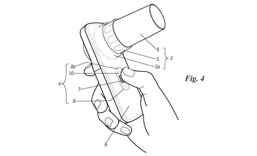
Figure 4 shows the medical device for the system
according to the invention in perspective view during use;
Figure 5 shows a block diagram of a method for
monitoring a patient's state of health performed through
the device in Figure 1;
Figure 6 shows a possible display of data associated
with a patient through use of the device in Figure 1;
Figure 7 shows another possible display of information
obtained through use of the device in Figure 1;
Figure 8 shows a possible configuration of a plan of
action provided by a doctor to a patient;
Figure 9 shows a possible display of self-diagnosis
results for a patient's status obtained through use of the
device in Figure 1; and
CA 03080114 2020-04-23
WO 2019/102324
PCT/IB2018/059039
Figures 10 and 11 show possible screens for ways of
setting up the device in Figure 1.
With reference to the figures mentioned, a medical
device for the system according to the invention is
5 generally indicated by 1 and comprises a body 2 with which
are stably associated a component 3 (or flow measurement
device) for carrying out a spirometry test and a component
4 for performing an oxygen measurement test comprising the
measurement of SPO2 (blood oxygen saturation) and heart
10 rate. Component 3 in particular is defined by an element
detecting respiratory flow 5a and the corresponding element
sensitive to respiratory flow 5 (defined by a turbine
caused to rotate by the air expelled forcefully by a
patient performing a spirometry test) with which may be
15 associated a tubular element 6 capable of acting as a
mouthpiece for the patient. Component 4 on the other hand
is defined by a reflecting photometric touch sensor 7
stably located in a suitable recessed seat 8 provided on
one surface 9 of device 1; this seat is of elongated shape,
substantially elliptical, and has a wall 8a which connects
an internal or back part 10 of such seat (where it faces
sensor 7) to wall or surface 9 of body 2 of device 1.
Typically, in order to calculate the Sp02 (which
indicates the percentage of haemoglobin bound to oxygen
present in arterial blood) the oxygen measuring device uses
the emission of two different signal sources - having a
wavelength in the red and infrared fields respectively -
CA 03080114 2020-04-23
WO 2019/102324
PCT/IB2018/059039
16
applied to the site bathed in the blood which is the object
of measurement: for example the finger.
A photodetector is capable of measuring the absorption
of each of the two signals from the haemoglobin: in fact a
portion of the emitted signals is absorbed by the
haemoglobin present at the site while another portion which
is not absorbed reaches the photodetector.
The quantities of the signals - red and infrared -
absorbed is proportional to the haemoglobin concentration,
as a result of which, knowing the quantity of signals
emitted, and measuring the quantity of signals reaching the
detector, the percentage value of the main oxygen
measurement parameter known as Sp02 can be calculated.
Because blood flow experiences changes due to
heartbeats, by recording the changes in the signal captured
by the photodetector it is also possible to calculate heart
rate.
Oxygen measurement devices can be classified into two
main categories: transmission and reflection types.
The transmission oxygen measurement device uses two
signal emitters - red and infrared - located over the
finger and a photodetector located under the finger. The
three fundamental components are assembled within a
specific sensor generally comprising a rigid cap or
flexible rubber cap similar to a finger stall, or a spring
clamp which is to be applied to the finger.
By adjusting the compression exerted by the rigid cap
CA 03080114 2020-04-23
WO 2019/102324
PCT/IB2018/059039
17
or the spring clamp it is possible to avoid changes in the
vascularisation of the blood. This possible change is
further controlled by using measurement caps or spring
clamps which are appropriate for the size of the finger on
which the Sp02 measurement is being carried out.
This however means that it is necessary to have
available caps or spring clamps of different sizes - small,
medium and large - for use on children, adolescents and
adults respectively, with a consequent increase in the
number of accessories.
The photometric touch sensor comprises a single
integrated chip comprising all the components necessary for
measuring oxygen. The chip in fact contains both the two
side-by-side emitters (one at the red wavelength and the
other in the infrared) as well as the photodetector.
It functions through reflection, in which the two
signals generated by the emitters are directed towards the
site used to measure oxygen, such as for example a finger.
The blood circulating in the finger absorbs the two signals
in different ways according to the haemoglobin present. In
addition to this the signals undergo partial reflection
which is captured by the photodetector.
By placing a finger on the top of the touch sensor the
patient is able to obtain measurement of the signals linked
to oxygen measurement.
In the case of transmission oxygen measurement - in
addition to the advantages mentioned above - the reflection
CA 03080114 2020-04-23
WO 2019/102324
PCT/IB2018/059039
18
method is extremely compact in that it incorporates all the
electronics processing the signals, including monitoring of
the current in the individual emitters and the gain applied
to the photodetector.
As a result of this it does not require a specific
sensor, nor a rigid cap or a cap of flexible rubber, nor a
spring clamp to be applied to the finger. In practice a
single reflection sensor can be used without distinction by
adults and children, wholly eliminating the need for
different accessories as is otherwise required by the
transition oxygen measurement device.
The shape of seat 8 is such that it can be adjusted to
the anatomy of a patient's finger and allow component 4 to
be used with patients of any kind, whether adults or
children. By positioning a finger on the top of touch
sensor 7 patients can obtain a reading of the signals
linked to oxygen measurement; this as shown in Figure 4.
This detection may or may not be simultaneous with the
spirometry test performed using component 3.
Sensitive component 4 is a single chip and is defined
by a so-called "reflecting" sensor which requires a mere
touch to measure Sp02 (the main parameter for the
measurement of oxygen). This is a radical innovation in
comparison with the conventional array of "transmission"
sensors hitherto available (with an emitter facing a
receiver), in which a finger or an earlobe has to be
inserted into a specific sensor connected to a medical
CA 03080114 2020-04-23
WO 2019/102324
PCT/IB2018/059039
19
device in order to be able to obtain the oxygen
measurement.
On the contrary, using reflecting photometric
technology, oxygen measurements can be performed simply by
touching the sensor, without any other particular action.
This reflecting photometric technology does not require
any specific applied sensor, so no adjustment to the
patient's physical characteristics is required and the
oxygen measurement device can be used without distinction
by children, adolescents and adults. It also makes it
suitable for use in the absence of medical personnel and it
is therefore ideal for self-measurement and the self-
management of health.
Within body 1 there is a control unit 13 capable of
being connected to a portable device (such as a smartphone
or tablet) or to an internet network present in the
environment in which device 1 is located through a BLE
(Bluetooth Low Energy) chip. In the latter case (the
preferred solution) the connection is made through a
network node, a gateway, a smartphone, a tablet or a fixed
computer used as an access point for the network.
Control unit 13 comprises an
incorporated
microcontroller which simultaneously manages all the
components of portable medical device 1 and which measures
patients' spirometry and oxygen measurement parameters
through components 3 and 4.
Device 1 is not provided with a visualiser or display
CA 03080114 2020-04-23
WO 2019/102324
PCT/IB2018/059039
and the data found by components 3 and 4 are sent for
display on a computer, tablet or smartphone to which device
1 can be connected via control unit 13 (in addition to
being capable of being "processed" by a suitable medical
5 application executed on a web server, as will be indicated
below).
Device 1 is therefore an element capable of detecting
patients' spirometry and/or oxygen measurement data, but it
is unable to display them directly (because it is not
10 provided with a display). This device is a single unit with
components 3 and 4 for carrying out the spirometry and
oxygen measurement tests, components which cannot be
separated from body 2.
In addition to this, these tests are also performed by
15 holding body 2 in one hand, taking care to place a finger
in seat 8 where the touch sensor is located.
The invention is a case of technological innovation
characterised by a major discontinuity with the state of
the art. As described, it comprises a portable device of
20 small size (see Figure 4 for comparison with a patient's
hand), which is simple to use and convenient to manufacture
and acquire. Device 1 detects the patient's data (through
the spirometry and/or oxygen measurement tests) and works
in combination with an innovative medical algorithm or
application which can also be loaded onto a smartphone, a
tablet, a computer or the like, which harmonises and
integrates objective data (vital parameters measured by
CA 03080114 2020-04-23
WO 2019/102324
PCT/IB2018/059039
21
spirometry and oxygen measurement) and the patient's
subjective data (symptoms). This application, constantly
operating with storage means in which a plan of action for
the patient is stored produces an objective indication of
state of health and corresponding changes, and suggests to
patients suffering from respiratory diseases what action
they should take consistent with what is specified in the
action plan (which in this case is digitised) provided and
updated by their own doctors and capable of enabling the
state of health of patients affected by respiratory disease
to be monitored (as indicated above).
With its ease of use and portability anywhere and at
any time medical device 1, together with the medical
application and digitised action plan (provided by the
doctor) enables anyone, even patients not used to serial
programmed tests, to monitor their own state of health so
that they can self-manage it more easily.
All patients will be able to check whether the
perceived (subjective) worsening corresponds to a real
(objective) change in clinical conditions envisaged in the
digitised action plan provided by their own doctors and
thanks to the immediate suggestions provided by the medical
application patients will be able to adjust their treatment
plans or, in extreme cases, go to emergency without
uselessly losing time.
The system according to the invention (which comprises
device 1, the medical application, the storage means and a
CA 03080114 2020-04-23
WO 2019/102324
PCT/IB2018/059039
22
portable device with a display such as a smartphone, tablet
or computer) and the method correlated with it are
therefore a pillar of tertiary prevention which is
concerned with treating the disease experienced when it
manifests itself clinically with symptoms and with
preventing its progress and improving prognosis by reducing
the risk of exacerbations. In addition to this, through its
technical characteristics, this system also has a very
specific part to play in secondary prevention programs
intended to discover diseases such as asthma or COPD when
they are at an asymptomatic stage, that is before they
become clinically manifest, and therefore at the earliest
possible stage. A classical example of secondary prevention
is screening studies for the early diagnosis of respiratory
diseases.
Finally, through its more general properties, that is
the low cost of device 1, its simple use relating to
widespread computerised facilities (for
example
smartphones), the potential for maximum penetration both as
regards uses and number of users, the system according to
the invention may become a tool in primary prevention for
removing risk factors in healthy individuals (particularly
in some categories of individuals at risk) to prevent the
occurrence of diseases and maintain a good state of health.
It is for example only necessary to think of primary
prevention intended to stop tobacco smoking, which is well
known to be the cause of respiratory diseases such as COPD.
CA 03080114 2020-04-23
WO 2019/102324
PCT/IB2018/059039
23
The system making use of device 1, as mentioned, is
mainly intended for patients suffering from respiratory
diseases whose own doctors have determined a specific
treatment action plan, and has properties which make it
suitable for use by both children and adults. Because it
enables patients to perform self-diagnosis of their own
states of health (without needing the "physical" presence
of medical personnel) the above-mentioned system also makes
it possible to reduce health personnel, and thus to bring
about significant economic savings from the point of view
of public and private organisations caring for the
abovementioned patients.
Through using device 1, the aforesaid system makes it
possible to differentiate objectively between exacerbations
and daily changes in symptoms which a patient presents or
can present.
It is known that the collection and interpretation of
accurate and objective data in significant quantities
relating to the respiratory capacity of patients and blood
oxygen saturation are a fundamental requirement for a valid
clinical assessment. This requirement is satisfied by the
present invention. In particular, the system according to
the invention makes it possible to obtain an objective
index - defined as the CEI (cardiorespiratory efficiency
index) - which is useful for identifying exacerbations when
they begin to develop, with the aim of facilitating timely
access to treatment and avoiding exposing patients to an
CA 03080114 2020-04-23
WO 2019/102324
PCT/IB2018/059039
24
unnecessary or inappropriate treatment plan.
Distinguishing variations in symptoms from an
exacerbation is challenging, but it is very important
because correct and timely treatment of an exacerbation is
certainly associated with rapid patient recovery.
As mentioned, in a preferred embodiment of the
invention, device 1, through its low-energy-consumption BLE
chip, communicates via the internet and interacts - through
access points to the network such as gateways, smartphones,
tablets, PC or any other hardware components provided with
BLE technology and connected to the internet - with a
medical application which can be run on comparison means
such as a microprocessor unit (which may also be the
microprocessor unit of the patient's computer or
smartphone) or a web server (or in the cloud) which is in
any event managed remotely by the treating physician; the
application is also capable of monitoring device 1 so that
the latter receives and executes commands and transmits
digital data relating to the oxygen and spirometry
measurements which it is capable of making, in real time.
Device 1 uses a negligible quantity of energy (because
there is no display and data is transferred to the
network), and therefore a set of batteries is sufficient to
perform thousands of tests. It is also known that Bluetooth
communication, through the cryptography characteristic of
such technology, ensures that sensitive medical data is
protected.
CA 03080114 2020-04-23
WO 2019/102324
PCT/IB2018/059039
The BLE communication system integrated in device 1
comprises the web-based application (mentioned above in the
sense that it is connected to the device via the internet)
to connect to and obtain data collected from that device
5 and show patients their own plan of action (on the display
of a tablet, computer or smartphone, as will be described
below).
As is known, and as already described in the
introductory part of the present document, for patients to
10 have a treatment "action plan" is a fundamental part of the
self-management of respiratory diseases, in particular as
far as asthma is concerned.
When significant changes in the levels of intensity of
symptoms and in values measured using spirometry and/or
15 oxygen measurement techniques are encountered, a plan of
action (written by the treating physician) includes all
information on the actions which patients must take to
reduce the symptoms and significantly reduce the risk of an
exacerbation. This reduces the risks of emergency treatment
20 and includes recommendations for actions which patients
should undertake, including the use of medication.
Normally doctors are concerned to include some
parameters which are useful for managing the disease in the
action plan reference values and in the corresponding alarm
25 thresholds. For example, the value of the envisaged peak
flow (spirometry) and/or Sp02 (oxygen measurement).
The plan of action also includes some symptoms of
CA 03080114 2020-04-23
WO 2019/102324
PCT/IB2018/059039
26
particular interest which patients must monitor in order to
identify whether the respiratory disease is worsening, so
as to obtain help very quickly and reduce the risk of an
exacerbation.
In view of the fact that device 1 can be connected to a
smartphone, tablet or computer, this plan of action is
stored in storage means linked to the internet and managed
by the treating physician who can introduce, update or
modify the action plan for each individual patient. This
action plan and consequent monitoring can be displayed (on
one of such devices or on an attached screen) for example
through a screen illustrated in Figure 9. The screen
illustrated here provides for various areas 100, 101, 102,
103 and 104 in which text relating to the measurement made
by device 1 is displayed (area 101, for example no
respiration), the severity of the data detected (area 102,
for example "all well" or "severe"), written instructions
relating to the health plan (area 103; for example
"respiratory function is worsening" and/or "if the symptoms
do not improve use the medication specified") and visual
indications relating to the patient's status (area 104, for
example advice is provided through coloured elements 104A,
104B and 104C if the patient is "well", if "the disease is
worsening" or if "the situation is serious" respectively).
Area 100 simply indicates that what is shown in the
other areas are the results of a check on the patient's
situation.
CA 03080114 2020-04-23
WO 2019/102324
PCT/IB2018/059039
27
When displayed, it corresponds to what is provided by
the patient's action plan provided by the doctor.
As mentioned, device 1 can be used to measure oxygen
and/or spirometry, and recent studies confirm that
exacerbations are associated with changes in some
physiological measurements. It will not be forgotten that
oxygen measurement measures Sp02 (blood oxygen saturation)
and HR (heart rate), while spirometry measures various
parameters including PEF (peak expiratory flow), FEV1
(forced expiratory volume in the first second), FVC (forced
vital capacity), FEV25-75 (forced expiratory flow between
25% and 75% of FVC), etc.
Up to now, when prescribing self-management, doctors
generally select only one of these spirometry parameters,
generally PEV or FEV1. In the case of patients who have
undergone lung transplants, on the other hand, the medical
scientific literature suggests the use of FEV25-75.
The invention instead makes it possible to measure all
the parameters listed above (Figure 6) and to display them
for example on the screen of a smartphone subdivided into
various quadrants: 200 (for the oxygen measurement), 201
(for spirometry) and 202 (for the total value shown by the
CEI index mentioned above).
The system according to the invention is therefore a
solution for evidence-based (digital) health assistance,
incorporated in real time, interactive, provided through a
hand-held unit, which includes (inseparably) both component
CA 03080114 2020-04-23
WO 2019/102324
PCT/IB2018/059039
28
4 defined by a touch sensor 7 to measure oxygen and
component 3 to measure spirometry, all connected to a
medical application installed on comparison means such as a
remote internet server (managed by the treating doctor) or
on a client which carries out the monitoring method
according to the invention.
The medical application (or the algorithm permitting
implementation of the aforesaid method) receives the
parameters for the measurements made, processes them,
compares them with corresponding reference values
originating from storage in storage means, which are
available and predefined (by the treating doctor in
relation to the specific patient using the device) and
generates data or a "score" relating to the patient's state
of health, said data or "score" being capable of being read
on a screen or displayed for example on a smartphone.
As a final result the application provides an objective
index - known as the CEI (cardiorespiratory efficiency
index) - of the patient's state of health and relative
changes and recommends (or not) that actions should be
undertaken in accordance with what has been specified by
the digitised action plan provided by the treating doctor.
It is known that the comparison means and the storage
means may be in the same device, such as an internet server
or a patient's "client".
The medical application performs the CEI calculation
which can be used to reach a decision relating to possible
CA 03080114 2020-04-23
WO 2019/102324
PCT/IB2018/059039
29
treatment or direct intervention on the patient by the
doctor. In a few seconds all patients can make measurements
of their "vital" parameters, the clinical results of which
- together with the scores for symptoms (indicated by X, Y,
K, W, J and XXX in Figure 7, where the upper quadrant 80
indicates "insert the scores for symptoms" and the severity
(mild (L), moderate (M) and severe (S) is indicated through
legend (81) and the digitised action plan configured
according to the needs of the situation - are automatically
managed by the medical application which is for example
resident on a remote internet server or client, with the
value of a face-to-face visit to the treating physician
himself.
It should be noted that the "score" given to a symptom
corresponds to the feeling of "severity" which the patient
experiences for that symptom.
In addition to managing measurements made by the
medical device and symptom scores, the medical application
also uses the instructions of the digitised action plan
shown in Figure 8, comparing them with the abovementioned
measurements.
As shown in that figure, the screen of the smartphone,
tablet or computer shows for example: a first window 90
relating to the doctor's action plan; a second window 91
with doctor's data (and in particular his telephone
number); a third window 92 relating to symptoms and
measured values; a fourth window 93 relating to different
CA 03080114 2020-04-23
WO 2019/102324
PCT/IB2018/059039
symptoms SY (which through A, B, C, D for simplicity
indicate the possible presence of coughing, chest pain,
absence of breathing and shortness of breath), measured
values (MV) of Sp02 and peak flow (PEF peak expiratory
5 flow) detected using the spirometry test, reference values
(RV) of Sp02 (BPF or best peak flow); a fifth window 94
relating to the treatment; and a sixth window 95 with the
treatment prescription (drugs, doses, frequency of taking
drugs indicated by M, D, F).
10 In this way the digitised action plan provides a number
of advantages in comparison with the traditional written
version:
= it supports decisions for creating an evidence-based
action plan;
15 = it ensures greater accessibility and mobility 24 hours
per day, 7 days per week;
= it permits a standardised assessment in real time for
monitoring diseases with interactive feedback on clinical
actions;
20 = it provides a reminder for automatic compliance with the
treatment plan;
= it improves efficacy and increases commitment from the
patient, who capitalises on the moments of learning
provided by the application;
25 = it reduces the risk threshold due to irrational behaviour
such as patients' "do-it-yourself" initiatives.
In situations where the client (smartphone, tablet,
CA 03080114 2020-04-23
WO 2019/102324
PCT/IB2018/059039
31
computer, etc.) uses an API (application programming
interface) of the Bluetooth Web type, this can communicate
directly with low-consumption Bluetooth devices directly
through a web browser. In this way the abovementioned
medical application can be installed in the server (or in
the cloud) and reached by the medical device via the web
(through connection with the smartphone or tablet, for
example), so that it can operate on any type of client,
regardless of the operating system installed (such as i0S,
MacOS, Linux, Android, Windows).
In the case of "web" applications, these are easy for
patients and doctors to reach; there is no need to install
them on their own clients and they can be updated at any
time to the benefit of all users accessing them.
Users can access the web application through a browser
according to the procedure in Figure 5. Once identified
(block 50), users access an application which adapts its
content to the specific requirement of their disease.
Through the browser the web application can provide
advice of any kind - from a reminder to proceed with
reading data to a reminder to take medication, to
presenting the results of tests made using device 1 (block
51).
Through the use of Bluetooth, the web application
communicates with the devices (block 52) and obtains
spirometry and oxygen measurement data from them (block
53).
CA 03080114 2020-04-23
WO 2019/102324
PCT/IB2018/059039
32
The application processes the CEI index (block 54) and
allows the data obtained to be stored (block 55). In
parallel it allows the system to be configured (block 56),
subdivided into a configuration for calculating the CEI
index (block 57), a configuration for the patient's action
plan (block 58) and a configuration for the patient's data
(block 59).
The web-based medical application can easily be
replaced by an application run directly on a client - which
may be the patient's smartphone or tablet - without the
need for a browser or a nearby network or any other
connection to the net.
For example, in the absence of an internet connection,
the client application can manage the system, display the
results of spirometry and oxygen measurements, request the
entry of symptoms and the level of intensity perceived by a
patient, process the CEI index, and directly interpret the
results on the basis of a personalised configuration
performed directly on the client in relation to the
patient's requirement.
The above is another substantial difference between
known solutions and the present invention. Starting from
U52013/0184540, for example, the prior document describes a
conventional medical tool provided with display, keys and
cables and only and exclusively operates with its own
embedded software which has been preloaded into the
instrument. On the contrary, device 1 does not provide for
CA 03080114 2020-04-23
WO 2019/102324
PCT/IB2018/059039
33
a display, or cables or control keys, or switching-on keys
and functioning is controlled via a smartphone on which a
Mobile Medical App is installed or directly connected to a
browser via Bluetooth Low Energy. This solution is
particularly advantageous both through the enormous
reduction in hardware costs and the possibility of using
applications which can be easily downloaded from an on-line
store, virtually infinitely extending the possibilities for
use of the dedicated software which is not embedded in
device 1. In this way, based on market offer, patients are
in a position to select the applications which best meet
their own needs. For example, in the case of asthmatic
patients the relative application takes into account the
action plan drafted by the doctor and parameters measured
by the instrument and transmitted to the smartphone are
used to establish the patient's state of health in relation
to the action plan. It should be borne in mind that the
action plan includes both therapeutic indications and the
interpretation of measured parameters (spirometry and
oxygen measurements) and the levels of symptoms (coughing,
dyspnoea, chest constriction, etc.) to indicate the most
appropriate treatment based on condition of health at that
time. Use of the new tool enables doctors to change action
plans remotely so that they can be made personalised and
perfectly appropriate to patients' requirements and the
typical variability of asthmatic disease.
In the case of other diseases - such as for example
CA 03080114 2020-04-23
WO 2019/102324
PCT/IB2018/059039
34
COPD or cystic fibrosis - the abovementioned application
can provide incentivising images capable of stimulating
patients when performing spirometry (through incentivising
images on the smartphone) to help them achieve maximum
respiratory performance so that the parameters measured are
as far as possible similar to or better than the reference
values. On the contrary, using the known solution in the
prior document, the incentivising images cannot be
displayed because when the instrument is held the display
cannot be seen while the spirometry test is being
performed.
The invention is therefore based on the fact that
beyond the well-known scoring of symptoms, there are some
tests such as oxygen measurements (which provide objective
parameters such as Sp02 and heart rate) and spirometry
(which provides objective parameters on airway function
such as PEF, FEV1, FVC, FEF25-75, etc.), assessment of
which can reliably distinguish exacerbation from stable
disease and/or daily variation in symptoms.
Use of device 1 automates the entire process described
above and enables the system to provide an objective index
of patients' conditions of health and corresponding changes
in them, as well as suggesting to patients suffering from
respiratory diseases what actions they should take to
improve their condition in accordance with the digitised
action plan provided by treating doctors, resident in the
cloud and constantly managed by the doctors themselves.
CA 03080114 2020-04-23
WO 2019/102324
PCT/IB2018/059039
By recording oxygen measurement and spirometry
parameters together with variations in daily symptoms the
invention is capable of distinguishing between normal
variation in a symptom and an exacerbation, with acceptable
5 sensitivity and specificity. This is through the CEI index,
which objectively confirms exacerbations and is
particularly suitable for patients suffering from
respiratory diseases.
The index takes correctly recorded variations in data
10 into account, both of the objective type such as the vital
parameters of spirometry and oxygen measurement, as well as
those of the subjective type such as symptoms commonly
present in patients suffering from respiratory diseases
(for example shortness of breath, coughing, chest pain, the
15 production of catarrh, sore throat, etc.).
The CEI calculation requires data comprising both
oxygen measurement and spirometry scores, symptom scores
and reference data obtained from the digitised action plan
which can influence patients' conditions of health. It will
20 be noted that in this specific instance the term "score"
indicates the degree of worsening or improvement of oxygen
measurement and spirometry parameters and symptoms.
Through this invention a method requiring performance
of the following tasks is therefore offered:
25 - definition of the predetermined time for collection of
the data required for calculating the CEI index,
- definition of sources of reference data on the basis of
CA 03080114 2020-04-23
WO 2019/102324
PCT/IB2018/059039
36
an action plan,
- collection of clinical data relating to oxygen
measurement and spirometry tests and patients' symptoms,
- conversion and adjustment of the detected data using
appropriate weighting factors set for example as shown in
Figures 10 and 11. Figure 10 shows the configuration of the
value for the weighting factors for further indexes, Sp02,
HR, SPIROMETRY (SPIRO) and symptoms (SYMPTOMS) recorded
from the patient respectively.
The CEI index is defined on the basis of the above-
mentioned scores, and this is obtained by allocating a
greater or lesser weight to the clinical data collected,
using adjustable weighting factors (Figures 10 and 11).
The CEI measurement unit is a number between 0 and 100,
where a higher value indicates a better condition of
health, and therefore synonymous with "better efficiency"
of the cardiorespiratory system.
The CEI index is calculated with contributions from the
four indexes relating to Sp02, heart rate (heart pulse
rate), spirometry and symptoms, each of which has its own
weighting factor (wf - weighting factor) (see Figure 10)
which may have a default value or alternatively may be set
by the doctor.
The value of each index is calculated on the basis of
the following scores:
= the algorithms which will be described below;
= what is established by the doctor in the action plan (or
CA 03080114 2020-04-23
WO 2019/102324
PCT/IB2018/059039
37
from the scientific literature) with alarm values,
reference values, etc.;
= the measured oxygen and spirometry values and the symptoms
provided as an input by the patient using the medical
application.
The formula for calculating the CEI is as follows:
CEI = (Ispo, = wfsp02) + ('HR =f
141, H) +
R, W fSPIRO) ('SYMPTOMS W fSYMPTOMS)
For the weighting factors mentioned above, the
relationship
W fSp02 W fHR W fSPIRO W fSYMPTOMS = 1
always applies.
If it is desired to place more importance on one of the
four components or indexes in comparison with the others it
is sufficient to increase the corresponding weighting
factor. It is obviously possible to assign a predefined
value to each weighting factor, taking care to ensure that
the sum of the factors is always equal to 1.
The following indicators are then analysed:
a) The blood oxygen saturation index: isp02.
The Sp02 index (obtained from the oxygen measuring
test) can be calculated bearing in mind two different
components represented by the partial indexes ispo2iandisp022.
The partial index /spo2idepends on the parameter
SP 2Meas
(measured value) in comparison with the mean value SpO2Avg
calculated over a predetermined period of time.
The partial index isp022 depends on the parameter SP 2Meas
in comparison with the reference value SpO2Rer.
CA 03080114 2020-04-23
WO 2019/102324
PCT/IB2018/059039
38
Let us assume that over a predetermined period of time
the mean value for Sp02 is 97%. This being the case, if Sp02
should fall to 77% there will be a ispo2 index of very close
to 0, synonymous with a serious problem with the patient's
health. If instead the mean Sp02 value were to be 80%, then
a fall in Sp02 to 77% would not produce the same fall in
the index.
The algorithm for analysing Sp02 considers two limit
values: an ideal maximum equal to 100% and a minimum
threshold equal to
SP 2Thr' Values of Sp02meas below Sp02Thr
will however produce a ispo2 index of zero, synonymous with
a serious problem with the patient's health.
The index is defined as follows:
if (Sp02 meas. > SpO2Thr) then Isp02 = 100 = 15021 = /sp022
otherwise Isp02 = 0
i
if (SP 2Meas > SP 2Av9) then Isp02 = 1
SpO2Meas ¨ 5pO2Thr
otherwise Ispo2 =
1 SP 2Avg ¨ SP 2Thr
if (Sp02meas > SpO2Rer) then 15022= 1
Sp02meõ ¨ SpO2Thr
otherwise 1,51,02 = ________________________________
2 SPO2Rer ¨ SpO2Thr
B) The heart rate index: 4m
The heart rate index (a parameter also obtained from
the oxygen measurement test) can be calculated taking into
CA 03080114 2020-04-23
WO 2019/102324
PCT/IB2018/059039
39
account four different components represented by the
partial indexes /HRi, /HR2, /HR3 and /HR4. Each of these components
IHRx is weighted with the corresponding weighting factor
whiRx =
The contribution of heart rate 4m .1/17,6 to the overall
CEI index is equal by definition to:
4
('HR WfHR) = IHRx W fHRx
x=1
The partial index 4m1 depends on bradycardia.
The partial index 4m2 depends on tachycardia.
The partial index 4m3 depends on increase in heart rate
within the normal range.
The partial index 4m4 depends on cardiac arrhythmia.
The following relationship:
wtHR = wtHR, wtHR2 wtHR, wtHR4
where vi/AR is the overall weighting factor for the heart
rate index, always applies for the four weighting factors.
If it is wished to give more importance to one of the
four components or indexes mentioned above in comparison
with the others it is sufficient to increase the relative
weighting factor. The invention makes it possible to assign
a value for each weighting factor, taking care to ensure
that the sum of all the terms is always equal to WAR.
The international scientific medical literature defines
a normal range for heart rate which generally lies between
55 and 100 beats/minute. Below the normal range is referred
to as bradycardia and above tachycardia. The further away
CA 03080114 2020-04-23
WO 2019/102324
PCT/IB2018/059039
from the normal range the more the calculated indexes fall,
synonymous with the greater impact of heart rate on
worsening of condition of health.
The partial index 4m1 is linked to bradycardia and the
5 parameter HR meas is used to calculate it.
In an actual example, the index is defined as follows:
ifHRmeas 55 thenIHRi = 100
if 50 HRmeas < 55 then IHRi = 80
if 45 HRmeas < 50 then IHRi = 60
if 40 HRmeas <45 then IHRi = 40
if HRmeas <40 then IHRi = 20
These values may however be changed without altering
the mechanism underlying the calculation.
The partial index 4/R2 is linked to tachycardia and the
10 parameter HR
-- meas is used to measure it.
The index is defined as follows:
if HRmeas 100 then IHR2 = 100
if 100 < HRmeas < 110 then IHR2 = 80
if 110 < HRmeas < 120 then IHR2 = 60
15 if 120 < HRmeas < 125 then IHR2 = 40
if HRmeas > 125 then IHR2 = 20
These values can however be changed without altering
the mechanism underlying the calculation.
The partial index 4m3 is linked to "heart rate" within
20 the normal range. The calculation takes into account the
possibility that the measured value HR
--meas may exceed the
mean value HRywg calculated over a predetermined period of
CA 03080114 2020-04-23
WO 2019/102324
PCT/IB2018/059039
41
time by at least two standard deviations (SD).
The index is defined as follows:
)
if HR mõs > (HRAvg + 2 = HRsD) then IHR3 = (HRAvg HR ). 100
Meas
otherwise IHR3 = 100
Arrhythmia is a heart anomaly which gives rise to
irregular sequences of heartbeats: too slow, too fast, or
without linear progression.
Device 1 is able to detect an irregular heartbeat on
the basis of a predetermined number of heartbeats
(typically 10) and calculates the mean and standard
deviation of the time intervals between the heartbeats
taken into consideration.
The arrhythmia index 4m4 is linked to the possible
presence of arrhythmia within the range 40-125
beats/minute. The index is proportional to the ratio
between the standard deviation 77,5D and the mean of the
time interval 77,4õ9. If the ratio between 77,5D and 77,4õ9
exceeds a predetermined threshold 7
7
- -ThrRatio (typically 0.06)
the beats are considered to be irregular.
The index is defined as follows:
TIsT,
if - TIT Ratio
(TIAvg then IHR4 = 0
TIsp
otherwise IHR4 = 100 = 1 ( ___________________________
TiThrRatio = TIAvg)
c) Spirometry index: IspiRo
SPIRO is the generic parameter representing spirometry
CA 03080114 2020-04-23
WO 2019/102324
PCT/IB2018/059039
42
and can be selected from those measured by the medical
device. Among the most important of these are: PEF, FEV1,
FVC, FEF25-75. Once the parameter has been selected the
medical application uses it to calculate its percentage
variation with respect to the reference value.
Given that the parameter SP/R00/0 represents the
percentage of the measured value SPIROmeas with respect to
the reference value SPIRORef:
SPIROmeas
SP/R00/0 = 100
SP/RORef
The spirometry index can be calculated taking into account
three different components represented by the partial
indexes ispiRoi, IspiRo2 IspiRo3 :
ISPIRO = 100 = isp/Rol ' isp/R02 ' isp/R03
The partial index ispiRoi depends on SP/R00/0 and its mean
value SP/R00/0Aõ.9.
if(SPIR0%," SPIRO%) then IspIROi = 1
if (SPIRO% < 10) then IspIROi = 0
SPIRO% ¨ 10
otherwise IspIROi = (SP IRO%Avg ¨ 10)
The partial index isp/R02 depends on SP/R00/0 and the
predetermined upper limit of the reference value which is
100%.
if (SPIRO% > 100) then I5pIRO2 = 1
CA 03080114 2020-04-23
WO 2019/102324
PCT/IB2018/059039
43
if (SPIRO% < 10) then IspIRO2 =
SPIRO% ¨ 10
otherwise IspIR02 = _______________________________
(100 ¨ 10)
It will be noted that 10 and 100 are the minimum and
maximum values of SPIRO% respectively.
The partial index IspiRo3 depends on the daily
variability in the spirometry parameter over a
predetermined period of time. Daily variability is an
indicator of the functioning of the airways.
The index IspiRo3 depends on the so-called "variability"
in the spirometry parameter with respect to the minimum
measured value SPIROmin and the maximum measured value
SPIROmõ recorded over a predetermined period of time.
In practice, when the variability threshold SPIROThrvar
indicated in the plan of action specified by the doctor
(typically 20%) is exceeded, then there is a greater risk
of exacerbation. In this case the index IspiRo3 produces a
fall in the spirometry index IspiRo which in turn brings
about a fall in the value of the overall CEI index to
indicate a worsening in condition of health.
The partial index linked to the variability in the
spirometry parameter is defined as follows:
(SP/ROMõ ¨ SPIROmil SPIROmin
if ________________ 100 > SPIROThrvar then IspiRo3 =
Max SPIROMõ
otherwise IspIR03 = 1
CA 03080114 2020-04-23
WO 2019/102324
PCT/IB2018/059039
44
The reference value is based on the anthropometric data for
the patient and can be found from tables or calculated
using formulae published by the main international
organisations operating in the field of respiratory
diseases. Alternatively the reference values can be defined
by considering the patient's better typical values.
d) Symptoms index: 'SYMPTOMS
The symptoms index depends on the value of the scores
for individual symptoms.
For simplicity of description we can by way of example
restrict the analysis (but not in any limiting way) to six
symptoms which are generally sufficient for the self-
management of respiratory diseases. Obviously the symptoms
may also be fewer in number.
Description Score Index Weighting factor
Shortness of breath scoresympi /sympi w f.
, ,.yrtipi
Coughing scoresymp2 Isyn1P2 W f.
J '3 YMP 2
Absence of breath score f.
syrnp 3 Isyrnp 3 w ' '3 YMP 3
Cardiac oppression score f.
syn1P4 Isyn1P4 w J '3 YMP 4
Production of catarrh scoresymps Isymp 5 WI:.
J '3 YMP 5
Heartburn scoresymp6 Isyn1P6 w f.
J '3 YMP 6
The term I sym pr 0 m s . W fSYMPTO MS of the CEI formula is by
definition equal to
6
'SYMPTOMS . W fSYMPTOMS = 1 I symp, . W fsympx
x=1
CA 03080114 2020-04-23
WO 2019/102324
PCT/IB2018/059039
The following relationship applies between the symptom
weighting factors:
WfSYMPTOMS = WfsYmP4 + WfsYmP2 + WfsYmP3 + WfsYmP4 + WfsYmPs + WfsYmP6
If it is desired to give more importance to one symptom
5 in comparison with the others it is sufficient to change
the corresponding weighting factor as for example indicated
in Figure 11. The invention makes it possible to assign a
value to each weighting factor, making sure to check that
the sum of all the terms is always equal to WfsympToms.
10 Each of the six symptoms will be assessed by the
patient with a score, for example from 0 to 3, assigned on
the basis of presence and level of severity: 0 = symptom
absent; 1 = mild symptom; 2 = moderate symptom; 3 = severe
symptom (the scale from 0 to 3 is purely indicative).
15 The partial indexes for symptoms are calculated using
the following formula (with x identifying the symptom
considered where 3 identifies the maximum score assigned to
each symptom):
3 ¨ scoresympx
20 /sympx = 100= __ 3
As indicated, the CEI index refers to the patient's
condition of health and variations in that condition.
Assessment of quantitative objectives provides important
indications which can give significant answers to improve
25 condition of health, thus initiating a virtuous cycle. In
fact the results of the current assessment become the basis
for planning subsequent improvement.
CA 03080114 2020-04-23
WO 2019/102324
PCT/IB2018/059039
46
The index introduced by the present invention can be
seen as a value which is representative of the efficiency
of the cardiorespiratory system over a certain period of
time; this in turn represents a general model of health
which is particularly suitable for patients suffering from
respiratory diseases.
The CEI index can be used to monitor patients' state of
health on a daily basis, so as to help them self-manage
their disease and improve their state of health. Use of
this index is useful to patients, health sector
professionals and other interested parties.
The purpose of the CEI index is to establish a
consistent approach for measuring the health condition of
patients and variations in that condition. This index can
be used as a tool for primary monitoring to ensure that the
condition of health of patients suffering from respiratory
diseases achieves the expected results.
The aim of the CEI index as an operating indicator is
to help patients to assess changes in cardiorespiratory
performance. In addition to this, the index is also
intended to provide an example of a method of calculation
which might be used as an objective approach to monitor the
efficacy of treatment administered to a patient.
According to the action plan provided by the doctor, it
is therefore possible to establish a suitable period of
time for monitoring a patient, based on spirometry and
oxygen measurement tests performed using device 1. A mean
CA 03080114 2020-04-23
WO 2019/102324
PCT/IB2018/059039
47
for the data used to calculate the CEI index can then be
established over a predetermined period of time using the
following technique. Over a number of days (for example 28
days or four weeks) the first element of the mean is
obtained by taking the mean of a subset, equal to the first
set of a number of days (for example the first seven days
or the first week). The subset is then changed "moving the
period forward", that is excluding the first day of the
first subset (for example the first day of week one) and
including the first day following the first subset (for
example the eighth day or the first day of the second
week). This new second subset will provide the second
element of the current mean. This process continues until
the entire period of interest is covered.
Thanks to the invention there is a system comprising a
medical device capable of interacting, through BLE chips,
with a medical application present in comparison means such
as a microprocessor unit or a client or a remote web
server, the medical device receives a request for the
transmission of physiological data (in digital form)
relating to oxygen measurement and spirometry tests
performed by a patient through using device 1 (and stored
in the memory unit in control unit 13) from the medical
application and sends digital data pertinent to the
operating state of the device.
The medical application processes the digital
physiological data, compares them with corresponding
CA 03080114 2020-04-23
WO 2019/102324
PCT/IB2018/059039
48
restored reference values available in the action plan
provided by the treating doctor, integrates them with the
"score" provided by the patient for his own systems and, as
a final result, provides a cardiorespiratory efficiency
index (CEI) relating to the patient's condition of health.
The application method, based on data managed by the
doctor, suggests actions which are to be taken in
accordance with reference values and alarm levels
originally configured in accordance with what the doctor
himself has prescribed in the patient's digital action
plan.
It is known that the device can perform an oxygen
measurement and a spirometry measurement simultaneously
with sending data to the web or before the request for
delivery of such data from the abovementioned application.
Placing a finger on photometric touch sensor 7 of portable
medical device 1 and simultaneously breathing into
component 3 (comprising the flow measurement device) thus
make it possible to make both oxygen and spirometry
measurements.
The invention has been demonstrated and described using
the solutions preferred for it; it will be clear to those
skilled in the art that various changes may be made to the
form and detail of the invention without thereby going
beyond the spirit and the field of application of the
invention as defined by the appended claims.
For example, the web-based medical application may
CA 03080114 2020-04-23
WO 2019/102324
PCT/IB2018/059039
49
easily be replaced by an application directly installed on
a client - which could be a smartphone or a tablet -
without any need for a browser or a nearby internet network
or any other connection. Such "local" application is
however updated via the internet by the application present
on the web managed by the doctor who has the patient's
action plan.
Another example of a different embodiment of the
proposed invention, which might be used in the light of any
structural changes made without going beyond the scope of
the invention, is different representations of the CEI
index value and its variations.
In addition to numerical display from 0 to 100, this
index may be displayed by an intuitive image which
expresses conditions of health and possible variations in
them, such as for example a tree or a plant in a flower
vase. In the case of deterioration the state of health
expressed by the CEI index value could effectively be
represented by fallen leaves or flowers which bend or wilt.
Conversely, if health improves, there could be new leaves
or new flowers, or leaves which grow and flowers which
flower.
Another type of representation of the CEI index value
could be an archery target where an arrow at the centre
represents an optimum condition of health, while the
further the arrow is from the centre the worse the
patient's health will be.
CA 03080114 2020-04-23
WO 2019/102324
PCT/IB2018/059039
Even these variants fall within the scope of the
following claims.