Note: Descriptions are shown in the official language in which they were submitted.
FLUID HANDLING DETECTORS
CROSS-REFERENCE
[0001] This is an international (PCT) application relating to and claiming the
benefit of
commonly-owned, co-pending U.S. Provisional Patent Application No. 62/565,746,
filed
September 29, 2017 and titled "FLUID HANDLING DETECTORS".
FIELD
[0002] The present disclosure relates generally to the field of detectors for
bodily
fluids, and in particular to the fluid handling of those bodily fluids in
those detectors.
BACKGROUND
[0003] In healthcare there is a need for continuous, non-invasive monitoring
of
physiological analytes, i.e. biomarkers, for assessing human performance,
health and
wellbeing. Although these analytes are present in blood, obtaining a blood
sample requires
an invasive sample collection, so other analyte sources may be preferred.
[0004] Widely recognized as being easily accessible, sweat and interstitial
fluid can
provide important information. Sweat contains many of the analytes that are
carried in
other bodily fluids, such as blood, which can provide significant information
which
enables one to diagnose ailments, health status, toxins, performance, and
other
physiological attributes even in advance of any physical sign. Furthermore,
sweat itself,
and the action of sweating, or other parameters, attributes, solutes, or
features on or
near skin or beneath the skin, can be measured to further reveal physiological
information. Thus, sweat sensing technologies can be used with wide ranging
applications from athletics, to first-responders and military, to pediatrics,
to
pharmacological monitoring, to personal digital health. The sensors can
measure one
analyte, such as sodium, chloride, or potassium ions, or combinations of
analytes. One
application would allow diabetics to monitor blood glucose without drawing
blood.
1
Date Recue/Date Received 2021-09-27
CA 03080898 2020-04-29
WO 2019/068047 PCT/US2018/053643
Another application is early detection of toxins in at-risk individuals, and,
in particular,
children.
[0005] Although humans have millions of sweat glands, collecting a sufficient
volume
of sweat is challenging. Sweat is difficult to collect for analysis either
because of a lack
of production, evaporation, or collection errors. Also collecting fresh sweat
and replacing
older sweat can cause problems for sensing applications. Accumulation of older
sweat
can lead to inaccurate readings.
[0006] Thus, there remains a need for improved fluid handling to collect a
sample
containing analytes for sensing applications. The properties and advantages of
the
present invention will become apparent to those of skill in the art upon
reading the
following disclosure.
SUM MARY
[0007] Covered embodiments are defined by the claims, not this summary. This
summary is a high-level overview of various aspects and introduces some of the
concepts that are further described in the Detailed Description section below.
This
summary is not intended to identify key or essential features of the claimed
subject
matter, nor is it intended to be used in isolation to determine the scope of
the claimed
subject matter. The subject matter should be understood by reference to
appropriate
portions of the entire specification, any or all drawings, and each claim.
[0008] In some embodiments, a detector detecting an analyte in a sample as
described herein includes a fluid-collecting porous material including a
sample-
collection surface and an analyte detection surface, the fluid-collecting
porous material
including at least one hydrophilic porous layer; and one or more sensors
mounted to the
analyte-detection surface. In some embodiments, the hydrophilic porous layer
includes
hydrophilic regions and hydrophobic regions, wherein the hydrophilic regions
form a first
hydrophilic surface area on the sample-collection surface and a second
hydrophilic
surface area on the analyte-detection surface, and wherein the first
hydrophilic surface
area is greater than the second hydrophilic surface area. For example, the
hydrophilic
porous layer optionally may include hydrophilic regions and hydrophobic
regions,
wherein the hydrophilic regions and hydrophobic regions form microfluidic
hydrophilic
2
CA 03080898 2020-04-29
WO 2019/068047 PCT/US2018/053643
channels between the sample-collection surface and the analyte-detection
surface. In
some embodiments, the at least one hydrophilic porous layer includes a
fluoropolymer,
polyurethane, polyolefin, polyester, polymeric organosilicon compound, or a
combination thereof.
[0009] The fluid-collecting porous material optionally may include at least
two adjacent
hydrophilic layers, wherein a first hydrophilic layer includes the sample-
collection
surface and a second hydrophilic layer includes the analyte-detection surface,
and
wherein the first and second hydrophilic layers are arranged with the sample-
collection
surface in fluid communication with the analyte-detection surface. In some
embodiments, the first hydrophilic porous layer has a larger pore size than
the second
hydrophilic porous layer. In some embodiments, the first hydrophilic porous
layer is
conformable to a surface upon which the sample is collected. In some
embodiments,
the second hydrophilic porous layer has a mass to area ratio of 4 grams per
square
meter (gsm) or less and/or has a bubble point of greater than 65 kPa. The
second
hydrophilic porous layer optionally may displace the sample laterally in the
portion
covered by the one or more sensor. For example, the area of the displaced
sample
optionally may be at least 20 mm2 in the second hydrophilic porous layer.
[0010] The sample collecting surface optionally may include pores having a
size small
enough to filter 97% of particulates having a diameter of greater than 0.07
microns from
the sample. The sample-collection surface and analyte-detection surface
optionally may
each include a material not dissolvable in water. The detector optionally may
further
include a liquid-proof barrier layer covering the one or more sensors and a
portion of the
analyte-detection surface, the liquid proof barrier including a polymeric
material or resin.
In some embodiments, the analyte-detection surface includes an exposed surface
region adjacent to and outside of the liquid-proof barrier layer.
[0011] In some embodiments, a detector detecting an analyte in a sample as
described herein includes a hydrophilic porous layer having a mass to area
ratio of 4
gsm or less and a first surface opposite a second surface; and one or more
sensors
mounted to the second surface, wherein the one or more sensors are adapted to
provide a response to the presence of an analyte in sample. For example, the
mass to
area ratio of the hydrophilic porous layer is 3 gsm or less or optionally may
be from 0.5
3
CA 03080898 2020-04-29
WO 2019/068047 PCT/US2018/053643
to 4 gsm. In some embodiments, the at least one hydrophilic porous layer
includes a
fluoropolymer, polyurethane, polyolefin, polyester, polymeric organosilicon
compound,
or a combination thereof. In some embodiments, the hydrophilic porous layer
has a
bubble point of greater than 65 kPa. In some embodiments, the hydrophilic
porous layer
displaces the sample laterally in the portion covered by the one or more
sensor. The
detector optionally may further include a second a hydrophilic layer including
a sample-
collection surface, wherein the second hydrophilic layer is adjacent to the
first surface.
In some embodiments, the second hydrophilic layer has a larger pore size than
the first
hydrophilic porous layer.
[0012] In some embodiments, a detector detecting an analyte in a sample as
described herein includes a porous material including a first hydrophilic
porous layer
and an adjacent second hydrophilic layer, wherein the first hydrophilic layer
includes a
sample-collection surface and the second hydrophilic layer includes an analyte-
detection surface, and wherein the first hydrophilic layer includes a bubble
point at least
50 kPa lower than a bubble point of the second hydrophilic layer; and one or
more
sensors mounted to the analyte detection surface, wherein the one or more
sensors are
adapted to provide a response to the presence of an analyte in sample. For
example,
the bubble point of the first hydrophilic layer optionally may be at least 60
kPa lower
than a bubble point of the second hydrophilic layer or optionally may be at
least 70 kPa
lower than a bubble point of the second hydrophilic layer. In some
embodiments, the
second hydrophilic porous layer has a bubble point of greater than 65 kPa,
greater than
100 kPa, greater than 150 kPa, or greater than 175 kPa.
[0013] The first and/or the second hydrophilic porous layer optionally may
include a
fluoropolymer, polyurethane, polyolefin, polyester, polymeric organosilicon
cornpound,
or a combination thereof. The second hydrophilic porous layer optionally may
displace
the sample laterally in the portion covered by the one or more sensor.
[0014] In some embodiments, a detector detecting an analyte in a sample as
described herein includes at least one hydrophilic porous layer having a
thickness from
0.5 to 50 pm, and a first surface opposite a second surface; and one or more
sensors
mounted to the second surface, wherein the one or more sensors are adapted to
provide a response to the presence of an analyte in sample. For example, the
thickness
4
CA 03080898 2020-04-29
WO 2019/068047 PCT/US2018/053643
of the at least one hydrophilic porous layer optionally may be from 1 to 5 pm.
The at
least one hydrophilic porous layer optionally may have a bubble point of
greater than 65
kPa.
[0015] The at least one hydrophilic porous layer optionally may include a
fluoropolymer, polyurethane, polyolefin, polyester, polymeric organosilicon
compound,
or a combination thereof. The second hydrophilic porous layer optionally may
displace
the sample laterally in the portion covered by the one or more sensor. The
detector
optionally may further include a second a hydrophilic layer including a sample-
collection
surface, wherein the second hydrophilic layer is adjacent to the first
surface. In some
embodiments, the second hydrophilic layer has a larger pore size than the
first
hydrophilic porous layer. The second hydrophilic layer optionally may include
a larger
pore size than the hydrophilic porous layer. The second hydrophilic layer
optionally may
include an average thickness from 5 pm to 100 pm. The second hydrophilic layer
optionally may include a non-uniform thickness.
[0016] In some embodiments, a detector detecting an analyte in a sample as
described herein includes a hydrophilic porous layer having a mass to area
ratio of 4
gsm or less, a bubble point of at least 65 kPa, and a thickness from 0.5 to 50
pm, and a
first surface opposite a second surface; and one or more sensors mounted to
the
second surface, wherein the one or more sensors are adapted to provide a
response to
the presence of an analyte in sample.
[0017] In some embodiments, a detector detecting an analyte in a sample as
described herein includes a porous material including a reservoir layer having
a
hydrophilic region and a hydrophobic region, and a collection layer including
a collecting
surface opposite the reservoir layer, wherein the collecting surface is in
fluid
communication with the reservoir layer; and one or more sensors mounted to the
hydrophilic region, wherein the one or more sensors are adapted to provide a
response
to the presence of the analyte. In some embodiments, the collection layer
includes at
least one hydrophilic region. In some embodiments, the reservoir layer has a
mass to
area ratio of 4 gsm or less and/or a bubble point of 65 kPa or more and/or a
thickness
from 0.5 to 50 pm.
CA 03080898 2020-04-29
WO 2019/068047 PCT/US2018/053643
[0018] The porous material optionally may include a fluoropolymer,
polyurethane,
polyolefin, polyester, polymeric organosilicon compound, or a combination
thereof. The
hydrophilic region of the reservoir layer optionally may include a coated
expanded
polytetrafluoroethylene. In some embodiments, the hydrophilic region of the
reservoir
layer has a larger pore size than the collection layer. The collection layer
optionally may
be conformable to a surface upon which the sample is collected. The collection
layer
optionally may include pores having a size small enough to filter 97% of
particulates
having a diameter of greater than 0.07 microns from the sample. In some
embodiments,
the detector further includes a liquid-proof barrier layer covering the one or
more
sensors and a portion of the reservoir layer, the liquid proof barrier
including a polymeric
material or resin.
[0019] In some embodiments, a detector detecting an analyte in a sample as
described herein includes hydrophilic layer having a first surface including a
hydrophobic region and a hydrophilic region, and a second surface including a
hydrophobic region and a liquid-barrier region; and one or more sensors
mounted within
the hydrophilic layer on the hydrophobic region of the first surface, wherein
the one or
more sensors are adapted to provide a response to the presence of the analyte.
[0020] In some embodiments, a detector detecting an analyte in a sample as
described herein includes a porous material having a hydrophilic region
surrounded by
a hydrophobic region, wherein the hydrophilic region includes a collection
zone, a
pathway and an evaporation zone, wherein the pathway provides a fluid
connection
between the collection zone and evaporation zone; one or more sensors mounted
to the
pathway; and a liquid-proof barrier layer covering the one or more sensors,
pathway
and collection zone. In some embodiments, the one or more sensors are adapted
to
provide a response to the presence of the analyte flowing through the pathway
from the
collection zone to the evaporation zone. In some embodiments, the hydrophobic
region
partially surrounds the hydrophilic region. The porous material optionally may
include a
fluoropolymer, polyurethane, polyolefin, polyester, polymeric organosilicon
compound,
or a combination thereof. The hydrophilic region of the porous material
optionally may
include a coated expanded polytetrafluoroethylene. In some embodiments, the
hydrophilic region of the porous material has a mass to area ratio of 4 gsm or
less
6
CA 03080898 2020-04-29
WO 2019/068047 PCT/US2018/053643
and/or a thickness from 0.5 to 50 pm and/or a bubble point of greater than 65
kPa. In
some embodiments, the porous material is a second porous layer and the
detector
further includes a first porous layer opposite the liquid-proof barrier layer,
wherein the
first porous layer includes a bubble point at least 50 kPa lower than a bubble
point of
the second porous layer.
[0021] In any embodiment described herein, the one or more sensors optionally
may
be adapted to provide a response to the presence of an analyte in sample. In
an
embodiment described herein, the analyte optionally may be a protein,
cytokine, ion,
metabolite, glucose, glucose oxidase, enzyme, hormone, DNA, peptide or
combinations
thereof. In any embodiment described herein, the one or more sensors
optionally may
be adapted to provide a response to a pH, temperature, humidity, or impedance.
In any
embodiment described herein, the sample optionally may be sweat, blood, urine,
salvia,
interstitial fluid, or other bodily fluid.
[0022] In any embodiment described herein, the detector optionally may further
include an adhesive for adhering the detector and/or optionally may further
including a
stimulator for heating the sample. In any embodiment described herein, the
sample
optionally may be collected directly into the detector without passing through
a volume
of oil. In any embodiment described herein, the sensor optionally may be in
the pores of
the hydrophilic region. In any embodiment described herein, the flow of the
sample
optionally may be 0.1 to 5 nL per minute per gland. In any embodiment
described
herein, the sensor optionally may be porous.
[0023] In some embodiments, a detector for detecting an analyte in a sample
collected
on skin of a subject includes a first layer having a first side and a second
side opposite
the first side, wherein the first layer comprises a synthetic porous membrane
having a
first average pore size, wherein at least a portion of the first layer is
hydrophilic, and
wherein the first layer is configured to conform to the skin; a second layer
having a first
side and a second side opposite the first side, wherein the second layer is
coupled to
the first layer such that at least a portion of the first side of the second
layer is directly
adjacent to at least a portion of the second side of the first layer, and
wherein the
second layer comprises a synthetic porous membrane having a second average
pore
size that is smaller than the first average pore size, wherein at least a
portion of the
7
CA 03080898 2020-04-29
WO 2019/068047 PCT/US2018/053643
second layer is hydrophilic; and at least one sensor configured to detect the
analyte,
wherein the at least one sensor is mounted to at least one of (a) the first
layer, or (b) the
second layer.
[0024] In some embodiments, the at least one sensor is mounted to the second
side of
the second layer.
[0025] In some embodiments, the detector also includes a liquid-proof layer
overlaying
at least a portion of the second side of the second layer so as to cover the
at least one
sensor.
[0026] In some embodiments, the second layer includes a hydrophilic region and
a
hydrophobic region. In some embodiments, the at least one sensor is mounted to
the
second side of the second layer so as to be positioned on the hydrophilic
region of the
second layer. In some embodiments, the hydrophobic region of the second layer
includes a barrier.
[0027] In some embodiments, the first layer includes a hydrophilic region and
a
hydrophobic region. In some embodiments, the hydrophilic region of the first
layer is
offset from the hydrophilic region of the second layer. In some embodiments,
the
hydrophobic region of the first layer includes a barrier positioned on the
first side of the
first layer. In some embodiments, the at least one sensor is positioned
between the
barrier and the hydrophobic portion of the first layer.
[0028] In some embodiments, the at least one sensor is positioned between the
hydrophilic region of the first layer and the hydrophilic region of the second
layer.
[0029] In some embodiments, the hydrophobic region of the second layer
comprises a
barrier positioned on the second side of the second layer. In some
embodiments, the at
least one sensor is positioned between the barrier and the hydrophilic portion
of the
second layer.
[0030] In some embodiments, at least one of (a) the first layer or (b) the
second layer
includes a fluoropolymer. In some embodiments, the fluoropolymer includes
expanded
polytetrafluoroethylene.
[0031] In some embodiments, the detector also includes an adhesive positioned
on at
least a portion of the first side of the first layer, the adhesive being
configured to adhere
the first layer to the skin.
8
CA 03080898 2020-04-29
WO 2019/068047 PCT/US2018/053643
[0032] In some embodiments, an average pore size of the first layer is from
0.04 to
200 pm. In some embodiments, an average pore size of the first layer is from
0.1 to 5
pm. In some embodiments, an average pore size of the second layer is from 0.03
to 10
pm. In some embodiments, an average pore size of the second layer is from 0.03
to 5
pm.
[0033] In some embodiments, a bubble point of the first layer is from 0.3 to
1500 kPa.
In some embodiments, a bubble point of the first layer is from 5 to 500 kPa.
In some
embodiments, a bubble point of the second layer is from 5 to 2000 kPa. In some
embodiments, a bubble point of the second layer is from 100 to 1000 kPa.
[0034] In some embodiments, a bubble point of the second layer is from 1.1 to
1000
times greater than a bubble point of the first layer. In some embodiments, a
bubble
point of the second layer is from 2 to 100 times greater than a bubble point
of the first
layer. In some embodiments, a bubble point of the second layer is from 1 to
1500 kPa
greater than a bubble point of the first layer. In some embodiments, a bubble
point of
the second layer is from 5 to 500 kPa greater than a bubble point of the first
layer.
[0035] In some embodiments, the first layer and the second layer are at least
partially
bonded to one another.
[0036] In some embodiments, the sensors are one of (1) printed onto the at
least one
of (a) the first layer or (b) the second layer, (2) physically retained in
proximity to the at
least one of (a) the first layer or (b) the second layer, (3) deposited onto
the at least one
of (a) the first layer or (b) the second layer, (4) adhered to the at least
one of (a) the first
layer or (b) the second layer, or (5) sandwiched between the first layer and
the second
layer.
BRIEF DESCRIPTION OF THE DRAWINGS
[0037] The accompanying drawings are included to provide a further
understanding of
the disclosure and are incorporated in and constitute a part of this
specification to
illustrate embodiments. Together with the description the drawings serve to
explain the
principles of the disclosure. The accompanying drawing figures referred to
herein are
not necessarily drawn to scale, but may be exaggerated to illustrate various
aspects of
9
CA 03080898 2020-04-29
WO 2019/068047 PCT/US2018/053643
the present disclosure, and in that regard, the drawing figures should not be
construed
as limiting.
[0038] FIG. 1 is a schematic of a detector attached to a person in accordance
with
embodiments disclosed herein.
[0039] FIG. 2A and 28 are cross-section views of a detector having a single
porous
material in accordance with embodiments disclosed herein
[0040] FIG. 20 is a cross-section view of the detector of FIG. 2A having a
liquid barrier
covering the target area in accordance with embodiments disclosed herein.
[0041] FIGS. 3A, 36 and 3C are cross-section views of a detector having a
porous
material with a first layer and a second layer in accordance with embodiments
disclosed
herein.
[0042] FIG. 3D is a cross-section view of the detector of FIG. 3A having a
liquid barrier
covering the target area in accordance with embodiments disclosed herein.
[0043] FIG. 4 is a cross-section view of a detector having a layer with
hydrophilic and
hydrophobic regions in accordance with embodiments disclosed herein.
[0044] FIG. 5 is a cross-section view of a detector having sensors within the
hydrophilic layer and mounted on a hydrophobic layer in accordance with
embodiments
disclosed herein.
[0045] FIG. 6 is a cross-section view of a detector having a first layer with
hydrophobic
and hydrophilic regions in accordance with embodiments disclosed herein.
[0046] FIG. 7A is a top view of a detector having a flow path in accordance
with
embodiments disclosed herein.
[0047] FIG. 7B is a cross-section view of the detector in FIG. 7A having one
porous
layer in accordance with embodiments disclosed herein.
[0048] FIG. 70 is a cross-section view of the detector in FIG. 7A having a
porous layer
with two asymmetrical porous materials in accordance with embodiments
disclosed
herein.
[0049] FIG. 8 is an image of the wetted area for Comparative Example A.
[0050] FIGS. 9A-9F are time-lapsed images of the wetted area for Example I.
[0051] FIG. 10 is a SEM image of the porous material in Example 1.
[0052] FIGS. 11A-11D are time-lapsed images of the wetted area for Example 2.
CA 03080898 2020-04-29
WO 2019/068047 PCT/US2018/053643
[0053] FIG. 12 is a SEM image of the porous material in Example 2.
[0054] FIGS. 13A-13D are time-lapsed images of the wetted area for Example 3.
[0055] FIG. 14 is a graph comparing the wetted areas of the examples over
time.
[0056] FIGS. 15A and 15B are schematics of the thickness measuring apparatus
used
to determine thickness of materials used in the Examples.
[0057] FIG. 16 is an illustration of a conductive ink pattern used in an
electrical
continuity test.
DETAILED DESCRIPTION
[0058] Persons skilled in the art will readily appreciate that various aspects
of the
present disclosure can be realized by any number of methods and apparatus
configured
to perform the intended functions. It should also be noted that the
accompanying figures
referred to herein are not necessarily drawn to scale, but may be exaggerated
to
illustrate various aspects of the present disclosure, and in that regard, the
drawing
figures should not be construed as limiting.
Devices and Methods
[0059] Disclosed herein are detectors for detecting one or more analytes in a
sample.
The sample is collected in a porous material. One or more sensors are adjacent
to the
porous material and are adapted to provide a response to the presence of an
analyte in
the sample. Advantageously the porous material collects a sample having a low
flow
rate and laterally displaces the sample over a large area that enhances
detection. This
allows the detector to have access to a sufficient volume of sample. In some
embodiments, the porous material continuously collects samples to displace
older
samples with fresh samples.
[0060] The detectors described herein are useful for analyzing bodily fluids
such as
sweat, blood, urine, saliva, interstitial fluid, or other bodily fluids. These
bodily fluids
contain small but quantifiable percentages of analytes, also referred to as
biomarkers.
These bodily fluids may contain various analytes such as an ion, a protein, a
cytokine, a
peptide, an enzyme, a metabolite, a hormone, or DNA. An increase in various
cytokines, for example, could be representative of trauma, infection, or
cancer. The
11
CA 03080898 2020-04-29
WO 2019/068047 PCT/US2018/053643
detectors described herein are not limited to use with bodily fluids, and may
alternatively
be useful for analyzing any fluid that may contain an analyte of interest. The
detector
may be used in a non-invasive manner that collects the sample while on a
person's
skin, or it may be used to collect samples from other surfaces, such as in
forensics. For
example, the detectors may be useful for analyzing blood sugar levels or sweat
rates for
hydration monitoring. Sweat rate may be measured in real time, for example by
detecting sodium and/or chloride ions in the sample. Additional medical uses
for the
detectors disclosed herein include medical uses such as monitoring and
detection of
cystic fibrosis, renal disease, or cardiovascular diseases.
[0061] Although the present invention may be used with several different
bodily fluids,
for purposes of clarity this disclosure will discuss the embodiments in terms
of collecting
sweat from a user.
[0062] Some fluid samples to be analyzed may be available in very small
volumes. For
example, sweat may contain analytes of interest, but the volume of sweat
available for
analysis varies depending on activity level, environment, and individual
physiology.
Previous efforts have found difficulty in collecting a volume suitable for
analysis or have
required a volume that exceeded a person's ability to produce sweat. While a
person
participating in strenuous activity may perspire heavily, e.g., over 1 nL per
minute per
gland, a person who is sedentary may perspire very little, e.g., under 1 nL
per minute
per gland. Similarly, infants, elderly individuals, or individuals in cooler
environments
may perspire very little. It should be understood that perspiration rates vary
between
people as well as between different locations on the same person. To provide a
sufficient volume of a sample, the porous material as described herein is
capable of
collecting a sample from a low flow rate, e.g., from 0.1 to 5 nL per minute
per gland, and
laterally displacing the sample over a suitable area, which is useful for
enhancing
detection, leading to improvements in sensing analytes. Each detector may have
a
different target area that is beneficial for sample detection. In particular,
the
embodiments described herein may be particularly useful to achieve target
areas that
are 20 mm2 or more, e.g., from 20 to 70 mm2. In some embodiments, the target
area
may be smaller as needed for the sensors.
12
CA 03080898 2020-04-29
WO 2019/068047 PCT/US2018/053643
[0063] In addition to providing a suitable area of sample, the lateral
displacement in of
the sample in the porous material is rapid and allows the detectors to provide
fast
response times. This leads to improvements in detection and reduces a loss in
sensitivity due to delays. In one embodiment, the lateral displacement of the
sample to
the target area is within a few minutes, such as less than 20 minutes, or less
than 10
minutes or less than 5 minutes.
[0064] When used to collect sweat on a person's skin the porous material can
rapidly
uptake sweat and prevent pooling of sweat on the skin surface. Pooling causes
user
discomfort and may further cause difficulties in adhering the detector to the
skin. After
the sweat is laterally displaced, the sweat may evaporate from the porous
material. The
rate of evaporation should allow sufficient time for the sensors to detect the
analytes of
interest. This further reduces excessive pooling and provides a replenishment
with fresh
sweat. Fresh sweat refers to sweat that is more recently secreted from a user
and is
understood to have analytes that reflect a user's present physical condition
more
accurately than earlier secreted sweat. After the sweat is secreted, the sweat
ages,
which reduces its effectiveness at providing a useful sample for detection.
The porous
material described herein provides moisture control to allow the sensors to
receive fresh
sweat over a large area. As further embodiments will describe, the porous
material can
provide a constant flow of fresh sweat to the sensor as well as reduce the
corn ingling of
fresh sweat with earlier secreted sweat.
[0065] In a first embodiment, a detector as described herein includes a porous
material for collecting a fluid, wherein the porous material incudes at least
one wettable
layer, or more particularly a hydrophilic layer. The porous material has a
sample-
collection surface and an analyte-detection surface. There are one or more
sensors
mounted to the analyte-detection surface. The sample is drawn through the
sample-
collection surface and laterally displaced so that sensors can contact a
sufficient area of
the sample in the analyte-detection surface.
[0066] In some embodiments, the one or more sensors are physically held in
contact
with the analyte-detection surface (e.g., with a clamp, clip, or other similar
mechanical
engagement). In some embodiments, the one or more sensors are printed (e.g.,
screen-printed) onto the analyte-detection surface. In some embodiments, the
one or
13
CA 03080898 2020-04-29
WO 2019/068047 PCT/US2018/053643
more sensors are deposited onto the analyte-detection surface. In some
embodiments,
the one or more sensors are adhered to the analyte-detection surface (e.g.,
using an
adhesive). In some embodiments, the one or more sensors are held in proximity
to the
analyte-detection surface by being sandwiched between two adjacent layers of
the fluid-
collecting porous material.
[0067] A suitable porous material for use herein has the ability to transmit
fluids
through the internal voids, i.e. pores, when the material is subjected to a
differential
pressure or concentration across it and is characterized by a Gurley number of
300 sec
or less. In some embodiments, a porous material described herein is
characterized by a
Gurley number of 50 sec or less, 10 sec or less, or 1 sec or less. The term
porous
indicates presence of voids, but not a specific size of voids within a
material. There are
many techniques by which to measure pore size, including but not limited to
bubble
point, mean flow pore size, liquid entry pressure, porosimetry, and image
analysis with
SEM, MicroCT, or other imaging tools. The presence of voids can be determined
with or
without the use of magnification, as appropriate, and may optionally be
determined by
the removal of materials that fill the voids.
[0068] In one embodiment, the porous material may include two or more porous
layers. An asymmetric configuration, with larger average pore size in regions
that collect
the sample fluid and smaller average pore sizes in regions adjacent to the
sensor(s)
may further laterally displace the sample. The two adjacent porous layers may
be in
fluid communication to allow the sample to pass between the layers. In
addition, the
porous material is wettable, e.g., hydrophilic, to retain the sample within
the voids.
[0069] As indicated above, the porous material is wettable and may be referred
to as
being hydrophilic. This allows the porous material to be wetted with a liquid
sample,
and, in particular, sweat. The hydrophilicity of the porous material can be
measured by
surface energy. Surface energy may be measured in Dynes per centimeter
(Dynes/cm)
using ACCU DYNE TESTI" Marker Pens (DIVERSIFIED Enterprises). In one
embodiment, the hydrophilic materials have a surface energy from 30 to 70
Dynes/cm.
In contrast, hydrophobic materials may have a surface energy of less than or
equal to
25 Dynes/cm, e.g., from 15 to 25 Dynes/cm. In contrast, hydrophobic materials
repel the
liquid sample, but may allow vapors to pass through.
14
CA 03080898 2020-04-29
WO 2019/068047 PCT/US2018/053643
[0070] In some embodiments, the porous material collects the sample and
conveys it
through the porous material toward the one or more sensors. For example, the
porous
material may convey the fluid by capillary action. In some embodiments, a
decrease in
pore size from the sample-collection surface to the analyte-detection area
provides a
driving force for moving a sample fluid through the porous material by
capillary action. In
addition, the smaller pore size or tight microstructure, assists in lateral
displacement of
the sample near the sensors. The analyte-detection area acts as a reservoir
for holding
the sample for a sufficient time to allow analysis.
[0071] In some embodiments the wettability of the porous material varies
across the
material, such that with regard to a particular fluid, the porous material has
high
wettability in some areas and low or no wettability in other areas. For
example, for an
aqueous sample fluid, the porous material may include hydrophilic regions and
hydrophobic regions. The various regions may further assist in controlling the
flow of the
sample through the porous material. In addition, hydrophobic regions may be
used to
control the evaporation of the sample after the detection.
[0072] In one embodiment, wettable and non-wettable regions may form a pattern
on a
surface of the porous material. The wettable and non-wettable regions may form
a
pattern through a cross-section of the porous material. Variation in
wettability across
and through the thickness of the porous material provides a flow path for a
sample fluid
through the material. For example, an aqueous sample of sweat may flow
primarily or
exclusively through the hydrophilic regions of a porous material and may not
flow
through hydrophobic regions of the same porous material. Thus, variation in
hydrophilicity/hydrophobicity across and through the thickness of the porous
material
may provide a flow path for an aqueous sample through the material. In some
embodiments, the wettable and non-wettable regions, or the hydrophilic and
hydrophobic regions, of the porous material form microfluidic wettable or
hydrophilic
channels between the sample-collection surface and the analyte-detection
surface.
[0073] For ease of description herein the terms hydrophilic and hydrophobic
may be
used to describe a porous material, but persons skilled in the art will
understand that for
a non-aqueous sample fluid those terms refer to the wettability of the porous
material by
the non-aqueous fluid.
CA 03080898 2020-04-29
WO 2019/068047 PCT/US2018/053643
[0074] The porous material includes a first surface and a second surface. The
first
surface may be a sample-collection surface and the second surface may be an
analyte-
detection surface. The first surface, referred to as a sample-collection
surface, may be
entirely hydrophilic or may include at least one hydrophilic region. The
hydrophilic
regions of the sample-collection surface function to absorb, or uptake, an
aqueous
sample, such as a bodily fluid, into the porous material, and the sample is
then
conveyed through the porous material to the analyte-detection surface.
[0075] To retain the sample, the second surface, referred to as an analyte-
detection
surface, may be entirely hydrophilic or may include at least one hydrophilic
region.
Although the first and second surface are hydrophilic, the relative degree of
hydrophilicity may vary between the surfaces. Also, each surface may have
hydrophobic regions. In some embodiments, one or more sensors are located in
or on a
hydrophilic region of the analyte-detection surface.
[0076] In some embodiments, a detector described herein include a porous
material
that includes at least two adjacent porous layers wherein a first layer
includes the
sample-collection surface and a second layer includes the analyte-detection
surface
and wherein the sample collection surface and the analyte-detection surface
are in fluid
communication. The two adjacent layers may both be hydrophilic porous layers
wherein
a first hydrophilic layer includes the sample-collection surface and a second
hydrophilic
layer includes the analyte-detection surface and wherein the sample collection
surface
and the analyte-detection surface are in fluid communication. In some
embodiments,
the first and second layers each include pores, wherein the average pore size
of the first
layer is larger than the average pore size in the second layer. This makes the
second
layer tighter and allows lateral displacement of the sample. In addition, the
more open
pores in the first layer allow the sample to diffuse through the layer and
into the second
layer. In some embodiments, where the pore size of the first layer is larger
than the pore
size of the second layer, the second layer may have a bubble point of 65 kPa
or more,
e.g., 100 kPa or more, 150 kPa or more, or 175 kPa or more. In terms of
ranges, the
bubble point of the second layer is from 65 to 1500 kPa, e.g., from 150 to
1000 kPa or
from 175 to 500 kPa. In one embodiment, the bubble point of the second layer
is greater
than the first layer. In some embodiments where the pore size of the first
layer is larger
16
CA 03080898 2020-04-29
WO 2019/068047 PCT/US2018/053643
than the pore size of the second layer, the difference in bubble point of the
first and
second layers is 50 kPa or more. The difference in pore size may be attributed
to the
different microstructures of each layer.
[0077] In some embodiments of a detector including at least two adjacent
porous
layers, the first layer (e.g., a layer having large pores) has an average pore
size that is
from 0.04 to 200 pm, or from 0.5 to 10 pm, or from 0.1 to 5 pm, or from 0.25
to 1 pm, or
from 0.35 to 0.4 pm. In some embodiments of a detector including at least two
adjacent
porous layers, the first layer has a bubble point that is from 0.3 to 1500
kPa, or from 2 to
1000 kPa, or from 5 to 500 kPa, or from 10 to 300 kPa, or from 150 to 200 kPa,
or from
180 to 200 kPa. In some embodiments of a detector including at least two
adjacent
porous layers, the second layer (e.g., a layer having small pores) has an
average pore
size that is from 0.03 to 10 pm, or from 0.03 to 5 pm, or from 0.03 to 0.5 pm,
or from 0.1
to 0.2 pm, or from 0.14 to 0.15 pm. In some embodiments of a detector
including at
least two adjacent porous layers, the second layer has an average bubble point
that is
from 5 to 2000 kPa, or from 50 to 1500 kPa, or from 100 to 1000 kPa, or from
200 to
800 kPa, or from 400 to 600 kPa. In some embodiments of a detector including
at least
two adjacent porous layers, the second layer has a bubble point that is from
1.1 to 1000
times the bubble point of the first layer, or from 2 to 100 times the bubble
point of the
first layer, or from 2 to 5 times the bubble point of the first layer, or from
2.5 to 3 times
the bubble point of the first layer. In some embodiments of a detector
including at least
two adjacent porous layers, the second layer has a bubble point that is 1 to
1500 kPa
greater than the bubble point of the first layer, or 5 to 500 kPa greater than
the bubble
point of the first layer, or from 50 to 400 kPa greater than the bubble point
of the first
layer, or from 300 to 350 kPa greater than the bubble point of the first
layer.
[0078] In some embodiments with two layers, the two layers are bonded to one
another. Any number of techniques may be used to bond together two or more
layers
of porous material. For example, the first and second layers may be adhered to
each
other, or to another layer or support structure, such as with a thermoplastic
resin,
elastomer, or other adhesive material, applied discontinuously so as to allow
for the flow
of fluid through the adhesive. Non-limiting examples of thermoplastic resins
include, but
are not limited to, fluorinated ethylene propylene (FEP), perfluoroalkoxy
polymer resin
17
CA 03080898 2020-04-29
WO 2019/068047 PCT/US2018/053643
(PFA), and tetrafluoroethylene hexafluoropropylene and vinyl idene fluoride,
polyvinylidene fluoride (PVDF) or combinations thereof. The adhesive may be
applied
as a surface coating or may be at least partially imbibed into the pores of
one or both
layers. Alternatively, the layers may be at least partially bonded together
without the aid
of an adhesive using techniques including, but not limited to, heat fusion,
sintering, and
the like. In some embodiments, two layers that are coated with a hydrophilic
coating
(e.g., EVOH) are bonded together by the hydrophilic coating. In some
embodiments,
such layers are bonded by coating with the hydrophilic coating and layering at
the same
time.
[0079] Without intending to be limited by any particular theory, skin is
nonplanar,
consisting of peaks and valleys, and typically has a peak-to-valley height on
the order of
60 pm. In embodiments with two layers, the layer in contact with the skin
surface may
be conformable to increase the collection of sweat and decrease pooling under
the
layer. Conforming to the skin decreases the void area between the skin surface
(e.g.,
the surface of the skin at the valleys) and layer. Minimizing the dead volume
of sweat
between the detector and skin surface advantageously allows measurements to be
taken on a smaller quantity of sweat. Reducing dead volume, isolating sweat
pores,
minimizing irritation, and other aspects are all desirable for prolonged
stimulation of
sweat for chronological monitoring applications. As a person perspires a sweat
sample
from the eccrine duct may pass directly through the sample collection surface
and into
the analyte-detection layer. Both layers for collecting the sample fluid are
also flexible to
conform closely to the skin. In one embodiment, the collection surface is part
of a layer
that is conformable and has a non-uniform thickness. The analyte-detection
layer may
also be flexible but generally has a consistent thickness to allow the lateral
displacement of the sample. The collection surface layer is adjacent to the
skin and may
be conformable to reduce skin abrasion and allow a person to wear the detector
throughout the day with minimal discomfort. To allow for a conformable layer,
the first
layer may be relatively thicker as compared to the second layer that includes
the
analyte-detection surface. Thus, in some embodiments, the first layer may have
a
variable thickness with an average thickness that is from 5 pm to 100 pm, or
from 10 to
50 pm, or from 10 to 20 pm. In some embodiments, the first layer is
sufficiently
18
CA 03080898 2020-04-29
WO 2019/068047 PCT/US2018/053643
conformable so as to be capable of extending 10 pm into a 60 pm deep valley in
the
skin, or 20 pm into a 60 pm deep valley in the skin, or 30 pm into a 60 pm
deep valley in
the skin, or 40 pm into a 60 pm deep valley in the skin, or 50 pm into a 60 pm
deep
valley in the skin. In contrast to the first layer, the second layer may be
relatively thin
and generally more uniform. The second layer may also be very thin to reduce
the size
of the detector. In one embodiment, the second layer has a thickness from 0.1
to 50 pm,
or from 1 to 30 pm, or from 1 to 20 pm, or from 1 to 10 pm, or from 1 to 5 pm.
The
second layer may include a mass to area ratio of 4 gsm or less. In some
embodiments
the entire fluid-collecting porous material includes a mass to area ratio 4
gsm or less,
e.g., 3 gsm or less or 1 gsm or less. Incidentally, the membrane may have a
mass per
area ratio range from 0.5 gsm to 4 gsm, e.g., from 0.5 to 3 gsm.
[0080] In embodiments with a sample-collection layer and an analyte-detection
layer,
the detection layer may be adapted to displace a sample laterally through the
detection
layer. For example, a sample may be absorbed into pores of a collection layer
through a
sample collection surface opposite the analyte-detection layer, travel through
the
sample collection layer (e.g. by capillary action) to the analyte-detection
layer, enter the
analyte-detection layer at a first location, and then travel through the
analyte-detection
layer in a lateral direction from the first location to a second location. In
some
embodiments, the lateral direction is in-line with the surface of the analyte
detection
layer, and, in particular, parallel or horizontal.
[0081] The sample or the surface from which the sample is taken may have
impurities
or other non-analyte components that could contaminate the sample and prevent
an
accurate analysis of the target analyte. For example, a sample collected from
skin may
include skin cells, dirt, oil, hair, or other debris. Such contaminates may
foul the
sensors. In some embodiments, pores of the sample collection surface of the
porous
material filter undesirable components from a sample. For example, in some
embodiments the sample-collection surface includes pores having a size small
enough
to filter 97% of particulates having a diameter of greater than 0.07 microns
from the
sample.
[0082] Materials useful as a porous material in the detectors described herein
include
but are not limited to fluoropolymers, polyurethanes, polyolefins, polyesters,
polymeric
19
organosilicon compounds, and copolymers, mixtures, and combinations thereof.
In some
embodiments, the porous material may include a fluoropolymeric material, such
as
polytetrafluoroethylene (PTFE); polyvinylfluoride (PVF); polyvinylidene
fluoride
(PVDF); perfluoroalkoxy (PFA); fluorinated
ethylene-propylene (FEP);
polychlorotrifluoroethylene (PCTFE); ethylene tetrafluoroethylene (ETFE);
polyvinylidene fluoride (PVDF); ethylene chlorotrifluoroethylene (ECTFE), or a
copolymer
thereof. In some embodiments, the porous material may include an expanded
fluoropolymer, such as expanded PTFE (ePTFE). In some embodiments, the porous
material may include a modified PTFE polymer, an expanded polypropylene (ePP),
an
expanded polyethylene (ePE), or a copolymer thereof. Useful ePTFE materials
may have
a microstructure comprising nodes, fibrils, and voids between the nodes and
fibrils. For
purposes of this disclosure, materials useful for the microporous layer do not
include
textiles or fibrous layers created from microporous fibers, such as paper.
Although paper
has a high capacity it tends to be too thick and does not adequately displace
a small volume
of a sample.
[0083] The first layer may have an ePTFE material having a microstructure of
elongated
nodes interconnected by fibrils which form a structural network of voids or
pores through
the spaces between the nodes and fibrils, which voids or pores extend through
the
thickness of the membrane and from one side of the membrane to the other. This
provides a very open microstructure. In one embodiment, nodes may be aligned
in
substantially elongated parallel configurations. These aligned elongated nodes
are
interconnected along their length by a myriad of microfibrils. The result is a
series of rib-
like rows of nodes, with each row connected by a multitude of fibrils. A
suitable ePTFE
material for the first layer is described in U.S. Patent No. 5,814,405 and
International
Patent Application Publication No. W02004/079208.
[0084] The second layer may have an ePTFE material with a tighter
microstructure as
compared with the first layer. A suitable ePTFE material for the first layer
is described in U.S.
Patent No. 7,306,729.
Date Recue/Date Received 2021-09-27
[0085] Materials useful as a porous material in the detectors described herein
may not be
inherently hydrophilic or inherently hydrophobic, but may be made partially or
entirely
hydrophilic or hydrophobic as desired by use of appropriate treatments and/or
coatings.
For example, an ePTFE membrane is hydrophobic, but can be made hydrophilic (or
made
to have hydrophilic regions) by applying a coating. One example of such a
coating is
ethylene-vinyl alcohol copolymer (EVOH) sold commercially as SoarnolTM. A
suitable
ePTFE with a hydrophilic coating is further described in U.S. Patent
Application
Publication No. 2013/0112621 Al. A functional TFE copolymer having a comonomer
with
a functional group, such as perfluoro (8-sulfonic acid fluoride-5-methyl-3,6-
dioxa-1 -
octene) (PSVE), may also provide a suitable hydrophilic porous material.
Another suitable
ePTFE material that has hydrophilic properties is described in U.S. Patent No.
9,139,669.
[0086] As described herein the porous material captures the sample and
laterally
displaces the sample. In one embodiment, the porous material has a porosity of
from
about 40 % to about 98 %, e.g. 70 to 90 %, including any coating. Although
previously
used materials such as desiccants and hydrogels can wick the sample, these
structures
are not sufficiently porous. Thus, in one embodiment the porous material does
not include
hydrogel. A hydrogel may cause sensor inaccuracy because it is difficult to
replenish with
fresh sweat.
[0087] Materials described above as useful as a porous material in the
detectors
described herein are not dissolvable in water. In some embodiments, a porous
material
useful in a detector disclosed herein includes materials not dissolvable in
water. In some
embodiments, a porous material useful in a detector disclosed herein includes
only
materials not dissolvable in water. In some embodiments, a sample-collection
surface
and/or an analyte detection surface as disclosed herein includes a material
not dissolvable
in water. In some embodiments, a sample-collection surface and/or an analyte
detection
surface as disclosed herein includes only materials not dissolvable in water.
In some
embodiments, a fluid sample is collected directly into the detector without
passing through
a volume of oil.
21
Date Recue/Date Received 2021-09-27
CA 03080898 2020-04-29
WO 2019/068047 PCT/US2018/053643
[0088] The detectors described herein may be adapted to facilitate flow of a
sample
fluid through the detector. For example, patterned hydrophilic/hydrophobic
regions can
be used to form pathways, such as microfluidic channels, through which the
sample
fluid flows. Liquid barrier regions or layers provide another means of
facilitating fluid flow
in a desired direction. Liquid barrier regions can be used to direct sample
fluid flow
through a detector where the porous material is entirely hydrophilic or where
the fluid
flow is further directed by patterned hydrophilic/hydrophobic regions.
[0089] In some embodiments, a liquid-proof barrier covers a portion of an
analyte
detection surface and one or more sensors. A liquid-proof barrier layer can
slow or
prevent evaporation of a sample fluid from a surface at and around a sensor.
[0090] In some embodiments, a liquid proof barrier layer can be used to direct
sample
flow past a sensor to continually provide fresh sample for real-time analysis
of a sample.
As one example, if a liquid proof barrier covers a region of an analyte
detection surface,
leaving another region uncovered and open to the external environment, a
sample fluid
will be able to evaporate from the uncovered region, but unable to evaporate
from the
covered region. As sample fluid evaporates from the uncovered region,
additional
sample will flow from the covered region into the uncovered region (e.g. by
capillary
action). Thus, the detector includes a pathway for the sample fluid. When one
or more
sensors are placed in that pathway, the sensors can detect change in
concentration of
an analyte over time.
[0091] A liquid-proof barrier can be formed by any suitable polymeric material
or resin.
In particular, hydrophobic polyurethane and fluoropolymer membranes,
acrylates, and
silicones may be used as the liquid-proof barrier layer. A person skilled in
the art could
determine a suitable polymer or resin for use as a liquid-proof barrier for a
specific
application.
[0092] The detectors described herein include one or more sensors adapted to
provide
a response to the presence of an analyte in a sample. The sensors can include
electrodes. In some embodiments, the analyte may be an ion, cytokine, protein,
peptide,
metabolite, glucose, glucose oxidase, enzyme, hormone, or DNA. For example,
the
analyte could be any analyte of interest including, but not limited to,
lactate, ethanol,
cortisol, urea, glucose, orexin-A, neuropeptide Y, Cytokine, Na, K, C1, or
NH4. In
22
CA 03080898 2020-04-29
WO 2019/068047 PCT/US2018/053643
some embodiments, the sensors are adapted to provide a response to a pH,
temperature, humidity, or impedance.
[0093] The sensors can include a variety of analyte probes or electrical
sensing
methods useful in embodiments of the present invention. A plurality of
electrodes or
arrangements are possible or an array of sensors may be used. In one
embodiment, the
sensor may have an electrode coated with an ion-selective membrane and a
reference
electrode. In one embodiment, the sensor may have at least three electrodes
that are
spaced-apart: a reference electrode, a working electrode, and a counter
electrode. The
reference electrodes may be made of silver chloride. Laterally displacing the
sample
over a target area allows sufficient volume of the sample to be in contact
with the
spaced-apart electrodes. In some embodiments, a sensor includes at least one
electrode that extends laterally and/or linearly along a portion of the
analyte detection
surface. Some sensor types, such as impedance, amperometric, or others,
require fluid
to make an electrical contact between two electrodes. In other embodiments, by
way of
example, a probe or electrical sensing method may be an aptamer, redox
couples, an
antibody layer, an enzyme layer, or an ionophore membrane. Further, a surface
that is
selective in some way for sensing without a specific probe layer (e.g.,
stripping
voltammetry) may be used. Generally, any surface that provides an electrical
response
to the presence of an analyte is adequate for use in embodiments of the
detectors
disclosed herein. Even surfaces that utilize an insulator on an electrically
conductive
surface, such as electrical capacitance or field-effect type sensors, are
included since
they also have an electrically conductive surface, and hence have an
electrical
response (be it direct or indirect) to the presence of an analyte. In some
embodiments,
one or more sensors are located on the surface of a porous material, for
example on an
analyte detection surface.
[0094] The sensors include all known variations of biosensors. The description
herein
shows sensors as simple individual elements. The sensors may be connected to
suitable electronics and may include, for example, such components as an
electronics
controller, communication circuit, memory, microcontrollers, transmitters,
receivers,
antennas, and other electronics useful in wearable sensors. If needed, a power
source
23
CA 03080898 2020-04-29
WO 2019/068047 PCT/US2018/053643
may also be included with the detector. The details of the electronics are not
limiting for
the purposes of the present disclosure.
[0095] In some embodiments, one or more sensors itself is porous. Porous
electrodes
may be, for example, a thin metal film that is porous, a fine metal wire mesh,
or a
porous layer of carbon nanotubes.
[0096] To reduce skin irritation, the detectors may not require sweat
stimulation to
generate sufficient volume of sweat for analysis. Prolonged stimulation of
sweat can be
problematic for some hyper sensitive individuals and can be avoided by the
porous
material disclosed herein. In other embodiments, the detectors described
herein may
include a heater for stimulating sweat to collect a sample fluid. In other
embodiments,
sweat stimulation may be applied by chemical, iontophoresis, electrical, or
other
mechanisms.
[0097] In some embodiments the detectors described herein may be adhered to a
surface comprising a sample. For example, the detectors may be adhered to skin
for
collecting and analyzing sweat produced by the skin. Thus, a detector
described herein
may include a suitable adhesive that is formed in a continuous layer or a
discontinuous
layer, e.g. of dots or lines or grids. The adhesive may be on a portion of the
sample
collection surface, for example in a pattern, so the device may be adhered to
a surface
without impeding sample collection from that surface. The adhesive may be
removable
and replaceable so the detector is reusable. Without being limiting, suitable
adhesives
may be dermally acceptable, electrically conductive, insulating, permeable,
impermeable, or have other various properties. Those skilled in the art will
recognize
that methods other than using adhesives to hold the detectors against skin may
be
used, such as but not limited to mechanical pressure, suction, embedding in
clothing,
braces or straps.
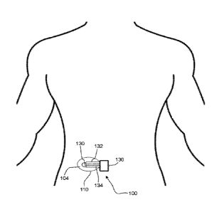
[0098] Turning now to the figures, FIG. 1 shows a detector 100 consistent with
embodiments described herein in the form of a patch affixed to the abdomen of
a user.
It should be understood that the detector may be applied in various forms to
different
locations of a user. The detector 100 includes a porous material 110 that is
adhered to
the user with adhesive (not shown). The porous material 110 collects sweat
(not shown)
and displaces the sweat laterally in the region 104 of two sensors 130, which
are
24
CA 03080898 2020-04-29
WO 2019/068047 PCT/US2018/053643
adjacent to insulation 132 and connected by wires 134 to a suitable
measurement
circuit/device 136. In other embodiments, the device may be a wireless
transmitter that
connects to a remote device.
[0099] FIGS. 2A and 2B are cross-sectional views of a detector 200 placed on
the
surface of skin 202 to collect sweat 204 containing analytes 206. The detector
200 is
adhered to the skin 202 by adhesive 208. The detector 200 includes a fluid-
collecting
porous material 210 comprising a sample-collection surface 220 and an analyte
detection surface 222. The porous material 210 may be flexible to follow the
contours in
the skin 202. The fluid-collecting porous material 210 contacts, absorbs, and
displaces
the sweat 204. FIG. 2A shows the sweat 204 containing the analytes 206 as it
is
secreted from eccrine ducts in the skin 202 and contacts the sample-collection
surface
220 of the fluid-collecting porous material 210. FIG. 2B shows the sweat 204
containing
the analytes 206 after it is absorbed and displaced laterally by the porous
material 210.
The sweat 204 is near the two sensors 230. As analytes 206 are laterally
displaced
within the analyte-detection surface 222 that is in contact with sensors 230,
a suitable
measurement circuit (not shown) is able to detect charge transfer, changes in
impedance, or other electrically measurable changes known by those skilled in
the art
that indicate the presence of analyte.
[00100] Although embodiments are shown with one or two porous layers, it
should be
understood that other embodiments may include additional porous layers.
[00101] In other embodiments, the fluid-collecting porous material may include
two or
more porous layers. FIGS. 3A, 3B, and 3C are cross-sectional views of a
detector 300
placed on the surface of skin 302 to collect sweat 304 containing analytes
306. The
detector 300 is adhered to the skin 302 by adhesive 308. The detector 300 has
a first
porous layer 312 and a second porous layer 314. The first porous layer 312
includes a
sample-collection surface 320 and the second porous layer 314 includes an
analyte
detection surface 322. The first porous layer 312 may be conformable to the
skin 302.
As shown the thickness of the first porous layer 312 may be non-uniform and
may be
compressed into the contours of the skin 302. The detector 300 further
includes two
sensors 330 mounted to the analyte-detection surface 322 and an insulation 332
for the
two sensors 330 and are part of the analyte-detection surface 322. FIG. 3A
shows the
CA 03080898 2020-04-29
WO 2019/068047 PCT/US2018/053643
sweat 304 containing the analytes 306 as it is secreted from the skin 302 and
contacts
the sample-collection surface 320 of the first porous layer 312.
[00102] FIG. 3B shows the sweat 304 containing the analytes 306 after it is
absorbed
into the first porous layer 312 and as it traverses the first porous layer 312
from the
sample collection surface 320 to the second porous layer 314. Due to the open
pore
structure, the sample rapidly transverses the layer through one or more
pathways, e.g.
microfluidic pathways. It should be understood that when there are multiple
sweat
glands in contact with the sensor that there may be several pathways from each
sweat
gland.
[00103] FIG. 3C shows the sweat 304 containing the analytes 306 after it is
absorbed
into and displaced laterally by the second porous layer 314. As analytes 306
are
laterally displaced within the second porous layer 314 and are in contact with
sensors
330, a suitable measurement circuit (not shown) is able to detect charge
transfer,
changes in impedance, or other electrically measurable changes known by those
skilled
in the art that indicate the presence of analyte. The rate of evaporation from
the second
porous layer 314 allows sufficient time for the sensors 330 to detect the
analytes.
[00104] A liquid-proof layer may be positioned over the sensors. As shown in
FIG. 2C,
liquid-proof layer 240 covers the sensors 230. Likewise, in FIG. 3D, liquid-
proof layer
340 covers the sensors 330. This barrier layer prevents egress of water from
the
outside environment which may result in a poor or false reading. In addition,
the liquid-
proof layer encourages the lateral displacement of the sample within the
second porous
layer by reducing evaporation. The liquid-proof layer may or may not be
transparent in
some embodiments. In further embodiments, the liquid-proof layer may cover the
entire
surface of the second porous layer. Liquid-proof layers may also be adjacent
the edges
of the porous material.
[00105] FIG. 4 is a cross-sectional view of a detector 400 mounted on the
surface of
skin 402 to collect sweat 404 containing analytes 406. The detector 400
includes a
sample collection layer 412 and a reservoir layer 414 that are in fluid
communication
with each other. The sample collection layer 412 includes a sample-collection
surface
420 opposite the reservoir layer 414. The reservoir layer 414 includes a
hydrophilic
region 440 and a hydrophobic region 442 surrounding the hydrophilic region
440. The
26
CA 03080898 2020-04-29
WO 2019/068047 PCT/US2018/053643
hydrophobic region 442 may partially or completely surround the hydrophilic
region 440.
The detector 400 further includes two sensors 430 mounted to the hydrophilic
region
440 of the reservoir layer 414. As the sample is laterally displaced through
the
hydrophilic region 440, the hydrophobic region 442 prevents liquid water
entry.
However, the vapor may be evaporated through the hydrophobic region 442 to the
external environment. This can facilitate the replenishment of the sweat in
the
hydrophilic region 440.
[00106] FIG. 5 is a cross-sectional view of a detector 500 mounted on the
surface of
skin 502 to collect sweat 504 containing analytes 506. The detector 500
includes a
hydrophilic porous material 516, a hydrophobic barrier 544 between a portion
of the
hydrophilic porous material 516 and the skin 502, an evaporation barrier 546
covering a
portion of the hydrophilic porous material 502 opposite the hydrophobic
barrier 544, and
two sensors 530 mounted between the hydrophilic porous material 516 and the
hydrophobic barrier 544. The hydrophobic barrier 544 and evaporation barrier
546 are
off-set so that the portion of the hydrophilic porous material 516 that
contacts the
evaporation barrier 546 and the skin 502 but not the hydrophobic barrier 544
forms a
sample collection zone 550; a second (middle) portion of the hydrophobic
porous
material 516 that contacts both the hydrophobic barrier 544 and the
evaporation barrier
546 forms a pathway 552; and a third portion of the hydrophobic porous
material 516
that contacts the hydrophobic barrier 544 but not the evaporation barrier 546
forms an
evaporation zone 554. The two sensors 530 are located in the pathway 552 of
the
hydrophilic porous material 516. In use, the sweat 504 containing analytes 506
enters
the fluid-collecting porous material 516 at the sample-collection zone 550,
traverses the
pathway 552, contacts one or more of the sensors 530, and exits the
hydrophilic porous
material 516 by evaporating at the evaporation zone 554.
[00107] FIG. 6 is a cross-sectional view of a detector 600 mounted on the
surface of
skin 602 to collect sweat 604 containing analytes 606. The detector 600
includes a
patterned porous layer 618 and a hydrophilic porous layer 616. The patterned
porous
layer 618 has hydrophilic regions 640 and hydrophobic regions 642. The
hydrophobic
regions 642 prevent egress of sweat and allow the detector to be positioned
over a
particular sweat gland. The detector 600 further includes a liquid barrier
layer 648 on a
27
CA 03080898 2020-04-29
WO 2019/068047 PCT/US2018/053643
first portion of the hydrophilic porous layer 616 opposite the patterned
porous layer 618,
an evaporation region 654, and at least one sensor 630 on the hydrophilic
porous layer
616 between the liquid barrier layer 648 and the evaporation region 654. In
use, sweat
604 containing analytes 606 enters the hydrophilic region 640 of the patterned
porous
layer 618, is laterally displaced throughout the hydrophilic region 640 past
the sensor
630 and toward the evaporation region 654, and exits the hydrophilic porous
layer 616
by evaporating at the evaporation region 654. Although one hydrophilic region
640 is
shown in FIG. 6, in further embodiments there may be multiple hydrophilic
regions to
provide a pathway for the collected sweat to enter into the hydrophilic porous
layer 616.
[00108] Although not shown in FIGS. 4-6, there may be an adhesive layer as
described
herein to attach the detector to skin.
[00109] Any detector disclosed herein may further include a liquid-proof
barrier covering
a portion of the analyte detection surface and one or more sensors.
[00110] In one embodiment, the detector may provide a flow path, or pathway,
for a
sample fluid. FIG. 7A is a top view of a detector 700 having a pathway for the
sample to
flow through. The detector includes two sensors 730 connected by wires 734 to
a
suitable measurement circuit/device 736. Although not shown, wires 734 may be
mounted on an insulation material. The detector 700 further includes a liquid-
proof
barrier 740 covering the sensors 730 and part of an analyte detection surface
722. For
purposes of illustration, the liquid-proof barrier 740 is shown as
transparent. The liquid-
proof barrier 740 prevents evaporation of a liquid sample and thus facilitates
the flow of
the sample through the pathway 736 to an uncovered evaporation region 754. The
pathway provides a fluid connection between a collection zone and an
evaporation
zone. This allows the sweat to be replenished and provides a fresh sample for
the
sensors to detect the presence of analytes.
[00111] FIG. 7B is a cross-sectional view of one embodiment of detector 700
along line
A-A. FIG. 78 shows an embodiment having only a single porous layer. In FIG.
7B, the
detector 700 is mounted on the surface of skin 702. The detector 700 includes
a fluid-
collecting porous material 710 comprising a sample-collection surface 720 and
an
analyte detection surface 722, and the detector 700 further includes a
hydrophilic region
742 and a hydrophobic region 744 surrounding the hydrophilic region 742. The
detector
28
700 further includes two sensors 730 mounted on pathway 736. A liquid-proof
barrier
734 covers the two sensors 730 and portion of the analyte-detection surface
722 to form
a collection zone. The liquid-proof barrier 740 prevents evaporation of a
liquid sample
and thus facilitates lateral displacement of a sample within the analyte
detection surface
to the evaporation zone. Detector 700 further includes an uncovered
evaporation region
754. In use, sweat 704 containing analytes 706 enters the fluid-collecting
porous material
710 below the liquid-proof barrier, traverses the hydrophilic region 742 of
the fluid-
collecting porous material 710 past the sensors 730 to the evaporation region
754, and
exits the fluid-collecting porous material 710 by evaporating at the
evaporation region
754.
[00112] FIG. 7C is a cross-sectional view of one embodiment of a detector 700
along
line A-A and shows an addition of an open porous layer, e.g., first porous
layer 712,
between the fluid-collecting porous material (second porous layer) 710, and
skin 702.
The first porous layer 712 includes a sample-collection surface 720, and the
second
porous layer 710 includes an analyte detection surface 722. The sample is
taken up
through the first porous layer 712 and delivered to the collection zone of the
second
porous layer 710. As the sample flows in pathway 726 past the sensors 730 the
various
analytes 706 may be detected. The evaporation zone of the second porous layer
710
allows replenishment of the sample through the first porous layer 712.
Examples
[00113] The following examples provide various non-limiting embodiments and
properties of the present invention. Although certain methods and equipment
are
described below, other methods or equipment determined suitable by one of
ordinary
skill in the art may be alternatively utilized.
Thickness
[00114] Thickness was measured using a laser micrometer (Keyence model no. LS-
7010). As shown in FIGS. 15A and B, a metal cylinder 1501 was aligned between
a
laser micrometer source 1502 and a laser micrometer receiver 1503. The shadow
1505
of the top of the cylinder 1501 is projected onto receiver 1503 as shown in
FIG 15A. The
position of the shadow was then reset as the "zero" reading of the laser
micrometer. As
29
Date Recue/Date Received 2022-07-22
CA 03080898 2020-04-29
WO 2019/068047 PCT/US2018/053643
shown in FIG. 158, a single layer of membrane 1504 is draped over the surface
of the
metal cylinder 1501 without overlap and without wrinkles, casting shadow 1506
onto the
receiver 1503. The laser micrometer then indicated the change in the position
of the
shadows 1505 and 1506 as the thickness of the sample. Each thickness was
measured
three times and averaged for each sample.
Gurley
[00115] The Gurley air flow test (Gurley Model 4340 Automatic Densometer)
measures
the time in seconds for 100 cm3 of air to flow through a 6.45 cm2 sample at
12.4 cm of
water pressure.
Matrix Tensile Strength (for Membranes)
[00116] Samples were prepared by using a die punch to cut ASTM D412 Type F
dogbone samples out of the ePTFE membrane. The membrane was placed on the
cutting table such that it was free from wrinkles in the area where the sample
was to be
cut. The die was then placed on the membrane (generally in the center 200 mm
of the
web) such that its long axis was parallel to the direction that would be
tested. Once the
die was aligned, pressure was applied to it to cut through the membrane web.
Upon
removal of this pressure, the dogbone sample for testing was inspected to
ensure it was
free from edge defects which may impact the tensile testing. At least 3
samples in the
machine direction and three samples in the transverse direction were prepared
in this
manner. Once samples were prepared, they were measured to determine their mass
using an analytical balance and their thickness using a Mitutoyo 547-400S
thickness
gage. Note that any suitable means for measuring thickness can be used. Each
sample
was subsequently tested to determine its tensile properties using an Instron
5500
tensile tester. The samples were inserted into the tensile tester and held
using lnstron
Catalog 2702-015 (rubber coated face plate) and 2702-016 (serrated face plate)
grip
plates such that each end of the sample was held between one rubber coated and
one
serrated face plate. The pressure applied to the grip plates was approximately
552 kPa.
The gauge length between the grips was set at 58.9 mm and the crosshead speed
(pulling speed) was set to a speed of 508 mm/m in. A 500 N load cell was used
to carry
CA 03080898 2020-04-29
WO 2019/068047 PCT/US2018/053643
out these measurements and data was collected at a rate of 50 points/sec. The
laboratory temperature was between 20 and 22.2 degrees Celsius to ensure
comparable results. Finally, if the sample happened to break at the grip
interface, the
data was discarded. At least 3 samples in the machine direction and three
samples in
the transverse direction were successfully pulled (no slipping out of or
breaking at the
grips) in order to characterize the membrane web.
[00117] The following equation was used to calculate the matrix tensile
strength:
MTS = ((Fmax/w)* p )/ mass:area
in which: MTS = matrix tensile strength in MPa, Frim = maximum load measured
during
test (Newtons), w = width of dogbone sample within the gauge length (meters),
p =
density of PTFE (2.2x10⁶ g/m3), mass:area = mass per area of sample as
described
below (g/m2).
Bubble Point
[00118] Bubble point pressures were measured according to the general
teachings of
ASTM F31 6-03 using a Capillary Flow Porometer (Model 3Gzh from Quantachrome
Instruments, Boynton Beach, Florida). The sample membrane was placed into the
sample chamber and wet with Silwick Silicone Fluid (available from Porous
Materials
Inc.) having a surface tension of 20.1 dynes/cm. The bottom clamp of the
sample
chamber had a 2.54 cm diameter, 0.159 cm thick porous metal disc insert
(Quantachrome part number 75461 stainless steel filter) was used to support
the
sample. Using the 3GWin software version 2.1 the following parameters were set
as
specified in the table immediately below. The values presented for bubble
point
pressure are the average of two measurements.
[00119] Bubble point pressure was converted to pore size using the following
equation:
Dgp = 4ylvcose / PBP
where Dgp is the pore size, ylv is the liquid surface tension, e is the
contact angle of the
fluid on the material surface, and Pgp is the bubble point pressure. It is
understood by
one skilled in the art that the fluid used in a bubble point measurement must
wet the
surface of the sample.
31
CA 03080898 2020-04-29
WO 2019/068047 PCT/US2018/053643
Mass to area
[00120] The mass per area of samples was measured according to the ASTM D 3776
(Standard Test Methods for Mass Per Unit Area (Weight) of Fabric) test method
(Option
C) using a Mettler-Toledo Scale, Model 1060. The scale was recalibrated prior
to
weighing specimens, and the results were reported in grams per square meter
(g/m2).
Electrical Continuity Test
[00121] An electrical continuity sensor was fabricated in the following
manner. A 50
micron thick PET film 8567K22 (McMaster Can, Robbinsville, NJ) was obtained.
The
PET film was screen-printed using conductive ink in the pattern shown in FIG.
16. The
pattern consists of 10 parallel traces, each with a width of approximately 370
microns
and spaced at a pitch of 1mm. At one end of the parallel traces, the printed
features
increase in width and spacing and terminate in 2mm wide pads. The conductive
ink
used was PE874 (E. I. du Pont de Nemours, Wilmington, DE). The screen printing
was
performed using a model MSP-088 screen printer (HMI Manufacturing, Lebanon,
NJ), a
stainless steel screen with 200 TPI (threads/wire per inch; -78.74 wires per
cm), 1.6 mil
(- 40.64 pm) wire diameter, and a 12.7 micron emulsion of the ink. After
printing the ink
was then dried in a convection oven at 120 C for 10 minutes.
[00122] The continuity sensor was placed on a flat smooth surface with the
printed
surface facing up. A membrane being tested was placed on the surface of the
continuity sensor.
[00123] A 100 nanoliter drop of saline solution 245-09-0072 (Target,
Minneapolis, MN)
was dispensed using a 0.5 microliter syringe 5190-0464 (Agilent, Santa Clara,
CA) onto
the simulated skin surface described in Comparative Example A. The simulated
skin
surface was placed face down on the membrane and continuity sensor so that the
droplet of saline was approximately centered within the ten, parallel traces.
A 200 gram
weight was placed on top of the simulated skin to ensure good contact between
the
simulated skin, the membrane, and the continuity sensor, and a timer was
started. A
Fluke 116 multimeter (Fluke Corporation, Everett, WA) was used in autorange
mode to
measure the electrical resistance between the individual pairs of the 10
parallel traces
32
CA 03080898 2020-04-29
WO 2019/068047 PCT/US2018/053643
by contacting the probe of the multimeter to the pad at the end of the
selected trace.
Continuity testing was first performed on the gap between adjacent parallel
traces.
Continuity is defined as having a resistance of 44 MOhms or less and
demonstrates
that the saline solution is in contact with both conductive traces being
tested. If
continuity was measured across a single gap, one probe was moved to the next
pad
and the continuity across 2 gaps was measured. This process was repeated until
the
largest number of gaps to measure continuity was identified. This number was
recorded at 30 seconds, 2 minutes, and 5 minutes. The best performing
continuity
sensor is one that quickly spreads the fluid over the largest distance. One
skilled in the
art will recognize that any number of tests can be performed on the fluid once
the fluid
is delivered to the electrodes of the sensor.
COMPARATIVE EXAMPLE A
[00124] For Comparative Example A, a simulated skin surface was created by
roughening up one face of a 6.35 mm thick sheet of polycarbonate with 80 grit
sandpaper. 10 ml of water was colored blue by adding several drops of blue
food
coloring and mixing thoroughly. 250 nL of the blue water was deposited on the
rough
surface of the polycarbonate using a syringe. A glass cover slide was place on
top of
the water and the water was viewed from above using a light microscope. The
water
was allowed to spread out between the glass coverslip and the polycarbonate
sheet,
coming to rest after about a minute. The wetted area was approximately
circular and
measured to be 4.9 mm2. Assuming a constant thickness, the 250 nL of water
formed a
51 pm thick film between the simulated skin and the glass cover slip. FIG. 8
is an image
of the wetted area of two drops of water on the simulated skin surface.
COMPARATIVE EXAMPLE B
[00125] The Electrical Continuity Test was performed as described, except the
membrane was excluded. In other words, the 100nL water droplet dispensed on
the
simulated skin was placed directly onto the parallel traces of the electrical
continuity
sensor. The largest number of gaps to measure conductivity after 30 seconds
was 1,
after 2 minutes was 1 and after 5 minutes was 1.
33
CA 03080898 2020-04-29
WO 2019/068047 PCT/US2018/053643
EXAMPLE 1
[00126] For Example 1, a porous expanded PTFE membrane was made based on the
teachings of U.S. Patent No. 7,306,729, with a mass/area of 0.69 g/m2, a
thickness of
5.0 pm, a porosity of 93.8%, a bubble point of 505.3 KPa, a bubble point pore
diameter
of 0.144 pm, having a Gurley airflow of 1.26 sec, and matrix tensile
properties of 771
MPa in the longitudinal direction and 345 MPa in the transverse direction. A
scanning
electron micrograph of the surface of the membrane can be seen in FIG. 10.
[00127] A coating solution was prepared by dissolving 3% Soarnol EVOH (Nippon
Gohsei, Arlington Heights, IL) into a solvent blend comprising 2-butanol,
isopropyl
alcohol and deionized water in the ratios of 1:2:3.53 respectively. The
mixture was
heated to 80 C for 4 hours with stirring, causing the polymer pellets to
completely
dissolve and create a clear solution. The solution was cooled to room
temperature.
[00128] The ePTFE membrane was mounted in a 100 mm diameter embroidery hoop
and the coating solution was applied to the ePTFE with a gloved finger,
spreading it
across the membrane. Excess solution was blotted away using a paper lab wipe.
The
coated membrane was placed in an oven at 80 C for 5 minutes to dry off the
solvent,
resulting in a coating of EVOH on the node and fibril structure of the ePTFE.
The
thickness of the EVOH coated ePTFE was 1.76 pm.
[00129] The wetting characteristic of the membrane was tested by first placing
a 250nL
drop of water as described in Comparative Example A onto the roughened side of
the
polycarbonate sheet described in Comparative Example A. A 30 mm square of the
coated ePTFE membrane was cut from the sample and placed on top of a 22 mm
square glass microscope coverslip, with the excess membrane wrapped around to
the
back of the coverslip. The coverslip and membrane were placed on top of the
drop of
water with the membrane side against the water. A timer was started and the
water was
viewed from above using a light microscope. The area of the wetted region was
measured at various time intervals and can be seen in FIGS. 9A-9F. The
presence of
the ePTFE membrane resulted in a much larger wetted area against the coverslip
than
without the ePTFE membrane. If the coverslip were replaced with a sensor, a
much
greater surface area of the sensor would be able to interact with the fluid
with the
34
CA 03080898 2020-04-29
WO 2019/068047 PCT/US2018/053643
inclusion of the ePTFE membrane. Table 1 reports the wetted area v. time in
minutes
for Example 1.
Table 1 ¨ Example 1
Time (min) Area (mm 2)
0.167 13.388
0.5 19.753
1 26.76
2.333 39.607
3.333 47.87
4 52.435
58.468
7 69.772
9 76.492
12 88.039
95.903
18 101.174
21 105.942
24 109.252
25.767 109.996
EXAMPLE 2
[00130] For Example 2, a porous expanded PTFE membrane was made based on the
teachings of W02004079208A3, with a mass/area of 4.1 g/m2, a thickness of 15.1
pm,
a porosity of 87.9%, a bubble point of 187.25 kPa, a bubble point pore
diameter of
0.3768 pm, having a Gurley airflow of 4.5 sec, and Matrix tensile properties
of 258 MPa
in the longitudinal direction and 328 MPa in the transverse direction. A
scanning
electron micrograph of the surface of the membrane can be seen in FIG. 12.
[00131] An EVOH coating solution was prepared as described in Example 1. The
porous expanded PTFE was coated as described in Example 1. The thickness of
the
EVOH coated ePTFE was 10.3 pm.
[00132] The wetting characteristic of the membrane was tested by first placing
a 250 nL
drop of water as described in Comparative Example A onto the roughened side of
the
polycarbonate sheet described in Comparative Example A. A 30 mm square of the
coated expanded PTFE membrane was cut from the sample and placed on top of a
22
mm square glass microscope coverslip, with the excess membrane wrapped around
to
CA 03080898 2020-04-29
WO 2019/068047 PCT/US2018/053643
the back of the coverslip. The coverslip and membrane were placed on top of
the drop
of water with the membrane side against the water. A timer was started and the
water
was viewed from above using a light microscope. The area of the wetted region
was
measured at various time intervals and can be seen in FIGS. 11A-11D.
[00133] The presence of the expanded PTFE membrane resulted in a much larger
wetted area against the coverslip than without the expanded PTFE membrane. If
the
coverslip were replaced with a sensor, a much greater surface area of the
sensor would
be able to interact with the fluid with the use of the expanded PTFE membrane.
In
comparison to the performance of the membrane in Example 1, this embodiment
results
in a smaller wetted area, but a much faster wetting rate. Table 2 reports the
wetted area
v. time in minutes for Example 2.
Table 2¨ Example 2
Time (min) Area (mm 2)
0.167 32.094
0.5 54.066
0.633 60.953
3 73.616
4 73.012
Example 3
[00134] A small pore ePTFE membrane as described in Example 1 and a large pore
membrane as described in Example 2 were layered one on top of the other and
mounted in a 100 mm diameter embroidery hoop.
[00135] An EVOH coating solution was prepared as described in Example 1. The
coating solution was applied to the large pore ePTFE with a gloved finger,
spreading it
across the membrane. The coating solution wet through both the large and small
pore
membrane. Excess solution was blotted away using a paper lab wipe. The coated
layered membrane was placed in an oven at 80 C for 5 minutes to dry off the
solvent,
resulting in a coating of EVOH on the node and fibril structure of the ePTFE.
The
thickness of the EVOH coated layered ePTFE was 10.0 pm.
[00136] The wetting characteristic of the layered ePTFE was tested by first
placing a
250nL drop of water as described in Comparative Example A onto the roughened
side
36
CA 03080898 2020-04-29
WO 2019/068047 PCT/US2018/053643
of the polycarbonate sheet described in Comparative Example A. A 30 mm square
of
the coated layered ePTFE membrane was cut from the sample and placed on top of
a
22mm square glass microscope coverslip, with the excess membrane wrapped
around
to the back of the coverslip. The small pore membrane was positioned against
the cover
slip. The coverslip and layered membrane were placed on top of the drop of
water with
the large pore membrane side against the water. A timer was started and the
water was
viewed from above using a light microscope. The area of the wetted region was
measured at various time intervals and can be seen in FIGS. 13A-13D. Table 3
reports
the wetted area v. time in minutes for Example 3.
Table 3¨ Example 3
Time (min) Area (mm 2)
0.167 25.423
0.5 44.547
1 66.919
2 90.07
3.333 95.203
4.333 97.859
99.931
7 101.437
103.678
13 104.995
16 105.468
21 106.065
[00137] The presence of the layered ePTFE resulted in a much larger wetted
area
against the coverslip than without the ePTFE membrane. The layered membrane
performed better than the large pore membrane described in Example 2 because
the
layered membrane created a much larger wetted area. The layered membrane
performed better than the small pore membrane described in Example 1 because
the
layered membrane created a large wetted area much more quickly.
[00138] If the coverslip were replaced with a sensor, a much greater surface
area of the
sensor would be able to interact with the fluid with the inclusion of the
layered ePTFE
membrane as compared to the single layer ePTFE membrane, or without an ePTFE
layer.
37
CA 03080898 2020-04-29
WO 2019/068047 PCT/US2018/053643
[00139] The results of Examples 1-3 are compared with Comparative Example A in
the
graph shown in FIG. 14. As shown, Comparative Example A fails to increase the
wetted
area, while Examples 1-3 show significant increases in the wetted area.
EXAMPLE 4
[00140] The small pore ePTFE membrane described in Example 1 was mounted in a
100 mm diameter embroidery hoop and coated with the EVOH solution described in
Example 1, using the method described in Example 1, resulting in a hydrophilic
membrane. The thickness of the EVOH coated ePTFE was 2.1 pm.
[00141] The hydrophilic membrane was tested in the Electrical Continuity Test.
The
membrane was positioned on top of the continuity sensor so that the transverse
direction of the membrane was parallel with the traces of the continuity
sensor.
[00142] The largest number of gaps to measure conductivity after 30 seconds
was 2,
after 2 minutes was 4, and after 5 minutes was 6. This system is much more
effective
at spreading the fluid over a large distance as compared to the system without
a
hydrophilic membrane, as described in Comparative Example B.
EXAMPLE 5
[00143] The large pore ePTFE membrane described in Example 2 was mounted in a
100 mm diameter embroidery hoop and coated with the EVOH solution described in
Example 1, using the method described in Example 1, resulting in a hydrophilic
membrane. The thickness of the EVOH coated ePTFE was 10.9 pm.
[00144] The hydrophilic membrane was tested in the Electrical Continuity Test.
The
membrane was positioned on top of the continuity sensor so that the
longitudinal
direction of the membrane was parallel with the traces of the continuity
sensor.
[00145] The largest number of gaps to measure conductivity after 30 seconds
was 5,
after 2 minutes was 5, and after 5 minutes was 5. This membrane is much more
effective at spreading the fluid over a large distance as compared to the
system without
a membrane, as described in Comparative Example B. This membrane was able to
spread the fluid more quickly, but across a slightly shorter distance than the
membrane
described in Example 4,
EXAMPLE 6
38
CA 03080898 2020-04-29
WO 2019/068047 PCT/US2018/053643
[00146] The small pore ePTFE membrane described in Example 1 and the large
pore
ePTFE membrane described in Example 2were layered and coated as described in
Example 3. The thickness of the layered EVOH coated ePTFE was 12.7 pm.
[00147] The hydrophilic layered membrane was tested in the Electrical
Continuity Test.
The membrane was positioned on top of the continuity sensor with the small
pore
ePTFE membrane in contact with the traces of the continuity sensor. The
transverse
direction of the small pore ePTFE was aligned the traces of the continuity
sensor.
[00148] The largest number of gaps to measure conductivity after 30 seconds
was 5,
after 2 minutes was 7, and after 5 minutes was 7. This layered membrane is
much
more effective at spreading the fluid over a large distance as compared to the
system
without a membrane. This membrane was able to spread the fluid more quickly
and
across a larger distance than the membrane described in Example 4 and Example
5.
[00149] As used herein, the conjunction "and" is intended to be inclusive and
the
conjunction "or" is not intended to be exclusive unless otherwise indicated.
For example,
the phrase "or, alternatively" is intended to be exclusive.
[00150] The use of the terms "a", "an", "the", or similar referents in the
context of
describing the invention (especially in the context of the claims) are to be
construed to
cover both the singular and the plural, unless otherwise indicated herein or
clearly
contradicted by context.
[00151] The terms "comprising," "having," "including," and "containing" are to
be
construed as open-ended terms (i.e., meaning "including, but not limited to,")
unless
otherwise noted.
[00152] As used herein, the term "about" refers to a degree of deviation
typical for a
particular property, composition, amount, value or parameter as identified;
such as
deviations based on experimental errors, measurement errors, approximation
errors,
calculation errors, standard deviations from a mean value, routine minor
adjustments,
and so forth.
[00153] As used herein, the term "conformable" is meant to describe a material
structure that is extendable or extensible in a first direction, which
recovers in a second
direction perpendicular to the first direction, and which is elongated to take
essentially
39
CA 03080898 2020-04-29
WO 2019/068047 PCT/US2018/053643
the same shape as a non-planar substrate, e.g., human skin, without
fracturing, tearing,
or otherwise breaking.
[00154] Recitation of ranges of values herein are merely intended to serve as
a
shorthand method of referring individually to each separate value falling
within the
range, unless otherwise indicated herein, and each separate value is
incorporated into
the specification as if it were individually recited herein.
[00155] All methods described herein can be performed in any suitable order
unless
otherwise indicated herein or otherwise clearly contradicted by context. The
use of any
and all examples, or exemplary language (e.g., "such as", "for example")
provided
herein, is intended merely to better illuminate the invention and does not
pose a
limitation on the scope of the invention unless otherwise claimed.
[00156] Detectors for detecting analytes have been described above both
generically
and with regard to specific embodiments. It will be apparent to those skilled
in the art
that various modifications and variations can be made in the embodiments
without
departing from the scope of the disclosure. Thus, it is intended that the
embodiments
cover modifications and variations provided they come within the scope of the
appended claims and their equivalents.