Note: Descriptions are shown in the official language in which they were submitted.
CA 03081630 2020-05-04
WO 2019/090418 PCT/CA2018/051398
1
TITLE:
WEARABLE BLOOD ANALYTE MEASUREMENT DEVICE AND METHOD FOR
MEASURING BLOOD ANALYTE CONCENTRATION
CROSS REFERENCES TO RELATED APPLICATIONS:
[0001]This application claims the benefit of and/or priority from United
States Provisional
Patent Application No. 62/638,364 filed on March 5, 2018, and United States
Provisional
Patent Application No. 62/584,900 filed on November 12, 2017, each of which is
incorporated herein by reference in its entirety.
FIELD:
[0002]This document relates to devices and methods for the measurement of
blood
analytes. Specifically, this document relates to devices and methods that
employ nuclear
magnetic resonance (NMR) technology to measure blood analyte concentrations.
BACKGROUND:
[0003]US Patent No. 5,685,300 (Kuenstner et al.) discloses a method of non-
invasive
and in-vitro glucose and cholesterol concentration measurement employing
nuclear
magnetic resonance (NMR) spectroscopy. The measurement comprises a ratio
formed
by dividing the area of the resonance of the desired analyte, e.g., glucose or
cholesterol,
by the area of the water resonance in a spectrum of blood or tissue. In the in-
vivo setting,
the spectrum is obtained either in linkage with the pulsation of blood or by
using a slice
selection gradient such as that employed in the magnetic resonance imager.
This
measurement is then correlated to a traditional serum analyte concentration.
[0004] International Patent Application Publication No. W02012/122462 (Tseng
et al.)
discloses a system and methods to perform non-invasive, real-time, continuous
or
episodic molecular detection and quantification of molecular species in a
sample or
animal or human subject using magnetic resonance. Such systems and methods may
be
applied to identify and quantify molecular species found in the body, which
may be useful
CA 03081630 2020-05-04
WO 2019/090418 PCT/CA2018/051398
2
for many aspects of medical care including without limitation prenatal
diagnosis, detecting
deep skin infections, performing cerebral spinal fluid assessment, measuring
arterial
blood gases, blood glucose, cardiac biomarkers, and creatinine flow rates.
SUMMARY:
[0005] The following summary is intended to introduce the reader to various
aspects of
the detailed description, but not to define or delimit any invention.
[0006]According to some aspects, a wearable blood analyte measurement device
includes a casing defining an appendage-receiving bore and having an interior
volume.
A plurality of magnets is within the interior volume. The magnets produce a
magnetic field
in the appendage-receiving bore. A nuclear magnetic resonance (NMR)
transceiver is
supported by the casing and is positioned to emit radiofrequency (RF) pulses
to and
receive NMR signals from the appendage-receiving bore. An electronics assembly
is
within the interior volume and is in communication with the NMR transceiver.
The
electronics assembly is operable to activate the NMR transceiver to emit an RF
pulse to
the appendage-receiving bore and to receive an NMR signal from the appendage-
receiving bore. A power source is in the interior volume and powers the NMR
transceiver
and the electronics assembly.
[0007] In some examples, the device further comprises a shim system operable
to
homogenize at least a section of the magnetic field.
[0008] In some examples, the shim system includes a dynamic shim system. The
dynamic
shim system can include an active shim coil within the interior volume and
extending
around the appendage-receiving bore. The active shim coil can be activatable
to
homogenize the section of the magnetic field. The electronics assembly can be
in
communication with the dynamic shim system to activate the active shim coil.
[0009] In some examples, the shim system includes a static shim system. The
static shim
system can include at least one ferromagnetic material within the interior
volume.
CA 03081630 2020-05-04
WO 2019/090418 PCT/CA2018/051398
3
[0010] In some examples, the shim system is operable to homogenize only a
section of
the magnetic field.
[0011] In some examples, the magnets are permanent magnets. The permanent
magnets
can include neodymium and/or Samarium Cobalt (SmCo).
[0012] In some examples, the magnets are arranged in an annulus around the
appendage-receiving bore. The annulus can have a radial wall thickness of less
than 5
mm. The annulus can have a radial wall thickness of between about 1 mm and
about 3
mm.
[0013] In some examples, the magnets are arranged in a plurality of rows
around the
appendage-receiving bore.
[0014] In some examples, the magnets are arranged to form a Halbach array.
[0015] In some examples, the magnets are arranged in a pattern of alternating
cylindrical
magnets and bar magnets.
[0016] In some examples, the device comprises between 2 and 32 of the magnets.
[0017] In some examples, the device comprises 2 magnets arranged on opposed
sides
of the appendage receiving bore.
[0018] In some examples, the magnetic field has a magnetic field strength of
less than 1
T. The magnetic field can have a magnetic field strength of between 0.05 T and
0.5 T.
The magnetic field can have a magnetic field strength of between 0.1 T and 0.3
T.
[0019] In some examples, the casing is non-metallic and non-ferromagnetic. The
casing
can have an inner section lining the appendage-receiving bore, and the inner
section can
be non-metallic and non-ferromagnetic.
[0020] In some examples, the device further includes a positioning guide for
guiding a
user in orienting the device at a target orientation. The positioning guide
can have a
centre-point, and the NMR transceiver can be circumferentially spaced from the
centre-
CA 03081630 2020-05-04
WO 2019/090418 PCT/CA2018/051398
4
point by between 45 degrees and 180 degrees, or by between 80 degrees and 150
degrees.
[0021] In some examples the device further includes a heart phase sensor
supported by
the casing and activatable to sense diastole and systole in a wearer when an
appendage
of the wearer is received in the appendage-receiving bore. The electronics
assembly can
be operable to activate the NMR transceiver during diastole, and the RF pulse
can be a
diastolic RF pulse and the NMR signal can be a diastolic NMR signal. The
electronics
assembly can be further operable to activate the NMR transceiver during
systole to emit
a systolic RF pulse to the appendage-receiving bore and receive a systolic NMR
signal
from the appendage-receiving bore.
[0022] In some examples, the power source powers the heart phase sensor.
[0023] In some examples, the electronics assembly includes an RF control
module in
communication with the NMR transceiver. The RF control module can be operable
to
activate the NMR transceiver to emit the diastolic RF pulse and the systolic
RF pulse to
the magnetic field. The NMR transceiver can be operable to communicate the
diastolic
NMR signal and the systolic NMR signal to the RF control module.
[0024] In some examples, the electronics assembly further includes a central
processing
unit (CPU) in communication with the heart phase sensor and the RF control
module, The
CPU can be operable to receive a heart phase signal from the heart phase
sensor, and
signal the RF control module to activate the NMR transceiver to emit the
diastolic RF
pulse during diastole and the systolic RF pulse during systole in response to
the heart
phase signal received from the heart phase sensor.
[0025] In some examples, the RF control module includes (i) an RF transmitter
sub-
module in communication with the CPU, (ii) an RF receiver sub-module with
quadrature
detection in communication with the CPU, and (iii) a duplexer in communication
with the
RF transmitter sub-module, the RF receiver sub-module, and the NMR
transceiver.
[0026] In some examples, the electronics assembly is further operable to
calculate a
blood analyte concentration based on the NMR signal.
CA 03081630 2020-05-04
WO 2019/090418 PCT/CA2018/051398
[0027] In some examples, the device further includes a data transmitter within
the interior
volume and in communication with the electronics assembly. The data
transmitter can be
operable to transmit the blood-analyte concentration to a secondary device
comprising a
display. The data transmitter can be operable to transmit the NMR signal to a
secondary
device. The data transmitter can be a Bluetooth transmitter.
[0028] In some examples, the wearable blood analyte measurement device has a
weight
of less than 50 grams, or of between 1 gram and 20 grams.
[0029] In some examples, the RF pulse sequence generates a balanced Steady
State
Free Precession (b-SSFP) signal. The b-SSFP signal can be generated by rapid,
repeated pulses with a constant repetition time.
[0030] In some examples, the RF pulse sequence includes a Carr-Purcell-Meiboom-
Gill
(CPMG) spin echo train. The CPMG can be composed of an initial excitation at
the Ernst
angle, and repeated 180 pulses with a constant repetition time.
[0031] In some examples, the device further includes at least one gradient
coil supported
by the casing and in communication with the electronics assembly.
[0032] In some examples the NMR transceiver comprises a single transceiver
coil. In
some examples, the NMR transceiver comprises at least one transmitter coil and
at least
one receiver coil. In some examples, the NMR transceiver comprises at least
one of a
surface coil and a solenoid coil.
[0033]According to some aspects, a kit includes the wearable blood analyte
measurement device, and a power charger for charging the power source.
[0034] According to some aspects, a method for measuring a blood analyte
concentration
includes a) using a device worn on an appendage of a user to create a magnetic
field
within the appendage; b) while the device is worn on the appendage, activating
the device
to emit an RF pulse to the appendage and receive an NMR signal from the
appendage,
c) calculating a blood-analyte concentration based on the NMR signal, and d)
displaying
the blood-analyte concentration on a display.
CA 03081630 2020-05-04
WO 2019/090418 PCT/CA2018/051398
6
[0035] In some examples, step b. further includes homogenizing at least a
section of the
magnetic field prior to emitting the RF pulse.
[0036] In some examples, the method includes repeating steps b. to d.
periodically.
[0037] In some examples, the method includes charging a battery of the device
prior to
step a, and using the battery to power the device during step b. Charging the
battery can
include inductively charging the battery of the device.
[0038] In some examples, the method includes repeating steps b. to d.
periodically over
a period of between 4 hours and 72 hours on a single charge of the battery.
[0039] In some examples, the device is in the form of a ring, and step a.
includes placing
the ring on a finger of the user.
[0040] In some examples, the device includes a plurality of permanent magnets
arranged
in an annulus, and step a. includes creating the magnetic field using the
annulus of
magnets.
[0041] In some examples, the method includes transmitting the blood-analyte
concentration to a secondary device including the display.
[0042] In some examples, the blood analyte concentration is a glucose
concentration, a
cholesterol concentration, a vitamin concentration, an alcohol concentration,
a mineral
concentration, or a drug concentration.
[0043] In some examples, step b. further includes sensing diastole and systole
in a user.
The RF pulse can be a diastolic RF pulse that is emitted during diastole and
the NMR
signal can be a diastolic NMR signal. The method can further include emitting
a systolic
RF pulse to the magnetic field during systole and receiving a systolic NMR
signal from
the magnetic field. Step c. can include calculating the blood-analyte
concentration based
on the diastolic NMR signal and the systolic NMR signal.
CA 03081630 2020-05-04
WO 2019/090418 PCT/CA2018/051398
7
[0044] In some examples, the RF pulse generates a balanced Steady State Free
Precession (b-SSFP) signal. The b-SSFP signal can be generated by rapid,
repeated
pulses with a constant repetition time.
[0045] In some examples, the RF pulse includes a Carr-Purcell-Meiboom-Gill
(CPMG)
spin echo train. The CPMG spin echo train can be composed of an initial
excitation at the
Ernst angle, and repeated 1800 pulses with a constant repetition time.
[0046] In some examples, step c. includes employing a T2 filter.
[0047]According to some aspects, a wearable blood analyte measurement device
includes a casing defining an appendage-receiving bore and having an interior
volume.
A plurality of magnets is within interior. The magnets produce a magnetic
field in the
appendage-receiving bore. A nuclear magnetic resonance (NMR) transceiver is
supported by the casing and positioned to emit radiofrequency (RF) pulses to
and receive
NMR signals from the appendage-receiving bore. A heart phase sensor is
supported by
the casing and is activatable to sense diastole and systole in a wearer when
an
appendage of the wearer is received in the appendage-receiving bore. An
electronics
assembly is within the interior volume and is in communication with the NMR
transceiver.
The electronics assembly is operable to activate the NMR transceiver during
diastole to
emit a diastolic RF pulse to the appendage-receiving bore and receive a
diastolic NMR
signal from the appendage-receiving bore, and activate the NMR transceiver
during
systole to emit a systolic RF pulse to the appendage-receiving bore and
receive a systolic
NMR signal from the appendage-receiving bore. A power source is in the
interior volume
and powers the NMR transceiver, the heart phase sensor, and the electronics
assembly.
[0048] In some examples, the device further includes a shim system operable to
homogenize at least a section of the magnetic field. In some examples, the
shim system
is operable to homogenize only a section of the magnetic field
[0049] In some examples, the shim system includes a dynamic shim system. The
dynamic
shim system can include an active shim coil within the interior volume and
extending
around the appendage-receiving bore. The active shim coil can be activatable
to
CA 03081630 2020-05-04
WO 2019/090418 PCT/CA2018/051398
8
homogenize the section of the magnetic field. The electronics assembly can be
in
communication with the dynamic shim system to activate the active shim coil.
[0050] In some examples, the shim system includes a static shim system. The
static shim
system can include at least one ferromagnetic material within the interior
volume.
[0051] In some examples, the magnets are permanent magnets. The permanent
magnets
can be or can include neodymium and/or Samarium Cobalt (SmCo). The magnets can
be
arranged in an annulus around the appendage-receiving bore. The annulus can
have a
radial wall thickness of less than 5 mm, for example between about 1 mm and
about 3
mm. The magnets can be arranged to form a Halbach array. The device can
include
between 2 and 32 of the magnets.
[0052] In some examples, the magnetic field has a magnetic field strength of
less than 1
T (Tesla). In some examples, the magnetic field has a magnetic field strength
of between
0.05 T and 0.5 T. In some examples, the magnetic field has a magnetic field
strength of
between 0.1 T and 0.3 T.
[0053] In some examples, the casing is non-metallic and non-ferromagnetic. In
some
examples, the casing has an inner section lining the appendage-receiving bore,
and the
inner section is non-metallic and non-ferromagnetic.
[0054] In some examples, the device includes a positioning guide for guiding a
user in
orienting the device at a target orientation. The positioning guide can have a
centre-point,
and the NMR transceiver can be circumferentially spaced from the centre-point
by
between 45 degrees and 180 degrees. For example, the NMR transceiver can be
circumferentially spaced from the centre-point by between 80 degrees and 150
degrees.
[0055] In some examples, the electronics assembly includes an RF control
module in
communication with the NMR transceiver. The RF control module can be operable
to
activate the NMR transceiver to emit the diastolic RF pulse and the systolic
RF pulse to
the magnetic field. The NMR transceiver can be operable to communicate the
diastolic
NMR signal and the systolic NMR signal to the RF control module.
CA 03081630 2020-05-04
WO 2019/090418 PCT/CA2018/051398
9
[0056] In some examples, the electronics assembly includes a central
processing unit
(CPU) in communication with the heart phase sensor and the RF control module.
The
CPU can be operable to receive a heart phase signal from the heart phase
sensor, and
signal the RF control module to activate the NMR transceiver to emit the
diastolic RF
pulse during diastole and the systolic RF pulse during systole in response to
the heart
phase signal received from the heart phase sensor.
[0057] In some examples, the RF control module includes (i) an RF transmitter
sub-
module in communication with the CPU, (ii) an RF receiver sub-module with
quadrature
detection in communication with the CPU, and (iii) a duplexer in communication
with the
RF transmitter sub-module, the RF receiver sub-module, and the NMR
transceiver.
[0058] In some examples, the electronics assembly is further operable to
calculate a
blood analyte concentration based on the diastolic NMR signal and the systolic
NMR
signal.
[0059] In some examples, the device includes a data transmitter within the
interior volume
and in communication with the electronics assembly. The data transmitter can
be
operable to transmit the blood-analyte concentration to a secondary device
comprising a
display, and/or to transmit the diastolic NMR signal and the systolic NMR
signal to a
secondary device. The data transmitter can be a Bluetooth transmitter.
[0060] In some examples, the device has a weight of less than 50 grams. In
some
examples, the weight is between 1 gram and 20 grams.
[0061] According to some aspects, a kit includes the wearable blood analyte
measurement device, and a power charger for charging the power source.
[0062] According to some aspects, a method for measuring a blood analyte
includes a)
using a device worn on an appendage of a user to create a magnetic field
within the
appendage. The method further includes b) while the device is worn on the
appendage,
activating the device to i) sense systole and diastole in the user; ii) emit a
diastolic RF
pulse to the appendage during diastole and receive a diastolic NMR signal from
the
appendage; and iii) emit a systolic RF pulse to the appendage during systole
and receive
CA 03081630 2020-05-04
WO 2019/090418 PCT/CA2018/051398
a systolic NMR signal from the appendage. The method further includes c)
calculating a
blood-analyte concentration based on the systolic NMR signal and the diastolic
NMR
signal, and d) displaying the blood-analyte concentration on a display.
[0063] In some examples, step b) further includes homogenizing at least a
section of the
magnetic field prior to sub-step ii.
[0064] In some examples, the method includes repeating steps b) to d)
periodically.
[0065] In some examples, the method includes charging a battery of the device
prior to
step a), and using the battery to power the device during step b). Charging
the battery of
the device can include inductively charging the battery of the device.
[0066] In some examples, the method includes repeating steps b) to d)
periodically over
a period of between 4 hours and 72 hours on a single charge of the battery.
[0067] In some examples, the device is in the form of a ring, and step a)
includes placing
the ring on a finger of the user. The device can include a plurality of
permanent magnets
arranged in an annulus to form a Halbach array, and step b) can include
creating the
magnetic field using the Halbach array.
[0068] In some examples, the method includes transmitting the blood-analyte
concentration to a secondary device having the display.
[0069] In some examples, the blood analyte concentration is a glucose
concentration, a
cholesterol concentration, a vitamin concentration, an alcohol concentration,
a mineral
concentration, or a drug concentration.
BRIEF DESCRIPTION OF THE DRAWINGS:
[0070]The drawings included herewith are for illustrating various examples of
articles,
methods, and apparatuses of the present specification and are not intended to
limit the
scope of what is taught in any way. In the drawings:
CA 03081630 2020-05-04
WO 2019/090418 PCT/CA2018/051398
11
[0071] Figure 1 is a perspective view of an example wearable blood analyte
measurement
device;
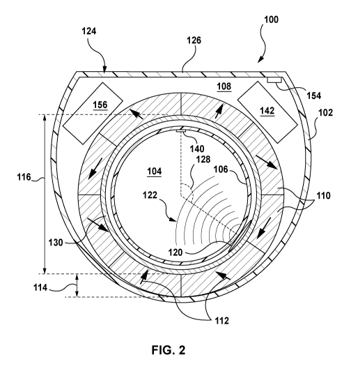
[0072] Figure 2 is a cross section taken along line 2-2 in Figure 1;
[0073] Figure 3 is a cross-section taken along line 3-3 in Figure 1;
[0074] Figure 4 is a schematic diagram of the electronics assembly of the
device of Figure
1;
[0075] Figure 5 is a cross-section taken through an alternative wearable blood
analyte
measurement device;
[0076] Figure 6 is a perspective view of the plurality of magnets of the
device of Figure 5;
[0077] Figure 7 is a cross-section taken through another example wearable
blood analyte
measurement device;
[0078] Figure 8 is a cross-section taken through another example wearable
blood analyte
measurement device;
[0079] Figure 9 is a cross-section taken through another example wearable
blood analyte
measurement device;
[0080]Figure 10A is a cross-section taken through another example wearable
blood
analyte measurement device;
[0081] Figure 10B is an enlarged view of a portion of the device of Figure
10A; and
[0082] Figure 11 is a flowchart showing a general example of the operation of
the devices
described herein.
DETAILED DESCRIPTION:
[0083]Various apparatuses or processes will be described below to provide an
example
of an embodiment of the claimed subject matter. No embodiment described below
limits
any claim and any claim may cover processes or apparatuses that differ from
those
CA 03081630 2020-05-04
WO 2019/090418 PCT/CA2018/051398
12
described below. The claims are not limited to apparatuses or processes having
all of the
features of any one apparatus or process described below or to features common
to
multiple or all of the apparatuses described below. It is possible that an
apparatus or
process described below is not an embodiment of any exclusive right granted by
issuance
of this patent application. Any subject matter described below and for which
an exclusive
right is not granted by issuance of this patent application may be the subject
matter of
another protective instrument, for example, a continuing patent application,
and the
applicants, inventors or owners do not intend to abandon, disclaim or dedicate
to the
public any such subject matter by its disclosure in this document.
[0084] In general, disclosed herein is a wearable blood analyte measurement
device, and
related methods. The device may be, for example, a piece of jewelry, such as a
ring, a
bracelet, an earring, or a necklace. The device employs nuclear magnetic
resonance
(NMR) technology ¨ i.e. emits radiofrequency (RF) pulses, and receives
resulting NMR
signals ¨ to determine (or to facilitate the determination of) the
concentration of an analyte
(such as glucose, cholesterol, a vitamin, alcohol, a mineral, or a drug) in
the blood of a
wearer. The device can be worn on an appendage (e.g. on a finger, a wrist, a
neck, a toe,
and earlobe, etc.), in order to create a magnetic field within the appendage,
and can be
activated to obtain NMR signals from the appendage. The NMR signals can be
processed
to determine a concentration of the analyte in the wearer's blood.
[0085] In general, the device can be used to non-invasively (i.e. without
puncturing the
skin) determine the concentration of an analyte in the blood of a wearer. This
may allow
for ease and comfort of use, and can facilitate patient compliance and promote
health.
[0086]The device may itself process the NMR signals and calculate the blood
concentration of the analyte based on the NMR signals, or may transmit the NMR
signals
to a secondary device (such as, for example, a smart-watch, a smart-phone, a
tablet, a
computer, or a drug delivery device) that processes and/or calculates the
blood
concentration of the analyte. The device may itself display the blood
concentration of the
analyte, or the secondary device may display the blood concentration of the
analyte.
CA 03081630 2020-05-04
WO 2019/090418 PCT/CA2018/051398
13
[0087] The device may be used, for example, by a person having a medical
condition in
order to monitor that medical condition. For example, the device may be worn
by a
diabetic wearer in order to monitor their blood-glucose concentration, or may
be worn by
a person suffering from high cholesterol in order to monitor their blood
cholesterol
concentration. As an alternative example, the device may be used by law
enforcement in
order to monitor substance use or abuse in a wearer. For example, the device
may be
worn in order to monitor the blood alcohol concentration or the concentration
of an illicit
substance (e.g. THC) in the blood of a wearer. As a further alternative
example, the device
may be worn in order to promote and/or maintain health and wellness. For
example, the
device may be worn by a wearer in order to monitor the concentration of a
vitamin or
mineral in their blood, or the concentration of another indicator of health or
wellness. As
an alternative example, the device may be worn in order to measure a
metabolite of a
drug.
[0088] The device may be used to measure the concentration of an analyte in a
wearer's
blood, as opposed to other tissues. This may provide valuable clinical or
other
information. For example, in the case of a diabetic patient, the concentration
of glucose
in the blood, as opposed to other tissues, can be of particular concern. In
some examples,
this is achieved by linking the NMR signals received by the device to the
heart phase of
the wearer. For example, the device can include a heart phase sensor, such as
an LED
(light emitting diode) heart rate monitor, that determines diastole and
systole in the
wearer. The difference in the NMR signals generated during diastole and during
systole
can be indicative of the concentration of an analyte in a wearer's blood (as
opposed to
other tissues), and can be used to calculate the concentration of the analyte
in the
wearer's blood. In other examples, this is achieved by taking advantage of
certain unique
properties of blood. For example, blood can have a high T2/T1 ratio.
Accordingly. the
device may employ balanced Steady State Free Precession (b-SSFP) pulse
sequences,
which are sensitive to tissues/molecules with a high T2/T1 ratio. For further
example,
since blood is in motion within the body, the device may employ phase contrast
magnetic
resonance angiography (MRA), in which magnetic resonance signals are sensitive
to
CA 03081630 2020-05-04
WO 2019/090418 PCT/CA2018/051398
14
moving spins. For further example, since blood has a relatively high T2 time,
the device
can employ a T2 filter, to filter out signals from other tissues.
[0089] In some examples, the device can be worn and used over a relatively
long period
of time, such as hours, days, or more. This can allow for regular and ongoing
monitoring
of a blood analyte concentration. For example, in the case of a diabetic
patient, the device
can be worn daily for the duration of the day, and blood glucose
concentrations can be
determined regularly over the course of the day. Concentrations can be, for
example,
determined continuously or intermittently (e.g. hourly or more).
[0090]The device can be configured to measure one or more specific analytes in
a
patient's blood. In such examples, since the analyte of concern is known, and
it is only
the concentration of the analyte that is to be determined, the device does not
necessarily
require the extremely high magnetic fields required of common NMR devices. For
example, NMR machines are often used in laboratories to determine the chemical
structure of an unknown compound. This can require magnetic field strengths of
20 Tesla
(T) or more. However, in the present example, since it is not necessary to
determine the
chemical structure of any compound, but merely the concentration of a known
compound,
a lower magnetic field strength can be used. For example, the device may have
a
magnetic field strength of less than 1 Tesla (T). Because a lower magnetic
field strength
is required, the device can be relatively small and light in weight (e.g.
small enough and
light enough to be worn on a finger). For example, the device may have a
weight of less
than 50 grams, or less than 20 grams, or between 1 gram and 20 grams.
[0091] Referring now to Figure 1, a first example of a wearable blood analyte
measurement device 100 is shown. In the example shown, the device 100 is in
the form
of a ring, which is wearable on a person's finger. In alternative examples, a
wearable
blood analyte measurement device may be in the form of a bracelet, a necklace,
an
earring, or another piece of jewelry or wearable item.
[0092] Referring still to Figure 1, in the example shown, the device 100
includes a casing
102, which supports various other parts of the device 100. The casing 102
defines an
appendage-receiving bore 104, in which an appendage of a wearer may sit. In
the
CA 03081630 2020-05-04
WO 2019/090418 PCT/CA2018/051398
example shown, wherein the device 100 is a ring, the appendage-receiving bore
104 is
for receiving a finger. In alternative examples, an appendage-receiving bore
may be for
receiving a wrist, an earlobe, a neck, a toe, an ankle, or another body part.
The casing
has an inner section 106 that lines the appendage-receiving bore.
[0093] Referring to Figure 2, in the example shown, the casing 102 has an
interior volume
108. As noted above, the casing 102 supports various other parts of the device
100.
These other parts may be within the interior volume 108, or exterior to and
mounted to
the casing 102.
[0094] In some examples, the casing 102 is made (in whole or in part) from a
non-
ferromagnetic and non-metallic material, such as but not limited to a plastic,
a ceramic, a
wood, a rubber, or a combination thereof. Such non-ferromagnetic and non-
metallic
materials may prevent, reduce, or minimize the interaction of the casing 102
with the
magnetic field of the device 100.
[0095] Referring to Figures 2 and 3, in the example shown, the device 100
includes a
plurality of magnets 110 within the interior volume 108. In the examples
shown, the
magnets are arranged around the appendage-receiving bore ¨ i.e. at least a
portion of
the bore 104 is positioned between at least two of the plurality of magnets
110. The
magnets 110 produce a magnetic field in the appendage-receiving bore 104.
[0096] In the example shown, the magnets 110 are permanent magnets that are
arranged
in an annulus around the appendage-receiving bore 104. Specifically, in the
example
shown, the magnets 110 are shaped as sectors of an arc, and are arranged to
form a
Halbach array, with the respective magnetic field of each magnet indicated by
arrows 112
(shown in Figure 2), so that a relatively strong magnetic field is produced in
the
appendage-receiving bore 104, and a relatively weak or zero magnetic field is
produced
outside of the annulus of magnets 110. The Halbach array may include, for
example,
between 8 and 32 magnets. In some examples, the Halbach array includes 16
magnets.
CA 03081630 2020-05-04
WO 2019/090418 PCT/CA2018/051398
16
[0097]The magnets 110 may in some examples be rare-earth magnets, such as
neodymium magnets or samarium cobalt magnets. In some examples, the magnets
110
are N-52 or N-55 grade neodymium magnets.
[0098] The magnets 110 may, for example, generate a magnetic field in the
appendage-
receiving bore 104 having a magnetic field strength of less than 1 T. For
example, the
magnetic field in the appendage-receiving bore 104 may have a magnetic field
strength
of between 0.05 T and 0.5 T, or between 0.1 T and 0.3 T, or between 0.15 and
0.35 T, or
about 0.32 T. The magnetic field serves to polarize nuclear spins within the
appendage
receiving bore 104.
[0099] Referring still to Figures 2 and 3, in some examples, the annulus of
magnets 110
has a radial wall thickness 114 of, for example, less than 5 mm. For example,
the radial
wall thickness 114 may be between about 1 mm and 3 mm. In some examples, the
annulus of magnets 110 has an inner diameter 116 of, for example, between 10
mm and
40 mm. For example, the inner diameter 116 may be about 20 mm. In some
examples,
the annulus of magnets 108 has a height 118 of, for example, between 2 mm and
10 mm.
For example, the height 118 may be about 5 mm.
[0100] In alternative examples (some of which will be described in detail
below with
reference to Figures 5 to 10B), the plurality of magnets may be of another
configuration,
and/or may be arranged around the appendage receiving bore in another
arrangement.
For example, the device may include a pair of magnets positioned on opposed
sides of
the appendage-receiving bore. For further example, the device may include
electromagnets instead of or in combination with permanent magnets. For
further
example, the plurality of magnets may be of another shape other than sectors
of an arc,
and/or may include additional magnets (e.g. rectangular magnets) interspersed
between
the magnets that are shaped as sectors of an arc.
[0101] Referring still to Figures 2 and 3, the device 100 further includes a
nuclear
magnetic resonance (NMR) transceiver 120 that is supported by the casing 102.
In the
example shown, the NMR transceiver 120 includes a single transceiver coil that
both
transmits and receives, and is positioned within the interior volume 108 and
adjacent the
CA 03081630 2020-05-04
WO 2019/090418 PCT/CA2018/051398
17
inner section 106 of the casing 102. The NMR transceiver 120 is positioned to
emit
radiofrequency (RF) pulses to the appendage-receiving bore 104, and to receive
NMR
signals from the appendage-receiving bore 104.
[0102] In alternative examples, as will be described below, the NMR
transceiver can
include a transmitter coil and a separate receiver coil. In further
alternative examples, the
NMR transceiver can include a plurality of transmitter coils, and/or a
plurality of receiver
coils, and/or a plurality of transceiver coils.
[0103]In the example shown, the NMR transceiver 120 includes a surface coil.
In
alternative examples, as will be described below, the NMR transceiver can
include one
or more solenoid coils.
[0104]The NMR transceiver 120 can be sized and configured to emit
radiofrequency
pulses to and receive NMR signals from the entire volume of the appendage-
receiving
bore 104, or only a section of the appendage-receiving bore 104. In the
example shown,
the device 100 is configured so that the NMR transceiver 120 emits
radiofrequency pulses
to and receives NMR signals from a section of the appendage-receiving bore
(this section
can be referred to herein as a 'target section', and is shown schematically in
Figure 2 at
reference character 122), and so that in use, the target section 122 is
readily positioned
within a highly perfused region of a finger. Particularly, referring back to
Figure 1, in the
example shown, the device 100 includes a positioning guide 124 for guiding a
user in
orienting the device 100 at a target orientation. In the example shown, the
positioning
guide 124 is formed by a flattened and enlarged section of the casing 102. A
wearer of
the ring may naturally be inclined to (and/or can be instructed to) position
this enlarged
section on the dorsal surface of the finger. Referring back to Figure 2, the
NMR
transceiver 120 is spaced from a centre-point 126 of the positioning guide 124
by a
spacing angle 128. The spacing angle 128 can in some examples be between about
45
degrees and about 180 degrees, or between about 80 degrees and about 150
degrees.
Because of the spacing angle 128, when the ring is worn with the enlarged
section on the
dorsal surface of the finger, the target section 122 captures the palmar
digital vein of the
wearer.
CA 03081630 2020-05-04
WO 2019/090418 PCT/CA2018/051398
18
[0105] In some examples (not shown), the device can include a suction
mechanism to
improve perfusion of blood in the target section. For example, the device can
include a
miniaturized suction cup on the casing adjacent the NMR transceiver.
[0106] In other examples, the positioning guide 124 can include another
feature instead
of or in addition to the enlarged section of the casing. For example, the
casing can be
relatively symmetrical in shape (i.e. a simple band without any enlarged
sections), but
can include a jewel or a marking or a stone or another visual feature that
serves as a
positioning guide.
[0107] In other examples, the target section of the appendage-receiving bore
can be
centred within the appendage-receiving bore, so that the orientation of the
device 100 is
immaterial.
[0108] In some examples, the device can be configured to boost the intensity
of the NMR
signal received by the NMR transceiver. For example, the device can employ
dynamic
nuclear polarisation (DNP) to boost the intensity of the NMR signal received
by the NMR
transceiver. In such examples, the device can include a microwave resonator
(e.g. a
single sided microwave resonator) supported by the casing. The microwave
resonator
can rely on free radicals naturally occurring in blood or on artificially
generated free
radicals. This will be described in further detail below with regards to
Figures 10A and
10B. In other examples, brute-force hyperpolarization can be used to boost the
intensity
of the NMR signal received by the transceiver. In such examples, coils (e.g. a
high-
temperature superconductor coil, together with electric cryocoolers) can be
positioned on
opposite ends of the target section and can be pulsed to create a strong
magnetic field
(e.g. 7 T) in the target section prior to obtaining a blood-analyte
measurement.
[0109] Referring to Figures 2 and 3, in the example shown, the device 100
further includes
a shim system 130 that is operable to homogenize the magnetic field or a
portion thereof
(e.g. a portion including the target section 122 of the magnetic field). As
used herein, the
term 'homogenize refers to an increase or improvement in the homogeneity of
the
magnetic flux density within the appendage receiving bore 104 or a portion
thereof. The
CA 03081630 2020-05-04
WO 2019/090418 PCT/CA2018/051398
19
term 'homogenize does not require that the appendage receiving bore 104 or a
portion
thereof be made perfectly or entirely homogeneous.
[0110] Referring to Figure 3, in the example shown, the shim system 130
includes both a
dynamic shim system 132, and a static shim system 134. The dynamic shim system
132
includes an active shim coil 136 within the interior volume 108 and extending
around the
appendage-receiving bore 104. As will be described in further detail below,
the active
shim coil 136 is activatable to homogenize the target section 122 of the
magnetic field.
The static shim system 134 includes one or more ferromagnetic materials 138
within the
interior volume 108, and extending around the appendage-receiving bore 104. In
the
example shown, both the active shim coil 136 and the ferromagnetic materials
138 are
positioned between the inner section of the casing 106 and the annulus of
magnets 110.
[0111] In alternative examples, the shim system may be of another
configuration. For
example, a shim system may include only one of a dynamic shim system and a
static
shim system.
[0112] Referring back to Figure 2, in the example shown, the device 100
further includes
a heart phase sensor 140 that is supported by the casing 102. In the example
shown, the
heart phase sensor 140 is exterior to the casing 102, and joined to the inner
section 106
of the casing 102, so that it is within the appendage-receiving bore 104. The
heart phase
sensor 140, when activated, can sense diastole and systole in a wearer when an
appendage of the wearer is received in the appendage-receiving bore 104. The
heart
phase sensor can be, for example, an LED heart monitor.
[0113] Referring still to Figure 2, the device 100 further includes an
electronics assembly
142 within the interior volume 108. In the example shown, the electronics
assembly 142
is in communication with the NMR transceiver 120, the heart phase sensor 140,
and the
shim system 130. As will be described in further detail below, in the example
shown, the
electronics assembly 142 is operable to activate the NMR transceiver 120 and
the active
shim coil 136 to homogenize the target section 122 of the magnetic field. The
electronics
assembly 142 is further operable to receive a heart phase signal from the
heart phase
sensor 140. The heart phase signal can be indicative of systole or diastole in
the wearer.
CA 03081630 2020-05-04
WO 2019/090418 PCT/CA2018/051398
The electronics assembly 142 is further operable to activate the NMR
transceiver 120
during diastole to emit a diastolic RF pulse to the target section 122 of the
appendage-
receiving bore 104, and receive a diastolic NMR signal from the target section
122 of the
appendage-receiving bore 104. The electronics assembly 142 is further operable
to
activate the NMR transceiver 120 during systole to emit a systolic RF pulse to
the target
section 122 of the appendage-receiving bore 104, and receive a systolic NMR
signal from
the target section 122 of the appendage-receiving bore. The diastolic NMR
signal and the
systolic NMR signal can then be processed to calculate the blood analyte
concentration
of the wearer.
[0114] Referring still to Figure 2, in the example shown, the device further
includes a data
transmitter 154 within the interior volume 108. The data transmitter 154 is in
communication with the electronics assembly 142. The data transmitter 154 can
be, for
example a Bluetooth transmitter (e.g. a Bluetooth 5.0 transmitter). The data
transmitter
154 can communicate signals between the electronics assembly 142 and a
secondary
device (not shown), such as a smart-phone, a smart-watch, a tablet, a
computer, a drug-
delivery device, or other device.
[0115] Referring to Figure 4, the electronics assembly 142 and the operation
of the device
100 will be described in further detail, by way of example.
[0116] In the example shown, the electronics assembly 142 includes an RF
control
module 144, a central processing unit (CPU) 146, and a shim control module
148. The
RF control module 144 is in communication with the NMR transceiver 120 and the
CPU
146. Specifically, the RF control module 144 includes an RF transmitter sub-
module 150
in communication with the CPU 146, an RF receiver sub-module 152, with
quadrature
detection, in communication with the CPU 146, and a duplexer 158 in
communication with
the RF transmitter sub-module 150, the RF receiver sub-module 152, and the NMR
transceiver 120. The shim control module 148 is in communication with the CPU
146, the
RF control module 144, and the active shim coil 132. The heart phase sensor
140 is in
communication with the CPU 146 and with the RF control module 144. The data
transmitter 154 is in communication with the CPU 146 and the RF control module
144.
CA 03081630 2020-05-04
WO 2019/090418 PCT/CA2018/051398
21
[0117]As used herein, the term CPU 146 refers to any unit or module or
processor or
assembly that can control and/or coordinate other parts of the electronics
assembly 142
or the device 100, or can process information received from other parts of the
electronics
assembly 142 or the device 100.
[0118] In some examples, as a first step in measuring the blood analyte
concentration,
the target section 122 of the appendage receiving bore 104 can be homogenized
by a
shimming operation. For example, the CPU 146 can signal the RF control module
144 to
activate the NMR transceiver 120, which emits a shim pulse to the target
section 122.
The NMR transceiver 120 can receive a shim signal from the target section 122
of the
magnetic field in response to the shim pulse. The RF control module 144 can
communicate the shim signal from the NMR transceiver 120 to the CPU 146. The
CPU
146 can then activate the shim control module 148 to adjust the current in the
active shim
coil 132, based on the shim signal. This can be repeated until the CPU 146
determines
that the target section 122 is sufficiently homogenized, based on the shim
signal. For
example, the target section 122 may be considered to be sufficiently
homogenized when
the field homogeneity is between 0.1 and 1.0 ppm. The shim system can then be
"locked".
[0119] In alternative examples, rather than the CPU 146 initiating and/or
coordinating the
shimming operation, the shimming operation can be controlled by the secondary
device,
via the data transmitter 154.
[0120] In some examples, the device can be configured to adjust the NMR
frequency in
order to account for temperature changes in the plurality of magnets 110. That
is, each
time an NMR scan is performed, a calibration operation may be performed in
order to
ascertain the magnetic field strength within the target section 122. The NMR
frequency
can then be adjusted based on the magnetic field strength.
[0121] When the shim is "locked" and the NMR frequency has been adjusted, the
device
100 can then perform a "scan" to obtain diastolic and systolic NMR signals.
For example,
with the heart phase sensor 140 sensing systole and diastole in the wearer,
the RF control
module 144 can activate the NMR transceiver 120 to emit the diastolic RF pulse
and the
systolic RF pulse to the target section 122 of the magnetic field, and receive
the resulting
CA 03081630 2020-05-04
WO 2019/090418 PCT/CA2018/051398
22
diastolic NMR signal and systolic NMR signal. Specifically, the CPU 146 can
receive the
heart phase signal from the heart phase sensor 140, and in response to the
heart phase
signal, can signal the RF control module 144 to activate the NMR transceiver
120 to emit
the diastolic RF pulse during diastole, and the systolic RF pulse during
systole. The NMR
transceiver 120 can communicate the diastolic NMR signal and the systolic NMR
signal
to the RF control module 144. Optionally, the device can perform multiple
scans in
sequence, and provide a blood-analyte concentration based on the multiple
scans.
[0122] In some examples (not shown), in order to achieve high sensitivity, the
number of
scans carried out in a given time period can be increased. For example, the
device 100
can carry out 128 scans per second or 64 scans per second. In some examples,
the scan
time can be artificially increased by increasing the number of receiver
channels detect the
NMR signal. This can be achieved by adding additional receiver coils and
preamplifiers
to the device.
[0123] In alternative examples, rather than the CPU 146 initiating and/or
coordinating the
scan, the scan can be controlled by the secondary device, via the data
transmitter 154.
[0124]In some examples, the CPU 146 can then calculate the blood-analyte
concentration based on the diastolic NMR signal and the systolic NMR signal.
Briefly, the
diastolic NMR signal and the systolic NMR signal can be processed to obtain a
diastolic
NMR spectrum and a systolic NMR spectrum, respectively. This can be achieved
by first
carrying out signal processing and denoising. For example, the free induction
decay
signal can be weighted to improve the signal to noise ratio or resolution, a
reference
deconvolution algorithm can be performed, and/or a denoising algorithm using
wavelets
can be performed. Then, Fourier transform and quantification can be performed.
For
example, a Fourier transform of the free induction decay can be performed to
generate
the NMR spectra. In each spectrum, the area under the analyte peak can be
compared
to the area under the water peak, to determine the analyte concentration. The
systolic
analyte concentration can then be subtracted from the diastolic concentration,
to
determine the blood analyte concentration. This can be done using various
signal
processing and calculating algorithms, which can be programmed into the
software of the
CA 03081630 2020-05-04
WO 2019/090418 PCT/CA2018/051398
23
CPU 146. The CPU 146 can then communicate the blood-analyte concentration to
the
data transmitter 154. The data transmitter 154 can then transmit the blood-
analyte
concentration to the secondary device. The secondary device can have a
display, and
can then display the blood-analyte concentration.
[0125] In other examples, the RF control module and/or the CPU can communicate
the
diastolic NMR signal and the systolic NMR signal to the data transmitter 154.
The data
transmitter 154 can then transmit the diastolic NMR signal and the systolic
NMR signal to
the secondary device, and the secondary device can calculate and optionally
display the
blood analyte concentration.
[0126] In further examples (not shown), the device can include a display, and
the device
can display the blood-analyte concentration.
[0127]In some examples, the electronics assembly 142 (or parts thereof) may be
provided on an ASIC (application specific integrated circuit) chip (not
shown).
[0128]The blood-analyte concentration can optionally be calculated and
displayed
periodically, optionally at regular intervals, while the device 100 is worn.
Alternatively, the
blood-analyte concentration can be calculated and displayed or upon receiving
a request
from a wearer. The request from the wearer can optionally be input into the
secondary
device and transmitted from the secondary device to the device 100, or can be
input
directly into the device 100. Optionally, the device 100 can include an alarm
function,
which can be triggered when the calculated blood analyte concentration is
above a set
value, or can trigger an alarm function in the secondary device.
[0129]Referring back to Figure 2, in the example shown, the device 100 further
includes
a power source 156 that powers the various parts of the device 100 (e.g. the
NMR
transceiver 120, the electronics assembly 142, the shim system 130, the data
transmitter
154, and the heart-phase monitor 140). The power source 156 can be a
rechargeable
battery, optionally an inductively rechargeable battery. The device 100 can
optionally be
sold in a kit with a charger (not shown), optionally an inductive charger, for
the battery.
The device 100 can optionally be worn on a daily basis, and can be removed
nightly for
CA 03081630 2020-05-04
WO 2019/090418 PCT/CA2018/051398
24
charging. Depending on the frequency at which the diastolic and systolic NMR
signals
are obtained, the battery may last for between 4 hours and 72 hours on a
single charge.
[0130] In some examples (not shown), in order to decrease noise in the NMR
signal, the
device can include an electric cryocooler for supercooling the receiver coil.
[0131]Referring now to Figure 5, an alternative device 500 is shown. In Figure
5,
elements that are like those of Figures 1 to 4 are referred to with like
reference numerals,
incremented by 400. In the example of Figure 5, the device 500 is similar to
the device
100; however, the device 500 includes a plurality of cylindrical magnets 510a
and a
plurality of rectangular bar magnets 510b, arranged in an alternating pattern.
The
magnets 510a and 510b are oriented to produce a dipole magnetic field within
the bore
504 of the device.
[0132] Referring to Figure 6 (wherein the magnets 510a and 510b are shown
separately
from the device), in the example shown, the device 500 includes a plurality of
annular
rows of magnets 510a and 510b. The use of rows can reduce magnetic field
inhomogeneities. In the example shown, 4 rows of magnets are used; however, in
alternative examples, another number of rows could be used.
[0133] In the example shown, the device 500 includes 16 cylindrical magnets
510a and
16 bar magnets 510b in each row; however, in alternative examples, another
number of
each type of magnet could be used. In some examples, the combined height 518
of the
rows can be between 2 mm and 20 mm. For example, the height 518 may be about
10
mm.
[0134] During manufacture of the device 500, the cylindrical magnets 510a can
be rotated
about their longitudinal axis, and the bar magnets 510b can be shifted
slightly radially
inward or outward, to adjust the local magnetic field. Furthermore, the rows
of magnets
can be rotated to adjust the local magnetic field.
[0135] Furthermore, in the example of Figure 5, the NMR transceiver includes a
pair of
surface coils 520a, 520b for receiving NMR signals, and a solenoid coil 520c
for
CA 03081630 2020-05-04
WO 2019/090418 PCT/CA2018/051398
transmitting RF pulses. In this example, the target section 522 can be
centrally located
within the appendage receiving bore 504.
[0136] Referring now to Figure 7, an alternative device 700 is shown. In
Figure 7,
elements that are like those of Figures 1 to 4 are referred to with like
reference numerals,
incremented by 600. In the example of Figure 7, the device 700 is similar to
the device
100; however, the device 500 includes a pair of magnets 710a, 710b, on opposed
sides
of the appendage receiving bore 704. The magnets 710a, 710b each have an
enlarged
region 760a, 760b, which produces a strong and relatively homogeneous magnetic
field
in target section 722.
[0137] Referring now to Figure 8, another alternative device 800 is shown. In
Figure 8,
elements that are like those of Figures 1 to 4 are referred to with like
reference numerals,
incremented by 700. In the example of Figure 8, the device 800 is similar to
the device
100; however, the device includes 8 magnets 810 in a Hallbach array, and the
NMR
transceiver includes a pair of surface coils 820a, 820b for receiving NMR
signals, and a
solenoid coil 820c for transmitting RF pulses. Furthermore, the device 800
does not
include a heart phase monitor. Instead, the concentration of an analyte in the
wearer's
blood (as opposed to other tissues) is measured by taking advantage of unique
nuclear
magnetic resonance properties of blood.
[0138] In some examples, the device can be configured to take advantage of the
T2/T1
ratio of blood (where T2 refers to the spin-spin relaxation time and Ti refers
to the spin-
lattice relaxation time). That is, blood has a relatively high T2/T1 ratio, as
compared to
other tissues. Balanced Steady State Fee Precession (b-SSFP) pulse sequences
are
sensitive to tissues/molecules with a high T2/T1 ratio. Accordingly, the
solenoid coil 820c
can emit rapid repeated pulses with a constant repetition time to generate a b-
SSFP
signal, in order to isolate the NMR signal from the blood in the target
section. In such
examples, since fat can also have a relatively high T2/T1 ratio, a fat
suppression pulse
may also be employed.
[0139] Alternatively or in addition, the device can be configured to take
advantage of the
relatively high T2 signal of blood. That is, blood gives a relatively high T2
signal, as
CA 03081630 2020-05-04
WO 2019/090418 PCT/CA2018/051398
26
compared to other tissues. Accordingly, the solenoid coli 820c can emit a CPMG
spin
echo train, which can include an initial excitation at the Ernst angle, and
repeated 180
degree pulses with a constant repetition time, in order to obtain the T2
signal from the
target section. A T2 filter can then be employed (e.g. in the electronics
assembly or in
the secondary device), to filter out relatively low T2 signals (e.g. signals
with a T2 of less
than 15 ms), leaving only the T2 signal from blood.
[0140] Referring now to Figure 9, another alternative device 900 is shown. In
Figure 9,
elements that are like those of Figures 1 to 4 are referred to with like
reference numerals,
incremented by 800. In the example of Figure 9, similarly to device 800, the
device 900
does not include a heart phase monitor. Instead, the device 900 includes a
gradient coil
962, and takes advantage of the fact that blood will be flowing through the
target section,
whereas other tissues will be stationary. The gradient coil can refocus the
spin of the
moving blood, while the spin from stationary tissues will remain unfocused, so
that the
NMR signal from the blood is isolated.
[0141] In alternative examples, the device can include additional gradient
coils, such as
a total of 2 gradient coils or 3 gradient coils.
[0142] Referring now to Figures 10A and 10B, another alternative device 1000
is shown.
In Figures 10A and 10B, elements that are like those of Figures 1 to 4 are
referred to with
like reference numerals, incremented by 900. In the example of Figures 10A and
10B,
the device 1000 includes three magnets 1010, which are positioned on only one
side of
device 1000, in a generally U-shaped configuration, to create a target section
1022
(shown in Figure 10B) adjacent the magnets 1010 and within the U-shape. A
material
1064 that provides magnetic shielding and thermal insulation can line the
magnets 1010.
Furthermore, the device 1000 is configured to employ DNP to boost the
intensity of the
NMR signal received by the NMR transceiver. That is, the device includes a
microwave
resonator 1066, and a capacitive micromachined ultrasonic transducer array
1068, which
are housed in the casing 1002, adjacent the NMR transceiver 1020, active shim
coils
1036 and the passive shimming materials 1034. A solid ultrasound coupling
medium
1070 is provided on the inner section 1006 of the casing 1002. The ultrasonic
transducer
CA 03081630 2020-05-04
WO 2019/090418 PCT/CA2018/051398
27
array 1068 can be used to generate free radicals in the blood, by sonolysis.
This can
result in polarization transitions. The microwave resonator 1066 can then
transmit a
microwave signal to the bore, 1004 to transfer the polarization to 1H spins,
in order to
boost the intensity of the NMR signal.
[0143] In some examples (not shown), a laser pulse can be used to create
cavitation
nuclei, which can facilitate sonolysis.
[0144] A summary flowchart of the general operation of the devices described
above is
shown in Figure 11.
[0145] As used herein, the term "NMR signal" can refer to an unprocessed NMR
signal,
such as an analog NMR signal, or a processed NMR signal, such as a digital NMR
signal
(e.g. resulting from processing of an analog NMR signal).
[0146] While the above description provides examples of one or more processes
or
apparatuses, it will be appreciated that other processes or apparatuses may be
within the
scope of the accompanying claims.
[0147] To the extent any amendments, characterizations, or other assertions
previously
made (in this or in any related patent applications or patents, including any
parent, sibling,
or child) with respect to any art, prior or otherwise, could be construed as a
disclaimer of
any subject matter supported by the present disclosure of this application,
Applicant
hereby rescinds and retracts such disclaimer. Applicant also respectfully
submits that any
prior art previously considered in any related patent applications or patents,
including any
parent, sibling, or child, may need to be re-visited.