Note: Descriptions are shown in the official language in which they were submitted.
CA 03081719 2020-05-01
WO 2019/090110 PCT/US2018/059003
COMPOSITIONS AND METHODS FOR TREATING CANCER WITH
ANTI-ROR1 IMMUNOTHERAPY
CROSS-REFERENCE TO RELATED APPLICATIONS
This application claims the benefit of priority under 35 U.S.C. Section 119(e)
to U.S.
Provisional Patent Application No. 62/581,284 filed on November 3, 2017, the
entire contents of
which are incorporated herein by reference.
SEQUENCE LISTING
The instant application contains a Sequence Listing which has been submitted
electronically
in ASCII format and is hereby incorporated by reference in its entirety. The
ASCII copy, created
on October 25, 2018, is named SequenceListing.txt and is 90.0 kilobytes in
size.
STATEMENT REGARDING FEDERALLY SPONSORED
RESEARCH OR DEVELOPMENT
This invention was created in the performance of a Cooperative Research and
Development Agreement with the National Institutes of Health, an Agency of the
Department of
Health and Human Services. The Government of the United States has certain
rights in this
invention.
FIELD OF THE DISCLOSURE
This application relates to the field of cancer, particularly to ROR1 antigen
binding
domains and chimeric antigen receptors (CARs) containing such ROR1 antigen
binding domains
and methods of use thereof
BACKGROUND
Cancer is one of the most deadly threats to human health. In the U.S. alone,
cancer affects
nearly 1.3 million new patients each year, and is the second leading cause of
death after
cardiovascular disease, accounting for approximately 1 in 4 deaths. Solid
tumors are responsible
CA 03081719 2020-05-01
WO 2019/090110 PCT/US2018/059003
for most of those deaths. Although there have been significant advances in the
medical treatment
of certain cancers, the overall 5-year survival rate for all cancers has
improved only by about 10%
in the past 20 years. Cancers, or malignant tumors, metastasize and grow
rapidly in an
uncontrolled manner, making treatment extremely difficult.
There are numerous unmet therapeutic needs in the treatment of solid and
liquid tumors.
ROR1, receptor tyrosine kinase-like orphan receptor 1, is an embryonic protein
that is highly
expressed in many cancer types, including CLL, carcinoma of the breast,
glioblastoma, lung
adenocarcinoma and sarcomas (Ewing sarcoma, osteosarcoma, rhabdomyosarcoma,
and
fibrosarcoma), and is generally absent in normal tissues (Suping Zhang, et
al., 2012, The Onco-
Embryonic Antigen ROR1 Is Expressed by a Variety of Human Cancers. Am J
Pathol, 181: 1903-
1910, Ashwini Balakrishnan, et al., 2017, Analysis of ROR1 Protein Expression
in Human Cancer
and Normal Tissues., Clin Cancer Res 23:3061-3071, Borcherding, Nicholas et
al., 2017, ROR1,
an Embryonic Protein with an Emerging Role in Cancer Biology. Protein & Cell
5.7 (2014): 496-
502). ROR1 has three splice variants, including a 104 kDa (up to 120 kDa
depending on
glycosylation) transmembrane glycoprotein comprised of 937 amino acids (1-29
signal peptide),
and 2 smaller variants of intracellular and secreted forms (GeneBank NP
005003, Masiakowski,
P., and Carroll, R.D., 1992, A Novel Family of Cell Surface Receptors with
Tyrosine Kinase-like
Domain, J Biol Chem 36: 26181-26190.). The presence of ROR1 on the surface of
transformed
cells indicates that targeting ROR1 will enable novel cancer treatments to be
developed for a range
of liquid cancer such as chronic lymphocytic leukemia (CLL) and other solid
tumors (Borcherding,
N., Kusner, D. et al., 2014, ROR1, an embryonic protein with an emerging role
in cancer biology.
Protein & Cell, 5:496-502).
Though generally absent in adult tissues, at least one report found ROR1
expression in
parathyroid; pancreatic islets; and regions of the esophagus, stomach, and
duodenum (Ashwini
Balakrishnan, et al., 2017, Analysis of ROR1 Protein Expression in Human
Cancer and Normal
Tissues., Clin Cancer Res 23:3061-3071), warranting caution in clinical
application of ROR1-
targeted anti-cancer therapies. ROR1 receptor contains a cytosolic protein
kinase domain, which,
according to some reports, participates in Wnt and EGFR signaling
(Borcherding, N., Kusner, D.
et al., 2014, ROR1, an embryonic protein with an emerging role in cancer
biology. Protein & Cell,
5:496-502). In tumors, ROR1 can induce epithelial to mesenchymal transition
(EMT), and
promote tumor proliferation, aggressiveness, and metastases formation, and
mediate resistance to
apoptosis (Yamaguchi, Tomoya, et al., 2012, "NKX2-1/TITF1/TTF-1-Induced ROR1
is required
to sustain EGFR survival signaling in lung adenocarcinoma." Cancer cell 21.3:
348-361;
Borcherding, N., Kusner, D. et al., 2014, ROR1, an embryonic protein with an
emerging role in
2
CA 03081719 2020-05-01
WO 2019/090110 PCT/US2018/059003
cancer biology. Protein & Cell, 5:496-502). Its role in contributing to tumor
phenotype indicates
that it may serve an important function in tumor initiation or progression and
therefore is a driver
protein.
Earlier approaches in cancer treatment include surgery, radiation therapy,
chemotherapy,
and, for blood tumors ¨ bone marrow transplant. However, the present first
line treatments warrant
further improvement. Such improvements are sought by the novel
immunotherapeutic strategies.
Ongoing pre-clinical investigations and clinical trials investigate targeting
ROR 1 antigen have
been developed using multiple modalities. T lymphocytes expressing ROR1-
specific CARs have
been tested both in murine and non-human primate systems (Huang X, Park H,
Greene J, Pao J,
Mulvey E, Zhou SX, et al., 2015, IGF1R- and ROR1-Specific CAR T Cells as a
Potential Therapy
for High Risk Sarcomas. PLoS ONE 10(7): e0133152; Hudecek M, Schmitt TM,
Baskar S, Lupo-
Stanghellini MT, Nishida T, Yamamoto TN, Bleakley M, Turtle CJ, Chang WC,
Greisman HA,
Wood B, Maloney DG, Jensen MC, Rader C, Riddell SR, 2010, The B-cell tumor-
associated
antigen ROR1 can be targeted with T cells modified to express a ROR1-specific
chimeric antigen
receptor. Blood 116:4532-41.). The lack of toxicity in non-human primates
lends confidence that
human studies can be approached (Berger, C., et al., 2015, Safety of targeting
ROR1 in primates
with chimeric antigen receptor-modified T cells. Cancer Immunol Res 3: 2016-
216.). Both
unmodified, and immunotoxin-linked antibodies to ROR1 have also been proposed
for therapeutic
use (Yang, Jiahui, et al., 2011, "Therapeutic potential and challenges of
targeting receptor tyrosine
kinase ROR1 with monoclonal antibodies in B-cell malignancies." PloS One 6.6:
e21018; Baskar,
Sivasubramanian, et al., 2012, "Targeting malignant B cells with an
immunotoxin against ROR1."
MAbs, 4:3, 349-361.). The present standard of care for B-lineage leukemias may
consists of
remission induction treatment by high dose of chemotherapy or radiation,
followed by
consolidation, and may feature stem cell transplantation and additional
courses of chemotherapy as
needed (see the world wide web at cancer.gov). High toxicity associated with
these treatments, as
well as the risk of complications, such as relapse, secondary malignancy, or
GVHD, motivate the
search for better therapeutic alternatives. Current open clinical trials
include ROR1-targeted T
cells for hematologic malignancy (Genetically Modified T-Cell Therapy in
Treating Patients with
Advanced ROR1+ Malignancies, NCT02706392, Sponsor: Fred Hutchinson Cancer
Research
Center, ClinicalTrials.gov accessed 9/20/2017.), and ROR1-specific antibody
for breast cancer
given in the context of chemotherapy (Study of Circumtuzumab and Paclitaxel
for Metastatic or
Locally Advanced, Unresectable Breast Cancer, NCT02776917, Sponsor: Barbara
Parker, MD,
University of California, San Diego, ClinicalTrials.gov accessed 9/20/2017).
3
CA 03081719 2020-05-01
WO 2019/090110 PCT/US2018/059003
Chimeric Antigen Receptors (CARs) are hybrid molecules comprising three
essential
units: (1) an extracellular antigen-binding motif, (2) linking/transmembrane
motifs, and (3)
intracellular T-cell signaling motifs (Long AH, Haso WM, Orentas RJ. Lessons
learned from a
highly-active CD22-specific chimeric antigen receptor, OncoImmunology. 2013; 2
(4):e23621).
The antigen-binding motif of a CAR is commonly fashioned after a single chain
Fragment
variable (ScFv), the minimal binding domain of an immunoglobulin (Ig)
molecule. Alternate
antigen-binding motifs, such as receptor ligands (i.e., IL-13 has been
engineered to bind tumor
expressed IL-13 receptor), intact immune receptors, library-derived peptides,
and innate immune
system effector molecules (such as NKG2D) also have been engineered. Alternate
cell targets for
CAR expression (such as NK or gamma-delta T cells) are also under development
(Brown CE et
al Clin Cancer Res. 2012;18(8):2199-209; Lehner M et al. PLoS One. 2012; 7
(2):e31210). There
remains significant work to be done with regard to defining the most active T-
cell population to
transduce with CAR vectors, determining the optimal culture and expansion
techniques, and
defining the molecular details of the CAR protein structure itself
The linking motifs of a CAR can be a relatively stable structural domain, such
as the
constant domain of IgG, or designed to be an extended flexible linker.
Structural motifs, such as
those derived from IgG constant domains, can be used to extend the ScFv
binding domain away
from the T-cell plasma membrane surface. This may be important for some tumor
targets where
the binding domain is particularly close to the tumor cell surface membrane
(such as for the
disialoganglioside GD2; Orentas et al., unpublished observations). To date,
the signaling motifs
used in CARs always include the CD3- chain because this core motif is the key
signal for T cell
activation. The first reported second-generation CARs featured CD28 signaling
domains and the
CD28 transmembrane sequence. This motif was used in third-generation CARs
containing
CD137 (4-1BB) signaling motifs as well (Zhao Y et al J Immunol. 2009; 183 (9):
5563-74). With
the advent of new technology, the activation of T cells with beads linked to
anti-CD3 and anti-
CD28 antibody, and the presence of the canonical "signal 2" from CD28 was no
longer required
to be encoded by the CAR itself Using bead activation, third-generation
vectors were found to be
not superior to second-generation vectors in in vitro assays, and they
provided no clear benefit
over second-generation vectors in mouse models of leukemia (Haso W, Lee DW,
Shah NN,
Stetler-Stevenson M, Yuan CM, Pastan IH, Dimitrov DS, Morgan RA, FitzGerald
DJ, Barrett
DM, Wayne AS, Mackall CL, Orentas RJ. Anti-CD22-chimeric antigen receptors
targeting B cell
precursor acute lymphoblastic leukemia, Blood. 2013; 121 (7):1165-74;
Kochenderfer JN et al.
Blood. 2012; 119 (12):2709-20). This is borne out by the clinical success of
CD19-specific
CARs that are in a second generation CD28/CD3- (Lee DW et al. American Society
of
4
CA 03081719 2020-05-01
WO 2019/090110 PCT/US2018/059003
Hematology Annual Meeting. New Orleans, LA; December 7-10, 2013) and CD137/CD3-
signaling formats (Porter DL et al. N Engl J Med. 2011; 365 (8): 725-33). In
addition to CD137,
other tumor necrosis factor receptor superfamily members such as 0X40 also are
able to provide
important persistence signals in CAR-transduced T cells (Yvon E et al. Clin
Cancer Res.
2009;15(18):5852-60). Equally important are the culture conditions under which
the CAR T-cell
populations were cultured, for example the inclusion of the cytokines IL-2, IL-
7, and/or IL-15
(Kaiser AD et al. Cancer Gene Ther. 2015; 22(2):72-78.
Current challenges in the more widespread and effective adaptation of CAR
therapy for
cancer relate to a paucity of compelling targets. Creating binders to cell
surface antigens is now
readily achievable, but discovering a cell surface antigen that is specific
for tumor while sparing
normal tissues remains a formidable challenge. One potential way to imbue
greater target cell
specificity to CAR-expressing T cells is to use combinatorial CAR approaches.
In one system, the
CD3- and CD28 signal units are split between two different CAR constructs
expressed in the
same cell; in another, two CARs are expressed in the same T cell, but one has
a lower affinity and
thus requires the alternate CAR to be engaged first for full activity of the
second (Lanitis E et al.
Cancer Immunol Res. 2013;1(1):43-53; Kloss CC et al. Nat Biotechnol.
2013;31(1):71-5). A
second challenge for the generation of a single ScFv-based CAR as an
immunotherapeutic agent
is tumor cell heterogeneity. At least one group has developed a CAR strategy
for glioblastoma
whereby the effector cell population targets multiple antigens (HER2, IL-13Ra,
EphA2) at the
same time in the hope of avoiding the outgrowth of target antigen-negative
populations. (Hegde M
et al. Mol Ther. 2013;21(11):2087-101).
T-cell-based immunotherapy has become a new frontier in synthetic biology;
multiple
promoters and gene products are envisioned to steer these highly potent cells
to the tumor
microenvironment, where T cells can both evade negative regulatory signals and
mediate effective
tumor killing. The elimination of unwanted T cells through the drug-induced
dimerization of
inducible caspase 9 constructs with chemical-based dimerizers, such as AP1903,
demonstrates one
way in which a powerful switch that can control T-cell populations can be
initiated
pharmacologically (Di Stasi A et al. N Engl J Med. 2011;365(18):1673-83). The
creation of
effector T-cell populations that are immune to the negative regulatory effects
of transforming
growth factor-0 by the expression of a decoy receptor further demonstrates the
degree to which
effector T cells can be engineered for optimal antitumor activity (Foster AE
et al. J Immunother.
2008;31(5):500-5). Thus, while it appears that CARs can trigger T-cell
activation in a manner
similar to an endogenous T-cell receptor, a major impediment to the clinical
application of this
technology to date has been limited in vivo expansion of CAR+ T cells, rapid
disappearance of the
CA 03081719 2020-05-01
WO 2019/090110 PCT/US2018/059003
cells after infusion, and disappointing clinical activity. This may be due in
part to the murine
origin of some of the CAR sequences employed.
The requirement of patients who have received either antibody or CAR-T therapy
to
subsequently undergo HSCT in order to maintain durable responses remains an
area of active
debate. Although high responses are reported for CD19 CAR-T trials, at least
20% of patients fail
in the near-term (Davis KL, Mackall CL, 2016, Blood Advances 1:265-268). The
best results at
12 months post-CAR19 treatment reported show a RFS of 55% and OS of 79% in
patients who
were able to receive the T cell product at the University of Pennsylvania
(Maude SL, Teachey DT,
Rheingold SR, Shaw PA, Aplenc R, Barrett DM, Barker CS, Callahan C, Frey NV,
Farzana N,
Lacey SF, Zheng A, Levine B, Melenhorst JJ, Motley L, Prter DL, June CH, Grupp
SA, 2016, J
Clin Oncol 34, no.15 suppl (May 2016) 3011-3011). Given the expected long term
responses of
50% or less, there remains significant clinical need for new B cell malignancy
targets such as
ROR1.
The present invention addresses these needs by providing CAR compositions and
therapeutic methods that can be used to treat cancers and other diseases
and/or conditions. In
particular, the present invention as disclosed and described herein provides
CARs that may be
used for the treatment of diseases, disorders or conditions associated with
dysregulated expression
of ROR1 and which CARs contain ROR1 antigen binding domains that exhibit a
high surface
expression on transduced T cells, exhibit a high degree of cytolysis of ROR1-
expressing cells, and
in which the transduced T cells demonstrate in vivo expansion and persistence.
SUMMARY
Novel anti-ROR1 antibodies or antigen binding domains thereof and chimeric
antigen
receptors (CARs) that contain such ROR1 antigen binding domains are provided
herein, as well as
host cells (e.g., T cells) expressing the receptors, and nucleic acid
molecules encoding the
receptors. The CARs exhibit a high surface expression on transduced T cells,
with a high degree
of cytolysis, and with transduced T cell expansion and persistence in vivo.
Methods of using the
disclosed CARs, host cells, and nucleic acid molecules are also provided, for
example, to treat a
cancer in a subject.
Thus, in one aspect, an isolated polynucleotide encoding a human anti-ROR1
antibody or a
fragment thereof is provided comprising a nucleic acid sequence selected from
the group
consisting of SEQ ID NOs: 1 and 7.
6
CA 03081719 2020-05-01
WO 2019/090110 PCT/US2018/059003
In one embodiment, an isolated polynucleotide encoding a fully human anti-ROR1
antibody or a fragment thereof is provided, wherein the antibody or a fragment
thereof comprises
a fragment selected from the group consisting of an Fab fragment, an F(ab1)2
fragment, an Fv
fragment, and a single chain Fv (ScFv).
In one embodiment, an isolated polynucleotide encoding a fully human anti-ROR1
antibody or a fragment thereof is provided, wherein the antibody or a fragment
thereof comprises
an amino acid sequence selected from the group consisting of SEQ ID NOs: 2 and
8.
In one aspect, an isolated nucleic acid molecule encoding a chimeric antigen
receptor
(CAR) is provided comprising, from N-terminus to C-terminus, at least one ROR1
antigen
binding domain encoded by a nucleotide sequence comprising a nucleic acid
sequence selected
from the group consisting of SEQ ID NOs: 1 and 7, at least one transmembrane
domain, and at
least one intracellular signaling domain.
In one embodiment, an isolated nucleic acid molecule encoding the CAR is
provided
wherein the encoded extracellular ROR1 antigen binding domain comprises at
least one single
chain variable fragment of an antibody that binds to ROR1.
In another embodiment, an isolated nucleic acid molecule encoding the CAR is
provided
wherein the encoded extracellular ROR1 antigen binding domain comprises at
least one heavy
chain variable region of an antibody that binds to ROR1.
In yet another embodiment, an isolated nucleic acid molecule encoding the CAR
is
provided wherein the encoded CAR extracellular ROR1 antigen binding domain
further comprises
at least one lipocalin-based antigen binding antigen (anticalins) that binds
to ROR1.
In one embodiment, an isolated nucleic acid molecule is provided wherein the
encoded
extracellular ROR1 antigen binding domain is connected to the transmembrane
domain by a linker
domain
In another embodiment, an isolated nucleic acid molecule encoding the CAR is
provided
wherein the encoded ROR1 extracellular antigen binding domain is preceded by a
sequence
encoding a leader or signal peptide.
In yet another embodiment, an isolated nucleic acid molecule encoding the CAR
is
provided comprising at least one ROR1 antigen binding domain encoded by a
nucleotide sequence
comprising a nucleic acid sequence selected from the group consisting of SEQ
ID NOs: 1 and 7,
and wherein the CAR additionally encodes an extracellular antigen binding
domain targets an
antigen that includes, but is not limited to, CD19, CD20, CD22, mesothelin,
CD33, CD38, CD123
(IL3RA), CD138, BCMA (CD269), GPC2, GPC3, FGFR4, c-Met, PSMA, Glycolipid F77,
EGFRvIII, GD-2, TSLPR, NY-ESO-1 TCR, MAGE A3 TCR, or any combination thereof
7
CA 03081719 2020-05-01
WO 2019/090110 PCT/US2018/059003
In certain embodiments, an isolated nucleic acid molecule encoding the CAR is
provided
wherein the additionally encoded extracellular antigen binding domain
comprises an anti-CD19
ScFv antigen binding domain, an anti-CD20 ScFv antigen binding domain, an anti-
CD22 ScFv
antigen binding domain, an anti-mesothelin ScFv antigen binding domain, an
anti-CD33 ScFv
antigen binding domain, an anti-CD38 ScFv antigen binding domain, an anti-
CD123 (IL3RA)
ScFv antigen binding domain, an anti-CD138 ScFv antigen binding domain, an
anti-BCMA
(CD269) ScFv antigen binding domain, an anti-GPC2 ScFv antigen binding domain,
an anti-
GPC3 ScFv antigen binding domain, an anti-FGFR4 ScFv antigen binding domain,
an anti-
TSLPR ScFv antigen binding domain an anti-c-Met ScFv antigen binding domain,
an anti-PMSA
ScFv antigen binding domain, an anti-glycolipid F77 ScFv antigen binding
domain, an anti-
EGFRvIII ScFv antigen binding domain, an anti-GD-2 ScFv antigen binding
domain, an anti-NY-
ES 0-1 TCR ScFv antigen binding domain, an anti-MAGE A3 TCR ScFv antigen
binding domain,
or an amino acid sequence with 85%, 90%, 95%, 96%, 97%, 98% or 99% identity
thereof, or any
combination thereof
In one aspect, the CARs provided herein further comprise a linker or spacer
domain.
In one embodiment, an isolated nucleic acid molecule encoding the CAR is
provided
wherein the extracellular ROR1 antigen binding domain, the intracellular
signaling domain, or
both are connected to the transmembrane domain by a linker (L), hinge (H), or
spacer domain.
In one embodiment, an isolated nucleic acid molecule encoding the CAR is
provided
wherein the encoded linker domain is derived from the extracellular domain of
CD8 or CD28, and
is linked to a transmembrane domain.
In another embodiment, an isolated nucleic acid molecule encoding the CAR is
provided
wherein the encoded CAR further comprises a transmembrane domain that
comprises a
transmembrane domain of a protein selected from the group consisting of the
alpha, beta or zeta
chain of the T-cell receptor, CD28, CD3 epsilon, CD45, CD4, CD5, CD8, CD9,
CD16, CD22,
CD33, CD37, CD64, CD80, CD83, CD86, CD134, CD137 and CD154, or a combination
thereof
In yet another embodiment, an isolated nucleic acid molecule encoding the CAR
is
provided wherein the encoded intracellular signaling domain further comprises
a CD3 zeta
intracellular domain.
In one embodiment, an isolated nucleic acid molecule encoding the CAR is
provided
wherein the encoded intracellular signaling domain is arranged on the N-
terminal side relative to
the CD3 zeta intracellular domain.
8
CA 03081719 2020-05-01
WO 2019/090110 PCT/US2018/059003
In another embodiment, an isolated nucleic acid molecule encoding the CAR is
provided
wherein the encoded at least one intracellular signaling domain comprises a
costimulatory
domain, a primary signaling domain, or a combination thereof
In further embodiments, an isolated nucleic acid molecule encoding the CAR is
provided
wherein the encoded at least one costimulatory domain comprises a functional
signaling domain
of 0X40, CD70, CD27, CD28, CD5, ICAM-1, LFA-1 (CD11a/CD18), ICOS (CD278),
DAP10,
DAP12, and 4-1BB (CD137), or a combination thereof
In one embodiment, an isolated nucleic acid molecule encoding the CAR is
provided that
further contains a leader sequence or signal peptide wherein the leader or
signal peptide (LP)
nucleotide sequence comprises the nucleotide sequence of SEQ ID NO: 19.
In yet another embodiment, an isolated nucleic acid molecule encoding the CAR
is
provided wherein the encoded leader sequence comprises the amino acid sequence
of SEQ ID
NO: 20.
In one aspect, a chimeric antigen receptor (CAR) is provided herein
comprising, from N-
terminus to C-terminus, at least one ROR1 antigen binding domain, at least one
transmembrane
domain, and at least one intracellular signaling domain.
In one embodiment, a CAR is provided wherein the extracellular ROR1 antigen
binding
domain comprises at least one single chain variable fragment of an antibody
that binds to the
antigen, or at least one heavy chain variable region of an antibody that binds
to the antigen, or a
combination thereof
In another embodiment, a CAR is provided wherein the at least one
transmembrane
domain comprises a transmembrane domain of a protein selected from the group
consisting of the
alpha, beta or zeta chain of the T-cell receptor, CD28, CD3 epsilon, CD45,
CD4, CD5, CD8,
CD9, CD16, CD22, CD33, CD37, CD64, CD80, CD86, CD134, CD137, CD154, TNFRSF19,
or a
combination thereof
In some embodiments, the CAR is provided wherein CAR additionally encodes an
extracellular antigen binding domain comprising CD19, CD20, CD22, mesothelin,
CD33, CD38,
CD123 (IL3RA), CD138, BCMA (CD269), GPC2, GPC3, FGFR4, TSLPR, c-Met, PSMA,
Glycolipid F77, EGFRvIII, GD-2, TSLPR, NY-ESO-1 TCR, MAGE A3 TCR, or an amino
acid
sequence with 85%, 90%, 95%, 96%, 97%, 98% or 99% identity thereof, or any
combination
thereof
In one embodiment, the CAR is provided wherein the extracellular antigen
binding domain
comprises an anti-CD19 ScFv antigen binding domain, an anti-CD20 ScFv antigen
binding
domain, an anti-CD22 ScFv antigen binding domain, an anti-mesothelin ScFv
antigen binding
9
CA 03081719 2020-05-01
WO 2019/090110 PCT/US2018/059003
domain, an anti-CD33 ScFv antigen binding domain, an anti-CD38 ScFv antigen
binding domain,
an anti-CD123 (IL3RA) ScFv antigen binding domain, an anti-CD138 ScFv antigen
binding
domain, an anti-BCMA (CD269) ScFv antigen binding domain, an anti-GPC2 ScFv
antigen
binding domain, an anti-GPC3 ScFv antigen binding domain, an anti-FGFR4 ScFv
antigen
binding domain, anti-TSLPR ScFv antigen binding domain, an anti-c-Met ScFv
antigen binding
domain, an anti-PMSA ScFv antigen binding domain, an anti-glycolipid F77 ScFv
antigen
binding domain, an anti-EGFRvIII ScFv antigen binding domain, an anti-GD-2
ScFv antigen
binding domain, an anti-NY-ESO-1 TCR ScFv antigen binding domain, an anti-MAGE
A3 TCR
ScFv antigen binding domain, or an amino acid sequence with 85%, 90%, 95%,
96%, 97%, 98%
or 99% identity thereof, or any combination thereof
In another embodiment, a CAR is provided wherein the at least one
intracellular signaling
domain comprises a costimulatory domain and a primary signaling domain.
In yet another embodiment, a CAR is provided wherein the at least one
intracellular
signaling domain comprises a costimulatory domain comprising a functional
signaling domain of
a protein selected from the group consisting of 0X40, CD70, CD27, CD28, CD5,
ICAM-1, LFA-
1 (CD11a/CD18), ICOS (CD278), DAP10, DAP12, and 4-1BB (CD137), or a
combination
thereof
In one embodiment, the nucleic acid sequence encoding a CAR comprises the
nucleic acid
sequence of SEQ ID NO: 3 (LTG 1941 LP-ScFV4-CD8H/CD8TM-41BB-CD3zeta CAR
nucleic
acid sequence (FIGURE 2A)). In one embodiment, the nucleic acid sequence
encodes a CAR
comprising the amino acid sequence of SEQ ID NO: 4 (LTG 1941 LP-ScFv4-
CD8H/CD8TM-
41BB-CD3zeta CAR amino acid sequence (FIGURE 2A)).
In another embodiment, the nucleic acid sequence encoding a CAR comprises the
nucleic
acid sequence of SEQ ID NO: 5 (LTG 2528 LP-ScFv4-IgG4H/CD8TM-41BB-CD3zeta CAR
nucleic acid sequence (FIGURE 2B)). In one embodiment, the nucleic acid
sequence encodes a
CAR comprising the amino acid sequence of SEQ ID NO: 6 (LTG 2528 LP-ScFv4-1-
IgG4H/CD8TM-41BB-CD3 zeta CAR amino acid sequence (FIGURE 2B)).
In another embodiment, the nucleic acid sequence encoding a CAR comprises the
nucleic
acid sequence of SEQ ID NO: 9 (LTG1942 LP-ScFv9-CD8H/CD8TM-41BB-CD3zeta CAR
nucleotide sequence (FIGURE 2C)). In one embodiment, the nucleic acid sequence
encodes a
CAR comprising the amino acid sequence of SEQ ID NO: 10 (LTG1942 LP-ScFv9-
CD8H/CD8TM-41BB-CD3zeta CAR amino acid sequence (FIGURE 2C)).
In another embodiment, the nucleic acid sequence encoding a CAR comprises the
nucleic
acid sequence of SEQ ID NO: 11 (LTG2529 LP-ScFv9-IgG4H/CD8TM-41BB-CD3zeta CAR
CA 03081719 2020-05-01
WO 2019/090110 PCT/US2018/059003
nucleic acid sequence (FIGURE 2D)). In one embodiment, the nucleic acid
sequence encodes a
CAR comprising the amino acid sequence of SEQ ID NO: 12 (LTG2529 LP-ScFv9-
IgG4H/CD8
TM-41BB-CD3zeta CAR amino acid sequence (FIGURE 2D)).
In one aspect, the CARs disclosed herein are modified to express or contain a
detectable
marker for use in diagnosis, monitoring, and/or predicting the treatment
outcome such as
progression free survival of cancer patients or for monitoring the progress of
such treatment.
In one embodiment, the nucleic acid molecule encoding the disclosed CARS can
be
contained in a vector, such as a viral vector. The vector is a DNA vector, an
RNA vector, a
plasmid vector, a cosmid vector, a herpes virus vector, a measles virus
vector, a lentivirus vector,
adenoviral vector, or a retrovirus vector, or a combination thereof
In certain embodiments, the vector further comprises a promoter wherein the
promoter is
an inducible promoter, a tissue specific promoter, a constitutive promoter, a
suicide promoter or
any combination thereof
In yet another embodiment, the vector expressing the CAR can be further
modified to
include one or more operative elements to control the expression of CAR T
cells, or to eliminate
CAR-T cells by virtue of a suicide switch. The suicide switch can include, for
example, an
apoptosis inducing signaling cascade or a drug that induces cell death. In a
preferred
embodiment, the vector expressing the CAR can be further modified to express
an enzyme such
thymidine kinase (TK) or cytosine deaminase (CD).
In another aspect, host cells including the nucleic acid molecule encoding the
CAR are
also provided. In some embodiments, the host cell is a T cell, such as a
primary T cell obtained
from a subject. In one embodiment, the host cell is a CD8+ T cell.
In yet another aspect, a pharmaceutical composition is provided comprising an
anti-tumor
effective amount of a population of human T cells, wherein the T cells
comprise a nucleic acid
sequence that encodes a chimeric antigen receptor (CAR), wherein the CAR
comprises at least
one extracellular antigen binding domain comprising a human ROR1 antigen
binding domain
comprising the amino acid sequence of SEQ ID NO. 2, or 8, at least one linker
domain, at least
one transmembrane domain, and at least one intracellular signaling domain,
wherein the T cells
are T cells of a human having a cancer. The cancer includes, inter alia, a
hematological cancer
such as leukemia (e.g., chronic lymphocytic leukemia (CLL), acute lymphocytic
leukemia (ALL),
or chronic myelogenous leukemia (CML), lymphoma (e.g., mantle cell lymphoma,
non-Hodgkin's
lymphoma or Hodgkin's lymphoma) or multiple myeloma, or a combination thereof
In one embodiment, a pharmaceutical composition is provided wherein the at
least one
transmembrane domain of the CAR contains a transmembrane domain of a protein
selected from
11
CA 03081719 2020-05-01
WO 2019/090110 PCT/US2018/059003
the group consisting of the alpha, beta or zeta chain of the T-cell receptor,
CD28, CD3 epsilon,
CD45, CD4, CD5, CD8, CD9, CD16, CD22, Mesothelin, CD33, CD37, CD64, CD80,
CD83,
CD86, CD134, CD137, CD154, TNFRSF19, or a combination thereof
In another embodiment, a pharmaceutical composition is provided wherein the
human
cancer includes an adult carcinoma comprising coral and pharynx cancer
(tongue, mouth,
pharynx, head and neck), digestive system cancers (esophagus, stomach, small
intestine, colon,
rectum, anus, liver, interhepatic bile duct, gallbladder, pancreas),
respiratory system cancers
(larynx, lung and bronchus), bones and joint cancers, soft tissue cancers,
skin cancers (melanoma,
basal and squamous cell carcinoma), pediatric tumors (neuroblastoma,
rhabdomyosarcoma,
osteosarcoma, Ewing's sarcoma), tumors of the central nervous system (brain,
astrocytoma,
glioblastoma, glioma), and cancers of the breast, the genital system (uterine
cervix, uterine corpus,
ovary, vulva, vagina, prostate, testis, penis, endometrium), the urinary
system (urinary bladder,
kidney and renal pelvis, ureter), the eye and orbit, the endocrine system
(thyroid), and the brain
and other nervous system, or any combination thereof
In yet another embodiment, a pharmaceutical composition is provided comprising
an anti-
tumor effective amount of a population of human T cells of a human having a
cancer wherein the
cancer is a refractory cancer non-responsive to one or more chemotherapeutic
agents. The cancer
includes hematopoietic cancer, myelodysplastic syndrome pancreatic cancer,
head and neck
cancer, cutaneous tumors, minimal residual disease (MRD) in acute
lymphoblastic leukemia
(ALL), acute myeloid leukemia (AML), adult B cell malignancies including, CLL
(Chronic
lymphocytic leukemia), CML (chronic myelogenous leukemia), non-Hodgkin's
lymphoma
(NHL), pediatric B cell malignancies (including B lineage ALL (acute
lymphocytic leukemia)),
multiple myeloma lung cancer, breast cancer, ovarian cancer, prostate cancer,
colon cancer,
melanoma or other hematological cancer and solid tumors, or any combination
thereof
In another aspect, methods of making CAR-containing T cells (hereinafter "CAR-
T cells")
are provided. The methods include transducing a T cell with a vector or
nucleic acid molecule
encoding a disclosed CAR that specifically binds ROR1, thereby making the CAR-
T cell.
In yet another aspect, a method of generating a population of RNA-engineered
cells is
provided that comprises introducing an in vitro transcribed RNA or synthetic
RNA of a nucleic
acid molecule encoding a disclosed CAR into a cell of a subject, thereby
generating a CAR-
expressing cell.
In yet another aspect, a method for diagnosing a disease, disorder or
condition associated
with the expression of ROR1 on a cell, is provided comprising a) contacting
the cell with a human
anti-ROR1 antibody or fragment thereof, wherein the antibody or a fragment
thereof comprises an
12
CA 03081719 2020-05-01
WO 2019/090110 PCT/US2018/059003
amino acid sequence selected from the group consisting of SEQ ID NOs: 2, or 8;
and b) detecting
the presence of ROR1 wherein the presence of ROR1 diagnoses for the disease,
disorder or
condition associated with the expression of ROR1.
In one embodiment, the disease, disorder or condition associated with the
expression of
ROR1 is cancer including hematopoietic cancer, myelodysplastic syndrome
pancreatic cancer,
head and neck cancer, cutaneous tumors, minimal residual disease (MRD) in
acute lymphoblastic
leukemia (ALL), acute myeloid leukemia (AML), adult B cell malignancies
including, CLL
(Chronic lymphocytic leukemia), CML (chronic myelogenous leukemia), non-
Hodgkin's
lymphoma (NHL), pediatric B cell malignancies (including B lineage ALL (acute
lymphocytic
leukemia)), multiple myeloma lung cancer, breast cancer, ovarian cancer,
prostate cancer, colon
cancer, melanoma or other hematological cancer and solid tumors, or any
combination thereof
In another embodiment, a method of diagnosing, prognosing, or determining risk
of a
ROR1-related disease in a mammal, is provided comprising detecting the
expression of ROR1 in a
sample derived from the mammal comprising: a) contacting the sample with a
human anti-ROR1
antibody or fragment thereof, wherein the antibody or a fragment thereof
comprises an amino acid
sequence selected from the group consisting of SEQ ID NOs: 2, or 8; and b)
detecting the
presence of ROR1 wherein the presence of ROR1 diagnoses for a ROR1-related
disease in the
mammal.
In another embodiment, a method of inhibiting ROR1-dependent T cell
inhibition, is
provided comprising contacting a cell with a human anti-ROR1 antibody or
fragment thereof,
wherein the antibody or a fragment thereof comprises an amino acid sequence
selected from the
group consisting of SEQ ID NOs: 2, or 8. In one embodiment, the cell is
selected from the group
consisting of a ROR1-expressing tumor cell, a tumor-associated macrophage, and
any
combination thereof
In another embodiment, a method of blocking T-cell inhibition mediated by a
ROR1-
expressing cell and altering the tumor microenvironment to inhibit tumor
growth in a mammal, is
provided comprising administering to the mammal an effective amount of a
composition
comprising an isolated anti-ROR1 antibody or fragment thereof, wherein the
antibody or a
fragment thereof comprises an amino acid sequence selected from the group
consisting of SEQ ID
NOs: 2, and 8. In one embodiment, the cell is selected from the group
consisting of a ROR1-
expressing tumor cell, a tumor-associated macrophage, and any combination
thereof
In another embodiment, a method of inhibiting, suppressing or preventing
immunosuppression of an anti-tumor or anti-cancer immune response in a mammal,
is provided
comprising administering to the mammal an effective amount of a composition
comprising an
13
CA 03081719 2020-05-01
WO 2019/090110 PCT/US2018/059003
isolated anti-ROR1 antibody or fragment thereof, wherein the antibody or a
fragment thereof
comprises an amino acid sequence selected from the group consisting of SEQ ID
NOs: 2, and 8.
In one embodiment, the antibody or fragment thereof inhibits the interaction
between a first cell
with a T cell, wherein the first cell is selected from the group consisting of
a ROR1-expressing
tumor cell, a tumor-associated macrophage, and any combination thereof
In another aspect, a method is provided for inducing an anti-tumor immunity in
a mammal
comprising administering to the mammal a therapeutically effective amount of a
T cell transduced
with vector or nucleic acid molecule encoding a disclosed CAR.
In another embodiment, a method of treating or preventing cancer in a mammal
is
provided comprising administering to the mammal one or more of the disclosed
CARs, in an
amount effective to treat or prevent cancer in the mammal. The method includes
administering to
the subject a therapeutically effective amount of host cells expressing a
disclosed CAR that
specifically binds ROR1 and/or one or more of the aforementioned antigens,
under conditions
sufficient to form an immune complex of the antigen binding domain on the CAR
and the
extracellular domain of ROR1 and/or one or more of the aforementioned antigens
in the subject.
In yet another embodiment, a method is provided for treating a mammal having a
disease,
disorder or condition associated with an elevated expression of a tumor
antigen, the method
comprising administering to the subject a pharmaceutical composition
comprising an anti-tumor
effective amount of a population of T cells, wherein the T cells comprise a
nucleic acid sequence
that encodes a chimeric antigen receptor (CAR), wherein the CAR includes at
least one
extracellular ROR1 antigen binding domain comprising the amino acid sequence
of SEQ ID NOs.
2, and 8, or any combination thereof, at least one linker or spacer domain, at
least one
transmembrane domain, at least one intracellular signaling domain, and wherein
the T cells are T
cells of the subject having cancer.
In yet another embodiment, a method is provided for treating cancer in a
subject in need
thereof comprising administering to the subject a pharmaceutical composition
comprising an anti-
tumor effective amount of a population of T cells, wherein the T cells
comprise a nucleic acid
sequence that encodes a chimeric antigen receptor (CAR), wherein the CAR
comprises at least
one ROR1 antigen binding domain comprising the amino acid sequence of SEQ ID
NOs. 2, or 8,
or any combination thereof, at least one linker or spacer domain, at least one
transmembrane
domain, at least one intracellular signaling domain, wherein the T cells are T
cells of the subject
having cancer. In some embodiments of the aforementioned methods, the at least
one
transmembrane domain comprises a transmembrane the alpha, beta or zeta chain
of the T-cell
receptor, CD28, CD3 epsilon, CD45, CD4, CD5, CD8, CD9, CD16, CD19, CD22,
Mesothelin,
14
CA 03081719 2020-05-01
WO 2019/090110 PCT/US2018/059003
CD33, CD37, CD64, CD80, CD83, CD86, CD134, CD137, CD154, TNFRSF16, TNFRSF19,
or a
combination thereof
In yet another embodiment, a method is provided for generating a persisting
population of
genetically engineered T cells in a human diagnosed with cancer. In one
embodiment, the method
comprises administering to a human a T cell genetically engineered to express
a CAR wherein the
CAR comprises at least one ROR1 antigen binding domain comprising the amino
acid sequence
of SEQ ID NOs. 2, or 8, or any combination thereof, at least one transmembrane
domain, and at
least one intracellular signaling domain wherein the persisting population of
genetically
engineered T cells, or the population of progeny of the T cells, persists in
the human for at least
one month, two months, three months, four months, five months, six months,
seven months, eight
months, nine months, ten months, eleven months, twelve months, two years, or
three years after
administration.
In one embodiment, the progeny T cells in the human comprise a memory T cell.
In
another embodiment, the T cell is an autologous T cell.
In all of the aspects and embodiments of methods described herein, any of the
aforementioned cancers, diseases, disorders or conditions associated with an
elevated expression
of a tumor antigen that may be treated or prevented or ameliorated using one
or more of the CARs
disclosed herein,
In yet another aspect, a kit is provided for making a chimeric antigen
receptor T-cell as
described supra or for preventing, treating, or ameliorating any of the
cancers, diseases, disorders
or conditions associated with an elevated expression of a tumor antigen in a
subject as described
supra, comprising a container comprising any one of the nucleic acid
molecules, vectors, host
cells, or compositions disclosed supra or any combination thereof, and
instructions for using the
kit.
It will be understood that the CARs, host cells, nucleic acids, and methods
are useful
beyond the specific aspects and embodiments that are described in detail
herein. The foregoing
features and advantages of the disclosure will become more apparent from the
following detailed
description, which proceeds with reference to the accompanying figures.
BRIEF DESCRIPTION OF THE FIGURES
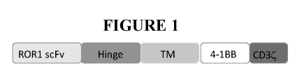
FIGURE 1 depicts a schematic of the general domain structure of CARs with
novel
extracellular ROR1 antigen binding domain sequences. A chimeric antigen
receptor is composed
CA 03081719 2020-05-01
WO 2019/090110 PCT/US2018/059003
of an extracellular ROR1-binding ScFv domain, a spacer or hinge domain
(derived from IgG4 or
CD8), a transmembrane domain, an intracellular signaling CD137 costimulatory
domain, and a
CD3zeta signaling domain.
FIGURES 2A-F depict nucleic acid and amino acid sequences of several chimeric
antigen
receptors (CARs) containing novel human extracellular ROR1 antigen binding
domain sequences.
The general scheme for the CARs includes, from the N terminus to the C
terminus, a Signal
Peptide (SP or LP, leader peptide), an human anti-ROR1 binder single chain
fragment variable
(ScFv), an extracellular linker (or hinge, H), a transmembrane domain (TM), a
4-1BB (CD137)
signaling domain, and a CD3zeta signaling domain.
FIGURE 2A depicts a lentiviral vector expressing the CAR LTG1941 (LP-ScFv4-
CD8H/CD8TM-41BB-CD3zeta) nucleic acid sequence (SEQ ID NO: 3) and the encoded
amino
acid sequence (SEQ ID NO: 4).
FIGURE 2B depicts a lentiviral vector expressing the CAR LTG2528 (LP-ScFv4-
IgG4H/CD8TM-41BB-CD3zeta) nucleic acid sequence (SEQ ID NO: 5) and the encoded
amino
acid sequence (SEQ ID NO: 6).
FIGURE 2C depicts a lentiviral vector expressing the CAR LTG1942 (LP-ScFv9-
CD8H/CD8TM-41BB-CD3zeta) nucleotide sequence (SEQ ID NO: 9) and the encoded
amino
acid sequence (SEQ ID NO: 10).
FIGURE 2D depicts a lentiviral vector expressing the CAR LTG2529 (LP-ScFv9-
IgG4H/CD8TM-41BB-CD3 zeta) nucleic acid sequence (SEQ ID NO 11) and the
encoded amino
acid sequence (SEQ ID NO: 12).
FIGURE 2E depicts a lentiviral vector expressing the CAR LTG 1943 (LP-
ControlScFv-
CD8H/CD8TM-41BB-CD3zeta) nucleic acid sequence (SEQ ID NO: 15) and the encoded
amino
acid sequence (SEQ ID NO: 16).
FIGURE 2F depicts a lentiviral vector expressing the CAR LTG2527: (LP-
ControlScFv-
IgG4H/CD8TM-41BB-CD3zeta) nucleic acid sequence (SEQ ID NO: 17) and the
encoded amino
acid sequence (SEQ ID NO: 18).
FIGURE 3 depicts anti-ROR1 CAR surface expression in primary human T cells.
CAR T
cells redirected to ROR1 tumor antigen via the use of ScFv domains were
generated by lentiviral
transduction with CAR expression constructs. CART detection was performed by
flow cytometry.
T cells were washed twice in cold PBS-EDTA buffer and stained with ROR1-Fc
peptide followed
by fluorescently labeled anti-human-Fc polyclonal F(ab)'2 fragment. Cells were
gated based on
forward scatter and side scatter, singlet discrimination, and 7AAD negativity
so that only viable
cells were analyzed. Data were acquired on MACSQuant 10 flow cytometer in the
APC channel.
16
CA 03081719 2020-05-01
WO 2019/090110 PCT/US2018/059003
Samples analyzed are listed along the left axis: UTD, un-transduced negative
control cells, GFP-
LV control transduction, LTG1941 (ScFv4), LTG1942 (ScFv9), and LTG1943
(control-ScFv).
The vertical dotted line denotes the gate for CAR expression and percent CAR
expression in each
population is listed, CAR%MFI.
FIGURE 4 depicts anti-ROR1 CART cells incorporating ScFv binders (LTG1941,
LTG1942,
and LTG1943) mediating cytolysis of ROR1-positive tumors in vitro. CAR T cells
expressing
anti-ROR1 constructs were incubated with ROR1-positive cell lines (Jeko-Luc
and A431-Luc), or
ROR1-negative line (Reh-luc), each line is stably transduced with firefly
luciferase, at effector to
target ratio (E:T) listed on the x-axis, overnight. CAR T cytotoxic activity
was assessed by
luciferase activity measurement as described in the Materials and Methods. UTD
¨ untransduced
T cell negative control, 1538-LTG1538 FMC63 murine anti-CD19 CAR positive
control.
FIGURE 5 depicts ROR1-specifc CART cell production of high levels of cytokines
when co-
cultured with the ROR1-positive leukemia line (Jeko, gray, or A432, light
gray), or T cells were
incubated with non-expressor (Reh) or incubated alone (gray, last column in
series). The assay
was carried out overnight at E:T ratio of 10:1, then supernatants were
analyzed for cytokine
concentrations by ELISA. N=2 technical replicates +/- SD. Negative controls:
UT-un-transduced
T cells, LTG1941, LTG1942, LTG1943, anti-ROR1-transduced T cells. LTG1398, GFP-
LV
transduced control T cells, as listed on the x-axis.
DETAILED DESCRIPTION
Definitions
As used herein, the singular forms "a," "an," and "the," refer to both the
singular as well as
plural, unless the context clearly indicates otherwise. For example, the term
"an antigen" includes
single or plural antigens and can be considered equivalent to the phrase "at
least one antigen." As
used herein, the term "comprises" means "includes." Thus, "comprising an
antigen" means
"including an antigen" without excluding other elements. The phrase "and/or"
means "and" or
"or." It is further to be understood that any and all base sizes or amino acid
sizes, and all
molecular weight or molecular mass values, given for nucleic acids or
polypeptides are
approximate, and are provided for descriptive purposes, unless otherwise
indicated. Although
many methods and materials similar or equivalent to those described herein can
be used, particular
suitable methods and materials are described below. In case of conflict, the
present specification,
including explanations of terms, will control. In addition, the materials,
methods, and examples
17
CA 03081719 2020-05-01
WO 2019/090110 PCT/US2018/059003
are illustrative only and not intended to be limiting. To facilitate review of
the various
embodiments, the following explanations of terms are provided:
The term "about" when referring to a measurable value such as an amount, a
temporal
duration, and the like, is meant to encompass variations of ±20% or in some
instances ±10%,
or in some instances ±5%, or in some instances ±1%, or in some instances
±0.1% from the
specified value, as such variations are appropriate to perform the disclosed
methods.
Unless otherwise noted, the technical terms herein are used according to
conventional
usage. Definitions of common terms in molecular biology can be found in
Benjamin Lewin,
Genes VII, published by Oxford University Press, 1999; Kendrew et al. (eds.),
The Encyclopedia
of Molecular Biology, published by Blackwell Science Ltd., 1994; and Robert A.
Meyers (ed.),
Molecular Biology and Biotechnology: a Comprehensive Desk Reference, published
by VCH
Publishers, Inc., 1995; and other similar references.
The present disclosure provides for ROR1 antibodies or fragments thereof as
well as
chimeric antigen receptors (CARs) having such ROR1 antigen binding domains.
The
enhancement of the functional activity of the CAR directly relates to the
enhancement of
functional activity of the CAR-expressing T cell. As a result of one or more
of these
modifications, the CARs exhibit both a high degree of cytokine-induced
cytolysis and cell surface
expression on transduced T cells, along with an increased level of in vivo T
cell expansion and
persistence of the transduced CAR-expressing T cell.
The unique ability to combine functional moieties derived from different
protein domains
has been a key innovative feature of Chimeric Antigen Receptors (CARs). The
choice of each of
these protein domains is a key design feature, as is the way in which they are
specifically
combined. Each design domain is an essential component that can be used across
different CAR
platforms to engineer the function of lymphocytes. For example, the choice of
the extracellular
binding domain can make an otherwise ineffective CAR be effective.
The invariable framework components of the immunoglobulin-derived protein
sequences
used to create the extracellular antigen binding domain of a CAR can either be
entirely neutral, or
they can self-associate and drive the T cell to a state of metabolic
exhaustion, thus making the
therapeutic T cell expressing that CAR far less effective. This occurs
independently of the
antigen binding function of this CAR domain Furthermore, the choice of the
intracellular
signaling domain(s) also can govern the activity and the durability of the
therapeutic lymphocyte
population used for immunotherapy. While the ability to bind target antigen
and the ability to
transmit an activation signal to the T cell through these extracellular and
intracellular domains,
respectively, are important CAR design aspects, what has also become apparent
is that the choice
18
CA 03081719 2020-05-01
WO 2019/090110 PCT/US2018/059003
of the source of the extracellular antigen binding fragments can have a
significant effect on the
efficacy of the CAR and thereby have a defining role for the function and
clinical utility of the
CAR.
Surprisingly and unexpectedly it has now been discovered that use of an
entirely human
antigen binding domain in a CAR, rather than using mouse-derived antigen
binding fragments
which are prone to induce anti-mouse immune response and CAR T elimination in
a host (c..1, the
UPenn-sponsored clinical trial using mouse derived SS1 ScFv sequence,
NCT02159716), may
also determine the functional activity of a CAR-expressing T cell.
In light of this discovery, a series of ROR1 binders from a human scFv
expression library
have been developed. These fully-human ROR1 CARs are less likely to induce an
allergic or
rejection response by the patient as they are no longer of murine origin (see
Maus MV, Haas AR,
Beatty GL, Albeda SM, Levine BL, Liu X, Zhao Y, Kalos M, June CH, 2013, Cancer
Immunology Research, 1:26-31). Thus, when these "fully human" CARs are
expressed in T cells
and then infused into patients, they are likely to be more therapeutically
effective. These human
sequence-derived CAR binders may be used for the treatment of human cancer,
leukemias, and
lymphomas that express the ROR1 antigen, including; but not limited to, B-CLL,
ovarian cancer,
triple negative breast cancer, lung adenocarcinoma, and glioblastoma
(Balakrishnan, A., et al.,
2016, Clin Cancer Res, 23:3061-3071; and Baskar, S., et al., 2008, Clin Cancer
Res 14:396-404;
and Jung, E.H., et al., Cell Biochem Funct, 34:149-157).
The CARs disclosed herein are expressed at a high level in a cell. A cell
expressing the
CAR has a high in vivo proliferation rate, produces large amounts of
cytokines, and has a high
cytotoxic activity against a cell having, on its surface, a ROR1 antigen to
which a CAR binds.
The use of a human extracellular ROR1 antigen-binding domain results in
generation of a CAR
that functions better in vivo, while avoiding the induction of anti-CAR
immunity in the host
immune response and the killing of the CAR T cell population. The CARs
expressing the entirely
human extracellular ROR1 ScFv antigen-binding domain exhibit superior
activities/properties
including i) prevention of poor CAR T persistence and function as seen with
mouse-derived
binding sequences; ii) lack of regional (i.e. intrapleural) delivery of the
CAR to be efficacious;
and iii) ability to generate CAR T cell designs based both on binders with
high and low affinity to
ROR1. This latter property allows investigators to better tune efficacy vs
toxicity, and/or tissue
specificity of the CAR T product, since lower-affinity binders may have higher
specificity to
tumors vs normal tissues due to higher expression of ROR1 on tumors than
normal tissue, which
may prevent on-target off tumor toxicity and bystander cell killing.
19
CA 03081719 2020-05-01
WO 2019/090110 PCT/US2018/059003
What follows is a detailed description of the inventive CARs including a
description of
their extracellular ROR1 antigen binding domain, the transmembrane domain and
the intracellular
domain, along with additional description of the CARs, antibodies and antigen
binding fragments
thereof, conjugates, nucleotides, expression, vectors, and host cells, methods
of treatment,
compositions, and kits employing the disclosed CARs.
A. Chimeric Antigen Receptors (CARs)
The CARs disclosed herein comprise at least one ROR1 antigen binding domain
capable
of binding to ROR1, at least one transmembrane domain, and at least one
intracellular domain.
A chimeric antigen receptor (CAR) is an artificially constructed hybrid
protein or
polypeptide containing the antigen binding domains of an antibody (e.g.,
single chain variable
fragment (ScFv)) linked to T-cell signaling domains via the transmembrane
domain.
Characteristics of CARs include their ability to redirect T-cell specificity
and reactivity toward a
selected target in a non-MHC-restricted manner, and exploiting the antigen-
binding properties of
monoclonal antibodies. The non-MHC-restricted antigen recognition gives T
cells expressing
CARs the ability to recognize antigen independent of antigen processing, thus
bypassing a major
mechanism of tumor escape. Moreover, when expressed in T-cells, CARs
advantageously do not
dimerize with endogenous T cell receptor (TCR) alpha and beta chains.
As disclosed herein, the intracellular T cell signaling domains of the CARs
can include,
for example, a T cell receptor signaling domain, a T cell costimulatory
signaling domain, or both.
The T cell receptor signaling domain refers to a portion of the CAR comprising
the intracellular
domain of a T cell receptor, such as, for example, and not by way of
limitation, the intracellular
portion of the CD3 zeta protein. The costimulatory signaling domain refers to
a portion of the
CAR comprising the intracellular domain of a costimulatory molecule, which is
a cell surface
molecule other than an antigen receptor or their ligands that are required for
an efficient response
of lymphocytes to antigen.
1. Extracellular Domain
In one embodiment, the CAR comprises a target-specific binding element
otherwise
referred to as an antigen binding domain or moiety. The choice of domain
depends upon the type
and number of ligands that define the surface of a target cell. For example,
the antigen binding
domain may be chosen to recognize a ligand that acts as a cell surface marker
on target cells
CA 03081719 2020-05-01
WO 2019/090110 PCT/US2018/059003
associated with a particular disease state. Thus examples of cell surface
markers that may act as
ligands for the antigen binding domain in the CAR include those associated
with viral, bacterial
and parasitic infections, autoimmune disease and cancer cells.
In one embodiment, the CAR can be engineered to target a tumor antigen of
interest by
way of engineering a desired antigen binding domain that specifically binds to
an antigen on a
tumor cell. Tumor antigens are proteins that are produced by tumor cells that
elicit an immune
response, particularly T-cell mediated immune responses. The selection of the
antigen binding
domain will depend on the particular type of cancer to be treated. Tumor
antigens include, for
example, a glioma-associated antigen, carcinoembryonic antigen (CEA), .beta.-
human chorionic
gonadotropin, alphafetoprotein (AFP), lectin-reactive AFP, thyroglobulin, RAGE-
1, MN-CA IX,
human telomerase reverse transcriptase, RU1, RU2 (AS), intestinal carboxyl
esterase, mut hsp70-
2, M-CSF, prostase, prostate-specific antigen (PSA), PAP, NY-ESO-1, LAGE-la,
p53, prostein,
PSMA, Her2/neu, survivin and telomerase, prostate-carcinoma tumor antigen-1
(PCTA-1),
MAGE, ELF2M, neutrophil elastase, ephrinB2, CD20, CD22, ROR1, insulin growth
factor (IGF)-
I, IGF-II, IGF-I receptor and CD19. The tumor antigens disclosed herein are
merely included by
way of example. The list is not intended to be exclusive and further examples
will be readily
apparent to those of skill in the art.
In one embodiment, the tumor antigen comprises one or more antigenic cancer
epitopes
associated with a malignant tumor. Malignant tumors express a number of
proteins that can serve
as target antigens for an immune attack. These molecules include, but are not
limited to, tissue-
specific antigens such as MART-1, tyrosinase and GP 100 in melanoma and
prostatic acid
phosphatase (PAP) and prostate-specific antigen (PSA) in prostate cancer.
Other target molecules
belong to the group of transformation-related molecules such as the oncogene
HER-2/Neu/ErbB-
2. Yet another group of target antigens are onco-fetal antigens such as
carcinoembryonic antigen
(CEA). In B-cell lymphoma the tumor-specific idiotype immunoglobulin
constitutes a truly
tumor-specific immunoglobulin antigen that is unique to the individual tumor.
B-cell
differentiation antigens such as CD19, CD20, CD22, BCMA, ROR1, and CD37 are
other
candidates for target antigens in B-cell lymphoma. Some of these antigens
(CEA, HER-2, CD19,
CD20, idiotype) have been used as targets for passive immunotherapy with
monoclonal antibodies
with limited success.
In one preferred embodiment, the tumor antigen is ROR1 and the tumors
associated with
expression of ROR1 comprise lung mesothelioma, ovarian, and pancreatic cancers
that express
high levels of the extracellular protein ROR1, or any combination thereof
21
CA 03081719 2020-05-01
WO 2019/090110 PCT/US2018/059003
The type of tumor antigen may also be a tumor-specific antigen (TSA) or a
tumor-
associated antigen (TAA). A TSA is unique to tumor cells and does not occur on
other cells in the
body. A TAA is not unique to a tumor cell and instead is also expressed on a
normal cell under
conditions that fail to induce a state of immunologic tolerance to the
antigen. The expression of
the antigen on the tumor may occur under conditions that enable the immune
system to respond to
the antigen. TAAs may be antigens that are expressed on normal cells during
fetal development
when the immune system is immature and unable to respond or they may be
antigens that are
normally present at extremely low levels on normal cells but which are
expressed at much higher
levels on tumor cells.
Non-limiting examples of TSAs or TAAs include the following: Differentiation
antigens
such as MART-1/MelanA (MART-I), gp100 (Pmel 17), tyrosinase, TRP-1, TRP-2 and
tumor-
specific multi-lineage antigens such as MAGE-1, MAGE-3, BAGE, GAGE-1, GAGE-2,
p15;
overexpressed embryonic antigens such as CEA; overexpressed oncogenes and
mutated tumor-
suppressor genes such as p53, Ras, HER-2/neu; unique tumor antigens resulting
from
chromosomal translocations; such as BCR-ABL, E2A-PRL, H4-RET, IGH-IGK, MYL-
RAR; and
viral antigens, such as the Epstein Barr virus antigens EBVA and the human
papillomavirus
(HPV) antigens E6 and E7. Other large, protein-based antigens include TSP-180,
MAGE-4,
MAGE-5, MAGE-6, RAGE, NY-ESO, p185erbB2, p180erbB-3, c-met, nm-23H1, PSA, TAG-
72,
CA 19-9, CA 72-4, CAM 17.1, NuMa, K-ras, beta-Catenin, CDK4, Mum-1, p 15, p
16, 43-9F,
5T4, 791Tgp72, alpha-fetoprotein, beta-HCG, BCA225, BTAA, CA 125, CA 15-3\CA
27.29\BCAA, CA 195, CA 242, CA-50, CAM43, CD68\Pl, CO-029, FGF-5, G250,
Ga733\EpCAM, HTgp-175, M344, MA-50, MG7-Ag, MOV18, NB/70K, NY-CO-1, RCAS1,
SDCCAG16, TA-90\Mac-2 binding protein\cyclophilin C-associated protein, TAAL6,
TAG72,
TLP, and TPS.
In one embodiment, the antigen binding domain portion of the CAR targets an
antigen that
includes but is not limited to CD19, CD20, CD22, ROR1, CD33, CD38, CD123,
CD138, BCMA,
c-Met, PSMA, Glycolipid F77, EGFRvIII, GD-2, FGFR4, TSLPR, NY-ESO-1 TCR, MAGE
A3
TCR, and the like.
In a preferred embodiment, the antigen binding domain portion of the CAR
targets the
extracellular ROR1 antigen.
In one preferred embodiment, the isolated nucleic acid molecule encoding the
extracellular
ROR1 binding domain scFv4 comprises a nucleotide sequence of SEQ ID NO: 1, or
a sequence
with 85%, 90%, 95%, 96%, 97%, 98% or 99% identity thereof In one embodiment,
an isolated
nucleic acid molecule is provided wherein the encoded extracellular ROR1
antigen binding
22
CA 03081719 2020-05-01
WO 2019/090110 PCT/US2018/059003
domain scFv4 comprises an amino acid sequence of SEQ ID NO: 2, or an amino
acid sequence
with 85%, 90%, 95%, 96%, 97%, 98% or 99% identity to an amino acid sequence of
SEQ ID NO:
2.
In one preferred embodiment, the isolated nucleic acid molecule encoding the
extracellular
ROR1 antigen binding domain ScV9 comprises a nucleotide sequence of SEQ ID NO:
7, or a
sequence with 85%, 90%, 95%, 96%, 97%, 98% or 99% identity thereof In one
embodiment, an
isolated nucleic acid molecule is provided wherein the encoded extracellular
ROR1 antigen
binding domain ScFv9 comprises an amino acid sequence of SEQ ID NO: 8, or an
amino acid
sequence with 85%, 90%, 95%, 96%, 97%, 98% or 99% identity to an amino acid
sequence of
SEQ ID NO: 8.
In one preferred embodiment, the isolated nucleic acid molecule encoding the
extracellular
ROR1 control ScFv antigen binding domain comprises a nucleotide sequence of
SEQ ID NO: 13,
or a sequence with 85%, 90%, 95%, 96%, 97%, 98% or 99% identity thereof In one
embodiment, an isolated nucleic acid molecule is provided wherein the encoded
extracellular
ROR1 control ScFv antigen binding domain comprises an amino acid sequence of
SEQ ID NO:
14, or an amino acid sequence with 85%, 90%, 95%, 96%, 97%, 98% or 99%
identity to an amino
acid sequence of SEQ ID NO: 14.
In the various embodiments of the ROR1-specific CARs disclosed herein, the
general
scheme is set forth in FIGURE 1 and includes, from the N-terminus to the C-
terminus, a signal or
leader peptide, anti-ROR1 ScFv, extracellular linker or hinge (H) domain,
transmembrane (TM)
domain, 4-1BB, CD3 zeta, wherein the bolded text represents the cloning sites
for linking
domains.
In one embodiment, the nucleic acid sequence encoding a CAR comprises the
nucleic acid
sequence of SEQ ID NO: 3, and encodes the CAR comprising the amino acid
sequence as set
forth in SEQ ID NO: 4 [LTG1941 LP-ScFv4-CD8H/CD8TM-41BB-CD3zeta amino acid
sequence (as depicted in Figure 2A)].
In one embodiment, the nucleic acid sequence encoding a CAR comprises the
nucleic acid
sequence of SEQ ID NO: 3, or a sequence with 85%, 90%, 95%, 96%, 97%, 98% or
99% identity
thereof, and encodes the CAR comprising the amino acid sequence as set forth
in SEQ ID NO: 4
or a sequence with 85%, 90%, 95%, 96%, 97%, 98% or 99% identity thereof
LTG1941 LP-
ScFv4-CD8H/CD8TM-41BB-CD3zeta amino acid sequence (as depicted in Figure 2A)].
In another embodiment, the nucleic acid sequence encoding a CAR comprises the
nucleic
acid sequence of SEQ ID NO: 5, and encodes the CAR comprising the amino acid
sequence as
23
CA 03081719 2020-05-01
WO 2019/090110 PCT/US2018/059003
set forth in SEQ ID NO: 6 [LTG2528 LP-ScFv4-IgG4H/CD8TM-41BB-CD3zeta amino
acid
sequence (as depicted in Figure 2B)].
In another embodiment, the nucleic acid sequence encoding a CAR comprises the
nucleic
acid sequence of SEQ ID NO: 5 or a sequence with 85%, 90%, 95%, 96%, 97%, 98%
or 99%
identity thereof, and encodes the CAR comprising the amino acid sequence as
set forth in SEQ ID
NO: 6 or a sequence with 85%, 90%, 95%, 96%, 97%, 98% or 99% identity thereof
[LTG2528
LP-ScFv4-CD8H/CD8TM-41BB-CD3zeta amino acid sequence (as depicted in Figure
2B)].
In another embodiment, the nucleic acid sequence encoding a CAR comprises the
nucleic
acid sequence of SEQ ID NO: 9, and encodes the CAR comprising the amino acid
sequence as set
forth in SEQ ID NO: 10 LTG1942 LP-ScFv9-CD8H/CD8TM-41BB-CD3zeta CAR amino acid
sequence (as depicted in Figure 2C)].
In another embodiment, the nucleic acid sequence encoding a CAR comprises the
nucleic
acid sequence of SEQ ID NO: 9 or a sequence with 85%, 90%, 95%, 96%, 97%, 98%
or 99%
identity thereof, and encodes the CAR comprising the amino acid sequence as
set forth in SEQ ID
NO: 10 or a sequence with 85%, 90%, 95%, 96%, 97%, 98% or 99% identity thereof
[LTG1942
LP-ScFv9-CD8H/CD8TM-41BB-CD3zeta CAR amino acid sequence (as depicted in
Figure 2C)].
In yet another embodiment, the nucleic acid sequence encoding a CAR comprises
the
nucleic acid sequence of SEQ ID NO: 11, and encodes the CAR comprising the
amino acid
sequence as set forth in SEQ ID NO: 12 [LTG2529 LP-ScFv9-IgG4H/CD8TM-41BB-
CD3zeta
amino acid sequence (as depicted in Figure 2D)].
In yet another embodiment, the nucleic acid sequence encoding a CAR comprises
the
nucleic acid sequence of SEQ ID NO: 11 or a sequence with 85%, 90%, 95%, 96%,
97%, 98% or
99% identity thereof, and encodes the CAR comprising the amino acid sequence
as set forth in
SEQ ID NO: 12 or a sequence with 85%, 90%, 95%, 96%, 97%, 98% or 99% identity
thereof
[LTG2529 LP-ScFv9-IgG4H/CD8TM-41BB-CD3zeta amino acid sequence (as depicted in
Figure
2D)].
In yet another embodiment, the nucleic acid sequence encoding a CAR comprises
the
nucleic acid sequence of SEQ ID NO: 15, and encodes the CAR comprising the
amino acid
sequence as set forth in SEQ ID NO: 16 [LTG1943 LP-controlScFv-CD8H/CD8TM-41BB-
CD3zeta amino acid sequence (as depicted in Figure 2E)].
In yet another embodiment, the nucleic acid sequence encoding a CAR comprises
the
nucleic acid sequence of SEQ ID NO: 15 or a sequence with 85%, 90%, 95%, 96%,
97%, 98% or
99% identity thereof, and encodes the CAR comprising the amino acid sequence
as set forth in
SEQ ID NO: 16 or a sequence with 85%, 90%, 95%, 96%, 97%, 98% or 99% identity
thereof
24
CA 03081719 2020-05-01
WO 2019/090110 PCT/US2018/059003
[LTG1943 LP-controlScFv-CD8H/CD8TM-41BB-CD3zeta amino acid sequence (as
depicted in
Figure 2E)].
In yet another embodiment, the nucleic acid sequence encoding a CAR comprises
the
nucleic acid sequence of SEQ ID NO: 17, and encodes the CAR comprising the
amino acid
sequence as set forth in SEQ ID NO: 18 [(LTG2527 LP-controlScFv-IgG4H/CD8TM-
41BB-
CD3zeta amino acid sequence (as depicted in Figure 2F)].
In yet another embodiment, the nucleic acid sequence encoding a CAR comprises
the
nucleic acid sequence of SEQ ID NO: 17 or a sequence with 85%, 90%, 95%, 96%,
97%, 98% or
99% identity thereof, and encodes the CAR comprising the amino acid sequence
as set forth in
SEQ ID NO: 18 or a sequence with 85%, 90%, 95%, 96%, 97%, 98% or 99% identity
thereof
[LTG2527 LP-controlScFv-IgG4H/CD8TM-41BB-CD3zeta amino acid sequence (as
depicted in
Figure 2F)].
The surface expression of anti-ROR1 CARs incorporating single chain fragment
variable
(ScFv) sequences reactive with ROR1 antigen, is shown in Example 2 infra and
summarized in
Table 2. The expression level for each ScFv¨containing CAR was determined by
flow cytometric
analysis of LV-transduced T cells from healthy donors using a recombinant ROR1-
Fc peptide,
followed by anti-human Fc F(ab')2 fragment conjugated to AF647, and detected
in the APC
channel, (cl, Figure 3). The ScFv-based anti-ROR1 CAR constructs LTG1941,
LTG1942-
LTG1943 were highly expressed in human primary T cells (as indicated by the
gated population)
as compared to non-transduced T cell controls (non-gated cell population).
Representative results
from one donor are shown.
As shown in Example 2 and Figure 4, high cytolytic activity of the ROR1 CARs
was
demonstrated when lentiviral vectors (LV) expressing the following CARs were
created and
tested for anti-leukemia activity. Each experimental CAR contains the 4-
1BB/CD3-zeta chain
signaling motif and the specific anti-ROR1 binding motif/domain noted therein.
Leukemia target
lines with ROR1 surface expression were used: Jeko and A431; and ROR1 negative
Reh. ScFv-
based anti-ROR1 CAR constructs LTG1941, LTG1942, and LTG1943 were able to
efficiently
lyse A431, whereas they had no specific lytic activity against Reh, (cf,
Figure 4). The ability to
lyse Jeko by anti-ROR1 CARs LTG1941 and LTG1942 differed, indicating
differential biological
activity. These results demonstrate the efficiency and specificity of the
generated CAR
constructs.
The capacity of anti-ROR1 CAR T cells for cytokine secretion was then
evaluated. Tumor
cells were co-incubated with CAR T cells or control T cells at effector to
target ratio of 10:1
overnight, and culture supernatants were analyzed by ELISA for IFN gamma, TNF
alpha and IL-2
CA 03081719 2020-05-01
WO 2019/090110 PCT/US2018/059003
(c.f, , Figure 5). Of note, CAR T-expressing cells LTG1942 and LTG1943
generated high levels
of IFN gamma, and LTG1941 generated moderate amounts of IFN-gamma only in
response to
Jeko, but not the A431 leukemia cell line. A similar result was seen for IL-2
and TNF-alpha
expression. Negative controls (untransduced T cells, UN, or T cells transduced
with the control
LTG1398 LV) yielded no appreciable cytokine induction. Importantly, the
results seen in
cytolytic and cytokine function highlight that LTG1942 performs similarly to
the control ScFv-
bearing CAR, LTG1943, whereas LTG1941 has much lower cytokine production and
ability to
lyse the Jeko leukemia cell line. Nevertheless, the ability to lyse A431 by
LTG1941 indicates that
we have a differentiated product that may be preferred if LTG1942 proves to
active or too toxic in
clinical studies of CAR-T.
Without being intended to limit to any particular mechanism of action, it is
believed that
possible reasons for the enhanced therapeutic function associated with the
exemplary CARs of the
invention include, for example, and not by way of limitation, a) improved
lateral movement
within the plasma membrane allowing for more efficient signal transduction, b)
superior location
within plasma membrane microdomains, such as lipid rafts, and greater ability
to interact with
transmembrane signaling cascades associated with T cell activation, c)
superior location within
the plasma membrane by preferential movement away from dampening or down-
modulatory
interactions, such as less proximity to or interaction with phosphatases such
as CD45, and d)
superior assembly into T cell receptor signaling complexes (i.e. the immune
synapse), or any
combination thereof
While the disclosure has been illustrated with an exemplary extracellular ROR1
ScFv
antigen binding domains, other nucleotide and/or amino acid variants within
the ROR1 variable
ScFv antigen binding domains may be used to derive heavy-chain only binding
domains, or
subsets thereof, and thus comprise the ROR1 antigen binding domains for use in
the CARs
described herein.
Depending on the desired antigen to be targeted, the CAR can be additionally
engineered
to include the appropriate antigen binding domain that is specific to the
desired antigen target.
For example, if ROR1 is the desired antigen that is to be targeted, an
antibody for ROR1 can be
used as the antigen bind domain incorporation into the CAR.
In one exemplary embodiment, the antigen binding domain portion of the CAR
additionally targets CD33. Preferably, the antigen binding domain in the CAR
is anti-CD33 ScFv,
wherein the nucleic acid sequence of the anti-CD33 ScFv comprises the sequence
set forth in SEQ
ID NO: 34. In one embodiment, the anti-CD33 ScFv comprises the nucleic acid
sequence that
26
CA 03081719 2020-05-01
WO 2019/090110 PCT/US2018/059003
encodes the amino acid sequence of SEQ ID NO: 35. In another embodiment, the
anti-CD33
ScFv portion of the CAR comprises the amino acid sequence set forth in SEQ ID
NO: 35.
In one exemplary embodiment, the antigen binding domain portion of the CAR
additionally targets mesothelin. Preferably, the antigen binding domain in the
CAR is anti-
mesothelin ScFv, wherein the nucleic acid sequence of the anti-mesothelin ScFv
comprises the
sequence set forth in SEQ ID NO: 36. In one embodiment, the anti-mesothelin
ScFv comprises the
nucleic acid sequence that encodes the amino acid sequence of SEQ ID NO: 37.
In another
embodiment, the anti-mesothelin ScFv portion of the CAR comprises the amino
acid sequence set
forth in SEQ ID NO: 37.
In one exemplary embodiment, the antigen binding domain portion of the CAR
additionally targets CD19. Preferably, the antigen binding domain in the CAR
is anti-CD19 ScFv,
wherein the nucleic acid sequence of the anti-mesothelin ScFv comprises the
sequence set forth in
SEQ ID NO: 32. In one embodiment, the anti-mesothelin ScFv comprises the
nucleic acid
sequence that encodes the amino acid sequence of SEQ ID NO: 33. In another
embodiment, the
anti-CD19 ScFv portion of the CAR comprises the amino acid sequence set forth
in SEQ ID NO:
33.
In one aspect of the present invention, there is provided a CAR capable of
binding to a
non-TSA or non-TAA including, for example and not by way of limitation, an
antigen derived
from Retroviridae (e.g. human immunodeficiency viruses such as HIV-1 and HIV-
LP),
Picornaviridae (e.g. poliovirus, hepatitis A virus, enterovirus, human
coxsackievirus, rhinovirus,
and echovirus), rubella virus, coronavirus, vesicular stomatitis virus, rabies
virus, ebola virus,
parainfluenza virus, mumps virus, measles virus, respiratory syncytial virus,
influenza virus,
hepatitis B virus, parvovirus, Adenoviridae, Herpesviridae [e.g. type 1 and
type 2 herpes simplex
virus (HSV), varicella-zoster virus, cytomegalovirus (CMV), and herpes virus],
Poxviridae (e.g.
smallpox virus, vaccinia virus, and pox virus), or hepatitis C virus, or any
combination thereof
In another aspect of the present invention, there is provided a CAR capable of
binding to
an antigen derived from a bacterial strain of Staphylococci, Streptococcus,
Escherichia coli,
Pseudomonas, or Salmonella. Particularly, there is provided a CAR capable of
binding to an
antigen derived from an infectious bacterium, for example, Helicobacter
pyloris, Legionella
pneumophilia, a bacterial strain of Mycobacteria sps. (e.g. M. tuberculosis,
M. avium, M.
intracellulare, M. kansaii, or M. gordonea), Staphylococcus aureus, Neisseria
gonorrhoeae,
Neisseria meningitides, Listeria monocytogenes, Streptococcus pyogenes, Group
A
Streptococcus, Group B Streptococcus (Streptococcus agalactiae), Streptococcus
pneumoniae, or
Clostridium tetani, or a combination thereof
27
CA 03081719 2020-05-01
WO 2019/090110 PCT/US2018/059003
2. Transmembrane Domain
With respect to the transmembrane domain, the CAR comprises one or more
transmembrane domains fused to the extracellular ROR1 antigen binding domain
of the CAR.
The transmembrane domain may be derived either from a natural or from a
synthetic
source. Where the source is natural, the domain may be derived from any
membrane-bound or
transmembrane protein.
Transmembrane regions of particular use in the CARs described herein may be
derived
from (i.e. comprise at least the transmembrane region(s) of) the alpha, beta
or zeta chain of the T-
cell receptor, CD28, CD3 epsilon, CD45, CD4, CD5, CD8, CD9, CD16, CD22,
mesothelin,
CD33, CD37, CD64, CD80, CD83, CD86, CD134, CD137, CD154, TNFRSF16, or
TNFRSF19.
Alternatively the transmembrane domain may be synthetic, in which case it will
comprise
predominantly hydrophobic residues such as leucine and valine. Preferably a
triplet of
phenylalanine, tryptophan and valine will be found at each end of a synthetic
transmembrane
domain. Optionally, a short oligo- or polypeptide linker, preferably between 2
and 10 amino acids
in length may form the linkage between the transmembrane domain and the
cytoplasmic signaling
domain of the CAR. A glycine-serine doublet provides a particularly suitable
linker.
In one embodiment, the transmembrane domain that naturally is associated with
one of the
domains in the CAR is used in addition to the transmembrane domains described
supra.
In some instances, the transmembrane domain can be selected or by amino acid
substitution to avoid binding of such domains to the transmembrane domains of
the same or
different surface membrane proteins to minimize interactions with other
members of the receptor
complex.
In one embodiment, the transmembrane domain in the CAR of the invention is the
CD8
transmembrane domain. In one embodiment, the CD8 transmembrane domain
comprises the
nucleic acid sequence of SEQ ID NO: 21. In one embodiment, the CD8
transmembrane domain
comprises the nucleic acid sequence that encodes the amino acid sequence of
SEQ ID NO: 22. In
another embodiment, the CD8 transmembrane domain comprises the amino acid
sequence of SEQ
ID NO: 22.
In one embodiment, the encoded transmembrane domain comprises an amino acid
sequence having at least one, two or three modifications (e.g., substitutions)
but not more than 20,
or 5 modifications (e.g., substitutions) of an amino acid sequence of SEQ ID
NO: 22, or a
sequence with 95-99% identity to an amino acid sequence of SEQ ID NO: 22.
28
CA 03081719 2020-05-01
WO 2019/090110 PCT/US2018/059003
In some instances, the transmembrane domain of the CAR comprises the
CD8.alpha.hinge
domain. In one embodiment, the CD8 hinge domain comprises the nucleic acid
sequence of SEQ
ID NO: 23. In one embodiment, the CD8 hinge domain comprises the nucleic acid
sequence that
encodes the amino acid sequence of SEQ ID NO: 24. In another embodiment, the
CD8 hinge
domain comprises the amino acid sequence of SEQ ID NO: 24, or a sequence with
95-99%
identify thereof
In one embodiment, an isolated nucleic acid molecule is provided wherein the
encoded
linker domain is derived from the extracellular domain of CD8, and is linked
to the
transmembrane CD8 domain, the transmembrane CD28 domain, or a combination
thereof
3. Spacer (Hinge, H) Domain
In the CAR, a spacer domain can be arranged between the extracellular domain
and the
transmembrane domain, or between the intracellular domain and the
transmembrane domain. The
spacer domain means any oligopeptide or polypeptide that serves to link the
transmembrane
domain with the extracellular domain and/or the transmembrane domain with the
intracellular
domain The spacer domain comprises up to 300 amino acids, preferably 10 to 100
amino acids,
and most preferably 25 to 50 amino acids.
In several embodiments, the linker can include a spacer element, which, when
present,
increases the size of the linker such that the distance between the effector
molecule or the
detectable marker and the antibody or antigen binding fragment is increased.
Exemplary spacers
are known to the person of ordinary skill, and include those listed in U.S.
Pat. Nos. 7,964,566,
7,498,298, 6,884,869, 6,323,315, 6,239,104, 6,034,065, 5,780,588, 5,665,860,
5,663,149,
5,635,483, 5,599,902, 5,554,725, 5,530,097, 5,521,284, 5,504,191, 5,410,024,
5,138,036,
5,076,973, 4,986,988, 4,978,744, 4,879,278, 4,816,444, and 4,486,414, as well
as U.S. Pat. Pub.
Nos. 20110212088 and 20110070248, each of which is incorporated by reference
herein in its
entirety.
The spacer domain preferably has a sequence that promotes binding of a CAR
with an
antigen and enhances signaling into a cell. Examples of an amino acid that is
expected to promote
the binding include cysteine, a charged amino acid, and serine and threonine
in a potential
glycosylation site, and these amino acids can be used as an amino acid
constituting the spacer
domain
As the spacer domain, the entire or a part of amino acid numbers 137-206 (SEQ
ID NO:
25) which is a hinge region of CD8.alpha. (NCBI RefS eq: NP--001759.3),
amino acid
29
CA 03081719 2020-05-01
WO 2019/090110 PCT/US2018/059003
numbers 135 to 195 of CD8.beta. (GenBank: AAA35664.1), amino acid numbers 315
to 396 of
CD4 (NCBI RefSeq: NP--000607.1), or amino acid numbers 137 to 152 of CD28
(NCBI
RefSeq: NP--006130.1) can be used. Also, as the spacer domain, a part of
a constant region
of an antibody H chain or L chain can be used. Further, the spacer domain may
be an artificially
synthesized sequence.
A spacer domain can also be comprised of elements of Immunoglobulin (Ig)
constant
domains, such as those derived for IgG4, including sequences that link
immunoglobulin domains
that comprise an immunoglobulin protein. The spacer or hinge domain exists at
the C-terminus of
the scFv ROR1-binding domain and extends to the CAR transmembrane domain. In
one
embodiment, the IgG4 hinge (H) domain comprises the nucleic acid sequence of
SEQ ID NO: 38.
In one embodiment, the CD8 hinge domain comprises the nucleic acid sequence
that encodes the
amino acid sequence of SEQ ID NO: 39. In another embodiment, the CD8 hinge
domain
comprises the amino acid sequence of SEQ ID NO: 39, or a sequence with 95-99%
identify
thereof
In some instances, the IgG4 constant regions serve as a hinge (H) and are
linked to the
transmembrane domain of CD8. In one embodiment, the IgG4H domain is linked to
the CD8
transmembrane domain and together comprises the nucleic acid sequence of SEQ
ID NO: 40. In
one embodiment, the IgG4H domain linked to the CD8 transmembrane domain
comprises the
nucleic acid sequence that encodes the amino acid sequence of SEQ ID NO: 41.
In another
embodiment, the IgG4H linked to the CD8 transmembrane domain comprises the
amino acid
sequence of SEQ ID NO: 41, or a sequence with 95-99% identify thereof
Further, in the CAR, a signal peptide sequence can be linked to the N-
terminus. The
signal peptide sequence exists at the N-terminus of many secretory proteins
and membrane
proteins, and has a length of 15 to 30 amino acids. Since many of the protein
molecules
mentioned above as the intracellular domain have signal peptide sequences, the
signal peptides
can be used as a signal peptide for the CAR. In one embodiment, the signal
peptide comprises the
amino acid sequence shown in SEQ ID NO: 20.
4. Intracellular Domain
The cytoplasmic domain or otherwise the intracellular signaling domain of the
CAR is
responsible for activation of at least one of the normal effector functions of
the immune cell in
which the CAR has been placed in. The term "effector function" refers to a
specialized function
of a cell. Effector function of a T cell, for example, may be cytolytic
activity or helper activity
CA 03081719 2020-05-01
WO 2019/090110 PCT/US2018/059003
including the secretion of cytokines. Thus the term "intracellular signaling
domain" refers to the
portion of a protein which transduces the effector function signal and directs
the cell to perform a
specialized function. While usually the entire intracellular signaling domain
can be employed, in
many cases it is not necessary to use the entire chain. To the extent that a
truncated portion of the
intracellular signaling domain is used, such truncated portion may be used in
place of the intact
chain as long as it transduces the effector function signal. The term
intracellular signaling domain
is thus meant to include any truncated portion of the intracellular signaling
domain sufficient to
transduce the effector function signal.
Preferred examples of intracellular signaling domains for use in the CAR
include the
cytoplasmic sequences of the T cell receptor (TCR) and co-receptors that act
in concert to initiate
signal transduction following antigen receptor engagement, as well as any
derivative or variant of
these sequences and any synthetic sequence that has the same functional
capability.
It is known that signals generated through the TCR alone are insufficient for
full activation
of the T cell and that a secondary or co-stimulatory signal is also required.
Thus, T cell activation
can be said to be mediated by two distinct classes of cytoplasmic signaling
sequence: those that
initiate antigen-dependent primary activation through the TCR (primary
cytoplasmic signaling
sequences) and those that act in an antigen-independent manner to provide a
secondary or co-
stimulatory signal (secondary cytoplasmic signaling sequences).
Primary cytoplasmic signaling sequences regulate primary activation of the TCR
complex
either in a stimulatory way, or in an inhibitory way. Primary cytoplasmic
signaling sequences that
act in a stimulatory manner may contain signaling motifs which are known as
immunoreceptor
tyrosine-based activation motifs or ITAMs.
Examples of ITAM containing primary cytoplasmic signaling sequences that are
of
particular use in the CARS disclosed herein include those derived from TCR
zeta (CD3 Zeta),
FcR gamma, FcR beta, CD3 gamma, CD3 delta, CD3 epsilon, CD5, CD22, CD79a,
CD79b, and
CD66d. Specific, non-limiting examples, of the ITAM include peptides having
sequences of
amino acid numbers 51 to 164 of CD3.zeta. (NCBI RefSeq: NP--932170.1),
amino acid
numbers 45 to 86 of Fc.epsilon.RI.gamma. (NCBI RefSeq: NP--004097.1),
amino acid
numbers 201 to 244 of Fc.epsilon.RI.beta. (NCBI RefSeq: NP--000130.1),
amino acid
numbers 139 to 182 of CD3.gamma. (NCBI RefSeq: NP--000064.1), amino acid
numbers
128 to 171 of CD3 .delta. (NCBI RefSeq: NP--000723.1), amino acid numbers
153 to 207
of CD3.epsilon. (NCBI RefSeq: NP--000724.1), amino acid numbers 402 to
495 of CD5
(NCBI RefSeq: NP--055022.2), amino acid numbers 707 to 847 of 0022 (NCBI
RefSeq:
NP--001762.2), amino acid numbers 166 to 226 of CD79a (NCBI RefSeq:
NP--
31
CA 03081719 2020-05-01
WO 2019/090110 PCT/US2018/059003
001774.1), amino acid numbers 182 to 229 of CD79b (NCBI RefSeq: NP--
000617.1), and
amino acid numbers 177 to 252 of CD66d (NCBI RefSeq: NP--001806.2), and
their variants
having the same function as these peptides have. The amino acid number based
on amino acid
sequence information of NCBI RefSeq ID or GenBank described herein is numbered
based on the
full length of the precursor (comprising a signal peptide sequence etc.) of
each protein. In one
embodiment, the cytoplasmic signaling molecule in the CAR comprises a
cytoplasmic signaling
sequence derived from CD3 zeta.
In a preferred embodiment, the intracellular domain of the CAR can be designed
to
comprise the CD3-zeta signaling domain by itself or combined with any other
desired cytoplasmic
domain(s) useful in the context of the CAR. For example, the intracellular
domain of the CAR
can comprise a CD3 zeta chain portion and a costimulatory signaling region.
The costimulatory
signaling region refers to a portion of the CAR comprising the intracellular
domain of a
costimulatory molecule. A costimulatory molecule is a cell surface molecule
other than an
antigen receptor or their ligands that is required for an efficient response
of lymphocytes to an
antigen. Examples of such costimulatory molecules include CD27, CD28, 4-1BB
(CD137),
0X40, CD30, CD40, PD-1, ICOS, lymphocyte function-associated antigen-1 (LFA-
1), CD2,
CD7, LIGHT, NKG2C, B7-H3, and a ligand that specifically binds with CD83, and
the like.
Specific, non-limiting examples, of such costimulatory molecules include
peptides having
sequences of amino acid numbers 236 to 351 of CD2 (NCBI RefSeq: NP--
001758.2), amino
acid numbers 421 to 458 of CD4 (NCBI RefSeq: NP--000607.1), amino acid
numbers 402 to
495 of CD5 (NCBI RefSeq: NP--055022.2), amino acid numbers 207 to 235 of
CD8.alpha.
(NCBI RefSeq: NP--001759.3), amino acid numbers 196 to 210 of CD83
(GenBank:
AAA35664.1), amino acid numbers 181 to 220 of CD28 (NCBI RefSeq: NP--
006130.1),
amino acid numbers 214 to 255 of CD137 (4-1BB, NCBI RefSeq: NP--
001552.2), amino
acid numbers 241 to 277 of CD134 (0X40, NCBI RefSeq: NP--003318.1), and
amino acid
numbers 166 to 199 of ICOS (NCBI RefSeq: NP--036224.1), and their
variants having the
same function as these peptides have. Thus, while the disclosure herein is
exemplified primarily
with 4-i BB as the co-stimulatory signaling element, other costimulatory
elements are within the
scope of the disclosure.
The cytoplasmic signaling sequences within the cytoplasmic signaling portion
of the CAR
may be linked to each other in a random or specified order. Optionally, a
short oligo- or
polypeptide linker, preferably between 2 and 10 amino acids in length may form
the linkage. A
glycine-serine doublet provides a particularly suitable linker.
32
CA 03081719 2020-05-01
WO 2019/090110 PCT/US2018/059003
In one embodiment, the intracellular domain is designed to comprise the
signaling domain
of CD3-zeta and the signaling domain of CD28. In another embodiment, the
intracellular domain
is designed to comprise the signaling domain of CD3-zeta and the signaling
domain of 4-1BB. In
yet another embodiment, the intracellular domain is designed to comprise the
signaling domain of
CD3-zeta and the signaling domain of CD28 and 4-1BB.
In one embodiment, the intracellular domain in the CAR is designed to comprise
the
signaling domain of 4-1BB and the signaling domain of CD3-zeta, wherein the
signaling domain
of 4-1BB comprises the nucleic acid sequence set forth in SEQ ID NO: 26 and
the signaling
domain of CD3-zeta comprises the nucleic acid sequence set forth in SEQ ID NO:
28 and in the
variant nucleic acid sequence set forth in SEQ ID NO: 30. In one embodiment,
the intracellular
domain in the CAR is designed to comprise the signaling domain of 4-1BB and
the signaling
domain of CD3-zeta, wherein the signaling domain of 4-1BB comprises the
nucleic acid sequence
set forth in SEQ ID NO: 26 and the signaling domain of CD3-zeta comprises the
nucleic acid
sequence set forth in SEQ ID NO: 28 and in the variant nucleic acid sequence
set forth in SEQ ID
NO: 30.
In one embodiment, the intracellular domain in the CAR is designed to comprise
the
signaling domain of 4-1BB and the signaling domain of CD3-zeta, wherein the
signaling domain
of 4-1BB comprises the nucleic acid sequence that encodes the amino acid
sequence of SEQ ID
NO: 27 and the signaling domain of CD3-zeta comprises the nucleic acid
sequence that encodes
the amino acid sequence of SEQ ID NO: 29 and in the variant nucleic acid that
encodes the amino
acid sequence of SEQ ID NO: 31.
In one embodiment, the intracellular domain in the CAR is designed to comprise
the
signaling domain of 4-1BB and the signaling domain of CD3-zeta, wherein the
signaling domain
of 4-1BB comprises the amino acid sequence set forth in SEQ ID NO: 27 and the
signaling
domain of CD3-zeta comprises the amino acid sequence set forth in SEQ ID NO:
29 and the
variant amino acid sequence set forth in SEQ ID NO: 31.
5. Additional Description of CARs
Also expressly included within the scope of the invention are functional
portions of the
CARs disclosed herein. The term "functional portion" when used in reference to
a CAR refers to
any part or fragment of one or more of the CARs disclosed herein, which part
or fragment retains
the biological activity of the CAR of which it is a part (the parent CAR).
Functional portions
encompass, for example, those parts of a CAR that retain the ability to
recognize target cells, or
33
CA 03081719 2020-05-01
WO 2019/090110 PCT/US2018/059003
detect, treat, or prevent a disease, to a similar extent, the same extent, or
to a higher extent, as the
parent CAR. In reference to the parent CAR, the functional portion can
comprise, for instance,
about 10%, 25%, 30%, 50%, 68%, 80%, 90%, 95%, or more, of the parent CAR.
The functional portion can comprise additional amino acids at the amino or
carboxy
terminus of the portion, or at both termini, which additional amino acids are
not found in the
amino acid sequence of the parent CAR. Desirably, the additional amino acids
do not interfere
with the biological function of the functional portion, e.g., recognize target
cells, detect cancer,
treat or prevent cancer, etc. More desirably, the additional amino acids
enhance the biological
activity, as compared to the biological activity of the parent CAR.
Included in the scope of the disclosure are functional variants of the CARs
disclosed
herein. The term "functional variant" as used herein refers to a CAR,
polypeptide, or protein
having substantial or significant sequence identity or similarity to a parent
CAR, which functional
variant retains the biological activity of the CAR of which it is a variant.
Functional variants
encompass, for example, those variants of the CAR described herein (the parent
CAR) that retain
the ability to recognize target cells to a similar extent, the same extent, or
to a higher extent, as the
parent CAR. In reference to the parent CAR, the functional variant can, for
instance, be at least
about 30%, 50%, 75%, 80%, 90%, 98% or more identical in amino acid sequence to
the parent
CAR.
A functional variant can, for example, comprise the amino acid sequence of the
parent
CAR with at least one conservative amino acid substitution. Alternatively, or
additionally, the
functional variants can comprise the amino acid sequence of the parent CAR
with at least one
non-conservative amino acid substitution. In this case, it is preferable for
the non-conservative
amino acid substitution to not interfere with or inhibit the biological
activity of the functional
variant. The non-conservative amino acid substitution may enhance the
biological activity of the
functional variant, such that the biological activity of the functional
variant is increased as
compared to the parent CAR.
Amino acid substitutions of the CARs are preferably conservative amino acid
substitutions. Conservative amino acid substitutions are known in the art, and
include amino acid
substitutions in which one amino acid having certain physical and/or chemical
properties is
exchanged for another amino acid that has the same or similar chemical or
physical properties.
For instance, the conservative amino acid substitution can be an
acidic/negatively charged polar
amino acid substituted for another acidic/negatively charged polar amino acid
(e.g., Asp or Glu),
an amino acid with a nonpolar side chain substituted for another amino acid
with a nonpolar side
chain (e.g., Ala, Gly, Val, He, Leu, Met, Phe, Pro, Trp, Cys, Val, etc.), a
basic/positively charged
34
CA 03081719 2020-05-01
WO 2019/090110 PCT/US2018/059003
polar amino acid substituted for another basic/positively charged polar amino
acid (e.g. Lys, His,
Arg, etc.), an uncharged amino acid with a polar side chain substituted for
another uncharged
amino acid with a polar side chain (e.g., Asn, Gin, Ser, Thr, Tyr, etc.), an
amino acid with a beta-
branched side-chain substituted for another amino acid with a beta-branched
side-chain (e.g., He,
Thr, and Val), an amino acid with an aromatic side-chain substituted for
another amino acid with
an aromatic side chain (e.g., His, Phe, Trp, and Tyr), etc.
The CAR can consist essentially of the specified amino acid sequence or
sequences
described herein, such that other components, e.g., other amino acids, do not
materially change
the biological activity of the functional variant.
The CARs (including functional portions and functional variants) can be of any
length,
i.e., can comprise any number of amino acids, provided that the CARs (or
functional portions or
functional variants thereof) retain their biological activity, e.g., the
ability to specifically bind to
antigen, detect diseased cells in a mammal, or treat or prevent disease in a
mammal, etc. For
example, the CAR can be about 50 to about 5000 amino acids long, such as 50,
70, 75, 100, 125,
150, 175, 200, 300, 400, 500, 600, 700, 800, 900, 1000 or more amino acids in
length.
The CARs (including functional portions and functional variants of the
invention) can
comprise synthetic amino acids in place of one or more naturally-occurring
amino acids. Such
synthetic amino acids are known in the art, and include, for example,
aminocyclohexane
carboxylic acid, norleucine, -amino n-decanoic acid, homoserine, S-
acetylaminomethyl-cysteine,
trans-3- and trans-4-hydroxyproline, 4-aminophenylalanine, 4-
nitrophenylalanine, 4-
chlorophenylalanine, 4-carboxyphenylalanine, 0-phenylserine 0-
hydroxyphenylalanine,
phenylglycine, a-naphthylalanine, cyclohexylalanine, cyclohexylglycine,
indoline-2-carboxylic
acid, 1,2,3,4-tetrahydroisoquinoline-3-carboxylic acid, aminomalonic acid,
aminomalonic acid
monoamide, N'-benzyl-N'-methyl-lysine, N',N'-dibenzyl-lysine, 6-hydroxylysine,
ornithine, -
aminocyclopentane carboxylic acid, a-aminocyclohexane carboxylic acid, a-
aminocycloheptane
carboxylic acid, a-(2-amino-2-norbornane)-carboxylic acid, y-diaminobutyric
acid, (3-
diaminopropionic acid, homophenylalanine, and a-tert-butylglycine.
The CARs (including functional portions and functional variants) can be
glycosylated,
amidated, carboxylated, phosphorylated, esterified, N-acylated, cyclized via,
e.g., a disulfide
bridge, or converted into an acid addition salt and/or optionally dimerized or
polymerized, or
conjugated.
The CARs (including functional portions and functional variants thereof) can
be obtained
by methods known in the art. The CARs may be made by any suitable method of
making
polypeptides or proteins. Suitable methods of de novo synthesizing
polypeptides and proteins are
CA 03081719 2020-05-01
WO 2019/090110 PCT/US2018/059003
described in references, such as Chan et al., Fmoc Solid Phase Peptide
Synthesis, Oxford
University Press, Oxford, United Kingdom, 2000; Peptide and Protein Drug
Analysis, ed. Reid,
R., Marcel Dekker, Inc., 2000; Epitope Mapping, ed. Westwood et al., Oxford
University Press,
Oxford, United Kingdom, 2001; and U.S. Patent 5,449,752. Also, polypeptides
and proteins can
be recombinantly produced using the nucleic acids described herein using
standard recombinant
methods. See, for instance, Sambrook et al., Molecular Cloning: A Laboratory
Manual, 3rd ed.,
Cold Spring Harbor Press, Cold Spring Harbor, NY 2001; and Ausubel et al.,
Current Protocols in
Molecular Biology, Greene Publishing Associates and John Wiley & Sons, NY,
1994. Further,
some of the CARs (including functional portions and functional variants
thereof) can be isolated
and/or purified from a source, such as a plant, a bacterium, an insect, a
mammal, e.g., a rat, a
human, etc. Methods of isolation and purification are well-known in the art.
Alternatively, the
CARs described herein (including functional portions and functional variants
thereof) can be
commercially synthesized by companies. In this respect, the CARs can be
synthetic, recombinant,
isolated, and/or purified.
B. Antibodies and Antigen Binding Fragments
One embodiment further provides a CAR, a T cell expressing a CAR, an antibody,
or
antigen binding domain or portion thereof, which specifically binds to one or
more of the antigens
disclosed herein. As used herein, a "T cell expressing a CAR," or a "CAR T
cell" means a T cell
expressing a CAR, and has antigen specificity determined by, for example, the
antibody-derived
targeting domain of the CAR.
As used herein, and "antigen binding domain" can include an antibody and
antigen
binding fragments thereof The term "antibody" is used herein in the broadest
sense and
encompasses various antibody structures, including but not limited to
monoclonal antibodies,
polyclonal antibodies, multi-specific antibodies (e.g., bispecific
antibodies), and antigen binding
fragments thereof, so long as they exhibit the desired antigen-binding
activity. Non-limiting
examples of antibodies include, for example, intact immunoglobulins and
variants and fragments
thereof known in the art that retain binding affinity for the antigen.
A "monoclonal antibody" is an antibody obtained from a population of
substantially
homogeneous antibodies, i.e., the individual antibodies comprising the
population are identical
except for possible naturally occurring mutations that may be present in minor
amounts.
Monoclonal antibodies are highly specific, being directed against a single
antigenic epitope. The
modifier "monoclonal" indicates the character of the antibody as being
obtained from a
36
CA 03081719 2020-05-01
WO 2019/090110 PCT/US2018/059003
substantially homogeneous population of antibodies, and is not to be construed
as requiring
production of the antibody by any particular method. In some examples, a
monoclonal antibody is
an antibody produced by a single clone of B lymphocytes or by a cell into
which nucleic acid
encoding the light and heavy variable regions of the antibody of a single
antibody (or an antigen
binding fragment thereof) have been transfected, or a progeny thereof In some
examples
monoclonal antibodies are isolated from a subject. Monoclonal antibodies can
have conservative
amino acid substitutions which have substantially no effect on antigen binding
or other
immunoglobulin functions. Exemplary methods of production of monoclonal
antibodies are
known, for example, see Harlow & Lane, Antibodies, A Laboratory Manual, 2nd
ed. Cold Spring
Harbor Publications, New York (2013).
Typically, an immunoglobulin has heavy (H) chains and light (L) chains
interconnected by
disulfide bonds. Immunoglobulin genes include the kappa, lambda, alpha, gamma,
delta, epsilon
and mu constant region genes, as well as the myriad immunoglobulin variable
domain genes.
There are two types of light chain, lambda (2\,) and kappa (lc). There are
five main heavy chain
classes (or isotypes) which determine the functional activity of an antibody
molecule: IgM, IgD,
IgG, IgA and IgE.
Each heavy and light chain contains a constant region (or constant domain) and
a variable
region (or variable domain; see, e.g., Kindt et al. Kuby Immunology, 6th ed.,
W.H. Freeman and
Co., page 91 (2007).) In several embodiments, the heavy and the light chain
variable regions
combine to specifically bind the antigen. In additional embodiments, only the
heavy chain
variable region is required. For example, naturally occurring camelid
antibodies consisting of a
heavy chain only are functional and stable in the absence of light chain (see,
e.g., Hamers-
Casterman et al., Nature, 363:446-448, 1993; Sheriff et al., Nat. Struct.
Biol., 3:733-736, 1996).
References to "VH" or "VH" refer to the variable region of an antibody heavy
chain, including
that of an antigen binding fragment, such as Fv, ScFv, dsFy or Fab. References
to "VL" or "VL"
refer to the variable domain of an antibody light chain, including that of an
Fv, ScFv, dsFy or Fab.
Light and heavy chain variable regions contain a "framework" region
interrupted by three
hypervariable regions, also called "complementarity-determining regions" or
"CDRs" (see, e.g.,
Kabat et al., Sequences of Proteins of Immunological Interest, U.S. Department
of Health and
Human Services, 1991). The sequences of the framework regions of different
light or heavy
chains are relatively conserved within a species. The framework region of an
antibody, that is the
combined framework regions of the constituent light and heavy chains, serves
to position and
align the CDRs in three-dimensional space.
37
CA 03081719 2020-05-01
WO 2019/090110 PCT/US2018/059003
The CDRs are primarily responsible for binding to an epitope of an antigen.
The amino
acid sequence boundaries of a given CDR can be readily determined using any of
a number of
well-known schemes, including those described by Kabat et al. ("Sequences of
Proteins of
Immunological Interest," 5th Ed. Public Health Service, National Institutes of
Health, Bethesda,
MD, 1991; "Kabat" numbering scheme), Al-Lazikani et al., (JMB 273,927-948,
1997; "Chothia"
numbering scheme), and Lefranc et al. ("IMGT unique numbering for
immunoglobulin and T cell
receptor variable domains and Ig superfamily V-like domains," Dev. Comp.
Immunol., 27:55-77,
2003; "IMGT" numbering scheme). The CDRs of each chain are typically referred
to as CDR1,
CDR2, and CDR3 (from the N-terminus to C-terminus), and are also typically
identified by the
chain in which the particular CDR is located. Thus, a VH CDR3 is the CDR3 from
the variable
domain of the heavy chain of the antibody in which it is found, whereas a VL
CDR1 is the CDR1
from the variable domain of the light chain of the antibody in which it is
found. Light chain
CDRs are sometimes referred to as LCDR1, LCDR2, and LCDR3. Heavy chain CDRs
are
sometimes referred to as LCDR1, LCDR2, and LCDR3.
An "antigen binding fragment" is a portion of a full length antibody that
retains the ability
to specifically recognize the cognate antigen, as well as various combinations
of such portions.
Non-limiting examples of antigen binding fragments include Fv, Fab, Fab', Fab'-
SH, F(ab')2;
diabodies; linear antibodies; single-chain antibody molecules (e.g. ScFv); and
multi-specific
antibodies formed from antibody fragments. Antibody fragments include antigen
binding
fragments either produced by the modification of whole antibodies or those
synthesized de novo
using recombinant DNA methodologies (see, e.g., Kontermann and Dubel (Ed),
Antibody
Engineering, Vols. 1-2, 2nd Ed., Springer Press, 2010).
A single-chain antibody (ScFv) is a genetically engineered molecule containing
the VH
and VL domains of one or more antibody(ies) linked by a suitable polypeptide
linker as a
genetically fused single chain molecule (see, for example, Bird et al.,
Science, 242:423 426, 1988;
Huston et al., Proc. Natl. Acad. Sci., 85:5879 5883, 1988; Ahmad et al., Clin.
Dev. Immunol.,
2012, doi:10.1155/2012/980250; Marbry, IDrugs, 13:543-549, 2010). The
intramolecular
orientation of the VH-domain and the VL-domain in a ScFv, is typically not
decisive for ScFvs.
Thus, ScFvs with both possible arrangements (VH-domain-linker domain-VL-
domain; VL-
domain-linker domain-VH-domain) may be used.
In a dsFy, the heavy and light chain variable chains have been mutated to
introduce a
disulfide bond to stabilize the association of the chains. Diabodies also are
included, which are
bivalent, bispecific antibodies in which VH and VL domains are expressed on a
single
polypeptide chain, but using a linker that is too short to allow for pairing
between the two
38
CA 03081719 2020-05-01
WO 2019/090110 PCT/US2018/059003
domains on the same chain, thereby forcing the domains to pair with
complementary domains of
another chain and creating two antigen binding sites (see, for example,
Holtiger et al., Proc. Natl.
Acad. Sci., 90:6444 6448, 1993; Poljak et al., Structure, 2:1121 1123, 1994).
Antibodies also include genetically engineered forms such as chimeric
antibodies (such as
humanized murine antibodies) and heteroconjugate antibodies (such as
bispecific antibodies). See
also, Pierce Catalog and Handbook, 1994-1995 (Pierce Chemical Co., Rockford,
IL); Kuby, J.,
Immunology, 3rd Ed., W.H. Freeman & Co., New York, 1997.
Non-naturally occurring antibodies can be constructed using solid phase
peptide synthesis,
can be produced recombinantly, or can be obtained, for example, by screening
combinatorial
libraries consisting of variable heavy chains and variable light chains as
described by Huse et al.,
Science 246:1275-1281 (1989), which is incorporated herein by reference. These
and other
methods of making, for example, chimeric, humanized, CDR-grafted, single
chain, and
bifunctional antibodies, are well known to those skilled in the art (Winter
and Harris, Immunol.
Today 14:243-246 (1993); Ward et al., Nature 341:544-546 (1989); Harlow and
Lane, supra,
1988; Hilyard et al., Protein Engineering: A practical approach (IRL Press
1992); Borrabeck,
Antibody Engineering, 2d ed. (Oxford University Press 1995); each of which is
incorporated
herein by reference).
An "antibody that binds to the same epitope" as a reference antibody refers to
an antibody
that blocks binding of the reference antibody to its antigen in a competition
assay by 50% or
more, and conversely, the reference antibody blocks binding of the antibody to
its antigen in a
competition assay by 50% or more. Antibody competition assays are known, and
an exemplary
competition assay is provided herein.
A "humanized" antibody or antigen binding fragment includes a human framework
region
and one or more CDRs from a non-human (such as a mouse, rat, or synthetic)
antibody or antigen
binding fragment. The non-human antibody or antigen binding fragment providing
the CDRs is
termed a "donor," and the human antibody or antigen binding fragment providing
the framework
is termed an "acceptor." In one embodiment, all the CDRs are from the donor
immunoglobulin in
a humanized immunoglobulin. Constant regions need not be present, but if they
are, they can be
substantially identical to human immunoglobulin constant regions, such as at
least about 85-90%,
such as about 95% or more identical. Hence, all parts of a humanized antibody
or antigen binding
fragment, except possibly the CDRs, are substantially identical to
corresponding parts of natural
human antibody sequences.
A "chimeric antibody" is an antibody which includes sequences derived from two
different
antibodies, which typically are of different species. In some examples, a
chimeric antibody
39
CA 03081719 2020-05-01
WO 2019/090110 PCT/US2018/059003
includes one or more CDRs and/or framework regions from one human antibody and
CDRs
and/or framework regions from another human antibody.
A "fully human antibody" or "human antibody" is an antibody which includes
sequences
from (or derived from) the human genome, and does not include sequence from
another species.
In some embodiments, a human antibody includes CDRs, framework regions, and
(if present) an
Fc region from (or derived from) the human genome. Human antibodies can be
identified and
isolated using technologies for creating antibodies based on sequences derived
from the human
genome, for example by phage display or using transgenic animals (see, e.g.,
Barbas et al. Phage
display: A Laboratory Manuel. 1st Ed. New York: Cold Spring Harbor Laboratory
Press, 2004.
Print.; Lonberg, Nat. Biotech., 23: 1117-1125, 2005; Lonenberg, Curr. Opin.
Immunol., 20:450-
459, 2008).
An antibody may have one or more binding sites. If there is more than one
binding site,
the binding sites may be identical to one another or may be different. For
instance, a naturally-
occurring immunoglobulin has two identical binding sites, a single-chain
antibody or Fab
fragment has one binding site, while a bispecific or bifunctional antibody has
two different
binding sites.
Methods of testing antibodies for the ability to bind to any functional
portion of the CAR
are known in the art and include any antibody-antigen binding assay, such as,
for example,
radioimmunoassay (RIA), ELISA, Western blot, immunoprecipitation, and
competitive inhibition
assays (see, e.g., Janeway et al., infra, U.S. Patent Application Publication
No. 2002/0197266 Al,
and U.S. Patent No. 7,338,929).
Also, a CAR, a T cell expressing a CAR, an antibody, or antigen binding
portion thereof,
can be modified to comprise a detectable label, such as, for instance, a
radioisotope, a fluorophore
(e.g., fluorescein isothiocyanate (FITC), phycoerythrin (PE)), an enzyme
(e.g., alkaline
phosphatase, horseradish peroxidase), and element particles (e.g., gold
particles).
C. Conjugates
A CAR, a T cell expressing a CAR, or monoclonal antibodies, or antigen binding
fragments thereof, specific for one or more of the antigens disclosed herein,
can be conjugated to
an agent, such as an effector molecule or detectable marker, using any number
of means known to
those of skill in the art. Both covalent and noncovalent attachment means may
be used.
Conjugates include, but are not limited to, molecules in which there is a
covalent linkage of an
effector molecule or a detectable marker to an antibody or antigen binding
fragment that
CA 03081719 2020-05-01
WO 2019/090110 PCT/US2018/059003
specifically binds one or more of the antigens disclosed herein. One of skill
in the art will
appreciate that various effector molecules and detectable markers can be used,
including (but not
limited to) chemotherapeutic agents, anti-angiogenic agents, toxins,
radioactive agents such as
1251, 32p, 14,,,
3H and 35S and other labels, target moieties and ligands, etc.
The choice of a particular effector molecule or detectable marker depends on
the particular
target molecule or cell, and the desired biological effect. Thus, for example,
the effector molecule
can be a cytotoxin that is used to bring about the death of a particular
target cell (such as a tumor
cell).
The procedure for attaching an effector molecule or detectable marker to an
antibody or
antigen binding fragment varies according to the chemical structure of the
effector. Polypeptides
typically contain a variety of functional groups; such as carboxylic acid
(COOH), free amine (-
NH2) or sulfhydryl (-SH) groups, which are available for reaction with a
suitable functional group
on an antibody to result in the binding of the effector molecule or detectable
marker.
Alternatively, the antibody or antigen binding fragment is derivatized to
expose or attach
additional reactive functional groups. The derivatization may involve
attachment of any of a
number of known linker molecules such as those available from Pierce Chemical
Company,
Rockford, IL. The linker can be any molecule used to join the antibody or
antigen binding
fragment to the effector molecule or detectable marker. The linker is capable
of forming covalent
bonds to both the antibody or antigen binding fragment and to the effector
molecule or detectable
marker. Suitable linkers are well known to those of skill in the art and
include, but are not limited
to, straight or branched-chain carbon linkers, heterocyclic carbon linkers, or
peptide linkers.
Where the antibody or antigen binding fragment and the effector molecule or
detectable marker
are polypeptides, the linkers may be joined to the constituent amino acids
through their side
groups (such as through a disulfide linkage to cysteine) or to the alpha
carbon amino and carboxyl
groups of the terminal amino acids.
In several embodiments, the linker can include a spacer element, which, when
present,
increases the size of the linker such that the distance between the effector
molecule or the
detectable marker and the antibody or antigen binding fragment is increased.
Exemplary spacers
are known to the person of ordinary skill, and include those listed in U.S.
Pat. Nos. 7,964,566,
7,498,298, 6,884,869, 6,323,315, 6,239,104, 6,034,065, 5,780,588, 5,665,860,
5,663,149,
5,635,483, 5,599,902, 5,554,725, 5,530,097, 5,521,284, 5,504,191, 5,410,024,
5,138,036,
5,076,973, 4,986,988, 4,978,744, 4,879,278, 4,816,444, and 4,486,414, as well
as U.S. Pat. Pub.
Nos. 20110212088 and 20110070248, each of which is incorporated by reference
herein in its
entirety.
41
CA 03081719 2020-05-01
WO 2019/090110 PCT/US2018/059003
In some embodiments, the linker is cleavable under intracellular conditions,
such that
cleavage of the linker releases the effector molecule or detectable marker
from the antibody or
antigen binding fragment in the intracellular environment. In yet other
embodiments, the linker is
not cleavable and the effector molecule or detectable marker is released, for
example, by antibody
degradation. In some embodiments, the linker is cleavable by a cleaving agent
that is present in
the intracellular environment (for example, within a lysosome or endosome or
caveolea). The
linker can be, for example, a peptide linker that is cleaved by an
intracellular peptidase or protease
enzyme, including, but not limited to, a lysosomal or endosomal protease. In
some embodiments,
the peptide linker is at least two amino acids long or at least three amino
acids long. However, the
linker can be 4, 5, 6, 7, 8, 9, 10, 11, 12, 13, 14 or 15 amino acids long,
such as 1-2, 1-3, 2-5, 3-10,
3-15, 1-5, 1-10, 1-15 amino acids long. Proteases can include cathepsins B and
D and plasmin, all
of which are known to hydrolyze dipeptide drug derivatives resulting in the
release of active drug
inside target cells (see, for example, Dubowchik and Walker, 1999, Pharm.
Therapeutics 83:67-
123). For example, a peptide linker that is cleavable by the thiol-dependent
protease cathepsin-B,
can be used (for example, a Phenylalanine -Leucine or a Glycine- Phenylalanine
-Leucine-Glycine
linker). Other examples of such linkers are described, for example, in U.S.
Pat. No. 6,214,345,
incorporated herein by reference. In a specific embodiment, the peptide linker
cleavable by an
intracellular protease is a Valine-Citruline linker or a Phenylalanine-Lysine
linker (see, for
example, U.S. Pat. No. 6,214,345, which describes the synthesis of doxorubicin
with the Valine-
Citruline linker).
In other embodiments, the cleavable linker is pH-sensitive, i.e., sensitive to
hydrolysis at
certain pH values. Typically, the pH-sensitive linker is hydrolyzable under
acidic conditions. For
example, an acid-labile linker that is hydrolyzable in the lysosome (for
example, a hydrazone,
semicarbazone, thiosemicarbazone, cis-aconitic amide, orthoester, acetal,
ketal, or the like) can be
used. (See, for example, U.S. Pat. Nos. 5,122,368; 5,824,805; 5,622,929;
Dubowchik and Walker,
1999, Pharm. Therapeutics 83:67-123; Neville et al., 1989, Biol. Chem.
264:14653-14661.) Such
linkers are relatively stable under neutral pH conditions, such as those in
the blood, but are
unstable at below pH 5.5 or 5.0, the approximate pH of the lysosome. In
certain embodiments,
the hydrolyzable linker is a thioether linker (such as, for example, a
thioether attached to the
therapeutic agent via an acylhydrazone bond (see, for example, U.S. Pat. No.
5,622,929).
In other embodiments, the linker is cleavable under reducing conditions (for
example, a
disulfide linker). A variety of disulfide linkers are known in the art,
including, for example, those
that can be formed using SATA (N-succinimidyl-S-acetylthioacetate), SPDP (N-
succinimidy1-3-
(2-pyridyldithio)propionate), SPDB (N-succinimidy1-3-(2-
pyridyldithio)butyrate) and SMPT (N-
42
CA 03081719 2020-05-01
WO 2019/090110 PCT/US2018/059003
succinimidyl-oxycarbonyl-alpha-methyl-alpha-(2-pyridyl-dithio)toluene)-, SPDB
and SMPT.
(See, for example, Thorpe et al., 1987, Cancer Res. 47:5924-5931; Wawrzynczak
et al., In
Immunoconjugates: Antibody Conjugates in Radioimagery and Therapy of Cancer
(C. W. Vogel
ed., Oxford U. Press, 1987); Phillips et al., Cancer Res. 68:92809290, 2008).
See also U.S. Pat.
No. 4,880,935.)
In yet other specific embodiments, the linker is a malonate linker (Johnson et
al., 1995,
Anticancer Res. 15:1387-93), a maleimidobenzoyl linker (Lau et al., 1995,
Bioorg-Med-Chem.
3(10):1299-1304), or a 3'-N-amide analog (Lau et al., 1995, Bioorg-Med-Chem.
3(10):1305-12).
In yet other embodiments, the linker is not cleavable and the effector
molecule or
detectable marker is released by antibody degradation. (See U.S. Publication
No. 2005/0238649
incorporated by reference herein in its entirety).
In several embodiments, the linker is resistant to cleavage in an
extracellular environment.
For example, no more than about 20%, no more than about 15%, no more than
about 10%, no
more than about 5%, no more than about 3%, or no more than about 1% of the
linkers, in a sample
of conjugate, are cleaved when the conjugate is present in an extracellular
environment (for
example, in plasma). Whether or not a linker is resistant to cleavage in an
extracellular
environment can be determined, for example, by incubating the conjugate
containing the linker of
interest with plasma for a predetermined time period (for example, 2, 4, 8,
16, or 24 hours) and
then quantitating the amount of free effector molecule or detectable marker
present in the plasma.
A variety of exemplary linkers that can be used in conjugates are described in
WO 2004-010957,
U.S. Publication No. 2006/0074008, U.S. Publication No. 20050238649, and U.S.
Publication No.
2006/0024317, each of which is incorporated by reference herein in its
entirety.
In several embodiments, conjugates of a CAR, a T cell expressing a CAR, an
antibody, or
antigen binding portion thereof, and one or more small molecule toxins, such
as a calicheamicin,
maytansinoids, dolastatins, auristatins, a trichothecene, and CC1065, and the
derivatives of these
toxins that have toxin activity, are provided.
Maytansine compounds suitable for use as maytansinoid toxin moieties are well
known in
the art, and can be isolated from natural sources according to known methods,
produced using
genetic engineering techniques (see Yu et al (2002) PNAS 99:7968-7973), or
maytansinol and
maytansinol analogues prepared synthetically according to known methods.
Maytansinoids are
mitototic inhibitors which act by inhibiting tubulin polymerization.
Maytansine was first isolated
from the east African shrub Maytenus serrata (U.S. Pat. No. 3,896,111).
Subsequently, it was
discovered that certain microbes also produce maytansinoids, such as
maytansinol and C-3
maytansinol esters (U.S. Pat. No. 4,151,042). Synthetic maytansinol and
derivatives and
43
CA 03081719 2020-05-01
WO 2019/090110 PCT/US2018/059003
analogues thereof are disclosed, for example, in U.S. Pat. Nos. 4,137,230;
4,248,870; 4,256,746;
4,260,608; 4,265,814; 4,294,757; 4,307,016; 4,308,268; 4,308,269; 4,309,428;
4,313,946;
4,315,929; 4,317,821; 4,322,348; 4,331,598; 4,361,650; 4,364,866; 4,424,219;
4,450,254;
4,362,663; and 4,371,533, each of which is incorporated herein by reference.
Conjugates
containing maytansinoids, methods of making same, and their therapeutic use
are disclosed, for
example, in U.S. Pat. Nos. 5,208,020; 5,416,064; 6,441,163 and European Patent
EP 0 425 235
Bl, the disclosures of which are hereby expressly incorporated by reference.
Additional toxins can be employed with a CAR, a T cell expressing a CAR, an
antibody,
or antigen binding portion thereof Exemplary toxins include Pseudomonas
exotoxin (PE), ricin,
abrin, diphtheria toxin and subunits thereof, ribotoxin, ribonuclease,
saporin, and calicheamicin,
as well as botulinum toxins A through F. These toxins are well known in the
art and many are
readily available from commercial sources (for example, Sigma Chemical
Company, St. Louis,
MO). Contemplated toxins also include variants of the toxins (see, for
example, see, U.S. Patent
Nos. 5,079,163 and 4,689,401).
Saporin is a toxin derived from Saponaria officinalis that disrupts protein
synthesis by
inactivating the 60S portion of the ribosomal complex (Stirpe et al.,
Bio/Technology, 10:405-412,
1992). However, the toxin has no mechanism for specific entry into cells, and
therefore requires
conjugation to an antibody or antigen binding fragment that recognizes a cell-
surface protein that
is internalized in order to be efficiently taken up by cells.
Diphtheria toxin is isolated from Corynebacterium diphtheriae. Typically,
diphtheria toxin
for use in immunotoxins is mutated to reduce or to eliminate non-specific
toxicity. A mutant
known as CRM107, which has full enzymatic activity but markedly reduced non-
specific toxicity,
has been known since the 1970's (Laird and Groman, J. Virol. 19:220, 1976),
and has been used
in human clinical trials. See, U.S. Patent No. 5,792,458 and U.S. Patent No.
5,208,021.
Ricin is the lectin RCA60 from Ricinus communis (Castor bean). For examples of
ricin,
see, U.S. Patent No. 5,079,163 and U.S. Patent No. 4,689,401. Ricinus communis
agglutinin
(RCA) occurs in two forms designated RCA60 and RCA120 according to their
molecular weights of
approximately 65 and 120 kD, respectively (Nicholson & Blaustein, J. Biochim.
Biophys. Acta
266:543, 1972). The A chain is responsible for inactivating protein synthesis
and killing cells.
The B chain binds ricin to cell-surface galactose residues and facilitates
transport of the A chain
into the cytosol (Olsnes et al., Nature 249:627-631, 1974 and U.S. Patent No.
3,060,165).
Ribonucleases have also been conjugated to targeting molecules for use as
immunotoxins
(see Suzuki et al., Nat. Biotech. 17:265-70, 1999). Exemplary ribotoxins such
as a-sarcin and
restrictocin are discussed in, for example Rathore et al., Gene 190:31-5,
1997; and Goyal and
44
CA 03081719 2020-05-01
WO 2019/090110 PCT/US2018/059003
Batra, Biochem. 345 Pt 2:247-54, 2000. Calicheamicins were first isolated from
Micromonospora
echinospora and are members of the enediyne antitumor antibiotic family that
cause double strand
breaks in DNA that lead to apoptosis (see, for example Lee et al., J.
Antibiot. 42:1070-87,1989).
The drug is the toxic moiety of an immunotoxin in clinical trials (see, for
example, Gillespie et al.,
Ann. Oncol. 11:735-41, 2000).
Abrin includes toxic lectins from Abrus precatorius. The toxic principles,
abrin a, b, c,
and d, have a molecular weight of from about 63 and 67 kD and are composed of
two disulfide-
linked polypeptide chains A and B. The A chain inhibits protein synthesis; the
B chain (abrin-b)
binds to D-galactose residues (see, Funatsu et al., Agr. Biol. Chem. 52:1095,
1988; and Olsnes,
Methods Enzymol. 50:330-335, 1978).
A CAR, a T cell expressing a CAR, monoclonal antibodies, antigen binding
fragments
thereof, specific for one or more of the antigens disclosed herein, can also
be conjugated with a
detectable marker; for example, a detectable marker capable of detection by
ELISA,
spectrophotometry, flow cytometry, microscopy or diagnostic imaging techniques
(such as
computed tomography (CT), computed axial tomography (CAT) scans, magnetic
resonance
imaging (MRI), nuclear magnetic resonance imaging NMRI), magnetic resonance
tomography
(MTR), ultrasound, fiberoptic examination, and laparoscopic examination).
Specific, non-limiting
examples of detectable markers include fluorophores, chemiluminescent agents,
enzymatic
linkages, radioactive isotopes and heavy metals or compounds (for example
super paramagnetic
iron oxide nanocrystals for detection by MRI). For example, useful detectable
markers include
fluorescent compounds, including fluorescein, fluorescein isothiocyanate,
rhodamine, 5-
dimethylamine-1 -napthalenesulfonyl chloride, phycoerythrin, lanthanide
phosphors and the like.
Bioluminescent markers are also of use, such as luciferase, Green fluorescent
protein (GFP),
Yellow fluorescent protein (YFP). A CAR, a T cell expressing a CAR, an
antibody, or antigen
binding portion thereof, can also be conjugated with enzymes that are useful
for detection, such as
horseradish peroxidase, 0-galactosidase, luciferase, alkaline phosphatase,
glucose oxidase and the
like. When a CAR, a T cell expressing a CAR, an antibody, or antigen binding
portion thereof, is
conjugated with a detectable enzyme, it can be detected by adding additional
reagents that the
enzyme uses to produce a reaction product that can be discerned. For example,
when the agent
horseradish peroxidase is present the addition of hydrogen peroxide and
diaminobenzidine leads
to a colored reaction product, which is visually detectable. A CAR, a T cell
expressing a CAR, an
antibody, or antigen binding portion thereof, may also be conjugated with
biotin, and detected
through indirect measurement of avidin or streptavidin binding. It should be
noted that the avidin
itself can be conjugated with an enzyme or a fluorescent label.
CA 03081719 2020-05-01
WO 2019/090110 PCT/US2018/059003
A CAR, a T cell expressing a CAR, an antibody, or antigen binding portion
thereof, may
be conjugated with a paramagnetic agent, such as gadolinium. Paramagnetic
agents such as
superparamagnetic iron oxide are also of use as labels. Antibodies can also be
conjugated with
lanthanides (such as europium and dysprosium), and manganese. An antibody or
antigen binding
fragment may also be labeled with a predetermined polypeptide epitopes
recognized by a
secondary reporter (such as leucine zipper pair sequences, binding sites for
secondary antibodies,
metal binding domains, epitope tags).
A CAR, a T cell expressing a CAR, an antibody, or antigen binding portion
thereof, can
also be conjugated with a radiolabeled amino acid. The radiolabel may be used
for both
diagnostic and therapeutic purposes. For instance, the radiolabel may be used
to detect one or
more of the antigens disclosed herein and antigen expressing cells by x-ray,
emission spectra, or
other diagnostic techniques. Further, the radiolabel may be used
therapeutically as a toxin for
treatment of tumors in a subject, for example for treatment of a
neuroblastoma. Examples of
labels for polypeptides include, but are not limited to, the following
radioisotopes or
radionucleotides: 3H, 14C, 15N, 35s, 90y, 99Tc, "In, 1251, 1311.
Means of detecting such detectable markers are well known to those of skill in
the art.
Thus, for example, radiolabels may be detected using photographic film or
scintillation counters,
fluorescent markers may be detected using a photodetector to detect emitted
illumination.
Enzymatic labels are typically detected by providing the enzyme with a
substrate and detecting
the reaction product produced by the action of the enzyme on the substrate,
and colorimetric
labels are detected by simply visualizing the colored label.
D. Nucleotides, Expression, Vectors, and Host Cells
Further provided by an embodiment of the invention is a nucleic acid
comprising a
nucleotide sequence encoding any of the CARs, an antibody, or antigen binding
portion thereof,
described herein (including functional portions and functional variants
thereof). The nucleic acids
of the invention may comprise a nucleotide sequence encoding any of the leader
sequences,
antigen binding domains, transmembrane domains, and/or intracellular T cell
signaling domains
described herein.
In some embodiments, the nucleotide sequence may be codon-modified. Without
being
bound to a particular theory, it is believed that codon optimization of the
nucleotide sequence
increases the translation efficiency of the mRNA transcripts. Codon
optimization of the
nucleotide sequence may involve substituting a native codon for another codon
that encodes the
46
CA 03081719 2020-05-01
WO 2019/090110 PCT/US2018/059003
same amino acid, but can be translated by tRNA that is more readily available
within a cell, thus
increasing translation efficiency. Optimization of the nucleotide sequence may
also reduce
secondary mRNA structures that would interfere with translation, thus
increasing translation
efficiency.
In an embodiment of the invention, the nucleic acid may comprise a codon-
modified
nucleotide sequence that encodes the antigen binding domain of the inventive
CAR. In another
embodiment of the invention, the nucleic acid may comprise a codon-modified
nucleotide
sequence that encodes any of the CARs described herein (including functional
portions and
functional variants thereof).
"Nucleic acid" as used herein includes "polynucleotide," "oligonucleotide,"
and "nucleic
acid molecule," and generally means a polymer of DNA or RNA, which can be
single-stranded or
double-stranded, synthesized or obtained (e.g., isolated and/or purified) from
natural sources,
which can contain natural, non-natural or altered nucleotides, and which can
contain a natural,
non-natural or altered internucleotide linkage, such as a phosphoroamidate
linkage or a
phosphorothioate linkage, instead of the phosphodiester found between the
nucleotides of an
unmodified oligonucleotide. In some embodiments, the nucleic acid does not
comprise any
insertions, deletions, inversions, and/or substitutions. However, it may be
suitable in some
instances, as discussed herein, for the nucleic acid to comprise one or more
insertions, deletions,
inversions, and/or substitutions.
A recombinant nucleic acid may be one that has a sequence that is not
naturally occurring
or has a sequence that is made by an artificial combination of two otherwise
separated segments
of sequence. This artificial combination is often accomplished by chemical
synthesis or, more
commonly, by the artificial manipulation of isolated segments of nucleic
acids, e.g., by genetic
engineering techniques, such as those described in Sambrook et al., supra. The
nucleic acids can
be constructed based on chemical synthesis and/or enzymatic ligation reactions
using procedures
known in the art. See, for example, Sambrook et al., supra, and Ausubel et
al., supra. For
example, a nucleic acid can be chemically synthesized using naturally
occurring nucleotides or
variously modified nucleotides designed to increase the biological stability
of the molecules or to
increase the physical stability of the duplex formed upon hybridization (e.g.,
phosphorothioate
derivatives and acridine substituted nucleotides). Examples of modified
nucleotides that can be
used to generate the nucleic acids include, but are not limited to, 5-
fluorouracil, 5-bromouracil, 5-
chlorouracil, 5-iodouracil, hypoxanthine, xanthine, 4-acetylcytosine, 5-
(carboxyhydroxymethyl)
uracil, 5 -carboxymethylaminomethy1-2-thiouridine, 5-
carboxymethylaminomethyluracil,
dihydrouracil, beta-D-galactosylqueosine, inosine, N6-isopentenyladenine, 1-
methylguanine, 1 -
47
CA 03081719 2020-05-01
WO 2019/090110 PCT/US2018/059003
methylinosine, 2,2-dimethylguanine, 2-methyladenine, 2-methylguanine, 3-
methylcytosine, 5-
methylcytosine, N6-substituted adenine, 7-methylguanine, 5-
methylaminomethyluracil, 5-
methoxyaminomethy1-2-thiouracil, b eta-D-marmo syl queo sine, 5 '-methoxycarb
oxymethyluracil, 5 -
methoxyuracil, 2-methylthio-N6-isopentenyladenine, uracil-5-oxyacetic acid
(v), wybutoxosine,
pseudouracil, queosine, 2-thiocytosine, 5-methyl-2-thiouracil, 2-thiouracil, 4-
thiouracil, 5-
methyluracil, uracil-5-oxyacetic acid methylester, 3- (3-amino-3-N-2-
carboxypropyl) uracil, and
2,6-diaminopurine. Alternatively, one or more of the nucleic acids of the
invention can be
purchased from companies, such as Integrated DNA Technologies (Coralville, IA,
USA).
The nucleic acid can comprise any isolated or purified nucleotide sequence
which encodes
any of the CARs or functional portions or functional variants thereof
Alternatively, the
nucleotide sequence can comprise a nucleotide sequence which is degenerate to
any of the
sequences or a combination of degenerate sequences.
An embodiment also provides an isolated or purified nucleic acid comprising a
nucleotide
sequence which is complementary to the nucleotide sequence of any of the
nucleic acids described
herein or a nucleotide sequence which hybridizes under stringent conditions to
the nucleotide
sequence of any of the nucleic acids described herein.
The nucleotide sequence which hybridizes under stringent conditions may
hybridize under
high stringency conditions. By "high stringency conditions" is meant that the
nucleotide sequence
specifically hybridizes to a target sequence (the nucleotide sequence of any
of the nucleic acids
described herein) in an amount that is detectably stronger than non-specific
hybridization. High
stringency conditions include conditions which would distinguish a
polynucleotide with an exact
complementary sequence, or one containing only a few scattered mismatches from
a random
sequence that happened to have a few small regions (e.g., 3-10 bases) that
matched the nucleotide
sequence. Such small regions of complementarity are more easily melted than a
full-length
complement of 14-17 or more bases, and high stringency hybridization makes
them easily
distinguishable. Relatively high stringency conditions would include, for
example, low salt and/or
high temperature conditions, such as provided by about 0.02-0.1 M NaCl or the
equivalent, at
temperatures of about 50-70 C. Such high stringency conditions tolerate
little, if any, mismatch
between the nucleotide sequence and the template or target strand, and are
particularly suitable for
detecting expression of any of the inventive CARs. It is generally appreciated
that conditions can
be rendered more stringent by the addition of increasing amounts of formamide.
Also provided is a nucleic acid comprising a nucleotide sequence that is at
least about 70%
or more, e.g., about 80%, about 90%, about 91 %, about 92%, about 93%, about
94%, about 95%,
48
CA 03081719 2020-05-01
WO 2019/090110 PCT/US2018/059003
about 96%, about 97%, about 98%, or about 99% identical to any of the nucleic
acids described
herein.
In an embodiment, the nucleic acids can be incorporated into a recombinant
expression
vector. In this regard, an embodiment provides recombinant expression vectors
comprising any of
the nucleic acids. For purposes herein, the term "recombinant expression
vector" means a
genetically-modified oligonucleotide or polynucleotide construct that permits
the expression of an
mRNA, protein, polypeptide, or peptide by a host cell, when the construct
comprises a nucleotide
sequence encoding the mRNA, protein, polypeptide, or peptide, and the vector
is contacted with
the cell under conditions sufficient to have the mRNA, protein, polypeptide,
or peptide expressed
within the cell. The vectors are not naturally-occurring as a whole.
However, parts of the vectors can be naturally-occurring. The recombinant
expression
vectors can comprise any type of nucleotides, including, but not limited to
DNA and RNA, which
can be single-stranded or double- stranded, synthesized or obtained in part
from natural sources,
and which can contain natural, non-natural or altered nucleotides. The
recombinant expression
vectors can comprise naturally-occurring or non-naturally-occurring
internucleotide linkages, or
both types of linkages. Preferably, the non-naturally occurring or altered
nucleotides or
internucleotide linkages do not hinder the transcription or replication of the
vector.
In an embodiment, the recombinant expression vector can be any suitable
recombinant
expression vector, and can be used to transform or transfect any suitable host
cell. Suitable
vectors include those designed for propagation and expansion or for expression
or both, such as
plasmids and viruses. The vector can be selected from the group consisting of
the pUC series
(Fermentas Life Sciences, Glen Burnie, MD), the pBluescript series
(Stratagene, LaJolla, CA), the
pET series (Novagen, Madison, WI), the pGEX series (Pharmacia Biotech,
Uppsala, Sweden),
and the pEX series (Clontech, Palo Alto, CA).
Bacteriophage vectors, such as 2\,()TIO, 2\,OTI 1, 2\ZapII (Stratagene),
EMBL4, and 2\,NMI
149, also can be used. Examples of plant expression vectors include pBI01,
pBI101.2, pBH01 .3,
pBI121 and pBIN19 (Clontech). Examples of animal expression vectors include
pEUK-C1,
pMAM, and pMAMneo (Clontech). The recombinant expression vector may be a viral
vector,
e.g., a retroviral vector or a lentiviral vector. A lentiviral vector is a
vector derived from at least a
portion of a lentivirus genome, including especially a self-inactivating
lentiviral vector as
provided in Milone et al., Mol. Ther. 17(8): 1453-1464 (2009). Other examples
of lentivirus
vectors that may be used in the clinic, include, for example, and not by way
of limitation, the
LENTIVECTOR® gene delivery technology from Oxford BioMedica plc, the
49
CA 03081719 2020-05-01
WO 2019/090110 PCT/US2018/059003
LENTIMAX.TM. vector system from Lentigen and the like. Nonclinical types of
lentiviral
vectors are also available and would be known to one skilled in the art.
A number of transfection techniques are generally known in the art (see, e.g.,
Graham et
al., Virology, 52: 456-467 (1973); Sambrook et al., supra; Davis et al., Basic
Methods in
Molecular Biology, Elsevier (1986); and Chu et al, Gene, 13: 97 (1981).
Transfection methods include calcium phosphate co-precipitation (see, e.g.,
Graham et al.,
supra), direct micro injection into cultured cells (see, e.g., Capecchi, Cell,
22: 479-488 (1980)),
electroporation (see, e.g., Shigekawa et al., BioTechniques, 6: 742-751
(1988)), liposome
mediated gene transfer (see, e.g., Mannino et al., BioTechniques, 6: 682-690
(1988)), lipid
mediated transduction (see, e.g., Feigner et al., Proc. Natl. Acad. Sci. USA,
84: 7413-7417
(1987)), and nucleic acid delivery using high velocity microprojectiles (see,
e.g., Klein et al,
Nature, 327: 70-73 (1987)).
In an embodiment, the recombinant expression vectors can be prepared using
standard
recombinant DNA techniques described in, for example, Sambrook et al., supra,
and Ausubel et
al., supra. Constructs of expression vectors, which are circular or linear,
can be prepared to
contain a replication system functional in a prokaryotic or eukaryotic host
cell. Replication
systems can be derived, e.g., from ColE1, 2 p. plasmid, 2, 5V40, bovine
papilloma virus, and the
like.
The recombinant expression vector may comprise regulatory sequences, such as
transcription and translation initiation and termination codons, which are
specific to the type of
host cell (e.g., bacterium, fungus, plant, or animal) into which the vector is
to be introduced, as
appropriate, and taking into consideration whether the vector is DNA- or RNA-
based. The
recombinant expression vector may comprise restriction sites to facilitate
cloning.
The recombinant expression vector can include one or more marker genes, which
allow for
selection of transformed or transfected host cells. Marker genes include
biocide resistance, e.g.,
resistance to antibiotics, heavy metals, etc., complementation in an
auxotrophic host to provide
prototrophy, and the like. Suitable marker genes for the inventive expression
vectors include, for
instance, neomycin/G418 resistance genes, hygromycin resistance genes,
histidinol resistance
genes, tetracycline resistance genes, and ampicillin resistance genes.
The recombinant expression vector can comprise a native or nonnative promoter
operably
linked to the nucleotide sequence encoding the CAR (including functional
portions and functional
variants thereof), or to the nucleotide sequence which is complementary to or
which hybridizes to
the nucleotide sequence encoding the CAR. The selection of promoters, e.g.,
strong, weak,
inducible, tissue-specific and developmental-specific, is within the ordinary
skill of the artisan.
CA 03081719 2020-05-01
WO 2019/090110 PCT/US2018/059003
Similarly, the combining of a nucleotide sequence with a promoter is also
within the skill of the
artisan. The promoter can be a non-viral promoter or a viral promoter, e.g., a
cytomegalovirus
(CMV) promoter, an SV40 promoter, an RSV promoter, or a promoter found in the
long-terminal
repeat of the murine stem cell virus.
The recombinant expression vectors can be designed for either transient
expression, for
stable expression, or for both. Also, the recombinant expression vectors can
be made for
constitutive expression or for inducible expression.
Further, the recombinant expression vectors can be made to include a suicide
gene. As
used herein, the term "suicide gene" refers to a gene that causes the cell
expressing the suicide
gene to die. The suicide gene can be a gene that confers sensitivity to an
agent, e.g., a drug, upon
the cell in which the gene is expressed, and causes the cell to die when the
cell is contacted with
or exposed to the agent. Suicide genes are known in the art (see, for example,
Suicide Gene
Therapy: Methods and Reviews, Springer, Caroline J. (Cancer Research UK Centre
for Cancer
Therapeutics at the Institute of Cancer Research, Sutton, Surrey, UK), Humana
Press, 2004) and
include, for example, the Herpes Simplex Virus (HSV) thymidine kinase (TK)
gene, cytosine
daminase, purine nucleoside phosphorylase, and nitroreductase.
An embodiment further provides a host cell comprising any of the recombinant
expression
vectors described herein. As used herein, the term "host cell" refers to any
type of cell that can
contain the inventive recombinant expression vector. The host cell can be a
eukaryotic cell, e.g.,
plant, animal, fungi, or algae, or can be a prokaryotic cell, e.g., bacteria
or protozoa. The host cell
can be a cultured cell or a primary cell, i.e., isolated directly from an
organism, e.g., a human.
The host cell can be an adherent cell or a suspended cell, i.e., a cell that
grows in suspension.
Suitable host cells are known in the art and include, for instance, DH5a E.
coli cells, Chinese
hamster ovarian cells, monkey VERO cells, COS cells, HEK293 cells, and the
like. For purposes
of amplifying or replicating the recombinant expression vector, the host cell
may be a prokaryotic
cell, e.g., a DH5a cell. For purposes of producing a recombinant CAR, the host
cell may be a
mammalian cell. The host cell may be a human cell. While the host cell can be
of any cell type,
can originate from any type of tissue, and can be of any developmental stage,
the host cell may be
a peripheral blood lymphocyte (PBL) or a peripheral blood mononuclear cell
(PBMC). The host
cell may be a T cell.
For purposes herein, the T cell can be any T cell, such as a cultured T cell,
e.g., a primary
T cell, or a T cell from a cultured T cell line, e.g., Jurkat, SupT1, etc., or
a T cell obtained from a
mammal. If obtained from a mammal, the T cell can be obtained from numerous
sources,
including but not limited to blood, bone marrow, lymph node, the thymus, or
other tissues or
51
CA 03081719 2020-05-01
WO 2019/090110 PCT/US2018/059003
fluids. T cells can also be enriched for or purified. The T cell may be a
human T cell. The T cell
may be a T cell isolated from a human. The T cell can be any type of T cell
and can be of any
developmental stage, including but not limited to, CD4+/CD8+ double positive T
cells, CD4+
helper T cells, e.g., Thl and Th2 cells, CD8+ T cells (e.g., cytotoxic T
cells), tumor infiltrating
cells, memory T cells, memory stem cells, i.e. Tscm, naive T cells, and the
like. The T cell may be
a CD8+ T cell or a CD4+ T cell.
In an embodiment, the CARs as described herein can be used in suitable non-T
cells. Such
cells are those with an immune-effector function, such as, for example, NK
cells, and T-like cells
generated from pluripotent stem cells.
Also provided by an embodiment is a population of cells comprising at least
one host cell
described herein. The population of cells can be a heterogeneous population
comprising the host
cell comprising any of the recombinant expression vectors described, in
addition to at least one
other cell, e.g., a host cell (e.g., a T cell), which does not comprise any of
the recombinant
expression vectors, or a cell other than a T cell, e.g., a B cell, a
macrophage, a neutrophil, an
erythrocyte, a hepatocyte, an endothelial cell, an epithelial cell, a muscle
cell, a brain cell, etc.
Alternatively, the population of cells can be a substantially homogeneous
population, in which the
population comprises mainly host cells (e.g., consisting essentially of)
comprising the
recombinant expression vector. The population also can be a clonal population
of cells, in which
all cells of the population are clones of a single host cell comprising a
recombinant expression
vector, such that all cells of the population comprise the recombinant
expression vector. In one
embodiment of the invention, the population of cells is a clonal population
comprising host cells
comprising a recombinant expression vector as described herein.
CARs (including functional portions and variants thereof), nucleic acids,
recombinant
expression vectors, host cells (including populations thereof), and antibodies
(including antigen
binding portions thereof), can be isolated and/or purified. For example, a
purified (or isolated)
host cell preparation is one in which the host cell is more pure than cells in
their natural
environment within the body. Such host cells may be produced, for example, by
standard
purification techniques. In some embodiments, a preparation of a host cell is
purified such that the
host cell represents at least about 50%, for example at least about 70%, of
the total cell content of
the preparation. For example, the purity can be at least about 50%, can be
greater than about
60%, about 70% or about 80%, or can be about 100%.
E. Methods of Treatment
52
CA 03081719 2020-05-01
WO 2019/090110 PCT/US2018/059003
It is contemplated that the CARs disclosed herein can be used in methods of
treating or
preventing a disease in a mammal. In this regard, an embodiment provides a
method of treating or
preventing cancer in a mammal, comprising administering to the mammal the
CARs, the nucleic
acids, the recombinant expression vectors, the host cells, the population of
cells, the antibodies
and/or the antigen binding portions thereof, and/or the pharmaceutical
compositions in an amount
effective to treat or prevent cancer in the mammal.
An embodiment further comprises lymphodepleting the mammal prior to
administering the
CARs disclosed herein. Examples of lymphodepletion include, but may not be
limited to,
nonmyeloablative lymphodepleting chemotherapy, myeloablative lymphodepleting
chemotherapy,
total body irradiation, etc.
For purposes of the methods, wherein host cells or populations of cells are
administered,
the cells can be cells that are allogeneic or autologous to the mammal.
Preferably, the cells are
autologous to the mammal. As used herein, allogeneic means any material
derived from a
different animal of the same species as the individual to whom the material is
introduced. Two or
more individuals are said to be allogeneic to one another when the genes at
one or more loci are
not identical. In some aspects, allogeneic material from individuals of the
same species may be
sufficiently unlike genetically to interact antigenically. As used herein,
"autologous" means any
material derived from the same individual to whom it is later to be re-
introduced into the
individual.
The mammal referred to herein can be any mammal. As used herein, the term
"mammal"
refers to any mammal, including, but not limited to, mammals of the order
Rodentia, such as mice
and hamsters, and mammals of the order Logomorpha, such as rabbits. The
mammals may be
from the order Carnivora, including Felines (cats) and Canines (dogs). The
mammals may be
from the order Artiodactyla, including Bovines (cows) and Swines (pigs) or of
the order
Perssodactyla, including Equines (horses). The mammals may be of the order
Primates, Ceboids,
or Simoids (monkeys) or of the order Anthropoids (humans and apes).
Preferably, the mammal is
a human.
With respect to the methods, the cancer can be any cancer, including any of
acute
lymphocytic cancer, acute myeloid leukemia, alveolar rhabdomyosarcoma, bladder
cancer (e.g.,
bladder carcinoma), bone cancer, brain cancer (e.g., medulloblastoma), breast
cancer, cancer of
the anus, anal canal, or anorectum, cancer of the eye, cancer of the
intrahepatic bile duct, cancer of
the joints, cancer of the neck, gallbladder, or pleura, cancer of the nose,
nasal cavity, or middle
ear, cancer of the oral cavity, cancer of the vulva, chronic lymphocytic
leukemia, chronic myeloid
cancer, colon cancer, esophageal cancer, cervical cancer, fibrosarcoma,
gastrointestinal carcinoid
53
CA 03081719 2020-05-01
WO 2019/090110 PCT/US2018/059003
tumor, head and neck cancer (e.g., head and neck squamous cell carcinoma),
Hodgkin lymphoma,
hypopharynx cancer, kidney cancer, larynx cancer, leukemia, liquid tumors,
liver cancer, lung
cancer (e.g., non-small cell lung carcinoma and lung adenocarcinoma),
lymphoma, mesothelioma,
mastocytoma, melanoma, multiple myeloma, nasopharynx cancer, non-Hodgkin
lymphoma, B-
chronic lymphocytic leukemia, hairy cell leukemia, acute lymphocytic leukemia
(ALL), and
Burkitt's lymphoma, ovarian cancer, pancreatic cancer, peritoneum, omentum,
and mesentery
cancer, pharynx cancer, prostate cancer, rectal cancer, renal cancer, skin
cancer, small intestine
cancer, soft tissue cancer, solid tumors, synovial sarcoma, gastric cancer,
testicular cancer, thyroid
cancer, and ureter cancer.
The terms "treat," and "prevent" as well as words stemming therefrom, as used
herein, do
not necessarily imply 100% or complete treatment or prevention. Rather, there
are varying
degrees of treatment or prevention of which one of ordinary skill in the art
recognizes as having a
potential benefit or therapeutic effect. In this respect, the methods can
provide any amount or any
level of treatment or prevention of cancer in a mammal.
Furthermore, the treatment or prevention provided by the method can include
treatment or
prevention of one or more conditions or symptoms of the disease, e.g., cancer,
being treated or
prevented. Also, for purposes herein, "prevention" can encompass delaying the
onset of the
disease, or a symptom or condition thereof
Another embodiment provides a method of detecting the presence of cancer in a
mammal,
comprising: (a) contacting a sample comprising one or more cells from the
mammal with the
CARs, the nucleic acids, the recombinant expression vectors, the host cells,
the population of
cells, the antibodies, and/or the antigen binding portions thereof, or the
pharmaceutical
compositions, thereby forming a complex, (b) and detecting the complex,
wherein detection of the
complex is indicative of the presence of cancer in the mammal.
The sample may be obtained by any suitable method, e.g., biopsy or necropsy. A
biopsy is
the removal of tissue and/or cells from an individual. Such removal may be to
collect tissue and/or
cells from the individual in order to perform experimentation on the removed
tissue and/or cells.
This experimentation may include experiments to determine if the individual
has and/or is
suffering from a certain condition or disease-state. The condition or disease
may be, e.g., cancer.
With respect to an embodiment of the method of detecting the presence of a
proliferative
disorder, e.g., cancer, in a mammal, the sample comprising cells of the mammal
can be a sample
comprising whole cells, lysates thereof, or a fraction of the whole cell
lysates, e.g., a nuclear or
cytoplasmic fraction, a whole protein fraction, or a nucleic acid fraction. If
the sample comprises
54
CA 03081719 2020-05-01
WO 2019/090110 PCT/US2018/059003
whole cells, the cells can be any cells of the mammal, e.g., the cells of any
organ or tissue,
including blood cells or endothelial cells.
The contacting can take place in vitro or in vivo with respect to the mammal.
Preferably,
the contacting is in vitro.
Also, detection of the complex can occur through any number of ways known in
the art.
For instance, the CARs disclosed herein, polypeptides, proteins, nucleic
acids, recombinant
expression vectors, host cells, populations of cells, or antibodies, or
antigen binding portions
thereof, described herein, can be labeled with a detectable label such as, for
instance, a
radioisotope, a fluorophore (e.g., fluorescein isothiocyanate (FITC),
phycoerythrin (PE)), an
enzyme (e.g., alkaline phosphatase, horseradish peroxidase), and element
particles (e.g., gold
particles) as disclosed supra.
Methods of testing a CAR for the ability to recognize target cells and for
antigen
specificity are known in the art. For instance, Clay et al., J. Immunol, 163:
507-513 (1999),
teaches methods of measuring the release of cytokines (e.g., interferon-y,
granulocyte/monocyte
colony stimulating factor (GM-CSF), tumor necrosis factor a (TNF-a) or
interleukin 2 (IL-2)). In
addition, CAR function can be evaluated by measurement of cellular
cytotoxicity, as described in
Zhao et al, J. Immunol , 174: 4415-4423 (2005).
Another embodiment provides for the use of the CARs, nucleic acids,
recombinant
expression vectors, host cells, populations of cells, antibodies, or antigen
binding portions thereof,
and/or pharmaceutical compositions of the invention, for the treatment or
prevention of a
proliferative disorder, e.g., cancer, in a mammal. The cancer may be any of
the cancers described
herein.
Any method of administration can be used for the disclosed therapeutic agents,
including
local and systemic administration. For example topical, oral, intravascular
such as intravenous,
intramuscular, intraperitoneal, intranasal, intradermal, intrathecal and
subcutaneous administration
can be used. The particular mode of administration and the dosage regimen will
be selected by
the attending clinician, taking into account the particulars of the case (for
example the subject, the
disease, the disease state involved, and whether the treatment is
prophylactic). In cases in which
more than one agent or composition is being administered, one or more routes
of administration
may be used; for example, a chemotherapeutic agent may be administered orally
and an antibody
or antigen binding fragment or conjugate or composition may be administered
intravenously.
Methods of administration include injection for which the CAR, CAR T Cell,
conjugates,
antibodies, antigen binding fragments, or compositions are provided in a
nontoxic
pharmaceutically acceptable carrier such as water, saline, Ringer's solution,
dextrose solution, 5%
CA 03081719 2020-05-01
WO 2019/090110 PCT/US2018/059003
human serum albumin, fixed oils, ethyl oleate, or liposomes. In some
embodiments, local
administration of the disclosed compounds can be used, for instance by
applying the antibody or
antigen binding fragment to a region of tissue from which a tumor has been
removed, or a region
suspected of being prone to tumor development. In some embodiments, sustained
intra-tumoral
(or near-tumoral) release of the pharmaceutical preparation that includes a
therapeutically
effective amount of the antibody or antigen binding fragment may be
beneficial. In other
examples, the conjugate is applied as an eye drop topically to the cornea, or
intravitreally into the
eye.
The disclosed therapeutic agents can be formulated in unit dosage form
suitable for
individual administration of precise dosages. In addition, the disclosed
therapeutic agents may be
administered in a single dose or in a multiple dose schedule. A multiple dose
schedule is one in
which a primary course of treatment may be with more than one separate dose,
for instance 1-10
doses, followed by other doses given at subsequent time intervals as needed to
maintain or
reinforce the action of the compositions. Treatment can involve daily or multi-
daily doses of
compound(s) over a period of a few days to months, or even years. Thus, the
dosage regime will
also, at least in part, be determined based on the particular needs of the
subject to be treated and
will be dependent upon the judgment of the administering practitioner.
Typical dosages of the antibodies or conjugates can range from about 0.01 to
about 30
mg/kg, such as from about 0.1 to about 10 mg/kg.
In particular examples, the subject is administered a therapeutic composition
that includes
one or more of the conjugates, antibodies, compositions, CARs, CAR T cells or
additional agents,
on a multiple daily dosing schedule, such as at least two consecutive days, 10
consecutive days,
and so forth, for example for a period of weeks, months, or years. In one
example, the subject is
administered the conjugates, antibodies, compositions or additional agents for
a period of at least
30 days, such as at least 2 months, at least 4 months, at least 6 months, at
least 12 months, at least
24 months, or at least 36 months.
In some embodiments, the disclosed methods include providing surgery,
radiation therapy,
and/or chemotherapeutics to the subject in combination with a disclosed
antibody, antigen binding
fragment, conjugate, CAR or T cell expressing a CAR (for example,
sequentially, substantially
simultaneously, or simultaneously). Methods and therapeutic dosages of such
agents and
treatments are known to those skilled in the art, and can be determined by a
skilled clinician.
Preparation and dosing schedules for the additional agent may be used
according to
manufacturer's instructions or as determined empirically by the skilled
practitioner. Preparation
56
CA 03081719 2020-05-01
WO 2019/090110 PCT/US2018/059003
and dosing schedules for such chemotherapy are also described in Chemotherapy
Service, (1992)
Ed., M. C. Perry, Williams & Wilkins, Baltimore, Md.
In some embodiments, the combination therapy can include administration of a
therapeutically effective amount of an additional cancer inhibitor to a
subject. Non-limiting
examples of additional therapeutic agents that can be used with the
combination therapy include
microtubule binding agents, DNA intercalators or cross-linkers, DNA synthesis
inhibitors, DNA
and RNA transcription inhibitors, antibodies, enzymes, enzyme inhibitors, gene
regulators, and
angiogenesis inhibitors. These agents (which are administered at a
therapeutically effective
amount) and treatments can be used alone or in combination. For example, any
suitable anti-
cancer or anti-angiogenic agent can be administered in combination with the
CARS, CAR- T
cells, antibodies, antigen binding fragment, or conjugates disclosed herein.
Methods and
therapeutic dosages of such agents are known to those skilled in the art, and
can be determined by
a skilled clinician.
Additional chemotherapeutic agents include, but are not limited to alkylating
agents, such
as nitrogen mustards (for example, chlorambucil, chlormethine,
cyclophosphamide, ifosfamide,
and melphalan), nitrosoureas (for example, carmustine, fotemustine, lomustine,
and streptozocin),
platinum compounds (for example, carboplatin, cisplatin, oxaliplatin, and
BBR3464), busulfan,
dacarbazine, mechlorethamine, procarbazine, temozolomide, thiotepa, and
uramustine;
antimetabolites, such as folic acid (for example, methotrexate, pemetrexed,
and raltitrexed),
purine (for example, cladribine, clofarabine, fludarabine, mercaptopurine, and
tioguanine),
pyrimidine (for example, capecitabine), cytarabine, fluorouracil, and
gemcitabine; plant alkaloids,
such as podophyllum (for example, etoposide, and teniposide), taxane (for
example, docetaxel and
paclitaxel), vinca (for example, vinblastine, vincristine, vindesine, and
vinorelbine);
cytotoxic/antitumor antibiotics, such as anthracycline family members (for
example,
daunorubicin, doxorubicin, epirubicin, idarubicin, mitoxantrone, and
valrubicin), bleomycin,
rifampicin, hydroxyurea, and mitomycin; topoisomerase inhibitors, such as
topotecan and
irinotecan; monoclonal antibodies, such as alemtuzumab, bevacizumab,
cetuximab, gemtuzumab,
rituximab, panitumumab, pertuzumab, and trastuzumab; photosensitizers, such as
aminolevulinic
acid, methyl aminolevulinate, porfimer sodium, and verteporfin; and other
agents , such as
alitretinoin, altretamine, amsacrine, anagrelide, arsenic trioxide,
asparaginase, axitinib,
bexarotene, bevacizumab, bortezomib, celecoxib, denileukin diftitox,
erlotinib, estramustine,
gefitinib, hydroxycarbamide, imatinib, lapatinib, pazopanib, pentostatin,
masoprocol, mitotane,
pegaspargase, tamoxifen, sorafenib, sunitinib, vemurafinib, vandetanib, and
tretinoin. Selection
57
CA 03081719 2020-05-01
WO 2019/090110 PCT/US2018/059003
and therapeutic dosages of such agents are known to those skilled in the art,
and can be
determined by a skilled clinician.
The combination therapy may provide synergy and prove synergistic, that is,
the effect
achieved when the active ingredients used together is greater than the sum of
the effects that
results from using the compounds separately. A synergistic effect may be
attained when the
active ingredients are: (1) co-formulated and administered or delivered
simultaneously in a
combined, unit dosage formulation; (2) delivered by alternation or in parallel
as separate
formulations; or (3) by some other regimen. When delivered in alternation, a
synergistic effect
may be attained when the compounds are administered or delivered sequentially,
for example by
different injections in separate syringes. In general, during alternation, an
effective dosage of
each active ingredient is administered sequentially, i.e. serially, whereas in
combination therapy,
effective dosages of two or more active ingredients are administered together.
In one embodiment, an effective amount of an antibody or antigen binding
fragment that
specifically binds to one or more of the antigens disclosed herein or a
conjugate thereof is
administered to a subject having a tumor following anti-cancer treatment.
After a sufficient
amount of time has elapsed to allow for the administered antibody or antigen
binding fragment or
conjugate to form an immune complex with the antigen expressed on the
respective cancer cell,
the immune complex is detected. The presence (or absence) of the immune
complex indicates the
effectiveness of the treatment. For example, an increase in the immune complex
compared to a
control taken prior to the treatment indicates that the treatment is not
effective, whereas a decrease
in the immune complex compared to a control taken prior to the treatment
indicates that the
treatment is effective.
F. Biopharmaceutical Compositions
Biopharmaceutical or biologics compositions (hereinafter, "compositions") are
provided
herein for use in gene therapy, immunotherapy and/or cell therapy that include
one or more of the
disclosed CARs, or T cells expressing a CAR, antibodies, antigen binding
fragments, conjugates,
CARs, or T cells expressing a CAR that specifically bind to one or more
antigens disclosed
herein, in a carrier (such as a pharmaceutically acceptable carrier). The
compositions can be
prepared in unit dosage forms for administration to a subject. The amount and
timing of
administration are at the discretion of the treating clinician to achieve the
desired outcome. The
compositions can be formulated for systemic (such as intravenous) or local
(such as intra-tumor)
administration. In one example, a disclosed CARs, or T cells expressing a CAR,
antibody,
58
CA 03081719 2020-05-01
WO 2019/090110 PCT/US2018/059003
antigen binding fragment, conjugate, is formulated for parenteral
administration, such as
intravenous administration. Compositions including a CAR, or T cell expressing
a CAR, a
conjugate, antibody or antigen binding fragment as disclosed herein are of
use, for example, for
the treatment and detection of a tumor, for example, and not by way of
limitation, a
neuroblastoma. In some examples, the compositions are useful for the treatment
or detection of a
carcinoma. The compositions including a CAR, or T cell expressing a CAR, a
conjugate,
antibody or antigen binding fragment as disclosed herein are also of use, for
example, for the
detection of pathological angiogenesis.
The compositions for administration can include a solution of the CAR, or T
cell
expressing a CAR, conjugate, antibody or antigen binding fragment dissolved in
a
pharmaceutically acceptable carrier, such as an aqueous carrier. A variety of
aqueous carriers can
be used, for example, buffered saline and the like. These solutions are
sterile and generally free of
undesirable matter. These compositions may be sterilized by conventional, well
known
sterilization techniques. The compositions may contain pharmaceutically
acceptable auxiliary
substances as required to approximate physiological conditions such as pH
adjusting and
buffering agents, toxicity adjusting agents, adjuvant agents, and the like,
for example, sodium
acetate, sodium chloride, potassium chloride, calcium chloride, sodium lactate
and the like. The
concentration of a CAR, or T cell expressing a CAR, antibody or antigen
binding fragment or
conjugate in these formulations can vary widely, and will be selected
primarily based on fluid
volumes, viscosities, body weight and the like in accordance with the
particular mode of
administration selected and the subject's needs. Actual methods of preparing
such dosage forms
for use in in gene therapy, immunotherapy and/or cell therapy are known, or
will be apparent, to
those skilled in the art.
A typical composition for intravenous administration includes about 0.01 to
about 30
mg/kg of antibody or antigen binding fragment or conjugate per subject per day
(or the
corresponding dose of a CAR, or T cell expressing a CAR, conjugate including
the antibody or
antigen binding fragment). Actual methods for preparing administrable
compositions will be
known or apparent to those skilled in the art and are described in more detail
in such publications
as Remington 's Pharmaceutical Science, 19th ed., Mack Publishing Company,
Easton, PA (1995).
A CAR, or T cell expressing a CAR, antibodies, antigen binding fragments, or
conjugates
may be provided in lyophilized form and rehydrated with sterile water before
administration,
although they are also provided in sterile solutions of known concentration.
The CARs, or T cells
expressing a CAR, antibody or antigen binding fragment or conjugate solution
is then added to an
infusion bag containing 0.9% sodium chloride, USP, and in some cases
administered at a dosage
59
CA 03081719 2020-05-01
WO 2019/090110 PCT/US2018/059003
of from 0.5 to 15 mg/kg of body weight. Considerable experience is available
in the art in the
administration of antibody or antigen binding fragment and conjugate drugs;
for example,
antibody drugs have been marketed in the U.S. since the approval of RITUXANO
in 1997. A CAR,
or T cell expressing a CAR, antibodies, antigen binding fragments and
conjugates thereof can be
administered by slow infusion, rather than in an intravenous push or bolus. In
one example, a
higher loading dose is administered, with subsequent, maintenance doses being
administered at a
lower level. For example, an initial loading dose of 4 mg/kg antibody or
antigen binding fragment
(or the corresponding dose of a conjugate including the antibody or antigen
binding fragment)
may be infused over a period of some 90 minutes, followed by weekly
maintenance doses for 4-8
weeks of 2 mg/kg infused over a 30 minute period if the previous dose was well
tolerated.
Controlled release parenteral formulations can be made as implants, oily
injections, or as
particulate systems. For a broad overview of protein delivery systems see,
Banga, A.J.,
Therapeutic Peptides and Proteins: Formulation, Processing, and Delivery
Systems, Technomic
Publishing Company, Inc., Lancaster, PA, (1995). Particulate systems include
microspheres,
microparticles, microcapsules, nanocapsules, nanospheres, and nanoparticles.
Microcapsules
contain the therapeutic protein, such as a cytotoxin or a drug, as a central
core. In microspheres,
the therapeutic is dispersed throughout the particle. Particles, microspheres,
and microcapsules
smaller than about 1 lam are generally referred to as nanoparticles,
nanospheres, and
nanocapsules, respectively. Capillaries have a diameter of approximately 5 lam
so that only
nanoparticles are administered intravenously. Microparticles are typically
around 100 lam in
diameter and are administered subcutaneously or intramuscularly. See, for
example, Kreuter, J.,
Colloidal Drug Delivery Systems, J. Kreuter, ed., Marcel Dekker, Inc., New
York, NY, pp. 219-
342 (1994); and Tice & Tabibi, Treatise on Controlled Drug Delivery, A.
Kydonieus, ed., Marcel
Dekker, Inc. New York, NY, pp. 315-339, (1992).
Polymers can be used for ion-controlled release of the CARs, or T cells
expressing a CAR,
antibody or antigen binding fragment or conjugate compositions disclosed
herein. Various
degradable and nondegradable polymeric matrices for use in controlled drug
delivery are known
in the art (Langer, Accounts Chem. Res. 26:537-542, 1993). For example, the
block copolymer,
polaxamer 407, exists as a viscous yet mobile liquid at low temperatures but
forms a semisolid gel
at body temperature. It has been shown to be an effective vehicle for
formulation and sustained
delivery of recombinant interleukin-2 and urease (Johnston et al., Pharm. Res.
9:425-434, 1992;
and Pec et al., I Parent. Sci. Tech. 44(2):58-65, 1990). Alternatively,
hydroxyapatite has been
used as a microcarrier for controlled release of proteins (Ijntema et al.,
Int. i Pharm.112:215-224,
CA 03081719 2020-05-01
WO 2019/090110 PCT/US2018/059003
1994). In yet another aspect, liposomes are used for controlled release as
well as drug targeting of
the lipid-capsulated drug (Betageri et al., Liposome Drug Delivery Systems,
Technomic
Publishing Co., Inc., Lancaster, PA (1993)). Numerous additional systems for
controlled delivery
of therapeutic proteins are known (see U.S. Patent No. 5,055,303; U.S. Patent
No. 5,188,837; U.S.
Patent No. 4,235,871; U.S. Patent No. 4,501,728; U.S. Patent No. 4,837,028;
U.S. Patent No.
4,957,735; U.S. Patent No. 5,019,369; U.S. Patent No. 5,055,303; U.S. Patent
No. 5,514,670; U.S.
Patent No. 5,413,797; U.S. Patent No. 5,268,164; U.S. Patent No. 5,004,697;
U.S. Patent No.
4,902,505; U.S. Patent No. 5,506,206; U.S. Patent No. 5,271,961; U.S. Patent
No. 5,254,342 and
U.S. Patent No. 5,534,496).
G. Kits
In one aspect, kits employing the CARs disclosed herein are also provided. For
example,
kits for treating a tumor in a subject, or making a CAR T cell that expresses
one or more of the
CARs disclosed herein. The kits will typically include a disclosed antibody,
antigen binding
fragment, conjugate, nucleic acid molecule, CAR or T cell expressing a CAR as
disclosed herein.
More than one of the disclosed antibodies, antigen binding fragments,
conjugates, nucleic acid
molecules, CARs or T cells expressing a CAR can be included in the kit.
The kit can include a container and a label or package insert on or associated
with the
container. Suitable containers include, for example, bottles, vials, syringes,
etc. The containers
may be formed from a variety of materials such as glass or plastic. The
container typically holds a
composition including one or more of the disclosed antibodies, antigen binding
fragments,
conjugates, nucleic acid molecules, CARs or T cells expressing a CAR. In
several embodiments
the container may have a sterile access port (for example the container may be
an intravenous
solution bag or a vial having a stopper pierceable by a hypodermic injection
needle). A label or
package insert indicates that the composition is used for treating the
particular condition.
The label or package insert typically will further include instructions for
use of a disclosed
antibodies, antigen binding fragments, conjugates, nucleic acid molecules,
CARs or T cells
expressing a CAR, for example, in a method of treating or preventing a tumor
or of making a
CAR T cell. The package insert typically includes instructions customarily
included in
commercial packages of therapeutic products that contain information about the
indications,
usage, dosage, administration, contraindications and/or warnings concerning
the use of such
therapeutic products. The instructional materials may be written, in an
electronic form (such as a
computer diskette or compact disk) or may be visual (such as video files). The
kits may also
61
CA 03081719 2020-05-01
WO 2019/090110 PCT/US2018/059003
include additional components to facilitate the particular application for
which the kit is designed.
Thus, for example, the kit may additionally contain means of detecting a label
(such as enzyme
substrates for enzymatic labels, filter sets to detect fluorescent labels,
appropriate secondary labels
such as a secondary antibody, or the like). The kits may additionally include
buffers and other
reagents routinely used for the practice of a particular method. Such kits and
appropriate contents
are well known to those of skill in the art.
EXAMPLES
This invention is further illustrated by the following examples, which are not
to be
construed in any way as imposing limitations upon the scope thereof On the
contrary, it is to be
clearly understood that resort may be had to various other embodiments,
modifications, and
equivalents thereof which, after reading the description herein, may suggest
themselves to those
skilled in the art without departing from the spirit of the present invention
and/or the scope of the
appended claims.
EXAMPLE 1
Isolation of ROR1-Specific Binders from a Fully Human Phage and Yeast-
Displayed ScFv library
MATERIALS AND METHODS:
a) Production of Fully Human ScFc (ScFv with Fc domain for analysis) binders
against
human ROR1
A naïve human ScFv (recombinant single chain fragment variable of
immunoglobulin)
phage display library (approximate diversity, 1010 unique specificities),
constructed from
peripheral blood B cells of 50 healthy donors (Z. Y. Zhu and D. S. Dimitrov,
unpublished data),
were used for selection of ScFvs for recombinant human ROR1 protein. Amplified
libraries of
1012 phage-displayed ScFv were incubated with 5, 3, and 1, pg of coated ROR1
in a 5x100-pi
volume, distributed equally in 5 wells of a 96-well plate for 2 h at room
temperature during the
first, second and third rounds of biopanning, respectively. After each round
of incubation, the
wells were washed 5 times for the first round and 10 times for the later
rounds with phosphate-
62
CA 03081719 2020-05-01
WO 2019/090110 PCT/US2018/059003
buffered saline containing 0.05% Tween 20 (PBST) to remove nonspecifically
bound phage, the
bound phage were mixed with TG1 competent cells for 1 hour at 37 C, and the
phage was
amplified from the infected cells and used in the next round of biopanning.
After the third round
of biopanning, 380 clones were randomly picked from the infected TG1 cells and
each inoculated
into 150 pl 2YT medium containing 100 pg/ml carbenicillin and 0.2% glucose in
96-well plates.
After the bacterial cultures reached an optical density at 600 nm (0D600) of
0.5, helper phage
M13K07 at a multiplicity of infection (MOT) of 10 and kanamycin at 50 pg/ml
(final
concentration) were added to the medium, and the plates were further incubated
at 30 C overnight
in a shaker at 250 rpm. The phage supernatants were mixed with 3% nonfat milk
in PBS at a 4:1
volume ratio and used for enzyme-linked immunosorbent assay (ELISA) to
identify clones of
phage displaying ScFvs with high ROR1 binding affinity. The supernatants were
incubated for 2
h at room temperature with recombinant human ROR1 coated at 50 ng per well in
96-well plates
and washed five times with PBST, (after overnight incubation at 4 C it was
blocked with 3%
nonfat milk in PBS and washed three times with PBS containing 0.05% Tween 20.)
ROR1-bound
phages were detected using horseradish peroxidase-conjugated goat anti-M13
antibody. After
incubation with the target antigen, the nonspecifically bound antibody was
removed by washing
wells, and the 3,3,'5,5'-tetramethylbenzidine (TMB) substrate was added, and
solution absorbance
at 450 nm (A450) measured. Clones that bound to ROR1 with A450 of >1.0 were
selected for
further characterization.
b) Expression and purification of selected soluble ScFvs.
The VH and VL of the selected clones were DNA sequenced, and the ScFvs encoded
by
clones with unique sequences were expressed and purified as described below.
Plasmids
extracted from these clones were used for transformation of HB2151 cells. A
single colony was
picked from the plate containing freshly transformed cells, inoculated into
200 ml 2YT medium
containing 100 pg/ml ampicillin and 0.2% glucose, and incubated at 37 C with
shaking at 250
rpm. When the culture OD at 600 nm reached 0.90, isopropyl-P-d-
thiogalactopyranoside at a 0.5
mM final concentration was added, and the culture was further incubated
overnight at 30 C. The
bacterial pellet was collected after centrifugation at 8,000 x g for 20 min
and resuspended in PBS
buffer containing 0.5 mU polymixin B (Sigma-Aldrich, St. Louis, MO). After 30
min incubation
with rotation at 50 rpm at room temperature, the resuspended pellet was
centrifuged at 25,000 x g
for 25 min at 4 C, and the supernatant was used for ScFv purification using
the Ni-NTA resin
following vendor protocol (Qiagen).
63
CA 03081719 2020-05-01
WO 2019/090110 PCT/US2018/059003
c) ELISA binding assay
50 pl of the diluted recombinant human ROR1 in PBS at 2ug/m1 was coated in a
96-well
plate at 4 C overnight. Purified ScFv with His and Flag tags were serially
diluted and added into
the target protein coated wells. After washing, a 1:3000 diluted HRP
conjugated anti-Flag
antibody was added for 1 hr at RT. After washing, 3, 3, 5, 5'-
Tetramethylbenzidine (TMB)
substrate was added, 1N H2504 was added to stop the reaction after incubation
at room
temperature for 10 minutes, and the O.D. was read at 450 nm to quantify the
relative ability of
ScFv to bind ROR1.
RESULTS:
Two ScFv clones specific for recombinant human ROR1 were identified. and
labeled as
human anti-ROR1 ScFv binders ScFv4 and ScFv9. The paucity of binding results
indicates that
these two binders are entirely unique and important new binding moieties for
ROR1 that can be
formulated into CAR and immunoglobulin therapeutic constructs. The generation
of chimeric
antigen receptors expressing the LTG1941, LTG1942, LTG2528, and LTG2529 human
anti-
ROR1 binders is outlined in Example 2, infra.
EXAMPLE 2
CARs Expressing Anti-ROR1 Fully Human Binding sequences.
Homo sapiens ROR1 (receptor tyrosine kinas-like orphan receptor 1) is a well-
investigated
oncoembryonic cell surface glycoprotein expressed on chronic lymphocytic
leukemia (CLL) and
various solid tumors such as subsets of sarcoma, carcinomas, or adenocarcinoma
of the lung. A
phase one study of anti-ROR1 antibody UC-961 (Cirtuzumab) for relapsed or
refractory chronic
lymphocytic leukemia is currently active (Sponsor, Thomas Kipps, NCT02222688).
Results for
this first-in-class study are pending. Also in early stage is a phase 1
clinical trial with a CAR-T
specific for ROR1 (Sponsor, Fred Hutchinson Cancer Research Center,
NCT02706392). We have
included a published ROR1 binder as our control (LTG1943, LTG2527), see
Hudecek et al., 2013,
Clin Cancer Res 19:3153-3164 to benchmark our studies and to demonstrate the
activity of our
constructs. Given the current advances with T-cell based therapy, including
the recent commercial
64
CA 03081719 2020-05-01
WO 2019/090110 PCT/US2018/059003
offering of anti-CD19 CARs, the development of cell-based immunotherapy for
CLL and other
malignancies expressing ROR1 featuring the CAR constructs presented here are
an innovative new
approach to treating human disease using binding moieties derived from human
sequences.
The novel anti-ROR1 CAR-T constructs described here have high levels of cell
surface
expression in primary human T cells and specific and potent cytotoxic and
cytokine functions
against ROR1-positive tumor cells. ROR1 CARs were designed using ROR1 binding
sequences
derived from ScFv candidates identified by phage display, as in Example 1, and
for
characterization were cloned into lentiviral expression vectors that contained
selected structural
and signaling domains under the control of the EF la promoter and tested in
vitro for transduction
efficiency, killing function and cytokine production in both model cell lines
and primary human T
cells. Table 1 summarizes the nomenclature used. CAR Construct LTG1943 is the
relevant
comparator, as this sequence has been proposed for clinical use (See KTE-C19,
Kite Pharma, and
CTL019, Novartis).
Table 1 ¨ Construct LTG numbers and corresponding ScFv binder designations
used
in the design of fully human ROR1 CARs
CAR Construct LTG# huCAR19 ScFv binder
UTD Untransduced T cell control
1538 FMC63 murine CAR19 control
1398 GFP expression vector
1941 ScFv4
1942 ScFv9
2528 ScFv4
2529 ScFv9
1943 Control-ScFv
2527 Control-ScFv
MATERIALS AND METHODS
(a) Cell Lines
All cell lines and reagents were purchased from American Tissue Culture
Collection
(ATCC, Manassass, VA), unless otherwise noted. The acute lymphocytic leukemia
cell line REH
and the mantle cell lymphoma line Jeko-1 (ACC-553 DSMZ, Leibniz Institute
DSMZ,
Braunschwieg, Germany), as well as the chronic myelogenous leukemia line K562
were cultured
CA 03081719 2020-05-01
WO 2019/090110 PCT/US2018/059003
in RPMI-1640 medium supplemented with 10% heat-inactivated fetal bovine serum
(FBS,
Hyclone, Logan, UT) and 2mM L-Glutamax (Thermo Fisher Scientific, Grand
Island, NY).
(b) Creation of Chimeric Antigen Receptor (CAR) ¨ Expression Vectors
ROR1 CAR constructs were generated by linking each scFy in frame to either CD8
hinge
and transmembrane domains (aa 141182-191, UniProt ID P01732), or IgG4 hinge
domain (aa 99-
110, UniProt sequence ID P01861), followed by CD8 transmembrane domain (aa 183-
203,
UniProt sequence ID P01732), 4-1BB (CD137, aa 214-255, UniProt sequence ID
Q07011)
transactivation domain and CD3 zeta signaling domain (CD247, aa 52-163, Ref
sequence ID:
NP 000725.1.). Leader sequence from human granulocyte macrophage colony
stimulating factor
receptor alpha subunit was included in all constructs in order to facilitate
CAR trafficking to the T
cell membrane. CAR constructs sequences were codon optimized and cloned into a
third
generation lentiviral plasmid backbone (Lentigen Technology Inc.,
Gaithersburg, MD).
(c) Primary T cell purification and transduction
Human primary T cells from healthy volunteers were purified from whole blood
or buffy
coats (purchased from commercial provider with donor's written consent) using
immunomagnetic
bead selection of CD4+ and CD8 + cells according to manufacturer's protocol
(Miltenyi Biotec,
Bergisch Gladbach, Germany). T cells were cultivated in TexMACS medium
supplemented with
200 IU/ml IL-2 at a density of 0.3 to 2 x 10⁶ cells/ml, activated with
CD3/CD28 MACS GMP T
Cell TransAct reagent (Miltenyi Biotec) and transduced on day 2 with
lentiviral vectors encoding
CAR constructs in the presence of 10 ug/ml protamine sulfate (Sigma-Aldrich,
St. Louis, MO)
overnight, and media exchanged on day 3. Cultures were propagated in TexMACS
medium
supplemented with 200 IU/ml IL-2 until harvest on day 8-12.
(d) Immune effector assays (CTL and cytokine)
To determine cell-mediated cytotoxicity (CTL assay), 5,000 target cells stably
transduced
with firefly luciferase were combined with CAR T cells at various effector to
target ratios (E:T)
and incubated overnight. SteadyGlo reagent (Promega, Madison WI) was added to
each well and
the resulting luminescence was analyzed on an EnSpire plate reader (Perkin
Elmer, Shelton,
Connecticut) and recorded as counts per second (sample CPS). Target only wells
(max CPS) and
66
CA 03081719 2020-05-01
WO 2019/090110 PCT/US2018/059003
target only wells plus 1% Tween-20 (min CPS) were used to determine assay
range. Percent
specific lysis was calculated as: (1-(sample CPS-min CPS)/(max CPS-min CPS)).
Cytokine
release assay was performed on supernatants harvested post co incubation of
effector and tumor
cell lines at E:T ratio of 10. Cytokines IFNg, than and IL-2 were measured by
ELISA in triplicates
(Thermo Fischer, Waltham, MA).
(e) Flow Cytometric analysis
For cell staining, half a million CAR T transduced cells were harvested from
culture,
washed two times in cold AutoMACS buffer supplemented with 0.5% bovine serum
albumin
(Miltenyi Biotec), and CAR surface expression detected by staining with ROR1-
Fc peptide (R&D,
Minneapolis, MN) followed by anti Fc-AF647 conjugate (Jackson ImmunoResearch,
West Grove,
PA). Non-transduced cells were used as negative controls. Dead cells in all
studies were excluded
by 7AAD staining (BD Biosciences, San Jose, CA). Cells were washed twice and
resuspended in
200 ul Staining Buffer before quantitative analysis by flow cytometry. Flow
cytometric analysis
was performed on a MACSQuant010 Analyzer (Miltenyi Biotec), and data plots
were generated
using FlowJo software (Ashland, OR).
RESULTS:
Fully human CAR T constructs targeting the ROR1 tumor antigen were designed by
combining in frame the sequences of leader peptide derived from GMCSFR, anti-
human ROR1
ScFv, CD8 or IgG4 hinge, CD8 transmembrane domain, 4-1BB costimulatiory domain
and CD3z
activation domain. Schematic diagram of the CAR T constructs and list of
constructs designed and
respective ScFv targeting domains is provided (Figure 1 and Table 1).
Untransduced T cells grown
under the same conditions (UTD) were included as controls.
All test and control CAR constructs were cloned into LV backbone expression
vectors under
the control of human Efl-alpha promoter, and used to produce lentiviral vector
particles by
transfection into 293 cells using a standard four-plasmid system. Activated
human primary T cells
were transduced with LV supernatants encoding CAR test constructs or controls,
and expanded to
culture day 8-10. In flow cytometric analysis, using recombinant a ROR1-Fc
fusion protein
followed by anti-Fc APC, all test CAR constructs demonstrated surface
expression in transduced
human T cells (Figure 3).
67
CA 03081719 2020-05-01
WO 2019/090110 PCT/US2018/059003
Table 2: List of ROR1 ¨ Targeting CAR Constructs
1941: FMC63-CD8 TM-41BB-CD3 zeta, control
2525: ScFv1-CD8 TM-41BB-CD3 zeta
1942: ScFv2-CD8TM-4-1BB-CD3 zeta
2529: ScFv3-CD8TM-4-1BB-CD3 zeta
1943: ScFv4-CD8TM-4-1BB-CD3 zeta
2627: ScFv5-CD8TM-4-1BB-CD3 zeta
T cells Transduced with Anti-ROR1 Chimeric Antigen Receptors Demonstrate
Cytokine
Expression and Cytolytic Activity.
On culture day 8-10, CAR T cells were combined with ROR1 ' mantle cell
lymphoma Jeko-1,
ROR1 ' epidermoid carcinoma A431, or ROR1- Reh leukemia cells at E:T ratios of
40:1, 20:1, or
10:1 to assess CAR T cytotoxic function (Figure 3). Positive control CAR
construct LTG1943,
based on anti ROR1 scFv R12 (ref 9), and negative controls T cells transduced
with lentiviral
vector encoding GFP (1398) or non-transduced T cells (UTD), were included for
comparison. All
constructs shown (LTG1941-1943) demonstrated dose-dependent, ROR1-specific
tumor killing.
The greatest cytotoxicity against ROR1 ' tumor lines (80% at E:T ratio of 40:1
in Jeko-1 ROR1 '
cells), was demonstrated by CAR construct LTG1942, which was comparable in
magnitude of
tumor killing to control CAR construct LTG1943, based on R12 anti ROR1 scFv
(ref 9). CAR
construct, LTG1941 was also cytotoxic to ROR1 ' tumor lines, but to a lesser
degree, and achieved
maximal specific lysis of 40% for Jeko-1 cells at E:T ratio of 40:1. In A431
line, which is less
sensitive to CART-mediate lysis, CAR LTG1941 was ineffective, however CAR
LTG1942 was
equally or more effective than the positive control CAR LTG1943.
The concentration of pro-inflammatory cytokines IFN-gamma, TNF-alpha and IL-2
secreted
by CAR T cells transduced with ROR1 CAR constructs, when challenged by ROR1-
positive and
ROR1-negative tumor cell lines, was then determined (Figure 4). CAR T cells
alone were included
for each construct, in order to test for basal levels of cytokine production.
T cells transduced with
GFP (LTG1398) were included as a negative control. Levels of TNF-alpha, IFN-
gamma and IL-2
were strongly induced by CAR T cell construct LTG1942 and the positive control
LTG1493, and
to a lesser degree by CARLTG1491 when challenged with ROR1 + Jeko-1, or A431
lines, but not
to ROR1- Reh control line. No cytokines induction was seen in the negative
control GFP or UTD
groups. Notably, LTG1941 cytokine production levels were quite low indicating
a differential
ability to activity T cell expressing this vector. This corresponded to the
lower level of cytolysis
68
CA 03081719 2020-05-01
WO 2019/090110 PCT/US2018/059003
against the Jeko cell line, but not A431, when comparing LTG1941 to LTG1942.
This
demonstrates first that identification of a binder is insufficient to
determine the true activity of a
CAR that includes this binder in its sequence. Importantly, these differences
may be crucial,
allowing CAR activity to be tuned in accordance with desired target antigen
density. The ability to
lyse tumor targets is intricately linked to both CAR expression levels on the
surface of the T cells
and target expression levels of tumor antigen on the target tumor cell surface
(Walker, A., et al.,
2017, Mol Ther 25:2189-2201). Thus, if ROR1 expression on normal T cell
triggers CAR-T
activity, for example by LTG1492, a construct like LTG1941 might be used to
allow differential
targeting of the cancer as opposed to low expression levels on normal tissues.
Furthermore, none
of the constructs demonstrated cytokine production above baseline in the
absence of tumor cell
targets. This is an important verification that no auto-activation exists as
has been reported for
some CAR constructs (Long, A.H. et al., 2015, Nature Med 20:581-590.
Therefore, CAR T
constructs LTG1941, LTG1942, and LTG1943 were specific for ROR1 tumor antigen
as expressed
on model tumor cell lines.
Overall, CAR LTG 1942 and LTG1941 demonstrated functional specificity
manifested
as tumor lytic activity and elaboration of cytokines in response to ROR1 '
tumors. The constructs
described in this application are thus promising candidates for clinical
applications.
Each of the applications and patents cited in this text, as well as each
document or
reference cited in each of the applications and patents (including during the
prosecution of each
issued patent; "application cited documents"), and each of the PCT and foreign
applications or
patents corresponding to and/or claiming priority from any of these
applications and patents, and
each of the documents cited or referenced in each of the application cited
documents, are hereby
expressly incorporated herein by reference, and may be employed in the
practice of the invention.
More generally, documents or references are cited in this text, either in a
Reference List before the
claims, or in the text itself; and, each of these documents or references
("herein cited references"),
as well as each document or reference cited in each of the herein cited
references (including any
manufacturer's specifications, instructions, etc.), is hereby expressly
incorporated herein by
reference.
The foregoing description of some specific embodiments provides sufficient
information
that others can, by applying current knowledge, readily modify or adapt for
various applications
such specific embodiments without departing from the generic concept, and,
therefore, such
adaptations and modifications should and are intended to be comprehended
within the meaning
and range of equivalents of the disclosed embodiments. It is to be understood
that the
phraseology or terminology employed herein is for the purpose of description
and not of
69
CA 03081719 2020-05-01
WO 2019/090110 PCT/US2018/059003
limitation. In the drawings and the description, there have been disclosed
exemplary
embodiments and, although specific terms may have been employed, they are
unless otherwise
stated used in a generic and descriptive sense only and not for purposes of
limitation, the scope of
the claims therefore not being so limited. Moreover, one skilled in the art
will appreciate that
certain steps of the methods discussed herein may be sequenced in alternative
order or steps may
be combined. Therefore, it is intended that the appended claims not be limited
to the particular
embodiment disclosed herein. Those skilled in the art will recognize, or be
able to ascertain using
no more than routine experimentation, many equivalents to the embodiments of
the invention
described herein. Such equivalents are encompassed by the following claims.
CA 03081719 2020-05-01
WO 2019/090110 PCT/US2018/059003
SEQUENCES OF THE DISCLOSURE
The nucleic and amino acid sequences listed below are shown using standard
letter
abbreviations for nucleotide bases, and three letter code for amino acids, as
defined in 37 C.F.R.
1.822. Only one strand of each nucleic acid sequence is shown, but the
complementary strand is
understood as included by any reference to the displayed strand. In the
accompanying sequence
listing:
SEQ ID NO: 1 Nucleotide Sequence of Anti-ROR1 binder: ScFV4
CAGGTGCAGCTGCAGGAGTCCGGCCCAGGACTGGTGAAGCCTTCGGAGAC
CCTGTCCCTCACCTGCACTGTCTCTGGTGGCTCCATCAGCAGTAGTAGTTA
CTACTGGGGCTGGATCCGCCAGCCCCCAGGGAAGGGGCTGGAGTGGATTG
GGAGTATCTATTATAGTGGGAGCACCTACTACAACCCGTCCCTCAAGAGT
CGAGTCACCATACCCGTAGACACGTCCAAGAACCAGTTCTCCCTGAAGCT
GAGCTCTGTGACCGCCGCAGACACGGCTGTGTATTACTGTGCGAGACACC
TGGGGGGTGATGCTTTTGATATCTGGGGCCAAGGGACCACGGTCACCGTC
TCCTCAGGAGGTGGCGGGTCTGGTGGTGGCGGTAGCGGTGGAGGCGGATC
CCTGCCTGTGCTGACTCAGCCCCCCTCGGTGTCAGTGGCCCCAGGACAGA
CGGCCAGGATTACCTGTGGGGGGAACAACATTGGAAGTAAAAGTGTGCA
CTGGTACCAGCAGAAGCCAGGCCAGGCCCCTGTGCTGGTCGTCTATGATG
ATAGCGACCGGCCCTCAGGGATCCCTGAGCGATTCTCTGGCTCCAACTCT
GGGAACACAGCCACTCTGACCATCAGCGGGACCCAGGCTATGGATGAGG
CTGACTACTTCTGTCAGTCTTATGATAGCAGCAATCCCGTGGTATTCGGCG
GAGGGACCCAGCTCACCGTTTTA
SEQ ID NO: 2 Amino Acid Sequence of Anti-ROR1 binder: ScFV4
QVQLQESGPGLVKPSETL SLTCTVSGGSISSS SYYVVGWIRQPPGKGLEWIGSIY
YSGSTYYNPSLKSRVTIPVDTSKNQFSLKLSSVTAADTAVYYCARHLGGDAF
DIWGQGTTVTVS SGGGGSGGGGSGGGGSLPVLTQPPSVSVAPGQTARITCGG
NNIGSKSVHWYQQKPGQAPVLVVYDDSDRPSGIPERFSGSNSGNTATLTISGT
QAMDEADYFCQSYDSSNPVVFGGGTQLTVL
SEQ ID NO: 3 ROR1-CAR DNA SEQ LTG1941 (LP-ScFV4-CD8H/CD8TM-
41BB-CD3 zeta)
ATGCTGCTGCTGGTGACCAGCCTGCTGCTGTGCGAACTGCCGCATCCGGC
GTTTCTGCTGATTCCGCAAGTTCAGCTGCAAGAATCAGGACCTGGGCTTGT
CAAACCATCTGAAACCCTCAGCTTGACTTGTACCGTATCAGGAGGGTCAA
TTTCAAGCTCATCCTACTATTGGGGATGGATCAGACAACCACCCGGGAAA
GGGCTCGAGTGGATAGGGTCCATATATTACAGCGGATCTACATACTACAA
CCCGTCATTGAAGTCCAGGGTAACGATTCCGGTGGACACTAGCAAGAATC
AGTTTAGCCTCAAGTTGAGCAGTGTAACTGCTGCGGACACGGCGGTATAT
TATTGTGCTCGACACCTCGGTGGAGATGCTTTTGACATATGGGGTCAAGG
71
CA 03081719 2020-05-01
WO 2019/090110
PCT/US2018/059003
GACAACAGTCACCGTTAGCTCAGGTGGAGGGGGTAGCGGGGGGGGCGGA
TCTGGGGGAGGCGGTTCATTGCCCGTACTTACACAGCCACCCTCTGTCAGC
GT C GCAC CTGGAC AAACC GC TC GCATCAC CTGTGGCGGAAATAATATAGG
TTCCAAGTCTGTTCATTGGTATCAGCAGAAACCGGGACAGGCCCCCGTCC
TTGTGGTGTATGATGATTCTGATAGGCCATCTGGTATCCCAGAACGGTTTT
CAGGTAGCAATTCAGGGAATACTGCCACTCTCACTATTAGCGGTACTCAA
GCTATGGATGAGGCCGACTATTTTTGCCAGAGCTACGACTCTAGTAACCC
AGTCGTGTTCGGGGGAGGGACCCAGTTGACCGTGCTGGCGGCCGCAACTA
CCACCCCTGCCCCTCGGCCGC CGACTC CGGCCCCAACCAT CGCAAGC CAA
CCCCTCTCCTTGCGCCCCGAAGCTTGCCGCCCGGCCGCGGGTGGAGCCGT
GCATAC C C GGGGGCT GGACTTT GC CT GC GATATCTACATTTGGGC C C C GCT
GGCCGGCACTTGCGGCGTGCTCCTGCTGTCGCTGGTCATCACCCTTTACTG
CAAGAGGGGCCGGAAGAAGCTGCTTTACATCTTCAAGCAGCCGTTCATGC
GGCCCGTGCAGACGACTCAGGAAGAGGACGGATGCTCGTGCAGATTCCCT
GAGGAGGAAGAGGGGGGATGCGAACTGCGCGTCAAGTTCTCACGGTCCG
CCGACGCCCCCGCATATCAACAGGGCCAGAATCAGCTCTACAACGAGCTG
AACCTGGGAAGGAGAGAGGAGTACGACGTGCTGGACAAGCGACGCGGAC
GCGACCCGGAGATGGGGGGGAAACCACGGCGGAAAAACCCTCAGGAAGG
ACTGTACAACGAACTCCAGAAAGACAAGATGGCGGAAGCCTACTCAGAA
ATCGGGATGAAGGGAGAGCGGAGGAGGGGAAAGGGTCACGACGGGCTGT
ACCAGGGACTGAGCACCGCCACTAAGGATACCTACGATGCCTTGCATATG
CAAGCACTCCCAC CCCGG
SEQ ID NO: 4 ROR1-CAR AA SEQ LTG1941 (LP-ScFV4-CD8H/CD8TM-41BB-
CD3 zeta)
MLLLVT S LLLCELPHPAFLL IP QV QLQE S GP GLVKP SETL SLTC TV S GGSIS S SS
YYWGWIRQP P GKGLEWI GS IYYS GSTYYNPS LKSRVTIPVDTSKNQFS LKLS S
VTAADTAVYYCARHLGGDAFDIWGQGTTVTVS SGGGGSGGGGS GGGGS LP
VLTQPP S VS VAP GQTARITCGGNNIGSKSVHWYQQKP GQAPVLVVYDDSDRP
SGIPERFSGSNS GNTATLTIS GTQAMDEADYFCQSYDS SNPVVFGGGTQLTVL
AAATTTPAPRPPTPAPTIASQPL SLRPEACRPAAGGAVHTRGLDFACDIYIWAP
LAGTCGVLLL S LVITLYCKRGRKKLLYIFKQ PFMRPVQTTQEED GC SCRFPEE
EEGGCELRVKFSRSADAPAYQQGQNQLYNELNLGRREEYDVLDKRRGRDPE
MGGKP RRKNP QEGLYNELQKDKMAEAY S EIGMKGERRRGKGHD GLYQGL S
TATKDTYDALHMQALPPR
SEQ ID NO: 5 ROR1-CAR DNA SEQ LTG2528 (LP-ScFV4-IgG4H/CD8TM-
41BB-CD3 zeta)
ATGCTGCTGCTGGTGACCAGCCTGCTGCTGTGCGAACTGCCGCATCCGGC
GTTTCTGCTGATTCCGCAAGTTCAGCTGCAAGAATCAGGACCTGGGCTTGT
CAAAC CATCTGAAAC C CTCAGCTTGACTTGTAC C GTATC AGGAGGGT CAA
TTTCAAGCTCATCCTACTATTGGGGATGGATCAGACAACCACCCGGGAAA
GGGCTCGAGTGGATAGGGTCCATATATTACAGCGGATCTACATACTACAA
CCCGTCATTGAAGTCCAGGGTAACGATTCCGGTGGACACTAGCAAGAATC
AGTTTAGCCTCAAGTTGAGCAGTGTAACTGCTGCGGACACGGCGGTATAT
TATTGTGCTCGACACCTCGGTGGAGATGCTTTTGACATATGGGGTCAAGG
GACAACAGTCACCGTTAGCTCAGGTGGAGGGGGTAGCGGGGGGGGCGGA
72
CA 03081719 2020-05-01
WO 2019/090110
PCT/US2018/059003
TCTGGGGGAGGCGGTTCATTGCCCGTACTTACACAGCCACCCTCTGTCAGC
GTCGCACCTGGACAAACCGCTCGCATCACCTGTGGCGGAAATAATATAGG
TTCCAAGTCTGTTCATTGGTATCAGCAGAAACCGGGACAGGCCCCCGTCC
TTGTGGTGTATGATGATTCTGATAGGCCATCTGGTATCCCAGAACGGTTTT
CAGGTAGCAATTCAGGGAATACTGCCACTCTCACTATTAGCGGTACTCAA
GCTATGGATGAGGCCGACTATTTTTGCCAGAGCTACGACTCTAGTAACCC
AGTCGTGTTCGGGGGAGGGACCCAGTTGACCGTGCTGGCGGCCGCAGAGT
CAAAATACGGTCCTCCGTGCCCTCCGTGTCCGATCTACATTTGGGCCCCGC
TGGCCGGCACTTGCGGCGTGCTCCTGCTGTCGCTGGTCATCACCCTTTACT
GCAAGAGGGGCCGGAAGAAGCTGCTTTACATCTTCAAGCAGCCGTTCATG
CGGCCCGTGCAGACGACTCAGGAAGAGGACGGATGCTCGTGCAGATTCCC
TGAGGAGGAAGAGGGGGGATGCGAACTGCGCGTCAAGTTCTCACGGTCC
GCCGACGCCCCCGCATATCAACAGGGCCAGAATCAGCTCTACAACGAGCT
GAACCTGGGAAGGAGAGAGGAGTACGACGTGCTGGACAAGCGACGCGGA
CGCGACCCGGAGATGGGGGGGAAACCACGGCGGAAAAACCCTCAGGAAG
GACTGTACAACGAACTCCAGAAAGACAAGATGGCGGAAGCCTACTCAGA
AATCGGGATGAAGGGAGAGCGGAGGAGGGGAAAGGGTCACGACGGGCT
GTACCAGGGACTGAGCACCGCCACTAAGGATACCTACGATGCCTTGCATA
TGCAAGCACTCCCACCCCGG
SEQ ID NO: 6 ROR1-CAR AA SEQ LTG2528 (LP-ScFV4-IgG4H/CD8TM-41BB-
CD3zeta)
MLLLVTSLLLCELPHPAFLLIPQVQLQESGPGLVKPSETLSLTCTVSGGSISS SS
YYWGWIRQPPGKGLEWIGSIYYSGSTYYNPS LKSRVTIPVDTSKNQFSLKLSS
VTAADTAVYYCARHLGGDAFDIWGQGTTVTVSSGGGGSGGGGSGGGGSLP
VLTQPPSVSVAPGQTARITCGGNNIGSKSVHWYQQKPGQAPVLVVYDDSDRP
SGIPERFSGSNSGNTATLTISGTQAMDEADYFCQSYDS SNPVVFGGGTQLTVL
AAAESKYGPPCPPCPIYIWAPLAGTCGVLLLSLVITLYCKRGRKKLLYIFKQPF
MRPVQTTQEED GC S CRF PEEEE GGCELRVKF S RS ADAPAYQ Q GQNQLYNELN
LGRREEYDVLDKRRGRDPEMGGKPRRKNPQEGLYNELQKDKMAEAYSEIM
KGERRRGKGHDGLYQGLSTATKDTYDALHMQALPPR
SEQ ID NO: 7 Nucleotide Sequence of Anti-ROR1 binder: ScFV9
CAGGCGGCCCAGGTACAGCTGCAGCAGTCAGGGGCTGAGGTGAAGAA
GCCTGGGTCCTCGGTGAAGGTCTCCTGCAAGGCTTCTGGAGGCACCTT
CAGCAGCTATGCTATCAGCTGGGTGCGACAGGCCCCTGGACAAGGGCT
TGAGTGGATGGGATGGATCAACCCTAACAGTGGTGGCACAAACTATGC
ACAGAGGTTTCAGGGCAGGGTCACCATGACCAGGGACACGTCCATCA
GCACAGCCTACATGGAGCTGAGCAGGCTGAGATCTGACGACACGGCC
GTGTATTACTGTGCGAGTTATAATGATGCTTTTGATATCTGGGGCCAAG
GCACCCTGGTCACCGTCTCCTCAGGAGGTGGCGGGTCTGGTGGTGGCG
GTAGCGGTGGTGGCGGATCCAATTTTATGCTGACTCAGCCCCACTCTG
TGTCGGAGTCTCCGGGGAAGACGGTAACCATCTCCTGCACCCGCAGCA
GTGGCAGCATTGCCAGCAACTATGTGCAGTGGTACCAGCAGCGCCCGG
GCAGTGCCCCCACCATTGTGATCTATGAGGATGATCAAAGACCCTCTG
GGGTCCCTGATCGGTTCTCTGGCTCCATCGACACCTCCTCCAACTCTGC
CTCCCTCACCATCTCTGGACTGCAGAGTGAGGACGAGGCTGACTACTA
73
CA 03081719 2020-05-01
WO 2019/090110
PCT/US2018/059003
CTGTCAGT CTTATGAGC C C GGCAATGGGGTATT C GGC GGAGGGAC CAA
GGTCACCGTCCTA
SEQ ID NO: 8 Amino Acid Sequence of Anti-ROR1 binder: ScFV9
QAAQVQLQQS GAEVKKP GS SVKVS CKAS GGTF S SYAISWVRQAPGQGLEW
MGWINPNS GGTNYAQRF Q GRV TMTRDT S IS TAYMEL S RLRS DDTAVYY CA
SYNDAFDIWGQGTLVTVS SGGGGS GGGGS GGGGSNF MLT QPHSV S ES P GKT
VTISCTRS SGSIASNYVQWYQQRPGSAPTIVIYEDDQRP S GVPDRF S GS IDTS S
NS AS LTI S GLQSEDEADYYCQSYEPGNGVFGGGTKVTVL
SEQ ID NO: 9 ROR1-CAR DNA SEQ LTG1942 (LP-ScFV9-CD8H/CD8TM-
41BB-CD3 zeta)
ATGCTGCTGCTGGTGACCAGCCTGCTGCTGTGCGAACTGCCGCATCCGGC
GTTTCTGCTGATTCCGCAGGCGGCCCAGGTACAGCTGCAGCAGTCAGGGG
CTGAGGTGAAGAAGCCTGGGTCCTCGGTGAAGGTCTCCTGCAAGGCTTCT
GGAGGCACCTTCAGCAGCTATGCTATCAGCTGGGTGCGACAGGCCCCTGG
ACAAGGGCTTGAGTGGATGGGATGGATCAACCCTAACAGTGGTGGCACA
AACTATGCACAGAGGTTTCAGGGCAGGGT CAC CATGAC CAGGGACAC GTC
CATCAGCACAGCCTACATGGAGCTGAGCAGGCTGAGATCTGACGACACGG
CCGTGTATTACTGTGCGAGTTATAATGATGCTTTTGATATCTGGGGCCAAG
GCACCCTGGTCACCGTCTCCTCAGGAGGTGGCGGGTCTGGTGGTGGCGGT
AGCGGTGGTGGCGGATCCAATTTTATGCTGACTCAGCCCCACTCTGTGTCG
GAGTCTCCGGGGAAGACGGTAACCATCTCCTGCACCCGCAGCAGTGGCAG
CATTGCCAGCAACTATGTGCAGTGGTACCAGCAGCGCCCGGGCAGTGCCC
CCACCATTGTGATCTATGAGGATGATCAAAGACCCTCTGGGGTCCCTGAT
CGGTTCTCTGGCTCCATCGACACCTC CTCCAACTCTGCCTCCCTCACCATC
TCTGGACTGCAGAGTGAGGACGAGGCTGACTACTACTGTCAGTCTTATGA
GCCCGGCAATGGGGTATTCGGCGGAGGGACCAAGGTCACCGTCCTAGCGG
CCGCAACTACCAC CCCTGC CC CTCGGCCGC CGACTCCGGCCC CAAC CATC
GCAAGCCAACCCCTCTCCTTGCGCCCCGAAGCTTGCCGCCCGGCCGCGGG
TGGAGCCGTGCATACCCGGGGGCTGGACTTTGCCTGCGATATCTACATTTG
GGCCCCGCTGGCCGGCACTTGCGGCGTGCTCCTGCTGTCGCTGGTCATCAC
CCTTTACTGCAAGAGGGGCCGGAAGAAGCTGCTTTACATCTTCAAGCAGC
CGTTCATGCGGCCCGTGCAGACGACTCAGGAAGAGGACGGATGCTCGTGC
AGATTCCCTGAGGAGGAAGAGGGGGGATGCGAACTGCGCGTCAAGTTCTC
ACGGTCCGCCGACGCCCCCGCATATCAACAGGGCCAGAATCAGCTCTACA
ACGAGCTGAACCTGGGAAGGAGAGAGGAGTACGACGTGCTGGACAAGCG
ACGCGGACGCGACCCGGAGATGGGGGGGAAACCACGGCGGAAAAACCCT
CAGGAAGGACTGTACAACGAACTCCAGAAAGACAAGATGGCGGAAGCCT
ACTCAGAAATCGGGATGAAGGGAGAGCGGAGGAGGGGAAAGGGTCACG
AC GGGCTGTAC CAGGGACTGAGCAC C GC C ACTAAGGATAC CTAC GATGC C
TTGCATATGCAAGCACTCCCACCCCGG
SEQ ID NO: 10 ROR1-CAR AA SEQ LTG1942 (LP-ScFV9-CD8H/CD8TM-41BB-
CD3 zeta)
MLLLVTSLLLCELPHPAFLLIPQAAQVQLQQ SGAEVKKPGS SVKVS CKASGGT
FS SYAI S WVRQAP GQ GLEWMGWINPN S GGTNYAQRF Q GRVTMTRDT S I S TA
YMELSRLRSDDTAVYYCASYNDAFDIWGQGTLVTVS SGGGGSGGGGS GGGG
SNFMLTQPHSVSES PGKTVTIS CTRS SGSIASNYVQWYQQRPGSAPTIVIYEDD
74
CA 03081719 2020-05-01
WO 2019/090110
PCT/US2018/059003
QRPS GVPDRFS GS IDTS SNSASLTISGLQSEDEADYYCQSYEP GNGVFGGGTK
VTVLAAATTTP APRPPTPAPTIAS QP L S LRPEAC RPAAGGAVHTRGLDFAC DIY
IWAPLAGTCGVLLLSLVITLYCKRGRKKLLYIFKQPFMRPVQTTQEEDGCSCR
FP EEEEGGCELRVKF S RS ADAPAYQ Q GQNQLYNELNLGRREEYDVLDKRRG
RDPEMGGKPRRKNPQEGLYNELQKDKMAEAYSEIGMKGERRRGKGHDGLY
QGL S TATKDTYDALHM QALP PR
SEQ ID NO: 11 ROR1-CAR DNA SEQ LTG2529 (LP-ScFV9-IgG4H/CD8TM-
41BB-CD3 zeta)
ATGCTGCTGCTGGTGACCAGCCTGCTGCTGTGCGAACTGCCGCATCCGGC
GTTTCTGCTGATTCCGCAGGCGGCCCAGGTACAGCTGCAGCAGTCAGGGG
CTGAGGTGAAGAAGC CTGGGTC CTC GGTGAAGGTCTC CT GCAAGGCTTC T
GGAGGCACCTTCAGCAGCTATGCTATCAGCTGGGTGCGACAGGCCCCTGG
ACAAGGGCTTGAGTGGATGGGATGGATCAACCCTAACAGTGGTGGCACA
AACTATGCACAGAGGTTTCAGGGCAGGGT CAC CATGAC CAGGGACAC GTC
CAT CAGCACAGC CTACATGGAGCTGAGCAGGCTGAGATC TGAC GACAC GG
CCGTGTATTACTGTGCGAGTTATAATGATGCTTTTGATATCTGGGGCCAAG
GCAC C C TGGTC AC C GTCTC C TCAGGAGGT GGC GGGTCTGGTGGTGGC GGT
AGCGGTGGTGGCGGATCCAATTTTATGCTGACTCAGCCCCACTCTGTGTCG
GAGTCTCCGGGGAAGACGGTAACCATCTCCTGCACCCGCAGCAGTGGCAG
CATTGC C AGCAACTATGTGC AGTGGTAC CAGC AGC GC C C GGGCAGTGC C C
CCACCATTGTGATCTATGAGGATGATCAAAGACCCTCTGGGGTCCCTGAT
CGGTTCTCTGGCTCCATCGACACCTC CTCCAACTCTGCCTCCCTCACCATC
TCTGGACTGCAGAGTGAGGACGAGGCTGACTACTACTGTCAGTCTTATGA
GC C C GGC AATGGGGTATT C GGC GGAGGGAC CAAGGT CAC C GTC CTAGC GG
CCGCAGAGTCAAAATACGGTCCTCCGTGC CCTCCGTGTCCGATCTACATTT
GGGCCCCGCTGGCCGGCACTTGCGGCGTGCTCCTGCTGTCGCTGGTCATCA
C C CTTTACTGCAAGAGGGGC C GGAAGAAGCTGCTTTAC AT CTTC AAGCAG
CCGTTCATGCGGCCCGTGCAGACGACTCAGGAAGAGGACGGATGCTCGTG
CAGATTC C CT GAGGAGGAAGAGGGGGGAT GC GAACTGC GC GTCAAGTTC T
CACGGTCCGCCGACGCCCCCGCATATCAACAGGGCCAGAATCAGCTCTAC
AAC GAGCTGAAC C T GGGAAGGAGAGAGGAGTAC GAC GT GCT GGACAAGC
GAC GC GGAC GC GAC CC GGAGATGGGGGGGAAAC CAC GGC GGAAAAAC C C
TCAGGAAGGACTGTACAACGAACTCCAGAAAGACAAGATGGCGGAAGCC
TACTCAGAAATCGGGATGAAGGGAGAGCGGAGGAGGGGAAAGGGTCACG
AC GGGCTGTAC CAGGGACTGAGCAC C GC C ACTAAGGATAC CTAC GATGC C
TTGCATATGCAAGCACTCCCACCCCGG
SEQ ID NO: 12 ROR1-CAR AA SEQ LTG2529 (LP-ScFV9-IgG4H/CD8TM-41BB-
CD3 zeta)
MLLLVTSLLLCELPHPAFLLIPQAAQVQLQQ S GAEVKKP GS SVKVS CKASGGT
FS SYAI S WVRQAP GQ GLEWMGWINPN S GGTNYAQRF Q GRVTMTRDT S I S TA
YMELSRLRSDDTAVYYCASYNDAFDIWGQGTLVTVS SGGGGSGGGGS GGGG
SNFMLTQPHSVSES P GKTVTIS CTRS SGSIASNYVQWYQQRPGSAPTIVIYEDD
QRPS GVPDRFS GS IDTS SNSASLTISGLQSEDEADYYCQSYEP GNGVFGGGTK
VTVLAAAE S KYGPP C PP CP IYIWAPLAGTC GVLLL SLVITLYCKRGRKKLLYIF
KQP FMRPVQTT QEEDGC S C RFPEEEE GGCELRVKF S RS ADAPAYQ Q GQNQ LY
CA 03081719 2020-05-01
WO 2019/090110
PCT/US2018/059003
NELNLGRREEYDVLDKRRGRDPEMGGKPRRKNPQEGLYNELQKDKMAEAY
SEIGMKGERRRGKGHDGLYQGLSTATKDTYDALHMQALPPR
SEQ ID NO: 13 Nucleotide Sequence of CONTROL ANTI-ROR1 binder:
CAAGAACAGCTTGTAGAGTCCGGCGGTAGATTGGTGACACCGGGGGGGA
GCCTTACCCTGTCTTGTAAGGCATCTGGGTTCGATTTCAGTGCGTATTATA
TGAGCTGGGTTC GGC AGGC GC C C GGGAAGGGGCTGGAATGGATAGC CAC
TATATAC C C GT CAT C C GGCAAGAC TTAC TAC GC GACTT GGGTAAAC GGGA
GGTTTACGATAAGCTCAGATAACGCCCAAAACACGGTTGATCTCCAAATG
AATAGCTTGAC C GCC GC TGATAGGGC GAC C TATTTCTGTGC GC GGGACT C
TTACGCTGATGACGGGGCCCTCTTCAATATATGGGGACCGGGAACGCTCG
TAACCATATCATCTGGAGGAGGTGGGAGCGGAGGCGGAGGGTCAGGTGG
GGGCGGGAGCGAACTCGTACTTACACAAT CTC CAAGC GTAAGC GCAGC GT
TGGGGAGTCCAGCAAAGATCACCTGCACTTTGTCAAGCGCCCACAAAACG
GATAC GATAGATTGGTAT CAGCAACTC CAAGGTGAAGC GC CAC GATAT CT
CAT GCAGGTAC AGAGC GAC GGGAGTTATACTAAGAGGC C C GGGGTC C CA
GACAGATTCAGTGGCAGCAGTTCAGGTGCCGACAGATACCTGATAATACC
CTCAGTTCAAGCCGATGATGAAGCCGATTACTACTGTGGGGCTGACTACA
TAGGTGGGTATGTTTTCGGGGGCGGCACTCAATTGACAGTTACAGGG
SEQ ID NO: 14 Amino Acid Sequence of CONTROL ANTI-ROR1 binder:
QEQLVESGGRLVTP GGSLTL SCKASGFDF SAYYMS WVRQAP GKGLEWIATIY
P S SGKTYYATWVNGRFTIS SDNAQNTVDLQMNSLTAADRATYFCARDSYAD
DGALFNIWGPGTLVTIS S GGGGSGGGGSGGGGSELVLTQ SP S V S AALGS PAKI
TCTLS SAHKTDTIDWYQQLQGEAPRYLMQVQ SDGSYTKRPGVPDRF S GS SS G
ADRYLIIP SVQADDEADYYCGADYIGGYVF GGGTQLTVTG
SEQ ID NO: 15 ROR1-CAR DNA SEQ CONTROL LTG1943 (LP-ControlScFv-
CD8H/CD8TM-41 BB -CD3 zeta)
ATGCTGCTGCTGGTGACCAGCCTGCTGCTGTGCGAACTGCCGCATCCGGC
GTTTCTGCTGATTCCGCAAGAACAGCTTGTAGAGTCCGGCGGTAGATTGG
TGACAC C GGGGGGGAGC CTTAC C C TGTC TTGTAAGGCATCTGGGTTC GAT
TTCAGTGC GTATTATAT GAGCTGGGTTC GGCAGGC GC C C GGGAAGGGGCT
GGAATGGATAGCCACTATATAC C C GTCATC C GGCAAGACTTACTAC GC GA
CTTGGGTAAAC GGGAGGTTTAC GATAAGC TCAGATAAC GC C C AAAACAC G
GTTGATCT C CAAATGAATAGC TTGAC C GC C GCT GATAGGGC GAC CTATTTC
TGTGCGCGGGACTCTTACGCTGATGACGGGGCCCTCTTCAATATATGGGG
ACCGGGAACGCTCGTAACCATATCATCTGGAGGAGGTGGGAGCGGAGGC
GGAGGGTCAGGTGGGGGCGGGAGCGAACTCGTACTTACACAATCTCCAA
GC GTAAGC GCAGC GTT GGGGAGTC CAGCAAAGAT CAC CTGCACTTTGTCA
AGC GC C C ACAAAAC GGATAC GATAGATTGGTAT CAGCAACTC CAAGGTGA
AGC GC CAC GATATC TCATGCAGGTACAGAGC GAC GGGAGTTATAC TAAGA
GGCCCGGGGTCCCAGACAGATTCAGTGGCAGCAGTTCAGGTGCCGACAGA
TAC C TGATAATAC C CTCAGTTCAAGC C GAT GAT GAAGC C GATTAC TAC TGT
GGGGCTGACTACATAGGTGGGTATGTTTTCGGGGGCGGCACTCAATTGAC
AGTTACAGGGGCGGCCGCAACTAC CAC C C CTGC C C CTC GGC C GC C GACTC
CGGCCCCAACCATCGCAAGCCAACCCCTCTCCTTGCGCCCCGAAGCTTGC
CGCCCGGCCGCGGGTGGAGCCGTGCATACCCGGGGGCTGGACTTTGCCTG
CGATATCTACATTTGGGCCCCGCTGGCCGGCACTTGCGGCGTGCTCCTGCT
76
CA 03081719 2020-05-01
WO 2019/090110
PCT/US2018/059003
GTCGCTGGTCATCACCCTTTACTGCAAGAGGGGCCGGAAGAAGCTGCTTT
ACATCTTCAAGCAGCCGTTCATGCGGCCCGTGCAGACGACTCAGGAAGAG
GACGGATGCTCGTGCAGATTCCCTGAGGAGGAAGAGGGGGGATGCGAAC
TGCGCGTCAAGTTCTCACGGTCCGCCGACGCCCCCGCATATCAACAGGGC
CAGAATCAGCTCTACAACGAGCTGAACCTGGGAAGGAGAGAGGAGTACG
AC GTGC TGGACAAGC GAC GC GGAC GC GAC C C GGAGATGGGGGGGAAAC C
AC GGC GGAAAAAC C CTCAGGAAGGACTGTACAAC GAAC TC CAGAAAGAC
AAGATGGCGGAAGCCTACTCAGAAATCGGGATGAAGGGAGAGCGGAGGA
GGGGAAAGGGTCAC GAC GGGCTGTAC CAGGGACTGAGCAC C GC CACTAA
GGATAC C TAC GATGC C TTGC ATATGCAAGCAC TC C CAC C C C GG
SEQ ID NO: 16 ROR1-CAR AA SEQ CONTROL LTG1943 (LP-ControlScFv-
CD8H/CD8TM-41BB-CD3zeta)
MLLLVTSLLLCELPHPAFLLIPQEQLVESGGRLVTPGGSLTLSCKASGFDFSAY
YMSWVRQAPGKGLEWIATIYP S SGKTYYATWVNGRFTIS S DNAQNTVDL QM
NS LTAADRATYF CARD S YADD GALFNIWGP GTLVTI S SGGGGSGGGGSGGGG
SELVLTQSP SVSAALGSPAKITCTLS S AHKTDTIDWYQQLQ GEAPRYLMQV QS
DGSYTKRP GVP DRF S GS S SGADRYLIIP SVQADDEADYYCGADYIGGYVFGG
GT QLTVTGAAATTTPAPRPPTPAPTIAS QPLSLRPEACRPAAGGAVHTRGLDF
ACDIYIWAPLAGTCGVLLL SLVITLYCKRGRKKLLYIFKQPFMRPVQTTQEED
GCS CRF PEEEEGGC ELRVKF S RS ADAP AYQ Q GQNQLYNELNL GRREEYDVLD
KRRGRDPEMGGKPRRKNPQEGLYNELQKDKMAEAYSEIGMKGERRRGKGH
DGLYQGLSTATKDTYDALHMQALPPR
SEQ ID NO: 17 ROR1-CAR DNA SEQ CONTROL LTG2527 (LP-ControlScFv-
IgG4H/CD8TM-41BB-CD3zeta)
ATGCTGCTGCTGGTGACCAGCCTGCTGCTGTGCGAACTGCCGCATCCGGC
GTTTCTGCTGATTCCGCAAGAACAGCTTGTAGAGTCCGGCGGTAGATTGG
TGACAC C GGGGGGGAGC CTTAC C C TGTC TTGTAAGGCATCTGGGTTC GAT
TTCAGTGC GTATTATAT GAGCTGGGTTC GGCAGGC GC C C GGGAAGGGGCT
GGAATGGATAGCCACTATATAC C C GTCATC C GGCAAGACTTACTAC GC GA
CTTGGGTAAAC GGGAGGTTTAC GATAAGC TCAGATAAC GC C C AAAACAC G
GTTGATCT C CAAATGAATAGC TTGAC C GC C GCT GATAGGGC GAC CTATTTC
TGTGCGCGGGACTCTTACGCTGATGACGGGGCCCTCTTCAATATATGGGG
ACCGGGAACGCTCGTAACCATATCATCTGGAGGAGGTGGGAGCGGAGGC
GGAGGGTCAGGTGGGGGCGGGAGCGAACTCGTACTTACACAATCTCCAA
GC GTAAGC GCAGC GTT GGGGAGTC CAGCAAAGAT CAC CTGCACTTTGTCA
AGC GC C C ACAAAAC GGATAC GATAGATTGGTAT CAGCAACTC CAAGGTGA
AGC GC CAC GATATC TCATGCAGGTACAGAGC GAC GGGAGTTATAC TAAGA
GGCCCGGGGTCCCAGACAGATTCAGTGGCAGCAGTTCAGGTGCCGACAGA
TAC C TGATAATAC C CTCAGTTCAAGC C GAT GAT GAAGC C GATTAC TAC TGT
GGGGCTGACTACATAGGTGGGTATGTTTTCGGGGGCGGCACTCAATTGAC
AGTTACAGGGGC GGC C GCAGAGT CAAAATAC GGTC CTC C GTGC C CTC C GT
GTCCGATCTACATTTGGGCCCCGCTGGCCGGCACTTGCGGCGTGCTCCTG
CTGTCGCTGGTCATCACCCTTTACTGCAAGAGGGGCCGGAAGAAGCTGCT
TTACATCTTCAAGCAGCCGTTCATGCGGCCCGTGCAGACGACTCAGGAAG
AGGACGGATGCTCGTGCAGATTCCCTGAGGAGGAAGAGGGGGGATGCGA
ACTGCGCGTCAAGTTCTCACGGTCCGCCGACGCCCCCGCATATCAACAGG
GC CAGAATCAGCTCTACAAC GAGC TGAAC CTGGGAAGGAGAGAGGAGTA
77
CA 03081719 2020-05-01
WO 2019/090110 PCT/US2018/059003
CGACGTGCTGGACAAGCGACGCGGACGCGACCCGGAGATGGGGGGGAAA
CCACGGCGGAAAAACCCTCAGGAAGGACTGTACAACGAACTCCAGAAAG
ACAAGATGGCGGAAGCCTACTCAGAAATCGGGATGAAGGGAGAGCGGAG
GAGGGGAAAGGGTCACGACGGGCTGTACCAGGGACTGAGCACCGCCACT
AAGGATACCTACGATGCCTTGCATATGCAAGCACTCCCACCCCGG
SEQ ID NO: 18 ROR1-CAR AA SEQ CONTROL LTG2527 (LP-ControlScFv-
IgG4H/CD8TM-41BB-CD3zeta)
MLLLVTSLLLCELPHPAFLLIPQEQLVESGGRLVTPGGSLTLSCKASGFDFSAY
YMSWVRQAPGKGLEWIATIYPSSGKTYYATWVNGRFTISSDNAQNTVDLQM
NSLTAADRATYFCARDSYADDGALFNIWGPGTLVTISSGGGGSGGGGSGGGG
SELVLTQSPSVSAALGSPAKITCTLSSAHKTDTIDWYQQLQGEAPRYLMQVQS
DGSYTKRPGVPDRFSGSSSGADRYLIIPSVQADDEADYYCGADYIGGYVFGG
GTQLTVTGAAAESKYGPPCPPCPIYIWAPLAGTCGVLLLSLVITLYCKRGRKK
LLYIFKQPFMRPVQTTQEEDGCSCRFPEEEEGGCELRVKFSRSADAPAYQQGQ
NQLYNELNLGRREEYDVLDKRRGRDPEMGGKPRRKNPQEGLYNELQKDKM
AEAYSEIGMKGERRRGKGHDGLYQGLSTATKDTYDALHMQALPPR
SEQ ID NO: 19 Nucleotide Sequence of leader/signal peptide sequence (LP)
ATGCTGCTGCTGGTGACCAGCCTGCTGCTGTGCGAACTGCCGCATCCGGCGTTTC
TGCTGATTCCG
SEQ ID NO: 20 Amino Acid sequence of leader/signal peptide sequence (LP)
MLLLVTSLLLCELPHPAFLLIP
SEQ ID NO: 21 Nucleotide Sequence of DNA CD8 transmembrane domain
ATTTGGGCCCCGCTGGCCGGCACTTGCGGCGTGCTCCTGCTGTCGCTGGTCATCA
CCCTTTACTGC
SEQ ID NO: 22 Amino Acid Sequence of CD8 transmembrane domain
IWAPLAGTCGVLLLSLVITLYC
SEQ ID NO: 23 Nucleotide Sequence of DNA CD8 hinge domain
ACTACCACCCCTGCCCCTCGGCCGCCGACTCCGGCCCCAACCATCGCAAGCCAAC
CCCTCTCCTTGCGCCCCGAAGCTTGCCGCCCGGCCGCGGGTGGAGCCGTGCATAC
CCGGGGGCTGGACTTTGCCTGCGATATCTAC
SEQ ID NO: 24 Amino Acid Sequence of CD8 hinge domain
TTTPAPRPPTPAPTIASQPLSLRPEACRPAAGGAVHTRGLDFACDIY
SEQ ID NO: 25 Amino Acid Sequence of amino acid numbers 137 TO 206 hinge and
transmembrane region of CD8.alpha. (NCBI REFSEQ: NP.SUB.--001759.3)
78
CA 03081719 2020-05-01
WO 2019/090110 PCT/US2018/059003
TTTPAPRPPTP APTIASQPL S LRPEACRPAAGGAVHTRGLDFACD IYIWAPL AG
TCGVLLLSLVITLYC
SEQ ID NO: 26 Nucleotide Sequence of signaling domain of 4-1BB
AAGAGGGGCCGGAAGAAGCTGCTTTACATCTTCAAGCAGCCGTTCATGCGGCCC
GT GCAGAC GACTC AGGAAGAGGAC GGATGCTC GTGCAGATTC C CT GAGGAGGAA
GAGGGGGGATGCGAACTG
SEQ ID NO: 27 Amino Acid Sequence of signaling domain of 4-1BB
KRGRKKLLYIFKQPFMRPVQTTQEEDGCSCRFPEEEEGGCEL
SEQ ID NO: 28 Nucleotide Sequence of intracellular signaling domain of CD3-
zeta
C GC GTC AAGTTC TCAC GGTC CGCCGAC GCC CCCGCATATC AACAGGGC CAGAAT
CAGCTCTACAACGAGCTGAACCTGGGAAGGAGAGAGGAGTACGACGTGCTGGAC
AAGC GAC GC GGAC GC GAC C C GGAGATGGGGGGGAAAC CAC GGC GGAAAAAC CC
TCAGGAAGGACTGTACAACGAACTCCAGAAAGACAAGATGGCGGAAGCCTACTC
AGAAATCGGGATGAAGGGAGAGCGGAGGAGGGGAAAGGGTCACGACGGGCTGT
AC CAGGGACTGAGCAC C GC C ACTAAGGATAC CTAC GATGC CTTGCATATGCAAG
CACTCCCACCC CGG
SEQ ID NO: 29 Amino Acid sequence of CD3-zeta
RVKF SRSADAPAYQQGQNQLYNELNLGRREEYDVLDKRRGRDPEMGGKPR
RKNP QE GLYNELQKDKMAEAY S EIGMKGERRRGKGHD GLYQ GL S TATKDT
YDALHMQALPPR
SEQ ID NO: 30 Nucleotide Sequence of intracellular signaling domain of CD3-
zeta, variant
C GC GTC AAGTTC TCAC GGTC CGCCGAC GCC CCCGCATATAAAC AGGGC CAGAAT
CAGCTCTACAACGAGCTGAACCTGGGAAGGAGAGAGGAGTACGACGTGCTGGAC
AAGC GAC GC GGAC GC GAC C C GGAGATGGGGGGGAAAC CAC GGC GGAAAAAC CC
TCAGGAAGGACTGTACAACGAACTCCAGAAAGACAAGATGGCGGAAGCCTACTC
AGAAATCGGGATGAAGGGAGAGCGGAGGAGGGGAAAGGGTCACGACGGGCTGT
AC CAGGGACTGAGCAC C GC C ACTAAGGATAC CTAC GATGC CTTGCATATGCAAG
CACTCCCACCC CGG
SEQ ID NO: 31 Amino Acid Sequence of CD3-zeta signaling domain, variant
RVKF SRSADAPAYKQGQNQLYNELNLGRREEYDVLDKRRGRDPEMGGKPR
RKNP QE GLYNELQKDKMAEAY S EIGMKGERRRGKGHD GLYQ GL S TATKDT
YDALHMQALPPR
SEQ ID NO: 32 Nucleotide Sequence of ScFV CD19 (FMC63)
GACATTCAGATGAC TCAGAC CAC CTCTTC CTT GTC C GC GTC ACTGGGAGACAGA
GT GAC CATCTC GTGTC GC GCAAGC CAGGATATC TC CAAGTAC CTGAACT GGTA
CCAACAGAAGCCCGACGGGACTGTGAAGCTGCTGATCTACCACACCTCACGCC
79
CA 03081719 2020-05-01
WO 2019/090110 PCT/US2018/059003
TGCACAGCGGAGTGCCAAGCAGATTCTCCGGCTCCGGCTCGGGAACCGATTAC
TCGCTTACCATTAGCAACCTCGAGCAGGAGGACATCGCTACCTACTTCTGCCAG
CAAGGAAATACCCTGCCCTACACCTTCGGCGGAGGAACCAAATTGGAAATCAC
CGGCGGAGGAGGCTCCGGGGGAGGAGGTTCCGGGGGCGGGGGTTCCGAAGTG
AAGCTCCAGGAGTCCGGCCCCGGCCTGGTGGCGCCGTCGCAATCACTCTCTGT
GACCTGTACCGTGTCGGGAGTGTCCCTGCCTGATTACGGCGTGAGCTGGATTCG
GCAGCCGCCGCGGAAGGGCCTGGAATGGCTGGGTGTCATCTGGGGATCCGAGA
CTACCTACTACAACTCGGCCCTGAAGTCCCGCCTGACTATCATCAAAGACAACT
CGAAGTCCCAGGTCTTTCTGAAGATGAACTCCCTGCAAACTGACGACACCGCC
ATCTATTACTGTGCTAAGCACTACTACTACGGTGGAAGCTATGCTATGGACTAC
TGGGGGCAAGGCACTTCGGTGACTGTGTCAAGC
SEQ ID NO: 33 Amino Acid Sequence of ScFV CD19 (FMC63)
DIQMTQTTSSLSASLGDRVTISCRASQDISKYLNWYQQKPDGTVKLLIYHTSR
LHSGVPSRFSGSGSGTDYSLTISNLEQEDIATYFCQQGNTLPYTFGGGTKLEIT
GGGGSGGGGSGGGGSEVKLQESGPGLVAPSQSLSVTCTVSGVSLPDYGVSWI
RQPPRKGLEWLGVIWGSETTYYNSALKSRLTIIKDNSKSQVFLKMNSLQTDD
TAIYYCAKHYYYGGSYAMDYWGQGTSVTVSS
SEQ ID NO: 34 Nucleotide Sequence of anti-CD33 ScFV (LTG1936)
CAGGTGCAGCTGGTGCAATCTGGGGCAGAGGTGAAAAAGCCCGGGGAGTCTCTG
AGGATCTCCTGTAAGGGTTCTGGATTCAGTTTTCCCACCTACTGGATCGGCTGGG
TGCGCCAGATGCCCGGGAAAGGCCTGGAGTGGATGGGGATCATCTATCCTGGTG
ACTCTGATACCAGATACAGCCCGTCCTTCCAAGGCCAGGTCACCATCTCAGCCGA
CAAGTCCATCAGCACCGCCTACCTGCAGTGGAGCAGCCTGAAGGCCTCGGACAC
CGCCATGTATTACTGTGCGAGACTAGTTGGAGATGGCTACAATACGGGGGCTTTT
GATATCTGGGGCCAAGGGACAATGGTCACCGTCTCTTCAGGAGGTGGCGGGTCT
GGTGGTGGCGGTAGCGGTGGTGGCGGATCCGATATTGTGATGACCCACACTCCA
CTCTCTCTGTCCGTCACCCCTGGACAGCCGGCCTCCATCTCCTGCAAGTCTAGTCA
GAGCCTCCTGCATAGTAATGGAAAGACCTATTTGTATTGGTACCTGCAGAAGCCA
GGCCAGCCTCCACAGCTCCTGATCTATGGAGCTTCCAACCGGTTCTCTGGAGTGC
CAGACAGGTTCAGTGGCAGCGGGTCAGGGACAGATTTCACACTGAAAATCAGCC
GGGTGGAGGCTGAGGATGTTGGGGTTTATTACTGCATGCAAAGTATACAGCTTCC
TATCACCTTCGGCCAAGGGACACGACTGGAGATTAAA
SEQ ID NO: 35 Amino Acid sequence of anti-CD33 ScFV (LTG1936)
QVQLVQSGAEVKKPGESLRISCKGSGFSFPTYWIGWVRQMPGKGLEWMGITYPGDS
DTRYSPSFQGQVTIS ADKSISTAYLQWSSLKASDTAMYYCARLVGDGYNTGAFDIW
GQGTMVTVS SGGGGSGGGGSGGGGSDIVMTHTPLSLSVTPGQPASISCKSSQSLLHS
NGKTYLYWYLQKPGQPPQLLIYGASNRFSGVPDRFSGSGSGTDFTLKISRVEAEDVG
VYYCMQSIQLPITFGQGTRLEIK
SEQ ID NO: 36 Nucleotide Sequence of anti-mesothelin ScFV (LTG1904)
GAGGTCCAGCTGGTACAGTCTGGGGGAGGCTTGGTACAGCCTGGGGGGTCCCTG
AGACTCTCCTGTGCAGCCTCTGGATTCACCTTTGATGATTATGCCATGCACTGGG
TCCGGCAAGCTCCAGGGAAGGGCCTGGAGTGGGTCTCAGGTATTAGTTGGAATA
CA 03081719 2020-05-01
WO 2019/090110 PCT/US2018/059003
GTGGTAGCATAGGCTATGCGGACTCTGTGAAGGGCCGATTCACCATCTCCAGAG
ACAACGCCAAGAACTCCCTGTATCTGCAAATGAACAGTCTGAGAGCTGAGGACA
CGGCCTTGTATTACTGTGCAAAAGATTTATCGTCAGTGGCTGGACCCTTTAACTA
CTGGGGCCAGGGCACCCTGGTCACCGTCTCCTCAGGAGGTGGCGGGTCTGGTGG
AGGCGGTAGCGGCGGTGGCGGATCCTCTTCTGAGCTGACTCAGGACCCTGCTGTG
TCTGTGGCCTTGGGACAGACAGTCAGGATCACATGCCAAGGAGACAGCCTCAGA
AGCTATTATGCAAGCTGGTACCAGCAGAAGCCAGGACAGGCCCCTGTACTTGTC
ATCTATGGTAAAAACAACCGGCCCTCAGGGATCCCAGACCGATTCTCTGGCTCCA
GCTCAGGAAACACAGCTTCCTTGACCATCACTGGGGCTCAGGCGGAGGATGAGG
CTGACTATTACTGTAACTCCCGGGACAGCAGTGGTAACCATCTGGTATTCGGCGG
AGGCACCCAGCTGACCGTCCTCGGT
SEQ ID NO: 37 Amino Acid sequence of anti-mesothelin ScFV (LTG1904)
EVQLVQS GGGLVQPGGSLRL S C AAS GFTFDDYAMHWVRQ AP GKGLEWV S GI S WN S
GS IGYAD S VKGRF TI S RDNAKNSLYLQMNSLRAEDTALYYCAKDLS SVAGPFNYWG
QGTLVTVS SGGGGS GGGGSGGGGS S SELTQDPAVSVALGQTVRITCQGDSLRSYYAS
WYQQKPGQAPVLVIYGKNNRP S GIPDRF S GS S SGNTASLTITGAQAEDEADYYCNSR
DS SGNHLVFGGGTQLTVLG
SEQ ID NO: 38 Nucleotide Sequence of IgG4H (hinge)
GAGTCAAAATACGGTCCTCCGTGCCCTCCGTGTCCG
SEQ ID NO: 39 Amino Acid Sequence of IgG4H (hinge)
ES KYGP PC PP CP
SEQ ID NO: 40 Nucleotide Sequence of hinge domain of IgG4H linked to CD8 TM
(transmembrane)
GAGTCAAAATACGGTCCTCCGTGCCCTCCGTGTCCGATCTACATTTGGGCC
CCGCTGGCCGGCACTTGCGGCGTGCTCCTGCTGTCGCTGGTCATCACCCTT
TACTGC
SEQ ID NO: 41 Amino Acid Sequence of hinge domain of IgG4H linked to CD8 TM
(transmembrane)
ESKYGPPCPPCPIYIWAPLAGTCGVLLLSLVITLYC
81