Note: Descriptions are shown in the official language in which they were submitted.
16096-10
Filing Version
CANNULA FOR PERCUTANEOUS MINIMALLY INVASIVE
CANNULATION OF THE VENA CAVA
The present invention relates to a cannula for percutaneous minimally
invasive cannulation of the vena cava.
Cardiosurgical operations require releasing the heart from the task of
pumping blood, and thus connecting to the patient's bloodstream an artificial
lung
heart in the extracorporeal circulation system. This device performs
mechanical
work by generating pressure to allow blood to be pumped through peripheral
vessels and oxygenate venous blood. The extracorporeal circulation system is a
device with cannulas connected to the main vessels of the heart.
In recent years, minimally invasive methods in which the whole procedure
is carried out through a small incision of the skin in the intercostal space,
are chosen
more often.
The isolation method is crucial for cannulation, i.e. closing the main veins
around the cannula so that blood only flows inside the cannula and not through
the
lumen of the vessel. Closing from the outside by so-called tourniquets is
typical for
standard surgery, i.e. by sternotomy. However, it can be very difficult or
even
impossible in minimally invasive surgery, which involves access to the heart
and
valve under the control of an endoscopic camera using special long tools. Such
operations involve a different way of connecting the extracorporeal
circulation, a
different cannulation site (vein and femoral artery in the groin), and
therefore the
need for completely different cannulas. Venous cannulas for minimally invasive
surgery equipped with a balloon or a special flange in their distal part,
which enable
the vein to close as effectively as clamping it from the outside, are known.
Typically, there are two cannulas, one into the inferior vena cava which is
inserted
into the femoral vein and the other into the inferior vena cava which is
inserted into
the jugular vein.
1
Date Recue/Date Received 2020-05-29
From the description of the invention in W00007654A1 a cannula is known
consisting of a curved distal part, an elastic central part and a proximal
part. An
inflated balloon is mounted on the distal part to facilitate anchoring of the
cannula
in the vessel. In the central part there is a second, generally cylindrical
balloon
located peripherally, which increases the diameter of the cannula lumen as a
result
of pumping. Two ports for inflating both balloons are located in the proximal
part.
Preferably the curvature of the distal part of the cannula is about 900.
A cannula used in procedures where temporary cardiac arrest occurs as a
result of cardioplegia is known from W09930766A1. The cannula is placed in the
patient's aorta using an extendable cutting blade located at the distal end of
the
cannula. After making the incision, the blade retracts into the cannula and is
then
removed from its lumen. At the same time, the sealing balloon located near the
cannula outlet opening is pumped. The cannula in its original state is placed
in the
longitudinal flange after sliding out it adopts the previously given curved
shape.
The ports used to administer the substance to the aorta or remove blockages
from
its lumen are at the proximal end.
The cannula equipped externally with a rigid trocar as a guide for placing
the cannula in the vessel, is known from US 6,129,713. The blade for cutting
the
vessel walls and an inflatable sealing balloon with a protective cover from
the blade
side is located at the distal end of the trocar. The cannula is pulled out and
takes the
previously set shape, after placing the trocar in the selected place.
Currently used solutions involve the necessity of tying the cannula and vein
to secure a tight, mechanical connection.
The cannula for percutaneous minimally invasive cannulation of the vena
cava which is a plastic tube having at least one conical or round end and
equipped
with at least one inflow opening that allows blood to enter its interior. The
essence
of the invention is that the tube having three longitudinal chambers including
a main
chamber, a first lateral chamber and a second lateral chamber, and at least
one
reinforced section ensuring constant internal diameter, is equipped from the
distal
side with a round end narrowing towards the end, in which there are
longitudinal
holes of a size enabling free venous blood inflow and a balloon. Below the
balloon
2
Date Recue/Date Received 2020-05-29
the reinforced section of the tube is located, the fragment of which is bent
at an
angle a of approximately 90 . The tube is terminated from the proximal side
with a
flexible cone, sealing the cannula light tightly, inside which there is a
valve closing
the main chamber and a port for inflating the balloon connected to the first
lateral
chamber. Inside the second lateral chamber the removable stiffener is located,
whose distal end reaches in the most extreme position the base of the balloon,
while
the proximal end of the stiffener passing through the cone is led out. In the
reinforced part, the cannula tube retains shape memory.
Preferably, the longer edge of the holes at the round end coincides with the
cannula axis.
In a preferred embodiment, the holes are distributed evenly around the
circumference of the round end.
Preferably, the holes are evenly distributed around the perimeter of the
round end in two rows and shifted in phase between rows.
In a preferred embodiment, the cannula tube is reinforced with a metal wire
solenoid.
In a preferred embodiment, the cannula tube is reinforced with a metal band
solenoid.
Preferably, the cannula tube is reinforced with a metal wire mesh of any
weave.
Preferably, the two-part integrated needle is mounted inside the round end
of the cannula. The needle is composed of a sharp part in the form of a
channel and
a round part which is located inside the sharp part. The round part has more
than
one inlet opening and both parts are equipped with separate springs and
coupled to
the trigger button.
Preferably, the cannula comprises a stylet which runs centrally through the
cannula tube, the distal end of stylet reaches the outlet of the round end and
the
proximal end is led through the cone to the outside and is equipped with an
ergonomic handle, which has the form of an ergonomic butterfly.
3
Date Recue/Date Received 2020-05-29
In a preferred embodiment, the cone is removably connected to the cannula
tube.
The main advantage of the solution according to the invention is to provide
tight protection during minimally invasive cardiac surgery. The cannula can be
used
in various operating techniques. It is convenient for the operator and
significantly
reduces the time to prepare the surgical region for surgery. In addition, the
use of
the cannula either eliminates the need for cutting at all or makes the cut
minimal,
even smaller than the diameter of the cannula.
The conical end of the cannula allows convenient placement of the cannula
inside the vessel and then easy insertion into the next incision. Removing the
cone
will extend the balloon inflating port, which port is equipped with a short
hose.
The design of the cannula allows you to create several versions adapted to
different needs, depending on which operating technique will be chosen by the
operator, ranging from the simplest and cheapest version to the most equipped
version intended for use in more demanding cases.
An important element of the cannula is the stiffener, which allows a straight
shape when the cannula enters the vessel. Its removal causes the cannula to
return
to the state in which part of the reinforced section located below the balloon
is
curved at an angle of approximately 90 .
According to an aspect of the invention is a cannula for percutaneous
minimally invasive cannulation of the vena cava, which is a plastic tube (1)
having
at least one conical (3) or round (2) end and equipped with at least one
inflow
opening (4), allowing blood to enter its interior,
wherein the tube (1) having three longitudinal chambers, including a main
chamber(I a), a first lateral chamber (la 1) and a second lateral chamber
(1a2), and
at least one reinforced section ensuring constant internal diameter, is
equipped from
the distal side with a round end (2) narrowing towards the end, in which there
are
longitudinal holes (4) of a size enabling free venous blood flow, and a
balloon (6)
below which a fragment of the reinforced tube section (1) is bent under an
angle a
of approximately 90'; from the proximal side, the tube (1) ends with a
flexible cone
4
Date Recue/Date Received 2020-05-29
(3), sealing the cannula tightly, inside which there is a valve (12) closing
the main chamber
(1a) and a port (5) for inflating the balloon (6) connected to the first
lateral chamber (lap, in
addition, inside the second lateral chamber (1a2) there is a removable
stiffener (8), whose
distal end in the most extreme position reaches the base of the balloon (6),
while the proximal
end of the stiffener (8) passing through the cone (3) is led out, and in the
reinforced part the
cannula tube (1) retains shape memory.
According to an aspect of the invention is a cannula for percutaneous
minimally
invasive cannulation of a vena cava, the cannula comprising:
a plastic tube having at least one conical or round end and equipped with at
least one
inflow opening for allowing blood to enter its interior,
wherein the plastic tube comprising three longitudinal chambers, including a
main
chamber, a first lateral chamber and a second lateral chamber, and at least
one reinforced
section ensuring constant internal diameter, is equipped from a distal side
with a round end
narrowing towards its end in which there are longitudinal holes of a size
enabling free venous
blood flow, and a balloon below which a fragment of the at least one
reinforced section is
bent under an angle of approximately 900;
wherein from a proximal side, the plastic tube ends with a flexible cone
sealing the
cannula tightly, inside which there is a valve closing the main chamber and a
port for
inflating the balloon connected to the first lateral chamber,
wherein inside the second lateral chamber is a removable stiffener whose
distal end in
a most extreme position reaches a base of the balloon, and wherein while a
proximal end of
the removable stiffener passing through the flexible cone is led out, in the
at least one
reinforced section the cannula plastic tube retains shape memory.
Brief Description of the Drawings
The subject of the invention is presented in the embodiments illustrated by
the drawings, where:
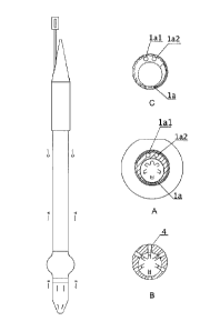
Fig. 1 shows a view of a cannula with one reinforced section with cross-
sections A-A, B-B and C-C;
Fig. 2a is a longitudinal section of the cannula with one reinforced section;
Fig. 2b is a longitudinal section of the cannula with two reinforced sections;
Fig. 2c is a longitudinal section of the bent part of the cannula;
Fig. 3 is an isometric view of the round end of the cannula;
Fig. 4a and Fig. 4b are views of a cannula with a guide and a stylet;
Date Recue/Date Received 2023-01-27
Fig. 5 is a longitudinal section of the round end of the cannula with guide
and sty let;
Fig. 6 is a longitudinal section of the round end with an integrated needle;
Fig. 7 is a longitudinal section of the integrated needle; and
Fig. 8 is a view of the integrated needle.
In the first embodiment shown in Figure 1, the carmula is a flexible tube 1
made of
plastic having three longitudinal chambers, a main chamber la, a first lateral
chamber 1 al and
a second lateral chamber 1a2, and from the distal side a round end 2 in which
there are
longitudinal holes 4 allowing venous blood to flow freely inside the cannula
tube 1. The holes
4 are evenly distributed around the perimeter of the round end 2.
Alternatively, the holes 4
can be distributed evenly
5a
Date Recue/Date Received 2023-01-27
on the circumference of the round end 2 in two rows and shifted in phase
between
rows.
Behind the round end 2 there is a soft section ml bounded from the proximal
side with a balloon 6, followed by a reinforced section, the fragment of which
is
bent at an angle of approximately 900. The reinforced section then passes into
the
soft section m2, terminated from the proximal side with the cone 3, sealing
the
cannula light tightly. Inside the cone 3 there is a port 5 for inflating the
balloon 6
and a valve 12 closing the main chamber I. The inflating port 5 is connected
to the
first lateral chamber lal, through which the filling fluid reaches the balloon
6. The
cannula is reinforced with a metal wire solenoid and retains shape memory in
the
reinforced part. The metal wire can be replaced with tape or mesh. Before
placing
the cannula in the vessel, a stiffener 8 equipped with ergonomic handle 13 is
introduced through the cone 3 into the second lateral chamber la2, which
forces the
tube 1 to take a straight shape. After removing the stiffener 8, the cannula
takes the
shape consistent with the anatomy and ratio of the angle of entry to the
chest.
The cone 3 is connected to the cannula tube 1 detachably so that it can be
removed
at the right moment and allows access to port 5.
In the second embodiment shown in Figure 2, the reinforced section of the
tube 1 is divided by the soft section m2, on which a clamp is applied. Whereas
cone
3 is located at the proximal end of the reinforced section.
The first use of the cannula is that the surgeon, through the incision in the
intercostal space gets into the area of the vena cava and the round end 2 is
inserted
through the incision of the vein into the lumen, after which it is attached
using
surgical methods. The next step is to remove the stiffener 8 from the second
lateral
chamber 1a2. Then another incision in the chest wall is made and a surgical
tool is
inserted into the chest near the operating region, after which the cone 3 is
gripped
with the tool and leads to a transcutaneous incision in the chest wall, and
then cone
3 is pushed out of the body through the percutaneous an incision in the chest
so that
6
Date Recue/Date Received 2020-05-29
the surgeon can use a soft cannula with a conical end outside of the patient's
body.
The cone 3 is then removed, thereby releasing port 5, then the balloon 6 is
filled
with liquid using a syringe. For a soft section m2 of cannula, a clamp is
inserted
and the extracorporeal circulation is connected to the cannula end. Then the
clamp
is removed and blood is already circulating in the closed extracorporeal
system. At
the end of the procedure, a clamp is applied to the soft section of the
cannula, the
extracorporeal circulation is disconnected, the fluid is removed from the
balloon 6,
and after all operations are performed, the cannula is removed.
The use of a cannula using the classic Seldinger method is that a long
Seldinger needle is inserted into the vena cava and a guide wire is inserted.
Then
the guide 11 in a form of flexible wire is threaded through the pin 7 from the
round
end 2 of the cannula and allows the cannula to be inserted into the vein along
the
guide 11. When the round end 2 is successfully placed in the vein, blood
appears in
the cannula. After inserting the appropriate part of the cannula, the stylet 7
and
guide wire 11 are being removed through the cone 3. After removing the stylet
7,
the blood is in the cannula. After this stage, the next steps are the same as
in the
first method.
Another way to use the cannula is to insert the cannula into the lumen of the
vessel without the need for a guide or surgical incision. For this purpose, a
cannula
equipped with an integrated needle 10 mounted inside the round end 2, as shown
in
Figure 6, is used. This needle consists of a sharp part 14 in the form of a
channel in
which the round part 15 is located, both parts are separate springs 16, 17 and
coupled to trigger button 18. The round portion has more than one inlet
opening to
allow blood to flow quickly into the cannula. The vein is punctured at an
appropriate
angle with an integrated needle 10. Acting with sufficient force, causes the
round
needle part 15 of the needle to hide under pressure on the vessel wall. After
piercing
the vessel wall, the needle round part 15, thanks to the action of the spring
17,
extends to secure the blade of the sharp part 14. With the correct angle of
attack,
the needle 10 is in the lumen of the vein and does not perforate both walls.
Blood
7
Date Recue/Date Received 2020-05-29
,
flows into the cannula and then the trigger button 18 is pressed at the round
end 2,
causing the needle 10 to hide inside the round end 2, which allows further
safe
insertion of the cannula to the proper depth while sliding the stiffener 8 out
of the
second lateral chamber 1a2 to achieve cannula bend according to anatomy.
After removing the pin, the procedure is identical to method 1 and 2.
8
Date Recue/Date Received 2020-05-29
