Note: Descriptions are shown in the official language in which they were submitted.
CA 03082036 2020-05-06
MONOSPECIFIC AND BISPECIFIC PROTEINS WITH IMMUNE
CHECKPOINT REGULATION FOR CANCER THERAPY
BACKGROUND
Field of Invention
[0001] The present invention relates to an antibody. More particularly, the
present invention relates to the antibody for cancer therapy.
Description of Related Art
[0002] The two major types of lymphocytes in humans are T (thymus-derived)
and B (bone marrow derived. These cells are derived from hematopoietic
stem cells in the bone marrow and fetal liver that have committed to the
lymphoid development pathway. The progeny of these stem cells follow
divergent pathways to mature into either B or T lymphocytes. Human
B-lymphocyte development takes place entirely within the bone marrow. T
cells, on the other hand, develop from immature precursors that leave the
marrow and travel through the bloodstream to the thymus, where they
proliferate and differentiate into mature T lymphocytes.
[0003] T cells
[0004] T-cells are the most abundant (about 75% of blood lymphocytes) and
potent immune killer cells. The role of effector T-cells in the anti-tumor
immune response is strongly supported by in vitro studies and the observation
that a high infiltration of CD8+ T cells in several types of tumors correlates
with
a favorable clinical prognostic (Fridman et al., 2012). The activation of
effector
1
Date Recue/Date Received 2020-05-06
CA 03082036 2020-05-06
naive T-cells requires at least three complementary signals: (i)
TCR-CD3/Ag-MHC interaction with the assistance of co-receptors (CD4 or
CD8); (ii) binding of co-stimulatory molecules such as CD80 or 0D86 to 0D28,
CD40/CD4OL; and (iii) accessory molecules such as cytokines.
[0005] Co-stimulation or the provision of two distinct signals to T-cells is a
widely accepted model of lymphocyte activation of resting T lymphocytes by
antigen-presenting cells (APCs) (Lafferty and Cunningham, 1975). This model
further provides for the discrimination of self from non-self and immune
tolerance (Bretscher and Cohn, 1970; Bretscher, 1999; Jenkins and Schwartz,
1987). The primary signal, or antigen specific signal, is transduced through
the
T-cell receptor (TCR) following recognition of foreign antigen peptide
presented
in the context of the major histocompatibility-complex (MHC). The second or
co-stimulatory signal is delivered to T-cells by co-stimulatory molecules
expressed on antigen-presenting cells (APCs), and induce T-cells to promote
clonal expansion, cytokine secretion and effector function (Lenschow et al.,
1996). In the absence of costimulation, T-cells can become refractory to
antigen stimulation, do not mount an effective immune response, and further
may result in exhaustion or tolerance to foreign antigens.
[0006] Immune checkpoint protein: PD-L1 and 0X40
[0007] Immune checkpoints refer to a group of inhibitory and stimulatory
pathways mostly initiated by ligand-receptor interaction hardwiring the immune
system, specifically T-cell mediated immunity, to maintain self-tolerance and
modulate the duration and amplitude of physiological responses in peripheral
tissues in order to minimize collateral tissue damages normally (PardoII,
2012).
2
Date Recue/Date Received 2020-05-06
CA 03082036 2020-05-06
Tumor cells co-opt certain checkpoint pathways as a major mechanism of
immune resistance. For example, programmed cell death protein 1 ligand,
PD-L1, is commonly up-regulated on tumor cell surface of human cancers.
The interaction of PD-L1 with its receptor, PD-1, expressed on tumor
infiltrated
lymphocytes (TILs), specifically on T cells, inhibits local T cell-mediated
response to escape the immune surveillance (Liang et al., 2006; Sznol and
Chen, 2013). Thus, the inhibition of immunosuppressive signals on cancer
cells, or direct agonistic stimulation of T cells, results in and/or induces a
strong
sustained anti-tumor immune response. Recent
clinical studies strongly
suggested blockage of immune checkpoint proteins via antibody or modulated
by soluble ligands or receptors are the most promising approaches to
activating
therapeutic antitumor immunity (Topalian et al., 2014). Currently, anti-PD-1
and anti-CTLA-4 (cytotoxic T-lymphocyte-associated antigen-4) antibodies have
been approved by FDA to treat diseases such as melanomas.
[0008] Another co-stimulator molecule is the 0X40 receptor (CD134), a member
of the TNFR superfamily, which is membrane-bound and is expressed primarily
on activated CD4+ T cells (Paterson et al., 1987). Signaling through the 0X40
receptor (hereinafter "0X40") is costimulatory to effector T cells and causes
proliferation of T-cells (Watts, 2005; Weinberg et al., 1994). Studies of 0X40
suggest that its major role is to dictate the number of effector T-cells that
accumulate in primary immune responses, and consequently to govern the
number of memory T-cells that subsequently develop and survive (Croft, 2003).
A number in vitro studies have been shown that 0X40 provides a costimulatory
signal resulting, in enhanced T cell proliferation and cytokine production.
3
Date Recue/Date Received 2020-05-06
CA 03082036 2020-05-06
[0009] Bi-specific/bi-functional antibody
[0010] The idea of using bispecific antibodies to efficiently retarget
effector
immune cells toward tumor cells emerged in the 1980s (Karpovsky et al., 1984;
Perez et al., 1985; Staerz et al., 1985). Bispecific scaffolds are generally
classified in two major groups with different pharmacokinetic properties,
based
on the absence or presence of an Fc fragment, IgG-like molecules and small
recombinant bispecific formats, most of them deriving from single chain
variable
fragment (scFv). Through their compact size, antibody fragments usually
penetrate tumors more efficiently than IgG-like molecules but this benefit is
mitigated by a short serum half-life (few hours) limiting their overall tumor
uptake and residence time (Goldenberg et al., 2007). By
contrast, the
presence of an Fc fragment, which binds to the neonatal Fc receptors, provides
a long serum half-life (>10 days) to the IgG-like formats, favoring tumor
uptake
and retention, but limits tumor penetration.
[0011] Recent studies have highlighted the therapeutic efficacy of
immunotherapy, a class of cancer treatments that utilize the patient's own
immune system to destroy cancerous cells. Within a tumor the presence of a
family of negative regulatory molecules, collectively known as "checkpoint
inhibitors," can inhibit T cell function to suppress anti-tumor immunity.
Checkpoint inhibitors, such as CTLA-4 and PD-1, attenuate T cell proliferation
and cytokine production.
Targeted blockade of CTLA-4 or PD-1 with
antagonist monoclonal antibodies (mAbs) releases the "brakes" on T cells to
boost anti-tumor immunity. Generating optimal "killer" CD8 T cell responses
also requires T cell receptor activation plus co-stimulation, which can be
4
Date Recue/Date Received 2020-05-06
CA 03082036 2020-05-06
provided through ligation of tumor necrosis factor receptor family members,
including 0X40 (0D134) and 4-1BB (0D137). 0X40 is of particular interest as
treatment with an activating (agonist) anti-0X40 mAb augments T cell
differentiation and cytolytic function leading to enhanced anti-tumor immunity
against a variety of tumors. When used as single agents, these drugs can
induce potent clinical and immunologic responses in patients with metastatic
disease (Linch et al., 2015).
SUMMARY
[0012] The present disclosure designed to investigate the bispecific antibody
with immunomodulatory aiming for the treatment of patient with cancers, such
as prostate cancer, lung cancer, NSCLC, melanoma, lymphoma, breast cancer,
head and neck cancer, RCC, or ovarian cancer were examined.
[0013] The present disclosure provides an antibody or an antigen-binding
portion thereof binding to 0X40 (0D134), comprising: a heavy chain variable
region comprising an amino acid sequence of at least about 80% sequence
homology to the amino acid sequence selected from the group consisting of
SEQ ID NO. 6, SEQ ID NO. 8, amino acid 128-246 of SEQ ID NO. 10, and
amino acid 124-241 SEQ ID NO. 13; and a light chain variable region
comprising an amino acid sequence of at least about 80% homology to the
amino acid sequence selected from the group consisting of amino acid 1-108 of
SEQ ID NO. 5, 1-108 of SEQ ID NO. 7, 1-112 of SEQ ID NO. 10, and 1-108 of
SEQ ID NO. 13.
Date Recue/Date Received 2020-05-06
CA 03082036 2020-05-06
[0014] In one embodiment, the antibody or the antigen-binding portion thereof
is
a single chain variable fragment (scFv) sequence selected from the group
consisting of SEQ ID NOs. 10, 11, 12, and 13.
[0015] In one embodiment, the antibody or the antigen-binding portion thereof
is
a bispecific antibody.
[0016] In one embodiment, the bispecific antibody comprises an immune
checkpoint protein binding site.
[0017] In one embodiment, the immune checkpoint protein binding site
comprises a programmed cell death protein 1 ligand (PD-L1) binding site, PD-1
binding site, epidermal growth factor receptor (EGFR) binding site, human
epidermal growth factor receptor 2 (HER2) binding site, cytotoxic
T-lymphocyte-associated antigen 4 (CTLA-4) binding site, or lymphocyte
activation gene 3 (LAG3) binding site.
[0018] The present disclosure also provides an antibody or an antigen-binding
portion thereof binding to PD-L1, comprising: a heavy chain variable domain
comprising an amino acid sequence of at least about 80% sequence homology
to the amino acid sequence selected from the group consisting of SEQ ID NO. 2
and SEQ ID NO. 4; and a light chain variable domain comprising an amino acid
sequence of at least about 80% homology to the amino acid sequence selected
from the group consisting of amino acid 1-111 of SEQ ID NO. 1 and 1-110 of
SEQ ID NO. 3.
[0019] The present disclosure also provides a bispecific antibody comprising
at
least one of polypeptide chain, wherein the polypeptide chain comprises an
0X40 binding site and a PD-L1 binding site. The 0X40 binding site comprises
6
Date Recue/Date Received 2020-05-06
CA 03082036 2020-05-06
a heavy chain variable region comprising an amino acid sequence of at least
about 80% sequence homology to the amino acid sequence selected from the
group consisting of SEQ ID NO. 6, SEQ ID NO. 8, amino acid 128-246 of SEQ
ID NO. 10, and amino acid 124-241 SEQ ID NO. 13; and a light chain variable
region comprising an amino acid sequence of at least about 80% homology to
the amino acid sequence selected from the group consisting of amino acid
1-108 of SEQ ID NO. 5, 1-108 of SEQ ID NO. 7, 1-112 of SEQ ID NO. 10 and
1-108 of SEQ ID NO. 13. The PD-L1 binding site comprises a heavy chain
variable domain comprising an amino acid sequence of at least about 80%
sequence homology to the amino acid sequence selected from the group
consisting of SEQ ID NO. 2 and SEQ ID NO. 4; and a light chain variable
domain comprising an amino acid sequence of at least about 80% homology to
the amino acid sequence selected from the group consisting of amino acid
1-111 of SEQ ID NO. 1 and 1-110 of SEQ ID NO. 3.
[0020] In one embodiment, the polypeptide chain further comprises a Fc domain,
a Fab fragment, and a scFv. The Fab fragment is connected to the N-terminus
of the Fe domain, and the Fab fragment comprises the PD-L1 binding site.
The scFv is connected to the C-terminus of the Fc domain, and the scFv
comprises the 0X40 binding site.
[0021] In one embodiment, the polypeptide chain further comprises a linker
between the Fc domain and the scFv.
[0022] In one embodiment, the scFv comprises an amino acid sequence
selected from the group consisting of amino acid 455-707 of SEQ ID NO. 18,
455-708 of SEQ ID NO. 19, 455-701 of SEQ ID NO. 20, 455-706 of SEQ ID NO.
7
Date Recue/Date Received 2020-05-06
CA 03082036 2020-05-06
21, 455-706 of SEQ ID NO. 22, 455-706 of SEQ ID NO. 23, 455-706 of SEQ ID
NO. 24, 455-706 of SEQ ID NO. 25, 455-706 of SEQ ID NO. 26, 455-706 of
SEQ ID NO. 27, 455-706 of SEQ ID NO. 28, and 455-706 of SEQ ID NO. 29.
[0023] In one embodiment, the bispecific antibody comprises one pairs of
polypeptide chains.
[0024] In one embodiment, the bispecific antibody is an IgG, IgE, IgM, IgD,
IgA,
or IgY antibody.
[0025] In one embodiment, the bispecific antibody is an IgG antibody.
[0026] In one embodiment, the IgG antibody is an IgG1, IgG2, IgG3, or IgG4
antibody.
[0027] The present disclosure also provides an antibody-drug conjugate
comprising a therapeutic agent, and an antibody or an antigen-binding portion
binding PD-L1 and/or 0X40, wherein the therapeutic agent is covalently
conjugated to the antibody or the antigen-binding portion by a linker.
[0028] In one embodiment, the antibody or an antigen-binding portion is
selected from the above mentioned antibody or an antigen-binding portion.
[0029] The present disclosure also provides a pharmaceutical composition
comprising the antibody, the antigen-binding portion thereof, or the
bispecific
antibody as above mentioned, and at least one pharmaceutically acceptable
carrier.
[0030] The present disclosure also provides a method of treating cancer
comprising administering to the subject in need thereof an effective amount of
8
Date Recue/Date Received 2020-05-06
CA 03082036 2020-05-06
the antibody, the antigen-binding portion thereof, or the bispecific antibody
as
above mentioned.
[0031] In one embodiment, the cancer is selected from the group consisting of
prostate cancer, lung cancer, Non-Small Cell Lung Cancer (NSCLC),
melanoma, lymphoma, breast cancer, head and neck cancer, renal cell
carcinoma (RCC), and ovarian cancer.
[0032] The present disclosure also provides a nucleic acid molecule encoding
the antibody, the antigen-binding portion thereof, or the bispecific antibody
as
above mentioned.
[0033] It is to be understood that both the foregoing general description and
the
following detailed description are by examples, and are intended to provide
further explanation of the invention as claimed.
BRIEF DESCRIPTION OF THE DRAWINGS
[0034j The invention can be more fully understood by reading the following
detailed description of the embodiment, with reference made to the
accompanying drawings as follows:
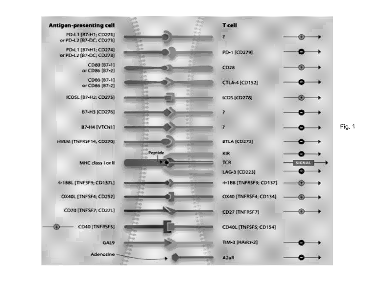
[0035] Fig. 1 shows immune checkpoints modulating T-cell mediated immunity.
Antibody either agonistic or antagonistic against the checkpoints, such as
anti-ICOS, anti-0D28, anti-0X40, and anti-CD27, or anti-PD-1, anti-CTLA4,
anti-LAG3, anti-BTLA, could be used to construct the bi-functional fusion
protein
depending on applications.
9
Date Recue/Date Received 2020-05-06
CA 03082036 2020-05-06
[0036] Figs. 2A and 2B show the screening of phage clone by direct ELISA for
PD-L1 expressed HEK293 cells.
[0037] Figs. 3A and 3B show the screening of phage clone by cell-based ELISA
with 0X40 expressed HEK293 cells.
[0038] Fig. 4 shows purified antibody leads specific for PD-L1 by SDS-PAGE
with non-reducing reagent to reveal the integrity and purity.
[0039] Fig. 5 shows purified antibody leads specific for 0X40 by SDS-PAGE
with non-reducing or reducing reagent to reveal the integrity and purity.
[0040] Fig. 6 shows examples of the direct ligand binding activity of purified
anti-immune check point proteins and anti-PD-L1 antibody leads against PD-L1.
Ligand pre-coated wells were first incubated with various concentrations of
antibody leads as indicated. The bound proteins were then detected with HRP
conjugated goat anti-human IgG Fab specific antibody and 01)450 readings were
plotted.
[00411 Fig. 7 shows examples of the direct ligand binding activity of purified
anti-immune check point proteins and anti-0X40 antibody leads against 0X40.
Ligand pre-coated wells were first incubated with various concentrations of
antibody leads as indicated. The bound proteins were then detected with HRP
conjugated goat anti-human IgG Fab specific antibody and 01)450 readings were
plotted.
[0042] Fig. 8 shows the flow analysis using PD-Li expression 293 cells. PD-L1
expression HEK293 cells were first incubated with purified antibody leads, and
the bound antibodies were detected with Alexa-488 conjugated goat anti-human
IgG (H+L) followed by fluorescence-activated cell sorter (FACS) analysis.
Date Recue/Date Received 2020-05-06
CA 03082036 2020-05-06
[0043] Fig. 9 shows the flow analysis using 0X40 expression 293 cells. 0X40
expression HEK293 cells were first incubated with purified anti-0X40 antibody
leads, and the bound antibodies were detected with Alexa-488 conjugated goat
anti-human IgG (H+L) followed by FACS analysis. NS: no staining.
[0044] Fig. 10 shows the blockage of PD-1/PD-L1 interaction with purified
anti-PD-L1 antibodies. Purified antibodies as indicated were applied with
biotinylated-PD-L1-Fc and recombinant human PD-1/His (hPD-1/His) to
evaluate the inhibition activity of PD-1/PD-L1 interaction. The
binding
recombinant PD-L1-Fc and hPD-1/His was detected by streptavidin-HRP and
analysis by ELISA.
[0045] Figs. 11A and 11B show anti-PD-L1 antibody leads with 1 or 10 pg/mL
stimulates T-cell proliferation and induces 1L-2 and/or 1FN-y production in a
mixed lymphocyte reaction (MLR) assay after 3 days (Fig. 11A) or 5 days (Fig.
11B) antibody treatment.
[0046] Fig. 12A shows the ability of anti-0X40 antibody leads to enhance the
CD3+ T cell activation with dosage response as well as reference antibody.
Fig. 12B shows the concentration of human IL-2 and IFN-y present in cell
culture media following 3 days of stimulation of human T cells with plate
bound
anti-CD3 and several concentrations of anti-0X40 antibody leads.
[0047] Figs. 13A and 13B show the concentration of human IL-2 (Fig. 13A) and
IFN-y (Fig. 13B) present in cell culture media following 3 days of stimulation
of
human T cells with plate bound anti-CD3 and several concentrations of 0X40
specific antibody leads.
11
Date Recue/Date Received 2020-05-06
CA 03082036 2020-05-06
[0048] Fig. 14 shows the structure of an antibody heavy chain Fc fused with an
0X40 specific scFv domain.
[0049] Fig. 15 shows examples of PAGE-gel analysis of anti-immune check
point antibodies-human 0X40 fusion proteins.
Purified fusion proteins,
anti-PD-L1-0X40 scFv fusion proteins were shown to have a molecular weight
about 220 kDa (non-reducing), and heavy chain fusion has about 85 kDa and
light chain is about about 25 kDa (reduced) in both antibody fusions.
[0050] Figs. 16A and 16B show bispecific antibody synergic stimulates T-cell
activation for IL-2 and IFN-y production in a mixed lymphocyte reaction (MLR)
assay after 3 days (Fig. 16A) or 5 days (Fig. 16B) with mono-, combined or
anti-PD-L1-0X40 scFv bispecific antibody treatment.
[0051] Figs. 17A to 17E respectively show the aggregation and purity
determination of Bi-specific antibodies, Anti-PD-L1-0X40 Ab and
Anti-PD-L1-0X40 Ab-V1 to V4, with 5 different linkers in 0X40 scFv.
[0052] Fig. 18 shows sequence variants among 0X40 clone B17 scFv of
Anti-PD-L1-0X40 Ab-V4 to V12 (SEQ ID NOS: 30-38).
[0053] Fig. 19 shows examples of PAGE-gel analysis of anti-immune check
point antibodies-human 0X40 fusion proteins.
Purified fusion proteins,
anti-PD-L1-0X40 Ab-V5 fusion proteins were shown to have a molecular weight
about 220 kDa (non-reducing), and heavy chain fusion has about 80 kDa and
light chain is about 30 kDa (reduced) in both antibody fusions.
[0054] Fig. 20 shows a flow chart illustrating the ELISA method for binding
activity evaluation of bispecific antibody variants.
12
Date Recue/Date Received 2020-05-06
CA 03082036 2020-05-06
[0055] Fig. 21 shows the human PD-L1 binding activity of the bispecific
antibody
variants and its EC50.
[0056] Fig. 22 shows the human 0X40 binding activity of the bispecific
antibody
variants and its EC50.
[0057] Fig. 23 shows the ex vivo serum stability of bispecific antibody
variant,
anti-PD-L1-0X40 Ab-V5.
[0058] Figs. 24A and 24B show the IL-2 production for 3 days (Fig. 24A) and
IFN-y production for 5 days (Fig. 24B) after modulating T cell with mono-,
combined or anti-PD-L1-0X40 Ab-V5 bispecific antibody treatment.
[0059] Fig. 25 is a graph showing the effect of anti-PD-L1-0X40 Ab-
V5bispecific
antibody treatment and monoclonal antibody treatment on the growth of P0-3
tumor in Fox Chase SCID Beige mice.
DETAILED DESCRIPTION
[0060] Reference will now be made in detail to the present embodiments of the
invention, examples of which are illustrated in the accompanying drawings.
Wherever possible, the same reference numbers are used in the drawings and
the description to refer to the same or like parts.
[0061] The present invention describes the expression, purification and
characterization of bi-functional proteins with isolated functional agonistic
0X40
scFv fused to the C-terminus of Fc domain of anti-immune checkpoint protein
antibodies. These proteins interact with its corresponding check point target
shall transmit the inhibitory or stimulatory signal to modulate T-cell
involved
immunity. The components of Fe fusion proteins in present invention are of all
13
Date Recue/Date Received 2020-05-06
CA 03082036 2020-05-06
human origins, and thus are expected to be non-immunogenic and can be used
as therapeutics in human.
[0062] Bispecific molecules such as bispecific antibodies (BsAbs) provide a
means of simultaneously targeting multiple epitopes on the same molecular
target or different targets with a single therapeutic agent. As
cancer
therapeutics, they have the potential to confer novel or more potent
activities,
lower the cost of goods and facilitate the development of new therapeutic
regimens in contrast to a mixture of two mAbs (Chames and Baty, 2009;
Hollander, 2009; Thakur and Lum, 2010).
Recently, catumaxomab, a
trifunctional bispecific antibody targeting human epithelial cell adhesion
molecule (EpCAM) and CD3 has shown a clear clinical benefit in patients with
peritoneal carcinomatosis of epithelial cancers (Heiss et al., 2010), and a
bispecific T-cell engaging (BiTE) antibody with dual specificity for CD19 and
CD3 has also demonstrated encouraging clinical activity in patients with CD19
expressing hematological malignancies (Bargou et al., 2008). Despite strong
interest in the development of bispecific molecules as cancer therapeutics,
technical challenges in the production of stable and active bispecific
molecules
have in the past hindered the clinical evaluation of most bispecific formats.
Many engineered antibody formats, including an IgG-like bispecific antibody
have compromised stability or solubility (Bargou et al., 2008; Demarest and
Glaser, 2008; Lu et al., 2005). Furthermore, several strategies have been
taken to increase the product quality and in vivo stability of bispecific
molecules,
including PEGylation, conjugation with human serum albumin and Fc
engineering (Muller et al., 2007; Ridgway et al., 1996). Bispecific single
chain
antibodies of the general form described above have the advantage that the
14
Date Recue/Date Received 2020-05-06
CA 03082036 2020-05-06
nucleotide sequence encoding the four V-domains, two linkers and one spacer
can be incorporated into a suitable host expression organism under the control
of a single promoter. This increases the flexibility with which these
constructs
can be designed as well as the degree of experimenter control during their
production. In addition, the Fc of IgG is a very another attractive scaffold
for
designing novel therapeutics because it contains all antibody functions except
the binding ability. Fc engineering is important for improving the
effectiveness
of the bispecific antibodies. Therefore, the IgG-based conformation is using
in
present invention for two independent target on immune cells or target cell in
immunotherapy.
[0063] Targeting immune-check point proteins are promising approaches to
activate antitumor immunity. Anti-check point proteins, such as PD-1, PD-L1,
CTLA-4, LAG3, etc., are currently evaluated clinically (Fig. 1).
Preliminary
data with blockers of immune checkpoint proteins have been shown to be able
to enhance antitumor immunity with the potential to produce durable clinical
responses. However, despite the remarkable clinical efficacy of these agents
in a number of malignancies, it has become clear that they are not
sufficiently
active for many patients. Numerous additional immunomodulatory pathways
as well as inhibitory factors expressed or secreted by myeloid and stromal
cells
in the tumor microenvironment are potential targets for synergizing with
immune
checkpoint blockade. Therefore, combining anticancer or bispecific antibody
therapies has been essential to achieve complete remission and cures for
patients with cancer.
Date Recue/Date Received 2020-05-06
CA 03082036 2020-05-06
[0064] The present invention describes the construction, expression and
characterization of anti-immune checkpoint protein antibody Fc fused with
different immune checkpoint protein specific scFv protein. The C-terminally
positioned 0X40 scFv in fusion constructs shall allow expanding the power of
fusion proteins beyond 0X40 activation approach if the fusion counterpart is
immune system potentiating agent, such as anti-EGFR, anti-HER2, or
anti-CTLA-4 antibody, for example.
[0065] Antibody generation from OmniMab library
[0066] For the generation of therapeutic antibodies against PD-L1 or 0X40,
selections with the OmniMab phagemid library were carried out. The
phagemid library is generated by AP Biosciences Inc. (APBio Inc.) from a
collection of over hundred health donors B cells. Phages for the 1st round of
pannings were prepared by Hyperphage (M13K07ApIll, Progen, Heidelberg,
Germany). Solid phase panning and cell panning against PD-L1 or 0X40 were
applied for PD-L1 or 0X40 specific binder selection and isolation from
OmniMab library. Solid phase panning was performed using recombinant
human PD-L1-Fc or 0X40-Fc (APBio Inc.) in the first round selection and then
HEK293 cells expressed PD-L1 or 0X40 were used for two and three round
enrichment. After three rounds selection, the specific PD-L1 or 0X40 binders
were screened and isolated by direct ELISA or cell-based ELISA with
corresponding recombinant protein (Figs. 2A, 2B, 3A, and 3B). Pre-coated
PD-L1-Fc recombinant proteins or 0X40 expressed 293 cells were blotted with
supernatant containing rescued phages for 1 hour and washed with PBS
containing 0.1 % Tween-20 for three times. Bound phages were detected by
16
Date Recue/Date Received 2020-05-06
CA 03082036 2020-05-06
HRP conjugated anti-M13 antibody (Roche) and TMB substrate was used for
signal development. The 0D450 readings were recorded. The positive binders
were isolated and sent for sequencing to confirm the sequence and diversity of
heavy chain and light chain. The variable region of heavy chain and light
chain
specific to PD-L1 or 0X40 were described from the SEQ ID NO. 1 to SEQ ID
NO. 8: SEQ ID NO. 1 is the light chain of PD-L1 clone 6, SEQ ID NO. 2 is the
variable region of heavy chain of PD-L1 clone 6, SEQ ID NO. 3 is the light
chain
of PD-L1 clone 32, SEQ ID NO. 4 is the variable region of heavy chain of PD-L1
clone 32, SEQ ID NO. 5 is the light chain of 0X40 clone B17, SEQ ID NO. 6 is
the variable region of heavy chain of 0X40 clone B17, SEQ ID NO. 7 is the
light
chain of 0X40 clone B19, SEQ ID NO. 8 is the variable region of heavy chain of
OX40 clone B19. As shown in the Figs. 2A, 2B, 3A and 3B, several clones
were isolated and known to be recognized specifically for corresponding
antigen
as comparing with negative control.
[0067] Subcloning and expression/purification of selected PD-L1 or 0X40
specific binder as IgG format
[0068] To facilitate the quick screening of specific binder with functionality
in T
cell activation, the heavy chains and light chains of positive binders against
PD-L1 or 0X40 by ELISA were then amplified, digested and sub-clone into
APBio specialized IgG expression vector carrying IgG4 constant region (SEQ ID
NO. 9). After sequence validation, the plasmids were then prepared and
transfected into HEK293 cells for antibody expression with 293 fectin
transfection reagent (Invitrogen). After 4 days culture, the antibody secreted
into serum-free medium is affinity purified from culture supernatant by
Protein G
17
Date Recue/Date Received 2020-05-06
CA 03082036 2020-05-06
chromatography. Purified antibody is then concentrated, followed by dialysis
in
PBS buffer. The final concentration of dialyzed protein is determined by
NanoDrop2000 spectrophotometer and the purity and integrity are determined
by SDS-PAGE with or without reducing reagent as shown in the Figs. 4 and 5.
The integrity of various purified antibody leads, either PD-L1 specific or
0X40
specific, is normal in the HEK293 cells as well as reference antibody,
MPDL3280A for PD-L1 or GSK3174998 for 0X40.
[0069] In one embodiment, the present disclosure provides an antibody or an
antigen-binding portion thereof binding to 0X40 (0D134), comprising a heavy
chain variable region and a light chain variable region. The heavy chain
variable region comprises an amino acid sequence of at least about 80%
sequence homology to the amino acid sequence selected from the group
consisting of SEQ ID NO. 6, SEQ ID NO. 8, amino acid 128-246 of SEQ ID NO.
10, and amino acid 124-241 SEQ ID NO. 13. In some examples, the heavy
chain variable region comprises an amino acid sequence of at least about 85%,
90%, or 95% sequence homology to the amino acid sequence as above
mentioned. The
light chain variable region comprising an amino acid
sequence of at least about 80% homology to the amino acid sequence selected
from the group consisting of amino acid 1-108 of SEQ ID NO. 5, 1-108 of SEQ
ID NO. 7, 1-112 of SEQ ID NO. 10, and 1-108 of SEQ ID NO. 13. In some
examples, the light chain variable region comprises an amino acid sequence of
at least about 85%, 90%, or 95% sequence homology to the amino acid
sequence as above mentioned.
18
Date Recue/Date Received 2020-05-06
CA 03082036 2020-05-06
[0070] In one embodiment, the present disclosure provides an antibody or an
antigen-binding portion thereof binding to PD-L1, comprising a heavy chain
variable domain and a light chain variable domain. The heavy chain variable
domain comprises an amino acid sequence of at least about 80% sequence
homology to the amino acid sequence selected from the group consisting of
SEQ ID NO. 2 and SEQ ID NO. 4. In some examples, the heavy chain
variable region comprises an amino acid sequence of at least about 85%, 90%,
or 95% sequence homology to the amino acid sequence as above mentioned.
The light chain variable domain comprises an amino acid sequence of at least
about 80% homology to the amino acid sequence selected from the group
consisting of amino acid 1-111 of SEQ ID NO. 1 and 1-110 of SEQ ID NO. 3.
In some examples, the light chain variable region comprises an amino acid
sequence of at least about 85%, 90%, or 95% sequence homology to the amino
acid sequence as above mentioned.
[0071] Binding activity determination for PD-L1, 0X40 specific IgG leads
by direct ELISA
[0072] Purified antibody leads against PD-L1 or 0X40 (anti-PD-L1 antibody
leads or anti-0X40 antibody leads) were then applied for ELISA binding
characterization on human PD-L1-Fc or 0X40-Fc in a direct coated setup.
Figs. 6 and 7 showed the ELISA binding result for anti-PD-L1 and anti-0X40
antibodies, respectively. For PD-L1 specific antibodies, most leads showed a
similar or better binding activity with reference antibody (Ref Ab, MPDL3280A,
Roche).
19
Date Recue/Date Received 2020-05-06
CA 03082036 2020-05-06
[0073] Purified human PD-L1 or 0X40 IgG1 Fc chimera (PD-L1-Fc or 0X40-Fc,
APBio) was dialyzed in Phosphate Buffered Saline (PBS), adjusted to 1mg/mL
and then diluted with PBS to a final concentration of 1 pg/mL. Nunc-lmmuno
Maxisorp 96 well plates were coated with 0.1 mL per well of recombinant
PD-L1-Fc or 0X40-Fc chimera leaving empty wells for nonspecific binding
controls and incubated at 4 C overnight. The PD-L1-Fc or 0X40-Fc chimera
solution was removed and the plates were washed three times with 0.4 mL
wash buffer (0.1 % Tween-20 in PBS). 0.4 mL blocking buffer (5% low-fat milk
powder in PBS) was added to all wells and incubated at room temperature for 1
hour with mixing. The blocking buffer was removed and plates washed three
times with 0.4 mL wash buffer. Serial dilutions of the PD-L1 or 0X40 test
antibodies were prepared in PBS and 0.1 mL diluted Ab was added per well.
Plates were incubated 1 hour at room temperature. Antibody solution was
removed and the plates washed three time with 0.4 mL wash buffer per well.
Horseradish peroxidase labeled goat anti-human IgG, F(ab')2 specific F(ab')2
antibody (Jackson Immunoresearch #109-036-097) was diluted 1:2000 with
PBS and added 0.1 mL per well. The plates were incubated 1 hour at room
temperature and washed with 0.4 mL per well wash buffer. 0.1 mL TMB
reagent (lnvitrogen) was added and incubated for 1 to 5 minutes at room
temperature. The reaction was stopped by adding 0.05 mL 1N HCI and
absorbance was read at 450 nm on a Bio-Tek Spectra. Calculated EC50 for
anti-PD-Li antibody leads to PD-L1 showed most leads possess good binding
activity as well as MPDL3280A (Ref Ab) by direct ELISA (Fig. 6). On the
contrary, most anti-0X40 antibody leads showed much lower binding activity as
comparing with reference antibody (Ref Ab, GSK3174998)(Fig. 7).
Date Recue/Date Received 2020-05-06
CA 03082036 2020-05-06
[0074] Binding activity determination for PD-L1 and 0X40 specific IgG
leads by FACS
[0075] Purified antibody leads (anti-PD-L1 antibody leads or anti-0X40
antibody
leads) were also applied for flow cytometry to determine and compare the
binding activity with PD-L1 or 0X40 expressed HEK293 cells. Figs. 8 and 9
show the binding activity of corresponding antibody leads as indicated by FACS
with stable expressed PD-L1 or 0X40 HEK293 cells.
[0076] FACS analysis of PD-L1 stable expression 293 cells stained with
anti-PD-L1 antibody leads to examine the PD-L1 binding activity, stable
expression cells were incubated with 1 pg/mL purified anti-PD-L1 antibody
leads, reference antibody (Ref Ab MPDL3280A) or with isotype antibody as
negative control on ice for 1 hr. The cells were washed three times with lx
PBS and then incubated with Alexa-488-conjugated goat anti-human IgG (H+L)
(Invitrogen Inc.) on ice for additional 1 hr. After staining, the cells were
washed
three times with 1x PBS, resuspended in 1x PBS/2 /0FBS before analyzed by
FACS Calibur (BD Biosciences, Inc.) and FlowJo (TreeStar, LLC). Same
scenario, the binding activity of anti-0X40 antibody leads for stable
expressed
0X40 HEK293 cells in Fig. 9 were also executed with a similar strategy and
analyzed as described above. As shown in the Fig. 8, most anti-PD-L1
antibody leads possess a good binding activity as well as reference antibody.
This indicated the phage clones selected from the OmniMab library indeed
recognize the native PD-L1 in the cells.
[0077] This phenomenon is also observed for anti-0X40 antibody leads as
shown in the Fig. 9. FACS analysis of 0X40 stable expression 293 cells clone
21
Date Recue/Date Received 2020-05-06
CA 03082036 2020-05-06
2D5 stained with purified anti-0X40 antibodies leads to examine the 0X40
binding activity, stable expression cells were incubated with 2 pg/mL anti-
0X40
reference Abs (0X40 ref.) or anti-CD137 reference Abs (0D137 ref.) as control
antibody on ice for 1hr. The cells were washed three times with 1x PBS and
then incubated with Alexa-488-conjugated goat anti-human IgG (H+L)
(Invitrogen Inc.) on ice for additional 1hr. After staining, the cells were
washed
three times with 1x PBS, resuspended in 1x PBS/2 /0FBS before analyzed by
FACS Calibur (BD Biosciences, Inc.) and FlowJo (TreeStar, LLC).
[0078] Ligand competition binding (ELISA Assay)
[0079] Antibody leads were showed the binding selectivity and affinity assay
used to evaluate the anti-PD-L1 antibody leads of present invention for their
ability to block binding of PD-L1 to PD-1.
[0080] Antibodies were tested for their ability to block the binding of the
human
PD-L1-Fc chimera (PD-L1-Fc) to recombinant human PD-1/His (hPD-1/His) by
ELISA. Purified recombinant hPD-1/His (APBio) was dialyzed to 1 mg/mL in
PBS and then conjugated with biotin (Abeam). Nunc Maxisorp 96 well pate
was coated with 250 ng hPD-1/His per well in PBS overnight. The hPD-1/His
solution was removed and the plates were washed three times with 0.4 mL
wash buffer (0.1 % Tween-20 in PBS). 0.4 mL blocking buffer (5% low-fat milk
powder in PBS) was added to all wells and incubated at room temperature for 1
hour with mixing. During the blocking step the antibody stocks were diluted in
a range from 200 nM to 0 nM in PBS with 2 folds serial dilution. Purified
recombinant biotinylated-PD-L1-Fc chimera was diluted to 4 pg/mL in PBS.
The PD-1/His coated plates were washed three times with 0.2 mL wash buffer
22
Date Recue/Date Received 2020-05-06
CA 03082036 2020-05-06
(0.1 % Tween 20 in PBS). 60 pL antibody dilutions (anti-PD-L1 antibody leads
or Ref Ab MPDL3280A) were added alone with 60 pL biotinylated-PD-L1-Fc
chimera and incubated at room temperature for 1 hour. Plates were washed
as described previously. Streptavidin-HRP was diluted 1:2000 in PBS, 100 pL
of the resulting solution added to the wells of the washed plated, and
incubated
at room temperature for 1 hour. Plates were washed as previously described,
100 pL TMB substrate solution was added to each well and incubated for 10
minutes. The reaction was stopped with 50 pL 1N HCI and absorbance at 450
nm read using Bio-Tek reader and showed in Fig. 10. Partial antibody leads
are showed to inhibit the interaction between PD-PD-L1 by competition ELISA.
Most antibody leads revealed a similar blocking activity as comparing with
reference antibody (Ref Ab MPDL3280A).
[0081] Enhanced stimulation of T cell activation by inhibition of
PD-1 :PD-L1 ligand interaction for anti-PD-L1 antibody
[0082] The PD-1 signaling pathway inhibits moderate TOR/0D28 costimulatory
signals, with cytokine production being reduced first without a decrease in T
cell
proliferation. As the TOR/0D28 costimulatory signals weaken, the PD-1
pathway dominates, with a great reduction in cytokine production accompanied
by a reduction in proliferation. Accordingly in order to confirm that the
inhibition of the PD-1 via inhibition of the interaction with PD-L1, human
antibodies of the invention enhances T cell activation, mixed lymphocyte
reactions (MLRs) are performed.
[0083] Monocytes from human whole blood were enriched by RosetteSepTM
Human Monocyte Enrichment Cocktail (Cat. No.15068) and cultured in
23
Date Recue/Date Received 2020-05-06
CA 03082036 2020-05-06
differentiation medium, RPMI 1640 with 10 /0FBS, 100 ng/mL (1000 tJ/mL)
GM-CSF, 100 ng/mL (500 tJ/mL) for 6 days. The differentiate dendritic cells
(DC) from monocyte were checked by DC-SIGN-PE, anti-CD14 conjugated with
FITC Ab, anti-CD83 conjugated with PE Ab, or anti-CD80 conjugated with FITC
Ab to validate the differentiation and used to be APCs in MLRs.
[0084] Allogenic CD4+ T cells from human whole blood were isolated by
RosetteSepTM Human CD4+ T Cell Enrichment Cocktail (Cat. NO. 15062).
The purity of CD4+ T cells were checked with anti-CD4 conjugated APC Ab to
make sure the purity is above 95% and then labeled with 1uM CFSE
(CellTraceTm CFSE cell proliferation kit, Life technologies, Cat. NO. C34554)
for T cells proliferation assay. Labeled CD4+ T cells were used to co-culture
with immature DC with different antibody leads as indicated for 3 and 5 days
to
see whether the antibody leads could restore the T cell activation through
blocking the interaction between PD-1 and PD-L1. After 3 and 5 days
incubation, the supernatant were collected for cytokine, such as IL-2 and IFN-
y
quantitation by ELISA. The addition of anti-PD-L1 antibody leads (clones 6,
32,
28, 51, 64, 27, and 37) to cultures of immature dendritic cells plus
allogeneic T
cells is predicted to result in an increase in T cell proliferation and
cytokine
production, as compared to isotype IgG (iso#1, #2) treated cultures and showed
in the Figs. 11A and 11B. The IL-2 and IFN-y production increase significantly
in the MLRs as comparing with isotype antibody treatment after 3 days (Fig.
11A) or 5 days (Fig. 11B) antibody treatment, especially for anti-PD-L1
antibody
clone 6. The cytokine increment is still obviously after 5 days antibody
treatment and similar to reference antibody (ref), MPDL3280A. This indicated
24
Date Recue/Date Received 2020-05-06
CA 03082036 2020-05-06
the anti-PD-L1 antibody clone 6 should be one of the potential leads for
bispecific antibody composite.
[0085] Agonistic activity assay of anti-0X40 antibody
[0086] In order to activate 0X40 costimulation of T-cell proliferation and
cytokine production, the purified antibody leads were functionally screened
for
their ability to enhance cytokine production, proliferation, and to induce
proliferation in human CD3+ 1-cells. The
anti-CD3 antibody (OKT3,
BioLegend Cat. No.317304) and anti-0X40 antibody leads (clones B6, B70,
B120, A4, B17, B19, and B30), reference antibody (GSK3174998) or isotype
antibodies (iso#1, #2) were coated in the Maxisorp 96-well plate. Meanwhile,
naive human CD3+T-ce11s were isolated from the human blood from heathy
adult volunteers using a commercially available RosetteSepTM Human T Cell
Enrichment Cocktail (STEMCELL Cat. No.15061) as manufacture's described.
The isolated CD3+ T cells were then labeled by CFSE (CellTraceTm CFSE cell
proliferation kit, Life technologies, Cat. NO. C34554) and seeded as 1 X 10⁶
cells/mL into the antibody pre-coated well containing RPMI 1640 medium, 10 /0
fetal bovine serum and 2.5 mM L-glutamine to determine the cell proliferation
and cytokine production. After 3 days culture, the cells were collected for
proliferation assay by flow cytometry and medium were then analyzed for IL-2
and IFN-y production by quantitation ELISA.
[0087] The screening of anti-0X40 antibody leads with agonistic activity in T
cell
activation was showed in the Fig. 12A. All anti-0X40 antibody leads showed
the ability to enhance the CD3+ T cell activation with dosage response as well
as reference antibody. Higher dosage antibody treatment showed obviously
Date Recue/Date Received 2020-05-06
CA 03082036 2020-05-06
higher T cell activation activity. Meanwhile, cytokine production (Fig. 12B),
such as IL-2 and IFN-y also revealed similar T cell activation response,
especially for anti-0X40 antibody lead clone B17. Cytokine is highly induction
after anti-0X40 antibody lead B17 3 days treatment. The enhancement is
much higher than reference antibody treatment, this implicated clone B17
should be one of the candidates for bispecific antibody construction.
[0088] As the data shown in the Figs. 13A and 13B, both anti-0X40 antibody
leads, clones B17 and B19, were showed a better agonistic activity in the
assay
after anti-0X40 antibody leads (B17 or B19) 3 days treatment. Either IL-2
production or IFN-y production shows an obvious enhancement upon antibody
treatment and revealed does-dependent correlation. Higher
cytokine
productions were recorded in higher dose antibody treatment.
[0089] In order to evaluate the agonistic activity of 0X40 antibody leads, B17
and B19, the EC50 were also determined as well as agonistic activity assay and
cytokine production were recorded for comparison.
[0090] Construction, Expression and Purification of Anti-PD-L1-0X40 scFv
antibody
[0091] Since the bispecific is designed as IgG based fused with scFv format,
the
structure of anti-immune checkpoint antibody Fc-terminally fused with 0X40
scFv. Antibody can be inhibitory anti-immune checkpoint antibodies, such as
anti-PD-L1, anti-PD-1, anti-CTLA4, anti-LAG3, etc., or stimulatory antibodies,
such as anti-0D28, anti-0D137, anti-0D27, anti-ICOS, etc. A linker is placed
between antibody Fc and 0X40 scFv to generate the bispecific antibody as
depicted in Fig. 14.
26
Date Recue/Date Received 2020-05-06
CA 03082036 2020-05-06
[0092] In some embodiment, the anti-PD-L1 antibody lead clone 6 is assigned
to be IgG form, on the other hand, the anti-0X40 antibody lead would be
transformed as scFv format to fuse at C-terminus of Fc region in anti-PD-L1
antibody lead clone 6. The transformation from antibody to scFv format could
result in the reduction of the binding activity or specificity; therefore
several
anti-0X40 antibody leads were used to scFv transformation. Construction of
bi-functional anti-PD-L1 antibody Fc fused with full-length 0X40 scFv (SEQ ID
NO. 10 as clone A4, SEQ ID NO. 11 as clone B17, SEQ ID NO. 12 as clone
B19, or SEQ ID NO. 13 as clone B120). A short flexible peptide linker,
(GGGGS)2 (SEQ ID NO. 14) was placed between, for example, anti-PD-L1
antibody heavy chain C-terminus of Fc region and N-terminal module of 0X40
scFv to ensure correct folding and minimize steric hindrance. The coding
sequences of anti-PD-L1-0X40 scFv antibodies were shown in SEQ ID NO. 16
(anti-PD-L1-clone 6 heavy chain-0X40 clone B17 scFv) and NO. 17
(anti-PD-L1-clone 6 heavy chain-0X40 clone B19 scFv). The constructed
antibody Fc fusion proteins were leaded by a signal peptide (SEQ ID NO. 15)
and expressed by mammalian cells, and purified from the transfected cell
culture supernatant via 1-step Protein G chromatography. As shown in Fig. 15,
greater than 90% purity can be obtained in a single step purification process
and shows that purified fusion proteins have correct molecular weight (Mw =
220kD).
[0093] Enhanced stimulation of T cell activation for anti-PD-L1-0X40 scFv
bispecific antibody leads in MLRs
27
Date Recue/Date Received 2020-05-06
CA 03082036 2020-05-06
[0094] To determine the synergic cooperation of bispecific antibody in
enhancing T cells activation through inhibition the interaction between PD-1
and
PD-L1 and agonistic activation of 0X40 signaling, the bispecific antibody
leads,
anti-PD-L1-0X40 scFv, were applied into MLRs as described above. IL-2 and
IFN-y production were then recorded after 3 or 5 days antibody treatment.
Mono-, combination or bispecific antibody was applied as equal amount or
equal mole to compare the synergic effect in T cell activation enhancement and
isotype IgG was used a negative control. As the data showed in the Figs. 16A
and 16B, the anti-PD-L1 antibody leads alone showed a significant IL-2
induction after 3 days treatment as well as reference antibody, MPDL3280A, on
the contrary, the anti-0X40 antibody leads is unable to increase obviously
upregulation of cytokine production, either after 3 days or 5 days antibody
treatment. This is
consisted with reference antibody, GSK3174998.
However, combination of the anti-0X40 antibodies and anti-PD-L1 antibodies
showed a significant upregulation of cytokine production after 3 and 5 days
antibody treatment. The synergic effect is also observed in the bispecific
antibody leads treatment and increment of cytokine production is similar as
well
as combination treatment. This indicated the anti-PD-L1-0X40 scFv bispecific
antibody leads also function as well as antibody combination treatment without
loss any binding activities in the scFv transformation.
[0095] Aggregation and purity determination of Bi-specific antibody
[0096] Since purified anti-PD-L1-clone 6-0X40 clone B17 scFv Ab revealed a
lower purity (74.07%) by SEC-HPLC analysis after a single column protein A
chromatography purification, therefore, several antibody variants were
28
Date Recue/Date Received 2020-05-06
CA 03082036 2020-05-06
generated to improve the purity and reduce the aggregation for the bispecific
antibody in the present invention. The linkers described as above were used
to replace the linker in 0X40 B17 scFv in the bispecific antibody,
anti-PD-L1-0X40 Ab (SEQ ID NO. 16), and produced as anti-PD-L1-0X40
Ab-V1 to V4 (SEQ ID NO. 18 to SEQ ID NO. 21) in the CHO cells. Those
variants were then purified and analyzed by XBridge Protein BEH SEC-HPLC
column (Waters, Cat. No.186007640). The data was summarized as below
Table 1, one of the bispecific antibody variants, anti-PD-L1-0X40 Ab-V4
revealed a significant improvement of antibody purity. The purity is enhanced
from 74.07 to 92.27%. Therefore, the anti-PD-L1-0X40 Ab-V4 was used to
engineer further to improve the antibody purity.
Table 1 Different linkers in 0X40 817 scFv
Abbreviation Heavy chain/light Linker in 0X40 B17 scFv Reference
chain
Anti-PD-L1-0X40 Anti-PD-L1-6-0X40 GGGGSGGGGSGGGGS Int. J. Mol. Sci.
Ab B17 scFv-L1 HC/ (SEQ ID NO: 39) 2014,15(12),
Anti-PD-L1 6 LC 23658-23671
Anti-PD-L1-0X40 Anti-PD-L1-6-0X40 SSGGGGSGGGGGGSS None
Ab-V1 B17 scFv-L2 HC/ RSSL (SEQ ID NO: 40)
Anti-PD-L1 6 LC
Anti-PD-L1-0X40 Anti-PD-L1-6-0X40 GGKGSGGKGTGGKGS Virol J. 2008;
Ab-V2 B17 scFv-L3 HC/ GGKGS (SEQ ID NO: 5:21
Anti-PD-L1 6 LC 41)
Anti-PD-L1-0X40 Anti-PD-L1-6-0X40 GSASAPTLFPLVS DOI:
Ab-V3 B17 scFv-L4 HC/ (SEQ ID NO: 42) 10.3892/mmr.
Anti-PD-L1 6 LC 2013.1502
Anti-PD-L1-0X40 Anti-PD-L1-6-0X40 GSTSGSGKPGSGEGS PMID: 8309948
Ab-V4 B17 scFv-L5 HC/ TKG (SEQ ID NO: 43)
Anti-PD-L1 6 LC
[0097] For characterization the size distribution of bi-specific antibodies,
samples were loaded onto XBridge Protein BEH SEC-HPLC column (Waters ,
Cat. No.186007640) using a Waters Alliance 2695 Separations Module.
29
Date Recue/Date Received 2020-05-06
CA 03082036 2020-05-06
Protein peak were detected at 280 nm using a Water 2996 PDA Detector. The
mobile phase was isocratic 25 mM sodium phosphate (Sigma, Cat. No.04272
and Cat. No.04269) with 200 mM NaCI (AMRESCO, Cat. No.0241), pH 6.8, at a
flow rate of 0.4 mL/min. Peak percentages were determined by the portions of
peak area as shown in Figs. 17A to 17E.
[0098] Anti-PD-L1-0X40 Ab-V4 revealed a significant purity improvement (Fig.
17E). The bispecific antibody was engineered further in the 0X40 B17 scFv
fragment to improve purity again. Several residues in the 0X40 B17 scFv
showed in Fig. 18 were substituted with difference amino acid and heavy chain
variants were pairing with anti-PD-L1 clone 6 light chain to generate several
bispecific antibody variants, from anti-PD-L1-0X40 Ab-V5 to V12 (SEQ ID NO.
22 to SEQ ID NO. 29), and then expressed and purified as mentioned above.
The purity of bispecific antibody variants were summarized as below Table 2,
the anti-PD-L1-0X40 scFv-V5 revealed the best purity in those antibody
variants. The purity is aroused up to 96.46%. This is showed a superior
purity for the engineered bispecific antibody and also revealed a good
development ability for this bispecific antibody in the future. As shown in
Fig.
19, the integrity of anti-PD-L1-0X40 Ab-V5 was also analyzed by SDS-PAGE
and showed a good integrity under reducing and non-reducing condition.
Table 2 Purity of Antibody
Antibody Purity by SEC-HPLC ( /0)
Anti-PD-L1-0X40 Ab-V4 92.27
Anti-PD-L1-0X40 Ab-V5 96.46
Anti-PD-Li-0X40 Ab-V6 86.36
Anti-PD-Li-0X40 Ab-V7 88.04
Date Recue/Date Received 2020-05-06
CA 03082036 2020-05-06
Anti-PD-L1-0X40 Ab-V8 90.00
Anti-PD-L1-0X40 Ab-V9 87.89
Anti-PD-L1-0X40 Ab-V10 86.56
Anti-PD-L1-0X40 Ab-V11 86.61
Anti-PD-L1-0X40 Ab-V12 84.78
[0099] Meanwhile, the engineered bispecific antibody variants were also
applied
for binding activity evaluation for human PD-L1 and 0X40 by direct ELISA as
shown in Fig. 20. All bispecific antibody variants as indicated showed the
same binding activity for human PD-L1 (Fig. 21), this binding activity is
similar
with anti-PD-L1 6 antibody. This phenomenon was also observed in the
human 0X40 binding assay (Fig. 22). Only anti-PD-L1-0X40 Ab-V10 showed
a weaker binding activity for human 0X40 as comparing with other variants. It
indicated the engineering of 0X40 scFv is not affected the 0X40 binding
activity. The binding activity is retained either for PD-L1 or 0X40. Since the
anti-PD-L1-0X40 Ab-V5 revealed a superior antibody purity and binding activity
for PD-L1 and 0X40, so the anti-PD-L1-0X40 Ab-V5 was chosen for serum
stability.
[0100] Ex vivo serum stability
[0101] The stability was assessed in human serum (BioreclamationIVT, Cat.
No.HMSRM) as well as serum from relevant preclinical species: rhesus monkey
(BioreclamationIVT, Cat. No.RHSSRM), and CD1 mouse (BioreclamationIVT,
Cat. No.MSESRM). Samples were added into different species serum for a
final concentration of 15 pg/mL and incubated at 37 C water bath. Serum
31
Date Recue/Date Received 2020-05-06
CA 03082036 2020-05-06
samples were collected after incubation times of 0, 1, 2, 3, 7, 10 and 14 day
and
stored frozen at -80 C until analysis.
[0102] Quantitation sandwich ELISA
[0103] 0X40-Fc was coated into ELISA plate (NUNC, Cat. No.442404) with 100
pL at 1 pg/mL in PBS and incubated for overnight at 4 C. Wash buffer was
prepared as PBS with 0.1% Tween-20 (Sigma, Cat. No.P2287-500mL) and
blocking buffer was prepared as 1% BSA (UniRegion, Cat.
No.UR-BSA001-100G) in wash buffer. Serum samples were prepared with
10-fold dilution with 3x serial dilution in blocking buffer and the standards
were
prepared at 10 nM with 3x serial dilution in blocking buffer. Biotinylated
PD-L1-Fc was labeled with Biotin Fast conjugation Kit (abeam, Cat.
No.ab201796) using standard protocol and prepared at 30 nM in blocking
buffer. Streptavidin-HRP (abeam, Cat. No.ab7403) was prepared at 1 pg/mL
in blocking buffer. All the samples were added into each well for 100 pL after
plates washed 3 times with wash buffer and incubated for 1 hour at ambient
temperature. TMB development with 100 pL TMB solution (Invitrogen, Cat.
No.00-2023) for 2 min and stopped with 100 pL 1N HCI solution (Merck, Cat.
No.1.00317.1000). O.D. 450 nm absorption was read by ELISA reader
(Biotek, Powerwave XS).
[0104] Anti-PD-L1-0X40 Ab-V5 was chosen for ex vivo serum stability because
of its superior purity and binding activity for PD-L1 and 0X40. The purified
bispecific antibodies were mixed with serum from different species, such as
human, mouse or monkey. After several days culture, the samples were took
and analyzed by sandwich ELISA to determine the antibody amount. As
32
Date Recue/Date Received 2020-05-06
CA 03082036 2020-05-06
shown in Fig. 23, the anti-PD-L1-0X40 Ab-V5 still showed a good serum
stability after 14 days culture at 37 C. The concentration of the antibody is
still
above 70% either in human, mouse or monkey. It is indicated the antibody
also have a good serum stability.
[0105] To measure the ability of the anti-PD-L1-0X40 Ab-V5 to modulate T cell
responsiveness purified T cells will be cultured with allogeneic dendritic
cells,
prepared by culturing monocytes in GM-CSF and IL-4 for few days. Parallel
plates were set up to allow collection of supernatants at day 3 and day 5 to
measure IL-2 and IFN-y respectively using a commercial ELISA kit. As the
data showed in Fig. 24A and 24B, the IL-2 and IFN-y production are highly
upregulated in the bispecific antibody treatment (V5) as well as combination
treatment after 3 or 5 days antibody treatment. Also, the enhancement is
obviously superior than the anti-PD-L1 Ab or anti-0X40 Ab treatment alone.
This implicated the engineered bispecific antibody, V5, still possess the
agonistic activity as well as combination treatment without functionality lost
and
could be developed as a therapeutic antibody for various solid tumor or cancer
in the future.
[0106] Anti-tumor activity of bispecific antibody (In vivo model)
[0107] The lack of rodent cross reactivity of the PD-L1 and 0X40 in bispecific
antibodies prevented the use of standard murine syngeneic or human xenograft
tumor models for the assessment of anti-human tumor efficacy of the
antibodies. Accordingly, a novel huPBL-SCID-Bg xenogeneic tumor mouse
model was generated using a SCID-Bg mouse (CB.17/Icr.Cg
PkrdcscidLystbg/CrI), which harbors the beige (Bg) mutation lack murine T and
B
33
Date Recue/Date Received 2020-05-06
CA 03082036 2020-05-06
lymphocytes and functional NK cells. The anti-human tumor efficacy of the
bispecific antibodies was assessed using this model as described below.
[0108] The P0-3 human prostate was obtained from American Type Culture
Collection and was cultured in RPMI-1640 (Invitrogen) with L-glutamine, sodium
pyruvate, penicillin/streptomycin, and 10% heat inactivated fetal bovine serum
(FBS, Gibco Cat. NO. 10437). Cells were grown to confluency in T-150 Falcon
flasks. Subsequently, cells were trypsinized (Trypsin 0.25%-EDTA; lnvitrogen)
and growth was scaled up to sufficient cell number for inoculation. Peripheral
blood lymphocytes (PBMCs) were isolated from heparinized blood using
LymphoprepTM in accordance with the manufactures' protocol (STEMCELL
Technologies Inc.). Counted cell suspensions were combine such that each
mouse received an injection of 0.75 x 10⁶ PBMCs and 3 x 10⁶ tumor cells in a
single bolus injection of 0.1 mL in PBS. In order to facilitate the tumor
cells
grown in the mouse, another 0.1 mL matrigel was then mixed with the combined
cell suspension and then immediately injected into prepare mice.
[0109] For each mouse, 0.2 mL volume of the combined cell suspension was
injected subcutaneously into the right flank of the animal. After 14 days
inoculation, the solid tumor is formed and reached around 250 to 300 mm3 and
the bispecific antibody (3 mg/kg of Anti-PD-L1-0X40 Ab-V5), PD-L1 reference
antibody (Ref Ab, MPDL3280A) or control antibody (lsotype) is challenged twice
per week for three weeks with intraperitoneal injection (i.p.). Tumor
measurement was made via Pressier caliper twice per week as well as test
sample administration for the duration of the experiments and body weights
were also recorded. Tumor volume was calculated using the following
34
Date Recue/Date Received 2020-05-06
CA 03082036 2020-05-06
calculation: length X width2 X 0.44= volume (mm3) and plotted in the Fig. 25.
Mice were removed from the study in the event that the tumor volume reached
2000 mm3 or animal lost 20% of body weight before termination of the
experiment. Similar results were observed when tumors were measured on
day 7 post inoculation, and the animals were randomized according to tumor
volume. For animal study, each group contained 6 mice. As the data showed
in the Fig. 25, the bispecific antibody showed a significant anti-tumor
efficiency
in P0-3 xenografted mouse model. The tumor size is shirked at 18 days post
tumor inoculation as well as PD-L1 reference antibody and continued to reduce
below 100 mm3. The P0-3 xenografted mouse model is preliminary
demonstrated the anti-tumor of bispecific antibody and revealed its potential
to
be a therapeutic drug lead in the future.
[0110] Collectively, these results indicated bi-specific antibody sustain its
immune checkpoint blocking in PD-1/PD-L1 signaling and agonistic activity for
0X40 signaling. Studies are ongoing to further investigate the biological
activity
of these proteins using proper animal model, such as P0-3 tumor in humanized
NOD.Cg-Prkdecid 112rgtmlwil/SzJ (NSG) model.
[OH!] The Fc region in the present invention could be from any immunoglobulin
isotypes, subclasses, allotypes, or engineered mutants, such as knob and hole
Fc fragment(s).
[0112] EXAMPLES
[0113] The example below describe the generation of monoclonal antibodies
suitable for therapeutic purpose targeting human PD-L1 and 0X40.
Composite, human anti- human PD-L1 and anti-0X40 antibodies were
Date Recue/Date Received 2020-05-06
CA 03082036 2020-05-06
generated from anti-PD-L1 antibody clone 6 and anti-0X40 antibody clone B17,
respectively. Segments of human V region sequence were sourced from
unrelated human antibody (germline and non-germline) sequence databases.
[0114] Example 1 Generation of IgG antibodies that bind to PD-L1 and
OX40
[0115] Certain antibodies provided by present invention were originally
generated from Fabs bind to human PD-L1 or 0X40. The Fabs were selected
from a phage display library, the OmniMab phagemid library, following
alternating panning on corresponding Fc fusion proteins (PD-L1-Fc or 0X40-Fc)
and cells expressing human corresponding protein (PD-L1 or 0X40). After
direct ELISA or cell-based ELISA screening, the positive clones were then
sequenced for heavy chain and light chain. These Fabs included those that
are designated as "OM-PD-L1-6", and "OM-PDL1-32" etc. for PD-L1;
"OM-0X40-A4", "OM-0X40-B17", and "OM-0X40-B19" etc. for 0X40. PD-L1
antibodies PD-L1-Clone 3, PD-L1-Clone 6, and PD-L1-Clone 32 disclosed in
this application were generated from "OM-PD-L1-6" and "OM-PDL1-32".
Meanwhile, 0X40 antibodies 0X40-A4, 0X40-B17, and 0X40-B19 disclosed in
this application were generated from "OM-0X40-A4", "OM-0X40-B17", and
"OM-0X40-B19" in HEK293 cell or CHO-S cells. And bispecific antibody
targeting PD-L1 and 0X40 simultaneously was designed as anti-PD-L1 6-0X40
scFv B17 antibody and anti-PD-L1 6-0X40 scFv B19 antibody. The amino
acid sequence of the light chain variable region and heavy chain variable
region
of a given Fab are identical to the amino acid sequence of the light chain
variable region and heavy chain variable region, respectively.
36
Date Recue/Date Received 2020-05-06
CA 03082036 2020-05-06
[0116] Example 2 In vitro binding of anti-PD-L1-0X40 scFv to its
corresponding target
[0117] Anti-PD-L1-0X40 bispecific antibody was constructed as shown in the
Fig. 14 and expressed in the HEK293 cells or CHO-S cell. The medium
containing bispecific antibody was affinity purified from culture supernatant
by
Protein G chromatography. Purified antibody is then concentrated, followed by
dialysis in PBS buffer and analyzed by SDS-PAGE as shown in the Fig 15. To
test direct binding of purified fusion proteins to PD-L1 or 0X40 on ELISA, 100
ng/well recombinant PD-L1 or 0X40 was coated in a 96-well ELISA plate.
Various concentrations of purified anti-PD-L1-0X40 scFv were then added to
each well and incubated for 1 hr. After washing, 1:5000 dilution of anti-Fab
HRP conjugate (Jackson lmmunochemicals) was added to each well and
incubated for another hour. After final washing, TMB substrate (Invitrogen
Inc.) was added and OD absorbance at 450 nm was measured. The data
analyzed by sigmoidal curve fitting using GraphPad Prism 5 and EC50 is
calculated.
[0118] Example 3 Antigen binding specificity of anti-PD-L1-0X40 scFv by
FACS analysis
[0119] To test anti-PD-L1-0X40 scFv antibody binding specificity, stable PD-L1
expression 293 cells, IFN-y stimulated A549 or WiDr were stained with 1 pg/mL
anti-PD-L1-0X40 scFv antibody for 1 hr on ice before wash three times with 1x
PBS. The bound antibody fusion proteins were detected with Alexa-488
conjugated goat anti-human IgG (H+L) followed by FACS analysis. lsotype
antibody was used as negative control for the test. Results
showed
37
Date Recue/Date Received 2020-05-06
CA 03082036 2020-05-06
anti-PD-L1-0X40 scFv sustains its antigen binding specificity as compared with
anti-PD-L1 alone. The binding specificity of anti-PD-L1-0X40 scFv antibody
was also tested using stable 0X40 expression 293 cells.
[0120] Example 4 In vitro immunomodulatory effect of bi-functional
proteins
[0121] To measure the ability of the anti-PD-L1-0X40 scFv antibodies to
modulate T cell responsiveness purified T cells will be cultured with
allogeneic
dendritic cells, prepared by culturing monocytes in GM-CSF and IL-4 for few
days. Parallel plates were set up to allow collection of supernatants at day 3
and day 5 to measure IL-2 and IFN-y respectively using a commercial ELISA
kit. Genentech/Roche's humanized anti-PD-L1, MPDL3280A will be produced
in-house and used as positive control. As the data showed in the Figs. 16A
and 16B, the IL-2 and IFN-y production are highly upregulated in the
bispecific
antibody treatment as well as combination treatment after 3 or 5 days antibody
treatment.
Especially, the bispecific antibody composited by anti-PD-L1
antibody clone 6 and anti-0X40 antibody clone B17 (anti-PD-L1-0X40 scFv
817 Ab) or combination (anti-PD-L1 clone 6 Ab plus anti-0X40 clone 817 Ab)
showed the enhancement of T cells activation is higher than the combination of
PD-L1 and 0X40 reference (PD-L1 Ref Ab plus 0X40 Ref Ab). This indicated
the anti-0X40 B17 antibody may possess a special epitope binding to result in
a
better agonistic activity as comparing with reference 0X40 antibody,
GSK3174998.
[0122] Example 5 Human leukocyte expansion induced by bispecific
antibodies in vivo
38
Date Recue/Date Received 2020-05-06
CA 03082036 2020-05-06
[0123] The lack of detectable cross-reactivity of the PD-L1 or 0X40 antibodies
with murine PD-L1 or 0X40 and the requirement for the presence of human
immune cells required the development of models for the in vivo functional
assessment of the bispecific antibodies. Mice
with the NOD genetic
background carrying the severe combined immunodeficient (SCID) mutation
and deficiency in the IL-2 receptor common gamma chain (commonly termed
NSG) are able to support the engraftment of large number of human peripheral
blood leukocytes (huPBL) and maintain engraftment for at least 30 days (King
et al., 2008). This mouse model, also known as huPBL-NSG model, was used
to assess the functional effect of in vivo systemic administration of the
antibodies on human immune cells.
[0124] Specifically, 6 million freshly isolated human PBMCs were adoptively
transferred via intravenous injection into huPBL-NSG mice. Nine days post
PBMC injections, the animals were administered a single 1 mg/kg dose of
mono-antibody, bispecific antibody or IgG4 isotype control antibody via
intraperitoneal injection. At day 24 to 28 post PBMC engraftment, PBMC were
stained with antibodies to human and murine 0D45 assessed via flow
cytometry. Forward and side scatter profiles were used to determine a
lymphocyte gate. Bispecific antibodies were able to enhance expansion of
human leukocytes as evidenced by increased proportion of human 0D45+ cells
in the peripheral blood of engrafted mice. For each group, n6 mice.
[0125] Example 6 Inhibition of PC-3 or A498 tumor cell growth in
huPBL-NSG by anti-PD-L1-0X40 scFv antibody
39
Date Recue/Date Received 2020-05-06
CA 03082036 2020-05-06
[0126] PD-L1 positive human prostate cancer cell line, PC-3 (ATCC# CRL-1435)
or kidney cancer cell line, A498 (ATCCe HTB-44Tm) can be used to establish
xenograft models in huPBL-NSG mice. For tumor formation, 3 x 10⁶ P0-3 cells
(or A498 cells) /mouse will be injected subcutaneously in huPBL-NSG mice as
described above. In order to assess the inhibitory effects on the tumor
growth,
different concentrations of anti-PD-L1-0X40 scFv antibody, reference antibody,
or isotype antibody from 0.1-3 mg/kg will be administered intravenously twice
weekly for 4 weeks in the mice after 14 days tumor cells implantation. The
tumor growth will be measured twice per week up to 5 weeks as described in
the Fox Chase SCIDeBeige mice model.
[0127] Example 7 Pharmacokinetic assessment of anti-PD-L1-0X40 scFv in
mice and monkeys
[0128] 10-40 mg / kg of bi-functional proteins, anti-PD-L1-0X40 scFv will be
administered into mice or monkeys via subcutaneous injection or intravenous
injection. Serum samples will be taken at different time points after the
injection up to 15 days. Concentrations of the Fc fusion protein in the serum
samples will be determined using a sandwiched ELISA assay.
[0129] Although the present invention has been described in considerable
detail
with reference to certain embodiments thereof, other embodiments are possible.
Therefore, the spirit and scope of the appended claims should not be limited
to
the description of the embodiments contained herein.
[0130] It will be apparent to those skilled in the art that various
modifications and
variations can be made to the structure of the present invention without
departing from the scope or spirit of the invention. In view of the foregoing,
it is
Date Recue/Date Received 2020-05-06
CA 03082036 2020-05-06
intended that the present invention cover modifications and variations of this
invention provided they fall within the scope of the following claims.
41
Date Recue/Date Received 2020-05-06