Note: Descriptions are shown in the official language in which they were submitted.
CA 03082368 2020-05-08
- 1 -
Description
Title of Invention: MEDICINE FOR TISSUE REGENERATION, AND
PREPARATION METHOD THEREFOR
Technical Field
[0001]
Related Applications
The present description includes the contents as
described in the description of Japanese Patent
Application No. 2017-216356 (filed on November 9, 2017),
which serves as the basis for the right of priority of
the present application.
Technical Field
The present invention relates to a cell-based
medicine containing mesenchymal stem cells, and a method
for producing the same. More specifically, the present
invention relates to a cell-based medicine containing
mesenchymal stem cells which is excellent in accumulating
at a site of injury, immunomodulatory effect and
neuroprotective effect and is suitable for tissue
regenerative medicine, and a method for producing the
same.
Background Art
[0002]
Date Recue/Date Received 2020-05-08
CA 03082368 2020-05-08
- 2 -
Mesenchymal stem cells (MSCs) are known to have a
protective effect on the brain (parenchyma and blood
vessels). It has been confirmed using an experimental
infarction model that MSC administration after cerebral
infarction reduces infarct volume and improves behavioral
functions (Non Patent Literatures 1 to 3 and Patent
Literature 1). Numerous treatments of cerebral
infarction patients by intravenous administration of MSCs
have also been performed, and improvements in motor
function and site of injury have been reported (Non
Patent Literature 4 and Patent Literature 2). In spinal
cord injury victims, intravenous administration of MSCs
was also found to restore function, promote axonal
regeneration, and reduce sites of injury.
[0003]
A number of action mechanisms have been speculated
regarding the treatment mechanism of MSCs, and these are
classified into three groups: neurotrophic/protective
effect by neurotrophic factors, angiogenic effect
(restoration of cerebral blood flow), and nerve
regeneration. The neurotrophic/protective effect is
expected to be exerted via humoral factors such as BDNF
(Brain Derived Neurotrophic Factor) and GDNF (Glial
Derived Neurotrophic Factor) which are neurotrophic
factors.
[0004]
Date Recue/Date Received 2020-05-08
CA 03082368 2020-05-08
- 3 -
As to the neuroprotective effect against spinal cord
injury, many neurotrophic factors and growth factors such
as BDNF, NT-3, NGF, PDGF, and GDNF have been reported to
be involved (Non Patent Literature 5), and Honmou et al.
have confirmed the neuroprotective effect of BDNF in vivo
(Non Patent Literatures 3 and 6). In addition,
intravenous administration of MSCs showed axonal
regeneration/sprouting of pyramidal and extrapyramidal
tracts and protection of corticospinal tract neurons in
the cerebral cortex. However, these effects are known to
be further enhanced when MSCs whose genes have been
manipulated to forcibly express BDNF are intravenously
administered (Non Patent Literature 7).
[0005]
There are two possible mechanisms for angiogenesis,
one is that MSCs accumulated in the lesion secrete
angiogenic factors and the like and induce angiogenesis,
and the other is that the administered MSCs differentiate
into vascular endothelium to form new blood vessels.
There are also two possible mechanisms for nerve
regeneration, one is that MSCs accumulated in the lesion
promote endogenous neurogenesis, and the other is that
the administered MSCs differentiate into nerve cells and
glial cells.
[0006]
As to the immunomodulatory effect of MSCs, it has
been reported that microglia are modulated by TSG-6, TGF-
Date Recue/Date Received 2020-05-08
CA 03082368 2020-05-08
-4-
1, and CX3CL1, which are secreted by MSCs, and the
microglia change from cytotoxic M1 type, which secretes
inflammatory cytokines such as TNF-a, IL-113, and IL-6, to
M2 type, which has a cytoprotective effect. (Non Patent
Literatures 8 to 11). In addition, it has been reported
that inflammation in nerve cells and glial cells is
suppressed by IL-4, IL-13, BDNF, IGF, and the like
secreted by M2 microglia, and as a result, secondary
neuropathy associated with necrosis and apoptosis is
suppressed (Non Patent Literatures 8 to 11). Furthermore,
it has been reported that transplanted MSCs increase the
expression of IL-4 and IL-13 at the site of spinal cord
injury, while reducing TNF-a and IL-6, thereby inducing a
switch from M1 macrophages, which have an inflammatory
effect, to M2 macrophages, which have an anti-
inflammatory effect, and promoting axonal regeneration
and functional recovery after spinal cord injury (Non
Patent Literature 12).
Citation List
Patent Literature
[0007]
Patent Literature 1: WO 2002/000849
Patent Literature 2: WO 2009/002503
Non Patent Literature
[0008]
Date Recue/Date Received 2020-05-08
CA 03082368 2020-05-08
- 5 -
Non Patent Literature 1: Iihoshi S. et al., Brain Res.
2004;1007:1-9.
Non Patent Literature 2: Nomura T. et al., Neuroscience.
2005;136:161-169.
Non Patent Literature 3: Honma T. et al., Exp. Neural.
2006;199:56-66.
Non Patent Literature 4: Honmou 0. et al., Brain.
2011;134:1790-1807.
Non Patent Literature 5: Hervey et al., 2015, Brain
Resarch 1619: 36-71.
Non Patent Literature 6: Osaka et al., 2010, Brain
Research 1343: 226-235.
Non Patent Literature 7: Sasaki et al., 2009, Journal of
Neuroscience 29(47): 14932-14941.
Non Patent Literature 8: Giunti et al., 2012, Stem Cells
30, 2044-53.
Non Patent Literature 9: Yoo et al., 2013, Neurobiology
of Disease 58, 249-257.
Non Patent Literature 10: Liu et al., 2014, Journal of
Neuroinflammation 11, 135.
Non Patent Literature 11: Noh et al., 2016, Stem Cells
Translatoinal Medicine 5, 1-12.
Non Patent Literature 12: Nakajima et al., 2012, Journal
of Neurotrauma 29, 1614-25.
Summary of Invention
Technical Problem
Date Recue/Date Received 2020-05-08
CA 03082368 2020-05-138
- 6 -
[0009]
An object of the present invention is to provide a
cell-based medicine containing MSCs which has an
excellent therapeutic effect, such as accumulating at the
site of injury, immunomodulatory effect (inflammation
modulatory effect) and neuroprotective effect, and a
method for producing the same.
Solution to Problem
[0010]
The inventors have studied an indicator for
evaluating the function of MSCs in order to solve the
above problem. The inventors have found that MSCs
prepared for a cell-based medicine express CX3CL1 by
cytokine stimulation, EGFR and/or ITGA4 expression is 90%
or more, and the immunomodulatory ability (inflammation
modulatory ability) and the ability to accumulate at the
site of injury of MSCs can be evaluated using these as an
indicator.
The present invention has been completed based on
these findings, and includes the following (1) to (11).
(1) A method for producing a cell-based medicine
comprising mesenchymal stem cells, the method comprising
the steps of:
a) adding an inflammatory cytokine to a culture
comprising mesenchymal stem cells, and confirming that
the mesenchymal stem cells express CX3CL1, and/or
Date Recue/Date Received 2020-05-08
CA 03082368 2020-05-08
- 7 -
b) confirming that 90% or more of the mesenchymal stem
cells express EGFR and/or ITGA4.
(2) The method according to (1), further comprising a
step of confirming the ability to secrete one or more
selected from BDNF, VEGF, and HGF in a culture comprising
mesenchymal stem cells; the method preferably comprising
a step of confirming the ability to secrete BDNF and/or
VEGF, and more preferably comprising a step of confirming
the ability to secrete BDNF. The secretion of BDNF, VEGF,
and HGF may be either secretion from unstimulated cells
or secretion from cells after stimulation with an
inflammatory cytokine.
(3) The method according to (1) or (2), wherein the
inflammatory cytokine is one or more selected from the
group consisting of TNF-a, INFy, IL-1, IL-6, IL-8, IL-12,
and IL-18.
(4) The method according to (1) or (2), wherein the
inflammatory cytokine includes TNF-a, INFy, and IL-6; the
inflammatory cytokine being preferably a mixture of TNF-a,
INFy, and IL-6.
(5) A cell-based medicine comprising mesenchymal stem
cells, wherein:
a) the mesenchymal stem cells express CX3CL1 under
stimulation with an inflammatory cytokine, and/or
b) 90% or more of the mesenchymal stem cells express EGFR
and/or ITGA4.
Date Recue/Date Received 2020-05-08
CA 03082368 2020-05-08
- 8 -
(6) The cell-based medicine according to (5), wherein the
mesenchymal stem cells have the ability to secrete one or
more selected from BDNF, VEGF, and HGF. The mesenchymal
stem cells preferably have the ability to secrete BDNF
and/or VEGF, and more preferably have the ability to
secrete BDNF.
(7) The cell-based medicine according to (5) or (6),
wherein the inflammatory cytokine is one or more selected
from the group consisting of TNF-a, INFy, IL-1, IL-6, IL-
8, IL-12, and IL-18.
(8) The cell-based medicine according to (5) or (6),
wherein the inflammatory cytokine includes TNF-a, INFy,
and IL-6.
(9) The cell-based medicine according to (5) or (6),
wherein the CX3CL1 expression level by stimulation with a
mixture of TNF-a, INFy, and IL-6 is greater than the sum
of CX3CL1 expression levels by stimulation with each TNF-
a, INFy, and IL-6 alone.
(10) A method for evaluating the immunomodulatory ability
of a cell-based medicine comprising mesenchymal stem
cells, the method comprising a step of stimulating the
mesenchymal stem cells with an inflammatory cytokine and
determining the expression of CX3CL1. The "inflammatory
cytokine" is preferably one or more selected from the
group consisting of TNF-a, INFy, IL-1, IL-6, IL-8, IL-12,
and IL-18, more preferable to include TNF-a, INFy, and
Date Recue/Date Received 2020-05-08
CA 03082368 2020-05-138
- 9 -
IL-6, and further preferable to include a mixture of TNF-
a, INFy, and IL-6.
(11) A method for evaluating the ability of a cell-based
medicine comprising mesenchymal stem cells to accumulate
at a site of injury, the method comprising a step of
evaluating whether the expression of EGFR and/or ITGA4 in
the mesenchymal stem cells is 90% or more.
Advantageous Effects of Invention
[0011]
According to the present invention, it is possible
to easily evaluate the immunomodulatory effect
(inflammation modulatory effect), accumulation at a site
of injury, and neuroprotective effect of MSCs, and to
provide a cell-based medicine containing highly
functional MSCs.
Brief Description of Drawings
[0012]
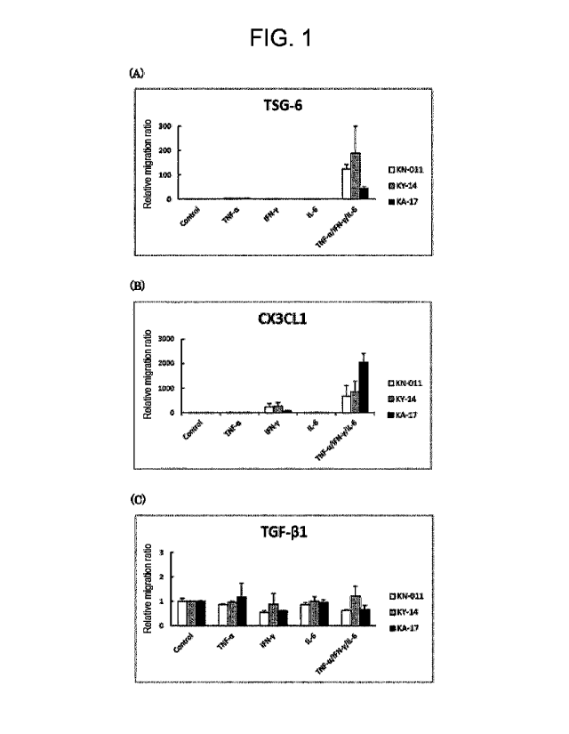
[Figure 1] Figure 1 shows the results of measuring by
real-time RT-PCR the gene expression (relative expression
ratio) of (A) TSG-6, (B) CX3CL1, and (C) TGF-P1 in MSC
samples (KN-011, KY-14, KA-17, 3 Lots). The graphs show,
from the left, no addition (Control), addition of TNF-a
(50 ng/ml), IFN-y (50 ng/ml), IL-6 (50 ng/mL) and INF-
a/IFN-y/IL-6 (all 50 ng/ml).
Date Recue/Date Received 2020-05-08
CA 03082368 2020-05-08
- 10 -
[Figure 2] Figure 2 shows the results of measuring by
ELISA the expression levels (pg/1.0 x 104 cells) of (A)
TSG-6, (B) CX3CL1, and (C) TGF-P1 in MSC samples (KN-011,
KY-14, KA-17, 3 Lots). The graphs show, from the left,
no addition (Naive), addition of TNF-a (50 ng/ml), IFN-y
(50 ng/ml), IL-6 (50 ng/mL) and TNF-a/IFN-y/IL-6 (all 50
ng/ml).
[Figure 3-1] Figure 3-1 shows the results of measuring by
real-time RT-PCR the gene expression (relative expression
ratio) of (A) VEGF, (B) HGF, (C) NGF, and (D) GDNF in MSC
samples (KN-011, KY-14, KA-17, 3 Lots). The graphs show,
from the left, no addition (Control), addition of TNF-a
(50 ng/ml), IFN-y (50 ng/ml), IL-6 (50 ng/mL) and INF-
a/IFN-y/IL-6 (all 50 ng/ml).
[Figure 3-2] Figure 3-2 shows the results of measuring by
real-time RT-PCR the gene expression (relative expression
ratio) of (E) PDGF-A, (F) PDGF-B, (G)PIGF, and (H) BDNF
in MSC samples (KN-011, KY-14, KA-17, 3 Lots). The
graphs show, from the left, no addition (Control),
addition of TNF-a (50 ng/ml), IFN-y (50 ng/ml), IL-6 (50
ng/mL) and TNF-a/IFN-y/IL-6 (all 50 ng/ml).
[Figure 4-1] Figure 4-1 shows the results of measuring by
ELISA the expression levels (pg/1.0 x 104 cells) of (A)
proBDNF, (B) mature PDNF, (C) NGF, and (D) GDNF in MSC
samples (KN-011, KY-14, KA-17, 3 Lots). The graphs show,
from the left, no addition (Naive), addition of TNF-a (50
Date Recue/Date Received 2020-05-08
CA 03082368 2020-05-08
- 11 -
ng/m1), IFN-y (50 ng/m1), IL-6 (50 ng/mL) and TNF-a/IFN-
y/IL-6 (all 50 ng/m1).
[Figure 4-2] Figure 4-2 shows the results of measuring by
ELISA the expression levels (pg/1.0 x 104 cells) of (E)
VEGF, (F) PIGF, (G) HGF, and (H) PDGF-AB in MSC samples
(KN-011, KY-14, KA-17, 3 Lots). The graphs show, from
the left, no addition (Naive), addition of TNF-a (50
ng/m1), IFN-y (50 ng/m1), IL-6 (50 ng/mL) and TNF-a/IFN-
y/IL-6 (all 50 ng/m1).
[Figure 5-1] Figure 5-1 shows the results of expression
analysis by flow cytometry of chemokine receptors (CCR1,
CCR2, CCR3, CCR4, CCR5, CXCR1, CXCR2, CXCR3, CXCR4, CXCR5,
CXCR6, CXCR7, and CX3CR1) and growth factor receptors
(PDGFRa, PDGFRb, FGF-R2, EGFR, HGFR, NGFR, IGF1R, VEGFR1,
VEGFR2, and Tie-2) in MSC samples (KN-011, KY-14, KA-17,
3 Lots).
[Figure 5-2] Figure 5-2 shows the results of expression
analysis by flow cytometry of adhesion factors (NCAD,
HCAM (CD44), NCAM, ALCAM, ITGAV, ITGA4, ITGB1, ITGB4,
VCAM1, and ICAM2) in MSC samples (KN-011, KY-14, KA-17, 3
Lots).
[Figure 6] Figure 6 shows the results of Migration Assay
of MSC samples (KN-011, KY-14, KA-17, 3 Lots) by
chemokine and growth factor stimulation (relative
expression ratio to unstimulated culture, Mean SD, *1.5
fold-change v.s Naive).
Date Recue/Date Received 2020-05-08
CA 03082368 2020-05-08
- 12 -
[Figure 7-1] Figure 7-1 shows the results of measuring by
real-time RT-PCR the gene expression (relative expression
ratio) of adhesion factors ((A) ITGB1 and (B) ITGA4), and
infiltration-related proteins ((C) MMP1) in MSC samples
(KN-011, KY-14, KA-17, 3 Lots). The graphs show, from
the left, no addition (Control), addition of INF-a (50
ng/ml), IFN-y (50 ng/ml), IL-6 (50 ng/mL) and INF-a/IFN-
y/IL-6 (all 50 ng/ml).
[Figure 7-2] Figure 7-2 shows the results of measuring by
real-time RT-PCR the gene expression (relative expression
ratio) of infiltration-related proteins ((D) MMP2, (E)
TIMP1, and (F) TIMP2) in MSC samples (KN-011, KY-14, KA-
17, 3 Lots). The graphs show, from the left, no addition
(Control), addition of INF-a (50 ng/ml) , IFN-y (50 ng/ml),
IL-6 (50 ng/mL) and INF-a/IFN-y/IL-6 (all 50 ng/ml).
Description of Embodiments
[0013]
1. Cell-based medicine containing mesenchymal stem cells
The cell-based medicine of the present invention
contains mesenchymal stem cells, and is characterized in
that: a) the mesenchymal stem cells express CX3CL1 under
cytokine stimulation, and/or b) 90% or more of the
mesenchymal stem cells express EGFR and/or ITGA4.
[0014]
[Mesenchymal stem cells]
Date Recue/Date Received 2020-05-08
CA 03082368 2020-05-08
- 13 -
The "mesenchymal stem cells" used in the present
invention are stem cells having pluripotency and self-
renewal ability which are present in trace amounts among
stromal cells of mesenchymal tissue, and are known to
have the ability to differentiate not only into
connective tissue cells such as osteocytes, chondrocytes,
and lipocytes, but also into nerve cells and
cardiomyocytes.
[0015]
As to the therapeutic mechanism of MSCs, numerous
action mechanisms have been speculated and proposed. For
example, MSCs are known to enable effective tissue
regeneration by accumulating at the site of injury. In
addition, it has been reported that MSCs have an
inhibitory effect on cell death and an inflammation
modulatory effect, and have a neuroprotective effect via
the secretion of neurotrophic factors (as mentioned
above).
[0016]
[CX3CL1 Expression]
The MSCs used in the present invention are
characterized by expressing CX3CL1 upon stimulation of an
inflammatory cytokine.
[0017]
CX3CL1 is a chemokine of the CXXXC motif, also
called fractalkine, expressed in activated vascular
endothelial cells, nerve cells, dendritic cells and
Date Recue/Date Received 2020-05-08
CA 03082368 2020-05-08
- 14 -
intestinal epithelial cells, and its expression is
induced by stimulation with an inflammatory cytokine.
Chemokines refer to a group of basic bioactive peptides
having a molecular weight of about 10 kDa, which have
chemotactic activity for leukocytes such as neutrophils,
monocytes, and lymphocytes, and play an important role in
inflammatory reactions. Chemokines are classified into
four subfamilies, CXC, CC, C, and CX3C, based on their
structural characteristics, and a seven-transmembrane
trimeric G protein-coupled receptor (GPCR) family
classified into CXCR, CCR, XCR, and CX3CR has been
identified for these chemokine subfamilies. In vivo,
CX3CL1 takes two forms, a membrane-bound form and a
secreted form, and functions not only as a chemokine but
also as a cell adhesion molecule showing integrin-
independent cell adhesion ability. The expression of
CX3CL1 is known to be involved in various diseases such
as rheumatoid arthritis and arteriosclerosis.
[0018]
The inhibitory effect on cell death and the
inflammation modulatory effect are known to be related to
the modulation effect of microglia and macrophage by MSCs
at the site of injury, but the inventors have found that
the characteristic expression of CX3CL1 in response to
stimulation with an inflammatory cytokine is useful as an
indicator of MSC's inflammation modulatory effect
(immunomodulatory effect), by real-time RT-PCR and ELISA
Date Recue/Date Received 2020-05-08
CA 03082368 2020-05-08
- 15 -
analysis. MSCs expressing CX3CL1 are expected to exert
an immunomodulatory (inflammation modulatory) effect by
modulating microglia/macrophages and inducing a switch
from M1 type having an inflammatory effect to M2 type
having an anti-inflammatory effect.
[0019]
Examples of the "inflammatory cytokine" to be used
include interleukins such as IL-1, IL-6, IL-8, IL-12, and
IL-18, TNF-a, and IFN-y. Among these, IL-6, TNF-a and
IFN-y are preferable, and it is more preferable to use a
mixture of IL-6, TNF-a and IFN-y.
[0020]
If MSCs express CX3CL1 in response to stimulation
with an inflammatory cytokine, the MSCs can be expected
to have an excellent inflammation modulatory effect
(immunomodulatory effect).
[0021]
In particular, the fact that when stimulated using
TNF-a, INFy, and IL-6, the CX3CL1 expression level by
stimulation with a mixture of TNF-a, INFy, and IL-6 is
greater than the sum of CX3CL1 expression levels by
stimulation with each TNF-a, INFy, and IL-6 alone can be
used as a characteristic indicator of functional (having
an excellent inflammation modulatory effect) MSCs.
[0022]
[EGFR and/or ITGA4 Expression]
Date Recue/Date Received 2020-05-08
CA 03082368 2020-05-08
- 16 -
The MSCs used in the present invention are
characterized in that 90% or more express EGFR (Epidermal
Growth Factor Receptor) and/or ITGA4 (Integrin subunit
Alpha 4).
[0023]
EGFR is a type of tyrosine kinase receptor and binds
to TGF-a, amphiregulin, and the like in addition to
epidermal growth factor (EGF) as a ligand. Receptor
tyrosine kinases such as EGFR transmit stimulation with
extracellular growth factors into cells and transmit the
stimulation to the nucleus by signal transduction. As a
result, the transcriptional activity in the nucleus is
increased, which alters protein synthesis and the
function and structure of cells. EGFR is known to play
an important role in the proliferation of various cells
and in the development and formation of organs in the
body.
[0024]
Integrins are one of the cell surface proteins and
are mainly cell adhesion molecules involved in the cell
adhesion to the extracellular matrix and signal
transduction from the extracellular matrix. An integrin
molecule is a heterodimer in which an a-chain and a p-
chain are associated at a ratio of 1: 1. At least 18
types of a-chains have been reported, and ITGA4 is one of
them. ITGB1 and ITGA4 are important for the adhesion to
vascular endothelium and have been reported to be related
Date Recue/Date Received 2020-05-08
CA 03082368 2020-05-08
- 17 -
to the accumulation of migrated cells at the site of
injury (James et al., 2007, Brigitte et al., 2006).
[0025]
Accumulation at the site of injury involves the
migratory ability of MSCs. The present inventors
performed receptor analysis by flow cytometry and
migration assay on chemokines and growth factors related
to migration, and found the expression of EGFR and/or
ITGA4 to be useful as an indicator of the migration
ability and ability to accumulate at the site of injury
of MSCs.
[0026]
The secretion of growth factors and the like such as
EGF and NGF is known to increase at the site of tissue
injury in trauma patients. In addition, the release of
EGF, bFGF, IL-6 and IL-8 from blood platelets during the
wound healing process has been confirmed (Ono et al.,
1995, Burns 21, 352-355, Zhuang et al., 2013, Asian
Pacific Journal of Tropical Medicine, 383-386, Werner et
al., 2003, Physiol Rev 83, 835-870). However, there are
no reports on the relationship between the expression of
EGFR and ITGA4 in MSCs and the accumulation at the site
of injury.
[0027]
If the expression of EGFR and/or ITGA4 of MSCs is
90% or more, the MSCs can be expected to have an
excellent ability to accumulate at the site of injury.
Date Recue/Date Received 2020-05-08
CA 03082368 2020-05-08
- 18 -
[0028]
[BDNF Expression]
It is preferable that the MSCs used in the present
invention secrete one or more trophic factors selected
from BDNF, VEGF and HGF, in addition to the expression of
CX3CL1, and the expression of EGFR and/or ITGA4. Among
these, the secretion of BDNF and/or VEGF is important,
and the secretion of BDNF is particularly important.
[0029]
BDNF (Brain-derived Neurotrophic Factor) is a
humoral protein that binds to a specific receptor TrkB on
the surface of target cells and regulates the growth of
nerve cells. BDNF acts on some neurons of the central
nervous system and peripheral nervous system, promoting
their maintenance and growth, and promoting
differentiation into new neurons and synapses. In the
brain, BDNF is activated in the hippocampus, cerebral
cortex, and cerebral basal ganglia, and is important for
long-term memory, but is also known to act on the retina,
motor neurons, kidneys, salivary glands, and prostate.
[0030]
VEGF (Vascular Endothelial Growth Factor) is a
growth factor that specifically acts on vascular
endothelial cells isolated from the culture of pituitary
cells. Since VEGF has the effect of promoting angiogenic
processes, including the proliferation of vascular
endothelial cells, and of enhancing vascular permeability,
Date Recue/Date Received 2020-05-08
CA 03082368 2020-05-08
- 19 -
it has been presumed to be related to various diseases
and symptoms in which angiogenesis plays an important
role (cancer, diabetic retinopathy, rheumatoid arthritis,
wound healing process)
[0031]
HGF (Hepatocyte Growth Factor) is a cytokine
purified as a factor that strongly promotes the
proliferation of primary cultured hepatocytes, and is an
important factor that promotes liver regeneration. HGF
exerts biological activity via c-Met receptors expressed
on target cells, and promotes cell proliferation, cell
motility, anti-apoptosis (cell death), morphogenesis
induction and angiogenesis, not only for hepatocytes but
also for various cells.
[0032]
Trophic factors and the like secreted by MSCs may be
involved in the neuroprotective effect. The present
inventors have examined the expression of neurotrophic
factors secreted by MSCs by real-time RP-PCR and ELISA,
and have confirmed that the expression of BDNF, VEGF, and
HGF, especially the expression of BDNF and/or VEGF, in
particular the expression of BDNF, was useful as an
indicator of the neuroprotective effect of MSCs.
[0033]
If the MSCs have the ability to secrete BDNF, VEGF
and/or HGF, it can be expected that the MSCs have the
ability to repair and regenerate the injured area and
Date Recue/Date Received 2020-05-08
CA 03082368 2020-05-08
- 20 -
have an excellent neuroprotective effect. Although MSCs
secrete BDNF, VEGF and/or HGF even when not stimulated,
the secretion ability may be confirmed by evaluating the
secretion from unstimulated cells or by evaluating the
secretion from cells after stimulation with an
inflammatory cytokine.
[0034]
As shown in the examples below, the MSCs used in the
present invention also express TGF-P1 in addition to
CX3CL1 as a factor involved in the inflammation
modulatory effect (immunomodulatory effect). In addition,
as factors involved in the migration ability, the
expression of NCAM, ALCAM, ITGAV, and ITGB1 was also
observed in addition to EGFR and/or ITGA4.
[0035]
[Expression Analysis]
In the present invention, the expression of the
above CX3CL1, EGFR, ITGA4, BDNF, VEGF, and HGF can be
easily determined by a method well-known in the art. For
example, real-time PCR (real-time RT-PCR), microarray,
Northern blot, and the like can be utilized for
expression analysis at the gene level. Moreover, ELISA,
flow cytometry (FCM), protein chips, and the like can be
utilized for expression analysis at the protein level.
[0036]
In particular, for the expression at the protein
level, in the case of cell surface proteins such as EGFR
Date Recue/Date Received 2020-05-08
CA 03082368 2020-05-08
- 21 -
and ITGA4, it is preferable to use flow cytometry (FCM)
in terms of simplicity and sensitivity, and in the case
of secretory proteins such as CX3CL1, BDNF, VEGF, and HGF,
it is preferable to use a bead-based assay in terms of
simplicity and sensitivity.
[0037]
[MSC Modulation]
The sources of the MSCs used in the present
invention include bone marrow, peripheral blood,
umbilical cord blood, fetal embryo, and the brain, but in
the present invention, MSCs derived from human bone
marrow or blood (bone marrow mesenchymal stem cells),
particularly human bone marrow MSCs are preferable.
[0038]
The cells may be cells induced to differentiate from
ES cells or induced pluripotent stem cells (such as iPS
cells), may be established cells, or may be cells
isolated and proliferated from living organisms. The
cells may be derived from allogeneic cells or derived
from autologous cells, but autologous cell-derived
(derived from the patient's own cells) MSCs are
preferable.
[0039]
In the MSCs used in the present invention, it is
preferable that the expression of at least one or more
selected from CD73, CD90, and CD105 is 90% or more,
and/or the expression of CD34 or CD45 is 5% or less.
Date Recue/Date Received 2020-05-08
CA 03082368 2020-05-08
- 22 -
More preferably, the MSCs used in the present invention
are characterized in that the expression of at least two
or more selected from CD73, CD90, and CD105 is 90% or
more, and/or the expression of CD34 and CD45 is 5% or
less. Further preferably, the MSCs used in the present
invention are characterized in that the expression of
CD73, CD90, and CD105 is 90% or more, and the expression
of CD34 or CD45 is 5% or less.
[0040]
Moreover, it is preferable that the MSCs used in the
present invention are cells that are 0D24 negative, which
is a differentiation marker, and maintain an
undifferentiated state. MSCs maintained in an
undifferentiated state have the characteristic that the
proliferation rate and the survival rate after
introduction into a living body are high. A method for
obtaining such undifferentiated MSCs has also been
developed, and details thereof are described in WO
2009/034708.
[0041]
The functional MSCs suitable for the cell-based
medicine of the present invention can be prepared, for
example, by proliferating cells separated from bone
marrow fluid or the like under conditions such that they
do not substantially come into contact with an
anticoagulant (such as heparin), using a culture medium
containing human serum (preferably autologous serum), and
Date Recue/Date Received 2020-05-08
CA 03082368 2020-05-08
- 23 -
containing no anticoagulant or an extremely low
concentration of an anticoagulant (such as heparin).
Here, "containing no anticoagulant or an extremely low
concentration of an anticoagulant" means that it does not
contain an effective amount of an anticoagulant as an
anticoagulant. Specifically, for example, in the case of
heparin or a derivative thereof, the effective amount as
an anticoagulant is usually about 20 to 40 U/mL, but in
the above method, by minimizing the amount added to a
blood collection tube for sampling in advance, the amount
in a sample collected from a living body is less than 5
U/mL, preferably less than 2 U/mL, further preferably
less than 0.2 U/mL, and the amount present in the culture
medium when cells are cultured is less than 0.5 U/mL,
preferably less than 0.2 U/mL, further preferably less
than 0.02 U/mL, based on the volume of the culture medium
(see WO 2009/034708).
[0042]
The density of the cells in the culture medium
affects the properties of the cells and the direction of
differentiation. In the case of MSCs, if the cell
density in the culture medium exceeds 8,500 cells/cm2,
the properties of the cells will change. Therefore, it
is preferable to subculture at a maximum of 8,500
cells/cm2 or less, and more preferably, to subculture
when the cell density reaches 5,500 cells/cm2 or more.
[0043]
Date Recue/Date Received 2020-05-08
CA 03082368 2020-05-08
- 24 -
In the above method, since a culture medium
containing human serum is used, it is desirable that the
number of medium changes is as small as possible, taking
into consideration the burden on the serum donor, and for
example, the medium is changed at least once a week, more
preferably once or twice a week.
[0044]
As for the culture, subculture is repeated until the
total number of cells reaches 108 cells or more. The
number of cells required may vary depending on the
purpose of use, but for example, the number of MSCs
required for transplantation for the treatment of
cerebral infarction is considered to be 107 cells or more.
According to the above method, 107 MSCs can be obtained
in about 12 days.
[0045]
The proliferated MSCs may be stored by a technique
such as cryopreservation (for example, in a deep freezer
at -152 C) until use, if necessary. For cryopreservation,
a culture medium (a culture medium for mammalian cells
such as RPMI) containing serum (preferably human serum,
more preferably autologous serum), dextran, and DMSO is
used as a cryopreservation solution. For example, cells
can be suspended in a cryopreservation solution
containing 20.5 mL of RPMI sterilized by standard
filtration, 20.5 mL of autologous serum collected from a
patient, 5 mL of dextran, and 5 mL of DMSO, and
Date Recue/Date Received 2020-05-08
CA 03082368 2020-05-08
- 25 -
cryopreserved at -150 C. For example, as DMSO, Cryosery
manufactured by Nipro Corporation can be used, and as
dextran, low molecular weight dextran L injection
manufactured by Otsuka Pharmaceutical can be used, but
they are not limited thereto.
[0046]
[Cell-based Medicine (Cell-based Preparation)]
The larger the number of MSCs contained in the cell-
based medicine of the present invention is, the more
preferable. However, when taking into account the time
of administration to a subject and the time required for
culturing, it is practical to use the minimum amount
showing the effects. Therefore, in a preferable aspect
of the present invention, the number of MSCs is 107 or
more, preferably 5 x 10⁷ or more, more preferably 108 or
more, and further preferably 5 x 10⁸ or more. The number
of administrations is not limited to one, and may be two
or more.
[0047]
The cell-based medicine of the present invention is
preferably a preparation for parenteral administration,
more preferably a preparation for parenteral systemic
administration, in particular a preparation for
intravenous administration. Dosage forms suitable for
parenteral administration include injections such as
solution injections, suspension injections, emulsion
injections, and extemporaneously prepared injections, and
Date Recue/Date Received 2020-05-08
CA 03082368 2020-05-08
- 26 -
implants. Preparations for parenteral administration are
in the form of an aqueous or non-aqueous isotonic sterile
solution or suspension. For example, pharmacologically
acceptable carriers or media, specifically, sterile water
or normal saline solution, a culture medium (in
particular, a culture medium used for culturing mammalian
cells such as RPMI), physiological buffers such as PBS,
vegetable oils, emulsifiers, suspending agents,
surfactants, stabilizers, excipients, vehicles,
preservatives, binding agents and the like are
appropriately combined and formulated into an appropriate
unit dosage form.
[0048]
Examples of aqueous solutions for injection include
normal saline solution, culture media, physiological
buffers such as PBS, isotonic solutions containing
glucose or other adjuvants, such as D-sorbitol, D-mannose,
D-mannitol, and sodium chloride, and these may be used
with a suitable solubilizing agent such as alcohol,
specifically ethanol, polyalcohol, propylene glycol,
polyethylene glycol, or a nonionic surfactant such as
polysorbate 80 or HCO-50.
[0049]
The cell-based medicine of the present invention is
a medicine for tissue regeneration, and in particular, it
is useful for treating dementia, chronic cerebral
infarction, chronic spinal cord injury, neurodegenerative
Date Recue/Date Received 2020-05-08
CA 03082368 2020-05-08
- 27 -
diseases, mental illnesses, higher dysfunctions and the
like, by synapse formation and a plasticity promoting
effect at the site of injury (lesion).
[0050]
2. Method for producing a cell-based medicine containing
mesenchymal stem cells
The present invention also provides a method for
producing a cell-based medicine containing mesenchymal
stem cells. The method for producing the cell-based
medicine of the present invention includes the steps of:
a) adding cytokines to a culture containing mesenchymal
stem cells, and confirming that the mesenchymal stem
cells express CX3CL1, and/or b) confirming that the
mesenchymal stem cells express EGFR and/or ITGA4.
[0051]
Examples of the "inflammatory cytokine" to be used
Include INF-a, INFy, IL-1, IL-6, IL-8, IL-12, and IL-18,
among these it is preferable to include INF-a, INFy, and
IL-6, and more preferable to use a mixture of INF-a, INFy,
and IL-6.
[0052]
The method for producing the cell-based medicine of
the present invention may further include a step of
confirming the presence of one or more selected from BDNF,
VEGF, and HGF in the culture (without cytokines). In
particular, it is important to confirm the presence of
Date Recue/Date Received 2020-05-08
CA 03082368 2020-05-08
- 28 -
BDNF and/or VEGF, and it is most important to confirm the
presence of BDNF.
[0053]
As described above, if MSCs express CX3CL1 by the
addition of inflammatory cytokines, the MSCs can be
expected to have an excellent inflammation modulatory
effect (immunomodulatory effect), and if 90% or more of
the MSCs express EGFR and/or ITGA4, the MSCs can be
expected to have an excellent ability to accumulate at
the site of injury. In addition, if any of the trophic
factors such as BDNF, VEGF, and HGF is present in the
culture medium, MSCs having a high neuroprotective effect
can be expected to be contained, and among these, the
presence of BDNF and/or VEGF, especially the presence of
BDNF may be an important indicator of MSCs having a high
neuroprotective effect. Although MSCs secrete BDNF, VEGF
and/or HGF even when not stimulated, the secretion
ability may be confirmed by evaluating the secretion from
unstimulated cells or by evaluating the secretion from
cells after stimulation with an inflammatory cytokine.
[0054]
It is preferable to use the expression at the
protein level rather than the gene level as an indicator
for the expression of the above CX3CL1, EGFR, ITGA4, BDNF,
VEGF, and HGF, which can be determined by the method
described in the previous section. In particular, in the
case of cell surface proteins such as EGFR and ITGA4, it
Date Recue/Date Received 2020-05-08
CA 03082368 2020-05-08
- 29 -
is preferable to use flow cytometry (FCM) in terms of
simplicity and sensitivity, and in the case of secretory
proteins such as CX3CL1, BDNF, VEGF, and HGF, it is
preferable to use a bead-based assay in terms of
simplicity and sensitivity.
[0055]
The MSCs used in the method for producing the cell-
based medicine of the present invention can be prepared
by proliferating cells separated from bone marrow fluid
or the like under conditions such that they do not
substantially come into contact with an anticoagulant
(such as heparin), using a culture medium containing
human serum (preferably autologous serum), and containing
no anticoagulant or an extremely low concentration of an
anticoagulant (such as heparin), as described in the
previous section, according to the description in WO
2009/034708. Here, "containing no anticoagulant or an
extremely low concentration of an anticoagulant" means
that it does not contain an effective amount of an
anticoagulant as an anticoagulant. Specifically, for
example, in the case of heparin or a derivative thereof,
the effective amount as an anticoagulant is usually about
20 to 40 U/mL. In the above-described method, by
minimizing the amount added to a blood collection tube
for sampling in advance, the amount in a sample collected
from a living body is less than 5 U/mL, preferably less
than 2 U/mL, further preferably less than 0.2 U/mL, and
Date Recue/Date Received 2020-05-08
CA 03082368 2020-05-08
- 30 -
the amount present in the medium when cells are cultured
is less than 0.5 U/mL, preferably less than 0.2 U/mL,
further preferably less than 0.02 U/mL, based on the
volume of the culture medium.
[0056]
3. Method for evaluating the immunomodulatory ability of
a cell-based medicine containing mesenchymal stem cells
The present invention also provides a method for
evaluating the immunomodulatory ability of a cell-based
medicine containing mesenchymal stem cells. The
evaluation method includes a step of stimulating
mesenchymal stem cells with an inflammatory cytokine and
determining the expression of CX3CL1. The "inflammatory
cytokines" to be used and the method for determining the
expression of CX3CL1 are as described in 1 and 2.
[0057]
If the MSCs after cytokine stimulation express
CX3CL1, the cell-based medicine containing the MSCs can
be evaluated as having high immunomodulatory ability. In
particular, if when stimulated using TNF-a, INFy, and IL-
6, the CX3CL1 expression level by stimulation with a
mixture of TNF-a, INFy, and IL-6 is greater than the sum
of CX3CL1 expression levels by stimulation with each TNF-
a, INFy, and IL-6 alone, the MSCs can be evaluated as
having high immunomodulatory ability.
[0058]
Date Recue/Date Received 2020-05-08
CA 03082368 2020-05-08
- 31 -
4. Method for evaluating the accumulation of a cell-based
medicine containing mesenchymal stem cells at a site of
injury
A method for evaluating the ability of a cell-based
medicine containing mesenchymal stem cells to accumulate
at a site of injury is also provided. The evaluation
method includes a step of confirming that 90% or more of
the mesenchymal stem cells express EGFR and/or ITGA4.
[0059]
If 90% or more of the MSCs express EGFR and/or ITGA4,
a cell-based medicine containing the MSCs can be
evaluated as having an excellent ability to accumulate at
the site of injury.
Examples
[0060]
Hereafter, the present invention is described
specifically with examples, but the present invention is
not limited to these examples.
[0061]
Example 1: Immunomodulatory effect
The inhibitory effect on cell death and
immunomodulatory effect of MSCs is known to be related to
the modulation effect of microglia and macrophages by
MSCs at the site of injury. Therefore, in order to
examine the immunomodulatory ability of a cell-based
medicine containing MSCs, the expression of TSG-6, CX3CL1,
Date Recue/Date Received 2020-05-08
CA 03082368 2020-05-08
- 32 -
and TGF-P1 as relevant factors was analyzed by real-time
RT-PCR and ELISA.
[0062]
1. Experimental methods and evaluation items
1.1 Cell culture
As MSC samples, samples of three different lots for
clinical trial (STR01) (KN-011, KY-14, and KA-17) were
used. The MSC samples were suspended in 14 mL of a
culture solution (10% human serum, 1% Penicillin-
streptomysin, 1% L-Glutamine) and seeded on a 150 mm dish
at a density of 0.7 to 1.0 x 10⁶ cells/dish. The cells
were cultured under the conditions of a temperature of
37 C and 5% CO2, and after confirming about 80%
confluency, the cells were subcultured and seeded at a
density of 5.0 x 10⁵ cells/dish. Subculture was
continued, and the cells were seeded at a density of 3.0
x 105 cells/dish on a 100 mm dish at the fourth passage.
Four passages of cells were used in all of the following
experimental systems.
[0063]
1.2 Collection of culture supernatant stimulated with
inflammatory cytokines and extraction of total RNA
Twenty-four hours after the fourth passage, the
culture solution was replaced with a normal culture
solution (10% FBS, 1% Penicillin-Streptomycin, 1% L-
Glutamine), and 10 mL of a culture solution with
inflammatory cytokines (TNF-a (50 ng/ml), IFN-y (50
Date Recue/Date Received 2020-05-08
CA 03082368 2020-05-08
- 33 -
ng/ml), IL-6 (50 ng/ml), and TNF-a/IFN-y/IL-6 (50 ng/ml
each) (5 Conditions, n = 3). The inflammatory cytokines
are thought to be secreted at the site of spinal cord
injury and to cause various cell disorders. Forty-eight
hours after the exchange, the culture supernatant was
collected and centrifuged (2280 g, 20 min). Thereafter,
200 1 of each was dispensed into 1.5 ml tubes and stored
in a -80 C freezer. After collecting the supernatant,
the cells were detached from the dish by trypsin
treatment, and were counted. After counting the cells,
total RNA was extracted using an RNA extraction kit
(QIAGEN). cDNA was synthesized from total RNA, and real-
time RT-PCR was performed using these as templates.
[0064]
2. Evaluation of results and criteria
2.1 Gene expression by real-time RT-PCR
cDNA was synthesized from total RNA extracted from
cells. A PCR reaction was performed with the synthesized
cDNA as templates, using Taqman probes for each of the
factors TSG-6, CX3CL1, and TGF-Pl. Based on the Ct value
of each target and the internal standard, the gene
expression levels of cells cultured with a normal culture
solution and with inflammatory cytokines were compared
and quantified by the AACt method.
[0065]
The test specimens were a specimen free of cytokines
(Naive), and specimens with TNF-a (50 ng/ml), IFN-y (50
Date Recue/Date Received 2020-05-08
CA 03082368 2020-05-08
- 34 -
ng/ml), IL-6 (50 ng/ml), and TNF-a/IFN-y/IL-6 (50 ng/ml
each). mRNA and culture supernatant were collected 48
hours after the start of culture, and real-time RT-PCR
was performed.
[0066]
2.2 Quantitation of secreted proteins by ELISA
Using the culture supernatant collected and stored
in 1.1 as a sample, the TSG-6, CX3CL1, and TGF-Pl in the
culture supernatant were quantified. The test specimens
were a specimen free of cytokines (Naive), and specimens
with TNF-a (50 ng/ml), IFN-y (50 ng/ml), IL-6 (50 ng/ml),
and TNF-a/IFN-y/IL-6 (50 ng/ml each). mRNA and culture
supernatant were collected 48 hours after the start of
culture, and real-time RT-PCR was performed.
[0067]
3. Results
3.1 Real-time RT-PCR (Figure 1)
The graph shows the relative expression ratio to the
control, and the table shows the Ct value. Gene
expression of TSG-6, CX3CL1, and TGF-Pl was confirmed in
all lots, and in particular, the expression of TSG-6 and
CX3CL1 was significantly increased by the addition of the
mixture of TNF-a/IFN-y/IL-6. These results suggest that
MSCs are involved in the modulation effect on microglia
and macrophages, and that TSG-6, CX3CL1, and TGF-Pl
contribute to this effect.
[0068]
Date Recue/Date Received 2020-05-08
CA 03082368 2020-05-08
- 35 -
3.2 Quantitation of secretory proteins by ELISA (Figure
2)
In all lots, TSG-6 and TGF-P1 showed no change in
the expression level due to cytokine stimulation.
However, although CX3CL1 showed almost no expression when
unstimulated or stimulated with a cytokine alone, a
significant expression was observed by the addition of
the mixture of TNF-a/IFN-y/IL-6. In addition, in the RT-
PCR results, the expression by the mixed stimulation with
TNF-a/IFN-y/IL-6 was greater than the sum of the
expressions by each stimulation alone.
[0069]
4. Discussion
The expression of CX3CL1 was hardly observed by
ELISA, but the expression by the mixed stimulation with
TNF-a/IFN-y/IL-6 was confirmed. In addition, the
expression by mixed stimulation with TNF-a/IFN-y/IL-6 is
greater than the sum of expressions by each stimulation
alone; this expression characteristic of CX3CL1 was not
observed for other immunomodulatory ability-related
factors secreted by MSCs (TSG-6 and TGF-P1). Therefore,
CX3CL1 was considered to be useful as an indicator for
evaluating the immunomodulatory ability of MSCs.
[0070]
Example 2: Neuroprotective effect
A plurality of trophic factors may be involved in
the neuroprotective effect of MSCs. The expression of
Date Recue/Date Received 2020-05-08
CA 03082368 2020-05-08
- 36 -
trophic factors (VEGF, HGF, NGF, GDNF, PDGF-A, PDGF-A,
PIGF, and BDNF) secreted by MSCs was analyzed.
[0071]
1. Experimental methods and evaluation items
The cell culture and preparation of total RNA were
performed by the method described in Example 1.
[0072]
2. Evaluation of results and criteria
Same as in Example 1.
[0073]
3. Results
3.1 Real-time RT-PCR (Figure 3)
The graph shows the relative expression ratio to the
control, and the table shows the Ct value. In MSCs
cultured in a culture solution free of inflammatory
cytokines, the expression of BDNF, NGF, and GDNF, which
are neurotrophic factors, VEGF, PDGF-A, and PIGF, which
are involved in angiogenesis, and HGF, which is involved
in the repair and regeneration of damaged tissues, was
confirmed. With the mixed stimulation with TNF-a/IFN-
y/IL-6, the expression of NGF was found to have a
tendency to increase.
[0074]
3.2 Quantitation of secretory proteins by ELISA (Figure
4)
In MSCs cultured in a culture solution free of
inflammatory cytokines, the secretion of mature-BDNF,
Date Recue/Date Received 2020-05-08
CA 03082368 2020-05-08
- 37 -
which is a neurotrophic factor, its precursor proBDNF,
and VEGF, which is involved in angiogenesis, was
confirmed. In addition, HGF and PIGF were confirmed in
two out of three specimens. On the other hand, the
secretion of NGF, GDNF and PDGF-AB was not confirmed.
With the mixed stimulation with TNF-a/IFN-y/IL-6, the
secretion amount of VEGF and HGF were found to have a
tendency to increase.
[0075]
4. Discussion
At the mRNA level, the expression of all trophic
factors was confirmed, but at the protein level,
secretion could be confirmed in all samples only for BDNF
and VEGF. With PIGF and HGF, confirmation was possible
in only two out of three lots.
[0076]
As to the neuroprotective effect against spinal cord
injury, numerous neurotrophic factors and growth factors
such as BDNF, NT-3, NGF, PDGF, and GDNF have been
reported to be involved, and the in vivo analysis of
Honmou et al. has confirmed the neuroprotective effect of
BDNF (cited previously, Nomura et al., 2005; Osaka et al.,
2010). In addition, these effects are known to be
further enhanced when intravenously administering BDNF-
MSCs which have been genetically modified to forcibly
express BDNF (cited previously, Sasaki et al., 2009,).
[0077]
Date Recue/Date Received 2020-05-08
CA 03082368 2020-05-08
- 38 -
These results are consistent with the above report,
and it is considered that BDNF secretion is particularly
important as an evaluation indicator of the
neuroprotective effect of MSCs. VEGF and HGF are also
considered useful for the functional evaluation of MSCs
in addition to BDNF.
[0078]
Example 3: MSC migration ability
In order to evaluate the accumulation of MSCs at the
site of injury, the in vitro migration ability of MSCs
was analyzed by FCM method, Migration Assay and real-time
RT-PCR method.
[0079]
1. Experimental methods and evaluation items
The cell culture and preparation of total RNA were
performed by the method described in Example 1.
[0080]
2. Evaluation of results and criteria
2.1 Flow cytometry (FCM) method
First, as an analysis of chemokines and growth
factors related to migration, the expression of each of
the following receptors was analyzed using the FCM method.
[0081]
<Receptors involved in migration>
Chemokine receptors:
CCR1, CCR2, CCR3, CCR4, CCR5, CXCR1, CXCR2, CXCR3,
CXCR4, CXCR5, CXCR6, CXCR7, CX3CR1
Date Recue/Date Received 2020-05-08
CA 03082368 2020-05-08
- 39 -
Growth factor receptors:
VEGFR1, VEGFR2, PDGFRP, EGFR, IGF-1R, FGF-R2, HGFR,
Tie-2
<Adhesion factors>
ICAM2, VCAM, ALCAM, HCAM (CD44), ITGAV, ITGA4, ITGB1
[0082]
2.2 Migration Assay
Next, the chemokines and growth factors shown below
were added to the culture medium, and the migration
ability of MSCs was studied using the Migration Assay
method.
[0083]
<Chemokines and growth factors>
VEGF, EGF, HGF, IGF-1, PDGF-AB, bFGF, ANGPT-1
MCP-1 (CCL2), MIP-1a (CCL3), RANTES (CCL5), Eotaxin-
1 (CCL11), MDC (CCL22),
Eotaxin-2 (CCL24), CRO-a (CXCL1), SDF-1 (CXCL12),
Fractalkine (CX3CL1)
[0084]
The Migration Assay was performed using FluoroBlok
(Corning).
1) The migration factor was added to the well plate, and
the insert was set,
2) the cell suspension was added to the top of the insert,
3) after 18 hours, the number of migrated cells was
counted by counting the stained cells using Calcein AM
(Dojindo Laboratories).
Date Recue/Date Received 2020-05-08
CA 03082368 2020-05-08
- 40 -
The results were evaluated by the relative migration
ratio where the number of migrated cells when no
chemokine or growth factor was added was 1Ø
Relative migration ratio = number of cells (with
migration factor)/number of cells (without migration
factor)
[0085]
2.3 Real-time RT-PCR
According to Example 1, with respect to the factors
relating to adhesion of cells to vascular endothelium and
infiltration into tissues, gene expression with and
without stimulation with an inflammatory cytokine was
analyzed by real-time RT-PCR.
<Adhesion factors>
ITGB1, ITGA4
<Infiltration-related proteins>
MMP1, MMP2, TIMP1, TIMP2
[0086]
3. Results
3.1 Analysis of chemokine receptors, growth factor
receptors and adhesion factors of MSCs by FCM method
(Table 1 and Figure 5)
For the chemokine receptors, the expression of CCR5,
CXCR3, and the like was observed in some cells, but none
was expressed in all cells. On the other hand, for the
growth factor receptors, the expression of EGFR, HGFR,
NGFR, and Tie2 was observed. For the adhesion factors,
Date Recue/Date Received 2020-05-08
CA 03082368 2020-05-08
- 41 -
the expression of NCAD, 0D44, NCAM, ALCAM, ITGA4, and
ITGB1, which are considered to be involved in the
adhesion of the migrated MSCs to vascular endothelial
cells, was observed.
[0087]
[Table 1]
Expression analysis of chemokine receptors and growth factor receptors by FCM
method
Chemokine receptor
Cell 7
CCR1 CCR2 CCR3 CCR4 CCR5 CXCR1 CXCR2 CXCR3 CXCR4 ILCXCRS CXCR6 CXCR7 CX3CR1
Omit ¨ ¨ ¨ ¨ ¨
0'002 _t+/- + _____ - __ +/- ' +1- -
Cell Growth factor receptor
PDGFRa PDGFRb FGF-R2 EGFR HGFR NG IGF1R VEGFR1 VEGFR2
Tle2
KNO11 +1¨ ¨ 41¨
10,14 ¨ ¨ , +1¨ + +1¨
leY002 1 +/¨
Adheeion factor
Cell
NCAD C044 NCAN1 ALCAM ITGAV ITGA4 ITGBI 11684 VCAM1 ICAM2
KNO11
KY14 +/-
101002 + + +
¨ : Not expressed at all
+1¨ Slightly expressed
: Expressed
[0088]
3.2 Migration Assay (Figure 6)
EGF, PDGF-AB, 13FGF, ANGPT-1, MCP-1 (CCL2), and MIP-
1a (CCL3) were found to promote migration, and this
tendency was especially significant with EGF and MCP-1
(CCL2).
[0089]
3.3 Real-time RT-PCR (Figure 7)
Date Recue/Date Received 2020-05-08
CA 03082368 2020-05-08
- 42 -
It was confirmed that ITGB1 and ITGA4, which are
adhesion factors, and MMP1, MMP2, TIMP1, and TIMP2, which
are related to infiltration, were expressed. In addition,
it was confirmed that MSCs stimulated with inflammatory
cytokines (TNF-a/IFN-y/IL-6) greatly increased the
expression of MMP1. ITGB1 and ITGA4 are important for
the adhesion to vascular endothelium and have been
reported to be related to the accumulation of migrated
cells at the site of injury (cited previously, James et
al., 2007). In addition, it is known that the MMP and
TIMP families thaw the basement membrane of cells, and
migrated cells infiltrate the site of injury (Caroline et
al., 2008, Mariusz et al., 2012). These reports and the
above results suggested that the MSCs have properties
related to the adhesion to vascular endothelium and
infiltration into tissues.
[0090]
4. Discussion
The results of the receptor analysis by FCM and of
the Migration Assay confirmed that the expression of EGFR
was particularly important as an indicator of MSC
migration ability. Moreover, the results of the FCM
analysis and real-time RT-PCR analysis confirmed that the
expression of ITGA4 was important as an indicator of MSC
migration ability.
Industrial Applicability
Date Recue/Date Received 2020-05-08
CA 03082368 2020-05-08
- 43 -
[0091]
According to the present invention, the function of
a cell-based medicine containing mesenchymal stem cells
can be appropriately assayed, and a cell-based medicine
containing mesenchymal stem cells suitable for tissue
regeneration can be provided.
[0092]
All publications, patents and patent applications
cited in the present specification are hereby
incorporated by reference in their entirety.
Date Recue/Date Received 2020-05-08