Note: Descriptions are shown in the official language in which they were submitted.
CA 03082611 2020-05-13
WO 2019/099993 PCT/US2018/061795
METHODS AND COMPOSITIONS FOR ALLEVIATING CYTOKINE
RELEASE SYNDROME
CROSS-REFERENCE TO RELATED APPLICATION
This application claims priority to U.S. Provisional Application No.:
62/587,965
filed on November 17, 2017, the content of which is hereby incorporated by
reference in
its entirety, and to which priority is claimed.
INTRODUCTION
The presently disclosed subject matter provides methods and compositions for
enhancing the immune response toward cancers and pathogens. It relates to
immunoresponsive cells comprising antigen-recognizing receptors (e.g.,
chimeric
antigen receptors (CARs) or T cell receptors (TCRs)) that are engineered to
express an
Interleukin-1 receptor antagonist ("IL-1Ra") polypeptide. These engineered
immunoresponsive cells are antigen-directed, promote recruitment of other
cytokines and
exhibit enhanced anti-target efficacy.
BACKGROUND OF THE INVENTION
Chimeric Antigen Receptor (CAR) modified T cells have shown extraordinary
promise in the clinic and are now an F.D.A. approved modality in relapse-
refractory B
cell Acute Lymphoblastic Leukemia (B-ALL) and Diffuse Large B cell Lymphoma
(DLBCL). Despite it s remarkable therapeutic benefit, CAR T cell therapy can
induce toxicities, among which, Cytokine Release Syndrome (CRS) is a major
concern.
CRS is a commonly occurring and potentially lethal toxicity that typically
presents
itself within days after CAR T cell infusion. In its severe form, CRS can
present
symptoms such as fever, hypotension, respiratory failure and elevation of pro-
inflammatory cytokines, including IL-6. Thus, CRS can be a hindrance to the
broad
application of CART cells 1-4. Therefore, there is a need for an effective
treatment of CRS
and/or a form of CART cell that reduces or avoids CRS.
Moreover, there are currently no reported mouse models in which current
clinical CRS treatments can be validated and new treatment modalities tested.
Therefore, there is a need for a suitable animal model for studying CRS.
SUMMARY OF THE INVENTION
The presently disclosed subject matter provides immunoresponsive cells (e.g.,
T
cells, Tumor Infiltrating Lymphocytes, Natural Killer (NK) cells, cytotoxic T
.. lymphocytes (CTLs), Natural Killer T (NK-T) cells or regulatory T cells)
that (a) express
1
CA 03082611 2020-05-13
WO 2019/099993 PCT/US2018/061795
an antigen-recognizing receptor (e.g., CAR or TCR) directed toward a target
antigen of
interest, and (b) express (and secrete) an interleukin 1 receptor antagonist
("IL-1Ra")
polypeptide. In certain non-limiting embodiments, the immunoresponsive cell
comprises
a nucleic acid encoding an IL-1Ra polypeptide (e.g., IL-1Ra polypeptide-
encoding
nucleic acid), in expressible form.
In certain non-limiting embodiments, the presently disclosed subject matter
provides an immunoresponsive cell (a) comprising an antigen-recognizing
receptor that
binds to an antigen, and (b) expressing or secreting an IL-1Ra polypeptide. In
certain
embodiments, the immunoresponsive cell comprises an exogenous IL-1Ra
polypeptide.
In certain embodiments, the immunoresponsive cell comprises a nucleic acid
encoding
an IL-1Ra polypeptide. In certain embodiments, binding of the antigen-
recognizing
receptor to the antigen is capable of activating the immunoresponsive cell. In
certain
embodiments, the antigen-recognizing receptor is a CAR.
In certain non-limiting embodiments, the presently disclosed subject matter
provides an immunoresponsive cell comprising (a) an antigen-recognizing
receptor (e.g.,
CAR or TCR) directed toward a target antigen of interest, and (b) a modified
promoter at
an endogenous (native) IL-1Ra gene locus. In certain embodiments, the modified
promoter enhances the gene expression of the endogenous IL-1Ra gene locus. In
certain
non-limiting embodiments, the modification comprises replacement of an
endogenous
promoter with a constitutive promoter or an inducible promoter, or insertion
of a
constitutive promoter or inducible promoter to the promoter region of the
endogenous
IL-1Ra gene locus. In certain non-limiting embodiments, the constitutive
promoter is
selected from the group consisting of a CMV promoter, an EFla promoter, a SV40
promoter, a PGK1 promoter, a Ubc promoter, a beta-actin promoter, and a CAG
promoter. In certain non-limiting embodiments, the inducible promoter is
selected from
the group consisting of a tetracycline response element (TRE) promoter and an
estrogen
response element (ERE) promoter.
In certain embodiments, the immunoresponsive cell constitutively expresses the
IL-1Ra polypeptide (mature or non-mature form of IL-1Ra protein). In certain
embodiments, the IL-1Ra polypeptide is secreted. The antigen-recognizing
receptor can
be a TCR or a CAR. In certain embodiments, the antigen-recognizing receptor is
a CAR.
In certain embodiments, the immunoresponsive cell is selected from the group
consisting
of a T cell (e.g., a cytotoxic T lymphocyte (CTL), a regulatory T cell, or a
Natural Killer
T (NK-T) cell), a Natural Killer (NK) cell, a human embryonic stem cell, and a
2
CA 03082611 2020-05-13
WO 2019/099993
PCT/US2018/061795
pluripotent stem cell from which lymphoid cells may be differentiated, a
macrophage, a
neutrophil, a monocyte, and a dendritic cell. In certain embodiments, the
immunoresponsive cell is a T cell. In certain embodiments, the
immunoresponsive cell
is autologous or allogenic.
The presently disclosed subject matter further provides immunoresponsive cells
comprising a modified CD4OL. The modification can be selected from the group
consisting of knock-down of CD4OL, knock-out of CD4OL, introduction of one or
more
mutation in a CD4OL gene, modification of the endogenous promoter of a CD4OL
gene,
modification of the endogenous enhancer elements of a CD4OL gene, modification
of the
transcription factors that control CD4OL expression, and combinations thereof
The presently disclosed subject matter further provides methods for producing
an
immunoresponsive cell disclosed herein. In certain embodiments, the methods
comprise
introducing into an immunoresponsive cell (a) a first nucleic acid sequence
that encodes
an antigen-recognizing receptor that binds to an antigen, and (b) a second
nucleic acid
sequence that encodes an IL-1Ra polypeptide. In certain embodiments, the
methods
comprise introducing into an immunoresponsive cell (a) a first nucleic acid
sequence that
encodes an antigen-recognizing receptor that binds to an antigen, and (b) a
second
nucleic acid sequence that encodes a modified CD4OL.
The presently disclosed subject matter further provides various nucleic acid
compositions. In certain embodiments, the nucleic acid composition comprises
(a) a first
nucleic acid sequence encoding an antigen-recognizing receptor (e.g., a CAR or
TCR)
that binds to an antigen and (b) a second nucleic acid sequence encoding an IL-
1Ra
polypeptide (mature or non-mature form of IL-1Ra). In certain embodiments, the
nucleic acid composition comprises (a) a first nucleic acid sequence encoding
an
antigen-recognizing receptor (e.g., a CAR or TCR) that binds to an antigen and
(b) a
second nucleic acid sequence encoding a modified CD4OL.
In certain non-limiting embodiments, the first or the second nucleic acid
sequence is operably linked to a promoter element constitutively or inducibly
expressed
in the immunoresponsive cell. The promoter for the first nucleic acid sequence
may be
the same or different from the promoter for the second nucleic acid sequence.
In certain
non-limiting embodiments, each of the first and second nucleic acid sequences
is
operably linked to a promoter element constitutively or inducibly expressed in
the
immunoresponsive cell. One or both of the first and second nucleic acid
sequences may
3
CA 03082611 2020-05-13
WO 2019/099993 PCT/US2018/061795
be comprised in a vector, which may be the same vector (bicistronic) or
separate vectors.
In certain non-limiting embodiments, the vector is a virus vector, e.g., a
retroviral vector.
In certain embodiments, the nucleic acid composition is comprised in a vector.
In
certain non-limiting embodiments, the vector is a virus vector, e.g., a
retroviral vector.
The presently disclosed subject matter also provides a vector comprising the
nucleic acid
composition disclosed herein.
The presently disclosed subject matter also provides various methods of
treatments. For example, the presently disclosed subject matter provides
methods of
treating and/or preventing a neoplasm in a subject, methods of reducing tumor
burden in
a subject, methods of lengthening survival of a subject having neoplasm (e.g.,
cancer),
methods of reducing at least one symptom of cytokine release syndrome (CRS) in
a
subject, methods of reducing the level of a cytokine in a subject, methods of
reducing the
level of a chemokine in a subject, and methods of treating or alleviating CRS
in a subject
who receives an immunotherapy, and methods of treating blood cancer in a
subject.
In certain embodiments, the level of a cytokine is reduced. In certain
embodiments, the cytokine is a pro-inflammatory cytokine. In certain
embodiments, the
cytokine is selected from the group consisting of IL-1 alpha, IL-1 beta, IL-6,
IL-8, IL-10,
TNF-a, IFN-y, IL-5, IL-2, IL-4, G-CSF, GM-CSF, M-CSF, IL-12, IL-15, and IL-17.
In certain embodiments, the chemokine is selected from the group consisting of
CCL2, CCL3, CCL5, and CXCL1.
In certain non-limiting embodiments, the immunoresponsive cells reduce the
level of one or more cytokine. In certain non-limiting embodiments, the one or
more
cytokine is selected from the group consisting of IL-la, IL-10, IL-6, IL-8, IL-
10, TNF-a,
IFN-y, IL-5, IL-2, IL-4, G-CSF, GM-CSF, M-CSF, IL-12, IL-15, and IL-17. In
certain
non-limiting embodiments, the immunoresponsive cells reduce the level of one
or more
chemokine. In certain embodiments, the one or more chemokine is selected from
the
group consisting of CCL2, CCL3, CCL5, and CXCL1.
In certain embodiments, each of the various methods disclosed herein comprises
administering to the subject an effective amount of the immunoresponsive cells
or the
pharmaceutical composition disclosed herein. In certain non-limiting
embodiments,
the method described herein does not comprise administering another therapy
for
preventing, treating and/or alleviating CRS.
4
CA 03082611 2020-05-13
WO 2019/099993 PCT/US2018/061795
In certain embodiments, each of the various methods disclosed herein comprises
administering to the subject an antibody that binds to CD4OL and an effective
amount of
the immunoresponsive cells, wherein the immunoresponsive cell comprises an
antigen-
recognizing receptor that binds to an antigen.
In certain embodiments, each of the various methods disclosed herein comprises
administering to the subject an inhibitor of IL-1 signaling and an
immunoresponsive cell
comprising an antigen-recognizing receptor that binds to an antigen. In
certain
embodiments, the inhibitor of IL-1 signaling is selected from the group
consisting of IL-
1 blocking agents, IL-1R1 blocking agents, and combinations thereof. In
certain
embodiments, the IL-1 blocking agents are selected from the group consisting
of IL-1Ra
polypeptides, antibodies that bind to IL-la, antibodies that bind to IL-113,
antibodies that
bind to both IL-la and IL-113, and combinations thereof In certain
embodiments, the IL-
1R1 blocking agents are selected from the group consisting of antibodies that
bind to IL-
1R1, antibodies that bind the IL-1 receptor accessory protein (IL-1RAP/IL-
1RAcP), IL-1
receptor 2 (IL-1R2/IL-1RII) polypeptides, and combinations thereof. In certain
embodiments, the IL-1Ra polypeptide is anakinra. In certain embodiments, the
IL-1
blocking agent is rilonacept. In certain embodiments, the antibody that binds
to IL-113 is
canakinumab.
The presently disclosed subject matter provides uses of the immunoresponsive
cell disclosed herein or the composition disclosed herein for use in a
therapy, e.g., for use
in reducing tumor burden, treating and/or preventing a neoplasm, lengthening
survival of
a subject having a neoplasm, and/or reducing at least one symptom of cytokine
release
syndrome (CRS) in response to a cancer or pathogen in a subject.
The presently disclosed subject matter provides uses of an antibody that binds
to
CD4OL and an effective amounts of immunoresponsive cells, wherein the
immunoresponsive cell comprises an antigen-recognizing receptor that binds to
an
antigen or the composition comprising thereof for use in a therapy, e.g., for
use in
reducing tumor burden, treating and/or preventing a neoplasm, lengthening
survival of a
subject having a neoplasm, and/or reducing at least one symptom of cytokine
release
syndrome (CRS) in response to a cancer or pathogen in a subject.
The presently disclosed subject matter provides uses of an inhibitor of IL-1
signaling and an immunoresponsive cells comprising an antigen-recognizing
receptor
that binds to an antigen or the composition comprising thereof for use in a
therapy, e.g.,
for use in reducing tumor burden, treating and/or preventing a neoplasm,
lengthening
5
CA 03082611 2020-05-13
WO 2019/099993 PCT/US2018/061795
survival of a subject having a neoplasm, and/or reducing at least one symptom
of
cytokine release syndrome (CRS) in response to a cancer or pathogen in a
subject.
The presently disclosed subject matter provides a kit for treating and/or
preventing a neoplasm (e.g., cancer) or a pathogen infection, reducing tumor
burden in a
subject, lengthening survival of a subject having neoplasm (e.g., cancer),
and/or treating
or alleviating CRS in a subject who receives an immunotherapy. In certain
embodiments, the kit comprises the immunoresponsive cells disclosed herein,
the
pharmaceutical composition disclosed herein, the nucleic acid composition
disclosed
herein, or the vector disclosed herein. In certain embodiments, the kit
further comprises
written instructions for treating and/or preventing a neoplasm or a pathogen
infection,
reducing tumor burden in a subject, lengthening survival of a subject having
neoplasm
(e.g., cancer), and/or treating or alleviating CRS in a subject who receives
an
immunotherapy.
In various non-limiting embodiments, the immunoresponsive cell is autologous
or allogeneic to its intended recipient subject.
In various embodiments of any of the aspects delineated herein, the antigen-
recognizing receptor is a TCR or a CAR. In various embodiments of any of the
aspects
delineated herein, the antigen-recognizing receptor is exogenous or
endogenous. In
various embodiments of any of the aspects delineated herein, the antigen-
recognizing
.. receptor is recombinantly expressed. In various embodiments of any of the
aspects
delineated herein, the antigen-recognizing receptor is expressed from a
vector. In
various embodiments of any of the aspects delineated herein, the antigen-
recognizing
receptor is a CAR. In certain embodiments, the CAR comprises an extracellular
antigen-
binding domain, a transmembrane domain, and an intracellular signaling domain.
In
certain embodiments, the CAR is 1928z.
In various embodiments of any of the aspects delineated herein, the antigen-
recognizing receptor is a TCR. In certain embodiments, the TCR is a
recombinant TCR.
In certain embodiments, the TCR is a non-naturally occurring TCR. In certain
embodiments, the TCR differs from any naturally occurring TCR by at least one
amino
acid residue. In certain embodiments, the TCR is modified from a naturally
occurring
TCR by at least one amino acid residue.
In various embodiments of any of the aspects delineated herein, the antigen to
which the antigen-recognizing receptor binds is a tumor antigen or a pathogen
antigen.
In certain embodiments, the antigen is a tumor antigen. In various embodiments
of any
6
CA 03082611 2020-05-13
WO 2019/099993 PCT/US2018/061795
of the aspects delineated herein, the tumor antigen is selected from the group
consisting
of CD19, MUC16, MUC1, CA1X, CEA, CD8, CD7, CD10, CD20, CD22, CD30, CD33,
CLL1 CD34, CD38, CD41, CD44, CD49f, CD56, CD74, CD133, CD138, a
cytomegalovirus (CMV) infected cell antigen, EGP-2, EGP-40, EpCAM, erb-B2,3,4,
.. FBP, Fetal acetylcholine receptor, folate receptor-a, GD2, GD3, HER-2,
hTERT, IL-
13R-a2, K-light chain, KDR, LeY, Li cell adhesion molecule, MAGE-Al,
Mesothelin,
ERBB2, MAGEA3, p53, MART1,GP100, Proteinase3 (PR1), Tyrosinase, Survivin,
hTERT, EphA2, NKG2D ligands, NY-ESO-1, oncofetal antigen (h5T4), PSCA, PSMA,
ROR1, TAG-72, VEGF-R2, WT-1, BCMA, CD123, CD44V6, NKCS1, EGF1R, EGFR-
VIII, ERBB, ITGB5, PTPRJ, SLC30A1, EMC10, SLC6A6, TNFRSF1B, CD82,
ITGAX, CR1, DAGLB, SEMA4A, TLR2, LTB4R, P2RY13, LILRB2, EMB, CD96,
LILRB3, LILRA6, LILRA2, ADGRE2, LILRB4, CD70, CCR1, CCR4, TACT, TRBC1,
and TRBC2. In certain embodiments, the antigen is CD19. Amino acid sequences
that
specifically bind to said antigens are known in the art or may be prepared
using methods
known in the art; examples include immunoglobulins, variable regions of
immunoglobulins (e.g. variable fragment ("Fv") or bivalent variable fragment
("Fab")),
single chain antibodies, etc. In certain embodiments, the antigen is a
pathogen antigen.
In various non-limiting embodiments of any of the aspects delineated herein,
the
exogenous IL-1Ra polypeptide is secreted. In various non-limiting embodiments
of any
of the aspects delineated herein, the IL-1Ra polypeptide is comprised in (and
expressed
from) a vector. In various non-limiting embodiments of any of the aspects
delineated
herein, the IL-1Ra polypeptide comprises a heterologous signal sequence at the
amino-
terminus (e.g., a signal sequence that is not naturally associated with IL-
1Ra). In various
embodiments of any of the aspects delineated herein, the heterologous signal
sequence is
selected from the group consisting of IL-2 signal sequence, the kappa leader
sequence,
the CD8 leader sequence, and combinations and/or synthetic variations thereof
which
retain the capacity to promote secretion of IL-1Ra polypeptide (either mature
or non-
mature). In certain embodiments, the IL-1Ra polypeptide is fused to a
transmembrane
polypeptide to obtain membrane-bound IL-1Ra on the immunoresponsive cells. In
certain embodiments, the IL-1Ra peptide is a mature form of IL-1Ra protein, or
a
functional fragment thereof. In certain embodiments, the IL-1Ra peptide
comprises an
amino acid sequence that is at least about 80% homologous to the sequence set
forth in
SEQ ID NO: 4 or SEQ ID NO: 21. In certain embodiments, wherein the IL-1Ra
peptide
comprises the amino acid sequence set forth in SEQ ID NO: 4 or SEQ ID NO: 21.
In
7
CA 03082611 2020-05-13
WO 2019/099993 PCT/US2018/061795
various embodiments of any of the aspects delineated herein, the IL-1Ra
polypeptide
enhances an immune response of the immunoresponsive cell. In certain
embodiments,
the exogenous IL-1Ra polypeptide prevents or alleviates CRS. In certain
embodiments,
the exogenous IL-1Ra polypeptide reduces the production of one or more
cytokine. In
certain non-limiting embodiments, the one or more cytokine is selected from
the group
consisting of IL-1 alpha, IL-1 beta, IL-6, IL-8, IL-10, TNF-a, IFN-y, IL-5, IL-
2, IL-4, G-
CSF, GM-CSF, M-CSF, IL-12, IL-15, and IL-17. In certain embodiments, the
exogenous IL-1Ra polypeptide reduces the production of one or more chemokine.
In
certain embodiments, the one or more chemokine is selected from the group
consisting
of CCL2, CCL3, CCL5, and CXCL1.
In various non-limiting embodiments of any of the aspects delineated herein,
the
immunoresponsive cell reduces and/or prevents the activation of an endogenous
myeloid
cell. In certain embodiments, the endogenous myeloid cell is selected from the
group
consisting of a monocyte, a macrophage, a neutrophil, a basophil, an
eosinophil, an
erythrocyte, a dendritic cell, a megakaryocyte, and immature myeloid cell of
granulocytic or monocytic lineage. In certain embodiments, the endogenous
myeloid
cell is a macrophage.
In various embodiments of any of the aspects delineated herein, the method
reduces the number of tumor cells, reduces tumor size, eradicates the tumor in
the
subject, reduces the tumor burden in the subject, eradicates the tumor burden
in the
subject, increases the period of time to relapse/recurrence, and/or increases
the period of
survival.
Illustrative neoplasia for which the presently disclosed subject matter can be
used
include, but are not limited to leukemias (e.g., acute leukemia, acute
lymphocytic
leukemia, acute myeloid leukemia (AML), acute myeloblastic leukemia, acute
promyelocytic leukemia, acute myelomonocytic leukemia, acute monocytic
leukemia,
acute erythroleukemia, chronic leukemia, chronic myelocytic leukemia, chronic
lymphocytic leukemia), polycythemia vera, lymphoma (Hodgkin's disease, non-
Hodgkin's disease), Waldenstrom's macroglobulinemia, heavy chain disease, and
solid
tumors such as sarcomas and carcinomas (e.g., fibrosarcoma, myxosarcoma,
liposarcoma, chondrosarcoma, osteogenic sarcoma, chordoma, angiosarcoma,
endotheliosarcoma, lymphangiosarcoma, lymphangioendotheliosarcoma, synovioma,
mesothelioma, Ewing's tumor, leiomyosarcoma, rhabdomyosarcoma, colon
carcinoma,
pancreatic cancer, breast cancer, ovarian cancer, prostate cancer, squamous
cell
8
CA 03082611 2020-05-13
WO 2019/099993 PCT/US2018/061795
carcinoma, basal cell carcinoma, adenocarcinoma, sweat gland carcinoma,
sebaceous
gland carcinoma, papillary carcinoma, papillary adenocarcinomas,
cystadenocarcinoma,
medullary carcinoma, bronchogenic carcinoma, renal cell carcinoma, hepatoma,
nile duct
carcinoma, choriocarcinoma, seminoma, embryonal carcinoma, Wilm's tumor,
cervical
.. cancer, uterine cancer, testicular cancer, lung carcinoma, small cell lung
carcinoma,
bladder carcinoma, epithelial carcinoma, glioma, astrocytoma, medulloblastoma,
craniopharyngioma, ependymoma, pinealoma, hemangioblastoma, acoustic neuroma,
oligodenroglioma, schwannoma, meningioma, melanoma, neuroblastoma, and
retinoblastoma).
In various non-limiting embodiments of any of the aspects delineated herein,
the
neoplasm is selected from the group consisting of blood cancer, B cell
leukemia,
multiple myeloma, acute lymphoblastic leukemia (ALL), acute myeloid leukemia
(AML), chronic lymphocytic leukemia, non-Hodgkin's lymphoma, and ovarian
cancer.
In certain embodiments, the blood cancer is one or more of B cell leukemia,
multiple
myeloma, acute lymphoblastic leukemia (ALL), chronic lymphocytic leukemia, and
non-
Hodgkin's lymphoma. In certain embodiments, the antigen is CD19. In certain
embodiments, the neoplasm is ovarian cancer, and the antigen is MUC16. In
certain
embodiments, the neoplasm is acute myeloid leukemia (AML).
Additionally, the presently disclosed subject matter provides novel mouse
models. In certain embodiments, the mouse exhibits one or more cytokine
release
syndrome (CRS)-related symptom. In certain embodiments, the mouse comprises:
(a) a tumor cell;
(b) an immunoresponsive cell comprising an antigen-recognizing receptor that
binds to an antigen, wherein the immunoresponsive cell is present in an amount
sufficient to induce one or more CRS-related symptom.
In certain embodiments, the mouse is an immunocompetent mouse. In certain
embodiments, the mouse is an immunodeficient mouse. In certain embodiments,
the
immunodeficient mouse is a SCID-beige mouse. In certain embodiments, the tumor
cell
is a human tumor cell or a murine tumor cell.
In certain embodiments, the mouse comprises at least about i07 of the
immunoresponsive cells. In certain embodiments, the mouse comprises at least
about
108 of the immunoresponsive cells. In certain embodiments, the
immunoresponsive cell
is a T cell. In certain embodiments, the antigen-recognizing receptor
comprised in the
immunoresponsive cell is a CAR.
9
CA 03082611 2020-05-13
WO 2019/099993 PCT/US2018/061795
In certain embodiments, the one or more CRS-related symptom is selected from
the group consisting of elevated level of one or more pro-inflammatory
cytokine, rapid
weight loss, piloerection, reduced activity, general presentation of malaise,
mortality and
any combination thereof. In certain embodiments, the one or more CRS-related
symptom is present about 12 hours after the introduction of the
immunoresponsive cells
to the mouse. In certain embodiments, the one or more pro-inflammatory
cytokine is
selected from the group consisting of IL-1 alpha, IL-1 beta, IL-6, IL-8, IL-
10, TNF-a,
and IFN-y. In certain embodiments, the mouse does not exhibit Graft versus
Host
Disease (GvHD).
The presently disclosed subject matter further provides uses of the mouse
model
disclosed herein for screening an agent that is capable of preventing,
alleviating and/or
treating cytokine release syndrome (CRS). In certain embodiments, the method
comprises: (a) administering a test agent to a mouse disclosed herein, and (b)
measuring
one or more CRS-related symptom in the mouse; and wherein alleviation of one
or more
CRS-related symptoms is indicates that the test agent is likely to be capable
of
preventing, alleviating and/or treating CRS. In certain embodiments, where the
alleviation of one or more CRS-related symptoms comprises decreased level of
one or
more of pro-inflammatory cytokine, weight gain, reduced and/or eliminated
piloerection,
reduced and/or eliminated malaise, prolonged survival, or a combination
thereof.
BRIEF DESCRIPTION OF THE FIGURES
The following Detailed Description, given by way of example, but not intended
to limit the presently disclosed subject matter to specific embodiments
described, may be
understood in conjunction with the accompanying drawings.
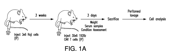
Figures 1A-1T depict a mouse model of CRS recapitulating clinical features of
the pathology. A) Schematic of mouse model. Raji tumor cells were
intraperitoneally
injected in mice and allowed to grow for three weeks. A high dose of CART
cells was
transferred, and mice were monitored over the following hours for symptoms of
CRS.
Mice were sacrificed, and cells were obtained for analysis through peritoneal
lavage or
tissue harvesting for further analysis. B) and Q) Percent weight change of
tumor bearing
mice after 1928z CAR T cell transfer. Weight per mouse was normalized to
starting
weight pre-CAR transfer (Tumor only n=12, Tumor + CAR n=18). C) and R) Percent
survival of mice after 1928z CAR T cell transfer (Tumor only n=12, Tumor + CAR
n=18). D) Serum levels of murine SAA3 at 42 hours post 1928z CAR T cell
transfer as
measured by ELISA (Baseline [tumor-free mice]/No tumor no CAR n=5, tumor only
CA 03082611 2020-05-13
WO 2019/099993 PCT/US2018/061795
n=5, tumor + CAR n=5, CAR only n=5). E)-L) and S) Serum cytokine levels 4.5
hours
before (pre-car) or 24 hours post 1928z CAR T cell transfer (CRS or Severe
CRS). Mice
that died from CRS were grouped under severe CRS while mice that survived but
suffered greater than 10% weight loss were grouped under CRS. "m" prefix
denotes
murine while "h" prefix denotes human. Cytokine levels were measured by
Cytokine
Bead Array (CBA). M)-0) and T) Species of origin of pro-inflammatory
cytokines. P)
Percent survival of tumor bearing mice treated with 1928z CAR T cells that
received
murine IL-6R blocking antibody or isotype (vehicle). S) Serum levels of murine
SAA3 at
42 hours post 1928z CART cell transfer as measured by ELISA (No tumor No CAR
n=5,
tumor only n=5, tumor+ CAR n=5, CAR only n=5). *P<0.05, **P<0.01, ***P<0.001
(Two-way ANOVA (B); (two-tailed unpaired two-sample t-test was used; log-rank
Mantel-Cox test (C and P). All data are means s.e.m.
Figures 2A-2R depict that tumor¨CAR T cell interactions selectively trigger
myeloid cell recruitment and activation. A) and B) Immunohistochemical
staining of
sections from 3-week tumor explants for Mac2. C) and P) Absolute counts of
myeloid
cell populations obtained by peritoneal lavage 60 hours after 1928z CAR T cell
transfer.
Phenotypes were analyzed by flow cytometry and absolute quantification was
performed
by the addition of counting beads. (Baseline [tumor free mice]/No tumor no CAR
n=5,
CAR only n=5, Tumor only n=6, Tumor + CAR n=7). D) Representative flow
cytometric plot showing Total Peritoneal Macrophages within the gated
population
[Resident Peritoneal Macrophages and CRS-Associated Macrophages (CAMs)]. Cells
were obtained from peritoneal lavage. E)-G) and Q) Absolute counts of myeloid
cell
populations obtained from multiple organs 18 hours after 1928z CAR T cell
transfer
(Tumor only n=4, Tumor + CAR n=4). H)-0) and R) Fold change of pro-
inflammatory
gene expression in myeloid populations as determined by RNAseq analysis. Fold
change
was determined by comparing each population under tumor only and tumor + CAR
conditions. Significant downregulation (green bars), significant upregulation
(red bars),
no significant change (gray bars). Gene expression levels were determined from
three
biological replicates for tumor only mice and three biological replicates for
tumor + CAR
mice. Each biological replicate consisted of pooled cells isolated from three
mice.
*P<0.05, **P<0.01, ***P<0.001 (Welch's two samples t-test (C and E-G);
(binomial
test, FDR-adjusted p-values (H-0). All data are means s.e.m. except C-E
which are
means s.d.
11
CA 03082611 2020-05-13
WO 2019/099993 PCT/US2018/061795
Figures 3A-3S depict that modulating macrophage function drastically alters
CRS
outcomes. A) Schematic of SFG retroviral cassette designed to co-express 1928z
and
murine CD4OL. B) and M) Percent weight change of tumor bearing mice after
1928z
CAR T cell transfer. Weight per mouse was normalized to starting weight pre-
CAR
transfer (Tumor only n=8, 1928z-LNGFR n=7, 1928z-mCD40L n=5). C) and D)
Representative flow cytometric plot showing Total Peritoneal Macrophages
within the
gated population [Resident Peritoneal Macrophages and CRS-Associated
Macrophages
(CAMs)]. Cells were obtained from peritoneal lavage. E) and 0) Percent of
CD40+ total
peritoneal macrophages, obtained by peritoneal lavage at 61 hours post 1928z-
LNGFR
or 1928z-mCD40L CAR T cell transfer, analyzed by flow cytometry. F)-I) and P)
Serum levels of murine cytokines at 18 hours post CAR T cell transfer.
Cytokine levels
were measured by Cytokine Bead Array (CBA). (Tumor only n=8, 1928z-LNGFR n=7,
1928z-mCD40L n=5). J) and Q) Percent of myeloid populations from peritoneum,
spleen and bone marrow expressing iNOS protein in tumor only mice and tumor +
CAR
mice. iNOS expression was determined by intracellular flow cytometry. (For
peritoneum
n=14 per group, for bone marrow and spleen n=10 per group). K) and R) Percent
weight
change of tumor bearing mice after 1928z CAR T cell transfer. Weight per mouse
was
normalized to starting weight pre-CAR transfer. Mice were treated with L-NIL
or
vehicle (PBS). (Tumor only n=7, CAR + L-NIL n=7, CAR + Vehicle n=8). L) and S)
Percent survival of tumor bearing mice after 1928z CAR T cell transfer
receiving 1400W
or Vehicle (PBS). (Vehicle n=20, 1400W n=13). N) Percent survival of tumor
bearing
mice after 1928z CAR T cell transfer (1928z-LNGFR n=16, 1928z-mCD40L n=13).
*P<0.05, **P<0.01, ***P<0.001 (Two-way ANOVA (B and K); (Two-tailed unpaired
two-sample t- test was used; (log-rank Mantel-Cox test (I) . All data are
means s.e.m.
Figures 4A-4R depict that augmented IL-1Ra response alleviated CRS-associated
mortality without compromising antitumor efficacy. A)-H) Fold change of IL-1
signaling component gene expression in myeloid populations as determined by
RNAseq
analysis. Fold change was determined by comparing each population under tumor
only
and tumor + CAR conditions. Significant downregulation (green bars),
significant
upregulation (red bars), no significant change (grey bars). Gene expression
levels were
determined from three biological replicates for tumor only mice and three
biological
replicates for tumor + CAR mice. Each biological replicate consisted of pooled
cells
isolated from three mice. I) Percent survival of tumor bearing mice after
1928z CAR T
cell transfer receiving Anakinra or Vehicle (PBS). (Anakinra n=11, Vehicle
n=10). J)
12
CA 03082611 2020-05-13
WO 2019/099993 PCT/US2018/061795
Percent of peritoneal macrophages expressing iNOS at 18 hours post CAR T cell
transfer. Mice were treated with isotype, murine IL-6 blocking antibody,
Anakinra or
murine IL-6 blocking antibody + Anakinra. (Tumor only=4, Isotype n=3, Anti-mIL-
6
n=3, Anakinra n=3, Anti-mIL-6 + Anakinra n=4). K) Schematic of SFG retroviral
cassette designed to co-express 1928z and murine IL-1Ra. L) Levels of murine
IL-1Ra
in supernatants of 1928z-LNGFR and 1928z-mIL-1Ra transduced CAR T cells after
48
hours in culture as determined by ELISA. M) Percent survival of tumor bearing
mice
after 1928z-LNGFR or 1928z-mIL-1Ra CAR T cell transfer. (1928z-LNGFR n=22,
1928z-mIL-1Ra n=18). N)-P) Serum levels of murine cytokines at 18 hours post
CAR T
cell transfer. Tumor bearing mice received 1928z-LNGFR or 1928z-mIL-1Ra CAR T
cells. Cytokine levels were measured by Cytokine Bead Array (CBA). Q)-R)
Percent
tumor free survival of NSG mice receiving 0.2e6 or 0.5e6 1928z-LNGFR or 1928z-
mIL-
1Ra CAR T cells. Tumors were injected intravenously on Day-4 and CAR T cells
on
Day 0. (Tumor only n=4, 0.2e6 1928z-LNGFR n=7, 0.2e6 1928z-mIL-1Ra n=7, 0.5e6
1928z-LNGFR n=11, 0.5e6 1928z-mIL-1Ra n=11). *P<0.05, **P<0.01, ***P<0.001
(binomial test, FDR-adjusted p-values (A-H); (Two-tailed unpaired two-sample t-
test
and one-way ANOVA were used; log-rank Mantel-Cox test (I, M, Q and R). All
data are
means s.e.m.
Figures 5A-5K depict cytokine levels and the effects to mouse tissues. A) and
E)
Serum levels of human and murine IFNy at 18 hours post 1928z CAR T cell
transfer as
measured by Cytokine Bead Array (CBA) (n=6). B) and G) Serum of murine IL-6
levels
at 18 hours post 1928z CAR T cell transfer as measured by Cytokine Bead Array
(CBA).
Mice were treated with a blocking antibody specific for the murine IL-6
receptor or
isotype (Isotype, n=3, Anti-mIL-6R n=3). C) and H) Representative tissue
sections
stained with H&E obtained from mice sacrificed after 2 days or 5 days of 1928z
CAR T
cell transfer and respective controls. D) Serum cytokine levels after 24 hours
of 1928z
CAR T cell treatment (No tumor no CAR n=5, tumor only n=4, CAR only n=5, Tumor
+
CAR n=3). F) Serum of murine IL-15/IL-15R complex levels at 18 hours post
1928z
CART cell transfer as measured by ELISA. All data are means s.e.m. Figures
5I-5K
depict representative tissue sections of mouse brains stained with H&E,
obtained from
tumor only or tumor + CAR treated mice one, two and five days after CAR T cell
transfer. (Day 1: Tumor only n=2 mice, Tumor + CAR n=3 mice), (Day 2: Tumor
only
n=3 mice, Tumor + CAR n=3 mice), (Day 5: Tumor only n=3 mice, Tumor + CAR n=2
mice). Day 1, Day 2 and Day 5 mice were derived from three independent
experiments.
13
CA 03082611 2020-05-13
WO 2019/099993
PCT/US2018/061795
(I. top row) Coronal section of the skull and the brain at the level of the
hippocampus (H)
and thalamus (T). The space between the cranial vault and the cerebrum on the
right
image is artefactual. (I. bottom row) Detail of the hippocampus and its
regions (CA1,
CA3, DG). A portion of the choroid plexus (Cp) of the ventricular system, the
cerebral
meninges (arrowhead), brain cortex (C) are shown. (J. top row) Coronal section
of the
brain at the level of the frontal lobes. (J. bottom row) Detail of the dorsal
aspect of the
cortex (C) including the meninges (arrowhead). (K. top row) Coronal section of
the brain
at the level of the striatum (S) and corpus callosum (Cc). (K. bottom row)
Detail of the
dorsal aspect of the cortex (C), including the cerebral meninges (arrowhead).
Figures 6A-6G depict myeloid cell and T cell populations in various tissues.
A)
and F) Percent weight change of tumor bearing or tumor free mice after 1928z
CAR T
cell transfer. Weight per mouse was normalized to starting weight pre-CAR
transfer
(Baseline [tumor free mice]/No tumor no CAR n=5, CAR only n=5, Tumor only n=6,
Tumor + CAR n=7). B)-D) and G) Absolute counts of myeloid cell populations
obtained
from various organs 18 hours after 1928z CAR T cell transfer. Phenotypes were
analyzed by flow cytometry and absolute quantification was performed by the
addition
of counting beads. (Tumor only n=4, Tumor + CAR n=4). E) Representative flow
cytometric plots of T cell distribution in various tissues 18 hours after
1928z CART cell
transfer. *P<0.05, **P<0.01, ***P<0.001 (Two-way ANOVA (A); (Two-tailed
unpaired two-sample t-test (B-D). Data are means s.e.m (A) and means s.d.
(B-D).
Figures 7A-7B depict gating strategy to phenotype and FACS sort myeloid
populations. A) Gating strategy to phenotype and FACS sort myeloid populations
in cells
obtained from peritoneal lavage. B) Gating strategy to phenotype and FACS sort
myeloid populations in cells obtained from murine spleens.
Figures 8A-8H depict effects of 1928z-LNGFR treatment and 1928z-mCD40L
treatment. A) Flow cytometric histogram of T cells transduced with 1928z-
LNGFR. B)
Percent survival of tumor bearing mice treated with 1928z-LNGFR or 1928z-
mCD40L
CAR T cells. (Tumor only n=9, 1928z-LNGFR n=7, 1928z-mCD40L n=7). C) and F)
Absolute counts of myeloid cell populations obtained by peritoneal lavage 61
hours after
1928z-LNGFR or 1928z-mCD40L CAR T cell transfer. Phenotypes were analyzed by
flow cytometry and absolute quantification of cells was performed by the
addition of
counting beads. (Tumor only n=8, 1928z-LNGFR n=7, 1928z-mCD40L n=5). D) and G)
Percent of CD40+ DCs, obtained by peritoneal lavage at 61 hours post 1928z-
LNGFR or
1928z-mCD40L CAR T cell transfer, analyzed by flow cytometry. (Tumor only n=9,
14
CA 03082611 2020-05-13
WO 2019/099993 PCT/US2018/061795
1928z-LNGFR n=7, 1928z-mCD40L n=5). E) Representative flow cytometric plots of
murine CD40 expression on the surface of the indicated myeloid populations. H)
Representative flow cytometric plots of murine CD40 expression on the surface
of the
indicated myeloid populations. *P<0.05, **13<0.01, ***P<0.001 (Two-tailed
unpaired
.. two-sample t-test (C) and One-way ANOVA (D) were used. All data are means
s.e.m.
Figure 9A-9B. A) and C) Absolute counts of iN0S+ myeloid cell populations
obtained by peritoneal lavage after 1928z CAR T cell transfer. iNOS expression
was
determined by intracellular flow cytometry and absolute quantification of
cells was
performed by the addition of counting beads. (Tumor only n=14, Tumor + CAR
n=14).
B) and D) Percent weight change of tumor bearing mice after 1928z CAR T cell
transfer.
Mice received 1400W or vehicle (PBS) Weight per mouse was normalized to
starting
weight pre-CAR transfer (Tumor only n=10, CAR + Vehicle n=8, CAR + 1400W n=8).
E) Percent peritoneal macrophages expressing iNOS at 18 hours post CAR T cell
transfer. Mice were treated with isotype, murine IL-6 blocking antibody, or
murine IL-lb
blocking antibody. (Tumor only n=6, Isotype n=3, Anti-mIL-6 n=8, Anti mIL-lb
n=4).
*P<0.05, **P<0.01, ***P<0.001 (Two-tailed unpaired two-sample t-test (A); Two-
way
ANOVA (B); one-way ANOVA (E)) All data are means s.e.m.
Figures 10A-10D. A) Flow cytometric histogram showing percentage of
transduced CAR T cells with 1928z-LNGFR and 1928z-mIL-1Ra constructs prior to
transfer to SCID-beige mice. B) Flow cytometric histogram showing percentage
of
transduced CAR T cells with 1928z-LNGFR and 1928z-mIL-1Ra constructs prior to
transfer to NSG mice. C) and D) Tumor derived (NALM-6) bioluminescent signal
from
NSG mice receiving 0.2e6 or 0.5e6 1928z-LNGFR or 1928z-mIL-1Ra CAR T cells.
Tumors were injected intravenously on Day-4 and CAR T cells on Day 0. (Tumor
only:
n=4, 0.2e6 1928z-LNGFR: n=7, 0.2e6 1928z-mIL-1Ra: n=7, 0.5e6 1928z-LNGFR:
n=11, 0.5e6 1928z-mIL- 1Ra: n=11).
DETAILED DESCRIPTION OF THE INVENTION
The presently disclosed subject matter provides cells, including genetically
modified immunoresponsive cells (e.g., T cells, NK cells, or CTL cells)
comprising a
combination of an antigen-recognizing receptor (e.g., TCR or CAR) and a
secretable IL-
1Ra polypeptide (e.g., an exogenous IL-1Ra polypeptide, or a nucleic acid
encoding an
IL-1Ra polypeptide). The presently disclosed subject matter also provides
methods of
using such cells for treating and/or preventing a neoplasm or other
diseases/disorders,
reducing tumor burden in a subject, lengthening survival of a subject having
neoplasm
CA 03082611 2020-05-13
WO 2019/099993 PCT/US2018/061795
(e.g., cancer), and/or treating or alleviating CRS in a subject who receives
an
immunotherapy. The presently disclosed subject matter is based, at least in
part, on the
discovery that a secretable IL-1Ra polypeptide alleviated cytokine release
syndrome
(CRS) in subjects receiving an immunotherapy (e.g., CAR-T cells).
The presently disclosed subject matter is at least based on the discovery of a
novel genetic construct that allows to prevent and/or reduce the severity of
CRS
effectively without the requirement for external administration of
pharmacological
agents, by co-expressing a CAR and IL-1Ra (encoded by IL-1RN gene) in T cells.
This
approach takes advantage of the natural function of endogenous IL-1Ra. This
novel
genetic construct when introduced into T cells allows for the constitutive co-
expression
of both the CAR protein and the IL-1Ra protein. Treatment of mice that
experience
CRS, with the T cells comprising such genetic construct (e.g., 1928z-IL-1Ra
CAR T
cells) are protected from CRS-related mortality. Moreover, in a mouse model
suitable to
compare the long-term anti-tumor efficacy of different CAR constructs, T cells
.. comprising such genetic construct (e.g., 1928z-IL-1Ra CAR T cells) have
equivalent
anti-tumor efficacy compared to their control counterparts (e.g., 1928z CAR T
cells that
do not co-express IL-1Ra). Therefore, the presently disclosed subject matter
allows for
CRS to be treated intrinsically by the CAR T cell itself without affecting
anti-tumor
efficacy, while removing the need external pharmacological intervention.
The novel genetic construct sets a paradigm of co-expression of
immunomodulatory molecules from engineered T cells in order to prevent,
mitigate
and/or ameliorate toxicities inherent to CAR-T cell therapy. Moreover, the
presently
disclosed subject matter provides methods of conditionally co-expressing such
immunomodulatory molecules in CAR-T cells by inducible promoters. Conditional
co-
expression in this context can be achieved through the use of specialized
promoters that
induce transcription only upon the binding of specific transcription factors.
In addition,
expression levels can be further adjusted by using constitutive promoters of
known
strength in order to achieve the desired levels of expression. Lastly, other
cell types
employed for immunotherapy, such as NK cells or macrophages, can be also
engineered
with such immunomodulatory molecules and be used alone or in combination with
CAR-
T cells.
1. Definitions
Unless defined otherwise, all technical and scientific terms used herein have
the
meaning commonly understood by a person skilled in the art. The following
references
16
CA 03082611 2020-05-13
WO 2019/099993 PCT/US2018/061795
provide one of skill with a general definition of many of the terms used in
the presently
disclosed subject matter: Singleton et al., Dictionary of Microbiology and
Molecular
Biology (2nd ed. 1994); The Cambridge Dictionary of Science and Technology
(Walker
ed., 1988); The Glossary of Genetics, 5th Ed., R. Rieger et al. (eds.),
Springer Verlag
(1991); and Hale & Marham, The Harper Collins Dictionary of Biology (1991). As
used
herein, the following terms have the meanings ascribed to them below, unless
specified
otherwise.
As used herein, the term "about" or "approximately" means within an acceptable
error range for the particular value as determined by one of ordinary skill in
the art,
.. which will depend in part on how the value is measured or determined, i.e.,
the
limitations of the measurement system. For example, "about" can mean within 3
or
more than 3 standard deviations, per the practice in the art. Alternatively,
"about" can
mean a range of up to 20%, up to 10%, up to 5%, or up to 1% of a given value.
Alternatively, particularly with respect to biological systems or processes,
the term can
mean within an order of magnitude, e.g., within 5-fold or within 2-fold, of a
value.
By "activates an immunoresponsive cell" is meant induction of signal
transduction or changes in protein expression in the cell resulting in
initiation of an
immune response. For example, when CD3 Chains cluster in response to ligand
binding
and immunoreceptor tyrosine-based inhibition motifs (ITAMs) a signal
transduction
cascade is produced. In certain embodiments, when an endogenous TCR or an
exogenous CAR binds to an antigen, a formation of an immunological synapse
occurs
that includes clustering of many molecules near the bound receptor (e.g. CD4
or CD8,
CD3 y/o/c/C, etc.). This clustering of membrane bound signaling molecules
allows for
ITAM motifs contained within the CD3 chains to become phosphorylated. This
phosphorylation in turn initiates a T cell activation pathway ultimately
activating
transcription factors, such as NF-KB and AP-1. These transcription factors
induce global
gene expression of the T cell to increase IL-2 production for proliferation
and expression
of master regulator T cell proteins in order to initiate a T cell mediated
immune response.
By "stimulates an immunoresponsive cell" is meant a signal that results in a
robust and sustained immune response. In various embodiments, this occurs
after
immune cell (e.g., T-cell) activation or concomitantly mediated through
receptors
including, but not limited to, CD28, CD137 (4-1BB), 0X40, ICOS, and MyD88.
Receiving multiple stimulatory signals can be important to mount a robust and
long-term
17
CA 03082611 2020-05-13
WO 2019/099993 PCT/US2018/061795
T cell mediated immune response. T cells can quickly become inhibited and
unresponsive to antigen. While the effects of these co-stimulatory signals may
vary, they
generally result in increased gene expression in order to generate long lived,
proliferative, and anti-apoptotic T cells that robustly respond to antigen for
complete and
.. sustained eradication.
The term "antigen-recognizing receptor" as used herein refers to a receptor
that is
capable of activating an immune or immunoresponsive cell (e.g., a T-cell) in
response to
its binding to an antigen. Non-limiting examples of antigen-recognizing
receptors
include native or endogenous T cell receptors ("TCRs"), and chimeric antigen
receptors
("CARs").
As used herein, the term "antibody" means not only intact antibody molecules,
but also fragments of antibody molecules that retain immunogen-binding
ability. Such
fragments are also well known in the art and are regularly employed both in
vitro and
in vivo. Accordingly, as used herein, the term "antibody" means not only
intact
immunoglobulin molecules but also the well-known active fragments F(a1302, and
Fab.
F(a1302, and Fab fragments that lack the Fe fragment of intact antibody, clear
more
rapidly from the circulation, and may have less non-specific tissue binding of
an intact
antibody (Wahl et al., I Nucl. Med. 24:316-325 (1983). As used herein,
antibodies
include whole native antibodies, bispecific antibodies; chimeric antibodies;
Fab, Fab',
single chain V region fragments (scFv), fusion polypeptides, and
unconventional
antibodies. In certain embodiments, an antibody is a glycoprotein comprising
at least
two heavy (H) chains and two light (L) chains inter-connected by disulfide
bonds. Each
heavy chain is comprised of a heavy chain variable region (abbreviated herein
as VH) and
a heavy chain constant (CH) region. The heavy chain constant region is
comprised of
three domains, CHL CH2 and CH3. Each light chain is comprised of a light chain
variable region (abbreviated herein as VL) and a light chain constant CL
region. The light
chain constant region is comprised of one domain, CL. The VH and VL regions
can be
further sub-divided into regions of hypervariability, termed complementarity
determining
regions (CDR), interspersed with regions that are more conserved, termed
framework
regions (FR). Each VH and VL is composed of three CDRs and four FRs arranged
from
amino-terminus to carboxy-terminus in the following order: FR1, CDR1, FR2,
CDR2,
FR3, CDR3, FR4. The variable regions of the heavy and light chains contain a
binding
domain that interacts with an antigen. The constant regions of the antibodies
may
mediate the binding of the immunoglobulin to host tissues or factors,
including various
18
CA 03082611 2020-05-13
WO 2019/099993
PCT/US2018/061795
cells of the immune system (e.g., effector cells) and the first component (Cl
q) of the
classical complement system.
As used herein, "CDRs" are defined as the complementarity determining region
amino acid sequences of an antibody which are the hypervariable regions of
.. immunoglobulin heavy and light chains. See, e.g., Kabat et al., Sequences
of Proteins of
Immunological Interest, 4th U. S. Department of Health and Human Services,
National
Institutes of Health (1987). Generally, antibodies comprise three heavy chain
and three
light chain CDRs or CDR regions in the variable region. CDRs provide the
majority of
contact residues for the binding of the antibody to the antigen or epitope. In
certain
embodiments, the CDRs regions are delineated using the Kabat system (Kabat, E.
A., et
at. (1991) Sequences of Proteins of Immunological Interest, Fifth Edition,
U.S.
Department of Health and Human Services, NIH Publication No. 91-3242).
As used herein, the term "single-chain variable fragment" or "scFv" is a
fusion
protein of the variable regions of the heavy (VH) and light chains (VL) of an
immunoglobulin covalently linked to form a VH: :VL heterodimer. The VH and VL
are
either joined directly or joined by a peptide-encoding linker (e.g., 10, 15,
20, 25 amino
acids), which connects the N-terminus of the VH with the C-terminus of the VL,
or the C-
terminus of the VH with the N-terminus of the VL. The linker is usually rich
in glycine
for flexibility, as well as serine or threonine for solubility. Despite
removal of the
constant regions and the introduction of a linker, scFv proteins retain the
specificity of
the original immunoglobulin. Single chain Fv polypeptide antibodies can be
expressed
from a nucleic acid including VH - and VL -encoding sequences as described by
Huston,
et al. (Proc. Nat. Acad. Sci. USA, 85:5879-5883, 1988). See, also, U.S. Patent
Nos.
5,091,513, 5,132,405 and 4,956,778; and U.S. Patent Publication Nos.
20050196754 and
20050196754. Antagonistic scFvs having inhibitory activity have been described
(see,
e.g., Zhao et al., Hyrbidoma (Larchmt) 2008 27(6):455-51; Peter et al., J
Cachexia
Sarcopenia Muscle 2012 August 12; Shieh et al., J Imuno12009 183(4):2277-85;
Giomarelli et al., Thromb Haemost 2007 97(6):955-63; Fife eta., J Clin Invst
2006
116(8):2252-61; Brocks et al., Immunotechnology 1997 3(3):173-84; Moosmayer et
al.,
Ther Immunol 1995 2(10:31-40). Agonistic scFvs having stimulatory activity
have been
described (see, e.g., Peter et al., J Bioi Chem 2003 25278(38):36740-7; Xie et
al., Nat
Biotech 1997 15(8):768-71; Ledbetter et al., Crit Rev Immuno11997 17(5-6):427-
55; Ho
et al., BioChim Biophys Acta 2003 1638(3):257-66).
19
CA 03082611 2020-05-13
WO 2019/099993 PCT/US2018/061795
As used herein, the term "affinity" is meant a measure of binding strength.
Affinity can depend on the closeness of stereochemical fit between antibody
combining
sites and antigen determinants, on the size of the area of contact between
them, and/or on
the distribution of charged and hydrophobic groups. As used herein, the term
"affinity"
also includes "avidity", which refers to the strength of the antigen-antibody
bond after
formation of reversible complexes. Methods for calculating the affinity of an
antibody
for an antigen are known in the art, including, but not limited to, various
antigen-binding
experiments, e.g., functional assays (e.g., flow cytometry assay).
The term "chimeric antigen receptor" or "CAR" as used herein refers to a
molecule comprising an extracellular antigen-binding domain that is fused to
an
intracellular signaling domain that is capable of activating or stimulating an
immunoresponsive cell, and a transmembrane domain. In certain embodiments, the
extracellular antigen-binding domain of a CAR comprises a scFv. The scFv can
be
derived from fusing the variable heavy and light regions of an antibody.
Alternatively or
additionally, the scFv may be derived from Fab's (instead of from an antibody,
e.g.,
obtained from Fab libraries). In certain embodiments, the scFv is fused to the
transmembrane domain and then to the intracellular signaling domain. In
certain
embodiments, the CAR is selected to have high binding affinity or avidity for
the
antigen.
As used herein, the term "nucleic acid molecules" include any nucleic acid
molecule that encodes a polypeptide of interest (e.g., an IL-1Ra polypeptide)
or a
fragment thereof. Such nucleic acid molecules need not be 100% homologous or
identical with an endogenous nucleic acid sequence, but may exhibit
substantial identity.
Polynucleotides having "substantial identity" or "substantial homology" to an
endogenous sequence are typically capable of hybridizing with at least one
strand of a
double-stranded nucleic acid molecule. By "hybridize" is meant a pair to form
a double-
stranded molecule between complementary polynucleotide sequences (e.g., a gene
described herein), or portions thereof, under various conditions of
stringency. (See, e.g.,
Wahl, G. M. and S. L. Berger (1987) Methods Enzymol. 152:399; Kimmel, A. R.
(1987)
Methods Enzymol. 152:507).
For example, stringent salt concentration will ordinarily be less than about
750
mM NaCl and 75 mM trisodium citrate, e.g., less than about 500 mM NaCl and 50
mM
trisodium citrate, or less than about 250 mM NaCl and 25 mM trisodium citrate.
Low
stringency hybridization can be obtained in the absence of organic solvent,
e.g.,
CA 03082611 2020-05-13
WO 2019/099993
PCT/US2018/061795
formamide, while high stringency hybridization can be obtained in the presence
of at
least about 35% formamide, and more e.g., at least about 50% formamide.
Stringent
temperature conditions will ordinarily include temperatures of at least about
30 C, of at
least about 37 C, or of at least about 42 C. Varying additional parameters,
such as
hybridization time, the concentration of detergent, e.g., sodium dodecyl
sulfate (SDS),
and the inclusion or exclusion of carrier DNA, are well known to those skilled
in the art.
Various levels of stringency are accomplished by combining these various
conditions as
needed. In certain embodiments, hybridization will occur at 30 C in 750 mM
NaCl, 75
mM trisodium citrate, and 1% SDS. In certain embodiments, hybridization will
occur at
.. 37 C in 500 mM NaCl, 50 mM trisodium citrate, 1% SDS, 35% formamide, and
100
[tg/m1 denatured salmon sperm DNA (ssDNA). In certain embodiments,
hybridization
will occur at 42 C in 250 mM NaCl, 25 mM trisodium citrate, 1% SDS, 50%
formamide, and 200 [tg/m1 ssDNA. Useful variations on these conditions will be
readily
apparent to those skilled in the art.
For most applications, washing steps that follow hybridization will also vary
in
stringency. Wash stringency conditions can be defined by salt concentration
and by
temperature. As above, wash stringency can be increased by decreasing salt
concentration or by increasing temperature. For example, stringent salt
concentration for
the wash steps can be less than about 30 mM NaCl and 3 mM trisodium citrate,
e.g., less
than about 15 mM NaCl and 1.5 mM trisodium citrate. Stringent temperature
conditions
for the wash steps will ordinarily include a temperature of at least about 25
C, e.g., of at
least about 42 C, e.g., of at least about 68 C. In certain embodiments, wash
steps will
occur at 25 C in 30 mM NaCl, 3 mM trisodium citrate, and 0.1% SDS. In certain
embodiments, wash steps occur at 42 C. in 15 mM NaCl, 1.5 mM trisodium
citrate, and
0.1% SDS. In certain embodiments, wash steps occur at 68 C in 15 mM NaCl, 1.5
mM
trisodium citrate, and 0.1% SDS. Additional variations on these conditions
will be
readily apparent to those skilled in the art. Hybridization techniques are
well known to
those skilled in the art and are described, for example, in Benton and Davis
(Science
196:180, 1977); Grunstein and Rogness (Proc. Natl. Acad. Sci., USA 72:3961,
1975);
Ausubel et al. (Current Protocols in Molecular Biology, Wiley Interscience,
New York,
2001); Berger and Kimmel (Guide to Molecular Cloning Techniques, 1987,
Academic
Press, New York); and Sambrook et al., Molecular Cloning: A Laboratory Manual,
Cold
Spring Harbor Laboratory Press, New York.
21
CA 03082611 2020-05-13
WO 2019/099993 PCT/US2018/061795
By "substantially identical" or "substantially homologous" is meant a
polypeptide
or nucleic acid molecule exhibiting at least about 50% homologous or identical
to a
reference amino acid sequence (for example, any one of the amino acid
sequences
described herein) or nucleic acid sequence (for example, any one of the
nucleic acid
sequences described herein). In certain embodiments, such a sequence is at
least about
60%, at least about 65%, at least about 70%, at least about 75%, at least
about 80%, at
least about 85%, at least about 90%, at least about 95%, at least about 99%,
or at least
about 100% homologous or identical to the sequence of the amino acid or
nucleic acid
used for comparison.
Sequence identity can be measured by using sequence analysis software (for
example, Sequence Analysis Software Package of the Genetics Computer Group,
University of Wisconsin Biotechnology Center, 1710 University Avenue, Madison,
Wis.
53705, BLAST, BESTFIT, GAP, or PILEUP/PRETTYBOX programs). Such software
matches identical or similar sequences by assigning degrees of homology to
various
substitutions, deletions, and/or other modifications. Conservative
substitutions typically
include substitutions within the following groups: glycine, alanine; valine,
isoleucine,
leucine; aspartic acid, glutamic acid, asparagine, glutamine; serine,
threonine; lysine,
arginine; and phenylalanine, tyrosine. In an exemplary approach to determining
the
degree of identity, a BLAST program may be used, with a probability score
between e-3
and e-100 indicating a closely related sequence.
By "analog" is meant a structurally related polypeptide or nucleic acid
molecule
having the function of a reference polypeptide or nucleic acid molecule.
The term "ligand" as used herein refers to a molecule that binds to a
receptor. In
certain embodiments, the ligand binds to a receptor on another cell, allowing
for cell-to-
cell recognition and/or interaction.
The term "constitutive expression" or "constitutively expressed" as used
herein
refers to expression or expressed under all physiological conditions.
By "disease" is meant any condition, disease or disorder that damages or
interferes with the normal function of a cell, tissue, or organ, e.g.,
neoplasm, and
pathogen infection of cell.
By "effective amount" is meant an amount sufficient to have a therapeutic
effect.
In certain embodiments, an "effective amount" is an amount sufficient to
arrest,
ameliorate, or inhibit the continued proliferation, growth, or metastasis
(e.g., invasion, or
migration) of a neoplasm and/or CRS.
22
CA 03082611 2020-05-13
WO 2019/099993 PCT/US2018/061795
By "enforcing tolerance" is meant preventing the activity of self-reactive
cells or
immunoresponsive cells that target transplanted organs or tissues.
By "endogenous" is meant a nucleic acid molecule or polypeptide that is
normally expressed in a cell or tissue.
By "exogenous" is meant a nucleic acid molecule or polypeptide that is not
endogenously present in a cell. The term "exogenous" would therefore encompass
any
recombinant nucleic acid molecule or polypeptide expressed in a cell, such as
foreign,
heterologous, and over-expressed nucleic acid molecules and polypeptides. By
"exogenous" nucleic acid is meant a nucleic acid not present in a native wild-
type cell;
for example, an exogenous nucleic acid may vary from an endogenous counterpart
by
sequence, by position/location, or both. For clarity, an exogenous nucleic
acid may have
the same or different sequence relative to its native endogenous counterpart;
it may be
introduced by genetic engineering into the cell itself or a progenitor
thereof, and may
optionally be linked to alternative control sequences, such as a non-native
promoter or
secretory sequence.
By a "heterologous nucleic acid molecule or polypeptide" is meant a nucleic
acid
molecule (e.g., a cDNA, DNA or RNA molecule) or polypeptide that is not
normally
present in a cell or sample obtained from a cell. This nucleic acid may be
from another
organism, or it may be, for example, an mRNA molecule that is not normally
expressed
in a cell or sample.
By "immunoresponsive cell" is meant a cell that functions in an immune
response or a progenitor, or progeny thereof
By "modulate" is meant positively or negatively alter. Exemplary modulations
include a about 1%, about 2%, about 5%, about 10%, about 25%, about 50%, about
75%,
.. or about 100% change.
By "increase" is meant to alter positively by at least about 5%. An alteration
may
be by about 5%, about 10%, about 25%, about 30%, about 50%, about 75%, about
100%
or more.
By "reduce" is meant to alter negatively by at least about 5%. An alteration
may
be by about 5%, about 10%, about 25%, about 30%, about 50%, about 75%, or even
by
about 100%.
By "isolated cell" is meant a cell that is separated from the molecular and/or
cellular components that naturally accompany the cell.
23
CA 03082611 2020-05-13
WO 2019/099993
PCT/US2018/061795
The terms "isolated," "purified," or "biologically pure" refer to material
that is
free to varying degrees from components which normally accompany it as found
in its
native state. "Isolate" denotes a degree of separation from original source or
surroundings. "Purify" denotes a degree of separation that is higher than
isolation. A
"purified" or "biologically pure" protein is sufficiently free of other
materials such that
any impurities do not materially affect the biological properties of the
protein or cause
other adverse consequences. That is, a nucleic acid or peptide is purified if
it is
substantially free of cellular material, viral material, or culture medium
when produced
by recombinant DNA techniques, or chemical precursors or other chemicals when
chemically synthesized. Purity and homogeneity are typically determined using
analytical chemistry techniques, for example, polyacrylamide gel
electrophoresis or
high-performance liquid chromatography. The term "purified" can denote that a
nucleic
acid or protein gives rise to essentially one band in an electrophoretic gel.
For a protein
that can be subjected to modifications, for example, phosphorylation or
glycosylation,
different modifications may give rise to different isolated proteins, which
can be
separately purified.
The term "antigen-binding domain" as used herein refers to a domain capable of
specifically binding a particular antigenic determinant or set of antigenic
determinants
present on a cell.
"Linker", as used herein, shall mean a functional group (e.g., chemical or
polypeptide) that covalently attaches two or more polypeptides or nucleic
acids so that
they are connected to one another. As used herein, a "peptide linker" refers
to one or
more amino acids used to couple two proteins together (e.g., to couple VH and
VL
domains). In certain embodiments, the linker comprises a sequence set forth in
GGGGSGGGGSGGGGS [SEQ ID NO: 23].
By "neoplasm" is meant a disease characterized by the pathological
proliferation
of a cell or tissue and its subsequent migration to or invasion of other
tissues or organs.
Neoplasia growth is typically uncontrolled and progressive, and occurs under
conditions
that would not elicit, or would cause cessation of, multiplication of normal
cells.
Neoplasia can affect a variety of cell types, tissues, or organs, including
but not limited
to an organ selected from the group consisting of bladder, bone, brain,
breast, cartilage,
glia, esophagus, fallopian tube, gallbladder, heart, intestines, kidney,
liver, lung, lymph
node, nervous tissue, ovaries, pancreas, prostate, skeletal muscle, skin,
spinal cord,
spleen, stomach, testes, thymus, thyroid, trachea, urogenital tract, ureter,
urethra, uterus,
24
CA 03082611 2020-05-13
WO 2019/099993 PCT/US2018/061795
and vagina, or a tissue or cell type thereof Neoplasia include cancers, such
as sarcomas,
carcinomas, or plasmacytomas (malignant tumor of the plasma cells).
By "receptor" is meant a polypeptide, or portion thereof, present on a cell
membrane that selectively binds one or more ligand.
By "recognize" is meant selectively binds to a target. A T cell that
recognizes a
tumor can expresses a receptor (e.g., a TCR or CAR) that binds to a tumor
antigen.
By "reference" or "control" is meant a standard of comparison. For example,
the
level of scFv-antigen binding by a cell expressing a CAR and an scFv may be
compared
to the level of scFv-antigen binding in a corresponding cell expressing CAR
alone.
By "secreted" is meant a polypeptide that is released from a cell via the
secretory
pathway through the endoplasmic reticulum, Golgi apparatus, and as a vesicle
that
transiently fuses at the cell plasma membrane, releasing the proteins outside
of the cell.
By "signal sequence" or "leader sequence" is meant a peptide sequence (e.g.,
5,
10, 15, 20, 25 or 30 amino acids) present at the N-terminus of newly
synthesized proteins
that directs their entry to the secretory pathway. Exemplary leader sequences
include, but
is not limited to, the IL-2 signal sequence: MYRMQLLSCIALSLALVTNS [SEQ ID
NO: 8] (human), MYSMQLASCVTLTLVLLVNS [SEQ ID NO: 24] (mouse); the kappa
leader sequence: METPAQLLFLLLLWLPDTTG [SEQ ID NO: 25] (human),
METDTLLLWVLLLWVPGSTG [SEQ ID NO: 26] (mouse); the CD8 leader sequence:
MALPVTALLLPLALLLHAARP [SEQ ID NO: 27] (human); the albumin signal
sequence: MKWVTFISLLFSSAYS [SEQ ID NO: 28] (human); and the prolactin signal
sequence: MDSKGSSQKGSRLLLLLVVSNLLLCQGVVS [SEQ ID NO: 29] (human).
By "soluble" is meant a polypeptide that is freely diffusible in an aqueous
environment (e.g., not membrane bound).
By "specifically binds" is meant a polypeptide or fragment thereof that
recognizes and binds to a biological molecule of interest (e.g., a
polypeptide), but which
does not substantially recognize and bind other molecules in a sample, for
example, a
biological sample, which naturally includes a presently disclosed polypeptide.
The term "tumor antigen" as used herein refers to an antigen (e.g., a
polypeptide)
that is uniquely or differentially expressed on a tumor cell compared to a
normal or non-
IS neoplastic cell. In certain embodiments, a tumor antigen includes any
polypeptide
expressed by a tumor that is capable of activating or inducing an immune
response via an
antigen recognizing receptor (e.g., CD19, MUC-16) or capable of suppressing an
immune response via receptor-ligand binding (e.g., CD47, PD-Ll/L2, B7.1/2).
CA 03082611 2020-05-13
WO 2019/099993 PCT/US2018/061795
The terms "comprises", "comprising", and are intended to have the broad
meaning ascribed to them in U.S. Patent Law and can mean "includes",
"including" and
the like.
As used herein, "treatment" refers to clinical intervention in an attempt to
alter
the disease course of the individual or cell being treated, and can be
performed either for
prophylaxis or during the course of clinical pathology. Therapeutic effects of
treatment
include, without limitation, preventing occurrence or recurrence of disease,
alleviation of
symptoms, diminishment of any direct or indirect pathological consequences of
the
disease, preventing metastases, decreasing the rate of disease progression,
amelioration
or palliation of the disease state, and remission or improved prognosis. By
preventing
progression of a disease or disorder, a treatment can prevent deterioration
due to a
disorder in an affected or diagnosed subject or a subject suspected of having
the disorder,
but also a treatment may prevent the onset of the disorder or a symptom of the
disorder
in a subject at risk for the disorder or suspected of having the disorder.
An "individual" or "subject" herein is a vertebrate, such as a human or non-
human animal, for example, a mammal. Mammals include, but are not limited to,
humans, primates, farm animals, sport animals, rodents and pets. Non-limiting
examples
of non-human animal subjects include rodents such as mice, rats, hamsters, and
guinea
pigs; rabbits; dogs; cats; sheep; pigs; goats; cattle; horses; and non-human
primates such
as apes and monkeys. The term "immunocompromised" as used herein refers to a
subject
who has an immunodeficiency. The subject is very vulnerable to opportunistic
infections,
infections caused by organisms that usually do not cause disease in a person
with a
healthy immune system, but can affect people with a poorly functioning or
suppressed
immune system.
Other aspects of the presently disclosed subject matter are described in the
following disclosure and are within the ambit of the presently disclosed
subject matter.
2. Antigen-Recognizing Receptors
The present disclosure provides antigen-recognizing receptors that bind to an
antigen of interest. In certain embodiments, the antigen-recognizing receptor
is a
chimeric antigen receptor (CAR). In certain embodiments, the antigen-
recognizing
receptor is a T-cell receptor (TCR). The antigen-recognizing receptor can bind
to a
tumor antigen or a pathogen antigen.
26
CA 03082611 2020-05-13
WO 2019/099993 PCT/US2018/061795
2.1. Antigens
In certain embodiments, the antigen-recognizing receptor binds to a tumor
antigen. Any tumor antigen (antigenic peptide) can be used in the tumor-
related
embodiments described herein. Sources of antigen include, but are not limited
to, cancer
proteins. The antigen can be expressed as a peptide or as an intact protein or
portion
thereof The intact protein or a portion thereof can be native or mutagenized.
Non-
limiting examples of tumor antigens include carbonic anhydrase IX (CA1X),
carcinoembryonic antigen (CEA), CD8, CD7, CD10, CD19, CD20, CD22, CD30, CD33,
CLL1, CD34, CD38, CD41, CD44, CD49f, CD56, CD74, CD133, CD138, CD123,
CD44V6, an antigen of a cytomegalovirus (CMV) infected cell (e.g., a cell
surface
antigen), epithelial glycoprotein-2 (EGP-2), epithelial glycoprotein-40 (EGP-
40),
epithelial cell adhesion molecule (EpCAM), receptor tyrosine-protein kinases
erb-B2,3,4
(erb-B2,3,4), folate-binding protein (FBP), fetal acetylcholine receptor
(AChR), folate
receptor-a, Ganglioside G2 (GD2), Ganglioside G3 (GD3), human Epidermal Growth
Factor Receptor 2 (HER-2), human telomerase reverse transcriptase (hTERT),
Interleukin-13 receptor subunit alpha-2 (IL-13Ra2), K-light chain, kinase
insert domain
receptor (KDR), Lewis Y (LeY), Li cell adhesion molecule (L1CAM), melanoma
antigen family A, 1 (MAGE-A1), Mucin 16 (MUC16), Mucin 1 (MUC1), Mesothelin
(MSLN), ERBB2, MAGEA3, p53, MART1,GP100, Proteinase3 (PR1), Tyrosinase,
Survivin, hTERT, EphA2, NKG2D ligands, cancer-testis antigen NY-ESO-1,
oncofetal
antigen (h5T4), prostate stem cell antigen (PSCA), prostate-specific membrane
antigen
(PSMA), ROR1, tumor-associated glycoprotein 72 (TAG-72), vascular endothelial
growth factor R2 (VEGF-R2), Wilms tumor protein (WT-1), BCMA, NKCS1, EGF1R,
EGFR-VIII, ERBB, ITGB5, PTPRJ, 5LC30A1, EMC10, SLC6A6, TNFRSF1B, CD82,
ITGAX, CR1, DAGLB, SEMA4A, TLR2, LTB4R, P2RY13, LILRB2, EMB, CD96,
LILRB3, LILRA6, LILRA2, ADGRE2, LILRB4, CD70, CCR1, CCR4, TACT, TRBC1,
and TRBC2.
In certain embodiments, the antigen-recognizing receptor binds to CD19. In
certain embodiments, the antigen-recognizing receptor binds to a murine CD19
polypeptide. In certain embodiments, the antigen-recognizing receptor binds to
a human
CD19 polypeptide. In certain embodiments, the antigen-recognizing receptor
binds to
exon 2 of CD19.
27
CA 03082611 2020-05-13
WO 2019/099993 PCT/US2018/061795
In certain embodiments, the antigen-recognizing receptor binds to an AML
antigen. Non-limiting examples of AML antigens disclosed in W02018027197,
which
is incorporated by reference in its entirety.
In certain embodiments, the antigen-recognizing receptor binds to a pathogen
.. antigen, e.g., for use in treating and/or preventing a pathogen infection
or other infectious
disease, for example, in an immunocompromised subject. Non-limiting examples
of
pathogen includes a virus, bacteria, fungi, parasite and protozoa capable of
causing
disease.
Non-limiting examples of viruses include, Retroviridae (e.g. human
.. immunodeficiency viruses, such as HIV-1 (also referred to as HDTV-III, LAVE
or
HTLV-III/LAV, or HIV-III; and other isolates, such as HIV-LP; Picornaviridae
(e.g.
polio viruses, hepatitis A virus; enteroviruses, human Coxsackie viruses,
rhinoviruses,
echoviruses); Calciviridae (e.g. strains that cause gastroenteritis);
Togaviridae (e.g.
equine encephalitis viruses, rubella viruses); Flaviridae (e.g. dengue
viruses, encephalitis
viruses, yellow fever viruses); Coronoviridae (e.g. coronaviruses);
Rhabdoviridae (e.g.
vesicular stomatitis viruses, rabies viruses); Filoviridae (e.g. ebola
viruses);
Paramyxoviridae (e.g. parainfluenza viruses, mumps virus, measles virus,
respiratory
syncytial virus); Orthomyxoviridae (e.g. influenza viruses); Bungaviridae
(e.g. Hantaan
viruses, bunga viruses, phleboviruses and Naira viruses); Arena viridae
(hemorrhagic
.. fever viruses); Reoviridae (e.g. reoviruses, orbiviurses and rotaviruses);
Birnaviridae;
Hepadnaviridae (Hepatitis B virus); Parvovirida (parvoviruses); Papovaviridae
(papilloma viruses, polyoma viruses); Adenoviridae (most adenoviruses);
Herpesviridae
(herpes simplex virus (HSV) 1 and 2, varicella zoster virus, cytomegalovirus
(CMV),
herpes virus; Poxviridae (variola viruses, vaccinia viruses, pox viruses); and
Iridoviridae
(e.g. African swine fever virus); and unclassified viruses (e.g. the agent of
delta hepatitis
(thought to be a defective satellite of hepatitis B virus), the agents of non-
A, non-B
hepatitis (class 1 =internally transmitted; class 2 =parenterally transmitted
(i.e. Hepatitis
C); Norwalk and related viruses, and astroviruses).
Non-limiting examples of bacteria include Pasteurella, Staphylococci,
.. Streptococcus, Escherichia coli, Pseudomonas species, and Salmonella
species. Specific
examples of infectious bacteria include but are not limited to, Helicobacter
pyloris,
Borelia burgdorferi, Legionella pneumophilia, Mycobacteria sps (e.g. M
tuberculosis,
M avium, M intracellulare, M kansaii, M gordonae), Staphylococcus aureus,
Neisseria
gonorrhoeae, Neisseria meningitidis, Listeria monocytogenes, Streptococcus
pyogenes
28
CA 03082611 2020-05-13
WO 2019/099993 PCT/US2018/061795
(Group A Streptococcus), Streptococcus agalactiae (Group B Streptococcus),
Streptococcus (viridans group), Streptococcus faecalis, Streptococcus bovis,
Streptococcus (anaerobic sps.), Streptococcus pneumoniae, pathogenic
Campylobacter
sp., Enterococcus sp., Haemophilus influenzae, Bacillus antracis,
corynebacterium
diphtheriae, corynebacterium sp., Erysipelothrix rhusiopathiae, Clostridium
perfringers,
Clostridium tetani, Enterobacter aerogenes, Klebsiella pneumoniae, Pasturella
multocida, Bacteroides sp., Fusobacterium nucleatum, Streptobacillus
moniliformis,
Treponema pallid/urn, Treponema pertenue, Leptospira, Rickettsia, and
Actinomyces
israelli
In certain embodiments, the pathogen antigen is a viral antigen present in
Cytomegalovirus (CMV), a viral antigen present in Epstein Barr Virus (EBV), a
viral
antigen present in Human Immunodeficiency Virus (HIV), or a viral antigen
present in
influenza virus.
2.2. T-cell receptor (TCR)
In certain embodiments, the antigen-recognizing receptor is a TCR. A TCR is a
disulfide-linked heterodimeric protein consisting of two variable chains
expressed as part
of a complex with the invariant CD3 chain molecules. A TCR is found on the
surface of
T cells, and is responsible for recognizing antigens as peptides bound to
major
histocompatibility complex (MHC) molecules. In certain embodiments, a TCR
comprises an alpha chain and a beta chain (encoded by TRA and TRB,
respectively). In
certain embodiments, a TCR comprises a gamma chain and a delta chain (encoded
by
TRG and TRD, respectively).
Each chain of a TCR is composed of two extracellular domains: Variable (V)
region and a Constant (C) region. The Constant region is proximal to the cell
membrane,
followed by a transmembrane region and a short cytoplasmic tail. The Variable
region
binds to the peptide/MHC complex. The variable domain of both chains each has
three
complementarity determining regions (CDRs).
In certain embodiments, a TCR can form a receptor complex with three dimeric
signaling modules CD36/6, CD3y/c and CD247 or cm When a TCR complex
engages with its antigen and MHC (peptide/MHC), the T cell expressing the TCR
complex is activated.
In certain embodiments, the antigen-recognizing receptor is a recombinant TCR.
In certain embodiments, the antigen-recognizing receptor is a non-naturally
occurring
TCR. In certain embodiments, the non-naturally occurring TCR differs from any
29
CA 03082611 2020-05-13
WO 2019/099993 PCT/US2018/061795
naturally occurring TCR by at least one amino acid residue. In certain
embodiments, the
non-naturally occurring TCR differs from any naturally occurring TCR by at
least about
2, about 3, about 4, about 5, about 6, about 7, about 8, about 9, about 10,
about 11, about
12, about 13, about 14, about 15, about 20, about 25, about 30, about 40,
about 50, about
60, about 70, about 80, about 90, about 100 or more amino acid residues. In
certain
embodiments, the non-naturally occurring TCR is modified from a naturally
occurring
TCR by at least one amino acid residue. In certain embodiments, the non-
naturally
occurring TCR is modified from a naturally occurring TCR by at least about 2,
about 3,
about 4, about 5, about 6, about 7, about 8, about 9, about 10, about 11,
about 12, about
13, about 14, about 15, about 20, about 25, about 30, about 40, about 50,
about 60, about
70, about 80, about 90, about 100 or more amino acid residues.
2.3. Chimeric Antigen Receptor (CAR)
In certain embodiments, the antigen-recognizing receptor is a CAR. CARs are
engineered receptors, which graft or confer a specificity of interest onto an
immune
effector cell. CARs can be used to graft the specificity of a monoclonal
antibody onto a
T cell; with transfer of their coding sequence facilitated by retroviral
vectors.
There are three generations of CARs. "First generation" CARs are typically
composed of an extracellular antigen-binding domain (e.g., a scFv), which is
fused to a
transmembrane domain, which is fused to cytoplasmic/intracellular signaling
domain.
"First generation" CARs can provide de novo antigen recognition and cause
activation of
both CD4+ and CD8+ T cells through their CD3t chain signaling domain in a
single
fusion molecule, independent of HLA-mediated antigen presentation. "Second
generation" CARs add intracellular signaling domains from various co-
stimulatory
molecules (e.g., CD28, 4-1BB, ICOS, 0X40) to the cytoplasmic tail of the CAR
to
provide additional signals to the T cell. "Second generation" CARs comprise
those that
provide both co-stimulation (e.g., CD28 or 4-1BB) and activation (CD3). "Third
generation" CARs comprise those that provide multiple co-stimulation (e.g.,
CD28 and
4-1BB) and activation (CD3). In certain embodiments, the antigen-recognizing
receptor
is a second generation CAR.
In certain non-limiting embodiments, the extracellular antigen-binding domain
of
the CAR (embodied, for example, an scFv or an analog thereof) binds to an
antigen with
a dissociation constant (Ka) of about 5 x 10' M or less. In certain
embodiments, the Ka
is about 5 x 10' M or less, about 1 x 10' M or less, 5 x 10' M or less, about
2 x 10' M
or less, about 1 x 10' M or less, about 9 x 10⁻⁸M or less, about 1 x 10⁻⁸M or
less, about
CA 03082611 2020-05-13
WO 2019/099993
PCT/US2018/061795
9 x 10⁻⁹ M or less, about 5 x 10⁻⁹ M or less, about 4 x 10⁻⁹ M or less, about
3 x 10⁻⁹ or
less, about 2 x 10⁻⁹M or less, or about 1 x 10⁻⁹M or less, or about 1 x 10⁻¹⁰
M or less. In
certain non-limiting embodiments, the Ka is about 1 x 10⁻⁹M or less. In
certain non-
limiting embodiments, the Ka is about 1 x 10⁴ M or less. In certain non-
limiting
embodiments, the Ka is from about 1 x 10⁻¹⁰ M to about 1 x 10⁶M. In certain
non-
limiting embodiments, the Ka is from about 1 x 10⁻⁹ M to about 1 x 10⁶M. In
certain
non-limiting embodiments, the Ka is from about 1 x 10⁻¹⁰ M to about 1 x 10⁻⁷M.
In
certain non-limiting embodiments, the Ka is from about 1 x 10⁻⁹M to about 1 x
107M.
Binding of the extracellular antigen-binding domain (for example, in an scFv
or
an analog thereof) can be confirmed by, for example, enzyme-linked
immunosorbent
assay (ELISA), radioimmunoassay (RIA), FACS analysis, bioassay (e.g., growth
inhibition), or Western Blot assay. Each of these assays generally detect the
presence of
protein-antibody complexes of particular interest by employing a labeled
reagent (e.g.,
an antibody, or an scFv) specific for the complex of interest. For example,
the scFv can
be radioactively labeled and used in a radioimmunoassay (RIA) (see, for
example,
Weintraub, B., Principles of Radioimmunoassays, Seventh Training Course on
Radioligand Assay Techniques, The Endocrine Society, March, 1986, which is
incorporated by reference herein). The radioactive isotope can be detected by
such
means as the use of a y counter or a scintillation counter or by
autoradiography. In
certain embodiments, the extracellular antigen-binding domain of the CAR is
labeled
with a fluorescent marker. Non-limiting examples of fluorescent markers
include green
fluorescent protein (GFP), blue fluorescent protein (e.g., EBFP, EBFP2,
Azurite, and
mKalamal), cyan fluorescent protein (e.g., ECFP, Cerulean, and CyPet), and
yellow
fluorescent protein (e.g., YFP, Citrine, Venus, and YPet).
In accordance with the presently disclosed subject matter, a CARs can comprise
an extracellular antigen-binding domain, a transmembrane domain and an
intracellular
signaling domain, wherein the extracellular antigen-binding domain
specifically binds to
an antigen, e.g., a tumor antigen or a pathogen antigen.
2.3.1. Extracellular Antigen-Binding Domain of A CAR
In certain embodiments, the extracellular antigen-binding domain specifically
binds to an antigen. In certain embodiments, the extracellular antigen-binding
domain is
an scFv. In certain embodiments, the scFv is a human scFv, a humanized scFv,
or a
murine scFv. In certain embodiments, the extracellular antigen-binding domain
is a
Fab, which is optionally crosslinked. In certain embodiments, the
extracellular antigen-
31
CA 03082611 2020-05-13
WO 2019/099993
PCT/US2018/061795
binding domain is a F(ab)2. In certain embodiments, any of the foregoing
molecules may
be comprised in a fusion protein with a heterologous sequence to form the
extracellular
antigen-binding domain. In certain embodiments, the scFv is identified by
screening
scFv phage library with an antigen-Fc fusion protein. In certain embodiments,
the
antigen is a tumor antigen. In certain embodiments, the antigen is a pathogen
antigen.
In certain embodiments, the extracellular antigen-binding domain of a
presently
disclosed CAR is a murine scFv. In certain embodiments, the extracellular
antigen-
binding domain of a presently disclosed CAR is a murine scFv that binds to a
murine
CD19 polypeptide.
In certain embodiments, the extracellular antigen-binding domain of a
presently
disclosed CAR is an scFv that binds to a human CD19 polypeptide. In certain
embodiments, the extracellular antigen-binding domain is a murine scFv, which
comprises the amino acid sequence of SEQ ID NO: 6 and specifically binds to a
human
CD19 polypeptide. In certain embodiments, the nucleotide sequence encoding the
amino
acid sequence of SEQ ID NO: 6 is set forth in SEQ ID NO: 7. In certain
embodiments,
the murine scFv comprises a heavy chain variable region (VH) comprising the
amino acid
sequence set forth in SEQ ID NO: 54. In certain embodiments, the murine scFV
comprises a light chain variable region (VL) comprising the amino acid
sequence set
forth in SEQ ID NO: 55. In certain embodiments, the murine scFV comprises VH
.. comprising the amino acid sequence set forth in SEQ ID NO: 54 and a VL
comprising the
amino acid sequence set forth in SEQ ID NO: 55 , optionally with (iii) a
linker sequence,
for example a linker peptide, between the VH and the VL. In certain
embodiments, the
linker comprises amino acids having the sequence set forth in SEQ ID NO: 23.
In
certain embodiments, the extracellular antigen-binding domain comprises a VH
comprising an amino acid sequence that is at least about 80% (e.g., at least
about 85%, at
least about 90%, or at least about 95%) homologous to SEQ ID NO: 54. For
example,
the extracellular antigen-binding domain comprises a VH comprising an amino
acid
sequence that is about 80%, about 81%, about 82%, about 83%, about 84%, about
85%,
about 86%, about 87%, about 88%, about 89%, about 90%, about 91%, about 92%,
about 93%, about 94%, about 95%, about 96%, about 97%, about 98%, or about 99%
homologous to SEQ ID NO: 54. In certain embodiments, the extracellular antigen-
binding domain comprises a VH comprising the amino sequence set forth in SEQ
ID NO:
54. In certain embodiments, the extracellular antigen-binding domain comprises
a VL
comprising an amino acid sequence that is at least about 80% (e.g., at least
about 85%, at
32
CA 03082611 2020-05-13
WO 2019/099993 PCT/US2018/061795
least about 90%, or at least about 95%) homologous to SEQ ID NO: 55. For
example,
the extracellular antigen-binding domain comprises a VL comprising an amino
acid
sequence that is about 80%, about 81%, about 82%, about 83%, about 84%, about
85%,
about 86%, about 87%, about 88%, about 89%, about 90%, about 91%, about 92%,
about 93%, about 94%, about 95%, about 96%, about 97%, about 98%, or about 99%
homologous to SEQ ID NO: 55. In certain embodiments, the extracellular antigen-
binding domain comprises a VL comprising the amino acid sequence set forth in
SEQ ID
NO: 55. In certain embodiments, the extracellular antigen-binding domain
comprises a
VH comprising an amino acid sequence that is at least about 80% (e.g., at
least about
85%, at least about 90%, or at least about 95%) homologous to SEQ ID NO: 54,
and a
VL comprising an amino acid sequence that is at least about 80% (e.g., at
least about
85%, at least about 90%, or at least about 95%) homologous to SEQ ID NO: 55.
In
certain embodiments, the extracellular antigen-binding domain comprises a VH
comprising the amino acid sequence set forth in SEQ ID NO: 54 and a VL
comprising the
amino acid sequence set forth in SEQ ID NO: 55. In certain embodiments, the
extracellular antigen-binding domain comprises a VH CDR1 comprising the amino
acid
sequence set forth in SEQ ID NO: 48, or a conservative modification thereof, a
VH
CDR2 comprising the amino acid sequence set forth in SEQ ID NO: 49 or a
conservative
modification thereof, and a VH CDR3 comprising the amino acid sequence set
forth in
SEQ ID NO: 50, a conservative modification thereof In certain embodiments, the
extracellular antigen-binding domain comprises a VH CDR1 comprising the amino
acid
sequence set forth in SEQ ID NO: 48, a VH CDR2 comprising the amino acid
sequence
set forth in SEQ ID NO: 49, and a VH CDR3 comprising the amino acid sequence
set
forth in SEQ ID NO: 50. In certain embodiments, the extracellular antigen-
binding
domain comprises a VL CDR1 comprising the amino acid sequence set forth in SEQ
ID
NO: 51 or a conservative modification thereof, a VL CDR2 comprising the amino
acid
sequence set forth in SEQ ID NO: 52 or a conservative modification thereof,
and a VL
CDR3 comprising the amino acid sequence set forth in SEQ ID NO: 53 or a
conservative
modification thereof. In certain embodiments, the extracellular antigen-
binding domain
comprises a VL CDR1 comprising the amino acid sequence set forth in SEQ ID NO:
51,
a VL CDR2 comprising the amino acid sequence set forth in SEQ ID NO: 52, and a
VL
CDR3 comprising the amino acid sequence set forth in SEQ ID NO: 53. In certain
embodiments, the extracellular antigen-binding domain comprises a VH CDR1
comprising the amino acid sequence set forth in SEQ ID NO: 48 or a
conservative
33
CA 03082611 2020-05-13
WO 2019/099993 PCT/US2018/061795
modification thereof, a VH CDR2 comprising the amino acid sequence set forth
in SEQ
ID NO: 49 or a conservative modification thereof, a VH CDR3 comprising the
amino
acid sequence set forth in SEQ ID NO: 50, a conservative modification thereof,
a VL
CDR1 comprising the amino acid sequence set forth in SEQ ID NO: 51 or a
conservative
modification thereof, a VL CDR2 comprising the amino acid sequence set forth
in SEQ
ID NO: 52 or a conservative modification thereof, and a VL CDR3 comprising the
amino
acid sequence set forth in SEQ ID NO: 53 or a conservative modification
thereof In
certain embodiments, the extracellular antigen-binding domain comprises a VH
CDR1
comprising amino acids having the sequence set forth in SEQ ID NO: 48, a VH
CDR2
comprising the amino acid sequence set forth in SEQ ID NO: 49, a VH CDR3
comprising
the amino acid sequence set forth in SEQ ID NO: 50, a VL CDR1 comprising the
amino
acid sequence set forth in SEQ ID NO: 51, a VL CDR2 comprising the amino acid
sequence set forth in SEQ ID NO: 52 and a VL CDR3 comprising the amino acid
sequence set forth in SEQ ID NO: 53. SEQ ID NOS: 6, 7 and 43 to 58 are
provided in
Table 1.
Table 1
Mouse anti-human CD19 scFv
CDRs 1 2 3
VH a.a. GYAFSSY [SEQ ID YPGDGD [SEQ ID KTISSVVDFYFDY
[SEQ
NO: 48] NO: 49] ID NO: 50]
nt Ggctatgcattcagta Tatcctggagatggtga Aagaccattagttcggtag
gctac [SEQ ID t [SEQ ID NO: 44]
tagatttctactttgacta
NO: 43] c [SEQ ID NO: 45]
VL a.a. KASQNVGTNVA [SEQ SATYRNS [SEQ ID QQYNRYPYT [SEQ ID
ID NO: 51] NO: 52] NO: 53]
nt Aaggccagtcagaatg Tcggcaacctaccggaa Caacaatataacaggtatc
tgggtactaatgtagc cagt [SEQ ID NO: cgtacacg [SEQ ID
c [SEQ ID NO: 47] NO: 56]
46]
Full VH a.a.
EVKLQQSGAELVRPGSSVKISCKASGYAFSSYWMNWVKQRPGQGLEWIGQIYPGDG
DINYNGKFKGQATLTADKSSSTAYMQLSGLISEDSAVYFCARKTISSVVDFYFDYW
GQGTTVTVSS [SEQ ID NO: 54]
nt
Gaggtgaagctgcagcagtctggggctgagctggtgaggcctgggtcctcagtgaa
gatttcctgcaaggcttctggctatgcattcagtagctactggatgaactgggtga
agcagaggcctggacagggtcttgagtggattggacagatttatcctggagatggt
gatactaactacaatggaaagttcaagggtcaagccacactgactgcagacaaatc
ctccagcacagcctacatgcagctcagcggcctaacatctgaggactctgcggtct
atttctgtgcaagaaagaccattagttcggtagtagatttctactttgactactgg
ggccaagggaccacggtcaccgtctcctca [SEQ ID NO: 57]
Full VL a.a.
DIELTQSPKFMSTSVGDRVSVICKASQNVGINVAWYQQKPGQSPKPLIYSATYRNS
GVPDRFIGSGSGTDFILTITNVQSKDLADYFCQQYNRYPYTSGGGIKLEIKR
[SEQ ID NO: 55]
nt
Gacattgagctcacccagtctccaaaattcatgtccacatcagtaggagacagggt
cagcgtcacctgcaaggccagtcagaatgtgggtactaatgtagcctggtatcaac
agaaaccaggacaatctcctaaaccactgatttactcggcaacctaccggaacagt
ggagtccctgatcgcttcacaggcagtggatctgggacagatttcactctcaccat
34
CA 03082611 2020-05-13
WO 2019/099993 PCT/US2018/061795
cactaacgtgcagtctaaagacttggcagactatttctgtcaacaatataacaggt
atccgtacacgtccggaggggggaccaagctggagatcaaacgg [ SEQ ID NO:
58]
scFv a.a. MALPVTALLLPLALLLHAEVKLQQSGAELVRPGSSVKI
SCKASGYAFSSYWMNWVK
(incl di
QRPGQGLEWIGQTYPGDGDTNYNGKFKGQATLTADKSSSTAYMQLSGLTSEDSAVY
u din
FCARKTI S SVVDFYFDYWGQGTTVTVS SGGGGSGGGGSGGGGSDIELTQSPKFMST
a CD8a
SVGDRVSVTCKASQNVGTNVAWYQQKPGQSPKPLIYSATYRNSGVPDRFTGSGSGT
leader DFTLTITNVQSKDLADYFCQQYNRYPYTSGGGTKLEIKR [SEQ ID NO: 6]
sequence nt Atggctctcccagtgactgccctactgcttcccctagcgcttctcctgcatgcaga
ggtgaagctgcagcagtctggggctgagctggtgaggcctgggtcctcagtgaaga
tttcctgcaaggcttctggctatgcattcagtagctactggatgaactgggtgaag
cagaggcctggacagggtcttgagtggattggacagatttatcctggagatggtga
tactaactacaatggaaagttcaagggtcaagccacactgactgcagacaaatcct
ccagcacagcctacatgcagctcagcggcctaacatctgaggactctgcggtctat
ttctgtgcaagaaagaccattagttcggtagtagatttctactttgactactgggg
ccaagggaccacggtcaccgtctcctcaggtggaggtggatcaggtggaggtggat
ctggtggaggtggatctgacattgagctcacccagtctccaaaattcatgtccaca
tcagtaggagacagggtcagcgtcacctgcaaggccagtcagaatgtgggtactaa
tgtagcctggtatcaacagaaaccaggacaatctcctaaaccactgatttactcgg
caacctaccggaacagtggagtccctgatcgcttcacaggcagtggatctgggaca
gatttcactctcaccatcactaacgtgcagtctaaagacttggcagactatttctg
tcaacaatataacaggtatccgtacacgtccggaggggggaccaagctggagatca
aacgg [ SEQ ID NO: 7]
As used herein, the term "a conservative sequence modification" refers to an
amino acid modification that does not significantly affect or alter the
binding
characteristics of the presently disclosed CAR (e.g., the extracellular
antigen-binding
domain of the CAR) comprising the amino acid sequence. Conservative
modifications
can include amino acid substitutions, additions and deletions. Modifications
can be
introduced into the human scFv of the presently disclosed CAR by standard
techniques
known in the art, such as site-directed mutagenesis and PCR-mediated
mutagenesis.
Amino acids can be classified into groups according to their physicochemical
properties
such as charge and polarity. Conservative amino acid substitutions are ones in
which the
amino acid residue is replaced with an amino acid within the same group. For
example,
amino acids can be classified by charge: positively-charged amino acids
include lysine,
arginine, histidine, negatively-charged amino acids include aspartic acid,
glutamic acid,
neutral charge amino acids include alanine, asparagine, cysteine, glutamine,
glycine,
isoleucine, leucine, methionine, phenylalanine, proline, serine, threonine,
tryptophan,
tyrosine, and valine. In addition, amino acids can be classified by polarity:
polar amino
acids include arginine (basic polar), asparagine, aspartic acid (acidic
polar), glutamic
acid (acidic polar), glutamine, histidine (basic polar), lysine (basic polar),
serine,
threonine, and tyrosine; non-polar amino acids include alanine, cysteine,
glycine,
isoleucine, leucine, methionine, phenylalanine, proline, tryptophan, and
valine. Thus,
one or more amino acid residues within a CDR region can be replaced with other
amino
CA 03082611 2020-05-13
WO 2019/099993 PCT/US2018/061795
acid residues from the same group and the altered antibody can be tested for
retained
function (i.e., the functions set forth in (c) through (1) above) using the
functional assays
described herein. In certain embodiments, no more than one, no more than two,
no more
than three, no more than four, no more than five residues within a specified
sequence or
a CDR region are altered.
The VH and/or VL amino acid sequences having at least about 80%, at least
about
85%, at least about 90%, or at least about 95% (e.g., about 81%, about 82%,
about 83%,
about 84%, about 85%, about 86%, about 87%, about 88%, about 89%, about 90%,
about 91%, about 92%, about 93%, about 94%, about 95%, about 96%, about 97%,
about 98%, or about 99%) homology to a specific sequence (e.g., SEQ ID NOs: 54
and
55) may contain substitutions (e.g., conservative substitutions), insertions,
or deletions
relative to the specified sequence(s), but retain the ability to bind to a
target antigen (e.g.,
CD19). In certain embodiments, a total of 1 to 10 amino acids are substituted,
inserted
and/or deleted in a specific sequence (e.g., SEQ ID NOs: 54 and 55). In
certain
embodiments, substitutions, insertions, or deletions occur in regions outside
the CDRs
(e.g., in the FRs) of the extracellular antigen-binding domain. In certain
embodiments,
the extracellular antigen-binding domain comprises VH and/or VL sequence
selected
from the group consisting of SEQ ID NOs: 54 and 55, including post-
translational
modifications of that sequence (SEQ ID NO: 54 and 55).
As used herein, the percent homology between two amino acid sequences is
equivalent to the percent identity between the two sequences. The percent
identity
between the two sequences is a function of the number of identical positions
shared by
the sequences (i.e. ,% homology = # of identical positions/total # of
positions x 100),
taking into account the number of gaps, and the length of each gap, which need
to be
introduced for optimal alignment of the two sequences. The comparison of
sequences
and determination of percent identity between two sequences can be
accomplished using
a mathematical algorithm.
The percent homology between two amino acid sequences can be determined
using the algorithm of E. Meyers and W. Miller (Comput. Appl. Biosci., 4:11-17
(1988))
which has been incorporated into the ALIGN program (version 2.0), using a
PAM120
weight residue table, a gap length penalty of 12 and a gap penalty of 4. In
addition, the
percent homology between two amino acid sequences can be determined using the
Needleman and Wunsch (J. Mol. Biol. 48:444-453 (1970)) algorithm which has
been
incorporated into the GAP program in the GCG software package (available at
36
CA 03082611 2020-05-13
WO 2019/099993 PCT/US2018/061795
www.gcg.com), using either a Blossum 62 matrix or a PAM250 matrix, and a gap
weight
of 16, 14, 12, 10, 8, 6, or 4 and a length weight of 1, 2, 3, 4, 5, or 6.
Additionally or alternatively, the amino acids sequences of the presently
disclosed subject matter can further be used as a "query sequence" to perform
a search
against public databases to, for example, identify related sequences. Such
searches can
be performed using the )(BLAST program (version 2.0) of Altschul, et al.
(1990) J. Mol.
Biol. 215:403-10. BLAST protein searches can be performed with the )(BLAST
program, score = 50, wordlength = 3 to obtain amino acid sequences homologous
to the
specified sequences (e.g., heavy and light chain variable region sequences of
scFv m903,
.. m904, m905, m906, and m900) disclosed herein. To obtain gapped alignments
for
comparison purposes, Gapped BLAST can be utilized as described in Altschul et
al.,
(1997) Nucleic Acids Res. 25(17):3389-3402. When utilizing BLAST and Gapped
BLAST programs, the default parameters of the respective programs (e.g.,
)(BLAST and
NBLAST) can be used.
2.3.2. Transmembrane Domain of a CAR
In certain non-limiting embodiments, the transmembrane domain of the CAR
comprises a hydrophobic alpha helix that spans at least a portion of the
membrane.
Different transmembrane domains result in different receptor stability. After
antigen
recognition, receptors cluster and a signal are transmitted to the cell. In
accordance with
the presently disclosed subject matter, the transmembrane domain of the CAR
can
comprise a CD8 polypeptide, a CD28 polypeptide, a CD3t polypeptide, a CD4
polypeptide, a 4-1BB polypeptide, an 0X40 polypeptide, an ICOS polypeptide, a
synthetic peptide (not based on a protein associated with the immune
response), or a
combination thereof
In certain embodiments, the transmembrane domain comprises a CD8
polypeptide. In certain embodiments, the CD8 polypeptide comprises or has an
amino
acid sequence that is at least about 85%, about 90%, about 95%, about 96%,
about 97%,
about 98%, about 99% or about 100% homologous to the sequence having a NCBI
Reference No: NP 001139345.1 (SEQ ID NO: 9) (homology herein may be determined
using standard software such as BLAST or FASTA) as provided below, or
fragments
thereof, and/or may optionally comprise up to one or up to two or up to three
conservative amino acid substitutions. In certain embodiments, the CD8
polypeptide
comprises or has an amino acid sequence that is a consecutive portion of SEQ
ID NO: 9
which is at least 20, or at least 30, or at least 40, or at least 50, and up
to 235 amino acids
37
CA 03082611 2020-05-13
WO 2019/099993 PCT/US2018/061795
in length. Alternatively or additionally, in non-limiting various embodiments,
the CD8
polypeptide comprises or has an amino acid sequence of amino acids 1 to 235, 1
to 50,
50 to 100, 100 to 150, 150 to 200, or 200 to 235 of SEQ ID NO: 9. In certain
embodiments, the CAR of the presently disclosed comprises a transmembrane
domain
comprising a CD8 polypeptide that comprises or has an amino acid sequence of
amino
acids 137 to 209 of SEQ ID NO: 9.
MALPVTALLLPLALLLHAARPSQFRVSPLDRIWNLGETVELKCQVLLSNPTSGCSWLFQPRGAAASPTELL
YLSQNKPKAAEGLDTQRFSGKRLGDIFVLILSDFRRENEGYYFCSALSNSIMYFSHFVPVFLPAKPITTPA
PRPPIPAPTIASQPLSLRPEACRPAAGGAVHIRGLDFACDIYIWAPLAGICGVLLLSLVITLYCNHRNRRR
VCKCPRPVVKSGDKPSLSARYV [SEQ ID NO: 9]
In certain embodiments, the CD8 polypeptide comprises or has an amino acid
sequence that is at least about 85%, about 90%, about 95%, about 96%, about
97%,
about 98%, about 99% or about 100% homologous to the sequence having a NCBI
Reference No: AAA92533.1 (SEQ ID NO: 10) (homology herein may be determined
using standard software such as BLAST or FASTA) as provided below, or
fragments
thereof, and/or may optionally comprise up to one or up to two or up to three
conservative amino acid substitutions. In certain embodiments, the CD8
polypeptide
comprises or has an amino acid sequence that is a consecutive portion of SEQ
ID NO: 10
which is at least about 20, or at least about 30, or at least about 40, or at
least about 50, or
at least about 60, or at least about 70, or at least about 100, or at least
about 200, and up
to 247 amino acids in length. Alternatively or additionally, in non-limiting
various
embodiments, the CD8 polypeptide comprises or has an amino acid sequence of
amino
acids 1 to 247, 1 to 50, 50 to 100, 100 to 150, 150 to 200, 151 to 219, or 200
to 247 of
SEQ ID NO: 10. In certain embodiments, the CAR of the presently disclosed
comprises
a transmembrane domain comprising a CD8 polypeptide that comprises or has an
amino
acid sequence of amino acids 151 to 219 of SEQ ID NO: 10.
1 MASPLTRFLS LNLLLMGESI ILGSGEAKPQ APELRIFPKK MDAELGQKVD LVCEVLGSVS
61 QGCSWLFQNS SSKLPQPTFV VYMASSHNKI TWDEKLNSSK LFSAVRDTNN KYVLTLNKFS
121 KENEGYYFCS VISNSVMYFS SVVPVLQKVN STTTKPVLRT PSPVHPTGTS QPQRPEDCRP
181 RGSVKGTGLD FACDIYIWAP LAGICVAPLL SLIITLICYH RSRKRVCKCP RPLVRQEGKP
241 RPSEKIV [SEQ ID NO: 10]
In certain embodiments, the CD8 polypeptide comprises or has the amino acid
sequence set forth in SEQ ID NO: 11, which is provided below:
STTTKPVLRTP SPVHPTGTSQPQRPEDCRPRGSVKGTGLDFACDIYIWAPLAGICVALLL SLIITLIC
Y [SEQ ID NO: 11]
38
CA 03082611 2020-05-13
WO 2019/099993 PCT/US2018/061795
In accordance with the presently disclosed subject matter, a "CD8 nucleic acid
molecule" refers to a polynucleotide encoding a CD8 polypeptide.
In certain embodiments, the CD8 nucleic acid molecule encoding the CD8
polypeptide having the amino acid sequence set forth in SEQ ID NO: 11
comprises or
has nucleic acids having the sequence set forth in SEQ ID NO: 12 as provided
below.
TCTACTACTACCAAGCCAGTGCTGCGAACTCCCTCACCTGTGCACCCTACCGGGACATCTCAGCCCCAGAG
ACCAGAAGATTGTCGGCCCCGTGGCTCAGTGAAGGGGACCGGATTGGACTTCGCCTGTGATATTTACATCT
GGGCACCCTTGGCCGGAATCTGCGTGGCCCTTCTGCTGTCCTTGATCATCACTCTCATCTGCTAC [SEQ
ID NO: 12]
In certain embodiments, the transmembrane domain of a presently disclosed CAR
comprises a CD28 polypeptide. The CD28 polypeptide can have an amino acid
sequence that is at least about 85%, about 90%, about 95%, about 96%, about
97%,
about 98%, about 99% or 100% homologous to the sequence having a NCBI
Reference
No: P10747 or NP 006130 (SEQ ID NO: 2), or fragments thereof, and/or may
optionally
comprise up to one or up to two or up to three conservative amino acid
substitutions. In
non-limiting certain embodiments, the CD28 polypeptide comprises or has an
amino acid
sequence that is a consecutive portion of SEQ ID NO: 2 which is at least 20,
or at least
30, or at least 40, or at least 50, and up to 220 amino acids in length.
Alternatively or
additionally, in non-limiting various embodiments, the CD28 polypeptide
comprises or
has an amino acid sequence of amino acids 1 to 220, 1 to 50, 50 to 100, 100 to
150, 114
to 220, 150 to 200, or 200 to 220 of SEQ ID NO: 2. In certain embodiments, the
CD28
polypeptide comprised in the transmembrane domain of a presently disclosed CAR
comprises or has an amino acid sequence of amino acids 153 to 179 of SEQ ID
NO: 2.
SEQ ID NO: 2 is provided below:
1 MLRLLLALNL FPSIQVTGNK ILVKQSPMLV AYDNAVNLSC KYSYNLFSRE FRASLHKGLD
61 SAVEVCVVYG NYSQQLQVYS KTGFNCDGKL GNESVTFYLQ NLYVNQTDIY FCKIEVMYPP
121 PYLDNEKSNG TIIHVKGKHL CPSPLFPGPS KPFWVLVVVG GVLACYSLLV TVAFIIFWVR
181 SKRSRLLHSD YMNMTPRRPG PTRKHYQPYA PPRDFAAYRS [SEQ ID NO: 2]
In accordance with the presently disclosed subject matter, a "CD28 nucleic
acid
molecule" refers to a polynucleotide encoding a CD28 polypeptide. In certain
embodiments, the CD28 nucleic acid molecule encoding the CD28 polypeptide
having
amino acids 153 to 179 of SEQ ID NO: 2 comprises or has nucleic acids having
the
sequence set forth in SEQ ID NO: 22 as provided below.
ttttgggtgctggtggtggttggtggagtcctggcttgctatagcttgctagtaacagtggcctttattat
tttctgggtg [SEQ ID NO: 22]
39
CA 03082611 2020-05-13
WO 2019/099993 PCT/US2018/061795
In certain embodiments, the intracellular signaling domain of the CAR
comprises
a human CD28 transmembrane domain. The human CD28 transmembrane domain can
comprise or have an amino acid sequence that is at least about 85%, about 90%,
about
95%, about 96%, about 97%, about 98%, about 99% or about 100% homologous to
SEQ
ID NO: 34 or fragments thereof, and/or may optionally comprise up to one or up
to two
or up to three conservative amino acid substitutions. SEQ ID NO: 34 is
provided below:
FWVLVVVGGV LACYSLLVTV AFT I FWV [ SEQ ID NO: 3 4 ] .
An exemplary nucleic acid sequence encoding the amino acid sequence of SEQ
ID NO: 34 is set forth in SEQ ID NO: 35, which is provided below.
TTTTGGGTGCTGGTGGTGGTTGGTGGAGTCCTGGCTTGCTATAGCTTGCTAGTAACAGTGGCCTTTATTAT
TTTCTGGGTG [ SEQ ID NO: 35]
In certain non-limiting embodiments, a CAR can also comprise a spacer region
that links the extracellular antigen-binding domain to the transmembrane
domain. The
spacer region can be flexible enough to allow the antigen binding domain to
orient in
different directions to facilitate antigen recognition. The spacer region can
be the hinge
region from IgGl, or the CH2CH3 region of immunoglobulin and portions of CD3,
a
portion of a CD28 polypeptide (e.g., a portion of SEQ ID NO: 2), a portion of
a CD8
polypeptide (e.g., a portion of SEQ ID NO: 9, or a portion of SEQ ID NO: 10),
a
variation of any of the foregoing which is at least about 80%, at least about
85%, at least
about 90%, or at least about 95% homologous thereto, or a synthetic spacer
sequence.
2.3.3. Intracellular Signaling Domain of a CAR
In certain non-limiting embodiments, an intracellular signaling domain of the
CAR comprises a CD3 polypeptide, which can activate or stimulate a cell (e.g.,
a cell of
the lymphoid lineage, e.g., a T cell). CD3 comprises 3 ITAMs, and transmits an
activation signal to the cell (e.g., a cell of the lymphoid lineage, e.g., a T
cell) after
antigen is bound. The intracellular signaling domain of the CD3-chain is the
primary
transmitter of signals from endogenous TCRs. In certain embodiments, the CD3
polypeptide comprises or has an amino acid sequence that is at least about
85%, about
90%, about 95%, about 96%, about 97%, about 98%, about 99% or about 100%
homologous to the sequence having a NCBI Reference No: NP 932170 (SEQ ID NO:
1), or fragments thereof, and/or may optionally comprise up to one or up to
two or up to
three conservative amino acid substitutions. In certain non-limiting
embodiments, the
CD3 polypeptide comprises or has an amino acid sequence that is a consecutive
portion
of SEQ ID NO: 1, which is at least 20, or at least 30, or at least 40, or at
least 50, and up
CA 03082611 2020-05-13
WO 2019/099993
PCT/US2018/061795
to 164 amino acids in length. Alternatively or additionally, in non-limiting
various
embodiments, the CD3t polypeptide comprises or has an amino acid sequence of
amino
acids 1 to 164, 1 to 50,50 to 100, 100 to 150, or 150 to 164 of SEQ ID NO: 1.
In certain
embodiments, the CD3t polypeptide comprises or has an amino acid sequence of
amino
acids 52 to 164 of SEQ ID NO: 1.
SEQ ID NO: 1 is provided below:
1 MKWKALFTAA ILQAQLPITE AQSFGLLDPK LCYLLDGILF IYGVILTALF LRVKFSRSAD
61 APAYQQGQNQ LYNELNLGRR EEYDVLDKRR GRDPEMGGKP QRRKNPQEGL YNELQKDKMA
121 EAYSEIGMKG ERRRGKGHDG LYQGLSTATK DTYDALHMQA LPPR [SEQ ID NO: 1]
In certain embodiments, the CD3t polypeptide comprises or has an amino acid
sequence that is at least about 85%, about 90%, about 95%, about 96%, about
97%,
about 98%, about 99% or about 100% homologous to the sequence having a NCBI
Reference No: NP 001106864.2 (SEQ ID NO: 13), or fragments thereof, and/or may
optionally comprise up to one or up to two or up to three conservative amino
acid
substitutions. In certain non-limiting embodiments, the CD3t polypeptide
comprises or
has an amino acid sequence that is a consecutive portion of SEQ ID NO: 13,
which is at
least about 20, or at least about 30, or at least about 40, or at least about
50, or at least
about 90, or at least about 100, and up to 188 amino acids in length.
Alternatively or
additionally, in non-limiting various embodiments, the CD3t polypeptide
comprises or
has an amino acid sequence of amino acids 1 to 164, 1 to 50, 50 to 100, 52 to
142, 100 to
150, or 150 to 188 of SEQ ID NO: 13. In certain embodiments, the CD3t
polypeptide
comprises or has an amino acid sequence of amino acids 52 to 142 of SEQ ID NO:
13.
SEQ ID NO: 13 is provided below:
1 MKWKVSVLAC ILHVRFPGAE AQSFGLLDPK LCYLLDGILF IYGVIITALY LRAKFSRSAE
61 TAANLQDPNQ LYNELNLGRR EEYDVLEKKR ARDPEMGGKQ RRRNPQEGVY NALQKDKMAE
121 AYSEIGTKGE RRRGKGHDGL YQDSHFQAVQ FGNRREREGS ELTRTLGLRA RPKACRHKKP
181 LSLPAAVS [SEQ ID NO: 13]
In certain embodiments, the CD3t polypeptide comprises or has the amino acid
sequence set forth in SEQ ID NO: 14, which is provided below:
RAKFSRSAETAANLQDPNQLYNELNLGRREEYDVLEKKRARDPEMGGKQQRRRNPQEGVYNALQKDKMAEA
YSEIGTKGERRRGKGHDGLYQGLSTATKDTYDALHMQTLAPR [SEQ ID NO: 14]
In accordance with the presently disclosed subject matter, a "CD3 nucleic acid
molecule" refers to a polynucleotide encoding a CD3 polypeptide. In certain
embodiments, the CD3t nucleic acid molecule encoding the CD3t polypeptide
having
41
CA 03082611 2020-05-13
WO 2019/099993 PCT/US2018/061795
the amino acid sequence set forth in SEQ ID NO: 14 comprises or has the
nucleotide
sequence set forth in SEQ ID NO: 15 as provided below.
AGAGCAAAATTCAGCAGGAGTGCAGAGACTGCTGCCAACCTGCAGGACCCCAACCAGCTCTACAATGAGCT
CAAT CTAGGGCGAAGAGAGGAATAT GAC GT CT T GGAGAAGAAGCGGGCT CGGGAT CCAGAGAT
GGGAGGCA
AACAG CAGAG GAG GAG GAAC C C C CAG GAAG G C GTATACAAT G CAC T GCAGAAAGACAAGAT
GGCAGAAGCC
TACAGT GAGAT CGGCACAAAAGGCGAGAGGCGGAGAGGCAAGGGGCAC GAT GGCCT T TAC CAGGGT CT
CAG
CACT GCCACCAAGGACACCTAT GAT GCCCT GCATAT GCAGACCCT GGCCCCT CGCTAA [SEQ ID NO:
15]
In certain embodiments, the intracellular signaling domain of the CAR
comprises
a human CD3t polypeptide. The human CD3t polypeptide can comprise or have an
amino acid sequence that is at least about 85%, about 90%, about 95%, about
96%, about
97%, about 98%, about 99% or about 100% homologous to SEQ ID NO: 32 or
fragments
thereof, and/or may optionally comprise up to one or up to two or up to three
conservative amino acid substitutions. SEQ ID NO: 32 is provided below:
RVKFSRSADA PAYQQGQNQL YNELNLGRRE EYDVLDKRRG RDPEMGGKPR RKNPQEGLYN
ELQKDKMAEA YSEIGMKGER RRGKGHDGLY QGLSTATKDT YDALHMQALP PR [SEQ ID NO:
32].
An exemplary nucleic acid sequence encoding the amino acid sequence of SEQ
ID NO: 32 is set forth in SEQ ID NO: 33, which is provided below.
AGAGTGAAGTTCAGCAGGAGCGCAGACGCCCCCGCGTACCAGCAGGGCCAGAACCAGCTCTATAACGAGCT
CAAT CTAGGAC GAAGAGAGGAGTAC GAT GT T T T GGACAAGAGAC GT GGCCGGGACCCT GAGAT
GGGGGGAA
AGCCGAGAAGGAAGAACCCTCAGGAAGGCCTGTACAATGAACTGCAGAAAGATAAGATGGCGGAGGCCTAC
AGTGAGATTGGGATGAAAGGCGAGCGCCGGAGGGGCAAGGGGCACGATGGCCTTTACCAGGGTCTCAGTAC
AGCCACCAAGGACACCTACGACGCCCTTCACATGCAGGCCCTGCCCCCTCGC [SEQ ID NO: 33]
In certain non-limiting embodiments, an intracellular signaling domain of the
CAR further comprises at least a co-stimulatory signaling region. In certain
embodiments, the co-stimulatory region comprises at least one co-stimulatory
molecule,
which can provide optimal lymphocyte activation. As used herein, "co-
stimulatory
molecules" refer to cell surface molecules other than antigen receptors or
their ligands
.. that are required for an efficient response of lymphocytes to antigen. The
at least one co-
stimulatory signaling region can include a CD28 polypeptide, a 4-1BB
polypeptide, an
0X40 polypeptide, an ICOS polypeptide, a DAP-10 polypeptide, or a combination
thereof The co-stimulatory molecule can bind to a co-stimulatory ligand, which
is a
protein expressed on cell surface that upon binding to its receptor produces a
co-
stimulatory response, i.e., an intracellular response that effects the
stimulation provided
when an antigen binds to its CAR molecule. Co-stimulatory ligands, include,
but are not
42
CA 03082611 2020-05-13
WO 2019/099993 PCT/US2018/061795
limited to CD80, CD86, CD70, OX4OL, and 4-1BBL. As one example, a 4-1BB ligand
(i.e., 4-1BBL) may bind to 4-1BB (also known as "CD137") for providing an
intracellular signal that in combination with a CAR signal induces an effector
cell
function of the CAR' T cell. CARs comprising an intracellular signaling domain
that
comprises a co-stimulatory signaling region comprising 4-1BB, ICOS or DAP-10
are
disclosed in U.S. 7,446,190, which is herein incorporated by reference in its
entirety.
In certain embodiments, the intracellular signaling domain of the CAR
comprises
a co-stimulatory signaling region that comprises a CD28 polypeptide. The CD28
polypeptide can comprise or have an amino acid sequence that is at least about
85%,
.. about 90%, about 95%, about 96%, about 97%, about 98%, about 99% or 100%
homologous to the sequence having a NCBI Reference No: P10747 or NP 006130
(SEQ
ID NO: 2), or fragments thereof, and/or may optionally comprise up to one or
up to two
or up to three conservative amino acid substitutions. In non-limiting certain
embodiments, the CD28 polypeptide comprises or has an amino acid sequence that
is a
consecutive portion of SEQ ID NO: 2 which is at least 20, or at least 30, or
at least 40, or
at least 50, and up to 220 amino acids in length. Alternatively or
additionally, in non-
limiting various embodiments, the CD28 polypeptide comprises or has an amino
acid
sequence of amino acids 1 to 220, 1 to 50, 50 to 100, 100 to 150, 114 to 220,
150 to 200,
or 200 to 220 of SEQ ID NO: 2. In certain embodiments, the intracellular
signaling
domain of the CAR comprises a co-stimulatory signaling region that comprises a
CD28
polypeptide comprising or having an amino acid sequence of amino acids 180 to
220 of
SEQ ID NO: 2.
In certain embodiments, the CD28 polypeptide comprises or has an amino acid
sequence that is at least about 85%, about 90%, about 95%, about 96%, about
97%,
about 98%, about 99% or about 100% homologous to the sequence having a NCBI
Reference No: NP 031668.3 (SEQ ID NO: 16), or fragments thereof, and/or may
optionally comprise up to one or up to two or up to three conservative amino
acid
substitutions. In non-limiting certain embodiments, the CD28 polypeptide
comprises or
has an amino acid sequence that is a consecutive portion of SEQ ID NO: 16
which is at
least about 20, or at least about 30, or at least about 40, or at least about
50, and up to
218 amino acids in length. Alternatively or additionally, in non-limiting
various
embodiments, the CD28 polypeptide comprises or has an amino acid sequence of
amino
acids 1 to 218, 1 to 50, 50 to 100, 100 to 150, 114 to 220, 150 to 200, 178 to
218, or 200
to 220 of SEQ ID NO: 16. In certain embodiments, the co-stimulatory signaling
region
43
CA 03082611 2020-05-13
WO 2019/099993 PCT/US2018/061795
of a presently disclosed CAR comprises a CD28 polypeptide that comprises or
has the
amino acids 178 to 218 of SEQ ID NO: 16.
SEQ ID NO: 16 is provided below:
1 MTLRLLFLAL NFFSVQVTEN KILVKQSPLL VVDSNEVSLS CRYSYNLLAK EFRASLYKGV
61 NSDVEVCVGN GNFTYQPQFR SNAEFNCDGD FDNETVTFRL WNLHVNHTDI YFCKIEFMYP
121 PPYLDNERSN GTIIHIKEKH LCHTQSSPKL FWALVVVAGV LFCYGLLVTV ALCVIWTNSR
181 RNRLLQSDYM NMTPRRPGLT RKPYQPYAPA RDFAAYRP [SEQ ID NO: 16]
In accordance with the presently disclosed subject matter, a "CD28 nucleic
acid
molecule" refers to a polynucleotide encoding a CD28 polypeptide. In certain
embodiments, a CD28 nucleic acid molecule that encodes a CD28 polypeptide
comprised in the co-stimulatory signaling region of a presently disclosed CAR
(e.g.,
amino acids 178 to 218 of SEQ ID NO: 16) comprises or has a nucleotide
sequence set
forth in SEQ ID NO: 17, which is provided below.
AATAGTAGAAGGAACAGACTCCTTCAAAGTGACTACATGAACATGACTCCCCGGAGGCCTGGGCTCACTCG
AAAGCCTTACCAGCCCTACGCCCCTGCCAGAGACTTTGCAGCGTACCGCCCC [SEQ ID NO: 17]
In certain embodiments, the intracellular signaling domain of the CAR
comprises
a human intracellular signaling domain of CD28. The human intracellular
signaling
domain of CD28 can comprise or have an amino acid sequence that is at least
about 85%,
about 90%, about 95%, about 96%, about 97%, about 98%, about 99% or about 100%
homologous to SEQ ID NO: 30 or fragments thereof, and/or may optionally
comprise up
to one or up to two or up to three conservative amino acid substitutions. SEQ
ID NO: 30
is provided below:
RSKRSRLLHS DYMNMTPRRP GPTRKHYQPY APPRDFAAYR S [SEQ ID NO: 30].
An exemplary nucleic acid sequence encoding the amino acid sequence of SEQ
ID NO: 30 is set forth in SEQ ID NO: 31, which is provided below.
AGGAGTAAGAGGAGCAGGCTCCTGCACAGTGACTACATGAACATGACTCCCCGCCGCCCCGGGCCCACCCG
CAAGCATTACCAGCCCTATGCCCCACCACGCGACTTCGCAGCCTATCGCTCC [SEQ ID NO: 31]
In certain embodiments, the intracellular signaling domain of the CAR
comprises
a human intracellular signaling domain of CD28. The human intracellular
signaling
domain of CD28 can comprise or have an amino acid sequence that is at least
about 85%,
about 90%, about 95%, about 96%, about 97%, about 98%, about 99% or about 100%
homologous to SEQ ID NO: 30 or fragments thereof, and/or may optionally
comprise up
to one or up to two or up to three conservative amino acid substitutions. SEQ
ID NO:
36is provided below:
44
CA 03082611 2020-05-13
WO 2019/099993
PCT/US2018/061795
AIEVMYPPPYLDNEKSNGTIIHVKGKHLCPSPLFPGPSKPFWVLVVVGGVLACYSLLVTVAFTIFWVRSKR
SRLLHSDYMNMTPRRPGPTRKHYQPYAPPRDFAAYRS [SEQ ID NO: 36].
An exemplary nucleic acid sequence encoding the amino acid sequence of SEQ
ID NO: 36 is set forth in SEQ ID NO: 37, which is provided below.
GCAATTGAAGT TAT GTATCCTCCTCCTTACCTAGACAAT GAGAAGAGCAATGGAACCAT TATCCAT GT GAA
AGGGAAACACCTTTGTCCAAGTCCCCTATTTCCCGGACCTTCTAAGCCCTTTTGGGTGCTGGTGGTGGTTG
GTGGAGTCCTGGCTTGCTATAGCTTGCTAGTAACAGTGGCCTTTATTATTTTCTGGGTGAGGAGTAAGAGG
AGCAGGCTCCTGCACAGTGACTACATGAACATGACTCCCCGCCGCCCCGGGCCCACCCGCAAGCATTACCA
GCCCTATGCCCCACCACGCGACTTCGCAGCCTATCGCTCC [SEQ ID NO: 37]
In certain embodiments, the intracellular signaling domain of the CAR
comprises
a co-stimulatory signaling region that comprises two co-stimulatory molecules:
CD28
and 4-1BB or CD28 and 0X40.
4-1BB can act as a tumor necrosis factor (TNF) ligand and have stimulatory
activity. The 4-1BB polypeptide can comprise or have an amino acid sequence
that is at
least about 85%, about 90%, about 95%, about 96%, about 97%, about 98%, about
99%
or about 100% homologous to the sequence having a NCBI Reference No: P41273 or
NP 001552 (SEQ ID NO: 3) or fragments thereof, and/or may optionally comprise
up to
one or up to two or up to three conservative amino acid substitutions.
SEQ ID NO: 3 is provided below:
1 MGNSCYNIVA TLLLVLNFER TRSLQDPCSN CPAGTFCDNN RNQICSPCPP NSFSSAGGQR
61 TCDICRQCKG VFRTRKECSS TSNAECDCTP GFHCLGAGCS MCEQDCKQGQ ELTKKGCKDC
121 CFGTFNDQKR GICRPWTNCS LDGKSVLVNG TKERDVVCGP SPADLSPGAS SVTPPAPARE
181 PGHSPQIISF FLALTSTALL FLLFFLTLRF SVVKRGRKKL LYIFKQPFMR PVQTTQEEDG
241 CSCRFPEEEE GGCEL [SEQ ID NO: 3]
In accordance with the presently disclosed subject matter, a "4-1BB nucleic
acid
molecule" refers to a polynucleotide encoding a 4-1BB polypeptide.
An 0X40 polypeptide can comprise or have an amino acid sequence that is at
least about 85%, about 90%, about 95%, about 96%, about 97%, about 98%, about
99%
or about 100% homologous to the sequence having a NCBI Reference No: P43489 or
NP 003318 (SEQ ID NO: 18), or fragments thereof, and/or may optionally
comprise up
to one or up to two or up to three conservative amino acid substitutions.
SEQ ID NO: 18 is provided below:
1 MCVGARRLGR GPCAALLLLG LGLSTVTGLH CVGDTYPSND RCCHECRPGN GMVSRCSRSQ
61 NTVCRPCGPG FYNDVVSSKP CKPCTWCNLR SGSERKQLCT ATQDTVCRCR AGTQPLDSYK
121 PGVDCAPCPP GHFSPGDNQA CKPWTNCTLA GKHTLQPASN SSDAICEDRD PPATQPQETQ
181 GPPARPITVQ PTEAWPRTSQ GPSTRPVEVP GGRAVAAILG LGLVLGLLGP LAILLALYLL
241 RRDQRLPPDA HKPPGGGSFR TPIQEEQADA HSTLAKI [SEQ ID NO: 18]
CA 03082611 2020-05-13
WO 2019/099993 PCT/US2018/061795
In accordance with the presently disclosed subject matter, an "0X40 nucleic
acid
molecule" refers to a polynucleotide encoding an 0X40 polypeptide.
An ICOS polypeptide can comprise or have an amino acid sequence that is at
least about 85%, about 90%, about 95%, about 96%, about 97%, about 98%, about
99%
or about 100% homologous to the sequence having a NCBI Reference No: NP 036224
(SEQ ID NO: 19) or fragments thereof, and/or may optionally comprise up to one
or up
to two or up to three conservative amino acid substitutions.
SEQ ID NO: 19 is provided below:
1 MKSGLWYFFL FCLRIKVLTG EINGSANYEM FIFHNGGVQI LCKYPDIVQQ FKMQLLKGGQ
61 ILCDLIKTKG SGNTVSIKSL KFCHSQLSNN SVSFFLYNLD HSHANYYFCN LSIFDPPPFK
121 VTLIGGYLHI YESQLCCQLK FWLPIGCAAF VVVCILGCIL ICWLTKKKYS SSVHDPNGEY
181 MFMRAVNTAK KSRLTDVTL [SEQ ID NO: 19]
In accordance with the presently disclosed subject matter, an "ICOS nucleic
acid
molecule" refers to a polynucleotide encoding an ICOS polypeptide.
In certain embodiments, a presently disclosed CAR further comprises an
inducible promoter, for expressing nucleic acid sequences in human cells.
Promoters for
use in expressing CAR genes can be a constitutive promoter, such as ubiquitin
C (UbiC)
promoter.
In certain embodiments, a presently disclosed CAR comprises an extracellular
antigen-binding domain that binds to CD19 (e.g., human CD19), a transmembrane
domain comprising a CD28 polypeptide (e.g., human CD28 polypeptide), and an
intracellular signaling domain comprising a CD3t polypeptide (e.g., a human
CD3
polypeptide), wherein the intracellular signaling domain comprises a co-
stimulatory
signaling region, namely, the CAR is a second generation CAR. In certain
embodiments,
the CAR is designated as "1928Z". In certain embodiments, the CAR (e.g.,
1928Z)
comprises an amino acid sequence that is at least about 85%, about 90%, about
95%,
about 96%, about 97%, about 98%, about 99% or about 100% homologous to the
amino
acid sequence set forth in SEQ ID NO: 5, which is provided below. SEQ ID NO: 5
includes a CD8 leader sequence at amino acids 1 to 18, and is able to bind to
CD19 (e.g.,
human CD19).
MALPVTALLLPLALLLHAEVKLQQSGAELVRPGSSVKISCKASGYAFSSYWMNWVKQRPGQGLEWIGQIYP
GDGDINYNGKFKGQATLTADKSSSTAYMQLSGLISEDSAVYFCARKTISSVVDFYFDYWGQGTIVIVSSGG
GGSGGGGSGGGGSDIELTQSPKFMSTSVGDRVSVICKASQNVGINVAWYQQKPGQSPKPLIYSATYRNSGV
PDRFIGSGSGTDFILTITNVQSKDLADYFCQQYNRYPYTSGGGIKLEIKRAAAIEVMYPPPYLDNEKSNGT
IIHVKGKHLCPSPLFPGPSKPFWVLVVVGGVLACYSLLVIVAFIIFWVRSKRSRLLHSDYMNMTPRRPGPT
46
CA 03082611 2020-05-13
WO 2019/099993
PCT/US2018/061795
RKHYQPYAPPRDFAAYRSRVKFSRSADAPAYQQGQNQLYNELNLGRREEYDVLDKRRGRDPEMGGKPRRKN
PQEGLYNELQKDKMAEAYSEIGMKGERRRGKGHDGLYQGLSTATKDTYDALHMQALPPR [ SEQ ID NO:
5]
An exemplary nucleic acid sequence encoding the amino acid sequence of SEQ
ID NO: 5 is set forth in SEQ ID NO: 20, which is provided below.
ATGGCTCTCCCAGTGACTGCCCTACTGCTTCCCCTAGCGCTTCTCCTGCATGCAGAGGTGAAGCTGCAGCA
GTCTGGGGCTGAGCTGGTGAGGCCTGGGTCCTCAGTGAAGATTTCCTGCAAGGCTTCTGGCTATGCATTCA
GTAGCTACTGGATGAACTGGGTGAAGCAGAGGCCTGGACAGGGTCTTGAGTGGATTGGACAGATTTATCCT
GGAGATGGTGATACTAACTACAATGGAAAGTTCAAGGGTCAAGCCACACTGACTGCAGACAAATCCTCCAG
CACAGCCTACATGCAGCTCAGCGGCCTAACATCTGAGGACTCTGCGGTCTATTTCTGTGCAAGAAAGACCA
TTAGTTCGGTAGTAGATTTCTACTTTGACTACTGGGGCCAAGGGACCACGGTCACCGTCTCCTCAGGTGGA
GGTGGATCAGGTGGAGGTGGATCTGGTGGAGGTGGATCTGACATTGAGCTCACCCAGTCTCCAAAATTCAT
GTCCACATCAGTAGGAGACAGGGTCAGCGTCACCTGCAAGGCCAGTCAGAATGTGGGTACTAATGTAGCCT
GGTATCAACAGAAACCAGGACAATCTCCTAAACCACTGATTTACTCGGCAACCTACCGGAACAGTGGAGTC
CCTGATCGCTTCACAGGCAGTGGATCTGGGACAGATTTCACTCTCACCATCACTAACGTGCAGTCTAAAGA
CTTGGCAGACTATTTCTGTCAACAATATAACAGGTATCCGTACACGTCCGGAGGGGGGACCAAGCTGGAGA
TCAAACGGGCGGCCGCAATTGAAGTTATGTATCCTCCTCCTTACCTAGACAATGAGAAGAGCAATGGAACC
ATTATCCATGTGAAAGGGAAACACCTTTGTCCAAGTCCCCTATTTCCCGGACCTTCTAAGCCCTTTTGGGT
GCTGGTGGTGGTTGGTGGAGTCCTGGCTTGCTATAGCTTGCTAGTAACAGTGGCCTTTATTATTTTCTGGG
TGAGGAGTAAGAGGAGCAGGCTCCTGCACAGTGACTACATGAACATGACTCCCCGCCGCCCCGGGCCCACC
CGCAAGCATTACCAGCCCTATGCCCCACCACGCGACTTCGCAGCCTATCGCTCCAGAGTGAAGTTCAGCAG
GAGCGCAGACGCCCCCGCGTACCAGCAGGGCCAGAACCAGCTCTATAACGAGCTCAATCTAGGACGAAGAG
AGGAGTACGATGTTTTGGACAAGAGACGTGGCCGGGACCCTGAGATGGGGGGAAAGCCGAGAAGGAAGAAC
CCTCAGGAAGGCCTGTACAATGAACTGCAGAAAGATAAGATGGCGGAGGCCTACAGTGAGATTGGGATGAA
AGGCGAGCGCCGGAGGGGCAAGGGGCACGATGGCCTTTACCAGGGTCTCAGTACAGCCACCAAGGACACCT
ACGACGCCCTTCACATGCAGGCCCTGCCCCCTCGC [SEQ ID NO: 20]
The presently disclosed subject matter also provides a nucleic acid
composition
comprising a first nucleic acid sequence encoding an antigen-recognizing
receptor that
binds to an antigen and a second nucleic acid sequence encoding an exogenous
IL-1Ra
polypeptide.
3. Immunoresponsive Cells
The presently disclosed subject matter provides immunoresponsive cells
comprising (a) an antigen-recognizing receptor (e.g., CAR or TCR) that binds
to an
antigen, and (b) a secretable IL-1Ra polypeptide. In certain embodiments, the
secretable
IL-1Ra polypeptide is an exogenous IL-1Ra polypeptide. In certain embodiments,
the
antigen-recognizing receptor is capable of activating the immunoresponsive
cell. In
certain embodiments, the secretable IL-1Ra polypeptide (e.g., exogenous IL-1Ra
polypeptide, such as a nucleic acid encoding an IL-1Ra polypeptide) is capable
of
47
CA 03082611 2020-05-13
WO 2019/099993 PCT/US2018/061795
promoting an anti-tumor effect of the immunoresponsive cell. The
immunoresponsive
cells can be transduced with an antigen-recognizing receptor and an exogenous
IL-1Ra
polypeptide such that the cells co-express the antigen-recognizing receptor
and the
exogenous IL-1Ra polypeptide.
In certain embodiments, the antigen-recognizing receptor (e.g., a CAR) targets
the T-cell receptor a constant (TRAC) locus, and the expression of the antigen-
recognizing receptor (e.g., a CAR) and the IL-1Rla is controlled by the native
TCR
alpha promoter elements, as disclosed in Eyquem J. et at Nature (2017); 543,
113-117,
which is incorporated by reference in its entireties.
The presently disclosed subject matter further provides immunoresponsive cells
comprising (a) an antigen-recognizing receptor (e.g., a CAR or a TCR) that
binds to an
antigen, and (b) a modified promoter at an endogenous IL-1Ra gene. In certain
embodiments, the modified promoter enhances the gene expression of the
endogenous
IL-1Ra gene. In certain embodiments, the IL-1Ra coding sequence is provided in
cis
with the antigen-recognizing receptor (e.g., a CAR) in a bicistronic vector,
and thus, both
antigen-recognizing receptor (e.g., a CAR) and IL-1Ra are under the
transcriptional
control of one promoter (e.g., the retroviral SFG vector promoter). In certain
embodiments, the endogenous IL-1Ra locus is modified to have induced
transcription
(e.g. by modifying the promoter or by providing/inducing upstream
transcription factors
that would result in the endogenous IL-1Ra gene expression).
The presently disclosed subject matter also provides immunoresponsive cells
comprising (a) an antigen-recognizing receptor (e.g., CAR or TCR) that binds
to an
antigen, and (b) a soluble antigen-binding fragment that binds to an IL-1
polypeptide, an
IL-1 receptor (IL-1R) polypeptide, or an IL-1 receptor accessory protein
polypeptide,
wherein binding of the soluble antigen-binding fragment to the IL-1
polypeptide, the IL-
1R polypeptide or the IL-1 receptor accessory protein polypeptide is capable
of
inhibiting IL-1/IL-1R signaling. In certain embodiments, the soluble antigen-
binding
fragment is a single-chain variable fragment (scFv). In certain embodiments,
the soluble
antigen-binding fragment is a single-domain antibody (e.g., a VH14 antibody).
In certain
embodiments, the antigen-recognizing receptor is capable of activating the
immunoresponsive cell. The immunoresponsive cells can be transduced with the
antigen-recognizing receptor and the soluble antigen-binding fragment such
that the cells
co-express the antigen-recognizing receptor and the soluble antigen-binding
fragment.
In certain embodiments, the soluble antigen-binding fragment binds to an IL-1
48
CA 03082611 2020-05-13
WO 2019/099993 PCT/US2018/061795
polypeptide (e.g., IL-1 alpha or IL-1 beta) and blocks its binding to IL-1R
(e.g., IL-1R1).
In certain embodiments, the soluble antigen-binding fragment binds to an IL-
1R1
polypeptide. In certain embodiments, the soluble antigen-binding fragment
binds to an
IL-1R1 polypeptide and inhibits the activation of the IL-1/IL-1R signaling. In
certain
embodiments, the soluble antigen-binding fragment binds to an IL-1 receptor
accessory
protein (e.g., IL-1RAP) and inhibits the activation of the IL-1/IL-1R
signaling.
The immunoresponsive cells of the presently disclosed subject matter can be
cells
of the lymphoid lineage. The lymphoid lineage, comprising B, T and natural
killer (NK)
cells, provides for the production of antibodies, regulation of the cellular
immune
system, detection of foreign agents in the blood, detection of cells foreign
to the host,
and the like. Non-limiting examples of immunoresponsive cells of the lymphoid
lineage
include T cells, Natural Killer (NK) cells, embryonic stem cells, and
pluripotent stem
cells (e.g., those from which lymphoid cells may be differentiated). T cells
can be
lymphocytes that mature in the thymus and are chiefly responsible for cell-
mediated
immunity. T cells are involved in the adaptive immune system. The T cells of
the
presently disclosed subject matter can be any type of T cells, including, but
not limited
to, helper T cells, cytotoxic T cells, memory T cells (including central
memory T cells,
stem-cell-like memory T cells (or stem-like memory T cells), and two types of
effector
memory T cells: e.g., TEM cells and TEMRA cells, Regulatory T cells (also
known as
suppressor T cells), Natural killer T cells, Mucosal associated invariant T
cells, and y6 T
cells. Cytotoxic T cells (CTL or killer T cells) are a subset of T lymphocytes
capable of
inducing the death of infected somatic or tumor cells. A patient's own T cells
may be
genetically modified to target specific antigens through the introduction of
an antigen-
recognizing receptor, e.g., a CAR or a TCR. In certain embodiments, the
immunoresponsive cell is a T cell. The T cell can be a CD4+ T cell or a CDS+ T
cell. In
certain embodiments, the T cell is a CD4+ T cell. In certain embodiments, the
T cell is a
CDS+ T cell.
Natural killer (NK) cells can be lymphocytes that are part of cell-mediated
immunity and act during the innate immune response. NK cells do not require
prior
activation in order to perform their cytotoxic effect on target cells.
Types of human lymphocytes of the presently disclosed subject matter include,
without limitation, peripheral donor lymphocytes, e.g., those disclosed in
Sadelain, M., et
at. 2003 Nat Rev Cancer 3:35-45 (disclosing peripheral donor lymphocytes
genetically
modified to express CARs), in Morgan, R.A., et at. 2006 Science 314:126-129
49
CA 03082611 2020-05-13
WO 2019/099993
PCT/US2018/061795
(disclosing peripheral donor lymphocytes genetically modified to express a
full-length
tumor antigen-recognizing T cell receptor complex comprising the a and f3
heterodimer),
in Panelli, M.C., et at. 2000 J Immunol 164:495-504; Panelli, M.C., et at.
2000 J
Immunol 164:4382-4392 (disclosing lymphocyte cultures derived from tumor
infiltrating
lymphocytes (TILs) in tumor biopsies), and in Dupont, J., et at. 2005 Cancer
Res
65:5417-5427; Papanicolaou, G.A., et al. 2003 Blood 102:2498-2505 (disclosing
selectively in vitro-expanded antigen-specific peripheral blood leukocytes
employing
artificial antigen-presenting cells (AAPCs) or pulsed dendritic cells). The
immunoresponsive cells (e.g., T cells) can be autologous, non-autologous
(e.g.,
allogeneic), or derived in vitro from engineered progenitor or stem cells.
In certain embodiments, the immunoresponsive cells are cells of the myeloid
lineage. Non-limiting examples of immunoresponsive cells of the myeloid
lineage
include macrophages, monocytes, neutrophils, basophils, eosinophils,
erythrocytes,
dendritic cells, and megakaryocytes or platelets. In certain embodiments, the
immunoresponsive cell is macrophage.
The presently disclosed immunoresponsive cells are capable of modulating the
tumor microenvironment. Tumors have a microenvironment that is hostile to the
host
immune response involving a series of mechanisms by malignant cells to protect
themselves from immune recognition and elimination. This "hostile tumor
microenvironment" comprises a variety of immune suppressive factors including
infiltrating regulatory CD4+ T cells (Tregs), myeloid derived suppressor cells
(MDSCs),
tumor associated macrophages (TAMs), immune suppressive cytokines including IL-
10
and TGF-f3, and expression of ligands targeted to immune suppressive receptors
expressed by activated T cells (CTLA-4 and PD-1). These mechanisms of immune
suppression play a role in the maintenance of tolerance and suppressing
inappropriate
immune responses, however within the tumor microenvironment these mechanisms
prevent an effective anti-tumor immune response. Collectively these immune
suppressive factors can induce either marked anergy or apoptosis of adoptively
transferred CAR modified T cells upon encounter with targeted tumor cells.
In certain embodiments, the presently disclosed immunoresponsive cells prevent
and/or alleviate and/or treat CRS in a subject who receives an immunotherapy
(e.g.,
CAR-T cell therapy). In certain embodiments, the presently disclosed
immunoresponsive cells reduce one or more symptoms of CRS of a subject, e.g.,
a
CA 03082611 2020-05-13
WO 2019/099993 PCT/US2018/061795
subject who receives an immunotherapy. In certain embodiments, the
immunoresponsive cells reduce the level of one or more cytokine, including,
but not
limited to, IL-1 alpha, IL-1 beta, IL-6, IL-8, IL-10, TNF-a, IFN-y, IL-5, IL-
2, IL-4, G-
CSF, GM-CSF, M-CSF, IL-12, IL-15, and IL-17. In certain embodiments, the one
or
more cytokine is associated with CRS. In certain embodiments, the one or more
cytokine is a pro- pro-inflammatory cytokine.
In certain embodiments, the immunoresponsive cells reduce the level of one or
more chemokine, including, but not limited to, CCL2, CCL3, CCL5, and CXCL1.
Interleukin-1 Receptor Antagonist
In certain embodiments, a presently disclosed immunoresponsive cell comprises
an exogenous IL-1Ra polypeptide. Interleukin-1 Receptor Antagonist (IL-1Ra)
(also
known as IL1RN, DIRA, IRAP, IL1F3, IL1RA, MVCD4, IL-1RN, IL-lra, IL-lra3,
ICIL-1RA; GenBank ID: 3557 (human), 16181 (mouse), 60582 (rat), 281860
(cattle),
100034236 (horse).) is a gene encoding a protein of the interleukin 1 cytokine
family,
which protein inhibits the activities of interleukin 1 alpha (IL1A) and
interleukin 1 beta
(IL1B), and modulates a variety of interleukin 1 related immune and
inflammatory
responses. The protein product of IL-1Ra includes, but is not limited to, NCBI
Reference Sequences NP 000568.1, NP 001305843.1, NP 776213.1, NP 776214.1,
NP 776215.1, XP 011509423.1 and XP 0052637181 In certain embodiments, the IL-
1Ra polypeptide is anakinra. In certain embodiments, the IL-1Ra polypeptide is
a
synthetic polypeptide.
In certain embodiments, the term "IL-1Ra" or "IL-1Ra cytokine" refers to the
bioactive form of IL-1Ra after secretion from a cell (e.g., a form where the
signal peptide
is cleaved off). A non-limiting example of human IL-1Ra has the following
amino acid
sequence set forth in SEQ ID NO: 4, which is provided below.
MEICRGLRSHLITLLLFLEHSETICRPSGRKSSKMQAFRIWDVNQKTFYLRNNQLVAGYLQGPNVNLEEKI
DVVPIEPHALFLGIHGGKMCLSCVKSGDETRLQLEAVNITDLSENRKQDKRFAFIRSDSGPTTSFESAACP
GWELCTAMEADQPVSLTNMPDEGVMVTKEYFQEDE [ SEQ ID NO: 4]
In certain embodiments, a murine IL-1Ra polypeptide comprises or has the amino
.. acid sequence set forth in SEQ ID NO: 21, which is provided below. In
certain
embodiments, a murine IL-1Ra polypeptide comprises or has an amino acid
sequence
that is at least about 80%, at least about 85%, at least about 90%, at least
about 95%, at
least about 99%, or at least about 100% homologous or identical to the
sequence set forth
in SEQ ID NO: 21. In certain embodiments, the IL-1Ra polypeptide comprises a
51
CA 03082611 2020-05-13
WO 2019/099993 PCT/US2018/061795
fragment of the amino acid sequence set forth in SEQ ID NO: 21, and the
fragment has
at least about 80%, at least about 85%, at least about 90%, at least about
95%, at least
about 99%, or at least about 100% activity and/or function of the IL-1Ra
polypeptide
having the amino acid sequence set forth in SEQ ID NO: 21.
MEICWGPYSHLISLLLILLFHSEAACRPSGKRPCKMQAFRIWDINQKTFYLRNNQLIAGYLQGPNIKLEEK
IDMVPIDLHSVFLGIHGGKLCLSCAKSGDDIKLQLEEVNITDLSKNKEEDKRFTFIRSEKGPTTSFESAAC
PGWELCTTLEADRPVSLINTPEEPLIVTKEYFQEDQ [ SEQ ID NO: 21]
In certain embodiments, a secretable IL-1Ra polypeptide refers to a
polypeptide
or a protein, the cytokine portion of which has at least about 80%, about 85%,
about
90%, about 95%, about 96%, about 97%, about 98%, about 99%, or about 100%
homologous to the cytokine portion of the protein product of IL-1Ra (GenBank
ID: 3557
(human), 16181 (mouse), 60582 (rat), 281860 (cattle), 100034236 (horse)), or a
fragment
thereof that has immunostimulatory activity. In certain non-limiting
embodiments, the
secretable IL-1Ra polypeptide comprises a cytokine portion and a signal
peptide,
optionally joined by a linker peptide. Non-limiting examples of secretable IL-
1Ra
polypeptides include NCBI Reference Sequences NP 000568.1, NP 001305843.1,
NP 776213.1 NP 776214.1 NP 776215.1 XP 011509423.1 and XP 005263718.1.
_ _ _
In certain non-limiting embodiments, the secretable IL-1Ra polypeptide
comprises a signal peptide, for example, an IL-2 signal peptide, a kappa
leader sequence,
a CD8 leader sequence or a peptide with essentially equivalent activity. In
certain
embodiments, the secretable IL-1Ra polypeptide comprises an IL-2 signal
peptide. In
certain embodiments, the IL-2 signal peptide comprises or has the amino acid
sequence
set forth in SEQ ID NO: 8.
In certain non-limiting embodiments, the immunoresponsive cells comprise and
express (is transduced to express) a second antigen-recognizing receptor,
which binds to
a second antigen that is different than the antigen to which the first antigen-
recognizing
receptor binds. The second antigen can be a tumor antigen (e.g., any tumor
antigens
disclosed herein) or a pathogen antigen (e.g., any pathogen antigens disclosed
herein).
The unpurified source of CTLs may be any known in the art, such as the bone
marrow, fetal, neonate or adult or other hematopoietic cell source, e.g.,
fetal liver,
peripheral blood or umbilical cord blood. Various techniques can be employed
to
separate the cells. For instance, negative selection methods can remove non-
CTLs
initially. mAbs are particularly useful for identifying markers associated
with particular
cell lineages and/or stages of differentiation for both positive and negative
selections.
52
CA 03082611 2020-05-13
WO 2019/099993 PCT/US2018/061795
A large proportion of terminally differentiated cells can be initially removed
by a
relatively crude separation. For example, magnetic bead separations can be
used initially
to remove large numbers of irrelevant cells. In certain embodiments, at least
about 80%,
usually at least 70% of the total hematopoietic cells are removed prior to
cell isolation.
Procedures for separation include, but are not limited to, density gradient
centrifugation; resetting; coupling to particles that modify cell density;
magnetic
separation with antibody-coated magnetic beads; affinity chromatography;
cytotoxic
agents joined to or used in conjunction with a mAb, including, but not limited
to,
complement and cytotoxins; and panning with antibody attached to a solid
matrix, e.g.
plate, chip, elutriation or any other convenient technique.
Techniques for separation and analysis include, but are not limited to, flow
cytometry, which can have varying degrees of sophistication, e.g., a plurality
of color
channels, low angle and obtuse light scattering detecting channels, impedance
channels.
The cells can be selected against dead cells, by employing dyes associated
with
dead cells such as propidium iodide (PI). In certain embodiments, the cells
are collected
in a medium comprising 2% fetal calf serum (FCS) or 0.2% bovine serum albumin
(BSA) or any other suitable, e.g., sterile, isotonic medium.
4. Vectors
Genetic modification of an immunoresponsive cell (e.g., a T cell or a NK cell)
can be accomplished by transducing a substantially homogeneous cell
composition with
a recombinant DNA construct. In certain embodiments, a retroviral vector
(either
gamma-retroviral or lentiviral) is employed for the introduction of the DNA
construct
into the cell. For example, a polynucleotide encoding an antigen-recognizing
receptor
can be cloned into a retroviral vector and expression can be driven from its
endogenous
promoter, from the retroviral long terminal repeat, or from a promoter
specific for a
target cell type of interest. Non-viral vectors may be used as well.
For initial genetic modification of an immunoresponsive cell to include an
antigen-recognizing receptor (e.g., a CAR or a TCR), a retroviral vector is
generally
employed for transduction, however any other suitable viral vector or non-
viral delivery
system can be used. The antigen-recognizing receptor and the IL-1Ra
polypeptide can
be constructed in a single, multicistronic expression cassette, in multiple
expression
cassettes of a single vector, or in multiple vectors. Examples of elements
that create
polycistronic expression cassette include, but is not limited to, various
viral and non-viral
Internal Ribosome Entry Sites (IRES, e.g., FGF-1 IRES, FGF-2 IRES, VEGF IRES,
53
CA 03082611 2020-05-13
WO 2019/099993 PCT/US2018/061795
IGF-II IRES, NF-KB IRES, RUNX1 IRES, p53 IRES, hepatitis A IRES, hepatitis C
IRES, pestivirus IRES, aphthovirus IRES, picornavirus IRES, poliovirus IRES
and
encephalomyocarditis virus IRES) and cleavable linkers (e.g., 2A peptides ,
e.g., P2A,
T2A, E2A and F2A peptides). Combinations of retroviral vector and an
appropriate
packaging line are also suitable, where the capsid proteins will be functional
for infecting
human cells. Various amphotropic virus-producing cell lines are known,
including, but
not limited to, PA12 (Miller, et al. (1985)Mol. Cell. Biol. 5:431-437); PA317
(Miller, et
at. (1986)Mol. Cell. Biol. 6:2895-2902); and CRIP (Danos, et al. (1988) Proc.
Natl.
Acad. Sci. USA 85:6460-6464). Non-amphotropic particles are suitable too,
e.g.,
particles pseudotyped with VSVG, RD114 or GALV envelope and any other known in
the art.
Possible methods of transduction also include direct co-culture of the cells
with
producer cells, e.g., by the method of Bregni, et at. (1992) Blood 80:1418-
1422, or
culturing with viral supernatant alone or concentrated vector stocks with or
without
.. appropriate growth factors and polycations, e.g., by the method of Xu, et
at. (1994) Exp.
Hemat. 22:223-230; and Hughes, et al. (1992)1 Cl/n. Invest. 89:1817.
Other transducing viral vectors can be used to modify an immunoresponsive
cell.
In certain embodiments, the chosen vector exhibits high efficiency of
infection and stable
integration and expression (see, e.g., Cayouette et al., Human Gene Therapy
8:423-430,
.. 1997; Kido et al., Current Eye Research 15:833-844, 1996; Bloomer et al.,
Journal of
Virology 71:6641-6649, 1997; Naldini et al., Science 272:263-267, 1996; and
Miyoshi et
al., Proc. Natl. Acad. Sci. U.S.A. 94:10319, 1997). Other viral vectors that
can be used
include, for example, adenoviral, lentiviral, and adena-associated viral
vectors, vaccinia
virus, a bovine papilloma virus, or a herpes virus, such as Epstein-Barr Virus
(also see,
for example, the vectors of Miller, Human Gene Therapy 15-14, 1990; Friedman,
Science 244:1275-1281, 1989; Eglitis et al., BioTechniques 6:608-614, 1988;
Tolstoshev
et al., Current Opinion in Biotechnology 1:55-61, 1990; Sharp, The Lancet
337:1277-
1278, 1991; Cornetta et al., Nucleic Acid Research and Molecular Biology
36:311-322,
1987; Anderson, Science 226:401-409, 1984; Moen, Blood Cells 17:407-416, 1991;
Miller et al., Biotechnology 7:980-990, 1989; LeGal La Salle et al., Science
259:988-
990, 1993; and Johnson, Chest 107:77S- 83S, 1995). Retroviral vectors are
particularly
well developed and have been used in clinical settings (Rosenberg et al., N.
Engl. J. Med
323:370, 1990; Anderson et al., U.S. Pat. No. 5,399,346).
54
CA 03082611 2020-05-13
WO 2019/099993 PCT/US2018/061795
Non-viral approaches can also be employed for genetic modification of an
immunoresponsive cell. For example, a nucleic acid molecule can be introduced
into an
immunoresponsive cell by administering the nucleic acid in the presence of
lipofection
(Feigner et al., Proc. Natl. Acad. Sci. U.S.A. 84:7413, 1987; Ono et al.,
Neuroscience
.. Letters 17:259, 1990; Brigham et al., Am. J. Med. Sci. 298:278, 1989;
Staubinger et al.,
Methods in Enzymology 101:512, 1983), asialoorosomucoid-polylysine conjugation
(Wu
et al., Journal of Biological Chemistry 263:14621, 1988; Wu et al., Journal of
Biological
Chemistry 264:16985, 1989), or by micro-injection under surgical conditions
(Wolff
et al., Science 247:1465, 1990). Other non-viral means for gene transfer
include
transfection in vitro using calcium phosphate, DEAE dextran, electroporation,
and
protoplast fusion. Liposomes can also be potentially beneficial for delivery
of DNA into
a cell. Transplantation of normal genes into the affected tissues of a subject
can also be
accomplished by transferring a normal nucleic acid into a cultivatable cell
type ex vivo
(e.g., an autologous or heterologous primary cell or progeny thereof), after
which the cell
(or its descendants) are injected into a targeted tissue or are injected
systemically.
Recombinant receptors can also be derived or obtained using transposases or
targeted
nucleases (e.g. Zinc finger nucleases, meganucleases, or TALE nucleases,
CRISPR).
Transient expression may be obtained by RNA electroporation.
Clustered regularly-interspaced short palindromic repeats (CRISPR) system is a
genome editing tool discovered in prokaryotic cells. When utilized for genome
editing,
the system includes Cas9 (a protein able to modify DNA utilizing crRNA as its
guide),
CRISPR RNA (crRNA, contains the RNA used by Cas9 to guide it to the correct
section
of host DNA along with a region that binds to tracrRNA (generally in a hairpin
loop
form) forming an active complex with Cas9), trans-activating crRNA (tracrRNA,
binds
to crRNA and forms an active complex with Cas9), and an optional section of
DNA
repair template (DNA that guides the cellular repair process allowing
insertion of a
specific DNA sequence). CRISPR/Cas9 often employs a plasmid to transfect the
target
cells. The crRNA needs to be designed for each application as this is the
sequence that
Cas9 uses to identify and directly bind to the target DNA in a cell. The
repair template
carrying CAR expression cassette need also be designed for each application,
as it must
overlap with the sequences on either side of the cut and code for the
insertion sequence.
Multiple crRNA's and the tracrRNA can be packaged together to form a single-
guide
RNA (sgRNA). This sgRNA can be joined together with the Cas9 gene and made
into a
plasmid in order to be transfected into cells.
CA 03082611 2020-05-13
WO 2019/099993 PCT/US2018/061795
A zinc-finger nuclease (ZFN) is an artificial restriction enzyme, which is
generated by combining a zinc finger DNA-binding domain with a DNA-cleavage
domain. A zinc finger domain can be engineered to target specific DNA
sequences
which allows a zinc-finger nuclease to target desired sequences within
genomes. The
.. DNA-binding domains of individual ZFNs typically contain a plurality of
individual zinc
finger repeats and can each recognize a plurality of basepairs. The most
common method
to generate new zinc-finger domain is to combine smaller zinc-finger "modules"
of
known specificity. The most common cleavage domain in ZFNs is the non-specific
cleavage domain from the type IIs restriction endonuclease FokI. Using the
endogenous
.. homologous recombination (HR) machinery and a homologous DNA template
carrying
CAR expression cassette, ZFNs can be used to insert the CAR expression
cassette into
genome. When the targeted sequence is cleaved by ZFNs, the HR machinery
searches for
homology between the damaged chromosome and the homologous DNA template, and
then copies the sequence of the template between the two broken ends of the
.. chromosome, whereby the homologous DNA template is integrated into the
genome.
Transcription activator-like effector nucleases (TALEN) are restriction
enzymes
that can be engineered to cut specific sequences of DNA. TALEN system operates
on
almost the same principle as ZFNs. They are generated by combining a
transcription
activator-like effectors DNA-binding domain with a DNA cleavage domain.
.. Transcription activator-like effectors (TALEs) are composed of 33-34 amino
acid
repeating motifs with two variable positions that have a strong recognition
for specific
nucleotides. By assembling arrays of these TALEs, the TALE DNA-binding domain
can
be engineered to bind desired DNA sequence, and thereby guide the nuclease to
cut at
specific locations in genome. cDNA expression for use in polynucleotide
therapy
.. methods can be directed from any suitable promoter (e.g., the human
cytomegalovirus
(CMV), simian virus 40 (SV40), or metallothionein promoters), and regulated by
any
appropriate mammalian regulatory element or intron (e.g. the elongation factor
la
enhancer/promoter/intron structure). For example, if desired, enhancers known
to
preferentially direct gene expression in specific cell types can be used to
direct the
.. expression of a nucleic acid. The enhancers used can include, without
limitation, those
that are characterized as tissue- or cell-specific enhancers. Alternatively,
if a genomic
clone is used as a therapeutic construct, regulation can be mediated by the
cognate
regulatory sequences or, if desired, by regulatory sequences derived from a
heterologous
source, including any of the promoters or regulatory elements described above.
56
CA 03082611 2020-05-13
WO 2019/099993 PCT/US2018/061795
The resulting cells can be grown under conditions similar to those for
unmodified
cells, whereby the modified cells can be expanded and used for a variety of
purposes.
5. Enhancing Endogenous IL-1Ra Gene Expression
Any targeted genome editing methods can be used to modify the
promoter/enhancer region of an IL-1Ra gene locus, and thereby enhancing the
endogenous expression of IL-1Ra in an immunoresponsive cell. In certain
embodiments,
the modification comprises replacement of an endogenous promoter with a
constitutive
promoter or an inducible promoter, or insertion of a constitutive promoter or
inducible
promoter to the promoter region of an IL-1Ra gene locus. In certain
embodiments, a
constitutive promoter is positioned on an IL-1Ra gene locus to drive gene
expression of
the endogenous IL-1Ra gene. Eligible constitutive promoters include, but are
not limited
to, a CMV promoter, an EFla promoter, a SV40 promoter, a PGK1 promoter, a Ubc
promoter, a beta-actin promoter, and a CAG promoter. Alternatively or
additionally, a
conditional or inducable promoter is positioned on an IL-1Ra gene locus to
drive gene
expression of the endogenous IL-1Ra gene. Non-limiting examples of conditional
promoters include a tetracycline response element (TRE) promoter and an
estrogen
response element (ERE) promoter. In addition, enhancer elements can be placed
in
regions other than the promoter region.
6. Genome Editing Methods
Any targeted genome editing methods can be used to modify the
promoter/enhancer region of an IL-1Ra gene locus. In certain embodiments, a
CRISPR
system is used to modify the promoter/enhancer region of an IL-1Ra gene locus.
In
certain embodiments, zinc-finger nucleases are used to modify the
promoter/enhancer
region of an IL-1Ra gene locus. In certain embodiments, a TALEN system is used
to
.. modify the promoter/enhancer region of an IL-1Ra gene locus.
Methods for delivering the genome editing agents/systems can vary depending on
the need. In certain embodiments, the components of a selected genome editing
method
are delivered as DNA constructs in one or more plasmids. In certain
embodiments, the
components are delivered via viral vectors. Common delivery methods include
but is not
limited to, electroporation, microinjection, gene gun, impalefection,
hydrostatic pressure,
continuous infusion, sonication, magnetofection, adeno-associated viruses,
envelope
protein pseudotyping of viral vectors, replication-competent vectors cis and
trans-acting
elements, herpes simplex virus, and chemical vehicles (e.g., oligonucleotides,
lipoplexes,
57
CA 03082611 2020-05-13
WO 2019/099993 PCT/US2018/061795
polymersomes, polyplexes, dendrimers, inorganic Nanoparticles, and cell-
penetrating
peptides).
Modification can be made anywhere within an IL-1Ra gene locus, or anywhere
that can impact gene expression of an IL-1Ra gene. In certain embodiments, the
modification occurs upstream of the transcriptional start site of an IL-1Ra
gene. In
certain embodiments, the modification occurs between the transcriptional start
site and
the protein coding region of an IL-1Ra gene. In certain embodiments, the
modification
occurs downstream of the protein coding region of an IL-1Ra gene. In certain
embodiments, the modification occurs upstream of the transcriptional start
site of an IL-
1Ra gene, wherein the modification produces a new transcriptional start site.
7. Modification of CD4OL
The presently disclosed subject matter also provides immunoresponsive cells
comprising a modified/altered CD4OL. The modification can be knock-down of
CD4OL
(e.g., reduced expression of CD4OL), and/or knock-out of CD4OL (e.g.,
elimination/deletion of CD4OL). Non-limiting examples of modifications of
CD4OL
include (a) knockout part of or the entirety of a CD4OL gene in the
immunoresponsive
cells; (b) introduction of mutation(s) within a CD4OL gene in the
immunoresponsive
cells, e.g., frameshift mutations that result in non-functional translated
proteins; (c)
modification (e.g., disruption) of the promoter and/or enhancer elements that
control the
expression of a CD4OL gene in the immunoresponsive cells; (d) downregulation
or
disruption of the function of the transcription factors that control CD4OL
expression
(e.g., can be performed in inducible or constitutive fashion), (e)
downregulation of
CD4OL protein by expressing inhibitory ribonucleotides targeting the CD4OL in
the
immunoresponsive cells (e.g., can be performed in inducible or constitutive
fashion); and
(f) modification of a CD4OL gene in the immunoresponsive cells to render it
resistant to
proteolytic cleavage thereby preventing CD4OL protein release in soluble form
from the
surface of the immunoresponsive cells.
The presently disclosed subject matter also provides immunoresponsive cells
comprising a soluble antigen-binding fragment that binds to a CD4OL
polypeptide. In
certain embodiments, binding of the soluble antigen-binding fragment to the
CD4OL
polypeptide is capable of inhibiting CD40/CD4OL signaling.
The presently disclosed subject matter further provides immunoresponsive cells
comprising a soluble antigen-binding fragment or soluble peptide that
antagonistically
58
CA 03082611 2020-05-13
WO 2019/099993 PCT/US2018/061795
bind to a CD40 polypeptide, binding of the soluble antigen-binding fragment or
soluble
peptide to the CD40 polypeptide prevents/inhibits the binding of CD4OL to
CD40.
Any suitable genetic editing methods and systems can be used to modify CD4OL.
The genome editing methods disclosed in Sections 4 and 6 can be used to modify
CD4OL. In certain embodiments, the modification of CD4OL comprises modifying
the
CD4OL gene, thereby reducing or eliminating the expression of CD4OL. In
certain
embodiments, a CRISPR system is used to modify a CD4OL gene. In certain
embodiments, the CRISPR system targets a coding region of a CD4OL gene. In
certain
embodiments, the CRISPR system targets a non-coding region of a CD4OL gene. In
.. certain embodiments, the CRISPR system targets exon 1 of a human CD4OL
gene. In
certain embodiments, the CRISPR system comprises a guide RNA (gRNA) that
targets
the exon 1 of a human CD4OL gene. In certain embodiments, the gRNA comprises
the
nucleotide sequence set forth in SEQ ID NO: 38, SEQ ID NO: 39, and SEQ ID NO:
40,
which are provided below.
CCAAACUUCUCCCCGAUCUG [SEQ ID NO: 38]
UGUGUAUCUUCAUAGAAGGU [SEQ ID NO: 39]
UCUUCAUAGAAGGUUGGACA [SEQ ID NO: 40]
In certain embodiments, the CRISPR system comprises a guide RNA (gRNA)
that targets the exon 2 of a human CD4OL gene. In certain embodiments, the
gRNA
comprises the nucleotide sequence set forth in SEQ ID NO: 41, and SEQ ID NO:
42,
which are provided below.
CAAAAUAGAUAGAAGAUGAA [SEQ ID NO: 41]
ACGAUACAGAGAUGCAACAC [SEQ ID NO: 42]
In certain embodiments, a zinc-finger nuclease is used to modify a CD4OL gene.
In certain embodiments, a TALEN system is used to modify a CD4OL gene. The
modification can be located in the coding region or the non-coding region
(e.g., promoter
region, enhancer region, etc.) of a CD4OL gene).
In certain embodiments, the modification of CD4OL comprises use of an RNAi
agent, including, but not limited to, shRNA, siRNA, LNA, dsRNA, and miRNA. In
certain embodiments, the RNAi agent comprises an shRNA. In certain
embodiments, the
RNAi agent (e.g., shRNA) targets one or more isoform of CD4OL and thereby
reduces or
eliminates the expression of CD4OL. In certain embodiments, the RNAi agent
(e.g.,
shRNA) is expressed from the same construct that expresses an antigen-
recognizing
receptor disclosed herein (e.g., a CAR or a TCR). In certain embodiments, a
same
59
CA 03082611 2020-05-13
WO 2019/099993 PCT/US2018/061795
promoter drives the expressions of both the RNAi agent (e.g., shRNA) and the
antigen-
recognized receptor (e.g., a CAR or a TCR). In certain embodiments, the
expressions of
the shRNA and the antigen-recognized receptor (e.g., a CAR or a TCR) are
driven by
difference promoters. In certain embodiments, the RNAi agent is capable of
reducing
the expression (e.g., endogenous expression) of CD4OL by about 5%, about 10%,
about
20%, about 30%, about 40%, about 50%, about 60%, about 70%, about 80%, about
90%,
about 95%, about 100% or any intermediate value or range thereof
The immunoresponsive cell comprising the modified/altered CD4OL can be an
immunoresponsive cell disclosed herein, e.g., an immunoresponsive cell
comprising an
.. antigen-recognizing receptor (e.g., CAR or TCR) that binds to an antigen
and a
secretable IL-1Ra polypeptide; or an immunoresponsive cell comprising an
antigen-
recognizing receptor (e.g., CAR or TCR) that binds to an antigen and a
modified
promoter at the endogenous CD4OL gene. In certain embodiments, the antigen-
recognizing receptor (e.g., a CAR) targets the TRAC locus, and the expression
of the
antigen-recognizing receptor (e.g., a CAR) and the IL-1Rla is controlled by
the native
TCR alpha promoter elements, as disclosed in Eyquem J. et al Nature (2017);
543, 113-
117, which is incorporated by reference in its entireties.
8. Polypeptides and Analogs
Also included in the presently disclosed subject matter are a CD19, CD28, CD3c
and IL-1Ra polypeptides or fragments thereof that are modified in ways that
enhance
their anti-neoplastic activity when expressed in an immunoresponsive cell. The
presently disclosed subject matter provides methods for optimizing an amino
acid
sequence or nucleic acid sequence by producing an alteration in the sequence.
Such
alterations may include certain mutations, deletions, insertions, or post-
translational
modifications. The presently disclosed subject matter further includes analogs
of any
naturally-occurring polypeptide disclosed herein (including, but not limited
to, CD19,
CD28, CD3c and IL-1Ra). Analogs can differ from a naturally-occurring
polypeptide
disclosed herein by amino acid sequence differences, by post-translational
modifications,
or by both. Analogs can exhibit at least about 85%, about 90%, about 91%,
about 92%,
about 93%, about 94%, about 95%, about 96%, about 97%, about 98%, about 99% or
more homologous to all or part of a naturally-occurring amino, acid sequence
of the
presently disclosed subject matter. The length of sequence comparison is at
least 5, 10,
15 or 20 amino acid residues, e.g., at least 25, 50, or 75 amino acid
residues, or more
than 100 amino acid residues. Again, in an exemplary approach to determining
the
CA 03082611 2020-05-13
WO 2019/099993 PCT/US2018/061795
degree of identity, a BLAST program may be used, with a probability score
between e-3
and e-1 indicating a closely related sequence. Modifications include in vivo
and in vitro
chemical derivatization of polypeptides, e.g., acetyl ation, carboxylation,
phosphorylation, or glycosylation; such modifications may occur during
polypeptide
synthesis or processing or following treatment with isolated modifying
enzymes.
Analogs can also differ from the naturally-occurring polypeptides by
alterations in
primary sequence. These include genetic variants, both natural and induced
(for example,
resulting from random mutagenesis by irradiation or exposure to
ethanemethylsulfate or
by site-specific mutagenesis as described in Sambrook, Fritsch and Maniatis,
Molecular
.. Cloning: A Laboratory Manual (2d ed.), CSH Press, 1989, or Ausubel et al.,
supra). Also
included are cyclized peptides, molecules, and analogs which contain residues
other than
L-amina acids, e.g., D-amino acids or non-naturally occurring or synthetic
amino acids,
e.g., .beta. or .gamma. amino acids.
In addition to full-length polypeptides, the presently disclosed subject
matter also
provides fragments of any one of the polypeptides or peptide domains disclosed
herein.
As used herein, the term "a fragment" means at least 5, 10, 13, or 15 amino
acids. In
certain embodiments, a fragment comprises at least 20 contiguous amino acids,
at least
30 contiguous amino acids, or at least 50 contiguous amino acids. In certain
embodiments, a fragment comprises at least 60 to 80, 100, 200, 300 or more
contiguous
amino acids. Fragments can be generated by methods known to those skilled in
the art or
may result from normal protein processing (e.g., removal of amino acids from
the
nascent polypeptide that are not required for biological activity or removal
of amino
acids by alternative mRNA splicing or alternative protein processing events).
Non-protein analogs have a chemical structure designed to mimic the functional
activity of a protein disclosed herein (e.g., IL-1Ra). Such analogs may exceed
the
physiological activity of the original polypeptide. Methods of analog design
are well
known in the art, and synthesis of analogs can be carried out according to
such methods
by modifying the chemical structures such that the resultant analogs increase
the anti-
neoplastic activity of the original polypeptide when expressed in an
immunoresponsive
cell. These chemical modifications include, but are not limited to,
substituting alternative
R groups and varying the degree of saturation at specific carbon atoms of a
reference
polypeptide. In certain embodiments, the protein analogs are relatively
resistant to in
vivo degradation, resulting in a more prolonged therapeutic effect upon
administration.
61
CA 03082611 2020-05-13
WO 2019/099993 PCT/US2018/061795
Assays for measuring functional activity include, but are not limited to,
those described
in the Examples below.
9. Administration
Compositions comprising the presently disclosed immunoresponsive cells or
compositions comprising thereof can be provided systemically or directly to a
subject for
treating and/or preventing a neoplasm, a pathogen infection, or an infectious
disease. In
certain embodiments, the presently disclosed immunoresponsive cells or
compositions
comprising thereof are directly injected into an organ of interest (e.g., an
organ affected
by a neoplasm). Alternatively, the presently disclosed immunoresponsive cells
or
compositions comprising thereof are provided indirectly to the organ of
interest, for
example, by administration into the circulatory system (e.g., the tumor
vasculature).
Expansion and differentiation agents can be provided prior to, during or after
administration of the cells or compositions to increase production of T cells,
NK cells, or
CTL cells in vitro or in vivo.
The presently disclosed immunoresponsive cells can be administered in any
physiologically acceptable vehicle, normally intravascularly, although they
may also be
introduced into bone or other convenient site where the cells may find an
appropriate site
for regeneration and differentiation (e.g., thymus). Usually, at least aboutl
x 105 cells
will be administered, eventually reaching aboutl x 1010 or more. The presently
disclosed immunoresponsive cells can comprise a purified population of cells.
Those
skilled in the art can readily determine the percentage of the presently
disclosed
immunoresponsive cells in a population using various well-known methods, such
as
fluorescence activated cell sorting (FACS). Suitable ranges of purity in
populations
comprising the presently disclosed immunoresponsive cells are about 50% to
about 55%,
about 5% to about 60%, and about 65% to about 70%. In certain embodiments, the
purity is about 70% to about 75%, about 75% to about 80%, or about 80% to
about 85%.
In certain embodiments, the purity is about 85% to about 90%, about 90% to
about 95%,
and about 95% to about 100%. Dosages can be readily adjusted by those skilled
in the
art (e.g., a decrease in purity may require an increase in dosage). The cells
can be
introduced by injection, catheter, or the like.
The presently disclosed compositions can be pharmaceutical compositions
comprising the presently disclosed immunoresponsive cells or their progenitors
and a
pharmaceutically acceptable carrier. Administration can be autologous or
heterologous.
For example, immunoresponsive cells, or progenitors can be obtained from one
subject,
62
CA 03082611 2020-05-13
WO 2019/099993 PCT/US2018/061795
and administered to the same subject or a different, compatible subject.
Peripheral blood
derived immunoresponsive cells or their progeny (e.g., in vivo, ex vivo or in
vitro
derived) can be administered via localized injection, including catheter
administration,
systemic injection, localized injection, intravenous injection, or parenteral
administration. When administering a therapeutic composition of the presently
disclosed
subject matter (e.g., a pharmaceutical composition comprising a presently
disclosed
immunoresponsive cell), it can be formulated in a unit dosage injectable form
(solution,
suspension, emulsion).
10. Formulations
Compositions comprising the presently disclosed immunoresponsive cells can be
conveniently provided as sterile liquid preparations, e.g., isotonic aqueous
solutions,
suspensions, emulsions, dispersions, or viscous compositions, which may be
buffered to
a selected pH. Liquid preparations are normally easier to prepare than gels,
other
viscous compositions, and solid compositions. Additionally, liquid
compositions are
somewhat more convenient to administer, especially by injection. Viscous
compositions,
on the other hand, can be formulated within the appropriate viscosity range to
provide
longer contact periods with specific tissues. Liquid or viscous compositions
can
comprise carriers, which can be a solvent or dispersing medium containing, for
example,
water, saline, phosphate buffered saline, polyol (for example, glycerol,
propylene glycol,
liquid polyethylene glycol, and the like) and suitable mixtures thereof
Sterile injectable solutions can be prepared by incorporating the genetically
modified immunoresponsive cells in the required amount of the appropriate
solvent with
various amounts of the other ingredients, as desired. Such compositions may be
in
admixture with a suitable carrier, diluent, or excipient such as sterile
water, physiological
saline, glucose, dextrose, or the like. The compositions can also be
lyophilized. The
compositions can contain auxiliary substances such as wetting, dispersing, or
emulsifying agents (e.g., methylcellulose), pH buffering agents, gelling or
viscosity
enhancing additives, preservatives, flavoring agents, colors, and the like,
depending upon
the route of administration and the preparation desired. Standard texts, such
as
"REMINGTON'S PHARMACEUTICAL SCIENCE", 17th edition, 1985, incorporated
herein by reference, may be consulted to prepare suitable preparations,
without undue
experimentation.
Various additives which enhance the stability and sterility of the
compositions,
including antimicrobial preservatives, antioxidants, chelating agents, and
buffers, can be
63
CA 03082611 2020-05-13
WO 2019/099993 PCT/US2018/061795
added. Prevention of the action of microorganisms can be ensured by various
antibacterial and antifungal agents, for example, parabens, chlorobutanol,
phenol, sorbic
acid, and the like. Prolonged absorption of the injectable pharmaceutical form
can be
brought about by the use of agents delaying absorption, for example, aluminum
monostearate and gelatin. According to the presently disclosed subject matter,
however,
any vehicle, diluent, or additive used would have to be compatible with the
genetically
modified immunoresponsive cells or their progenitors.
The compositions can be isotonic, i.e., they can have the same osmotic
pressure
as blood and lacrimal fluid. The desired isotonicity of the compositions may
be
.. accomplished using sodium chloride, or other pharmaceutically acceptable
agents such
as dextrose, boric acid, sodium tartrate, propylene glycol or other inorganic
or organic
solutes. Sodium chloride can be for buffers containing sodium ions.
Viscosity of the compositions, if desired, can be maintained at the selected
level
using a pharmaceutically acceptable thickening agent. For example,
methylcellulose is
readily and economically available and is easy to work with. Other suitable
thickening
agents include, for example, xanthan gum, carboxymethyl cellulose,
hydroxypropyl
cellulose, carbomer, and the like. The concentration of the thickener can
depend upon the
agent selected. The important point is to use an amount that will achieve the
selected
viscosity. Obviously, the choice of suitable carriers and other additives will
depend on
.. the exact route of administration and the nature of the particular dosage
form, e.g., liquid
dosage form (e.g., whether the composition is to be formulated into a
solution, a
suspension, gel or another liquid form, such as a time release form or liquid-
filled form).
The quantity of cells to be administered will vary for the subject being
treated. In
certain embodiments, between about 104 and about 1010, between about 105 and
about
109, or between about 106 and about 108, at least about 1 x 10⁵ of the
presently disclosed
immunoresponsive cells are administered to a subject. More effective cells may
be
administered in even smaller numbers. In certain embodiments, at least about
lx 105, at
least about 2x 10⁵, at least about 5x10⁵, at least about lx 106, at least
about lx 107, at least
about 1x10⁸, about 2x10⁸, about 3x10⁸, about 4x10⁸, or about 5x10⁸ of the
presently
disclosed immunoresponsive cells are administered to a subject. The precise
determination of what would be considered an effective dose may be based on
factors
individual to each subject, including their size, age, sex, weight, and
condition of the
particular subject. Dosages can be readily ascertained by those skilled in the
art from
this disclosure and the knowledge in the art.
64
CA 03082611 2020-05-13
WO 2019/099993 PCT/US2018/061795
The skilled artisan can readily determine the amount of cells and optional
additives, vehicles, and/or carrier in compositions and to be administered in
methods.
Typically, any additives (in addition to the active cell(s) and/or agent(s))
are present in
an amount of 0.001% to 50% (weight) solution in phosphate buffered saline, and
the
active ingredient is present in the order of micrograms to milligrams, such as
about
0.0001% to about 5 wt %, about 0.0001% to about 1 wt %, about 0.0001% to about
0.05
wt% or about 0.001% to about 20 wt %, about 0.01% to about 10 wt %, or about
0.05%
to about 5 wt %. For any composition to be administered to an animal or human,
the
followings can be determined: toxicity such as by determining the lethal dose
(LD) and
LD50 in a suitable animal model e.g., rodent such as mouse; the dosage of the
composition(s), concentration of components therein and timing of
administering the
composition(s), which elicit a suitable response. Such determinations do not
require
undue experimentation from the knowledge of the skilled artisan, this
disclosure and the
documents cited herein. And, the time for sequential administrations can be
ascertained
without undue experimentation.
11. Methods of Treatments
The presently disclosed subject matter also provides various methods for
treatments. For example, the presently disclosed subject matter provides
methods of
reducing at least one symptom of cytokine release syndrome (CRS) in a subject,
methods
of reducing tumor burden in a subject, methods of treating and/or preventing a
neoplasm
in a subject, methods of lengthening survival of a subject having a neoplasm,
and
methods of treating and/or preventing a pathogen infection or other infectious
disease in
a subject (e.g., such as an immunocompromised human subject). In certain
embodiments, the level of a cytokine is reduced. In certain embodiments, the
cytokine is
a pro-inflammatory cytokine. In certain embodiments, the cytokine is selected
from the
group consisting of IL-1 alpha, IL-1 beta, IL-6, IL-8, IL-10, TNF-a, IFN-y, IL-
5, IL-2,
IL-4, G-CSF, GM-CSF, M-CSF, IL-12, IL-15, and IL-17.
In certain embodiments, each of the various methods comprises administering an
effective amount of presently disclosed immunoresponsive cells or a
composition (e.g., a
pharmaceutical composition) comprising thereof.
In certain embodiments, the effective amount is an amount sufficient to
achieve
the desired effect, be it palliation of an existing condition or prevention of
recurrence.
For treatment, the amount administered is an amount effective in producing the
desired
CA 03082611 2020-05-13
WO 2019/099993
PCT/US2018/061795
effect. An effective amount can be provided in one or a series of
administrations. An
effective amount can be provided in a bolus or by continuous perfusion.
In certain embodiments, each of the various methods comprises administering to
the subject: (a) an effective amount of immunoresponsive cells or a
composition (e.g., a
pharmaceutical composition) comprising thereof, wherein the immunoresponsive
cell
comprises an antigen-recognizing receptor that binds to an antigen; and (b) an
antibody
that binds to CD4OL. In certain embodiments, the antigen-recognizing receptor
is a
chimeric antigen receptor (CAR). In certain embodiments, the immunoresponsive
cell
further comprises an exogenous IL-1Ra polypeptide. In certain embodiments, the
immunoresponsive cell further comprises a modified promoter at an endogenous
IL-1Ra
gene locus. In certain embodiments, the antibody is an antagonist antibody. In
certain
embodiments, the antibody blocks CD4OL signaling of an immunoresponsive cell.
In
certain embodiments, the antibody blocks CD4OL signaling of a tumor cell. In
certain
embodiments, the antibody blocks CD4OL signaling of a myeloid cell. In certain
embodiments, the antibody is a monoclonal antibody. In certain embodiments,
the
antibody is a human antibody or a humanized antibody. In certain embodiments,
the
antibody is a chimeric antibody. In certain embodiments, the antibody is a
scFv. In
certain embodiments, the antibody is a IgG class antibody.
In certain embodiments, each of the various methods comprises administering to
the subject (a) an inhibitor of IL-1 signaling, and (b) an immunoresponsive
cell
comprising an antigen-recognizing receptor that binds to an antigen. In
certain
embodiments, the inhibitor of IL-1 signaling is selected from the group
consisting of IL-
1 blocking agents, IL-1R1 blocking agents, and combinations thereof. As used
herein,
the term "IL-1 blocking agents" refers to agents that are capable of blocking
IL-1 (alpha
or beta) from binding to its receptor IL-1R1. As used herein, the term "IL-1R1
blocking
agents" refers to agents that are capable of blocking IL-1R1 from binding to
IL-1, and
agents that are capable of preventing/inhibiting IL-1RAP from forming a
functional
signaling complex with IL-1R1.
In certain embodiments, IL-1 blocking agents are selected from the group
consisting of IL-1Ra polypeptides, antibodies that bind to IL-la, antibodies
that bind to
IL-113, and antibodies that bind to both IL-la and IL-113, and combinations
thereof. In
certain embodiments, the IL-1R1 blocking agents are selected from the group
consisting
of antibodies that bind to IL-1R1, antibodies that bind to IL-1RAP, IL-1R2
polypeptides,
and combinations thereof. In certain embodiments, the IL-1 blocking agent is
rilonacept.
66
CA 03082611 2020-05-13
WO 2019/099993 PCT/US2018/061795
In certain embodiments, the IL-1 blocking agent is an antibody that binds to
IL-10. In
certain embodiments, the IL-10 is canakinumab. In certain embodiments, the IL-
1Ra
polypeptide is anakinra. In certain embodiments, each of the above-noted
antibodies
(e.g., antibodies binding to IL-1R1, antibodies binding to IL-la, antibodies
binding to
.. IL-10, antibodies binding to both IL-la and IL-10, antibodies binding to IL-
1R1, and
antibodies binding to IL-1RAP), is an antagonist antibody. In certain
embodiments, each
of the above-noted antibodies is a monoclonal antibody. In certain
embodiments, each of
the above-noted antibodies is a human antibody or a humanized antibody. In
certain
embodiments, each of the above-noted antibodies is a chimeric antibody. In
certain
embodiments, each of the above-noted antibodies is a scFv. In certain
embodiments,
each of the above-noted antibodies is a IgG class antibody.
An "effective amount" (or, "therapeutically effective amount") is an amount
sufficient to effect a beneficial or desired clinical result upon treatment.
An effective
amount can be administered to a subject in one or more doses. In terms of
treatment, an
effective amount is an amount that is sufficient to palliate, ameliorate,
stabilize, reverse
or slow the progression of the disease, or otherwise reduce the pathological
consequences of the disease. The effective amount is generally determined by
the
physician on a case-by-case basis and is within the skill of one in the art.
Several factors
are typically taken into account when determining an appropriate dosage to
achieve an
effective amount. These factors include age, sex and weight of the subject,
the condition
being treated, the severity of the condition and the form and effective
concentration of
the immunoresponsive cells administered.
For adoptive immunotherapy using antigen-specific T cells, cell doses in the
range of about 105-1010 (e.g., at least about 1 x 10⁵, at least about 1 x 10⁶,
e.g., about 109)
are typically infused. Upon administration of the presently disclosed cells
into the host
and subsequent differentiation, T cells are induced that are specifically
directed against
the specific antigen. The modified cells can be administered by any method
known in
the art including, but not limited to, intravenous, subcutaneous, intranodal,
intratumoral,
intrathecal, intrapleural, intraperitoneal and directly to the thymus.
Non-limiting examples of neoplasia include blood cancers (e.g. leukemias,
lymphomas, and myelomas), ovarian cancer, breast cancer, bladder cancer, brain
cancer,
colon cancer, intestinal cancer, liver cancer, lung cancer, pancreatic cancer,
prostate
cancer, skin cancer, stomach cancer, glioblastoma, throat cancer, melanoma,
neuroblastoma, adenocarcinoma, glioma, soft tissue sarcoma, and various
carcinomas
67
CA 03082611 2020-05-13
WO 2019/099993
PCT/US2018/061795
(including prostate and small cell lung cancer). Suitable carcinomas further
include any
known in the field of oncology, including, but not limited to, astrocytoma,
fibrosarcoma,
myxosarcoma, liposarcoma, oligodendroglioma, ependymoma, medulloblastoma,
primitive neural ectodermal tumor (PNET), chondrosarcoma, osteogenic sarcoma,
pancreatic ductal adenocarcinoma, small and large cell lung adenocarcinomas,
chordoma, angiosarcoma, endotheliosarcoma, squamous cell carcinoma,
bronchoalveolarcarcinoma, epithelial adenocarcinoma, and liver metastases
thereof,
lymphangiosarcoma, lymphangioendotheliosarcoma, hepatoma, cholangiocarcinoma,
synovioma, mesothelioma, Ewing's tumor, rhabdomyosarcoma, colon carcinoma,
basal
cell carcinoma, sweat gland carcinoma, papillary carcinoma, sebaceous gland
carcinoma,
papillary adenocarcinoma, cystadenocarcinoma, medullary carcinoma,
bronchogenic
carcinoma, renal cell carcinoma, bile duct carcinoma, choriocarcinoma,
seminoma,
embryonal carcinoma, Wilms' tumor, testicular tumor, medulloblastoma,
craniopharyngioma, ependymoma, pinealoma, hemangioblastoma, acoustic neuroma,
.. oligodendroglioma, meningioma, neuroblastoma, retinoblastoma, leukemia,
multiple
myeloma, Waldenstrom's macroglobulinemia, and heavy chain disease, breast
tumors
such as ductal and lobular adenocarcinoma, squamous and adenocarcinomas of the
uterine cervix, uterine and ovarian epithelial carcinomas, prostatic
adenocarcinomas,
transitional squamous cell carcinoma of the bladder, B and T cell lymphomas
(nodular
and diffuse) plasmacytoma, acute and chronic leukemias, malignant melanoma,
soft
tissue sarcomas and leiomyosarcomas. In certain embodiments, the neoplasm is
selected
from the group consisting of blood cancers (e.g. leukemias, lymphomas, and
myelomas),
ovarian cancer, prostate cancer, breast cancer, bladder cancer, brain cancer,
colon cancer,
intestinal cancer, liver cancer, lung cancer, pancreatic cancer, prostate
cancer, skin
cancer, stomach cancer, glioblastoma, and throat cancer. In certain
embodiments, the
presently disclosed immunoresponsive cells and compositions comprising thereof
can be
used for treating and/or preventing blood cancers (e.g., leukemias, lymphomas,
and
myelomas) or ovarian cancer, which are not amenable to conventional
therapeutic
interventions. In certain embodiments, the neoplasm is a solid tumor.
The subjects can have an advanced form of disease, in which case the treatment
objective can include mitigation or reversal of disease progression, and/or
amelioration
of side effects. The subjects can have a history of the condition, for which
they have
already been treated, in which case the therapeutic objective will typically
include a
decrease or delay in the risk of recurrence.
68
CA 03082611 2020-05-13
WO 2019/099993 PCT/US2018/061795
Suitable human subjects for therapy typically comprise two treatment groups
that
can be distinguished by clinical criteria. Subjects with "advanced disease" or
"high
tumor burden" are those who bear a clinically measurable tumor. A clinically
measurable
tumor is one that can be detected on the basis of tumor mass (e.g., by
palpation, CAT
scan, sonogram, mammogram or X-ray; positive biochemical or histopathologic
markers
on their own are insufficient to identify this population). A pharmaceutical
composition
is administered to these subjects to elicit an anti-tumor response, with the
objective of
palliating their condition. Ideally, reduction in tumor mass occurs as a
result, but any
clinical improvement constitutes a benefit. Clinical improvement includes
decreased risk
or rate of progression or reduction in pathological consequences of the tumor.
A second group of suitable subjects is known in the art as the "adjuvant
group."
These are individuals who have had a history of neoplasm, but have been
responsive to
another mode of therapy. The prior therapy can have included, but is not
restricted to,
surgical resection, radiotherapy, and traditional chemotherapy. As a result,
these
individuals have no clinically measurable tumor. However, they are suspected
of being at
risk for progression of the disease, either near the original tumor site, or
by metastases.
This group can be further subdivided into high-risk and low-risk individuals.
The
subdivision is made on the basis of features observed before or after the
initial treatment.
These features are known in the clinical arts, and are suitably defined for
each different
neoplasia. Features typical of high-risk subgroups are those in which the
tumor has
invaded neighboring tissues, or who show involvement of lymph nodes.
Another group have a genetic predisposition to neoplasm but have not yet
evidenced clinical signs of neoplasm. For instance, women testing positive for
a genetic
mutation associated with breast cancer, but still of childbearing age, can
wish to receive
one or more of the immunoresponsive cells described herein in treatment
prophylactically to prevent the occurrence of neoplasm until it is suitable to
perform
preventive surgery.
As a consequence of the surface expression of an antigen-recognizing receptor
that binds to a tumor antigen and a secretable IL-1Ra polypeptide (e.g., an
exogenous IL-
1Ra polypeptide), adoptively transferred immunoresponsive cells (e.g., T
cells) are
endowed with alleviated CRS. Furthermore, subsequent to their localization to
tumor or
viral infection and their proliferation, the T cells turn the tumor or viral
infection site into
a highly conductive environment for a wide range of immune cells involved in
the
69
CA 03082611 2020-05-13
WO 2019/099993 PCT/US2018/061795
physiological anti-tumor or antiviral response (tumor infiltrating
lymphocytes, NK-,
NKT- cells, dendritic cells, and macrophages).
Additionally, the presently disclosed subject matter provides methods for
treating
and/or preventing a pathogen infection (e.g., viral infection, bacterial
infection, fungal
infection, parasite infection, or protozoal infection) in a subject, e.g., in
an
immunocompromised subject. The method can comprise administering an effective
amount of the presently disclosed immunoresponsive cells or a composition
comprising
thereof to a subject having a pathogen infection. Exemplary viral infections
susceptible
to treatment include, but are not limited to, Cytomegalovirus (CMV), Epstein
Barr Virus
(EBV), Human Immunodeficiency Virus (HIV), and influenza virus infections.
In certain non-limiting embodiments, the subject does not receive another
therapy
for preventing, treating and/or alleviating CRS, e.g. a pharmacological
intervention. In
certain embodiments, the methods are suitable for treatment of a subject
without prior,
concurrent, simultaneous, or subsequent treatment with one or more other
therapies for
preventing, treating and/or alleviating CRS, e.g. a pharmacological
intervention. In
certain embodiments, the subject does not subsequently or simultaneously
receive a
therapies for preventing, treating and/or alleviating CRS, or does not go on
to do so
within a certain period of time, such as about 1 day, about 2 days, about 3
days, about 4
days, about 5 days, about 6 days, about 1 week, about 2 weeks, about 3 weeks,
about 4
weeks, about 1 month, 2, months, 3 months, 4 months, 5 months, 6 months, 9
months or
1 year subsequent to the administration of the immunoresponsive cells or
composition
comprising thereof.
12. Kits
The presently disclosed subject matter provides kits for treating and/or
preventing
a neoplasm or a pathogen infection in a subject. In certain embodiments, the
kit
comprises an effective amount of the presently disclosed immunoresponsive
cells or a
pharmaceutical composition comprising thereof In certain embodiments, the kit
comprises a sterile container; such containers can be boxes, ampules, bottles,
vials, tubes,
bags, pouches, blister-packs, or other suitable container forms known in the
art. Such
containers can be made of plastic, glass, laminated paper, metal foil, or
other materials
suitable for holding medicaments. In certain non-limiting embodiments, the kit
includes
an isolated nucleic acid molecule encoding an antigen-recognizing receptor
(e.g., a CAR
or a TCR) directed toward an antigen of interest and an isolated nucleic acid
molecule
CA 03082611 2020-05-13
WO 2019/099993
PCT/US2018/061795
encoding an IL-1Ra polypeptide in expressible (and secretable) form, which may
optionally be comprised in the same or different vectors.
If desired, the immunoresponsive cells and/or nucleic acid molecules are
provided together with instructions for administering the cells or nucleic
acid molecules
to a subject having or at risk of developing a neoplasm or pathogen or immune
disorder.
The instructions generally include information about the use of the
composition for the
treatment and/or prevention of neoplasm or a pathogen infection. In certain
embodiments, the instructions include at least one of the following:
description of the
therapeutic agent; dosage schedule and administration for treatment or
prevention of a
neoplasm, pathogen infection, or immune disorder or symptoms thereof;
precautions;
warnings; indications; counter-indications; over-dosage information; adverse
reactions;
animal pharmacology; clinical studies; and/or references. The instructions may
be
printed directly on the container (when present), or as a label applied to the
container, or
as a separate sheet, pamphlet, card, or folder supplied in or with the
container.
13. Novel Mouse Model for CRS and Method of Making and Use
The presently disclosed subject matter provides novel mouse models that
recapitulate clinical features of CRS, which can be used for screening
therapeutic
agents for preventing, alleviating and/or treating CRS. In certain
embodiments, the
presently disclosed subject matter provides a mouse comprising (a) a tumor
cell and (b)
an immunoresponsive cell comprising an antigen-recognizing receptor that binds
to an
antigen. In certain embodiments, the immunoresponsive cell is allogeneic. In
certain
embodiments, the immunoresponsive cell is present in an amount sufficient to
induce
one or more CRS-related symptom
The mouse can be an immunocompetent mouse or an immunodeficient mouse.
In certain embodiments, the mouse is an immunodeficient mouse. In certain
embodiments, the immunodeficient mouse is a SCID-beige mouse. The tumor cell
can
be a human tumor cell (e.g., a Raji tumor cell) or a murine tumor cell. In
certain
embodiments, the tumor cell is a human tumor cell.
In certain embodiments, the mouse comprises at least about 103, about 104,
about
105, about 106, about 107, about 108, about 109, about 1010 of the
immunoresponsive
cells. In certain embodiments, the immunoresponsive cell is a T cell. In
certain
embodiments, the antigen-recognizing receptor comprised in the
immunoresponsive
cell is a CAR.
The presently disclosed subject matter also provides methods for making such
71
CA 03082611 2020-05-13
WO 2019/099993 PCT/US2018/061795
mouse. In certain embodiments, the method comprises introducing a presently
disclosed immunoresponsive cell into a mouse comprising a tumor cell. In
certain
embodiments, the method further comprises introducing the tumor cell to a
mouse (e.g.,
an immunodeficient mouse). In certain embodiments, the method further
comprises
introducing a presently disclosed immunoresponsive cell into the mouse after
detectable
tumor growth in the mouse. To allow tumor growth, the mouse can be engrafted
with
the tumor cells for about one day, about two days, about three days, about
four days,
about five days, about six days, about one week, about two weeks, about three
weeks,
about four weeks, about one month, about two months, about three months, about
four
months, about five months, about one year or more, or any intermediate time
period
thereof
In certain embodiments, the mouse exhibits one or more CRS-related symptom,
including, but not limited to, elevated level of one or more pro-inflammatory
cytokine,
rapid weight loss, piloerection, reduced activity, general presentation of
malaise,
mortality or a combination thereof. In certain embodiments, the one or more
symptom is present about 12 hours after the introduction of the
immunoresponsive cells
to the mouse. In certain embodiments, the one or more pro-inflammatory
cytokine is
selected from the group consisting of IL-1 alpha, IL-1 beta, IL-6, IL-8, IL-
10, TNF-a,
and IFN-y. In certain embodiments, the mouse does not exhibit Graft versus
Host
Disease (GvHD).
The mouse can be used for screening an agent that is capable of preventing,
alleviating and/or treating CRS. The presently disclosed subject matter
provides
methods for screening an agent that is capable of preventing, alleviating
and/or treating
CRS. In certain embodiments, the method comprises:
(a) administering a test agent to a mouse disclosed herein, and
(b) measuring one or more CRS-related symptoms in the mouse;
wherein alleviation of one or more CRS-related symptoms indicates that the
test agent is
likely to be capable of preventing, alleviating and/or treating CRS. Non-
limiting
examples of alleviation of one or more CRS-related symptoms include decreased
level of
one or more of pro-inflammatory cytokine, weight gain, reduced and/or
eliminated
piloerection, reduced and/or eliminated malaise, prolonged survival, and
combinations
thereof.
72
CA 03082611 2020-05-13
WO 2019/099993
PCT/US2018/061795
The test agent can be administered to the mouse in any suitable ways,
including,
but not limited to, systemically or locally, via enteral administration or
parenteral
administration, or topically.
EXAMPLE S
The practice of the present disclosure employs, unless otherwise indicated,
conventional techniques of molecular biology (including recombinant
techniques),
microbiology, cell biology, biochemistry and immunology, which are well within
the
purview of the skilled artisan. Such techniques are explained fully in the
literature, such
as, "Molecular Cloning: A Laboratory Manual", second edition (Sambrook, 1989);
"Oligonucleotide Synthesis" (Gait, 1984); "Animal Cell Culture" (Freshney,
1987);
"Methods in Enzymology" "Handbook of Experimental Immunology" (Weir, 1996);
"Gene Transfer Vectors for Mammalian Cells" (Miller and Cabs, 1987); "Current
Protocols in Molecular Biology" (Ausubel, 1987); "PCR: The Polymerase Chain
Reaction", (Mullis, 1994); "Current Protocols in Immunology" (Coligan, 1991).
These
techniques are applicable to the production of the polynucleotides and
polypeptides
disclosed herein, and, as such, may be considered in making and practicing the
presently
disclosed subject matter. Particularly useful techniques for particular
embodiments will
be discussed in the sections that follow.
The following examples are put forth so as to provide those of ordinary skill
in
the art with a complete disclosure and description of how to make and use the
presently
disclosed cells and compositions, and are not intended to limit the scope of
what the
inventors regard as their invention.
Example 1
Introduction
Chimeric antigen receptor (CAR) therapy targeting CD19 is an effective
treatment for chemorefractory, relapsed B cell malignancies, especially acute
lymphoblastic leukemia (ALL)'. While a majority of patients will achieve a
complete response following a single infusion of CD19 CAR T cells2, 3, the
broad
applicability of this treatment is hampered by the occurrence of severe
cytokine release
syndrome (CRS), which is characterized by fever, hypotension and respiratory
insufficiency associated with elevated serum cytokines including interleukin-6
(IL6)2-5.
Although manageable, severe CRS may result in multi-organ dysfunction and
death
in the absence of effective therapeutic intervention4, 6-9. CRS usually occurs
within
days of CAR T cell infusion at the time of peak CAR T cell expansion and, in
73
CA 03082611 2020-05-13
WO 2019/099993 PCT/US2018/061795
ALL, is most frequent and more severe in patients with high tumor burden2 3,5.
A
hallmark of CRS is responsiveness to monoclonal antibody-mediated IL-6
receptor
blockade, although this intervention is not always successful and may require
further
treatment with high dose corticosteroids4, 6-9. Improved therapeutic and
preventive
treatments require a better understanding of CRS physiopathology, which has so
far
remained elusive. A murine model of CRS was provided wherein the CRS that,
like
the human syndrome, develops within 2-3 days of CAR T cell infusion, may be
lethal
and is responsive to IL-6 receptor blockade. Its severity was not mediated by
donor
T cell-derived cytokines but rather by host derived IL-6, interleukin-1 (IL-1)
and
Nitric Oxide (NO) that are produced by host myeloid cells, especially
macrophages.
Materials and methods
Cell culture. Burkitt Lymphoma Raji cells and NALM-6 pre-B-ALL cells were
obtained from ATCC. Raji GFP-FLuc and NALM-6-GFP-FLuc cells were cultured in
RPMI (Invitrogen) supplemented with 10% FBS (HyClone), 10mM HEPES
(Invitrogen),
L-Glutamine 2mM (Invitrogen), NEAA lx (Invitrogen), 0.55mM mercaptoethanol,
(Invitrogen), Penicillin-Streptomycin 50U/m1 (Invitrogen). Raji and NALM- 6
cells were
routinely tested for mycoplasma and found negative.
T cells. Primary human T cells were purified from buffy coats of healthy
donors
by negative magnetic selection (Pan T Cell Isolation Kit, Miltenyi). Purified
T cells were
cultured in XVIVO 15 (Lonza) supplemented with 5% Human Serum AB (Gemini),
10mM HEPES, 2mM GlutaMax (Invitrogen), lx MEM Vitamin Solution (Invitrogen),
1mM Sodium Pyruvate (Invitrogen), Penicillin-Streptomycin 50U/m1 (Invitrogen),
60U/m1 recombinant IL-2.
Mice. Mice were treated under a protocol approved by the MSKCC Institutional
Animal Care and Use Committee. CRS Model: 6-8 week old female C.B.Igh-
lb/GbmsTac- Prkdc"idLystbgN7 (SCID-beige) mice (Taconic) were
intraperitoneally
injected with 3 million Raji-GFP-Fluc cells and tumors were left to grow for
20 days.
Tumor burden was evaluated by in vivo bioluminescent imaging two days prior to
CAR
T cell transfer. Outliers, mice with inconsistently higher or lower tumor
burdens were
.. excluded from the experiment. Mice were injected intraperitoneally with 30
million
CAR' T cells in PBS supplemented with 2% Human Serum. Control mice received
PBS
supplemented with 2% Human Serum. Stress test model: 6-8 week-old male NOD.Cg-
Prkdc"id112rgl/SzJ (NSG) mice (Jackson Laboratory) were inoculated with 0.5 x
106
NALM-6-GFP-Fluc cells by tail vein injection followed by with 0.2 x 10⁶ or
with 0.5 x
74
CA 03082611 2020-05-13
WO 2019/099993
PCT/US2018/061795
106 CAR T cells four days later. Bioluminescence imaging utilized the Xenogen
IVIS
Imaging System (Xenogen) with Living Image software (Xenogen) for acquisition
of
imaging datasets. Tumor Burden was assessed as previously described34.
Mouse treatment. Anakinra was administered intraperitoneally at 30mg/kg once
.. per day for 5 days, starting 5 hours prior to CAR T cell transfer. Anti-
murine IL-6 (clone
MP5-20F3, BioXcell) and anti-murine IL-6R (clone 15A7, BioXcell) were
administered
intraperitoneally once per day at 25mg/kg for the first dose and 12.5mg/kg for
subsequent doses for 5 days starting 5 hours prior to CAR T cell transfer. L-
NIL (Enzo
Life Sciences) or 1400W (Cayman Chemical) were administered intraperitoneally
at
5mg/kg once per day for 5 days starting 5 hours prior to CAR T cell transfer.
Flow Cytometry. Antibodies were titrated for optimal staining. The following
fluorophore conjugated antibodies were used ("h" prefix denotes anti-human,
"m" prefix
denotes anti- mouse): hCD4 BUV395 (clone RPA-T4, BD), hCD8 PE-Cy7 (clone SK1,
eBioscience), hCD3 PerCP-efluor710 (clone OKT3, eBioscience), hCD19 BUV737
(clone 5J25C1, BD), hLNGFR BB515 (clone C40-1457, BD), mF4/80 BV421 and
BV711 (clone T45-2342, BD), mLy6C Alexa Fluor 647 and BV786 (clone ER-MP20,
AbdSerotec and clone HK1.4, BioLegend respectively), mMHCII BB515 (clone 2G9,
BD), mCD11 c BV650 (clone N418, BioLegend), mLy6G APC-Fire750 (clone 1A8,
BioLegend), mSIGLEC-F PE-CF594 (clone E50-2440, BD), mCD40 BV786 (clone
3/23, BD), mCD40L PE (clone MR1, BD), mCD1lb BUV395 (clone M1/70, BD),
mNOS2 PE-Cy7 (clone CXNFT, eBioscience). For flow cytometry with live cells 7-
AAD (BD) was used as a viability dye. For flow cytometry with fixed cells
eFluor506
fixable viability dye (eBioscience) was used. Fc receptors were blocked using
Fc
Receptor Binding Inhibitor Antibody Human (eBioscience) and Fc Block Mouse
(Miltenyi). Cells were fixed using the Intracellular Fixation and
Permeabilization Buffer
Set (eBioscience) according to the manufacturer's instructions. For CAR
staining a
Alexa Fluor 647 conjugated goat anti-mouse antibody was used (Jackson
Immunoresearch). For cell counting, Countbrite beads were used (Invitrogen)
according
to the manufacturer's instructions.
Retroviral Vector Constructs and Retroviral Production. The 1928z-LNGFR
construct has been previously described35. 1928z-mCD40L and 1928z-mIL1RN were
prepared using standard molecular biology techniques. To obtain the 1928z-
mCD40L
construct, the cDNA for murine CD4OL was inserted in the place of LNGFR. To
obtain
the 1928z-mIL-1Ra construct, the cDNA for murine IL-1Ra was inserted in the
place of
CA 03082611 2020-05-13
WO 2019/099993
PCT/US2018/061795
LNGFR. Plasmids encoding the SFG y-retroviral (RV) vector36 were prepared as
previously described35. VSV-G pseudotyped retroviral supernatants derived from
transduced gpg29 fibroblasts (H29) were used to construct stable retroviral-
producing
RD114 cell lines as previously described37. T cells were activated with
CD3/CD28 T
cell Activator Dynabeads (Invitrogen) immediately after purification, at a 1:1
bead-to-
cell ratio. After 48 hours of bead activation, T cells were transduced with
retroviral
supernatants by centrifugation on Retronectin (Takara)-coated plates in order
to obtain
1928z-LNGFR, 1928z- mCD40L or 1928z-mIL-1Ra CAR T cells. Transduction
efficiency was verified three days later by flow cytometry. CAR T cells were
injected in
mice 7 days after the first T cell activation.
Cytokine measurements. Serum/plasma cytokines were measured using
Cytometric Bead Arrays (BD) or ELISA kits for mouse IL-1Ra (Thermo-Fisher)
mouse
SAA3 (Millipore), as per the manufacturer's instructions.
Animal pathology. Mice were transferred to the pathology core facility of
Memorial Sloan Kettering where they were sacrificed by cardiac puncture.
Tissues
obtained were fixed in 10% buffered formalin and were further processed for
H&E
staining and immunohistochemistry.
RNA extraction and Transcriptome Sequencing. Cells were sorted directly into
750u1 of Trizol LS (Invitrogen). The volume was adjusted to lml with PBS and
extraction was performed according to instructions provided by the
manufacturer. After
ribogreen quantification and quality control of Agilent BioAnalyzer, total RNA
underwent amplification using the SMART-seq V4 (Clonetech) ultra low input RNA
kit
for sequencing. For 2-10 ng of total RNA, 12 cycles of amplification were
performed.
For lesser amount (0.13 to2 ng), 13 cycles of amplification were performed.
Subsequently, 10 ng of amplified cDNA was used to prepare Illumina hiseq
libraries
with the Kapa DNA library preparation chemistry (Kapa Biosystems) using 8
cycles of
PCR. Samples were barcoded and run on Hiseq 2500 1T, in a 50bp/50bp Paired end
run,
using the TruSeq SBS Kit v3 (Illumina). An average of 38.5 million paired
reads were
generated per sample and the percent of mRNA bases was over 77% on average.
RNAseq Analysis. The output FASTQ data files were mapped (2 pass method) to
the target genome (MNI10 assembly) using the STAR RNA aligner, resolving reads
across splice junctions (ENSEMBL assembly). The first mapping pass used a list
of
known annotated junctions from ENSEMBL. Novel junctions found in the first
pass
were then added to the known junctions, after which a second mapping pass was
76
CA 03082611 2020-05-13
WO 2019/099993 PCT/US2018/061795
performed using the RemoveNoncanoncial flag. After mapping, the output SAM
files
were post- processed using PICARD tools to add read groups,
AddOrReplaceReadGroups, sort the files and covert to BAM format. The
expression
count matrix for the mapped reads was then computed using HTSeq. Finally,
DESeq was
used to normalize the full dataset and analyze differential expression between
sample
groups.
Program Version. HT SEQ: htseq/HTSeq-0.5.3. PICARD: picard/picard-tool s-
1.124 R; R/R-3.2Ø STAR: star/STAR-STAR 2.5.0a. SAMTOOLS:
samtools/samtools-0.1.19.
Results
To model CAR T cell-induced CRS, conditions were established whereby a high
number of CD19 CAR T cells engaged a high tumor burden and yielded overt
toxicity within 2-3 days2-5 (Figure 1A). In mice with established
intraperitoneal Raj i
tumors, the administration of 30 million 1928z CAR T cells reproducibly
elicited
an acute inflammatory response associated with weight loss (Figures 1B and
1Q),
piloerection, reduced activity, general presentation of malaise and eventual
mortality
(Figures 1C and 1R). Similar to the elevation of C-Reactive Protein (CRP)
observed
in the clinic2, 3, 5 the murine equivalent SAA31 , "was significantly elevated
(Figure
1D), as were pro-inflammatory cytokines and chemokines including IL-6 (Figure
1E). The overall levels of these cytokines, including mIL6, mCCL2, mG-CSF, hIL-
3,
hIFN-y, hGM-CSF, hIL-2 correlated strongly with CRS severity and survival
(Figures
1E-1L and 1s). The xenogeneic nature of this model was taken advantage of to
discern the origin of these cytokines and chemokines. Thus, IL-6 was produced
by
endogenous murine cells while IFN-y and GM-CSF were products of the CAR T
cells.
mIL6 and hIL6./2, were not elevated in the absence of CART cells (Figures 1M-
10, 1T
and 5D), establishing that this cardinal feature of CRS was the result a
multicellular
interaction and not the outcome of a T cell- tumor cell interaction.
Furthermore, the lack
of activity of human IFN-y'3 and GM-CSF14 on the murine receptor suggested
that these
CAR T cell-derived cytokines were not required for CRS in this model (Figures
1K,
5A and 5E ), although they could still contribute to it in an autologous
setting. In
accordance with clinical experience2-5, treating mice with a murine IL-6R
blocking
antibody prevented CRS- associated mortality (Figures 1P, 5B and 5G).
Histopathological analyses performed 2 and 5 days following CAR T cell
infusion
did not reveal any evidence of Graft-versus-Host Di sease (GVHD) (Figures 5C
and
77
CA 03082611 2020-05-13
WO 2019/099993
PCT/US2018/061795
511), or evidence of neurotoxicity (Figures 51-5K) consistent with the longer
time that
would be required to develop GVHD and further supporting that this
inflammatory
response was initiated by engagement of the CAR on tumor cells.
The high serum levels of murine 11,6, a predominantly myeloid-derived
.. cytokine together with the presence of tumor-infiltrating myeloid cells
(Figures 2A
and 2B) prior to CAR T cell transfer, led to the hypothesis that myeloid cells
would be
intimately involved with the induction of CRS. Only after infusion of CAR T
cells in the
presence of tumor was toxicity observed (Figures 6A and 6F) and were myeloid
cells found in greater abundance in the peritoneum, (Figures 2C and 2P),
including
neutrophils, eosinophils, dendritic cells (DCs), monocytes, macrophages, and
activated
macrophages (Figures 2D and 7A). The rapid elevation of myeloid cell numbers
in
mouse peritonea, already noticeable 18 hours after CAR T cell administration
(Figure
2E) suggested that recruitment was a major contributor to this rapid
accumulation. To
address whether these alterations were regional or systemic, neutrophils,
eosinophils,
DCs, monocytes and macrophages in other organs (spleen, bone marrow, lungs,
liver,
peripheral blood) were enumerated. Whereas neutrophils, DCs and macrophages
accumulated in the peritoneum, other perturbations were limited to an
elevation of
macrophage counts in the spleen and neutrophils in peripheral blood,
coinciding with
neutrophil depletion in bone marrow. Thus, the gross changes in the myeloid
compartment were confined to the tumor site and the spleen (Figures 2E-2G, 2Q,
6B-6D
and 6G). Since IL-6 is a signature cytokine of CRS, it was hypothesized that
the
presence of IL-6 producing cells would identify the main physiopathological
sites.
Dendritic cell (DC), macrophage and monocytic populations were therefore
purified
from peritoneum and spleen (Figures 7A and 7B) and RNAseq analysis were
performed (neutrophils do not typically produce IL-615,). Remarkably, only
peritoneal but not splenic DCs, monocytes and macrophages showed upregulated
IL-6
transcripts (Figures 211-20 and 2R). As CAR T cells were only found in the
peritoneum (Figure 6E), these findings suggested that IL-6 induction requires
proximity
of CART cells and myeloid cells.
The xenogeneic model was again taken advantage of to further probe the role
of T cell-myeloid cell interactions by expressing murine CD4OL in human CART
cells.
CD4OL is mainly expressed by T cells, while DCs, monocytes and macrophages
express the CD40 receptor16, but human CD4OL does not functionally interact
with
the murine CD40 receptor-17. mCD4OL was constitutively expressed in CAR T
cells
78
CA 03082611 2020-05-13
WO 2019/099993
PCT/US2018/061795
using a bicistronic vector (Figures 3A and 8 A ). CD4OL expression resulted in
more severe and sustained weight loss in mice (Figures 3B and 3M) and
significantly increased mortality in the 1928z-mCD4OL group (Figure 8B).
Moreover,
comparable numbers of recruited myeloid cells in both CAR and CAR/mCD4OL
treatment groups (Figures 8C and 8F) suggested that the increased severity of
CRS
was due to qualitative and not quantitative changes in the myeloid
compartment.
Indeed, in mice receiving 1928z-mCD4OL an overwhelming accumulation of
activated
macrophages was noticed (Figures 3C and 3D). Notably, while cell-surface
expression of CD40 was exclusive to macrophages and DCs in peritoneal myeloid
cells (Figure 8E), only macrophages down- regulated its expression in the
presence
of CAR/mCD4OL T cells (Figures 3G, 8 C, 8D, 8F and 8G). Down-regulation of
cell-surface CD40 was in accordance with functional CD40 signaling18-20, thus
establishing that macrophages were directly affected by the introduction of
mCD4OL.
In line with the increased severity of the observed CRS, levels of murine
inflammatory cytokines were also significantly increased, including IL-6,
which was
known to be directly induced by CD4OL signaline (Figures 3F -3! and 3P ).
These findings further supported the hypothesis that proximal interactions of
CAR T
cells and myeloid cells were critical to the severity of CRS.
To further investigate the function of macrophages, they were examined for
expression of inducible Nitric Oxide Synthase (iNOS), an enzyme known to be
predominantly expressed by activated macrophages22. In line with the finding
that
local interactions with CAR T cells were a key driver of CRS, only peritoneal
but
not splenic or bone marrow myeloid populations significantly increased iNOS
production (Figures 3J and 3Q). Macrophages showed the highest induction of
iNOS (Figures 3J and 3Q) and numerically were the most significant population
expressing the protein (Figures 9A and 9C). While well-regulated iNOS activity
could
have protective effects, aberrant Nitric Oxide (NO) production could lead to
adverse
events such as severe vasodilation and subsequent hypotension23, 24, a
clinical entity
often observed in CAR T cells trials as pressor-resistant hypotension4. To
test the
relevance of iNOS in this mouse model, mice were treated with either of two
selective
iNOS inhibitors, L-NIL25, or 1400W26. L-NIL-treated mice exhibited a robust
reversal of
toxicity as witnessed by weight loss (Figures 3K and 3R) in a non-lethal CRS
episode (data not shown). Treatment with 1400W significantly prevented
mortality
prevention from CRS and reversed toxicity (Figures 3L, 3S and 9B). Taken
79
CA 03082611 2020-05-13
WO 2019/099993
PCT/US2018/061795
together, these data support that modulation of macrophage activity radically
alters
CRS outcomes.
Having observed the importance of iNOS in this model, the role of IL-6 and
IL-1 were further examined as both cytokines were known inducers of iNOS27,
28. The
RNAseq data in myeloid cell types harvested at the onset of CRS showed that
the type 1
IL1 receptor (IL-1R1), which is required for functional IL-1 signaling, was
exclusively upregulated in peritoneal myeloid cells but not splenic cells
(Figures 4A-
4D). Conversely, splenic myeloid cells upregulated the type 2 IL-1 receptor
(IL-1R2),
which does not functionally signal and serves as a decoy receptor. Moreover,
the
upregulation of IL-1 receptor antagonist (IL1RN/IL-1Ra) was observed in
splenic
myeloid cells (Figures 4E-411), which suggested a natural response towards IL-
1
signaling inhibition derived from the spleen in contrast to a mixed response
in the
peritoneum29. In light of these findings it was hypothesized that native IL-1
suppression
was insufficient to inhibit pro-inflammatory effects of IL-1 and intervening
pharmacologically to enhance anti-IL-1 responses would mitigate CRS symptoms.
Indeed, IL-1 blockade by Anakinra completely abrogated CRS-related mortality
(Figure 41). In order to obtain more insight in the protective mechanism of IL-
1
blocking and how it compared to IL-6 blocking, the impact of Anakinra on
macrophage activation was assessed through induction of iNOS expression
levels.
Interestingly, both blockades resulted in similarly reduced iNOS + macrophage
fractions. Combinatorial IL-1/IL-6 blockade, however, did not further decrease
the
fraction of iN0S+ macrophages, suggesting that the inhibition afforded by
these
blockades affects the same pathway (Figures 4J and 9E). Therefore,
downregulation
of iNOS was identified as a unifying mechanism by which IL-6 and IL-1
blockades can in
part protect mice from ongoing, acute CRS.
In order to prevent CRS mortality without any exogenous intervention, the
endogenous IL-1 inhibitor, IL-1 receptor antagonist (IL1RN/IL-1Ra) was taken
advantage of and a novel CAR construct was designed, which constitutively
produces
IL-1Ra (Figures 4K and 4L). First, it was confirmed that this novel construct
protected from CRS-associated mortality (Figures 4M and 1 OA) while CAR-T cell
activation remained unaffected as assessed by CAR T cell ¨ derived serum
cytokine
levels (Figures 4N - 4 P). Second, the "stress test" model was employed to
ascertain
whether long-term antitumor efficacy3 at limiting CAR T cell doses could be
affected
by IL-1Ra expression. At two different doses, 1928z-mIL-1Ra matched the anti-
tumor
CA 03082611 2020-05-13
WO 2019/099993
PCT/US2018/061795
efficacy of 1928z-LNGFR (Figures 4Q and 4R and 10B-10D). Therefore, a novel
actionable target for CRS was identified and a "CRS-blocking" CAR construct
was
design that largely prevents CRS-associated mortality in mice without any
further
external intervention. The benefits of an IL-1 blockade through IL-1Ra are
especially
intriguing given its ability to cross the blood brain barrier31, whereas
tocilizumab
presumably cannot4. Human microglia are known to be activated by IL-1 to
produce
iNOS and pro-inflammatory cytokines32,33 and therefore blocking IL-1 could
both
protect from severe CRS and reduce the severity of CAR T cell related
neurotoxicity.
Table 2.
Elevated in CRS
Patients
. (Davila et al. (20014), Elevated in mouse Source
in mouse
Cytokme Teachey et al. (2016), model model
Hay
et el. (2017)
IFNg Yes Yes Human
TNFa Yes Yes Human
GM-CSF Yes Yes Human
G-CSF Yes Yes Mouse
IL-lb No No Mouse****
IL-2 No Yes Human
IL-5 Yes/No/Yes*** Yes*** Mouse
IL-6 Yes Yes Mouse
IL-8** Yes Yes Mouse
IL-10 Yes Yes* Human
IL-12 No No Mouse
IL-13 No No Human
IL-1 5 No No Mouse
IL-1 7 No No Mouse
CCL2 Yes Yes Mouse
CCL3 Yes Yes Both
CCL4 Yes Yes Mouse
CCL5 No No Mouse
TNFRII Yes Yes* Human
Eotaxin No No Not detected
81
CA 03082611 2020-05-13
WO 2019/099993
PCT/US2018/061795
* Only with CD4OL
** CXCL1 is the murine equivalent of human IL-
*** Elevated in Davila et al., Hay et al.
****When above detection levels
Table 2 shows cytokines differentially upregulated in patients with CRS or
severe CRS in the available literature (Davila et al. 2014, Teachey et al.
2016, Hay et al.
2017) compared to cytokines upregulated in mice with CRS or severe CRS. Mouse
cytokine data were compiled from multiple independent experiments. Green boxes
indicate clinical agreement with our mouse model, red boxes indicate
differences in this
mouse model and orange indicates differing clinical observations between the
three
clinical studies. Main source of cytokine is noted under "source in mouse
model"
column. When a cytokine was produced by both murine and human cells the main
source was determined by comparing the fold difference between the averages of
the
two sources. When the fold-difference was less than four-fold the source was
attributed
as "both".
Table 3.
Cytokine Human on mouse
IFNg No"
TNFa Partial'
GM-CSF No"
G-CSF Yes"
IL-2 Yes"
IL-3 No"
IL-8 Yes"
IL-10 Yes"
IL-13 Yes"
IL-17 Yes4
Cytokine Mouse on human
82
CA 03082611 2020-05-13
WO 2019/099993 PCT/US2018/061795
G-CSF Yes"
IL-5 Yes4
IL-6 No"
IL-15 Yes"
Table 3 is a cross-species reactivity chart of human and murine cytokines
detected in our mouse model. "Human on mouse" column indicates whether
cytokines
of human origin are active on the cognate murine receptor. "Mouse on human"
column
indicates whether cytokines of murine origin are active on the cognate human
receptor.
Human TNF-a can signal through the murine p55 TNF receptor but not the p75 TNF
receptor.
In summary, the myeloid system was directly involved in the pathogenesis of
CRS. It was established that CAR-T cells activate and recruit myeloid cells
within the
microenvironment of antitumor activity. An important concept framed by these
findings is the impact of co-localization of CART cells with myeloid cells
within the
milieu of antitumor activity. Selectively modulating macrophage activity with
either
CD4OL, iNOS inhibitors or Anakinra revealed their integral role in defining
CRS
outcomes. IL-1 was further identified as a novel actionable target, suitable
to treat
acute CRS and diminish its severity. These findings informed the design of an
IL-
1Ra-secreting CAR construct that can demonstrably prevent CRS-related
mortality
while maintaining intact antitumor efficacy.
References
1. Sadelain, M., Riviere, I. & Riddell, S. Therapeutic T cell engineering.
Nature 545, 423-431 (2017).28541315
2. Maude, S.L., Frey, N., Shaw, P.A., Aplenc, R., Barrett, D.M., Bunin,
N.J.,
Chew, A., Gonzalez, V.E., Zheng, Z., Lacey, S.F., Mahnke, Y.D., Melenhorst,
J.J.,
Rheingold, SR., Shen, A., Teachey, D.T., Levine, B.L., June, C.H., Porter,
D.L. &
Grupp, S.A. Chimeric antigen receptor T cells for sustained remissions in
leukemia. N
Engl J Med 371, 1507-1517(2014)25317870
3. Davila, M.L., Riviere, I., Wang, X., Bartido, S., Park, J., Curran, K.,
Chung, S.S., Stefanski, J., Borquez-Ojeda, 0., Olszewska, M., Qu, J.,
Wasielewska, T.,
He, Q., Fink, M., Shinglot, H., Youssif, M., Satter, M., Wang, Y., Hosey, J.,
Quintanilla,
H., Halton, E., Bernal, Y., Bouhassira, D.C., Arcila, M.E., Gonen, M., Roboz,
G.J.,
83
CA 03082611 2020-05-13
WO 2019/099993 PCT/US2018/061795
Maslak, P., Douer, D., Frattini, M.G., Giralt, S., Sadelain, M. & Brentj ens,
R. Efficacy
and toxicity management of 19-28z CAR T cell therapy in B cell acute
lymphoblastic
leukemia. Science translational medicine 6, 224ra225 (2014).24553386
4. Neelapu, S.S., Tummala, S., Kebriaei, P., Wierda, W., Gutierrez, C.,
Locke, F.L., Komanduri, K.V., Lin, Y., Jain, N., Daver, N., Westin, J.,
Gulbis, A.M.,
Loghin, M.E., de Groot, J.F., Adkins, S., Davis, SE., Rezvani, K., Hwu, P. &
Shpall,
E.J. Chimeric antigen receptor T-cell therapy - assessment and management of
toxicities.
Nat Rev Clin Oncol (2017).28925994
5. Lee, D.W., Kochenderfer, J.N., Stetler-Stevenson, M., Cui, Y.K.,
Delbrook, C., Feldman, S.A., Fry, T.J., Orentas, R., Sabatino, M., Shah, N.N.,
Steinberg,
S.M., Stroncek, D., Tschernia, N., Yuan, C., Zhang, H., Zhang, L., Rosenberg,
S.A.,
Wayne, A.S. & Mackall, C.L. T cells expressing CD19 chimeric antigen receptors
for
acute lymphoblastic leukaemia in children and young adults: a phase 1 dose-
escalation
trial. Lancet 385, 517-528 (2015).25319501
6. Oluwole, 0Ø & Davila, M.L. At The Bedside: Clinical review of
chimeric antigen receptor (CAR) T cell therapy for B cell malignancies.
Journal of
leukocyte biology 100, 1265-1272 (2016).27354412
7. Brudno, J.N. & Kochenderfer, J.N. Toxicities of chimeric
antigen receptor
T cells: recognition and management. Blood 127, 3321-3330 (2016).27207799
8. Bonifant, C.L., Jackson, H.J., Brentj ens, R.J. & Curran, K.J. Toxicity
and
management in CAR T-cell therapy. Mol Ther Oncolytics 3, 16011 (2016).27626062
9. Barrett, D.M., Teachey, D.T. & Grupp, S.A. Toxicity management
for
patients receiving novel T-cell engaging therapies. Curr Opin Pediatr 26, 43-
49
(2014).24362408
10. Cray, C., Zaias, J. & Altman, N.H. Acute phase response in animals: a
review.
Comp Med 59, 517-526 (2009).20034426
11. Meek, R.L., Eriksen, N. & Benditt, E.P. Murine serum amyloid A3 is a
high density apolipoprotein and is secreted by macrophages. Proc Natl Acad Sci
U S A
89, 7949-7952 (1992).1518819
12. van der Stegen, S.J., Davies, D.M., Wilkie, S., Foster, J., Sosabowski,
J.K., Burnet, J., Whilding, L.M., Petrovic, R.M., Ghaem-Maghami, S., Mather,
S.,
Jeannon, J.P., Parente-Pereira, A.C. & Maher, J. Preclinical in vivo modeling
of cytokine
84
CA 03082611 2020-05-13
WO 2019/099993
PCT/US2018/061795
release syndrome induced by ErbB-retargeted human T cells: identifying a
window of
therapeutic opportunity? Journal of immunology 191, 4589-4598 (2013).24062490
13. Raziuddin, A., Sarkar, F.H., Dutkowski, R., Shulman, L., Ruddle, F.H. &
Gupta,
S.L. Receptors for human alpha and beta interferon but not for gamma
interferon
are specified by human chromosome 21. Proc Natl Acad Sci U S A 81, 5504-5508
(1984).6206498
14. Manz, M.G. Human-hemato-lymphoid-system mice: opportunities and
challenges. Immunity 26, 537-541 (2007).17521579
15. Rincon, M. Interleukin-6: from an inflammatory marker to a target for
inflammatory diseases. Trends in immunology 33, 571-577 (2012).22883707
16. Quezada, S.A., Jarvinen, L.Z., Lind, E.F. & Noelle, R.J.
CD40/CD154
interactions at the interface of tolerance and immunity. Annual review of
immunology
22, 307-328 (2004).15032580
17. Bossen, C., Ingold, K., Tardivel, A., Bodmer, J.L., Gaide, 0., Hertig,
S.,
Ambrose, C., Tschopp, J. & Schneider, P. Interactions of tumor necrosis factor
(TNF)
and TNF receptor family members in the mouse and human. The Journal of
biological
chemistry 281, 13964-13971 (2006).16547002
18. Tucker, T.A. & Schwiebert, L.M. CD40 ligation decreases its protein
half-life at the cell surface. European journal of immunology 38, 864-869
(2008).18253927
19. Wang, H., Zhao, C., Zhang, M., Lee, C.M., Reddy, E.P. & Kung, S.K. A
novel role of the scaffolding protein JLP in tuning CD40-induced activation of
dendritic
cells. Immunobiology 218, 835-843 (2013).23182713
20. Wang, H.M., Yan, Q., Yang, T., Cheng, H., Du, J., Yoshioka, K., Kung,
S.K. & Ding,
G.H. Scaffold protein JLP is critical for CD40 signaling in B lymphocytes. The
Journal of biological chemistry 290, 5256-5266 (2015).25586186
21. Elgueta, R., Benson, M.J., de Vries, V.C., Wasiuk, A., Guo, Y. &
Noelle,
R.J. Molecular mechanism and function of CD40/CD4OL engagement in the immune
system. Immunol Rev 229, 152-172 (2009).19426221
22. Murray, P.J. & Wynn, T.A. Protective and pathogenic functions of
macrophage subsets. Nat Rev Immunol 11, 723-737 (2011).21997792
CA 03082611 2020-05-13
WO 2019/099993 PCT/US2018/061795
23. Ohashi, Y., Kawashima, S., Hirata, K., Yamashita, T., Ishida, T.,
Inoue,
N., Sakoda, T., Kurihara, H., Yazaki, Y. & Yokoyama, M. Hypotension and
reduced
nitric oxide-elicited vasorelaxation in transgenic mice overexpressing
endothelial nitric
oxide synthase. The Journal of clinical investigation 102, 2061-2071
(1998).9854041
24. Titheradge, M.A. Nitric oxide in septic shock. Biochim Biophys Acta
1411, 437-455 (1999).10320674
25. Moore, W.M., Webber, R.K., Jerome, G.M., Tjoeng, F.S., Misko, T.P. &
Currie,
M.G. L-N6-(1-iminoethyl)lysine: a selective inhibitor of inducible nitric
oxide
synthase. J Med Chem 37, 3886-3888 (1994).7525961
26. Garvey, E.P., Oplinger, J.A., Furfine, E.S., Kiff, R.J., Laszlo, F.,
Whittle,
B.J. & Knowles, R.G. 1400W is a slow, tight binding, and highly selective
inhibitor of
inducible nitric-oxide synthase in vitro and in vivo. The Journal of
biological chemistry
272, 4959-4963 (1997).9030556
27. Dinarello, C.A. Immunological and inflammatory functions of the
interleukin- 1 family. Annual review of immunology 27, 519-550 (2009).19302047
28. Saini, A.S., Shenoy, G.N., Rath, S., Bal, V. & George, A. Inducible
nitric
oxide synthase is a major intermediate in signaling pathways for the survival
of plasma
cells. Nature immunology 15, 275-282 (2014).24441790
29. Sims, J.E. & Smith, D.E. The IL-1 family: regulators of immunity. Nat
Rev Immunol 10, 89-102 (2010).20081871
30. Zhao, Z., Condomines, M., van der Stegen, S.J.C., Perna, F., Kloss,
C.C.,
Gunset, G., Plotkin, J. & Sadelain, M. Structural Design of Engineered
Costimulation
Determines Tumor Rejection Kinetics and Persistence of CART Cells. Cancer Cell
28,
415-428 (2015).26461090
31. Gutierrez, E.G., Banks, W.A. & Kastin, A.J. Blood-borne interleukin-1
receptor antagonist crosses the blood-brain barrier. Journal of
neuroimmunology 55,
153-160 (1994).7829665
32. Liu, J., Zhao, M.L., Brosnan, C.F. & Lee, S.C. Expression of type II
nitric
oxide synthase in primary human astrocytes and microglia: role of IL-lbeta and
IL-1
receptor antagonist. Journal of immunology 157, 3569-3576 (1996).8871657
33. Tarassishin, L., Suh, H.S. & Lee, S.C. LPS and IL-1 differentially
activate
mouse and human astrocytes: role of CD14. Glia 62, 999-1013 (2014).24659539
86
CA 03082611 2020-05-13
WO 2019/099993
PCT/US2018/061795
34. Gade, T.P., Hassen, W., Santos, E., Gunset, G., Saudemont, A., Gong,
M.C., Brentjens, R., Zhong, X.S., Stephan, M., Stefanski, J., Lyddane, C.,
Osborne, J.R.,
Buchanan, TM., Hall, S.J., Heston, W.D., Riviere, I., Larson, S.M., Koutcher,
J.A. &
Sadelain, M. Targeted elimination of prostate cancer by genetically directed
human T
lymphocytes. Cancer Res 65, 9080-9088 (2005).16204083
35. Maher, J., Brentj ens, R.J., Gunset, G., Riviere, I. & Sadelain, M.
Human
T- lymphocyte cytotoxicity and proliferation directed by a single chimeric
TCRzeta
/CD28 receptor. Nat Biotechnol 20, 70-75 (2002).11753365
36. Riviere, I., Brose, K. & Mulligan, R.C. Effects of retroviral vector
design
on expression of human adenosine deaminase in murine bone marrow transplant
recipients engrafted with genetically modified cells. Proc Natl Acad Sci U S A
92, 6733-
6737 (1995).7624312
37. M.C., Latouche, J.B., Krause, A., Heston, W.D., Bander, N.H. &
Sadelain, M. Cancer patient T cells genetically targeted to prostate-specific
membrane
antigen specifically lyse prostate cancer cells and release cytokines in
response to
prostate-specific membrane antigen. Neoplasia 1, 123-127 (1999).10933046.
38. Raziuddin, A., et al. Receptors for human alpha and beta interferon but
not gamma interferon are specified by human chromosome 21. Proc Natl Acad Sci
U S
A 81, 5504-5508 (1984).
39. Manz, M.G. Human-hemato-lymphoid-system mice: opportunities and
challenges. Immunity 26, 537-541 (2007).
40. Fitzgerald, K., O'Neill, L., Gearing, A. & Callard, R. The
Cytokine
Factsbook, (Academic Press / Elsevier, 2001).
Embodiments of the presently disclosed subject matter
From the foregoing description, it will be apparent that variations and
modifications may be made to the presently disclosed subject matter to adopt
it to
various usages and conditions. Such embodiments are also within the scope of
the
following claims.
The recitation of a listing of elements in any definition of a variable herein
includes definitions of that variable as any single element or combination (or
sub-
combination) of listed elements. The recitation of an embodiment herein
includes that
87
CA 03082611 2020-05-13
WO 2019/099993
PCT/US2018/061795
embodiment as any single embodiment or in combination with any other
embodiments or
portions thereof.
All patents and publications mentioned in this specification are herein
incorporated by reference to the same extent as if each independent patent and
publication was specifically and individually indicated to be incorporated by
reference.
88