Note: Descriptions are shown in the official language in which they were submitted.
CA 03082954 2020-05-19
WO 2019/109013 PCT/US2018/063439
TRANSCATHETER DEVICE FOR INTERATRIAL ANASTOMOSIS
CROSS-REFERENCE
[001] This application claims priority to U.S. Provisional Patent Application
No. 62/592,630, filed
November 30, 2017 and U.S. Provisional Patent Application No. 62/715,922,
filed August 8, 2018,
each of which is entirely incorporated herein by reference.
BACKGROUND OF THE INVENTION
[002] Congestive heart failure (CHF) is a chronic condition affecting 6
million people in the US
and 23 million people worldwide. Incidence is expected to rise in the next 10
years with 650,000
new cases diagnosed annually in the US. Heart failure is the most common cause
of U.S. hospital
admission in patients over 65 and accounts for almost 1 million
hospitalizations annually with this
number set to rise substantially. Thus, heart failure remains a major epidemic
with significant
associated healthcare costs.
SUMMARY OF THE INVENTION
[003] Described herein, in some embodiments, are device assemblies and methods
that create a
specifically-sized/prescribed aperture between the right and left atria of the
heart of a mammal for
the relief of elevated left atrial pressure. Disclosed herein, in some
embodiments, are transcatheter
interatrial septum excision device assemblies and methods configured to create
a sized interatrial
aperture between the right and left atria of a heart for the relief of
elevated left atrial pressure.
Disclosed herein, in some embodiments, are device assemblies for treating
heart failure, for
example, congestive heart failure. Disclosed herein, in some embodiments, are
device assemblies
and methods for interatrial anastomosis that achieve tissue excision using an
energy-based tissue
cutter. In some embodiments, the energy is in the range of radio frequency
(RF) spectrum. In some
embodiments, the energy is of any one or more electromagnetic wave frequencies
(e.g., infrared
frequencies). In some embodiments, the energy is thermal and/or laser energy.
In some
embodiments, such energy-based tissue cutters advantageously facilitates more
efficient, accurate,
controllable tissue cutting than traditional mechanical tissue cutting, thus
greatly simplifies the
surgical procedure and increases the success rate of interatrial anastomosis.
Overview
[004] In some embodiments, the device assemblies disclosed herein comprise one
or more of a
delivery catheter, a tissue stabilizer (or equivalently herein, a tissue
retention element) attached to a
tissue stabilizer catheter (or equivalently herein, a tissue retention
catheter) having a lumen and a
penetrating tip that permits passage of a guidewire, an expandable tissue
cutter (or equivalently
herein, a tissue cutter) attached to a tissue cutter catheter (or equivalently
herein, a tissue cutter
catheter) having another lumen that permits passage of the tissue stabilizer
catheter. In some
- 1 -
CA 03082954 2020-05-19
WO 2019/109013 PCT/US2018/063439
embodiments, the device assemblies disclosed herein comprise one or more of a
(third) catheter
having a central lumen that permits passage of one or more of the components
herein to and from the
right atrium. In some embodiments, the tissue stabilizer catheter has a lumen
that permits passage of
an additional dilator catheter with a penetrating tip that has another lumen
that permits passage of a
guidewire. In some embodiments, the tissue stabilizer catheter has a lumen
that permits passage of
the tissue cutter catheter and the tissue cutter catheter has another lumen
that permits passage of the
dilator catheter or a guidewire. In some embodiments, the tip of the tissue
stabilizing catheter
penetrates the tissue so that its lumen permits passage to a guidewire.
[005] In some embodiments, disclosed herein are device assemblies that include
a guidewire,
which is a part of the device assemblies or a separate 'off the shelf'
component which enables use of
the device assembly.
[006] In some embodiments, the device assemblies disclosed herein include a
guide catheter
(catheter 1) which features a pre-bent shape, steerability, or deflectability
to orient other components
of the assembly at a substantially perpendicular angle with respect to the
interatrial septum. In some
embodiments, the guide catheter serves the role of constraining and delivering
a tissue stabilizer. In
some embodiments, no such guide catheter is needed.
[007] In some embodiments, the device assembly disclosed herein includes a
tissue stabilizer,
which is attached to the guidewire, guide catheter (catheter 1), tissue
stabilizer catheter (catheter 2),
or tissue cutter catheter (catheter 3), and has a collapsed state of a first
diameter and a deployed state
of a second greater diameter; this component is used as a mechanism for tissue
retention or
stabilization to ensure the excised tissue is retained by the device. In some
embodiments, the radio
frequency (RF) cathode or anode is incorporated into the tissue stabilizer,
while the other of the RF
cathode or anode is incorporated into other element of the device assembly or
external to the device
assembly but in contact with the body of the mammal.
[008] In some embodiments, the device assembly disclosed herein includes a
tissue cutter, or
equivalently, a tissue cutter, attached to a catheter (catheter 3, equivalent
as 'tissue cutter catheter'),
which is made of a conductive material and connected to an RF generator by a
conductive wire.
[009] In some embodiments, the device assembly disclosed herein includes an RF
energy supply or
RF cathode. In some embodiments, an RF cathode, or RF supply is incorporated
in the tissue cutter.
[010] In some embodiments, the device assembly disclosed herein includes an RF
energy sink (RF
anode, or RF return) to draw RF energy from the RF cathode out of the body,
thus defining the field
across which RF energy is transmitted. In some embodiments, the RF energy sink
is placed within
the body and connected to wires which leave the body and travel back to the RF
generator, or a pad
that is placed on the surface of the body and connected to wires that travel
back to the RF generator.
In some embodiments, the RF cathode or anode is incorporated into the tissue
stabilizer.
- 2 -
CA 03082954 2020-05-19
WO 2019/109013
PCT/US2018/063439
[OM In
some embodiments, the device assembly disclosed herein includes a delivery
catheter
(catheter 4), which houses all other device components and their respective
catheters prior to
deployment.
[012] In some embodiments, the device assembly disclosed herein includes an RF
generator, which
is stationed outside of the sterile field and connected to the RF cathode and
anode through a sterile
connector that crosses the sterile field and transmits RF energy to and from
the RF anode and
cathode, respectively.
[013] Disclosed herein, in some embodiments, are device assemblies for
interatrial anastomosis of
a mammal for treating congestive heart failure, the device assemblies
comprising: a delivery
catheter, the delivery catheter having a delivery lumen and being steerable or
bendable; a radio
frequency (RF) generator, the RF generator being remotely located from the
delivery catheter; and
an expandable tissue cutter enclosed within the delivery lumen, the expandable
tissue cutter attached
to a tissue cutter catheter and configured to expand when outside the delivery
lumen, wherein the
expandable tissue cutter is electrically connected to the RF generator, the
tissue cutter catheter
coaxial to and slidable within the delivery catheter, and the tissue cutter
catheter comprising a first
lumen. In some embodiments, the expandable tissue cutter comprises one or more
conductive
materials. In some embodiments, the expandable tissue cutter is connected to
the RF generator by a
conductive wire.
[014] Disclosed herein, in some embodiments, are device assemblies for
interatrial anastomosis of
a mammal for treating congestive heart failure, the device assembly
comprising: a delivery catheter,
the delivery catheter having a delivery lumen and being steerable or bendable;
a radio frequency
(RF) generator, the RF generator being remotely located from the delivery
catheter;
[015] Disclosed herein, in some embodiments, is an expandable tissue cutter
enclosed within the
delivery lumen, the expandable tissue cutter attached to a tissue cutter
catheter and configured to
expand when outside the delivery lumen, wherein the expandable cutter is
electrically connected to
the RF generator, the tissue cutter catheter coaxial to and slidable within
the delivery catheter, and
the tissue cutter catheter comprising a first lumen; and an expandable tissue
stabilizer enclosed
within the delivery lumen, the expandable tissue stabilizer attached to a
tissue stabilizer catheter at or
near a distal end and configured to expand when outside the delivery catheter
or the tissue cutter
catheter, the tissue stabilizer catheter coaxial to and slidable within the
first lumen and the tissue
stabilizer catheter comprising a second lumen. In some embodiments, the tissue
cutter catheter is
coaxial to and slidable within the second lumen of the tissue stabilizer
catheter, and the expandable
tissue cutter is configured to expand when outside the delivery catheter of
the tissue stabilizing
catheter.
[016] Disclosed herein, in some embodiments, is a device assembly to create a
sized aperture in
the septum between the right and left atria of the heart of a mammal for
treating congestive heart
- 3 -
CA 03082954 2020-05-19
WO 2019/109013 PCT/US2018/063439
failure, the device assembly comprising: a) a delivery catheter, the delivery
catheter having a
delivery lumen; b) a first connector and a second connector to a radio
frequency (RF)
generator; the RF generator being remotely located from the delivery catheter;
c) an
expandable tissue cutter enclosed within the delivery lumen, the expandable
tissue cutter
attached to a tissue cutter catheter comprising an expanded configuration
outside of the delivery
lumen, wherein the expandable tissue cutter comprises a cathode electrically
coupled to the first
connector for the RF generator, the tissue cutter catheter coaxial to and
slidable within the
delivery catheter; and d) an expandable tissue stabilizer enclosed within the
delivery lumen,
the expandable tissue stabilizer attached to a tissue stabilizer catheter and
adjacent to the tissue
cutter catheter, wherein the expandable tissue stabilizer comprises an
expanded configuration
outside of the delivery lumen, the tissue stabilizer catheter coaxial to and
slidable within the
delivery catheter; wherein the tissue cutter catheter is distal to the tissue
stabilizer catheter in the
delivery catheter. In some embodiments, the expandable tissue cutter comprises
one or more
conductive materials. In some embodiments, the expandable tissue cutter is
connectable to the
first connector for the RF generator by a conductive wire. In some
embodiments, the conductive
wire is at least partly within a wall of the tissue cutter catheter or at
least partly along the tissue
cutter catheter. In some embodiments, the expandable tissue cutter comprises
an RF cathode. In
some embodiments, the device assembly further comprises an RF skin patch anode
connectable
to the second connector of the RF generator. In some embodiments, the
expandable tissue
stabilizer comprises an RF anode. In some embodiments, the RF generator
generates RF energy
from the RF cathode through tissue of the mammal to the RF anode. In some
embodiments, the
RF cathode is in contact with a body of the mammal. In some embodiments, a
distance between
the RF anode and the RF cathode is within a range of about 1 mm to about 2
meters. In some
embodiments, the expandable tissue cutter comprises an RF anode. In some
embodiments, the RF
generator generates RF energy from an RF cathode through tissue of the mammal
to the RF
anode. In some embodiments, the distance between the RF anode and the RF
cathode is within a
range of about 1 mm to about 2 meters. In some embodiments, the RF cathode is
a ring-shaped
electrode. In some embodiments, the RF anode is ring-shape. In some
embodiments, the device
assembly further comprising a guide catheter, a dilator catheter, a dilator
lumen to permit
translation over the guidewire, a distal dilator shaft comprising a lumen, a
dilator tip coaxial to
the guide catheter, a tissue stabilizer strut, and a tissue cutter strut. In
some embodiments, the RF
generator generates alternating current with an alternating frequency within a
range of about
300kHz to about 3MHz or a power within a second range of about 1 Watt to about
500 Watts. In
some embodiments, the expandable tissue cutter and the expandable tissue
stabilizer comprise
superelastic shape memory alloy. In some embodiments, the expandable tissue
cutter assumes a
- 4 -
CA 03082954 2020-05-19
WO 2019/109013 PCT/US2018/063439
generally planer ring-like configuration when deployed and unconstrained
outside of the tissue
cutter catheter. In some embodiments, the expandable tissue stabilizer assumes
a generally planar
ring-like configuration when deployed and unconstrained outside of the tissue
stabilizer catheter.
In some embodiments, an expanded cross-sectional profile of the cutting
portion of the
expandable tissue cutter comprises a non-circular cross-sectional profile,
such as an oval,
triangle, square, hexagon, octagon, or other polygon. In some embodiments, an
expanded cross-
sectional profile of the stabilizing portion of the expandable tissue
stabilizer comprises a non-
circular cross-sectional profile, such as an oval, triangle, square, hexagon,
octagon, or other
polygon. In some embodiments, the generally planer cutting portion of the
expandable tissue
cutter comprises an expanded dimension between 4.0 mm and 12.0 mm at the
widest dimension.
In some embodiments, the generally planer contacting portion of the expandable
tissue stabilizer
comprises an expanded dimension between 5.0 mm and 18.0 mm at the widest
dimension. In
some embodiments, a cutting dimension of the expandable tissue cutter is
adjustable. In some
embodiments, a dimension of the contacting portion of the expandable tissue
stabilizer is
adjustable. In some embodiments, the expandable tissue cutter and the
expandable tissue
stabilizer comprise one or more conductive materials.
[017] Disclosed herein, in some embodiments, are methods of operating a device
assembly to
create a sized aperture in the septum between the right and left atria of the
heart of a mammal for
treating congestive heart failure, the method comprising: a) delivering the
device assembly to
the right atria of the heart in proximity to a center of an interatrial
septum, the device assembly
comprising: a delivery catheter, the delivery catheter having a delivery
lumen; a distal dilator
catheter with a distal dilator tip; a first connector and a second connector
to a radio frequency
(RF) generator; the RF generator being remotely located from the delivery
catheter; an
expandable tissue cutter enclosed within the delivery lumen, the expandable
tissue cutter
attached to a tissue cutter catheter, wherein the expandable tissue cutter
comprises a cathode
electrically coupled to the first connector for the RF generator, the tissue
cutter catheter coaxial
to and slidable within the delivery catheter; and an expandable tissue
stabilizer enclosed within
the delivery lumen, the expandable tissue stabilizer attached to a tissue
stabilizer catheter and
adjacent to the tissue cutter catheter, the tissue stabilizer catheter coaxial
to and slidable within
the delivery catheter, wherein the tissue cutter catheter is distal to the
tisue stabilizer catheter in
the delivery catheter; b) advancing the distal dilator tip of the assembly
across the interatrial
septum such that the distal dilator catheter is positioned within the left
atrium with the remaining
half of the delivery catheter residing within the right atrium; c) advancing
the distal dilator
catheter with respect to all other components to unsheath and deploy, fully
expand and lock in
place the expandable tissue cutter, support struts, and tissue cutter cathode;
d) withdrawing the
- 5 -
CA 03082954 2020-05-19
WO 2019/109013 PCT/US2018/063439
tissue cutter catheter proximally such that tissue cutter cathode of the
expandable tissue cutter is
brought into contact with the left atrial face of the septum; e) retracting
the tissue stabilizer
catheter with respect to all other device components to unsheath, deploy,
fully expand and lock
in place the tissue stabilizer, support struts, and stabilizing portion of the
tissue stabilizer within
the right atrium; f) advancing the deployed tissue stabilizer proximally such
that the stabilizing
portion of the tissue stabilizer is brought into contact with the right atrial
face of the septum
opposing the tissue cutter cathode of the expandable tissue cutter; g)
providing an anode to a
surface of the mammal comprising a connector to electrically coupled to the
second connector
for the RF generator; h) coupling the cathode to the first connector for the
RF generator; i)
coupling the anode to the second connector for the RF generator; j) energizing
the cathode using
the RF generator causing the tissue cutter to cut a coin of tissue forming an
anastomosis in the
atrial septum; k) retracting the excised tissue coin, the tissue cutter, the
tissue stabilizer and a
portion of the distal dilator catheter proximally into the right atrium; 1)
advancing the tissue
cutter catheter and distal dilator catheter distally such that the excised
tissue coin is collapsed
within the struts of the tissue cutter; m) capturing the excised tissue coin
and end of the tissue
cutter within a cage formed by the tissue stabilizer struts and the
stabilizing portion of the tissue
stabilizer and withdrawing proximally, first, into the tissue stabilizer
catheter, then into the
delivery catheter; and n) completely withdrawing the device from the septum
and atrium.
[018] Disclosed herein, in some embodiments, are methods for excision of an
interatrial
septum of a mammal for treating congestive heart failure using a transcatheter
device assembly,
the methods comprising: advancing an expandable tissue cutter over a guidewire
and across the
interatrial septum to a left atrium, the expandable tissue cutter in a
compressed state; expanding
and moving the tissue cutter to provide tensioning to the interatrial septum
in the left atrium;
translating the tissue cutter to be in contact with the interatrial septum;
transmitting RF power
between an RF cathode and an RF anode across the interatrial septum thereby
creating an
aperture, wherein the RF cathode or the RF anode is located on the expandable
tissue cutter and
the other of the RF cathode or the RF anode is located on a delivery catheter
or in contact with
tissue of the mammal; and resheathing the expandable tissue cutter into the
delivery catheter
with the cut interatrial septum. In some embodiments, an expandable tissue
stabilizer is
deployed on the opposite side of the interatrial septum to provide tissue
stabilization prior to
transmitting RF power across the interatrial septum, thereby creating an
aperture.
[019] Disclosed herein, in some embodiments, are methods for excision of an
interatrial septum for
treating congestive heart failure using a transcatheter device assembly, the
methods comprising:
puncturing through a fossa ovalis of an interatrial septum and advancing a
guidewire to a left atrium;
advancing an expandable tissue stabilizer over the guidewire and across the
interatrial septum, the
- 6 -
CA 03082954 2020-05-19
WO 2019/109013 PCT/US2018/063439
expandable tissue stabilizer in a compressed state; deploying and moving the
tissue stabilizer to
provide tensioning to the interatrial septum in the left atrium; delivering an
expandable tissue cutter
to a right atrium, the expandable tissue cutter in a second compressed state
housed in a delivery
catheter of the device assembly; expanding the expandable tissue cutter in the
right atrium;
translating the cutter forward to be in contact with the interatrial septum
thereby sandwiching the
interatrial septum between the expandable tissue cutter and the expandable
tissue stabilizer;
transmitting RF power between an RF cathode and an RF anode across the
interatrial septum thereby
creating an aperture, wherein the RF cathode or the RF anode is located on the
expandable tissue
stabilizer; and resheathing the expandable tissue cutter and the expandable
tissue stabilizer into the
delivery catheter with the cut interatrial septum.
BRIEF DESCRIPTION OF THE DRAWINGS
[020] The novel features of the device assemblies herein are set forth with
particularity in the
appended claims. A better understanding of the features and advantages of the
present disclosure
will be obtained by reference to the following detailed description that sets
forth illustrative
embodiments, in which the principles of the device assemblies herein are
utilized, and the
accompanying drawings of which:
[021] FIG. 1 is a schematic diagram of an exemplary embodiment of the RF
energy-based device
assemblies for interatrial anastomosis; in this case, the RF generator,
cathode, and anode of the
device assemblies.
[022] FIG. 2 is an illustration of an exemplary embodiment of the RF energy-
based device
assemblies for interatrial anastomosis.
[023] FIGS. 3A-3C show an exemplary embodiment of the RF energy-based device
assemblies for
interatrial anastomosis; in this case, a tissue cutter of the device
assemblies;
[024] FIGS. 4A-4D show an exemplary embodiment of the RF energy-based device
assemblies for
interatrial anastomosis;
[025] FIGS. 5A-5D show an exemplary embodiment of the RF energy-based device
assemblies for
interatrial anastomosis;
[026] FIGS. 6A-6B show an exemplary embodiment of the RF energy-based device
assemblies for
interatrial anastomosis; in this case, a tissue cutter of the device
assemblies;
[027] FIGS. 7A-7B show an exemplary embodiment of the RF energy-based device
assemblies for
interatrial anastomosis; in this case, a tissue cutter of the device
assemblies; a tissue stabilizer of the
device assemblies; a fine mesh of the tissue cutter to facilitate retention of
excised tissue within the
device assemblies post-cutting; a fine mesh of the tissue stabilizer to
facilitate retention of excised
tissue within the device assemblies post-cutting; a dilator tip to facilitate
passage of the device
assemblies across the interatrial septum;
- 7 -
CA 03082954 2020-05-19
WO 2019/109013 PCT/US2018/063439
[028] FIG. 8 show an exemplary embodiment of the RF energy-based device
assemblies for
interatrial anastomosis; in this case, a tissue cutter of the device
assemblies;
[029] FIGS. 9A-9C show an exemplary embodiment of the RF energy-based device
assemblies for
interatrial anastomosis; in this case, a tissue cutter of the device
assemblies;
[030] FIGS. 10A-10D show an exemplary embodiment of the RF energy-based device
assemblies
for interatrial anastomosis;
[031] FIG. 11 show an exemplary embodiment of the RF energy-based device
assemblies for
interatrial anastomosis;
[032] FIGS. 12A-12B show an exemplary embodiment of the RF energy-based device
assemblies
for interatrial anastomosis;
[033] FIGS. 13A-13B show an exemplary embodiment of the RF energy-based device
assemblies
for interatrial anastomosis;
[034] FIGS. 14A-14B show an exemplary embodiment of the RF energy-based device
assemblies
for interatrial anastomosis;
[035] FIGS. 15A-15D show an exemplary embodiment of the RF energy-based device
assemblies
for interatrial anastomosis; in this case, the RF cathode and anode of the
device assemblies;
[036] FIGS. 16A-16B show an exemplary embodiment of the RF energy-based device
assemblies
for interatrial anastomosis;
[037] FIGS. 17A-17D show an exemplary embodiment of the RF energy-based device
assemblies
for interatrial anastomosis;
[038] FIGS. 18A-18C show an exemplary embodiment of the RF energy-based device
assemblies
for interatrial anastomosis; in this case, the tissue cutter of the device
assemblies;
[039] FIGS. 19A-19B show an exemplary embodiment of the RF energy-based device
assemblies
for interatrial anastomosis; in this case; a tissue cutter of the device
assemblies;
[040] FIGS. 20A-20D show an exemplary embodiment of the RF energy-based device
assemblies
for interatrial anastomosis; in this case; a tissue cutter of the device
assemblies;
[041] FIG. 21 shows an exemplary embodiment of the RF energy-based device
assemblies for
interatrial anastomosis; in this case; a tissue cutter of the device
assemblies;
[042] FIG. 22 shows an exemplary embodiment of the RF energy-based device
assemblies for
interatrial anastomosis; in this case; a tissue cutter of the device
assemblies; and
[043] FIGS. 23A-23C show an exemplary embodiment of the RF energy-based device
assemblies
for interatrial anastomosis; in this case; a tissue cutter of the device
assemblies.
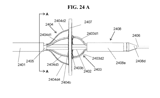
[044] FIGS. 24A-24B shows an exemplary side profile and perspective view of an
embodiment of
another RF energy-based device assembly for interatrial anastomosis;
[045] FIG. 24C shows an exemplary end view of the embodiment of the RF energy-
based device
assembly for interatrial anastomosis of FIG. 24A;
- 8 -
CA 03082954 2020-05-19
WO 2019/109013 PCT/US2018/063439
[046] FIG. 25A shows an exemplary embodiment of the RF cautery electrode
element of FIG.
24A;
[047] FIG. 25B shows an exemplary embodiment of the RF cautery electrode
element of FIG.
24A, wherein the electrode features a secondary cell architecture to increase
structural rigidity in its
expanded state;
[048] FIG. 26 shows an exemplary embodiment of the tissue stabilizer element
of FIG. 24A;
[049] FIGS. 27A-27F show an exemplary embodiment of the RF energy-based device
assembly of
FIG. 24A and the sequential deployment method and operation for interatrial
anastomosis;
[050] FIGS. 28A-28K show an exemplary embodiment of the RF energy-based device
assembly
of FIG. 24A and the sequential deployment method, operation method, excised
tissue capture and
device retraction for interatrial anastomosis.
DETAILED DESCRIPTION OF THE INVENTION
[051] CHF is marked by declining function of the heart muscle, either due to a
weakening of its
pumping ability, known as heart failure with reduced ejection fraction
(HFrEF), or a stiffening of the
muscle with decreased ability to fill with blood prior to ejection, known as
heart failure with
preserved ejection fraction (HFpEF). Inability of the heart to eject or fill
with blood leads to
symptoms of shortness of breath, fatigue, and significant functional
limitation. Prevalence of HFrEF
and HFpEF are roughly equal though rates of HFpEF are rising faster than
HFrEF. With poor flow
of blood from the heart to vital organs, the renin-angiotensin-aldosterone
system (RAAS) is
activated which signals the body to retain fluid, thereby increasing pressure
in the heart chambers. In
particular, as the left atrial pressure (LAP) rises, fluid backs up into the
pulmonary circulation
leading to pulmonary edema and severe shortness of breath. While LAP in normal
adults ranges
from 10-15 mmHg, patients with heart failure frequently have LAP in the 30-40
mmHg range,
which, in some embodiments, spikes during periods of increased heart demand.
[052] Existing pharmacologic treatments for heart failure attempt to remove
excess fluid in the
body through renal excretion (diuretics), neurohormonal blockade, or dilation
of peripheral blood
vessels in order to reduce the stress-load on a failing heart. These
pharmacologic therapies offer
some symptomatic relief and have shown slight mortality benefit in treating
HFrEF, but importantly
have not been shown to improve survival for those with HFpEF.
[053] There are limited device-based therapies for heart failure. Mechanical
circulatory support, in
which a motorized pump is surgically implanted and takes over the function for
the failing heart, is
highly invasive and is reserved for end-stage progression of disease.
Percutaneous mechanical
pumps are used in an acute setting but are only approved for short-term use.
Similarly, intra-aortic
balloon pumps, which decrease cardiac afterload and improve coronary
perfusion, are used only in
the acute inpatient settings. Finally, cardiac resynchronization therapies, in
which an implantable
- 9 -
CA 03082954 2020-05-19
WO 2019/109013 PCT/US2018/063439
pacemaker improves coordinated contraction of failing ventricles, has shown
good results for
improving mortality for patients with heart failure and concomitant electrical
conduction
abnormalities.
[054] Experimental therapies have sought to reduce elevated left atrial
pressure by implanting a
metal stent within the interatrial septum which creates a shunt between the
high-pressure left atrium
towards the low-pressure right atrium. Since the right atrium and the venous
reservoir are highly
compliant, left-to-right blood shunting, in some embodiments, effectively
lower left atrial pressure
without a significant elevation of right atrial pressure, thereby relieving
symptoms and improving
cardiac mechanics. Early human data from these interatrial shunts are showing
promise with
improved functional status and hemodynamic parameters.
[055] The optimal size for these interatrial shunts is unknown, though it has
been approximated
using simulation data and early animal studies. Importantly, the size of the
interatrial aperture must
be large enough to allow effective left atrial offloading, without allowing
too much blood to flow to
the right side such that undue stress is placed on the right atrium and
ventricle. It is widely accepted
among clinicians that individuals presenting with congenital atrial septal
defects warrant closure if
the defect size results in a shunt fraction greater than 50%. Accordingly,
sizing an interatrial shunt
such that no more than 50% of left atrial blood is shunted is important to
reduce long-term adverse
effects.
[056] Implantable interatrial shunts have a number of disadvantages. Since a
foreign body is left
within the heart chambers and makes contact with blood, clotting and
thrombosis is a risk that will
likely require pharmacologic anticoagulation, either long-term or until
endothelialization of the
device's surface occurs. The implant also carries the risk of device-fracture,
dislodgement, or
embolization. The implanted stent in some embodiments also makes it difficult
for subsequent
transseptal procedures as it could limit the degree of freedom for a catheter
to move within the left
atrium. Finally, should closure ever become desirable, a bulky stent, in some
embodiments, adds to
the difficulty of sealing off the interatrial shunt.
[057] Balloon atrial septostomy is a procedure with an associated medical
device which attempts to
create an interatrial aperture to allow mixing of blood between the left and
right sides of the heart.
This device is used in the pediatric population to treat congenital heart
lesions prior to definitive
surgical correction. A deflated balloon, with or without blades attached, is
introduced via the venous
system across the interatrial septum and into the left atrium. The balloon is
subsequently inflated and
pulled proximally thereby tearing the septum and opening an interatrial
aperture. This device
generates an interatrial aperture that is not reproducible from patient to
patient. Since the septum is
torn, the resultant tissue flaps remain in place and eventually fuse back
together. The aperture
created by these device assemblies uniformly close over a period of months.
The temporary nature of
these interatrial apertures makes them suitable for the short-term treatment
of congenital birth
- 10 -
CA 03082954 2020-05-19
WO 2019/109013 PCT/US2018/063439
defects but they are not useful in the adult heart failure population where a
more durable therapy is
desired.
[058] Thus, a device that is capable of creating a sized atrial aperture for
the relief of atrial
pressure, without requiring an implant and in a manner which ensures "long-
term" patency, is
advantageous. Using such a device would achieve the equivalent physiology to
an implantable stent
without the negative sequelae of a leave-behind device. It is desirable to
create a precisely-sized
aperture that could remain patent for the duration of a desired therapeutic
benefit. Since this therapy
would most likely be beneficial for a patient population with high burden of
comorbidities, creating
such an aperture through a minimally invasive procedure is also advantageous.
It is therefore the
goal of this device to enable the creation of a precisely-sized aperture
through a small (<18 Fr, <6.0
mm, <0.236 in.) percutaneous puncture.
[059] In some embodiments, "distal" herein refers to a location, apart, or an
element, e.g. of the
device assembly herein, that is situated further away from the operator of the
device assembly, and
proximal" herein refers to a location, a part, or an element that is situated
nearer to the operator of
the device assembly. For example, the tip of the guidewire in FIG. 4D is
distal to the delivery
catheter or the distal tip of the delivery catheter.
[060] Unless otherwise defined, all technical terms used herein have the same
meaning as
commonly understood by one of ordinary skill in the art to which this
disclosure belongs. As used in
this specification and the appended claims, the singular forms "a," "an," and
"the" include plural
references unless the context clearly dictates otherwise. Any reference to
"or" herein is intended to
encompass "and/or" unless otherwise stated. As used in this specification and
the claims, unless
otherwise stated, the term "about," and "approximately" refers to variations
of less than or equal to
+/- 1%, +/- 2%, +/- 3%, +/- 4%, +/- 5%, +/- 6%, +/- 7%, +/- 8%, +/- 9%, +/-
10%, +/- 11%, +/- 12%,
+/- 14%, +/- 15%, or +/- 20% of the numerical value depending on the
embodiment. As a non-
limiting example, about 100 meters represents a range of 95 meters to 105
meters (which is +/- 5%
of 100 meters), 90 meters to 110 meters (which is +/- 10% of 100 meters), or
85 meters to 115
meters (which is +/- 15% of 100 meters) depending on the embodiments.
[061] As used in this specification and the appended claims, unless otherwise
stated, the term
"coapt" refers to the action of mating or bringing two things together.
[062] The present disclosure relates to RF energy-based device assemblies and
methods for
treating heart failure by reducing elevated blood pressure in the left atrium
of a heart of a mammal.
Disclosed herein, in some embodiments, are transcatheter interatrial septum
excision device
assemblies configured to create a sized atrial aperture between the right and
left atria of a heart for
the relief of left elevated atrial pressure to allow shunting of no more than
50% of the left atrium
blood to the right atrium of the heart. Instead of using mechanical tissue
cutters, disclosed herein, in
some embodiments, are transcatheter interatrial septum excision device
assemblies that utilize RF
-11-
CA 03082954 2020-05-19
WO 2019/109013 PCT/US2018/063439
energy-based tissue cutters. In some embodiments, such RF energy-based tissue
cutters
advantageously facilitate more efficient, accurate, controllable tissue
cutting than traditional
mechanical tissue Disclosed herein, in some embodiments, are device assemblies
for interatrial
anastomosis of a mammal for treating congestive heart failure, the device
assemblies comprising: a
delivery catheter, the delivery catheter having a delivery lumen and being
steerable or bendable; a
radio frequency (RF) generator, the RF generator being remotely located from
the delivery catheter;
and an expandable tissue cutter enclosed within the delivery lumen, the
expandable tissue cutter
attached to a tissue cutter catheter and configured to expand when outside the
delivery lumen,
wherein the expandable tissue cutter is electrically connected to the RF
generator, the tissue cutter
catheter coaxial to and slidable within the delivery catheter, and the tissue
cutter catheter comprising
a first lumen. In some embodiments, the expandable tissue cutter comprises one
or more conductive
materials. In some embodiments, the expandable tissue cutter is connected to
the RF generator by a
conductive wire. In some embodiments, the conductive wire is at least partly
within a wall of the
tissue cutter catheter or at least partly along the tissue cutter catheter.
[063] In some embodiments, the expandable tissue cutter comprises an RF anode,
and the RF
generator is configured to generate RF energy from an RF cathode through
tissue of the mammal to
the RF anode. In some embodiments, the RF cathode is in contact with a body of
the mammal. In
some embodiments, a distance between the RF anode and the RF cathode is within
a range of about
1 mm to about 2 meters. In some embodiments, the expandable tissue cutter
comprises an RF
cathode, and the RF generator is configured to generate RF energy from the RF
cathode through
tissue of the mammal to an RF anode. In some embodiments, the distance between
the RF anode and
the RF cathode is within a range of about 1 mm to about 2 meters. In some
embodiments, the RF
anode is in contact with a body of the mammal. In some embodiments, the RF
cathode is a single-
point electrode, a patch electrode, or a ring electrode. In some embodiments,
the device assemblies
comprise an RF anode, wherein the RF anode is located on a guidewire, a guide
catheter, or the
delivery catheter. In some embodiments, the RF anode is a single-point
electrode, a patch electrode,
or a ring electrode. In some embodiments, the RF generator is configured to
generate alternating
current with an alternating frequency within a range of about 300 kHz to about
3MHz or a power
within a second range of about 1 Watt to about 500 Watts. In some embodiments,
the RF generator
is configured to output a constant voltage, power, or current during at least
part of operation of the
device assembly. In some embodiments, the RF generator is configured to output
a current, voltage,
or power having at least a part of a sine wave. In some embodiments, the RF
generator comprises a
monitor that is configured to monitor a parameter at the tissue cutter. In
some embodiments, the RF
generator comprises an adjuster configured to adjust an output of the RF
generator based on the
monitored parameter. In some embodiments, the RF generator comprises a pump
configured to
circulate a cooling agent to the tissue cutter thereby regulate a temperature
of the tissue cutter. In
- 12 -
CA 03082954 2020-05-19
WO 2019/109013 PCT/US2018/063439
some embodiments, the expandable tissue cutter is at least partly insulated or
at least partly non-
conductive. In some embodiments, at least a part of a distal cutting edge of
the tissue cutter is not
insulated or non-conductive. In some embodiments, the device assemblies
comprise a centralizer
mounted outside of the tissue cutter catheter and slidably engaged with the
delivery catheter. In some
embodiments, said centralizer is configured to provide centralization between
the tissue cutter and
the delivery catheter. In some embodiments, the tissue cutter is configured to
be deployed within a
left atrium of the mammal and pulled toward a right atrium of the mammal,
thereby provides tissue
stabilization and retention during operation of the device assembly. In some
embodiments, the
device assemblies comprise a guidewire. In some embodiments, the guidewire is
configured to
extend from a distal end of the delivery lumen and pass through an initial
puncture site in an
interatrial septum between a right atrium and a left atrium of the mammal at
approximately a fossa
ovalis to provide a working track for the device assembly into the left
atrium. In some embodiments,
the guidewire is coaxially located and slidably engaged with the first lumen.
In some embodiments,
the device assemblies comprise a guide catheter, wherein the guide catheter is
coaxially located
within the first lumen, and wherein the guide catheter comprises a second
lumen within which the
guidewire is configured to slide. In some embodiments, excised tissue by the
tissue cutter from an
interatrial septum is captured and maintained at least by the tissue cutter.
In some embodiments, the
tissue cutter is configured to be withdrawn into the delivery lumen collapsed,
wherein the tissue
stabilizer is simultaneously fully collapsed inside the tissue cutter,
capturing an excised tissue
therein. In some embodiments, a cutting dimension of the expandable tissue
cutter is adjustable.
Disclosed herein, in some embodiments, are device assemblies for interatrial
anastomosis of a
mammal for treating congestive heart failure, the device assembly comprising:
a delivery catheter,
the delivery catheter having a delivery lumen and being steerable or bendable;
a radio frequency
(RF) generator, the RF generator being remotely located from the delivery
catheter; an expandable
tissue cutter enclosed within the delivery lumen, the expandable tissue cutter
attached to a tissue
cutter catheter and configured to expand when outside the delivery lumen,
wherein the expandable
cutter is electrically connected to the RF generator, the tissue cutter
catheter coaxial to and slidable
within the delivery catheter, and the tissue cutter catheter comprising a
first lumen; and an
expandable tissue stabilizer enclosed within the delivery lumen, the
expandable tissue stabilizer
attached to a tissue stabilizer catheter at or near a distal end and
configured to expand when outside
the delivery catheter or the tissue cutter catheter, the tissue stabilizer
catheter coaxial to and slidable
within the delivery lumen or first lumen; the tissue stabilizer catheter
comprising a second lumen
that is slidably engaged and coaxial to the tissue cutter catheter or the
dilator tip catheter. In some
embodiments, the expandable tissue cutter comprises one or more conductive
materials. In some
embodiments, the expandable tissue cutter is connected to the RF generator by
a conductive wire. In
- 13 -
CA 03082954 2020-05-19
WO 2019/109013 PCT/US2018/063439
some embodiments, the conductive wire is at least partly within a wall of the
tissue cutter catheter or
at least partly along the tissue cutter catheter.
[064] In some embodiments, the expandable tissue cutter comprises an RF anode,
and the RF
generator is configured to generate RF energy from an RF cathode through
tissue of the mammal to
the RF anode. In some embodiments, the RF cathode is in contact with a body of
the mammal. In
some embodiments, the distance between the RF anode and the RF cathode is
within a range of
about 1 mm to about 2 meters. In some embodiments, the expandable tissue
cutter comprises an RF
cathode, and the RF generator is configured to generate RF energy from the RF
cathode through
tissue of the mammal to an RF anode. In some embodiments, a distance between
the RF anode and
the RF cathode is within a range of about 1 mm to about 2 meters. In some
embodiments, the RF
anode is in contact with a body of the mammal. In some embodiments, the device
assemblies
comprise an RF anode, wherein the RF anode is located on a guidewire, a guide
catheter, the tissue
stabilizer catheter, the tissue stabilizer, or the delivery catheter. In some
embodiments, the RF
generator is configured to generate alternating current with an alternating
frequency within a range
of about 300 kHz to about 31\41-1z or a power within a second range of about 1
Watt to about 500
Watts. In some embodiments, the RF generator is configured to output a
constant voltage, power, or
current for at least part of operation of the device assembly. In some
embodiments, the RF generator
is configured to output a current, voltage, or power having at least a portion
of a sine wave. In some
embodiments, the RF generator comprises a monitor that is configured to
monitor a parameter at the
tissue cutter. In some embodiments, the RF generator comprises an adjuster
configured to adjust an
output of the RF generator based on the monitored parameter. In some
embodiments, the RF
generator comprises a pump configured to circulate a cooling agent to the
tissue cutter thereby
regulate a temperature of the tissue cutter. In some embodiments, the tissue
cutter is at least partly
insulated or at least partly non-conductive. In some embodiments, at least a
part of a distal cutting
edge of the tissue cutter is not insulated or non-conductive. In some
embodiments, the device
assemblies comprise a centralizer mounted outside of the tissue cutter
catheter and slidably engaged
with the delivery catheter. In some embodiments, said centralizer is
configured to provide
centralization between the tissue cutter and the delivery catheter. In some
embodiments, the tissue
cutter is configured to be deployed within a left atrium of the mammal and
pulled toward a right
atrium of the mammal, thereby providing tissue stabilization and retention
during operation of the
device assembly. In some embodiments, the device assemblies comprise a
guidewire. In some
embodiments, the guidewire is configured to extend from a distal end of the
delivery lumen and pass
through an initial puncture site in an interatrial septum between a right
atrium and a left atrium of the
mammal at approximately a fossa ovalis to provide a working track for the
device assembly into the
left atrium. In some embodiments, the guidewire is coaxially located and
slidably engaged within the
first lumen. In some embodiments, the device assemblies comprise a guide
catheter, wherein the
- 14 -
CA 03082954 2020-05-19
WO 2019/109013 PCT/US2018/063439
guide catheter is coaxially located within the first lumen, and wherein the
guide catheter comprises a
second lumen within which the guidewire is configured to slide. In some
embodiments, excised
tissue by the tissue cutter from an interatrial septum is captured and
maintained, at least by the tissue
cutter. In some embodiments, the tissue cutter comprises a self-expandable
stent with a distal edge
that is blunt, rounded, squared, or hexagonal shaped so that the distal edge
does not puncture an
interatrial septum before any RF energy is applied to the interatrial septum.
In some embodiments,
the tissue cutter comprises a self-expandable stent and a flexible metal loop
attached at or near a
distal end of the stent. In some embodiments, the tissue cutter comprises one
or more of: a flexible
metal loop; a self-expandable coil; a self-expandable stent; a self-expandable
metal wire; a rolled
sheet; one or more self-expandable posts; a hinged strut; a balloon; a self-
expandable mesh; a
mechanically-actuated jaw; or a combination thereof. In some embodiments, the
expandable tissue
cutter is configured to be expanded by one or more energy biasing element. In
some embodiments,
the expandable tissue cutter is configured to be expanded by mechanical
actuation, e.g., via an
umbrella mechanism. In some embodiments, the expandable tissue cutter is
configured to be self-
expanding such that it assumes full expansion from its collapsed state upon
unsheathing/unconstraining the tissue cutter. In some embodiments, the tissue
cutter comprises a
mesh, the mesh configured to help retain excised tissue within the tissue
cutter. In some
embodiments, the tissue cutter is configured to be withdrawn into the delivery
lumen collapsed,
wherein the tissue stabilizer is simultaneously fully collapsed inside the
tissue cutter, capturing an
excised tissue therein. In some embodiments, a cutting dimension of the
expandable tissue cutter is
adjustable and wherein a dimension of the expandable tissue stabilizer is
adjustable. In some
embodiments, a distal end of the tissue stabilizer catheter is configured to
extend along a track of a
guidewire and pass through an initial puncture site such that the tissue
stabilizer also extends past an
interatrial septum into a left atrium. In some embodiments, the tissue
stabilizer is coaxially expanded
within a left atrium such that a dimension thereof is large enough to prevent
the tissue stabilizer from
pulling back through an initial puncture site and such that the tissue
stabilizer provides a supporting,
tensioning effect on an interatrial septum around the initial puncture site.
In some embodiments, the
expandable cutter is configured to be slidably advanced and coaxially expanded
to a cutting
dimension greater than an expanded dimension of the tissue stabilizer. In some
embodiments, the
tissue cutter catheter is configured to extend distally until an fully
expanded tissue cutter engages a
right atrial side of an interatrial septum at or about the fossa ovalis, such
that the tissue cutter pierces
and cuts completely through an interatrial septum, thereby creating an
interatrial pressure relief
opening in the interatrial septum, wherein the interatrial pressure relief
opening is sufficiently large
to allow blood flow through the interatrial pressure relief opening from the
left atrium to the right
atrium such that no more than 50% of left atrial blood is shunted to the right
atrium, and wherein the
interatrial pressure relief opening is sufficiently large, and/or of such
shape, in order to slow a
- 15 -
CA 03082954 2020-05-19
WO 2019/109013 PCT/US2018/063439
natural healing process of the tissue to maintain patency of the interatrial
pressure relief opening in
the interatrial septum without implanting a stent or valve therein. In some
embodiments, the tissue
stabilizer is configured to be partially collapsed and the tissue stabilizer
catheter is configured to be
retracted until the excised tissue is captured and at least a portion of the
partially collapsed tissue
stabilizer is pulled into an opening of the tissue cutter, with the tissue
cutter being at least partially
expanded. In some embodiments, the tissue stabilizer or the tissue cutter
comprises: an inflatable
balloon; expanding tines; an expanding mesh; at least one curved wire; an
expanding plate; an
expanding disc; an expanding fan; a spring coil; at least one strut; at least
one hinged arm; an
umbrella stretcher; or a combination thereof In some embodiments, a tissue
stabilizer material for
anything other than an inflatable balloon comprises a shape memory alloy
comprising: nitinol;
nickel-titanium; copper-aluminum- nickel; or zinc-gold-copper. In some
embodiments, a tissue
cutter material comprises a shape memory alloy comprising: nitinol; nickel-
titanium; stainless steel;
copper-aluminum- nickel; zinc-gold-copper; or a combination thereof. In some
embodiments, the
tissue cutter comprises: a wire mesh; a wire that connects sharpened teeth; a
collapsible hole saw
configuration; a collapsible, open-end cylinder-shape configuration; a
collapsible, open-end barrel-
shape configuration; a collapsible, open-end cone-shaped configuration; or a
combination thereof In
some embodiments, an expanded dimension of the tissue stabilizer is greater
than an expanded
dimension of the tissue cutter. In some embodiments, an expanded dimension of
the tissue cutter is
between about 1% and about 50% smaller than the expanded dimension of the
tissue stabilizer. In
some embodiments, the tissue cutter is configured to cut an aperture or hole
that is: circular in shape;
oval in shape; triangular in shape; squared shaped; rectangular in shape; or
polygon in shape; or a
combination thereof In some embodiments, an expanded dimension of the tissue
stabilizer is less
than an expanded dimension of the tissue cutter. In some embodiments, an
expanded dimension of
the tissue cutter is between about 1% and about 50% larger than the expanded
dimension of the
tissue stabilizer. In some embodiments, an expanded dimension of the tissue
stabilizer is greater than
an expanded dimension of the tissue cutter. In some embodiments, an expanded
dimension of the
tissue stabilizer is between about 1% and about 50% larger than the expanded
dimension of the
tissue cutter. In some embodiments, the device assemblies comprise a
hydrophilic coating on the
guidewire. In some embodiments, the device assemblies comprise a hydrophobic
coating on the
guidewire. In some embodiments, the device assemblies comprise a
force/pressure sensor
incorporated into the distal tip of the guidewire. In some embodiments, the
device assemblies
comprise an oxygen saturation detection sensor incorporated into the
guidewire. In some
embodiments, a sensor or several sensors are incorporated into any one or more
of the catheters of
the device. In some embodiments, a sensor or several sensors are incorporated
into the tissue cutter.
In some embodiments, a sensor or several sensors are incorporated into the
tissue stabilizer. In some
embodiments, the device assemblies comprise a cutting point or edge
incorporated into a distal tip of
- 16 -
CA 03082954 2020-05-19
WO 2019/109013 PCT/US2018/063439
the guidewire. In some embodiments, the device assemblies comprise a curved or
shaped end
incorporated into a distal tip of the guidewire. In some embodiments, the
tissue stabilizer comprises
radiopaque marker bands at strategic locations so as to: orient device
positioning within a body,
orient its relationship to other system components, and to permit visibility
and confirmation of its
deployment state. In some embodiments, the tissue stabilizer and/or tissue
cutter provides embolic
protection by ensuring that any excised tissue is captured and retained within
the device assembly. In
some embodiments, the tissue stabilizer comprising the inflatable balloon
comprises a protective
skirt that protects proximal edges of the inflated balloon. In some
embodiments, the protective skirt
expands and collapses relative to a state of the balloon. In some embodiments,
the tissue stabilizer
and/or the tissue cutter comprises: an expanding mesh; an expanding plate; an
expanding disc; an
expanding fan; expanding posts or tines; or an expanding coil; wherein the
tissue stabilizer and/or
the tissue cutter is fabricated from a shape memory alloy that expands in an
outward direction to
assume an orientation at an approximately 90 angle with respect to the
interatrial septum after
completely passing through an interatrial septum, and is configured to be
pulled back to engage the
septum, to stabilize it prior to and after engagement with the tissue
stabilizer or tissue cutter, and
wherein, following engagement of the tissue cutter, the tissue stabilizer is
collapsed in the same
direction from which it opened, capturing an excised portion of tissue cut
from the septum as the
tissue cutter is resheathed such that the excised tissue and tissue stabilizer
collapse into the delivery
catheter. In some embodiments, the tissue stabilizer and/or tissue cutter
comprises: at least one strut;
at least one hinged arm; or an umbrella stretcher; wherein the tissue
stabilizer expands in an outward
direction to assume an orientation at an approximately 90 angle with respect
to the interatrial
septum after completely passing through an interatrial septum, and is
configured to be pulled back to
engage the septum, to stabilize it prior to and after engagement with the
tissue stabilizer or tissue
cutter; and wherein following activation of the tissue cutter, the tissue
stabilizer is collapsed back in
the same direction from which it opened, capturing an excised tissue cut from
the septum as the
tissue cutter is resheathed such that the excised tissue and tissue stabilizer
collapse into the delivery
catheter. In some embodiments, the tissue stabilizer comprises: at least one
curved wire; or a spring
coil; wherein the tissue stabilizer is fabricated from a shape memory alloy
that is configured to
expand after completely passing through the septum, in an outward direction
transverse to a
proximal-distal axis and having a radial dimension that is greater than or
less than a tissue cutter
dimension and is configured to be pulled back to engage the septum, to
stabilize it prior to and after
engagement with the tissue cutter; and wherein following activation of the
tissue cutter, the tissue
stabilizer is collapsed in the same direction from which it opened, capturing
an excised portion of
tissue cut from the septum as the tissue cutter is resheathed such that the
excised tissue and tissue
stabilizer fit into the delivery catheter. In some embodiments, the expandable
tissue stabilizer is self-
expanding when unsheathed. In some embodiments, the expandable tissue cutter
is self-expanding
- 17 -
CA 03082954 2020-05-19
WO 2019/109013 PCT/US2018/063439
when unsheathed. In some embodiments, the delivery catheter is wire-reinforced
or braided. In some
embodiments, the delivery catheter comprises a reinforced distal tip. In some
embodiments, the
delivery catheter includes a bend radius of about 0.5 inches to about 4
inches. In some embodiments,
the guide catheter is configured to bend in a predetermined manner towards an
interatrial septum. In
some embodiments, the expandable cutter, after expansion, is configured to
create a plurality of
perforations in an interatrial septum. In some embodiments, the expandable
tissue cutter is
configured to translate through the interatrial septum, thereby creating a
complete cut at the
interatrial septum following expansion. In some embodiments, the tissue cutter
comprises a proximal
edge and a distal edge. In some embodiments, the proximal edge does not expand
when the tissue
cutter is fully expanded. In some embodiments, the tissue stabilizer comprises
more than one
expandable mesh discs, at least one of the more than one expandable mesh discs
expands when
proximal to an interatrial septum and in a right atrium. In some embodiments,
two of the plural
expandable mesh discs sandwich the interatrial septum in between discs when
expanded. In some
embodiments, two of the plural expandable mesh discs contact and sandwich the
interatrial septum
in between discs when expanded. In some embodiments, the tissue stabilizer
comprises more than
one expandable mesh discs, one of the plural expandable mesh discs is
configured to plug a distal
opening of the tissue cutter or a distal opening of the delivery catheter when
the tissue stabilizer is
resheathed. In some embodiments, one of the plural expandable mesh discs is
configured to capture
a distal end of the tissue cutter when the tissue stabilizer is resheathed. In
some embodiments, the
one of the plural expandable mesh discs includes a width that is greater than
a width of a distal end
of the tissue cutter. In some embodiments, the plural expandable mesh discs
comprise shape memory
alloy or metal. In some embodiments, the tissue cutter comprises a stent. In
some embodiments, the
tissue cutter comprises one or more of: a plurality of stent cells formed by
struts, a plurality of struts
that are optionally distally connected, a metal loop, and a fine mesh. In some
embodiments, the
metal loop is flexible and the plurality of struts is radially-distributed and
connected to the tissue
cutter catheter. In some embodiments, the metal loop is at least partly
conductive or at least partly
non-conductive. In some embodiments, the metal loop comprises shape memory
material or non-
shape memory material. In some embodiments, the plurality of struts comprises
shape memory
material, rigid material, energy biasing material, or a combination thereof.
In some embodiments, the
plurality of struts is at least partly conductive or at least partly non-
conductive. In some
embodiments, the fine mesh is configured to facilitate retention of excised
tissue within the device
assemblies post-cutting. In some embodiments, the tissue cutter comprises
shape memory material,
energy biasing material, or both. In some embodiments, the delivery catheter
further comprises a
split sheath catheter configured to enable sheathing and unsheathing of the
tissue cutter. In some
embodiments, the delivery catheter further is a split sheath catheter
configured to enable sheathing
and unsheathing of the tissue cutter. In some embodiments, the tissue
stabilizer comprises a fine
- 18 -
CA 03082954 2020-05-19
WO 2019/109013 PCT/US2018/063439
mesh configured to facilitate retention of excised tissue within the device
assemblies post-cutting. In
some embodiments, the device assembly further comprises a dilator tip
configured to facilitate
passage of the device assembly over a guidewire and/or across an interatrial
septum.
[065] Disclosed herein, in some embodiments, are methods for excision of an
interatrial septum of
a mammal for treating congestive heart failure using a transcatheter device
assembly, the methods
comprising: advancing an expandable tissue cutter over a guidewire and across
the interatrial septum
to a left atrium, the expandable tissue cutter in a compressed state;
expanding and moving the tissue
cutter to provide tensioning to the interatrial septum in the left atrium;
translating the tissue cutter to
be in contact with the interatrial septum; transmitting RF power between an RF
cathode and an RF
anode across the interatrial septum thereby creating an aperture, wherein the
RF cathode or the RF
anode is located on the expandable tissue cutter and the other of the RF
cathode or the RF anode is
located on a delivery catheter or in contact with tissue of the mammal; and
resheathing the
expandable tissue cutter into the delivery catheter with the cut interatrial
septum. In some
embodiments, an expandable tissue stabilizer is deployed on the opposite side
of the interatrial
septum to provide tissue stabilization prior to transmitting RF power across
the interatrial septum,
thereby creating an aperture.
[066] Disclosed herein, in some embodiments, are methods for excision of an
interatrial septum for
treating congestive heart failure using a transcatheter device assembly, the
methods comprising:
advancing an expandable tissue stabilizer across the interatrial septum, the
expandable tissue
stabilizer in a compressed state; deploying and moving the tissue stabilizer
to provide tensioning to
the interatrial septum in the left atrium; delivering an expandable tissue
cutter to a right atrium, the
expandable tissue cutter in a second compressed state housed in a delivery
catheter of the device
assembly; expanding the expandable tissue cutter in the right atrium;
translating the tissue cutter
forward to be in contact with the interatrial septum, thereby sandwiching the
interatrial septum
between the expandable tissue cutter and the expandable tissue stabilizer;
transmitting RF power
between an RF cathode and an RF anode across the interatrial septum, thereby
creating an aperture,
wherein the RF cathode or the RF anode is located on the expandable tissue
stabilizer; and
resheathing the expandable tissue cutter and the expandable tissue stabilizer
into the delivery
catheter with the excised tissue. In some embodiments, the methods comprise
allowing vascular
access of the device assembly through a femoral vein. In some embodiments, the
method comprises
puncturing through a fossa ovalis of an interatrial septum and advancing a
guidewire to a left atrium.
In some embodiments, the tissue stabilizer and/or the tissue cutter is
advanced over a guidewire. In
some embodiments, no guidewire is required, and the device (any device
described herein) is
advanced across the septum without pre-puncture by a guidewire, and without
being guided by a
guidewire. In some embodiments, expanding the expandable cutter in the right
atrium comprises
translation of the delivery catheter relative to the tissue cutter. In some
embodiments, the guidewire
- 19 -
CA 03082954 2020-05-19
WO 2019/109013 PCT/US2018/063439
remains in the left atrium following transseptal puncture. In some
embodiments, the excised tissue
comprises at least a portion of the interatrial septum. In some embodiments,
deploying the tissue
stabilizer comprises deploying more than one tissue stabilizing disc
simultaneously or at different
time points. In some embodiments, one of said tissue stabilizing discs is
deployed in the left atrium.
In some embodiments, one of said tissue stabilizing discs is deployed in the
right atrium. In some
embodiments, the methods comprise removing the resheathed device assembly from
the subject. In
some embodiments, advancing the guide catheter over the guidewire to the
interatrial septum
comprises advancing the guide catheter out of the delivery catheter. In some
embodiments,
puncturing through a fossa ovalis of an interatrial septum is performed using
an off-the-shelf
transseptal kit. In some embodiments, resheathing of the tissue cutter and the
tissue stabilizer
comprises plugging a distal opening of the delivery catheter with the tissue
stabilizer. In some
embodiments, resheathing of the tissue cutter and the tissue stabilizer
comprises plugging a distal
opening of the tissue cutter with the tissue stabilizer during resheathing. In
some embodiments, the
methods comprise removing the resheathed device assembly from the subject.
[067] In some embodiments, off-the-shelf transseptal puncture kits are
configured for use with the
transcatheter interatrial septum excision device assemblies herein, thus
simplifying the design of the
transcatheter interatrial septum excision device by removing the penetrating
tip and guidewire from
the main device assembly, and thereby reducing complexity and cost. An example
of such an off-
the-shelf transseptal puncture kits is the Swartz Tm Braided Transseptal
Guiding Introducers LAMPTm
Series, model number 407366, with a 180 cm length with a 0.035 inch diameter.
In some
embodiments, an off-the-self vascular access sheath is used to deploy the
device assembly into the
femoral vein.
Guidewire
[068] In some embodiments, a guidewire is placed across the interatrial septum
using standard
transseptal puncture techniques and provides a working track along which the
device assembly is
advanced. In some embodiments, individual components of the device are
translated along the
guidewire in relation to one another and the interatrial septum. In some
embodiments, a guidewire is
included in the device assembly. In some embodiments, a guidewire is not
included in the device
assembly. In some embodiments, the guidewire features an expandable element at
its distal end to
act as a tissue stabilizer and tissue retention element.
Guide Catheter
[069] In some embodiments, a rigid guide catheter with a pre-bent shape to
guide the tissue cutter
towards the septum at a substantially perpendicular orientation is included in
the device assemblies
herein. In some embodiments, this alignment is also accomplished using a
steerable or deflectable
catheter. In some embodiments, the guide catheter has a central lumen, through
which the guidewire
passes. In some embodiments, a guide catheter is not required at all; in these
embodiments, a pre-
- 20 -
CA 03082954 2020-05-19
WO 2019/109013 PCT/US2018/063439
bent shape, steerability or deflectability is incorporated as a feature of the
guidewire, catheter 1,
catheter 2, catheter 3, catheter 4, or their combinations.
(Expandable) Tissue Stabilizer / Tissue Retention Element
[070] In some embodiments, a tissue stabilizer provides counter tension to the
interatrial septum
during activation of the tissue cutter so as to minimize any unintended tissue
deformation, rotation,
or displacement due to unbalanced forces. In some embodiments, the tissue
stabilizer also provides
tension to the interatrial septum so as to minimize wall motion as the heart
beats. In some
embodiments, the tissue stabilizer also doubles as a tissue retention element
to prevent the excised
tissue from inadvertently coming free from the device assembly, and permits
translation and packing
of the excised tissue into the delivery catheter prior to removal of the
device assembly from the
body. In some embodiments, the tissue stabilizer includes one or more of: a
balloon, a self-
expanding mesh, a coil, and manually actuated flexible struts. In some
embodiments, the tissue
stabilizer is connected to catheter 2 (e.g., the tissue stabilizer catheter),
which features a central
lumen that permits internal translation/passage of the guidewire and/or guide
catheter. In some
embodiments, the tissue stabilizer is connected to the guidewire and is made
of a self-expanding
mesh that is constrained within a separate catheter prior to delivery to the
left atrium. In some
embodiments, this self-expanding mesh resides in a collapsed state within the
guide catheter
(catheter 1) prior to deployment. In some embodiments, the RF anode is
incorporated into the tissue
stabilizer. In some embodiments, a distinct tissue stabilizer is not needed,
as these functions are
performed by the tissue cutter. In some embodiments, the tissue cutter is
deployed within the left
atrium and pulled backwards, thereby dually serving the purpose of tissue
stabilization and retention.
(Expandable) Tissue Cutter / Cutting Element
[071] In some embodiments, an RF electrosurgery tissue cutter, or equivalently
herein, an RF
electrosurgery tissue cutter, includes an expanding structure that features an
exposed conductive
surface area. In some embodiments, the tissue cutter is delivered to the
septum and, upon
deployment/expansion, energization and actuation, excises a portion of tissue
to yield a prescribed
aperture. In some embodiments, the tissue cutter is connected, by conductive
wire that runs along the
length of catheter 3 (tissue cutter catheter), to the RF generator, and acts
as the RF cathode directing
energy into tissue. In some embodiments, this conductive wire is embedded
within the walls of
catheter 3 or alternatively run within or along the length of other catheters
in the device assembly. In
some embodiments, it is advantageous for the tissue cutter to have a very
small surface area of
exposed conductive material such that energy density immediately adjacent to
the tissue cutter is
concentrated highly enough to achieve a desired tissue effect. In some
embodiments, the very small
surface area of exposed conductive material is in the range of about 0.01% to
about 50% percent of
the total surface area of the tissue cutter. In some embodiments, the very
small surface area of
exposed conductive material is in the range of about 0.01% to about 1% percent
of the total surface
-21-
CA 03082954 2020-05-19
WO 2019/109013 PCT/US2018/063439
area of the tissue cutter. In some embodiments, the very small surface area of
exposed conductive
material is in the range of about 0.1% to about 1% percent of the total
surface area of the tissue
cutter. In some embodiments, the very small surface area of exposed conductive
material is in the
range of about 0.1% to about 5% percent of the total surface area of the
tissue cutter. In some
embodiments, the very small surface area of exposed conductive material is in
the range of about
0.1% to about 10% percent of the total surface area of the tissue cutter. In
some embodiments, the
very small surface area of exposed conductive material is in the range of
about 1% to about 15%
percent of the total surface area of the tissue cutter. In some embodiments,
it is desirable to
selectively energize discrete portions of the tissue cutter in rapid
succession to maintain high energy
density surrounding the energized portion of the tissue cutter without the
need to energize the entire
tissue cutter at once. In some embodiments, it is also advantageous to
maintain a small, defined gap
between the tissue cutter and the interatrial septum during energy application
to permit electric
arcing and minimize any undesired thermal effects to the tissue. In some
embodiments, such gap is
maintained through methods of device use or through device features
incorporated to ensure the gap
is maintained. In some embodiments, it is advantageous to rotate or
oscillate/vibrate the tissue cutter
during or following tissue excision to prevent, minimize, or disrupt char
formation.
[072] In some embodiments, RF is any frequency or combination of frequencies
within the
electromagnetic spectrum which is/are associated with radio waves. When an RF
current, voltage,
and/or power is supplied from an RF cathode, the current then propagates
through tissue, and/or any
other RF conductive media between the RF cathode and RF anode.
[073] In some embodiments, an RF cathode is the electrode from which an RF
current leaves; such
current is in the direction in which positive electrical charges move. In some
embodiments, an RF
anode is the electrode from which RF current flows into. In some embodiments,
an RF cathode or
anode is a single-point electrode, a ring electrode, a plate electrode, a
pointed electrode, a blade-
shaped electrode, a patch electrode, or any other types of electrodes.
[074] In some embodiments, the device herein, such as the tissue cutter is
configured to be used in
electrosurgery, with the application of a radio frequency alternating polarity
electrical current to
tissue as a means to cut. In electrosurgical procedures, the tissue is heated
by the RF electric current.
In some embodiments, only the tissue to be cut is heated without heating other
tissue in its close
vicinity. In some embodiments, only part of the tissue to be cut is heated. In
some embodiments,
such heating of tissue is controlled by the size, shape, and/or geometry of
the conductive region of
the tissue cutter. In some embodiments, the tissue is heated to a
predetermined temperature, or a
predetermined range of temperatures. In some embodiments, the tissue to be cut
is heated to no less
than 60 C, 70 C, 80 C, 90 C, 100 C or even higher temperatures. In some
embodiments, the tissue
to be cut is heated to be in the range of 50 C to 100 C.
- 22 -
CA 03082954 2020-05-19
WO 2019/109013 PCT/US2018/063439
RF Energy Sink (RF anode or RF return)
[075] In some embodiments, an RF anode acts as a RF current sink for the RF
cathode to define
the field in which RF energy is transmitted within the body of the mammal. In
some embodiments,
the device assembly disclosed herein includes an RF energy sink (RF anode, or
RF return) to draw
RF energy from the RF cathode out of the body, thus defining the field across
which RF energy is
transmitted. Referring to FIG. 1, in some embodiments, the RF generator 117
generates RF current
that is transmitted to the RF cathode 103b via a conductive wire, e.g., a
straight wire, a coaxial cable.
In some embodiments, the RF cathode transmits RF energy via RF current/voltage
through the tissue
107 to the RF anode 113b, and the RF anode receives RF energy and then
transmits the energy back
to the RF generator via a conductive connection. In some embodiments, the RF
anode is positioned
in close proximity to the RF cathode or external to, but in contact with, the
body, (e.g. a skin patch
electrode). In some embodiments, the RF anode is incorporated as a feature of
the tissue stabilizer.
In some embodiments, the RF anode is on the guidewire, the tissue stabilizer
catheter, the tissue
cutter catheter or the delivery catheter.
Delivery Catheter
[076] In some embodiments, the delivery catheter is the main housing catheter
for all other
components of the device assembly (excluding the RF generator and, in some
embodiments, the RF
energy sink). In some embodiments, the tissue cutter is housed towards the
distal end of the delivery
catheter and ensures that the tissue cutter remains collapsed prior to
deployment. In some
embodiments, the delivery catheter additionally permits packing of the excised
tissue within its
lumen. In some embodiments, the delivery catheter features a pre-bent shape,
steerability, or
deflectability to facilitate orientation with respect to the interatrial
septum.
RF Generator
[077] In some embodiments, an apparatus that generates alternating current,
voltage, and/or power
in the radiofrequency spectrum, in the range of about 300 kHz to about 31\41-
1z at a power level in the
range of about 5W to about 300W. In some embodiments, an apparatus that
generates alternating
current, voltage, and/or power in the radiofrequency spectrum, in the range of
about 9 kHz to about
300MHz. In some embodiments, the RF generator, stationed outside of the
sterile field, is connected
to the RF anode and cathode through a sterile connector that crosses into the
sterile field and
transmits RF energy through wires that connect to the RF anode and cathode. In
some embodiments,
the RF generator is operated by outputting constant voltage, constant power,
and/or constant current.
In some embodiments, the RF generator outputs a constant sine wave throughout
the duration of
tissue cutting. In some embodiments, the RF signal output is interrupted and
dampened such that RF
energy is applied for a fixed percentage of operation time of the device
assembly, in the range of
0.01% to 99.9% of the operation time. In some embodiments, these different RF
energy output
modes yield varied tissue effects. In some embodiments, it is anticipated that
a constant sine wave
-23-
CA 03082954 2020-05-19
WO 2019/109013 PCT/US2018/063439
yields the desired tissue vaporization without thermal injury or char
formation. In some
embodiments, the RF generator includes batteries to obviate the need for wires
crossing into/out of
the sterile field. In some embodiments, the RF generator is within or outside
the sterile field. In some
embodiments, the RF generator monitors the temperature or impedance at or near
the distal end of
the tissue cutter and adjusts the power, voltage, or current output to ensure
continuous tissue cutting.
In some embodiments, the RF generator includes a pump to circulate chilled
coolant such as saline
through the tissue cutter catheter to regulate its temperature and
minimize/prevent char formation.
[078] In some embodiments, the RF generator includes a power source, an on/off
switch, a
processor, a computer memory, a communications element, an electrical
connection to the RF
cathode, and an electrical connection to the RF anode. In some embodiments,
the RF generator is
manually controlled. In some embodiments, the RF generator is automatically
controlled using a
feedback system, e.g. temperature feedback.
[079] In some embodiments, the RF generator includes an operating system
configured to perform
executable instructions. The operating system is, for example, software,
including programs and
data, which manages the device's hardware and provides services for execution
of applications. In
some embodiments, the RF generator includes a storage and/or memory device.
The storage and/or
memory device is one or more physical apparatuses used to store data or
programs on a temporary or
permanent basis. In some embodiments, the RF generator includes a display to
send visual
information to a user. In some embodiments, the RF generator includes an input
device to receive
information from a user. In some embodiments, the RF generator herein includes
one or more non-
transitory computer readable storage media encoded with a program including
instructions
executable by the operating system of an optionally networked digital
processing device. In some
embodiments, the RF generator herein includes at least one computer program,
or use of the same. In
some embodiments, a computer program includes a sequence of instructions,
executable in the
digital processing device's CPU, written to perform a specified task.
[080] Referring to FIG. 2, in some embodiments, the device assemblies include
a delivery catheter
201, a tissue cutter catheter 202 to which a tissue cutting balloon 203e is
attached that contains a
tissue cutter cathode 203b, a tissue stabilizing balloon 204e which is
attached at the distal end of a
tissue stabilizer catheter 205, and a guidewire 206 which is slidably engaged
within the lumen of the
tissue stabilizer catheter. In some embodiments, the RF cathode 203b is
located on or near the distal
end of the tissue cutter, and the RF anode 213b is located on or near the
proximal end of the tissue
stabilizer. Both the tissue cutter and tissue stabilizer includes at least one
expandable or inflatable
balloon.
[081] In some embodiments, the methods disclosed herein using the device
assemblies includes
one or more procedural steps selected from the following steps, but not
necessarily in the exact
order:
- 24 -
CA 03082954 2020-05-19
WO 2019/109013 PCT/US2018/063439
- vascular access is achieved through the femoral vein using standard
techniques (e.g.,
Seldinger method);
- transseptal puncture through the fossa ovalis of the interatrial septum
is performed using
standard interventional techniques), leaving a guidewire in place;
- the device assembly is introduced, e.g., through the groin, and delivered
over the guidewire
to the right atrium;
- the tissue stabilizer is introduced over the guidewire and across the
interatrial septum to the
left atrium; there, it is expanded in diameter by inflation (in the case of a
balloon), mechanical
actuation or unsheathing of a self-expanding material (alternatively, the
tissue cutter is
introduced over the guidewire and across the interatrial septum to the left
atrium; there, the tissue
cutter is unsheathed by pulling its constraining catheter backwards relative
to the tissue cutter);
- the tissue cutter is delivered (via delivery catheter) to the right
atrium; there, the tissue cutter
is unsheathed within the right atrium by pulling its constraining catheter
backwards
(Alternatively, the tissue stabilizer is delivered (via delivery catheter) to
the right atrium; there, it
is expanded in diameter by inflation (in the case of a balloon), mechanical
actuation, or
unsheathing of a self-expanding material);
- after (or in the process of) unsheathing, the tissue cutter is fully
expanded;
- the tissue cutter is translated over the guidewire until it is in
touching the interatrial septum,
thus sandwiching the tissue between the tissue cutter and tissue stabilizer;
- the tissue cutter is energized, sending RF current across the septum
(between the RF cathode
and anode) to create an aperture whose shape is prescribed by the geometry of
the tissue cutter;
and
the following steps are performed in any order depending on embodiment:
- the tissue cutter is collapsed;
- the tissue stabilizer is pulled backwards with respect to the delivery
catheter, thereby packing
the excised tissue within the delivery catheter;
- the tissue stabilizer is collapsed; and
- the device is removed from the body.
Stents
[082] In some embodiments, the tissue cutter disclosed herein includes a
stent. In some
embodiments, the tissue cutter takes the form of a self-expanding (e.g., shape
memory material)
stent, which is sharpened along the full length of its distal edge, and self-
expands upon deployment
from the delivery catheter (catheter 4, not shown) within the right atrium.
The tissue cutter is fully
insulated with the exception of its distal edge; its proximal end is attached
to the tissue cutter
catheter and is coupled to the RF generator by a wire that runs within or
along the tissue cutter
-25-
CA 03082954 2020-05-19
WO 2019/109013 PCT/US2018/063439
catheter, In some embodiments, the tissue cutter catheter features a central
lumen that contains the
guidewire, guide catheter, and tissue stabilizer.
[083] In some embodiments, the device is advanced over a guidewire, with the
tissue stabilizer
being attached to its respective catheter (catheter 2), and delivered across
the septum where it is
actuated to fully expand. In some embodiments, the delivery catheter (catheter
4) is pulled
backwards with respect to the tissue cutter catheter, thereby exposing and
expanding the stent and
tissue cutter. In some embodiments, the RF anode is incorporated into the
tissue stabilizer, which for
example, is a ring electrode on the tissue stabilizer catheter (in the left
atrium), or a pad placed on
the surface of the body of the mammal. Once properly positioned, in some
embodiments, the tissue
cutter is energized by the RF generator, resulting in tissue disruption and
excision. In some
embodiments, the tissue cutter, excised tissue and tissue stabilizer are
collapsed within the delivery
catheter (catheter 4) and the device is removed from the body.
[084] In some embodiments, the tissue cutter disclosed herein includes a
stent. In some
embodiments, as illustrated in FIGS. 3A-3C, the tissue cutter takes the form
of a self-expanding
(e.g., shape memory material) stent. In some embodiments, the stent pattern is
laser-cut onto a shape
memory material cylindrical tube. In some embodiments, the stent 303 has
approximately diamond
pattern shaped cells. In some embodiments, the stent cells are of other
shapes, for example, circle,
triangle, rectangular, etc. In some embodiments, as illustrated by FIG. 3C,
the stent 303 has at least
three axisymmetric struts 303m that are distally connected by a ring 303b at
its expanding diameter.
The ring 303b in some embodiments is within (coaxially closer to the
longitudinal axis of the
catheter 302 of FIG. 3A) the stent 303 of FIG. 3A, as an alternative to the
ring 303b shown in FIG.
3B. Alternatively, the embodiment ring of FIG. 3C is used as a stabilizer
externally (further from
the longitudinal axis of the catheter 302 of FIG. 3A) and coaxially with the
embodiment of FIG. 3A.
Alternatively, the embodiment of FIG. 3C is used as a tissue cutter with the
ring 303b being
sharpened for cutting the septum, or energized for cutting the septum using RF
energy (as a
cathode). In some embodiments, as illustrated by FIGS. 3A-3B, the stent tips
303j are sharpened at
its distal edge and can penetrate through the interatrial septum and act as an
anchoring mechanism to
position the stent substantially perpendicular to the interatrial septum. In
some embodiments, the
stent 303 is fully insulated 303h with the exception of its distal tips 303j,
the crowns of the struts
303k, the valleys of the struts 3031 and/or the ring 303b. In some
embodiments, the proximal end of
the stent is attached to the tissue cutter catheter 302 and is coupled to the
RF generator by a wire (not
shown) that runs within or along the tissue cutter catheter. In some
embodiments, the stent is
(partially) insulated or comprised of a non-conductive material such as:
Parylene; PTFE. In some
embodiments, the stent self-expands upon deployment from a catheter (delivery
catheter or tissue
stabilizer catheter, not shown) within the right atrium or the left atrium,
with the expanding diameter
facing the interatrial septum. In some embodiments, once properly positioned,
the tissue cutter is
-26-
CA 03082954 2020-05-19
WO 2019/109013 PCT/US2018/063439
energized by the RF generator and translates through the interatrial septum,
resulting in tissue
disruption and excision. In some embodiments, the stent is fully insulated
with exception of its distal
tips and the sharpened edge, once properly positioned (the distal tips have
penetrated through the
interatrial septum), the tissue cutter is energized by the RF generator and
the tissue cutter is rotated,
resulting in tissue disruption and excision. In some embodiments, the self-
expanding stent is
deployed within the left atrium using a split sheath catheter with a tapered,
penetrating tip to
facilitate crossing the interatrial septum. In some embodiments, the split
sheath is moved distally to
unsheath the stent 303 which distal edge faces the interatrial septum. FIG.
3B, in a particular
embodiment, shows a metal wire loop 303b that is tethered through small holes
303n at multiple
points in the crowns of the struts 303k, thus maintaining a gap between the
loop and distal edge of
the stent. In this embodiment, the stent 303 is fully insulated with exception
of its distal tips 303j and
the crowns of the struts 303k. In some embodiments, the loop 303b is sutured,
welded, soldered,
brazed, glued, threaded, or wrapped through, in, or around the distal edge of
the stent. In some
embodiments, the loop 303b takes the form of a circle, polygon, lasso, or any
other geometrical
shapes once the stent is fully expanded. In some embodiments, the stent merely
acts as a self-
expanding scaffold to expand the metal loop to a desired geometry. In some
embodiments, the self-
expanding stent is a braided stent in a tubular mesh configuration. In some
embodiments, the tissue
cutter catheter 302 features a central lumen that contains the guidewire,
guide catheter, and tissue
stabilizer.
[085] In some embodiments, the stent tissue cutter is deployed within the left
atrium over a
guidewire, with its uninsulated/cutting edge oriented towards the interatrial
septum. In some
embodiments, the tissue stabilizer is excluded from the transcatheter
interatrial septum excision
device. As illustrated in FIGS. 4A-4D, the stent tissue cutter 403 is
collapsed and housed in a split
sheath catheter 408. In some embodiments, catheter 408 comprises a cap 408a
and a shaft 408e. The
catheter shaft has a central lumen 408f that is slidably engaged with the
guidewire 406. The cap
408a has a tapered, penetrating tip 408d to facilitate catheter 408 in
crossing the interatrial septum
(over the guidewire) from the right atrium to the left atrium. The cap of
catheter 408a has a second
lumen 408b that houses the collapsed stent tissue cutter 403. FIG. 4D shows a
detailed view of FIG.
4C. In some embodiments, the stent tissue cutter is partially insulated with
insulation 403h, leaving
its cutting edge 403b exposed (oriented to face the septum), and its back end
coupled to the RF
generator through a wire (not shown). In some embodiments, the stent tissue
cutter 403 is attached to
tissue cutter catheter 402, having a central lumen, slidably engaged with the
shaft 408e of catheter
408. In some embodiments, a centralizer 411 having a central lumen mounted to
the outer diameter
of tissue cutter catheter 402 and the internal diameter of the stent tissue
cutter 408e is incorporated.
In some embodiments, the stent tissue cutter 403 is coupled to the RF
generator by a conductive wire
that runs within or along tissue cutter catheter 402. In some embodiments, an
electrode is placed
-27-
CA 03082954 2020-05-19
WO 2019/109013 PCT/US2018/063439
external to, but in contact with, the body (e.g. a skin patch electrode) to
act as the RF anode. In some
embodiments, a ring electrode resides on tissue cutter catheter 402 within the
right atrium and acts as
the RF anode, thereby eliminating the need for an externally-placed RF anode.
[086] In some embodiments, the device assembly 400 is advanced forward to the
left atrium such
that the cap 408a of catheter 408 crosses the septum 407. The tissue cutter
catheter 402 and the
delivery catheter 401 are held stationary as catheter 408 is advanced forward
to unsheath the stent
tissue cutter 403 and deploy its cutting edge. In some embodiments, the
cutting edge of the stent
tissue cutter is brought in contact with the septum 407 by pulling the tissue
cutter catheter 402
backwards such that RF energy is delivered. In some embodiments, post-tissue
cutting, the stent
tissue cutter 403 is collapsed by sliding catheter 408 backwards (proximally)
with respect to tissue
cutter catheter 402 that is stationary or moved distally to support the stent
to collapse in the cap
408a. In some embodiments, the excised tissue is packed within the stent 403
itself, followed by the
second lumen 408b of the cap 408a of catheter 408. In some embodiments, the
tissue cutter dually
acts as a tissue retention element, given that the excised tissue remains
within to the tissue cutter
post-tissue excision.
[087] In some embodiments, the tissue cutter remains in the right atrium. The
device is advanced
over a guidewire, with the tissue stabilizer being attached to its respective
catheter (catheter 2), and
delivered across the septum where it is actuated to fully expand. In some
embodiments, the delivery
catheter (catheter 4) is pulled backwards with respect to the tissue cutter
catheter, thereby exposing
and expanding the stent tissue cutter. In some embodiments, the RF anode is
incorporated into the
tissue stabilizer, which for example, is a ring electrode on the tissue
stabilizer catheter (in the left
atrium), or a pad placed on the surface of the body of the mammal. Once
properly positioned, in
some embodiments, the tissue cutter is energized by the RF generator,
resulting in tissue disruption
and excision. In some embodiments, the tissue cutter, excised tissue and
tissue stabilizer are
collapsed within the delivery catheter and the device is removed from the
body.
[088] In some embodiments, the stent tissue cutter has a blunt, rounded,
squared, or hexagonal
shaped distal edge such that it does not puncture the tissue before RF energy
is applied to the
septum; it merely engages the tissue along the length of its distal edge prior
to energy application.
[089] In some embodiments, the stent tissue cutter is comprised of a shape
memory material such
as: nickel-titanium; copper-aluminum-nickel; zinc-gold-copper; or a
combination thereof.
[090] In some embodiments, the stent is fully insulated or comprised of a non-
conductive material.
In some embodiments, the tissue cutter includes a flexible, metal loop, e.g.,
303b, affixed to the
distal end of the stent at one or multiple points. In some embodiments,
electric arcing is generated
between the flexible metal loop and the tissue thus facilitate tissue cutting,
e.g., FIGS. 3A-3B. In
some embodiments, the stent merely acts as a self-expanding scaffold to expand
the metal loop to a
desired geometry. In some embodiments, the stent features pointed tips, e.g.,
305, that penetrate the
-28-
CA 03082954 2020-05-19
WO 2019/109013 PCT/US2018/063439
tissue and act as an anchoring mechanism to position the stent substantially
perpendicular to the
septum. In some embodiments, the metal loop is sutured, welded, soldered,
brazed, glued, threaded,
or wrapped through, in, or around the distal edge of the stent. In some
embodiments the metal loop
takes the form of a circle, polygon, or lasso once the stent is fully
expanded.
[091] In some embodiments, the metal loop follows the contours of the teeth at
the distal edge of
the stent. In some embodiments, the metal loop is anchored at discrete points,
thus maintaining a gap
between the metal loop and stent between anchoring points. In some
embodiments, the metal loop is
anchored to the distal edge of the stent.
[092] In some embodiments, the transcatheter interatrial septum excision
device includes a split
catheter. In embodiments shown in FIGS. 4 and 5, the split sheath catheter 408
together with
catheter 401 form the delivery catheter whose lumen 408b, together with the
lumen of catheter 401,
form the delivery lumen; and split sheath catheter 508, together with catheter
501, form the delivery
catheter whose lumen 508b, together with the lumen 501a, form the delivery
lumen.
[093] In some embodiments, the tissue cutter and tissue stabilizer are both
self-expanding stents
with an uninsulated edge on each stent acting as the RF cathode 503b and anode
513b, respectively.
Referring to FIGS. 5A-5D, catheter 508 comprises a cap 508a and a shaft 508e,
the catheter shaft
having a central lumen (not shown), slidably engaged with the guidewire (not
shown); the cap has a
tapered tip 508d to facilitate the catheter in crossing the septum 507 (over
the guidewire) from the
right atrium to the left atrium; the cap of catheter 508a features a second
lumen 508b that houses the
collapsed tissue stabilizer 504. In some embodiments, the tissue stabilizer
504 is partially insulated
504h, leaving its uninsulated edge exposed, and is mounted to the distal
aspect of the tissue stabilizer
catheter 505. In some embodiments, a conductive wire connects the tissue
stabilizer to the RF
generator, running within the walls of the tissue stabilizer catheter 505. In
some embodiments, the
stent tissue cutter 503 is at least partially insulated 503h, leaving its
uninsulated edge exposed, and is
mounted to the distal end of the tissue cutter catheter 502. In some
embodiments, a conductive wire
connects the tissue cutter to the RF generator, running at least partly within
the walls of the tissue
cutter catheter 502. In some embodiments, the tissue cutter catheter 502 has a
central lumen that is
slidably engaged with the tissue stabilizer catheter 505. In some embodiments,
a delivery catheter
501 has a central lumen that is slidably engaged with the tissue cutter
catheter 502. In some
embodiments, a centralizer 511 having a central lumen mounted to the outer
diameter of the tissue
stabilizer catheter 505 and the internal diameter of the tissue stabilizer 504
is incorporated.
[094] In some embodiments, as shown in FIG. 5A, the device assembly is
advanced into the body
over a guidewire (not shown) until the entire cap 508a of tissue stabilizer
catheter 505 is advanced
into the left atrium. In some embodiments, the tissue stabilizer is unsheathed
and deployed in the left
atrium by advancing catheter 508 forward with respect to the other device
components. In some
embodiments, the tissue stabilizer catheter 505 is pulled backwards with
respect to other device
-29-
CA 03082954 2020-05-19
WO 2019/109013 PCT/US2018/063439
components to bring the tissue stabilizer in contact with the septum, as shown
in FIG. 5B. In some
embodiments, the delivery catheter 501 is pulled back while other device
components of the device
assembly 500 are held stationary to unsheath and deploy the stent tissue
cutter 503 within the right
atrium. In some embodiments, the tissue cutter catheter 502 is advanced
forward with respect to the
other device components until the stent tissue cutter is in contact with the
septum, as in FIGS. 5C-
5D. In some embodiments, the tissue stabilizer and stent tissue cutter
`mate/coapf on opposing sides
of the septum to create a sandwiching effect. In some embodiments, energy is
then applied by the RF
generator to excise tissue and yield an aperture. In some embodiments, the
delivery catheter is
advanced to resheath the stent tissue cutter, whereas catheter 508 is pulled
back to resheath the tissue
stabilizer stent 504. In some embodiments, the excised tissue is pierced and
retained by the tissue
stabilizer catheter 505. In some embodiments, the tissue stabilizer is
deployed in the right atrium
and the tissue stabilizer catheter is pushed distally to make contact with the
septum, whereas the
tissue cutter is deployed in the left atrium and the tissue cutter catheter is
pulled proximally to
achieve apposition with the septum before RF energy is applied.
Struts with Metal/Wire Loop
[095] In some embodiments, as shown in FIGS. 7A-7B, the tissue cutter includes
a flexible metal
loop 703 that is deployed into its expanded state through a plurality of
radially-distributed struts
703d connected to the distal end of the tissue cutter catheter 702; these
struts provide structural
rigidity and coaxial alignment with respect to the tissue cutter catheter. In
some embodiments, the
metal loop folds up in a standard fold, coil, a plurality of loops/petals, an
accordion, a rolled or
straight configuration in its collapsed/initial state pre-deployment. In some
embodiments, the metal
loop is composed of wire. In some embodiments, the metal loop is cut from a
cylindrical tube. In
some embodiments, the metal loop comprises of a conductive shape memory metal.
In some
embodiments, the metal loop is comprised of a conductive non-shape memory
metal. In some
embodiments, the metal loop is set back (recessed) from the tips of the struts
by a fixed distance (e.g.
about 0.1 to about 10.0 mm). In some embodiments the metal loop features
smaller loops/petals
703g that wrap around the individual struts. In some embodiments, the arc
length of the metal loop is
fixed. In some embodiments, the ends of the metal loop come together and run
along the length of
the tissue cutter catheter, such that the metal loop is be actuated as a snare
loop.
[096] In some embodiments, the struts of the tissue cutter comprise conductive
shape memory
material(s), such that when unconstrained (through slidable translation of its
constraining catheter
712/701 with respect to the tissue cutter catheter 702), they flare open,
thereby expanding the metal
loop to a desired conformation, as illustrated in FIG. 7B. In some
embodiments, the struts ensure
coaxial alignment between the metal loop and tissue cutter catheter 702 and,
by extension, with all
other catheters within the device assembly (in addition to the guidewire 706).
In some embodiments,
the struts are pre-bent to assume U-bend configurations when fully expanded,
thereby permitting the
- 30 -
CA 03082954 2020-05-19
WO 2019/109013 PCT/US2018/063439
struts to bend approximately 1800 backwards to orient the metal loop to face
the interatrial septum
(upon delivery to and deployment within the left atrium). In some embodiments,
the struts are pre-
bent and is configured to expand to a flared cross-section in a plane
determined by the radial
direction and the proximal-distal direction. (to a larger diameter than its
constrained diameter). In
some embodiments, the cross-section is of different shapes, such as a bell
shape, a conical shape, a
U-shape, or any other geometrical shape. In some embodiments, the geometry
towards the tips of the
struts features a step-up in width to facilitate seating of the metal loop. In
some embodiments, the
geometry towards the tips of the struts features a step-up in width, with
narrowing of width in
between to facilitate seating of the metal loop. In some embodiments, the
geometry towards the tips
of the struts features narrowing of its width, without a step-up in width, to
facilitate seating of the
metal loop. In some embodiments, the struts of the tissue cutter are not
comprised of a shape
memory material, but are connected to one another by energy biasing elements,
such as springs,
which are compressed when collapsed/constrained, but flare the struts outwards
radially when
unconstrained (by translating its constraining catheter backwards), thereby
expanding the non-shape
memory metal loop. In some embodiments, the struts are mechanically actuated
through an umbrella
mechanism, wherein two struts (an actuation strut and an expansion strut) are
connected by a hinge
point and are connected to an actuation catheter and a tissue cutter catheter,
respectively, by another
hinge point, such that when the actuation catheter is translated forward in
relation to the tissue cutter
catheter, the actuation strut rotates the expansion strut outwards radially by
its hinge point on the
expansion catheter. In some embodiments, this radial expansion expands a metal
loop to a desired
diameter by the degree in which the actuation catheter is translated.
Additionally, in some
embodiments, the actuation catheter is replaced by an actuation wire pulley
system, whereas
translation by the actuation wire expands the expansion strut. In some
embodiments, the expansion
mechanism is driven by the metal loop, rather than the struts to which the
metal loop is attached. In
some embodiments, the struts and metal loop are fully insulated or,
alternatively, the struts and metal
loop are comprised of a non-conductive material and a separate (conductive)
metal loop is affixed to
the insulated (or non-conductive) metal loop to act as the tissue cutter. In
some embodiments, the
struts are insulated while the metal loop is uninsulated. In some embodiments,
the struts and metal
loop are both partially insulated. In some embodiments, a fine mesh or
membrane 703f, composed of
a textile, polymer, or metal, is incorporated between and/or around the struts
of the tissue cutter and
connected to the metal loop 703b, as in FIG. 7A, or, alternatively, between
and/or around the struts
but not connected to the metal loop, or, alternatively, within the plane of
the metal loop, or,
alternatively, in the plane of the metal loop, but set recessed/set back to
help ensure all excised tissue
is retained within the device assembly post-cutting. In some embodiments, the
metal loop is affixed
to the distal tips of the struts; alternatively, the metal loop is set back
(recessed) from the tips by a
fixed distance (e.g. about 0.1 to about 10.0 mm). In some embodiments, the
tissue cutter is deployed
- 3 1 -
CA 03082954 2020-05-19
WO 2019/109013 PCT/US2018/063439
within the left atrium and pulled proximally to engage and cut tissue. In some
embodiments, a split
sheath catheter is used to permit sheathing and unsheathing of the tissue
cutter within the left atrium.
In some embodiments, the tissue cutter is deployed within the right atrium and
advanced distally to
engage and cut tissue.
[097] In some embodiments, as illustrated in FIGS. 7A-7B the tissue stabilizer
704 includes one,
or a plurality of, self-expanding (shape memory) struts. In some embodiments,
the tips of the tissue
stabilizer struts 704a are sharpened to permit penetration through the
interatrial septum. In some
embodiments, the tips of the tissue stabilizer struts are blunt to prevent
penetration through the
interatrial septum. In some embodiments, the tissue stabilizer 704 expands to
assume a diameter that
is greater than the expanded diameter of the tissue cutter 703 when fully
deployed. In some
embodiments, the tissue stabilizer expands to assume a diameter that is less
than the expanded
diameter of the tissue cutter when fully deployed. In some embodiments, a fine
mesh or membrane
704f, composed of a textile, polymer, or metal, is incorporated between and/or
around the struts of
the tissue stabilizer, as in FIG. 7A, to help ensure all excised tissue is
retained within the device
assembly post-cutting. In some embodiments, the tissue stabilizer is fully
insulated. In some
embodiments, the tissue stabilizer is partially insulated, such that the
[distal] tips of tissue stabilizer
are uninsulated to permit RF energization. In some embodiments, the tissue
stabilizer takes the form
of a balloon, shape memory mesh, shape memory coil, and self-expanding (e.g.,
shape memory)
cage. In some embodiments, the RF anode is placed on a pad external to the
body on the patient's
skin. In some embodiments, the RF anode resides proximal to the interatrial
septum 707, but distal to
the tissue stabilizer 704. In some embodiments, the RF anode resides distal to
the interatrial septum,
but proximal to the tissue stabilizer. In some embodiments, the tissue
stabilizer is deployed within
the right atrium and advanced distally to contact and stabilize tissue. In
some embodiments, the
tissue stabilizer is deployed within the left atrium and pulled proximally to
contact and stabilize
tissue. In some embodiments, a split sheath catheter, with dilator tip 715
connected to dilator tip
catheter 716, is used to permit sheathing and unsheathing of the tissue
stabilizer within the left
atrium.
[098] In some embodiments, as shown in FIGS. 20A-20D, the tissue cutter
includes a flexible
metal loop 2003b mounted to one or more struts 2009a through a series of
anchor points 2009b at
the distal end of an expansion catheter 2009 that features hinge points at its
midpoint and proximal
and distal ends of each strut to permit their bending outwards (radially) when
compressed axially
through translation with respect to an actuation catheter 2010 (attached to
the expansion catheter
2009 at its distal end). In some embodiments, the metal loop 2003b expands as
the struts are
expanded (FIGS. 20B-20D). In some embodiments, the tissue cutter is deployed
within the right
atrium and advanced distally to engage and cut tissue, or, alternatively, is
deployed within the left
atrium and pulled proximally to engage and cut tissue. In some embodiments,
the struts and/or
- 32 -
CA 03082954 2020-05-19
WO 2019/109013 PCT/US2018/063439
catheters includes self-expanding metal, non-self-expanding metal, a polymer,
or a polymer/metal
blend. In some embodiments, the struts are insulated while the metal loop is
uninsulated. In some
embodiments, the struts and metal loop are both partially insulated. In some
embodiments, a distinct
tissue stabilizer or tissue stabilizer catheter is not required when the
tissue cutter is deployed in the
left atrium. In some embodiments, a distinct tissue stabilizer or tissue
stabilizer catheter is used when
the tissue cutter is deployed in the left atrium.
[0096] In some embodiments, as illustrated in FIGS. 7A-7B the device assembly
includes a dilator
tip 715 to facilitate passage of the device over a guidewire and across the
interatrial septum.
Struts
[099] In some embodiments, the tissue cutter includes a flexible, metal loop
that is deployed into
its expanded state through a series of struts connected to the distal end of
the tissue cutter catheter. In
some embodiments, the metal loop is folded up in a standard fold, coil, loops,
accordion, rolled or
straight configuration in its collapsed/initial state pre-deployment. In some
embodiments, the metal
loop is composed of wire. In some embodiments, the metal loop is cut from a
cylindrical tube. In
some embodiments, the metal loop is a shape memory metal. In some embodiments,
the metal loop
is set back (recessed) from the tips of the struts by a fixed distance (e.g.
about 0.1 to about 10.0 mm).
In some embodiments, the metal loop features smaller loops/petals that wrap
around the struts. In
some embodiments, the ends of the metal loop meet and run along the length of
the tissue cutter
catheter, such that the loop is actuated as a snare. In some embodiments,
these struts comprise shape
memory material(s), such that when unconstrained (through slidable translation
of delivery catheter
with respect to tissue cutter catheter) they flare open, thereby opening the
metal loop to a desired
(expanded) conformation. In some embodiments, the struts ensure coaxial
alignment between the
metal loop and tissue cutter catheter and, by extension, with all other
catheters within the device (in
addition to the guidewire). In some embodiments, the struts are pre-bent to
assume U-bends when
fully expanded, thereby permitting the struts to bend approximately 180
backwards and orient the
metal loop to face the interatrial septum upon delivery to and deployment
within the left atrium.
[100] In some embodiments, the struts are not made of a shape memory material
but are connected
to one another by energy biasing element such as springs, which are compressed
when
collapsed/constrained, but flare the struts outward radially when
unconstrained (by pulling the
delivery catheter backwards), thereby expanding the non-shape memory metal
loop.
[101] In some embodiments, these struts are mechanically actuated through an
umbrella
mechanism, wherein two struts (an actuation strut and an expansion strut) are
connected by a hinge
point and are connected to an actuation catheter and a tissue cutter catheter,
respectively, by another
hinge point, such that when the actuation catheter is translated forward in
relation to the tissue cutter
catheter, the actuation strut rotates the expansion strut outwards radially by
its hinge point on the
- 33 -
CA 03082954 2020-05-19
WO 2019/109013 PCT/US2018/063439
expansion catheter. In some embodiments, this radial expansion expands a metal
loop to a desired
diameter by the degree in which the actuation catheter is translated.
Additionally, in some
embodiments, the actuation catheter is replaced by an actuation wire pulley
system, whereas
translation by the actuation wire expands the expansion strut.
[102] In some embodiments, the metal loop comprises a conductive shape memory
material and is
connected to the tissue cutter catheter through a series of radially-
distributed struts, which provide
structural rigidity and coaxial alignment with respect to the tissue cutter
catheter. In some
embodiments, the expansion mechanism is driven by the metal loop, rather than
the struts to which
the metal loop is attached. In some embodiments, the struts and metal loop are
both comprised of a
conductive shape memory material, with the struts being insulated and the
metal loop uninsulated. In
some embodiments, the struts and metal loop is partially insulated. In some
embodiments, the struts
and metal loop are fully insulated, or made of non-conductive material and a
separate (conductive)
metal loop is affixed to the insulated (or non-conductive) metal loop to act
as the tissue cutter.
[103] In some embodiments, a fine mesh composed of a textile, polymer, or
metal, is incorporated
between the struts and connected to the metal loop, or within the plane of the
metal loop, or parallel
to the tissue plane and/or the metal loop, but set back to help ensure all
excised tissue is retained
within the device assembly post-cutting. In some embodiments, the metal loop
is affixed to the distal
tips of the struts; alternatively, the metal loop is set back (recessed) from
the tips by a fixed distance
(e.g. about 0.1 to about 10.0 mm) to ensure a small, defined gap between the
metal loop and tissue
once the strut tips engage tissue.
Cages with Metal Loop
[104] In some embodiments, as illustrated in FIGS. 19A-19B, the tissue cutter
includes a flexible
metal loop 1903b on the face, e.g., distal face or distal face, of a self-
expanding (shape memory)
cage 1903p, that is deployed within the right atrium, thereby expanding the
metal loop. In some
embodiments, the metal loop is mounted on the distal face of the cage such
that it makes contact
with the interatrial septum 1907. In some embodiments, the proximal ends of
the cage struts are
connected to the distal end of the tissue cutter catheter 1902, which features
a central lumen within
which an actuation catheter 1914 is slidably engaged. In some embodiments, the
central ends of the
struts that comprise the cage face are connected to the actuation catheter
1914. In some
embodiments, the delivery catheter 1901 features a central lumen and is
slidably engaged with the
tissue cutter catheter. In some embodiments, the cage is deployed by pulling
the delivery catheter
proximally and allowing the self-expanding cage to deploy, or through
mechanical actuation by
sliding the actuation catheter proximally with respect to the tissue cutter
catheter. In some
embodiments, the RF anode is placed on the tissue retention element 1904, on
the tissue retention
element catheter 1905, or on a pad placed external to the body on the
patient's skin. In some
embodiments, the metal loop comprises shape memory material and drives the
full expansion of the
- 34 -
CA 03082954 2020-05-19
WO 2019/109013 PCT/US2018/063439
cage when unconstrained from the delivery catheter. In some embodiments,
multiple flexible,
electrically-isolated metal loops are mounted to the proximal or distal face
of the cage, each having a
unique expanded size, ranging from about 2 mm to about 5 mm, upon expansion of
the cage, such
that tissue is excised by selectively directing RF energy to the metal loop
corresponding to the
desired aperture size.
[105] In some embodiments, the tissue cutter includes a flexible metal loop on
the proximal face
(if deployed in the right atrium) or distal face (if deployed in the left
atrium) of a self-expanding
(shape memory) cage that is placed across the septum over a guidewire. In some
embodiments,
catheter comprises of a cap and a shaft. In some embodiments, the catheter
shaft features a central
lumen that is slidably engaged with the guidewire. In some embodiments, the
catheter cap features a
tapered tip to facilitate the catheter in crossing the septum (over the
guidewire) from the right atrium
to the left atrium. In some embodiments, the catheter cap features a second
lumen that houses the
collapsed cage. In some embodiments, the cage is expanded within the left
atrium, thereby
expanding the metal loop such that it is RF energized to cut tissue. In some
embodiments, the metal
loop is placed on the proximal face of the cage such that it makes contact
with the septum, or is
placed around the curved face of the cage such that when the proximal face is
in contact with the
septum, a small, defined gap is maintained between the metal loop and tissue.
In some embodiments,
the RF anode takes the form of a ring electrode and is placed proximal to the
septum along the tissue
cutter catheter or on other device components that reside within the right
atrium. In some
embodiments, a distinct tissue stabilizer or tissue stabilizer catheter is not
required, as these
functions are performed by the cage upon which the metal loop is mounted. In
some embodiments, a
distinct tissue stabilizer or tissue stabilizer catheter is not required when
the tissue cutter is deployed
in the left atrium.
Coils and Rolls
[106] In some embodiments, as in FIGS. 6A-6B, the tissue cutter takes the form
of a self-
expanding coil 603a that expands in diameter and shortens (for example along
the proximal - distal
axis) upon deployment from the delivery catheter 601 within the right atrium.
In some embodiments,
the coil 603a self-expands as the delivery catheter 601 is pulled backwards
while the tissue cutter
catheter 602 is held stationary. In some embodiments, the delivery catheter
601 features a lumen that
is slidably engaged with tissue cutter catheter 602. In some embodiments, the
tissue cutter catheter
602 features a lumen that provides a passageway for the guidewire 606, the
coil is mounted to the
tissue cutter catheter 602. In some embodiments, the loop of the coil is
dimensioned to correct for its
off-center positioning within the tissue cutter catheter 602. In some
embodiments, the coil is
insulated with insulation 603h with the exception of the distal circular coil,
such that a closed
electrode loop (RF cathode) 603b is formed upon full coil deployment. In some
embodiments, the
coil is connected to the RF generator at its proximal end, through conductive
wire that runs within
- 35 -
CA 03082954 2020-05-19
WO 2019/109013 PCT/US2018/063439
the walls of the tissue cutter catheter (not shown). In some embodiments, the
coil features two
magnets 603c that couple once the coil is sufficiently exposed to form a
closed polygon or circular
loop. Once deployed, in some embodiments, the electrode 603b is advanced
towards the interatrial
septum 607 and energized, and the excised tissue plug remains to be speared on
the guidewire. In
some embodiments, the spring-like shape of the self-expanding coil affords the
cutter 603 improved
pushability to achieve apposition between the electrode and septum.
[107] In some embodiments, a tissue stabilizer (not shown) will be deployed in
the left atrium. In
some embodiments, the tissue stabilizer catheter is slidably engaged with the
lumen of the tissue
cutter catheter 602. In some embodiments, the tissue stabilizer catheter
features a lumen that
provides a passageway for the guidewire 606. Once the guidewire is positioned
in the left atrium, the
tissue stabilizer catheter can cross the interatrial septum and be deployed in
the left atrium; the tissue
stabilizer catheter can be retracted back into the tissue cutter catheter. In
some embodiments, the
tissue cutter 603 can be deployed in the right atrium; both tissue cutter and
tissue stabilizer are
brought in contact with the interatrial septum, sandwiching the septum in
between.
[108] In some embodiments, as illustrated in FIG. 8, the tissue cutter is a
self-expanding coil
(shape memory) 803athat initially snakes outwards radially, when it is
advanced forward out of the
tissue cutter catheter 802 its first lumen 802a, and ultimately forms a closed
loop electrode 803b that
is brought in contact with the septum. In some embodiments, only the electrode
loop is exposed and
uninsulated, insulation 803h covers the coil where it is connected to the loop
along its length that is
slidably engaged within the first lumen of the tissue cutter catheter. In some
embodiments, two
magnets (not shown) reside at the beginning and end of the exposed,
uninsulated coil, which couple
to form a closed loop electrode. In some embodiments, the tissue cutter
catheter 802 features a
double-staged lumen as illustrated in FIG. 8; its second lumen 802b is
slidably engaged with the
tissue retention element catheter 805 and its first lumen 802a houses the self-
expanding coil. In some
embodiments, the diameter of the loop is large enough so that it can travel
over the tissue retention
element, despite its off-center positioning. In some embodiments, the tissue
retention element has a
smaller diameter than the electrode loop. In some embodiments, the
size/diameter of the two lumens
of the tissue cutter catheter is similar to each other. In some embodiments,
the first lumen is smaller
or larger than the second lumen. In some embodiments, the self-expanding coil
is dimensioned to
correct for its off-center positioning within the tissue cutter catheter 802.
In some embodiments,
prior to deployment of the coil, any of the previously described tissue
retention elements 804 is
deployed within the left atrium. In some embodiments, the tissue retention
catheter has a lumen that
is slidably engaged with the guidewire 806.
[109] In some embodiments, the self-expanding coil is deployed in the right
atrium. In some
embodiments, the tissue cutter catheter 802 will cross the septum over the
guidewire 806 to deploy
the tissue retention element in the left atrium. After deploying the tissue
retention element, in some
- 36 -
CA 03082954 2020-05-19
WO 2019/109013 PCT/US2018/063439
embodiments, the tissue cutter catheter is retracted back in the right atrium
and the self-expanding
coil 803a is deployed by pushing the coil distally. In some embodiments, both
tissue retention
element and tissue cutter are brought in contact with the septum to achieve
apposition, before RF
energy is applied. After energization, the tissue plug, in some embodiments,
remains speared on the
tissue retention catheter 805 and is packed in the delivery catheter 801 after
the self-expanding coil
is retracted back into the first lumen 802a of the tissue cutter catheter.
[110] In some embodiments, the self-expanding coil is deployed in the left
atrium. In some
embodiments, the tissue cutter catheter 802 crosses the septum over the
guidewire 806 to deploy the
tissue retention element in the left atrium. After deploying the tissue
retention element, in some
embodiments, the self-expanding coil 803a is deployed in the left atrium by
pushing the coil distally.
In some embodiments, the tissue cutter catheter is retracted back into the
right atrium after the loop
electrode 803b is deployed. In some embodiments, the electrode loop is brought
in contact with the
septum to achieve apposition, before RF energy is applied, the tissue
retention element remains
distal from the electrode loop. In some embodiments, after energization, the
tissue plug remains
speared on the tissue retention catheter 805 and is packed in the delivery
catheter 801 after the self-
expanding coil is retracted back into the first lumen 802a of the tissue
cutter catheter.
[111] In some embodiments, as illustrated in FIG. 21, the tissue cutter takes
the form of a rolled
sheet 2103q that is fully-insulated 2103h with the exception of at least part
of its distal end. In some
embodiments, upon deployment from delivery catheter 2101, the sheet unrolls
and the distal end
expands to assume a larger diameter. In some embodiments, the proximal end of
the sheet is attached
to a tissue cutter catheter 2102. In some embodiments, the sheet includes a
self-expanding (shape
memory) metal. In some embodiments, any one of the described tissue retention
elements herein
2104 is incorporated to capture and retain excised tissue.
[112] In some embodiments, the tissue cutter takes the form of one or more
single-point electrodes
connected to one or more self-expanding posts that, when deployed, expand
outwards (radially)
from the tissue cutter catheter such that a precisely-sized interatrial
aperture is created upon RF
energization and rotation of the tissue cutter. In some embodiments, the self-
expanding post(s) are
deployed by pulling the delivery catheter proximally to unsheath the tissue
cutter catheter. In some
embodiments, the tissue cutter catheter features a central lumen and is
slidably engaged with the
tissue retention element catheter that crosses the septum to deploy the tissue
retention element 2104
within the left atrium.
[113] In some embodiments, as illustrated in FIG. 22, the tissue cutter takes
the form of one or
more single-point electrodes 2203b mounted on one or more expansion struts
2203r that is
mechanically actuated through an umbrella mechanism, and anchored or supported
by an expansion
catheter 2209. In some embodiments, two struts - an actuation strut 2203s and
the expansion strut -
act as a single arm and are connected by a hinge point 2203t; the struts are
connected to an actuation
- 37 -
CA 03082954 2020-05-19
WO 2019/109013 PCT/US2018/063439
catheter 2214 and expansion catheter, respectively, by hinge points. In some
embodiments, the
actuation catheter is translated in relation to the expansion catheter to
rotate the actuation strut the
expansion strut outwards (radially) from its hinge point on the expansion
catheter. In some
embodiments, the expansion strut(s) is self-expanding such that when
unsheathed, the strut(s)
expands outwards (radially) from the expansion catheter, thereby eliminating
the requirement for an
actuation catheter and actuation strut(s). In some embodiments, the self-
expanding strut(s) is
deployed by pulling the delivery catheter 2201 proximally to unsheath the
expansion catheter. In
some embodiments, the expansion catheter features a central lumen and is
slidably engaged with the
tissue retention element catheter 2205 that crosses the interatrial septum to
deploy the tissue
retention element 2204 within the left atrium. Post-expansion, a precisely-
sized interatrial aperture is
created upon RF energization and rotation of the tissue cutter.
[114] In some embodiments, as illustrated in FIGS. 23A-23C, the tissue cutter
2303 takes the form
of one or more struts 2309a that expand to assume a bent configuration at a
series of hinge points
2309b at the distal end of an expansion catheter 2309. In some embodiments,
one or more electrodes
2303b are fixated or otherwise attached at the hinge point(s) of the strut(s).
In some embodiments,
the struts are located towards the distal end of the expansion catheter 2309.
In some embodiments,
the hinge point is located at the midpoint and/or proximal and distal ends of
each strut; these hinge
points allow the struts to bend outwards radially as the actuation catheter
2314 is translated with
respect to the expansion catheter. In some embodiments, post-expansion, the
electrodes are brought
in contact with the interatrial septum; a precisely-sized interatrial aperture
is subsequently created
upon RF energization and rotation of the tissue cutter. In some embodiments,
the tissue cutter is
positioned within the right atrium and a tissue retention element [not shown,
positioned within the
left atrium] captures and retains the excised tissue. In some embodiments, the
tissue cutter is
positioned within the left atrium and oriented such that the electrodes face
the interatrial septum;
post-tissue cutting, the tissue cutter captures and retains the excised
tissue, thereby doubling as a
tissue retention element. In these embodiments, the struts are comprised of
self-expanding metal,
non-self-expanding metal, a polymer, or a polymer/metal blend
[115] In still further embodiments, referring now to FIGS 24A-24B, the device
assembly includes
a delivery catheter 2401, an electrode catheter 2402 to which a partially
insulated electrode 2403
having a cathode 2403b is attached, and comprising electrode strut portions
2403d1, d2, d3, a tissue
stabilizer catheter 2405 to which an insulated tissue stabilizer 2404 having
stabilizing ring 2404b is
attached, and comprising tissue stabilizer strut portions 2404d1, d2, d3, d4,
and a distal dilator
catheter (not shown) to which a distal dilator 2408 comprising a dilator tip
2408d and dilator shaft
2408e are attached. The inner lumen of the distal dilator catheter is slidably
engaged with and
translatable over any off-the-shelf guidewire 2406 having a size range between
0.014" to 0.035" in
diameter. In embodiments such as those shown in FIGS. 24A-28K, the anode of
the device is
- 38 -
CA 03082954 2020-05-19
WO 2019/109013 PCT/US2018/063439
included as a back patch in electrical communication with the RF generator,
and thus to the RF
cathode of the tissue cutter. Alternatively, the tissue stabilizer may include
an anode portion, such
for example the ring 2404b, or another portion of the stabilizer or of the
device.
[116] As shown in FIG. 24C, the distal device assembly, viewed in cross
section from the
proximal end towards the distal end of the device, encompasses: 1) a distal
dilator lumen 2408f to
permit translation over a guidewire with dilator catheter 2408g, 2) a distal
dilator catheter 2408, 3)
an electrode catheter 2402, 4) an electrode attachment portion 2403t adjoining
the electrode catheter,
5) an embedded insulated power line 2402c, 6) a tissue stabilizer attachment
portion 2404t, 7) a
distal dilator shaft 2408e, 8) a delivery catheter 2401 within the delivery
catheter lumen 2401a, 9) an
electrode strut portion 2403d1, 10) an electrode cutting portion 2403b, 11) a
tissue stabilizer strut
portion 2404d1, 12) a tissue stabilizer stabilizing portion or ring 2404b, and
13) a tissue stabilizer
catheter 2405 within the tissue stabilizer catheter lumen 2405b.
[117] In some embodiments, the electrode serves as the RF cathode. A grounding
pad placed on
the surface of the body serves as the RF anode. In some embodiments, the RF
anode is incorporated
into the tissue stabilizer at its attachment portion in the form of an
uninsulated section or,
alternatively, an uninsulated metal-based radiopaque marker ring or band (e.g.
platinum, platinum-
iridium, gold, nickel-titanium (nitinol), and/or palladium). In some
embodiments, the RF anode is
alternatively incorporated into the distal tip of the delivery catheter in the
form of a metal-based
radiopaque marker ring, band, or ink (e.g. platinum, platinum-iridium, gold,
nitinol, and/or
palladium).
Electrode Assembly
[118] Electrode ¨ Design
[119] As shown in FIGS. 25A-25B, the electrode 2503 takes the form of a single-
part stent
comprised of a superelastic shape memory alloy (also known as pseudoelastic
behavior materials),
such as nickel-titanium/nitinol (alternative alloys include copper-aluminum,
copper-aluminum-
nickel, copper-aluminum-beryllium, and copper-zinc-aluminum); the electrode is
laser cut from
tubing having wall thickness between 0.05mm to 0.30mm and diameter between
1.0mm to 1.8mm,
having a specific laser cut pattern (FIG. 25A) and heat set into a specific
expanded form (as seen in
FIGS. 24A and 24B ¨ re: 2403), which allows for one end of the component to
expand to assume a
larger dimension between 4.0mm and 12.0mm in diameter when unconstrained while
the other end
maintains the dimension of the tubing from which is was cut to provide a rigid
body to which its
corresponding catheter (e.g.: 2808z) is attached. The electrode is primarily
laser cut from a single
tube and typically has no discrete parts; however, the primary cell
architecture of the electrode is
described in three primary portions (shown in FIG 25A): (1) a cutting portion
2503b, (2) a strut
portion 2503d, and (3) an attachment portion 2503t.
- 39 -
CA 03082954 2020-05-19
WO 2019/109013 PCT/US2018/063439
[120] (1) The cutting portion 2503b assumes a flat/planar, ring-like
configuration when
unconstrained, having an expanded dimension between 4.0mm and 12.0mm in
diameter. The
cutting portion is brought into contact with the left atrial face of the
septum and generates plasma on
its proximal face when energized with radiofrequency (RF) energy. The cutting
portion in some
embodiments, when energized by the RF energy-conducting struts produces a
layer of plasma on its
proximal face (face in contact with the septum 2407). The size of the cutting
portion being small
results in this effect; such sizes i.e. widths and diameters (when multiplied
resulting in surface
areas), are described below and elsewhere herein. In some embodiments, the
cutting portion features
a series of attachment points to the struts and strain-relief sections shaped
to permit folding and
unfolding of the electrode in a predictable manner without plastically
yielding, deforming, or
breaking. The attachment points of the cutting portion to the struts have
variably-decreasing
thickness from 0.140mm to 0.065mm, rounded intersections, and connect to the
struts at a 90 angle
to ensure a durable connection between cutting portion and struts to prevent
breakage during
expansion and collapse of the electrode. The strain-relief sections of the
cutting portion are
dimensioned to maximize their bend radius (0.02mm to 1.40mm) when constrained
to minimize
flexure during expansion. The cutting portion is between 0.05mm to 0.25mm in
width to withstand
thermal damage during RF energization and cutting, such that the electrode
maintains structural
integrity to permit collapse post-cutting. The cutting portion in some
embodiments alternatively
expands to assume a non-circular cross-sectional profile, such as an oval,
triangle, square, hexagon,
octagon, or other polygon. Nevertheless, the cutting portion in embodiments
herein folds radially
and circumferentially inwards upon collapsing the electrode, thereby securely
grasping the excised
tissue within its lumen. The cutting portion in some embodiments is
electrically uninsulated or,
alternatively, electrically insulated on its distal face only.
[121] (2) The strut portion 2503d permits expansion and collapse of the
electrode, provides axial
and circumferential strength when unconstrained and during RF energization and
cutting, and
transmits RF energy from the attachment portion to the cutting portion. The
struts permit expansion
and collapse of the electrode by unconstraining or constraining it through
translation and/or rotation
of the distal dilator such that the struts collapse the cutting portion as
they collapse within the distal
dilator. The struts in some embodiments are radially distributed in a
plurality between 2 ¨ 20mm,
each having a width between 0.2mm to 2.5mm and a thickness similarly of width
between 0.2mm to
2.5mm. The struts are rigid to withstand inadvertent collapse of the electrode
as it is brought into
contact with the left atrial face of the septum and placed under tension
during RF energization and
cutting. The thicknesses and widths noted herein combined with the material
choice, in this example,
nitinol, result in sufficient rigidity. Other materials noted herein at the
similar dimensions
alternatively are sufficiently rigid. The struts are sufficiently flexible to
permit intentional collapse
by the distal dilator upon intentional translation and/or rotation with
respect to the electrode.
-40-
CA 03082954 2020-05-19
WO 2019/109013 PCT/US2018/063439
Similarly, the thicknesses and widths combined with the material choice, in
this example, nitinol,
result in sufficient flexibility. Other materials noted herein at the similar
dimensions alternatively are
sufficiently flexible. The struts are connected by one or more expandable,
circumferential rings to
add rigidity to the electrode in its deployed state. In some embodiments
provided herein where the
cutter is energy-based, not mechanically based (sharpened blades), the struts
are electrically
insulated to prevent current leakage such that all RF energy transmitted to
the attachment portion is
conducted to the cutting portion, with the exception of the proximal edge of
the struts, to facilitate
plasma generation during RF energization and cutting.
[122] (3) The attachment portion 2503t permits coupling of the electrode to
its corresponding
catheter (e.g.: 2408g) and power line (e.g.: 2402c), and provides coaxial
alignment of the electrode
with all other device/system components. The attachment portion in the
embodiment in FIG. 25, at
least, includes of a recessed cut-out 2503v at the distal end of the electrode
to facilitate the low-
profile attachment of a power line. The power line is electrically coupled to
the attachment portion
of the electrode by means of soldering, thermal linking with a laser or
welder, brazing, or
mechanically swaging to provide a durable and consistent electrical connection
between the
electrode and power line to ensure that all RF energy transmitted through the
power line gets
conducted to the attachment portion and remainder of the electrode,
accordingly. The attachment
portion in some embodiments features one or a plurality of cut-outs at its
distal end 2503u, having an
expanded dimension between 0.5mm to 2.0mm in diameter to facilitate reflowing
of glue or plastic
to permit durable attachment of the electrode to its respective catheter. The
rigid nature of the
attachment portion permits central alignment of the electrode with its
corresponding catheter, which
effectively aligns the electrode assembly with all other device/system
components, thereby ensuring
coaxial alignment of the electrode with the tissue stabilizer and delivery
catheter. The attachment
portion, with power line electrically coupled, is electrically insulated to
prevent current leakage, such
that all RF energy is transmitted to the cutting portion of the electrode. The
power line is comprised
of a copper-based wire (e.g. copper, copper clad steel) and may feature a
coating of insulation (e.g.
polyimide, polyamide-imide). The gauge of the wire comprising the power line
is between 40 AWG
and 20 AWG.
[123] As shown in FIG. 25B, the electrode may feature a secondary cell
architecture to increase
structural rigidity in its expanded state. While the electrode is primarily
laser cut from a single tube
and typically has no discrete parts, this secondary cell architecture is
referred to as comprising a
connected array of U-shaped portions (2503w), Y-shaped portions (2503x), and r-
shaped portions
(2503y) interwoven into the primary cell architecture. The r-shaped portions
are connected to the
struts of the primary cell architecture. The U-shaped portions are not
directly connected to the
primary cell architecture. The Y-shaped portions, at their base or apex have
an arm extending out. A
complete array , in some embodiments, include or comprise the sections r, UAn,
Y, UAn, and an r
-41-
CA 03082954 2020-05-19
WO 2019/109013 PCT/US2018/063439
connected in series, wherein n is any integer. Additional embodiments may
feature (r, UAn, Y, UAn,
r)Am completed array sections repeating m times between posts. A Y-portion
that is cylindrically
aligned with the recessed cut-out 2503v at the distal end of the electrode may
have an additional U-
section at its distal end that bridges the Y-portion to either side of the
recessed cut-out 2503v at the
attachment section. The connected array of U-, Y-, and r-shaped portions may
connect to the struts
anywhere along its length in order to tune the position of the increased
structural rigidity on the
electrode. In some embodiments, the secondary cell architecture comprises a
plurality of the
connected array of U-, Y-, and r-shaped portions down the length of the strut
section. Additionally in
these embodiments, the Y-shaped portion may connect to another Y-portion or
all the way to the
attachment portion of the electrode. It is important that in each potential
array permutation that the
connecting arm section of each Y-portion be the closest part of the array to
the electrode attachment
section. This design ensures that when collapsing the electrode with the
dilator catheter the
secondary architectures are closed as well to prevent them from getting caught
on the dilator catheter
as it translates over the electrode. In an alternative embodiment, the
secondary architecture's arrays
have no Y- sections, but the arrays are heat set to be angled further radially
inward when fully
opened to ensure that the secondary arrays fully collapse upon collapse of the
tissue stabilizer by the
delivery catheter. The secondary cell architecture is intended to increase the
parts resistance to
deformation from shear force, compression, and tensile forces during actuation
without affecting the
power requirements to initiate plasma cutting by the electrode as the
secondary cell architecture is
intended to be fully insulated. The arc length of each of the connected arrays
of the secondary cell
architecture comprises a length that it does not physically limit the degree
to which the electrode
may expand and collapse. In some embodiments, the arc length of an array is of
varied length,
shorter, or longer the closer it is to the electrode cutting section to ensure
that the array has an arc
length that allows for the stent to fully open while still supplying added
stability. Given that the
electrode is laser cut from the same starting pattern and its heat setting,
the folding and opening of
the secondary cell architecture is designed to not impinge on the primary cell
architecture in any way
as they expand and collapse. The secondary cell architecture improves the
mechanics of the
electrode and serves as an embolic protection mechanism - the cells
effectively decrease the pore
size between electrode struts for emboli to flow through and into the systemic
vasculature.
[124] Electrode - Electrical Insulation
[125] The electrical insulation applied to the electrode includes a dielectric
coating, applied with
thickness between 5p.m and 30p,m, and provides a dielectric strength to resist
dielectric breakdown
during RF energization and cutting. The insulation on the electrode is
flexible so as not to
compromise electrode flexibility.
[126] The electrical insulation applied to the electrode, in some embodiments,
includes or
comprises a chemical vapor deposited poly(p-xylylene) polymer such (e.g.
parylene C, parylene N),
- 42 -
CA 03082954 2020-05-19
WO 2019/109013 PCT/US2018/063439
polyurethane (PU), polytetrafluoroethylene (PTFE), expanded PTFE (ePTFE),
polyimide (PI),
polyester, polyethylene terephthalate (PET), silicone, or a copolymer of any
of the aforementioned
materials.
[127] Electrode ¨ Catheter
[128] The catheter attached to the electrode 2402 is a dual-lumen catheter.
The primary lumen may
feature a PTFE liner to facilitate lubricious translation and/or rotation with
respect to the distal
dilator catheter. Reinforcement of the primary lumen may, in some embodiments,
includes or
comprises a woven or braided material having a greater stiffness such as nylon
or stainless steel to
improve catheter pushability and flexibility. The secondary lumen may feature
a PI lining with the
power line constrained within to electrically isolate the power line from all
other device/system
components and body. A polymer jacket is, in some embodiments, incorporated to
cover both
lumens, having a high durometer from 10D to 90D (Shore value) to maximize
catheter pushability
while maintaining flexibility, and reflowed to impart a circular cross-
sectional profile to the
electrode catheter.
[129] The electrode is securely attached to the electrode catheter by
reflowing the outer polymer
jacket, through the cut-outs of the attachment portion of the electrode, to
the inner polymer lining,
thereby encapsulating and electrically isolating the power line.
[130] The electrode catheter may alternatively be a single lumen catheter.
[131] The electrode catheter features a metal-based radiopaque marker that may
take the form of a
ring, band, or ink (e.g. platinum, platinum-iridium, gold, nitinol, palladium)
at its distal tip permit
fluoroscopic visualization.
[132] Electrode - Embolic Protection
[133] The electrode may feature a filter or membrane between or around its
struts to capture
potential particulate and emboli generated during or post-RF energization and
cutting. The filter or
membrane may , in some embodiments, includes or comprises dip-coated PU having
pore size
between 0.001mm to 1.000mm to permit the capture of particulate while
permitting blood flow
across the membrane. The filter or membrane may alternatively , in some
embodiments, includes or
comprises a woven or braided PET, PTFE, ePTFE, or ePTFE membrane "clamshelled"
around the
struts having pore size between 0.001mm to 1.000mm. The filter or membrane may
alternatively
comprise a woven or braided nitinol mesh having pore size between 0.001mm to
1.000mm.
Tissue Stabilizer Assembly
[134] Tissue Stabilizer ¨ Design
[135] The tissue stabilizer 2604 takes the form of a single-part stent
comprised of a superelastic
shape memory alloy such as nitinol (alternative alloys include copper-
aluminum, copper-aluminum-
nickel, copper-aluminum-beryllium, and copper-zinc-aluminum); the tissue
stabilizer is laser cut
from tubing having wall thickness between 0.05mm to 0.30mm and diameter
between 1.5mm to
-43-
CA 03082954 2020-05-19
WO 2019/109013 PCT/US2018/063439
2.5mm, having a specific laser cut pattern (FIG. 26) and heat set into a
specific expanded form (as
seen in FIGS. 24A-24B ¨ re: 2404), which allows for one end of the component
to expand to
assume a larger dimension between 5.0mm and 18.0mm in diameter when
unconstrained while the
other end maintains the dimension of the tubing from which is was cut to
provide a rigid body to
which its corresponding catheter is attached.
[136] The tissue stabilizer is laser cut from a single tube and typically has
no discrete parts;
however, the primary cell architecture of the tissue stabilizer is, in some
embodiments, comprised of
three portions (shown in FIG 26): (1) a stabilizing portion or ring 2604b, (2)
a strut portion 2604d,
and (3) an attachment portion 2604t.
[137] (1) The stabilizing portion or ring 2604b assumes a flat/planar, ring-
like configuration when
unconstrained, having an expanded dimension between 5.0mm and 18.0mm in
diameter. The
stabilizing portion is brought into contact with the right atrial face of the
septum to tension of the
septum during RF energization and cutting. The stabilizing portion, in some
embodiments, features a
series of attachment points to the struts and strain-relief sections shaped to
permit folding and
unfolding of the tissue stabilizer in a predictable manner without plastically
yielding, deforming, or
breaking. The attachment points of the stabilizing portion to the struts in
the embodiment of FIG. 26,
at least, have variably-decreasing thickness from 0.140mm to 0.065mm, rounded
intersections, and
connect to the struts at a 90 angle to ensure a durable connection between
stabilizing portion and
struts to prevent breakage during expansion and collapse of the tissue
stabilizer. The strain-relief
sections of the stabilizing portion are dimensioned to maximize their bend
radius (0.02mm to
1.40mm) when constrained to minimize flexure during expansion. The stabilizing
portion is
between 0.05mm to 0.25mm in width and/or thickness to withstand thermal damage
as it tensions
the septum during RF energization and cutting by the electrode, such that the
tissue stabilizer
maintains structural integrity to permit collapse post-cutting. The
stabilizing portion alternatively, in
some embodiments, expand to assume a non-circular cross-sectional profile,
such as an oval,
triangle, square, hexagon, octagon, or other polygon. The stabilizing portion
folds inwards and
circumferentially upon collapsing the tissue stabilizer, thereby securely
grasping the electrode and
excised tissue within its lumen. The stabilizing portion is fully electrically
uninsulated.
(2) The strut portion 2604d permits expansion and collapse of the tissue
stabilizer, provides axial
and circumferential strength when unconstrained and during RF energization and
cutting, and
encapsulates the electrode and excised tissue post-cutting, thereby
prohibiting the electrode from
contacting any unintended intracardiac structures. The struts permit expansion
and collapse of the
tissue stabilizer by unconstraining or constraining it through translation
and/or rotation with respect
to the delivery catheter such that the struts collapse the stabilizing portion
as they collapse within the
delivery catheter. The struts are radially distributed in a plurality between
2 ¨ 20mm, each having a
width between 0.2mm to 2.5mm and thickness of between 0.2mm to 2.5mm. The
struts are
- 44 -
CA 03082954 2020-05-19
WO 2019/109013 PCT/US2018/063439
sufficiently rigid to withstand inadvertent collapse of the tissue stabilizer
as it is brought into contact
with the right atrial face of the septum and placed under tension during RF
energization and cutting.
The thicknesses and widths noted herein combined with the material choice, in
this example, nitinol,
result in sufficient rigidity. Other materials noted herein at the similar
dimensions alternatively are
sufficiently rigid. The struts are sufficiently flexible to permit collapse by
the delivery catheter upon
translation and/or rotation with respect to the tissue stabilizer. The
thicknesses and widths noted
herein combined with the material choice, in this example, nitinol, result in
sufficient flexibility.
Other materials noted herein at the similar dimensions alternatively are
sufficiently flexible. When
the stabilizer is not used as an anode or cathode, the struts are fully
electrically insulated to prevent
current arcing during RF energization and cutting, and during collapse over
the electrode post-
cutting. In such embodiments an external patch is attached to the patient, for
example to their skin
(e.g. their back), and be electrically coupled to the RF generator, as
described herein.
[138] (3) The attachment portion 2604t, in some embodiments, permits coaxial
alignment of the
tissue stabilizer to its corresponding catheter and all other device/system
components. The
attachment portion, in some embodiments, features one or a plurality of cut-
outs at its proximal end
2604u, having an expanded dimension between 0.5mm to 2.0mm in diameter, to
facilitate reflowing
of glue or plastic to permit durable attachment of the tissue stabilizer to
its respective catheter. The
rigid nature of the attachment portion, in some embodiments, permits central
alignment of the tissue
stabilizer with its corresponding catheter, which effectively aligns the
tissue stabilizer assembly with
all other device/system components, thereby ensuring coaxial alignment of the
tissue stabilizer with
the electrode and delivery catheter. The attachment portion, in some
embodiments, is fully
electrically insulated to prevent current arcing from the electrode during RF
energization and cutting.
[139] The tissue stabilizer, in some embodiments, features a secondary cell
architecture to increase
structural rigidity in its expanded state. While the tissue stabilizer is
generally laser cut from a single
tube and typically has no discrete parts, this secondary cell architecture is
referred to comprising a
connected array of U-, Y-, and r-shaped portions interwoven into the primary
cell architecture. The
r-shaped portions are connected to the struts of the primary cell
architecture. The U-shaped portions
are not directly connected to the primary cell architecture. The Y-shaped
portions, at their base or
apex have an arm extending out. A complete array may , in some embodiments,
include or comprise
the sections r, UAn, Y, UAn, and an r connected in series wherein n is any
integer. Additional
embodiments may feature (r, UAn, Y, UAn, r)Am completed array sections
repeating m times
between posts. The connected array of U-, Y-, and r-shaped portions may
connect to the struts
anywhere along its length in order to tune the positioning of increased
structural rigidity on the tissue
stabilizer. In different embodiments, the secondary cell architecture can , in
some embodiments,
include or comprise of a plurality of the connected array of U-, Y-, and r-
shaped portions down the
length of the strut section. In these embodiments, the Y-shaped portion may
connect to another Y-
- 45 -
CA 03082954 2020-05-19
WO 2019/109013 PCT/US2018/063439
portion or all the way to the attachment portion of the primary cell
architecture. It is important that in
each array that the connecting arm section of each Y-portion be the closest
part of the array to the
tissue stabilizer attachment section. This design ensures that when collapsing
the tissue stabilizer
with the delivery catheter the secondary architectures are closed as well to
prevent them from getting
caught on the delivery catheter as it translates over the tissue stabilizer.
In an alternative
embodiment, the secondary architecture's arrays have no Y- sections, but the
arrays are heat set to be
angled further radially inward when fully opened to ensure that the secondary
arrays fully collapse
upon collapse of the tissue stabilizer by the delivery catheter. The secondary
cell architecture is
intended to increase the resistance to deformation from shear force,
compression, and tensile forces
during actuation. The arc length of each of the connected arrays of the
secondary cell architectures is
long enough such that it does not physically limit the degree in which the
tissue stabilizer can fully
open and close. In some embodiments, the arc length of an array is of varied
length, shorter, or
longer the closer it is to the tissue stabilizer stabilizing section opening
end to ensure that the array
has sufficient arc length to allow for the part to fully open while still
increasing stability of the part.
Since it is all cut from the same starting pattern, the folding and opening of
the secondary cell
architecture is intended to not impinge on the primary cell architecture in
any way as they expand
and collapse.
[140] The secondary cell architecture improves the mechanics of the tissue
stabilizer and serves as
a small emboli protection mechanism - it effectively decreases the pore size
between the tissue
stabilizer struts for emboli to flow through to and into the systemic
vasculature.
[141] The tissue stabilizer, in some embodiments, is used to swallow the
excised tissue and
electrode by translating the distal end of the device into the delivery
catheter, such that the delivery
catheter initially collapses the tissue stabilizer, which in turn causes the
struts and secondary cell
architecture of the tissue stabilizer to apply radial compression on the
electrode, which in turn
applies radial compression on the excised tissue, thereby permitting all three
components to be
swallowed by the delivery catheter in a single motion.
[142] In some embodiments, the electrode and tissue stabilizer become 'mated'
after cutting by the
engagement of a series of hooks or tabs. These hooks or tabs may be
incorporated circumferentially
along the strut portion of the tissue stabilizer such that when the electrode
is translated into the
mouth of the tissue stabilizer the hooks/tabs are engaged by the electrode
cutting portion. After
engagement, the cut tissue is retained within the mated cage. By mechanically
coupling the electrode
and tissue stabilizer, the entire system may be efficiently made to collapse
by advancing of the
delivery catheter. In an alternative of this embodiment, the tabs are
incorporated into the electrode
and mated with a structure on the tissue stabilizer to achieve a similar
coupling. In some
embodiments, the coupled system is collapsed through translation of the distal
dilator catheter.
[143] Tissue Stabilizer - Electrical Insulation
-46-
CA 03082954 2020-05-19
WO 2019/109013 PCT/US2018/063439
[144] The electrical insulation applied to the tissue stabilizer comprises, in
some embodiments, a
dielectric coating, applied with thickness between 5p.m and 30p,m, and
provides dielectric strength to
resist dielectric breakdown during RF energization and cutting. The insulation
on the electrode is
flexible so as not to compromise electrode flexibility.
[145] The electrical insulation applied to the tissue stabilizer may , in some
embodiments, include
or comprise a chemical vapor deposited poly(p-xylylene) polymer such (e.g.
parylene C, parylene
N), PU, PTFE, ePTFE, PI, polyester, PET, silicone, or a copolymer of any of
the aforementioned
materials.
[146] Tissue Stabilizer ¨ Catheter
[147] The catheter 2405, attached to the tissue stabilizer, in some
embodiments, is a single lumen
catheter. Its lumen may feature a PTFE liner to facilitate lubricious
translation and/or rotation with
respect to the electrode catheter. A polyether block amide (PEBA) jacket is
incorporated to
reinforce the lumen, having a high durometer from 10D to 90D (Shore value) to
maximize catheter
pushability and flexibility, and reflowed to impart a circular cross-sectional
profile to the tissue
stabilizer catheter.
[148] The tissue stabilizer, in some embodiments, is securely attached to the
tissue stabilizer
catheter by reflowing an outer polymer jacket, through the cut-outs of the
attachment portion of the
tissue stabilizer, to the inner polymer lining of the tissue stabilizer
catheter, thereby encapsulating
the attachment portion of the tissue stabilizer.
[149] The tissue stabilizer catheter may alternatively take the form of a dual
lumen catheter. In
such embodiments, the second lumen would house a conductive wire to serve as a
return path for RF
energy transmitted to an RF anode located on the tissue stabilizer. This
conductive wire is comprised
of a copper-based wire (e.g. copper, copper clad steel) and may feature a
coating of insulation (e.g.
PI, polyamide-imide). The gauge of the conductive wire is between 40 AWG and
20 AWG.
[150] The tissue stabilizer catheter, in some embodiments, features a metal-
based radiopaque
marker that may take the form of a ring, band, or ink (e.g. platinum, platinum-
iridium, gold, nitinol,
palladium) at its distal tip permit fluoroscopic visualization.
[151] Tissue Stabilizer - Embolic Protection
[152] The tissue stabilizer, in some embodiments, features a filter or
membrane between or around
its struts to capture potential particulate and emboli generated during or
post-RF energization and
cutting. The filter or membrane , in some embodiments, include or comprise dip-
coated PU having
pore size between 0.001mm to 1.000mm to permit the capture of particulate
while permitting blood
flow across the membrane. The filter or membrane may alternatively comprise a
woven or braided
PET, PTFE, ePTFE, or ePTFE membrane clamshelled around the struts having pore
size between
0.001mm to 1.000mm. The filter or membrane may alternatively comprise a woven
or braided
nitinol mesh having pore size between 0.001mm to 1.000mm.
-47-
CA 03082954 2020-05-19
WO 2019/109013 PCT/US2018/063439
Distal Dilator Assembly
[153] Distal Dilator ¨ Design
[154] The distal dilator 2408 is comprised of two portions: (1) a dilator
shaft 2408e and (2) a
dilator tip 2408d.
[155] (1) The dilator shaft 2408e, composed of a polymer having a high
durometer (10D to 90D in
Shore value) to facilitate collapsing of the electrode. The dilator shaft is,
in some embodiments,
transparent to permit visualization of the electrode in its collapsed state
prior to insertion of the
device/system into the body. The dilator shaft is, in some embodiments,
composed of PET, PEBA,
polyether ether ketone (PEEK), PTFE, silicone, polystyrene (PS), PU, latex, or
a copolymer thereof.
The dilator shaft may alternatively feature radially-distributed cut-outs
towards its proximal end,
thereby permitting a transition to a larger size to accommodate for the
collapse and packing of the
electrode and excised tissue post-cutting. The dilator shaft may alternatively
feature an embedded
overlapping, incongruous ring composed of an alloy (e.g. nitinol, stainless
steel), towards its
proximal end, thereby permitting a transition to a larger size to accommodate
for the collapse and
packing of the electrode and excised tissue post-cutting. The dilator shaft
may alternatively feature a
length of flexible material at its proximal end (e.g. silicone, polyurethane,
or PEBAX), thereby
permitting a transition to a larger dimension to permit collapse and packing
of the electrode with
excised tissue post-cutting. The dilator is, in some embodiments, doped with a
radiopaque polymer
or feature an embedded radiopaque metal band to permit visualization under
fluoroscopic imaging.
[156] (2) The dilator tip 2408d is attached to the dilator shaft and is
atraumatic in profile to
minimize any inadvertent damage or puncture within the left atrium. The
dilator tip, in some
embodiments, features a taper between 10 and 450 to facilitate device crossing
of the septum to the
left atrium. The dilator shaft, in some embodiments, comprises PET, PEBA,
PEEK, PTFE, silicone,
PS, PU, latex, or a copolymer or a combination thereof. The dilator tip, in
some embodiments,
features a metal-based radiopaque marker that may take the form of a ring,
band, or ink (e.g.
platinum, platinum-iridium, gold, nitinol, palladium) at its distal tip permit
fluoroscopic
visualization. The dilator shaft and tip may alternatively be fabricated as a
single part and composed
of polypropylene, PET, PEBA, PEEK, PTFE, silicone, PS, PU, latex, barium
sulfate (or sulphate), or
a copolymer or combination thereof The distal dilator, in some embodiments,
mates with the distal
end of the delivery catheter. The distal dilator may alternatively reside and
be freely translatable
within and beyond the delivery catheter.
[157] Distal Dilator ¨ Catheter
[158] The catheter attached to the distal dilator, in some embodiments, is a
single lumen, thin-
walled PI catheter. The distal dilator catheter may feature a PTFE liner to
facilitate lubricious
translation and/or rotation with respect to the guidewire 2406. The distal
dilator, in some
-48-
CA 03082954 2020-05-19
WO 2019/109013 PCT/US2018/063439
embodiments, is securely attached to the distal dilator catheter by plastic
reflow, overmolding, or
glue.
Device/System Delivery Catheter
[159] Delivery Catheter ¨ Design
[160] The delivery catheter 2401, in some embodiments, is a steerable catheter
having a single
primary lumen and channels for pull wires. Its lumen 2401a features a PTFE
liner to facilitate
lubricious translation and/or rotation with respect to the tissue stabilizer
catheter. A PEBAX jacket
and woven or braided stainless steel of high durometer and per inch cross
(PIC) count, respectively,
is, in some embodiments, used to reinforce the lumen and maximize catheter
pushability and
flexibility. The delivery catheter may feature a predefined distal curve
shape. The delivery catheter
is, in some embodiments, steerable or deflectable (uni-directional, bi-
directional, 4-way or
omnidirectional) via one or more pull wires embedded along the length of its
shaft. The delivery
catheter may have a bend radius between 45 and 2700. The delivery catheter,
in some
embodiments, features a metal-based radiopaque marker ring or band (e.g.
platinum, platinum-
iridium, gold, nitinol, and/or palladium) at its distal tip to permit
fluoroscopic visualization. The
radiopaque marker ring or band additionally provides a hoop strength that
facilitates collapsing of
the tissue stabilizer post-cutting. The delivery catheter, in some
embodiments, features radiopaque
marker ink at its distal tip to permit fluoroscopic visualization.
[161] In embodiments wherein the RF anode is incorporated into the distal tip
of the delivery
catheter or is incorporated into a portion of the tissue stabilizer such as
the stabilizing ring 2404b,
the delivery catheter comprises a conductive wire to serve as a return path
for RF energy transmitted
to the RF anode. This conductive wire is comprised of a copper-based wire
(e.g. copper, copper clad
steel) and may feature a coating of insulation (e.g. polyimide, polyamide-
imide). The gauge of the
conductive wire is between 40 AWG and 20 AWG.
Device/System Handle
[162] Component Deployment and Positioning
[163] The handle of the device features an actuator to permit translation
and/or rotation of the
distal dilator catheter, the electrode catheter, and the tissue stabilizer
catheter, in addition to steering
of the delivery catheter. The distal dilator catheter and electrode catheter,
in some embodiments,
translates a maximum distance of 60mm beyond the distal tip of the delivery
catheter. The maximum
translatable distance between the distal dilator catheter and electrode
catheter, in some embodiments,
is limited to the minimum translation required to deploy the electrode. The
tissue stabilizer catheter,
in some embodiments, translates between approximately 20mm to 20mm proximally
and distally
with respect to the tip of the delivery catheter. The electrode, in some
embodiments, is deployed by
either translating, and/or rotating, the distal dilator catheter forward, or
translating and/or rotating the
electrode catheter backwards. Deployment and collapsing of the electrode is,
in some embodiments,
-49-
CA 03082954 2020-05-19
WO 2019/109013 PCT/US2018/063439
performed by actuation of a screw/rotation mechanism or a simple translation
mechanism. The
device handle features, in some embodiments, a safety stop that must be
disengaged prior to
translating and/or rotating the electrode catheter with respect to the distal
dilator catheter; the
intention of the stop feature is to prevent undesired or premature deployment
of the electrode prior to
RF energization and cutting. Alternativelyõ in some embodiments, the mechanism
by which the
handle is used to actuate electrode deployment simultaneously couples the
power line with the RF
generator, thereby inhibiting RF energization of the electrode until the
electrode has been deployed.
[164] RF Energization and Cutting
[165] RF energization and cutting is, in some embodiments, completely manual
to provide the end
user with control over the onset, duration, and cessation of RF energization,
in addition to tactile
input and feedback regarding translation of the electrode pre- and post-
cutting and tensioning of the
septum with the tissue stabilizer pre-cutting. In some embodiments, an ERBE
250D RF generator
unit is used to deliver 150W of monopolar RF energy in a 1-second pulse. In
some embodiments, a
grounding pad is placed on an exterior skin surface of the patient.
[166] Automation of one or more procedural steps is, in some embodiments,
accomplished by
incorporating buttons, knobs, or actuators into the handle.
[167] As RF energy is transmitted, the RF generator is, in some embodiments,
configured to
measure and detect changes in impedance, temperature, current output, or a
fixed time duration to
provide the end user with an indication of cut completion.
Device Actuation
[168] In some embodiments, as depicted in FIGS. 27A-F, the device is inserted
and advanced over
a guidewire 2706, placed through the femoral vein using standard transseptal
puncture methods, and
delivered to the right atrium. In embodiments wherein the delivery of the
catheter is steerable or
deflectable, the device is oriented to assume a position that is approximately
perpendicular to the
septum to minimize tissue distortion, maximize centralization of device
positioning with respect to
the guidewire, and improve device apposition such that the distal dilator tip
2708d of the distal
dilator catheter 2708 is in the appropriate proximity with respect to the
interatrial septum 2707 (A).
The guidewire 2706, then the distal dilator tip 2708d is advanced across the
interatrial septum such
that the distal dilator catheter 2708 is positioned within the left atrium (B)
with the remaining half of
the delivery catheter 2701 residing within the right atrium. The distal
dilator catheter 2708 is moved
distally with respect to all other device/system components to unsheath and
deploy the electrode
2703, support struts, electrode cathode 2703b, and expose the electrode
catheter 2702 (C).
Alternatively, it is also possible to deploy the electrode 2703 by pulling
electrode catheter 2702
proximally with respect to distal dilator catheter 2708. The electrode
catheter 2702 is withdrawn
proximally such that the cutting portion of the electrode 2703b is brought
into contact with the left
atrial face of the septum 2707 (D). The tissue stabilizer catheter 2705 is
advanced distally with
- 50 -
CA 03082954 2020-05-19
WO 2019/109013 PCT/US2018/063439
respect to all other device/system components to unsheath and deploy the
tissue stabilizer 2704,
support struts, and the stabilizing portion / stabilizer ring 2704b within the
right atrium; post-
deployment, it is further advanced such that the stabilizing portion of the
tissue stabilizer is brought
into contact with the right atrial face of the septum (opposing the electrode)
(E). Alternatively, the
delivery lumen is withdrawn proximally relative to the tissue stabilizer 2704
to unsheath and deploy
the tissue stabilizer 2704, support struts, and the stabilizing portion /
stabilizer ring 2704b within the
right atrium; post-deployment, the tissue stabilizer is advanced distally such
that the stabilizing
portion of the tissue stabilizer is brought into contact with the right atrial
face of the septum
(opposing the electrode) (E). Thus, it is also possible to deploy tissue
stabilizer 2704 by retracting
the delivery catheter 2701 with respect to tissue stabilizer catheter 2705.
Once the electrode and
tissue stabilizer have been positioned on opposing faces of the septum, the
electrode is
simultaneously energized using an RF generator and withdrawn proximally to
excise a coin of tissue
from the septum (F).
[169] Post-tissue cutting, the electrode 2703 and tissue stabilizer 2704
remain mated in an
overlapped state with the electrode 2703 and the coin of excised tissue nested
within the stabilizer
contact ring 2704b and struts of the tissue stabilizer 2704, thereby forming a
cage within which the
excised tissue is enclosed. The cage containing the captured electrode and
excised tissue is
subsequently withdrawn proximally into the right atrium and collapsed into the
delivery catheter
2701 with the excised tissue.
[170] The excised tissue is, in some embodiments, withdrawn towards the
attachment portion of
the tissue stabilizer within the tissue stabilizer catheter 2705, prior to the
collapse of the tissue
stabilizer 2704 and electrode 2703.
[171] In an alternative embodiment, the tissue stabilizer 2704 and electrode
2703 do not mate post-
cutting; they collapse into the delivery catheter 2701 and distal dilator
2708, respectively.
[172] The excised tissue may remain primarily within the electrode 2703 and is
secured first by
retracting the distal dilator catheter 2708 with respect to the electrode in
order to collapse the
electrode around the excised tissue. Subsequently, the delivery catheter 2701
is advanced (or all
other device/system components retracted with respect to the delivery
catheter) to 'swallow' the
collapsed electrode and excised tissue prior to removal of the device from the
body.
[173] In some embodiments, as depicted in FIGS. 28A-K, the device is inserted
and advanced
over a guidewire 2806, and delivered to the right atrium. In embodiments
wherein the delivery of the
catheter is steerable or deflectable, the device is oriented to assume a
position that is approximately
perpendicular to the septum to minimize tissue distortion, maximize
centralization of device
positioning with respect to the guidewire, and improve device apposition such
that the distal dilator
tip 2808d of the distal dilator catheter 2808 is in the appropriate proximity
with respect to the
interatrial septum 2807. The guidewire 2806, then the distal dilator tip 2808d
and distal dilator 2808,
-51-
CA 03082954 2020-05-19
WO 2019/109013 PCT/US2018/063439
comprising a cutting element fixator 2808z and distal dilator catheter 2808g,
is advanced across the
interatrial septum such that the distal dilator is positioned within the left
atrium (A). The distal
dilator catheter 2808 is advanced distally with respect to all other
device/system components to
unsheath and deploy the electrode 2803 and support struts, and expose the
electrode catheter 2802
(B). Alternatively, it is also possible to deploy the electrode 2803 by
pulling electrode catheter 2802
proximally with respect to distal dilator catheter 2708. The electrode 2803,
support struts (e.g.:
2803d3) and electrode cathode 2803b, fully expand and lock in place (C). The
electrode catheter
2802, cutting element fixator 2808z and distal dilator catheter 2808g are
withdrawn proximally such
that the cutting portion 2803b of the electrode 2803 is brought into contact
with the left atrial face of
the septum 2807 (D). The tissue stabilizer catheter 2805 is moved proximally
with respect to all
other device/system components to unsheath and deploy the tissue stabilizer
2804, support struts,
and stabilizing portion of the tissue stabilizer within the right atrium (E);
The tissue stabilizer 2804,
support struts (e.g.: 2804d2), and stabilizing ring 2804b (which may or may
not be an anode,
depending on the embodiment) of the tissue stabilizer fully expand and lock in
place (F). Post-
deployment, tissue stabilizer catheter 2805 is advanced (distally) pushing the
tissue stabilizer 2804
such that the stabilizing ring 2804b of the tissue stabilizer is brought into
contact with the right atrial
face of the septum 2807 (opposing the electrode) (G). Alternatively, the
delivery lumen is withdrawn
proximally relative to the tissue stabilizer 2804 to unsheath and deploy the
tissue stabilizer 2704,
support struts, and the stabilizing portion / stabilizer ring 2804b within the
right atrium; post-
deployment, the tissue stabilizer is advanced distally such that the
stabilizing portion of the tissue
stabilizer is brought into contact with the right atrial face of the septum
(opposing the electrode).
Thus, it is also possible to deploy tissue stabilizer 2804 by retracting the
delivery catheter 2801 with
respect to tissue stabilizer catheter 2805. Once the electrode 2803 and tissue
stabilizer 2804 have
been positioned on opposing faces of the septum 2807, the electrode is
simultaneously energized
using an RF generator, causing the electrode 2803 to cut a coin of tissue
forming an anastomosis in
the atrial septum (H). The excised tissue coin, electrode 2803, tissue
stabilizer 2804 and a portion of
the distal catheter 2808 are retracted proximally into the right atrium (I).
The electrode catheter
2802, cutting element fixator 2808z and distal dilator catheter 2808g are
advanced distally such that
the excised tissue coin is collapsed within the struts of the electrode 2803
(J). The excised tissue coin
and end of the electrode 2803 are then captured within a cage formed by the
tissue stabilizer struts
(e.g.: 2804d1, d2, d3, d4) and the stabilizing portion of the tissue
stabilizer (e.g.: 2804b) and
withdrawn proximally, first, into tissue stabilizer catheter 2805, then into
the delivery catheter 2801,
before completely withdrawing the device from the septum and atrium (K). The
reassembled device
is then ultimately removed from the body.
[174] In an alternative embodiment, the tissue stabilizer 2804 and electrode
2803 do not mate post-
cutting; they collapse into the delivery catheter 2801 and distal dilator
2808, respectively.
- 52 -
CA 03082954 2020-05-19
WO 2019/109013 PCT/US2018/063439
[175] The excised tissue may remain primarily within the electrode 2803 and is
secured first by
retracting the distal dilator catheter 2808 with respect to the electrode in
order to collapse the
electrode around the excised tissue. Subsequently, the delivery catheter 2801
is advanced (or all
other device/system components retracted with respect to the delivery
catheter) to 'swallow' the
collapsed electrode and excised tissue prior to removal of the device from the
body.
Balloons
[176] In some embodiments, as illustrated in FIGS. 10A-10D, the tissue cutter
includes a flexible
metal loop 1003b mounted to a balloon 1003e at or near the distal end of the
tissue cutter catheter
1002. In some embodiments, the metal loop is affixed to or near the distal end
of a cylindrical
balloon that is expanded in the right atrium. In some embodiments, the metal
loop is positioned on
the distal face of the balloon (FIG. 10A) or alternatively, along the curved
face of the balloon (FIG.
10B) such that when the distal face of the balloon is brought in contact with
the septum 1007, a
small, defined gap is maintained between the tissue and metal loop. In some
embodiments, the tissue
cutter and tissue cutter catheter 1002 feature a central lumen 1002a to permit
translation of the tissue
stabilizer catheter 1005 and tissue stabilizer 1004. In some embodiments, the
tissue stabilizer
catheter 1005 features a central lumen that is slidably engaged with the
guidewire 1006. In some
embodiments, tissue stabilizer 1004 is a balloon that takes any of the forms
described herein. In
some embodiments, the RF anode is incorporated into the tissue stabilizer, the
tissue stabilizer
catheter, or an external electrode (e.g. skin patch). In some embodiments, the
tissue cutter catheter
1002 and tissue cutter 1003 are housed within the delivery catheter 1001 prior
to deployment of the
tissue cutter and following tissue excision.
[177] In some embodiments, as illustrated in FIG. 11, the tissue cutter
includes a flexible metal
loop 1103b on the proximal face of a cylindrical balloon 1103e that is placed
across the septum, in
the left atrium, over a guidewire 1106. In some embodiments, the balloon is
expanded within the left
atrium, thereby expanding the metal loop such that it is RF energized to cut
tissue. In some
embodiments, the metal loop 1103b is placed on the proximal face of the
balloon such that it makes
contact with the septum 1107, or is placed around the curved face of the
cylindrical balloon (not
shown) such that when the proximal face is in contact with the septum, a
small, defined gap is
maintained between the metal loop and tissue. In some embodiments, the RF
anode 1113b is placed
proximal to the septum along the tissue cutter catheter 1102 or on other
device components residing
within the right atrium. In some embodiments, a distinct tissue stabilizer or
tissue stabilizer catheter
is not required, as these functions are performed by the balloon 1103e upon
which the metal loop
1103b is mounted. In these embodiments, the balloon is pulled proximally to
achieve apposition
between the metal loop and septum.
[178] In some embodiments, the metal loop 1103b is expanded to any size with a
maximal
diameter ranging from about 2 mm to about 15 mm by expanding the balloon 1103e
to the
- 53 -
CA 03082954 2020-05-19
WO 2019/109013 PCT/US2018/063439
corresponding diameter. In some embodiments, the maximal diameter is in a
plane transverse to the
distal-proximal axis.
[179] In some embodiments, the balloon includes one or more of a cylindrical
shape, conical
shape, reverse conical shape, at least part of a pyramid shape, a dumbbell
shape, a sphere shape, a
dome shape, at least part of a football shape, and at least part of a spindle
shape.
[180] In some embodiments, multiple flexible metal loops is mounted to or near
the proximal or
distal face of the balloon (depending on where to deploy the tissue cutter),
each having a unique
expanded size, ranging from about 2 mm to about 15 mm, upon expansion of the
balloon to the
corresponding diameter, such that tissue is excised by selectively directing
RF energy to the metal
loop corresponding to the desired aperture size.
[181] In some embodiments, the RF cathode and anode are mounted on the same
balloon, as
illustrated in FIGS. 12A-12B. In some embodiments, the balloon 1203e is
dumbbell in shape (two
cylindrical 'discs' connected by a narrow 'neck') and advanced (deflated,
within the delivery
catheter) across the septum 1207, such that its neck resides in-plane within
the septum. In some
embodiments, the RF cathode 1203b and anode 1213b take the form of flexible
metal loops mounted
on the inner face (septum-facing sides) of each disc; the metal loops make
contact with the septum
once the balloon is fully inflated. In some embodiments, the metal loops are
mounted along the
curved face of each disc such that a small, defined gap is maintained between
the metal loops and
septum. In some embodiments, a distinct tissue stabilizer is not required as
the dumbbell-shaped
balloon suffices. In some embodiments, a guide catheter is not required, as
the inflated dumbbell
shape will interact with the septum to ensure that the metal loops are
positioned parallel to the
septum 1207. In some embodiments, the RF cathode is placed on the proximal
disc in the right
atrium and the RF anode is placed on the distal disc in the left atrium. In
other embodiments, the RF
cathode is placed on the distal disc in the left atrium and the RF anode is
placed on the proximal disc
in the right atrium.
[182] In some embodiments, as illustrated in FIGS. 13A-13B, the tissue cutter
is a flexible metal
loop 1303b mounted around the midpoint of the curved face of a cylindrical
balloon 1303e. In some
embodiments, the balloon 1303e is mounted to a tissue cutter catheter 1302
that features a central
lumen and is slidably engaged with the guidewire 1306. In some embodiments,
the balloon 1303e
(FIG. 13A) is advanced (deflated, out of the delivery catheter 1301) across
the septum 1307 such
that the metal loop 1303b resides in-plane within the septum. In some
embodiments, the delivery
catheter 1301 is pulled back to unsheath the balloon 1303e; the balloon 1303e
is inflated to dilate the
tissue and RF energization is applied to the metal loop 1303b, thereby
vaporizing the tissue radially
to create an aperture. In some embodiments, RF energization is applied to the
metal loop 1303b pre-
balloon inflation; the RF energization and balloon inflation are alternated
stepwise until the balloon
1303e is fully inflated. In some embodiments, RF energization is applied to
the metal loop 1303b as
- 54 -
CA 03082954 2020-05-19
WO 2019/109013 PCT/US2018/063439
the balloon 1303e transitions from its deflated to fully-inflated state. In
some embodiments, the outer
edges of the balloon 1303e flare out and assume a larger diameter upon
expansion to facilitate and
secure positioning of the balloon 1303e across and on each side of the septum.
Shape Memory Meshes
[183] In some embodiments, the tissue cutter is a conductive metal loop
affixed to a self-expanding
(with shape memory material, e.g. Nitinol) cylindrical mesh and mounted to the
distal face (septum
facing side) of the mesh, or around the curved face of the cylindrical mesh
and set back (recessed)
from the distal face by a fixed distance in the range of about 0.1mm to about
10.0mm. In some
embodiments, the mesh features a central lumen that is slidably engaged with
the tissue stabilizer
catheter. The mesh is mounted to the tissue cutter catheter and housed in a
collapsed state within the
delivery catheter. In some embodiments, any of the described tissue stabilizer
herein can used with
these embodiments. In some embodiments, the RF anode is placed on the tissue
stabilizer, the tissue
stabilizer catheter (in the form of a ring electrode), or external to, but in
contact with, the body (e.g. a
skin patch electrode).
[184] Following transseptal puncture and delivery/deployment of the tissue
stabilizer, in some
embodiments, the cylindrical mesh is deployed in the right atrium by pulling
the delivery catheter
backwards, thereby exposing the metal loop. In some embodiments, the mesh is
brought into contact
with the septum prior to RF energization.
[185] In some embodiments, as illustrated in FIGS. 9A-9C, the tissue cutter is
a conductive metal
loop 903b affixed to a self-expanding (shape memory) cylindrical mesh 903o and
mounted to its
proximal face (septum facing side), or around the curved face of the mesh and
set back (recessed)
from the proximal face by a fixed distance (0.1-10.0 mm). In some embodiments,
the mesh is
mounted to the tissue cutter catheter 902; both featuring a central lumen that
is slidably engaged with
the guidewire 906. In some embodiments, the mesh 903o is housed in a collapsed
state within the
delivery catheter 901. In order to deploy the mesh 903o within the left
atrium, the delivery catheter,
in some embodiments, crosses the septum 907 to the left atrium. In some
embodiments, the delivery
catheter 901 is pulled backwards to deploy the mesh 903o. In some embodiments
the delivery
catheter is further pulled backwards within the right atrium to expose the
ring electrode RF anode
913b that resides on the tissue catheter 902. In some embodiments, the tissue
cutter catheter 902 is
pulled backwards to bring the mesh 903c in contact with the septum 907. Post-
tissue cutting, in some
embodiments, the excised tissue and mesh 903o are packed within the delivery
catheter 901. In some
embodiments, the RF anode is external to, but in contact with, the body (e.g.
a skin patch electrode).
[186] In some embodiments, the mesh includes one or more of a cylindrical
shape, conical shape,
reverse conical shape, at least part of a pyramid shape, a dumbbell shape, a
sphere shape, a dome
shape, at least part of a football shape, and at least part of a spindle
shape.
- 55 -
CA 03082954 2020-05-19
WO 2019/109013 PCT/US2018/063439
[187] In some embodiments, as illustrated in FIGS. 18A-18C, a self-expanding
(shape memory)
mesh 1803o in the shape of a dumbbell is composed of two electrically-isolated
discs (or bulbs) and
attached to the end of a wire. In some embodiments, flexible metal loops are
mounted on the inward
faces (septum-facing sides) of each mesh disc, or alternatively, on around
curved face of each disc
(set back with respect to the face contacting the septum). In some
embodiments, the discs are
constrained within a delivery catheter and unconstrained to permit expansion
on either side of the
septum 1807. In some embodiments, the metal loops act as the RF cathode 1803b
and anode 1813b.
In some embodiments, the RF cathode resides on the distal disc and the RF
anode resides on the
proximal disc; in other embodiments, the RF cathode resides on the proximal
disc and the RF anode
resides on the distal disc. In some embodiments, the discs serve to ensure and
maintain parallel
alignment of the metal loops with septum and firmly engage the tissue during
and post-cutting. In
some embodiments, the discs are mounted on a catheter (not shown) which
features a central lumen
is slidably engaged with a guidewire; a separate delivery catheter houses the
collapsed discs and its
respective catheter. In some embodiments, only one metal loop (RF cathode) is
mounted on either
disc with the RF anode residing on the delivery catheter or external to, but
in contact with, the body
(e.g. a skin patch electrode).
[188] In some embodiments, expansion of the self-expanding (shape memory) mesh
is actuated by
constraining the mesh from a housing catheter through translation, a forward
screwing motion,
removal of a retaining pin, motor control, or incorporation of magnets.
[189] In some embodiments, the metal loop is expanded to any size with a
maximal diameter
ranging from about 2 mm to about 15 mm by expanding the mesh to the
corresponding diameter. In
some embodiments, the maximal diameter is within a plane transverse to the
proximal-distal axis.
[190] In some embodiments, multiple flexible, electrically-isolated metal
loops are mounted to the
proximal or distal face of the self-expanding (shape memory) mesh, each having
a unique expanded
size, ranging from about 2 mm to about 15 mm, upon expansion of the mesh to
the corresponding
diameter, such that tissue is excised by selectively directing RF energy to
the metal loop
corresponding to the desired aperture size.
[191] In some embodiments, the self-expanding (shape memory) mesh is fully
insulated with the
exception of a small, exposed circular surface area. In some embodiments, this
exposed circular
surface area acts as an electrical conductor and performs the functions of the
metal loop by serving
as a source or return for RF current. In some embodiments, a tissue cutter
with partly conductive
self-expanding mesh(es) does not require any metal loop(s).
Guillotines
[192] In some embodiments, as illustrated in FIGS. 14A-14B, the tissue cutter
takes the form of
mechanically-actuated jaws that open and close through the actuation of a wire
pulley system that
closes the jaws 1403i when pulled taut and opens when released (no pulling).
In this embodiment,
- 56 -
CA 03082954 2020-05-19
WO 2019/109013 PCT/US2018/063439
each jaw is connected to the RF generator through conductive wire, with one
jaw acting as the RF
cathode and the other jaw as the RF anode. In this embodiment, the jaws 1403i
are advanced over a
guidewire 1406 to the septum 1407, and actuated to bite and coapt with the
septum; RF energization
is subsequently applied to create an aperture. In some embodiments, the
excised tissue is retained
within the jaws 1403i of the tissue cutter, which is retrieved by delivery
catheter, therefore acting as
a tissue retention element. In some embodiments, a secondary tissue stabilizer
1404 is introduced
within the left atrium to tent the tissue into the mouth of the jaws 1403i.
[193] In some embodimentsõ as illustrated in FIGS. 15A-15D, the RF cathode
1503b and anode
1513b take the form of semicircular conductive metal strips that line the
distal edge of the delivery
catheter 1501 or tissue cutter catheter 1502 such that as the tissue
stabilizer 1504 pulls the septum
1507 into the delivery catheter 1501 or tissue cutter catheter 1502, the metal
strips 1503b and 1513b
are RF energized, and an aperture is created within the septum 1507; the
excised tissue is packed
into the delivery catheter 1501 or tissue cutter catheter 1502 by the tissue
stabilizer 1504. In some
embodiments, the metal strips are set back (recessed) from the distal edge of
the delivery catheter
1501 or tissue cutter catheter 1502 to ensure a small, defined gap between the
metal strips 1503b and
1513b and the septum 1507. In some embodiments, the tissue stabilizer 1504
expands to a diameter
that is less than the inner diameter of the delivery catheter 1501 or tissue
cutter catheter 1502.
[194] In some embodiments, as illustrated in FIGS. 16A-16B, the tissue cutter
takes the form of a
non-expandable ring 1603b along the edge of the delivery catheter 1601 or
tissue cutter catheter
1602. In some embodiments, the tissue stabilizer 1604 acts as the RF anode. In
some embodiments,
once the tissue stabilizer 1604 is positioned within the left atrium, the
tissue stabilizer is pulled
proximally into the delivery catheter 1601 or tissue cutter catheter 1602; RF
energy is then applied to
create aperture within the septum 1607.
Additional Cutting Element Expansion Mechanisms
[195] In some embodiments, the tissue cutter includes a flexible conductive
loop that expands to
adopt a horseshoe conformation upon deployment. As illustrated in FIGS. 17A-
17D, the delivery
catheter 1701 features two off-center lumens 1701a and 1701b and a recessed
area 1701c at its distal
end. In some embodiments, the guidewire 1706 resides within the first lumen
1701a. In some
embodiments, the second lumen 1701b houses one end of the metal loop 1703b in
its collapsed state
and permits forward translation of the metal loop for full expansion (FIG.
17D). In some
embodiments, the other side of the metal loop is fixed in the recessed area of
the delivery catheter
1701. In some embodiments, the fully expanded metal loop is maintained coaxial
to the guidewire.
In some embodiments, the metal loop wraps around the extruded tube 1701d of
the second lumen in
its unexpanded state so that the guidewire resides internal to the
circumference of the metal loop.
The tissue retention element is deployed by running a separate tissue
retention element catheter over
the guidewire, through lumen 1701a, into the left atrium.
- 57 -
CA 03082954 2020-05-19
WO 2019/109013 PCT/US2018/063439
[196] In some embodiments, the tissue cutter includes a stainless steel,
cobalt chromium, or other
type of plastically deformable conductive material in the form of a ring or
stent-like structure that
increases in diameter through balloon expansion.
Additional Device Features
[197] In some embodiments, the device assemblies herein include the
incorporation of
oscillation/vibration, or actuation using a motor or piezoelectric circuit. In
some embodiments,
similarly, rotation is incorporated to minimize or prevent tissue adhesion as
the tissue cutter is
energized and cuts tissue.
[198] In some embodiments, the guidewire acts as the anode, which eliminates
the necessity of
having an additional catheter that comprises an electrode shaped ring.
[199] In some embodiments, the expandable tissue stabilizer features an array
of individual
ferromagnetic strips radially distributed to form an array (facing the
septum). In some embodiments,
the tissue cutter catheter has a disc magnet positioned in between the tissue
cutter and the interatrial
septum, which is undersized to the inner diameter of the delivery catheter,
coaxially arranged around
the tissue cutter catheter its distal tip. In some embodiments, the disc
magnet is coaxially arranged
around a separate catheter that is slidably engaged within the tissue cutter
catheter and that has an
inner lumen, the tissue stabilizer catheter is coaxial and slidable within
this separate catheter. Upon
deployment of the tissue stabilizer across the septum into the left atrium, in
some embodiments, the
disc magnet is advanced forward to make contact with the septum such that the
septum becomes
locked between the magnetic disc and the ferromagnetic array (thereby securing
and stabilizing the
septum). RF energy is applied to the tissue cutter to cut an aperture and the
excised tissue is packed
within the delivery catheter while being locked in between the magnetic disc
and the tissue
stabilizer. In some embodiments, the tissue cutter and magnetic disc are
deployed in the left atrium,
whereas the tissue stabilizer is deployed in the right atrium and both
magnetic disc and the
ferromagnetic array are facing the septum.
[200] In some embodiments, suction is applied by a separate suctioning
catheter and suction cup to
the proximal side of the septum (within the right atrium) to stabilize the
tissue during tissue cutting
and excision. In some embodiments, the suctioning catheter is slidably engaged
within the tissue
cutter catheter and has a central lumen through which the tissue stabilizer
catheter passes. In some
embodiments, suction is applied so that the cup and septum coapt. Post-tissue
cutting, in some
embodiments, applied suction draws the excised tissue within the tissue cutter
catheter, thereby
ensuring tissue capture and retention. In some embodiments, a tissue
stabilizer is used on the distal
side of the septum (within the left atrium) to stabilize the tissue when
suction is being applied, and
during tissue cutting and excision. In some embodiments, the cup includes
materials such as rubber,
silicone, or other polymers.
- 58 -
CA 03082954 2020-05-19
WO 2019/109013 PCT/US2018/063439
[201] In some embodiments, various catheters disclosed herein include one or
more materials of
polymer, metal, or metal/polymeric braided/coiled reinforcement to permit
pushability for device
insertion/introduction into the body.
[202] In some embodiments, various catheters disclosed herein have a porthole
to permit rapid
wire exchange during device insertion/introduction into the body. In some
embodiments, the
delivery catheter contains one or more radiopaque markers to facilitate
navigation and delivery to the
heart (right atrium, left atrium, interatrial septum). In some embodiments,
the distal tip of the
delivery catheter is reinforced with a rigid or shape memory material to
facilitate
unsheathing/resheathing of the tissue cutter. In some embodiments, the
delivery catheter ranges from
5-24Fr. In some embodiments, a temperature sensor is included at the distal
end of the tissue cutter
catheter to monitor the temperature of the area where tissue cutting is taking
place to ensure that
temperatures stay within a predetermined range. In some embodiments, an audio
or visual
feedback/warning system is incorporated into a catheter handle to notify
users/operators of
deviations from the predetermined range.
[203] In some embodiments, an impedance sensor is included to monitor
impedance between the
RF cathode and anode to confirm completion of tissue excision, or to provide
direct feedback to the
RF generator for modulation of voltage based on a measured increase in tissue
impedance. In some
embodiments, the impedance sensor is includes one or more components of the
device assemblies
disclosed herein.
[204] In some embodiments, the tissue stabilizer is used to direct ultrasound
energy radially
outwards and then axially towards the septum to permit ultrasound-mediated
cutting of the septum.In
some embodiments, the tissue stabilizer is used to align reflective strips and
direct laser energy
radially outwards and then axially towards the septum to permit laser-mediated
cutting of the
septum.
[205] In some embodiments, the tissue cutter catheter features an internal
circuit between the RF
generator and the tissue cutter that limits the amount of current transmitted
to the tissue cutter to a
predetermined threshold; in some embodiments, an audio or visual
feedback/warning system is
incorporated into a catheter handle to notify users of excess input energy
usage. In some
embodiments, the internal circuit on the tissue cutter catheter or other
components of the assembly
prevents current transmission if the input energy is below a desired
threshold; an audio or visual
feedback/warning system is incorporated into a catheter handle, or the RF
generator, to warn users of
insufficient input energy usage.
[206] In some embodiments, the tissue cutter catheter features a saline
irrigation channel to flush
saline throughout the catheter; saline or other solution is circulated through
the distal tip and out the
proximal end or, alternatively, through the distal end. In some embodiments,
the saline irrigation
system is a stand-alone unit, manually operated with a syringe, or integrated
into the RF generator.
- 59 -
CA 03082954 2020-05-19
WO 2019/109013 PCT/US2018/063439
[207] In some embodiments, one or more electrodes or conductive elements
disclosed herein are
coated with Polytetrafluoroethylene (PTFE) to minimize char buildup.
Plasma-Based Methods
[208] In some embodiments, the tissue cutter is insulated with the exception
of a 1-100[im edge
that transmits 15-2000 of current at 0.5k-4.0kV at 1-60Hz pulses from the RF
generator to ionize
the tissue surrounding the conductive edge of the tissue cutter to produce a
thin layer of plasma for
tissue excision.
Enablement / Data Summary
[209] Embodiments of the invention disclosed herein were fabricated and tested
in a series of
benchtop tests, acute animal studies with same-day sacrifice, and chronic
animal studies through
which pigs were allowed to survive for up to 5 months to assess durability of
the interatrial aperture
created using the device. One such embodiment was used to create an
approximately 8mm-diameter
interatrial aperture in two live male Yorkshire cross pigs. The procedures
were successfully
performed under fluoroscopic and intracardiac echo guidance. No complications
were encountered.
30-day follow up with fluoroscopy and intracardiac echo confirmed ongoing
patency of the
interatrial apertures with no evidence of regrowth.
[210] For these tests, the electrode comprised a single-part nitinol stent
with a circular cutting
portion having an expanded outer diameter of 8mm, width of 0.0635mm, and 4
struts. The electrode
was coated with a ll[im-thick film of parylene C on all surfaces excluding the
cutting portion. The
electrode was coupled to a 32 AWG copper wire and attached to a 5Fr stainless
steel reinforced
polyimide catheter. The electrode resided in a collapsed state within an 8Fr
distal dilator shaft prior
to deployment. The tissue stabilizer comprised a single-part nitinol stent
with a stabilizing portion
having an expanded diameter of llmm, 4 struts, and an attachment portion that
was attached to a 6Fr
catheter. The tissue stabilizer was housed in a collapsed state within a
steerable delivery catheter
having an 8.5-9.0Fr inner diameter and 12Fr outer diameter. The device was
inserted through a 14Fr
vascular access sheath in the femoral vein over a .024" transseptal guidewire.
An ERBE 250D RF
generator unit was used to deliver 150W of monopolar RF energy in a 1-second
pulse. A grounding
pad was placed on the pigs' backside.
[211] While preferred embodiments disclosed herein have been shown and
described herein, it will
be obvious to those skilled in the art that such embodiments are provided by
way of example only.
Numerous variations, changes, and substitutions will now occur to those
skilled in the art without
departing from the disclosure herein. It should be understood that various
alternatives to the
embodiments of the device assemblies described herein may be employed in
practicing the device
assemblies herein. It is intended that the following claims define the scope
of the device assemblies
herein and that methods and structures within the scope of these claims and
their equivalents be
covered thereby.
- 60 -