Note: Descriptions are shown in the official language in which they were submitted.
CA 03083109 2020-05-20
WO 2019/112899
PCT/US2018/063362
IMMUNOTHERAPIES USING ENHANCED iPSC DERIVED EFFECTOR CELLS
RELATED APPLICATION
[0001] This application claims priority to U.S. Provisional Application
Serial No.
62/596,659, filed December 8, 2017 and U.S. Provisional Application Serial No.
62/657,626, filed April 13, 2018, the disclosures of which are hereby
incorporated by
reference in their entirety.
REFERENCE TO SEQUENCE LISTING SUBMITTED ELECTRONICALLY
[0002] This application incorporates by reference a Comupter Readable Form
(CRF) of
a Sequence Listing in ASCII text format submitted with this application,
entitled 13601-
195-228 SEQ LISTING.txt, was created on November 30, 2018, and is 36,336 bytes
in
size.
FIELD OF THE INVENTION
[0003] The present disclosure is broadly concerned with the field of off-
the-shelf
immunocellular products. More particularly, the present disclosure is
concerned with the
strategies for developing multifunctional effector cells capable of delivering
therapeutically
relevant properties in vivo. The cell products developed under the present
disclosure
address critical limitations of patient-sourced cell therapies.
BACKGROUND OF THE INVENTION
[0004] The field of adoptive cell therapy is currently focused on using
patient- and
donor- sourced cells, which makes it particularly difficult to achieve
consistent
manufacturing of cancer immunotherapies and to deliver therapies to all
patients who may
benefit. There is also the need to improve the efficacy and persistence of
adoptively
transferred lymphocytes to promote favorable patient outcome. Lymphocytes such
as T
cells and natural killer (NK) cells, are potent anti-tumor effectors that play
an important role
in innate and adaptive immunity. However, the use of these immune cells for
adoptive cell
therapies remain to be challenging and have unmet needs for improvement.
Therefore,
CA 03083109 2020-05-20
WO 2019/112899
PCT/US2018/063362
there are significant opportunities remain to harness the full potential of T
and NK cells, or
other lymphocytes in adoptive immunotherapy.
SUMMARY OF THE INVENTION
[0005] There is a need for functionally improved effector cells that
address issues
ranging from response rate, cell exhaustion, loss of transfused cells
(survival and/or
persistence), tumor escape through target loss or lineage switch, tumor
targeting precision,
off-target toxicity, off-tumor effect, to efficacy against solid tumors, i.e.,
tumor
microenvironment and related immune suppression, recruiting, trafficking and
infiltration.
[0006] It is an object of the present invention to provide methods and
compositions to
generate derivative non-pluripotent cells differentiated from a single cell
derived iPSC
(induced pluripotent stem cell) clonal line, which iPSC line comprises one or
several
genetic modifications in its genome. Said one or several genetic modifications
include
DNA insertion, deletion, and substitution, and which modifications are
retained and remain
functional in subsequently derived cells after differentiation, expansion,
passaging and/or
transplantation.
[0007] The iPSC derived non-pluripotent cells of the present application
include, but
not limited to, CD34 cells, hemogenic endothelium cells, HSCs (hematopoietic
stem and
progenitor cells), hematopoietic multipotent progenitor cells, T cell
progenitors, NK cell
progenitors, T cells, NKT cells, NK cells, and B cells. The iPSC derived non-
pluripotent
cells of the present application comprise one or several genetic modifications
in their
genome through differentiation from an iPSC comprising the same genetic
modifications.
The engineered clonal iPSC differentiation strategy for obtaining genetically
engineered
derivative cells requires that the developmental potential of the iPSC in a
directed
differentiation is not adversely impacted by the engineered modality in the
iPSC, and also
that the engineered modality functions as intended in the derivative cell.
Further, this
strategy overcomes the present barrier in engineering primary lymphocytes,
such as T cells
or NK cells obtained from peripheral blood, as such cells are difficult to
engineer, with
engineering of such cells often lacking reproducibility and uniformity,
resulting in cells
exhibiting poor cell persistence with high cell death and low cell expansion.
Moreover, this
2
CA 03083109 2020-05-20
WO 2019/112899
PCT/US2018/063362
strategy avoids production of a heterogenous effector cell population
otherwise obtained
using primary cell sources which are heterogenous to start with.
[0008] Some aspects of the present invention provide genome-engineered
iPSCs
obtained using a method comprising (I), (II) or (III), reflecting a strategy
of genomic
engineering subsequently to, simultaneously with, and prior to the
reprogramming process,
respectively:
[0009] (I): genetically engineering iPSCs by one or both of (i) and (ii),
in any order:
(i) introducing into iPSCs one or more construct(s) to allow targeted
integration at selected
site(s); (ii) (a) introducing into iPSCs one or more double stranded break(s)
at selected
site(s) using one or more endonuclease capable of selected site recognition;
and (b)
culturing the iPSCs of step (I)(ii)(a) to allow endogenous DNA repair to
generate targeted
in/dels at the selected site(s); thereby obtaining genome-engineered iPSCs
capable of
differentiation into partially or fully differentiated cells.
[00010] (II): genetically engineering reprogramming non-pluripotent cells
to obtain the
genome-engineered iPSCs comprising: (i) contacting non-pluripotent cells with
one or more
reprogramming factors, and optionally a small molecule composition comprising
a TGFP
receptor/ALK inhibitor, a MEK inhibitor, a GSK3 inhibitor and/or a ROCK
inhibitor to
initiate reprogramming of the non-pluripotent cells; and (ii) introducing into
the
reprogramming non-pluripotent cells of step (II)(i) one or both of (a) and
(b), in any order:
(a) one or more construct(s) to allow targeted integration at a selected site;
(b) one or more
double stranded break(s) at a selected site using at least one endonuclease
capable of
selected site recognition, then the cells of step (II)(ii)(b) are cultured to
allow endogenous
DNA repair to generate targeted in/dels at the selected site(s); as such the
obtained genome-
engineered iPSCs comprise at least one functional targeted genomic editing,
and said
genome-engineered iPSCs are capable of differentiation into partially or fully
differentiated
cells.
[00011] (III): genetically engineering non-pluripotent cells for
reprogramming to
obtain genome-engineered iPSCs comprising (i) and (ii): (i) introducing into
non-
pluripotent cells one or both of (a) and (b), in any order: (a) one or more
construct(s) to
allow targeted integration at selected site(s); (b) one or more double
stranded break(s) at a
3
CA 03083109 2020-05-20
WO 2019/112899
PCT/US2018/063362
selected site using at least one endonuclease capable of selected site
recognition, wherein
the cells of step (III)(i)(b) are cultured to allow endogenous DNA repair to
generate
targeted in/dels at the selected sites; and (ii) contacting the cells of step
(III)(i) with one or
more reprogramming factors, and optionally a small molecule composition
comprising a
TGFP receptor/ALK inhibitor, a MEK inhibitor, a GSK3 inhibitor and/or a ROCK
inhibitor,
to obtain genome-engineered iPSCs comprising targeted editing at selected
sites; thereby
obtaining genome-engineered iPSCs comprising at least one functional targeted
genomic
editing, and said genome-engineered iPSCs are capable of being differentiated
into partially
differentiated cells or fully-differentiated cells.
[00012] In one embodiment of the above method, the at least one targeted
genomic
editing at one or more selected sites comprises insertion of one or more
exogenous
polynucleotides encoding safety switch proteins, targeting modalities,
receptors, signaling
molecules, transcription factors, pharmaceutically active proteins and
peptides, drug target
candidates, or proteins promoting engraftment, trafficking, homing, viability,
self-renewal,
persistence, and/or survival of the genome-engineered iPSCs or derivative
cells thereof In
some embodiments, the exogenous polynucleotides for insertion are operatively
linked to
(1) one or more exogenous promoters comprising CMV, EFla, PGK, CAQ UBC, or
other
constitutive, inducible, temporal-, tissue-, or cell type- specific promoters;
or (2) one or
more endogenous promoters comprised in the selected sites comprising AAVS1,
CCR5,
ROSA26, collagen, HTRP, H11, beta-2 microglobulin, GAPDH, TCR or RUNX1, or
other
locus meeting the criteria of a genome safe harbor. In some embodiments, the
genome-
engineered iPSCs generated using the above method comprise one or more
different
exogenous polynucleotides encoding protein comprising caspase, thymidine
kinase,
cytosine deaminase, modified EGFR, or B-cell CD20, wherein when the genome-
engineered iPSCs comprise two or more suicide genes, the suicide genes are
integrated in
different safe harbor locus comprising AAVS1, CCR5, ROSA26, collagen, HTRP,
H11,
H11, beta-2 microglobulin, GAPDH, TCR or RUNX1. In one embodiment, the
exogenous
polynucleotide encodes a partial or full peptide of IL2, IL4, IL6, IL7, IL9,
IL10, IL11,
IL12, IL15, IL18, IL21, and/or respective receptors thereof. In some
embodiments, the
partial or full peptide of IL2, IL4, IL6, IL7, IL9, IL10, IL11, IL12, IL15,
IL18, IL21, and/or
4
CA 03083109 2020-05-20
WO 2019/112899
PCT/US2018/063362
respective receptors thereof encoded by the exogenous polynucleotide is in a
form of fusion
protein.
[00013] In some other embodiments, the genome-engineered iPSCs generated
using
the method provided herein comprise in/del at one or more endogenous genes
associated
with targeting modality, receptors, signaling molecules, transcription
factors, drug target
candidates, immune response regulation and modulation, or proteins suppressing
engraftment, trafficking, homing, viability, self-renewal, persistence, and/or
survival of the
iPSCs or derivative cells thereof In some embodiments, the endogenous gene for
disruption
comprises at least one of B2M, TAP1, TAP2, Tapasin, NLRC5, PD1, LAG3, TIM3,
RFXANK, CIITA, RFX5, RFXAP, and any gene in the chromosome 6p21 region.
[00014] In yet some other embodiments, the genome-engineered iPSCs
generated
using the method provided herein comprise a caspase encoding exogenous
polynucleotide
at AAVS1 locus, and a thymidine kinase encoding exogenous polynucleotide at
H11 locus.
[00015] In still some other embodiments, approach (I), (II) and/or (III)
further
comprises: contacting the genome-engineered iPSCs with a small molecule
composition
comprising a MEK inhibitor, a GSK3 inhibitor and a ROCK inhibitor, to maintain
the
pluripotency of the genomic-engineered iPSCs. In one embodiments, the obtained
genome-
engineered iPSCs comprising at least one targeted genomic editing are
functional, are
differentiation potent, and are capable of differentiating into non-
pluripotent cells
comprising the same functional genomic editing.
[00016] The present invention also provides the followings.
[00017] One aspect of the present application provides a cell or a
population thereof,
wherein the cell is an induced pluripotent cell (iPSC), a clonal iPSC, or an
iPS cell line cell,
or a derivative cell obtained from differentiating any of the above said iPSC;
and wherein
any of the above said cell comprises a high affinity non-cleavable CD16
(hnCD16) or a
variant thereof and (2) one or both of a chimeric antigen receptor (CAR), and
a partial or
full peptide of a cell surface expressed exogenous cytokine or a receptor
thereof. In some
embodiments of the obtained derivative cell from iPSC differentiation, the
derivative cell is
a hematopoietic cell, including, but not limited to, CD34 cells, hemogenic
endothelium
cells, HSCs (hematopoietic stem and progenitor cells), hematopoietic
multipotent
CA 03083109 2020-05-20
WO 2019/112899
PCT/US2018/063362
progenitor cells, T cell progenitors, NK cell progenitors, T cells, NKT cells,
NK cells, and
B cells; which hematopoietic cell (i.e., derivative CD34 cell, derivative
hemogenic
endothelium cells derivative hematopoietic stem and progenitor cell,
derivative
hematopoietic multipotent progenitor cell, derivative T cell progenitor,
derivative NK cell
progenitor, derivative T cell, derivative NKT cell, derivative NK cell, or
derivative B cell)
comprises longer telomeres in comparison to its native counterpart cell
obtained from
peripheral blood, umbilical cord blood, or any other donor tissues.
[00018] In some embodiments of said iPSC and its derivative cell comprising
a
hnCD16 or a variant thereof, a CAR, and an optional partial or full peptide of
a cell surface
expressed exogenous cytokine or a receptor thereof, the cell further comprises
one or more
of the following genomic editing: (i) B2M null or low; (ii) CIITA null or low;
(iii)
introduced expression of HLA-G or non-cleavable HLA-G; (iv) at least one of
the
genotypes listed in Table 1; (v) deletion or reduced expression in at least
one of TAP1,
TAP2, Tapasin, NLRC5, PD1, LAG3, TIM3, RFXANK, CIITA, RFX5, RFXAP, and any
gene in the chromosome 6p21 region; and (vi) introduced or increased
expression in at least
one of HLA-E, 41BBL, CD3, CD4, CD8, CD16, CD47, CD113, CD131, CD137, CD80,
PDL1, A2AR, CAR, TCR, Fc receptor, an engager, and a surface triggering
receptor for
coupling with bi- or multi- specific or universal engager.
[00019] In some embodiments of said iPSC and its derivative cell comprising
at least a
hnCD16 or a variant thereof and a CAR, and optional additional genomic editing
as
described above and throughout this application, the cell may comprise (i) one
or more
exogenous polynucleotides integrated in one safe harbor locus; or (ii) more
than two
exogenous polynucleotides integrated in different safe harbor loci. In some
embodiments,
the safe harbor locus comprises at least one of AAVS1, CCR5, ROSA26, collagen,
HTRP,
H11, beta-2 microglobulin, GAPDH, TCR or RUNX1. In one particular embodiment,
the
safe harbor locus TCR is a constant region of TCR alpha.
[00020] In some embodiments of the cell or population thereof, the cell
comprising a
high affinity non-cleavable CD16 (hnCD16), a CAR, with or without a partial or
full
peptide of a cell surface expressed exogenous cytokine or a receptor thereof,
and optionally
one or more of the additional genomic editing above is a derivative NK or a
derivative T
6
CA 03083109 2020-05-20
WO 2019/112899
PCT/US2018/063362
cell, and the derivative NK or a derivative T cell has at least one of the
following
characteristics including, but not limited to: (i) improved persistency and/or
survival; (ii)
increased resistance to native immune cells; (iii) increased cytotoxicity;
(iv) improved
tumor penetration; (v) enhanced or acquired ADCC; (vi) enhanced ability in
migrating,
and/or activating or recruiting bystander immune cells, to tumor sites; (vii)
enhanced ability
to reduce tumor immunosuppression; and (viii) improved ability in rescuing
tumor antigen
escape, when compared to its native counterpart NK or T cell obtained from
peripheral
blood, umbilical cord blood, or any other donor tissues.
[00021] In one embodiment of the cell or population thereof, the cell
comprising a
hnCD16 or a variant thereof comprises at least any one of the followings: (a)
F176V and
S197P in ectodomain domain of CD16; (b) a full or partial ectodomain
originated from
CD64; (c) a non-native (or non-CD16) transmembrane domain; (d) a non-native
(or non-
CD16) intracellular domain; (e) a non-native (or non-CD16) signaling domain;
(f) a non-
native stimulatory domain; and (g) transmembrane, signaling, and stimulatory
domains that
are not originated from CD16, and are originated from a same or different
polypeptide. In
some embodiments, the non-native transmembrane domain is derived from CD3D,
CD3E,
CD3G CD3c CD4, CD8, CD8a, CD8b, CD27, CD28, CD40, CD84, CD166, 4-1BB,
0X40, ICOS, ICAM-1, CTLA-4, PD-1, LAG-3, 2B4, BTLA, CD16, IL7; IL12, 11,15,
KIR2DL.=1, K1R2DS1. NKp30, NKp.=14, NKp46, NKG2C, NKG2D, or I cell receptor
(TCR)
poly pepti de in some embodiments, the non-native stimui atory domain is
derived from
CD27, CD28, 4-1BB, 0X40, ICOS, PD-1, LAG-3, 2B4, BTLA, DAP10, DAP12, CTLA-4,
or NKG2D polypeptide. In some other embodiments, the non-native signaling
domain is
derived from CD3c 2B4, DAP10, DAP12, DNAM1, CD137 (41BB), IL21, IL?, 11_12,
IL15, NKp30, NKp44, NKp46, NKG2C, or NKG2D polypeptide. In some particular
embodiments of a hnCD16 variant, the non-native transmembrane domain is
derived from
NKG2D, the non-native stimulatory domain is derived from 2B4, and the non-
native
signaling domain is derived from CDK
[00022] In one embodiment of the cell or population thereof, the cell
comprises a
hnCD16 or a variant thereof and a CAR, and wherein the CAR could be any one or
more of
the followings: (i) T cell specific or NK cell specific; (ii) bi-specific
antigen binding CAR;
7
CA 03083109 2020-05-20
WO 2019/112899
PCT/US2018/063362
(iii) a switchable CAR; (iv) a dimerized CAR; (v) a split CAR; (vi) a multi-
chain CAR;
(vii) an inducible CAR; (viii) co-expressed with another CAR; (ix) co-
expressed with a
partial or full peptide of a cell surface expressed exogenous cytokine or a
receptor thereof,
optionally in separate constructs or in a bi-cistronic construct; (xi) co-
expressed with a
checkpoint inhibitor, optionally in separate constructs or in a bi-cistronic
construct; (xii) is
specific to CD19 or BCMA; and/or (xiii) is specific to any one of ADGRE2,
carbonic
anhydrase IX (CA1X), CCRI, CCR4, carcinoembryonic antigen (CEA), CD3, CD5,
CD7,
CD8, CD10, CD20, CD22, CD30, CD33, CD34, CD38, CD41, CD44, CD44V6, CD49f,
CD56, CD70, CD74, CD99, CD123, CD133, CD138õ CDS, CLEC12A, an antigen of a
cytomegalovirus (CMV) infected cell, epithelial glycoprotein2 (EGP 2),
epithelial
glycoprotein-40 (EGP-40), epithelial cell adhesion molecule (EpCAM), EGFRvIII,
receptor
tyrosine-protein kinases erb- B2,3,4, EGFIR, EGFR-VIII, ERBB folate-binding
protein
(FBP), fetal acetylcholine receptor (AChR), folate receptor-a, Ganglioside G2
(GD2),
Ganglioside G3 (GD3), human Epidermal Growth Factor Receptor 2 (HER-2), human
telomerase reverse transcriptase (hTERT), ICAM-1, Integrin B7, Interleukin-13
receptor
subunit alpha-2 (IL-13Ra2), K-light chain, kinase insert domain receptor
(KDR), Lewis A
(CA19.9), Lewis Y (LeY), Li cell adhesion molecule (L1-CAM), LILRB2, melanoma
antigen family A 1 (MAGE-A1), MICA/B, Mucin 1 (Muc-1), Mucin 16 (Muc-16),
Mesothelin (MSLN), NKCSI, NKG2D ligands, c-Met, cancer-testis antigen NY-ESO-
1,
oncofetal antigen (h5T4), PRAME, prostate stem cell antigen (PSCA), PRAME
prostate-
specific membrane antigen (PSMA), tumor- associated glycoprotein 72 (TAG-72),
TIM-3,
TRBCI, TRBC2, vascular endothelial growth factor R2 (VEGF- R2), Wilms tumor
protein
(WT-1), and a pathogen antigen.
[00023] In some of the embodiments, in which a checkpoint inhibitor is co-
expressed
with a CAR, the checkpoint inhibitor is an antagonist to one or more
checkpoint molecules
comprising PD-1, PDL-1, TIM-3, TIGIT, LAG-3, CTLA-4, 2B4, 4-1BB, 4-1BBL, A2aR,
BATE, BTLA, CD39, CD47, CD73, CD94, CD96, CD160, CD200, CD200R, CD274,
CEACAM1, CSF-1R, Foxpl, GARP, HVEM, DO, EDO, TDO, LAIR-1, MICA/B, NR4A2,
MAFB, OCT-2, Rara (retinoic acid receptor alpha), TLR3, VISTA, NKG2A/HLA-E, or
inhibitory KIR. The checkpoint inhibitor co-expressed with the CAR could be an
antibody,
8
CA 03083109 2020-05-20
WO 2019/112899
PCT/US2018/063362
or humanized or Fe modified variants or fragments and functional equivalents
and
biosimilars thereof, specific to any of the above checkpoint molecules. In
some
embodiments, the CAR of any one of (i) to (ix) may be inserted at TRAC locus.
In some
embodiments, the CAR of any one of (i) to (ix) inserted at TRAC locus may be
driven by
an endogenous promoter of TCR. In some embodiments, the insertion of the CAR
of any
one of (i) to (ix) at TRAC locus leads TCR knockout.
[00024] In one embodiment of the cell or population thereof, the cell
comprising a
hnCD16 or a variant thereof and a CAR further comprises a partial or full
peptide of a cell
surface expressed exogenous cytokine or a receptor thereof, and wherein the
exogenous
cytokine or a receptor thereof may comprise at least one of IL2, IL4, IL6,
IL7, IL9, ILI ,
IL11, IL12, IL15, IL18, IL21, and respective receptor thereof; or may comprise
at least one
of: (i) co-expression of IL15 and IL15Ra by using a self-cleaving peptide;
(ii) a fusion
protein of IL15 and IL15Ra; (iii) an IL15/IL15Ra fusion protein with
intracellular domain
of IL15Ra truncated; (iv) a fusion protein of IL15 and membrane bound Sushi
domain of
IL15Ra; (v) a fusion protein of IL15 and IL15Rf3; (vi) a fusion protein of
IL15 and
common receptor yC, wherein the common receptor yC is native or modified; and
(vii) a
homodimer of IL15Rf3; wherein any one of (i)-(vii) can be co-expressed with a
CAR in
separate constructs or in a bi-cistronic construct. In some embodiments, the
partial or full
peptide of a cell surface exogenous cytokine or a receptor is transiently
expressed in the cell
provided herein.
[00025] In one embodiment of the cell or population thereof, the cell
comprising a
hnCD16 or a variant thereof and a CAR is a derivative NK or a derivative T
cell, wherein
the derivative NK cell is capable of recruiting, and/or migrating T cells to
tumor sites, and
wherein the derivative NK or the derivative T cell is capable of reducing
tumor
immunosuppression in the presence of one or more checkpoint inhibitors. In
some
embodiments, the checkpoint inhibitors are antagonists to one or more
checkpoint
molecules comprising PD-1, PDL-1, TIM-3, TIGIT, LAG-3, CTLA-4, 2B4, 4-1BB, 4-
1BBL, A2aR, BATE, BTLA, CD39, CD47, CD73, CD94, CD96, CD160, CD200, CD200R,
CD274, CEACAM1, CSF-1R, Foxpl, GARP, HVEM, DO, EDO, TDO, LAIR-1, MICA/B,
NR4A2, MAFB, OCT-2, Rara (retinoic acid receptor alpha), TLR3, VISTA,
NKG2A/HLA-
9
CA 03083109 2020-05-20
WO 2019/112899
PCT/US2018/063362
E, or inhibitory KIR. In some other embodiments, the checkpoint inhibitors
comprise either
(a) one or more of atezolizumab, avelumab, durvalumab, ipilimumab, IPH4102,
IPH43,
IPH33, lirimumab, monalizumab, nivolumab, pembrolizumab, and their derivatives
or
functional equivalents; or (b) at least one of atezolizumab, nivolumab, and
pembrolizumab.
[00026] Another aspect of the present application provides a composition
comprising any of the cells or populations thereof as described above, and
throughout
this application. In some embodiments, the iPSC or iPSC derived cells
(derivative cells)
may comprise any one of the genotypes listed in Table 1 of this application.
In some
embodiments, the iPSC or derivative cell therefrom comprises hnCD16 and a CAR.
In
some embodiments, the iPSC or derivative cell therefrom comprises hnCD16, a
CAR, and a
partial or full peptide of a cell surface expressed exogenous cytokine or a
receptor thereof
as provided above and throughout this application. In some embodiments of
cells
comprising hnCD16, and a CAR, the CAR is specific to CD19. In some other
embodiments of cells comprising hnCD16, and a CAR, the CAR is specific to
CD269
(BCMA). In yet some other embodiments, the CAR is specific to any one of
ADGRE2,
carbonic anhydrase IX (CA1X), CCRI, CCR4, carcinoembryonic antigen (CEA), CD3,
CD5, CD7, CD8, CD10, CD20, CD22, CD30, CD33, CD34, CD38, CD41, CD44,
CD44V6, CD49f, CD56, CD70, CD74, CD99, CD123, CD133, CD138õ CDS, CLEC12A,
an antigen of a cytomegalovirus (CMV) infected cell (e.g., a cell surface
antigen), epithelial
glycoprotein2 (EGP 2), epithelial glycoprotein-40 (EGP-40), epithelial cell
adhesion
molecule (EpCAM), EGFRvIII, receptor tyrosine-protein kinases erb- B2,3,4,
EGFIR,
EGFR-VIII, ERBB folate-binding protein (FBP), fetal acetylcholine receptor
(AChR),
folate receptor-a, Ganglioside G2 (GD2), Ganglioside G3 (GD3), human Epidermal
Growth
Factor Receptor 2 (HER-2), human telomerase reverse transcriptase (hTERT),
ICAM-1,
Integrin B7, Interleukin-13 receptor subunit alpha-2 (IL-13Ra2), x-light
chain, kinase insert
domain receptor (KDR), Lewis A (CA19.9), Lewis Y (LeY), Li cell adhesion
molecule
(L1-CAM), LILRB2, melanoma antigen family A 1 (MAGE-A1), MICA/B, Mucin 1 (Muc-
1), Mucin 16 (Muc-16), Mesothelin (MSLN), NKCSI, NKG2D ligands, c-Met, cancer-
testis
antigen NY-ESO-1, oncofetal antigen (h5T4), PRAME, prostate stem cell antigen
(PSCA),
CA 03083109 2020-05-20
WO 2019/112899
PCT/US2018/063362
PRAME prostate-specific membrane antigen (PSMA), tumor- associated
glycoprotein 72
(TAG-72), TIM-3, TRBCI, TRBC2, vascular endothelial growth factor R2 (VEGF-
R2),
Wilms tumor protein (WT-1), and various pathogen antigen known in the art.
[00027] Accordingly, a further aspect of the present application provides a
composition for therapeutic use, which comprises, in addition to any of the
derivative cell
as provided herein, one or more therapeutic agents. In some embodiments of the
composition for therapeutic use, the therapeutic agents comprise a peptide, a
cytokine, a
checkpoint inhibitor, a mitogen, a growth factor, a small RNA, a dsRNA (double
stranded
RNA), mononuclear blood cells, feeder cells, feeder cell components or
replacement factors
thereof, a vector comprising one or more polynucleic acids of interest, an
antibody, a
chemotherapeutic agent or a radioactive moiety, or an immunomodulatory drug
(IMiD). In
some embodiments of the composition for therapeutic use, the checkpoint
inhibitor used
with the provided cells comprises one or more antagonists checkpoint molecules
comprising PD-1, PDL-1, TIM-3, TIGIT, LAG-3, CTLA-4, 2B4, 4-1BB, 4-1BBL, A2aR,
BATE, BTLA, CD39, CD47, CD73, CD94, CD96, CD160, CD200, CD200R, CD274,
CEACAM1, CSF-1R, Foxpl, GARP, HVEM, DO, EDO, TDO, LAIR-1, MICA/B, NR4A2,
MAFB, OCT-2, Rara (retinoic acid receptor alpha), TLR3, VISTA, NKG2A/HLA-E, or
inhibitory KIR. In some embodiments of the composition for therapeutic use,
the
checkpoint inhibitor used with the provided cells comprises one or more of
atezolizumab,
avelumab, durvalumab, ipilimumab, IPH4102, IPH43, IPH33, lirimumab,
monalizumab,
nivolumab, pembrolizumab, and their derivatives or functional equivalents. In
some
other embodiments of the composition for therapeutic use, the checkpoint
inhibitor used
with the provided cells comprises at least one of atezolizumab, nivolumab, and
pembrolizumab.
[00028] In some embodiments of the composition for therapeutic use, the
antibody
used with the provided cells comprises any one of the anti-CD20, anti-HER2,
anti-CD52,
anti-EGFR, anti-CD123, anti-GD2, anti-PDL1, and/or anti-CD38 antibody. In some
embodiments of the composition for therapeutic use, the antibody used with the
provided
cells comprises one or more of retuximab, veltuzumab, ofatumumab, ublituximab,
ocaratuzumab, obinutuzumab, trastuzumab, pertuzumab, alemtuzumab, certuximab,
11
CA 03083109 2020-05-20
WO 2019/112899
PCT/US2018/063362
dinutuximab, avelumab, daratumumab, isatuximab, M0R202, 7G3, CSL362,
elotuzumab,
and their humanized or Fe modified variants or fragments and their functional
equivalents
and biosimilars. In still some other embodiments of the composition for
therapeutic use,
the antibody used with the provided cells comprises daratumumab.
[00029] The present application also provides a therapeutic use of the cell
or
therapeutic composition as described herein by introducing the composition to
a subject
suitable for adoptive cell therapy. In some embodiments, the subject suitable
for and in
need of the adoptive cell therapy has an autoimmune disorder; a hematological
malignancy;
a solid tumor; cancer, or a virus infection.
[00030] A further aspect of the present application provides a method of
manufacturing the derivative cell as described herein, and the method
comprises
differentiating an iPSC comprising a hnCD16 and a CAR, and optionally one or
more of: (i)
B2M null or low; (ii) CIITA null or low; (iii) introduced expression of HLA-G
or non-
cleavable HLA-G; (iv) a partial or full peptide of a cell surface expressed
exogenous
cytokine or a receptor thereof; (v) at least one of the genotypes listed in
Table 1; (vi)
deletion or reduced expression in at least one of TAP1, TAP2, Tapasin, NLRC5,
PD1,
LAG3, TIM3, RFXANK, CIITA, RFX5, RFXAP, and any gene in the chromosome 6p21
region; and (vii) introduced or increased expression in at least one of HLA-E,
41BBL, CD3,
CD4, CD8, CD16, CD47, CD113, CD131, CD137, CD80, PDL1, A2AR, CAR, TCR, Fe
receptor, an engager, and surface triggering receptor for coupling with bi- or
multi- specific
or universal engagers.
[00031] In some embodiments of the manufacturing method, the method further
comprises genomically engineering a clonal iPSC to insert a hnCD16 or a
variant thereof,
or a CAR, and optionally to knock out B2M and CIITA, or to introduce
expression of HLA-
G or non-cleavable HLA-Q and/or to introduce a partial or full peptide of a
cell surface
expressed exogenous cytokine or a receptor thereof. In some embodiments, for
cells
comprising both a CAR and a partial or full peptide of a cell surface
expressed exogenous
cytokine or a receptor thereof, the two modalities are co-expressed in
separate constructs or
in a bi-cistronic construct. In some embodiments of the manufacturing method,
the
genomic engineering of an iPSC comprises targeted editing. In some
embodiments, the
12
CA 03083109 2020-05-20
WO 2019/112899
PCT/US2018/063362
targeted editing comprises deletion, insertion, or in/del. In some
embodiments, the targeted
editing is carried out by CRISPR, ZFN, TALEN, homing nuclease, homology
recombination, or any other functional variation of these methods.
[00032] The present application further provides CRISPR mediated editing of
clonal
iPSCs, thereby producing edited clonal iPSCs comprising a hnCD16 or a variant
thereof
and a CAR, or at least one of the genotypes listed in Table 1. All genotypes
listed in Table
1 comprise hnCD16 and CAR insertion.
[00033] Additional aspects of the present application provide a method of
preventing
or reducing tumor antigen escape and/or tumor relapse comprises administering
to a subject
under the treatment effector cells comprising a hnCD16 or a variant thereof, a
CAR, and
optionally a partial or full peptide of a cell surface expressed exogenous
cytokine or a
receptor thereof; and an antigen specific monoclonal antibody, or any of the
humanized or
Fc modified variants or fragments, functional equivalents and biosimilars
thereof, wherein
the antigen targeted by the antibody is different from tumor antigen
recognized by the CAR.
In some embodiments, the effector cell comprises at least one of the genotypes
listed in
Table 1.
[00034] Various objects and advantages of the compositions and methods as
provided
herein will become apparent from the following description taken in
conjunction with the
accompanying drawings wherein are set forth, by way of illustration and
example, certain
embodiments of this invention.
BRIEF DESCRIPTION OF THE DRAWINGS
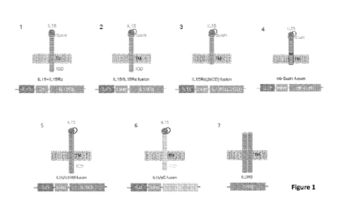
[00035] Figure 1 is a graphic representation of several construct designs
for cell
surface expressed cytokine or receptor thereof in iPSC derived cells. IL15 is
used as an
illustrative example, which can be replaced with other desirable cytokines.
[00036] Figure 2 is a graphic representation of flow cytometry of mature
iPSC-derived
NK cells that demonstrates stepwise engineering of hnCD16 expression, B2M
knockout
(loss of HLA-A2 expression), HLA-G expression, and IL-15/IL-15ra (LNGFR)
construct
expression.
13
CA 03083109 2020-05-20
WO 2019/112899
PCT/US2018/063362
[00037] Figure 3 is a graphic representation of telomere length determined
by flow
cytometry, and the mature derivative NK cells from iPSC maintain longer
telomeres
compared to adult peripheral blood NK cells.
[00038] Figure 4 shows that B2M knockout eliminates in vitro recognition of
engineered derivative NK cells by allogeneic CD8+ T cells.
[00039] Figure 5 shows that expression of HLA-G rescues B2M-/- iPSCs from
killing
by NK cells. A: Loss of HLA-I results in increased cytotoxicity against the
engineered
iPSCs when incubated with allogeneic PBMC. B: By expression of HLA-G on B2M-/-
iPSC allogeneic killing of the engineered iPSCs was partially reversed. Loss
of iPSCs was
measured over time using the Incucyte ZoomTM imaging system, and data are
normalized to
the number of iPSCs in wells without effector cells, setting time = 0 to 100%
for each
condition.
[00040] Figure 6 shows that a single dose of hnCD16/B2M-/-HLA-G iNK induced
tumor regression in an in vivo xenograft model of ovarian cancer. A: IVIS
images of each
mouse over a period of 32 days post injection. B: Time-course of tumor
progression by
IVIS imaging.
[00041] Figure 7 shows that IL15/IL15Ra construct promotes iNK cells
differentiation
and survival of in vitro independent of addition of soluble, exogenous IL15.
A: iNK cells
of each indicated genotype were differentiated with or without the addition of
soluble IL15.
B: iNK cells were extensively washed and place back into culture in
concentrations of
soluble IL15 ranging from lOng/m1 to 0 ng/ml for 7 days for observation of
soluble IL15-
independent cell growth.
[00042] Figure 8 shows that the expression of IL15/IL15Ra construct
enhances iNK
persistence in vivo in the absence of soluble IL15. iNK cells were adoptively
transferred to
A: immunocompromised NOG mice; B: NOG mice transgenic for human IL15.
[00043] Figure 9 is a graphic representation of phenotyping of CAR-
expressing iPSC
derived NK cells using flow cytometric analysis of surface markers.
[00044] Figure 10 is a graphic representation of the anti-tumor activity of
CAR4-
expresing derivative NK cells co-cultured with europium-loaded meso-high
target cells at
14
CA 03083109 2020-05-20
WO 2019/112899
PCT/US2018/063362
various effector to target (E:T) ratios. A: K562 meso-high target cells; B:
A1847 meso-high
target cells.
[00045] Figure 11 shows the tumor burden determined by weekly
bioluminescent
imaging in xenograft NSG mouse model inoculated with luciferase-expressing
A1847 meso
high cells, and one dose of 1.5E7 NK cells of different genotypes 4-day post
A1847
inoculation.
[00046] Figure 12 shows A: quantified tumor burden determined using
bioluminescence; and B: Kaplan-Meier curve representing the percent survival,
of NSG
mice group inoculated with luciferase-expressing A1847 meso high cells, and
one dose of
1.5E7 NK cells of different genotypes 4 day-post A1847 inoculation. n=5 for
all groups.
[00047] Figure 13 shows enhanced persistence of CAR-iNK in vivo by
measurement
of percentage of derivative CAR-NK cells from cells collected from (A)
peripheral blood,
(B) spleen, and (C) peritoneal, assessed by flow cytometry. Each dot
represents one
recipient mouse. Median SEM is shown, and P<0.05.
[00048] Figure 14 is a graphic representation of the creation of CD16
expressing
derivative T cell from clonal population of hnCD16-iPSCs. A: hnCD16-iPSC flow
analysis; B: hnCD16-iT flow analysis.
[00049] Figure 15 shows ADCC mediated target cell elimination by hnCD16-iT
cells.
[00050] Figure 16 shows that the expression of both CAR and hnCD16 does not
perturb hematopoietic differentiation of CAR-hnCD16-iPSC to effector cells
such as
derivative T cells and derivative NK cells. Flow cytometry analysis of A: CAR-
hnCD16
iPSCs; B: CAR-hnCD16 iCD34 cells; C: CAR-hnCD16 derivative T cells.
[00051] Figure 17 shows that activated hnCD16-iNK cells produce soluble
factors that
enhance T cell activation measured by percentage of CD69 positive T cells.
[00052] Figure 18 shows that activated hnCD16-iNK cells produce soluble
factors that
enhance T cell migration, quantified by flow cytometry of T cell migration
form the upper
to the lower chamber in the trans-well assay.
[00053] Figure 19 shows that derivative NK cells enhance T cell migration
in vivo
from A: blood into B: peritoneum of injected mouse model as quantified by flow
cytometry. Each data point represents an individual mouse.
CA 03083109 2020-05-20
WO 2019/112899
PCT/US2018/063362
[00054] Figure 20 shows IncuCyteTM real time imaging of tumor spheroid
growth and
formation over 84 hours, which is defined by the applied algorithm mask.
[00055] Figure 21. shows A: a representative IncuCyteTM imaging of
derivative NK
cell infiltration of a SKOV3 spheroid formation during the continuous
monitoring of the
changes in the Spheroid Size and Total Integrated Fluorescence Intensity over
time; B: T
cells alone failed to penetrate the center of the spheroid, but addition of
derivative NK cells
promoted T cell infiltration and the subsequent spheroid destruction.
[00056] Figure 22 shows that the co-expression of IL-1511L-15ra (labeled as
IL-15RF)
enhances NK-CAR19 expressing derivative NK cells in vitro persistence in the
absence of
soluble IL-2.
[00057] Figure 23 shows that iPSC derived NK co-culture enhances T cell
infiltration
of tumor spheroids by measuring total integrated green fluorescence intensity
within the
largest red object mask.
[00058] Figure 24 shows that iPSC derived NK cells synergize with T cells
to enhance
production of (A) TNFa and (B) IFNy by both CD4+ and CD8+ T cells during co-
culture
with SKOV-3 tumor spheroids.
[00059] Figure 25 shows that engineered CAR-iT cells expressing a high
affinity, non-
cleavable version of CD16 represents an opportunity for a secondary approach
to target
tumors and mitigate tumor antigen escape through ADCC. CAR and hnCD16 ADCC-
mediated cytotoxicity are both used against CD19+/+ and CD19-/- Raji cells.
Survival of
target cells was quantified by Incucyte Zoom after 36 hours in the presence
and absence of
anti-CD20 monoclonal antibody Rituximab.
[00060] Figure 26 shows the cellular expansion of TRAC-CAR-iT cells during
the 35
day differentiation process, resulting more than 40,000 fold increase in
cellular yield from
starting TRAC-CAR TiPSC in one production run. The differentiated synthetic
cells were
transfered from monolayer to suspension culture at around day 28.
[00061] Figure 27 represents that D35 TRAC-CAR-iT cells were assessed for
the
generation of (A) the proinflammatory cytokines IFNg and TNFa; and (B) the pro-
survival
cytokine IL-2 in response to PMA/ionomycin stimulation for 4 hours.
16
CA 03083109 2020-05-20
WO 2019/112899
PCT/US2018/063362
[00062] Figure 28 demonstrates the comparison of in vitro cytotoxicity
between (A)
primary CAR-T cells and (B) synthetic T cells TRAC-CAR-iT using a 18hr flow
cytometry
assay using wildtype (CD19+/+) or knockout (CD19-/-) NALM-6 as target cells.
Three
independent experiments on three separate primary CAR-T cells and three
independent
experiments on 3 separate TRAC-CAR-iT production batches were used.
[00063] Figure 29 shows that TRAC-CAR-iT cells were assessed for chemotaxis
in
response to the indicated thymus-derived chemokines in a trans-well migration
assay using
(A) D20 TRAC-CAR-iT cells, and (B) D28 TRAC-CAR-iT cells.
[00064] Figure 30 shows enhanced NK cell maturation in iPSC-derived NK
cells
expressing hnCD16, anti-CD19 CAR, and IL15/IL15Ra: (A) increased production of
granzyme B; and (B) increased expression of KIR2DL3 and KIR2DL1.
[00065] Figure 31 shows that the expression of IL15/IL15Ra promotes iNK
persistence and antigen-driven expansion: (A) 1 x i07 CAR iNK or CAR-
IL15/IL15Ra iNK
cells were injected IV into immunocompromised NOD mice on days 0, 7, and 14.
IL-2 was
administered IP twice weekly for the first 3 weeks, and iNK cells were
measured in the
blood weekly for 9 weeks for persistence assessment; (B) 5 x 10 Nalm6 cells
were
transplanted into NSG mice IV. 4 and 11 days later, 5 x 10⁶ CAR iNK or CAR-IL-
15/IL-
15ra iNK were injected IV, and iNK cell counts in the blood was determined
weekly by
flow cytometry for cell expansion assessment. IL-2 was administered IP twice
weekly.
[00066] Figure 32 shows that (A) CAR-IL15/IL15Ra iNK cells has improved
survival
in a highly aggressive disseminated model of B cell lymphoma (p = 0.018, CAR-
IL-15/IL-
15ra iNK vs CAR iNK); and (B) CAR-IL15/IL15Ra iNK cells prevent tumor
progression in
vivo in a Nalm6 xenograft model of leukemia.
[00067] Figure 33 shows in vitro cytotoxicity of hnCD16-CAR-IL15/IL15Ra iNK
cells against (A) Nalm6 and Nalm6 CD19-/- measured using hnCD16-CAR-IL-15/IL-
15ra
iNK cells at increasing E:T ratios in a 4 hour cytotoxicity assay; (B) ARH-77
leukemia cells
or (C) ARH-77 CD19-/- cells were used to measure direct cytotoxicity and
rituximab-
induced ADCC in a 4 hour cytotoxicity assays with unmodified iNK cells as
control.
[00068] Figure 34 shows hnCD16, CAR, and IL-15/IL-15ra modalities synergize
to
eradicate CD19+ and CD19- targets in a mixed-culture cytotoxicity assay.
Parental ARH-
17
CA 03083109 2020-05-20
WO 2019/112899
PCT/US2018/063362
77 cells (CD19+) and ARH-77 CD19- cells were transduced with red and green
fluorescent
tags, respectively. These cells were mixed 1:1 and used as target cells in a
long-term
cytotoxicity assay utilizing various iNK cell populations as effector cells in
the presence or
absence of rituximab antibody. The frequency of green CD19- and red CD19+
targets was
measured throughout the assay using the Incucyte imaging system to quantitate
cytotoxicity
against both target cells within a single well. The data are plotted as the
frequency of target
cells remaining for both target types normalized to the no effector cell
(tumor cell only)
control.
[00069] Figure 35 shows that TRAC-CAR iT cells are not alloreactive against
HLA-
mismatched healthy cells using Mixed Lymphocyte Reaction (MLR) assay, which
compares the proliferative response of TRAC-CAR iT cells and primary CAR-T
cells
against HLA-mismatched PBMC-derived T cells as target cells. Responder cells
were
labeled with cell trace dye and assessed after 4 days for dye dilution by flow
cytometry.
[00070] Figure 36 shows that the TRAC-CAR iT cells expressing a hnCD16
represents a secondary approach to target tumor. CAR and hnCD16 ADCC-mediated
cytotoxicity against CD19+/+ and CD19-/- Raji cells were compared. Survival of
target
cells was quantified by flow cytometry after 72 hours in the presence and
absence of anti-
CD20 monoclonal antibody Rituximab.
DETAILED DESCRIPTION OF THE INVENTION
[00071] Genomic modification of iPSCs (induced pluripotent stem cells)
includes
polynucleotide insertion, deletion and substitution. Exogenous gene expression
in genome-
engineered iPSCs often encounters problems such as gene silencing or reduced
gene
expression after prolonged clonal expansion of the original genome-engineered
iPSCs, after
cell differentiation, and in dedifferentiated cell types from the cells
derived from the
genome-engineered iPSCs. On the other hand, direct engineering of primary
immune cells
such as T or NK cells is challenging, and presents a hurdle to the preparation
and delivery
of engineered immune cells for adoptive cell therapy. The present invention
provides an
efficient, reliable, and targeted approach for stably integrating one or more
exogenous
genes, including suicide genes and other functional modalities, which provide
improved
18
CA 03083109 2020-05-20
WO 2019/112899
PCT/US2018/063362
therapeutic properties relating to engraftment, trafficking, homing,
migration, cytotoxicity,
viability, maintenance, expansion, longevity, self-renewal, persistence,
and/or survival into
iPSC derivative cells obtained through directed iPSC differentiation, which
derivative cells
include but are not limited to HSC (hematopoietic stem and progenitor cell), T
cell
progenitor cells, NK cell progenitor cells, T cells, NKT cells, NK cells.
[00072] Definitions
[00073] Unless otherwise defined herein, scientific and technical terms
used in
connection with the present application shall have the meanings that are
commonly
understood by those of ordinary skill in the art. Further, unless otherwise
required by
context, singular terms shall include pluralities and plural terms shall
include the singular.
[00074] It should be understood that this invention is not limited to the
particular
methodology, protocols, and reagents, etc., described herein and as such may
vary. The
terminology used herein is for the purpose of describing particular
embodiments only, and
is not intended to limit the scope of the present invention, which is defined
solely by the
claims.
[00075] As used herein, the articles "a," "an," and "the" are used herein
to refer to one
or to more than one (i.e. to at least one) of the grammatical object of the
article. By way of
example, "an element" means one element or more than one element.
[00076] The use of the alternative (e.g., "or") should be understood to
mean either one,
both, or any combination thereof of the alternatives.
[00077] The term "and/or" should be understood to mean either one, or both
of the
alternatives.
[00078] As used herein, the term "about" or "approximately" refers to a
quantity,
level, value, number, frequency, percentage, dimension, size, amount, weight
or length that
varies by as much as 15%, 10%, 9%, 8%, 7%, 6%, 5%, 4%, 3%, 2% or 1% compared
to a
reference quantity, level, value, number, frequency, percentage, dimension,
size, amount,
weight or length. In one embodiment, the term "about" or "approximately"
refers a range of
quantity, level, value, number, frequency, percentage, dimension, size,
amount, weight or
length 15%, 10%, 9%, 8%, 7%, 6%, 5%, 4%, 3%, 2%, or 1%
about a
19
CA 03083109 2020-05-20
WO 2019/112899
PCT/US2018/063362
reference quantity, level, value, number, frequency, percentage, dimension,
size, amount,
weight or length.
[00079] As used herein, the term "substantially" or "essentially" refers to
a quantity,
level, value, number, frequency, percentage, dimension, size, amount, weight
or length that
is about 90%, 91%, 92%, 93%, 94%, 95%, 96%, 97%, 98%, or 99% or higher
compared to
a reference quantity, level, value, number, frequency, percentage, dimension,
size, amount,
weight or length. In one embodiment, the terms "essentially the same" or
"substantially the
same" refer a range of quantity, level, value, number, frequency, percentage,
dimension,
size, amount, weight or length that is about the same as a reference quantity,
level, value,
number, frequency, percentage, dimension, size, amount, weight or length.
[00080] As used herein, the terms "substantially free of' and "essentially
free of' are
used interchangeably, and when used to describe a composition, such as a cell
population or
culture media, refer to a composition that is free of a specified substance or
its source
thereof, such as, 95% free, 96% free, 97% free, 98% free, 99% free of the
specified
substance or its source thereof, or is undetectable as measured by
conventional means. The
term "free of' or "essentially free of' a certain ingredient or substance in a
composition
also means that no such ingredient or substance is (1) included in the
composition at any
concentration, or (2) included in the composition functionally inert, but at a
low
concentration. Similar meaning can be applied to the term "absence of," where
referring to
the absence of a particular substance or its source thereof of a composition.
[00081] Throughout this specification, unless the context requires
otherwise, the words
"comprise," "comprises" and "comprising" will be understood to imply the
inclusion of a
stated step or element or group of steps or elements but not the exclusion of
any other step
or element or group of steps or elements. In particular embodiments, the terms
"include,"
"has," "contains," and "comprise" are used synonymously.
[00082] By "consisting of' is meant including, and limited to, whatever
follows the
phrase "consisting of." Thus, the phrase "consisting of' indicates that the
listed elements
are required or mandatory, and that no other elements may be present.
[00083] By "consisting essentially of' is meant including any elements
listed after the
phrase, and limited to other elements that do not interfere with or contribute
to the activity
CA 03083109 2020-05-20
WO 2019/112899
PCT/US2018/063362
or action specified in the disclosure for the listed elements. Thus, the
phrase "consisting
essentially of' indicates that the listed elements are required or mandatory,
but that no other
elements are optional and may or may not be present depending upon whether or
not they
affect the activity or action of the listed elements.
[00084] Reference throughout this specification to "one embodiment," "an
embodiment," "a particular embodiment," "a related embodiment," "a certain
embodiment," "an additional embodiment," or "a further embodiment" or
combinations
thereof means that a particular feature, structure or characteristic described
in connection
with the embodiment is included in at least one embodiment of the present
invention. Thus,
the appearances of the foregoing phrases in various places throughout this
specification are
not necessarily all referring to the same embodiment. Furthermore, the
particular features,
structures, or characteristics may be combined in any suitable manner in one
or more
embodiments.
[00085] The term "ex vivo" refers generally to activities that take place
outside an
organism, such as experimentation or measurements done in or on living tissue
in an
artificial environment outside the organism, preferably with minimum
alteration of the
natural conditions. In particular embodiments, "ex vivo" procedures involve
living cells or
tissues taken from an organism and cultured in a laboratory apparatus, usually
under sterile
conditions, and typically for a few hours or up to about 24 hours, but
including up to 48 or
72 hours or longer, depending on the circumstances. In certain embodiments,
such tissues or
cells can be collected and frozen, and later thawed for ex vivo treatment.
Tissue culture
experiments or procedures lasting longer than a few days using living cells or
tissue are
typically considered to be "in vitro," though in certain embodiments, this
term can be used
interchangeably with ex vivo.
[00086] The term "in vivo" refers generally to activities that take place
inside an
organism.
[00087] As used herein, the terms "reprogramming" or "dedifferentiation" or
"increasing cell potency" or "increasing developmental potency" refers to a
method of
increasing the potency of a cell or dedifferentiating the cell to a less
differentiated state. For
example, a cell that has an increased cell potency has more developmental
plasticity (i.e.,
21
CA 03083109 2020-05-20
WO 2019/112899
PCT/US2018/063362
can differentiate into more cell types) compared to the same cell in the non-
reprogrammed
state. In other words, a reprogrammed cell is one that is in a less
differentiated state than the
same cell in a non-reprogrammed state.
[00088] As used herein, the term "differentiation" is the process by which
an
unspecialized ("uncommitted") or less specialized cell acquires the features
of a specialized
cell such as, for example, a blood cell or a muscle cell. A differentiated or
differentiation-
induced cell is one that has taken on a more specialized ("committed")
position within the
lineage of a cell. The term "committed", when applied to the process of
differentiation,
refers to a cell that has proceeded in the differentiation pathway to a point
where, under
normal circumstances, it will continue to differentiate into a specific cell
type or subset of
cell types, and cannot, under normal circumstances, differentiate into a
different cell type or
revert to a less differentiated cell type. As used herein, the term
"pluripotent" refers to the
ability of a cell to form all lineages of the body or soma (i.e., the embryo
proper). For
example, embryonic stem cells are a type of pluripotent stem cells that are
able to form
cells from each of the three germs layers, the ectoderm, the mesoderm, and the
endoderm.
Pluripotency is a continuum of developmental potencies ranging from the
incompletely or
partially pluripotent cell (e.g., an epiblast stem cell or EpiSC), which is
unable to give rise
to a complete organism to the more primitive, more pluripotent cell, which is
able to give
rise to a complete organism (e.g., an embryonic stem cell).
[00089] As used herein, the term "induced pluripotent stem cells" or,
iPSCs, means
that the stem cells are produced from differentiated adult, neonatal or fetal
cells that have
been induced or changed, i.e., reprogrammed into cells capable of
differentiating into
tissues of all three germ or dermal layers: mesoderm, endoderm, and ectoderm.
The iPSCs
produced do not refer to cells as they are found in nature.
[00090] As used herein, the term "embryonic stem cell" refers to naturally
occurring
pluripotent stem cells of the inner cell mass of the embryonic blastocyst.
Embryonic stem
cells are pluripotent and give rise during development to all derivatives of
the three primary
germ layers: ectoderm, endoderm and mesoderm. They do not contribute to the
extra-
embryonic membranes or the placenta, i.e., are not totipotent.
22
CA 03083109 2020-05-20
WO 2019/112899
PCT/US2018/063362
[00091] As used herein, the term "multipotent stem cell" refers to a cell
that has the
developmental potential to differentiate into cells of one or more germ layers
(ectoderm,
mesoderm and endoderm), but not all three. Thus, a multipotent cell can also
be termed a
"partially differentiated cell." Multipotent cells are well known in the art,
and examples of
multipotent cells include adult stem cells, such as for example, hematopoietic
stem cells
and neural stem cells. "Multipotent" indicates that a cell may form many types
of cells in a
given lineage, but not cells of other lineages. For example, a multipotent
hematopoietic cell
can form the many different types of blood cells (red, white, platelets,
etc.), but it cannot
form neurons. Accordingly, the term "multipotency" refers to a state of a cell
with a degree
of developmental potential that is less than totipotent and pluripotent.
[00092] Pluripotency can be determined, in part, by assessing pluripotency
characteristics of the cells. Pluripotency characteristics include, but are
not limited to: (i)
pluripotent stem cell morphology; (ii) the potential for unlimited self-
renewal; (iii)
expression of pluripotent stem cell markers including, but not limited to
SSEA1 (mouse
only), SSEA3/4, SSEA5, TRA1-60/81, TRA1-85, TRA2-54, GCTM-2, TG343, TG30,
CD9, CD29, CD133/prominin, CD140a, CD56, CD73, CD90, CD105, OCT4, NANOQ
SOX2, CD30 and/or CD50; (iv) ability to differentiate to all three somatic
lineages
(ectoderm, mesoderm and endoderm); (v) teratoma formation consisting of the
three
somatic lineages; and (vi) formation of embryoid bodies consisting of cells
from the three
somatic lineages.
[00093] Two types of pluripotency have previously been described: the
"primed" or
"metastable" state of pluripotency akin to the epiblast stem cells (EpiSC) of
the late
blastocyst, and the "Naïve" or "Ground" state of pluripotency akin to the
inner cell mass of
the early/preimplantation blastocyst. While both pluripotent states exhibit
the characteristics
as described above, the naive or ground state further exhibits: (i) pre-
inactivation or
reactivation of the X-chromosome in female cells; (ii) improved clonality and
survival
during single-cell culturing; (iii) global reduction in DNA methylation; (iv)
reduction of
H3K27me3 repressive chromatin mark deposition on developmental regulatory gene
promoters; and (v) reduced expression of differentiation markers relative to
primed state
pluripotent cells. Standard methodologies of cellular reprogramming in which
exogenous
23
CA 03083109 2020-05-20
WO 2019/112899
PCT/US2018/063362
pluripotency genes are introduced to a somatic cell, expressed, and then
either silenced or
removed from the resulting pluripotent cells are generally seen to have
characteristics of the
primed-state of pluripotency. Under standard pluripotent cell culture
conditions such cells
remain in the primed state unless the exogenous transgene expression is
maintained,
wherein characteristics of the ground-state are observed.
[00094] As used herein, the term "pluripotent stem cell morphology" refers
to the
classical morphological features of an embryonic stem cell. Normal embryonic
stem cell
morphology is characterized by being round and small in shape, with a high
nucleus-to-
cytoplasm ratio, the notable presence of nucleoli, and typical inter-cell
spacing.
[00095] As used herein, the term "subject" refers to any animal, preferably
a human
patient, livestock, or other domesticated animal.
[00096] A "pluripotency factor," or "reprogramming factor," refers to an
agent capable
of increasing the developmental potency of a cell, either alone or in
combination with other
agents. Pluripotency factors include, without limitation, polynucleotides,
polypeptides, and
small molecules capable of increasing the developmental potency of a cell.
Exemplary
pluripotency factors include, for example, transcription factors and small
molecule
reprogramming agents.
[00097] "Culture" or "cell culture" refers to the maintenance, growth
and/or
differentiation of cells in an in vitro environment. "Cell culture media,"
"culture media"
(singular "medium" in each case), "supplement" and "media supplement" refer to
nutritive
compositions that cultivate cell cultures.
[00098] "Cultivate," or "maintain," refers to the sustaining, propagating
(growing)
and/or differentiating of cells outside of tissue or the body, for example in
a sterile plastic
(or coated plastic) cell culture dish or flask. "Cultivation," or
"maintaining," may utilize a
culture medium as a source of nutrients, hormones and/or other factors helpful
to propagate
and/or sustain the cells.
[00099] As used herein, the term "mesoderm" refers to one of the three
germinal
layers that appears during early embryogenesis and which gives rise to various
specialized
cell types including blood cells of the circulatory system, muscles, the
heart, the dermis,
skeleton, and other supportive and connective tissues.
24
CA 03083109 2020-05-20
WO 2019/112899
PCT/US2018/063362
[000100] As used herein, the term "definitive hemogenic endothelium" (RE)
or
"pluripotent stem cell-derived definitive hemogenic endothelium" (iHE) refers
to a subset
of endothelial cells that give rise to hematopoietic stem and progenitor cells
in a process
called endothelial-to-hematopoietic transition. The development of
hematopoietic cells in
the embryo proceeds sequentially from lateral plate mesoderm through the
hemangioblast
to the definitive hemogenic endothelium and hematopoietic progenitors.
[000101] The term "hematopoietic stem and progenitor cells," "hematopoietic
stem
cells," "hematopoietic progenitor cells," or "hematopoietic precursor cells"
refers to cells
which are committed to a hematopoietic lineage but are capable of further
hematopoietic
differentiation and include, multipotent hematopoietic stem cells
(hematoblasts), myeloid
progenitors, megakaryocyte progenitors, erythrocyte progenitors, and lymphoid
progenitors. Hematopoietic stem and progenitor cells (HSCs) are multipotent
stem cells that
give rise to all the blood cell types including myeloid (monocytes and
macrophages,
neutrophils, basophils, eosinophils, erythrocytes, megakaryocytes/platelets,
dendritic cells),
and lymphoid lineages (T cells, B cells, NK cells). The term "definitive
hematopoietic stem
cell" as used herein, refers to CD34+ hematopoietic cells capable of giving
rise to both
mature myeloid and lymphoid cell types including T cells, NK cells and B
cells.
Hematopoietic cells also include various subsets of primitive hematopoietic
cells that give
rise to primitive erythrocytes, megakarocytes and macrophages.
[000102] As used herein, the terms "T lymphocyte" and "T cell" are used
interchangeably and refer to a principal type of white blood cell that
completes maturation
in the thymus and that has various roles in the immune system, including the
identification
of specific foreign antigens in the body and the activation and deactivation
of other immune
cells. AT cell can be any T cell, such as a cultured T cell, e.g., a primary T
cell, or a T cell
from a cultured T cell line, e.g., Jurkat, SupT1, etc., or a T cell obtained
from a mammal.
The T cell can be CD3+ cells. The T cell can be any type of T cell and can be
of any
developmental stage, including but not limited to, CD4+/CD8+ double positive T
cells,
CD4+ helper T cells (e.g., Thl and Th2 cells), CD8+ T cells (e.g., cytotoxic T
cells),
peripheral blood mononuclear cells (PBMCs), peripheral blood leukocytes
(PBLs), tumor
infiltrating lymphocytes (TILs), memory T cells, naïve T cells, regulator T
cells, gamma
CA 03083109 2020-05-20
WO 2019/112899
PCT/US2018/063362
delta T cells (y6 T cells), and the like. Additional types of helper T cells
include cells such
as Th3 (Treg), Th17, Th9, or Tfh cells. Additional types of memory T cells
include cells
such as central memory T cells (Tem cells), effector memory T cells (Tern
cells and
TEMRA cells). The T cell can also refer to a genetically engineered T cell,
such as a T cell
modified to express a T cell receptor (TCR) or a chimeric antigen receptor
(CAR). The T
cell can also be differentiated from a stem cell or progenitor cell.
[000103] "CD4+ T cells" refers to a subset of T cells that express CD4 on
their surface
and are associated with cell-mediated immune response. They are characterized
by the
secretion profiles following stimulation, which may include secretion of
cytokines such as
IFN-gamma, TNF-alpha, IL2, IL4 and IL10. "CD4" are 55-kD glycoproteins
originally
defined as differentiation antigens on T-lymphocytes, but also found on other
cells
including monocytes/macrophages. CD4 antigens are members of the
immunoglobulin
supergene family and are implicated as associative recognition elements in MEW
(major
histocompatibility complex) class II-restricted immune responses. On T-
lymphocytes they
define the helper/inducer subset.
[000104] "CD8+ T cells" refers to a subset of T cells which express CD8 on
their
surface, are MEW class I-restricted, and function as cytotoxic T cells. "CD8"
molecules are
differentiation antigens found on thymocytes and on cytotoxic and suppressor T-
lymphocytes. CD8 antigens are members of the immunoglobulin supergene family
and are
associative recognition elements in major histocompatibility complex class I-
restricted
interactions.
[000105] As used herein, the term "NK cell" or "Natural Killer cell" refer
to a subset of
peripheral blood lymphocytes defined by the expression of CD56 or CD16 and the
absence
of the T cell receptor (CD3). As used herein, the terms "adaptive NK cell" and
"memory
NK cell" are interchangeable and refer to a subset of NK cells that are
phenotypically CD3-
and CD56+, expressing at least one of NKG2C and CD57, and optionally, CD16,
but lack
expression of one or more of the following: PLZF, SYK, FceRy, and EAT-2. In
some
embodiments, isolated subpopulations of CD56+ NK cells comprise expression of
CD16,
NKG2C, CD57, NKG2D, NCR ligands, NKp30, NKp40, NKp46, activating and
inhibitory
KIRs, NKG2A and/or DNAM-1. CD56+ can be dim or bright expression.
26
CA 03083109 2020-05-20
WO 2019/112899
PCT/US2018/063362
[000106] As used herein, the term "NKT cells" or "natural killer T cells"
refers to
CD id-restricted T cells, which express a T cell receptor (TCR). Unlike
conventional T cells
that detect peptide antigens presented by conventional major
histocompatibility (MHC)
molecules, NKT cells recognize lipid antigens presented by CD1d, a non-
classical MHC
molecule. Two types of NKT cells are recognized. Invariant or type I NKT cells
express a
very limited TCR repertoire - a canonical a-chain (Va24-Ja18 in humans)
associated with a
limited spectrum of 13 chains (Vf311 in humans). The second population of NKT
cells, called
non-classical or non-invariant type II NKT cells, display a more heterogeneous
TCR af3
usage. Type I NKT cells are considered suitable for immunotherapy. Adaptive or
invariant
(type I) NKT cells can be identified with the expression of at least one or
more of the
following markers, TCR Va24-Ja18, Vb11, CD1d, CD3, CD4, CD8, aGalCer, CD161
and
CD56.
[000107] As used herein, the term "isolated" or the like refers to a cell,
or a population
of cells, which has been separated from its original environment, i.e., the
environment of
the isolated cells is substantially free of at least one component as found in
the environment
in which the "un-isolated" reference cells exist. The term includes a cell
that is removed
from some or all components as it is found in its natural environment, for
example, isolated
from a tissue or biopsy sample. The term also includes a cell that is removed
from at least
one, some or all components as the cell is found in non-naturally occurring
environments,
for example, isolated form a cell culture or cell suspension. Therefore, an
isolated cell is
partly or completely separated from at least one component, including other
substances,
cells or cell populations, as it is found in nature or as it is grown, stored
or subsisted in non-
naturally occurring environments. Specific examples of isolated cells include
partially pure
cell compositions, substantially pure cell compositions and cells cultured in
a medium that
is non-naturally occurring. Isolated cells may be obtained from separating the
desired cells,
or populations thereof, from other substances or cells in the environment, or
from removing
one or more other cell populations or subpopulations from the environment.
[000108] As used herein, the term "purify" or the like refers to increasing
purity. For
example, the purity can be increased to at least 50%, 60%, 70%, 80%, 90%, 95%,
99%, or
100%.
27
CA 03083109 2020-05-20
WO 2019/112899
PCT/US2018/063362
[000109] As used herein, the term "encoding" refers to the inherent
property of specific
sequences of nucleotides in a polynucleotide, such as a gene, a cDNA, or a
mRNA, to serve
as templates for synthesis of other polymers and macromolecules in biological
processes
having either a defined sequence of nucleotides (i.e., rRNA, tRNA and mRNA) or
a defined
sequence of amino acids and the biological properties resulting therefrom.
Thus, a gene
encodes a protein if transcription and translation of mRNA corresponding to
that gene
produces the protein in a cell or other biological system. Both the coding
strand, the
nucleotide sequence of which is identical to the mRNA sequence and is usually
provided in
sequence listings, and the non-coding strand, used as the template for
transcription of a
gene or cDNA, can be referred to as encoding the protein or other product of
that gene or
cDNA.
[000110] A "construct" refers to a macromolecule or complex of molecules
comprising
a polynucleotide to be delivered to a host cell, either in vitro or in vivo. A
"vector," as used
herein refers to any nucleic acid construct capable of directing the delivery
or transfer of a
foreign genetic material to target cells, where it can be replicated and/or
expressed. The
term "vector" as used herein comprises the construct to be delivered. A vector
can be a
linear or a circular molecule. A vector can be integrating or non-integrating.
The major
types of vectors include, but are not limited to, plasmids, episomal vector,
viral vectors,
cosmids, and artificial chromosomes. Viral vectors include, but are not
limited to,
adenovirus vector, adeno-associated virus vector, retrovirus vector,
lentivirus vector, Sendai
virus vector, and the like.
[000111] By "integration" it is meant that one or more nucleotides of a
construct is
stably inserted into the cellular genome, i.e., covalently linked to the
nucleic acid sequence
within the cell's chromosomal DNA. By "targeted integration" it is meant that
the
nucleotide(s) of a construct is inserted into the cell's chromosomal or
mitochondrial DNA at
a pre-selected site or "integration site". The term "integration" as used
herein further refers
to a process involving insertion of one or more exogenous sequences or
nucleotides of the
construct, with or without deletion of an endogenous sequence or nucleotide at
the
integration site. In the case, where there is a deletion at the insertion
site, "integration" may
28
CA 03083109 2020-05-20
WO 2019/112899
PCT/US2018/063362
further comprise replacement of the endogenous sequence or a nucleotide that
is deleted
with the one or more inserted nucleotides.
[000112] As used herein, the term "exogenous" is intended to mean that the
referenced
molecule or the referenced activity is introduced into, or non-native to, the
host cell. The
molecule can be introduced, for example, by introduction of an encoding
nucleic acid into
the host genetic material such as by integration into a host chromosome or as
non-
chromosomal genetic material such as a plasmid. Therefore, the term as it is
used in
reference to expression of an encoding nucleic acid refers to introduction of
the encoding
nucleic acid in an expressible form into the cell. The term "endogenous"
refers to a
referenced molecule or activity that is present in the host cell. Similarly,
the term when used
in reference to expression of an encoding nucleic acid refers to expression of
an encoding
nucleic acid contained within the cell and not exogenously introduced.
[000113] As used herein, a "gene of interest" or "a polynucleotide sequence
of interest"
is a DNA sequence that is transcribed into RNA and in some instances
translated into a
polypeptide in vivo when placed under the control of appropriate regulatory
sequences. A
gene or polynucleotide of interest can include, but is not limited to,
prokaryotic sequences,
cDNA from eukaryotic mRNA, genomic DNA sequences from eukaryotic (e.g.,
mammalian) DNA, and synthetic DNA sequences. For example, a gene of interest
may
encode an miRNA, an shRNA, a native polypeptide (i.e. a polypeptide found in
nature) or
fragment thereof; a variant polypeptide (i.e. a mutant of the native
polypeptide having less
than 100% sequence identity with the native polypeptide) or fragment thereof;
an
engineered polypeptide or peptide fragment, a therapeutic peptide or
polypeptide, an
imaging marker, a selectable marker, and the like.
[000114] As used herein, the term "polynucleotide" refers to a polymeric
form of
nucleotides of any length, either deoxyribonucleotides or ribonucleotides or
analogs
thereof. The sequence of a polynucleotide is composed of four nucleotide
bases: adenine
(A); cytosine (C); guanine (G); thymine (T); and uracil (U) for thymine when
the
polynucleotide is RNA. A polynucleotide can include a gene or gene fragment
(for
example, a probe, primer, EST or SAGE tag), exons, introns, messenger RNA
(mRNA),
transfer RNA, ribosomal RNA, ribozymes, cDNA, recombinant polynucleotides,
branched
29
CA 03083109 2020-05-20
WO 2019/112899
PCT/US2018/063362
polynucleotides, plasmids, vectors, isolated DNA of any sequence, isolated RNA
of any
sequence, nucleic acid probes and primers. Polynucleotide also refers to both
double- and
single-stranded molecules.
[000115] As used herein, the term "peptide," "polypeptide," and "protein"
are used
interchangeably and refer to a molecule having amino acid residues covalently
linked by
peptide bonds. A polypeptide must contain at least two amino acids, and no
limitation is
placed on the maximum number of amino acids of a polypeptide. As used herein,
the terms
refer to both short chains, which are also commonly referred to in the art as
peptides,
oligopeptides and oligomers, for example, and to longer chains, which
generally are
referred to in the art as polypeptides or proteins. "Polypeptides" include,
for example,
biologically active fragments, substantially homologous polypeptides,
oligopeptides,
homodimers, heterodimers, variants of polypeptides, modified polypeptides,
derivatives,
analogs, fusion proteins, among others. The polypeptides include natural
polypeptides,
recombinant polypeptides, synthetic polypeptides, or a combination thereof.
[000116] "Operably-linked" refers to the association of nucleic acid
sequences on a
single nucleic acid fragment so that the function of one is affected by the
other. For
example, a promoter is operably-linked with a coding sequence or functional
RNA when it
is capable of affecting the expression of that coding sequence or functional
RNA (i.e., the
coding sequence or functional RNA is under the transcriptional control of the
promoter).
Coding sequences can be operably-linked to regulatory sequences in sense or
antisense
orientation.
[000117] As used herein, the term "genetic imprint" refers to genetic or
epigenetic
information that contributes to preferential therapeutic attributes in a
source cell or an iPSC,
and is retainable in the source cell derived iPSCs, and/or the iPSC-derived
hematopoietic
lineage cells. As used herein, "a source cell" is a non-pluripotent cell that
may be used for
generating iPSCs through reprogramming, and the source cell derived iPSCs may
be further
differentiated to specific cell types including any hematopoietic lineage
cells. The source
cell derived iPSCs, and differentiated cells therefrom are sometimes
collectively called
"derived" or "derivative" cells depending on the context. For example,
derivative effector
cells, or derivative NK cells or derivative T cells, as used throughout this
application are
CA 03083109 2020-05-20
WO 2019/112899
PCT/US2018/063362
cells differentiated from an iPSC, as compared to their primary counterpart
obtained from
natural/native sources such as peripheral blood, umbilical cord blood, or
other donor
tissues. As used herein, the genetic imprint(s) conferring a preferential
therapeutic attribute
is incorporated into the iPSCs either through reprogramming a selected source
cell that is
donor-, disease-, or treatment response- specific, or through introducing
genetically
modified modalities to iPSC using genomic editing. In the aspect of a source
cell obtained
from a specifically selected donor, disease or treatment context, the genetic
imprint
contributing to preferential therapeutic attributes may include any context
specific genetic
or epigenetic modifications which manifest a retainable phenotype, i.e. a
preferential
therapeutic attribute, that is passed on to derivative cells of the selected
source cell,
irrespective of the underlying molecular events being identified or not. Donor-
, disease-, or
treatment response- specific source cells may comprise genetic imprints that
are retainable
in iPSCs and derived hematopoietic lineage cells, which genetic imprints
include but are
not limited to, prearranged monospecific TCR, for example, from a viral
specific T cell or
invariant natural killer T (iNKT) cell; trackable and desirable genetic
polymorphisms, for
example, homozygous for a point mutation that encodes for the high-affinity
CD16 receptor
in selected donors; and predetermined HLA requirements, i.e., selected HLA-
matched
donor cells exhibiting a haplotype with increased population. As used herein,
preferential
therapeutic attributes include improved engraftment, trafficking, homing,
viability, self-
renewal, persistence, immune response regulation and modulation, survival, and
cytotoxicity of a derived cell. A preferential therapeutic attribute may also
relate to antigen
targeting receptor expression; HLA presentation or lack thereof; resistance to
tumor
microenvironment; induction of bystander immune cells and immune modulations;
improved on-target specificity with reduced off-tumor effect; resistance to
treatment such as
chemotherapy.
[000118] The term "enhanced therapeutic property" as used herein, refers to
a
therapeutic property of a cell that is enhanced as compared to a typical
immune cell of the
same general cell type. For example, an NK cell with an "enhanced therapeutic
property"
will possess an enhanced, improved, and/or augmented therapeutic property as
compared to
a typical, unmodified, and/or naturally occurring NK cell. Therapeutic
properties of an
31
CA 03083109 2020-05-20
WO 2019/112899
PCT/US2018/063362
immune cell may include, but are not limited to, cell engraftment,
trafficking, homing,
viability, self-renewal, persistence, immune response regulation and
modulation, survival,
and cytotoxicity. Therapeutic properties of an immune cell are also manifested
by antigen
targeting receptor expression; HLA presentation or lack thereof; resistance to
tumor
microenvironment; induction of bystander immune cells and immune modulations;
improved on-target specificity with reduced off-tumor effect; resistance to
treatment such as
chemotherapy.
[000119] As used herein, the term "engager" refers to a molecule, e.g. a
fusion
polypeptide, which is capable of forming a link between an immune cell, e.g. a
T cell, a NK
cell, a NKT cell, a B cell, a macrophage, a neutrophil, and a tumor cell; and
activating the
immune cell. Examples of engagers include, but are not limited to, bi-specific
T cell
engagers (BiTEs), bi-specific killer cell engagers (BiKEs), tri-specific
killer cell engagers,
or multi- specific killer cell engagers, or universal engagers compatible with
multiple
immune cell types.
[000120] As used herein, the term "surface triggering receptor" refers to a
receptor
capable of triggering or initiating an immune response, e.g. a cytotoxic
response. Surface
triggering receptors may be engineered, and may be expressed on effector
cells, e.g. a T
cell, a NK cell, a NKT cell, a B cell, a macrophage, a neutrophil. In some
embodiments, the
surface triggering receptor facilitates bi- or multi- specific antibody
engagement between
the effector cells and specific target cell e.g. a tumor cell, independent of
the effector cell's
natural receptors and cell types. Using this approach, one may generate iPSCs
comprising a
universal surface triggering receptor, and then differentiate such iPSCs into
populations of
various effector cell types that express the universal surface triggering
receptor. By
"universal", it is meant that the surface triggering receptor can be expressed
in, and
activate, any effector cells irrespective of the cell type, and all effector
cells expressing the
universal receptor can be coupled or linked to the engagers having the same
epitope
recognizable by the surface triggering receptor, regardless of the engager's
tumor binding
specificities. In some embodiments, engagers having the same tumor targeting
specificity
are used to couple with the universal surface triggering receptor. In some
embodiments,
engagers having different tumor targeting specificity are used to couple with
the universal
32
CA 03083109 2020-05-20
WO 2019/112899
PCT/US2018/063362
surface triggering receptor. As such, one or multiple effector cell types can
be engaged to
kill one specific type of tumor cells in some case, and to kill two or more
types of tumors in
some other cases. A surface triggering receptor generally comprises a co-
stimulatory
domain for effector cell activation and an anti-epitope that is specific to
the epitope of an
engager. A bi-specific engager is specific to the anti-epitope of a surface
triggering receptor
on one end, and is specific to a tumor antigen on the other end.
[000121] As used herein, the term "safety switch protein" refers to an
engineered
protein designed to prevent potential toxicity or otherwise adverse effects of
a cell therapy.
In some instances, the safety switch protein expression is conditionally
controlled to
address safety concerns for transplanted engineered cells that have
permanently
incorporated the gene encoding the safety switch protein into its genome. This
conditional
regulation could be variable and might include control through a small
molecule-mediated
post-translational activation and tissue-specific and/or temporal
transcriptional regulation.
The safety switch could mediate induction of apoptosis, inhibition of protein
synthesis,
DNA replication, growth arrest, transcriptional and post-transcriptional
genetic regulation
and/or antibody-mediated depletion. In some instance, the safety switch
protein is activated
by an exogenous molecule, e.g. a prodrug, that when activated, triggers
apoptosis and/or
cell death of a therapeutic cell. Examples of safety switch proteins, include,
but are not
limited to suicide genes such as caspase 9 (or caspase 3 or 7), thymidine
kinase, cytosine
deaminase, B-cell CD20, modified EGFR, and any combination thereof. In this
strategy, a
prodrug that is administered in the event of an adverse event is activated by
the suicide-
gene product and kills the transduced cell.
[000122] As used herein, the term "pharmaceutically active proteins or
peptides" refer
to proteins or peptides that are capable of achieving a biological and/or
pharmaceutical
effect on an organism. A pharmaceutically active protein has healing curative
or palliative
properties against a disease and may be administered to ameliorate relieve,
alleviate,
reverse or lessen the severity of a disease. A pharmaceutically active protein
also has
prophylactic properties and is used to prevent the onset of a disease or to
lessen the severity
of such disease or pathological condition when it does emerge.
Pharmaceutically active
proteins include an entire protein or peptide or pharmaceutically active
fragments thereof It
33
CA 03083109 2020-05-20
WO 2019/112899
PCT/US2018/063362
also includes pharmaceutically active analogs of the protein or peptide or
analogs of
fragments of the protein or peptide. The term pharmaceutically active protein
also refers to
a plurality of proteins or peptides that act cooperatively or synergistically
to provide a
therapeutic benefit. Examples of pharmaceutically active proteins or peptides
include, but
are not limited to, receptors, binding proteins, transcription and translation
factors, tumor
growth suppressing proteins, antibodies or fragments thereof, growth factors,
and/or
cytokines.
[000123] As used herein, the term "signaling molecule" refers to any
molecule that
modulates, participates in, inhibits, activates, reduces, or increases, the
cellular signal
transduction. Signal transduction refers to the transmission of a molecular
signal in the
form of chemical modification by recruitment of protein complexes along a
pathway that
ultimately triggers a biochemical event in the cell. Signal transduction
pathways are well
known in the art, and include, but are not limited to, G protein coupled
receptor signaling,
tyrosine kinase receptor signaling, integrin signaling, toll gate signaling,
ligand-gated ion
channel signaling, ERK/MAPK signaling pathway, Wnt signaling pathway, cAMP-
dependent pathway, and IP3/DAG signaling pathway.
[000124] As used herein, the term "targeting modality" refers to a
molecule, e.g., a
polypeptide, that is genetically incorporated into a cell to promote antigen
and/or epitope
specificity that includes but not limited to i) antigen specificity as it
related to a unique
chimeric antigen receptor (CAR) or T cell receptor (TCR), ii) engager
specificity as it
related to monoclonal antibodies or bispecific engager, iii) targeting of
transformed cell, iv)
targeting of cancer stem cell, and v) other targeting strategies in the
absence of a specific
antigen or surface molecule.
[000125] As used herein, the term "specific" or "specificity" can be used
to refer to the
ability of a molecule, e.g., a receptor or an engager, to selectively bind to
a target molecule,
in contrast to non-specific or non-selective binding.
[000126] The term "adoptive cell therapy" as used herein refers to a cell-
based
immunotherapy that, as used herein, relates to the transfusion of autologous
or allogenic
lymphocytes, identified as T or B cells, genetically modified or not, that
have been
expanded ex vivo prior to said transfusion.
34
CA 03083109 2020-05-20
WO 2019/112899
PCT/US2018/063362
[000127] A "therapeutically sufficient amount", as used herein, includes
within its
meaning a non-toxic but sufficient and/or effective amount of the particular
therapeutic
and/or pharmaceutical composition to which it is referring to provide a
desired therapeutic
effect. The exact amount required will vary from subject to subject depending
on factors
such as the patient's general health, the patient's age and the stage and
severity of the
condition. In particular embodiments, a therapeutically sufficient amount is
sufficient
and/or effective to ameliorate, reduce, and/or improve at least one symptom
associated with
a disease or condition of the subject being treated.
[000128] Differentiation of pluripotent stem cells requires a change in the
culture
system, such as changing the stimuli agents in the culture medium or the
physical state of
the cells. The most conventional strategy utilizes the formation of embryoid
bodies (EBs) as
a common and critical intermediate to initiate the lineage-specific
differentiation.
"Embryoid bodies" are three-dimensional clusters that have been shown to mimic
embryo
development as they give rise to numerous lineages within their three-
dimensional area.
Through the differentiation process, typically few hours to days, simple EBs
(for example,
aggregated pluripotent stem cells elicited to differentiate) continue
maturation and develop
into a cystic EB at which time, typically days to few weeks, they are further
processed to
continue differentiation. EB formation is initiated by bringing pluripotent
stem cells into
close proximity with one another in three-dimensional multilayered clusters of
cells,
typically this is achieved by one of several methods including allowing
pluripotent cells to
sediment in liquid droplets, sedimenting cells into "U" bottomed well-plates
or by
mechanical agitation. To promote EB development, the pluripotent stem cell
aggregates
require further differentiation cues, as aggregates maintained in pluripotent
culture
maintenance medium do not form proper EBs. As such, the pluripotent stem cell
aggregates
need to be transferred to differentiation medium that provides eliciting cues
towards the
lineage of choice. EB-based culture of pluripotent stem cells typically
results in generation
of differentiated cell populations (ectoderm, mesoderm and endoderm germ
layers) with
modest proliferation within the EB cell cluster. Although proven to facilitate
cell
differentiation, EBs, however, give rise to heterogeneous cells in variable
differentiation
state because of the inconsistent exposure of the cells in the three-
dimensional structure to
CA 03083109 2020-05-20
WO 2019/112899
PCT/US2018/063362
differentiation cues from the environment. In addition, EBs are laborious to
create and
maintain. Moreover, cell differentiation through EB is accompanied with modest
cell
expansion, which also contributes to low differentiation efficiency.
[000129] In comparison, "aggregate formation," as distinct from "EB
formation," can
be used to expand the populations of pluripotent stem cell derived cells. For
example,
during aggregate-based pluripotent stem cell expansion, culture media are
selected to
maintain proliferation and pluripotency. Cells proliferation generally
increases the size of
the aggregates forming larger aggregates, these aggregates can be routinely
mechanically or
enzymatically dissociated into smaller aggregates to maintain cell
proliferation within the
culture and increase numbers of cells. As distinct from EB culture, cells
cultured within
aggregates in maintenance culture maintain markers of pluripotency. The
pluripotent stem
cell aggregates require further differentiation cues to induce
differentiation.
[000130] As used herein, "monolayer differentiation" is a term referring to
a
differentiation method distinct from differentiation through three-dimensional
multilayered
clusters of cells, i.e., "EB formation." Monolayer differentiation, among
other advantages
disclosed herein, avoids the need for EB formation for differentiation
initiation. Because
monolayer culturing does not mimic embryo development such as EB formation,
differentiation towards specific lineages are deemed as minimal as compared to
all three
germ layer differentiation in EB.
[000131] As
used herein, a "dissociated" cell refers to a cell that has been substantially
separated or purified away from other cells or from a surface (e.g., a culture
plate surface).
For example, cells can be dissociated from an animal or tissue by mechanical
or enzymatic
methods. Alternatively, cells that aggregate in vitro can be dissociated from
each other, such
as by dissociation into a suspension of clusters, single cells or a mixture of
single cells and
clusters, enzymatically or mechanically. In yet another alternative
embodiment, adherent
cells are dissociated from a culture plate or other surface. Dissociation thus
can involve
breaking cell interactions with extracellular matrix (ECM) and substrates
(e.g., culture
surfaces), or breaking the ECM between cells.
[000132] As used herein, "feeder cells" or "feeders" are terms describing
cells of one
type that are co-cultured with cells of a second type to provide an
environment in which the
36
CA 03083109 2020-05-20
WO 2019/112899
PCT/US2018/063362
cells of the second type can grow, expand, or differentiate, as the feeder
cells provide
stimulation, growth factors and nutrients for the support of the second cell
type. The feeder
cells are optionally from a different species as the cells they are
supporting. For example,
certain types of human cells, including stem cells, can be supported by
primary cultures of
mouse embryonic fibroblasts, or immortalized mouse embryonic fibroblasts. In
another
example, peripheral blood derived cells or transformed leukemia cells support
the
expansion and maturation of natural killer cells. The feeder cells may
typically be
inactivated when being co-cultured with other cells by irradiation or
treatment with an anti-
mitotic agent such as mitomycin to prevent them from outgrowing the cells they
are
supporting. Feeder cells may include endothelial cells, stromal cells (for
example, epithelial
cells or fibroblasts), and leukemic cells. Without limiting the foregoing, one
specific feeder
cell type may be a human feeder, such as a human skin fibroblast. Another
feeder cell type
may be mouse embryonic fibroblasts (MEF). In general, various feeder cells can
be used in
part to maintain pluripotency, direct differentiation towards a certain
lineage, enhance
proliferation capacity and promote maturation to a specialized cell type, such
as an effector
cell.
[000133] As used herein, a "feeder-free" (FF) environment refers to an
environment
such as a culture condition, cell culture or culture media which is
essentially free of feeder
or stromal cells, and/or which has not been pre-conditioned by the cultivation
of feeder
cells. "Pre-conditioned" medium refers to a medium harvested after feeder
cells have been
cultivated within the medium for a period of time, such as for at least one
day. Pre-
conditioned medium contains many mediator substances, including growth factors
and
cytokines secreted by the feeder cells cultivated in the medium. In some
embodiments, a
feeder-free environment is free of both feeder or stromal cells and is also
not pre-
conditioned by the cultivation of feeder cells.
[000134] "Functional" as used in the context of genomic editing or
modification of
iPSC, and derived non-pluripotent cells differentiated therefrom, or genomic
editing or
modification of non-pluripotent cells and derived iPSCs reprogrammed
therefrom, refers to
(1) at the gene level--successful knocked-in, knocked-out, knocked-down gene
expression,
transgenic or controlled gene expression such as inducible or temporal
expression at a
37
CA 03083109 2020-05-20
WO 2019/112899
PCT/US2018/063362
desired cell development stage, which is achieved through direct genomic
editing or
modification, or through "passing-on" via differentiation from or
reprogramming of a
starting cell that is initially genomically engineered; or (2) at the cell
level¨successful
removal, adding, or altering a cell function/characteristics via (i) gene
expression
modification obtained in said cell through direct genomic editing, (ii) gene
expression
modification maintained in said cell through "passing-on" via differentiation
from or
reprogramming of a starting cell that is initially genomically engineered;
(iii) down-stream
gene regulation in said cell as a result of gene expression modification that
only appears in
an earlier development stage of said cell, or only appears in the starting
cell that gives rise
to said cell via differentiation or reprogramming; or (iv) enhanced or newly
attained cellular
function or attribute displayed within the mature cellular product, initially
derived from the
genomic editing or modification conducted at the iPSC, progenitor or
dedifferentiated
cellular origin.
[000135] "HLA deficient", including HLA-class I deficient, or HLA-class II
deficient,
or both, refers to cells that either lack, or no longer maintain, or have
reduced level of
surface expression of a complete MHC complex comprising a HLA class I protein
heterodimer and/or a HLA class II heterodimer, such that the diminished or
reduced level is
less than the level naturally detectable by other cells or by synthetic
methods.
[000136] "Modified HLA deficient iPSC," as used herein, refers to HLA
deficient iPSC
that is further modified by introducing genes expressing proteins related but
not limited to
improved differentiation potential, antigen targeting, antigen presentation,
antibody
recognition, persistence, immune evasion, resistance to suppression,
proliferation,
costimulation, cytokine stimulation, cytokine production (autocrine or
paracrine),
chemotaxis, and cellular cytotoxicity, such as non-classical HLA class I
proteins (e.g.,
HLA-E and HLA-G), chimeric antigen receptor (CAR), T cell receptor (TCR), CD16
Fc
Receptor, BCL11b, NOTCH, RUNX1, IL15, 41BB, DAP10, DAP12, CD24, CD3z,
41BBL, CD47, CD113, and PDLl. The cells that are "modified HLA deficient" also
include cells other than iPSCs.
[000137] "Fc receptors," abbreviated FcR, are classified based on the type
of antibody
that they recognize. For example, those that bind the most common class of
antibody, IgG
38
CA 03083109 2020-05-20
WO 2019/112899
PCT/US2018/063362
are called Fc-gamma receptors (FcyR), those that bind IgA are called Fc-alpha
receptors
(FcaR) and those that bind IgE are called Fc-epsilon receptors (FccR). The
classes of FcR's
are also distinguished by the cells that express them (macrophages,
granulocytes, natural
killer cells, T and B cells) and the signaling properties of each receptor. Fc-
gamma
receptors (FcyR) includes several members, FcyRI (CD64), FcyRIIA (CD32),
FcyRIIB
(CD32), FcyRIIIA (CD16a), FcyRTIIB (CD16b), which differ in their antibody
affinities due
to their different molecular structure
[000138] "Chimeric Fc Receptor," abbreviated as CFcR, are terms used to
describe
engineered Fc receptors having their native transmembrane and/or intracellular
signaling
domains modified, or replaced with non-native transmembrane and/or
intracellular
signaling domains. In some embodiments of the chimeric Fc receptor, in
addition to having
one of, or both, transmembrane and signaling domains being non-native, one or
more
stimulatory domains can be introduced to the intracellular portion of the
engineered Fc
receptor to enhance cell activation, expansion and function upon triggering of
the receptor.
Unlike chimeric antigen receptor (CAR) which contains antigen binding domain
to target
antigen, the chimeric Fc receptor binds to an Fc fragment, or the Fc region of
an antibody,
or the Fc region comprised in an engager or a binding molecule and activating
the cell
function with or without bringing the targeted cell close in vicinity. For
example, a Fcy
receptor can be engineered to comprise selected transmembrane, stimulatory,
and/or
signaling domains in the intracellular region that respond to the binding of
IgG at the
extracellular domain, thereby generating a CFcR. In one example, a CFcR is
produced by
engineering CD16, a Fcy receptor, by replacing its transmembrane domain and/or
intracellular domain. To further improve the binding affinity of the CD16
based CFcR, the
extracellular domain of CD64 or the high-affinity variants of CD16 (F176V, for
example)
can be incorporated. In some embodiments of the CFcR where high affinity CD16
extracellular domain is involved, the proteolytic cleavage site comprising a
serine at
position 197 is eliminated or is replaced such at the extracellular domain of
the receptor is
non-cleavable, i.e., not subject to shedding, thereby obtaining a hnCD16 based
CFcR.
[000139] CD16, a FcyR receptor, has been identified to have two isoforms,
Fc receptors
FcyRIIIa (CD16a) and FcyRIIIb (CD16b). CD16a is a transmembrane protein
expressed by
39
CA 03083109 2020-05-20
WO 2019/112899
PCT/US2018/063362
NK cells, which binds monomeric IgG attached to target cells to activate NK
cells and
facilitate antibody-dependent cell-mediated cytotoxicity (ADCC). "High
affinity CD16,"
"non-cleavable CD16," or "high affinity non-cleavable CD16 (hnCD16)," as used
herein,
refers to a natural or non-natural variant of CD16. The wildtype CD16 has low
affinity and
is subject to extodomain shedding, a proteolytic cleavage process that
regulates the cells
surface density of various cell surface molecules on leukocytes upon NK cell
activation.
F176V and F158V are exemplary CD16 polymorphic variants having high affinity.
A
CD16 variant having the cleavage site (position 195-198) in the membrane-
proximal region
(position 189-212) altered or eliminated is not subject to shedding. The
cleavage site and
the membrane-proximal region are described in detail in W02015148926, the
complete
disclosures of which are incorporated herein by reference. The CD16 S197P
variant is an
engineered non-cleavable version of CD16. A CD16 variant comprising both F158V
and
S197P has high affinity and is non-cleavable. Another exemplary high affinity
and non-
cleavable CD16 (hnCD16) variant is an engineered CD16 comprising an ectodomain
originated from one or more of the 3 exons of the CD64 ectodomain.
I. Cells and Compositions Useful for Adoptive Cell Therapies with Enhanced
Properties
[000140] Provided herein is a strategy to systematically engineer the
regulatory
circuitry of a clonal iPSC without impacting the differentiation potency of
the iPSC and cell
development biology of the iPSC and its derivative cells, while enhancing the
therapeutic
properties of the derivative cells. The derivative cells are functionally
improved and
suitable for adoptive cell therapies following a combination of selective
modalities being
introduced to the cells at the level of iPSC through genomic engineering. It
was unclear,
prior to this invention, whether altered iPSCs comprising one or more provided
genetic
editing still have the capacity to enter cell development, and/or to mature
and generate
functional differentiated cells while retaining modulated activities.
Unanticipated failures
during directed cell differentiation from iPSCs have been attributed to
aspects including,
but not limited to, development stage specific gene expression or lack
thereof, requirements
for HLA complex presentation, protein shedding of introduced surface
expressing
CA 03083109 2020-05-20
WO 2019/112899
PCT/US2018/063362
modalities, and need for reconfiguration of differentiation protocols enabling
phenotypic
and/or functional change in the cell. The present application has shown that
the one or
more selected genomic modifications as provided herein does not negatively
impact iPSC
differentiation potency, and the functional effector cells derived from the
engineered iPSC
have enhanced and/or acquired therapeutic properties attributable to the
individual or
combined genomic modifications retained in the effector cells following the
iPSC
differentiation.
1. hnCD16 knock-in
[000141] CD16 has been identified as two isoforms, Fc receptors FcyRIIIa
(CD16a;
NM 000569.6) and FcyRIIIb (CD16b; NM 000570.4). CD16a is a transmembrane
protein
expressed by NK cells, which binds monomeric IgG attached to target cells to
activate NK
cells and facilitate antibody-dependent cell-mediated cytotoxicity (ADCC).
CD16b is
exclusively expressed by human neutrophils. "High affinity CD16," "non-
cleavable
CD16," or "high affinity non-cleavable CD16," as used herein, refers to
various CD16
variants. The wildtype CD16 has low affinity and is subject to ectodomain
shedding, a
proteolytic cleavage process that regulates the cells surface density of
various cell surface
molecules on leukocytes upon NK cell activation. F176V (also called F158V in
some
publications) is an exemplary CD16 polymorphic variant having high affinity;
whereas
Si 97P variant is an example of genetically engineered non-cleavable version
of CD16. An
engineered CD16 variant comprising both F176V and 5197P has high affinity and
is non-
cleavable, which was described in greater detail in W02015/148926, and the
complete
disclosure of which is incorporated herein by reference. In addition, a
chimeric CD16
receptor with the ectodomain of CD16 essentially replaced with at least a
portion of CD64
ectodomain can also achieve the desired high affinity and non-cleavable
features of a CD16
receptor capable of carrying out ADCC. In some embodiments, the replacement
ectodomain of a chimeric CD16 comprises one or more of EC1, EC2, and EC3 exons
of
CD64 (UniPRotKB P12314 or its isoform or polymorphic variant).
[000142] As such, a high-affinity non-cleavable CD16 receptor (hnCD16), in
some
embodiments, comprises both F176V and 5197P; and in some embodiments,
comprises
F176V and with the cleavage region eliminated. In some other embodiments, a
hnCD16
41
CA 03083109 2020-05-20
WO 2019/112899
PCT/US2018/063362
comprises a sequence having identity of at least 50%, 55%, 60%, 65%, 70%, 75%,
80%,
85%, 90%, 95%, 99%, 100%, or any percentage in-between, when compared to any
of the
exemplary sequences, SEQ ID NOs. 1-3, each comprises at least a portion of
CD64
ectodomain. SEQ ID NOs. 1-3 are encoded respectively by exemplifying SEQ ID
NOs. 4-
6. As used herein and throughout the application, the percent identity between
two
sequences is a function of the number of identical positions shared by the
sequences (i.e., %
identity = # of identical positions/total # of positions x 100), taking into
account the number
of gaps, and the length of each gap, which need to be introduced for optimal
alignment of
the two sequences. The comparison of sequences and determination of percent
identity
between two sequences can be accomplished using a mathematical algorithm
recognized in
the art.
SEQ ID NO. 1:
MWFLTTLLLWVPVDGQVDTTKAVITLQPPWVSVFQEETVTLHCEVLHLPGSSSTQWFLNGT
ATQTSTPSYRITSASVNDSGEYRCQRGLSGRSDPIQLEIHRGWLLLQVSSRVFTEGEPLAL
RCHAWKDKLVYNVLYYRNGKAFKFFHWNSNLTILKTNISHNGTYHCSGMGKHRYTSAGISV
TVKELFPAPVLNASVTSPLLEGNLVTLSCETKLLLQRPGLQLYFSFYMGSKTLRGRNTSSE
YQILTARREDSGLYWCEAATEDGNVLKRSPELELQVLGLQLPTPVWFHYQVSFCLVMVLLF
AVDTGLYFSVKTNIRSSTRDWKDHKFKWRKDPQDK
(340 a.a. CD64 domain-based construction; CD16TM; CD16ICD)
SEQ ID NO. 2
MWFLTTLLLWVPVDGQVDTTKAVITLQPPWVSVFQEETVTLHCEVLHLPGSSSTQWFLNGT
ATQTSTPSYRITSASVNDSGEYRCQRGLSGRSDPIQLEIHRGWLLLQVSSRVFTEGEPLAL
RCHAWKDKLVYNVLYYRNGKAFKFFHWNSNLTILKTNISHNGTYHCSGMGKHRYTSAGISV
TVKELFPAPVLNASVTSPLLEGNLVTLSCETKLLLQRPGLQLYFSFYMGSKTLRGRNTSSE
YQILTARREDSGLYWCEAATEDGNVLKRSPELELQVLGLFFPPGYQVSFCLVMVLLFAVDT
GLYFSVKTNIRSSTRDWKDHKFKWRKDPQDK
(336 a.a. CD64 exon-based construction; CD16TM; CD16ICD)
SEQ ID NO. 3
MWFLTTLLLWVPVDGQVDTTKAVITLQPPWVSVFQEETVTLHCEVLHLPGSSSTQWFLNG
TATQTSTPSYRITSASVNDSGEYRCQRGLSGRSDPIQLEIHRGWLLLQVSSRVFTEGEPL
ALRCHAWKDKLVYNVLYYRNGKAFKFFHWNSNLTILKTNISHNGTYHCSGMGKHRYTSAG
ISVTVKELFPAPVLNASVTSPLLEGNLVTLSCETKLLLQRPGLQLYFSFYMGSKTLRGRN
TSSEYQILTARREDSGLYWCEAATEDGNVLKRSPELELQVLGFFPPGYQVSFCLVMVLLF
AVDTGLYFSVKTNIRSSTRDWKDHKFKWRKDPQDK
42
CA 03083109 2020-05-20
WO 2019/112899
PCT/US2018/063362
(335 a.a. CD64 exon-based construction; CD16TM; CD16ICD)
SEQ ID NO. 4
cttggagaca acatgtggtt cttgacaact ctgctccttt gggttccagt tgatgggcaa
gtggacacca caaaggcagt gatcactttg cagcctccat gggtcagcgt gttccaagag
gaaaccgtaa ccttgcattg tgaggtgctc catctgcctg ggagcagctc tacacagtgg
tttctcaatg gcacagccac tcagacctcg acccccagct acagaatcac ctctgccagt
gtcaatgaca gtggtgaata caggtgccag agaggtctct cagggcgaag tgaccccata
cagctggaaa tccacagagg ctggctacta ctgcaggtct ccagcagagt cttcacggaa
ggagaacctc tggccttgag gtgtcatgcg tggaaggata agctggtgta caatgtgctt
tactatcgaa atggcaaagc ctttaagttt ttccactgga attctaacct caccattctg
aaaaccaaca taagtcacaa tggcacctac cattgctcag gcatgggaaa gcatcgctac
acatcagcag gaatatctgt cactgtgaaa gagctatttc cagctccagt gctgaatgca
tctgtgacat ccccactcct ggaggggaat ctggtcaccc tgagctgtga aacaaagttg
ctcttgcaga ggcctggttt gcagctttac ttctccttct acatgggcag caagaccctg
cgaggcagga acacatcctc tgaataccaa atactaactg ctagaagaga agactctggg
ttatactggt gcgaggctgc cacagaggat ggaaatgtcc ttaagcgcag ccctgagttg
gagcttcaag tgcttggcct ccagttacca actcctgtct ggtttcatta ccaagtctct
ttctgcttgg tgatggtact cctttttgca gtggacacag gactatattt ctctgtgaag
acaaacattc gaagctcaac aagagactgg aaggaccata aatttaaatg gagaaaggac
cctcaagaca aa
SEQ ID NO. 5
cttggagaca acatgtggtt cttgacaact ctgctccttt gggttccagt tgatgggcaa
gtggacacca caaaggcagt gatcactttg cagcctccat gggtcagcgt gttccaagag
gaaaccgtaa ccttgcattg tgaggtgctc catctgcctg ggagcagctc tacacagtgg
tttctcaatg gcacagccac tcagacctcg acccccagct acagaatcac ctctgccagt
gtcaatgaca gtggtgaata caggtgccag agaggtctct cagggcgaag tgaccccata
cagctggaaa tccacagagg ctggctacta ctgcaggtct ccagcagagt cttcacggaa
ggagaacctc tggccttgag gtgtcatgcg tggaaggata agctggtgta caatgtgctt
tactatcgaa atggcaaagc ctttaagttt ttccactgga attctaacct caccattctg
aaaaccaaca taagtcacaa tggcacctac cattgctcag gcatgggaaa gcatcgctac
acatcagcag gaatatctgt cactgtgaaa gagctatttc cagctccagt gctgaatgca
tctgtgacat ccccactcct ggaggggaat ctggtcaccc tgagctgtga aacaaagttg
ctcttgcaaa ggcctggttt gcagctttac ttctccttct acatgggcag caagaccctg
cgaggcaaga acacatcctc taaataccaa atactaactg ctagaagaga agactctgga
ttatactgat gcgaagctgc cacagaggat ggaaatgtcc ttaagcgcag ccctgagttg
aagcttcaag tgcttagttt gttctttcca cctgggtacc aagtctcttt ctgcttggta
atggtactcc tttttgcagt ggacacagga ctatatttct ctgtgaagac aaacattcaa
agctcaacaa gagactggaa gaaccataaa tttaaatgga gaaaagaccc tcaagacaaa
SEQ ID NO. 6
atgtggttct tgacaactct gctcctttgg gttccaattg atggacaagt agacaccaca
aaggcagtaa tcactttgca gcctccatga gtcagcgtgt tccaagagga aaccgtaacc
ttgcactatg aggtgctcca tctgcctggg agcagctcta cacaatggtt tctcaatggc
acagccactc agacctcgac ccccagctac agaatcacct ctgccagtgt caatgacaat
43
CA 03083109 2020-05-20
WO 2019/112899
PCT/US2018/063362
ggtgaataca ggtgccagag aggtctctca gggcgaagtg accccataca gctggaaatc
cacagaggct ggctactact gcaggtctcc agcagagtct tcacggaagg agaacctctg
gccttgaggt gtcatgcgtg gaaggataag ctggtgtaca atgtgcttta ctatcgaaat
ggcaaagcct ttaagttttt ccactggaac tctaacctca ccattctgaa aaccaacata
agtcacaatg gcacctacca ttgctcaggc atgggaaagc atcgctacac atcagcagga
atatctgtca ctgtgaaaga gctatttcca gctccagtgc tgaatgcatc tgtgacatcc
ccactcctgg aggggaatct ggtcaccctg agctgtgaaa caaagttgct cttgcagagg
cctggtttgc agctttactt ctccttctac atgggcagca agaccctgcg aggcaggaac
acatcctctg aataccaaat actaactgct agaagagaag actctgggtt atactggtgc
gaggctgcca cagaagatgg aaatgtcctt aagcgcaacc ctgagttgga gcttcaagtg
cttggcttct ttccacctgg gtaccaagtc tctttctgct tggtaatggt actccttttt
gcagtggaca caggactata tttctctgta aagacaaaca ttcaaagctc aacaagagac
tggaaggacc ataaatttaa atggagaaag gaccctcaag acaaa
[000143] Accordingly, provided herein are clonal iPSCs genetically
engineered to
comprise, among other editing as contemplated and described herein, a high-
affinity non-
cleavable CD16 receptor (hnCD16), wherein the genetically engineered iPSCs are
capable
of differentiating into effector cells comprising the hnCD16 introduced to the
iPSCs. In
some embodiments, the derived effector cells comprising hnCD16 are NK cells.
In some
embodiments, the derived effector cells comprising hnCD16 are T cells. The
exogenous
hnCD16 expressed in iPSC or derivative cells thereof has high affinity in
binding to not
only ADCC antibodies or fragments thereof, but also to bi-, tri-, or multi-
specific engagers
or binders that recognize the CD16 or CD64 extracellular binding domains of
said hnCD16.
The bi-, tri-, or multi- specific engagers or binders are further described
below in this
application (see section 1.6). As such, the present application provides a
derivative effector
cell or a cell population thereof, preloaded with one or more pre-selected
ADCC antibody
through high-affinity binding with the extracellular domain of the hnCD16
expressed on the
derivative effector cell, in an amount sufficient for therapeutic use in a
treatment of a
condition, a disease, or an infection as further detailed in section V. below,
wherein said
hnCD16 comprises an extracellular binding domain of CD64, or of CD16 having
F176V
and S197P.
[000144] In some other embodiments, the native CD16 transmembrane- and/or
the
intracellular- domain of a hnCD16 is further modified or replaced, such that a
chimeric Fc
receptor (CFcR) is produced to comprise a non-native transmembrane domain, a
non-native
stimulatory domain and/or a non-native signaling domain. The term "non-native"
used
herein means that the transmembrane, stimulatory or signaling domain are
derived from a
44
CA 03083109 2020-05-20
WO 2019/112899
PCT/US2018/063362
different receptor other than the receptor which provides the extracellular
domain. In the
illustration here, the CFcR based on CD16 or variants thereof does not have a
transmembrane, stimulatory or signaling domain that is derived from CD16. In
some
embodiments, the exogenous hnCD16 based CFcR comprises a non-native
transmembrane
domain derived from CD3D, CD3E, CD3Q CD3, CD4, CD8, CD8a, CD8b, CD27, CD28,
CD40, CD84, CD166, 4-1BB, 0X40, ICOS, ICAM-1, CTLA-4, PD-1, LAG-3, 2B4,
BTLA, CD16, IL7, IL12, ILi5, KIR2DI4, KIR2DS1, Ni(p30, NKp44, 1\TKp46, NKG2C,
NKG2D, T cell receptor pobypeptide In some embodiments, the exogenous hnCD16
based
CFcR comprises a non-native stimulatory/inhibitory domain derived from CD27,
CD28, 4-
1BB, 0X40, ICOS, PD-1, LAG-3, 2B4, BTLA, DAP10, DAP12, CTLA-4, or NKG2D
polypeptide. In some embodiments, the exogenous hnCD16 based CFcR comprises a
non-
native signaling domain derived from CD3, 2B4, DAP10, DAP12, DNAM1, CD137
(41BB), IL21, 11:7, IL12, IL15, NKp30, NKp44, NK1)46, NKG2C, or NKG2D
polypeptide.
In one embodiment of hnCD16, the provided chimeric receptor comprises a
transmembrane
domain and a signaling domain both derived from one of 11,7, II:12,
NKp30; NKp44,
NKp46, NKG2C, and NKG2D polypeptide. One particular embodiment of the hnCD16
based chimeric Fc receptor comprises a transmembrane domain of NKG2D, a
stimulatory
domain of 2B4, and a signaling domain of CD3; wherein the extracellular domain
of the
hnCD16 is derived from a full length or partial sequence of the extracellular
domain of
CD64 or CD16, wherein the extracellular domain of CD16 comprises F176V and
S197P.
Another embodiment of the hnCD16 based chimeric Fc receptor comprises a
transmembrane domain and a signaling domain of CD3; wherein the extracellular
domain
of the hnCD16 is derived from a full length or partial sequence of the
extracellular domain
of CD64 or CD16, wherein the extracellular domain of CD16 comprises F176V and
S197P.
[000145] The
various embodiments of hnCD16 based chimeric Fc receptor as described
above are capable of binding, with high affinity, to the Fc region of an
antibody or fragment
thereof; or to the Fc region of a bi-, tri-, or multi- specific engager or
binder. Upon binding,
the stimulatory and/or signaling domains of the chimeric receptor enable the
activation and
cytokine secretion of the effector cells, and the killing of the tumor cells
targeted by the
antibody, or said bi-, tri-, or multi- specific engager or binder having a
tumor antigen
CA 03083109 2020-05-20
WO 2019/112899
PCT/US2018/063362
binding component as well as the Fe region. Without being limited by theory,
through the
non-native transmembrane, stimulatory and/or signaling domains, or through an
engager
binding to the ectodomain, of the hnCD16 based chimeric Fe receptor, the CFcR
could
contribute to effector cells' killing ability while increasing the effector
cells' proliferation
and/or expansion potential. The antibody and the engager can bring tumor cells
expressing
the antigen and the effector cells expressing the CFcR into a close proximity,
which also
contributes to the enhanced killing of the tumor cells. Exemplary tumor
antigen for bi-, tri-,
multi- specific engager or binders include, but are not limited to, B7H3,
BCMA, CD10,
CD19, CD20, CD22, CD24, CD30, CD33, CD34, CD38, CD44, CD79a, CD79b, CD123,
CD138, CD179b, CEA, CLEC12A, CS-1, DLL3, EGFR, EGFRvIII, EPCAM, FLT-3,
FOLR1, FOLR3, GD2, gpA33, HER2, HM1.24, LGR5, MSLN, MCSP, MICA/B, PSMA,
PAMA, P-cadherin, and ROR1. Some non-limiting exemplary bi-, tri-, multi-
specific
engager or binders suitable for engaging effector cells expressing the hnCD16
based CFcR
in attacking tumor cells include CD16 (or CD64)-CD30, CD16 (or CD64)-BCMA,
CD16
(or CD64)-IL15-EPCAM, and CD16 (or CD64)-IL15-CD33.
[000146] Unlike the endogenous CD16 receptor expressed by primary NK cells
which
gets cleaved from the cellular surface following NK cell activation, the non-
cleavable
versions of CD16 in derivative NK avoids CD16 shedding and maintains constant
expression. In derivative NK cell, non-cleavable CD16 increases expression of
TNFa and
CD107a indicative of improved cell functionality. Non-cleavable CD16 also
enhances the
antibody-dependent cell-mediated cytotoxicity (ADCC), and the engagement of bi-
, tri-, or
multi- specific engagers. ADCC is a mechanism of NK cell mediated lysis
through the
binding of CD16 to antibody-coated target cells. The additional high affinity
characteristics
of the introduced hnCD16 in derived NK cell also enables in vitro loading of
ADCC
antibody to the NK cell through hnCD16 before administering the cell to a
subject in need
of a cell therapy. As provided, the hnCD16 may comprise F176V and 5197P in
some
embodiments, or may comprise a full or partial ectodomain originated from CD64
as
exemplified by SEQ ID NO: 1, 2 or 3, or may further comprises at least one of
non-native
transmembrane domain, stimulatory domain and signaling domain. As disclosed,
the
present application also provides a derivative NK cell or a cell population
thereof,
46
CA 03083109 2020-05-20
WO 2019/112899
PCT/US2018/063362
preloaded with one or more pre-selected ADCC antibody in an amount sufficient
for
therapeutic use in a treatment of a condition, a disease, or an infection as
further detailed in
section V. below.
[000147] Unlike primary NK cells, mature T cells from a primary source
(i.e.,
natural/native sources such as peripheral blood, umbilical cord blood, or
other donor
tissues) do not express CD16. It was unexpected that iPSC comprising an
expressed
exogenous non-cleavable CD16 did not impair the T cell developmental biology
and was
able to differentiate into functional derivative T cells that not only express
the exogenous
CD16, but also are capable of carrying out function through an acquired ADCC
mechanism.
This acquired ADCC in the derivative T cell can additionally be used as an
approach for
dual targeting and/or to rescue antigen escape often occurred with CAR-T cell
therapy,
where the tumor relapses with reduced or lost CAR-T targeted antigen
expression or
expression of a mutated antigen to avoid recognition by the CAR (chimerical
antigen
receptor). When said derivative T cell comprises acquired ADCC through
exogenous CD16
expression, and when an antibody targets a different tumor antigen from the
one targeted by
the CAR, the antibody can be used to rescue CAR-T antigen escape and reduce or
prevent
relapse or recurrence of the targeted tumor often seen in CAR-T treatment.
Such a strategy
to reduce and/or prevent antigen escape while achieving dual targeting is
equally applicable
to NK cells expressing one or more CARs. The various CARs that can be used in
this
antigen escape reduction and prevention strategy is further delineated below.
[000148] As such, the present invention provides a derivative T cell
comprising an
exogenous CD16. In some embodiments, the hnCD16 comprised in the derivative T
cell
comprises F176V and 5197P. In some other embodiments, the hnCD16 comprised in
the
derivative T cell comprises a full or partial ectodomain originated from CD64
as
exemplified by SEQ ID NO: 1, 2 or 3, or may further comprises at least one of
non-native
transmembrane domain, stimulatory domain and signaling domain. As explained,
such
derivative T cells have an acquired mechanism to target tumors with a
monoclonal antibody
meditated by ADCC to enhance the therapeutic effect of the antibody. As
disclosed, the
present application also provides a derivative T cell or a cell population
thereof, preloaded
47
CA 03083109 2020-05-20
WO 2019/112899
PCT/US2018/063362
with one or more pre-selected ADCC antibody in an amount sufficient for
therapeutic use in
a treatment of a condition, a disease, or an infection as further detailed in
section V below.
2. CAR expression
[000149] Applicable to the genetically engineered iPSC and derivative
effector cell
thereof may be any CAR design known in the art. CAR, a chimerical antigen
receptor, is a
fusion protein generally including an ectodomain that comprises an antigen
recognition
region, a transmembrane domain, and an endo-domain. In some embodiments, the
ectodomain can further include a signal peptide or leader sequence and/or a
spacer. In some
embodiments, the endo-domain can further comprise a signaling peptide that
activates the
effector cell expressing the CAR. In some embodiments, the antigen recognition
domain
can specifically bind an antigen. In some embodiments, the antigen recognition
domain can
specifically bind an antigen associated with a disease or pathogen. In some
embodiments,
the disease-associated antigen is a tumor antigen, wherein the tumor may be a
liquid or a
solid tumor. In some embodiments, the CAR is suitable to activate either T or
NK cells
expressing said CAR. In some embodiments, the CAR is NK cell specific for
comprising
NK-specific signaling components. In certain embodiments, said T cells are
derived from a
CAR expressing iPSCs, and the derivative T cells may comprise T helper cells,
cytotoxic T
cells, memory T cells, regulatory T cells, natural killer T cells, 43 T cells,
y6 T cells, or a
combination thereof. In certain embodiments, said NK cells are derived from a
CAR
expressing iPSCs.
[000150] In certain embodiments, said antigen recognition region comprises
a murine
antibody, a human antibody, a humanized antibody, a camel Ig, a shark heavy-
chain-only
antibody (VNAR), Ig NAR, a chimeric antibody, a recombinant antibody, or
antibody
fragment thereof. Non-limiting examples of antibody fragment include Fab,
Fab', F(ab)'2,
F(ab)'3, Fv, single chain antigen binding fragment (scFv), (scFv)2, disulfide
stabilized Fv
(dsFv), minibody, diabody, triabody, tetrabody, single-domain antigen binding
fragments
(sdAb, Nanobody), recombinant heavy-chain-only antibody (VHH), and other
antibody
fragments that maintain the binding specificity of the whole antibody. Non-
limiting
examples of antigen that may be targeted by a CAR include ADGRE2, carbonic
anhydrase
IX (CA1X), CCRI, CCR4, carcinoembryonic antigen (CEA), CD3, CD5, CD7, CD8,
CD10,
48
CA 03083109 2020-05-20
WO 2019/112899
PCT/US2018/063362
CD19, CD20, CD22, CD30, CD33, CD34, CD38, CD41, CD44, CD44V6, CD49f, CD56,
CD70, CD74, CD99, CD123, CD133, CD138, CD269 (BCMA), CDS, CLEC12A, an
antigen of a cytomegalovirus (CMV) infected cell (e.g., a cell surface
antigen), epithelial
glycoprotein2 (EGP 2), epithelial glycoprotein-40 (EGP-40), epithelial cell
adhesion
molecule (EpCAM), EGFRvIII, receptor tyrosine-protein kinases erb- B2,3,4,
EGFIR,
EGFR-VIII, ERBB folate-binding protein (FBP), fetal acetylcholine receptor
(AChR),
folate receptor-a, Ganglioside G2 (GD2), Ganglioside G3 (GD3), human Epidermal
Growth
Factor Receptor 2 (HER-2), human telomerase reverse transcriptase (hTERT),
ICAM-1,
Integrin B7, Interleukin-13 receptor subunit alpha-2 (IL-13Ra2), K-light
chain, kinase insert
domain receptor (KDR), Lewis A (CA19.9), Lewis Y (LeY), Li cell adhesion
molecule
(L1-CAM), LILRB2, melanoma antigen family A 1 (MAGE-A1), MICA/B, Mucin 1 (Muc-
1), Mucin 16 (Muc-16), Mesothelin (MSLN), NKCSI, NKG2D ligands, c-Met, cancer-
testis
antigen NY-ESO-1, oncofetal antigen (h5T4), PRAME, prostate stem cell antigen
(PSCA),
PRAME prostate-specific membrane antigen (PSMA), tumor- associated
glycoprotein 72
(TAG-72), TIM-3, TRBCI, TRBC2, vascular endothelial growth factor R2 (VEGF-
R2),
Wilms tumor protein (WT-1), and various pathogen antigen known in the art. Non-
limiting
examples of pathogen includes virus, bacteria, fungi, parasite and protozoa
capable of
causing diseases.
[000151] In some embodiments, the transmembrane domain of a CAR comprises a
full
length or at least a portion of the native or modified tratismernbratie region
of CD3D,
CD3E, CD3Q CD3c CD4, CD8, CD8a, CD8b, CD27, CD28, CD40, CD84, CD166, 4-
1BB, 0X40, ICOS, ICAM-1, CTLA-4, PD-1, LAG-3, 2B4, BTLA, CD16, 11-7, IL12,
11.15,
KIR2DL.4, KIR2DS I NKp30, NKp.=14 NKp46, NKCi2C, NKG2D, T cell receptor
polypepti de.
[000152] In some embodiments, the signaling peptide of the endo-domain (or
intracellular domain) comprises a full length or at least a portion of a
polypeptide of CD3c
2B4, DAP10, DAP12, DNAM1, CD137 (41BB), IL21, IL7, IL12, IL15, NKp30, NKp44,
NKp46, NKG2C, or NKG2D. In one embodiment, the signaling peptide of a CAR
comprises an amino acid sequence that is at least about 85%, about 90%, about
95%, about
49
CA 03083109 2020-05-20
WO 2019/112899
PCT/US2018/063362
96%, about 97%, about 98%, or about 99% identity to at least one ITAM
(immunoreceptor
tyrosine-based activation motif) of CDK
[000153] In certain embodiments, said endo-domain further comprises at
least one
costimulatory signaling region. Said costimulatory signaling region can
comprise a full
length or at least a portion of a polypeptide of CD27, CD28, 4-1BB, 0X40,
ICOS, PD-1,
LAG-3, 2B4, BTLA, DAP10, DAP12, CTLA-4, or NKG2D, or any combination thereof.
In one embodiment, the CAR applicable to the cells provided in this
application comprises
a co-stimulatory domain derived from CD28, and a signaling domain comprising
the native
or modified ITAM1 of CD3c represented by an amino acid sequence of at least
about 85%,
about 90%, about 95%, about 96%, about 97%, about 98%, or about 99% identity
to SEQ
ID NO: 7. In a further embodiment, the CAR comprising a co-stimulatory domain
derived
from CD28, and a native or modified ITAM1 of CD3t also comprises a hinge
domain and
trans-membrane domain derived from CD28, wherein an scFv may be connected to
the
trans-membrane domain through the hinge, and the CAR comprises an amino acid
sequence
of at least about 85%, about 90%, about 95%, about 96%, about 97%, about 98%,
or about
99% identity to SEQ ID NO: 8.
SEQ ID NO: 7
RSKRSRLLHSDYMNMTPRRPGPTRKHYQPYAPPRDFAAYRSRVKFSRSADAPAYQQGQNQ
LYNELNLGRREEYDVLDKRRGRDPEMGGKPRRKNPQEGLFNELQKDKMAEAFSEIGMKGE
RRRGKGHDGLFQGLSTATKDTFDALHMQALPPR
(153 a.a. CD28 co-stim + CD3ITAM)
SEQ ID NO: 8
IEVMYPPPYLDNEKSNGTIIHVKGKHLCPSPLFPGPSKPFWVLVVVGGVLACYSLLVTVA
FIIFWVRSKRSRLLHSDYMNMTPRRPGPTRKHYQPYAPPRDFAAYRSRVKFSRSADAPAY
QQGQNQLYNELNLGRREEYDVLDKRRGRDPEMGGKPRRKNPQEGLFNELQKDKMAEAFSE
IGMKGERRRGKGHDGLFQGLSTATKDTFDALHMQALPPR
(219 a.a. CD28 hinge + CD28 TM + CD28 co-stim + CD3ITAM)
[000154] In another embodiment, the CAR applicable to the cells provided in
this
application comprises a transmembrane domain derived from NKG2D, a co-
stimulatory
domain derived from 2B4, and a signaling domain comprising the native or
modified CD3c
represented by an amino acid sequence of at least about 85%, about 90%, about
95%, about
96%, about 97%, about 98%, or about 99% identity to SEQ ID NO: 9. Said CAR
CA 03083109 2020-05-20
WO 2019/112899
PCT/US2018/063362
comprising a transmembrane domain derived from NKG2D, a co-stimulatory domain
derived from 2B4, and a signaling domain comprising the native or modified
CD3t may
further comprise a CD8 hinge, wherein the amino acid sequence of such a
structure is of at
least about 85%, about 90%, about 95%, about 96%, about 97%, about 98%, or
about 99%
identity to SEQ ID NO: 10.
SEQ ID NO: 9
SNLFVASWIAVMIIFRIGMAVAIFCCFFFPSWRRKRKEKQSETSPKEFLTIYEDVKDLKT
RRNHEQEQTFPGGGSTIYSMIQSQSSAPTSQEPAYTLYSLIQPSRKSGSRKRNHSPSFNS
TIYEVIGKSQPKAQMPARLSRKELENFDVYSRVKFSRSADAPAYKQGQNQLYNELNLGRR
EEYDVLDKRRGRDPEMGGKPRRKNPQEGLYNELQKDKMAEAYSEIGMKGERRRGKGHDGL
YQGLSTATKDTYDALHMQALPPR
(263 a.a NKG2D TM + 2B4 + CD3)
SEQ ID NO: 10
TTTPAPRPPTPAPTIASOPLSLRPEACRPAAGGAVHTRGLDFACDSNL FVASWIAVM I IF
RI GMAVAI FCCF FF PSWRRKRKEKQS ETS PKEFLTI YEDVKDLKTRRNHEQEQTF PGGGS
TIYSMIQSQSSAPTSQEPAYTLYSLIQPSRKSGSRKRNHSPSFNSTIYEVIGKSQPKAQN
PARLSRKELENFDVYSRVKFSRSADAPAYKQGQNQLYNELNLGRREEYDVLDKRRGRDPE
MGGKPRRKNPQEGLYNELQKDKMAEAYSEIGMKGERRRGKGHDGLYQGLSTATKDTYDAL
HMQALPPR
(308 a.a CD8 hinge + NKG2D TM + 2B4 + CD3)
[000155] Non-limiting CAR strategies further include heterodimeric,
conditionally
activated CAR through dimerization of a pair of intracellular domain (see for
example, U.S.
Pat. No. 9587020); split CAR, where homologous recombination of antigen
binding, hinge,
and endo- domains to generate a CAR (see for example, U.S. Pub. No.
20170183407);
multi-chain CAR that allows non-covalent link between two transmembrane
domains
connected to an antigen binding domain and a signaling domain, respectively
(see for
example, U.S. Pub. No. 20140134142); CARs having bispecific antigen binding
domain
(see for example, U.S. Pat. No. 9447194), or having a pair of antigen binding
domains
recognizing same or different antigens or epitopes (see for example, U.S. Pat
No. 8409577),
or a tandem CAR (see for example, Hegde et al., J Clin Invest.
2016;126(8):3036-3052);
inducible CAR (see for example, U.S. Pub. Nos. 20160046700, 20160058857,
20170166877); switchable CAR (see for example, U.S. Pub. No: 20140219975); and
any
other designs known in the art.
51
CA 03083109 2020-05-20
WO 2019/112899
PCT/US2018/063362
[000156] Provided herein therefore include derivative cells obtained from
differentiating genomically engineered iPSCs, wherein both the iPSCs and the
derivative
cells comprise one or more CARs along with additional modified modalities,
including, but
not limited to, expression of an exogenous hnCD16. In one particular
embodiment, the
iPSC and its derivative cells comprises hnCD16, and a CAR targeting a selected
tumor or
viral antigen, wherein the derivative cells are NK or T cells, and wherein the
derivative
cells may be used with, through hnCD16 binding, one or more ADCC antibodies or
a bi-,
tri- or multi- specific engager that target a tumor antigen different from the
one targeted by
CAR to avoid or to reduce tumor antigen escape while achieving dual targeting
of the same
tumor.. In a further embodiment, the iPSC and its derivative T cells
comprising a CAR
have the CAR inserted in a TCR constant region, leading to TCR knock out, and
placing
CAR expression under the control of the endogenous TCR promoter. In some
embodiments, derivative TCR null CAR-T cell derived from engineered iPSCs
further
comprise hnCD16 having an ectodomain native to CD16 (F176V and/or S197P) or
derived
from CD64, and native or non-native transmembrane, stimulatory and signaling
domains.
In another embodiment, the iPSC and its derivative NK cells comprising a CAR
have the
CAR inserted in the NKG2A locus or NKG2D locus, leading to NKG2A or NKG2D
knock
out, and placing CAR expression under the control of the endogenous NKG2A or
NKG2D
promoter.
3. Exogenously introduced cytokine and/or cytokine signaling
[000157] By avoiding systemic high-dose administration of clinically
relevant
cytokines, the risk of dose-limiting toxicities due to such a practice is
reduced while
cytokine mediated cell autonomy being established. To achieve lymphocyte
autonomy
without the need to additionally administer soluble cytokines, a partial or
full peptide of one
or more of IL2, IL4, IL6, IL7, IL9, IL10, IL11, IL12, IL15, IL18, IL21, and/or
their
respective receptor is introduced to the cell to enable cytokine signaling
with or without the
expression of the cytokine itself, thereby maintaining or improving cell
growth,
proliferation, expansion, and/or effector function with reduced risk of
cytokine toxicities.
In some embodiments, the introduced cytokine and/or its respective native or
modified
receptor for cytokine signaling are expressed on the cell surface. In some
embodiments, the
52
CA 03083109 2020-05-20
WO 2019/112899
PCT/US2018/063362
cytokine signaling is constitutively activated. In some embodiments, the
activation of the
cytokine signaling is inducible. In some embodiments, the activation of the
cytokine
signaling is transient and/or temporal.
[000158] Figure 1 presents several construct designs using IL 15 as an
illustrative
example. The transmembrane (TM) domain of any of the designs in Figure 1 can
be native
to IL15 receptor, or may be modified or replaced with transmembrane domain of
any other
membrane bound proteins.
[000159] Design 1: IL15 and IL15Ra are co-expressed by using a self-
cleaving peptide,
mimicking trans-presentation of IL15, without eliminating cis-presentation of
IL15.
[000160] Design 2: IL15Ra is fused to IL15 at the C-terminus through a
linker,
mimicking trans-presentation without eliminating cis-presentation of IL15 as
well as
ensuring IL15 membrane-bound. The recombinant protein comprises an amino acid
sequence at least about 85%, 90%, 91%, 92%, 93%, 94%, 95%, 96%, 97%, 98% or
99%
identical to SEQ ID NO: 11, and that the recombination protein comprises an
IL15 pro-
peptide downstream of a signal peptide. SEQ ID NO: 12 describes an exemplary
DNA
sequence encoding the amino acid sequence of SEQ ID NO: 11.
SEQ ID NO: 11
MDWTWILFLVAAATRVHSGIHVFILGCFSAGLPKTEANWVNVISDLKKIEDL IQSMHIDA
TL YTES DVHP S C KVTAMKCFLL EL QV I SLE SGDAS I HDTVENL I ILANNSLS SNGNVTES
GCKECEELEEKNIKEFLQSFVHIVQMFINTSSGGGSGGGGSGGGGSGGGGSGGGSLQITC
PP PMSVEHAD IWVKSYSLYSRERY CNS GF KRKAGTS S LTECVLNKATNVAHWTTP SL KC
IRDPALVHQRPAP P STVTTAGVTPQP ES L S PS GKE PAAS S PS SNNTAATTAAIVPGSQLM
PS KS PS TGTTE I S SHE S SHGTP SQTTAKNWEL TASASHQP PGVY PQGHSDTTVAI S TS TV
LLCGLSAVSLLACYLKSRQTPPLASVEMEAMEAL PVTWGTSSRDEDLENCSHHL
(414 a.a.)
SEQ ID NO: 12
ATGGACTGGACCTGGATTCTGTTCCTGGTCGCGGCTGCAACGCGAGTCCATAGCGGTATC
CATGTTTTTATTCTTGGGTGTTTTTCTGCTGGGCTGCCTAAGACCGAGGCCAACTGGGTA
AATGT CAT CAGT GAC C T CAAGAAAATAGAAGAC C TTATACAAAGCATGCACATT GATGC T
ACTCTCTACACTGAGTCAGATGTACATCCCTCATGCAAAGTGACGGCCATGAAATGTTTC
CTCCTCGAACTTCAAGTCATATCTCTGGAAAGTGGCGACGCGTCCATCCACGACACGGTC
GAAAAC CTGATAATACTCGC TAATAATAGTCTCTCTTCAAAT GGTAAC GTAACCGAGT CA
GGTTGCAAAGAGTGCGAAGAGTTGGAAGAAAAAAACATAAAGGAGTTCCTGCAAAGTTTC
GTGCACATTGTGCAGATGTTCATTAATACCTCTAGCGGCGGAGGATCAGGTGGCGGTGGA
AGCGGAGGTGGAGGCTCCGGTGGAGGAGGTAGTGGCGGAGGTTCTCTTCAAATAACTTGT
CCTCCACCGATGTCCGTAGAACATGCGGATATTTGGGTAAAATCCTATAGCTTGTACAGC
CGAGAGCGGTATATCTGCAACAGCGGCTTCAAGCGGAAGGCCGGCACAAGCAGCCTGACC
53
CA 03083109 2020-05-20
WO 2019/112899
PCT/US2018/063362
GAGTGCGTGCTGAACAAGGC CACCAACGTGGC CCACTGGACCAC CC CTAGCCTGAAGTGC
ATCAGAGATCCCGCCCTGGTGCATCAGCGGCCTGCCCCTCCAAGCACAGTGACAACAGCT
GGCGTGAC CC CC CAGC CTGAGAGC CTGAGC CCTTCTGGAAAAGAGC CTGC CGCCAGCAGC
CC CAGCAGCAACAATACTGC CGCCAC CACAGC CGCCATCGTGCCTGGATCTCAGCTGATG
CC CAGCAAGAGC CCTAGCAC CGGCAC CACCGAGATCAGCAGC CACGAGTCTAGC CACGGC
AC CC CATCTCAGAC CACCGC CAAGAACTGGGAGCTGACAGCCAGCGCCTCTCAC CAGC CT
CCAGGCGTGTACCCTCAGGGCCACAGCGATACCACAGTGGCCATCAGCACCTCCACCGTG
CTGCTGTGTGGACTGAGCGCCGTGTCACTGCTGGCCTGCTACCTGAAGTCCAGACAGACC
CCTCCACTGGCCAGCGTGGAAATGGAAGCCATGGAAGCACTGCCCGTGACCTGGGGCACC
AGCTCCAGAGATGAGGATCTGGAAAACTGCTCCCACCACCTG
(1242 n.a.)
[000161] Design 3: IL15Ra with truncated intracellular domain is fused to
IL15 at the
C-terminus through a linker, mimicking trans-presentation of IL15, maintaining
IL15
membrane-bound, and additionally eliminating potential cis-presentation. Such
a construct
comprises an amino acid sequence of at least 75%, 80%, 85%, 90%, 95% or 99%
identity to
SEQ ID NO: 13, which may be encoded by an exemplary nucleic acid sequence
represented
by SEQ ID NO: 14.
SEQ ID NO: 13
MDWTWI LFLVAAATRVHSGIHVF I LGCFSAGLPKTEANWVNVI SDLKKI EDL I QSMH I DA
TLYTESDVHPSCKVTAMKCFLLELQVI S LE SGDAS IHDTVENL I I LANNSLS SNGNVTES
GCKECEELEEKNI KEFLQS FVH I VQMF INTS SGGGSGGGGSGGGGSGGGGSGGGS LQ I TC
P P PMSVEHAD I WVKSYS LYSRERYI CNSGFKRKAGTS SLTECVLNKATNVAHWTTPSLKC
I RDPALVHQRPAP P STVTTAGVTPQPE SLS P SGKE PAAS S PS SNNTAATTAAIVPGSQLM
P S KS PSTGTTE I S SHE S SHGTPSQTTAKNWELTASASHQPPGVYPQGHSDTTVAI STSTV
LLCGLSAVSLLACYLKSRQ
(379 a.a.; signal and linker peptides are underlined)
SEQ ID NO: 14
ATGGACTGGACCTGGATTCTGTTCCTGGTCGCGGCTGCAACGCGAGTCCATAGCGGTATC
CATGTTTTTATTCTTGGGTGTTTTTCTGCTGGGCTGCCTAAGACCGAGGCCAACTGGGTA
AATGTCATCAGTGACCTCAAGAAAATAGAAGACCTTATACAAAGCATGCACATTGATGCT
ACTCTCTACACTGAGTCAGATGTACATCCCTCATGCAAAGTGACGGCCATGAAATGTTTC
CTCCTCGAACTTCAAGTCATATCTCTGGAAAGTGGCGACGCGTCCATCCACGACACGGTC
GAAAACCTGATAATACTCGCTAATAATAGTCTCTCTTCAAATGGTAACGTAACCGAGTCA
GGTTGCAAAGAGTGCGAAGAGTTGGAAGAAAAAAACATAAAGGAGTTCCTGCAAAGTTTC
GTGCACATTGTGCAGATGTTCATTAATACCTCTAGCGGCGGAGGATCAGGTGGCGGTGGA
AGCGGAGGTGGAGGCTCCGGTGGAGGAGGTAGTGGCGGAGGTTCTCTTCAAATAACTTGT
CCTCCACCGATGTCCGTAGAACATGCGGATATTTGGGTAAAATCCTATAGCTTGTACAGC
CGAGAGCGGTATATCTGCAACAGCGGCTTCAAGCGGAAGGCCGGCACAAGCAGCCTGACC
GAGTGCGTGCTGAACAAGGCCACCAACGTGGCCCACTGGACCACCCCTAGCCTGAAGTGC
ATCAGAGATCCCGCCCTGGTGCATCAGCGGCCTGCCCCTCCAAGCACAGTGACAACAGCT
GGCGTGACCCCCCAGCCTGAGAGCCTGAGCCCTTCTGGAAAAGAGCCTGCCGCCAGCAGC
54
CA 03083109 2020-05-20
W02019/112899
PCT/US2018/063362
CCCAGCAGCAACAATACTGCCGCCACCACAGCCGCCATCGTGCCTGGATCTCAGCTGATG
CCCAGCAAGAGCCCTAGCACCGGCACCACCGAGATCAGCAGCCACGAGTCTAGCCACGGC
ACCCCATCTCAGACCACCGCCAAGAACTGGGAGCTGACAGCCAGCGCCTCTCACCAGCCT
CCAGGCGTGTACCCTCAGGGCCACAGCGATACCACAGTGGCCATCAGCACCTCCACCGTG
CTGCTGTGTGGACTGAGCGCCGTGTCACTGCTGGCCTGCTACCTGAAGTCCAGACAGTGA
(1140 n.a.)
[000162] One having ordinary skill in the art would appreciate that the
signal peptide
and the linker sequences above are illustrative and in no way limit their
variations suitable
for use as a signal peptide or linker. There are many suitable signal peptide
or linker
sequences known and available to those in the art. The ordinary skilled in the
art
understands that the signal peptide and/or linker sequences may be substituted
for another
sequence without altering the activity of the functional peptide led by the
signal peptide or
linked by the linker.
[000163] Design 4: Since Design 3 construct was shown to be functional in
promoting
effector cell survival and expansion, demonstrating that the cytoplasmic
domain of IL15Ra
can be omitted without negatively impacting the autonomous feature of the
effector cell
equipped with IL15 in such a design, Design 4 is a construct providing another
working
alternative of Design 3, from which essentially the entire IL15Ra is removed
except for the
Sushi domain, fused with IL15 at one end and a transmembrane domain on the
other (mb-
Sushi), optionally with a linker between the Sushi domain and the trans-
membrane domain.
The fused IL5/mb-Sushi is expressed at cell surface through the transmembrane
domain of
any membrane bound protein. With a construct such as Design 4, unnecessary
signaling
through IL15Ra, including cis-presentation, is eliminated when only the
desirable trans-
presentation of IL15 is retained. In some embodiments, the component
comprising IL15
fused with Sushi domain comprises an amino acid sequence of at least 75%, 80%,
85%,
90%, 95% or 99% identity to SEQ ID NO: 15, which may be encoded by an
exemplary
nucleic acid sequence represented by SEQ ID NO: 16.
SEQ ID NO: 15
MDWTWILFLVAAATRVHSGIHVFILGCFSAGLPKTEANWVNVISDLKKIEDLIQSMHIDA
TLYTESDVHPSCKVTAMKCFLLELQVISLESGDASIHDTVENLIILANNSLSSNGNVTES
GCKECEELEEKNIKEFLQSFVHIVQMFINTSSGGGSGGGGSGGGGSGGGGSGGGSLQITC
PPPMSVEHADIWVKSYSLYSRERYICNSGFKRKAGTSSLTECVLNKATNVAHWTTPSLKC
IR
CA 03083109 2020-05-20
WO 2019/112899
PCT/US2018/063362
(242 a.a.; signal and linker peptides are underlined)
SEQ ID NO: 16
ATGGACTGGACCTGGATTCTGTTCCTGGTCGCGGCTGCAACGCGAGTCCATAGCGGTATC
CATGTTTTTATTCTTGGGTGTTTTTCTGCTGGGCTGCCTAAGACCGAGGCCAACTGGGTA
AATGTCATCAGTGACCTCAAGAAAATAGAAGACCTTATACAAAGCATGCACATTGATGCT
ACTCTCTACACTGAGTCAGATGTACATCCCTCATGCAAAGTGACGGCCATGAAATGTTTC
CTCCTCGAACTTCAAGTCATATCTCTGGAAAGTGGCGACGCGTCCATCCACGACACGGTC
GAAAACCTGATAATACTCGCTAATAATAGTCTCTCTTCAAATGGTAACGTAACCGAGTCA
GGTTGCAAAGAGTGCGAAGAGTTGGAAGAAAAAAACATAAAGGAGTTCCTGCAAAGTTTC
GTGCACATTGTGCAGATGTTCATTAATACCTCTAGCGGCGGAGGATCAGGTGGCGGTGGA
AGCGGAGGTGGAGGCTCCGGTGGAGGAGGTAGTGGCGGAGGTTCTCTTCAAATAACTTGT
CCTCCACCGATGTCCGTAGAACATGCGGATATTTGGGTAAAATCCTATAGCTTGTACAGC
CGAGAGCGGTATATCTGCAACAGCGGCTTCAAGCGGAAGGCCGGCACAAGCAGCCTGACC
GAGTGCGTGCTGAACAAGGCCACCAACGTGGCCCACTGGACCACCCCTAGCCTGAAGTGC
ATCAGA
(726 n.a.)
[000164] One having ordinary skill in the art would appreciate that the
signal peptide
and the linker sequences above are illustrative and in no way limit their
variations suitable
for use as a signal peptide or linker. There are many suitable signal peptide
or linker
sequences known and available to those in the art. The ordinary skilled in the
art
understands that the signal peptide and/or linker sequences may be substituted
for another
sequence without altering the activity of the functional peptide led by the
signal peptide or
linked by the linker.
[000165] Design 5: A native or modified IL1510 is fused to IL15 at the C-
terminus
through a linker, enabling constitutive signaling and maintaining IL15
membrane-bound
and trans-representation.
[000166] Design 6: A native or modified common receptor yC is fused to IL15
at the C-
terminus through a linker for constitutive signaling and membrane bound trans-
presentation
of the cytokine. The common receptor yC is also called the common gamma chain
or
CD132, also known as IL2 receptor subunit gamma or IL2RG yC is a cytokine
receptor sub-unit that is common to the receptor complexes for many
interleukin receptors,
including, but not limited to, IL2, IL4, IL7, IL9, IL15 and IL21 receptor.
[000167] Design 7: Engineered IL1510 that forms homodimer in absence of
IL15 is
useful for producing constitutive signaling of the cytokine.
56
CA 03083109 2020-05-20
WO 2019/112899
PCT/US2018/063362
[000168] In
some embodiments, one or more of cytokine IL2, IL4, IL6, IL7, IL9, IL10,
IL11, IL12, IL15, IL18 and IL21, and/or receptors thereof, may be introduced
to iPSC using
one or more of the designs in Figure 1, and to its derivative cells upon iPSC
differentiation.
In some embodiments, IL2 or IL15 cell surface expression and signaling is
through the
construct illustrated in any one of Designs 1-7. In some embodiments, IL4,
IL7, IL9, or
IL21 cell surface expression and signaling is through the construct
illustrated in Design 5,
6, or 7, by using either a common receptor or a cytokine specific receptor.
The
transmembrane (TM) domain of any of the designs in Figure 1 can be native to
respective
cytokine receptor, or may be modified or replaced with transmembrane domain of
any other
membrane bound proteins.
[000169] In
iPSCs and derivative cells therefrom comprising both CAR and exogenous
cytokine and/or cytokine receptor signaling, the CAR and IL may be expressed
in separate
construct, or may be co-expressed in a bi-cistronic construct comprising both
CAR and IL.
In some further embodiments, IL15 in a form represented by any of the
construct designs in
Figure 1 can be linked to either the 5' or the 3' end of a CAR expression
construct through a
self-cleaving 2A coding sequence, illustrated as, for example, CAR-2A-IL15 or
IL15-2A-
CAR. As such, the IL15 and CAR are in a single open reading frame (ORF). In
one
embodiment, the CAR-2A-IL15 or IL15-2A-CAR construct comprises IL15 in Design
3 of
Figure 1. In another embodiment, the CAR-2A-IL15 or IL15-2A-CAR construct
comprises
IL15 in Design 3 of Figure 1. In yet another embodiment, the CAR-2A-IL15 or
IL15-2A-
CAR construct comprises IL15 in Design 7 of Figure 1. When CAR-2A-IL15 or IL15-
2A-
CAR is expressed, the self-cleaving 2A peptide allows the expressed CAR and
IL15
dissociate, and the dissociated IL15 can then be presented at cell surface.
The CAR-2A-
IL15 or IL15-2A-CAR bi-cistronic design allows a coordinated CAR and IL15
expression
both in timing and quantity, and under the same control mechanism that may be
chosen to
incorporate, for example, an inducible promoter for the expression of the
single ORF. Self-
cleaving peptides are found in members of the Picornaviridae virus family,
including
aphthoviruses such as foot-and-mouth disease virus (FMDV), equine rhinitis A
virus
(ERAV), Thosea asigna virus (TaV) and porcine tescho virus- 1 (PTV-I)
(Donnelly, ML, et
al, J. Gen. Virol, 82, 1027-101 (2001); Ryan, MD, et al., J. Gen. Virol., 72,
2727-2732
57
CA 03083109 2020-05-20
WO 2019/112899
PCT/US2018/063362
(2001)), and cardioviruses such as Theilovirus (e.g., Theiler's murine
encephalomyelitis)
and encephalomyocarditis viruses. The 2 A peptides derived from FMDV, ERAV,
PTV-I,
and TaV are sometimes also referred to as "F2A", "E2A", "P2A", and "T2A",
respectively.
[000170] The bi-cistronic CAR-2A-IL15 or IL15-2A-CAR embodiment as
disclosed
herein for IL15 is also contemplated for expression of any other cytokine
provided herein,
for example, IL2, IL4, IL6, IL7, IL9, IL10, IL11, IL12, IL18, and IL21. In
some
embodiments, IL2 cell surface expression and signaling is through the
construct illustrated
in any of the Designs 1-7. In some other embodiments, IL4, IL7, IL9, or IL21
cell surface
expression and signaling is through the construct illustrated in Design 5, 6,
or 7, either
using a common receptor and/or a cytokine specific receptor.
4. HLA-I- and HLA-H- deficiency
[000171] Multiple HLA class I and class II proteins must be matched for
histocompatibility in allogeneic recipients to avoid allogeneic rejection
problems. Provided
herein is an iPSC cell line with eliminated or substantially reduced
expression of both HLA
class I and HLA class II proteins. HLA class I deficiency can be achieved by
functional
deletion of any region of the HLA class I locus (chromosome 6p21), or deletion
or reducing
the expression level of HLA class-I associated genes including, not being
limited to, beta-2
microglobulin (B2M) gene, TAP 1 gene, TAP 2 gene and Tapasin. For example, the
B2M
gene encodes a common subunit essential for cell surface expression of all HLA
class I
heterodimers. B2M null cells are HLA-I deficient. HLA class II deficiency can
be
achieved by functional deletion or reduction of HLA-II associated genes
including, not
being limited to, RFXANK, CIITA, RFX5 and RFXAP. CIITA is a transcriptional
coactivator, functioning through activation of the transcription factor RFX5
required for
class II protein expression. CIITA null cells are HLA-II deficient. Provided
herein is an
iPSC line and its derivative cells with both B2M and CIITA knocked out,
wherein the
obtained derivative effector cells enable allogeneic cell therapies by
eliminating the need
for MEW (major histocompatibility complex) matching, and avoid recognition and
killing
by host (allogeneic) T cells.
[000172] For some cell types, a lack of class I expression leads to lysis
by NK cells. To
overcome this "missing self' response, HLA-G may be optionally knocked in to
avoid NK
58
CA 03083109 2020-05-20
WO 2019/112899
PCT/US2018/063362
cell recognition and killing of the HLA-I and HLA-II deficient effector cells
derived from
an engineered iPSC. In one embodiment, the HLA-I and HLA-II deficient iPSC and
its
derivative cells further comprise hnCD16, and optionally one or both of CAR
and IL,
without adversely impacting the differentiation potential of the iPSC and
function of the
derived effector cells including derivative T and NK cells.
5. Genetically engineered iPSC line and derivative cells provided herein
[000173] In light of the above, the present application provides an iPSC,
an iPS cell line
cell, or a derivative cell therefrom comprising both hnCD16 and CAR
expression, wherein
the derivative cells are functional effector cells obtained from
differentiation of the iPSC
comprising an hnCD16 and a CAR. In some embodiments, the derivative cells are
hematopoietic cells include, but are not limited to, mesodermal cells with
definitive
hemogenic endothelium (RE) potential, definitive HE, CD34 hematopoietic cells,
hematopoietic stem and progenitor cells, hematopoietic multipotent progenitors
(MPP), T
cell progenitors, NK cell progenitors, myeloid cells, neutrophil progenitors,
T cells, NKT
cells, NK cells, B cells, neutrophils, dendritic cells, and macrophages. In
some
embodiments, the functional derivative hematopoietic cells comprise effector
cells such as
T, NK, and regulatory cells.
[000174] In some embodiments, the derivative cells comprise NK or T cells.
iPSC
derived NK or T cells comprising both hnCD16 and CAR are useful for overcoming
or
reducing tumor relapse associated with tumor antigen escape observed in CAR-T
only
therapies by combining an antibody with a CAR targeted treatment, provided
that the
antibody and the CAR have specificity to different antigens of the tumor.
Derivative CAR-
T cells expressing hnCD16 have acquired ADCC, providing an additional
mechanism for
tumor killing in addition to CAR targeting. In some embodiments, the
derivative cells
comprise NK cells. iPSC derived NK cells comprising hnCD16 and CAR have
enhanced
cytotoxicity, are effective in recruiting by-stander cells including T cells
to infiltrate and kill
tumor cells.
[000175] Additionally provided is an iPSC, an iPS cell line cell, or a
derivative cell
therefrom comprising a polynucleotide encoding an hnCD16 and a polynucleotide
encoding
a CAR, and a polynucleotide encoding at least one exogenous cytokine and/or
its receptor
59
CA 03083109 2020-05-20
WO 2019/112899
PCT/US2018/063362
(IL) to enable cytokine signaling contributing to cell survival, persistence
and/or expansion,
wherein the iPSC line is capable of directed differentiation to produce
functional derivative
hematopoietic cells having improved survival, persistency, expansion, and
effector cell
function. The exogenously introduced cytokine signaling(s) comprise the
signaling of any
one, or two, or more of IL2, IL4, IL6, IL7, IL9, IL10, IL11, IL12, IL15, IL18,
and IL21. In
some embodiments, the introduced partial or full peptide of cytokine and/or
its respective
receptor for cytokine signaling are expressed on the cell surface. In some
embodiments, the
cytokine signaling is constitutively activated. In some embodiments, the
activation of the
cytokine signaling is inducible. In some embodiments, the activation of the
cytokine
signaling is transient and/or temporal. In some embodiments, the
transient/temporal
expression of a cell surface cytokine/cytokine receptor is through a
retrovirus, Sendai virus,
an adenovirus, an episome, mini-circle, or RNAs including mRNA. In some
embodiments,
the exogenous cell surface cytokine and/or receptor comprised in the
hnCD16/CAR iPSC or
derivative cells thereof enables IL7 signaling. In some embodiments, the
exogenous cell
surface cytokine and/or receptor comprised in the hnCD16/CAR iPSC or
derivative cells
thereof enables IL10 signaling. In some embodiments, the exogenous cell
surface cytokine
and/or receptor comprised in the hnCD16/CAR iPSC or derivative cells thereof
enables
IL15 signaling. In some embodiments of said hnCD16/CAR/IL iPSC, the IL15
expression
is through construct 3 of Figure 1. In some embodiments of said hnCD16/CAR/IL
iPSC,
the IL15 expression is through construct 4 of Figure 1. Said hnCD16/CAR/IL
iPSC and its
derivative cells of the above embodiments are capable of maintaining or
improving cell
growth, proliferation, expansion, and/or effector function autonomously
without contacting
additionally supplied soluble cytokines in vitro or in vivo. In some
embodiments,
hnCD16/CAR/IL iPSC and its derivative effector cells can be used with an
antibody to
induce ADCC to synergize with CAR targeted tumor killing by reducing or
eliminating
tumor antigen escape and the subsequent tumor relapse.
[000176] Also provided is an iPSC, an iPS cell line cell, or a derivative
cell therefrom
comprising an hnCD16; a CAR; an IL; a B2M knockout and/or a CIITA knockout;
and
optionally, a polynucleotide encoding HLA-Q wherein the iPSC is capable of
directed
differentiation to produce functional derivative hematopoietic cells. In one
embodiment of
CA 03083109 2020-05-20
WO 2019/112899
PCT/US2018/063362
the iPSC and its derivative NK or T cell, the cells comprise hnCD16/CAR/IL/B2M-
/- CIITA
-
/- and are both HLA-I and HLA-II deficient, and can be used with an antibody
to induce
ADCC along with CAR targeted tumor cell kill, wherein the iPSC and its
effector cell have
improved persistence and/or survival. In some embodiments, the effector cell
has increased
persistence and/or survival in vivo.
[000177] As such, provided herein include an iPSC therefrom comprising an
hnCD16
and a CAR, and optionally one, two, or all three of: an exogenous
cytokine/receptor, a B2M
knockout, and a CIITA knockout; wherein when B2M is knocked out, a
polynucleotide
encoding HLA-G is optionally introduced, and wherein the iPSC is capable of
directed
differentiation to produce functional derivative hematopoietic cells. Also
included in this
application are functional iPSC derivative hematopoietic cells comprising an
hnCD16 and a
CAR, and optionally one, two, or all three of: an exogenous cytokine/receptor,
a B2M
knockout, and a CIITA knockout; wherein when B2M is knocked out, a
polynucleotide
encoding HLA-G is optionally introduced, and wherein the derivative
hematopoietic cells
include, but are not limited to, mesodermal cells with definitive hemogenic
endothelium
(RE) potential, definitive HE, CD34 hematopoietic cells, hematopoietic stem
and
progenitor cells, hematopoietic multipotent progenitors (MPP), T cell
progenitors, NK cell
progenitors, myeloid cells, neutrophil progenitors, T cells, NKT cells, NK
cells, B cells,
neutrophils, dendritic cells, and macrophages.
[000178] As such, the present application provides iPSCs and its functional
derivative
hematopoietic cells, which comprise any one of the following genotypes in
Table 1. As
provided, "IL" stands for one of IL2, IL4, IL6, IL7, IL9, IL10, IL11, IL12,
IL15, IL18, and
IL21, depending on which specific cytokine/receptor expression is selected.
When iPSCs
and its functional derivative hematopoietic cells have a genotype comprising
both CAR and
IL, the CAR and IL are comprised in a bi-cistronic expression cassette
comprising a 2A
sequence. As comparison, in some other embodiments, CAR and IL are in separate
expression cassettes comprised in iPSCs and its functional derivative
hematopoietic cells.
In one particular embodiment, comprised in the iPSCs and its functional
derivative effector
cells expressing both CAR and IL, is IL15 in a construct 3 or 4 of Figure 1,
wherein the
IL15 construct is comprised in an expression cassette with, or separate from,
the CAR.
61
CA 03083109 2020-05-20
WO 2019/112899
PCT/US2018/063362
Table 1: Applicable Genotypes of the Cells Provided:
1 hnCD16 CAR 7 hnCD16 CAR B2M-/-CIITA HLA-G
2 hnCD16 CAR IL 8 hnCD16 CAR IL B2M-/-
3 hnCD16 CAR B2M-/- 9 hnCD16 CAR IL CIITA-/-
4 hnCD16 CAR CIITA-/- 10 hnCD16 CAR IL B2M-/CIITA
-
hnCD16 CAR B2M-/- CIITA-/- 11 hnCD16 CAR IL B2M-/- HLA-G
6 hnCD16 CAR B2M-/- HLA-G 12 hnCD16 CAR IL B2M-/-CIITA HLA-G
6. Additional modifications
[000179] In some embodiments, the iPSC, and its derivative effector cells
comprising
any one of the genotypes in Table 1 may additionally comprise deletion or
reduced
expression in at least one of TAP1, TAP2, Tapasin, NLRC5, PD1, LAG3, TIM3,
RFXANK,
RFX5, RFXAP, and any gene in the chromosome 6p21 region; or introduced or
increased
expression in at least one of HLA-E, 41BBL, CD3, CD4, CD8, CD47, CD113, CD131,
CD137, CD80, PDL1, A2AR, TCR, Fc receptor, an engager, and surface triggering
receptor
for coupling with bi-, multi- specific or universal engagers.
[000180] Bi- or multi- specific engagers are fusion proteins consisting of
two or more
single-chain variable fragments (scFvs) of different antibodies, with at least
one scFv binds
to an effector cell surface molecule, and at least another to a tumor cell via
a tumor specific
surface molecule. The exemplary effector cell surface molecules, or surface
triggering
receptor, that can be used for bi- or multispecific engager recognition, or
coupling, include,
but are not limited to, CD3, CD28, CD5, CD16, NKG2D, CD64, CD32, CD89, NKG2C,
and a chimeric Fc receptor as disclosed herein. In some embodiments, the CD16
expressed
on the surface of effector cells for engager recognition is a hnCD16,
comprising CD16
(containing F176V and optionally Si 97P) or CD64 extracellular domain, and
native or non-
native transmembrane, stimulatory and/or signaling domains as described in
section 1.2. In
some embodiments, the CD16 expressed on the surface of effector cells for
engager
recognition is a hnCD16 based chimeric Fc receptor (CFcR). In some
embodiments, the
hnCD16 based CFcR comprises a transmembrane domain of NKG2D, a stimulatory
domain
of 2B4, and a signaling domain of CD3; wherein the extracellular domain of the
hnCD16
is derived from a full length or partial sequence of the extracellular domain
of CD64 or
62
CA 03083109 2020-05-20
WO 2019/112899
PCT/US2018/063362
CD16; and wherein the extracellular domain of CD16 comprises F176V and
optionally
Si 97P. The exemplary tumor cell surface molecules for bi- or multi- specific
engager
recognition include, but are not limited to, B7H3, BCMA, CD10, CD19, CD20,
CD22,
CD24, CD30, CD33, CD34, CD38, CD44, CD79a, CD79b, CD123, CD138, CD179b,
CEA, CLEC12A, CS-1, DLL3, EGFR, EGFRvIII, EPCAM, FLT-3, FOLR1, FOLR3, GD2,
gpA33, HER2, HM1.24, LGR5, MSLN, MCSP, MICA/B, PSMA, PAMA, P-cadherin,
ROR1. In one embodiment, the bispecific antibody is CD3-CD19. In another
embodiment,
the bispecific antibody is CD16-CD30 or CD64-CD30. In another embodiment, the
bispecific antibody is CD16-BCMA or CD64-BCMA. In still another embodiment,
the
bispecific antibody is CD3-CD33. In yet another embodiment, the bispecific
antibody
further comprises a linker between the effector cell and tumor cell antigen
binding domains,
for example, a modified IL15 as a linker for effector NK cells to facilitate
effector cell
expansion (called TriKE, or Trispecific Killer Engager, in some publications).
In one
embodiment, the TriKE is CD16-IL15-EPCAM or CD64-IL15-EPCAM. In another
embodiment, the TriKE is CD16-IL15-CD33 or CD64-IL15-CD33. In yet another
embodiment, the TriKE is NKG2C-IL15-CD33.
[000181] In
some embodiments, the surface triggering receptor for bi- or multi- specific
engager could be endogenous to the effector cells, sometimes depending on the
cell types.
In some other embodiments, one or more exogenous surface triggering receptors
could be
introduced to the effector cells using the methods and compositions provided
herein, i.e.,
through additional engineering of an iPSC comprising a genotype listed in
Table 1, then
directing the differentiation of the iPSC to T, NK or any other effector cells
comprising the
same genotype and the surface triggering receptor as the source iPSC.
7. Antibodies for immunotherapy
[000182] In
some embodiments, in addition to the genomically engineered effector cells
as provided herein, additional therapeutic agent comprising an antibody, or an
antibody
fragment that targets an antigen associated with a condition, a disease, or an
indication may
be used with these effector cells in a combinational therapy. In some
embodiments, the
antibody is a monoclonal antibody. In some embodiments, the antibody is a
humanized
antibody, a humanized monoclonal antibody, or a chimeric antibody. In some
embodiments,
63
CA 03083109 2020-05-20
WO 2019/112899
PCT/US2018/063362
the antibody, or antibody fragment, specifically binds to a viral antigen. In
other
embodiments, the antibody, or antibody fragment, specifically binds to a tumor
antigen. In
some embodiments, the tumor or viral specific antigen activates the
administered iPSC
derived effector cells to enhance their killing ability. In some embodiments,
the antibodies
suitable for combinational treatment as an additional therapeutic agent to the
administered
iPSC derived effector cells include, but are not limited to, anti-CD20
(rituximab,
veltuzumab, ofatumumab, ublituximab, ocaratuzumab, obinutuzumab), anti-HER2
(trastuzumab, pertuzumab), anti-CD52 (alemtuzumab), anti-EGFR (certuximab),
anti-GD2
(dinutuximab), anti-PDL1 (avelumab), anti-CD38 (daratumumab, isatuximab,
M0R202),
anti-CD123 (7G3, CSL362), anti-SLAMF7 (elotuzumab); and their humanized or Fc
modified variants or fragments, or their functional equivalents and
biosimilars. In some
embodiments, the iPSC derived effector cells comprise hematopoietic lineage
cells
comprising a genotype listed in Table 1. In some embodiments, the iPSC derived
effector
cells comprise NK cells comprising a genotype listed in Table 1. In some
embodiments, the
iPSC derived effector cells comprise T cells comprising a genotype listed in
Table 1. In
some embodiments of a composition useful for treating liquid or solid tumors,
the
composition comprises iPSC derived NK or T cells comprising hnCD16 and a CAR,
and an
antibody that has different antigen specificity from the CAR. In some further
embodiments,
the CAR comprised in the hnCD16 expressing derivative NK or T cells targets
any one of
CD19, BCMA, CD20, CD22, CD123, HER2, CD52, EGFR, GD2, and PDL1; and the cells
can be used with any antibody that targets a different antigen from the one
recognized by
the CAR to reduce or prevent tumor antigen escape from the CAR targeting. For
example,
if in one embodiment the CAR of the derived NK or T cells which also express
hnCD16
targets CD123, the antibody to be used in combination with the cells is not an
anti-CD123
antibody. In some other embodiments, the iPSC derived NK or T cells used in a
combinational treatment comprise hnCD16, IL15, and a CAR; wherein the IL15 is
co- or
separately expressed with the CAR; and IL15 is in any one of the forms
presented in
constructs 1 to 7 of Figure 1; and wherein the combinational treatment
comprises an
antibody targeting a different antigen as compared to the CAR. In some
particular
64
CA 03083109 2020-05-20
WO 2019/112899
PCT/US2018/063362
embodiments, IL15 is in a form of construct 3, 4, or 7 when it is co- or
separately expressed
with the CAR.
8. Checkpoint inhibitors
[000183] Checkpoints are cell molecules, often cell surface molecules,
capable of
suppressing or downregulating immune responses when not inhibited. It is now
clear that
tumors co-opt certain immune-checkpoint pathways as a major mechanism of
immune
resistance, particularly against T cells that are specific for tumor antigens.
Checkpoint
inhibitors (CI) are antagonists capable of reducing checkpoint gene expression
or gene
products, or deceasing activity of checkpoint molecules, thereby block
inhibitory
checkpoints, restoring immune system function. The development of checkpoint
inhibitors
targeting PD1/PDL1 or CTLA4 has transformed the oncology landscape, with these
agents
providing long term remissions in multiple indications. However, many tumor
subtypes are
resistant to checkpoint blockade therapy, and relapse remains a significant
concern. One
aspect of the present application provides a therapeutic approach to overcome
CI resistance
by including genomically-engineered functional derivative cells as provided in
a
combination therapy with CI. In one embodiment of the combination therapy, the
derivative cells are NK cells. In another embodiment of the combination
therapy, the
derivative cells are T cells. In addition to exhibiting direct antitumor
capacity, the
derivative NK cells provided herein have been shown to resist PDL1-PD1
mediated
inhibition, and to have the ability to enhance T cell migration, to recruit T
cells to the tumor
microenvironment, and to augment T cell activation at the tumor site.
Therefore, the tumor
infiltration of T cell facilitated by the functionally potent genomically-
engineered derivative
NK cells indicate that said NK cells are capable of synergizing with T cell
targeted
immunotherapies, including the checkpoint inhibitors, to relieve local
immunosuppression
and to reduce tumor burden.
[000184] In one embodiment, the derived NK cell for checkpoint inhibitor
combination
therapy comprises an hnCD16 and a CAR, and optionally one, two, or three of:
B2M
knockout, CIITA knockout, and an exogenous cell surface cytokine and/or
receptor
expression; wherein when B2M is knocked out, a polynucleotide encoding HLA-G
is
optionally included. In some embodiments, the derivative NK cell comprises any
one of
CA 03083109 2020-05-20
WO 2019/112899
PCT/US2018/063362
the genotypes listed in Table 1. In some embodiments, the above derivative NK
cell
additionally comprises deletion or reduced expression in at least one of TAP1,
TAP2,
Tapasin, NLRC5, PD1, LAG3, TIM3, RFXANK, RFX5, RFXAP, and any gene in the
chromosome 6p21 region; or introduced or increased expression in at least one
of HLA-E,
41BBL, CD3, CD4, CD8, CD47, CD113, CD131, CD137, CD80, PDL1, A2AR, CAR, TCR,
Fc receptor, an engager, and surface triggering receptor for coupling with bi-
, multi-
specific or universal engagers.
[000185] In another embodiment, the derived T cell for checkpoint inhibitor
combination therapy comprises an hnCD16 and a CAR, and optionally one, two, or
three
of: B2M knockout, CIITA knockout, and an exogenous cell surface cytokine
and/or
receptor expression; wherein when B2M is knocked out, a polynucleotide
encoding HLA-G
is optionally included. In some embodiments, the derivative T cell comprises
any one of
the genotypes listed in Table 1. In some embodiments, the above derivative T
cell
additionally comprises deletion or reduced expression in at least one of TAP1,
TAP2,
Tapasin, NLRC5, PD1, LAG3, TIM3, RFXANK, RFX5, RFXAP, and any gene in the
chromosome 6p21 region; or introduced or increased expression in at least one
of HLA-E,
41BBL, CD3, CD4, CD8, CD47, CD113, CD131, CD137, CD80, PDL1, A2AR, CAR, TCR,
Fc receptor, an engager, and surface triggering receptor for coupling with bi-
, multi-
specific or universal engagers.
[000186] Above said derivative NK or T cell is obtained from
differentiating an iPSC
clonal line comprising an hnCD16 and a CAR, and optionally one, two, or three
of: B2M
knockout, CIITA knockout, and an exogenous cell surface cytokine and/or
receptor
expression; wherein when B2M is knocked out, a polynucleotide encoding HLA-G
is
optionally introduced. In some embodiments, above said iPSC clonal line
further
comprises deletion or reduced expression in at least one of TAP1, TAP2,
Tapasin, NLRC5,
PD1, LAG3, TIM3, RFXANK, RFX5, RFXAP, and any gene in the chromosome 6p21
region; or introduced or increased expression in at least one of HLA-E, 41BBL,
CD3, CD4,
CD8, CD47, CD113, CD131, CD137, CD80, PDL1, A2AR, CAR, TCR, Fc receptor, an
engager, and surface triggering receptor for coupling with bi-, multi-
specific or universal
engagers.
66
CA 03083109 2020-05-20
WO 2019/112899
PCT/US2018/063362
[000187] Suitable checkpoint inhibitors for combination therapy with the
derivative NK
or T cells as provided herein include, but are not limited to, antagonists of
PD-1 (Pdcdl,
CD279), PDL-1 (CD274), TIM-3 (Havcr2), TIGIT (WUCAM and Vstm3), LAG-3 (Lag3,
CD223), CTLA-4 (Ctla4, CD152), 2B4 (CD244), 4-1BB (CD137), 4-1BBL (CD137L),
A2aR, BATE, BTLA, CD39 (Entpdl), CD47, CD73 (NT5E), CD94, CD96, CD160, CD200,
CD200R, CD274, CEACAM1, CSF-1R, Foxpl, GARP, HVEM, DO, EDO, TDO, LAIR-1,
MICA/B, NR4A2, MAFB, OCT-2 (Pou2f2), retinoic acid receptor alpha (Rara),
TLR3,
VISTA, NKG2A/HLA-E, and inhibitory KIR (for example, 2DL1, 2DL2, 2DL3, 3DL1,
and
3DL2).
[000188] In some embodiments, the antagonist inhibiting any of the above
checkpoint
molecules is an antibody. In some embodiments, the checkpoint inhibitory
antibodies may
be murine antibodies, human antibodies, humanized antibodies, a camel Ig, a
shark heavy-
chain-only antibody (VNAR), Ig NAR, chimeric antibodies, recombinant
antibodies, or
antibody fragments thereof. Non-limiting examples of antibody fragments
include Fab,
Fab', F(ab)'2, F(ab)'3, Fv, single chain antigen binding fragments (scFv),
(scFv)2, disulfide
stabilized Fv (dsFv), minibody, diabody, triabody, tetrabody, single-domain
antigen binding
fragments (sdAb, Nanobody), recombinant heavy-chain-only antibody (VHH), and
other
antibody fragments that maintain the binding specificity of the whole
antibody, which may
be more cost-effective to produce, more easily used, or more sensitive than
the whole
antibody. In some embodiments, the one, or two, or three, or more checkpoint
inhibitors
comprise at least one of atezolizumab (anti-PDL1 mAb), avelumab (anti-PDL1
mAb),
durvalumab (anti-PDL1 mAb), tremelimumab (anti-CTLA4 mAb), ipilimumab (anti-
CTLA4 mAb), IPH4102 (anti-KIR), IPH43 (anti-MICA), IPH33 (anti-TLR3),
lirimumab
(anti-KIR), monalizumab (anti-NKG2A), nivolumab (anti-PD1 mAb), pembrolizumab
(anti-PD1 mAb), and any derivatives, functional equivalents, or biosimilars
thereof.
[000189] In some embodiments, the antagonist inhibiting any of the above
checkpoint
molecules is microRNA-based, as many miRNAs are found as regulators that
control the
expression of immune checkpoints (Dragomir et al., Cancer Biol Med. 2018,
15(2):103-
115). In some embodiments, the checkpoint antagonistic miRNAs include, but are
not
limited to, miR-28, miR-15/16, miR-138, miR-342, miR-20b, miR-21, miR-130b,
miR-34a,
67
CA 03083109 2020-05-20
WO 2019/112899
PCT/US2018/063362
miR-197, miR-200c, miR-200, miR-17-5p, miR-570, miR-424, miR-155, miR-574-3p,
miR-513, and miR-29c.
[000190] Some embodiments of the combination therapy with the provided
derivative
NK or T cells comprise at least one checkpoint inhibitor to target at least
one checkpoint
molecule; wherein the derivative cells have a genotype listed in Table 1. Some
other
embodiments of the combination therapy with the provided derivative NK or T
cells
comprise two, three or more checkpoint inhibitors such that two, three, or
more checkpoint
molecules are targeted. In some embodiments of the combination therapy
comprising at
least one checkpoint inhibitor and the derivative cells having a genotype
listed in Table 1,
said checkpoint inhibitor is an antibody, or a humanized or Fc modified
variant or fragment,
or a functional equivalent or biosimilar thereof, and said checkpoint
inhibitor is produced
by the derivative cells by expressing an exogenous polynucleotide sequence
encoding said
antibody, or a fragment or variant thereof. In some embodiments, the exogenous
polynucleotide sequence encoding the antibody, or a fragment or a variant
thereof that
inhibits a checkpoint is co-expressed with a CAR, either in separate
constructs or in a bi-
cistronic construct comprising both CAR and the sequence encoding the
antibody, or the
fragment thereof In some further embodiments, the sequence encoding the
antibody or the
fragment thereof can be linked to either the 5' or the 3' end of a CAR
expression construct
through a self-cleaving 2A coding sequence, illustrated as, for example, CAR-
2A-CI or CI-
2A-CAR. As such, the coding sequences of the checkpoint inhibitor and the CAR
are in a
single open reading frame (ORF). When the checkpoint inhibitor is delivered,
expressed
and secreted as a payload by the derivative effector cells capable of
infiltrating the tumor
microenvironment (TME), it counteracts the inhibitory checkpoint molecule upon
engaging
the TME, allowing activation of the effector cells by activating modalities
such as CAR or
activating receptors. In some embodiments, the checkpoint inhibitor co-
expressed with
CAR inhibits at least one of the checkpoint molecules: PD-1, PDL-1, TIM-3,
TIGIT, LAG-
3, CTLA-4, 2B4, 4-1BB, 4-1BBL, A2aR, BATE, BTLA, CD39 (Entpdl), CD47, CD73
(NT5E), CD94, CD96, CD160, CD200, CD200R, CD274, CEACAM1, CSF-1R, Foxpl,
GARP, HVEM, IDO, EDO, TDO, LAIR-1, MICA/B, NR4A2, MAFB, OCT-2 (Pou2f2),
retinoic acid receptor alpha (Rara), TLR3, VISTA, NKG2A/HLA-E, and inhibitory
KIR. In
68
CA 03083109 2020-05-20
WO 2019/112899
PCT/US2018/063362
some embodiments, the checkpoint inhibitor co-expressed with CAR in a
derivative cell
having a genotype listed in Table 1 is selected from a group comprising
atezolizumab,
avelumab, durvalumab, tremelimumab, ipilimumab, IPH4102, IPH43, IPH33,
lirimumab,
monalizumab, nivolumab, pembrolizumab, and their humanized, or Fc modified
variants,
fragments and their functional equivalents or biosimilars. In some
embodiments, the
checkpoint inhibitor co-expressed with CAR is atezolizumab, or its humanized,
or Fc
modified variants, fragments or their functional equivalents or biosimilars.
In some other
embodiments, the checkpoint inhibitor co-expressed with CAR is nivolumab, or
its
humanized, or Fc modified variants, fragments or their functional equivalents
or
biosimilars. In some other embodiments, the checkpoint inhibitor co-expressed
with CAR
is pembrolizumab, or its humanized, or Fc modified variants, fragments or
their functional
equivalents or biosimilars.
[000191] In some other embodiments of the combination therapy comprising
the
derivative cells provided herein and at least one antibody inhibiting a
checkpoint molecule,
said antibody is not produced by, or in, the derivative cells and is
additionally administered
before, with, or after the administering of the derivative cells having a
genotype listed in
Table 1. In some embodiments, the administering of one, two, three or more
checkpoint
inhibitors in a combination therapy with the provided derivative NK or T cells
are
simultaneous or sequential. In one embodiment of the combinational treatment
comprising
derived NK cells or T cells having a genotype listed in Table 1, the
checkpoint inhibitor
included in the treatment is one or more of atezolizumab, avelumab,
durvalumab,
tremelimumab, ipilimumab, IPH4102, IPH43, IPH33, lirimumab, monalizumab,
nivolumab, pembrolizumab, and their humanized or Fc modified variants,
fragments and
their functional equivalents or biosimilars. In some embodiments of the
combination
treatment comprising derived NK cells or T cells having a genotype listed in
Table 1, the
checkpoint inhibitor included in the treatment is atezolizumab, or its
humanized or Fc
modified variant, fragment and its functional equivalent or biosimilar. In
some
embodiments of the combination treatment comprising derived NK cells or T
cells having a
genotype listed in Table 1, the checkpoint inhibitor included in the treatment
is nivolumab,
or its humanized or Fc modified variant, fragment or its functional equivalent
or biosimilar.
69
CA 03083109 2020-05-20
WO 2019/112899
PCT/US2018/063362
In some embodiments of the combination treatment comprising derived NK cells
or T cells
having a genotype listed in Table 1, the checkpoint inhibitor included in the
treatment is
pembrolizumab, or its humanized or Fc modified variant, fragment or its
functional
equivalent or biosimilar.
Methods for Targeted Genome Editing at Selected Locus in iPSCs
[000192] Genome editing, or genomic editing, or genetic editing, as used
interchangeably herein, is a type of genetic engineering in which DNA is
inserted, deleted,
and/or replaced in the genome of a targeted cell. Targeted genome editing
(interchangeable
with "targeted genomic editing" or "targeted genetic editing") enables
insertion, deletion,
and/or substitution at pre-selected sites in the genome. When an endogenous
sequence is
deleted at the insertion site during targeted editing, an endogenous gene
comprising the
affected sequence may be knocked-out or knocked-down due to the sequence
deletion.
Therefore, targeted editing may also be used to disrupt endogenous gene
expression with
precision. Similarly used herein is the term "targeted integration," referring
to a process
involving insertion of one or more exogenous sequences, with or without
deletion of an
endogenous sequence at the insertion site. In comparison, randomly integrated
genes are
subject to position effects and silencing, making their expression unreliable
and
unpredictable. For example, centromeres and sub-telomeric regions are
particularly prone to
transgene silencing. Reciprocally, newly integrated genes may affect the
surrounding
endogenous genes and chromatin, potentially altering cell behavior or favoring
cellular
transformation. Therefore, inserting exogenous DNA in a pre-selected locus
such as a safe
harbor locus, or genomic safe harbor (GSH) is important for safety,
efficiency, copy
number control, and for reliable gene response control.
[000193] Targeted editing can be achieved either through a nuclease-
independent
approach, or through a nuclease-dependent approach. In the nuclease-
independent targeted
editing approach, homologous recombination is guided by homologous sequences
flanking
an exogenous polynucleotide to be inserted, through the enzymatic machinery of
the host
cell.
CA 03083109 2020-05-20
WO 2019/112899
PCT/US2018/063362
[000194] Alternatively, targeted editing could be achieved with higher
frequency
through specific introduction of double strand breaks (DSBs) by specific rare-
cutting
endonucleases. Such nuclease-dependent targeted editing utilizes DNA repair
mechanisms
including non-homologous end joining (NHEJ), which occurs in response to DSBs.
Without
a donor vector containing exogenous genetic material, the NHEJ often leads to
random
insertions or deletions (in/dels) of a small number of endogenous nucleotides.
In
comparison, when a donor vector containing exogenous genetic material flanked
by a pair
of homology arms is present, the exogenous genetic material can be introduced
into the
genome during homology directed repair (HDR) by homologous recombination,
resulting
in a "targeted integration."
[000195] Available endonucleases capable of introducing specific and
targeted DSBs
include, but not limited to, zinc-finger nucleases (ZEN), transcription
activator-like effector
nucleases (TALEN), RNA-guided CRISPR (Clustered Regular Interspaced Short
Palindromic Repeats) systems. Additionally, DICE (dual integrase cassette
exchange)
system utilizing phiC31 and Bxbl integrases is also a promising tool for
targeted
integration.
[000196] ZFNs are targeted nucleases comprising a nuclease fused to a zinc
finger DNA
binding domain. By a "zinc finger DNA binding domain" or "ZFBD" it is meant a
polypeptide domain that binds DNA in a sequence-specific manner through one or
more
zinc fingers. A zinc finger is a domain of about 30 amino acids within the
zinc finger
binding domain whose structure is stabilized through coordination of a zinc
ion. Examples
of zinc fingers include, but not limited to, C2H2zinc fingers, C3H zinc
fingers, and C4 zinc
fingers. A "designed" zinc finger domain is a domain not occurring in nature
whose
design/composition results principally from rational criteria, e.g.,
application of substitution
rules and computerized algorithms for processing information in a database
storing
information of existing ZFP designs and binding data. See, for example, U.S.
Pat. Nos.
6,140,081; 6,453,242; and 6,534,261; see also WO 98/53058; WO 98/53059; WO
98/53060; WO 02/016536 and WO 03/016496. A "selected" zinc finger domain is a
domain
not found in nature whose production results primarily from an empirical
process such as
phage display, interaction trap or hybrid selection. ZFNs are described in
greater detail in
71
CA 03083109 2020-05-20
WO 2019/112899
PCT/US2018/063362
U.S. Pat. No. 7,888,121 and U.S. Pat. No. 7,972,854, the complete disclosures
of which are
incorporated herein by reference. The most recognized example of a ZFN in the
art is a
fusion of the FokI nuclease with a zinc finger DNA binding domain.
[000197] A TALEN is a targeted nuclease comprising a nuclease fused to a
TAL
effector DNA binding domain. By "transcription activator-like effector DNA
binding
domain", "TAL effector DNA binding domain", or "TALE DNA binding domain" it is
meant the polypeptide domain of TAL effector proteins that is responsible for
binding of the
TAL effector protein to DNA. TAL effector proteins are secreted by plant
pathogens of the
genus Xanthomonas during infection. These proteins enter the nucleus of the
plant cell, bind
effector-specific DNA sequences via their DNA binding domain, and activate
gene
transcription at these sequences via their transactivation domains. TAL
effector DNA
binding domain specificity depends on an effector-variable number of imperfect
34 amino
acid repeats, which comprise polymorphisms at select repeat positions called
repeat
variable-diresidues (RVD). TALENs are described in greater detail in US Patent
Application No. 2011/0145940, which is herein incorporated by reference. The
most
recognized example of a TALEN in the art is a fusion polypeptide of the FokI
nuclease to a
TAL effector DNA binding domain.
[000198] Another example of a targeted nuclease that finds use in the
subject methods
is a targeted Spoil nuclease, a polypeptide comprising a Spoil polypeptide
having
nuclease activity fused to a DNA binding domain, e.g. a zinc finger DNA
binding domain, a
TAL effector DNA binding domain, etc. that has specificity for a DNA sequence
of interest.
See, for example, U.S. Application No. 61/555,857, the disclosure of which is
incorporated
herein by reference.
[000199] Additional examples of targeted nucleases suitable for the present
invention
include, but not limited to Bxbl, phiC31, R4, PhiBT1, and W13/SPBc/TP901-1,
whether
used individually or in combination.
[000200] Other non-limiting examples of targeted nucleases include
naturally occurring
and recombinant nucleases; CRISPR related nucleases from families including
cas, cpf, cse,
csy, csn, csd, cst, csh, csa, csm, and cmr; restriction endonucleases;
meganucleases; homing
endonucleases, and the like.
72
CA 03083109 2020-05-20
WO 2019/112899
PCT/US2018/063362
[000201] As an exemplary example, CRISPR/Cas9 requires two major
components: (1)
a Cas9 endonuclease and (2) the crRNA-tracrRNA complex. When co-expressed, the
two
components form a complex that is recruited to a target DNA sequence
comprising PAM
and a seeding region near PAM. The crRNA and tracrRNA can be combined to form
a
chimeric guide RNA (gRNA) to guide Cas9 to target selected sequences. These
two
components can then be delivered to mammalian cells via transfection or
transduction.
[000202] DICE mediated insertion uses a pair of recombinases, for example,
phiC31
and Bxbl, to provide unidirectional integration of an exogenous DNA that is
tightly
restricted to each enzymes' own small attB and attP recognition sites. Because
these target
att sites are not naturally present in mammalian genomes, they must be first
introduced into
the genome, at the desired integration site. See, for example, U.S.
Application Publication
No. 2015/0140665, the disclosure of which is incorporated herein by reference.
[000203] One aspect of the present invention provides a construct
comprising one or
more exogenous polynucleotides for targeted genome integration. In one
embodiment, the
construct further comprises a pair of homologous arm specific to a desired
integration site,
and the method of targeted integration comprises introducing the construct to
cells to enable
site specific homologous recombination by the cell host enzymatic machinery.
In another
embodiment, the method of targeted integration in a cell comprises introducing
a construct
comprising one or more exogenous polynucleotides to the cell, and introducing
a ZFN
expression cassette comprising a DNA-binding domain specific to a desired
integration site
to the cell to enable a ZFN-mediated insertion. In yet another embodiment, the
method of
targeted integration in a cell comprises introducing a construct comprising
one or more
exogenous polynucleotides to the cell, and introducing a TALEN expression
cassette
comprising a DNA-binding domain specific to a desired integration site to the
cell to enable
a TALEN-mediated insertion. In another embodiment, the method of targeted
integration in
a cell comprises introducing a construct comprising one or more exogenous
polynucleotides
to the cell, introducing a Cas9 expression cassette, and a gRNA comprising a
guide
sequence specific to a desired integration site to the cell to enable a Cas9-
mediated
insertion. In still another embodiment, the method of targeted integration in
a cell comprises
introducing a construct comprising one or more att sites of a pair of DICE
recombinases to
73
CA 03083109 2020-05-20
WO 2019/112899
PCT/US2018/063362
a desired integration site in the cell, introducing a construct comprising one
or more
exogenous polynucleotides to the cell, and introducing an expression cassette
for DICE
recombinases, to enable DICE-mediated targeted integration.
[000204] Promising sites for targeted integration include, but are not
limited to, safe
harbor loci, or genomic safe harbor (GSH), which are intragenic or extragenic
regions of
the human genome that, theoretically, are able to accommodate predictable
expression of
newly integrated DNA without adverse effects on the host cell or organism. A
useful safe
harbor must permit sufficient transgene expression to yield desired levels of
the vector-
encoded protein or non-coding RNA. A safe harbor also must not predispose
cells to
malignant transformation nor alter cellular functions. For an integration site
to be a
potential safe harbor locus, it ideally needs to meet criteria including, but
not limited to:
absence of disruption of regulatory elements or genes, as judged by sequence
annotation; is
an intergenic region in a gene dense area, or a location at the convergence
between two
genes transcribed in opposite directions; keep distance to minimize the
possibility of long-
range interactions between vector-encoded transcriptional activators and the
promoters of
adjacent genes, particularly cancer-related and microRNA genes; and has
apparently
ubiquitous transcriptional activity, as reflected by broad spatial and
temporal expressed
sequence tag (EST) expression patterns, indicating ubiquitous transcriptional
activity. This
latter feature is especially important in stem cells, where during
differentiation, chromatin
remodeling typically leads to silencing of some loci and potential activation
of others.
Within the region suitable for exogenous insertion, a precise locus chosen for
insertion
should be devoid of repetitive elements and conserved sequences and to which
primers for
amplification of homology arms could easily be designed.
[000205] Suitable sites for human genome editing, or specifically, targeted
integration,
include, but are not limited to the adeno-associated virus site 1 (AAVS1), the
chemokine
(CC motif) receptor 5 (CCR5) gene locus and the human orthologue of the mouse
R05A26
locus. Additionally, the human orthologue of the mouse H11 locus may also be a
suitable
site for insertion using the composition and method of targeted integration
disclosed herein.
Further, collagen and HTRP gene loci may also be used as safe harbor for
targeted
integration. However, validation of each selected site has been shown to be
necessary
74
CA 03083109 2020-05-20
WO 2019/112899
PCT/US2018/063362
especially in stem cells for specific integration events, and optimization of
insertion
strategy including promoter election, exogenous gene sequence and arrangement,
and
construct design is often needed.
[000206] For targeted in/dels, the editing site is often comprised in an
endogenous gene
whose expression and/or function is intended to be disrupted. In one
embodiments, the
endogenous gene comprising a targeted in/del is associated with immune
response
regulation and modulation. In some other embodiments, the endogenous gene
comprising a
targeted in/del is associated with targeting modality, receptors, signaling
molecules,
transcription factors, drug target candidates, immune response regulation and
modulation,
or proteins suppressing engraftment, trafficking, homing, viability, self-
renewal,
persistence, and/or survival of stem cells and/or progenitor cells, and the
derived cells
therefrom.
[000207] As such, one aspect of the present invention provides a method of
targeted
integration in a selected locus including genome safe harbor or a preselected
locus known
or proven to be safe and well-regulated for continuous or temporal gene
expression such as
the B2M, TAP1, TAP2 or tapasin locus as provided herein. In one embodiment,
the genome
safe harbor for the method of targeted integration comprises one or more
desired integration
site comprising AAVS1, CCR5, ROSA26, collagen, HTRP, H11, beta-2
microglobulin,
GAPDH, TCR or RUNX1, or other loci meeting the criteria of a genome safe
harbor. In one
embodiment, the method of targeted integration in a cell comprising
introducing a construct
comprising one or more exogenous polynucleotides to the cell, and introducing
a construct
comprising a pair of homologous arm specific to a desired integration site and
one or more
exogenous sequence, to enable site specific homologous recombination by the
cell host
enzymatic machinery, wherein the desired integration site comprises AAVS1,
CCR5,
ROSA26, collagen, HTRP, H11, beta-2 microglobulin, GAPDH, TCR or RUNX1, or
other
loci meeting the criteria of a genome safe harbor.
[000208] In another embodiment, the method of targeted integration in a
cell comprises
introducing a construct comprising one or more exogenous polynucleotides to
the cell, and
introducing a ZFN expression cassette comprising a DNA-binding domain specific
to a
desired integration site to the cell to enable a ZFN-mediated insertion,
wherein the desired
CA 03083109 2020-05-20
WO 2019/112899
PCT/US2018/063362
integration site comprises AAVS1, CCR5, ROSA26, collagen, HTRP, H11, beta-2
microglobulin, GAPDH, TCR or RUNX1, or other loci meeting the criteria of a
genome
safe harbor. In yet another embodiment, the method of targeted integration in
a cell
comprises introducing a construct comprising one or more exogenous
polynucleotides to
the cell, and introducing a TALEN expression cassette comprising a DNA-binding
domain
specific to a desired integration site to the cell to enable a TALEN-mediated
insertion,
wherein the desired integration site comprises AAVS1, CCR5, ROSA26, collagen,
HTRP,
H11, beta-2 microglobulin, GAPDH, TCR or RUNX1, or other loci meeting the
criteria of a
genome safe harbor. In another embodiment, the method of targeted integration
in a cell
comprises introducing a construct comprising one or more exogenous
polynucleotides to
the cell, introducing a Cas9 expression cassette, and a gRNA comprising a
guide sequence
specific to a desired integration site to the cell to enable a Cas9-mediated
insertion, wherein
the desired integration site comprises AAVS1, CCR5, ROSA26, collagen, HTRP,
H11, beta-
2 microglobulin, GAPDH, TCR or RUNX1, or other loci meeting the criteria of a
genome
safe harbor. In still another embodiment, the method of targeted integration
in a cell
comprises introducing a construct comprising one or more att sites of a pair
of DICE
recombinases to a desired integration site in the cell, introducing a
construct comprising one
or more exogenous polynucleotides to the cell, and introducing an expression
cassette for
DICE recombinases, to enable DICE-mediated targeted integration, wherein the
desired
integration site comprises AAVS1, CCR5, ROSA26, collagen, HTRP, H11, beta-2
microglobulin, GAPDH, TCR or RUNX1, or other loci meeting the criteria of a
genome
safe harbor.
[000209]
Further, as provided herein, the above method for targeted integration in a
safe
harbor is used to insert any polynucleotide of interest, for example,
polynucleotides
encoding safety switch proteins, targeting modality, receptors, signaling
molecules,
transcription factors, pharmaceutically active proteins and peptides, drug
target candidates,
and proteins promoting engraftment, trafficking, homing, viability, self-
renewal,
persistence, and/or survival of stem cells and/or progenitor cells. In some
other
embodiments, the construct comprising one or more exogenous polynucleotides
further
comprises one or more marker genes. In one embodiment, the exogenous
polynucleotide in
76
CA 03083109 2020-05-20
WO 2019/112899
PCT/US2018/063362
a construct of the invention is a suicide gene encoding safety switch protein.
Suitable
suicide gene systems for induced cell death include, but not limited to
Caspase 9 (or
caspase 3 or 7) and AP1903; thymidine kinase (TK) and ganciclovir (GCV);
cytosine
deaminase (CD) and 5-fluorocytosine (5-FC). Additionally, some suicide gene
systems are
cell type specific, for example, the genetic modification of T lymphocytes
with the B-cell
molecule CD20 allows their elimination upon administration of mAb Rituximab.
Further,
modified EGFR containing epitope recognized by cetuximab can be used to
deplete
genetically engineered cells when the cells are exposed to cetuximab. As such,
one aspect
of the invention provides a method of targeted integration of one or more
suicide genes
encoding safety switch proteins selected from caspase 9 (caspase 3 or 7),
thymidine kinase,
cytosine deaminase, modified EGFR, and B-cell CD20.
[000210] In some embodiments, one or more exogenous polynucleotides
integrated by
the method herein are driven by operatively linked exogenous promoters
comprised in the
construct for targeted integration. The promoters may be inducible, or
constructive, and
may be temporal-, tissue- or cell type- specific. Suitable constructive
promoters for methods
of the invention include, but not limited to, cytomegalovirus (CMV),
elongation factor la
(EF la), phosphoglycerate kinase (PGK), hybrid CMV enhancer/chicken 13-actin
(CAG) and
ubiquitin C (UBC) promoters. In one embodiment, the exogenous promoter is CAG
[000211] The exogenous polynucleotides integrated by the method herein may
be
driven by endogenous promoters in the host genome, at the integration site. In
one
embodiment, the method of the invention is used for targeted integration of
one or more
exogenous polynucleotides at AAVS1 locus in the genome of a cell. In one
embodiment, at
least one integrated polynucleotide is driven by the endogenous AAVS1
promoter. In
another embodiment, the method of the invention is used for targeted
integration at
R05A26 locus in the genome of a cell. In one embodiment, at least one
integrated
polynucleotide is driven by the endogenous R05A26 promoter. In still another
embodiment, the method of the invention is used for targeted integration at
H11 locus in the
genome of a cell. In one embodiment, at least one integrated polynucleotide is
driven by the
endogenous H11 promoter. In another embodiment, the method of the invention is
used for
targeted integration at collagen locus in the genome of a cell. In one
embodiment, at least
77
CA 03083109 2020-05-20
WO 2019/112899
PCT/US2018/063362
one integrated polynucleotide is driven by the endogenous collagen promoter.
In still
another embodiment, the method of the invention is used for targeted
integration at HTRP
locus in the genome of a cell. In one embodiment, at least one integrated
polynucleotide is
driven by the endogenous HTRP promoter. Theoretically, only correct insertions
at the
desired location would enable gene expression of an exogenous gene driven by
an
endogenous promoter.
[000212] In
some embodiments, the one or more exogenous polynucleotides comprised
in the construct for the methods of targeted integration are driven by one
promoter. In some
embodiments, the construct comprises one or more linker sequences between two
adjacent
polynucleotides driven by the same promoter to provide greater physical
separation
between the moieties and maximize the accessibility to enzymatic machinery.
The linker
peptide of the linker sequences may consist of amino acids selected to make
the physical
separation between the moieties (exogenous polynucleotides, and/or the protein
or peptide
encoded therefrom) more flexible or more rigid depending on the relevant
function. The
linker sequence may be cleavable by a protease or cleavable chemically to
yield separate
moieties. Examples of enzymatic cleavage sites in the linker include sites for
cleavage by a
proteolytic enzyme, such as enterokinase, Factor Xa, trypsin, collagenase, and
thrombin. In
some embodiments, the protease is one which is produced naturally by the host
or it is
exogenously introduced. Alternatively, the cleavage site in the linker may be
a site capable
of being cleaved upon exposure to a selected chemical, e.g., cyanogen bromide,
hydroxylamine, or low pH. The optional linker sequence may serve a purpose
other than the
provision of a cleavage site. The linker sequence should allow effective
positioning of the
moiety with respect to another adjacent moiety for the moieties to function
properly. The
linker may also be a simple amino acid sequence of a sufficient length to
prevent any steric
hindrance between the moieties. In addition, the linker sequence may provide
for post-
translational modification including, but not limited to, e.g.,
phosphorylation sites,
biotinylation sites, sulfation sites, y-carboxylation sites, and the like. In
some embodiments,
the linker sequence is flexible so as not hold the biologically active peptide
in a single
undesired conformation. The linker may be predominantly comprised of amino
acids with
small side chains, such as glycine, alanine, and serine, to provide for
flexibility. In some
78
CA 03083109 2020-05-20
WO 2019/112899
PCT/US2018/063362
embodiments about 80 or 90 percent or greater of the linker sequence comprises
glycine,
alanine, or serine residues, particularly glycine and serine residues. In
several embodiments,
a G4S linker peptide separates the end-processing and endonuclease domains of
the fusion
protein. In other embodiments, a 2A linker sequence allows for two separate
proteins to be
produced from a single translation. Suitable linker sequences can be readily
identified
empirically. Additionally, suitable size and sequences of linker sequences
also can be
determined by conventional computer modeling techniques. In one embodiment,
the linker
sequence encodes a self-cleaving peptide. In one embodiment, the self-cleaving
peptide is
2A. In some other embodiments, the linker sequence provides an Internal
Ribosome Entry
Sequence (IRES). In some embodiments, any two consecutive linker sequences are
different.
[000213] The method of introducing into cells a construct comprising
exogenous
polynucleotides for targeted integration can be achieved using a method of
gene transfer to
cells known per se. In one embodiment, the construct comprises backbones of
viral vectors
such as adenovirus vector, adeno-associated virus vector, retrovirus vector,
lentivirus vector,
Sendai virus vector. In some embodiments, the plasmid vectors are used for
delivering
and/or expressing the exogenous polynucleotides to target cells (e.g., pAl-
11, pXT1,
pRc/CMV, pRc/RSV, pcDNAI/Neo) and the like. In some other embodiments, the
episomal
vector is used to deliver the exogenous polynucleotide to target cells. In
some
embodiments, recombinant adeno-associated viruses (rAAV) can be used for
genetic
engineering to introduce insertions, deletions or substitutions through
homologous
recombinations. Unlike lentiviruses, rAAVs do not integrate into the host
genome. In
addition, episomal rAAV vectors mediate homology-directed gene targeting at
much higher
rates compared to transfection of conventional targeting plasmids. In some
embodiments,
an AAV6 or AAV2 vector is used to introduce insertions, deletions or
substitutions in a
target site in the genome of iPSCs. In some embodiments, the genomically
modified iPSCs
and its derivative cells obtained using the methods and composition herein
comprise at least
one genotype listed in Table 1.
Method of Obtaining and Maintaining Genome-engineered iPSCs
79
CA 03083109 2020-05-20
WO 2019/112899
PCT/US2018/063362
[000214] The present invention provides a method of obtaining and
maintaining
genome-engineered iPSCs comprising one or more targeted editing at one or more
desired
sites, wherein the targeted editing remains intact and functional in expanded
genome-
engineered iPSCs or the iPSCs derived non-pluripotent cells at the respective
selected
editing site. The targeted editing introduces into the genome iPSC, and
derivative cells
therefrom, insertions, deletions, and/or substitutions, i.e., targeted
integration and/or in/dels
at selected sites. In comparison to direct engineering patient-sourced,
peripheral blood
originated primary effector cells, the many benefits of obtaining genomically
engineered
derivative cells through editing and differentiating iPSC as provided herein
include, but are
not limited to: unlimited source for engineered effector cells; no need for
repeated
manipulation of the effector cells especially when multiple engineered
modalities are
involved; the obtained effector cells are rejuvenated for having elongated
telomere and
experiencing less exhaustion; the effector cell population is homogeneous in
terms of
editing site, copy number, and void of allelic variation, random mutations and
expression
variegation, largely due to the enabled clonal selection in engineered iPSCs
as provided
herein.
[000215] In particular embodiments, the genome-engineered iPSCs comprising
one or
more targeted editing at one or more selected sites are maintained, passaged
and expanded
as single cells for an extended period in the cell culture medium shown in
Table 2 as Fate
Maintenance Medium (FMM), wherein the iPSCs retain the targeted editing and
functional
modification at the selected site(s). The components of the medium may be
present in the
medium in amounts within an optimal range shown in Table 2. The iPSCs cultured
in FMM
have been shown to continue to maintain their undifferentiated, and ground or
naive,
profile; genomic stability without the need for culture cleaning or selection;
and are readily
to give rise to all three somatic lineages, in vitro differentiation via
embryoid bodies or
monolayer (without formation of embryoid bodies); and in vivo differentiation
by teratoma
formation. See, for example, U.S. Application No. 61/947,979, the disclosure
of which is
incorporated herein by reference.
Table 2: Exemplary media for iPSC reprogramming and maintenance
CA 03083109 2020-05-20
WO 2019/112899
PCT/US2018/063362
Conventional hESC Fate Reprogramming Fate Maintenance
Medium (Cony.)
Medium (FR1VI) Medium (FMM)
DMEM/F12 DMEM/F12 DMEM/F12
Knockout Serum Knockout Serum Knockout Serum
N2
B27
Glutamine Glutamine Glutamine (1x)
Non-Essential Amino Acids Non-Essential Amino Acids Non-Essential Amino Acids
13-mercaptoethanol P-mercaptoethanol P-mercaptoethanol
bFGF (0.2-50 ng/mL) bFGF (2-500 ng/mL) bFGF (2-500 ng/mL)
LIF (0.2-50 ng/mL) LIF (0.2-50 ng/mL)
Thiazovivin (0.1-25 uM) Thiazovivin (0.1-25 uM)
PD0325901 (0.005-2 uM) PD0325901 (0.005-2 uM)
CHIR99021 (0.02-5 uM) CHIR99021 (0.02-5 uM)
SB431542 (0.04-10 uM)
In combination with MEF Feeder-free, in combination with MatrigelTM or
Vitronectin
[000216] In some embodiments, the genome-engineered iPSCs comprising one or
more
targeted integration and/or in/dels are maintained, passaged and expanded in a
medium
comprising MEKi, GSKi, and ROCKi, and free of, or essentially free of, TGFP
receptor/ALK5 inhibitors, wherein the iPSCs retain the intact and functional
targeted
editing at the selected sites.
[000217] Another aspect of the invention provides a method of generating
genome-
engineered iPSCs through targeted editing of iPSCs; or through first
generating genome-
engineered non-pluripotent cells by targeted editing, and then reprogramming
the
81
CA 03083109 2020-05-20
WO 2019/112899
PCT/US2018/063362
selected/isolated genome-engineered non-pluripotent cells to obtain iPSCs
comprising the
same targeted editing as the non-pluripotent cells. A further aspect of the
invention
provides genome-engineering non-pluripotent cells which are concurrently
undergoing
reprogramming by introducing targeted integration and/or targeted in/dels to
the cells,
wherein the contacted non-pluripotent cells are under sufficient conditions
for
reprogramming, and wherein the conditions for reprogramming comprise
contacting non-
pluripotent cells with one or more reprogramming factors and optionally small
molecules.
In various embodiments of the method for concurrent genome-engineering and
reprogramming, the targeted integration and/or targeted in/dels may be
introduced to the
non-pluripotent cells prior to, or essentially concomitantly with, initiating
reprogramming
by contacting the non-pluripotent cells with one or more reprogramming factors
and
optionally small molecules.
[000218] In some embodiments, to concurrently genome-engineer and reprogram
non-
pluripotent cells, the targeted integration and/or in/dels may also be
introduced to the non-
pluripotent cells after the multi-day process of reprogramming is initiated by
contacting the
non-pluripotent cells with one or more reprogramming factors and small
molecules, and
wherein the vectors carrying the constructs are introduced before the
reprogramming cells
present stable expression of one or more endogenous pluripotent genes
including but not
limited to SSEA4, Tra181 and CD30.
[000219] In some embodiments, the reprogramming is initiated by contacting
the non-
pluripotent cells with at least one reprogramming factor, and optionally a
combination of a
TGFP receptor/ALK inhibitor, a MEK inhibitor, a GSK3 inhibitor and a ROCK
inhibitor
(FRM; Table 2). In some embodiments, the genome-engineered iPSCs through any
methods
above are further maintained and expanded using a mixture of comprising a
combination of
a MEK inhibitor, a GSK3 inhibitor and a ROCK inhibitor (FMM; Table 2).
[000220] In some embodiments of the method of generating genome-engineered
iPSCs,
the method comprises: genomic engineering an iPSC by introducing one or more
targeted
integration and/or in/dels into iPSCs to obtain genome-engineered iPSCs having
at least one
genotype listed in Table 1. Alternatively, the method of generating genome-
engineered
iPSCs comprises: (a) introducing one or more targeted editing into non-
pluripotent cells to
82
CA 03083109 2020-05-20
WO 2019/112899
PCT/US2018/063362
obtain genome-engineered non-pluripotent cells comprising targeted integration
and/or
in/dels at selected sites, and (b) contacting the genome-engineered non-
pluripotent cells
with one or more reprogramming factors, and optionally a small molecule
composition
comprising a TGFO receptor/ALK inhibitor, a MEK inhibitor, a GSK3 inhibitor
and/or a
ROCK inhibitor, to obtain genome-engineered iPSCs comprising targeted
integration and/or
in/dels at selected sites. Alternatively, the method of generating genome-
engineered iPSCs
comprises: (a) contacting non-pluripotent cells with one or more reprogramming
factors,
and optionally a small molecule composition comprising a TGFP receptor/ALK
inhibitor, a
MEK inhibitor, a GSK3 inhibitor and/or a ROCK inhibitor to initiate the
reprogramming of
the non-pluripotent cells; (b) introducing one or more targeted integration
and/or in/dels
into the reprogramming non-pluripotent cells for genome-engineering; and (c)
obtaining
clonal genome-engineered iPSCs comprising targeted integration and/or in/dels
at selected
sites.
[000221] The reprogramming factors are selected from the group consisting
of OCT4,
SOX2, NANOQ KLF4, LIN28, C-MYC, ECAT1, UTF1, ESRRB, SV4OLT, HESRQ
CDH1, TDGF1, DPPA4, DNMT3B, ZIC3, L1TD1, and any combinations thereof, as
disclosed in PCT/US2015/018801 and PCT/US16/57136, the disclosure of which are
incorporated herein by reference. The one or more reprogramming factors may be
in a form
of polypeptide. The reprogramming factors may also be in a form of
polynucleotides, and
thus are introduced to the non-pluripotent cells by vectors such as, a
retrovirus, a Sendai
virus, an adenovirus, an episome, a plasmid, and a mini-circle. In particular
embodiments,
the one or more polynucleotides encoding at least one reprogramming factor are
introduced
by a lentiviral vector. In some embodiments, the one or more polynucleotides
introduced by
an episomal vector. In various other embodiments, the one or more
polynucleotides are
introduced by a Sendai viral vector. In some embodiments, the one or more
polynucleotides
introduced by a combination of plasmids. See, for example, U.S. Application
No.
62/571,105, the disclosure of which is incorporated herein by reference.
[000222] In some embodiments, the non-pluripotent cells are transferred
with multiple
constructs comprising different exogenous polynucleotides and/or different
promoters by
multiple vectors for targeted integration at the same or different selected
sites. These
83
CA 03083109 2020-05-20
WO 2019/112899
PCT/US2018/063362
exogenous polynucleotides may comprise a suicide gene, or a gene encoding
targeting
modality, receptors, signaling molecules, transcription factors,
pharmaceutically active
proteins and peptides, drug target candidates, or a gene encoding a protein
promoting
engraftment, trafficking, homing, viability, self-renewal, persistence, and/or
survival of the
iPSCs or derivative cells thereof In some embodiments, the exogenous
polynucleotides
encode RNA, including but not limited to siRNA, shRNA, miRNA and antisense
nucleic
acids. These exogenous polynucleotides may be driven by one or more promoters
selected
form the group consisting of constitutive promoters, inducible promoters,
temporal-specific
promoters, and tissue or cell type specific promoters. Accordingly, the
polynucleotides are
expressible when under conditions that activate the promoter, for example, in
the presence
of an inducing agent or in a particular differentiated cell type. In some
embodiments, the
polynucleotides are expressed in iPSCs and/or in cells differentiated from the
iPSCs. In one
embodiment, one or more suicide gene is driven by a constitutive promoter, for
example
Capase-9 driven by CAG These constructs comprising different exogenous
polynucleotides
and/or different promoters can be transferred to non-pluripotent cells either
simultaneously
or consecutively. The non-pluripotent cells subjecting to targeted integration
of multiple
constructs can simultaneously contact the one or more reprogramming factors to
initiate the
reprogramming concurrently with the genomic engineering, thereby obtaining
genome-
engineered iPSCs comprising multiple targeted integration in the same pool of
cells. As
such, this robust method enables a concurrent reprogramming and engineering
strategy to
derive a clonal genomically engineered hiPSC with multiple modalities
integrated to one or
more selected target sites. In some embodiments, the genomically modified
iPSCs and its
derivative cells obtained using the methods and composition herein comprise at
least one
genotype listed in Table 1.
IV. A method of Obtaining Genetically Engineered Non-pluripotent Cells by
Differentiating Genome-engineered iPSC
[000223] A further aspect of the present invention provides a method of in
vivo
differentiation of genome-engineered iPSC by teratoma formation, wherein the
differentiated cells derived in vivo from the genome-engineered iPSCs retain
the intact and
84
CA 03083109 2020-05-20
WO 2019/112899
PCT/US2018/063362
functional targeted editing including targeted integration and/or in/dels at
the desired site(s).
In some embodiments, the differentiated cells derived in vivo from the genome-
engineered
iPSCs via teratoma comprise one or more inducible suicide genes integrated at
one or more
desired site comprising AAVS1, CCR5, ROSA26, collagen, HTRP H11, beta-2
microglobulin, GAPDH, TCR or RUNX1, or other loci meeting the criteria of a
genome
safe harbor. In some other embodiments, the differentiated cells derived in
vivo from the
genome-engineered iPSCs via teratoma comprise polynucleotides encoding
targeting
modality, or encoding proteins promoting trafficking, homing, viability, self-
renewal,
persistence, and/or survival of stem cells and/or progenitor cells. In some
embodiments, the
differentiated cells derived in vivo from the genome-engineered iPSCs via
teratoma
comprising one or more inducible suicide genes further comprises one or more
in/dels in
endogenous genes associated with immune response regulation and mediation. In
some
embodiments, the in/del is comprised in one or more endogenous check point
genes. In
some embodiments, the in/del is comprised in one or more endogenous T cell
receptor
genes. In some embodiments, the in/del is comprised in one or more endogenous
MHC
class I suppressor genes. In some embodiments, the in/del is comprised in one
or more
endogenous genes associated with the major histocompatibility complex. In some
embodiments, the in/del is comprised in one or more endogenous genes
including, but not
limited to, B2M, PD1, TAP1, TAP2, Tapasin, TCR genes. In one embodiment, the
genome-
engineered iPSC comprising one or more exogenous polynucleotides at selected
site(s)
further comprises a targeted editing in B2M (beta-2-microglobulin) encoding
gene.
[000224] In particular embodiments, the genome-engineered iPSCs comprising
one or
more genetic modifications as provided herein are used to derive hematopoietic
cell
lineages or any other specific cell types in vitro, wherein the derived non-
pluripotent cells
retain the functional genetic modifications including targeted editing at the
selected site(s).
In one embodiment, the genome-engineered iPSC-derived cells include, but are
not limited
to, mesodermal cells with definitive hemogenic endothelium (RE) potential,
definitive HE,
CD34 hematopoietic cells, hematopoietic stem and progenitor cells,
hematopoietic
multipotent progenitors (MPP), T cell progenitors, NK cell progenitors,
myeloid cells,
neutrophil progenitors, T cells, NKT cells, NK cells, B cells, neutrophils,
dendritic cells,
CA 03083109 2020-05-20
WO 2019/112899
PCT/US2018/063362
and macrophages, wherein these cells derived from the genome-engineered iPSCs
retain the
functional genetic modifications including targeted editing at the desired
site(s).
[000225] Applicable differentiation methods and compositions for obtaining
iPSC-
derived hematopoietic cell lineages include those depicted in, for example,
International
Application No. PCT/US2016/044122, the disclosure of which is incorporated
herein by
reference. As provided, the methods and compositions for generating
hematopoietic cell
lineages are through definitive hemogenic endothelium (RE) derived from
pluripotent stem
cells, including hiPSCs under serum-free, feeder-free, and/or stromal-free
conditions and in
a scalable and monolayer culturing platform without the need of EB formation.
Cells that
may be differentiated according to the provided methods range from pluripotent
stem cells,
to progenitor cells that are committed to particular terminally differentiated
cells and
transdifferentiated cells, and to cells of various lineages directly
transitioned to
hematopoietic fate without going through a pluripotent intermediate.
Similarly, the cells that
are produced by differentiating stem cells range from multipotent stem or
progenitor cells,
to terminally differentiated cells, and to all intervening hematopoietic cell
lineages.
[000226] The methods for differentiating and expanding cells of the
hematopoietic
lineage from pluripotent stem cells in monolayer culturing comprise contacting
the
pluripotent stem cells with a BMP pathway activator, and optionally, bFGF. As
provided,
the pluripotent stem cell-derived mesodermal cells are obtained and expanded
without
forming embryoid bodies from pluripotent stem cells. The mesodermal cells are
then
subjected to contact with a BMP pathway activator, bFGF, and a WNT pathway
activator to
obtain expanded mesodermal cells having definitive hemogenic endothelium (RE)
potential
without forming embryoid bodies from the pluripotent stem cells. By subsequent
contact
with bFGF, and optionally, a ROCK inhibitor, and/or a WNT pathway activator,
the
mesodermal cells having definitive RE potential are differentiated to
definitive RE cells,
which are also expanded during differentiation.
[000227] The methods provided herein for obtaining cells of the
hematopoietic lineage
are superior to EB-mediated pluripotent stem cell differentiation, because EB
formation
leads to modest to minimal cell expansion, does not allow monolayer culturing
which is
86
CA 03083109 2020-05-20
WO 2019/112899
PCT/US2018/063362
important for many applications requiring homogeneous expansion, and
homogeneous
differentiation of the cells in a population, and is laborious and low
efficiency.
[000228] The provided monolayer differentiation platform facilitates
differentiation
towards definitive hemogenic endothelium resulting in the derivation of
hematopoietic stem
cells and differentiated progeny such as T, B, NKT and NK cells. The monolayer
differentiation strategy combines enhanced differentiation efficiency with
large-scale
expansion enables the delivery of therapeutically relevant number of
pluripotent stem cell-
derived hematopoietic cells for various therapeutic applications. Further, the
monolayer
culturing using the methods provided herein leads to functional hematopoietic
lineage cells
that enable full range of in vitro differentiation, ex vivo modulation, and in
vivo long term
hematopoietic self-renewal, reconstitution and engraftment. As provided, the
iPSC derived
hematopoietic lineage cells include, but not limited to, definitive hemogenic
endothelium,
hematopoietic multipotent progenitor cells, hematopoietic stem and progenitor
cells, T cell
progenitors, NK cell progenitors, T cells, NK cells, NKT cells, B cells,
macrophages, and
neutrophils.
[000229] The method for directing differentiation of pluripotent stem cells
into cells of
a definitive hematopoietic lineage, wherein the method comprises: (i)
contacting pluripotent
stem cells with a composition comprising a BMP activator, and optionally bFGF,
to initiate
differentiation and expansion of mesodermal cells from the pluripotent stem
cells; (ii)
contacting the mesodermal cells with a composition comprising a BMP activator,
bFGF,
and a GSK3 inhibitor, wherein the composition is optionally free of TGFP
receptor/ALK
inhibitor, to initiate differentiation and expansion of mesodermal cells
having definitive RE
potential from the mesodermal cells; (iii) contacting the mesodermal cells
having definitive
RE potential with a composition comprising a ROCK inhibitor; one or more
growth factors
and cytokines selected from the group consisting of bFGF, VEGF, SCF, IGF, EPO,
IL6, and
IL11; and optionally, a Wnt pathway activator, wherein the composition is
optionally free of
TGFP receptor/ALK inhibitor, to initiate differentiation and expansion of
definitive
hemogenic endothelium from pluripotent stem cell-derived mesodermal cells
having
definitive hemogenic endothelium potential.
87
CA 03083109 2020-05-20
WO 2019/112899
PCT/US2018/063362
[000230] In some embodiments, the method further comprises contacting
pluripotent
stem cells with a composition comprising a MEK inhibitor, a GSK3 inhibitor,
and a ROCK
inhibitor, wherein the composition is free of TGFP receptor/ALK inhibitors, to
seed and
expand the pluripotent stem cells. In some embodiments, the pluripotent stem
cells are
iPSCs, or naive iPSCs, or iPSCs comprising one or more genetic imprints; and
the one or
more genetic imprints comprised in the iPSC are retained in the hematopoietic
cells
differentiated therefrom. In some embodiments of the method for directing
differentiation
of pluripotent stem cells into cells of a hematopoietic lineage, the
differentiation of the
pluripotent stem cells into cells of hematopoietic lineage is void of
generation of embryoid
bodies, and is in a monolayer culturing form.
[000231] In some embodiments of the above method, the obtained pluripotent
stem cell-
derived definitive hemogenic endothelium cells are CD34+. In some embodiments,
the
obtained definitive hemogenic endothelium cells are CD34+CD43-. In some
embodiments,
the definitive hemogenic endothelium cells are CD34+CD43-CXCR4-CD73-. In some
embodiments, the definitive hemogenic endothelium cells are CD34+ CXCR4-CD73-.
In
some embodiments, the definitive hemogenic endothelium cells are CD34+CD43-
CD93-.
In some embodiments, the definitive hemogenic endothelium cells are CD34+ CD93-
.
[000232] In some embodiments of the above method, the method further
comprises (i)
contacting pluripotent stem cell-derived definitive hemogenic endothelium with
a
composition comprising a ROCK inhibitor; one or more growth factors and
cytokines
selected from the group consisting of VEGF, bFGF, SCF, Flt3L, TPO, and IL7;
and
optionally a BMP activator; to initiate the differentiation of the definitive
hemogenic
endothelium to pre-T cell progenitors; and optionally, (ii) contacting the pre-
T cell
progenitors with a composition comprising one or more growth factors and
cytokines
selected from the group consisting of SCF, Flt3L, and IL7, but free of one or
more of
VEGF, bFGF, TPO, BMP activators and ROCK inhibitors, to initiate the
differentiation of
the pre-T cell progenitors to T cell progenitors or T cells. In some
embodiments of the
method, the pluripotent stem cell-derived T cell progenitors are
CD34+CD45+CD7+. In
some embodiments of the method, the pluripotent stem cell-derived T cell
progenitors are
CD45+CD7+.
88
CA 03083109 2020-05-20
WO 2019/112899
PCT/US2018/063362
[000233] In
yet some embodiments of the above method for directing differentiation of
pluripotent stem cells into cells of a hematopoietic lineage, the method
further comprises:
(i) contacting pluripotent stem cell-derived definitive hemogenic endothelium
with a
composition comprising a ROCK inhibitor; one or more growth factors and
cytokines
selected from the group consisting of VEGF, bFGF, SCF, Flt3L, TPO, IL3, IL7,
and IL15;
and optionally, a BMP activator, to initiate differentiation of the definitive
hemogenic
endothelium to pre-NK cell progenitor; and optionally, (ii) contacting
pluripotent stem
cells-derived pre-NK cell progenitors with a composition comprising one or
more growth
factors and cytokines selected from the group consisting of SCF, Flt3L, IL3,
IL7, and IL15,
wherein the medium is free of one or more of VEGF, bFGF, TPO, BMP activators
and
ROCK inhibitors, to initiate differentiation of the pre-NK cell progenitors to
NK cell
progenitors or NK cells. In some embodiments, the pluripotent stem cell-
derived NK
progenitors are CD3-CD45+CD56+CD7+. In some embodiments, the pluripotent stem
cell-
derived NK cells are CD3-CD45+CD56+, and optionally further defined by NKp46+,
CD57+ and CD16+.
[000234] Therefore, using the above differentiation methods, one may obtain
one or
more population of iPSC derived hematopoietic cells (i) CD34+ RE cells
(iCD34), using
one or more culture medium selected from iMPP-A, iTC-A2, iTC-B2, iNK-A2, and
iNK-
B2; (ii) definitive hemogenic endothelium (iHE), using one or more culture
medium
selected from iMPP-A, iTC-A2, iTC-B2, iNK-A2, and iNK-B2; (iii) definitive
HSCs, using
one or more culture medium selected from iMPP-A, iTC-A2, iTC-B2, iNK-A2, and
iNK-
B2; (iv) multipotent progenitor cells (iMPP), using iMPP-A; (v) T cell
progenitors (ipro-T),
using one or more culture medium selected from iTC-A2, and iTC-B2; (vi) T
cells (iTC),
using iTC-B2; (vii) NK cell progenitors (ipro-NK), using one or more culture
medium
selected from iNK-A2, and iNK-B2; and/or (viii) NK cells (iNK), and iNK-B2. In
some
embodiments, the medium:
a. iCD34-C comprises a ROCK inhibitor, one or more growth factors and
cytokines
selected from the group consisting of bFGF, VEGF, SCF, IL6, IL11, IGF, and
EPO,
and optionally, a Wnt pathway activator; and is free of TGFP receptor/ALK
inhibitor;
89
CA 03083109 2020-05-20
WO 2019/112899
PCT/US2018/063362
b. iMPP-A comprises a BMP activator, a ROCK inhibitor, and one or more
growth
factors and cytokines selected from the group consisting of TPO, IL3, GMCSF,
EPO, bFGF, VEGF, SCF, IL6, Flt3L and IL11;
c. iTC-A2 comprises a ROCK inhibitor; one or more growth factors and
cytokines
selected from the group consisting of SCF, Flt3L, TPO, and IL7; and
optionally, a
BMP activator;
d. iTC-B2 comprises one or more growth factors and cytokines selected from
the
group consisting of SCF, Flt3L, and IL7;
e. iNK-A2 comprises a ROCK inhibitor, and one or more growth factors and
cytokines selected from the group consisting of SCF, Flt3L, TPO, IL3, IL7, and
IL15; and optionally, a BMP activator, and
f. iNK-B2 comprises one or more growth factors and cytokines selected from
the
group consisting of SCF, Flt3L, IL7 and IL15.
[000235] In some embodiments, the genome-engineered iPSC-derived cells
obtained
from the above methods comprise one or more inducible suicide gene integrated
at one or
more desired integration sites comprising AAVS1, CCR5, ROSA26, collagen, HTRP,
H11,
beta-2 microglobulin, GAPDH, TCR or RUNX1, or other loci meeting the criteria
of a
genome safe harbor. In some other embodiments, the genome-engineered iPSC-
derived
cells comprise polynucleotides encoding safety switch proteins, targeting
modality,
receptors, signaling molecules, transcription factors, pharmaceutically active
proteins and
peptides, drug target candidates, or proteins promoting trafficking, homing,
viability, self-
renewal, persistence, and/or survival of stem cells and/or progenitor cells.
In some
embodiments, the genome-engineered iPSC-derived cells comprising one or more
suicide
genes further comprise one or more in/del comprised in one or more endogenous
genes
associated with immune response regulation and mediation, including, but not
limited to,
check point genes, endogenous T cell receptor genes, and MHC class I
suppressor genes. In
one embodiment, the genome-engineered iPSC-derived cells comprising one or
more
suicide genes further comprise an in/del in B2M gene, wherein the B2M is
knocked out.
[000236] Additionally, applicable dedifferentiation methods and
compositions for
obtaining genomic-engineered hematopoietic cells of a first fate to genomic-
engineered
CA 03083109 2020-05-20
WO 2019/112899
PCT/US2018/063362
hematopoietic cells of a second fate include those depicted in, for example,
International
Publication No. W02011/159726, the disclosure of which is incorporated herein
by
reference. The method and composition provided therein allows partially
reprogramming a
starting non-pluripotent cell to a non-pluripotent intermediate cell by
limiting the
expression of endogenous Nanog gene during reprogramming; and subjecting the
non-
pluripotent intermediate cell to conditions for differentiating the
intermediate cell into a
desired cell type. In some embodiments, the genomically modified iPSCs and its
derivative
cells obtained using the methods and composition herein comprise at least one
genotype
listed in Table 1.
V. Therapeutic Use of Derivative Immune Cells with Functional Modalities
Differentiated from Genetically Engineered iPSCs
[000237] The present invention provides, in some embodiments, a composition
comprising an isolated population or subpopulation functionally enhanced
derivative
immune cells that have been differentiated from genomically engineered iPSCs
using the
methods and compositions as disclosed. In some embodiments, the iPSCs comprise
one or
more targeted genetic editing which are retainable in the iPSC-derived immune
cells,
wherein the genetically engineered iPSCs and derivative cells thereof are
suitable for cell
based adoptive therapies. In one embodiment, the isolated population or
subpopulation of
genetically engineered immune cell comprises iPSC derived CD34 cells. In one
embodiment, the isolated population or subpopulation of genetically engineered
immune
cell comprises iPSC derived HSC cells. In one embodiment, the isolated
population or
subpopulation of genetically engineered immune cell comprises iPSC derived
proT or T
cells. In one embodiment, the isolated population or subpopulation of
genetically
engineered immune cell comprises iPSC derived proNK or NK cells. In one
embodiment,
the isolated population or subpopulation of genetically engineered immune cell
comprises
iPSC derived immune regulatory cells or myeloid derived suppressor cells
(MDSCs). In
some embodiments, the iPSC derived genetically engineered immune cells are
further
modulated ex vivo for improved therapeutic potential. In one embodiment, an
isolated
population or subpopulation of genetically engineered immune cells that have
been derived
91
CA 03083109 2020-05-20
WO 2019/112899
PCT/US2018/063362
from iPSC comprises an increased number or ratio of naive T cells, stem cell
memory T
cells, and/or central memory T cells. In one embodiment, the isolated
population or
subpopulation of genetically engineered immune cell that have been derived
from iPSC
comprises an increased number or ratio of type I NKT cells. In another
embodiment, the
isolated population or subpopulation of genetically engineered immune cell
that have been
derived from iPSC comprises an increased number or ratio of adaptive NK cells.
In some
embodiments, the isolated population or subpopulation of genetically
engineered CD34
cells, HSC cells, T cells, NK cells, or myeloid derived suppressor cells
derived from iPSC
are allogeneic. In some other embodiments, the isolated population or
subpopulation of
genetically engineered CD34 cells, HSC cells, T cells, NK cells, or MDSC
derived from
iPSC are autogenic.
[000238] In some embodiments, the iPSC for differentiation comprises
genetic
imprints selected to convey desirable therapeutic attributes in effector
cells, provided that
cell development biology during differentiation is not disrupted, and provided
that the
genetic imprints are retained and functional in the differentiated
hematopoietic cells derived
from said iPSC.
[000239] In some embodiments, the genetic imprints of the pluripotent stem
cells
comprise (i) one or more genetically modified modalities obtained through
genomic
insertion, deletion or substitution in the genome of the pluripotent cells
during or after
reprogramming a non-pluripotent cell to iPSC; or (ii) one or more retainable
therapeutic
attributes of a source specific immune cell that is donor-, disease-, or
treatment response-
specific, and wherein the pluripotent cells are reprogrammed from the source
specific
immune cell, wherein the iPSC retain the source therapeutic attributes, which
are also
comprised in the iPSC derived hematopoietic lineage cells.
[000240] In some embodiments, the genetically modified modalities comprise
one or
more of: safety switch proteins, targeting modalities, receptors, signaling
molecules,
transcription factors, pharmaceutically active proteins and peptides, drug
target candidates;
or proteins promoting engraftment, trafficking, homing, viability, self-
renewal, persistence,
immune response regulation and modulation, and/or survival of the iPSCs or
derivative
cells thereof. In some embodiments, the genetically modified iPSC and the
derivative cells
92
CA 03083109 2020-05-20
WO 2019/112899
PCT/US2018/063362
thereof comprise a genotype listed in Table 1. In some other embodiments, the
genetically
modified iPSC and the derivative cells thereof comprising a genotype listed in
Table 1
further comprise additional genetically modified modalities comprising (1) one
or more of
deletion or reduced expression of TAP1, TAP2, Tapasin, NLRC5, PD1, LAG3, TIM3,
RFXANK, CIITA, RFX5, or RFXAP, and any gene in the chromosome 6p21 region; and
(2)
introduced or increased expression of HLA-E, 41BBL, CD3, CD4, CD8, CD47,
CD113,
CD131, CD137, CD80, PDL1, A2AR, CAR, TCR, Fc receptor, or surface triggering
receptors for coupling with bi- or multi- specific or universal engagers.
[000241] In still some other embodiments, the hematopoietic lineage cells
comprise
the therapeutic attributes of the source specific immune cell relating to a
combination of at
least two of the followings: (i) one or more antigen targeting receptor
expression; (ii)
modified HLA; (iii) resistance to tumor microenvironment; (iv) recruitment of
bystander
immune cells and immune modulations; (iv) improved on-target specificity with
reduced
off-tumor effect; and (v) improved homing, persistence, cytotoxicity, or
antigen escape
rescue.
[000242] In some embodiments, the iPSC derivative hematopoietic cells
comprising a
genotype listed in Table 1, and said cells express at least one cytokine
and/or its receptor
comprising IL2, IL4, IL6, IL7, IL9, IL10, IL11, IL12, IL15, IL18, or IL21, or
any modified
protein thereof, and express at least a CAR and an hnCD16. In some
embodiments, the
engineered expression of the cytokine(s) and the CAR(s) is NK cell specific.
In some other
embodiments, the engineered expression of the cytokine(s) and the CAR(s) is T
cell specific.
In one embodiment, the CAR of the derivative hematopoietic cell comprises a
binding
domain recognizing any one of CD19, BCMA, CD20, CD22, CD123, HER2, CD52, EGFR,
GD2, and PDL1 antigen. In some embodiments, the antigen specific derivative
effector
cells target a liquid tumor. In some embodiments, the antigen specific
derivative effector
cells target a solid tumor. In some embodiments, the antigen specific iPSC
derivative
effector cells are capable of rescuing tumor antigen escape.
[000243] A variety of diseases may be ameliorated by introducing the immune
cells of
the invention to a subject suitable for adoptive cell therapy. In some
embodiments, the iPSC
derivative hematopoietic cells as provided is for allogeneic adoptive cell
therapies.
93
CA 03083109 2020-05-20
WO 2019/112899
PCT/US2018/063362
Additionally, the present invention provides, in some embodiments, therapeutic
use of the
above therapeutic compositions by introducing the composition to a subject
suitable for
adoptive cell therapy, wherein the subject has an autoimmune disorder; a
hematological
malignancy; a solid tumor; or an infection associated with HIV, RSV, EBV, CMV,
adenovirus, or BK polyomavirus.
[000244] Examples of hematological malignancies include, but are not
limited to,
acute and chronic leukemias (acute myelogenous leukemia (AML), acute
lymphoblastic
leukemia (ALL), chronic myelogenous leukemia (CML), lymphomas, non-Hodgkin
lymphoma (NHL), Hodgkin's disease, multiple myeloma, and myelodysplastic
syndromes.
Examples of solid cancers include, but are not limited to, cancer of the
brain, prostate,
breast, lung, colon, uterus, skin, liver, bone, pancreas, ovary, testes,
bladder, kidney, head,
neck, stomach, cervix, rectum, larynx, and esophagus. Examples of various
autoimmune
disorders include, but are not limited to, alopecia areata, autoimmune
hemolytic anemia,
autoimmune hepatitis, dermatomyositis, diabetes (type 1), some forms of
juvenile
idiopathic arthritis, glomerulonephritis, Graves' disease, Guillain-Barre
syndrome,
idiopathic thrombocytopenic purpura, myasthenia gravis, some forms of
myocarditis,
multiple sclerosis, pemphigus/pemphigoid, pernicious anemia, polyarteritis
nodosa,
polymyositis, primary biliary cirrhosis, psoriasis, rheumatoid arthritis,
scleroderma/systemic sclerosis, Sjogren's syndrome, systemic lupus,
erythematosus, some
forms of thyroiditis, some forms of uveitis, vitiligo, granulomatosis with
polyangiitis
(Wegener's). Examples of viral infections include, but are not limited to, HIV-
(human
immunodeficiency virus), HSV- (herpes simplex virus), KSHV- (Kaposi's sarcoma-
associated herpesvirus), RSV- (Respiratory Syncytial Virus), EBV- (Epstein-
Barr virus),
CMV- (cytomegalovirus), VZV (Varicella zoster virus), adenovirus-, a
lentivirus-, a BK
polyomavirus- associated disorders.
[000245] The treatment using the derived hematopoietic lineage cells of
embodiments
disclosed herein could be carried out upon symptom, or for relapse prevention.
The terms
"treating," "treatment," and the like are used herein to generally mean
obtaining a desired
pharmacologic and/or physiologic effect. The effect may be prophylactic in
terms of
completely or partially preventing a disease and/or may be therapeutic in
terms of a partial
94
CA 03083109 2020-05-20
WO 2019/112899
PCT/US2018/063362
or complete cure for a disease and/or adverse effect attributable to the
disease. "Treatment"
as used herein covers any intervention of a disease in a subject and includes:
preventing the
disease from occurring in a subject which may be predisposed to the disease
but has not yet
been diagnosed as having it; inhibiting the disease, i.e., arresting its
development; or
relieving the disease, i.e., causing regression of the disease. The
therapeutic agent or
composition may be administered before, during or after the onset of a disease
or an injury.
The treatment of ongoing disease, where the treatment stabilizes or reduces
the undesirable
clinical symptoms of the patient, is also of particular interest. In
particular embodiments,
the subject in need of a treatment has a disease, a condition, and/or an
injury that can be
contained, ameliorated, and/or improved in at least one associated symptom by
a cell
therapy. Certain embodiments contemplate that a subject in need of cell
therapy, includes,
but is not limited to, a candidate for bone marrow or stem cell
transplantation, a subject
who has received chemotherapy or irradiation therapy, a subject who has or is
at risk of
having a hyperproliferative disorder or a cancer, e.g. a hyperproliferative
disorder or a
cancer of hematopoietic system, a subject having or at risk of developing a
tumor, e.g., a
solid tumor, a subject who has or is at risk of having a viral infection or a
disease associated
with a viral infection.
[000246] When evaluating responsiveness to the treatment comprising the
derived
hematopoietic lineage cells of embodiments disclosed herein, the response can
be measured
by criteria comprising at least one of: clinical benefit rate, survival until
mortality,
pathological complete response, semi-quantitative measures of pathologic
response, clinical
complete remission, clinical partial remission, clinical stable disease,
recurrence-free
survival, metastasis free survival, disease free survival, circulating tumor
cell decrease,
circulating marker response, and RECIST (gesponse Evaluation Criteria In Solid
Tumors)
criteria.
[000247] The
therapeutic composition comprising derived hematopoietic lineage cells
as disclosed can be administered in a subject before, during, and/or after
other treatments.
As such the method of a combinational therapy can involve the administration
or
preparation of iPSC derived immune cells before, during, and/or after the use
of an
additional therapeutic agent. As provided above, the one or more additional
therapeutic
CA 03083109 2020-05-20
WO 2019/112899
PCT/US2018/063362
agents comprise a peptide, a cytokine, a checkpoint inhibitor, a mitogen, a
growth factor, a
small RNA, a dsRNA (double stranded RNA), mononuclear blood cells, feeder
cells, feeder
cell components or replacement factors thereof, a vector comprising one or
more
polynucleic acids of interest, an antibody, a chemotherapeutic agent or a
radioactive moiety,
or an immunomodulatory drug (IMiD). The administration of the iPSC derived
immune
cells can be separated in time from the administration of an additional
therapeutic agent by
hours, days, or even weeks. Additionally or alternatively, the administration
can be
combined with other biologically active agents or modalities such as, but not
limited to, an
antineoplastic agent, a non-drug therapy, such as, surgery.
[000248] In some embodiments of a combinational cell therapy, the
therapeutic
combination comprises the iPSC derived hematopoietic lineage cells provided
herein and
an additional therapeutic agent that is an antibody, or an antibody fragment.
In some
embodiments, the antibody is a monoclonal antibody. In some embodiments, the
antibody
may be a humanized antibody, a humanized monoclonal antibody, or a chimeric
antibody.
In some embodiments, the antibody, or antibody fragment, specifically binds to
a viral
antigen. In other embodiments, the antibody, or antibody fragment,
specifically binds to a
tumor antigen. In some embodiments, the tumor or viral specific antigen
activates the
administered iPSC derived hematopoietic lineage cells to enhance their killing
ability. In
some embodiments, the antibodies suitable for combinational treatment as an
additional
therapeutic agent to the administered iPSC derived hematopoietic lineage cells
include, but
are not limited to, anti-CD20 (e.g., rituximab, veltuzumab, ofatumumab,
ublituximab,
ocaratuzumab, obinutuzumab), anti-HER2 (e.g., trastuzumab, pertuzumab), anti-
CD52
(e.g., alemtuzumab), anti-EGFR (e.g., certuximab), anti-GD2 (e.g.,
dinutuximab), anti-
PDL1 (e.g., avelumab), anti-CD38 (e.g., daratumumab, isatuximab, M0R202), anti-
CD123
(e.g., 7G3, CSL362), anti-SLAMF7 (elotuzumab), and their humanized or Fc
modified
variantsor fragments or their functional equivalents or biosimilars.
[000249] In some embodiments, the additional therapeutic agent comprises
one or
more checkpoint inhibitors. Checkpoints are referred to cell molecules, often
cell surface
molecules, capable of suppressing or downregulating immune responses when not
inhibited. Checkpoint inhibitors are antagonists capable of reducing
checkpoint gene
96
CA 03083109 2020-05-20
WO 2019/112899
PCT/US2018/063362
expression or gene products, or deceasing activity of checkpoint molecules.
Suitable
checkpoint inhibitors for combination therapy with the derivative effector
cells, including
NK or T cells, as provided herein include, but are not limited to, antagonists
of PD-1
(Pdcdl, CD279), PDL-1 (CD274), TIM-3 (Havcr2), TIGIT (WUCAM and Vstm3), LAG-3
(Lag3, CD223), CTLA-4 (Ctla4, CD152), 2B4 (CD244), 4-1BB (CD137), 4-1BBL
(CD137L), A2aR, BATE, BTLA, CD39 (Entpdl), CD47, CD73 (NT5E), CD94, CD96,
CD160, CD200, CD200R, CD274, CEACAM1, CSF-1R, Foxpl, GARP, HVEM, DO,
EDO, TDO, LAIR-1, MICA/B, NR4A2, MAFB, OCT-2 (Pou2f2), retinoic acid receptor
alpha (Rara), TLR3, VISTA, NKG2A/HLA-E, and inhibitory KIR (for example, 2DL1,
2DL2, 2DL3, 3DL1, and 3DL2).
[000250] Some embodiments of the combination therapy comprising the
provided
derivative effector cells further comprise at least one inhibitor targeting a
checkpoint
molecule. Some other embodiments of the combination therapy with the provided
derivative effector cells comprise two, three or more inhibitors such that
two, three, or more
checkpoint molecules are targeted. In some embodiments, the effector cells for
combination therapy as described herein are derivative NK cells as provided.
In some
embodiments, the effector cells for combination therapy as described herein
are derivative
T cells. In some embodiments, the derivative NK or T cells for combination
therapies are
functionally enhanced as provided herein. In some embodiments, the two, three
or more
checkpoint inhibitors may be administered in a combination therapy with,
before, or after
the administering of the derivative effector cells. In some embodiments, the
two or more
checkpoint inhibitors are administered at the same time, or one at a time
(sequential).
[000251] In some embodiments, the antagonist inhibiting any of the above
checkpoint
molecules is an antibody. In some embodiments, the checkpoint inhibitory
antibodies may
be murine antibodies, human antibodies, humanized antibodies, a camel Ig, a
shark heavy-
chain-only antibody (VNAR), Ig NAR, chimeric antibodies, recombinant
antibodies, or
antibody fragments thereof. Non-limiting examples of antibody fragments
include Fab,
Fab', F(ab)'2, F(ab)'3, Fv, single chain antigen binding fragments (scFv),
(scFv)2, disulfide
stabilized Fv (dsFv), minibody, diabody, triabody, tetrabody, single-domain
antigen binding
fragments (sdAb, Nanobody), recombinant heavy-chain-only antibody (VHH), and
other
97
CA 03083109 2020-05-20
WO 2019/112899
PCT/US2018/063362
antibody fragments that maintain the binding specificity of the whole
antibody, which may
be more cost-effective to produce, more easily used, or more sensitive than
the whole
antibody. In some embodiments, the one, or two, or three, or more checkpoint
inhibitors
comprise at least one of atezolizumab, avelumab, durvalumab, ipilimumab,
IPH4102,
IPH43, IPH33, lirimumab, monalizumab, nivolumab, pembrolizumab, and their
derivatives or functional equivalents.
[000252] The
combination therapies comprising the derivative effector cells and one or
more check inhibitors are applicable to treatment of liquid and solid cancers,
including but
not limited to cutaneous T-cell lymphoma, non-Hodgkin lymphoma (NHL), Mycosis
fungoides, Pagetoid reticulosis, Sezary syndrome, Granulomatous slack skin,
Lymphomatoid papulosis, Pityriasis lichenoides chronica, Pityriasis
lichenoides et
varioliformis acuta, CD30+ cutaneous T-cell lymphoma, Secondary cutaneous
CD30+ large
cell lymphoma, non- mycosis fungoides CD30 cutaneous large T-cell lymphoma,
Pleomorphic T-cell lymphoma, Lennert lymphoma, subcutaneous T-cell lymphoma,
angiocentric lymphoma, blastic NK-cell lymphoma, B-cell Lymphomas, hodgkins
lymphoma (HL), Head and neck tumor; Squamous cell carcinoma, rhabdomyocarcoma,
Lewis lung carcinoma (LLC), non-small cell lung cancer, esophageal squamous
cell
carcinoma, esophageal adenocarcinoma, renal cell carcinoma (RCC), colorectal
cancer
(CRC), acute myeloid leukemia (AML), breast cancer, gastric cancer, prostatic
small cell
neuroendocrine carcinoma (SCNC), liver cancer, glioblastoma, liver cancer,
oral squamous
cell carcinoma, pancreatic cancer, thyroid papillary cancer, intrahepatic
cholangiocellular
carcinoma, hepatocellular carcinoma, bone cancer, metastasis, and
nasopharyngeal
carcinoma.
[000253] In some embodiments, other than the derivative effector cells as
provided
herein, a combination for therapeutic use comprises one or more additional
therapeutic
agents comprising a chemotherapeutic agent or a radioactive moiety.
Chemotherapeutic
agent refers to cytotoxic antineoplastic agents, that is, chemical agents
which preferentially
kill neoplastic cells or disrupt the cell cycle of rapidly-proliferating
cells, or which are
found to eradicate stem cancer cells, and which are used therapeutically to
prevent or
98
CA 03083109 2020-05-20
WO 2019/112899
PCT/US2018/063362
reduce the growth of neoplastic cells. Chemotherapeutic agents are also
sometimes referred
to as antineoplastic or cytotoxic drugs or agents, and are well known in the
art.
[000254] In some embodiments, the chemotherapeutic agent comprises an
anthracycline, an alkylating agent, an alkyl sulfonate, an aziridine, an
ethylenimine, a
methylmelamine, a nitrogen mustard, a nitrosourea, an antibiotic, an
antimetabolite, a folic
acid analog, a purine analog, a pyrimidine analog, an enzyme, a
podophyllotoxin, a
platinum-containing agent, an interferon, and an interleukin. Exemplary
chemotherapeutic
agents include, but are not limited to, alkylating agents (cyclophosphamide,
mechlorethamine, mephalin, chlorambucil, heamethylmelamine, thiotepa,
busulfan,
carmustine, lomustine, semustine), animetabolites (methotrexate, fluorouracil,
floxuridine,
cytarabine, 6-mercaptopurine, thioguanine, pentostatin), vinca alkaloids
(vincristine,
vinblastine, vindesine), epipodophyllotoxins (etoposide, etoposide
orthoquinone, and
teniposide), antibiotics (daunorubicin, doxorubicin, mitoxantrone,
bisanthrene, actinomycin
D, plicamycin, puromycin, and gramicidine D), paclitaxel, colchicine,
cytochalasin B,
emetine, maytansine, and amsacrine. Additional agents include
aminglutethimide, cisplatin,
carboplatin, mitomycin, altretamine, cyclophosphamide, lomustine (CCNU),
carmustine
(BCNU), irinotecan (CPT-11), alemtuzamab, altretamine, anastrozole, L-
asparaginase,
azacitidine, bevacizumab, bexarotene, bleomycin, bortezomib, busulfan,
calusterone,
capecitabine, celecoxib, cetuximab, cladribine, clofurabine, cytarabine,
dacarbazine,
denileukin diftitox, diethlstilbestrol, docetaxel, dromostanolone, epirubicin,
erlotinib,
estramustine, etoposide, ethinyl estradiol, exemestane, floxuridine, 5-
flourouracil,
fludarabine, flutamide, fulvestrant, gefitinib, gemcitabine, goserelin,
hydroxyurea,
ibritumomab, idarubicin, ifosfamide, imatinib, interferon alpha (2a, 2b),
irinotecan,
letrozole, leucovorin, leuprolide, levamisole, meclorethamine, megestrol,
melphalin,
mercaptopurine, methotrexate, methoxsalen, mitomycin C, mitotane,
mitoxantrone,
nandrolone, nofetumomab, oxaliplatin, paclitaxel, pamidronate, pemetrexed,
pegademase,
pegasparagase, pentostatin, pipobroman, plicamycin, polifeprosan, porfimer,
procarbazine,
quinacrine, rituximab, sargramostim, streptozocin, tamoxifen, temozolomide,
teniposide,
testolactone, thioguanine, thiotepa, topetecan, toremifene, tositumomab,
trastuzumab,
tretinoin, uracil mustard, valrubicin, vinorelbine, and zoledronate. Other
suitable agents are
99
CA 03083109 2020-05-20
WO 2019/112899
PCT/US2018/063362
those that are approved for human use, including those that will be approved,
as
chemotherapeutics or radiotherapeutics, and known in the art. Such agents can
be
referenced through any of a number of standard physicians' and oncologists'
references (e.g.
Goodman & Gilman's The Pharmacological Basis of Therapeutics, Ninth Edition,
McGraw-
Hill, N.Y., 1995) or through the National Cancer Institute website
(fda.gov/cder/cancer/druglistframe.htm), both as updated from time to time.
[000255] Immunomodulatory drugs (IMiDs) such as thalidomide, lenalidomide,
and
pomalidomide stimulate both NK cells and T cells. As provided herein, IMiDs
may be used
with the iPSC derived therapeutic immune cells for cancer treatments.
[000256] Other than an isolated population of iPSC derived hematopoietic
lineage
cells included in the therapeutic compositions, the compositions suitable for
administration
to a patient can further include one or more pharmaceutically acceptable
carriers (additives)
and/or diluents (e.g., pharmaceutically acceptable medium, for example, cell
culture
medium), or other pharmaceutically acceptable components. Pharmaceutically
acceptable
carriers and/or diluents are determined in part by the particular composition
being
administered, as well as by the particular method used to administer the
therapeutic
composition. Accordingly, there is a wide variety of suitable formulations of
therapeutic
compositions of the present invention (see, e.g., Remington's Pharmaceutical
Sciences, 17th
ed. 1985, the disclosure of which is hereby incorporated by reference in its
entirety).
[000257] In one embodiment, the therapeutic composition comprises the
pluripotent
cell derived T cells made by the methods and composition disclosed herein. In
one
embodiment, the therapeutic composition comprises the pluripotent cell derived
NK cells
made by the methods and composition disclosed herein. In one embodiment, the
therapeutic
composition comprises the pluripotent cell derived CD34+ RE cells made by the
methods
and composition disclosed herein. In one embodiment, the therapeutic
composition
comprises the pluripotent cell derived HSCs made by the methods and
composition
disclosed herein. In one embodiment, the therapeutic composition comprises the
pluripotent
cell derived MDSC made by the methods and composition disclosed herein. A
therapeutic
composition comprising a population of iPSC derived hematopoietic lineage
cells as
disclosed herein can be administered separately by intravenous,
intraperitoneal, enteral, or
100
CA 03083109 2020-05-20
WO 2019/112899
PCT/US2018/063362
tracheal administration methods or in combination with other suitable
compounds to affect
the desired treatment goals.
[000258] These pharmaceutically acceptable carriers and/or diluents can be
present in
amounts sufficient to maintain a pH of the therapeutic composition of between
about 3 and
about 10. As such, the buffering agent can be as much as about 5% on a weight
to weight
basis of the total composition. Electrolytes such as, but not limited to,
sodium chloride and
potassium chloride can also be included in the therapeutic composition. In one
aspect, the
pH of the therapeutic composition is in the range from about 4 to about 10.
Alternatively,
the pH of the therapeutic composition is in the range from about 5 to about 9,
from about 6
to about 9, or from about 6.5 to about 8. In another embodiment, the
therapeutic
composition includes a buffer having a pH in one of said pH ranges. In another
embodiment, the therapeutic composition has a pH of about 7. Alternatively,
the therapeutic
composition has a pH in a range from about 6.8 to about 7.4. In still another
embodiment,
the therapeutic composition has a pH of about 7.4.
[000259] The invention also provides, in part, the use of a
pharmaceutically
acceptable cell culture medium in particular compositions and/or cultures of
the present
invention. Such compositions are suitable for administration to human
subjects. Generally
speaking, any medium that supports the maintenance, growth, and/or health of
the iPSC
derived immune cells in accordance with embodiments of the invention are
suitable for use
as a pharmaceutical cell culture medium. In particular embodiments, the
pharmaceutically
acceptable cell culture medium is a serum free, and/or feeder-free medium. In
various
embodiments, the serum-free medium is animal-free, and can optionally be
protein-free.
Optionally, the medium can contain biopharmaceutically acceptable recombinant
proteins.
Animal-free medium refers to medium wherein the components are derived from
non-
animal sources. Recombinant proteins replace native animal proteins in animal-
free
medium and the nutrients are obtained from synthetic, plant or microbial
sources. Protein-
free medium, in contrast, is defined as substantially free of protein. One
having ordinary
skill in the art would appreciate that the above examples of media are
illustrative and in no
way limit the formulation of media suitable for use in the present invention
and that there
are many suitable media known and available to those in the art.
101
CA 03083109 2020-05-20
WO 2019/112899
PCT/US2018/063362
[000260] The isolated pluripotent stem cell derived hematopoietic lineage
cells can
have at least 50%, 60%, 70%, 80%, 90%, 95%, 98%, or 99% T cells, NK cells, NKT
cells,
proT cells, proNK cells, CD34+ RE cells, HSCs, B cells, myeloid-derived
suppressor cells
(MDSCs), regulatory macrophages, regulatory dendritic cells, or mesenchymal
stromal
cells. In some embodiments, the isolated pluripotent stem cell derived
hematopoietic
lineage cells has about 95% to about 100% T cells, NK cells, proT cells, proNK
cells,
CD34+ RE cells, or myeloid-derived suppressor cells (MDSCs). In some
embodiments, the
present invention provides therapeutic compositions having purified T cells or
NK cells,
such as a composition having an isolated population of about 95% T cells, NK
cells, proT
cells, proNK cells, CD34+ RE cells, or myeloid-derived suppressor cells
(MDSCs) to treat
a subject in need of the cell therapy.
[000261] In one embodiment, the combinational cell therapy comprises a
therapeutic
antibody or a fragment thereof and a population of NK cells derived from
genomically
engineered iPSCs comprising a genotype listed in Table 1, wherein the derived
NK cells
comprise an hnCD16 and a CAR. In another embodiment, the combinational cell
therapy
comprises a therapeutic antibody or a fragment thereof and a population of T
cells derived
from genomically engineered iPSCs comprising a genotype listed in Table 1,
wherein the
derived T cells comprise an hnCD16 and a CAR. In some embodiments, the
combinational
cell therapy comprises at least one of rituximab, veltuzumab, ofatumumab,
ublituximab,
ocaratuzumab, obinutuzumab, trastuzumab, pertuzumab, alemtuzumab, certuximab,
dinutuximab, avelumab, daratumumab, isatuximab, M0R202, 7G3, CSL362 and
elotuzumab; and a population of NK or T cells derived from genomically
engineered iPSCs
comprising a genotype listed in Table 1, wherein the derived NK or T cells
comprise an
hnCD16 and a CAR. In yet some other embodiments, the combinational cell
therapy
comprises elotuzumab, and a population of NK or T cells derived from
genomically
engineered iPSCs comprising a genotype listed in Table 1, wherein the derived
NK or T
cells comprise an hnCD16 and a CAR targeting CD19, BCMA, CD38, CD20, CD22, or
CD123. In still some additional embodiments, the combinational cell therapy
comprises
rituximab, and a population of NK or T cells derived from genomically
engineered iPSCs
102
CA 03083109 2020-05-20
WO 2019/112899
PCT/US2018/063362
comprising a genotype listed in Table 1, wherein the derived NK or T cells
comprise an
hnCD16 and a CAR and one or more exogenous cytokine.
[000262] As a person of ordinary skill in the art would understand, both
autologous
and allogeneic hematopoietic lineage cells derived from iPSC based on the
methods and
composition herein can be used in cell therapies as described above. For
autologous
transplantation, the isolated population of derived hematopoietic lineage
cells are either
complete or partial HLA-match with the patient. In another embodiment, the
derived
hematopoietic lineage cells are not HLA-matched to the subject, wherein the
derived
hematopoietic lineage cells are NK cells or T cell with HLA I and HLA II null.
[000263] In some embodiments, the number of derived hematopoietic lineage
cells in
the therapeutic composition is at least 0.1 x 10⁵ cells, at least 1 x 10⁵
cells, at least 5 x 10⁵
cells, at least 1 x 10⁶ cells, at least 5 x 10⁶ cells, at least 1 x 10⁷ cells,
at least 5 x 10⁷ cells,
at least 1 x 10⁸ cells, at least 5 x 10⁸ cells, at least 1 x 10⁹ cells, or at
least 5 x 10⁹ cells, per
dose. In some embodiments, the number of derived hematopoietic lineage cells
in the
therapeutic composition is about 0.1 x 10⁵ cells to about 1 x 10⁶ cells, per
dose; about 0.5 x
106 cells to about lx 107 cells, per dose; about 0.5 x 10⁷ cells to about 1 x
108 cells, per
dose; about 0.5 x 10⁸ cells to about 1 x 10⁹ cells, per dose; about 1 x 10⁹
cells to about 5 x
109 cells, per dose; about 0.5 x 10⁹ cells to about 8 x 10⁹ cells, per dose;
about 3 x 10⁹ cells
to about 3 x 10¹⁰ cells, per dose, or any range in-between. Generally, 1 x 10⁸
cells/dose
translates to 1.67 x 10⁶ cells/kg for a 60 kg patient.
[000264] In one embodiment, the number of derived hematopoietic lineage
cells in the
therapeutic composition is the number of immune cells in a partial or single
cord of blood,
or is at least 0.1 x 10⁵ cells/kg of bodyweight, at least 0.5 x 10⁵ cells/kg
of bodyweight, at
least 1 x 10⁵ cells/kg of bodyweight, at least 5 x 10⁵ cells/kg of bodyweight,
at least 10 x
105 cells/kg of bodyweight, at least 0.75 x 10⁶ cells/kg of bodyweight, at
least 1.25 x 10⁶
cells/kg of bodyweight, at least 1.5 x 10⁶ cells/kg of bodyweight, at least
1.75 x 10⁶ cells/kg
of bodyweight, at least 2 x 10⁶ cells/kg of bodyweight, at least 2.5 x 10⁶
cells/kg of
bodyweight, at least 3 x 10⁶ cells/kg of bodyweight, at least 4 x 10⁶ cells/kg
of bodyweight,
at least 5 x 10⁶ cells/kg of bodyweight, at least 10 x 106 cells/kg of
bodyweight, at least 15
x 106 cells/kg of bodyweight, at least 20 x 106 cells/kg of bodyweight, at
least 25 x 106
103
CA 03083109 2020-05-20
WO 2019/112899
PCT/US2018/063362
cells/kg of bodyweight, at least 30 x 106 cells/kg of bodyweight, 1 x 10⁸
cells/kg of
bodyweight, 5 x 10⁸ cells/kg of bodyweight, or 1 x 10⁹ cells/kg of bodyweight.
[000265] In one embodiment, a dose of derived hematopoietic lineage cells
is
delivered to a subject. In one illustrative embodiment, the effective amount
of cells
provided to a subject is at least 2 x 10⁶ cells/kg, at least 3 x 10⁶ cells/kg,
at least 4 x
106cells/kg, at least 5 x 10⁶ cells/kg, at least 6 x 10⁶ cells/kg, at least 7
x 106 cells/kg, at
least 8 x 10⁶ cells/kg, at least 9 x 10⁶ cells/kg, or at least 10 x 106
cells/kg, or more cells/kg,
including all intervening doses of cells.
[000266] In another illustrative embodiment, the effective amount of cells
provided to
a subject is about 2 x 10⁶ cells/kg, about 3 x 10⁶ cells/kg, about 4 x
106cells/kg, about 5 x
106 cells/kg, about 6 x 10⁶ cells/kg, about 7 x 10⁶ cells/kg, about 8 x 10⁶
cells/kg, about 9 x
106 cells/kg, or about 10 x 106 cells/kg, or more cells/kg, including all
intervening doses of
cells.
[000267] In another illustrative embodiment, the effective amount of cells
provided to
a subject is from about 2 x 10⁶ cells/kg to about 10 x 106 cells/kg, about 3 x
106 cells/kg to
about 10 x 106 cells/kg, about 4 x 10⁶ cells/kg to about 10 x 106 cells/kg,
about 5 x
106 cells/kg to about 10 x 106 cells/kg, 2 x 10⁶ cells/kg to about 6 x 10⁶
cells/kg, 2 x
106 cells/kg to about 7 x 10⁶ cells/kg, 2 x 10⁶ cells/kg to about 8 x 10⁶
cells/kg, 3 x
106 cells/kg to about 6 x 10⁶ cells/kg, 3 x 10⁶ cells/kg to about 7 x 10⁶
cells/kg, 3 x
106 cells/kg to about 8 x 10⁶ cells/kg, 4 x 10⁶ cells/kg to about 6 x 10⁶
cells/kg, 4 x
106 cells/kg to about 7 x 10⁶ cells/kg, 4 x 10⁶ cells/kg to about 8 x 10⁶
cells/kg, 5 x
106 cells/kg to about 6 x 10⁶ cells/kg, 5 x 10⁶ cells/kg to about 7 x 10⁶
cells/kg, 5 x
106 cells/kg to about 8 x 10⁶ cells/kg, or 6 x 10⁶cells/kg to about 8 x 10⁶
cells/kg, including
all intervening doses of cells.
[000268] In some embodiments, the therapeutic use of derived hematopoietic
lineage
cells is a single-dose treatment. In some embodiments, the therapeutic use of
derived
hematopoietic lineage cells is a multi-dose treatment. In some embodiments,
the multi-dose
treatment is one dose every day, every 3 days, every 7 days, every 10 days,
every 15 days,
every 20 days, every 25 days, every 30 days, every 35 days, every 40 days,
every 45 days,
or every 50 days, or any number of days in-between.
104
CA 03083109 2020-05-20
WO 2019/112899
PCT/US2018/063362
[000269] The compositions comprising a population of derived hematopoietic
lineage
cells of the invention can be sterile, and can be suitable and ready for
administration (i.e.,
can be administered without any further processing) to human patients. A cell
based
composition that is ready for administration means that the composition does
not require
any further processing or manipulation prior to transplant or administration
to a subject. In
other embodiments, the invention provides an isolated population of derived
hematopoietic
lineage cells that are expanded and/or modulated prior to administration with
one or more
agents. For derived hematopoietic lineage cells that genetically engineered to
express
recombinant TCR or CAR, the cells can be activated and expanded using methods
as
described, for example, in U.S. Patents 6,352,694.
[000270] In certain embodiments, the primary stimulatory signal and the co-
stimulatory signal for the derived hematopoietic lineage cells can be provided
by different
protocols. For example, the agents providing each signal can be in solution or
coupled to a
surface. When coupled to a surface, the agents can be coupled to the same
surface (i.e., in
"cis" formation) or to separate surfaces (i.e., in "trans" formation).
Alternatively, one agent
can be coupled to a surface and the other agent in solution. In one
embodiment, the agent
providing the co-stimulatory signal can be bound to a cell surface and the
agent providing
the primary activation signal is in solution or coupled to a surface. In
certain embodiments,
both agents can be in solution. In another embodiment, the agents can be in
soluble form,
and then cross-linked to a surface, such as a cell expressing Fc receptors or
an antibody or
other binding agent which will bind to the agents such as disclosed in U.S.
Patent
Application Publication Nos. 20040101519 and 20060034810 for artificial
antigen
presenting cells (aAPCs) that are contemplated for use in activating and
expanding T
lymphocytes in embodiments of the present invention.
[000271] Some variation in dosage, frequency, and protocol will necessarily
occur
depending on the condition of the subject being treated. The person
responsible for
administration will, in any event, determine the appropriate dose, frequency
and protocol
for the individual subject.
105
CA 03083109 2020-05-20
WO 2019/112899
PCT/US2018/063362
EXAMPLES
[000272] The following examples are offered by way of illustration and not
by way of
limitation.
EXAMPLE 1 ¨ Materials and Methods
[000273] To effectively select and test suicide systems under the control
of various
promoters in combination with different safe harbor loci integration
strategies, a proprietary
hiPSC platform of the applicant was used, which enables single cell passaging
and high-
throughput, 96-well plate-based flow cytometry sorting, to allow for the
derivation of clonal
hiPSCs with single or multiple genetic modulations.
[000274] hiPSC Maintenance in Small Molecule Culture: hiPSCs were routinely
passaged as single cells once confluency of the culture reached 75%-90%. For
single-cell
dissociation, hiPSCs were washed once with PBS (Mediatech) and treated with
Accutase
(Millipore) for 3-5 min at 37 C followed with pipetting to ensure single-cell
dissociation.
The single-cell suspension was then mixed in equal volume with conventional
medium,
centrifuged at 225 x g for 4 min, resuspended in FMNI, and plated on Matrigel-
coated
surface. Passages were typically 1:6-1:8, transferred tissue culture plates
previously coated
with Matrigel for 2-4 hr in 37 C and fed every 2-3 days with FMM. Cell
cultures were
maintained in a humidified incubator set at 37 C and 5% CO2.
[000275] Human iPSC engineering with ZEN, CRISPR for targeted editing of
modalities of interest: Using R05A26 targeted insertion as an example, for ZFN
mediated
genome editing, 2 million iPSCs were transfected with mixture of 2.5ug ZFN-L
(FTV893),
2.5ug ZFN-R (FTV894) and 5ug donor construct, for AAVS I targeted insertion.
For
CRISPR mediated genome editing, 2 million iPSCs were transfected with mixture
of 5ug
R05A26-gRNA/Cas9 (FTV922) and 5ug donor construct, for R05A26 targeted
insertion.
Transfection was done using Neon transfection system (Life Technologies) using
parameters 1500V, 10ms, 3 pulses. On day 2 or 3 after transfection,
transfection efficiency
was measured using flow cytometry if the plasmids contain artificial promoter-
driver GFP
and/or RFP expression cassette. On day 4 after transfection, puromycin was
added to the
medium at concentration of 0.1ug/m1 for the first 7 days and 0.2ug/m1 after 7
days to select
106
CA 03083109 2020-05-20
WO 2019/112899
PCT/US2018/063362
the targeted cells. During the puromycin selection, the cells were passaged
onto fresh
matrigel-coated wells on day 10. On day 16 or later of puromycin selection,
the surviving
cells were analyzed by flow cytometry for GFP+ iPS cell percentage.
[000276] Bulk sort and clonal sort of genome-edited iPSCs: iPSCs with
genomic
targeted editing using ZFN or CRISPR-Cas9 were bulk sorted and clonal sorted
of
GFP+SSEA4+TRA181+ iPSCs after 20 days of puromycin selection. Single cell
dissociated targeted iPSC pools were resuspended in chilled staining buffer
containing
Hanks' Balanced Salt Solution (MediaTech), 4% fetal bovine serum (Invitrogen),
lx
penicillin/streptomycin (Mediatech) and 10 mM Hepes (Mediatech); made fresh
for optimal
performance. Conjugated primary antibodies, including SSEA4-PE, TRA181-Alexa
Fluor-
647 (BD Biosciences), were added to the cell solution and incubated on ice for
15 minutes.
All antibodies were used at 7 [EL in 100 [EL staining buffer per million
cells. The solution
was washed once in staining buffer, spun down at 225 g for 4 minutes and
resuspended in
staining buffer containing 10 [EM Thiazovivn and maintained on ice for flow
cytometry
sorting. Flow cytometry sorting was performed on FACS Aria II (BD
Biosciences). For
bulk sort, GFP+SSEA4+TRA181+ cells were gated and sorted into 15m1 canonical
tubes
filled with 7 ml FMM. For clonal sort, the sorted cells were directly ejected
into 96-well
plates using the 100 [EM nozzle, at concentrations of 3 events per well. Each
well was
prefilled with 200 [EL FMM supplemented with 5 [Eg/mL fibronectin and lx
penicillin/streptomycin (Mediatech) and previously coated overnight with 5x
Matrigel. 5x
Matrigel precoating includes adding one aliquot of Matrigel into 5 mL of
DMEM/F12, then
incubated overnight at 4 C to allow for proper resuspension and finally added
to 96-well
plates at 50 [EL per well followed by overnight incubation at 37 C. The 5x
Matrigel is
aspirated immediately before the addition of media to each well. Upon
completion of the
sort, 96-well plates were centrifuged for 1-2 min at 225 g prior to
incubation. The plates
were left undisturbed for seven days. On the seventh day, 150 [EL of medium
was removed
from each well and replaced with 100 [EL FMM. Wells were refed with an
additional 100 [EL
FMM on day 10 post sort. Colony formation was detected as early as day 2 and
most
colonies were expanded between days 7-10 post sort. In the first passage,
wells were
washed with PBS and dissociated with 30 [EL Accutase for approximately 10 min
at 37 C.
107
CA 03083109 2020-05-20
WO 2019/112899
PCT/US2018/063362
The need for extended Accutase treatment reflects the compactness of colonies
that have sat
idle in culture for prolonged duration. After cells are seen to be
dissociating, 200 [IL of
FMA4 is added to each well and pipetted several times to break up the colony.
The
dissociated colony is transferred to another well of a 96-well plate
previously coated with
5x Matrigel and then centrifuged for 2 min at 225 g prior to incubation. This
1:1 passage is
conducted to spread out the early colony prior to expansion. Subsequent
passages were
done routinely with Accutase treatment for 3-5 min and expansion of 1:4-1:8
upon 75-90%
confluency into larger wells previously coated with lx Matrigel in FMM. Each
clonal cell
line was analyzed for GFP fluorescence level and TRA1-81 expression level.
Clonal lines
with near 100% GFP+ and TRA1-81+ were selected for further PCR screening and
analysis. Flow cytometry analysis was performed on Guava EasyCyte 8 HT
(Millipore) and
analyzed using Flowjo (FlowJo, LLC).
EXAMPLE 2 ¨Construct and Design of Cell Surface Expressed Cytokine for
Autonomous Derivative Cells
[000277] In the present application, it is shown that replacing exogenous
soluble
recombinant cytokines not only support in vitro derivation of hematopoietic
cells from
iPSCs but also support derivative effector cell in vivo persistence and
survival. By avoiding
systemic high-dose administration of clinically relevant cytokines, the risk
of dose-limiting
toxicities due to such a practice is reduced while cytokine mediated cell
autonomy being
established. Figure 1 presents several construct designs using IL15 as an
illustrative
example. In particular, Design 3 demonstrates that IL15Ra with truncated
intracellular
domain is fused to IL15 at the C-terminus through a linker, mimicking trans-
presentation of
IL15 and maintaining IL15 membrane-bound, and additionally eliminating
potential cis-
presentation. As an alternative to Design 3, Design 4 essentially has the
entire IL15Ra
removed except for the Sushi domain, which is fused with IL15 at one end and a
transmembrane domain on the other (mb-Sushi), optionally with a linker between
the Suchi
domain and the trans-membrane domain. The fused IL5/mb-Sushi is expressed at
cell
surface through the transmembrane domain of any membrane bound protein. With a
construct such as Design 4, unnecessary signaling through IL15Ra, including
cis-
presentation, is eliminated when only the desirable trans-presentation of IL15
is retained.
108
CA 03083109 2020-05-20
WO 2019/112899
PCT/US2018/063362
EXAMPLE 3 -- Stepwise Engineering of iPSC and Validation of Modified
Derivative
NK Cells
[000278] Induced pluripotent stem cells (iPSCs) were serially engineered to
obtain high
affinity non-cleavable CD16 (hnCD16) expression, loss of HLA-I by knocking out
B2M
gene, loss of HLA-II by knocking out CIITA, overexpression of the non-
classical HLA
molecule HLA-Q and expression of a linked IL15/IL15Ra construct. After each
engineering step, iPSCs were sorted for the desired phenotype prior to the
next engineering
step. Engineered iPSC can then be maintained in vitro, or differentiated to NK
cells over an
approximately 44-day period of differentiation and expansion to yield around
1E6 mature
NK cells from a single iPSC input. These derivative NK cells can then be cryo-
preserved
and delivered to patients on-demand.
[000279] In this exemplary illustration, iPSCs genetically engineered to
contain
hnCD16 expression have enhanced cytotoxicity. iNK derived from iPSCs without
engineering has very low CD16 levels, whereas the endogenous CD16 receptor
expressed
by NK cells from peripheral blood gets cleaved from the cellular surface
following NK cell
activation. In comparison, the non-cleavable version of CD16 in iNK derived
from iPSCs
engineered to contain hnCD16 expresses the modified CD16 and maintains a
constant
expression level by avoiding CD16 shedding. In derivative NK cell, non-
cleavable CD16
increases expression of TNFa and CD107a indicative of improved cell
functionality. Non-
cleavable CD16 also enhance the antibody-dependent cell-mediated cytotoxicity
(ADCC).
ADCC is a mechanism of NK cell mediated lysis through the binding of CD16 to
antibody-
coated target cells.
[000280] Unlike NK cells, mature T cells from a primary (i.e.,
natural/native sources
such as peripheral blood, umbilical cord blood, or other donor tissues) do not
express
CD16. In the present invention, it is unexpected that iPSC comprising an
expressed
exogenous non-cleavable CD16 did not impair the T cell developmental biology
and was
able to differentiate into functional derivative T cells that not only express
the exogenous
CD16, but also are capable of carrying out function through an acquired ADCC
mechanism.
[000281] To achieve HLA complex modifications, the hnCD16 iPSC line was
transfected with B2M-targeting gRNA pair in a plasmid expressing Cas9 nickase,
which is
109
CA 03083109 2020-05-20
WO 2019/112899
PCT/US2018/063362
engineered to provide less off-target effects compared to wild type Cas9. The
B2M-/- and
HLA I-deficient clones using targeted editing were further analyzed by clonal
genomic
DNA sequencing and were confirmed to have small deletions or insertions
leading to B2M
knockout phenotype. B2M knockout creates HLA I-deficient cells.
[000282] The hnCD16 expressing and HLA class I deficient iPSCs were then
genetically engineered, for example, using lentivirus to introduce a HLA-G/B2M
fusion
protein. The modified version of HLA-G avoids cleavage and thus further
enhances
persistence of HLA class I modified iPSCs. The obtained iPSC line was
subsequently
genetically engineered to contain the IL15/IL15Ra fusion protein. The modified
iPSCs
maintained their hematopoietic differentiation capability, as the variously
modified iPSC
lines (hnCD16; hnCD16/B2M-/-, hnCD16/B2M-/-CIITA-/-; hnCD16/B2M-/-HLA-G;
hnCD16/B2M-/-CIITA-/-HLA-G; hnCD16/B2M-/-HLA-G/IL15-IL15Ra; hnCD16/B2M-/-
CIITA-/-HLA-G/IL15-IL15Ra) were differentiated respectively to NK or T cells
through
CD34+ cells having the definitive RE potential.
[000283] Flow cytometry of obtained mature iPSC-derived NK cells in Figure
2
showed the stepwise engineering of hnCD16 expression, B2M knockout, HLA-G
expression and IL15/IL15Ra expression, demonstrating that the expression
alteration of the
therapeutically relevant proteins through iPSC genomic engineering is
maintained during
hematopoietic differentiation without perturbing the in vitro directed
development of the
cell into a desired cell fate.
EXAMPLE 4 -- iPSC Derived Natural Killer (NK) Cells Having Enhanced Function
and Persistence
[000284] Telomere shortening occurs with cellular aging and is associated
with stem
cell dysfunction and cellular senescence. It is shown here that the mature iNK
cells
obtained through directed differentiation of iPSCs contain longer telomeres
compared to
adult peripheral blood NK cells. Telomere length was determined by flow
cytometry for
iPSC, adult peripheral blood NK cells, and iPSC-derived NK cells using the
1301 T cell
leukemia line as a control (100%) with correction for the DNA index of Gon
cells. As shown
in Figure 3, iPSC-derived NK cells maintain significantly longer telomere
length when
110
CA 03083109 2020-05-20
WO 2019/112899
PCT/US2018/063362
compared to adult peripheral blood NK cells (p=.105, ANOVA), representing
greater
proliferation, survival and persistence potential in the iPSC derived NK
cells.
[000285] Mature, differently engineered iPSC-derived NK cell line cells
were
respectively incubated at various E:T (effector : target) ratios with
allogeneic CD8+ T cells
primed against the same iNK donor. Results are the average of two donors and
are
normalized to % of target cells only for each target iNK cell genotype. 48
hours later,
remaining iNK target cells were counted by flow cytometry. As shown in Figure
4, absence
of B2M/HLA-I deficiency resulted in iNK cell loss due to T cell recognition
and
cytotoxicity; whereas knockout of B2M eliminates in vitro recognition of
engineered iNK
cells by allogeneic CD8+ T cells.
[000286] B2M-/-iPSCs have improved persistence because the ability to evade
T cell
mediated killing. These cells may, however, still be subjected to peripheral
NK induced
recognition and killing. When differently engineered iPSC lines were incubated
with
allogeneic PBMC, the respective loss of iPSC was measured over time using the
Incucyte
Zoom imaging system. As shown in Figure 5A, loss of HLA-I (B2M-/-) in iPSCs
results in
increased cytotoxicity and loss of iPSCs in comparison to hnCD16 iPSCs.
However, the
loss of the HLA-I deficient cells can be at least partially reversed by
expression of HLA-G
on B2M-/- iPSC as shown in Figure 5B. Therefore, expression of HLA-G rescues
B2M-/-
iPSC from killing by allogeneic NK cells.
[000287] To test cell functionality in vivo, NSG mice were transplanted
intraperitoneal
injection (IP) with SKOV-3-Luciferase ovarian tumor cells prior to treatment
with a single
dose of anti-HER2 antibody on day 4, either alone or in combination with 1E7
hnCD16/B2M-/-/HLA-G iNK cells. Tumor progression was measured by IVIS (in vivo
imaging system) imaging to monitor tumor progression. As shown in Figure 6A,
the IVIS
images of each mouse or in Figure 6B, time course of tumor progression by IVIS
imaging,
a single dose of hnCD16/B2MHLA-G iNK induced tumor regression in the in vivo
xenograft model of ovarian cancer.
[000288] Further, the expression of exogenous IL15/IL15Ra in iPSCs and iNK
was
shown to promote iPSC directed differentiation to derivative NK cells and the
survival of
the derived iNK cells in vitro independent of addition of soluble, exogenous
IL15. As
111
CA 03083109 2020-05-20
WO 2019/112899
PCT/US2018/063362
shown in Figure 7A, hnCD16 iPSCs, hnCD16/B2MHLA-G iPSCs differentiate better
with
the addition of soluble IL15 in the medium; whereas IL15/IL15Ra expressing
hnCD16/B2M-/-HLA-G iPSCs differentiated equally well with or without the
addition of
soluble IL15. The effect of exogenous IL15/IL15Ra expression in derived iNK
cells was
demonstrated in Figure 7B describing the soluble IL15 titration test. As shown
in Figure
7B, iNK cells were extensively washed and place back into culture in
concentrations of
soluble IL15 ranging from 0 ng/ml to 10 ng/ml for 7 days, the growth and
expansion of iNK
cell expressing IL15/IL15Ra was shown to be independent of soluble IL15 in the
culture.
[000289]
Expression of IL15/IL15Ra construct enhances iNK persistence in vivo in the
absence of soluble IL15. Eight million hnCD16/B2M"/HLA-G or hnCD16/B2M/HLA-
GUI5 iNK cells were adoptively transferred to either immunocompromised NOG
mice
(Figure 8A) or NOG transgenic mice expressing a human IL15 (Figure 8B). As
shown in
Figure 8A, only cells expressing the IL15/IL15Ra construct persisted in vivo
in the absence
of soluble IL15 in a manner similar to both iNK genotypes in the presence of
serum IL15
(Figure 8B). As such, expression of IL15 linked with the IL15 receptor alpha
chain (IL15-
IL15Ra) not only enhances NK cell directed genomically engineered iPSC
differentiation,
but also supports engineered derivative NK cell survival in the absence of
exogenous
growth factors in vitro and in vivo.
[000290] Co-
expression of IL15/IL15Ra construct with CAR was then investigated for
potential synergy between the two modalities in creating a highly effective,
persistent, and
targeted NK cell therapy. An NK-CAR is a chimeric antigen receptor optimized
for NK
cells to mediate a strong increase in NK cell signaling, which comprises at
least one domain
(transmembrane domain, co-stimulatory domain, or cytoplasmic signaling domain)
that is
derived from a molecule expressed in NK cells, such as, for example, CD16,
NKp44,
NKp46, NKG2D, 2B4 (CD244), CD137 (41BB), IL21, DAP10, DAP12, and/or CDK The
NK cell optimized CAR (NK-CAR) used for demonstration herein has a backbone
containing the NKG2D transmembrane domain, the 2B4 co-stimulatory domain, and
a
CD3t signaling domain that comprises one or more ITAMs. As shown in Figure 22,
NK-
CAR19 expressing derivative NK cells with and without co-expression of
IL15/IL15Ra
construct (IL-15RF in Figure 22) were cultured for 9 days in the presence and
absence of
112
CA 03083109 2020-05-20
WO 2019/112899
PCT/US2018/063362
about 250 U/ml exogenous human IL-2. As such, the expression of IL15/IL15Ra
eliminated soluble cytokine dependence of the NK-CAR19 iNK cells. Not only in
vitro
persistence, the expression of IL15/IL15Ra also promotes iNK persistence in
vivo (Figure
31 A), and enhances in vivo antigen-driven expansion as well (Figure 31B).
Alternative
IL15 construct was also used to test its ability in supporting derivative cell
survival,
persistency and expansion, including CAR and IL co-expression in bi-cistronic
vector.
[000291] Next, in the Nalm6 and Raji xenograft models, mice treated with
CAR-
IL15/IL15Ra iNK cells demonstrate extend survival compared to control groups.
A total of
2.5 x 10⁵ Raji-luciferase (Raji-luc) cells were injected IV into
immunocompromised NSG
mice. The next day, the mice were treated with 5 x 10⁶ unmodified iNK cells,
CAR iNK
cells, or CAR-IL15/IL15Ra iNK cells. Tumor progression was monitored by weekly
bioluminescent imaging (BLI) (not shown), and the overall survival was
monitored and
demonstration in Figure 32A. As shown, the CAR-IL15/IL15Ra iNK were
statistically
superior to CAR iNK in extending survival (p = 0.018, CAR-IL15/IL15Ra iNK vs
CAR
iNK) in this highly aggressive disseminated model of B cell lymphoma.
Moreover, in the
leukemia model, Nalm6 cells were transplanted intraperitoneally into NSG mice.
Mice
were treated 4 days later with 1 x 10⁷ iNK cells expressing either a
conventional 1928z
CAR construct or an NK-centric CAR with or without IL15/IL15Ra. Tumor
progression in
individual mice was measured by bioluminescent imaging (not shown), and the
overall
survival for each treatment group is shown and compared in Figure 32B, with
CAR-
IL15/IL15Ra iNK being the cells capable of preventing tumor progression and
maintaining
the survival of the animals.
[000292] Accordingly, these derivative NK cells having modified HLA class I
and/or II,
expressing hnCD16 and/or IL15 have increased resistance to immune detection
and/or
prolonged in vivo persistence, and thus provide a source of universal, off-the-
shelf
therapeutic regimen for not only blood cancers but also solid cancers. In
addition, iPSC-
NK cells also induce T cell migration out of circulation in vivo as
demonstrated by the T
cell recruitment assay further detailed in Examples 7 and 8.
113
CA 03083109 2020-05-20
WO 2019/112899
PCT/US2018/063362
EXAMPLE 5¨ Derivative NK Cells Expressing a CAR and hnCD16 Synergize with
Antibody in Eliminating Target Cells
[000293] Derivative NK cells from CAR-expressing iPSCs were obtained
according to
the directed differentiation platform described herein. Figure 9 describes the
phenotype of
CAR-expressing iPSC derived NK cells, using flow cytometric analysis of
surface markers
of CD56 and CD3, the expression of transcriptional marker GFP, and NK cell
surface
receptors NKp44, CD16, FasL in the gate of CD56+ NK cell populations: PBNK
(peripheral blood NK) cells, iPSC-NK cells, T CAR-iPSC-NK cells (derivative NK
cells
expressing a CAR designed for T cells), and CAR4(meso)-iPSC-NK cells
(derivative NK
cells expressing a CAR designed specifically for NK cells). In this example,
the illustrative
T CAR has the structure of SS1-CD28-41BK, whereas the NK CAR has the structure
of
SS1-NKG2D-2B4 (CAR4), both targeting mesothelin expressing tumor cells. Both T-
CAR and NK-CAR expressing derivative NK cells were found to lose expression of
KIRs
(CD158a, h, bl/b2, el/e2, and I; not shown) in comparison to PBNK cells.
[000294] The anti-tumor activity of CAR4 expressing derivative NK cells was
demonstrated when respective cells were co-cultured with europium-loaded meso-
high
target cells such as K562meso cells (Figure 10A) and A1847 cells (Figure 10B)
at various
effector to target ratios (E:T). The mean of % specific tumor cell lysis S.D
is shown, and
the data is representation of three europium release assays. As shown in
Figure 10, iPSC-
NK cells, T-CAR(meso)-iPSC-NK cells, and CAR4(-)-iPSC-NK cells (CAR4 without
scFV) were less able to kill both mesohigh targets compared to NK-CAR4(meso)-
iPSC-NK
cells. Clonally-derived CAR4(meso)-iPSC-NK cells (#1 and #4) recapitulate the
strong
cytolytic activity as pooled (non-clonal) CAR4(meso)-iPSC-NK against mesohigh
targets.
[000295] To evaluate activity of the CAR4-iPSC-NK cells in vivo, killing of
the A1847
ovarian cancer cells was tested in a mouse xenograft model. In Figure 11, PBNK
cells,
iPSC-NK cells, T CAR(meso)-iPSC-NK cells, and CAR4(meso)-iPSC-NK cells were
compared. NSG mice were inoculated intraperitoneal with luciferase-expressing
A1847
cells and 4 days later received a single injection of 1.5E7 respective NK
cells
intraperitoneally. In this assay, IL15 were administrated to promote NK
survival and
expansion in vivo, which procedure, however, as shown by Example 6 can be
omitted or
114
CA 03083109 2020-05-20
WO 2019/112899
PCT/US2018/063362
replaced by expressing the exemplary IL15/IL15Ra construct to confer
derivative NK cell
autonomy in vivo. Mice were monitored, and tumor burden was quantified by
bioluminescent imaging (BLI) until day 49. As shown in Figure 11, compared to
untreated
tumor bearing mice, treatment with NK cells produced a significant reduction
in tumor
burden after 7 days (P<0.0001), and the greatest anti-tumor activity was seen
with the
CAR4-iPSC-NK cells on and after day 28 (P=0.0044 compared to iPSC-NK cells;
P<0.0001 compared to T CAR-iPSC-NK cells). This significantly improved anti-
tumor
activity of the CAR4-iPSC-NK cells compared to PB-NK cells, iPSC-NK cells, and
T-
CAR-iPSC-NK cells, persisted at all time points (Figure 12A) and lead to
markedly
improved survival compared to iPSC-NK cells (P=0.0017, HR=0.2236, 95%CI:
0.009016-
0.2229) and T-CAR-iPSC-NK cells (P=0.0018, HR=0.2153, 95%CI: 0.009771-0.2511),
as
well as compared to PB-NK cells (P=0.0018, Figure 12B). To evaluate the in
vivo
persistence of NK cells post-injection, blood, spleen, and peritoneal fluid
were tested for the
presence of NK cells during the course of treatment. At day 10, CAR4-iPSC-NK
cells were
significantly increased in the circulation (Figure 13A, mean 4.28%, P=0.0143),
spleen
(Figure 13B, mean 7.22%, P=0.0066), and peritoneal fluid (Figure 13C, mean
9.80%,
P=0.0410) compared to PB-NK cells, iPSC-NK cells. After 21 days, the number of
CAR4-
iPSC-NK cells returned to the similar level as PB-NK cells, iPSC-NK cells. By
day 28, few
NK cells are seen in these tissues. Together, these studies demonstrate that
NK-CAR-
expressing iPSC-derived NK cells mediate improved anti-tumor activity compared
to non-
CAR expressing PB-NK cells or iPSC-NK cells, as well as improved activity
compared to
T-CAR-expressing iPSC-NK cells.
[000296] Further in view of the above Example 4, the collective data
presented herein
support an enhanced derivative NK cell product by expressing both NK CAR and
hnCD16,
in addition to B2M, HLA-G and IL15/IL15Ra. Figure 30 shows that NK cell
maturation
was enhanced in hnCD16-CAR-IL-15/IL-15Ra iNK cells, as demonstrated by
increased
production of granzyme B associated with NK killing ability and increased
expression of
KIR2DL3 and KIR2DL1, conferring licensing status for the NK cells to acquire
effector
functions. In vitro cytotoxicity of hnCD16-CAR-IL15/IL15Ra iNK cells against
Nalm6
and ARH-77 target cell lines were investigated. Figure 33A compares the
cytotoxicity of
115
CA 03083109 2020-05-20
WO 2019/112899
PCT/US2018/063362
hnCD16-CAR-IL15/IL15Ra iNK cells at increasing E:T ratios in a 4 hour
cytotoxicity
assay against Nalm6 /CD19+ and Nalm6/CD19- cells, showing CD19 specific
killing by
said iNK cells. Further ARH-77/CD19+ leukemia cells (Figure 33 B) or ARH-
77/CD19-
cells (Figure 33 C) were used to measure direct cytotoxicity and rituximab-
induced ADCC
of hnCD16-CAR-IL15/IL15Ra iNK cells with or without the presence of rituximab
using 4-
hour cytotoxicity assays, with unmodified iNK cells as control. The results
showed that the
hnC16-CAR-IL15/IL15Ra iNK cells mediate both CAR-directed and ADCC against B
cell
malignancies in vitro. In a further functional analysis, parental ARH-77 cells
(CD19+) and
ARH-77 CD19- cells were transduced with red and green fluorescent tags,
respectively.
These cells were then mixed 1:1 and used as target cells in a long-term
cytotoxicity assay
utilizing various iNK cell populations as effector cells in the presence or
absence of
rituximab antibody. The frequency of green CD19- and red CD19+ targets was
measured
throughout the assay using the IncucyteTm imaging system to quantitate
cytotoxicity against
both target cells within a single well. In a mixed-culture cytotoxicity assay
demonstrated in
Figure 34, the data are plotted as the frequency of target cells remaining for
both target
types (CD19+ or CD19-) normalized to the no effector cell (tumor cell only)
control, and
the hnCD16, CAR, and IL15/IL15Ra modalities of the effector cell are shown to
synergize
in eradicating both CD19+ and CD19- targets in a mixed-culture cytotoxicity
assay in vitro.
[000297] The derivative NK cells provided herein deliver a consistent and
universal
therapeutic allogenic cell product that is persistent in vivo, has improved
killing, and
delivers synergy when used in combination therapies, for example, using ADCC
by
incorporating therapeutic antibodies, or using T cell targeted immunotherapies
by
incorporating the checkpoint inhibitors, which is further illustrated in
Example 7.
EXAMPLE 6¨ Enhanced Derivative T Cells for Combination Therapy to Rescue
CAR Antigen Escape
[000298] Clonal hiPSCs were transduced with the high affinity, non-
cleavable CD16 Fc
receptor (hnCD16) and differentiated into derivative T cells following the
directed
differentiation protocols as provided herein. Figure 14A demonstrates the
expression of
116
CA 03083109 2020-05-20
WO 2019/112899
PCT/US2018/063362
hnCD16 on the cellular surface of hiPSC as seen by flow cytometry. Figure 14B
depicts the
expression of hnCD16 on the cell surface of hiPSC-derived CD8ab+ T cells.
[000299] The hnCD16-expressing derivative T cells were cocultured with
tumor target
cells in the presence and absence of antibody at increasing effector to target
ratios and
cellular cytotoxicity was assessed by fluorescence live cell imaging of the
target cells.
Figure 15 demonstrates that hnCD16 expressing iPSC derived T cells have
acquired the
capability of ADCC against antibody labeled target cells.
[000300] Next, as shown in Figure 16, clonal hiPSCs were transduced with
lentivirus
containing a chimeric antigen receptor (CAR) and the hnCD16 Fc receptor to
express both
CAR and hnCD16 on the surface of hiPSC. To demonstrate that the expression of
CAR and
hnCD16 does not perturb hematopoietic differentiation, CAR+hnCD16+ hiPSC were
differentiated using the iCD34 differentiation platform as provided. The
expression of both
CAR and hnCD16 on the CD34+ population after 10 days of differentiation is
shown by
flow cytometry. The CAR+hnCD16+ iCD34 cells were then differentiation to NK or
T cells
and the expression of CAR and hnCD16 in the respective derivative effector
cells was
monitored by flow cytometry. The expression of CAR and hnCD16 is maintained on
the
cell surface of both hiPSC-derived NK and T cells.
[000301] To investigate whether CAR-hnCD16 iT cells can carry out hnCD16-
mediated
ADCC in addition to CAR-mediated cytotoxicity to effectuate dual cytotoxic
targeting and
mitigate CAR antigen escape of tumor cell targeted by CD19-CAR-T cells, CD19
and
CD20 expressing tumor cells (CD19+/+, or CD19+) and tumor cells expressing
only CD20
(CD19-/-; or CD19-) were used in analyses. With the presence of anti-CD20
Rituximab,
engagement of CD16 by the Fc portion of monoclonal antibodies activates ADCC
of the
hnCD16 expressing T cells, and as a result the CAR-hnCD16 iT cells have
cytotoxicity
against both CD19+/+ and CD19-/- Raji cells (Figure 25A). Survival of target
cells was
quantified by IncucyteZoomTM after 36 hours in the presence and absence of
anti-CD20
monoclonal antibody Rituximab. As shown in Figure 25, CAR-hnCD16 iT cells
mediated
substantial CAR-mediated killing of CD19+/+ targets and hnCD16 ADCC-mediated
killing
of CD19-/- CD20+ targets.
117
CA 03083109 2020-05-20
WO 2019/112899
PCT/US2018/063362
[000302] In a further demonstration, peripheral blood derived T cells with
targeted
insertion of a CD19 CAR into the T cell receptor a (TRAC) locus under the
transcriptional
control of its endogenous regulatory elements were reprogrammed to generate a
single cell-
derived clonal TRAC-targeted CAR expressing master hiPSC line (TRAC-CAR
TiPSC).
The clone was characterized to be pluripotent (>95% SSEA4 / TRA181) and
consisted of
bi-allelic disruption of TRAC locus. During the stage-specific
differentiation, despite the
loss of TCR expression, the TRAC-CAR hiPSC line is able to develop into CD34
positive
cells which were then differentiated towards CD8 positive cells with uniform
CAR
expression (95 5%). At around day 28 of T cell differentiation, the derived
cells were able
to grow and expand further in suspension in the absence of TCR expression
(Figure 26).
This synthetic T cell (TRAC-CAR iT) demonstrated in vitro functional
capability of
eliciting an efficient cytotoxic T lymphocyte response to CD19 antigen
challenge with
production of effector cytokines (IFNy, TNFa, IL2), degranulation (CD107a/b,
Perforin,
Granzyme B), proliferation (>85% entry into cell cycle) and upregulation of
activation
markers CD69 and CD25. The production of IFNy and TNFa by mature TRAC-CAR iT
cells is markedly higher than primary T cells expressing CAR (Figure 27). The
TRAC-
CAR iT also targets tumor in an antigen specific manner, and without
variability in antigen
specific cytotoxicity seen in primary T cells expressing CAR (Figure 28). D20
and D28
TRAC-CAR-iT cells were then assessed for chemotaxis in response to the
indicated
thymus-derived chemokines in Figure 29 in a trans-well migration assay.
Developing T
cells lose migratory capacity to thymus-derived chemokines during maturation,
emphasizing the important implication of derived NK cell's ability in
enhancing T cell
migration, infiltration and activation demonstrated in the Examples herein.
[000303] The TRAC-CAR TiPSC is then further engineered to express a hnCD16
receptor, and the resulting iPSC line is differentiated into synthetic T cells
(TRAC-CAR-
hnCD16 iT) that lack the T cell signatory TCR expression, express a CAR driven
by
endogenous TCR promoter, and also express a high affinity non-cleavable CD16.
In Figure
35, the proliferative response of TRAC-CAR iT cells and primary CAR-T cells
against
HLA-mismatched PBMC-derived T cells was compared using mixed lymphocyte
reaction
assay. The responder cells, i.e., TRAC-CAR iT cells and primary CAR-T cells,
were
118
CA 03083109 2020-05-20
WO 2019/112899
PCT/US2018/063362
respectively labeled with cell trace dye and assessed by flow cytometry after
4 days for dye
dilution reflective of cell expansion resulted from alloreactivation against
HLA-mismatched
PBMC-derived T cells. Figure 35 shows that TRAC-CAR iT cells are not
alloreactive
against HLA-mismatched healthy cells, as opposed to primary CAR T cells
showing dye
dilution after alloreaction triggered or associated cell expansion. The
analyses and
characterization of TRAC-CAR-hnCD16 iT for its dual cytotoxic targeting and
capability in
mitigating antigen escape of tumor cell targeted by a CAR are further
conducted, and as
shown in Figure 36 engineered TRAC-CAR-hnCD16 iT cells provides a secondary
approach to target tumor by expressing an hnCD16 which enables ADCC not native
to T
cells.
[000304] Derivative T cells expressing a CAR and hnCD16 can not only target
malignancies recognized by CAR, such derivative T cells further acquired ADCC
mechanism not normally seen in native mature effector T cells (primary T cell
from
peripheral blood, umbilical cord blood, or other tissues). In addition, the
iPSC derived T
cells have longer telomere length and are less exhausted in comparison to
native mature
effector T cells isolated from primary sources. As such, derivative T cells
obtained from
iPSC differentiation expressing both CAR and hnCD16 can synergize with
therapeutic
antibodies to enhance CAR-T directed tumor elimination by addressing CAR-T
cell antigen
escape through acquired ADCC mechanism. Exemplary therapeutic antibodies
targeting
liquid or solid tumors to be used with hnCD16 mediated ADCC include, but are
not limited
to, anti-CD20 (rituximab, veltuzumab, ofatumumab, ublituximab, ocaratuzumab,
obinutuzumab), anti-Her2 (trastuzumab), anti-CD52 (alemtuzumab), anti-EGFR
(certuximab), anti-CD38 (daratumumab), anti-SLAMF7 (elotuzumab), and their
humanized
and Fc modified variants and functional equivalents. Additionally, the design
of bi- and
trispecific antibodies, fusing the Fab region of the antibody targeting the
tumor cell antigen,
such as the anti- CD19, CD20, and CD33 antigens, in combination with another
Fab region
recognizing CD16 on NK cell leads to stimulation of the NK cells followed by
tumor cell
killing.
119
CA 03083109 2020-05-20
WO 2019/112899
PCT/US2018/063362
EXAMPLE 7¨ Derivative NK Cells for Combination Therapy with Checkpoint
Inhibitor Antagonists
[000305] Checkpoints are cell molecules, often cell surface molecules,
capable of
suppressing or downregulating immune responses when not inhibited. Checkpoint
inhibitors are antagonists capable of reducing checkpoint gene expression or
gene products,
or deceasing activity of checkpoint molecules. The development of checkpoint
inhibitors
(CI) targeting PD1/PDL1 or CTLA4 has transformed the oncology landscape, with
these
agents providing long term remissions in multiple indications. However, many
tumor
subtypes are resistant to checkpoint blockade therapy, and relapse remains a
significant
concern. Therefore, novel therapeutic approaches with the ability to overcome
CI resistance
are needed.
[000306] In this disclosure, the derivative NK cells provided herein are
shown to have
the ability to both recruit T cells to the tumor microenvironment and augment
T cell
activation at the tumor site. As shown in Figure 17, activated iNK cells
produce soluble
factors that enhance T cell activation. In this assay, hnCD16 iNK cells were
combined with
either K562 or K562 expressing high levels of PDL1 in the presence of an ADCC-
inducing
anti-PDL1 antibody. After overnight incubation, supernatants were collected
and incubated
on allogeneic donor T cells for 4 or 24 hours prior to flow cytometry staining
for donor T
cells' expression of the T cell activation marker, CD69. The derivative NK
cells generated
through in vitro directed differentiation from an induced pluripotent stem
cell are negative
for cell surface PD-1. As such the expression of PDL1 on the derivative NK
cells had no
discernable effect on NK cell cytotoxicity. Further, the addition of anti-PDL1
antibody had
no effect on cytotoxicity or degranulation of the derivative NK cells,
suggesting that these
cells are resistant to PDL1-PD1 mediated inhibition. Additionally, the
activation of the
derivative NK cells induced the secretion of soluble factors, including the
increased
upregulation of CD69 as compared to primary NK cells including peripheral
blood (PB) or
umbilical cord blood (UCB) NK cells, evidencing enhanced capability to
activate T cells.
[000307] Using conventional transwell migration assays, directed migration
of activated
T cells was promoted upon secretion of CCL3, CCL4, CXCL10 and other soluble
factors
by the activated derivative NK cells of this invention. In this assay, hnCD16
iNK cells
120
CA 03083109 2020-05-20
WO 2019/112899
PCT/US2018/063362
were combined with either SKOV-3 or SKOV-3-PDL1 expressing high levels of PDL1
in
the presence of an ADCC-inducing anti-PDL1 antibody. After overnight
incubation,
supernatants were collected and incubated in the lower chamber of a standard
transwell
chemotaxis chamber with allogeneic donor T cells in the upper chamber for 24
hours. After
incubation, T cell migration to the lower chamber was quantified by flow
cytometry.
Accordingly, as shown in Figure 18, activated iNK cells (the middle column)
enhance T cell
migration.
[000308] Moreover, upon activation, the derivative NK cells herein exhibit
direct
antitumor capacity evidenced by the cells' production of copious inflammatory
cytokines
and chemokines, including interferon gammas (IFNy), CCL3, CCL4, CXCL10, and
CCL22. IFNy plays a critical role in regulating anti-tumor T cell activity. In
this in vivo
assay, NSG mice were injected with 1E7 iNK cells I.P. (intraperitoneal), or
5E6 activated T
cells R.O. (retro-orbital), or both. Four days later, the peripheral blood and
peritoneal
cavity were assessed for the presence of T cells by flow cytometry. Compared
with mice
receiving T cells but no derivative NK cells, mice that received iPSC derived
NK cells I.P.
had reduced T cell frequency in peripheral blood (Figure 19A) and increased T
cells in the
peritoneal cavity (Figure 19B). In Figure 19A and 19B, each data point
represents an
individual mouse. As shown in this in vivo recruitment model, the derivative
NK cells were
observed to enhance T cell migration, by recruiting activated T cells out of
the circulation
and into the peritoneum.
[000309] Moreover, utilizing an in vitro three-dimensional tumor spheroid
model
(further described in an Example below), enhanced infiltration of T cells into
tumor
spheroids in the presence of the provided derivative NK cells was observed.
For 30,000
green fluorescently labelled T cells were either incubated alone with SKOV-3
microspheres
(red nuclei; lower panel of Figure 20) or in combination with 15,000 iNK cells
(upper panel
of Figure 20) and imaged over 15 hours. As shown in Figure 20, T cells alone
failed to
penetrate the center of the spheroid, but addition of iNK cells promoted T
cell infiltration to
tumor spheroid and tumor spheroid destruction.
[000310] The enhanced T cell infiltration of tumor spheroids and enhanced
cytotoxicity
when co-cultured with derived NK cells are further shown quantitatively in
Figure 23, by
121
CA 03083109 2020-05-20
WO 2019/112899
PCT/US2018/063362
measuring total integrated green fluorescence intensity within the largest red
object mask.
Infiltration of T cells into SKOV-3 spheroids was measured for 24 hours of co-
culture with
derived NK cells (1:1 ratio), CD3+ T cells (2:1 ratio), or iNK (1:1 ratio) + T
cells (2:1
ratio), and as shown in Figure 23A, derived NK cells enhance T cell
infiltration of the
tumor spheroids.
[000311] Co-culture of T cells and iPSC derived NK cells in a 3D tumor
spheroid
model also led to tumor cell killing and enhanced production of IFNy and TNFa.
After 7
days of effector cell incubation with SKOV-3 spheroids (derived NK cells (1:1
ratio), CD3+
T cells (2:1 ratio), or iNK (1:1 ratio) + T cells (2:1 ratio)), supernatants
were collected and
assessed for TNFa and IFNy production (Figure 24 A and B). Co-culture of T
cells with
iPSC-NK led to increased cytokine production for both CD4+ and CD8+ T cells,
demonstrating that iPSC derivative NK cells synergize with T cells in
enhancing production
of IFNy and TNFa for solid tumor killing in a spheroid model.
[000312] As such, by promoting recruitment of T cells to the tumor site and
by
enhancing T cell activation and infiltration, these functionally potent
derivative NK cells
are evidenced to be capable of synergizing with other T cell targeted
immunotherapies,
including the checkpoint inhibitors, to relieve local immunosuppression and to
reduce
tumor burden. Together, these data provide evidence supporting an allogenic
combination
therapy comprising derivative NK cells provided herein; and supporting the
master
pluripotent cell line as a renewable source for manufacturing of derivative NK
cells in vitro.
As demonstrated herein, the master pluripotent cell line could be one with
desired genomic
editing in order to obtain functionally enhanced derivative cell products for
purpose of a
range of allogenic combination therapies comprising one or more T cell
targeted therapeutic
agents.
[000313] Suitable checkpoint inhibitors for combination therapy with the
derivative NK
cells as provided herein include, but are not limited to, antagonists of PD-1
(Pdcdl, CD279),
PDL-1 (CD274), TIM-3 (Havcr2), TIGIT (WUCAM and Vstm3), LAG-3 (Lag3, CD223),
CTLA-4 (Ctla4, CD152), 2B4 (CD244), 4-1BB (CD137), 4-1BBL (CD137L), A2aR,
BATE, BTLA, CD39 (Entpdl), CD47, CD73 (NT5E), CD94, CD96, CD160, CD200,
CD200R, CD274, CEACAM1, CSF-1R, Foxpl, GARP, HVEM, DO, EDO, TDO, LAIR-1,
122
CA 03083109 2020-05-20
WO 2019/112899
PCT/US2018/063362
MICA/B, NR4A2, MAFB, OCT-2 (Pou2f2), retinoic acid receptor alpha (Rara),
TLR3,
VISTA, NKG2A/HLA-E, and inhibitory KIR (for example, 2DL1, 2DL2, 2DL3, 3DL1,
and
3DL2). Some embodiments of the combination therapy with the provided
derivative NK
cells comprise an inhibitor targeting one checkpoint molecule. Some other
embodiments of
the combination therapy with the provided derivative NK cells comprise two,
three or more
inhibitors such that two, three, or more checkpoint molecules are targeted. In
some
embodiments, the administering of two, three or more checkpoint inhibitors in
a
combination therapy with the provided derivative NK cells are simultaneous, or
sequential.
In some embodiments, the antagonist inhibiting any of the above checkpoint
molecules is
an antibody. In some embodiments, the checkpoint inhibitory antibodies may be
murine
antibodies, human antibodies, humanized antibodies, a camel Ig, a shark heavy-
chain-only
antibody (VNAR), Ig NAR, chimeric antibodies, recombinant antibodies, or
antibody
fragments thereof. Non-limiting examples of antibody fragments include Fab,
Fab', F(ab)'2,
F(ab)'3, Fv, single chain antigen binding fragments (scFv), (scFv)2, disulfide
stabilized FIT
(dsFv), minibody, diabody, triabody, tetrabody, single-domain antigen binding
fragments
(sdAb, Nanobody), recombinant heavy-chain-only antibody (VHH), and other
antibody
fragments that maintain the binding specificity of the whole antibody, which
may be more
cost-effective to produce, more easily used, or more sensitive than the whole
antibody. In
some embodiments, the one, or two, or three, or more checkpoint inhibitors
comprise at
least one of atezolizumab, avelumab, durvalumab, ipilimumab, IPH4102, IPH43,
IPH33,
lirimumab, monalizumab, nivolumab, pembrolizumab, and their derivatives or
functional equivalents.
[000314] The combination therapies comprising the derivative NKs and one or
more
check inhibitors are applicable to treatment of liquid and solid cancers,
including but not
limited to cutaneous T-cell lymphoma, non-Hodgkin lymphoma, Mycosis fungoides,
Pagetoid reticulosis, Sezary syndrome, Granulomatous slack skin, Lymphomatoid
papulosis, Pityriasis lichenoides chronica, Pityriasis lichenoides et
varioliformis acuta,
CD30+ cutaneous T-cell lymphoma, Secondary cutaneous CD30+ large cell
lymphoma,
non- mycosis fungoides CD30 cutaneous large T-cell lymphoma, Pleomorphic T-
cell
lymphoma, Lennert lymphoma, subcutaneous T-cell lymphoma, angiocentric
lymphoma,
123
CA 03083109 2020-05-20
WO 2019/112899
PCT/US2018/063362
blastic NK-cell lymphoma, B-cell Lymphomas, hodgkins lymphoma (HL), Head and
neck
tumor; Squamous cell carcinoma, rhabdomyocarcoma, Lewis lung carcinoma (LLC),
non-
small cell lung cancer, esophageal squamous cell carcinoma, esophageal
adenocarcinoma,
renal cell carcinoma (RCC), colorectal cancer (CRC), acute myeloid leukemia
(AML),
breast cancer, gastric cancer, prostatic small cell neuroendocrine carcinoma
(SCNC), liver
cancer, glioblastoma, liver cancer, oral squamous cell carcinoma, pancreatic
cancer, thyroid
papillary cancer, intrahepatic cholangiocellular carcinoma, hepatocellular
carcinoma, bone
cancer, metastasis, and nasopharyngeal carcinoma.
[000315] When evaluating responsiveness to the combination therapy
comprising the
proved derivative NK cells and anti-immune checkpoint inhibitor(s), the
response can be
measured by criteria comprising at least one of: clinical benefit rate,
survival until mortality,
pathological complete response, semi-quantitative measures of pathologic
response, clinical
complete remission, clinical partial remission, clinical stable disease,
recurrence-free
survival, metastasis free survival, disease free survival, circulating tumor
cell decrease,
circulating marker response, and RECIST (Response Evaluation Criteria In Solid
Tumors)
criteria.
EXAMPLE 8¨ A Three-Dimensional Tumor Spheroid Model Mimicking Solid Tumor
for Evaluating in vivo Cell Infiltration and Related Functions
[000316] Automated, Color tagged, three-dimensional tumor spheroid model
(also
called in vivo tumorigenicity model) was developed in-house and utilized by
IncuCyteTM S3
to enable monitoring and quantifying cancer growth and morphological changes
induced by
derivative effector cells provided herein. This 3D tumor spheroid model is
capable of
deriving many in vitro solid cancer systems that are physiologically relevant
for evaluating
effector cell in vivo function, interaction with tumor cells, and tumor cell
responses, which
cannot be achieved by its 2D alternative currently used for cancer immunology
research.
[000317] Here a number of solid tumor cell lines were first tested for
their ability to
form a 3D tumor spheroid. Once an in vivo tumorigenicity model for a tumor is
established, the effects of derivative NK and/or T cell with or without
functional
enhancement in combination therapies with checkpoint inhibitor(s) were
analyzed and
124
CA 03083109 2020-05-20
WO 2019/112899
PCT/US2018/063362
compared, and the cells' in vivo capability in tumor cell targeted killing, T
cell (or any other
bystander effector cell) recruitment, tumor infiltration were assessed.
[000318] Tumor spheroids are initiated in round-bottom 96 well tissue
culture plates in
the presence of matrigel using adherent cell lines that have previously been
transduced with
the nuclear localized fluorescent protein NucLightTM Red (NLR). Cell lines
used for
establishing tumor spheroids include SKOV-3 (ovarian cancer), A549 (lung
cancer), MCF7
(breast cancer), PANC1 (pancreas cancer), and many others (see for example,
Arya et. al., J.
Chem. Pharm. Res., 2011, 3(6):514-520). Tumor spheroid formation proceeds over
approximately 3-4 days and is quantified by red fluorescent image analysis on
the Incucyte
ZoomTM imaging system. As an illustration, Figure 20 shows that SKOV3-NLR
transduced
cells form the spheres in the presence of 2.5% MTG over the 84 hour process,
with each
frame at indicated time point captured by live kinetics imaging (top panel)
and defined by
the applied algorithm mask (bottom panel).
[000319] For tumor cytotoxicity experiments, effector cells, such as the
provided
derivative NK or T cells, were added to the spheroids in defined numbers.
Following
addition of effector cells, destruction of the tumor spheroid structure was
monitored
visually by IncucyteTM imaging and quantified by measurement of red
fluorescence area,
intensity, or integrated intensity. Effector cell tumor infiltration can be
quantified in
commercially available image analysis software after export of images from
each timepoint.
[000320] Figure 21A shows derivative NK cell infiltration into the SKOV3-
NLR
spheroid over 48 hours. Derivative NK cells were pre-labeled with
RapidCytoLightTM
Green reagent, and mask (magenta) defines the spheroid core. The changes in
the spheroid
size and total integrated fluorescence intensity were continuously monitored
over time
(Figure 21A).
[000321] The observation of T cell infiltration into spheroids was
accomplished using T
cells fluorescently labelled with a green fluorescent dye. Tumor infiltration
by T cells is
shown in Figure 21B. A total of 30,000 green fluorescently labelled T cells
were either
incubated alone with SKOV-3 microspheres (red nuclei) or in combination with
15,000 iNK
cells (not labeled) and imaged over 15 hours. The T cell infiltration was
determined by
visualizing the accumulation of green T cells within the red tumor spheroid.
As shown in
125
CA 03083109 2020-05-20
WO 2019/112899
PCT/US2018/063362
Figure 21B, T cells alone failed to penetrate the center of the tumor
spheroid, but addition
of iNK cells promoted T cell infiltration and tumor spheroid destruction,
providing support
to a combination therapy using derivative NK cells of this application with
checkpoint
inhibitors in vivo to target solid cancers.
[000322] One skilled in the art would readily appreciate that the methods,
compositions,
and products described herein are representative of exemplary embodiments, and
not
intended as limitations on the scope of the invention. It will be readily
apparent to one
skilled in the art that varying substitutions and modifications may be made to
the present
disclosure disclosed herein without departing from the scope and spirit of the
invention.
[000323] All patents and publications mentioned in the specification are
indicative of
the levels of those skilled in the art to which the present disclosure
pertains. All patents and
publications are herein incorporated by reference to the same extent as if
each individual
publication was specifically and individually indicated as incorporated by
reference.
[000324] The present disclosure illustratively described herein suitably
may be
practiced in the absence of any element or elements, limitation or limitations
that are not
specifically disclosed herein. Thus, for example, in each instance herein any
of the terms
"comprising," "consisting essentially of," and "consisting of' may be replaced
with either
of the other two terms. The terms and expressions which have been employed are
used as
terms of description and not of limitation, and there is no intention that in
the use of such
terms and expressions of excluding any equivalents of the features shown and
described or
portions thereof, but it is recognized that various modifications are possible
within the
scope of the present disclosure claimed. Thus, it should be understood that
although the
present disclosure has been specifically disclosed by preferred embodiments
and optional
features, modification and variation of the concepts herein disclosed may be
resorted to by
those skilled in the art, and that such modifications and variations are
considered to be
within the scope of this invention as defined by the appended claims.
126