Note: Descriptions are shown in the official language in which they were submitted.
CA 03083344 2020-05-22
DESCRIPTION
Title of the Invention: PRODUCTION METHOD FOR CELL MASS
INCLUDING NEURAL CELLS/TISSUE AND NON-NEURAL EPITHELIAL TISSUE,
AND CELL MASS FROM SAME
[Technical Field]
[0001]
The present invention relates to a method for producing
from a pluripotent stem cell a cell mass containing a neural
cell or neural tissue, and nonneural epithelial tissue, and a
io cell mass therefrom.
[Background Art]
[0002]
Non-patent document 1 reports that human cornea organoid
was produced by suspension culturing of aggregates prepared
/5 from human iPS cells in the presence of a Wnt signal
transduction pathway inhibiting substance and a mouse sarcoma-
derived basement membrane preparation (Matrigel). However, to
avoid contamination with xenogeneic components and undetermined
factors, a method for producing a cell mass containing a neural
20 cell or neural tissue, and nonneural epithelial tissue has been
demanded, which does not require use of a mouse sarcoma-derived
basement membrane preparation.
[0003]
Non-patent document 2 reports that a three-dimensional
25 crystallin lens was produced by adhesion culturing of human iPS
cells on a flat plane. Non-patent document 3 reports that a
colony composed of central nervous system, retina, cornea,
crystallin lens, and epidermis was two-dimensionally formed by
adhesion culturing of human iPS cells on a flat plane. However,
30 a method for efficiently producing a cell mass containing a
neural cell or neural tissue, and nonneural epithelial tissue
by suspension culturing not requiring a container subjected to
a specific treatment and permitting easy scaling up has been
desired.
35 [0004]
1
Date Regue/Date Received 2020-05-22
CA 03083344 2020-05-22
Patent document 1 reports that anterior ocular segment
tissue such as cornea, crystalline lens, and the like was
formed three-dimensionally by forming a cell aggregate in the
absence of a Wnt signal transduction pathway inhibiting
substance, reacting 5 nM BMP4 with the obtained aggregate for a
long time, and performing suspension culturing in a serum-free
medium.
However, since the recombinant protein (BMP4) is
expensive, a method for producing a cell mass containing a
/o neural cell or neural tissue, and nonneural epithelial tissue
which does not require use of a recombinant protein at a high
concentration for a long time has been desired.
[Document List]
[Patent document]
[0005]
patent document 1: WO 2015/020091
[non-patent documents]
[0006]
non-patent document 1: Foster et al. Scientific Reports, 7,
2017.
non-patent document 2: Fu et al. Investigative Ophthalmology &
Visual Science, 58.1, 2017: 517-527.
non-patent document 3: Hayashi et al. Nature, 531.7594, 2016:
376-380.
[SUMMARY OF THE INVENTION]
[Problems to be Solved by the Invention]
[0007]
An object of the present invention is to provide a method
for efficiently producing, from pluripotent stem cells, a cell
mass containing a neural cell or neural tissue, and nonneural
epithelial tissue. Particularly, it aims to provide a method
for efficiently producing a cell mass which uses a feeder-free
cultured pluripotent stem cell as a starting material, permits
reduction of the amount of expensive recombinant protein to be
used, and produces the cell mass at a lower cost.
2
Date Recue/Date Received 2020-05-22
CA 03083344 2020-05-22
[Means of Solving the Problems]
[0008]
The present inventors have conducted intensive studies in
an attempt to solve the aforementioned problems and found that
a cell mass containing a neural cell or neural tissue, and
nonneural epithelial tissue can be produced efficiently by
suspension culturing in the presence of a Wnt signal
transduction pathway inhibiting substance and addition of a BMP
signal transduction pathway activating substance. Furthermore,
/o they have found that production efficiency of a cell mass can
be improved by treating a pluripotent stem cell with 1) a TGFp
family signal transduction pathway inhibiting substance and/or
a Sonic hedgehog signal transduction pathway activating
substance in the absence of a feeder cell. In addition, they
have succeeded in differentiation induction of a cell mass
containing a neural cell or neural tissue, and nonneural
epithelial tissue by BMP4 at a lower concentration than before
by optimizing the addition conditions of the BMP signal
transduction pathway activating substance, and identifying the
optimal time for the addition to be within 72 hr from the start
of suspension culturing.
That is, the present invention relates to the following.
[0009]
[1] A method for producing a cell mass comprising 1) neural
cells or neural tissue and 2) nonneural epithelial tissue,
comprising the following steps (1) and (2):
(1) a first step of suspension-culturing pluripotent stem cells
to form a cell aggregate in the presence of a Wnt signal
transduction pathway inhibiting substance,
(2) a second step of suspension-culturing the aggregate
obtained in the first step in the presence of a BMP signal
transduction pathway activating substance, thereby obtaining a
cell mass comprising 1) neural cells or neural tissue and 2)
nonneural epithelial tissue.
[2] A method for producing a cell mass comprising 1) neural
3
Date Recue/Date Received 2020-05-22
CA 03083344 2020-05-22
cells or neural tissue and 2) nonneural epithelial tissue,
comprising the following steps (a), (1) and (2):
(a) step a of maintenance-culturing pluripotent stem cells in
the absence of feeder cells and in a medium containing 1) a
TGEI3 family signal transduction pathway inhibiting substance
and/or a Sonic hedgehog signal transduction pathway activating
substance, and 2) a factor for maintaining an undifferentiated
state,
(1) a first step of suspension-culturing the pluripotent stem
cells, which were maintenance-cultured in step a, to form a
cell aggregate in the presence of a Wnt signal transduction
pathway inhibiting substance,
(2) a second step of suspension-culturing the aggregate
obtained in the first step in the presence of a BMP signal
/5 transduction pathway activating substance, thereby obtaining a
cell mass comprising 1) neural cells or neural tissue and 2)
nonneural epithelial tissue.
[3] The production method of the above-mentioned [1] or [2],
wherein the BMP signal transduction pathway activating
substance in the step (2) is added within 0.5 hr to 72 hr from
the start of the suspension culturing of the pluripotent stem
cells in the step (1).
[4] The production method of the above-mentioned [1] or [2],
wherein the BMP signal transduction pathway activating
substance in the step (2) is added during a period when not
less than 10% of the cells of the surface layer of the
aggregate formed in the step (1) form a tight junction.
[5] The production method of any of the above-mentioned [1] to
[4], wherein the BMP signal transduction pathway activating
substance is at least one kind of protein selected from the
group consisting of BMP2, BMP4, BMP7, BMP13, and GDF7.
[6] The production method of any of the above-mentioned [1] to
[4], wherein the BMP signal transduction pathway activating
substance is BMP4.
[7] The production method of the above-mentioned [6], wherein
4
Date Recue/Date Received 2020-05-22
CA 03083344 2020-05-22
the suspension culturing in the step (2) is performed in a
medium with a concentration of the BMP4 of 10 pM - 5 nM.
[8] The production method of any of the above-mentioned [1] to
[7], wherein the Wnt signal transduction pathway inhibiting
substance has an inhibitory activity against non-canonical Wnt
pathway.
[9] The production method of any of the above-mentioned [1] to
[8], wherein the Wnt signal transduction pathway inhibiting
substance is a PORCN inhibitor.
lo [10] The production method of the above-mentioned [9], wherein
the PORCN inhibitor is at least one kind of compound selected
from the group consisting of IWP-2, IWP-3, IWP-4, IWP-L6, IWP-
12, LGK-974, Wnt-059, ETC-159, and GNF-6231.
[11] The production method of any of the above-mentioned [1] to
[7], wherein the Wnt signal transduction pathway inhibiting
substance is a TANK inhibitor.
[12] The production method of the above-mentioned [11], wherein
the TANK inhibitor is at least one kind of compound selected
from the group consisting of IWR1-endo, XAV939, and MN-64.
[13] The production method of any of the above-mentioned [1] to
[12], wherein the culturing in the step (1) and/or the step (2)
is performed in the further presence of a TGFp signal
transduction pathway inhibiting substance.
[14] The production method of the above-mentioned [13], wherein
the TGFp signal transduction pathway inhibiting substance is an
A1k5/TGFpR1 inhibitor.
[15] The production method of the above-mentioned [14], wherein
the Alk5/TGFpR1 inhibitor is at least one kind of compound
selected from the group consisting of SB431542, SB505124,
SB525334, LY2157299, GW788388, LY364947, SD-208, EN-7197, A 83-
01, and RepSox.
[16] The production method of any of the above-mentioned [2] to
[15], wherein the Sonic hedgehog signal transduction pathway
activating substance in the step (a) is at least one kind of
compound selected from the group consisting of SAG,
5
Date Recue/Date Received 2020-05-22
CA 03083344 2020-05-22
Purmorphamine, and GSA-10.
[17] The production method of the above-mentioned [16], wherein
the concentration of the Sonic hedgehog signal transduction
pathway activating substance contained in the medium in the
step (a) is a concentration showing a Sonic hedgehog signal
transduction promoting activity corresponding to that of 10 nM
to 700 nM SAG.
[18] The production method of any of the above-mentioned [1] to
[17], wherein the suspension culturing in the step (1) and/or
lo step (2) is suspension culturing using a serum-free medium.
[19] The production method of the above-mentioned [18], wherein
the serum-free medium is a serum-free medium containing a serum
replacement.
[20] A cell mass comprising 1) neural cells or neural tissue
and 2) nonneural epithelial tissue obtained by the production
method of any of the above-mentioned [1] to [19].
[21] A method for producing a nonneural epithelial tissue sheet,
comprising the following steps (1) - (4):
(1) a first step of suspension-culturing pluripotent stem cells
to form a cell aggregate in the presence of a Wnt signal
transduction pathway inhibiting substance,
(2) a second step of suspension-culturing the aggregate
obtained in the first step in the presence of a BF signal
transduction pathway activating substance, thereby obtaining a
cell mass comprising 1) neural cells or neural tissue and 2)
nonneural epithelial tissue,
(3) a third step of collecting 2) nonneural epithelial tissue
from the cell mass obtained in the second step,
(4) a fourth step of dispersing the 2) nonneural epithelial
tissue obtained in the third step and culturing same on a flat
plane, thereby obtaining a nonneural epithelial tissue sheet.
[22] A method for producing a nonneural epithelial tissue sheet,
comprising the following step (a) and the following steps (1) -
(4):
(a) step a of maintenance-culturing pluripotent stem cells in
6
Date Recue/Date Received 2020-05-22
CA 03083344 2020-05-22
the absence of feeder cells and in a medium containing 1) a
TGFS family signal transduction pathway inhibiting substance
and/or a Sonic hedgehog signal transduction pathway activating
substance, and 2) a factor for maintaining an undifferentiated
state,
(1) a first step of suspension-culturing the pluripotent stem
cells, which were maintenance-cultured in step a, to form a
cell aggregate in the presence of a Wnt signal transduction
pathway inhibiting substance,
(2) a second step of suspension-culturing the aggregate
obtained in the first step in the presence of a BMP signal
transduction pathway activating substance, thereby obtaining a
cell mass comprising 1) neural cells or neural tissue and 2)
nonneural epithelial tissue,
(3) a third step of collecting 2) nonneural epithelial tissue
from the cell mass obtained in the second step,
(4) a fourth step of dispersing the 2) nonneural epithelial
tissue obtained in the third step and culturing same on a flat
plane, thereby obtaining a nonneural epithelial tissue sheet.
[23] The production method of the above-mentioned [21] or [22],
wherein the method for collecting the nonneural epithelial
tissue in the step (3) is perfoLmed by freeze-thawing of the
cell mass.
[24] The production method of the above-mentioned [23], wherein
the nonneural epithelial tissue is collected by freeze-thawing
the cell mass according to a slow freezing method.
[25] The production method of any of the above-mentioned [21]
to [24], wherein the nonneural epithelial tissue of the 2) is
cornea or a precursor tissue thereof.
[26] A nonneural epithelial tissue sheet obtained by the
production method of any of the above-mentioned [21] to [25].
[27] A cell mass comprising 1) neural cells or neural tissue
and 2) nonneural epithelial tissue, wherein not less than 30%
of the surface of the 1) neural cells or neural tissue is
coated with 2) nonneural epithelial tissue, and wherein
7
Date Recue/Date Received 2020-05-22
CA 03083344 2020-05-22
a space having a distance between the 1) neural cells or
neural tissue and the 2) nonneural epithelial tissue on the
outer side of not less than 30 pm is formed in at least a part
of the surface region of the 1) neural cells or neural tissue
coated with the 2) nonneural epithelial tissue.
[28] The cell mass of the above-mentioned [27], wherein the 2)
nonneural epithelial tissue is a nonneural epithelial tissue
capable of maintaining a sphere-like structure autonomously
formed by epithelial cells even in a culture medium.
/0 [29] The cell mass of the above-mentioned [27] or [28], wherein
the 2) nonneural epithelial tissue has epithelial cell polarity.
[30] The cell mass of any of the above-mentioned [27] to [29],
wherein the 2) nonneural epithelial tissue has a basement
membrane-like structure.
/5 [31] The cell mass of the above-mentioned [30], wherein the
basement membrane-like structure is formed between the 1)
neural cells or neural tissue and the 2) nonneural epithelial
tissue.
[32] The cell mass of any of the above-mentioned [27] to [31],
20 wherein the 2) nonneural epithelial tissue is pseudostratified
epithelium.
[33] The cell mass of any of the above-mentioned [27] to [31],
wherein the 2) nonneural epithelial tissue is stratified
epithelium.
25 [34] The cell mass of any of the above-mentioned [27] to [33],
wherein the 2) nonneural epithelial tissue is cornea or
precursor tissue thereof.
[35] The cell mass of any of the above-mentioned [27] to [34],
wherein the 1) neural cells or neural tissue are/is central
30 nervous system cells or tissue or precursor tissue thereof.
[36] The cell mass of the above-mentioned [35], wherein the
central nervous system cell or tissue is retina.
[37] The cell mass of any of the above-mentioned [27] to [36],
wherein a part of the 2) nonneural epithelial tissue is placode
35 or placode-derived tissue.
8
Date Recue/Date Received 2020-05-22
CA 03083344 2020-05-22
[38] The cell mass of the above-mentioned [37], wherein the
placode is cranial placode.
[39] The cell mass of the above-mentioned [37], wherein the
placode-derived tissue is crystalline lens.
[40] A method for evaluating toxicity or efficacy of a test
substance, comprising
a step of bringing the cell mass comprising 1) neural cells or
neural tissue and 2) nonneural epithelial tissue of any of the
above-mentioned [27] to [39] into contact with a test substance,
m and
a step of detecting an influence of the test substance on
the cells or tissue.
[41] A method for evaluating toxicity or efficacy of a test
substance, comprising a step of bringing a cell mass comprising
1) neural cells or neural tissue and 2) nonneural epithelial
tissue obtained by the production method of any of the above-
mentioned [1] to [19] into contact with a test substance, and
a step of detecting an influence of the test substance on
the cells or tissue.
[42] A method for evaluating toxicity or efficacy of a test
substance, comprising a step of bringing a nonneural epithelial
tissue sheet obtained by the method of any of the above-
mentioned [21] to [25] into contact with a test substance, and
a step of detecting an influence of the test substance on
the nonneural epithelial tissue sheet.
[43] The method of any of the above-mentioned [40] to [42],
wherein the detection step includes
a step of staining with a dye the cell mass or nonneural
epithelial tissue sheet after contact with the test substance,
a step of extracting the dye from the stained cell mass
or nonneural epithelial tissue sheet, and
a step of quantifying the amount of the extracted dye to
evaluate stimulability of the test substance.
[44] A therapeutic drug for a disease based on a disorder of a
sensory organ, comprising a cell mass obtained by the method of
9
Date Recue/Date Received 2020-05-22
CA 03083344 2020-05-22
any of the above-mentioned [1] to [19].
[45] A therapeutic drug for a disease based on a disorder of
cornea, comprising a nonneural epithelial tissue sheet obtained
by the method of the above-mentioned [25].
[46] A method for treating a disease based on a disorder of a
sensory organ in a non-human animal, comprising a step of
transplanting an effective amount of 2) nonneural epithelial
tissue from a cell mass obtained by the method of any of the
above-mentioned [1] to [19] to a target in need of the
io transplantation.
[47] A reagent for evaluating toxicity or efficacy of a test
substance, comprising a cell mass obtained by the method of any
of the above-mentioned [1] to [19].
[Effect of the Invention]
/5 [0010]
According to the present invention, a cell mass
containing a neural cell or neural tissue and nonneural
epithelial tissue can be produced efficiently from pluripotent
stem cells at a low cost.
20 In addition, a cell mass with a space between the neural
cells or neural tissue and the nonneural epithelial tissue,
that can easily separate and collect the nonneural epithelial
tissue from the neural cells or neural tissue can be provided.
[Brief Description of the Drawings]
25 [0011]
[Fig. 1] The upper panel of Fig. 1 is a diagram
schematically showing a procedure for producing a cell mass
from human ES cells in Comparative Example 1. The lower panels
A and B are diagrams showing bright-field observation images by
30 an inverted microscope of the cell mass 28 days after the start
of suspension culturing in Comparative Example 1. The lower
panels C - M show the results of examination by fluorescent
immunostaining of the expression state of each cell marker in
the cell mass 28 days after the start of suspension culturing.
35 C - F respectively show RLDH3, Chx10, pan-cytokeratin (Pan CK)
Date Recue/Date Received 2020-05-22
CA 03083344 2020-05-22
and nuclear-stained image thereof. G - J respectively show Rx,
Pax6, 3III tubulin (Tujl) and nuclear-stained image thereof. K
- M respectively show Bfl, N-Cadherin and nuclear-stained image
thereof.
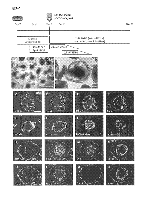
[Fig. 2-1] The upper panel of Fig. 2-1 is a diagram
schematically showing a procedure for producing a cell mass
containing neural tissue and nonneural epithelial tissue from
human ES cells in Example 1. The lower panels A and B are
diagrams showing bright-field observation images by an inverted
lo microscope of the cell mass 28 days after the start of
suspension culturing in Example 2. The lower panels C - R show
the results of examination by fluorescent immunostaining of the
expression state of each cell marker in the cell mass 28 days
after the start of suspension culturing. C and D respectively
/5 show stained image of Chx10 and nuclear-stained image thereof.
E and F respectively show stained image of RLDH3 and nuclear-
stained image thereof. G and H respectively show stained image
of NCAM and nuclear-stained image thereof. I and J
respectively show stained image of N-Cadherin and nuclear-
20 stained image thereof. K - N respectively show stained images
of EpCAM, Sixl, p63 and nuclear-stained image thereof. 0 and P
respectively show stained image of PDGFRP and nuclear-stained
image thereof. Q and R respectively show stained image of
cytokeratin 18 (CK18) and nuclear-stained image thereof.
25 [Fig. 2-2] S - AN of Fig. 2-2 show the results of
examination by fluorescent immunostaining of the expression
state of each cell marker in the cell mass 28 days after the
start of suspension culturing. S and T respectively show
stained image of cytokeratin 19 (CK19) and nuclear-stained
30 image thereof. U and V respectively show stained image of pan-
cytokeratin and nuclear-stained image thereof. W - Y
respectively show stained images of C-Maf, Soxl and nuclear-
stained image thereof. Z - AS respectively show stained images
of Proxl, acetylated tubulin (AcTub) and nuclear-stained image
35 thereof. AC - AE respectively show stained images of L-Maf,
11
Date Recue/Date Received 2020-05-22
CA 03083344 2020-05-22
Crystalline aA (Cry aA) and nuclear-stained image thereof. AF
- AH respectively show stained images of Emx2, N-Cadherin and
nuclear-stained image thereof. Al - AK respectively show
stained images of Pax6, 13111 tubulin and nuclear-stained image
thereof. AL - AN respectively show stained images of Sixl,
pan-cytokeratin and nuclear-stained image thereof.
[Fig. 2-3] AO - AV of Fig. 2-3 show the results of
examination by fluorescent immunostaining of the expression
state of each cell marker in the cell mass 28 days after the
/0 start of suspension culturing. AO - AP respectively show
stained image of EpCAM and nuclear-stained image thereof. AQ -
AR respectively show stained image of Laminin and nuclear-
stained image thereof. AS - AV respectively show stained
images of RLDH3, Chx10, pan-cytokeratin and nuclear-stained
image thereof. The lower panel AW is a diagram schematically
showing the structure of a cell mass containing neural tissue
and nonneural epithelial tissue on day 28 of culturing.
[Fig. 3] Fig. 3A - C show the results of examination by
fluorescent immunostaining of the expression state of each cell
marker in the cell mass 28 days after the start of suspension
culturing in Example 2. A - C respectively show stained images
of N-Cadherin and EpCAM and nuclear-stained image thereof. The
lower panel of Fig. 3D shows the results of measurement, using
analysis software, of images obtained by fluorescence
immunostaining.
[Fig. 4] The upper panel of Fig. 4 is a diagram
schematically showing a procedure for producing a cell mass
containing neural tissue and nonneural epithelial tissue from
human ES cells in Example 3. The lower panels A - C are
diagrams showing bright-field observation images by an inverted
microscope of the cell mass 90 days after the start of
suspension culturing in Example 3. The lower panels D - N show
the results of examination by fluorescent immunostaining of the
expression state of each cell marker in the cell mass 90 days
after the start of suspension culturing. D - K respectively
12
Date Recue/Date Received 2020-05-22
CA 03083344 2020-05-22
show stained images of cytokeratin 5 (CK5), cytokeratin 12
(CK12) and Laminin and nuclear-stained image thereof. L - N
respectively show stained images of Mucin4 (MUC4) and Pax6 and
nuclear-stained image thereof.
[Fig. 5] The upper panel of Fig. 5 is a diagram
schematically showing a procedure for producing a cell mass
containing neural tissue and nonneural epithelial tissue from
human ES cells by changing the time of addition of BMP4 in
Example 4. The lower panels A - F are diagrams showing bright-
/0 field observation images by an inverted microscope of the cell
masses 10 days after the start of suspension culturing in
Example 2. Diagrams of bright-field observation images by an
inverted microscope of the cell masses 10 days after the start
of suspension culturing and formed from A: control cells
without addition of BMP4, B: cells added with BMP4
simultaneously with the start of suspension culturing, and C -
F: cells added with BMP4 on days 1, 2, 3 and 6, respectively,
after the start of suspension culturing.
[Fig. 6] The upper panel of Fig. 6 is a diagram
schematically showing a procedure for preparing a cell
aggregate in the production process of a cell mass containing
neural tissue and nonneural epithelial tissue from human ES
cells in Example 5. The lower panels A and B are diagrams
showing bright-field observation images by an inverted
microscope of the cell aggregate respectively 2 and 3 days
after the start of suspension culturing in Example 5. The
lower panels C - J show the results of examination by
fluorescent immunostaining of the expression state of each cell
marker in the cell aggregate 2 or 3 days after the start of
suspension culturing. C and D respectively show stained images
of ZO-1 and nuclear-stained image thereof in the cell aggregate
2 days after the start of suspension culturing. G and H
respectively show stained images of N-Cadherin (NCad) and
nuclear-stained image thereof in the cell aggregate 2 days
after the start of suspension culturing. E and F respectively
13
Date Recue/Date Received 2020-05-22
CA 03083344 2020-05-22
show stained images of ZO-1 and nuclear-stained image thereof
in the cell aggregate 3 days after the start of suspension
culturing. I and J respectively show stained images of N-
Cadherin (NCad) and nuclear-stained image thereof in the cell
aggregate 3 days after the start of suspension culturing.
[Fig. 7] The upper panel of Fig. 7 is a diagram
schematically showing a procedure for producing a cell mass
containing neural tissue and nonneural epithelial tissue from
human ES cells by changing the concentration of BMP4 added in
/o Example 6. The lower panels A - H are diagrams showing bright-
field observation images by an inverted microscope of the cell
masses 10 days after the start of suspension culturing in
Example 6. Diagrams of bright-field observation images by an
inverted microscope of the cell masses 10 days after the start
of suspension culturing and formed from A: control cells
without addition of BMP4, and B - H: cells obtained by adding
BMP4 at varied concentrations (0.1 nM, 0.25 nM, 0.5 nM, 0.75 nM,
1 nM, 1.5 nM, 5 nM) on day 2 after the start of suspension
culturing and further performing suspension culturing.
[Fig. 8] The upper panel of Fig. 8 is a diagram
schematically showing a procedure for examining the effect of
each Wnt signal transduction pathway inhibiting substance on
the production of a cell mass containing neural tissue and
nonneural epithelial tissue from human ES cells in Example 7.
The lower panels A - K are diagrams showing bright-field
observation images by an inverted microscope of the cell mass
28 days after the start of suspension culturing in Example 7.
Diagrams of bright-field observation images by an inverted
microscope of the cell masses 10 days after the start of
suspension culturing and formed from A: control without
addition of a Wnt signal transduction pathway inhibiting
substance, and B - K: cells obtained by adding various kinds of
Wnt signal transduction pathway inhibiting substance at varied
concentrations at the start of suspension culturing and further
performing suspension culturing.
14
Date Recue/Date Received 2020-05-22
CA 03083344 2020-05-22
[Fig. 9] The upper panel of Fig. 9 is a diagram
schematically showing a procedure for examining the effect of a
pre-treatment with a compound before the start of suspension
culturing on the production of a cell mass containing neural
tissue and nonneural epithelial tissue from human ES cells in
Example 8. The lower panels A - C are diagrams showing bright-
field observation images by an inverted microscope of the cell
mass 15 days after the start of suspension culturing in Example
8. A is an example of a nearly spherical Grade 1 cell mass in
lo which not less than 80% of the entire circumference is covered
with nonneural epithelium, B is an example of a Grade 2 cell
mass in which 80% to 40% of the entire circumference is covered
with nonneural epithelium or which is irregularly shaped; and C
is an example of a Grade 3 cell mass in which a ratio of
nonneural epithelium on the surface of the cell mass is not
more than 40%. D is a graph showing the results of quality
evaluation of the cell masses formed after a compound pre-
treatment under respective conditions.
[Fig. 10] The upper panel of Fig. 10 is a diagram
schematically showing a procedure for producing a cell mass
containing neural tissue and nonneural epithelial tissue from
human ES cells in Example 9. The lower panel A is a diagram
showing bright-field observation image by an inverted
microscope of the cell mass 10 days after the start of
suspension culturing in Example 9. B and C are diagrams
showing bright-field observation images by an inverted
microscope of the cell masses 28 days after the start of
suspension culturing in Example 9. The lower panels D - S show
the results of examination by fluorescent immunostaining of the
expression state of each cell marker in the cell mass 28 days
after the start of suspension culturing. D - G respectively
show stained images of Sixl, NCAM and E-Cadherin and nuclear-
stained image thereof. H - K respectively show stained images
of RLDH3, Chx10 and pan-cytokeratin and nuclear-stained image
thereof. L - 0 respectively show stained images of Pax6, Rx
Date Recue/Date Received 2020-05-22
CA 03083344 2020-05-22
and pm tubulin and nuclear-stained image thereof. P S
respectively show stained images of p63, N-Cadherin and EpCAM
and nuclear-stained image thereof.
[Fig. 11] The upper panel of Fig. 11 is a diagram
schematically showing a procedure for producing a cell mass
containing neural tissue and nonneural epithelial tissue from
human ES cells in Example 10. The lower panels are diagrams
showing bright-field observation images by an inverted
microscope of the cell masses in Example 10, in which A is a
/0 cell mass 34 days after the start of suspension culturing, B is
nonneural epithelial tissue alone isolated from A, and C is
isolated nonneural epithelial tissue converted to single cells
by an enzyme treatment and on day 3 after being seeded in a
cell culture dish.
[Fig. 121 The upper panel of Fig. 12 is a diagram
schematically showing a procedure for producing a cell mass
containing neural tissue and nonneural epithelial tissue from
human ES cells in Example 11. The lower panel A is a graph
showing the results of evaluation of the cell masses by a
fluorescein staining method after treating the cell masses with
compounds of GHS classifications 1 and 2 in Example 11.
[Description of Embodiments]
[0012]
1. Definition
In the present invention, "stem cell" means an
undifferentiated cell having differentiation potency and
proliferative capacity (particularly self-renewal competence)
maintaining differentiation potency. The stem cell includes
subpopulations such as pluripotent stem cell, multipotent stem
cell, unipotent stem cell and the like according to the
differentiation potency. Pluripotent stem cell refers to a
stem cell capable of being cultured in vitro and having a
potency to differentiate into any cell constituting living
organisms (tissue derived from three germ layers (ectoderm,
mesoderm, endoderm) (pluripotency). The multipotent stem cell
16
Date Recue/Date Received 2020-05-22
CA 03083344 2020-05-22
means a stem cell having a potency to differentiate into plural
types of tissues or cells, though not all kinds. The unipotent
stem cell means a stem cell having a potency to differentiate
into a particular tissue or cell.
[0013]
Pluripotent stem cell can be induced from fertilized egg,
clone embryo, germ stem cell, stem cell in a tissue, somatic
cell or the like. Examples of the pluripotent stem cell
include embryonic stem cell (ES cell), EG cell (embryonic germ
lo cell), and induced pluripotent stem cell (iPS cell). Muse cell
(Multi-lineage differentiating stress enduring cell) obtained
from mesenchymal stem cell (MSC), and GS cell produced from
reproductive cell (e.g., testis) are also encompassed in the
pluripotent stem cell. Embryonic stem cell was first
established in 1981, and has also been applied to the
generation of knockout mouse since 1989. In 1998, human
embryonic stem cell was established, which is also being
utilized for regenerative medicine. ES cell can be produced by
culturing an inner cell mass on a feeder cell or in a medium
containing LIF. The production methods of ES cell are
described in, for example, WO 96/22362, WO 02/101057, US
5,843,780, US 6,200,806, and US 6,280,718. Embryonic stem
cells are available from given organizations, or a commercially
available product can be purchased. For example, human
embryonic stem cells, KhES-1, KhES-2 and KhES-3, are available
from Kyoto University's Institute for Frontier Medical Sciences.
EB5 cell, which is a mouse embryonic stem cell, is available
from Incorporated Administrative Agency RIKEN, and D3 cell line,
which is a mouse embryonic stem cell, is available from ATCC.
Nuclear transfer ES cell (ntES cell), which is one of the ES
cells, can be established from a clone embryo produced by
transplanting the cell nucleus of a somatic cell into an
enucleated egg.
[0014]
EG cell can be produced by culturing a primordial germ
17
Date Recue/Date Received 2020-05-22
CA 03083344 2020-05-22
cell in a medium containing mSCF, LIF and bFCF (Cell, 70: 841-
847, 1992).
[0015]
The "induced pluripotent stem cell" in the present
invention is a cell induced to have pluripotency by
reprogramming a somatic cell by a known method and the like.
Specifically, a cell induced to have pluripotency by
reprogramming differentiated somatic cells such as fibroblast,
and peripheral blood mononuclear cell by the expression of a
m combination of a plurality of genes selected from the group
consisting of reprogramming genes including 0ct3/4, 5ox2, Klf4,
Myc (c-Myc, N-Myc, L-Myc), Clisl, Nanog, Sall4, 1in28, and
Esrrb can be mentioned. Induced pluripotent stem cell was
established by Yamanaka et al. in mouse cell in 2006 (Cell,
2006, 126(4), pp.663-676). In 2007, Induced pluripotent stem
cell was also established from human fibroblast, and has
pluripotency and self-renewal competence similar to those of
embryonic stem cells (Cell, 2007, 131(5), pp.861-872; Science,
2007, 318(5858), pp.1917-1920; Nat. Biotechnol., 2008, 26(1),
pp.101-106). Besides the production method based on direct
reprogramming by gene expression, induced pluripotent stem cell
can also be obtained from somatic cell by the addition of a
compound and the like (Science, 2013, 341, pp. 651-654).
[0016]
While the somatic cell used for producing induced
pluripotent stem cell is not particularly limited, tissue-
derived fibroblast, blood-lineage cells (e.g., peripheral blood
mononuclear cell, T cell), hepatocyte, pancreatic cell,
intestinal epithelial cell, and smooth muscle cell can be
mentioned.
[0017]
When induced pluripotent stem cell is produced by
reprogramming by the expression of several kinds of genes (e.g.,
4 factors of 0ct3/4, Sox2, Klf4, and Myc), the means for gene
expression is not particularly limited. Examples of the
18
Date Recue/Date Received 2020-05-22
CA 03083344 2020-05-22
aforementioned means include an infection method using a virus
vector (e.g., retrovirus vector, lentivirus vector, Sendaivirus
vector, adenovirus vector, adeno-associated virus vector), a
gene transfer method using a plasmid vector (e.g., plasmid
vector, episomal vector) (e.g., calcium phosphate method,
lipofection method, retronectin method, electroporation method),
a gene transfer method using an RNA vector (e.g., calcium
phosphate method, lipofection method, electroporation method),
and a method with direct injection of protein.
/o [0018]
The pluripotent stem cell to be used in the present
invention is preferably ES cell or induced pluripotent stem
cell.
[0019]
As the multipotent stem cell, tissue stem cells (also
called stem cell in a tissue, tissue-specific stem cell or
somatic stem cell) such as hematopoietic stem cell, neural stem
cell, retinal stem cell, and mesenchymal stem cell can be
mentioned.
[0020]
Genetically-modified pluripotent stem cells can be
produced by using, for example, a homologous recombination
technique. Examples of the gene on the chromosome to be
modified include a cell marker gene, a histocompatibility
antigen gene, a gene related to a disease due to a disorder of
neural cell and so on. A target gene on the chromosome can be
modified using the methods described in Manipulating the Mouse
Embryo, A Laboratory Manual, Second Edition, Cold Spring Harbor
Laboratory Press (1994); Gene Targeting, A Practical Approach,
IRL Press at Oxford University Press (1993); Biomanual Series 8,
Gene Targeting, Making of Mutant Mouse using ES cell, YODOSHA
CO., LTD. (1995); and so on.
[0021]
To be specific, for example, the genome gene of the
target gene to be modified (e.g., cell marker gene,
19
Date Recue/Date Received 2020-05-22
CA 03083344 2020-05-22
histocompatibility antigen gene, disease-related gene and so
on) is isolated, and a targetting vector used for homologous
recombination of the target gene is produced using the isolated
genome gene. The produced targetting vector is introduced into
stem cells and the cells that showed homologous recombination
between the target gene and the targetting vector are selected,
whereby stem cells having the modified gene on the chromosome
can be produced.
[0022]
/0 Examples of the method for isolating genome gene of the
target gene include known methods described in Molecular
Cloning, A Laboratory Manual, Second Edition, Cold Spring
Harbor Laboratory Press (1989), Current Protocols in Molecular
Biology, John Wiley & Sons (1987-1997) and so on. The genome
gene of the target gene can also be isolated using genomic DNA
library screening system (manufactured by Genome Systems),
Universal GenomeWalker Kits (manufactured by CLONTECH) and so
on.
[0023]
Production of targetting vector used for homologous
recombination of the target gene, and efficient selection of a
homologous recombinant can be performed according to the
methods described in Gene Targeting, A Practical Approach, IRL
Press at Oxford University Press (1993); Biomanual Series 8,
Gene Targeting, Making of Mutant Mouse using ES cell, YODOSHA
CO., LTD. (1995); and so on. As the targetting vector, any of
replacement type or insertion type can be used. As the
selection method, methods such as positive selection, promoter
selection, negative selection, po1yA selection and so on can be
used.
Examples of a method for selecting the desired homologous
recombinant from the selected cell lines include Southern
hybridization method, PCR method and so on for the genomic DNA.
[0024]
The "mammal" in the present invention encompasses rodents,
Date Recue/Date Received 2020-05-22
CA 03083344 2020-05-22
ungulata, carnivora, and primates. The rodents encompass mouse,
rat, hamster, and guinea pig. Ungulata encompass swine, bovine,
goat, horse, and sheep. Carnivora encompasses dog, and cat.
The "primates" in the present invention refers to mammals
belonging to the primate, and the primates include prosimian
such as lemur, loris, tupai etc, and anthropoidea such as
monkey, ape, and human.
[0025]
The pluripotent stem cells to be used in the present
invention are mammalian pluripotent stem cells, preferably
pluripotent stem cells of rodents (e.g., mouse, rat) or
primates (e.g., human, monkey), most preferably a human
pluripotent stem cell.
[0026]
The "signal transduction" in the present invention refers
to the mechanism of the cells for transmission of information,
amplification and processing of, and response to biochemical
stimulation, such as processes and mechanisms in which receptor
proteins present in the cell membrane and the like bind to
chemical substances and the like to cause a structural change,
which in turn is transmitted sequentially in the cell as a
stimulation to finally cause reactions such as gene expression,
channel opening and the like.
[0027]
The "cell adhesion" in the present invention refers to
cell-cell adhesion and cell-extracellular matrix adhesion.
Adhesion of cells to culture vessels and the like that occurs
under an artificial culture environment in vitro is also
included in the cell adhesion. As the kind of the cell
adhesion, anchoring junction, communicating junction, occluding
junction can be mentioned.
[0028]
The "Tight junction" in the present invention refers to,
among cell-cell adhesions, occluding junctions found in
vertebrates and chordates. A tight junction is formed between
21
Date Recue/Date Received 2020-05-22
CA 03083344 2020-05-22
epithelial cells. Whether a tight junction is present in
tissues of biological origin or cell masses produced by the
production method of the present invention and the like can be
detected by, for example, methods such as immunohistochemistry
and the like using an antibody (anti-claudin antibody, and
anti-ZO-1 antibody) to a constituent component of the tight
junction.
[0029j
The "suspension culturing" or "suspension culturing
/0 method" in the present invention refers to culturing while
maintaining a state in which cells, cell aggregates or cell
masses are suspended in a culture medium and a method of
performing the culturing. That is, the suspension culturing is
performed under conditions in which cells, cell aggregates or
cell masses are not adhered to a culture vessel and the like,
and culturing performed under conditions permitting adhesion to
a culture vessel and the like (adhesion culturing or adhesion
culturing method) is not included in the category of suspension
culturing. In this case, adhesion of cell means that a strong
cell-substratum junction, which is one type of cell adhesion,
is formed between a cell, cell aggregate or cell mass and a
culture vessel. More particularly, suspension culturing refers
to culturing under conditions in which a strong cell-substratum
junction is not formed between a cell, cell aggregate or cell
mass and a culture vessel, and adhesion culturing refers to
culturing under conditions in which a strong cell-substratum
junction is formed between a cell, cell aggregate or cell mass
and a culture vessel and the like.
In a cell aggregate or cell mass in suspension culturing,
a plane attachment is formed between a cell and a cell. In a
cell aggregate or cell mass in suspension culturing, a cell-
substratum junction is hardly formed with a culture vessel and
the like and, even if it is formed, its contribution is small.
In some embodiments, an endogenous cell-substratum junction is
present inside the aggregate or cell mass, but a cell-
22
Date Recue/Date Received 2020-05-22
CA 03083344 2020-05-22
substratum junction is hardly formed with a culture vessel and
the like and, even if it is formed, its contribution is small.
The plane attachment between a cell and a cell means that
a cell attaches to another cell via planes. More particularly,
the plane attachment between a cell and a cell means that, for
example, not less than 1%, preferably not less than 3%, more
preferably not less than 5%, of the surface area of a cell
adheres to the surface of another cell. A surface of a cell
can be observed by staining with a reagent (e.g., DiI) that
lo stains membranes, immunostaining of cell adhesion factors (e.g.,
E-cadherin and N-cadherin).
[0030]
The culture vessel to be used when performing suspension
culturing is not particularly limited as long as it enables
/5 "culturing in suspension" and those of ordinary skill in the
art can appropriately determine same. Examples of such culture
vessel include flask, tissue culture flask, dish, petri dish,
tissue culture dish, multidish, microplate, microwell plate,
micropore, multiplate, multiwell plate, chamber slide, schale,
20 tube, tray, culture bag, spinner flask, and roller bottle. To
enable suspension culturing, these culture vessels are
preferably non-cell-adhesive. Useful non-cell-adhesive culture
vessels include culture vessels whose surfaces have not
undergone an artificial treatment for improving adhesiveness to
25 cells (e.g., surface treatment with extracellular matrix such
as basement membrane preparation, laminin, entactin, collagen,
and gelatin, or coating treatment with polymer such as
polylysine, and polyornithine or positive electric charge
treatment and the like), and the like. As a non-cell-adhesive
30 culture vessel, culture vessels whose surfaces have been
artificially treated to decrease adhesiveness to the cells
(e.g., superhydrophilic treatment with MPC polymer and the like,
and protein low adsorption treatment) and the like can be used.
Roller culturing using spinner flask, and roller bottle may be
35 performed. The culture surface of the culture vessel may be a
23
Date Recue/Date Received 2020-05-22
CA 03083344 2020-05-22
flat bottom or may have concaves and convexes.
[0031]
The medium to be used for culturing cells in the present
invention can be prepared from a medium generally used for
culturing animal cells as a basal medium. Examples of the
basal medium include media that can be used for culturing
animal cells such as BME medium, BGJb medium, CMRL1066 medium,
Glasgow MEN medium, Improved MEN Zinc Option medium, IMDM
medium, Medium199 medium, Eagle MEN medium, aMEM medium, DMEM
/0 medium, F-12 medium, DMEM/F-12 medium, IMDM/F12 medium, Ham's
medium, RPNI1640 medium, Fischer's medium, and mixed medium
thereof.
For culturing pluripotent stem cells, a medium for
culturing pluripotent stem cells using the above-mentioned
/5 basal medium as the base, preferably a known medium for
embryonic stem cells and/or induced pluripotent stem cells, a
medium for culturing pluripotent stem cells under feeder free
can be used. For example, feeder-free medium such as Essential
8 medium, TeSR medium, mTeSR medium, mTeSR-E8 medium, and
20 StemFit medium can be mentioned.
[0032]
The "serum-free medium" in the present invention means a
medium free of unadjusted or unpurified serum. In the present
invention, a medium containing purified blood-derived
25 components and animal tissue-derived components (e.g., growth
factor) is also included in a serum-free medium unless
unadjusted or unpurified serum is contained therein.
[0033]
The serum-free medium may contain a serum replacement.
30 Examples of the serum replacement include one appropriately
containing albumin, transferrin, fatty acid, collagen precursor,
trace element, 2-mercaptoethanol or 3' thiolglycerol, or
equivalents of these, and so on. Such serum replacement may be
prepared by, for example, the method described in W098/30679.
35 The serum replacement may be a commercially available product.
24
Date Recue/Date Received 2020-05-22
CA 03083344 2020-05-22
Examples of such commercially available serum replacement
include KnockoutTM Serum Replacement (Life Technologies:
hereinafter sometimes referred to as KSR), Chemically Defined
Lipid Concentrated (manufactured by Life Technologies) and
GlUtaMaXTM (manufactured by Life Technologies), B27
(manufactured by Life Technologies), and N2 (manufactured by
Life Technologies).
[0034]
The serum-free medium to be used for suspension culturing
may appropriately contain a fatty acid or lipid, amino acid
(e.g., non-essential amino acids), vitamin, growth factor,
cytokine, antioxidant, 2-mercaptoethanol, pyruvic acid,
buffering agent, inorganic salts and so on.
[0035]
To avoid complicated preparation, a serum-free medium
supplemented with an appropriate amount (e.g., about 0.5% to
about 30%, preferably about 1% to about 20%) of commercially
available KSR (manufactured by Life Technologies) (e.g., medium
of 1:1 mixture of F-12 medium and IMDM medium supplemented with
1 x Chemically-defined Lipid concentrated, 5% KSR and 450 pM 1-
monothioglycerol) may be used as such serum-free medium. In
addition, as a product equivalent to KSR, the medium disclosed
in JP-A-2001-508302 can be mentioned.
[0036]
The "serum-containing medium" in the present invention
means a medium containing unadjusted or unpurified serum. The
medium may contain a fatty acid, lipid, amino acid (e.g., non-
essential amino acids), vitamin, growth factor, cytokine,
antioxidant, 2-mercaptoethanol, 1-monothioglycerol, pyruvic
acid, buffering agent, inorganic salts and so on. For example,
when a pluripotent stem cell is induced to differentiate into a
retinal tissue and the like by using a basement membrane
preparation such as Matrigel and the like, a serum-containing
medium can be used (Cell Stem Cell, 10(6), 771-775 (2012)).
[0037]
Date Recue/Date Received 2020-05-22
CA 03083344 2020-05-22
The culturing in the present invention is preferably
performed under xeno-free conditions. The "xeno-free" means
conditions eliminating components derived from species
different from that of the cell to be cultured.
[0038]
The medium to be used in the present invention is
preferably a medium containing chemically determined components
(Chemically defined medium; CDM) to avoid contamination with
chemically undetermined components.
[0039]
The "basement membrane-like structure" in the present
invention means a thin membrane structure composed of
extracellular matrix. The basement membrane is formed on the
basal side of epithelial cells in a living body. The
/5 components of the basement membrane include type IV collagen,
laminin, heparan sulfate proteoglycan (perlecan),
entactin/nidogen, cytokine, growth factor and the like.
Whether a basement membrane is present in a tissue derived from
a living body and in a cell mass prepared by the production
method of the present invention is determined by, for example,
tissue staining such as PAM staining and the like, and a method
such as immunohistochemistry using an antibody against a
constituent component of the basement membrane (anti-laminin
antibody, anti-type IV collagen antibody, etc.), and the like.
[0040]
The "basement membrane preparation" in the present
invention is one containing a basement membrane constituent
component having functions to control epithelial cell-like cell
morphology, differentiation, proliferation, motility,
functional expression, and the like when desired cells having
basement membrane formability are seeded thereon and cultured.
For example, when the cells and tissues produced by the present
invention are dispersed and further subjected to adhesion
culturing, they can be cultured in the presence of a basement
membrane preparation. As used herein, the "basement membrane
26
Date Regue/Date Received 2020-05-22
CA 03083344 2020-05-22
constituent component" refers to an extracellular matrix
molecule as a thin membrane present between an epithelial cell
layer and an interstitial cell layer and the like in animal
tissues. A basement membrane preparation can be produced, for
example, by removing cells adhered to the support via a
basement membrane and having the ability to form the basement
membrane from the support by using a solution having the
ability to dissolve the lipids of the cells, an alkaline
solution or the like. Examples of the basement membrane
lo preparation include products commercially available as basement
membrane products (e.g., MatrigelTM (manufactured by Becton,
Dickinson and Company: hereinafter sometimes referred to as
Matrigel)) and GeltrexTM (manufactured by Life Technologies),
and extracellular matrix molecules known as basement membrane
/5 components (e.g., laminin, type-IV collagen, heparan sulfate
proteoglycan, and entactin).
[0041]
In the present invention, a basement membrane preparation
such as Matrigel (manufactured by Corning) which is extracted
20 from a tissue or cell of Engelbreth-Holm-Swarm (EHS) mouse
sarcoma and the like and solubilized and the like can be used
for culturing cells and tissues. Similarly, as a basement
membrane component used for cell culturing, human solubilized
amniotic membrane (manufactured by Bioresource Application
25 Institute, Co.), human recombinant laminin produced by HEK293
cell (manufactured by BioLamina), human recombinant laminin
fragment (manufactured by Nippi, Inc.), and human recombinant
vitronectin (manufactured by Thermo Fisher) can also be used.
To avoid contamination with components derived from different
30 organism species and to avoid the risk of infections, preferred
is a recombinant protein whose components are clear.
[0042]
In the present invention, the "medium containing a
substance X" and "in the presence of a substance X"
35 respectively refer to a medium supplemented with an exogenous
27
Date Recue/Date Received 2020-05-22
CA 03083344 2020-05-22
substance X or a medium containing an exogenous substance X,
and in the presence of an exogenous substance X. The exogenous
substance X is distinguished from the endogenous substance X
which is the substance X endogenously expressed, secreted or
produced by, for example, the cells or tissues present in the
medium.
For example, a "medium containing a Sonic hedgehog signal
transduction pathway activating substance" is a medium
supplemented with an exogenous Sonic hedgehog signal
io transduction pathway activating substance or a medium
containing an exogenous Sonic hedgehog signal transduction
pathway activating substance.
[0043]
In the present invention, a "feeder cell" refers to a
cell other than a stem cell that co-exists when culturing the
stem cell. Examples of the feeder cells used for culturing
pluripotent stem cells while maintaining undifferentiated state
include mouse fibroblasts (MEF), human fibroblasts, and SNL
cells. As the feeder cells, feeder cells that underwent a
growth suppression treatment is preferable. Examples of the
growth suppression treatment include treatment with a growth
inhibitor (e.g., mitomycin C), and UV irradiation. Feeder
cells used for culturing pluripotent stem cells while
maintaining undifferentiated state contributes to the
maintenance of undifferentiated state of pluripotent stem cell
by secretion of humoral factors (preferably factor for
maintaining undifferentiated state), or production of scaffolds
for cell adhesion (extracellular matrix).
[0044]
In the present invention, an "aggregate" of cells refers
to a clump formed by assembly of cells dispersed in a medium,
wherein the cells are adhered to each other. Cell clumps,
embryoid bodies, spheres, spheroids, and organoids are also
encompassed in the cell aggregates. Preferably, a plane
attachment is formed between a cell and a cell in the aggregate
28
Date Recue/Date Received 2020-05-22
CA 03083344 2020-05-22
of cells. In some embodiments, cells sometimes form a cell-
cell junction and/or a cell adhesion, for example, adherence
junction, in some or all of the aggregates. The "aggregate" in
the present invention specifically includes an aggregate
produced in the first step of the below-mentioned "2.
Production method of cell mass containing neural cells or
neural tissue and nonneural epithelial tissue", which is formed
by cells dispersed at the time of the start of the suspension
culturing.
io In the present invention, "uniformed aggregates" means
that the size of each aggregate is constant when a plurality of
aggregates are cultured, and that the variance in the length of
the maximum diameter is small when the size of the aggregates
are evaluated by the length of the maximum diameter. More
/5 specifically, it means that not less than 75% of aggregates in
the whole aggregate population are within mean 100%,
preferably mean 50%, more preferably mean 20%, of the
maximum diameter in the population of the cell masses.
[0045]
20 In the present invention, to "form unifolmed aggregates"
means to rapidly aggregate a given number of dispersed cells to
form cell aggregates uniform in size, when gathering the cells
to form cell aggregates and culturing the aggregates in
suspension.
25 "Dispersion" refers to dividing cells or a tissue into
small cell debris (not less than 2 cells and not more than 100
cells, preferably not more than 50 cells) or single cells by a
dispersion treatment such as enzymatic treatment, and physical
treatment. A given number of dispersed cells is a collection
30 of a certain number of cell debris or single cells.
Examples of the method of dispersing pluripotent stem
cells include a mechanical dispersion treatment, a cell
dispersion solution treatment, and a cell protecting agent
addition treatment. These treatments may be performed in
35 combination. Preferably, a cell dispersion solution treatment
29
Date Regue/Date Received 2020-05-22
CA 03083344 2020-05-22
is performed and then a mechanical dispersion treatment is
performed.
As a method of mechanical dispersion treatment, a
pipetting treatment or scraping operation by a scraper can be
mentioned.
As a cell dispersion solution to be used for the cell
dispersion solution treatment, a solution containing any of
enzymes such as trypsin, collagenase, hyaluronidase, elastase,
pronase, DNase, and papain, and a chelating agent such as
ethylenediaminetetraacetic acid etc. can be mentioned. A
commercially available cell dispersion solution such as Accumax
(manufactured by Innovative cell technologies) and TrypLE
Select (manufactured by Life Technologies) can also be used.
As a cell protecting agent to be used for a cell
protector addition treatment, FGF signal transduction pathway
activating substance, heparin, ROCK inhibiting substance, serum,
or serum replacement can be mentioned. As a preferable cell
protecting agent, a ROCK inhibiting substance can be mentioned.
For example, a method for dispersing pluripotent stem
cells includes a method involving treating a colony of
pluripotent stem cells with a cell dispersion solution
(Accumax) in the presence of a ROCK inhibiting substance as a
cell protecting agent, and further dispersing them by pipetting.
[0046]
In the production method of the present invention, it is
preferable to form an aggregate of pluripotent stem cells by
rapidly gathering the pluripotent stem cells. When an
aggregate of pluripotent stem cells is formed in such a manner,
an epithelium-like structure can be formed with good
reproducibility in the cells induced and differentiated from
the formed aggregate. Examples of the experimental operation
to form an aggregate include a method involving keeping cells
in a small space by using a plate with small wells (e.g., plate
with wells having a base area of about 0.1 - 2.0 cm2 when
calculated in terms of flat bottom), micropore and so on, a
Date Recue/Date Received 2020-05-22
CA 03083344 2020-05-22
method involving aggregating cells by centrifugation for a
short time using a small centrifugation tube. As a plate with
small wells, for example, 24 well plate (area of about 1.88 cm2
when calculated in terms of flat bottom), 48 well plate (area
of about 1.0 cm2 when calculated in terms of flat bottom), 96
well plate (area of about 0.35 cm2 when calculated in terms of
flat bottom, inner diameter about 6 - 8 mm), and 384 well plate
can be mentioned. Preferred is 96 well plate. As a shape of
the plate with small wells, the shape of the bottom surface
io when the well is seen from above is, for example, polygon,
rectangle, ellipse, true circle, preferably true circle. As a
shape of the plate with small wells when the well is seen from
the side well, the shape of the bottom surface is preferably a
structure having high outer circumference and low inner concave,
which is, for example, U-bottom, V-bottom or M-bottom,
preferably U-bottom or V-bottom, most preferably V-bottom. As
a plate with small wells, a cell culture dish (e.g., 60 mm -
150 mm dish, culture flask) with a concave convex, or dent on
the bottom surface may also be used. The bottom surface of a
plate with small wells is preferably a non-cell-adhesive bottom
surface, preferably the aforementioned non-cell-adhesive-coated
bottom surface.
Formation of aggregates of pluripotent stem cells, and
formation of an epithelium -like structure in each cell forming
the aggregate can be determined based on the size and cell
number of the aggregate, macroscopic morphology of the
aggregate, microscopic morphology by tissue staining analysis
and uniformity thereof, expression of differentiation and
undifferentiation markers and uniformity thereof, control of
expression of differentiation marker and synchronism thereof,
reproducibility of differentiation efficiency between
aggregates, and so on.
[0047]
The "tissue" in the present invention refers to a
structure of a cell population having a structure in which
31
Date Recue/Date Received 2020-05-22
CA 03083344 2020-05-22
plural types of cells having different morphologies and
properties are sterically arranged in a given pattern.
[0048]
In the present invention, the "neural tissue" refers to a
tissue constituted of neural cells including cerebrum, midbrain,
cerebellum, spinal cord, retina, peripheral nerve and the like
in the developing stage or adult stage. A neural tissue
sometimes forms an epithelial structure (neuroepithelium)
having a layer structure, and the amount of neuroepithelium in
a neural tissue can be evaluated by bright field observation
using an optical microscope.
[0049]
In the present invention, the "neural cell" refers to a
cell other than epidermal lineage cell in a tissue derived from
ectoderm. That is, it includes cells such as neural precursor
cell, neuron (neuronal cell), glia cell, neural stem cell,
neuron precursor cell, glial precursor cell and the like. The
neural cell also encompasses cell constituting the below-
mentioned retinal tissue (retinal cell), retinal progenitor
cell, retinal layer-specific neuron, neural retinal cell, and
retinal pigment epithelial cell. The neural cell can be
identified by using Nestin, Tun, PSA-NCAM, N-cadherin and the
like as a marker.
Neuron is a functional cell that forms a neural circuit
and contributes to signal transmission, and can be identified
by using the expression of immature neuronal markers such as
TuJ1, Dcx, and HuC/D and/or mature neuronal cell markers such
as Map2, and NeuN as an index.
As glial cell, astrocyte, oligodendrocyte, and Milner
glia can be mentioned. As an astrocyte marker, GFAP can be
mentioned; as an oligodendrocyte marker, 04 can be mentioned;
and as a Mtiller glia marker, CRALBP and the like can be
mentioned.
The neural stem cell is a cell having differentiation
potency (multipotency) into neuron and glial cell, and
32
Date Recue/Date Received 2020-05-22
CA 03083344 2020-05-22
proliferative capacity (sometimes referred to as self-renewal
competence) maintaining multipotency. As the neural stem cell
marker, Nestin, Sox2, Musashi, Hes family, CD133 etc. can be
mentioned; however, these markers are markers for
progenitor/precursor cells in general and are not considered
neural stem cell-specific markers. The number of neural stem
cells can be evaluated by neurosphere assay, and clonal assay.
The neuronal precursor cell is a cell having
proliferative capacity, which produces neuron and does not
/0 produce glial cell. As a neuronal precursor cell marker, Tbr2,
Tel etc. can be mentioned. Alternatively, an immature neuronal
marker (TuJ1, Dcx, HuC/D)-positive and growth marker (Ki67, pH3,
MCM)-positive cell can also be identified as a neuronal
precursor cell.
The glial precursor cell is a cell having proliferative
capacity, which produces glial cell and does not produce neuron.
The neural precursor cell is an assembly of precursor
cells including neural stem cell, neuronal precursor cell and
glial precursor cell, and has proliferative capacity and
neuron- and glial cell- productivity. The neural precursor
cell can be identified using Nestin, GLAST, Sox2, Soxl, Musashi,
Pax6 and the like as markers. Alternatively, a neural cell
marker-positive and growth marker (Ki67, pH3, MCM)-positive
cell can also be identified as a neural precursor cell.
[0050]
In the present invention, the "retinal tissue" means a
retinal tissue in which at least two or more types of cells
such as photoreceptor cells, horizontal cells, bipolar cells,
amacrin cells, retinal ganglion cells, their
progenitor/precursor cells and retinal progenitor cells and so
on, which constitute respective retinal layers in retina in
vivo, are sterically arranged in layers. Which retinal layer
is constituted by each cell can be confirmed by a known method,
for example, presence or absence of the expression of a cell
marker or the level thereof, and the like.
33
Date Recue/Date Received 2020-05-22
CA 03083344 2020-05-22
[0051]
The "retinal layer" in the present invention means each
layer constituting the retina. Specific examples thereof
include retinal pigment epithelial layer, photoreceptor cell
layer, external limiting membrane, outer nuclear layer, outer
plexiform layer, inner nuclear layer, inner plexiform layer,
ganglion cell layer, nerve fiber layer and inner limiting
membrane.
[0052]
The "retinal progenitor cell" in the present invention
refers to a progenitor cell capable of differentiating into any
mature retinal cell including photoreceptor cell, horizontal
cell, bipolar cell, amacrine cell, retinal ganglion cell,
retinal pigment epithelial cell and the like.
The photoreceptor precursor cell, precursor cell of
horizontal cell, precursor cell of bipolar cell, precursor cell
of amacrine cell, precursor cell of retinal ganglion cell, and
retinal pigment epithelial precursor cell respectively refers
to precursor cell committed to differentiate into photoreceptor
cell, horizontal cell, bipolar cell, amacrine cell, retinal
ganglion cell, and retinal pigment epithelial cell.
[0053]
In the present invention, the "retinal layer-specific
neuron" is a cell constituting a retina layer and is a neuron
specific to the retinal layer. Examples of the retinal layer-
specific neuron include bipolar cell, retinal ganglion cells,
amacrine cell, horizontal cell, photoreceptor, retinal pigment
epithelial cell, rod cell and cone cell.
[0054]
The "retinal cell" in the present invention encompasses
the aforementioned retinal progenitor cell and retina layer-
specific nerve cell.
[0055]
Examples of the retinal cell marker include Rx (also
referred to as Rax), Aldhla3, and PAX6 expressed in retinal
34
Date Recue/Date Received 2020-05-22
CA 03083344 2020-05-22
progenitor cell, Nkx2.1 expressed in progenitor cell of
hypothalamus neuron but not expressed in retinal progenitor
cell, Soxl expressed in hypothalamus neuroepithelium but not
expressed in retina, Crx and Blimpl expressed in precursor cell
of photoreceptor, and the like. Examples of the marker of the
retinal layer-specific neuron include Chx10, PKCa and L7
expressed in bipolar cell, TUJI and Brn3 expressed in retinal
ganglion cells, Calretinin expressed in amacrine cell,
Calbindin expressed in horizontal cell, Rhodopsin and Recoverin
lo expressed in mature photoreceptor, Nrl expressed in rod cell,
Rxr-gamma expressed in cone cell, and RPE65 and Mitf expressed
in retinal pigment epithelial cell,.
[0056]
In the present invention, the "nonneural epithelial
tissue" refers to a tissue other than the neuroepithelial
tissues among the tissues having an epithelial structure. An
epithelial tissue can also be formed from any germ layer of
ectoderm, mesoderm, endoderm, or nutrition ectoderm. The
epithelial tissue includes epithelium, mesothelium, and
endothelium. Examples of the tissue included in the nonneural
epithelial tissue include epidermis, corneal epithelium, nasal
cavity epithelium, mouth cavity epithelium, trachea epithelium,
bronchus epithelium, airway epithelium, kidney epithelium,
renal cortex epithelium, placenta epithelium and the like.
Epithelial tissues are generally connected by various
intercellular junctions, and form tissues having a monolayer or
multilayer structure. Confirmation of the presence or absence
of such epithelial tissues and quantification of the amount
thereof can be performed by observation with an optical
microscope or a method such as immunohistochemistry using
antibodies against epithelial cell markers (anti-E-Cadherin
antibody, anti-N-Cadherin antibody, anti-EpCAM antibody etc.)
or the like.
[0057]
In the present invention, the "epithelial polarity" shows
Date Recue/Date Received 2020-05-22
CA 03083344 2020-05-22
spatially formed bias of the distribution and cellular
functions in epithelial cells. For example, corneal epithelial
cells are localized in the outermost layer of the eyeball,
express apical-specific proteins such as membrane-bound mucins
(MUC-1, 4, 16) and the like on the apical side to retain tears,
and express basal-specific proteins such as ce6 integrin, pl
integrin and the like on the basal side to adhere to the
basement membrane.
Whether epithelial polarity is present in a tissue
lo derived from a living body and in a cell mass prepared by the
production method of the present invention can be detected by,
for example, a method such as immunohistochemistry using
Phalloidin, an apical marker (anti-MUC-1 antibody, anti-PKC-
zeta antibody etc.) and a basal marker (anti-a6 integrin
antibody, anti-31 integrin antibody etc.) and the like.
[0058]
Cornea is one kind of nonneural epithelial tissue, and is
a transparent watch-glass-like tissue that occupies about 1/6
anterior of the outer layer of the eyeball wall. Examples of
the partial structure of the cornea include, but are not
limited to, corneal epithelium, Bowman's membrane, corneal
stroma, Descemet's membrane, and corneal endothelium,. Cornea
is usually composed of five layers consisting of a corneal
epithelium, Bowman's membrane, corneal stroma, Descemet's
membrane, and corneal endothelium in order from the surface of
the body. Induction of cornea, a partial structure thereof, or
a precursor tissue thereof can be confirmed by the expression
of a marker. Examples of the marker of cornea, a partial
structure thereof, or a precursor tissue thereof include pan-
cytokeratin (corneal epithelium precursor tissue), cytokeratin
18 (corneal epithelium precursor tissue), cytokeratin 19
(corneal epithelium precursor tissue), EpCAM (corneal
epithelium precursor tissue), PDGFR-P (surface layer ectoderm),
Sixl (surface layer ectoderm and placode), E-cadherin (corneal
epithelium precursor tissue), N-cadherin (corneal epithelial
36
Date Recue/Date Received 2020-05-22
CA 03083344 2020-05-22
stem cell, precursor tissue), cytokeratin 3 (corneal
epithelium), cytokeratin 12 (corneal epithelium), cytokeratin
14 (corneal epithelium), p63 (corneal epithelium), ZO-1
(corneal epithelium), PDGFR-a (corneal stroma, corneal
endothelium, or precursor tissue thereof), Pitx2 (precursor
tissue of corneal stroma and corneal endothelium), ABCG2
(precursor tissue of corneal stroma and corneal endothelium)
and the like. In one embodiment, the precursor tissue of the
corneal epithelium contained in the cell mass produced by the
method of the present invention is a pan-cytokeratin-positive
and E-cadherin-positive epithelial cell layer. In one
embodiment, the corneal epithelium contained in the cell mass
produced by the method of the present invention is a
cytokeratin 3-positive, cytokeratin 12-positive, cytokeratin
14-positive, p63-positive and ZO-1-positive epithelial
structure. In one embodiment, the precursor tissue of corneal
stroma and corneal endothelium contained in the cell mass
produced by the method of the present invention is an aggregate
layer of mesenchymal cells. In one embodiment, the aggregate
layer of the mesenchymal cells is pan-cytokeratin positive,
PDGFR-a positive, or Pitx2 positive and ABCG2 positive. While
the corneal stroma and corneal endothelium are both derived
from mesenchymal cells, corneal endothelium takes an
epithelialized endothelial cell layer-like form. Thus, it is
possible to distinguish corneal stroma (or precursor tissue
thereof) from corneal endothelium (or precursor tissue thereof)
and confirm induction of corneal endothelium or precursor
tissue thereof by perfolming the above-mentioned analysis of
marker expression, as well as morphological observation.
[0059]
In the present invention, the "placode" refers to
primordia of organs formed by thickening of a part of epidermal
ectoderm mainly in the developmental process of vertebrate. As
the tissue derived from placode, crystallin lens, nose, inner
ear, trigeminal nerve and the like can be mentioned. As a
37
Date Recue/Date Received 2020-05-22
CA 03083344 2020-05-22
marker of placode or a precursor tissue thereof (preplacode
region), Sixl, Six4, Dlx5, Eya2 and the like can be mentioned.
[0060]
The lens placode is a crystallin lens precursor tissue
composed of a thickened epidermal ectoderm cell layer. In the
embryonic development, it is formed by the contact of the optic
vesicle with the epidermal ectoderm and thickening of the
contact region.
The crystallin lens is one of the tissues derived from
/o placode, and is a tissue that plays the role of a lens that
refracts light entering the eyeball from outside and focuses to
the retina. Examples of the partial structure of crystallin
lens include, but are not limited to, crystallin lens
epithelium, nucleus of crystallin lens, and crystallin lens
capsule,. As the precursor tissue of crystallin lens, lens
placode, and lens vesicle can be mentioned.
The lens vesicle is a vesicle formed by invagination of
lens placode.
Induction of crystallin lens, a partial structure thereof,
or a precursor tissue thereof can be confirmed by the
expression of the marker. Examples of the marker of crystallin
lens, a partial structure thereof, or a precursor tissue
thereof include, but are not limited to, Sixl (surface layer
ectoderm and placode), FoxCl (placode), Emx2 (placode), Soxl
(central nervous system and crystallin lens precursor tissue),
FoxE3 (crystallin lens precursor tissue), Proxl (crystallin
lens precursor tissue), L-Maf (crystallin lens precursor
tissue), a, p and y crystallin (crystallin lens) and the like.
In one embodiment, the lens placode is an L-Maf positive,
thickened epidermal ectoderm cell layer. In one embodiment,
the lens vesicle is an L-Maf positive vesicle.
[0061]
As one embodiment of the cell mass containing neural
tissue and nonneural epithelial tissue described below, which
is produced by the production method of the present invention,
38
Date Recue/Date Received 2020-05-22
CA 03083344 2020-05-22
anterior ocular segment tissue can be mentioned. A cell mass
containing anterior ocular segment tissue or a partial
structure thereof, or a precursor tissue thereof can be
obtained by inducing, in the aggregate produced from a
pluripotent stem cell, differentiation of pluripotent cells
into anterior ocular segment tissue or a partial structure
thereof, or precursor tissue thereof. The above-mentioned
anterior ocular segment tissue includes cornea tissue which is
one kind of nonneural epithelial tissue, crystallin lens which
is one kind of placode-derived tissue, and retinal tissue which
is one kind of neural tissue.
[0062]
2. Production method of cell mass containing neural cells or
neural tissue and nonneural epithelial tissue
The present invention provides a method for producing a
cell mass comprising 1) neural cells or neural tissue and 2)
nonneural epithelial tissue. In the following, it is also to
be referred to as the production method of the present
invention.
One embodiment of the production method of the present
invention is a method for producing a cell mass containing
neural cells or neural tissue and nonneural epithelial tissue,
including the following steps (1) and (2):
(1) a first step of suspension-culturing pluripotent stem cells
to form a cell aggregate in the presence of a Wnt signal
transduction pathway inhibiting substance,
(2) a second step of suspension-culturing the aggregate
obtained in the first step in the presence of a BMP signal
transduction pathway activating substance, thereby obtaining a
cell mass containing 1) neural cells or neural tissue and 2)
nonneural epithelial tissue.
[0063]
A more preferable one embodiment of the production method
of the present invention is a method for producing a cell mass
containing 1) neural cells or neural tissue and 2) nonneural
39
Date Recue/Date Received 2020-05-22
CA 03083344 2020-05-22
epithelial tissue, including the following steps (a), (1) and
(2):
(a) step a of maintenance-culturing pluripotent stem cells in
the absence of feeder cells and in a medium containing 1) a
TGFp family signal transduction pathway inhibiting substance
and/or a Sonic hedgehog signal transduction pathway activating
substance, and 2) a factor for maintaining an undifferentiated
state,
(1) a first step of suspension-culturing the pluripotent stem
cell, which was maintenance-cultured in step a, to form a cell
aggregate in the presence of a Wnt signal transduction pathway
inhibiting substance,
(2) a second step of suspension-culturing the aggregate
obtained in the first step in the presence of a BMP signal
transduction pathway activating substance, thereby obtaining a
cell mass containing 1) neural cells or neural tissue and 2)
nonneural epithelial tissue.
[0064]
<Step (a)>
The step a of maintenance-culturing pluripotent stem
cells in the absence of feeder cells in a medium containing 1)
a TGET) family signal transduction pathway inhibiting substance
and/or a Sonic hedgehog signal transduction pathway activating
substance, and 2) a factor for maintaining undifferentiated
state is explained below.
When pluripotent stem cells are treated with a TGFp
family signal transduction pathway inhibiting substance and/or
a Sonic hedgehog signal transduction pathway activating
substance in step a, and subjected to suspension culturing in
the first step, the condition of the pluripotent stem cells
changes, the efficiency of nonneural epithelial tissue
formation is improved, the quality of aggregate is improved,
differentiation becomes easier, cell death does not occur
easily, and a cell aggregate maintaining a densely
undifferentiated state of the inside can be produced with high
Date Recue/Date Received 2020-05-22
CA 03083344 2020-05-22
efficiency.
[0065]
It is preferable to perform step (a) in the absence of
feeder cells.
The absence of feeder cells (feeder-free) in the present
invention means a condition substantially free of feeder cells
(e.g., the ratio of the number of feeder cells relative to the
total number of cells is not more than 3%).
[0066]
lo The medium to be used in step (a) is not particularly
limited as long as it is a medium enabling culturing of
pluripotent stem cells to maintain undifferentiated state under
feeder-free conditions (feeder-free medium). For example, a
medium containing a factor for maintaining an undifferentiated
/5 state, and a TGFp family signal transduction pathway inhibiting
substance and/or a Sonic hedgehog signal transduction pathway
activating substance can be mentioned.
[0067]
To enable culturing to maintain undifferentiated state,
20 the medium used in step (a) contains a factor for maintaining
undifferentiated state. The factor for maintaining
undifferentiated state is not particularly limited as long as
it is a substance having an action to suppress differentiation
of pluripotent stem cells. Examples of the factor for
25 maintaining undifferentiated state widely used by those of
ordinary skill in the art include an FGF signal transduction
pathway activating substance, a TGF8 family signal transduction
pathway activating substance, and insulin in the case of primed
pluripotent stem cells (e.g., human ES cells, human iPS cells).
30 As the FGF signal transduction pathway activating substance,
fibroblast growth factors (e.g., bFGF, FGF4, FGF8) can be
specifically mentioned. As the TGFp family signal transduction
pathway activating substance, a TGFp signal transduction
pathway activating substance, a Nodal/Activin signal
35 transduction pathway activating substance can be mentioned. As
41
Date Recue/Date Received 2020-05-22
CA 03083344 2020-05-22
the TGFp signal transduction pathway activating substance, for
example, TGFpl, TGFp2 can be mentioned. As the Nodal/Activin
signal transduction pathway activating substance, for example,
Nodal, Activin A, Activin B can be mentioned. When human
pluripotent stem cells (human ES cells, human iPS cells) are
cultured, the medium in step (a) preferably contains bFGF as a
factor for maintaining undifferentiated state.
[0068]
The factor for maintaining undifferentiated state to be
used in the present invention is generally a factor for
maintaining undifferentiated state of mammals. The mammals are,
for example, those mentioned above. Since the factor for
maintaining undifferentiated state may have cross-reactivity
among mammal species, a factor for maintaining undifferentiated
/5 state of any mammal may also be used as long as the
undifferentiated state of the pluripotent stem cells to be
cultured can be maintained. Preferably, a factor for
maintaining undifferentiated state of a mammal of the same
species as the cells to be cultured is used. For example, for
the culturing of human pluripotent stem cells, human factor for
maintaining undifferentiated states (e.g., bFGF, FGF4, FGF8,
EGF, Nodal, Activin A, Activin B, TGFp 1, and TGFP 2) are used.
Here, the "human protein X" means that protein X (a factor for
maintaining undifferentiated state etc.) has the amino acid
sequence of protein X naturally expressed in human in vivo.
[0069]
The factor for maintaining undifferentiated state to be
used in the present invention is preferably isolated.
Being "isolated" means that an operation to remove
factors other than the intended component or cell has been
performed, and the component or cell is no longer in a
naturally occurring state. Therefore, "isolated protein X"
does not include an endogenous protein X produced from the
cells or tissues to be cultured, and contained in a cell or
tissue or in the medium. The purity of the "isolated protein
42
Date Recue/Date Received 2020-05-22
CA 03083344 2020-05-22
X" (percentage of the weight of protein X to the total protein
weight) is generally not less than 70%, preferably not less
than 80%, more preferably not less than 90%, further preferably
not less than 99%, most preferably 100%.
In one embodiment, the present invention comprises a step
of providing an isolated factor for maintaining
undifferentiated state. In one embodiment, it includes a step
of exogenously adding an isolated factor for maintaining
undifferentiated state to a medium used in step (a).
/0 Alternatively, a factor for maintaining undifferentiated state
may be added in advance to a medium to be used in step (a).
[0070]
The concentration of the factor for maintaining
undifferentiated state in the medium to be used in step (a) is
/5 a concentration capable of maintaining the undifferentiated
state of the pluripotent stem cells to be cultured, and can be
appropriately determined by those of ordinary skill in the art.
For example, when bFGF is used as a factor for maintaining
undifferentiated state in the absence of feeder cells, the
20 concentration thereof is generally about 4 ng/mL - 500 ng/mL,
preferably about 10 ng/mL - about 200 ng/mL, more preferably
about 30 ng/mL - 150 ng/mL.
[0071]
As the feeder-free medium, many synthetic media have been
25 developed and are commercially available and, for example,
Essential 8 medium can be mentioned. Essential 8 medium is
DMEM/F12 medium containing L-ascorbic acid-2-phosphate
magnesium (64 mg/1), sodium selenium (14 pg/1), insulin (19.4
mg/1), NaHCO3 (543 mg/1), transferrin (10.7 mg/1), bFGF (100
30 ng/mL), and a TGFp family signal transduction pathway
activating substance (TGFp 1 (2 ng/mL) or Nodal (100 ng/mL)) as
additives (Nature Methods, 8, 424-429 (2011)). Examples of the
commercially available feeder-free medium include Essential 8
(manufactured by Life Technologies), S-medium (manufactured by
35 DS Pharma Biomedical), StemPro (manufactured by Life
43
Date Recue/Date Received 2020-05-22
CA 03083344 2020-05-22
Technologies), hESF9, mTeSR1 (manufactured by STEMCELL
Technologies), mTeSR2 (manufactured by STEMCELL Technologies),
and TeSR-E8 (manufactured by STEMCELL Technologies). In
addition to these, StemFit (manufactured by Ajinomoto Co.,
Inc.) can be mentioned as the feeder-free medium. The present
invention can be performed conveniently by using these in the
above-mentioned step (a). The StemFit medium contains bFGF as
a component for maintaining an undifferentiated state as
described in Nakagawa et al., Scientific Reports, 4, 3594, 2014.
/o [0072]
While the medium used for step (a) may be a serum-
containing medium or a serum-free medium, it is preferably a
serum-free medium, to avoid contamination with chemically-
undefined components.
[0073]
To avoid contamination with a chemically-undefined
component, a medium to be used for step (a) is preferably a
medium whose components are chemically-defined.
[0074]
In step (a), the pluripotent stem cells may be cultured
under any conditions of suspension culturing and adhesion
culturing, preferably adhesion culturing.
[0075]
For culturing pluripotent stem cells under feeder-free
conditions in step (a), the aforementioned feeder-free medium
can be used as a medium.
For culturing pluripotent stem cells under feeder-free
conditions in step (a), an appropriate matrix may be used as a
scaffold to provide a scaffold in stead of the feeder cells to
the pluripotent stem cell. The pluripotent stem cells are
subjected to adhesion culturing in a cell container whose
surface is coated with a matrix as a scaffold.
[0076]
As a matrix available as a scaffold, laminin (Nat
Biotechnol 28, 611-615 (2010)), laminin fragment (Nat Commun 3,
44
Date Recue/Date Received 2020-05-22
CA 03083344 2020-05-22
1236 (2012)), basement membrane preparation (Nat Biotechnol 19,
971-974 (2001)), gelatin, collagen, heparan sulfate
proteoglycan, entactin, vitronectin and the like can be
mentioned.
[0077]
"Laminin" is a heterotrimer molecule consisting of a, p,
y chains and an extracellular matrix protein containing
isoforms having different subunit chain compositions.
Specifically, laminin has about 15 kinds of isoforms based on
the combinations of heterotrimers with 5 kinds of a chains, 4
kinds of p chains and 3 kinds of y chains. The name of laminin
is determined by combining respective numbers of a chain (al -
a5), p chain (131 - 134) and y chain (y1 - y4). For example, a
laminin having a combination of a5 chain, 4 chain, yl chain is
named laminin 511. In the present invention, laminin 511 is
preferably used (Nat Biotechnol 28, 611-615 (2010)).
[0078]
A laminin fragment to be used in the present invention is
not particularly limited as long as it has adhesiveness to
pluripotent stem cells and enables maintenance culturing of
pluripotent stem cell under feeder-free conditions, and is
preferably E8 fragment. Laminin E8 fragment was identified as
a fragment with strong cell adhesion activity among the
fragments obtained by digestion of laminin 511 with elastase
(EMBO J., 3:1463-1468, 1984, J. Cell Biol., 105:589-598, 1987).
In the present invention, E8 fragment of laminin 511 is
preferably used (Nat Commun 3, 1236 (2012), Scientific Reports
4, 3549 (2014)). The laminin E8 fragment to be used in the
present invention is not required to be an elastase digestion
product of laminin and may be a recombinant. Alternatively, it
may be produced by a gene recombinant animal (Bombyx mori etc.).
To avoid contamination of unidentified components, a
recombinant laminin fragment is preferably used in the present
invention. An E8 fragment of laminin 511 is commercially
available and can be purchased from, for example, Nippi, Inc.
Date Recue/Date Received 2020-05-22
CA 03083344 2020-05-22
and the like.
[0079]
To avoid contamination with unidentified component, the
laminin or laminin fragment to be used in the present invention
is preferably isolated.
[0080]
Preferably, in the culturing of pluripotent stem cells
under feeder-free conditions in step (a), the human pluripotent
stem cells are cultured in an adhered state in a cell container
/o with surface coated with isolated laminin 511 or E8 fragment of
laminin 511 (most preferably, E8 fragment of laminin 511).
[0081]
While the period for the culturing of pluripotent stem
cells in step (a) is not particularly limited as long as the
effect of improving the quality of the aggregate formed in
subsequent step (1) can be achieved, it is generally 0.5 - 144
hr, preferably 2 - 96 hr, more preferably 6 - 48 hr, further
preferably 12 - 48 hr, further more preferably 18 - 28 hr (e.g.,
24 hr).
That is, step (a) is started 0.5 - 144 hr (preferably, 18
- 28 hr) before the start of step (1), and step (1) is
continuously performed on completion of step (a).
[0082]
The culturing conditions such as culturing temperature,
and CO2 concentration in step (a) can be appropriately
determined. The culturing temperature is, for example, about
C to about 40 C, preferably about 37 C. The CO2
concentration is, for example, about 1% to about 10%,
preferably about 5%.
30 [0083]
In one preferable embodiment, human pluripotent stem
cells (e.g., human ES cell, human iPS cells) are cultured in an
adhered state in the absence of feeder cells and in a serum-
free medium containing bFGF. The adhesion culturing is
preferably performed in a cell container with surface coated
46
Date Recue/Date Received 2020-05-22
CA 03083344 2020-05-22
with laminin 511, E8 fragment of laminin 511 or vitronectin.
The adhesion culturing is preferably performed using StemFit
medium as a feeder-free medium.
[0084]
In one preferable embodiment, human pluripotent stem
cells (e.g., human ES cell, human iPS cells) are cultured in
suspension in the absence of feeder cells and in a serum-free
medium containing bFGF. In the suspension-culturing, human
pluripotent stem cells may form an aggregate of human
pluripotent stem cells.
[0085]
The Sonic hedgehog (hereinafter sometimes referred to as
Shh) signal transduction pathway activating substance is a
substance capable of enhancing signal transduction mediated by
/5 Shh. Examples of the Shh signal transduction pathway
activating substance include proteins belonging to the Hedgehog
family (e.g., Shh, Ihh), Shh receptor, Shh receptor agonist,
Smo agonist, Purmorphamine, GSA-10, Hh-Ag1.5, and 20(S)-
Hydroxycholesterol or SAG (Smoothened Agonist; N-Methyl-N'-(3-
pyridinylbenzy1)-N'-(3-chlorobenzo[b]thiophene-2-carbony1)-1,4-
diaminocyclohexane). The Shh signal transduction pathway
activating substance is preferably SAG.
The concentration of the Shh transduction pathway
activating substance in the medium can be appropriately
determined to fall within a range capable of achieving the
aforementioned effects. In step (a), SAG is generally used at
a concentration of about 1 nM - about 2000 nM, preferably about
10 nM - about 1000 nM, more preferably about 10 nM - about 700
nM, further preferably about 50 nM - about 700 nM, particularly
preferably about 100 nM - about 600 nM, most preferably about
100 nM - about 500 nM. When an Shh signal transduction pathway
activating substance other than SAG is used, it is desirably
used at a concentration that shows Shh signal transduction
promoting activity equivalent to that of SAG at the
aforementioned concentration. Sonic hedgehog signal
47
Date Recue/Date Received 2020-05-22
CA 03083344 2020-05-22
transduction promoting activity can be determined by a method
well known to those of ordinary skill in the art, for example,
reporter gene assay focusing on the expression of Gill gene
(Oncogene (2007) 26, 5163-5168). For example, as the Shh
transduction pathway activating substance having Sonic hedgehog
signal transduction promoting activity corresponding to that of
nM to 700 nM SAG, 20 nM to 2 TIM of Purmorphamine, 20 nM to 3
pM of GSA-10, 10 nM to 1 pM of Hh-Ag1.5 and the like can be
mentioned.
lo [0086]
The TGFP family signal transduction pathway (i.e., TGFp
superfamily signal transduction pathway) is a signal
transduction pathway intracellularly transduced by Smad family
with TGFp, Nodal/Activin or BMP as a ligand.
[0087]
The TGFp family signal transduction pathway inhibiting
substance is a substance that inhibits TGFp family signal
transduction pathway, that is, a signal transduction pathway
transduced by the Smad family. Specifically, a TGFp signal
transduction pathway inhibiting substance, a Nodal/Activin
signal transduction pathway inhibiting substance and a BMP
signal transduction pathway inhibiting substance can be
mentioned. As the TGFp family signal transduction pathway
inhibiting substance, a TGFp signal transduction pathway
inhibiting substance is preferable.
[0088]
The TGFp signal transduction pathway inhibiting substance
is not particularly limited as long as it is a substance
inhibiting a signal transduction pathway caused by TGFp, and
may be any of nucleic acid, protein and low-molecular organic
compound. As the substance, for example, a substance directly
acting on TGFP (e.g., protein, antibody, and aptamer), a
substance suppressing expression of gene encoding TGFp (e.g.,
antisense oligonucleotide, and siRNA), a substance that
inhibits the binding of TGFP receptor and TGFp, and a substance
48
Date Recue/Date Received 2020-05-22
CA 03083344 2020-05-22
that inhibits physiological activity caused by signal
transduction by the TGFp receptor (e.g., TGFp receptor
inhibitor, and Smad inhibitor) can be mentioned. As a protein
known as a TGFP signal transduction pathway inhibiting
substance, Lefty and the like can be mentioned.
[0089]
As a TGFP signal transduction pathway inhibiting
substance, compounds well known to those of ordinary skill in
the art can be used. Specifically, Alk5/TGF13R1 inhibitors such
lo as SB431542 (sometimes to be abbreviated as SB431 in the
present specification), SB505124, SB525334, LY2157299,
LY2109761, GW788388, LY364947, SD-208, EW-7197, A 83-01, and
RepSox, and SMAD3 inhibitors such as SIS3 and the like can be
mentioned. SB431542 (4-(5-benzol[1,3]dioxo1-5-y1-4-pyridin-2-
y1-1H-imidazol-2-y1)-benzamide) and A-83-01 (3-(6-methy1-2-
pyridiny1)-N-phenyl-4-(4-guinoliny1)-1H-pyrazole-1-
carbothioamide) are compounds known as inhibitors of TGFp
receptor (ALK5) and Activin receptor (ALK4/7) (i.e., TGFpR
inhibitor). SIS3 is a TGFp signal transduction pathway
inhibiting substance that inhibits phosphorylation of SMAD3
which is an intracellular signal transduction factor under the
control of TGFP receptor. The TGFP signal transduction pathway
inhibiting substance used in the present invention is
preferably SB431542 or A-83-01.
The concentration of a TGFp signal transduction pathway
inhibiting substance in the medium can be appropriately
determined as long as the aforementioned effect can be achieved.
When SB431542 is used as the TGFP transduction pathway
inhibiting substance in step (a), it is typically used at a
concentration of about 1 nM - about 100 pM, preferably about 10
nM - about 50 pM, more preferably about 100 nM - about 50 pM,
further preferably about l pM - about 10 pM. When a TGFp
signal transduction pathway inhibiting substance other than
SB431542 is used, it is desirably used at a concentration that
shows TGFp signal transduction pathway inhibiting activity
49
Date Recue/Date Received 2020-05-22
CA 03083344 2020-05-22
equivalent to that of SB431542 at the aforementioned
concentration.
[0090]
<Step (1)>
The first step of suspension-culturing pluripotent stem
cells maintained under undifferentiation conditions, preferably
pluripotent stem cell obtained in step (a), to form a cell
aggregate in the presence of a Wnt signal transduction pathway
inhibiting substance is explained.
/0 [0091]
The Wnt signal transduction pathway is a signal
transduction pathway that uses a Wnt family protein as a ligand
and mainly uses Frizzled as a receptor. Examples of the signal
pathway include the classical Wnt pathway transmitted by p-
Catenin (Canonical Wnt pathway), as well as a p-catenin-
independent non-classical Wnt pathway (Non-Canonical Wnt
pathway) and the like. The Non-Canonical Wnt pathway includes
Planar Cell Polarity (PCP) pathway, Wnt/Calcium pathway, Wnt-
RAP1 pathway, Wnt-Ror2 pathway, Wnt-PKA pathway, Wnt-GSK3MT
pathway, Wnt-aPKC pathway, Wnt-RYK pathway, and Wnt-mTOR
pathway. In the Non-Canonical Wnt pathway, a common signal
transduction factor which is also activated in other signaling
pathways other than Wnt is present. In the present invention,
such factors are also considered the constitution factors of
the Wnt signal transduction pathway and inhibiting substances
of the factors are also included in the Wnt signal transduction
pathway inhibiting substance.
[0092]
In the present invention, the Wnt signal transduction
pathway inhibiting substance is not limited as long as it can
suppress signal transduction induced by Wnt family proteins.
It may be any of nucleic acid, protein and low-molecular
organic compound. Examples of the substance include a
substance that inhibits Wnt processing and extracellular
secretion, a substance that directly acts on Wnt (e.g.,
Date Recue/Date Received 2020-05-22
CA 03083344 2020-05-22
antibody, and aptamer), a substance that suppresses expression
of a gene encoding Wnt (e.g., antisense oligonucleotide, and
siRNA), a substance that suppresses binding of Wnt receptor and
Wnt, and a substance that suppresses physiological activity
caused by signal transduction by Wnt receptor. As a protein
known as a Wnt signal transduction pathway inhibiting substance,
proteins belonging to secreted Frizzled Related Protein (sFRP)
class (sFRP1 to 5, Wnt inhibitory Factor-1 (IF-1), Cerberus),
proteins belonging to Dickkopf (Dkk) class (Dkkl to 4, Kremen)
/o and the like can be mentioned.
[0093]
As the Wnt signal transduction pathway inhibiting
substance, a compound well known to those of ordinary skill in
the art can be used. As the Wnt signal transduction pathway
inhibiting substance, for example, Porcupine inhibitor (to be
also referred to as PORCN inhibitor), Frizzled inhibitor, Dvl
inhibitor, Tankyrase inhibitor (to be also referred to as TANK
inhibitor), casein kinase 1 inhibitor, catenin responsive
transcription inhibitor, p300 inhibitor, CBP inhibitor, and
BCL-9 inhibitor (Am J Cancer Res. 2015; 5(8): 2344-2360) can be
mentioned. While the action mechanism has not been reported,
KY 02111 and KY03-I can be recited as the Wnt signal
transduction pathway inhibiting substance.
As the PORCN inhibitor, for example, IWP-2, IWP-3, IWP-4,
IWP-L6, IWP-12, LGK-974, ETC-159, GNF-6231, and Wnt-059 can be
mentioned.
As the TANK inhibitor, for example, IWR1-endo, XAV939,
MN-64, WIKI4, TC-E 5001, JW 55, and AZ6102 can be mentioned.
The Wnt signal transduction pathway inhibiting substance
to be used in the present invention is preferably PORCN
inhibitor, TANK inhibitor or KY 02111, more preferably PORCN
inhibitor.
The PORCN inhibitor to be used in the present invention
is preferably IWP-2 or Wnt-059. The TANK inhibitor to be used
in the present invention is preferably XAV939.
51
Date Recue/Date Received 2020-05-22
CA 03083344 2020-05-22
The Wnt signal transduction pathway inhibiting substance
to be used in the present invention also preferably has
inhibiting activity on Non-Canonical Wnt pathway. As a Wnt
signal transduction pathway inhibiting substance having
inhibiting activity on Non-Canonical Wnt pathway, for example,
PORCN inhibitor, anti-Frizzled antibody, and Box5 peptide can
be mentioned. It is known that Porcupine is involved in lipid
modification of Canonical Wnt pathway ligands Wntl, Wnt3a and
the like, as well as Non-Canonical Wnt pathway ligands Wnt5a,
lo Wnt5b, Wntll (Dev Biol. 2012 Jan 15; 361(2):392-402., Biochem J.
2007 Mar 15; 402 (Pt 3): 515-523.), and PORCN inhibitor inhibits
both the Canonical Wnt pathway and Non-Canonical Wnt pathway.
The concentration of the Wnt signal transduction pathway
inhibiting substance in the medium can be appropriately
determined to fall within a range capable of achieving the
aforementioned effects. For example, when IWP-2 which is one
kind of PORCN inhibitor is used as the Wnt signal transduction
pathway inhibiting substance, the concentration thereof is
typically about 0.01 pM - about 30 pM, preferably about 0.1 pM
- about 30 pM, more preferably about 2 pM. When Wnt-059 which
is one kind of PORCN inhibitor is used, the concentration
thereof is typically about 1 pM - about 30 pM, preferably about
100 pM - about 30 pM, more preferably about 1 nM - about 1 pM.
When XAV939 which is one kind of TANK inhibitor is used, the
concentration thereof is typically about 0.01 pM - about 30 pM,
preferably about 0.1 pM - about 30 pM, more preferably about 1
pM. When KY 02111 is used, the concentration thereof is
typically about 0.01 pM - about 30 pM, preferably about 0.1 pM
- about 30 pM, more preferably about 5 pM.
[0094]
In step (1), the suspension culturing is preferably
performed in the presence of a Wnt signal transduction pathway
inhibiting substance and a TGFp signal transduction pathway
inhibiting substance.
As a TGFp signal transduction pathway inhibiting
52
Date Recue/Date Received 2020-05-22
CA 03083344 2020-05-22
substance to be used in step (1), those similar to the ones
exemplified in step (a) can be used. The TGFp signal
transduction pathway inhibiting substances in step (a) and step
(1) may be the same or different, preferably the same.
The concentration of the TGFP signal transduction pathway
inhibiting substance in the medium can be appropriately
determined to fall within a range capable of achieving the
aforementioned effects. When SB431542 is used as the TGFp
signal transduction pathway inhibiting substance, it is
/o typically used at a concentration of about 1 nM - about 100 pM,
preferably about 10 nM - about 50 pM, more preferably about 100
nM - about 50 pM, further preferably about SOO nM - about 10 pM.
When a TGFp signal transduction pathway inhibiting substance
other than SB431542 is used, it is desirably used at a
concentration showing a TGFp signal transduction pathway
inhibiting activity equivalent to that of SB431542 at the
aforementioned concentration.
[0095]
The medium used in step (1) is not particularly limited
as long as it is as described in the above-mentioned definition.
The medium to be used in step 04 may be a serum-containing
medium or serum-free medium. To avoid contamination of
chemically-undefined components, a serum-free medium is
preferably used in the present invention. To avoid complicated
preparation, for example, a serum-free medium supplemented with
an appropriate amount of a commercially available serum
replacement such as KSR and so on (e.g., medium of 1:1 mixture
of IMDM and F-12, which is supplemented with 5% KSR, 450 uM 1-
monothioglycerol and lx Chemically Defined Lipid Concentrate,
or GMEM medium supplemented with 5% - 20% KSR, NEAA, pyruvic
acid, 2-mercaptoethanol) is preferably used. The amount of KSR
to be added to a serum-free medium in the case of human ES cell
is generally about 1% to about 30%, preferably about 2% to
about 20%.
[0096]
53
Date Recue/Date Received 2020-05-22
CA 03083344 2020-05-22
For folmation of an aggregate in step (1), a dispersing
operation of the pluripotent stem cells obtained in step (a) is
preferably first performed. The "dispersed cells" obtained by
the dispersing operation refers to a state where, for example,
not less than 70% of cells are single cells and not more than
30% of cells are clumps of 2 - 50 cells. Preferably, as the
dispersed cells, a state where not less than 80% of cells are
single cells, and not more than 20% of cells are clumps of 2 -
50 cells can be mentioned. The dispersed cells refer to a
m state almost free of mutual adhesion of cells (e.g., plane
attachment). In some embodiments, dispersed cells refer to a
state almost free of cell-cell junction (e.g., adhesive bond).
A dispersion operation of the pluripotent stem cells
obtained in step (a) may contain the above-mentioned mechanical
/5 dispersion treatment, cell dispersion solution treatment, and
cell protecting agent addition treatment. These treatments may
be performed in combination. Preferably, a cell dispersion
solution treatment is performed simultaneously with a cell
protecting agent addition treatment and then a mechanical
20 dispersion treatment is performed.
As a cell protecting agent to be used for the cell
protecting agent addition treatment, an FGF signal transduction
pathway activating substance, heparin, ROCK inhibiting
substance, myosin inhibiting substance, serum, or serum
25 replacement can be mentioned. As a preferable cell protecting
agent, a ROCK inhibiting substance can be mentioned. To
suppress cell death of pluripotent stem cells (particularly,
human pluripotent stem cells) induced by dispersion, a Rho-
associated coiled-coil kinase (ROCK) inhibiting substance may
30 be added from the start of the first step culturing. As a ROCK
inhibiting substance, Y-27632, Fasudil (HA1077), H-1152, HA-100
and the like can be mentioned. Alternatively, a cell
protecting agent after preparation can also be used. Examples
of the cell protecting agent after preparation include
35 RevitaCell Supplement (manufactured by Thermo Fisher
54
Date Recue/Date Received 2020-05-22
CA 03083344 2020-05-22
Scientific), and CloneR (manufactured by Stemcell Technologies).
As a cell dispersion solution to be used for the cell
dispersion solution treatment, a solution containing any of
enzymes such as trypsin, collagenase, hyaluronidase, elastase,
pronase, DNase, papain and so on, and a chelating agent such as
ethylenediaminetetraacetic acid and so on can be mentioned. A
commercially available cell dispersion solution such as TrypLE
Select (manufactured by Thermo Fisher Scientific), TrypLE
Express (manufactured by Thermo Fisher Scientific), Accumax
lo (manufactured by Innovative Cell Technologies) can also be used.
As a method of mechanical dispersion treatment, a
pipetting treatment or scraping by a scraper can be mentioned.
The dispersed cells are suspended in the above-mentioned medium.
[0097]
15 Then, a suspension of the dispersed pluripotent stem
cells is seeded in the above-mentioned culture vessel, and the
dispersed pluripotent stem cells are cultured under a condition
non-adhesive to the culture vessel, whereby plural cells are
gathered to form an aggregate.
20 [0098]
In this case, plural cell aggregates may be
simultaneously formed in one culture vessel by seeding the
dispersed pluripotent stem cells in a comparatively large
culture vessel such as a 10 cm dish. To prevent easy
25 occurrence of size dispersion of each aggregate, for example, a
given amount of the dispersed pluripotent stem cells are placed
in each well of a multiwell plate (U-bottom, V-bottom) such as
a 96-well microplate, and static culturing is performed,
whereby the cells rapidly aggregate to preferably form one
30 aggregate in each well. The aggregates formed in each well are
recovered from plural wells, whereby a population of uniformed
aggregates can be obtained.
[0099]
In step (1), to avoid complicated preparation of the
35 aggregate, a three-dimensional cell culture container
Date Recue/Date Received 2020-05-22
CA 03083344 2020-05-22
permitting exchange of the medium of the whole plate while
aggregates are contained in each well may also be used.
Examples of the three-dimensional cell culture container
include PrimeSurface 96 Slit well plate (manufactured by
SUMITOMO BAKELITE CO., LTD.) and the like.
[0100]
The concentration of the pluripotent stem cells in step
(1) can be appropriately set so that cell aggregates can be
more uniformly and efficiently formed. For example, when human
/0 pluripotent stem cells (e.g., human ES cell, human iPS cell
obtained in step (a))) are cultured in suspension using a 96-
well microwell plate, a liquid prepared to achieve generally
about 1 x 10³ to about 1 x 10⁵ cells, preferably about 3 x 10³
to about 5 x 10⁴ cells, more preferably about 4 x 10³ to about
/5 2 x 10⁴ cells, further preferably about 4 x 10³ to about 1.6 x
104 cells, particularly preferably about 8 x 10³ to about 1.2 x
104 cells, per well is added to the wells, and the plate is
left to stand to form aggregates.
[0101]
20 The culturing conditions such as culturing temperature,
CO2 concentration and so on in step (1) can be appropriately
determined. The culturing temperature is, for example, about
30 C to about 40 C, preferably about 37 C. The CO2
concentration is, for example, about 1% to about 10%,
25 preferably about 5%.
[0102]
In step (1), when a medium change operation is performed,
for example, it can be performed by an operation to add a fresh
medium without discarding the existing medium (medium addition
30 operation), an operation to discard about a half amount of the
existing medium (about 30 - 90%, for example, about 40 - 60% of
the volume of the existing medium) and add about a half amount
of a fresh medium (about 30 - 90%, for example, about 40 - 60%
of the volume of the existing medium) (half-medium change
35 operation), and an operation to discard about the whole amount
56
Date Recue/Date Received 2020-05-22
CA 03083344 2020-05-22
of the existing medium (not less than 90% of the volume of the
existing medium) and add about the whole amount of a fresh
medium (not less than 90% of the volume of the existing medium)
(full- medium change operation) and the like.
When a particular component (e.g., differentiation-
inducing factor) is added at a certain time point, for example,
an operation to calculate the final concentration, to discard
about a half amount of the existing medium, and to add about a
half amount of a fresh medium containing a particular component
io at a concentration higher than the final concentration (half
amount medium change operation) may be performed.
When the concentration of a component contained in the
existing medium is to be decreased by dilution at a certain
time point, for example, the medium change operation may be
performed plural times per day, preferably plural times (e.g.,
2 - 3 times) within 1 hr. Also, when the concentration of a
component contained in the existing medium is to be decreased
by dilution at a certain time point, the cell or aggregate may
be transferred to another culture container.
While the tool used for the medium change operation is
not particularly limited, for example, pipetter, pipetteman,
multichannel pipetteman, and continuous dispenser, can be
mentioned. For example, when a 96 well plate is used as a
culture container, a multichannel pipetteman may be used.
[0103]
The period for suspension culturing necessary for forming
a cell aggregate can be determined as appropriate according to
the pluripotent stem cell to be used, so that the cells can be
aggregated uniformly. To form uniformed cell aggregates, it is
desirably as short as possible. The steps for the dispersed
cells to form cell aggregates can be divided into a step for
gathering cells, and a step for forming cell aggregates from
the gathered cells. In a step of seeding the dispersed cells
(i.e., at the time of the start of suspension culturing) to
allow for gathering of the cells in case of human pluripotent
57
Date Recue/Date Received 2020-05-22
CA 03083344 2020-05-22
stem cell (e.g., human ES cell, human iPS cell), for example,
the gathered cells are formed preferably within about 24 hr,
more preferably within about 12 hr. In the step of seeding the
dispersed cells (i.e., at the time of the start of suspension
culturing) to allow for forming a cell aggregate in the case of
human pluripotent stem cells (e.g., human ES cell, human iPS
cells), the aggregate is formed, for example, preferably within
about 72 hr, more preferably within about 48 hr. The period
for cell aggregate formation can be appropriately adjusted by
lo controlling the tools for aggregating the cells, centrifugation
conditions and so on.
[0104]
Formation of cell aggregates can be determined based on
the size and cell number of the aggregates, macroscopic
is morphology, microscopic morphology and uniformity thereof by
tissue staining analysis, expression of markers for
differentiation and undifferentiated state and uniformity
thereof, control of expression of differentiation marker and
synchronism thereof, reproducibility of differentiation
20 efficiency between the aggregates, and so on.
[0105]
After aggregate formation, the aggregate may be
continuously cultured as it is. The period for suspension
culturing in step (1) is generally about 8 hr - 6 days,
25 preferably about 12 hr - 48 hr.
[0106]
<step (2)>
The second step in which the cell aggregate obtained in
step (1) is cultured in suspension in the presence of a BMP
30 signal transduction pathway activating substance to obtain a
cell mass containing 1) neural cells or neural tissue and 2)
nonneural epithelial tissue is explained below.
[0107]
The BP signal transduction pathway activating substance
35 iS a substance capable of enhancing signal transduction
58
Date Recue/Date Received 2020-05-22
CA 03083344 2020-05-22
mediated by BMP. Examples of the BMP signal transduction
pathway activating substance include BMP proteins such as BMP2,
BMP4, and BMP7, GDF proteins such as GDF7, anti-BMP receptor
antibody, BMP partial peptide and so on.
BMP2 protein and BMP4 protein are available from, for
example, R&D Systems, BMP7 protein is available from Biolegend,
and GDF7 protein is available from, for example, Wako Pure
Chemical Industries, Ltd. The BMP signal transduction pathway
activating substance is preferably BMP4.
The concentration of the BMP signal transduction pathway
activating substance in the medium can be appropriately
determined to fall within a range capable of affording the
aforementioned effects. When BMP4 is used as a BMP signal
transduction pathway activating substance, it is generally used
at a concentration of about 1 pM - about 100 nM, preferably
about 10 pM - about 50 nM, more preferably about 100 pM - about
nM, further preferably about 250 pM - about 10 nM. When a
BMP signal transduction pathway activating substance other than
BMP4 is used, it is desirably used at a concentration that
20 shows BMP signal transduction pathway promoting activity
equivalent to that of BMP4 at the aforementioned concentration.
[0108]
The medium used in step (2) is not particularly limited
as long as it contains a BMP signal transduction pathway
25 activating substance. The medium to be used in step (2) may be
a serum-containing medium or serum-free medium. To avoid
contamination of chemically-undefined components, a serum-free
medium is preferably used in the present invention. To avoid
complicated preparation, for example, a serum-free medium
supplemented with an appropriate amount of a commercially
available serum replacement such as KSR and so on (e.g., medium
of 1:1 mixture of IMDM and F-12, which is supplemented with 5%
KSR, 450 M 1-monothioglycerol and lx Chemically Defined Lipid
Concentrate, or GMEM medium supplemented with 5% - 20% KSR,
NEAA, pyruvic acid, 2-mercaptoethanol) is preferably used. The
59
Date Recue/Date Received 2020-05-22
CA 03083344 2020-05-22
amount of KSR to be added to a serum-free medium in the case of
human ES cell is generally about 1% to about 30%, preferably
about 2% to about 20%. In the second step, the pluripotent
stem cell aggregate obtained in the first step is cultured in
suspension in a medium (preferably serum-free medium)
containing a BMP signal transduction pathway activating
substance to form a cell mass, whereby the quality of the cell
mass is improved. To be specific, a cell mass containing 1)
neural cells or neural tissue and 2) nonneural epithelial
m tissue, wherein 1) neural cells or neural tissue (preferably
not less than 30% of the surface thereof) is coated on 2)
nonneural epithelial tissue, more preferably not less than 30
pm of a space is formed between 1) neural cells or neural
tissue and 2) nonneural epithelial tissue can be formed with
high efficiency.
[0109]
It is preferable to perform step (2) without adding a
Sonic hedgehog signal transduction pathway activating substance.
Step (2) is preferably performed substantially in the absence
of a Sonic hedgehog signal transduction pathway activating
substance. Step (2) may also be performed in the presence of a
TGFp signal transduction pathway inhibiting substance.
[0110]
3. Cell mass containing neural cells or neural tissue, and
nonneural epithelial tissue
The present invention provides a cell mass containing 1)
neural cells or neural tissue and 2) nonneural epithelial
tissue, preferably a cell mass characterized in that 1) neural
cells or neural tissue are(is) coated with 2) nonneural
epithelial tissue (preferably not less than 30% of the surface
of 1) is coated with 2)). In the following, it is also
referred to as the cell mass of the present invention. The
cell mass of the present invention can be preferably produced
by the above-mentioned production method of the present
invention.
Date Recue/Date Received 2020-05-22
CA 03083344 2020-05-22
In the cell mass of the present invention, 1) neural
cells or neural tissue are(is) preferably cells or tissue of
the central nervous system, or precursor tissue thereof and, as
the cell or tissue of the central nervous system, retina,
cerebral cortex, diencephalon (e.g., hypothalamus) and the
cells derived from such tissues can be mentioned.
In the cell mass of the present invention, 2) nonneural
epithelial tissue preferably has epithelial cell polarity and
more preferably has a basement membrane-like structure between
/o 1) neural cells or neural tissue.
The nonneural epithelial tissue is preferable
pseudostratified epithelium or stratified epithelium. The
nonneural epithelial tissue is specifically, for example,
epidermis or precursor tissue thereof, cornea or precursor
/5 tissue thereof, and oral epithelium or precursor tissue thereof,
more preferably cornea or precursor tissue thereof.
In the cell mass of the present invention, an embodiment
in which a part of the 2) nonneural epithelial tissue is
placode or placode-derived tissue is also preferable. As the
20 placode, cranial placode can be mentioned and as the placode-
derived tissue, crystallin lens, inner ear tissue, and
trigeminal nerve can be mentioned.
In the cell mass of the present invention, a space of not
less than 30 pm, preferably not less than 40 pm, more
25 preferably not less than 50 um, typically 30 - 1000 pm, may be
formed between 1) neural cells or neural tissue and 2)
nonneural epithelial tissue. When such space is formed between
1) and 2), nonneural epithelial tissue (e.g., cornea) existing
outside can be separated and collected from the neural cells or
30 neural tissue existing inside. Thus, it can be provided as a
more accurate transplant material.
Furthermore, tissue (e.g., tissue composed of neural
crest-derived cells, tissue composed of mesenchymal cells) may
be formed between 1) neural cells or neural tissue and 2)
35 nonneural epithelial tissue.
61
Date Regue/Date Received 2020-05-22
CA 03083344 2020-05-22
[0111]
4. Production method of nonneural epithelial tissue sheet
The present invention provides a production method of a
nonneural epithelial tissue sheet.
One embodiment of the production method of a nonneural
epithelial tissue sheet of the present invention is a
production method of a nonneural epithelial tissue sheet
including the following steps (1) - (4):
(1) a first step of suspension-culturing pluripotent stem cells
/0 to form a cell aggregate in the presence of a Wnt signal
transduction pathway inhibiting substance,
(2) a second step of suspension-culturing the aggregate
obtained in the first step in the presence of a BMP signal
transduction pathway activating substance, thereby obtaining a
/5 cell mass containing 1) neural cells or neural tissue and 2)
nonneural epithelial tissue,
(3) a third step of collecting 2) nonneural epithelial tissue
from the cell mass obtained in the second step,
(4) a fourth step of dispersing the 2) nonneural epithelial
20 tissue obtained in the third step and culturing same on a flat
plane, thereby obtaining a nonneural epithelial tissue sheet.
[0112]
Another embodiment of the production method of the
nonneural epithelial tissue sheet of the present invention is a
25 method for producing a nonneural epithelial tissue sheet,
comprising the following step (a) before the above-mentioned
steps (1) - (4):
(a) step a of culturing pluripotent stem cells in the absence
of feeder cells and in a medium comprising 1) a TGE13 family
30 signal transduction pathway inhibiting substance and/or a Sonic
hedgehog signal transduction pathway activating substance, and
2) a factor for maintaining undifferentiated state.
The step (a), step (1) and step (2) in this method can be
performed in the same manner as in step (a), step (1) and step
35 (2) in the above-mentioned production method of the cell mass
62
Date Recue/Date Received 2020-05-22
CA 03083344 2020-05-22
of the present invention.
[0113]
<Step (3)>
In step (3), the step of collecting the nonneural
epithelial tissue from the cell mass obtained in the second
step is typically performed by detaching and collecting
nonneural epithelial tissue existing outside the cell mass by
using tweezers and the like under microscopic observation. As
a method for collecting the nonneural epithelial tissue in the
lo third step, a freeze-thawing method, preferably a freeze-
thawing method using a slow freezing method, can be mentioned
and is preferably used. According to the method, a cell mass
having nonneural epithelial tissue on the outside and neuron or
neural tissue on the inside is subjected to freeze-thawing
whereby the nonneural epithelial tissue on the outside is
detached from the cell mass without a physical treatment.
[0114]
<Step (4)>
In step (4), a nonneural epithelial tissue-derived cell
sheet can be formed by subjecting the nonneural epithelial
tissue obtained in the third step to a dispersion treatment and
culturing the dispersed cells and/or cell aggregate on a flat
plane. The dispersion treatment may include mechanical
dispersion treatment, cell dispersion solution treatment or
cell protecting agent addition treatment, mentioned earlier.
These treatments may be performed in combination. For the flat
plane culturing of dispersed cells and/or cell aggregate, a
method typically performed in the pertinent field is
appropriately selected and used.
Another embodiment of the method for producing the
nonneural epithelial tissue sheet of the present invention is a
production method of a nonneural epithelial tissue sheet
including performing the above-mentioned step (a) before the
above-mentioned steps (1) - (4).
[0115]
63
Date Recue/Date Received 2020-05-22
CA 03083344 2020-05-22
5. Method for performing compound stimulability test
The present invention can provide a method for performing
a compound stimulability test using the aforementioned "cell
mass containing neural cells or neural tissue and nonneural
epithelial tissue" or "nonneural epithelial tissue sheet".
As a method for performing a compound stimulability test,
a method for evaluating toxicity or efficacy of a test
substance, including a step of bringing a "cell mass containing
neural cells or neural tissue and nonneural epithelial tissue"
or a "nonneural epithelial tissue sheet" into contact with a
test substance, and a step of detecting an influence of the
test substance on the cells or tissue can be mentioned.
One embodiment of the method for perfoLming a compound
stimulability test of the present invention is a method for
/5 performing a compound stimulability test in which toxicity and
efficacy of the compound is evaluated by staining with a dye
and extraction.
One embodiment of the method for performing a compound
stimulability test of the present invention is a method for
performing a compound stimulability test including the
following steps (A) - (D):
(A) a step of bringing a "cell mass containing neural cells or
neural tissue and nonneural epithelial tissue" or a "nonneural
epithelial tissue sheet" into contact with a test substance,
(B) a step of staining with a dye the "cell mass containing
neural cells or neural tissue and nonneural epithelial tissue"
or "nonneural epithelial tissue sheet" contacted with the test
substance,
(C) a step of extracting the dye from the stained "cell mass
containing neural cells or neural tissue and nonneural
epithelial tissue" or "nonneural epithelial tissue sheet",
(D) a step of quantifying the amount of the extracted dye and
evaluating the stimulability of the evaluation target compound.
[0116]
<Step (A)>
64
Date Recue/Date Received 2020-05-22
CA 03083344 2020-05-22
The step of bringing a "cell mass containing neural cells
or neural tissue and nonneural epithelial tissue" or a
"nonneural epithelial tissue sheet" into contact with a test
substance is explained below.
[0117]
The target to be in contact with a test substance may be
a cell mass or a nonneural epithelial tissue sheet. The
nonneural epithelial tissue constituting such cell mass or
nonneural epithelial tissue sheet preferably forms a tight
/0 junction, is more preferably pseudostratified epithelium or
stratified epithelium and, further preferably, the nonneural
epithelial tissue is cornea. These nonneural epithelial
tissues preferably form a basement membrane-like structure. As
the nonneural epithelial tissue sheet, any form of a flat plane
/5 cell culture dish, and a thin film-like cell culture vessel
such as Transwell and the like can be used. Such cell culture
dish or cell culture vessel may be coated with an extracellular
matrix such as laminin, or a synthetic matrix such as poly-D-
lysine to promote cell adhesion.
20 [0118]
The test substance to be used in step (A) can be used as
it is or used after diluting with a solvent. As the solvent to
be used for the dilution, a solvent that does not affect the
test results is preferable. For example, physiological saline,
25 PBS, HBSS, DMEM, and a medium for cell culturing can be used.
When the test substance is insoluble in the culture medium and
good suspension property cannot be achieved, DMSO, ethanol,
mineral oil, or the like can be used as a solubilizing solvent
as necessary.
30 [0119]
The culturing conditions such as culturing temperature,
CO2 concentration and so on in the exposure conditions of the
test substance in step (A) can be appropriately determined.
The culturing temperature is, for example, about 30 C to about
35 40 C, preferably about 37 C. The CO2 concentration is, for
Date Recue/Date Received 2020-05-22
CA 03083344 2020-05-22
example, about 1% to about 10%, preferably about 5%. The
exposure period of the test substance in step (A) can also be
determined as appropriate. The exposure period is, for example,
30 sec to 2 days, preferably 1 min to 1 day.
To ensure reliability of the experimental system, it is
preferable to evaluate known positive control and negative
control compounds along with the evaluation of the test
substance. As the positive control compound, compounds with
GHS classification of 1 or 2 relating to damage or irritation
io to eyes, for example, sodium dodecyl sulfate, acetic acid, and
benzalkonium chloride can be used.
[0120]
<step (B)>
A step of staining the "cell mass containing neural cells
or neural tissue and nonneural epithelial tissue" or "nonneural
epithelial tissue sheet" contacted with the test substance
obtained in step (A) with a dye is explained below.
[0121]
The dye used in step (B) is not limited as long as damage
of tight junction caused by the test substance can be evaluated.
As the dye, one having low toxicity to cells and low effect on
the test results is preferable. One example is fluorescein.
[0122]
The staining conditions such as concentration of the dye,
solvent for dissolving the dye, and staining time in step (B)
can be set as appropriate. When fluorescein is used, the
concentration of the dye is, for example, 0.0001% to 1%,
preferably 0.02%. The solvent for dissolving the dye is, for
example, physiological saline, PBS, HBSS, a medium for cell
culturing such as DMEM, preferably PBS. In the case of
fluorescein, the staining time is, for example, 10 sec to 10
min, preferably 30 seconds. For the purpose of reducing the
background, the sample may be washed before and after staining
as appropriate under the staining conditions in step (B).
[0123]
66
Date Recue/Date Received 2020-05-22
CA 03083344 2020-05-22
<Step (C)>
A step of extracting the dye from the stained "cell mass
containing neural cells or neural tissue and nonneural
epithelial tissue" or "nonneural epithelial tissue sheet"
obtained in step (B) is explained below.
[0124]
The extraction conditions for dye in step (C) such as a
solvent for dissolving the dye, and extraction time can be
appropriately set. Examples of the solvent for dissolving the
dye include physiological saline, PBS, HBSS, a medium for cell
culturing such as DMEM, preferably the same as the solvent used
for dissolving the dye in step (B). When fluorescein is used,
the extraction time is, for example, 10 sec to 24 hr,
preferably 10 min. As the extraction conditions in step (C), a
tube or dish containing the stained sample may be shaken.
[0125]
<Step (D)>
A step of quantifying the amount of the extracted dye
obtained in step (C) and evaluating the stimulability of the
evaluation target compound is explained below.
[0126]
The method for quantifying the amount of dye extracted in
step (D) is not limited as long as the amount of dye in the
solvent can be evaluated. As such quantification method, for
example, absorption spectrophotometry and fluorescence
spectroscopy can be mentioned, and fluorescence spectroscopy is
preferred.
[0127]
The stimulability of a test substance can be evaluated by
comparing measured in step (D), the measurement value of the
concentration of the dye extracted from a test substance
treatment sample with the measurement values of the positive
control treatment group and the negative control treatment
group.
[0128]
67
Date Recue/Date Received 2020-05-22
CA 03083344 2020-05-22
6. Reagent for evaluation of toxicity and efficacy
The cell mass of the present invention, a cell mass
produced by the production method of the present invention, and
a nonneural epithelial tissue sheet produced by the present
invention may be sensory tissues. Therefore, a reagent for
evaluating the toxicity and efficacy of a test substance
containing the cell mass of the present invention, a cell mass
produced by the production method of the present invention, or
a nonneural epithelial tissue sheet produced by the present
io invention can be provided.
[0129]
7. Therapeutic drug and treatment method of disease
A therapeutic drug for a disease based on a disorder of a
sensory organ containing the cell mass of the present invention,
a cell mass produced by the production method of the present
invention, or a nonneural epithelial tissue sheet produced by
the present invention can be provided.
Examples of the therapeutic drug for a disease based on a
disorder of a sensory organ include a suspension containing the
cell mass of the present invention or a cell mass produced by
the production method of the present invention, and a graft
containing a nonneural epithelial tissue sheet produced by the
present invention.
Examples of the suspension include a liquid obtained by
suspending a cell mass in an artificial lacrimal fluid or
physiological saline. The suspension may contain nonneural
epithelial cells isolated from the cell mass, and may also
contain a factor that promotes adhesion of the cells, such as
extracellular matrix, and hyaluronic acid.
The graft containing a nonneural epithelial tissue sheet
may be a graft in which a nonneural epithelial tissue sheet is
formed on a membrane-like carrier such as an amniotic membrane,
and a collagen gel membrane.
[0130]
Furthermore, a method for treating a disease based on a
68
Date Recue/Date Received 2020-05-22
CA 03083344 2020-05-22
disorder of a sensory organ, including a step of transplanting
an effective amount of nonneural epithelial tissue from the
cell mass of the present invention, a cell mass produced by the
production method of the present invention, or a nonneural
epithelial tissue sheet produced by the present invention to a
target in need of the transplantation can be provided.
[0131]
When the nonneural epithelial tissue contained in the
cell mass is corneal epithelium tissue, the aforementioned
lo therapeutic drug or treatment method can be used for treating a
disease in which corneal epithelial functions such as barrier
function and the like are impaired. Examples of the disease
include recurrent corneal dystrophy, Stevens-Johnson syndrome,
chemical trauma, and corneal epithelium stem cell exhaustion.
The aforementioned disease based on a disorder of a
sensory organ may be an animal disease based on a disorder of a
sensory organ, or a disease based on a disorder of a sensory
organ in a non-human animal, such as vision organ, auditory
organ, olfactory organ, sense of taste organ, and skin.
[Example]
[0132]
The present invention is explained in more detail in the
following by referring to Examples, which are not to be
construed as limiting the scope of the present invention.
Unless particularly limited, the reagents and materials to be
used are commercially available.
[0133]
Comparative Example 1: Neural tissue produced from human ES
cell
Human ES cells (KhES-1 strain, obtained from Kyoto
University) were cultured under feeder-free conditions
according to the method described in Scientific Reports, 4,
3594 (2014). As the feeder-free medium, StemFit medium (AKO2N,
manufactured by Ajinomoto Co., Inc.) was used and, as the
feeder-free scaffold, Laminin 511-E8 (manufactured by Nippi,
69
Date Recue/Date Received 2020-05-22
CA 03083344 2020-05-22
Inc.) was used.
[0134]
As specific maintenance culture operation, subconfluent
human ES cells (KhES-1 strain) were first washed with PBS,
subjected to an enzyme treatment using Accumax (manufactured by
Innovative Cell Technologies), StemFit medium was added, and
the cells were scraped from the surface of the culture dish by
using a cell scraper and dispersed into single cells by
pipetting. Thereafter, the aforementioned human ES cells
/o dispersed into single cells were seeded in a plastic culture
dish coated with Laminin 511-E8, and cultured under feeder-free
conditions in StemFit medium in the presence of Y27632 (ROCK
inhibiting substance, manufactured by Wako Pure Chemical
Industries, Ltd., 10 pM). When a 6-well plate (manufactured by
/5 Corning, for cell culture, culture area 9.5 cm2) was used as
the aforementioned plastic culture dish, the number of plated
cells of the aforementioned human ES cells dispersed into
single cells was adjusted to 1.2 x 10⁴. One day after seeding,
the entire amount of the medium was changed with StemFit medium
20 free of Y27632. Thereafter, once in 1 - 2 days, the entire
amount of the medium was changed with StemFit medium free of
Y27632. Thereafter, the cells were cultured until 7 days after
seeding when they reached subconfluence (60% of culture area is
covered with cells). When the cultured cells were used for
25 differentiation induction, SB-431542 (TGF-13 signal transduction
pathway inhibiting substance, manufactured by Wako Pure
Chemical Industries, Ltd., 5 pM) and SAG (Shh signal pathway
activating substance, manufactured by Enzo Life Sciences, 300
nM) were added at 6 days after seeding and simultaneously with
30 the medium change with Stemfit medium.
[0135]
The thus-prepared subconfluent human ES cells were washed
with PBS, subjected to an enzyme treatment using Accumax, a
serum-free medium for differentiation induction was added, and
35 the cells were scraped from the surface of the culture dish by
Date Regue/Date Received 2020-05-22
CA 03083344 2020-05-22
using a cell scraper and dispersed into single cells by
pipetting.
Thereafter, the aforementioned human ES cells dispersed
into single cells were suspended in 100 pl of a serum-free
medium at 1 x 10⁴ cells per well of a non-cell-adhesive 96-well
culture plate (PrimeSurface 96V-bottom plate, manufactured by
SUMITOMO BAKELITE), and cultured in suspension under the
conditions of 37 C, 5% 002. As the serum-free medium
(gfCDM+KSR) therefor, a serum-free medium which is a 1:1
/o mixture of F-12+Glutamax medium (manufactured by Thermo Fisher
Scientific) and IMDM+Glutamax medium (manufactured by Thermo
Fisher Scientific) supplemented with 5% Knockout Serum
Replacement (manufactured by Thermo Fisher Scientific), 450 pM
1-mono thioglycerol (manufactured by Wako Pure Chemical
Industries, Ltd.), lx Chemically defined lipid concentrate
(manufactured by Thermo Fisher Scientific), 50 unit/ml
penicillin-50 pg/ml streptomycin (manufactured by Nacalai
Tesque) was used. At the time of the start of suspension
culturing (day 0 after the start of suspension culturing, start
of step 1), Y27632 (final concentration 20 pM), IWP-2 (Wnt
signal transduction pathway inhibiting substance, manufactured
by Tocris Bioscience, 2 pM), and SB-431542 (TGF-8 signal
transduction pathway inhibiting substance, manufactured by Wake
Pure Chemical Industries, Ltd., 1 pM) were added to the
aforementioned serum-free medium. Thereafter, a serum-free
medium not containing Y27632 and containing IWP-2 and SB-431542
was added at 100 pl per well at 3 days after the start of
suspension culturing. Thereafter, a half amount of the medium
was changed with a serum-free medium not containing Y27632 and
containing IWP-2 and SB-431542 (also abbreviated as 5B431 in
Fig. 1) was added at 6, 10, 13, 17, 21, 24 days after the start
of suspension culturing. Cell masses were collected in a dish
28 days after the start of suspension culturing, and subjected
to bright field observation under an inverted microscope
(manufactured by KEYENCE CORPORATION, BIOREVO) (Figs. 1A-B).
71
Date Recue/Date Received 2020-05-22
CA 03083344 2020-05-22
The scale bar at the lower right of Fig, íA shows 1000 pm, and
the scale bar at the lower right of Fig. 1B shows 200 pm. As a
result, cell masses were formed from human pluripotent stem
cells by the above-mentioned differentiation induction method.
[0136]
The aforementioned cell masses at 28 days after the start
of suspension culturing were each fixed with 4% para-
formaldehyde at room temperature for 15 min, immersed in 20%
sucrose/PBS at 4 C overnight as a cryoprotection treatment, and
cryosections were prepared. The cryosections were subjected to
fluorescence immunostaining with an antibody against RLDH3 that
is expressed in nerve and retinal tissues (manufactured by
Sigma Aldrich, rabbit), an anti-Chx10 antibody (manufactured by
Santa Cruz Biotechnology, goat), an anti-Rx antibody
(manufactured by Takara Bio Inc., guinea pig), an anti-Bfl
antibody (manufactured by Takara Bio Inc., rabbit) which is a
cerebral cortex marker, an anti-Pax6 antibody (manufactured by
Covance, rabbit) which is a cornea and central nervous system
marker, an anti-pan-cytokeratin (Pan CK) antibody (manufactured
by Sigma Aldrich, mouse) which is a cornea and nonneural tissue
marker, an anti-III tubulin (Tujl) antibody (manufactured by
Sigma Aldrich Ltd., mouse) which is a neuron marker, or an
anti-N-Cadherin antibody (manufactured by BD Bioscience, mouse)
which is a neuroepithelial marker. Multiple staining was
performed using, as fluorescence-labeled secondary antibodies,
Alexa488-labeled donkey anti-rabbit antibody (manufactured by
Thermo Fisher Scientific), CF555¨labeled donkey anti-mouse
antibody (manufactured by Biotium), CF555-labeled donkey anti-
goat antibody, CF543-labeled donkey anti-guinea pig antibody,
and Alexa647-labeled donkey anti-mouse antibody. Hoechst33342
(manufactured by Sigma Aldrich) was used for comparison
staining of nucleus.
Upright fluorescence microscope Axio Imager M2 and the
attached software, Axio Vision (manufactured by Carl Zeiss),
were used for observation and obtainment of images of the
72
Date Recue/Date Received 2020-05-22
CA 03083344 2020-05-22
stained sections. The scale bar at the lower right of Fig. 1C
shows 200 pm. Fig. 1F is a comparison stained image of the
nucleus for Figs. 10, D, E, Fig. 1J is a comparison stained
image of the nucleus for Figs. 1G, H, I, and Fig. 1M is a
comparison stained image of the nucleus for Figs. 1K and L.
As a result, since the cell masses at 28 days after the
start of suspension culturing which were induced by the above-
mentioned differentiation induction method were negative for
RLDH3, Chx10, Rx expressed in retina (Fig. 10, D, G), positive
/o for Bfl, Pax6 expressed in cerebrum and central nervous system
(Figs. 1K, H), and positive for Tuj1, N-Cadherin which are
neuron markers (Figs. 1I, L), it was found that they are cell
masses formed from the cells in the central nervous system.
Since pan-cytokeratin positive tissue cannot be confirmed, it
was found that nonneural epithelial tissue was not contained
(Fig. 1E).
[0137]
Example 1: cell mass containing neural tissue and nonneural
epithelial tissue produced from human ES cells
Human ES cells (KhES-1 strain, obtained from Kyoto
University) were cultured under feeder-free conditions
according to the method described in Scientific Reports, 4,
3594 (2014). As the feeder-free medium, StemFit medium was
used and, as the feeder-free scaffold, Laminin 511-E8 was used.
A specific maintenance culturing operation was performed in the
same manner as in Comparative Example 1. Thereafter,
suspension culturing in a 96-well plate under conditions
similar to those in Comparative Example 1 was started.
Thereafter, a serum-free medium not containing Y27632 and
containing IWP-2, SS-431542, BMP4 was added at 100 pl per well
on day 2 from the start of suspension culturing. BMP4 was
added at 3 nM to the medium such that the final concentration
thereof in the well was 1.5 nM. Thereafter, a half amount of
the medium was changed with a serum-free medium not containing
Y27632 or BMP and containing IWP-2 and SB-431542 was added on
73
Date Recue/Date Received 2020-05-22
CA 03083344 2020-05-22
days 6, 10, 13, 17, 21, 24 from the start of suspension
culturing. Cell masses were collected in a dish on day 28 from
the start of suspension culturing, and subjected to bright
field observation under an inverted microscope (manufactured by
KEYENCE CORPORATION, BIOREVO) (Figs. 2A-B). The scale bar at
the lower right of Fig. 2A shows 1000 pm, and the scale bar at
the lower right of Fig. 2B shows 200 pm. As a result, cell
masses with a diameter of about 1 mm and containing neural
tissue and nonneural epithelial tissue were formed from human
/o ES cells by the above-mentioned differentiation induction
method. Furthermore, it was found that the nonneural
epithelial tissue on the outside is rapidly swollen from day 21
to day 28 of suspension culturing and a space is formed between
the tissue and the neural tissue in the inside.
(0138]
The aforementioned cell masses on day 28 from the start
of suspension culturing were each fixed with 4% para-
formaldehyde at room temperature for 15 min, immersed in 20%
sucrose/PBS at 4 C overnight, and subjected to a cryoprotection
treatment, and cryosections were prepared. The cryosections
were subjected to fluorescence immunostaining with an anti-
Chx10 antibody, an anti-RLDH3 antibody, an anti-CD56/NCAM
antibody (manufactured by BioLegend, mouse), an anti-N-Cadherin
antibody which are nerve and retinal tissue markers, an anti-
CD326/EpCAM antibody (manufactured by R&D Systems, goat), an
anti-Sixl antibody (manufactured by Sigma Aldrich, rabbit), an
anti-p63 antibody (manufactured by Santa Cruz Biotechnology,
mouse), an anti-PDGFRp antibody (manufactured by R&D Systems,
goat), an anti-cytokeratin 18 antibody (manufactured by Sigma
Aldrich, mouse), an anti-cytokeratin 19 antibody (manufactured
by Thermo Fisher Scientific, mouse), an anti-pan-cytokeratin
antibody which are nonneural epithelial tissue and cornea
markers, an anti-Laminin antibody (manufactured by KYOWA PHARMA
CHEMICAL CO.,LTD., mouse) which is a basement membrane marker,
an anti-PIII tubulin (Tujl) antibody which is a neuron marker,
74
Date Recue/Date Received 2020-05-22
CA 03083344 2020-05-22
an anti-C-Maf antibody (manufactured by R&D Systems, mouse), an
anti-Proxl antibody (manufactured by R&D Systems, goat), an
anti-L-Maf antibody (manufactured by abcam, rabbit), an anti-
Crystallin aA antibody (manufactured by R&D Systems, goat)
which is a crystallin lens marker, an anti-Soxl antibody
(manufactured by R&D Systems, goat) which is a crystallin lens
and central nervous system marker, an anti-Emx2 antibody
(manufactured by R&D Systems, sheep) which is a placode marker,
and an anti-acetylated tubulin antibody (manufactured by Santa
lo Cruz Biotechnology, mouse) which is a stabilized microtubule
marker. Multiple staining was performed using, as
fluorescence-labeled secondary antibodies, Alexa488-labeled
donkey anti-rabbit antibody, CF555¨labeled donkey anti-mouse
antibody, CF555-labeled donkey anti-goat antibody, CF543-
labeled donkey anti-sheep antibody, Alexa647-labeled donkey
anti-mouse antibody, and Alexa647-labeld donkey anti-goat
antibody. Hoechst 33342 (manufactured by Sigma Aldrich) was
used for comparison staining of nucleus. Fig. 2D is a
comparison stained image of the nucleus for C, Fig. 2F is that
for E, Fig. 2H is that for G, Fig. 2J is that for I, Fig. 2N is
that for K, L and M, Fig. 2P is that for 0, Fig. 2R is that for
Q, Fig. 2T is that for S, Fig. 2V is that for U, Fig. 2Y is
that for W and X, Fig. 2AB is that for Z and AA, Fig. 2AE is
that for AC and AD, Fig. 2AH is that for AF and AG, Fig. 2AK is
that for Al and AJ, Fig. 2AN is that for AL and AM, Fig. 2AP is
that for AO, Fig. 2AR is that for AQ, Fig. 2AV is that for AS,
AT and AU. Upright fluorescence microscope Axio Imager M2 and
the attached software, Axio Vision, were used for observation
and obtainment of images of the stained sections. The scale
bar at the lower right of Fig. 2C, Fig. 2S shows 200 pm, and
the scale bar at the lower right of Fig. 2W, AO shows 100 pm.
[0139]
As a result, it was found that the inside of the cell
masses on day 28 from the start of suspension culturing which
were induced by the above-mentioned differentiation induction
Date Recue/Date Received 2020-05-22
CA 03083344 2020-05-22
method was Chx10, RLDH3, NCAM, N-Cadherin positive epithelial
tissue of neural retina (Figs. 2C-J), and the outside was EpCAM,
Sixl, p63, PDGFRp, cytokeratin 18, 19, pan-cytokeratin positive
nonneural epithelial tissue of cornea and eye surface ectoderm
(Figs. 2K-V). In addition, a part of the nonneural epithelial
tissue was thickened to a thickness of about 100 pm, and the
thickened part was C-Maf, L-Maf, Soxl, Proxl, Emx2, Pax6, N-
Cadherin, Crystalline aA positive and Tujl negative. It was
found that lens placode was formed in the cell masses by the
/0 above-mentioned production method, lens vesicle was invaginated,
pan-cytokeratin, EpCAM, Sixl positive cornea tissue was formed
to cover the surface of the invaginated crystallin lens, and
Laminin-positive basement membrane tissue was foLmed on the
side in contact with the retinal tissue near the center of the
formed crystallin lens. In addition, since the Laminin-
positive basement membrane tissue was formed only on one
surface of the epithelial tissue, it was found that the formed
cornea tissue has epithelial cell polarity. Furthermore, since
the nuclei of the formed cornea tissue are layered, it was
found that the formed cornea tissue was pseudostratified
epithelium or stratified epithelium (Figs. 2W-AR). In addition,
it was found that pan-cytokeratin-positive, non-epithelial
mesenchymal cells are present between the inner retinal tissue
positive for RLDH3 and Chx10 and the outer nonneural epithelial
tissue positive for pan-cytokeratin (Figs. 2AS-AV). A
schematic drawing of the cell mass formed by the above-
mentioned production method is shown in Fig. 2AW.
[0140]
Example 2: Measurement of space between neural tissue and
nonneural epithelial tissue in cell mass produced from human ES
cells
The aforementioned cell masses on day 28 from the start
of suspension culturing and obtained in the aforementioned
Example 1 were subjected to fluorescence double staining
according to the method described in Example 1 and using an
76
Date Recue/Date Received 2020-05-22
CA 03083344 2020-05-22
anti-N-Cadherin antibody which is a nerve and retinal
epithelial tissue marker and an anti-CD326/EpCAM antibody which
is a nonneural epithelial tissue and corneal marker, and
analyzed using Axio Vision which is a software attached to a
fluorescence microscope (Figs. 3A-D). The scale bar at the
lower right of Fig. 3A, Fig. 3D shows 200 pm. As a result, the
space formed between the N-Cadherin positive neural tissue in
the inside of the cell masses at 28 days of culture and EpCAM
positive nonneural epithelial tissue on the outside thereof is
/0 33.37 pm at minimum and 118.34 pm at maximum, and it was found
that a space of not less than 30 pm was formed between
neuroepithelium and nonneural epithelium on almost the entire
circumference of the cell masses (Figs. 3A-D). The above-
mentioned space is free from nucleus and is acellular; however,
distribution of non-epithelial cells could be confirmed in a
part thereof.
[0141]
Example 3: Long-term culturing and maturation of cell mass
containing neural tissue and nonneural epithelial tissue and
produced from human ES cells
Human ES cells (KhES-1 strain, obtained from Kyoto
University) were cultured under feeder-free conditions
according to the method described in Scientific Reports, 4,
3594 (2014). As the feeder-free medium, StemFit medium was
used and, as the feeder-free scaffold, Laminin 511-E8 was used.
A specific maintenance culturing operation was performed in the
same manner as in Comparative Example 1. Thereafter,
suspension culturing was performed using a 96-well plate under
the same conditions as in Example 1. On day 28 from the start
of suspension culturing, cell masses were placed in a 10 cm
dish for suspension culture (manufactured by SUMITOMO BAKELITE
CO., LTD.) at 48 cell masses per one dish, and cultured in
suspension in a serum-free medium up to day 90 of culturing.
As the serum-free medium (gfCDM+KSR) therefor, a serum-free
medium which is a 1:1 mixture of F-12+Glutamax medium
77
Date Recue/Date Received 2020-05-22
CA 03083344 2020-05-22
(manufactured by Thermo Fisher Scientific) and IMDM+Glutamax
medium (manufactured by Thermo Fisher Scientific) supplemented
with 10% Knockout Serum Replacement (manufactured by Thermo
Fisher Scientific), 450 pM 1-mono thioglycerol (manufactured by
Wako Pure Chemical Industries, Ltd.), lx Chemically defined
lipid concentrate (manufactured by Thermo Fisher Scientific),
50 unit/ml penicillin-50 pg/ml streptomycin (manufactured by
Nacalai Tesque) was used at 15 ml per one dish, and a half
amount of the medium was changed every 3 to 4 days.
[0142]
The aforementioned cell masses on day 90 from the start
of suspension culturing were subjected to bright field
observation under an inverted microscope (manufactured by
KEYENCE CORPORATION, BIOREVO) (Figs. 4A-C). The scale bar at
the lower right of Fig. 4A shows 1000 pm, and the scale bar at
the lower right of Figs. 4B, 4C shows 200 pm. As a result of
observation, the tissue outside the cell mass on day 90 of
culturing autonomously maintained a spherical structure in the
culture medium (Fig. 4A). The hollow tissue on the outside was
formed from epithelial tissue in which cells were closely
adhered to each other (Fig. 4C). After observation, the cell
masses were fixed with 4% para-formaldehyde at room temperature
for 15 min, immersed in 20% sucrose/PBS at 4 C overnight, and
subjected to a cryoprotection treatment, and cryosections were
prepared. The cryosections were subjected to fluorescence
immunostaining with an anti-cytokeratin 12 antibody
(manufactured by Santa Cruz Biotechnology, goat) which is a
mature corneal epithelium marker, an anti-cytokeratin 5
antibody (manufactured by Spring Biosciences, rabbit) and an
anti-Mucin4 antibody (manufactured by R&D Systems, goat) which
are cornea epithelial cell markers, an anti-Pax6 antibody which
is a cornea and central nervous system cell marker, and an
anti-Laminin antibody which is a basement membrane marker under
the conditions similar to those in Example 1. The scale bar at
the lower right of Figs. 4D, 4H, 4L shows 200 pm.
78
Date Recue/Date Received 2020-05-22
CA 03083344 2020-05-22
[0143]
As a result, in the cell mass at day 90 of culturing, the
nonneural epithelial tissue on the outside was elongated, and a
space of not less than 200 um was formed. It was found that
the above-mentioned nonneural epithelial tissue is cytokeratin
12, cytokeratin 5, Mucci4-positive corneal epithelium tissue
and has basement membrane tissue constituted of Laminin and the
like. In addition, it was found that Mucin4 expressed on the
apical side of corneal epithelial cell membrane surface is
/0 localized on the outer surface of nonneural epithelial tissue,
and the formed nonneural epithelial tissue has epithelial cell
polarity. The inside tissue was Pax6-positive retinal or
central nervous system tissue. From the above-mentioned
results, it was found that matured corneal epithelium tissue
was obtained from the cell masses by suspension culturing (Figs.
4D-N).
[0144]
Example 4: Consideration of timing of addition of BMP4 in
production of cell mass containing neural tissue and nonneural
epithelial tissue from human ES cells (1)
Human ES cells (KhES-1 strain, obtained from Kyoto
University) were cultured under feeder-free conditions
according to the method described in Scientific Reports, 4,
3594 (2014). As the feeder-free medium, StemFit medium was
used and, as the feeder-free scaffold, Laminin 511-E8 was used.
Specific maintenance culturing operation was performed in
the same manner as in Comparative Example 1. Thereafter,
suspension culturing in a 96-well plate under conditions
similar to those in Comparative Example 1 was started. The
condition without addition of a BMP signal transduction pathway
activating substance during the suspension culturing period was
taken as the control (-BMP4 condition, Fig. 5A). In the
condition with the addition of a BMP signal transduction
pathway activating substance simultaneously with the start of
the suspension culturing (Day0+BMP4 condition, Fig. 5B), human
79
Date Recue/Date Received 2020-05-22
CA 03083344 2020-05-22
recombinant BMP4 (manufactured by R & D Systems, 1.5 nM, start
of step 2) was added. Thereafter, in the condition with the
addition of a BMP signal transduction pathway activating
substance on days 1, 2, 3 from the start of suspension
culturing (Dayl - 3+BMP4 condition, Figs. 5C-E), a serum-free
medium not containing Y27632 and containing IWP-2, SB-431542,
BMP4 was added at 100 pl per well. BMP4 was added at 3 nM to
the medium such that the final concentration thereof in the
well was 1.5 nM. Only in the Day0+BMP4 condition, a serum-free
lo medium not containing Y27632 and containing IWP-2, SB-431542,
BMP4 was added at 100 pl per well on day 3 from the start of
suspension culturing. In the condition with the addition of a
BMP signal transduction pathway activating substance on day 6
from the start of differentiation induction (Day6+BMP4
condition, Fig. 5F), a serum-free medium not containing Y27632
or BMP4 and containing IWP-2 and SB-431542 was added at 100 pl
per well on day 3 from the start of differentiation induction,
and a half amount of the medium was changed on day 6 with a
serum-free medium not containing Y27632 and containing IWP-2,
SB-431542, BMP4. Under other conditions, a half amount of the
medium was changed with a serum-free medium not containing
127632 or BMP and containing IWP-2 and SB-431542 on day 6 from
the start of suspension culturing. On day 10 from the start of
suspension culturing, bright field observation was performed
using an inverted microscope (manufactured by KEYENCE
CORPORATION, BIOREVO) (Figs. 5A-B).
[0145]
As a result, under the condition without addition of BMP4,
embryoid body with a smooth surface was formed, and a nonneural
epithelium-like structure could not be confirmed on the
embryoid body surface (Fig. 5A). In the condition with the
addition of BMP4 simultaneously with the start of
differentiation induction (Day0+BMP4), formation of embryoid
body was markedly inhibited (Fig. 5B). In the condition with
the addition of BMP4 on day 1 and day 2 from the start of
Date Recue/Date Received 2020-05-22
CA 03083344 2020-05-22
differentiation induction (Dayl, 2+BMP4), cell masses
containing neural tissue and nonneural epithelial tissue and
having a two-layer structure in which a center part with a
neuroepithelium-like structure is covered with a nonneural
epithelium-like layer were formed (Figs. 5C, D). By a
comparison of the both conditions, addition of BMP4 on day 2
resulted in the formation of embryoid body which is about 1.3
times larger in the diameter and about 2.2 times greater in the
volume (Fig. 5D) than the embryoid body obtained under the
/o condition with the addition of BMP4 on day 1 (Fig. 5C). In the
condition with the addition of BMP4 on day 3 and day 6 from the
start of differentiation induction, the efficiency of formation
of nonneural epithelium-like tissue on the outside was lower
than that of the condition with the addition of BMP4 on day 2.
From the above-mentioned results, it was found that addition of
a BMP signal transduction pathway activating substance within
72 hr from the start of suspension culturing is effective for
the formation of a cell mass containing neural tissue and
nonneural epithelial tissue from human pluripotent stem cells.
[0146]
Example 5: Consideration of timing of addition of BMP4 in
production of cell mass containing neural tissue and nonneural
epithelial tissue from human ES cells (2)
Human ES cells (KhES-1 strain, obtained from Kyoto
University) were cultured under feeder-free conditions
according to the method described in Scientific Reports, 4,
3594 (2014). As the feeder-free medium, StemFit medium was
used and, as the feeder-free scaffold, Laminin 511-E8 was used.
Specific maintenance culturing operation was performed in
the same manner as in Comparative Example 1. Thereafter,
suspension culturing in a 96-well plate under conditions
similar to those in Comparative Example 1 was performed.
Bright field observation of aggregates was performed using an
inverted microscope on day 2 and day 3 from the start of
suspension culturing. The scale bar at the lower right of Fig.
81
Date Regue/Date Received 2020-05-22
CA 03083344 2020-05-22
6A shows 200 pm. As a result of observation, the aggregate on
day 2 from the start of suspension culturing which is suitable
for the addition of BMP4 showed concaves and convexes on the
surface, and had a distorted shape (Fig. 6A). On the other
hand, the aggregate on day 3 from the start of suspension
culturing showed a decrease in the concaves and convexes found
on day 2, and had a shape close to a sphere (Fig. 6B).
[0147]
The aforementioned, the aggregates on day 2 and day 3
m from the start of suspension culturing were fixed by the method
described in Example 1 and cryosections were prepared. The
cryosections were subjected to fluorescence immunostaining
using an anti-N-Cadherin (NCad) antibody which is a
neuroepithelial marker and an anti-Z0-1 antibody (manufactured
by Thermo Fisher Scientific, rabbit) which is a tight junction
marker. The fluorescence-labeled secondary antibody and
comparison staining of nucleus used were those described in
Example 1. The scale bar at the lower right of Fig. 6C shows
100 pm. As a result, in the aggregate on day 2 from the start
of suspension culturing, a part of the cells closest to the
surface layer of the aggregate was ZO-1 positive and formed a
tight junction (Fig. 6C). On the other hand, in the aggregate
on day 3 from the start of suspension culturing, ZO-1 positive
cells were taken up inside the aggregate and localization was
not seen on the outermost layer (Fig. 6E). In addition, in the
aggregate on day 3 from the start of suspension culturing, N-
Cadherin was strongly expressed, and the two-layer structure of
the region where the cells in the outer layer are more closely
adhered to each other and the region where the cells inside the
aggregate are sparse could be more clearly confirmed (Figs. 61,
J). From the above-mentioned results, it was found that
detection of tight junction of the cells in the outermost layer
of aggregates can be used as a method for deteLmining the
timing of addition of BMP4 in the process of producing a cell
mass containing neural tissue and nonneural epithelial tissue.
82
Date Recue/Date Received 2020-05-22
CA 03083344 2020-05-22
[0148]
Example 6: Consideration of concentration of BMP4 in production
of cell mass containing neural tissue and nonneural epithelial
tissue from human ES cells
Human ES cells (KhES-1 strain, obtained from Kyoto
University) were cultured under feeder-free conditions
according to the method described in Scientific Reports, 4,
3594 (2014). As the feeder-free medium, StemFit medium was
used and, as the feeder-free scaffold, Laminin 511-E8 was used.
_to Specific maintenance culturing operation was performed in the
same manner as in Comparative Example 1.
[0149]
The thus-prepared subconfluent human ES cells were washed
with PBS, subjected to an enzyme treatment using Accumax, a
/5 serum-free medium for differentiation induction was added, and
the cells were scraped from the surface of the culture dish by
using a cell scraper and dispersed into single cells by
pipetting. Thereafter, the aforementioned human ES cells
dispersed into single cells were suspended in 100 pl of a
20 serum-free medium at 1 x 10⁴ cells per well of a non-cell-
adhesive 96-well culture plate (PrimeSurface 96V-bottom plate,
manufactured by SUMITOMO BAKELITE), and cultured in suspension
under the conditions of 37 C, 5% CO2. As the serum-free medium
(gfCDM+KSR) therefor, a serum-free medium which is a 1:1
25 mixture of F-12+Glutamax medium (manufactured by Thermo Fisher
Scientific) and IMDM+Glutamax medium (manufactured by Thermo
Fisher Scientific) supplemented with 5% Knockout Serum
Replacement (manufactured by Thermo Fisher Scientific), 450 pM
1-mono thioglycerol (manufactured by Wako Pure Chemical
30 Industries, Ltd.), lx Chemically defined lipid concentrate
(manufactured by Thermo Fisher Scientific), 50 u/ml penicillin-
50 pg/ml streptomycin (manufactured by Nacalai Tesque) was used.
At the time of the start of suspension culturing (day 0 from
the start of suspension culturing, start of step 1), Y27632
35 (final concentration 20 pM), IWP-2 (2 pM), and SB-431542 (1 pM)
83
Date Recue/Date Received 2020-05-22
CA 03083344 2020-05-22
were added to the aforementioned serum-free medium. Thereafter,
a serum-free medium not containing Y27632 and containing IWP-2,
SB-431542 and BMP4 was added at 100 pl per well on day 2 from
the start of suspension culturing. In this case, the amounts
of BMP4 to be added were 8 conditions of 0.1 nM, 0.25 nM, 0.5
nM, 0.75 nM, 1 nM, 1.5 nM, 5 nM and no addition control. BMP4
was added at twice the concentration set for the medium so that
the final concentration set for the well could be achieved.
Thereafter, a half amount of the medium was changed with a
serum-free medium not containing Y27632 or BMP and containing
IWP-2 and SB-431542 on day 6 from the start of suspension
culturing. Bright field observation was performed under an
inverted microscope (manufactured by KEYENCE CORPORATION,
BIOREVO) on day 10 from the start of suspension culturing (Figs.
7A-H). The scale bar at the lower right of Fig. 7A shows 200
pm.
[0150]
As a result, under the conditions without addition of
BMP4, embryoid body with a smooth surface was formed, and a
nonneural epithelium-like structure could not be confirmed on
the embryoid body surface (Fig. 7A). On the other hand, under
the conditions with the addition of BMP4, a neuroepithelium-
like cell mass was present in the inside at any concentration
of from 0.1 nM to 5 nM, and a cell mass having nonneural
epithelial tissue on the surface was formed (Figs. 7B-H). From
the above-mentioned results, it was found that when BMP4 is
added on day 2 from the start of suspension culturing,
nonneural epithelial tissue can be formed on the surface of
cell mass as long as the concentration is not less than 0.1 nM.
[0151]
Example 7: Effect of each Wnt signal transduction pathway
inhibiting substance on production of cell mass containing
neural tissue and nonneural epithelial tissue from human ES
cells
Human ES cells (KhES-1 strain) were cultured under
84
Date Recue/Date Received 2020-05-22
CA 03083344 2020-05-22
feeder-free conditions according to the method described in
Scientific Reports, 4, 3594 (2014). As the feeder-free medium,
StemFit medium was used and, as the feeder-free scaffold,
Laminin 511-E8 was used. Specific maintenance culturing
operation was performed in the same manner as in Example 1.
[0152]
The thus-prepared subconfluent human ES cells were washed
with PBS, subjected to an enzyme treatment using Accumax, a
mukessei medium medium for differentiation induction was added,
Lo and the cells were scraped from the surface of the culture dish
by using a cell scraper and dispersed into single cells by
pipetting. Thereafter, the aforementioned human ES cells
dispersed into single cells were suspended in 100 pl of a
serum-free medium at 1.2 x 10⁴ cells per well of a non-cell-
adhesive 96-well culture plate (PrimeSurface 96V-bottom plate,
manufactured by SUMITOMO BAKELITE), and cultured in suspension
under the conditions of 37 C, 5% CO2. As the serum-free medium
(gfCDM+KSR) therefor, a serum-free medium which is a 1:1
mixture of F-12+Glutamax medium and IMDM+Glutamax medium
supplemented with 5% Knockout Serum Replacement, 450 pM 1-mono
thioglycerol, lx Chemically defined lipid concentrate, 50 u/ml
penicillin-50 pg/ml streptomycin was used. At the time of the
start of suspension culturing (day 0 from the start of
suspension culturing, start of step 1), Y27632 (final
concentration 20 pM) and SB-431542 (1 pM) were added to the
aforementioned serum-free medium. As the Wnt signal
transduction pathway inhibiting substance, IWP-2 (manufactured
by Tocris Bioscience, 2 or 5 TIM), C-59 (manufactured by Cayman
Chemicals, 2 pM), IWP-L6 (manufactured by AdooQ bioscience, 1
or 10 pM), LGK974 (manufactured by Cayman Chemicals, 1 or 5 pM),
KY 02111 (manufactured by Cayman Chemicals, 1 or 5 pM), or
XAV939 (manufactured by Cayman Chemicals, 1 pM) was added at
the start of the suspension culturing, and the condition with
the addition of DMSO alone as a control of no addition of a Wnt
signal transduction pathway inhibiting substance was performed.
Date Recue/Date Received 2020-05-22
CA 03083344 2020-05-22
Thereafter, a serum-free medium not containing Y27632 and
containing each Wnt signal transduction pathway inhibiting
substance, SB-431542 and BMP4 was added at 100 pl per well on
day 2 from the start of suspension culturing. BMP4 was added
at 3 nM to the medium so that the final concentration in the
well could be 1.5 nM. A half amount of the medium was changed
with a serum-free medium not containing Y27632 or BMP and
containing each Wnt signal transduction pathway inhibiting
substance and SB-431542 on day 6 from the start of suspension
/0 culturing. Bright field observation was performed under an
inverted microscope (manufactured by KEYENCE CORPORATION,
BIOREVO) on day 10 from the start of suspension culturing (Figs.
8A-K). The scale bar at the lower right of Fig. 8A shows 200
pm.
[0153]
As a result, in the condition without the addition of a
Wnt signal transduction pathway inhibiting substance, nonneural
epithelial tissue was not formed on the surface of embryoid
body even when BMP4 was added on day 2 from the start of
suspension culturing (Fig. 8A). On the other hand, under
respective conditions with the addition of IWP-2, C-59, IWP-L6,
LGK974, KY02111, XAV939 from the time point of the start of
suspension culturing, a neuroepithelium-like cell mass was
present in the inside, and a cell mass having nonneural
epithelial tissue on the surface was formed (Figs. 8B-K). From
the above-mentioned results, it was found that the addition of
a Wnt signal transduction pathway inhibiting substance is
useful for the formation of a cell mass having nonneural
epithelial tissue by the addition of BMP4.
[0154]
Example 8: Effect of compound treatment before differentiation
induction in production of cell mass containing neural tissue
and nonneural epithelial tissue from human ES cells
Human ES cells (KhES-1 strain) were cultured under
feeder-free conditions according to the method described in
86
Date Recue/Date Received 2020-05-22
CA 03083344 2020-05-22
Scientific Reports, 4, 3594 (2014). As the feeder-free medium,
StemFit medium was used and, as the feeder-free scaffold,
Laminin 511-E8 was used. Specific maintenance culturing
operation was performed in the same manner as in Example 1.
Cells under 4 conditions were prepared including conditions in
which, when the cultured cells are used for differentiation
induction, nothing is simultaneously added with medium change
with Stemfit medium (Control), conditions in which 300 nM SAG
alone is added (+300 nM SAG), conditions in which 5 pM SE-
.10 431542 alone is added (+5 uM SB-431542), and conditions in
which SAG and SB431542 are simultaneously added (+SAG/SB) at 6
days after seeding.
[0155]
The next day of the above-mentioned treatment, human ES
cells under 4 conditions that reached subconfluence were each
subjected to differentiation induction by suspension culturing
into a cell mass containing neural tissue and nonneural
epithelial tissue according to the method described in Example
1. Thereafter, on day 15 of differentiation induction, bright
field observation was performed using an inverted microscope
(manufactured by KEYENCE CORPORATION, BIOREVO). The cell
masses formed after each pre-treatment were evaluated in three
stages of cell masses with near spherical form in which not
less than 80% of the whole circumference is coated with
nonneural epithelium (Grade 1, e.g.=Fig. 9A); cell masses in
which 80% to 40% of the whole circumference is coated with
nonneural epithelium, or with a distorted shape (Grade 2,
e.g.-Fig. 9B); and cell masses in which the proportion of
nonneural epithelium on the cell mass surface is not more than
40% (Grade 3, e.g.=Fig. 9C).
[0156]
As a result, cultured cells are used for differentiation
induction, 32 cell masses under the condition with no addition
simultaneously with the medium change of Stemfit medium 6 days
after seeding (Control) contained 6 Grade 1 cell masses, 18
87
Date Recue/Date Received 2020-05-22
CA 03083344 2020-05-22
Grade 2 cell masses, and 8 Grade 3 cell masses. The 32 cell
masses under the condition with addition of 300 nM SAG alone
(+300 nM SAG) contained 10 Grade 1 cell masses, 18 Grade 2 cell
masses, and 4 Grade 3 cell masses. The 32 cell masses under
the condition with addition of 5 pM SB-431542 alone (+5 uM SB-
431542) contained 28 Grade 1 cell masses, 4 Grade 2 cell masses,
and 0 Grade 3 cell mass. The 32 cell masses under the
condition with simultaneous addition of SAG and SB431542
(+SAG/SB) contained 30 Grade 1 cell masses, 2 Grade 2 cell
/0 masses, and 0 Grade 3 cell mass. The evaluation results are
shown in Fig. 9D as a graph. From the above-mentioned results,
it was shown that the proportion of Grade 1 cell masses with
near spherical form in which not less than 80% of the whole
circumference is coated with nonneural epithelium as compared
to Control with no treatment is drastically improved by
performing a pre-treatment with 5 11M SB-431542 or a pre-
treatment by simultaneous addition of SAG and 5B431542 when the
cultured cells are used for differentiation induction (Fig. 9D).
[0157]
Example 9: Cell mass containing neural tissue and nonneural
epithelial tissue and produced from human iPS cells
Human iPS cells (201B7 strain, obtained from iPS Academia
Japan, Inc.) were cultured under feeder-free conditions
according to the method described in Scientific Reports, 4,
3594 (2014). As the feeder-free medium, StemFit medium was
used and, as the feeder-free scaffold, Laminin 511-E8 was used.
A specific maintenance culturing operation was performed in the
same manner as in Comparative Example 1. Thereafter,
suspension culturing was performed using a 96-well plate under
the same conditions as in Example 1. A serum-free medium not
containing Y27632 and containing IWP-2, SB-431542 and BMP4 was
added at 100 pl per well on day 2 from the start of suspension
culturing. BMP4 was added at 3 nM to the medium so that the
final concentration in the well could be 1.5 nM. A half amount
of the medium was changed with a serum-free medium not
88
Date Recue/Date Received 2020-05-22
CA 03083344 2020-05-22
containing Y27632 or BM P and containing IWP-2 and SB-431542 on
day 6 from the start of suspension culturing. Bright field
observation was performed under an inverted microscope
(manufactured by KEYENCE CORPORATION, BIOREVO) on day 10 and
day 28 from the start of suspension culturing (Figs. 10A-C).
The scale bar at the lower right of Figs. 10A and C shows 200
pm, and the scale bar at the lower right of Fig. 10B shows 100
pm. As a result, it was found that a cell mass in which
nonneural epithelial tissue surrounds neural tissue of the
/o center part from the human IFS cell line 201B7 was formed as in
the case of production from human ES cell line KhES-1 in
Example 1.
[0158]
Cryosections were prepared by the method described in
Example 1 from the cell masses prepared from the aforementioned
human iPS cell line 201B7 and on day 28 from the start of
suspension culturing and fluorescence immunostaining was
performed using an anti-Chx10 antibody, an anti-RLDH3 antibody,
an anti-CD56/NCAM antibody, an anti-N-Cadherin antibody, an
anti-Pax6 antibody or an anti-Rx antibody which is a nerve and
retinal tissue marker, an anti-CD326/EpCAM antibody, an anti-E-
Cadherin antibody (manufactured by R&D Systems, goat), an anti-
Sixl antibody, an anti-p63 antibody (manufactured by Santa Cruz
Biotechnology, rabbit), or an anti-pan-cytokeratin antibody
which is a nonneural epithelial tissue and corneal marker, or
an anti-13 III tubulin (Tujl) antibody which is a neuron marker.
Fluorescence-labeled secondary antibody and conditions of
microscopic observation were as described in the method of
Example 1. Fig. 10G is a comparison stained image of the
nucleus for Figs. 10D-F, Fig. 10K is that for Figs. 10H-J, Fig.
100 is that for Figs. 10L-N, and Fig. 105 is that for Figs.
10P-R.
[0159]
As a result, it was found that cell masses in which the
inside is Chx10, RLDH3, NCAM, N-Cadherin, Pax6, Rx, Tujl
89
Date Recue/Date Received 2020-05-22
CA 03083344 2020-05-22
positive neural retinal tissue, and the outside is EpCAM, E-
Cadherin, Sixl, p63, pan-cytokeratin positive nonneural
epithelial tissue were formed from iPS cell line 201B7 by the
above-mentioned production method, as in the case of production
from human ES cell line KhES-1 in Example 1 (Figs. 100-S).
[0160]
Example 10: Production Example of corneal epithelium sheet from
human ES cells
Cell masses were prepared by the method described in
lo Example 3 from human ES cell line KhES-1. Cell masses were
collected by micro pipetting on day 28 from the start of
suspension culturing, placed in a dish for suspension culture
(manufactured by SUMITOMO BAKELITE CO., LTD.), and further
cultured in suspension under the conditions of 37 C, 5% CO2 for
25 6 days. As the serum-free medium (gfCDM+KSR) therefor, a
serum-free medium which is a 1:1 mixture of F-12+Glutamax
medium (manufactured by Thermo Fisher Scientific) and
IMDM+Glutamax medium (manufactured by Thermo Fisher Scientific)
supplemented with 10% Knockout Serum Replacement (manufactured
20 by Thermo Fisher Scientific), 450 pM 1-mono thioglycerol
(manufactured by Wako Pure Chemical Industries, Ltd.),
1xChemically defined lipid concentrate (manufactured by Thermo
Fisher Scientific), 50 unit/ml penicillin-50 pg/ml streptomycin
(manufactured by Nacalai Tesque) was used. To isolate corneal
25 epithelial cells, the neural tissue in the inside was removed
from the embryoid body on day 34 from the start of suspension
culturing by using No.5 precision tweezers under a stereoscopic
microscope and only the corneal epithelium tissue on the
outside was recovered. Bright field observation under an
30 inverted microscope was performed before (Fig. 11A) and after
(Fig. 11B) recovery of corneal epithelial tissue. The scale
bar at the lower right of Fig. 11A shows 200 pm. As a result
of observation, it was found that corneal epithelium tissue
present on the outside of the cell masses could be efficiently
35 isolated (Fig. 11B). Thereafter, the recovered tissue was
Date Recue/Date Received 2020-05-22
CA 03083344 2020-05-22
washed with PBS, Accumax+10 pM Y-27632 was added, and the
mixture was reacted in a warm bath at 37 C for 15 min. After
the first reaction, the tissue was dissociated by pipetting,
and further reacted in a warm bath at 37 C for 5 min. After
completion of the second reaction and after pipetting again,
10% KSR gfcdm medium was added, and impurities were removed by
a cell strainer with a pore size of 40 pm (manufactured by
Corning). The obtained cell suspension was centrifuged at 220G
for 5 min, the supernatant was removed, the cells were
/o resuspended in 10% KSR gfcdm+10 pM Y-27632, and the cells were
counted. A 4 well plate (manufactured by Thermo Fisher) was
coated with laminin 511E8 at 0.5 pg/cm2, and the cells were
seeded at a density of 5 x 10⁴ cells/cm2. The serum-free
medium used therefor was 10% KSR gfcdm supplemented with 10 pM
/5 Y-27632 and 10 ng/ml bFGF, and the cells were cultured under
the conditions of 37 C, 5% CO2. After culturing for 3 days,
bright field observation under an inverted microscope was
performed (Fig. 11C). The scale bar at the lower right of Fig.
11C shows 100 pm. As a result of observation, adhesion of the
20 isolated cornea epithelial cells to the device and close
adhesion of the cells were observed and it was shown that a
corneal epithelium sheet can be formed.
[0161]
Example 11: Compound stimulability test using cell mass
25 containing neural tissue and nonneural epithelial tissue and
prepared from human ES cells
Cell masses were prepared by the method described in
Example 3 from human ES cell line KhES-1. Cell masses were
collected using micropipette on day 28 from the start of
30 suspension culturing, placed in a dish for suspension culture
(manufactured by SUMITOMO BAKELITE CO., LTD.), and further
cultured in suspension under the conditions of 37 C, 5% CO2 for
9 days. As the serum-free medium (gfCDM+KSR) therefor, a
serum-free medium which is a 1:1 mixture of F-12+Glutamax
35 medium (manufactured by Thermo Fisher Scientific) and
91
Date Recue/Date Received 2020-05-22
CA 03083344 2020-05-22
IMDM+Glutamax medium (manufactured by Thermo Fisher Scientific)
supplemented with 10% Knockout Serum Replacement (manufactured
by Thermo Fisher Scientific), 450 pM 1-mono thioglycerol
(manufactured by Wako Pure Chemical Industries, Ltd.),
1xChemically defined lipid concentrate (manufactured by Thermo
Fisher Scientific), 50 unit/ml penicillin-50 pg/ml streptomycin
(manufactured by Nacalai Tesque) was used.
[0162]
The cell masses on day 37 of suspension culturing and
/o prepared by the above-mentioned method were added to 1.5 ml
microtubes at 3 masses per tube, and washed twice with PBS.
Thereafter, a compound solution obtained by diluting a test
compound with PBS to a concentration of 2.5% was added to the
microtube, and the cell masses were treated with a compound for
/5 24 hr under the conditions of 37 C, 5% CO2. As the test
compounds at this time, promethazine hydrochloride
(manufactured by Sigma Aldrich) with GHS eye stimulability
classified into Category 1: severe eye damage (irreversible
action), and 4-formylbenzoic acid (manufactured by Tokyo
20 Chemical Industry Co., Ltd.) with that classified into Category
2: irritating (reversible action) were used, and cell mass
treated with PBS alone was used as a control without addition.
The cell masses after the compound treatment were washed 3
times with PBS, further stained with a 0.02% fluorescein/PBS
25 solution (manufactured by Sigma Aldrich) for 30 sec, and then
washed 4 times with PBS. The cell masses after washing were
treated with 200 pl PBS for 10 min and fluorescein incorporated
in the cell masses was extracted. The concentration of
fluorescein in the extract prepared by the above-mentioned
30 method was measured using a fluorescent plate reader (2104
EnVision multilabel counter, manufactured by Perkin Elmer)
under the conditions of excitation 485 nm and emission 535 nm.
[0163]
As a result, a significant difference was found in the
35 elution amount after incorporation of fluorescein between the
92
Date Recue/Date Received 2020-05-22
CA 03083344 2020-05-22
cell masses treated with promethazine hydrochloride in Category
1 and the cell masses treated with 4-formylbenzoic acid in
Category 2 (Fig. 12A). At this time, the elution amount from
the cell masses treated only with PBS as a control was below
the detection limit. From the above-mentioned results, it was
shown that a compound stimulability test in vitro can be
performed using a cell mass containing neural tissue and
nonneural epithelial tissue and produced by the production
method described in the present specification.
lo [Industrial Applicability]
[0164]
According to the present invention, a cell mass
containing neural tissue such as retina and the like, neural
cells and nonneural epithelial tissue such as cornea and the
like can be produced efficiently from pluripotent stem cells at
a low cost.
This application is based on a patent application No.
2017-226308 filed in Japan (filing date: November 24, 2017),
the contents of which are incorporated in full herein.
93
Date Recue/Date Received 2020-05-22