Note: Descriptions are shown in the official language in which they were submitted.
CA 03083354 2020-05-22
WO 2019/108756 PCT/US2018/063001
INTERACTION OF FIBROBLASTS AND IMMUNE CELLS FOR ACTIVATION AND
USES THEREOF
[0001] This application claims priority to U.S. Provisional Patent Application
Serial
No. 62/591,858, filed November 29, 2017, which is incorporated by reference
herein in its
entirety.
TECHNICAL FIELD
[0002] Embodiments of the disclosure concern at least the fields of cell
biology,
molecular biology, immunology, and medicine.
BACKGROUND
[0003] Transplantation of cellular therapies, particularly
allotransplantation, allows for
the utilization of "universal donor" approaches. The possibility of
allotransplanting cells allows
for utilization of cells that are optimized for therapeutic activity. For
example, in the case of
autologous bone marrow therapies, the cellular fraction possessing angiogenic
or trophic
activity decreases with age and is further compounded by co-morbidities of the
patient, such as
diabetes or peripheral artery disease. The possibility of utilizing allogeneic
cells allows for
administration of a cellular product that is optimized for efficiency.
[0004] Allogeneic cellular products have the disadvantage of rejection by the
immune
system of the host. There are two types of rejection processes that are known
to occur. Direct
rejection is stimulated by engagement of recipient T cell receptors with donor
MHC II
molecules, as is classically mediated by CD4+ T cells. Indirect rejection
occurs when recipient
antigen presenting cells engulf donor cells and present them on MHC I, thus
stimulating CD8+
T cells. It is known that in the rejection of allogeneic cells that both the
direct and indirect
pathways of antigen presentation are involved in immune-mediated destruction.
Current means
of inhibiting cellular rejection include use of calcineurin inhibitors, such
as cyclosporine, or
inhibitors of mTOR, such as rapacymin and everolimus.
[0005] While cellular therapy trials have been performed using a variety of
allogeneic
cells, a variety of trials required the use of continued immune suppression.
Whether fetal-
derived stem cells, pancreatic islets, or embryonic derived tissues, the use
of continual immune
1
CA 03083354 2020-05-22
WO 2019/108756 PCT/US2018/063001
suppression has been utilized. Unfortunately, continual immune suppression
predisposes the
individual to increased risk of infections, neoplasia, and organ failure,
particularly renal failure
in the case of calcineurin inhibitor-containing regimens.
[0006] Thus, there is a need in the art for decreasing immunogenicity of cells
that are to
be utilized for therapeutic purposes. In addition, there is a need in the art
for improved cellular
therapies for a variety of medical conditions.
BRIEF SUMMARY
[0007] Embodiments of the disclosure encompass compositions, methods, and
systems
for modifying cells and for cellular therapy. In particular embodiments the
disclosure concerns
interaction of a first type of cells with a second type of cells and/or
certain agent(s) and
includes modification(s) to the first and/or second type of cells as a result
of the interaction. In
specific embodiments, the disclosure includes compositions, methods, and
systems in which
fibroblasts are modified upon exposure to certain cells and/or certain
agent(s). In specific
embodiments, the disclosure concerns compositions, methods, and systems in
which certain
cells are modified upon exposure to fibroblasts and/or certain agent(s)
produced from
fibroblasts. In particular embodiments, the interaction of fibroblasts with
one or more other
types of cells (and optionally that interaction also includes one or more
certain agent(s)) results
in modification of the fibroblasts and/or the other type of cells. In specific
embodiments, the
other types of cells includes at least immune cells.
[0008] In certain embodiments, exposure of fibroblasts to one or more types of
immune
cells results in modifications to the fibroblasts and/or to the immune cells.
In certain aspects,
exposure of fibroblasts and/or agents produced therefrom result in
modifications to one or
more types of immune cells and/or exposure of one or more types of immune
cells and/or
agents produced therefrom result in modification to the fibroblasts. In
specific embodiments,
one or more agents are also included in the exposure and may or may not be
exogenously
provided, such as in other cases where they are endogenous to an environment
and/or cell
and/or tissue.
[0009] In specific embodiments, methods of the disclosure occur ex vivo, such
as in a
culture. In particular cases, the methods occur by the hand of man and do not
encompass
2
CA 03083354 2020-05-22
WO 2019/108756 PCT/US2018/063001
ordinary or random occurrences in a body. The methods of the disclosure are
non-natural, in
particular aspects. In specific embodiments, the concentrations of cells used
in a method of
exposing one type of cells to another type of cells does not occur in nature
and does not happen
randomly in nature. In specific embodiments, the concentration of one or more
agents used in
a method of exposing the one or more agents to one or more types of cells does
not occur in
nature and does not happen randomly in nature. The modification of any types
of cells
encompassed by the disclosure that occurs ex vivo or in vitro does not occur
in vivo naturally in
the same manner.
[0010] The disclosure encompasses therapeutic uses of cells, including
fibroblasts,
immune cells, and mixtures thereof In at least some cases the fibroblasts have
been modified
prior to their exposure to the immune cells, such as activated, or exposed to
conditions that are
not normally found in the body, and in other cases immune cells, or their
derivatives, have
been modified, such as activated, prior to their exposure to the fibroblasts.
[0011] In some embodiments, the present disclosure is directed to systems,
methods,
and compositions for reducing the immunogenicity of cells to be used in
cellular
transplantation therapy. In general embodiments, a population of cells is
subjected to one or
more compositions comprising one or more types of media and/or one or more
agents capable
of reducing the immunogenicity of the population of cells. In particular
embodiments of the
disclosure, methods are directed to a population of cells wherein the cells
comprise at least
fibroblasts, which may be of any type.
[0012] In certain embodiments, the disclosure pertains to the use of one or
more agents
to decrease immunogenicity of fibroblasts, such as in order to allow for
transplantation,
including at least allotransplantation of fibroblasts without rejection
occurring (or xenogeneic
or syngeneic translplantation). In at least some cases, the agent(s) are
capable of
downregulating expression of one or more immunogenic molecules in the
fibroblasts.
[0013] Embodiments of the disclosure provide means of utilizing fibroblasts as
allogeneic (or xenogeneic or syngeneic) therapeutic cells through modification
of culture
conditions in order to decrease immunogenicity of the fibroblasts. In one
embodiment of the
disclosure, fibroblasts are extracted from sources with lower immunogenicity
(e.g. placental
fibroblasts, omental tissue derived fibroblasts, cord blood derived
fibroblasts, etc.). In another
3
CA 03083354 2020-05-22
WO 2019/108756 PCT/US2018/063001
embodiment, fibroblasts of any level of immunogenicity are subjected to
interferon gamma
(IFN-gamma), such as upon culture ex vivo, which without being restricted to
mechanism has
been demonstrated by the inventors to reduce immunogenicity. The reduction in
immunogenicity is exemplified by inhibiting the ability of the fibroblasts to
evoke alloreactive
T cell responses, in specific embodiments. In specific embodiments of the
disclosure, these
modified fibroblast cells are referred to as "universal donor" fibroblasts,
meaning that they may
be administered to any individual, including in a manner that does not evoke a
deleterious
immune response in the recipient individual . In some cases, fibroblasts that
are extracted from
sources with lower immunogenicity are also subjected to sufficient amounts of
IFN-gamma to
reduce immunogenicity further.
[0014] In one embodiment of the disclosure, fibroblasts are cultured ex vivo
for
preserving viability and proliferative ability of fibroblasts. The disclosure
provides for the
modification of known culture techniques to decrease recognition of
fibroblasts by the recipient
immune system. In one embodiment fibroblasts are cultured in conditions that
lack xenogeneic
components, such as xenogeneic-free medium; in some cases the media is free of
fetal calf
serum, for example. In specific embodiments, the disclosure encompasses the
substitution of
fetal calf serum with one or more other agents, such as those that facilitate
reduction of
immunogenicity of fibroblasts, for example, human platelet rich plasma,
platelet lysate,
umbilical cord blood serum, autologous serum, and/or one or more defined
cytokines, such as
fibroblast growth factor 1-18, epidermal growth factor, leukemia inhibitory
factor, insulin like
growth factor, angiopoietin, and vascular endothelial growth factor.
[0015] In one embodiment of the disclosure, effective amounts of fibroblasts
as
prepared in methods encompassed by the disclosure are administered to an
individual for a
therapy or prevention of one or more medical conditions. In specific
embodiments, the
fibroblasts are administered to stimulate new blood vessel formation, a
process termed
angiogenesis. In other situations, the fibroblasts administered are utilized
to repair a blood
vessel defect. Exemplary defects include loss of endothelial responsiveness,
reduction in
elasticity, and/or reduction in prothrombogenicity. Although the fibroblasts
may stimulate new
blood vessel formation directly or indirectly, in some embodiments stimulation
of angiogenesis
is accomplished by one or more growth factors released by the fibroblasts
and/or interaction
between the fibroblasts and one or more types of cells in the recipient
individual(s).
4
CA 03083354 2020-05-22
WO 2019/108756 PCT/US2018/063001
[0016] Specific embodiments of the disclosure provide methods for therapy of
angiogenesis. In the art, angiogenesis therapy has previously been limited to
stem cells;
however embodiments of the disclosure provide methods of angiogenesis therapy
performed
with fibroblasts. In the art, angiogenesis therapy has been described as a
"biological bypass,"
the underlying idea being that through administration of agent(s) capable of
inducing
collateralization; a more natural type of "bypass" can be achieved. In the
art, it has been
observed that ischemic muscles secrete angiogenic factors in response to
hypoxia and that to
some extent natural angiogenesis does occur in animal models of critical limb
ischemia (CLI)
and in humans (Milkiewicz et al., 2004; van Weel et al., 2007). In one
embodiment of the
disclosure, an effective amount of universal donor fibroblasts are
administered to a subject, for
example by injection, such as intramuscular injection, in hypoxic areas or any
area in need of
new blood vessel formation or repair.
[0017] Embodiments of the disclosure provide methods for co-administration of
universal donor fibroblasts with one or more agents that stimulate
angiogenesis. In a specific
embodiment of the disclosure, methods are provided for co-administration of
universal donor
fibroblasts with VEGF, as an example. In one embodiment of the disclosure,
universal donor
fibroblasts derived from fibroblasts that have been treated under conditions
to reduce
immunogenicity are utilized to stimulate VEGF production from endogenous cells
of the
individual.
[0018] In a specific embodiment of the disclosure, methods are provided for co-
administration of universal donor fibroblasts with FGF-1, for example. In the
art it is known
that cytokine FGF-1 is "upstream" of VEGF, hence it is believed to stimulate
numerous
angiogenic processes so as to result in creation of more mature vessels
(McDonnell et at.,
2005). HIF-lalpha may be co-administered as an alternative or in addition to
FGF-1 and/or
VEGF with the fibroblasts to the individual as well, in certain embodiments.
[0019] In particular embodiments of the disclosure, one or more angiogenic
agent(s)
are expressed in universal donor fibroblasts via one or more recombinant
expression vectors
operable in eukaryotic cells, and the expression of the angiogenic agent(s)
may be regulated by
a constitutive promoter or an inducible promoter or a tissue-specific
promoter, for example. In
specific embodiments, the vector is a viral vector, such as a retrovirus,
lentivirus, adenovirus,
CA 03083354 2020-05-22
WO 2019/108756 PCT/US2018/063001
adeno-associated virus, or herpes simplex virus, or the vector is a non-viral
vector, such as
naked DNA or plasmid DNA or minicircle DNA, for example.
[0020] Embodiments of the disclosure provide methods of reducing
immunogenicity of
particular types of fibroblasts. Fibroblasts may be derived from various
tissues or organs, such
as skin, heart, blood vessels, bone marrow, skeletal muscle, liver, pancreas,
brain, foreskin,
which can be obtained by biopsy (where appropriate) or upon autopsy. In some
aspects, the
cells comprise fibroblasts, which can be from a fetal, neonatal, adult origin,
or a combination
thereof
[0021] Fibroblasts for use in any methods of the disclosure may be exposed to
certain
medium component(s), in specific embodiments. For example, fibroblasts may be
re-
suspended in ex vivo culture in medium supplemented with (a) one or more
agents to decrease
immunogenicity, (2) serum, and/or (3)serum-replacement. Serum can be of any
source
including fetal bovine serum, human serum or serum replacement. In some
embodiments
serum replacement refers to cytokine/growth factor-comprising media, in which
case the
cytokines/growth factors are provided at concentrations similar to those found
in serum, and
therefore capable of allowing for cellular growth and activity in a manner
similar to that
provided by serum based media. Examples of serum replacement media are
described in the
following U.S. Patent Nos. and incorporated by reference: 9,637,721;
9,580,687; 6,162,643;
6,103,529; 6,048,728; 7,709,229; 4,560,655; and 5,324,666. In some
embodiments, human
serum or serum replacement is utilized in order to provide a xenogeneic-free
environment for
the cells, such as foreskin feeder cells. In specific embodiments, subsequent
to culture, the
human foreskin feeder cells of the present disclosure are capable of forming
monolayers when
attached to a solid phase such as a tissue culture plate [Dugdale and Siddall
(1969) J. Med.
Lab. Technol. 26: 31-5]. In particular embodiments, this characteristic of the
human foreskin
feeder cells of the present disclosure renders these cells as suitable
universal donors because of
high replicative ability and responsiveness to treatment with IFN-gamma with
respect to
reduction of immunogenicity. In one embodiment, the fibroblasts are treated
with certain
concentrations of interferon gamma. One of skill in the art will appreciate
that fibroblasts from
different sources may possess different levels of reduction of immunogenicity
at differing
concentrations of interferon gamma. In specific embodiments, the disclosure
provides methods
for assessment of immunogenicity to be performed, e.g. quantifying the ability
to modulate
6
CA 03083354 2020-05-22
WO 2019/108756 PCT/US2018/063001
mixed lymphocyte reaction. Such an assessment of the immunogenicity by mixed
lymphocyte
reaction may be considered in determining whether or not to use such a certain
population of
fibroblasts.
[0022] Mixed lymphocyte reaction may be performed by co-culturing fibroblasts
that
have been treated with interferon gamma together with allogeneic lymphocytes.
Proliferative
indexes of the allogeneic lymphocytes are usually taken to represent the
degree of
immunogenicity of stimulator fibroblast cells. In some embodiments of the
disclosure,
fibroblasts are mitotically inactivated before culture with lymphocytes.
Mitotic inactivation
may be performed by treatment with mitomycin C or other agents that block
proliferation
without reducing viability. When chemical agents are utilized for mitotic
inactivation, the
chemical agents may be washed off of the cells in order to prevent inhibition
of responding
lymphocytes.
[0023] In some embodiments of the disclosure, assessment of cytokine
production by
responding lymphocytes may be performed. Cytokines that may be assessed
include
interleukin (IL)-1, which is involved in stimulation of inflammatory processes
and
macrophage; IL 2, which is associated with T cell and NK cell activation;
interferon gamma,
which activates macrophages, as well as assists in Thl polarization; and IL-12
and IL-18,
which activate NK cells and are associated with development of cellular
cytotoxic responses.
In specific embodiments for the practice of the methods of the disclosure it
is useful that
responding lymphocytes in mixed lymphocyte reaction produce less inflammatory
cytokines.
Conversely, assessment of immune suppressive or anti-inflammatory cytokines
may also be
performed within the context of the disclosure. Examples of cytokines include
IL-4, which
stimulates Th2 cells, IL10, which stimulates, infra al/a, T regulatory cells,
and TGF-B which
inhibits inflammation.
[0024] In some embodiments of the disclosure, genetic modification of
fibroblasts is
utilized to cause reduction of immunogenicity of the fibroblasts. One method
provides for
genetic modification that includes cytoplasmic transfer with cells possessing
reduced
immunogenicity, such as immature dendritic cells. In another embodiment, gene
editing is
utilized to selectively excise inflammation evoking genes, such as HLA, CD80,
CD86, CD40,
CD5, TNF-alpha, IL-6, IL-8, IL-12, IL-15, IL-18, IL-17, IL-21, IL-23, and/or
IL-27.
7
CA 03083354 2020-05-22
WO 2019/108756 PCT/US2018/063001
[0025] In particular embodiments of the disclosure, one or more
immunomodulatory
agent(s) are expressed in the universal donor fibroblasts, for example via a
recombinant
expression vector operable in eukaryotic cells, and the expression of the
immunomodulatory
agent(s) may be regulated by a constitutive promoter or an inducible promoter
or a tissue-
specific promoter. In specific embodiments, the vector is a viral vector, such
as a retrovirus,
lentivirus, adenovirus, adeno-associated virus, or herpes simplex virus, or
the vector is a non-
viral vector, such as naked DNA or plasmid DNA or minicircle DNA.
Immunomodulatory
agents for transfection include at least the following: Fas ligand, TGF-beta,
IL-4, IL-10, HLA-
G, indolamine 2,3 deoxygenase (DO), galectin family members, Galectin 3,
arginase, and/or
IL-20, as examples.
[0026] Embodiments of the disclosure include methods of inducing dendritic
cell
maturation by exposing immature dendritic cells to stressed fibroblast cells,
and in some cases
the fibroblast cells are stressed with hyperthermia and/or serum deprivation.
[0027] In some embodiments, there are methods of generating angiogenic
macrophages
and the methods may include the steps of a) obtaining a monocyte and/or
monocyte progenitor
cell; and b) contacting the monocyte and/or monocytic progenitor cell with
fibroblast cells
under conditions to endow the monocyte and/or monocytic progenitor cell with
an ability to
stimulate angiogenesis.
[0028] In certain embodiments, there are methods provided for treating virally-
induced
hepatic failure in an individual by administering to the individual an
effective amount of a
population of fibroblast cells that have been preconditioned with one or more
stress-inducing
stimuli.
[0029] Various quality control means are known in the art for practitioners of
the
disclosure to perform clinical administration of the cells. Examples of
criteria for qualification
of the cells includes marker identification using means such as flow
cytometry, viability,
endotoxin content, as well as assessment for microbial and mycoplasma
contamination.
[0030] The foregoing has outlined rather broadly the features and technical
advantages
of the present invention in order that the detailed description of the
invention that follows may
be better understood. Additional features and advantages of the invention will
be described
8
CA 03083354 2020-05-22
WO 2019/108756 PCT/US2018/063001
hereinafter which form the subject of the claims of the invention. It should
be appreciated by
those skilled in the art that the conception and specific embodiment disclosed
may be readily
utilized as a basis for modifying or designing other structures for carrying
out the same
purposes of the present invention. It should also be realized by those skilled
in the art that such
equivalent constructions do not depart from the spirit and scope of the
invention as set forth in
the appended claims. The novel features which are believed to be
characteristic of the
invention, both as to its organization and method of operation, together with
further objects and
advantages will be better understood from the following description when
considered in
connection with the accompanying figures. It is to be expressly understood,
however, that each
of the figures is provided for the purpose of illustration and description
only and is not
intended as a definition of the limits of the present invention.
BRIEF DESCRIPTION OF THE DRAWINGS
[0031] For a more complete understanding of the present invention, reference
is now
made to the following descriptions taken in conjunction with the accompanying
drawings.
[0032] FIG. 1 illustrates specific examples of interactions of fibroblasts
and/or one or
more agents produced therefrom with one or more types of immune cells and/or
one or more
agents produced therefrom.
[0033] FIG. 2 shows that interferon gamma pretreatment decreases
allostimulatory
activity of fibroblasts.
[0034] FIG. 3 demonstrates that interferon gamma treated fibroblasts inhibit
interferon
gamma production from allogeneic lymphocytes.
[0035] FIG. 4 demonstrates that interferon gamma treated fibroblasts
stimulates
interleukin-4 production from allogeneic lymphocytes.
[0036] FIG. 5 shows that interferon gamma treated fibroblasts inhibits TNF-
alpha
production from allogeneic lymphocytes.
[0037] FIG. 6 exhibits that platelet rich plasma pretreatment decreases
allostimulatory
activity of fibroblasts.
9
CA 03083354 2020-05-22
WO 2019/108756 PCT/US2018/063001
[0038] FIG. 7 shows the treatment of collagen-induced arthritis by fibroblasts
vs. IFN-
gamma pretreated fibroblasts.
[0039] FIG. 8 illustrates the treatment of collagen-induced arthritis by
fibroblasts and
PRP pretreated fibroblasts.
[0040] FIG. 9 demonstrates that the addition of foreskin fibroblasts
accelerates
regulatory T cells ("Tregs") expansion in ex vivo culture. Foreskin
fibroblasts were pre-plated
at 50% confluency prior to addition of cord blood cells. The "X" symbols
indicate the growth
of Tregs in co-culture with fibroblasts and cocktail. The square symbols
indicate the growth of
Tregs in co-culture with fibroblasts alone. The triangle symbols indicate the
growth of Tregs in
culture with cocktail alone. At initiation of culture, 50,000 Tregs were
added. Counting of
Tregs was performed by flow cytometry.
[0041] FIGS. 10A and 10B show that lymphocyte-activated fibroblasts
significantly
reduced levels of alanine aminotransferase (ALT; FIG. 10A) and aspartate
aminotransferase
(AST; FIG. 10B).
[0042] FIGS. 11A and 11B show pathological changes of livers in mice (FIG.
11A)
treated with CCL4 compared to CCL4 and activated fibroblasts, including
histological sections
of the liver (FIG. 11B).
[0043] FIG. 12 shows VEGF production from HUVEC cultures following exposure to
fibroblast-treated monocytes, fibroblasts alone, or monocytes alone.
[0044] FIG. 13 shows IL-33 production from HUVEC cultures following exposure
to
fibroblast-treated monocytes, fibroblasts alone, or monocytes alone.
[0045] FIG. 14 shows proliferation of HUVEC cells following exposure to
fibroblast-
treated monocytes, fibroblasts alone, or monocytes alone.
[0046] FIG. 15 shows proliferation of HUVEC cells following exposure to
fibroblast-
treated PBMCs, PBMCs alone, or fibroblasts alone.
[0047] FIG. 16 demonstrates that increased expression of CD80 was observed in
culture with stressed fibroblasts as compared to control fibroblasts.
CA 03083354 2020-05-22
WO 2019/108756 PCT/US2018/063001
[0048] FIG. 17 shows that increased expression of CD86 was observed in culture
with
stressed fibroblasts as compared to control fibroblasts.
[0049] FIG. 18 shows augmentation of CD8 proliferation by culture on
hyperthermia
or serum-deprived fibroblasts.
[0050] FIG. 19 shows treatment of cardiac infarct mouse model using cultured
cells.
Diamonds reflect monocytes, squares are fibroblasts, triangles are saline, and
"x" is a
combination of monocyte and fibroblast.
[0051] FIGS. 20A-20C show reduction in C-reactive protein following exposure
of an
individual with GVHD to an effective amount of fibroblasts.
[0052] FIGS. 21A-21C show reduction in TNF alpha following exposure of an
individual with GVHD to an effective amount of fibroblasts.
[0053] FIGS. 22A-22C show reduction in IL-6 following exposure of an
individual
with GVHD to an effective amount of fibroblasts.
[0054] FIGS. 23A-23C show augmentation of IFNy production following exposure
of
an individual with GVHD to an effective amount of fibroblasts.
[0055] FIGS. 24A-24C show augmentation of Natural Killer NK) activity
following
exposure of an individual with GVHD to an effective amount of fibroblasts
DETAILED DESCRIPTION
I. Examples of Definitions
[0056] In keeping with long-standing patent law convention, the words "a" and
"an"
when used in the present specification in concert with the word comprising,
including the
claims, denote "one or more." Some embodiments of the disclosure may consist
of or consist
essentially of one or more elements, method steps, and/or methods of the
disclosure. It is
contemplated that any method or composition described herein can be
implemented with
respect to any other method or composition described herein.
11
CA 03083354 2020-05-22
WO 2019/108756 PCT/US2018/063001
[0057] As used herein, the term "about" or "approximately" refers to a
quantity, level,
value, number, frequency, percentage, dimension, size, amount, weight or
length that varies by
as much as 30, 25, 20, 25, 10, 9, 8, 7, 6, 5, 4, 3, 2 or 1 % to a reference
quantity, level, value,
number, frequency, percentage, dimension, size, amount, weight or length. In
particular
embodiments, the terms "about" or "approximately" when preceding a numerical
value
indicates the value plus or minus a range of 15%, 10%, 5%, or 1%. With respect
to biological
systems or processes, the term can mean within an order of magnitude,
preferably within 5-
fold, and more preferably within 2-fold, of a value. Unless otherwise stated,
the term 'about'
means within an acceptable error range for the particular value.
[0058] As used herein, the term "activated fibroblasts" refers to fibroblasts
treated with
one or more stimuli capable of inducing one or more alterations in the cell:
metabolic,
immunological, growth factor-secreting, surface marker expression, and/or
production of
microvesicles.
[0059] As used herein, the term "activated immune cells" refers to immune
cells treated
with one or more stimuli capable of inducing one or more alterations in the
cell: metabolic,
immunological, growth factor secreting, surface marker expression, and
production of
microvesicals.
[0060] The term "administered" or "administering", as used herein, refers to
any
method of providing a composition to an individual such that the composition
has its intended
effect on the patient. For example, one method of administering is by an
indirect mechanism
using a medical device such as, but not limited to a catheter, applicator gun,
syringe etc. A
second exemplary method of administering is by a direct mechanism such as,
local tissue
administration, oral ingestion, transdermal patch, topical, inhalation,
suppository etc.
[0061] As used herein, "allogeneic" refers to tissues or cells from another
body that in a
natural setting are immunologically incompatible or capable of being
immunologically
incompatible, although from one or more individuals of the same species.
[0062] As used herein, the term "allotransplantation" refers to the
transplantation of
organs, tissues, and/or cells from a donor to a recipient, where the donor and
recipient are
12
CA 03083354 2020-05-22
WO 2019/108756 PCT/US2018/063001
different individuals, but of the same species. Tissue transplanted by such
procedures is
referred to as an allograft or allotransplant.
[0063] As used herein, the terms "allostimulatory" and "alloreactive" refer to
stimulation and reaction of the immune system in response to an allologous
antigens, or
"alloantigens" or cells expressing a dissimilar HLA haplotype.
[0064] As used herein, the term "angiogenesis" refers to a physiological
process
involving the growth of new blood vessels from pre-existing vessels and
includes initiating
angiogenesis, the formation of new blood vessel by initiating from existing
ones, and splitting
angiogenesis (intussusception: the formation of new blood vessel by splitting
off existing
ones).
[0065] As used herein, the term "autoimmunity" refers to the system of immune
responses of an organism against its own healthy cells and tissues.
[0066] As used herein, "autologous" refers to tissues or cells that are
derived or
transferred from the same individual's body (i.e., autologous blood donation;
an autologous
bone marrow transplant).
[0067] As used herein, the term "autotransplantation" refers to the
transplantation of
organs, tissues, and/or cells from one part of the body in an individual to
another part in the
same individual, i.e., the donor and recipient are the same individual. Tissue
transplanted by
such "autologous" procedures is referred to as an autograft or autotransplant.
[0068] The term "biologically active" refers to any molecule having
structural,
regulatory or biochemical functions. For example, biological activity may be
determined, for
example, by restoration of wild-type growth in cells lacking protein activity.
Cells lacking
protein activity may be produced by many methods (i.e., for example, point
mutation and
frame-shift mutation). Complementation is achieved by transfecting cells that
lack protein
activity with an expression vector that expresses the protein, a derivative
thereof, or a portion
thereof. In other cases, a fragment of a gene product (such as a protein) may
be considered
biologically active (or it may be referred to as functionally active) if it
retains the activity of the
full-length gene product, although it may be at a reduced but detectable level
of the activity of
the full-length gene product.
13
CA 03083354 2020-05-22
WO 2019/108756 PCT/US2018/063001
[0069] "Cell culture" is an artificial in vitro system containing viable
cells, whether
quiescent, senescent or (actively) dividing. In a cell culture, cells are
grown and maintained at
an appropriate temperature, typically a temperature of 37 C and under an
atmosphere typically
containing oxygen and CO2. Culture conditions may vary widely for each cell
type though, and
variation of conditions for a particular cell type can result in different
phenotypes being
expressed. The most commonly varied factor in culture systems is the growth
medium. Growth
media can vary in concentration of nutrients, growth factors, and the presence
of other
components. The growth factors used to supplement media are often derived from
animal
blood, such as calf serum.
[0070] "Chronic wound" means a wound that has not completely closed in twelve
weeks since the occurrence of the wound in a patient having a condition,
disease or therapy
associated with defective healing. Conditions, diseases or therapies
associated with defective
healing include, for example, diabetes, arterial insufficiency, venous
insufficiency, chronic
steroid use, cancer chemotherapy, radiotherapy, radiation exposure, and
malnutrition. A
chronic wound includes defects resulting in inflammatory excess (e.g.,
excessive production of
IL-6, tumor necrosis factor-alpha (TNF-alpha), and MMPs), a deficiency of
important growth
factors needed for proper healing, bacterial overgrowth and senescence of
fibroblasts. A
chronic wound has an epithelial layer that fails to cover the entire surface
of the wound and is
subject to bacterial colonization.
[0071] As used herein, the term "collateralization" refers to the growth of a
blood
vessel or several blood vessels that serve the same end organ or vascular bed
as another blood
vessel that cannot adequately supply that end organ or vascular bed
sufficiently
[0072] Throughout this specification, unless the context requires otherwise,
the words
"comprise", "comprises" and "comprising" will be understood to imply the
inclusion of a
stated step or element or group of steps or elements but not the exclusion of
any other step or
element or group of steps or elements. By "consisting of' is meant including,
and limited to,
whatever follows the phrase "consisting of." Thus, the phrase "consisting of'
indicates that the
listed elements are required or mandatory, and that no other elements may be
present. By
"consisting essentially of' is meant including any elements listed after the
phrase, and limited
to other elements that do not interfere with or contribute to the activity or
action specified in
14
CA 03083354 2020-05-22
WO 2019/108756 PCT/US2018/063001
the disclosure for the listed elements. Thus, the phrase "consisting
essentially of' indicates
that the listed elements are required or mandatory, but that no other elements
are optional and
may or may not be present depending upon whether or not they affect the
activity or action of
the listed elements.
[0073] The term "drug", "agent" or "compound" as used herein, refers to any
pharmacologically active substance capable of being administered that achieves
a desired
effect. Drugs or compounds can be synthetic or naturally occurring, non-
peptide, proteins or
peptides, oligonucleotides, or nucleotides (DNA and/or RNA), polysaccharides
or sugars.
[0074] "Growth factor" can be a naturally occurring, endogenous or exogenous
protein, or recombinant protein, capable of stimulating cellular proliferation
and/or cellular
differentiation and/or cellular migration.
[0075] The term "individual", as used herein, refers to a human or animal that
may or
may not be housed in a medical facility and may be treated as an outpatient of
a medical
facility. The individual may be receiving one or more medical compositions via
the interne.
An individual may comprise any age of a human or non-human animal and
therefore includes
both adult and juveniles (i.e., children) and infants. It is not intended that
the term "individual"
connote a need for medical treatment, therefore, an individual may voluntarily
or involuntarily
be part of experimentation whether clinical or in support of basic science
studies. The term
"subject" or "individual" may be used interchangeably and refers to any
organism or animal
subject that is an object of a method or material, including mammals, e.g.,
humans, laboratory
animals (e.g., primates, rats, mice, rabbits), livestock (e.g., cows, sheep,
goats, pigs, turkeys,
and chickens), household pets (e.g., dogs, cats, and rodents), horses, and
transgenic non-human
animals.
[0076] As used herein, the term "ischemia" or "ischemic condition" refers to
an
inadequate blood supply to an organ or part of the body. Such conditions may
comprise cardiac
ischemia, ischemic colitis, mesenteric ischemia, ischemic stroke, brain
ischemia, renal
ischemia, and limb ischemia.
[0077] "Mesenchymal stem cell" or "MSC" refers to cells that are (1) adherent
to
plastic, (2) express CD73, CD90, and CD105 antigens, while being CD14, CD34,
CD45, and
CA 03083354 2020-05-22
WO 2019/108756 PCT/US2018/063001
HLA-DR negative, and (3) possess ability to differentiate to osteogenic,
chondrogenic and
adipogenic lineage, for example. In specific embodiments, mesenchymal stem
cells do not
express substantial (such as more than 50% compared to baseline) levels of HLA-
DR, CD117,
CD45, or a combination thereof. As used herein, "mesenchymal stromal cell"
(which may also
be referred to as "mesenchymal stem cell") or "MSC" can be derived from any
tissue
including, but not limited to, bone marrow, adipose tissue, amniotic fluid,
endometrium,
trophoblast-derived tissues, cord blood, Wharton jelly, placenta, amniotic
tissue, derived from
pluripotent stem cells, and tooth. As used herein, "mesenchymal stromal cell"
or "MSC"
includes cells that are CD34 positive upon initial isolation from tissue but
are similar to cells
described about phenotypically and functionally. As used herein, "MSC"
includes cells that
are isolated from tissues using cell surface markers selected from the list
comprised of NGF-R,
PDGF-R, EGF-R, IGF-R, CD29, CD49a, CD56, CD63, CD73, CD105, CD106, CD140b,
CD146, CD271, MSCA-1, SSEA4, STRO-1 and STRO-3 or any combination thereof, and
satisfy the ISCT criteria either before or after expansion. As used herein,
"mesenchymal
stromal cell" or "MSC" includes cells described in the literature as bone
marrow stromal stem
cells (BMSSC), marrow-isolated adult multipotent inducible cells (MIAMI)
cells, multipotent
adult progenitor cells (MAPC), mesenchymal adult stem cells (MASCS), MultiStem
,
Prochymal , remestemcel-L, Mesenchymal Precursor Cells (MPCs), Dental Pulp
Stem Cells
(DPSCs), PLX cells, PLX-PAD, AlloStem , Astrostem , Ixmyelocel-T, MSC-NTF,
NurOwnTM, StemedyneTm-MSC, Stempeucel , Stempeuce1CLI, Stempeucel0A, HiQCell,
Hearticellgram-AMI, Revascor , Cardiorel , Cartistem , Pneumostem , Promostem
,
Homeo-GH, AC607, PDA001, 5B623, CX601, AC607, Endometrial Regenerative Cells
(ERC), adipose-derived stem and regenerative cells (ADRCs).
[0078] Reference throughout this specification to "one embodiment," "an
embodiment," "a particular embodiment," "a related embodiment," "a certain
embodiment,"
"an additional embodiment," or "a further embodiment" or combinations thereof
means that a
particular feature, structure or characteristic described in connection with
the embodiment is
included in at least one embodiment of the present disclosure. Thus, the
appearances of the
foregoing phrases in various places throughout this specification are not
necessarily all
referring to the same embodiment. Furthermore, the particular features,
structures, or
characteristics may be combined in any suitable manner in one or more
embodiments.
16
CA 03083354 2020-05-22
WO 2019/108756 PCT/US2018/063001
[0079] The term "pharmaceutically" or "pharmacologically acceptable", as used
herein,
refer to molecular entities and compositions that do not produce adverse,
allergic, or other
untoward reactions when administered to an animal or a human.
[0080] The term, "pharmaceutically acceptable carrier", as used herein,
includes any
and all solvents, or a dispersion medium including, but not limited to, water,
ethanol, polyol
(for example, glycerol, propylene glycol, and liquid polyethylene glycol, and
the like), suitable
mixtures thereof, and vegetable oils, coatings, isotonic and absorption
delaying agents,
liposome, commercially available cleansers, and the like. Supplementary
bioactive ingredients
also can be incorporated into such carriers.
[0081] The terms "reduce," "inhibit," "diminish," "suppress," "decrease,"
"prevent" and
grammatical equivalents (including "lower," "smaller," etc.) when in reference
to the
expression of any symptom in an untreated subject relative to a treated
subject, mean that the
quantity and/or magnitude of the symptoms in the treated subject is lower than
in the untreated
subject by any amount that is recognized as clinically relevant by any
medically trained
personnel. In one embodiment, the quantity and/or magnitude of the symptoms in
the treated
subject is at least 10% lower than, at least 25% lower than, at least 50%
lower than, at least
75% lower than, and/or at least 90% lower than the quantity and/or magnitude
of the symptoms
in the untreated subject.
[0082] "Therapeutic agent" means to have "therapeutic efficacy" in modulating
angiogenesis and/or wound healing and an amount of the therapeutic is said to
be a "angiogenic
modulatory amount", if administration of that amount of the therapeutic is
sufficient to cause a
significant modulation (i.e., increase or decrease) in angiogenic activity
when administered to a
subject (e.g., an animal model or human patient) needing modulation of
angiogenesis.
[0083] As used herein, the term "therapeutically effective amount" is
synonymous with
"effective amount", "therapeutically effective dose", and/or "effective dose"
and refers to the
amount of compound that will elicit the biological, cosmetic or clinical
response being sought
by the practitioner in an individual in need thereof. As one example, an
effective amount is the
amount sufficient to reduce immunogenicity of a group of cells. As a non-
limiting example, an
effective amount is an amount sufficient to promote formation of a blood
supply sufficient to
support the transplanted tissue. As another non-limiting example, an effective
amount is an
17
CA 03083354 2020-05-22
WO 2019/108756 PCT/US2018/063001
amount sufficient to promote formation of new blood vessels and associated
vasculature
(angiogenesis) and/or an amount sufficient to promote repair or remodeling of
existing blood
vessels and associated vasculature. The appropriate effective amount to be
administered for a
particular application of the disclosed methods can be determined by those
skilled in the art,
using the guidance provided herein. For example, an effective amount can be
extrapolated from
in vitro and in vivo assays as described in the present specification. One
skilled in the art will
recognize that the condition of the individual can be monitored throughout the
course of
therapy and that the effective amount of a compound or composition disclosed
herein that is
administered can be adjusted accordingly.
[0084] As used herein, the term "transplantation" refers to the process of
taking living
tissue or cells and implanting it in another part of the body or into another
body.
[0085] "Treatment," "treat," or "treating" means a method of reducing the
effects of a
disease or condition. Treatment can also refer to a method of reducing the
disease or condition
itself rather than just the symptoms. The treatment can be any reduction from
pre-treatment
levels and can be but is not limited to the complete ablation of the disease,
condition, or the
symptoms of the disease or condition. Therefore, in the disclosed methods,
treatment" can refer
to a 10%, 20%, 30%, 40%, 50%, 60%, 70%, 80%, 90%, or 100% reduction in the
severity of
an established disease or the disease progression, including reduction in the
severity of at least
one symptom of the disease. For example, a disclosed method for reducing the
immunogenicity
of cells is considered to be a treatment if there is a detectable reduction in
the immunogenicity
of cells when compared to pre-treatment levels in the same subject or control
subjects. Thus,
the reduction can be a 10, 20, 30, 40, 50, 60, 70, 80, 90, 100%, or any amount
of reduction in
between as compared to native or control levels. It is understood and herein
contemplated that
"treatment" does not necessarily refer to a cure of the disease or condition,
but an improvement
in the outlook of a disease or condition. In specific embodiments, treatment
refers to the
lessening in severity or extent of at least one symptom and may alternatively
or in addition
refer to a delay in the onset of at least one symptom.
II. Functional Interaction of Fibroblasts and Immune Cells
[0086] In particular embodiments of the present disclosure, there is
biological,
functional cross-talk between first and second non-identical types of cells,
and that interation
18
CA 03083354 2020-05-22
WO 2019/108756 PCT/US2018/063001
results (directly or indirectly) in at least one of the two types of cells to
have one or more
modifications such that is may be utilized for a particular therapeutic
purpose. In some cases,
there is biological, functional cross-talk between a first and second type of
cells and that
interaction causes each type of the cells to have one or more modifications
such that both types
of cells may be utilized for respective therapeutic purposes. In some
embodiments, exposure
of fibroblasts to immune cells (including monocytes, monocyte progenitor
cells, PBMCs,
MSCs, and so forth) or other cells render the respective fibroblasts and
immune or other cells
capable of expressing one or more agents and/or harboring one or more
activities that the
respective cells were not capable of in the absence of the exposure. In
particular embodiments,
the fibroblast cells and the immune or other cells are allogeneic with respect
to one another,
although in other embodiments they are autologous to one another.
[0087] In particular embodiments, the disclosure concerns an in vitro method
of
producing activated fibroblasts or activated immune cells or activated immune
cell derivatives,
comprising the step of exposing fibroblasts to immune cells under conditions
such that one or
more agents from the immune cells activates the fibroblasts to become
activated fibroblasts. In
specific embodiments, in addition to this one or more antigens from the
fibroblasts triggers
production of one or more cytokines from the immune cells to produce activated
immune cells.
In certain embodiments there is an in vitro method of producing activated
fibroblasts or
activated immune cells or activated immune cell derivatives by exposing
fibroblasts to one or
more growth factors and/or one or more cytokines under conditions such that
the one or more
growth factors and/or one or more cytokines activates the fibroblasts to
become activated
fibroblasts.
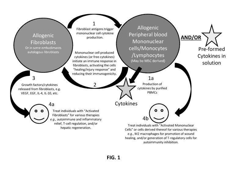
[0088] Embodiments of this scheme are illustrated in FIG. 1. For the sake of
brevity,
hereafter in the example of FIG. 1 the term "monocytes" will serve merely as a
representative
of peripheral blood mononuclear cells such as immune lymphocytes or other
cells including
MSCs, for example; dendritic cells may be utilized. In element 1 of FIG. 1,
fibroblasts (from
any source) and monocytes are exposed to and/or are in contact with one
another, for example
in a liquid media capable of maintaining cell viability and allowing for
interaction of molecules
and extracellular vesicles. As a result, fibroblast antigens trigger cytokine
production from the
monocytes. In some cases, cytokines and/or growth factors in solution are
exposed to the
fibroblasts in lieu of or in addition to exposing the fibroblasts to the
monocytes. The
19
CA 03083354 2020-05-22
WO 2019/108756 PCT/US2018/063001
monocytes or free cytokines activate an immune response in the fibroblast
cells, thereby
activating the fibroblast cells for a "healing/injury response" and reducing
the immunogenicity
of the fibroblast cells (element 2), in at least some cases. This reduction in
immunogenicity
facilitates the use of the fibroblasts in individuals from which the
fibroblasts were not
originally sourced, in at least some cases.
[0089] The resultant activated fibroblasts may now express one or more certain
activation markers and/or produce one or more growth factors and/or one or
more cytokines
(e.g. VEGF, EGF, IL-4, IL-10, etc. )(element 3) such that these factors are
useful for numerous
therapeutic interventions (e.g. angiogenesis, reduction in inflammatory
response, etc.), in
particular embodiments (element 4a). In element 4a, individuals suffering from
an array of
maladies can be given activated fibroblast cell therapy for the alleviation of
one or more of
their symptoms, e.g., endogenous autoimmune activity, chronic inflammation, T-
cell
hyperactivity, or hepatic regeneration, for example.
[0090] Additionally or alternatively, the co-cultured mononuclear cells (or
cells
produced therefrom) following exposure to the fibroblasts can be used for the
treatment of one
or more other maladies, e.g. using M1-to-M2 activated macrophages for the
promotion of
wound healing and/or the generation of T-regulatory cells to inhibit
autoimmunity, for example
(element 4b).
[0091] In one embodiment of the disclosure, universal donor fibroblasts are
administered to an individual to stimulate new blood vessel formation, a
process termed
angiogenesis. In some embodiments stimulation of angiogenesis is accomplished
by one or
more growth factors released by fibroblasts, and/or interaction between
fibroblasts and cells of
the recipient. In particular embodiments, such fibroblasts have been exposed
at least to IFN-
gamma.
[0092] Specific embodiments of the disclosure provide methods for angiogenesis
therapy. Embodiments of the disclosure provide methods of angiogenesis therapy
performed
with fibroblasts. In certain embodiments of the disclosure, a therapeutically
effective amount of
universal donor fibroblasts are administered to a subject for the purpose of
stimulating
angiogenesis. In some cases, the fibroblasts are provided to the individual
without the intended
purpose to stimulate angiogenesis but the fibroblasts still stimulate
angiogenesis. Embodiments
CA 03083354 2020-05-22
WO 2019/108756 PCT/US2018/063001
of the disclosure provide methods for co-administration of universal donor
fibroblasts with one
or more agents, including one or more agents that stimulate angiogenesis.
[0093] In a specific embodiment of the disclosure, methods are provided for co-
administration of universal donor fibroblasts with a therapeutically effective
amount of one or
more growth factors, such as VEGF, including purified VEGF. Other growth
factors include
the following: HGF, FGF-1, FGF-2, FGF-5, EGF, BDNF, PDGF, and/or angiopoietin.
In one
embodiment of the disclosure, universal donor fibroblasts are utilized to
stimulate growth
factor(s) (such as VEGF) production from cells of the individual. In a
specific embodiment of
the disclosure, methods are provided for co-administration of universal donor
fibroblasts with a
therapeutically effective amount of FGF-1, such as purified FGF-1 as an
example.
[0094] In particular embodiments of the disclosure, one or more angiogenic
agent(s)
are expressed in the universal donor fibroblasts of the disclosure via a
recombinant expression
vector operable in eukaryotic cells, and the expression of the angiogenic
agent(s) may be
regulated by a constitutive promoter or an inducible promoter or a tissue-
specific promoter. In
specific embodiments, the vector is a viral vector, such as a retrovirus,
lentivirus, adenovirus,
adeno-associated virus, or herpes simplex virus, or the vector is a non-viral
vector, such as
naked DNA or plasmid DNA or minicircle DNA. In specific cases, the
recombinantly
expressed angiogenic agent(s) may comprise VEGF, FGF-1, FGF-2, FGF-5, EGF,
angiopoietin, HIF-1-alpha, PDGF, HGF, or combinations thereof
[0095] Although in specific embodiments there is cross-talk between two types
of cells
and/or one or more agents thereof, in some cases a first type of cells is
modified by a second
type of cells when the second type of cells is not concomitantly modified by a
first type of
cells.
[0096] The following examples illustrate certain applications for use of
fibroblasts
and/or immune cells following their concomitant exposure to each other (and/or
exposure to
certain agents) and modifications of the respective cell type(s) thereafter.
21
CA 03083354 2020-05-22
WO 2019/108756 PCT/US2018/063001
A. Reducing cellular immunogenicity of ex vivo cultured cells
[0097] In some embodiments of the disclosure, there are methods of reducing
the
immunogenicity of a cell population, wherein the population is subjected to a
composition
comprising IFN-gamma and optionally one or more additional agent(s) and/or
condition(s).
[0098] In general embodiments, a population of cells is subjected to one or
more
compositions comprised of one or more particular media and/or one or more
agents such that
the composition(s) are capable of reducing the immunogenicity of the
population of cells. In
particular embodiments of the disclosure, methods are directed to a population
of cells wherein
the cells are fibroblasts of any type and the fibroblasts become modified such
that they have
reduced immunogenicity and may be utilized in a therapeutic capacity. In
certain embodiments,
the fibroblasts may be of any kind, including placental fibroblasts or
foreskin fibroblasts, for
example. In other embodiments, cells other than fibroblasts are modified such
that they have
reduced immunogenicity compared to in the absence of the method, such that the
cells may be
pancreatic beta cells, pancreatic islets, hepatocytes, neurons, chondrocytes,
pluripotent stem
cells, or derivatives thereof; such cells may or may not be in a mixture with
one or more types
of fibroblasts. In embodiments wherein the cells are pluripotent stem cells,
the stem cells may
comprise inducible pluripotent stem cells, stress induced stem cells,
parthenogenic derived
stem cells, embryonic stem cells, somatic cell nuclear transfer derived stem
cells, or derivatives
thereof, for example. In certain cases, methods of the disclosure are directed
to autologous
cells. In other cases, methods of the disclosure are directed to allogeneic
cells, xenogeneic
cells, or syngeneic cells.
[0099] Embodiments of the disclosure provide means of utilizing fibroblasts
(or other
types of cells, as noted above) as allogeneic therapeutic cells through
modification of culture
conditions in order to decrease immunogenicity of the fibroblasts. In one
embodiment of the
disclosure, fibroblasts are extracted from sources with lower immunogenicity
(e.g. placental
fibroblasts, etc.). In another embodiment, fibroblasts are cultured ex vivo
and subjected to
interferon gamma (IFN-gamma), which without being restricted to mechanism, has
been
demonstrated by the inventors to reduce immunogenicity. The reduction in
immunogenicity
may be exemplified by inhibiting the ability of the fibroblasts to evoke
alloreactive T cell
responses.
22
CA 03083354 2020-05-22
WO 2019/108756 PCT/US2018/063001
[0100] In specific embodiments, the disclosure provides methods for assessment
of
immunogenicity to be performed, e.g., quantifying the ability to modulate
mixed lymphocyte
reaction. Mixed lymphocyte reactions are well known in the art. Typically,
mixed lymphocyte
reaction is performed by co-culturing fibroblasts (in this case, that have
been treated with
interferon gamma) together with allogeneic lymphocytes. In certain
embodiments, parameters
of the mixed lymphocyte reaction that indicate modulation in immunogenicity
comprise T cell
proliferation, cytokine secretion, and cytotoxicity. Methods for quantifying T
cell proliferation,
cytokine secretion, and cytotoxicity are well known in the art. In certain
embodiments,
modulation of immunogenicity can be determined by quantifying the secretion of
one or more
cytokines comprising TNF-alpha, Interferon gamma, interleukin (IL)-1, IL-2, IL-
6, IL-7, IL-8,
IL-12, IL-15, IL-17, IL-33, or a combination thereof.
[0101] In specific embodiments, the disclosure provides methods that pertain
to the
administration of cells with reduced immunogenicity to an individual in need
thereof The
population of cells with reduced immunogenicity may be administered as
desired. Depending
upon the response desired, the manner of administration, the life of the
cells, and/or the number
of cells present, various protocols may be employed.
B. Cellular transplantation therapy for immunomodulation
[0102] Autoimmune diseases are characterized by an excessive reaction of the
immune
system against endogenous tissue. The immune system erroneously recognizes
endogenous
tissue as foreign bodies to be combated. This results in severe inflammatory
reactions, which
lead to damage to organs affected by them. An important part in distinguishing
between
endogenous and exogenous structures is played by T lymphocytes or T cells,
which are
"trained" in the thymus to dock only onto endogenous cell surface molecules,
the so-called
MEW molecules, and thus to tolerate endogenous structures.
[0103] In autoimmune diseases, a group of T cells behaves abnormally. In
addition to
the still functioning defense from exogenous molecules and organisms, they now
also attack
endogenous structure. Organs or tissues are perceived as exogenous. There can
be various
consequences: if vital structures are affected, an autoimmune disease will
take a fatal course.
The immune system directs its defense against these structures, cellular and
also humoral
defense reactions are set in motion, and autoantibodies are formed, as a
result of which the
23
CA 03083354 2020-05-22
WO 2019/108756 PCT/US2018/063001
organs affected in the course of time cease to function. Most commonly, the
immune system is
weakened and the body becomes susceptible to all kinds of diseases. Under some
circumstances, recognition of the exogenous is also disrupted, and as a result
the spreading of
degenerated cancer cells (for example) can no longer be effectively prevented,
and those
affected are more susceptible to infectious diseases. In the course of the
disease, cells of the
immune system destroy the endogenous structures, while the body's repair
mechanisms attempt
as far as possible to regenerate the damaged organ parts. As a rule, without
treatment this
erroneous attack of the defensive system continues throughout life or until
the complete
destruction of the target structure.
[0104] Autoimmune diseases are treated according to the organ affected. In
this, the
basic principle of the causal therapy is to suppress the activity of the
immune system by
administration of immunosuppressants, e.g., cortisone. These substances are
characterized by
multiple systemic side-effects and interactions, owing to which attempts have
been made to
develop new drugs which specifically influence the mechanisms involved in the
disease event.
[0105] In one embodiment of the disclosure, modified fibroblasts are
administered to
an individual for treatment of an autoimmunity or inflammatory disorder. In
some
embodiments of the disclosure, fibroblast cells are cultured ex vivo and
subjected to conditions
that reduce their immunogenicity, and then the fibroblasts are utilized to
stimulate anti-
inflammatory and/or immunomodulatory properties. Additional embodiments are
directed to
methods of administration of the cells to an individual in need thereof for
the purpose of
treating an autoimmune and/or inflammatory condition.
[0106] The present disclosure is directed to systems, methods, and
compositions for
reducing the immunogenicity of cells to be used in cellular transplantation
therapy. In general
embodiments, a population of cells is subjected to one or more compositions
comprising one or
more types of media and/or one or more agents capable of reducing the
immunogenicity of the
population of cells. In particular embodiments of the disclosure, methods are
directed to a
population of cells wherein the cells comprise at least fibroblasts.
[0107] Embodiments of the disclosure provide means of utilizing fibroblasts as
allogeneic therapeutic cells through modification of culture conditions in
order to decrease
immunogenicity of the fibroblasts. In one embodiment of the disclosure,
fibroblasts are
24
CA 03083354 2020-05-22
WO 2019/108756 PCT/US2018/063001
extracted from sources with lower immunogenicity (e.g. placental fibroblasts,
etc.). In another
embodiment, fibroblasts are subjected to interferon gamma (IFN-gamma), such as
upon culture
ex vivo, which without being restricted to mechanism, has been demonstrated by
the inventors
to reduce immunogenicity. The reduction in immunogenicity is exemplified by
inhibiting the
ability of the fibroblasts to evoke alloreactive T cell responses, in specific
embodiments. In
specific embodiments of the disclosure, these modified fibroblast cells are
universal donor
fibroblasts.
[0108] Certain methods of the disclosure are directed to reducing the
immunogenicity
of fibroblasts of any kind, including foreskin fibroblasts as an example. In
certain
embodiments, fibroblasts are re-suspended in ex vivo culture in medium, such
as medium
supplemented with serum or serum-replacement. Serum can be of any source
including fetal
bovine serum, human serum or serum replacement. In certain embodiments human
serum or
serum replacement is utilized in order to provide a xenogeneic-free
environment for the
fibroblasts (for example, foreskin feeder cells). In one embodiment, the
fibroblasts are treated
with one or more certain concentrations of IFN-y.
[0109] Embodiments of the present disclosure are directed to systems and
methods for
the use of fibroblast cells, either autologous or allogeneic, for treatment of
inflammatory and
autoimmune conditions. Methods and compositions of the disclosure encompass
certain
manipulated cells for the treatment of inflammatory and autoimmune conditions.
In particular,
the cells include at least fibroblasts of any kind. Means of manipulation of
fibroblasts are
disclosed, as well as fibroblasts of different tissue origins, which actively
inhibit inflammatory
and/or autoimmune processes. In one embodiment of the disclosure, fibroblasts
are utilized for
their ability to inhibit immune responses and also utilized as a cellular
therapy for prevention
and/or treatment of autoimmune conditions. In one embodiment, fibroblasts are
generated or
manipulated and utilized in mixed lymphocyte reactions to assess their ability
to suppress
immune activation. In a specific embodiment, fibroblasts are treated with one
or more
particular agents and/or conditions to be able to directly or indirectly treat
inflammatory and/or
autoimmune processes. In particular embodiments, the agent comprises
interferon gamma
and/or platelet rich plasma, and in some cases at least interferon gamma
and/or platelet rich
plasma (and/or platelet rich lysate) can endow the ability of the fibroblasts
to directly or
indirectly actively suppress immune responses. Fibroblasts cultured under
these conditions are
CA 03083354 2020-05-22
WO 2019/108756 PCT/US2018/063001
administered into individuals suffering from autoimmune or inflammatory
disorders or at risk
thereof. The route of administration, dosage and frequency is determined as a
function of the
disease process, as well as stage of the disease, and can be optimized per
routine practices in
medicine.
[0110] In one embodiment, allogeneic (or xenogeneic or syngeneic) fibroblasts
are
administered to an individual in a non-manipulated manner (for example,
without prior
exposure to one or more particular agents, such as interferon gamma) but
selected from sources
naturally characterized by immune modulatory activity, such as placental
fibroblasts or adipose
tissue-associated fibroblasts, for example. In other embodiments of the
disclosure, any
fibroblasts are cultured under conditions capable of inducing retro-
differentiation so as to
endow an immature phenotype for the fibroblasts, wherein the immature
phenotype correlates
with enhanced anti-inflammatory and/or immune modulatory potential. For
example,
fibroblasts may be cultured in the presence of one or more histone deacetylase
inhibitors, such
as valproic acid (Moon et al., 2008; Huang et al., 2011). In addition to HDAC
inhibitors, other
means of inducing dedifferentiation of the fibroblasts may also be utilized in
the context of the
current disclosure, such as 8-Br-cAMP (Wang et al., 2011); M-CSF treatment (Li
et al., 2016);
exposure to reveresine (Li et at., 2016); and/or exposure to stem cell
extracts (Xiong et at.,
2014). Characterization of fibroblast dedifferentiation can be performed by
assessment of
extracellular markers, such as CXCR4, VEGFR-2, CD34, and/or CD133, as well as
intracellular markers such as SOX-2, NANOG, and/or OCT-4.
[0111] In some embodiments of the disclosure, fibroblast cells that have been
dedifferentiated may be utilized for immunomodulation express one or more
markers selected
from the group consisting of Telomerase, Nanog, Sox2, beta-III-Tubulin, NF-M,
MAP2, APP,
GLUT, NCAM, NeuroD, Nurrl, GFAP, NG2, Oligl, Alkaline Phosphatase, Vimentin,
Osteonectin, Osteoprotegrin, Osterix, Adipsin, Erythropoietin, SM22-alpha,
HGF, c-MET,
alpha-l-Antriptrypsin, Ceruloplasmin, AFP, PEPCK 1, BDNF, NT-4/5, TrkA, BMP2,
BMP4,
FGF2, FGF4, PDGF, PGF, TGFalpha, TGFbeta, VEGF and a combination thereof.
[0112] In one embodiment of the disclosure, fibroblasts are administered
together with
one or more agents that possess immune-modulatory properties. Such agents
include the
following examples: 1) hydroxychloroquine, which acts in part as a toll like
receptor (TLR) 7/9
26
CA 03083354 2020-05-22
WO 2019/108756 PCT/US2018/063001
antagonist, thus decreasing innate immune activation (Sun et at., 2007); 2)
leflunomide, an
antimetabolite that inhibits pyrimidine synthesis and protein tyrosine kinase
activity (Chong et
at., 1999), which results in suppression of T cell responses (Dimitrova et
al., 2002), and has
been also demonstrated to inhibit dendritic cell (DC) activation (Kirsch et
at., 2005); 3)
injectable gold compounds (such as auranofin) which directly or through
metabolites such as
dicyanogold (i) have been demonstrated to inhibit T cell and antigen
presenting cell activation
(Tepperman et al., 1999; Han et al., 2008), as well as cause Th2 deviation
(Kim et al., 2001);
4) sulfasalazine, which has been used since 1950, acts primarily through
inhibition of
cycloxygenase and lipoxygenase (Taggart et at., 1987); and 5) methotrexate, an
antifolate that
inhibits T cell activation and proliferation and that has been one of the
golden standards for RA
(Bansard et at., 2009). Typically, combinations of disease-modifying anti-
rheumatic drugs
(DMARDs) with glucocorticoids are used, or alternatively one or more pulses of
high dose
glucocorticoids are administered to cause a general inhibition of inflammation
(Bijlsma et at.,
2006). Concentrations of fibroblasts used are dependent on stage of the
disease, as well as
patient history and responsiveness to prior therapy.
[0113] In one embodiment of the disclosure, fibroblasts to be used for
immunomodulation are genetically engineered, for example to express: a) one or
more
autoantigens; and/or b) one or more immune modulatory proteins. The engineered
cells are
subsequently used for induction of immunological tolerance. The
characteristics of the
individual and disease may dictate which genes are to be used for engineering
of fibroblasts, in
at least some cases. For example, in situations of type 1 diabetes, numerous
autoantigens are
known in the field, for example IGRP (Fuchs et at., 2017), IA-2 (Guerra et
at., 2016),
Proinsulin-2 (Babon et al., 2016), and GAD65 (Phelps et al., 2016). In these
cases, the
autoantigen may be transfected into the fibroblasts in polynucleotide form and
the fibroblasts
are either cultured to allow for immune modulation or transfected with genes
allowing for
immune modulation. Genes of particular interest for transfection to induce
immune modulation
include at least the following: Fas ligand, TGF-beta, IL-4, IL-10, HLA-G,
indolamine 2,3
deoxygenase, galectin family members, Galectin 3, arginase, and/or IL-20 (de
Jesus et at.,
2016; Wang et al., 2011; Zhao et al., 2010; Min et al., 2001; Cancedda et al.,
2001). Any of
the genes described herein or active portions thereof may be cloned into
mammalian
expression constructs comprising promoter sequences enabling expression in
fibroblast cells
27
CA 03083354 2020-05-22
WO 2019/108756 PCT/US2018/063001
such as the CMV promoter [Artuc et at., Exp. Dermatol. 1995, 4:317-21].
Examples of suitable
constructs include, but are not limited to pcDNA3, pcDNA3.1 (+/-), pGL3,
PzeoSV2 (+/-),
pDisplay, pEF/myc/cyto, pCMV/myc/cyto (each of which is commercially available
from
Invitrogen, for example), or the pSH expression vector that enables a
regulated polynucleotide
expression in human foreskin cells [Ventura and Villa, 1993, Biochem. Biophys.
Commun.
192: 867-9]. Examples of retroviral vector and packaging systems are those
commercially
available from Clontech, San Diego, Calif, USA, including Retro-X vectors
pLNCX and
pLXSN, which permit cloning into multiple cloning sites and the transgene is
transcribed from
CMV promoter. Vectors derived from Mo-MuLV are also included such as pBabe,
where the
transgene will be transcribed from the 5'LTR promoter. After completing
plasmid transfection
fibroblasts are harvested by a means allowing for detachment from tissue
culture plates, for
example, by trypsinization and transferred to either a 6-well (Nunc, Denmark)
or a 24-well
plate (Nunc) for proliferation. Approximately 3 days post-transfection, the
cell media is
changed to media suitable for proliferation and expansion of modified
fibroblasts. One
example is Neurobasal A (NBA) proliferation medium comprising Neurobasal-A
(Invitrogen),
1% D-glucose (Sigma Aldrich), 1% Penicillin/Streptomycin/Glutamine
(Invitrogen), 2% B27
supplement with Retinoic acid (Invitrogen), 0.2% EGF (Peprotech, USA), 0.08%
FGF-2
(Peprotech), 0.2% Heparin (Sigma Aldrich, USA) and Valproic acid (Sigma
Aldrich) to a
concentration of 1 M. The media is then subsequently changed, such as thrice
weekly, and
cells are re-plated regularly (for example, 2-8 times up to a maximum of
weekly re-plating,
becoming more regular as colonies began to develop) to remove non-reprogrammed
cells until
widespread colony formation is achieved. Various quality control means are
known in the art
for practitioners of the disclosure to perform clinical administration of the
cells. Example
criteria for qualification of the cells includes marker identification using
means such as flow
cytometry, viability, endotoxin content, as well as assessment for microbial
and mycoplasma
contamination.
[0114] In one embodiment of the disclosure, fibroblasts are cultured ex vivo
using
means known in the art for preserving viability and proliferative ability of
fibroblasts. The
disclosure provides for the modification of known culture techniques to
decrease recognition of
fibroblasts by the recipient immune system. In one embodiment fibroblasts are
cultured in
conditions that lack xenogeneic components, such as fetal calf serum.
Xenogeneic components
28
CA 03083354 2020-05-22
WO 2019/108756 PCT/US2018/063001
are known to trigger immunological reactions, including elicitation of
antibody and T cell
reactions (Selvaggi et at., 1997; Mackensen et at., 2000; Kadri et at., 2007;
Forni et at., 1976;
Lauer et at., 1983). In many individuals, natural antibodies of the IgM
isotype exist to fetal calf
serum associated components (Irie et at., 1974), causing rejection,
inflammation or anaphylaxis
subsequent to administration of cells grown in the presence of fetal calf
serum (Macy et at.,
1989). In specific embodiments, the disclosure encompasses the substitution of
fetal calf serum
with human platelet rich plasma, platelet lysate, umbilical cord blood serum,
autologous serum,
and/or defined cytokine mixes as an additional feature to reduce the
immunogenicity of
fibroblasts. Means of culturing tissues in xenogeneic-free medium are known in
the art for
other cell types and are incorporated by reference (Riordan et at., 2015).
[0115] Embodiments of the disclosure provide methods of reducing
immunogenicity of
particular types of fibroblasts. Fibroblasts may be derived from various
tissues or organs, such
as skin, heart, blood vessels, bone marrow, skeletal muscle, liver, pancreas,
brain, and/or
foreskin, which can be obtained by biopsy (where appropriate) or upon autopsy.
In some
aspects, the cells comprise fibroblasts, which can be from a fetal, neonatal,
adult origin, or a
combination thereof.
[0116] Foreskin fibroblasts may be re-suspended in ex vivo culture in medium
supplemented with one or more agents to decrease immunogenicity, serum and/or
serum-
replacement. Serum can be of any source including fetal bovine serum, human
serum or serum
replacement (for example, replacement with human platelet lysate (Riordan et
at., 2007;
Schallmoser et al., 2007; Capelli et al., 2007; Carrancio et al., 2008;
Salvade et al., 2010;
Bieback et at., 2009))). In some embodiments, human serum or serum replacement
is utilized
in order to provide a xenogeneic-free environment for the foreskin feeder
cells. Subsequent to
culture the human foreskin feeder cells of the present disclosure are capable
of forming
monolayers when attached to a solid phase such as a tissue culture plate
[Dugdale and Siddall
(1969) J. Med. Lab. Technol. 26: 31-5]. This characteristic of the human
foreskin feeder cells
of the present disclosure makes these cells suitable universal donors because
of high replicative
ability and responsiveness to treatment with IFN-gamma with respect to
reduction of
immunogenicity. One of skill in the art will appreciate that fibroblasts from
different sources
may possess different reduction of immunogenicity at differing concentrations
of interferon
gamma. In specific embodiments the disclosure provides methods for assessment
of
29
CA 03083354 2020-05-22
WO 2019/108756 PCT/US2018/063001
immunogenicity to be performed, e.g. quantifying the ability to modulate mixed
lymphocyte
reaction.
[0117] Typically, to measure the immunogenicity of the fibroblasts, mixed
lymphocyte
reaction is performed by co-culturing fibroblasts that have been treated with
interferon gamma
together with allogeneic lymphocytes. Proliferative indexes of the allogeneic
lymphocytes are
usually taken to represent the degree of immunogenicity of stimulator
fibroblast cells. In some
embodiments of the disclosure, fibroblasts are mitotically inactivated before
culture with
lymphocytes. Mitotic inactivation may be performed by treatment with one or
more agents,
such as mitomycin C or other agents, that block proliferation without reducing
viability. When
chemical agents are utilized for mitotic inactivation, the chemical agents are
washed off the
cells in order to prevent inhibition of responding lymphocytes.
[0118] In other embodiments of the disclosure, the treated fibroblast cells
are
characterized for their level of immunogenicity, such as assessment of
cytokine production by
responding lymphocytes. Examples of cytokines of note include IL-1, which is
involved in
stimulation of inflammatory processes and macrophages; IL-2, which is
associated with T cell
and NK cell activation; interferon gamma, which activates macrophages, as well
as assists in
Thl polarization; and/or IL-12 and/or IL-18, which activate NK cells and are
associated with
development of cellular cytotoxic responses. It is desirable for the practice
of the disclosure
that responding lymphocytes in a mixed lymphocyte reaction produce less
inflammatory
cytokines. Thus, in particular embodiments fibroblasts are selected for use
when they elicit in
responding lymphocytes the production of IL-2, IFN-gamma, IL-12, and/or IL-18.
Conversely, assessment of immune suppressive or anti-inflammatory cytokines
may also be
performed within the context of the disclosure. The cytokines include
interleukin 4, which
stimulates Th2 cells; interleukin 10, which stimulates, infra al/a, T
regulatory cells; and TGF-
B that inhibits inflammation.
[0119] In some embodiments of the disclosure, genetic modification of
fibroblasts is
performed to cause reduction of immunogenicity of the fibroblasts. One method
provides for
genetic modification that includes cytoplasmic transfer with cells possessing
reduced
immunogenicity, such as immature dendritic cells. In another embodiment, gene
editing is
utilized to selectively excise inflammation-evoking genes, such as HLA or
costimulatory
CA 03083354 2020-05-22
WO 2019/108756 PCT/US2018/063001
molecules such as CD40, CD80, CD86, TNF-alpha, HMGB-1, IFN-gamma, IL-1 beta,
IL-17,
FAP, IL-18, IL-33, or a combination thereof.
[0120] In particular embodiments of the disclosure, one or more
immunomodulatory
agent(s) are expressed in universal donor fibroblasts via a recombinant
expression vector
operable in eukaryotic cells, and the expression of the immunomodulatory
agent(s) may be
regulated by a constitutive promoter or an inducible promoter or a tissue-
specific promoter. In
specific embodiments, the vector is a viral vector, such as a retrovirus,
lentivirus, adenovirus,
adeno-associated virus, or herpes simplex virus, or the vector is a non-viral
vector, such as
naked DNA or plasmid DNA or minicircle DNA. Polynucleotides of particular
interest for
transfection into the fibroblasts include at least the following: Fas ligand,
TGF-beta, IL-4, IL-
10, HLA-G, indolamine 2,3 deoxygenase (IDO), galectin family members, Galectin
3,
arginase, IL-20, HGF, PDGF-BB, EGF, IGF, GDF-5, GDF-11, Angiopoietin, FGF-1,
FGF-2,
FGF-5, FGF-15, or a combination thereof In specific cases, recombinantly
expressed
angiogenic agent(s) may comprise FAS ligand, IL-2, IL-4, IL-10, IL-20, IL-35,
HLA-G, 1-309,
DO, iNOS, CD200, Galectin 3, arginase, PGE-2, TGF-beta, CTLA-4, PD-L1, IFN-
gamma, or
a combination thereof.
[0121] Various quality control means are known in the art for practitioners of
the
disclosure to perform clinical administration of the cells. Example criteria
for qualification of
the cells includes marker identification using means such as flow cytometry,
viability, and/or
endotoxin content, as well as assessment for microbial and mycoplasma
contamination.
[0122] In one embodiment of the disclosure, universal donor fibroblasts are
administered to an individual for treatment of an autoimmunity or inflammatory
disorder. In
some embodiments of the disclosure, cells are cultured ex vivo and subjected
to conditions that
reduce immunogenicity and stimulate anti-inflammatory and/or immunomodulatory
properties.
Additional embodiments are directed to methods of administration of the cells
to an individual
in need thereof for the purpose of treating an autoimmune and/or inflammatory
condition.
[0123] Particular embodiments of the disclosure provide a method for treating
one or
more autoimmune or inflammatory condition(s) such as Acute Disseminated
Encephalomyelitis, Acute necrotizing hemorrhagic leukoencephalitis, Addison's
disease,
adhesive capsulitis, Agammaglobulinemia, Alopecia areata, Amyloidosis,
Ankylosing
31
CA 03083354 2020-05-22
WO 2019/108756 PCT/US2018/063001
spondylitis, Anti-GBM nephritis, Antiphospholipid syndrome (APS), Anti-TBM
nephritis,
arthofibrosis, atrial fibrosis, autoimmune angioedema, autoimmune aplastic
anemia,
autoimmune dysautonomia, autoimmune hepatitis, autoimmune hyperlipidemia,
autoimmune
immunodeficiency, autoimmune inner ear disease (AIED), autoimmune myocarditis,
autoimmune neutropenia, autoimmune oophoritis, autoimmune pancreatitis,
autoimmune
retinopathy, autoimmune thrombocytopenic purpura (ATP), autoimmune thyroid
disease,
autoimmune urticarial, axonal and neuronal neuropathies, Balo disease,
Behcet's disease,
benign mucosal pemphigold, bullous pemphigoid, cardiomyopathy, Castleman
disease, Celiac
disease, Chagas disease, chronic fatigue syndrome, Chronic inflammatory
demyelinating
polyneuropathy (CIDP), chronic Lyme disease, Chronic recurrent multifocal
ostomyelitis
(CRMO), Churg-Strauss syndrome, cicatricial pemphigold, cirrhosis, Cogans
syndrome, cold
agglutinin disease, congenital heart block, Coxsackie myocarditis, CREST
disease, Crohn's
disease, Cystic Fibrosis, deficiency of the interleukin-1 receptor antagonist,
demyelinating
neuropathies, dermatitis herpetiformis, dermatomyosis, Devic's disease
(neuromyelitis optica),
discoid lupus, Dressler's syndrome, Dupuytren's contracture, endometriosis,
endomyocardial
fibrosis, eosinophilic esophagitis, eosinophilic facsciitis, erythema nodosum,
essential mixed
cryoglobulinemia, Evans syndrome, experimental allergic encephalomyelitis,
Familial
Mediterranean Fever, Fibromyalgia Fibrosing alveolitis, Giant cell arteritis
(temporal arteritis),
giant cell myocarditis, glomerulonephritis, Glomerulonephritis, Goodpasture's
syndrome,
Graft-versus-host disease (GVHD), granulomatosus with polyangitis, Graves'
disease,
Guillain-Barre syndrome, Hashimoto's encephalitis, Hashimoto's thyroiditis,
hemolytic
anemia, Henoch-Schonlein purpura, hepatitis, herpes gestationis,
hypogammaglobulinemia,
idiopathic pulmonary fibrosis, Idiopathic thrombocytopenic purpura (ITP), IgA
nephropathy,
IgG4-related sclerosing disease, Immunoregulatory lipoproteins, inclusion body
myositis,
inflammatory bowel disorders, interstitial cystitis, juvenile arthritis,
Juvenile diabetes (Type 1
diabetes), juvenile myositis, Kawasaki syndrome, keloid, Lambert-Eaton
syndrome,
leukocytoclastic vasculitis, lichen planus, lichen sclerosis, ligneous
conjunctivitis, linear IgA
disease, Lupus (SLE), Lyme disease, mediastinal fibrosis, Meniere's disease,
microscopic
polyangitis, Mixed connective tissue disease (MCTD), Mooren's ulcer, Mucha-
Habermann
disease, Multiple Sclerosis (MS), Myasthenia gravis, myelofibrosis, Myositis,
narcolepsy,
Neonatal Onset Multisystem Inflammatory Disease, nephrogenic systemic
fibrosis,
Neuromyelitis optica (Devic's), neutropenia, nonalcoholic fatty liver disease,
nonalcoholic
32
CA 03083354 2020-05-22
WO 2019/108756 PCT/US2018/063001
steatohepatitis (NASH), ocular-cicatricial pemphigold, optic neuritis,
palindromic rheumatism,
paraneoplastic cerebellar degeneration, Paroxysmal nocturnal hemoglobinuria
(PNH), Parry
Romberg syndrome, Pars planitis (peripheral uveitis), Parsonnage-Turner
syndrome, Pediatric
Autoimmune Neuropsychiatric Disorders Associated with Streptococcus (PANDAS),
Pemphigus, Peripheral neuropathy, Perivenous encephalomyelitis, Pernicious
anemia,
Peyronie's disease, POEMS syndrome, polyarteritis nodosa, polymyalgia
rhematica,
polymyositis, postmyocardial infarction syndrome, postpericardiotomy syndrome,
primary
biliary cirrhosis, primary sclerosing cholangitis, progesterone dermatitis,
progressive massive
fibrosis, psoriasis, psoriatic arthritis, pure red cell aplasia, pyoderma
gangrenosum, Raynauds
phenomenon, reactic arthritis, reflex sympathetic dystrophy, Reiter's
syndrome, relapsing
polychondritis, restless legs syndrome, retroperitoneal fibrosis, rheumatic
fever, rheumatoid
arthritis, sarcoidosis, Schmidt syndrome, scleritis, scleroderma, Sjogren's
syndrome, sperm and
testicular autoimmunity, stiff person syndrome, subacute bacterial
endocarditis, Susac's
syndrome, sympathetic ophthalmia, systemic lupus erythematosus (SLE),
Takayasu's arteritis,
temporal arteritis, Thrombocytopenic purpura (TTP), Tolosa-Hunt syndrome,
transverse
myelitis, Tumor Necrosis Factor Receptor-associated Periodic Syndrome, Type 1
diabetes,
Type I autoimmune polyglandular syndrome, Type II autoimmune polyglandular
syndrome,
Type III autoimmune polyglandular syndrome, ulcerative colitis,
undifferentiated connective
tissue disease, uveitis, vasculitis, vesiculobullous dermatosis, Vitiligo, and
Wegener's
granulomatosis (now termed Granulomatosis with Polyangitis (GPA).
[0124] Certain methods of the disclosure are directed to reducing the
immunogenicity
of fibroblasts, including at least foreskin fibroblasts. In certain
embodiments, fibroblasts are re-
suspended in ex vivo culture in medium supplemented with serum or serum-
replacement.
Serum can be of any source including fetal bovine serum, human serum and/or
serum
replacement. In certain embodiments human serum or serum replacement is
utilized in order to
provide a xenogeneic-free environment for the foreskin feeder cells. In one
embodiment the
foreskin fibroblasts are treated with concentrations of IFN-gamma ranging from
0.1-500 Units
per milliliter (IU/ml), including ranges of concentrations described elsewhere
herein.
[0125] In certain embodiments a therapeutically effective amount of modified
cells are
co-administered with one or more immunomodulatory agents(s) to an individual.
Exemplary
immunomodulatory agents may comprise FAS ligand, IL-2R, IL-1 Ra, IL-2, IL-4,
IL-8, IL-10,
33
CA 03083354 2020-05-22
WO 2019/108756 PCT/US2018/063001
IL-20, IL-35, HLA-G, PD-L1, 1-309, IDO, iNOS, CD200, Galectin 3, sCR1,
arginase, PGE-2,
aspirin, atorvastatin, fluvastatin, lovastatin, pravastatin, rosuvastatin,
simvastatin, pitavastatin,
n-acetylcysteine, rapamycin, IVIG, naltrexone, TGF-beta, VEGF, PDGF, CTLA-4,
anti-
CD45RB antibody, hydroxychloroquine, leflunomide, auranofin, dicyanogold,
sulfasalazine,
methotrexate, glucocorticoids, etanercept, adalimumab, abatacept, anakinra,
certolizumab,
Etanercept-szzs, golimumab, infliximab, rituximab, tocilizumab, cyclosporine,
IFN-gamma,
everolimus, rapamycin, or combinations thereof
[0126] Particular embodiments of the disclosure provide a method for treating
or
preventing one or more autoimmune or inflammatory condition(s) comprising:
Acute
Disseminated Encephalomyelitis, Acute necrotizing hemorrhagic
leukoencephalitis, Addison's
disease, adhesive capsulitis, Agammaglobulinemia, Alopecia areata,
Amyloidosis, Ankylosing
spondylitis, Anti-GBM nephritis, Antiphospholipid syndrome (APS), Anti-TBM
nephritis,
arthofibrosis, atrial fibrosis, autoimmune angioedema, autoimmune aplastic
anemia,
autoimmune dysautonomia, autoimmune hepatitis, autoimmune hyperlipidemia,
autoimmune
immunodeficiency, autoimmune inner ear disease (AIED), autoimmune myocarditis,
autoimmune neutropenia, autoimmune oophoritis, autoimmune pancreatitis,
autoimmune
retinopathy, autoimmune thrombocytopenic purpura (ATP), autoimmune thyroid
disease,
autoimmune urticarial, axonal and neuronal neuropathies, Balo disease,
Behcet's disease,
benign mucosal pemphigold, bullous pemphigoid, cardiomyopathy, Castleman
disease, Celiac
disease, Chagas disease, chronic fatigue syndrome, Chronic inflammatory
demyelinating
polyneuropathy (CIDP), chronic Lyme disease, Chronic recurrent multifocal
ostomyelitis
(CRMO), Churg-Strauss syndrome, cicatricial pemphigold, cirrhosis, Cogans
syndrome, cold
agglutinin disease, congenital heart block, Coxsackie myocarditis, CREST
disease, Crohn's
disease, Cystic Fibrosis, deficiency of the interleukin-1 receptor antagonist,
demyelinating
neuropathies, dermatitis herpetiformis, dermatomyosis, Devic's disease
(neuromyelitis optica),
discoid lupus, Dressler's syndrome, Dupuytren's contracture, endometriosis,
endomyocardial
fibrosis, eosinophilic esophagitis, eosinophilic facsciitis, erythema nodosum,
essential mixed
cryoglobulinemia, Evans syndrome, experimental allergic encephalomyelitis,
Familial
Mediterranean Fever, Fibromyalgia Fibrosing alveolitis, Giant cell arteritis
(temporal arteritis),
giant cell myocarditis, glomerulonephritis, Glomerulonephritis, Goodpasture's
syndrome,
Graft-versus-host disease (GVHD), granulomatosus with polyangitis, Graves'
disease,
34
CA 03083354 2020-05-22
WO 2019/108756 PCT/US2018/063001
Guillain-Barre syndrome, Hashimoto's encephalitis, Hashimoto's thyroiditis,
hemolytic
anemia, Henoch-Schonlein purpura, hepatitis, herpes gestationis,
hypogammaglobulinemia,
idiopathic pulmonary fibrosis, Idiopathic thrombocytopenic purpura (ITP), IgA
nephropathy,
IgG4-related sclerosing disease, Immunoregulatory lipoproteins, inclusion body
myositis,
inflammatory bowel disorders, interstitial cystitis, juvenile arthritis,
Juvenile diabetes (Type 1
diabetes), juvenile myositis, Kawasaki syndrome, keloid, Lambert-Eaton
syndrome,
leukocytoclastic vasculitis, lichen planus, lichen sclerosis, ligneous
conjunctivitis, linear IgA
disease, Lupus (SLE), Lyme disease, mediastinal fibrosis, Meniere's disease,
microscopic
polyangitis, Mixed connective tissue disease (MCTD), Mooren's ulcer, Mucha-
Habermann
disease, Multiple Sclerosis (MS), Myasthenia gravis, myelofibrosis, Myositis,
narcolepsy,
Neonatal Onset Multisystem Inflammatory Disease, nephrogenic systemic
fibrosis,
Neuromyelitis optica (Devic's), neutropenia, nonalcoholic fatty liver disease,
nonalcoholic
steatohepatitis (NASH), ocular-cicatricial pemphigold, optic neuritis,
palindromic rheumatism,
paraneoplastic cerebellar degeneration, Paroxysmal nocturnal hemoglobinuria
(PNH), Parry
Romberg syndrome, Pars planitis (peripheral uveitis), Parsonnage-Turner
syndrome, Pediatric
Autoimmune Neuropsychiatric Disorders Associated with Streptococcus (PANDAS),
Pemphigus, Peripheral neuropathy, Perivenous encephalomyelitis, Pernicious
anemia,
Peyronie's disease, POEMS syndrome, polyarteritis nodosa, polymyalgia
rhematica,
polymyositis, postmyocardial infarction syndrome, postpericardiotomy syndrome,
primary
biliary cirrhosis, primary sclerosing cholangitis, progesterone dermatitis,
progressive massive
fibrosis, psoriasis, psoriatic arthritis, pure red cell aplasia, pyoderma
gangrenosum, Raynauds
phenomenon, reactic arthritis, reflex sympathetic dystrophy, Reiter's
syndrome, relapsing
polychondritis, restless legs syndrome, retroperitoneal fibrosis, rheumatic
fever, rheumatoid
arthritis, sarcoidosis, Schmidt syndrome, scleritis, scleroderma, Sjogren's
syndrome, sperm and
testicular autoimmunity, stiff person syndrome, subacute bacterial
endocarditis, Susac's
syndrome, sympathetic ophthalmia, systemic lupus erythematosus (SLE),
Takayasu's arteritis,
temporal arteritis, Thrombocytopenic purpura (TTP), Tolosa-Hunt syndrome,
transverse
myelitis, Tumor Necrosis Factor Receptor-associated Periodic Syndrome, Type 1
diabetes,
Type I autoimmune polyglandular syndrome, Type II autoimmune polyglandular
syndrome,
Type III autoimmune polyglandular syndrome, ulcerative colitis,
undifferentiated connective
tissue disease, uveitis, vasculitis, vesiculobullous dermatosis, Vitiligo, and
Granulomatosis
with Polyangitis (GPA).
CA 03083354 2020-05-22
WO 2019/108756 PCT/US2018/063001
[0127] Growth of fibroblasts for immune modulation may be performed in a
variety of
tissue culture media, for example. In one embodiment of the disclosure, one
medium for the
culturing of the cells comprises Dulbecco's Modified Essential Media (DMEM),
including
DMEM-low glucose (DMEM-LG) (Invitrogen, Carlsbad, CA). The DMEM-LG may be
supplemented with serum, such as fetal bovine serum and/or human serum.
Typically, 15%
(v/v) fetal bovine serum (e.g. defined fetal bovine serum, Hyclone, Logan
Utah) is added,
along with antibiotics/antimycotics ((preferably 100 Unit/milliliter
penicillin, 100
milligrams/milliliter streptomycin, and 0.25 microgram/milliliter amphotericin
B; Invitrogen,
Carlsbad, Calif.)), and 0.001% (v/v) 2-mercaptoethanol (Sigma, St. Louis MO).
In some cases,
different growth media are used, or different supplementations are provided,
and these are
normally indicated in the text as supplementations to Growth Medium. In
certain chemically-
defined media, the cells may be grown without serum present at all. In such
cases, the cells
may require certain growth factors, which can be added to the medium to
support and sustain
the cells. Certain factors to be added for growth on serum-free media include
one or more of
bFGF, EGF, IGF-I, and PDGF, in some cases. In some embodiments, two, three or
all four of
the factors are added to serum-free or chemically defined media. In other
embodiments, LIF is
added to serum-free medium to support or improve growth of the cells.
C. Expansion of regulatory T cells in ex vivo culture
[0128] The present example provides a solution for a long-felt need in the art
for
expanding particular T cells.
[0129] T regulatory cells are an essential component of the immune system
protecting
the body against autoimmune attack. This is illustrated by early studies in
which neonatally
thymectomized mice suffered from systemic autoimmunity, which were rescued by
transfer of
CD4 cells (Hon i et at., 2003). Subsequent studies identified the T regulatory
(Treg) phenotype
as possessing the IL-2 receptor CD25, which is somewhat problematic given that
this receptor
is found on activated T cells as well. Peripheral blood contains a small
population of T cell
lymphocytes that express the T regulatory phenotype ("Treg"), i.e., positive
for both CD4 and
CD25 antigens. There are several subsets of Treg cells. One subset of
regulatory cells develops
in the thymus. Thymic-derived Treg cells function by a cytokine-independent
mechanism,
which involves cell to cell contact. They are essential for the induction and
maintenance of
36
CA 03083354 2020-05-22
WO 2019/108756 PCT/US2018/063001
self-tolerance and for the prevention of autoimmunity (Shevach et at., 2000).
These regulatory
cells prevent the activation and proliferation of autoreactive T cells that
have escaped thymic
deletion or recognize extrathymic antigens, thus they are critical for
homeostasis and immune
regulation, as well as for protecting the host against the development of
autoimmunity. Thus,
immune regulatory CD4+ CD25+ T cells are often referred to as "professional
suppressor cells."
Suppression of immunity by Treg cells occurs through several mechanisms. One
is direct lysis
of activated T cells (Onishi et al., 2008; Gondek et al., 2005), another one
is inhibition of
dendritic cell maturation (Miyara et at., 2007), thus inhibiting ability of
the antigen presenting
cell arm of the immune system to initiate or perpetuate T cell responses.
[0130] Naturally arising CD4+ CD25+ Treg cells (nTregs) are a distinct cell
population
of cells that are positively selected on high affinity ligands in the thymus
and that have been
shown to play an important role in the establishment and maintenance of
immunological
tolerance to self antigens (Sakaguchi et at., 2009). Deficiencies in the
development and/or
function of these cells have been associated with severe autoimmunity in
humans and various
animal models of congenital or induced autoimmunity.
[0131] Treg cells manifest their tolerogenic effects directly via cell-to-cell
contact or
indirectly via soluble factors. Although the suppressive mechanisms of these
cells remain to be
fully elucidated, blockade of IL-2 expression in effector T cells (Teff),
physical elimination of
Teff cells, induction of tolerogenic dendritic cells (DCs) via CTLA-4/B7 axis,
and inhibition of
Teff cells via TGF-beta and IL-10 are some of the mechanisms that have been
implicated to
date. It also has been shown that reverse signaling through CTLA-4/CD80 into
Teff cells plays
an important role in their inhibition by Treg cells. Similarly, interactions
between CTLA-4 on
Treg cells and CD80 on DCs can result in reverse signaling and upregulation of
the
indoleamine dioxygenase enzyme that is involved in tolerance via the
regulation of tryptophan
metabolism.
[0132] Embodiments of the disclosure provide compositions and methods for
providing
or improving cell therapy to an individual in need thereof. Embodiments
encompass both
therapies and methods of generating therapies for individuals in need of cell
therapy, such as
individuals with one or more autoimmune or other diseases.
37
CA 03083354 2020-05-22
WO 2019/108756 PCT/US2018/063001
[0133] In particular embodiments, there are methods and compositions for
expanding
Tregs, including nTregs, such that the Tregs can be used for a therapy for an
individual. In
specific embodiments, the Tregs are expanded without the subsequent reversion
of the cells to
an undesired type, such as reversion of nTregs to T effector cells.
Accordingly, such an
expansion methodology allows for the establishment of a cell bank useful for
stimulation of
immune modulatory properties, such as immune suppressor properties.
[0134] In one aspect of the present disclosure, nTreg expansion can be
performed by
isolating nTregs from a desired cell source and subsequently expanding the
cells in culture in
the presence of a primary signal and a co-stimulatory signal found on
fibroblasts (or
combination of fibroblasts with other agents). Agents useful for stimulating a
primary signal
and an a co-stimulatory signal on Tregs may be used in soluble form, attached
to the surface of
a fibroblast cell, and/or immobilized on a surface as described herein.
[0135] In one embodiment both primary and co-stimulatory agents are co-
immobilized
on a surface, for example a substrate (such as a bead) or an engineered cell.
In one
embodiment, the molecule providing the primary activation signal, such as a
CD3 ligand
(crosslinking antibody or a lectin, for example), and the co-stimulatory
molecule, such as a
CD28 ligand, are coupled to or loaded on the same substrate, for example, a
particle, bead or
an engineered cell. Upon expansion, the cells can be administered alone or
with other immune
regulatory cells to treat one or more autoimmune diseases.
[0136] In another embodiment, the disclosure provides a method of expanding
Tregs,
such as nTregs, to significant numbers using a repetitive stimulation
procedure. As an
example, allogeneic naïve cells, such as CD4 cells, are cultured together with
interferon
gamma and/or platelet rich plasma-cultured fibroblasts. In one embodiment, the
method of
expanding nTregs comprises repeatedly stimulating nTregs, for example based
upon cell size.
In specific cases, nTregs exhibiting a cell size about the size of a resting
nTreg are chosen for
repeated stimulation. In some instances, the size of a resting nTreg is about
8.51.tm. That is, the
invention encompasses the discovery that cell size is a parameter that
contributes to the success
of expanding nTregs without losing nTreg phenotype and suppressor activity. In
another
embodiment, the method of expanding nTregs comprises repeatedly stimulating
nTregs in the
presence of a particular agent (such as Rapamycin or everolimus, or any other
inhibitor of the
38
CA 03083354 2020-05-22
WO 2019/108756 PCT/US2018/063001
mammalian target of rapamycin (mTOR)) In certain cases, nTregs isolated from
peripheral
blood are re-stimulated in the presence of Rapamycin. That is, the invention
encompasses the
embodiment that Rapamycin contributes to the success of expanding nTregs
isolated from
peripheral blood without losing nTreg phenotype and suppressor activity in
cases wherein
fibroblast cells are added to the co-culture. In particular embodiments, the
expanded cells
produced by methods of the disclosure maintain Foxp3 profile indicative of
nTregs. In one
embodiment, the population of expanded nTregs expresses specific natural Treg
markers, such
as Foxp3 and Latency Associated Peptide (LAP); display Treg-specific
demethylation in the
Foxp3 gene, and contain very few IL-2-, IFN-gamma-, IL-17-secreting cells. The
expanded
cells of the disclosure also are able to suppress autoimmunity, as observed in
an exemplary
collagen-induced arthritis model. In other embodiments, at least a portion of
the active cell
population may be stored for later implantation/infusion. The population may
be divided into
more than one aliquot or unit such that part of the population of nTregs is
retained for later
application while part is applied immediately to the patient. Moderate to long-
term storage of
all or part of the cells in a cell bank is also within the scope of this
disclosure. For the purpose
of the disclosure, Treg and nTreg may be utilized in the context of the
methods.
[0137] In one embodiment of the disclosure, Treg cells that are generated,
either in
vitro or in vivo, are further augmented in activity by modulation of the host
microbiome.
Modulation of microbiome may be performed by administration of either
probiotics and/or
prebiotics. It is known in the art that the intestinal microbiota drives host
immune homeostasis
by regulating the differentiation and expansion of Treg, Thl, and Th2 cells.
It has been
demonstrated that Foxp3 + Treg cell deficiency results in gut microbial
dysbiosis and
autoimmunity. Means of remodeling the microbiome include administration of
Lactobacillus
reuteri, which is associated in animal models of autoimmunity with prolonged
survival and
reduced multi-organ inflammation. In one embodiment of the disclosure, L.
reuteri is
administered to change the metabolomic profile disrupted by Treg cell
deficiency and/or to
restore levels of the purine metabolite inosine. Accordingly, in one
embodiment of the
disclosure administration of inosine together with generation of Treg cells is
utilized to
stimulate enhanced suppressive activity and thereby treatment of autoimmunity.
Means of
administering inosine and modulation of microbiome for augmentation of Treg
cells are
described in the following papers, which are incorporated by reference: He et
al., 2017; Hrdy et
39
CA 03083354 2020-05-22
WO 2019/108756 PCT/US2018/063001
at., 2016; Thakur et at., 2016; Haileselassie et at., 2016; Liu et at., 2014;
Kim et at., 2014;
Narushima et at., 2014; Mercadante et at., 2014; Yoshida et at., 2013;
Barletta et at., 2013;
Smelt et al., 2013; Atarashi et al., 2013; Smelt et al., 2013; Qiu et al.,
2013; Liu et al., 2013;
Zhao et al., 2013; Jeon et al., 2012; Jong et al., 2012; Lopez et al., 2012;
Lopez et al., 2012;
Fink et al., 2010; Lavasani et al., 2010; Lyons et al., 2010; de Rooks et al.,
2010; Karimi et al.,
2009; and Feleszko et at., 2007.
[0138] In one embodiment, Treg cells are generated by the activation of
mature,
peripheral CD4+ T cells in the presence of fibroblasts. In some embodiments,
fibroblasts are
cultured under conditions associated with immune modulation. Studies have
indicated that
peripherally derived Treg cells mediate their inhibitory activities by
producing
immunosuppressive cytokines, such as transforming growth factor-beta (TGF-
beta) and IL-10.
Accordingly, in one embodiment co-culture of fibroblasts and T cells are
performed at various
concentrations and levels of TFG-beta and IL-10 and are assessed as a marker
to select
optimum Treg generating conditions. Assessment of tolerogenic activities based
on production
of IL-10 and TGF-beta are described in the following papers and incorporated
by reference:
Kingsley et al., 2002 J. Immunol. 168: 1080; Nakamura et al., 2001 J. Exp.
Med. 194: 629-
644). In any event, embodiments of the disclosure include methods wherein
fibroblasts and/or
T cells are exposed to TGF-beta and/or IL-10 to effectively produce Tregs.
[0139] In one embodiment of the disclosure, existing methods of expanding Treg
cells
in vitro are utilized with the adaptation of utilizing fibroblast cells as
feeder layers.
Concentrations of fibroblasts useful for the practice of the disclosure
typically range from 1
fibroblast for every 1 T cell to 100 T cells for every 1 fibroblast (or
approximations thereof).
In more particular embodiments, the fibroblast to T cell ratios are
approximately 1, 2, 3, 4, 5, 6,
7, 8, 9, 10, or more fibroblasts for every T cell. In some embodiments
addition of one or more
growth factors, such as IL-2, are added for exposure of the fibroblasts and/or
T cells to be
expanded. Means of culturing Treg cells in absence of fibroblasts are provided
by Trenado et
at. who reported the first evaluation of the therapeutic efficacy of ex vivo
activated and
expanded CD4+ CD25+ regulatory cells in an in vivo mouse model of disease
(Trenado et at.,
2002 J. Clin. Invest. 112(11): 1688-1696).
CA 03083354 2020-05-22
WO 2019/108756 PCT/US2018/063001
[0140] In one embodiment of the disclosure, treated fibroblasts are used to
replace
mesenchymal stem cells for in vitro or in vivo expansion of Treg cells,
although in some cases
both are utilized. Means to induce Treg generation in vitro using mesenchymal
stem cells
(MSC) are replaced by techniques that according to the current disclosure can
be adaptive by
substituting mesenchymal stem cells with fibroblasts (see Chen et at., 2017,
incorporated by
reference herein). Previous studies have demonstrated that mesenchymal stem
cells are capable
of producing growth factors associated with cellular proliferation, such as
FGF (Lai et at.,
2011; Coutu et al., 2011), VEGF (Kinnaird et al., 2004; Kinnarid et al., 2004;
Kwon et al.,
2014), IGF-1(Montes et at., 2009) and HGF (Shen et at., 2015). In fact, MSC
feeder layers
have previously been used to expand hematopoietic (Walenda et at., 2011; Fong
et at., 2012),
and pluripotent stem cells (Zou et al., 2016; Silva et al., 2014) while
maintaining these cells in
an undifferentiated state. Furthermore, MSC have been demonstrated to promote
generation of
Treg cells in vitro (Cahill et al., 2015; Wang et al., 2015; Kwon et al.,
2014; Luz-Crawford et
at., 2013; Erkers et at., 2013; Engela et at., 2013), and in vivo (Bai et at.,
2012; Lee et at.,
2015; Treacy et al., 2014). In one embodiment of the disclosure, fibroblasts
are utilized as a
substitute for MSC.
[0141] General embodiments of the disclosure provide improved methods, systems
and
compositions directed to the expansion of immune cells following exposure to
one or more
agents and/or exposure to fibroblasts, including modified fibroblasts. In
specific embodiments,
provided herein are methods of expanding a population of T regulatory cells,
for example in ex
vivo culture, upon exposure to fibroblasts that have been exposed to IFN-
gamma, platelet rich
plasma (including human), or a combination thereof.
[0142] In certain embodiments, T cells, such as regulatory T cells to be
utilized in
therapeutic or preventative methods of the disclosure, are isolated from
tissues comprising
them, such as thymus, peripheral blood, menstrual blood, adipose tissue, bone
marrow,
placenta, cord blood, cerebral spinal fluid, or a combination thereof, and in
specific cases the T
cells are isolated from an individual that will be receiving the T cells upon
their expansion
using methods of the disclosure. In specific cases, the T cells are obtained
from the tissues of
one individual, the cells are expanded using methods of the disclosure, and
then are provided to
a different individual. In other cases the T cells to be subjected to the
fibroblasts may be
otherwise obtained, such as commercially.
41
CA 03083354 2020-05-22
WO 2019/108756 PCT/US2018/063001
[0143] In specific embodiments the population of T cells is subjected one or
more
times to one or more compositions comprising fibroblast cells, including
modified fibroblast
cells and, optionally, one or more additional agent(s) and/or condition(s). In
specific
embodiments modified fibroblast cells are fibroblasts that have been exposed
at least to IFN-
gamma. The exposure of the fibroblast cells may occur ex vivo or may occur in
vivo when the
fibroblast cells and/or the IFN-gamma are provided to the individual
exogenously (as opposed
to fibroblasts being exposed in the body to IFN-gamma in an unmodified,
natural setting). In
particular embodiments, the fibroblasts are manipulated by the hand of man in
at least one step
of the method.
[0144] In certain embodiments the fibroblasts may be derived from tissues
comprising
skin, heart, blood vessels, bone marrow, skeletal muscle, liver, pancreas,
brain, and/or foreskin.
In specific embodiments, the fibroblasts are placental, fetal, neonatal or
adult or mixtures
thereof In additional embodiments, the ratio of fibroblast cells to T cells in
culture comprises a
ratio of 1:1, 1:2, 1:3, 1:4, 1:5, 1:6, 1:7, 1:8, 1:9, 1:10, 1:15, 1:20, 1:25,
1:30, 1:35, 1:40, 1:45,
1:50, 1:55, 1:60, 1:65, 1:70, 1:75, 1:80, 1:85, 1:90, 1:95, 1:100, 2:1, 3:1,
4:1, 5:1, 6:1, 7:1, 8:1,
9:1, 10:1, 15:1, 20:1, 25:1, 30:1, 35:1, 40:1, 45:1, 50:1, 55:1, 60:1, 65:1,
70:1, 75:1, 80:1, 85:1,
90:1, 95:1, 100:1, 200:1, 300:1, and so forth. In particular embodiments, the
ratio of fibroblast
to T cell in co-culture is 2:1. In alternative embodiments, the ratio of T
cells to fibroblasts in
culture comprises a ratio of 1:1, 1:2, 1:3, 1:4, 1:5, 1:6, 1:7, 1:8, 1:9,
1:10, 1:15, 1:20, 1:25,
1:30, 1:35, 1:40, 1:45, 1:50, 1:55, 1:60, 1:65, 1:70, 1:75, 1:80, 1:85, 1:90,
1:95, 1:100, 2:1, 3:1,
4:1, 5:1, 6:1, 7:1, 8:1, 9:1, 10:1, 15:1, 20:1, 25:1, 30:1, 35:1, 40:1, 45:1,
50:1, 55:1, 60:1, 65:1,
70:1, 75:1, 80:1, 85:1, 90:1, 95:1, 100:1, 200:1, 300:1, and so forth.
[0145] In particular embodiments, the co-culture comprising the T cells and
modified
fibroblasts (and/or one or more additional agents and/or conditions)
composition may comprise
one or more additional agents including a CD3 ligand (crosslinking antibody or
a lectin, for
example), a CD28 ligand (CD80 or CD86 or a crosslinking antibody, for
example), rapamycin,
IL-10, TGF-beta, IL-2, hCG, PGE-2, estrogen, progesterone, salicylic acid,
everolimus, or a
combination thereof. In specific embodiments, the one or more additional
agents are in solution
with the fibroblasts, attached to the surface of a fibroblast cell,
immobilized on a bead,
immobilized on the surface of an engineered cell (for example, fibroblast or
CHO cell
engineered to express one or more costimulatory molecules and/or one or more
crosslinking
42
CA 03083354 2020-05-22
WO 2019/108756 PCT/US2018/063001
antibodies and/or more or more engineered receptors, such as chimeric antigen
receptors or T
cell receptors) and/or being expressed within the fibroblasts.
[0146] In specific embodiments, the regulatory T cells are derived from
allogeneic
naïve CD4+ T cells, CD8 cells, gamma delta T cells, or a mixture thereof
[0147] In additional embodiments, a composition to which fibroblasts are
exposed to
includes human platelet rich plasma at a concentration of at least or no more
than 1, 2, 3, 4, 5,
6,7, 8,9, 10, 11, 12, 13, 14, 15, 16, 17, 18, 19, 20, 21, 22, 23, 24, 25, 26,
27, 28, 29, 30, 31, 32,
33, 34, 35, 36, 37, 38, 39, 40, 41, 42, 43, 44, 45, 46, 47, 48, 49, or 50% of
volume of the
composition.
[0148] In specific embodiments, the population of regulatory T cells is
selected to have
a diameter of about 7-12, 7-11, 7-10, 7-9, 7-8, 8-12, 8-11, 8-10, 8-9, 9-12, 9-
11, 9-10, 10-12,
10-11, or 11-12 m. The diameter may be 7, 8, 9, 10, 11, or 12 m. In
particular cases, the
majority of the population of regulatory T cells has a diameter of about 8.5
m. In other cases,
at least about 50%, 60%, 70%, 80%, 90%, or more of the cells in the population
have a
diameter of about 7-12 m.
[0149] Following expansion of Treg cells upon exposure to modified
fibroblasts,
produced regulatory T cell populations may be utilized or may be stored for
later use.
Additional embodiments provide means for dividing the population of cells into
two or more
aliquots such that at least one aliquot is preserved for later use and at
least one aliquot is
administered to an individual to treat an autoimmune or inflammatory
condition.
[0150] Additional embodiments are directed to methods wherein the regulatory T
cells
are administered to an individual to treat at least one autoimmune or
inflammatory condition.
In some cases, a fraction of the prepared T cell population is provided to one
individual for the
treatment of one condition, whereas another fraction of the prepared T cell
population is
provided to another individual for the treatment of the same or a different
autoimmune or
inflammatory condition.
[0151] In particular embodiments, a prepared T cell population is provided to
an
individual with an autoimmune or inflammatory condition, such as Acute
Disseminated
Encephalomyelitis, Acute necrotizing hemorrhagic leukoencephalitis, Addison's
disease,
43
CA 03083354 2020-05-22
WO 2019/108756 PCT/US2018/063001
adhesive capsulitis, Agammaglobulinemia, Alopecia areata, Amyloidosis,
Ankylosing
spondylitis, Anti-GBM nephritis, Antiphospholipid syndrome (APS), Anti-TBM
nephritis,
arthofibrosis, atrial fibrosis, autoimmune angioedema, autoimmune aplastic
anemia,
autoimmune dysautonomia, autoimmune hepatitis, autoimmune hyperlipidemia,
autoimmune
immunodeficiency, autoimmune inner ear disease (AIED), autoimmune myocarditis,
autoimmune neutropenia, autoimmune oophoritis, autoimmune pancreatitis,
autoimmune
retinopathy, autoimmune thrombocytopenic purpura (ATP), autoimmune thyroid
disease,
autoimmune urticarial, axonal and neuronal neuropathies, Balo disease,
Behcet's disease,
benign mucosal pemphigold, bullous pemphigoid, cardiomyopathy, Castleman
disease, Celiac
disease, Chagas disease, chronic fatigue syndrome, Chronic inflammatory
demyelinating
polyneuropathy (CIDP), chronic Lyme disease, Chronic recurrent multifocal
ostomyelitis
(CRMO), Churg-Strauss syndrome, cicatricial pemphigold, cirrhosis, Cogans
syndrome, cold
agglutinin disease, congenital heart block, Coxsackie myocarditis, CREST
disease, Crohn's
disease, Cystic Fibrosis, deficiency of the interleukin-1 receptor antagonist,
demyelinating
neuropathies, dermatitis herpetiformis, dermatomyosis, Devic's disease
(neuromyelitis optica),
discoid lupus, Dressler's syndrome, Dupuytren's contracture, endometriosis,
endomyocardial
fibrosis, eosinophilic esophagitis, eosinophilic facsciitis, erythema nodosum,
essential mixed
cryoglobulinemia, Evans syndrome, experimental allergic encephalomyelitis,
Familial
Mediterranean Fever, Fibromyalgia Fibrosing alveolitis, Giant cell arteritis
(temporal arteritis),
giant cell myocarditis, glomerulonephritis, Glomerulonephritis, Goodpasture's
syndrome,
Graft-versus-host disease (GVHD), granulomatosus with polyangitis, Graves'
disease,
Guillain-Barre syndrome, Hashimoto's encephalitis, Hashimoto's thyroiditis,
hemolytic
anemia, Henoch-Schonlein purpura, hepatitis, herpes gestationis,
hypogammaglobulinemia,
idiopathic pulmonary fibrosis, Idiopathic thrombocytopenic purpura (ITP), IgA
nephropathy,
IgG4-related sclerosing disease, Immunoregulatory lipoproteins, inclusion body
myositis,
inflammatory bowel disorders, interstitial cystitis, juvenile arthritis,
Juvenile diabetes (Type 1
diabetes), juvenile myositis, Kawasaki syndrome, keloid, Lambert-Eaton
syndrome,
leukocytoclastic vasculitis, lichen planus, lichen sclerosis, ligneous
conjunctivitis, linear IgA
disease, Lupus (SLE), Lyme disease, mediastinal fibrosis, Meniere's disease,
microscopic
polyangitis, Mixed connective tissue disease (MCTD), Mooren's ulcer, Mucha-
Habermann
disease, Multiple Sclerosis (MS), Myasthenia gravis, myelofibrosis, Myositis,
narcolepsy,
Neonatal Onset Multisystem Inflammatory Disease, nephrogenic systemic
fibrosis,
44
CA 03083354 2020-05-22
WO 2019/108756 PCT/US2018/063001
Neuromyelitis optica (Devic's), neutropenia, nonalcoholic fatty liver disease,
nonalcoholic
steatohepatitis (NASH), ocular-cicatricial pemphigold, optic neuritis,
palindromic rheumatism,
paraneoplastic cerebellar degeneration, Paroxysmal nocturnal hemoglobinuria
(PNH), Parry
Romberg syndrome, Pars planitis (peripheral uveitis), Parsonnage-Turner
syndrome, Pediatric
Autoimmune Neuropsychiatric Disorders Associated with Streptococcus (PANDAS),
Pemphigus, Peripheral neuropathy, Perivenous encephalomyelitis, Pernicious
anemia,
Peyronie's disease, POEMS syndrome, polyarteritis nodosa, polymyalgia
rhematica,
polymyositis, postmyocardial infarction syndrome, postpericardiotomy syndrome,
primary
biliary cirrhosis, primary sclerosing cholangitis, progesterone dermatitis,
progressive massive
fibrosis, psoriasis, psoriatic arthritis, pure red cell aplasia, pyoderma
gangrenosum, Raynauds
phenomenon, reactic arthritis, reflex sympathetic dystrophy, Reiter's
syndrome, relapsing
polychondritis, restless legs syndrome, retroperitoneal fibrosis, rheumatic
fever, rheumatoid
arthritis, sarcoidosis, Schmidt syndrome, scleritis, scleroderma, Sjogren's
syndrome, sperm and
testicular autoimmunity, stiff person syndrome, subacute bacterial
endocarditis, Susac's
syndrome, sympathetic ophthalmia, systemic lupus erythematosus (SLE),
Takayasu's arteritis,
temporal arteritis, Thrombocytopenic purpura (TTP), Tolosa-Hunt syndrome,
transverse
myelitis, Tumor Necrosis Factor Receptor-associated Periodic Syndrome, Type 1
diabetes,
Type I autoimmune polyglandular syndrome, Type II autoimmune polyglandular
syndrome,
Type III autoimmune polyglandular syndrome, ulcerative colitis,
undifferentiated connective
tissue disease, uveitis, vasculitis, vesiculobullous dermatosis, Vitiligo, or
Wegener's
granulomatosis (now termed Granulomatosis with Polyangitis (GPA), for example
[0152] In particular embodiments the regulatory T cells are administered to an
individual with one or more additional immune regulatory cells, such as
immature dendritic
cells, NKT cells, TR1 cells, gamma delta T cells, CD5 B cells, or a mixture
thereof.
[0153] In additional embodiments, the regulatory T cells may be co-
administered to an
individual with one or more immunomodulatory agents, such as inosine, FAS
ligand, IL-2R,
IL-1 Ra, IL-2, IL-4, IL-8, IL-10, IL-20, IL-35, HLA-G, PD-L1, 1-309, DO, iNOS,
CD200,
Galectin 3, sCR1, arginase, PGE-2, aspirin, atorvastatin, fluvastatin,
lovastatin, pravastatin,
rosuvastatin, simvastatin, pitavastatin, n-acetylcysteine, rapamycin, IVIG,
naltrexone, TGF-
beta, VEGF, PDGF, CTLA-4, anti-CD45RB antibody, hydroxychloroquine,
leflunomide,
auranofin, dicyanogold, sulfasalazine, methotrexate, glucocorticoids,
etanercept, adalimumab,
CA 03083354 2020-05-22
WO 2019/108756 PCT/US2018/063001
abatacept, anakinra, certolizumab, Etanercept-szzs, golimumab, infliximab,
rituximab,
tocilizumab, cyclosporine, IFN-gamma, everolimus, rapamycin, or a combination
thereof.
[0154] Specific embodiments of the disclosure include modification of the
administered regulatory T cells (in some cases in addition to the modified
fibroblasts) by
modulation of the microbiome of the individual to which the cells are provided
("the host"). In
particular embodiments, the host microbiome is modulated by a composition
comprising one or
more prebiotics and/or one or more probiotics. The host microbiome may be
modified prior to,
during, and/or after delivery of the expanded T cells by the fibroblasts, and
the microbiome
modification in the host can occur through oral consumption of one or more
microbes, rectal
implantation, and/or fecal transplantation, and so forth. In specific
embodiments, the host
microbiome is modified by addition of one or more bacteria to the host.
[0155] In particular embodiments, a composition is provided to the host that
comprises
one or more probiotics, wherein probiotics comprise, consist of, or consist
essentially of
Ace tanaerobacterium, Acetivibrio, Alicyclobacillus, Alkahphilus,
Anaerofustis,
Anaerosporobacter, Anaerostipes, Anaerotruncus, Anoxy bacillus, Bacillus,
Bacteroides,
Blautia, Brachyspira, Brevi bacillus, Bryantella, Bulleidia, Butyricicoccus,
Butyrivibrio,
Catenibacterium, Chlamydiales, Clostridiaceae, Clostridiales, Clostridium,
Collinsella,
Coprobacillus, Coprococcus, Coxiella, Deferribacteres, Desulfitobacterium,
Desulfotomaculum, Dorea, Eggerthella, Erysipelothrix, Erysipelotrichaceae,
Ethanoligenens,
Eubacterium, Faecalibacterium, Filifactor, Flavonifractor, Flexistipes,
Fulvimonas,
Fusobacterium, Gemmiger, Geobacillus, Gloeobacter, Holdemania,
Hydrogenoanaerobacterium, Kocuria, Lachnobacterium, Lachnospira,
Lachnospiraceae,
Lactobacillus, Lactonifactor, Leptospira, Lutispora, Lysinibacillus,
Mollicutes, Moorella,
Nocardia, Oscillibacter, Oscillospira, Paenibacillus, Papillibacter,
Pseudoflavonifractor,
Robinsoniella, Rose buria, Ruminococcaceae, Ruminococcus, Saccharomonospora,
Sarcina,
Solobacterium, Sporobacter, Sporolactobacillus, Streptomyces, Subdoligranulum,
Sutterella,
Syntrophococcus, Thermoanaerobacter, Thermobifida, Turicibacter, Acetonema,
Amphibacillus, Ammonifex, Anaerobacter, Caldicellulosiruptor, Caloramator,
Candidatus,
Carboxydibrachium, Carboxydothermus, Cohnella, Dendrosporobacter
Desulfitobacterium,
Desulfosporosinus, Halobacteroides, Heliobacterium, Heliophilum, Heliorestis,
Lachnoanaerobaculum, Oceanobacillus, Orenia (S.), Oxalophagus, Oxobacter,
Pelospora,
46
CA 03083354 2020-05-22
WO 2019/108756 PCT/US2018/063001
Pelotomaculum, Propionispora, Sporohalobacter, Sporomusa, Sporosarcina,
Sporotomaculum, Symbiobacterium, Syntrophobotulus, Syntrophospora,
Terribacillus,
Thermosinus or a combination thereof. In particular embodiments, the
composition of
probiotics comprises Lactobacillus reuteri.
[0156] In certain embodiments the probiotic composition may comprise, consist
of, or
consist essentially of no more than 1, no more than 2, no more than 3, no more
than 4, no more
than 5, no more than 6, no more than 7, no more than 8, no more than 9, no
more than 10, no
more than 11 , no more than 12, no more than 13, no more than 14, no more than
15, no more
than 16, no more than 17, no more than 18, no more than 19, no more than 20,
no more than
50, or no more than 100 probiotics. In certain embodiments the probiotic
composition may
comprise, consist of, or consist essentially of at least 1, at least 2, at
least 3, at least 4, at least 5,
at least 6, at least 7, at least 8, at least 9, at least 10, at least 11 , at
least 12, at least 13, at least
14, at least 15, at least 16, at least 17, at least 18, at least 19, at least
20, at least 50, or at least
100 probiotics.
[0157] In particular embodiments the composition may comprise, consist of, or
consist
essentially of between 1 and 100; 1 and 50;1 and 20; 1 and 10; 2 and 10; 3 and
10; 4 and 10; 5
and 10; 6 and 10; 7 and 10; 8 and 10; 9 and 10; 1 and 9; 2 and 9; 3 and 9; 4
and 9; 5 and 9; 6
and 9; 7 and 9; 8 and 9; 1 and 8;, 2 and 8; 3 and 8; 4 and 8; 5 and 8; 6 and
8; 7 and 8; 1 and 7; 2
and 7; 3 and 7; 4 and 7; 5 and 7; 6 and 7; 1 and 6; 2 and 6; 3 and 6; 4 and 6;
5 and 6; 1 and 5; 2
and 5; 3 and 5; 4 and 5; 1 and 4; 2 and 4; 3 and 4; 1 and 3; 2 and 3; or 1 and
2; or, there is only
1 type of probiotics.
[0158] In some embodiments a probiotic composition comprises, consists of, or
consists essentially of one type of probiotic present in amounts of at least
or no more than 2, 5,
10, 25, 50, 75, 100 or more than 100 times greater than any other type of
probiotics present in
the composition to be delivered to the host.
[0159] In particular embodiments, the majority of probiotics in a probiotic
composition
are Lactobacillus reuteri. In select embodiments, Lactobacillus reuteri
comprises at least or no
more than 25, 30, 35, 40, 45, 50, 55, 60, 65, 70, 75, 80, 85, 90, 95, 96, 97,
98, 99, or greater
than 99% of the probiotics in the probiotic composition.
47
CA 03083354 2020-05-22
WO 2019/108756 PCT/US2018/063001
[0160] In certain embodiments, the relative presence of probiotics in the
composition is
expressed as a ratio of a first type of probiotic to a second type of
probiotic comprising,
consisting of, or consisting essentially of 1:1 or a ratio of 1:2, 1:3, 1:4,
1:5, 1:6, 1:7, 1:8, 1:9,
1:10, 1:15, 1:20, 1:25; 1:50; 1:75, 1:100, 1:200, 1:500, 1:1000, 1:10,000,
1:100,000 or greater
than 1:100,000.
[0161] In particular embodiments, one or more prebiotics are given to an
individual
receiving the fibroblast-expanded T cells. The prebiotics of a prebiotic
composition may
comprise, consist of, or consist essentially of a monomer or polymer selected
from the group
consisting of arabinoxylan, xylose, soluble fiber dextran, soluble corn fiber,
polydextrose,
lactose, N-acetyl-lactosamine, glucose, galactose, fructose, rhamnose,
mannose, uronic acids,
3'-fucosyllactose, 3' sialylactose, 6'-sialyllactose, lacto-N-neotetraose, 2'-
2'-fucosyllactose,
arabinose, fructose, fucose, lactose, galactose, glucose, mannose, D-xylose,
xylitol, ribose,
xylobiose, sucrose, maltose, lactose, lactulose, trehalose, cellobiose, and a
combination thereof.
[0162] In certain embodiments the composition may comprise, consist of, or
consist
essentially of no more than 1, no more than 2, no more than 3, no more than 4,
no more than 5,
no more than 6, no more than 7, no more than 8, no more than 9, no more than
10, no more
than 11 , no more than 12, no more than 13, no more than 14, no more than 15,
no more than
16, no more than 17, no more than 18, no more than 19, no more than 20, no
more than 25, no
more than 30, or no more than 35 type(s) of prebiotics. In certain embodiments
the
composition may comprise, consist of, or consist essentially of at least 1, at
least 2, at least 3, at
least 4, at least 5, at least 6, at least 7, at least 8, at least 9, at least
10, at least 11 , at least 12, at
least 13, at least 14, at least 15, at least 16, at least 17, at least 18, at
least 19, at least 20, at least
25, at least 30, or at least 35 type(s) of prebiotics.
[0163] In additional embodiments a prebiotic composition may comprise, consist
of, or
consist essentially of between 1 and 35; 1 and 30; 1 and 25; 1 and 20; 1 and
10; 2 and 10; 3 and
10; 4 and 10; 5 and 10; 6 and 10;, 7 and 10; 8 and 10; 9 and 10; 1 and 9; 2
and 9; 3 and 9; 4 and
9;, 5 and 9; 6 and 9; 7 and 9; 8 and 9; 1 and 8; 2 and 8; 3 and 8; 4 and 8; 5
and 8; 6 and 8; 7 and
8; 1 and 7; 2 and 7; 3 and 7; 4 and 7; 5 and 7; 6 and 7; 1 and 6; 2 and 6; 3
and 6; 4 and 6; 5 and
6; 1 and 5; 2 and 5; 3 and 5; 4 and 5; 1 and 4;, 2 and 4; 3 and 4; 1 and 3, 2
and 3; or 1 and 2; or
there is one type of prebiotic.
48
CA 03083354 2020-05-22
WO 2019/108756 PCT/US2018/063001
[0164] In particular embodiments a prebiotic composition may comprise, consist
of, or
consist essentially of at least one type of prebiotic present in an amount of
at least or no more
than 2, 5, 10, 25, 50, 75, 100 or more than 100 times greater than any other
type of prebiotic(s)
present in the composition.
[0165] In some embodiments one of more prebiotics in the composition are
present at a
concentration of at least l[tM, or at least 2 M, or at least 3 M, or at least
4 M, or at least
M, or at least 6 M, or at least 7 M, or at least 8 M, or at least 9 M, or at
least 10[tM, or at
least 20[tM, or at least 30[tM, or at least 40[tM, or at least 50[tM, or at
least 60[tM, or at least
70[tM, or at least 80[tM, or at least 90[tM, or at least 100[tM, or at least
150[tM, or at least
200[tM, or at least 250[tM, or at least 300[tM, or at least 350[tM, or at
least 400[tM, or at least
450[tM, or at least 500[tM, or at least 550[tM, or at least 600[tM, or at
least 650[tM, or at least
700[tM, or at least 750[tM, or at least 800[tM, or at least 850[tM, or at
least 900[tM, or at least
950[tM, or at least 1mM, or at least 2mM, or at least 3mM, or at least 4mM, or
at least 5mM,
or at least 6mM, or at least 7mM, or at least 8mM, or at least 9mM, or at
least 10mM.
D. Treatment of Liver Medical Conditions
[0166] Embodiments of the disclosure pertain to the field of treatment or
prevention of
liver medical conditions, such as liver failure, including for augmenting
liver regenerative
processes in an individual in need thereof. More particularly, the disclosure
pertains to
utilization of fibroblast cells that have been endowed with immune modulatory
properties to
stimulate liver regeneration and, at least in some cases, while at the same
time reducing liver
fibrosis.
[0167] Liver failure is a major burden on the health care system and the 7th
largest
cause of death in industrialized countries. To date the only cure for liver
failure is
transplantation, which is severely limited by lack of donors and adverse
effects of chronic
immune suppression. Although stem cell therapies are currently in development
for treatment
of liver failure, these possess numerous shortcomings. Embryonic and iPS
derived stem cells
are all difficult to grow in large quantities and possess the possibility of
carcinogenesis or
teratoma formation. Additionally, ectopic tissue differentiation in the
hepatic
microenvironment could have devastating consequences. Adult stem cells offer
the possibility
49
CA 03083354 2020-05-22
WO 2019/108756 PCT/US2018/063001
of inducing some clinical benefit, however responses to date have not been
profound. This is
in part because of the inability of adult stem cells to fully take over
hepatic tissue. The present
disclosure encompasses immune modulation, whether by adult stem cells, or by
immunocytes,
for example, as a means of inhibiting liver failure and inducing regression of
disease.
[0168] In some embodiments, there is a method of treating liver failure in an
individual
by providing to the individual an effective amount of a a fibroblast cell
population that has
been exposed to immune cells; mesenchymal stem cells, CD34+ cells; very small
embryonic
like stem cells; Sertoli cells; or a mixture thereof.
[0169] In one aspect of the disclosure, fibroblasts that have been primed with
enhanced
hepatogenic activity are provided to an individual with or at risk for a liver
medical condition,
such as to accelerate the process of normal liver regeneration, or to protect
the process of
normal liver regeneration from fibrosis. It has been demonstrated that up to
70% resection of
the liver results in complete regeneration (Fausto et at., 2006;
Michalopoulos, 2011). However
this is in situations where there is no inhibition of hepatocyte
proliferation. In these situations,
the liver depends on proliferation of oval cells.
[0170] In one aspect of the disclosure, primed fibroblasts are administered in
order to
allow individuals to undergo procedures such as living donor transplantation,
two-stage
hepatectomies, and/or split liver transplantation, which would be impossible
for certain
individuals with various liver pathologies or fibrosis.
[0171] There are three phases to liver regeneration in which an intervention
can be
made through the use of fibroblasts that have been pre-activated or "primed"
according to this
disclosure, the three phases being the following: a) Priming; b)
Proliferation; and c)
Termination (Fausto et at., 2006). It is important to note that hepatocytes
are not terminally
differentiated cells but cells that reside in a state of proliferative
quiescence. Specifically, they
share features with other regenerative cells, such hematopoietic stem cells,
in that they are
normally in the GO phase of cell cycle. This is altered during liver
regeneration[NRH]. While
classical liver regeneration is mediated by hepatocytes (Fausto et at., 2006;
Miyaoka and
Miyajima, 2013) in certain situations, such as in liver failure, the ability
of the hepatocytes to
mediate regeneration is limited and liver progenitor cells (LPCs) must carry
out the process.
CA 03083354 2020-05-22
WO 2019/108756 PCT/US2018/063001
[0172] Given the potent regenerative nature of the liver, combined with the
possibility
that extrahepatic cellular sources may contribute to regeneration, numerous
attempts have been
made to utilize cellular therapy for treatment of liver failure. The original
hepatic cellular
therapies involved the administration of allogeneic hepatocytes, which was
attempted in animal
models more than 30 years ago and is experimentally used clinically.
Unfortunately, major
hurdles exist that block this procedures from routine use. In specific
embodiments of the
disclosure stimulation of LPC may be performed by administration of immune
cells that
provide growth factor support for these cells. In particular embodiments, this
includes
administration of cord blood mononuclear cells or monocytes that have been
cultured to
possess augmented HGF and other hepatogenic growth factors.
[0173] It is known in the art that MSC are capable of possessing some activity
against
liver failure, however these have not been harnessed properly in the clinical
setting. In
embodiments of the disclosure, MSC are manipulated immunologically to induce
optimized
therapeutic effects for the treatment of liver failure. Although in terms of
clinical translation
bone marrow MSC are the most advanced, several other sources of MSC are known
that
possess various properties that may be useful for specific conditions. Bone
marrow is also a
source for hematopoietic stem cells (HSCs), which have also been used for
liver regeneration.
Likewise, human placenta is an easily accessible source of abundant MSCs,
which can be
differentiated in vitro. Finally, MSCs with tissue regenerative abilities can
also be isolated
from adipose tissue and induced to hepatocytes in large numbers.
[0174] Adipose tissue is an attractive alternative to bone marrow as a source
of stem
cells for treatment of degenerative conditions in general and liver failure
specifically (Ishikawa
et al., 2010; Puglisi et al,. 2011), for the following reasons: a) extraction
of adipose derived
cells is a simpler procedure that is much less invasive than bone marrow
extraction; b) adipose
tissue contains a higher content of mesenchymal stem cells (MSC) as compared
to bone
marrow, therefore shorter in vitro expansion times are needed; and c) MSC from
adipose tissue
do not decrease in number with aging (Strioga et al., 2012; Yi and Song, 2012;
Mosna et al.,
2010).
[0175] MSCs utilized in methods of the disclosure may be sourced from any
tissue,
including adipose, hepatic, placenta-derived, umbilical-derived, menstrual
blood derived,
51
CA 03083354 2020-05-22
WO 2019/108756 PCT/US2018/063001
peripheral blood derived, pacental derived and so forth. In one embodiment of
the invention,
MSC are utilized together with endothelial cells and/or endothelial progenitor
cells to
accelerate the process of liver regeneration and/or to induce regression of
fibrotic tissues.
[0176] Indeed it is within the context of the current disclosure to transfect
adipose
tissue MSCs (AT-MSC) with immune modulatory genes to use them for immune
modulation.
Selected genes that are useful for the practice of the methods of the
disclosure are dependent on
the phase of liver regeneration where modulation is sought. For example if
increased priming
is sought, MSC may be transfected with IL-6, complement components Clq, Clr,
Cis, C3a
and/or C3b, or TLR activators such as BCG, imiqimod, beta-glucan, hsp65,
hsp90, HMGB-1,
lipopolysaccharide, Pam3CSK4, Poly I: Poly C, Flagellin, MALP-2,
Imidazoquinoline
Resiquimod, CpG oligonucleotides, zymozan, peptidoglycan, lipoteichoic acid,
lipoprotein
from gram-positive bacteria, lipoarabinomannan from mycobacteria, Polyadenylic-
polyuridylic
acid, monophosphoryl lipid A, single stranded RNA, double stranded RNA, 852A,
rintatolimod, Gardiquimod, and lipopolysaccharide peptides. If augmentation of
the
proliferative phase is sought, MSC may be transfected with growth factors such
as HGF,
VEGF, or PDGF, for example. If stimulation of antifibrotic mechanisms is
required, cells may
be transfected with various MMPs, such as MMP-1, MMP-3, MMP-9.
[0177] In specific embodiments, bone marrow mononuclear cells (BMNICs) that
are
used are utilized in the methods encompassed by the disclosure. In particular
embodiments,
BMMCs are CD133-positive and/or CD34-positive. MMPs are important in liver
regeneration
not only because of their ability to cleave through fibrotic tissue in order
to alter the local
environment but also because of their role in angiogenesis, which is important
for liver
regeneration (Bellayr et at., 2009; Malemud, 2006; Kawai et at., 2012).
Accordingly, the
combination of agents that stimulate MMP expression together with MSC within
the context of
this embodiment is as a therapeutic mixture.
[0178] Various populations of mesenchymal stem cells may be used for the
practice of
embodiments of the disclosure. In addition to bone marrow, adipose, or
umbilical cord derived
mesenchymal stem cells, amniotic membrane mesenchymal stem cells may be
utilized as
immune modulatory cells. In one specific embodiment, 8x8 cm2 sections of
amniotic
membrane are obtained. They were washed with 1.0 M phosphate-buffered saline
(PBS; pH
52
CA 03083354 2020-05-22
WO 2019/108756 PCT/US2018/063001
7.2) containing 300 IU/ml penicillin and 300 mg/ml streptomycin (Gibco, Grand
Island, NY,
USA), and are immediately immersed in Dulbecco's modified Eagle's medium
(DMEM)-high
glucose (Gibco), supplemented with 10% fetal bovine serum (FBS; Gibco), 300
IIU/ml
penicillin and 300 mg/ml streptomycin. All samples are processed within 12-15
h after
collection. The amniotic membranes are treated with 0.1% collagenase I (Sigma-
Aldrich, St
Louis, MO, USA) in 1.0M PBS (pH 7.2) and are incubated at 37 C for 20 min.
Each amniotic
membrane is washed three times with low-glucose DMEM (Gibco), and the detached
cells are
harvested after a gentle massage of the amniotic membrane. The cells are
centrifuged at 300g
for 10 min at 37 C, and subsequently resuspended in RPMI 1640 medium with 10%
FBS, then
grown in 25 cm2 flasks at a density of lx 106 cells/ml. After 24 h incubation,
nonadherent cells
are removed. The culture medium is replaced every 3 days. Adherent cells are
cultured until
they reached 80-90% confluence. Cells are subsequently selected based on
quality control
procedures including purity (eg >90% CD90 and CD105 positive), sterility
(e.g., lack of
endotoxin and mycoplasma/bacterial contamination) and potency (e.g. ability to
immune
modulate in vitro by suppressing production of inflammatory cytokines such as
IFN-gamma).
Cells may subsequently be utilized for perilymphatic or intralymphatic
administration. The
present disclosure contemplates the collection and delivery of a naturally
occurring population
of MSC derived from i, placental/umbilical cord, bone marrow, skin, or tooth
pulp tissue. In
accordance with the disclosure, the MSCs may generally be an adherent cell
population
expressing markers CD90 and CD105 (>90%) and lacking expression of CD34 and
CD45 and
MHC class II (<5%) as detected by flow cytometry, although other markers
described herein
may be utilized.
[0179] Cell expansion for cells originating from any of the abovementioned
tissues
above may occur in clean room facilities purposely built for cell therapy
manufacture and
meeting GMP clean room classification. In a sterile class II biologic safety
cabinet located in a
class 10,000 clean production suite, cells were thawed under controlled
conditions and washed
in a 15 mL conical tube with 10 ML of complete DMEM-low glucose media (cDMEM)
(GibcoBRL, Grand Island, N.Y.) supplemented with 20% Fetal Bovine Serum
(Atlas) from
dairy cattle confirmed to have no B SE % Fetal Bovine Serum specified to have
Endotoxin
level less than or equal to 100 EU/mL (with levels routinely less than or
equal to 10 EU/mL)
and hemoglobin level less than or equal to 30 mg/di (levels routinely less
than or equal to 25
53
CA 03083354 2020-05-22
WO 2019/108756 PCT/US2018/063001
mg/di). The serum lot used is sequestered and one lot was used for all
experiments. Cells are
subsequently placed in a T-225 flask containing 45 mL of cDMEM and cultured
for 24 hours
at 37 C at 5% CO2 in a fully humidified atmosphere. This allowed the MSC to
adhere. Non-
adherent cells were washed off using cDMEM by gentle rinsing of the flask.
This resulted in
approximately 6 million cells per initiating T-225 flask. The cells of the
first flask were then
split into 4 flasks. Cells were grown for 4 days after which approximately 6
million cells per
flask were present (24 million cells total). This scheme was repeated but
cells were not
expanded beyond 10 passages, and were then banked in 6 million cell aliquots
in sealed vials
for delivery. All processes in the generation, expansion, and product
production were
performed under conditions and testing that was compliant with current Good
Manufacturing
Processes and appropriate controls, as well as Guidances issued by the FDA in
1998 Guidance
for Industry: Guidance for Human Somatic Cell Therapy and Gene Therapy; the
2008
Guidance for FDA Reviewers and Sponsors Content and Review of Chemistry,
Manufacturing,
and Control (CMC) Information for Human Somatic Cell Therapy Investigational
New Drug
Applications (INDs); and the 1993 FDA points-to-consider document for master
cell banks
were all followed for the generation of the cell products described. Donor
cells are collected in
sterile conditions, shipped to a contract manufacturing facility, assessed for
lack of
contamination and expanded. The expanded cells are stored in cryovials of
approximately 6
million cells/vial, with approximately 100 vials per donor. At each step of
the expansion
quality control procedures were in place to ensure lack of contamination or
abnormal cell
growth.
[0180] In specific embodiments, mesenchymal stem cells are optimized to
possess
heightened immune modulatory properties. In one embodiment this may be
performed by
exposure of mesenchymal stem cells to hypoxic conditions; specifically,
hypoxic conditions
can comprise an oxygen level of lower than 10%. In some embodiments, hypoxic
conditions
comprise up to about 7% oxygen. For example, hypoxic conditions can comprise
up to about
7%, up to about 6%, up to about 5%, up to about 4%, up to about 3%, up to
about 2%, or up to
about 1% oxygen. As another example, hypoxic conditions can comprise up to 7%,
up to 6%,
up to 5%, up to 4%, up to 3%, up to 2%, or up to 1% oxygen. In some
embodiments, hypoxic
conditions comprise about 1% oxygen up to about 7% oxygen. For example,
hypoxic
conditions can comprise about 1% oxygen up to about 7% oxygen; about 2% oxygen
up to
54
CA 03083354 2020-05-22
WO 2019/108756 PCT/US2018/063001
about 7% oxygen; about 3% oxygen up to about 7% oxygen; about 4% oxygen up to
about 7%
oxygen; about 5% oxygen up to about 7% oxygen; or about 6% oxygen up to about
7%
oxygen. As another example, hypoxic conditions can comprise 1% oxygen up to 7%
oxygen;
2% oxygen up to 7% oxygen; 3% oxygen up to 7% oxygen; 4% oxygen up to 7%
oxygen; 5%
oxygen up to 7% oxygen; or 6% oxygen up to 7% oxygen. As another example,
hypoxic
conditions can comprise about 1% oxygen up to about 7% oxygen; about 1% oxygen
up to
about 6% oxygen; about 1% oxygen up to about 5% oxygen; about 1% oxygen up to
about 4%
oxygen; about 1% oxygen up to about 3% oxygen; or about 1% oxygen up to about
2%
oxygen. As another example, hypoxic conditions can comprise 1% oxygen up to 7%
oxygen;
1% oxygen up to 6% oxygen; 1% oxygen up to 5% oxygen; 1% oxygen up to 4%
oxygen; 1%
oxygen up to 3% oxygen; or 1% oxygen up to 2% oxygen. As another example,
hypoxic
conditions can comprise about 1% oxygen up to about 7% oxygen; about 2% oxygen
up to
about 6% oxygen; or about 3% oxygen up to about 5% oxygen. As another example,
hypoxic
conditions can comprise 1% oxygen up to 7% oxygen; 2% oxygen up to 6% oxygen;
or 3%
oxygen up to 5% oxygen. In some embodiments, hypoxic conditions can comprise
no more
than about 2% oxygen. For example, hypoxic conditions can comprise no more
than 2%
oxygen. In specific cases, hypoxic conditions include <5%, <4%, <3%, <2%, <1%,
or 0%.
E. In vitro and In vivo Reprogramming of Macrophages to an M2
Phenotype Using Fibroblasts
[0181] Certain embodiments of the disclosure encompass cellular preparations
useful
for inhibition of inflammation and/or stimulation of angiogenesis in an
individual in need
thereof In specific embodiments, cells generated by methods of the disclosure
are utilized for
inhibition of inflammation and/or stimulation of angiogenesis in an individual
in need thereof.
In one embodiment, culture of fibroblast cells (and/or one or more agents)
with monocytes
and/or monocytic progenitors results in immune cells useful for inhibition of
inflammation
and/or stimulation of angiogenesis. In one embodiment, M2 macrophages are
generated by
culture of certain cells with fibroblast cells and utilized as therapeutic
cells for stimulating
angiogenesis and inhibition of inflammation and characterized. In certain
embodiments, the
therapeutic cells are denoted by expression of arginase and/or having reduced
ability to
produce nitric oxide (NO). Although in some cases the cells produced by the
method are tested
CA 03083354 2020-05-22
WO 2019/108756 PCT/US2018/063001
for expression of arginase and/or a reduction in the ability to produce NO
prior to delivery to
the individual, in other cases they are not.
[0182] Certain embodiments pertain to the matter of angiogenesis stimulation,
and in
specific embodiments the disclosure pertains to generation of angiogenesis
using macrophages
produced from culture of monocytes and/or monocytic progenitor cells with
fibroblast
populations, including certain fibroblasts.
[0183] In one embodiment, fibroblasts are used to enhance generation of M2
macrophages when the fibroblasts are co-cultured with monocytes and/or
monocytic precursors
(which may also referred to as monocytic progenitors), and thereafter M2
macrophages
produced from the monocytes and/or monocyctic precursors are useful in a
variety of settings
therapeutically. For example, M2 macrophages may be utilized for reduced
myocardial injury
after infarction, and such M2 macrophages may suppress inflammation, they may
stimulate
angiogenesis, and/or they may inhibit M1 macrophage formation (M1 macrophages
are
associated with pathological cardiac remodeling and progression to heart
failure (Liu et al.,
2015; He et al., 2015). Accordingly, in one aspect of the disclosure, M2
macrophages are
generated in vitro and are administered systemically or locally into an
individual suffering
from myocardial infarction or at risk thereof.
[0184] In some embodiments of the disclosure, fibroblast-cultured M2 cells are
administered into an area in an individual in need of cardiac repair together
with one or more
angiogenic growth factors, such as TPO, SCF, IL-1, IL-3, IL-6, IL-7, IL-11,
flt-3L, G-CSF,
GM-CSF, Epo, FGF-1, FGF-2, FGF-4, FGF-20, IGF, EGF, NGF, LIF, PDGF, BMPs,
activin-
A, VEGF, or a combination thereof
[0185] In some embodiments of the disclosure, fibroblasts are co-cultured with
immune
cells of any kind (including monocytes, monocyte progenitor cells, PBMCs,
MSCs, and so
forth) in the presence of at least PGE-2. In further embodiments, the
concentration of PGE-2
exposed to the cells is approximately between 1-10,000 ng/mL, 7500 ng/mL, 1-
5000 ng/mL, 1-
2500 ng/mL, 1,000 ng/mL, 1-750 ng/mL, 1-500 ng/mL, 250-10000 ng/mL, 250-7500
ng/mL,
250-5000 ng/mL, 500-10000 ng/mL, 500-5000 ng/mL, 500-2500 ng/mL, 1000-10000
ng/mL,
1000-7500 ng/mL, 1000-5000 ng/mL, 5000-10000 ng/mL, 5000-7500 ng/mL, and so
forth. In
some embodiments, the concentration of PGE-2 is approximately 1 ng/mL, 5
ng/mL, 10
56
CA 03083354 2020-05-22
WO 2019/108756 PCT/US2018/063001
ng/mL, 20 ng/mL, 25 ng/mL, 50 ng/mL, 75 ng/mL, 100 ng/mL, 125 ng/mL, 150
ng/mL, 200
ng/mL, 250 ng/mL, 500 ng/mL, 750 ng/mL, 1,000 ng/mL, 2,000 ng/mL, 2,500 ng/mL,
5,000
ng/mL, 7,500 ng/mL, or 10,000 ng/mL. In specific embodiments, the fibroblasts
are co-
cultured with immune cells in the presence of PGE-2 for a period of at least
or no more than or
no less than 1-72, 1-50, 1-25, 25-72, 25-50, or 50-72 hours, for example. In
some
embodiments, the fibroblasts are co-cultured with immune cells in the presence
of PGE-2 for a
period of approximately 1 hour, 2 hours, 3 hours, 4 hours, 5 hours, 6 hours, 7
hours, 8 hours, 9
hours, 10 hours, 12 hours, 14 hours, 16 hours, 18 hours, 20 hours, 22 hours,
24 hours, 26 hours,
28 hours, 30 hours, 32 hours, 34 hours, 36 hours, 38 hours, 40 hours, 42
hours, 44 hours, 46
hours, 48 hours, 50 hours, 52 hours, 54 hours, 56 hours, 58 hours, 60 hours,
62 hours, 64 hours,
66 hours, 68 hours, 70 hours, or 72 hours. In certain embodiments the co-
culturing of
fibroblasts and immune cells with PGE-2 endows the combined culture the
ability to generate
synergistic amounts of VEGF, PDGF-BB, and/or EGF, which is beneficial for
cardiac
regeneration in at least some aspects. In another embodiment, the co-culture
of fibroblasts and
immune cells in the presence of PGE-2 generates a therapeutic population of M2
macrophages
that synergize with the fibroblasts to induce cardiac regeneration. In certain
embodiments, the
combination of cultured fibroblasts and immune cells are administered to the
patient of at least
or no more than 3 days to 15 days after the patient suffers myocardial
infarction. In certain
embodiments, the combination of cultured fibroblasts and immune cells are
administered to the
patient approximately 3 days, 4 days, 5 days, 6 days, 7 days, 8 days, 9 days,
10 days, 11 days,
12 days, 13 days, 14 days, or 15 days after the patient suffers myocardial
infarction.
[0186] Certain embodiments provide methods of treating a patient subsequent to
a
myocardial infact comprising the steps of optionally obtaining fibroblasts;
culturing fibroblasts
with immune cells in order to gain cardiac-repairative properties; and
administering the
fibroblast-immune cell combination into a patient having suffered an infarct
or suspected to
have had or be having or will have an infarct. The cells could be from an
autologous or
allogeneic source with respect to an individual. The fibroblasts could be
derived from tissue
selected from the group consisting of adipose, dermal, umbilical cord,
foreskin, placental,
omental, and a combination thereof. In some embodiments, culturing the
fibroblast-immune
cell combination comprises culturing in the presence of at least PGE-2. In
further
embodiments, the concentration of PGE-2 supplied to the cells is approximately
between 1-
57
CA 03083354 2020-05-22
WO 2019/108756 PCT/US2018/063001
10,000 ng/mL. In further embodiments, the concentration of PGE-2 is between 1-
1,000 ng/mL.
In some embodiments, the concentration of PGE-2 is approximately 1 ng/mL, 5
ng/mL, 10
ng/mL, 20 ng/mL, 25 ng/mL, 50 ng/mL, 75 ng/mL, 100 ng/mL, 125 ng/mL, 150
ng/mL, 200
ng/mL, 250 ng/mL, 500 ng/mL, 750 ng/mL, 1,000 ng/mL, 2,000 ng/mL, 2,500 ng/mL,
5,000
ng/mL, 7,500 ng/mL, or 10,000 ng/mL. In specific embodiments, the fibroblasts
are co-
cultured with immune cells in the presence of PGE-2 for a period of at least
or no more than 1
hour to 72 hours. In some embodiments, the fibroblasts are co-cultured with
immune cells in
the presence of PGE-2 for a period of approximately 1 hour, 2 hours, 3 hours,
4 hours, 5 hours,
6 hours, 7 hours, 8 hours, 9 hours, 10 hours, 12 hours, 14 hours, 16 hours, 18
hours, 20 hours,
22 hours, 24 hours, 26 hours, 28 hours, 30 hours, 32 hours, 34 hours, 36
hours, 38 hours, 40
hours, 42 hours, 44 hours, 46 hours, 48 hours, 50 hours, 52 hours, 54 hours,
56 hours, 58 hours,
60 hours, 62 hours, 64 hours, 66 hours, 68 hours, 70 hours, or 72 hours. In
certain
embodiments the co-culturing of fibroblasts and immune cells with PGE-2 endows
the
combined culture the ability to generate synergistic amounts of VEGF, PDGF-BB,
and EGF,
which is beneficial for cardiac regeneration. In another embodiment, the co-
culture of
fibroblasts and immune cells in the presence of PGE-2 generates a therapeutic
population of
M2 macrophages that synergize with the fibroblasts to induce cardiac
regeneration. In certain
embodiments, the combination of cultured fibroblasts and immune cells are
administered to the
patient of at least or no more than 3 days and 15 days after the patient
suffers myocardial
infarction. In certain embodiments, the combination of cultured fibroblasts
and immune cells
are administered to the patient approximately 3 days, 4 days, 5 days, 6 days,
7 days, 8 days, 9
days, 10 days, 11 days, 12 days, 13 days, 14 days, or 15 days after the
patient suffers
myocardial infarction.
[0187] The disclosure encompasses the co-culture of fibroblasts with immune
cells that
allows for M2 macrophage cells to be generated, however, optimal generation
may be apparent
after culture in the presence of at least PGE-2, in at least some cases. The
uses of the
fibroblast-immune co-cultured cells can be applied to areas in which M2 cells
have been
applied. For example, it has been shown that mechanistically, M2 macrophages
are associated
with reduced myocardial injury after infarction by stimulation of angiogenesis
(Dayan et al.,
2011), and this is incorporated by reference. Elegant studies in mice
genetically deficient for
the M2 stimulatory cytokine interleukin 13 have shown increased infarct size
and reduction in
58
CA 03083354 2020-05-22
WO 2019/108756 PCT/US2018/063001
post-infarct healing in mice deficient of M2 macrophages. These results were
replicated in
another study in which depletion of M2 macrophages was accomplished by
deficiency in the
CSF-1 receptor signaling pathway. Treatments that exacerbate heart failure
post-infarct
decrease M2 macrophage accumulation. Additionally, therapies that induce
acceleration of
healing and angiogenesis have been shown in many contexts to stimulate
generation of M2
macrophage. For example, administration of mesenchymal stem cells has been
shown to
induce a decrease in M1 macrophages and an increase in M2 macrophages, which
correlates
with improvement in post infarct recovery by stimulation of angiogenesis.
Mechanistically,
M2 macrophages are known to suppress inflammation, as well as to stimulation
angiogenesis,
which is an important aspect of post-infarct healing. Additionally, M2
macrophages inhibit
M1 macrophage formation. This is important as M1 macrophages are associated
with
pathological cardiac remodeling and progression to heart failure. Accordingly,
in one aspect of
the invention, M2 macrophages are generated in vitro and administered
systemically, or locally
into a patient suffering from myocardial infarction.
[0188] Embodiments of the disclosure encompass methods of treating or
preventing or
delaying the onset and/or severity of a myocardial infarction for an
individual in need thereof.
Methods may comprise deliverying a therapeutically effective amount of
fibroblasts to the
individual, including locally to the heart. In some cases, the fibroblasts
have been modified.
In specific embodiments, fibroblasts are exposed to a sufficient amount of
immune cells, such
as monocytes (autologous or allogeneic, for example) under conditions to endow
the
fibroblasts and/or the immune cells monocytes with cardiac-reparative
properties; in specific
cases the fibroblasts and/or immune cells are provided to an individual in
need thereof. The
individual may be in need for having had a myocardial infarction, presently
having a
myocardial infarction, or at risk of having a myocardial infarction because of
personal or
family history of heart disease including having had a myocardial infarction.
In cases wherein
the individual has already had a myocardial infarction, the fibroblasts and/or
immune cells may
be provided to repair the heart from the damage cause in the first myocardial
infarction and/or
to prevent or reduce in severity (or delay the onset of) a second or more
myocardial
infarction(s).
[0189] In cases wherein fibroblasts and/or immune cells (whether or not they
have been
exposed to one another in culture as described herein) are provided to an
individual for a
59
CA 03083354 2020-05-22
WO 2019/108756 PCT/US2018/063001
cardiac condition, they may be administered intramyocardially, administered
into the infarct
related artery by balloon catheter, or administered in retrograde using
balloon collusion to the
coronary sinus, for example.
[0190] In particular embodiments, the immune cells for the culture may be
monocytes
that are obtained by plastic adherence, obtained by flow cytometric
purification for the marker
CD14, or obtained by magnetic activated sorting (MACS) purification for the
marker CD14.
The fibroblasts and monocytes may be cultured at a ratio of approximately 1 to
1. The
monocytes may express arginase activity after tissue culture with the
fibroblasts.
[0191] Certain embodiments of the disclosure provide methods and compositions,
including therapeutic compositions, for treating inflammation of any kind in
the body.
Particular embodiments provide methods and therapeutic compositions for
treating myocardial
infarction and/or acceleration of post-infarction healing processes by
inducing an M2
polarization of macrophages. Methods include those comprising administering a
composition
comprising fibroblasts to one or more sites of the inflammation and/or
macrophages, and the
composition may further comprise one or more proteins selected from the group
consisting of
interleukin-4, interleukin-10, IL-1Ra, TGF-beta, sTNF-RII, IGF-I, EGF, HGF,
PDGF-AB,
PDGF-BB, VEGF, and sIL-1RII, and a combination thereof In specific
embodiments, the
concentration of each protein in the composition is greater than the
concentration of the protein
in normal blood, together with the presence of fibroblasts. In specific
embodiments, the
concentration of the protein in the composition is greater than the
concentration of the protein
in normal blood, and in particular aspects the protein(s) are administered in
the presence of
fibroblasts. The compositions may also comprise white blood cells, platelets,
concentrated
plasma, bone marrow aspirate, adipose tissue, fractions thereof, and a
combination thereof. In
specific embodiments, a protein solution is generated by obtaining a cytokine
cell suspension
from a subject in need of treatment and fractionating the cytokine cell
suspension to produce an
autologous protein solution comprising IL-4, IL-10, IL-1Ra, and/or TGF-beta,
which are
administered together with fibroblasts. The fibroblasts may be allogeneic to
the recipient
individual or they may be autologous to the individual. The composition may
then be
administered to the site of inflammation in a human or other animal subject.
In a specific case,
fractionating comprises placing blood in a container with a separator operable
to separate the
CA 03083354 2020-05-22
WO 2019/108756 PCT/US2018/063001
blood into two or more fractions; and centrifuging the separator to generate a
platelet-rich
plasma fraction. The platelet-rich plasma may be contacted with a solid
extraction material.
F. Stimulation of Angiogenesis and/or Acceleration of Wound Healing with
Fibroblast-Reprogrammed Autologous T cells
[0192] Disclosed are methods of stimulating angiogenesis and/or acceleration
of wound
healing by administration of certain cells to an individual in need thereof In
particular
embodiments, the individual receives an effective amount of autologous T cells
that are
reprogrammed by fibroblasts (including allogeneic, for example) under
conditions capable of
endowing the T cells with an inhibition of Thl cytokine profile and
augmentation of Th2/Th3
profiles, as well as inducing a genetic program in the T cells to allow the
responding T cells to
initiate expression of a pro-angiogenic gene program, wherein the pro-
angiogenic gene
program comprises at least in some cases of nuclear translocation of HIF-1
alpha, in some
cases. In one embodiment, novel means of treating one or more ischemic
conditions in the
body are provided.
[0193] In particular embodiments regarding the field of angiogenesis, the
disclosure
pertains to stimulation of angiogenesis using immune cells, including endowing
T cells of the
immune system with the ability to stimulate angiogenesis by a prior co-
culturing of the T cells
with fibroblast cells (for example, allogeneic cells). Such T cells may be
referred to as
activated T cells.
[0194] Embodiments of the disclosure encompass the use of allogeneic
fibroblasts to
endow a proangiogenic phenotype to T cells by means of a co-culture system. In
some
embodiments, the co-culture comprises fibroblast cells, including adherent
fibroblast cells,
together with non-adherent cellular populations that comprise T cells or with
purified T cells.
In one embodiment of the disclosure, fibroblasts are cultured in a suitable
tissue culture media,
which may be selected from known media such as Roswell Park Memorial Institute
(RPMI-
1640), Dublecco's Modified Essential Media (DMEM), Eagle's Modified Essential
Media
(EMEM), Optimem, Iscove's Media, or combinations thereof together with fetal
calf serum,
and/or platelet lysate and/or platelet rich plasma, at a concentration of
approximately 10% by
volume. In some cases, the fibroblasts are allowed to adhere to a substrate
(that may occur
naturally), and subsequently T cells are added. In one particular embodiment
the T cells are
61
CA 03083354 2020-05-22
WO 2019/108756 PCT/US2018/063001
part of a population of peripheral blood mononuclear cells. The fibroblasts
and T cells are
cultured together for a particular time period, such as between 1 hour to two
weeks. In specific
embodiments they are cultured together for at least about or no more than
about 1, 2, 3, 4, 5, 6,
7, 8, 9, 10, 11, 12, 13, 14, 15, 16, 17, 18, 19, 20, 21, 22, 23, or 24 hours
or 1, 2, 3, 4, 5, 6, 7
days or 1 or 2 weeks. In specific embodiments the fibroblasts and T cells are
cultured together
for about 48 hours. Subsequently, non-adherent T cells and cellular
populations comprising T
cells are extracted and utilized for administration for stimulation of
angiogenesis. Without
being restricted to mechanism, the T cells that have been produced by tissue
culture with
fibroblasts may stimulate angiogenesis through directly acting as mitogens for
endothelial cells
and/or may stimulate angiogenesis by inducing production of pro-angiogenic
cytokines in cells
of the body, for example, in cells of the blood.
[0195] In some embodiments, T cells are extracted from co-culture with the
fibroblasts
by removal of non-adherent cells and, in the absence of further processing, in
some cases T
cells are further purified by one or more assays. In some cases, the non-
adherent cells are
further purified with the use of magnetic activated cell sorting (MACS). In
situations where
MACS is used, selection for one or more T cell markers is performed. In one
embodiment, the
T cell marker CD25 is used to select activated T cells, following which the
CD25+ T cells are
administered for treatment of a medical condition, such as ischemic disease,
wounds, etc., in an
individual in need thereof. In a particular embodiment, treatment of ischemic
disease is
performed by intramuscular administration of the T cells following culture of
the T cells with
fibroblasts. Ischemic disease includes at least critical limb ischemia,
cardiac ischemia, lumbar
ischemia, and intestinal ischemia. In other embodiments, T cells are
administered to accelerate
wound healing in an individual with a wound, such as a burn, cut, abrasion,
laceration,
puncture, avulsion, or a combination thereof
[0196] In some embodiments, following exposure to fibroblasts the T cells are
administered to an individual as part of a biodegradable implant, although in
alternative
embodiments the implant is not biodegradable or the T cells are administered
in the absence of
an implant. In specific embodiments, the implant comprises T cells and one or
more
angiogenic factors released by the T cells (including following implantation
of the implant). In
this type of an embodiment, the implant may comprise a carrier that may be
chosen so as to
remain within the implanted site for a prolonged period and slowly release the
angiogenic
62
CA 03083354 2020-05-22
WO 2019/108756 PCT/US2018/063001
factor(s) from the T cells contained therein to the surrounding environment.
This mode of
delivery allows the angiogenic factor(s) to remain in therapeutically
effective amounts within
the site for a prolonged period. By providing the angiogenic factor(s) within
a carrier, the
advantage of releasing the angiogenic factor(s) directly into the target area
is realized. In some
embodiments, the carrier for the implant is provided in an injectable form.
Injectability allows
the carrier to be delivered in a minimally invasive method, and the delivery
may be
percutaneous, intramuscular, subcutaneous, intra-omental, intraventricular,
and/or intrathecal,
for example. In some embodiments, the injectable carrier is a gel. In others,
the injectable
carrier comprises hyaluronic acid (HA), as an example.
[0197] In some embodiments for the T cell graft, or implant, the carrier
comprises a
porous matrix having an average pore size of at least 25 micrometers. In
certain embodiments,
the porous matrix has an average pore size of between 25 micrometers and 110
micrometers.
When the average pore size is in this range, in specific embodiments the
porous matrix will
also act as a scaffold for in-migrating of cells, such as monocytes,
endothelial progenitor cells,
myeloid progenitor cells, and/or mesenchymal stem cells, capable of becoming
cells that
stimulate angiogenesis in the targeted area. Numerous examples of organic
materials that can
be used to form the porous matrix are known to one of skill in the art,
including but not limited
to, collagen, polyamino acids, or gelatin. The collagen source may be
artificial (i.e.,
recombinant), or autologous, or allogenic, or xenogeneic relative to the
mammal receiving the
implant. The collagen may also be in the form of an atelopeptide or
telopeptide collagen.
Additionally, collagens from sources associated with high level of
angiogenesis, such as
placental-derived collagen, may be used. Examples of synthetic polymers that
can be used to
form the matrix include, but are not limited to, polylactic acids,
polyglycolic acids, or a
combination of polylactic/polyglycolic acids. The matrix material may be
comprised of one or
more resorbable polymers and/or one or more non-resorbable polymers. One of
skill in the art
will appreciate that the terms porous or semi-porous refer to the varying
density of the pores in
the matrix. Scaffold structures may be used in some embodiments for anchoring
or enhancing
or substantially causing adhesion between the implant and anatomical
structures (such
anatomical structures may be bone, cartilage, nerve, tendon, and/or ligament,
for example). In
some embodiments the means of adhering the implant to the anatomical
structure(s) may be a
gel. The gel together with the implant can be injected to the graft site, in
some embodiments
63
CA 03083354 2020-05-22
WO 2019/108756 PCT/US2018/063001
under arthroscopic fluid conditions. The gel can be a biological or synthetic
gel formed from a
bioresorbable or bioabsorbable material that in at least some cases has the
ability to resorb in a
timely fashion in the body environment. Suitable scaffold agents are also
known to one of skill
in the art and may include hyaluronic acid, fibrin glue, fibrin clot, collagen
gel, alginate gel,
gelatin-resorcin-formalin adhesive, mussel-based adhesive,
dihydroxyphenylalanine-based
adhesive, chitosan, transglutaminase, poly(amino acid)-based adhesive,
cellulose-based
adhesive, polysaccharide-based adhesive, synthetic acrylate-based adhesives,
platelet rich
plasma (PRP) gel, platelet poor plasma (PPP) gel, clot of PRP, clot of PPP,
Matrigel,
Monostearoyl Glycerol co-Succinate. (MGSA), Monostearoyl Glycerol co-
Succinate/polyethylene glycol (MGSA/PEG) copolymers, laminin, elastin,
proteoglycans,
poly(N-isopropylacrylamide), poly(oxyalkylene), a copolymer of poly(ethylene
oxide)-
poly(propylene oxide), poly(vinyl alcohol) and a combination thereof.
[0198] In some embodiments, a pliable scaffold is utilized so as to allow the
scaffold to
adjust to the dimensions of the target site of implantation. For instance, the
scaffold can
comprise a gel-like material or an adhesive material, as well as a foam or
mesh structure. In
certain embodiments, the scaffold is a bioabsorbable material. The scaffold
can be formed from
a polymeric foam component having pores with an open cell pore structure. The
pore size can
vary, but in particular cases the pores are sized to allow tissue or
angiogenic ingrowth. In some
embodiment, the pore size is in the range of about 40 to 900 micrometers. The
polymeric foam
component can, optionally, comprise a reinforcing component, such as for
example, woven,
knitted, warped knitted (i.e., lace-like), non-woven, and/or braided
structures. In some
embodiments, where the polymeric foam component comprises a reinforcing
component, the
foam component can be integrated with the reinforcing component such that the
pores of the
foam component penetrate the mesh of the reinforcing component and interlock
with the
reinforcing component. In some embodiments, one or more angiogenic growth
factor(s) are
predominantly released from a sustained delivery device by its diffusion
through a sustained
delivery device (such as through a polymer). In others, the angiogenic
factor(s) are
predominantly released from a sustained delivery device by the biodegradation
of the sustained
delivery device (such as biodegradation of a polymer). In some embodiments,
the implant
comprises a bioresorbable material whose gradual erosion causes the gradual
release of the
angiogenic factors. In some embodiments, the implant comprises a bioresorbable
polymer. In
64
CA 03083354 2020-05-22
WO 2019/108756 PCT/US2018/063001
particular embodiments, the bioresorbable polymer has a half-life of at least
one month.
Accordingly, in some embodiments, the implant comprises the co-polymer poly-DL-
lactide-co-
glycolide (PLG) admixed with one or more angiogenic factor(s).
[0199] In some embodiments, the implant is comprised of a hydrogel, including
consisting of or consisting essentially of a hydrogel. Hydrogels can also be
used to deliver one
or more angiogenic factor(s) in a time-release manner to one or more areas of
hypoperfusion. A
"hydrogel", as defined herein, is a substance formed when an organic polymer
(natural or
synthetic) is set or solidified to create a three-dimensional open-lattice
structure that entraps
molecules of water or other solution to form a gel. The solidification can
occur, e.g., by
aggregation, coagulation, hydrophobic interactions, or cross-linking. The
hydrogels may
rapidly solidify to keep the angiogenic factor(s) in proximity to either the
blood vessel
causative of hypoperfusion or the area associated with hypoperfusion. In some
embodiments,
the hydrogel is a fine, powdery synthetic hydrogel. Suitable hydrogels exhibit
an optimal
combination of such properties as compatibility with the matrix polymer of
choice, and
biocompatability. The hydrogel can include one or more of the following:
polysaccharides,
proteins, polyphosphazenes, poly(oxyethylene)-poly(oxypropylene) block
polymers,
poly(oxyethylene)-poly(oxypropylene) block polymers of ethylene diamine,
poly(acrylic
acids), poly(methacrylic acids), copolymers of acrylic acid and methacrylic
acid, poly(vinyl
acetate), and/or sulfonated polymers.
[0200] In particular embodiments, the disclosure includes T cells capable of
producing
one or more angiogenic growth factors. In certain embodiments, the T cells
(including CD4,
CD8, gamma delta, NK T cells, or mixtures thereof) may be generated by the
steps comprising
a) providing a fibroblast population (placental, fetal, neonatal or adult or
mixtures thereof, for
example); b) contacting the fibroblast population with an allogeneic
population of cells, the
population comprising T cells; and c) culturing the allogeneic fibroblasts
with the allogeneic
cell population comprising T cells for a time sufficient and under sufficient
conditions to
endow the T cells with one or more angiogenic characteristics, such as the
ability to produce
one or more angiogenic growth factors, including an ability that is lacking in
T cells that were
not previously exposed to fibroblasts. Examples of angiogenic growth factors
include at least
(a) PDGF-BB; (b) angiopoietin; (c) VEGF; (d) EGF; (e) FGF-1; (f) FGF-2; (g)
FGF-5; (h)
MMP3; (i) MMP9; and/or (j) stromelysin.
CA 03083354 2020-05-22
WO 2019/108756 PCT/US2018/063001
[0201] In some cases, the co-culture of the fibroblasts and T cells comprises
one or
more other agents, such as one or more immunomodulatory agents. In specific
embodiments,
the immunomodulatory agent comprises FAS ligand, IL-2R, IL-1 Ra, IL-2, IL-4,
IL-8, IL-10,
IL-20, IL-35, HLA-G, PD-L1, 1-309, IDO, iNOS, CD200, Galectin 3, sCR1,
arginase, PGE-2,
aspirin, atorvastatin, fluvastatin, lovastatin, pravastatin, rosuvastatin,
simvastatin, pitavastatin,
n-acetylcysteine, rapamycin, IVIG, naltrexone, TGF-beta, VEGF, PDGF, CTLA-4,
anti-
CD45RB antibody, hydroxychloroquine, leflunomide, auranofin, dicyanogold,
sulfasalazine,
methotrexate, glucocorticoids, etanercept, adalimumab, abatacept, anakinra,
certolizumab,
Etanercept-szzs, golimumab, infliximab, rituximab, tocilizumab, cyclosporine,
IFN-gamma,
everolimus, rapamycin, or a combination thereof.
[0202] In specific embodiments, the fibroblasts and the population comprising
T cells
are cultured in a liquid media, such as Roswell Park Memorial Institute (RPMI-
1640),
Dublecco's Modified Essential Media (DMEM), Eagle's Modified Essential Media
(EMEM),
Optimem, Iscove's Media, or combinations thereof In specific cases, one or
more additional
agents are added to the media, such as human platelet rich plasma, platelet
lysate, umbilical
cord blood serum, autologous serum, human serum, serum replacement, or
combinations
thereof In some cases, the serum replacement comprises at least or no more
than 1, 2, 3, 4, 5,
6,7, 8,9, 10, 11, 12, 13, 14, 15, 16, 17, 18, 19, 20, 21, 22, 23, 24, 25, 26,
27, 28, 29, 30, 31, 32,
33, 34, 35, 36, 37, 38, 39, 40, 41, 42, 43, 44, 45, 46, 47, 48, 49, or 50% of
the volume of a
media composition. In specific embodiments, human platelet rich plasma
comprises at least or
no more than 1, 2, 3, 4, 5, 6, 7, 8, 9, 10, 11, 12, 13, 14, 15, 16, 17, 18,
19, 20, 21, 22, 23, 24,
25, 26, 27, 28, 29, 30, 31, 32, 33, 34, 35, 36, 37, 38, 39, 40, 41, 42, 43,
44, 45, 46, 47, 48, 49,
or 50% of the volume of a media composition. In some cases, platelet lysate
comprises at least
or no more than 1, 2, 3, 4, 5, 6, 7, 8, 9, 10, 11, 12, 13, 14, 15, 16, 17, 18,
19, 20, 21, 22, 23, 24,
25, 26, 27, 28, 29, 30, 31, 32, 33, 34, 35, 36, 37, 38, 39, 40, 41, 42, 43,
44, 45, 46, 47, 48, 49,
or 50% of the volume of a media composition. In some cases, umbilical cord
blood serum
comprises at least or no more than 1, 2, 3, 4, 5, 6, 7, 8, 9, 10, 11, 12, 13,
14, 15, 16, 17, 18, 19,
20, 21, 22, 23, 24, 25, 26, 27, 28, 29, 30, 31, 32, 33, 34, 35, 36, 37, 38,
39, 40, 41, 42, 43, 44,
45, 46, 47, 48, 49, or 50% of the volume of a media composition. In certain
cases, autologous
serum comprises at least or no more than 1, 2, 3, 4, 5, 6, 7, 8, 9, 10, 11,
12, 13, 14, 15, 16, 17,
18, 19, 20, 21, 22, 23, 24, 25, 26, 27, 28, 29, 30, 31, 32, 33, 34, 35, 36,
37, 38, 39, 40, 41, 42,
66
CA 03083354 2020-05-22
WO 2019/108756 PCT/US2018/063001
43, 44, 45, 46, 47, 48, 49, or 50% of the volume of a media composition. In
particular cases,
human serum comprises at least or no more than 1, 2, 3, 4, 5, 6, 7, 8, 9, 10,
11, 12, 13, 14, 15,
16, 17, 18, 19, 20, 21, 22, 23, 24, 25, 26, 27, 28, 29, 30, 31, 32, 33, 34,
35, 36, 37, 38, 39, 40,
41, 42, 43, 44, 45, 46, 47, 48, 49, or 50% of the volume of a media
composition.
[0203] In particular embodiments, IL-2, IL-3, IL-7, and/or IL-15 are included
in the
tissue culture with the fibroblasts and a population of cells that comprises T
cells (for example,
PBMCs) to enhance T cell proliferation, and in specific cases IL-2 may be
added at a
concentration ranging from about 10 picograms per ml of tissue culture media
to about 1
microgram per ml of tissue culture media.
[0204] In some cases, one or more certain agents are exposed to the tissue
culture to
induce expression of one or more particular gene products in the T cells. For
example, in some
cases anti-CD3 and/or anti-CD28 antibodies are exposed to the T cells to
induce expression of
CD25 in the T cells. In specific cases, anti-CD3 and/or anti-CD28 antibodies
are immobilized
to a solid surface such as beads and/or tissue culture flasks, for example,
and also exposed to
the tissue culture media at a concentration and time sufficient to induce T
cell expression of
CD25, CD69, and/or CTLA4.
G. Optimization of Dendritic Cell Maturation Using Stressed Fibroblasts
[0205] In some embodiments, the disclosure pertains to the field of
immunotherapy,
more specifically, the disclosure pertains to the use of antigen presenting
cells for induction of
immunity towards specific antigens, more specifically, the disclosure pertains
to the use of
stressed fibroblasts (stressed being any condition that is not physiological)
for augmenting
immunogenicity of dendritic cells. Specific embodiments of the disclosure
utilize properties of
stressed fibroblasts to induce dendritic cell maturation.
[0206] Methods of increasing antigen presenting ability of dendritic cells are
provided
in which certain cells are cultured with fibroblasts that have already been
stressed (although the
fibroblasts may be stressed in the presence of the dendritic cells). In one
embodiment of the
disclosure, monocytes are cultured under conditions suitable for dendritic
cell differentiation,
and subsequently cultured with stressed fibroblasts, for example to induce
maturation and
optimized antigen presenting activity of dendritic cells produced from the
monocytes. In one
67
CA 03083354 2020-05-22
WO 2019/108756 PCT/US2018/063001
embodiment the dendritic cells are allogeneic to the fibroblasts. In another
embodiment, the
dendritic cells are autologous to the fibroblasts, although they may be
allogeneic in specific
cases.
[0207] The present embodiment provides the use of stressed fibroblast cells
for
stimulation of dendritic cell maturation. Stimulation of dendritic cell
maturation includes
enhancement of expression in the dendritic cells of costimulatory molecules
such as CD40,
CD80, and CD86, as well as enhancement of the ability to increase activation
of T cells, in at
least some cases. In one embodiment, the disclosure encompasses exposure of
fibroblasts to
nonphysiological conditions for sufficient duration to induce expression of
one or more heat
shock proteins and/or alter growth factor production activity of the
fibroblasts, and exemplary
growth factors include EGF, FGF-1, FGF-2, FGF-5, VEGF, PDGF, PDGF-BB,
angiopoietin,
IGF-1 and/or HGF. In one embodiment the fibroblasts are allogeneic to the
dendritic cells, and
in another embodiment the fibroblasts are autologous to the dendritic cells.
[0208] Exposure to stress for the fibroblasts may be performed using various
means
known in the art. In one embodiment, fibroblasts are exposed to hyperthermia,
and the
hyperthermia may comprise an elevation of temperature for at least or no more
than 1 to 8
hours, and in some cases the elevation in temperature is at least or no more
than 39, 40, 41, or
42 Celsius. In specific cases, cells are exposed to hyperthermia for about 4
hours at a
temperature of approximately 40 Celsius. In some embodiments, dendritic cells
are generated
from peripheral blood mononuclear cells (PBMC) of individuals and CD8 cells
are purified
using magnetic activated cell sorting (MACS) from PBMC (for example). In other
embodiments, CD8 T cells are extracted from tumor infiltrating lymphocytes.
[0209] In one aspect of the disclosure, dendritic cells are isolated according
to
conventional protocols and are exposed to stressed fibroblasts. In specific
embodiments, a
mechanism(s) behind stimulating dendritic cell activation is to generate the
fibroblast in a
manner to mimic innate immune system-activating signals, which are described
as "danger"
signals, such as toll like receptor (TLR) agonists that are associated with
tissue injury or
pathogenic threat. The present embodiment focuses on the activation of
dendritic cells
because, in contrast to other antigen presenting cells such as the macrophage
or the B cell,
dendritic cells exhibit magnitudes of higher ability to stimulate T cell
responses both in antigen
68
CA 03083354 2020-05-22
WO 2019/108756 PCT/US2018/063001
specific systems as well as in polyclonal experiments, such as in mixed
lymphocyte reaction
(Banchereau and Steinman, 1998). It is known that in peripheral tissues
(outside of lymph
nodes), DCs capture antigens through several complementary mechanisms
including
phagocytosis and receptor mediated endocytosis. Immature DC are known to
possess high
degree of phagocytic activity and low levels of antigen presenting activity.
Normally, DCs in
peripheral tissues are immature. These immature DCs have the ability to
efficiently capture
antigens; they can accumulate MHC class II molecules in that late endosome-
lysosomal
compartment; they can express low levels of co-stimulatory molecules; they can
express a
unique set of chemokine receptors (such as CCR7) that allow their migration to
lymphoid
tissues; and they have a limited capacity for secreting cytokines (Trombette
and Mellman,
2005). The disclosure encompasses means of mimicking the natural process of DC
activation
that occurs in vivo, but in the disclosure encompasses the induction of this
ex vivo.
[0210] The disclosure provides means of generating activated DC, such as by
exposure
to one or more stimulatory signals that are expressed on stressed fibroblasts,
such as a toll like
receptor agonists, including heat shock protein, for example hsp90 and/or
hsp70. The exposure
to the stressed fibroblasts causes DC to possess decreased phagocytic activity
and allows the
DC to then migrate into the draining lymph nodes through the afferent
lymphatics, in at least
some cases. During the trafficking process, DC degrade ingested proteins into
peptides that
bind to both MHC class I molecules and MHC class II molecules. This allows the
DC to: a)
perform cross presentation in that they ingest exogenous antigens but present
peptides in the
MHC I pathway; and b) activate both CD8 (via MHC I) and CD4 (via MHC II).
Interestingly,
lipid antigens are processed via different pathways and are loaded onto non-
classical MHC
molecules of the CD1 family (Itano and Jenkins, 2003).
[0211] The disclosure encompasses means of inducing maturation of DC, and in
some
cases the maturation is associated with the downregulation of antigen-capture
activity, the
increased expression of surface MHC class II molecules and costimulatory
molecules, the
ability to secrete cytokines, as well as the acquisition of CCR7, which allows
migration of the
DC into the draining lymph node. The ligation of the costimulatory receptor
CD40 (also known
as TNFRSF5) is an essential signal for the differentiation of immature DCs
into fully mature
DCs that are able to launch adaptive T cell-mediated immunity (Caux et al.,
1994). However,
DC maturation alone does not result in a unique DC phenotype. Instead, the
different signals
69
CA 03083354 2020-05-22
WO 2019/108756 PCT/US2018/063001
that are provided by different microbes or viruses either directly or through
the surrounding
immune cells induce DCs to acquire distinct phenotypes that eventually
contribute to different
immune responses. Indeed, DC maturation varies according to different microbes
because
microbes express different pathogen associated molecular patterns (PAMPs) that
trigger
distinct DC molecular sensors, which are called pattern recognition receptors
(PPRs).
Strikingly, although most microbes activate DCs, a few can block DC maturation
through
various pathways (Pulendran et al., 2001). Tissue-localized DCs can also be
polarized into
distinct phenotypes by the products released from surrounding immune cells
that respond to
injury. For example, yd-T cells and NK cells release interferon-y (IFNy), mast
cells release pre-
formed IL-4 and TNF, pDCs secrete IFNa, stromal cells secrete IL-15 and thymic
stromal
lymphopoietin (TSLP). Accordingly, in one aspect of the disclosure, stressed
fibroblast/immature dendritic cell progenitors are cultured in the presence of
additional cells
and/or cell-produced factors such as IFN-gamma, IL-4 TNF, IFN-alpha, IL-15
and/or TSLP to
mimic an in vivo immune response.
[0212] These cytokines induce the differentiation of progenitor cells or of
precursor
cells such as monocytes into distinct inflammatory DCs that yield unique types
of T cells. On
interaction of CD4 and CD8 T cells with DC, these cells can subsequently
differentiate into
antigen-specific effector T cells with different functions. CD4 T cells can
become T helper 1
(TH1) cells, TH2 cells, TH17 cells or T follicular helper (T) cells that help
B cells to
differentiate into antibody-secreting cells, as well as Treg cells. Naive CD8
T cells can give
rise to effector cytotoxic T lymphocytes (CTLs). An interesting activity of DC
is that in
addition to stimulating immune responses through the activation of naïve T
cells, DC are also
able to act as inhibitory cells. This is either directly, through inhibition
of T cell activation
and/or induction of T cell anergy (Lutz et al., 2000), as well as indirectly
through stimulation
of T regulatory (Treg) cells (Turnquist and Thomson, 20908; Pletinckx et al,
2011). It is
interesting that not only Treg cells, but also anergic T cells are capable of
inhibiting DC
activation (Frasca et al., 2002; Vendetti et al., 2000; Veldhoen et al.,
2006), thus possibly
stimulating a self-maintaining immune regulatory feedback loop. In fact, such
a scenario has
been previously reported where Treg stimulate immature DC and the immature DC
in turn
stimulate production of new Treg cells (Guillot et al., 2003; Roelofs-Haarhuis
et al., 2003).
CA 03083354 2020-05-22
WO 2019/108756 PCT/US2018/063001
[0213] Numerous types of DC-based therapies may be generated upon exposure of
stressed fibroblasts to increase activation of DC. Such therapies include
Provenge (sipuleucel-
T), which is approved by the FDA for treatment of androgen resistant prostate
cancer and is a
cellular product derived from autologous peripheral blood mononuclear cell
(PBMC) derived
dendritic cells that have been grown using a chimeric protein comprised of GM-
CSF and the
prostate specific antigen, prostatic acid phosphatase (Sternberg et al., 2014;
Gomela et al.,
2014); DCs pulsed with particular peptides, such as PSM-P1, or ¨P2; DC that
were pulsed with
a GM-CSF-PAP fusion protein; PSA protein pulsed DC (Barrou et al., 2004); DCs
loaded with
antigenic peptides derived from prostate stem cell antigen (PSCA(14-22)),
prostatic acid
phosphatase (PAP(299-307)), prostate-specific membrane antigen (PSMA(4-12)),
and/or
prostate-specific antigen (PSA(154-163)) (Waeckerle-Men et al., 2006), and so
forth.
[0214] In one embodiment of the disclosure, there are methods of inducing
dendritic
cell maturation (mature dendritic cells such as myeloid dendritic cells or
lymphoid dendritic
cells), comprising the steps of (a) obtaining a dendritic cell in an immature
state (myeloid
dendritic cells or lymphoid dendritic cells); and (b) culturing the immature
dendritic cell in the
presence of a fibroblast cell population that has been stressed (the
fibroblasts may be obtained
from any tissue, including at least dermis, penile foreskin, adipose tissue,
placental tissue, and
so forth). In specific embodiments, myeloid dendritic cells in an immature
state express high
levels of CD83 and/or IL-10 and/or express low levels of IL-12, CD40, CD80,
and/or CD86.
In specific embodiments, immature myeloid dendritic cell expresses high
levels, such as more
than 50% compared to an erythrocyte, of CD83, IL-10, or both. In specific
embodiments,
immature dendritic cells are a poor stimulator of mixed lymphocyte reaction
and/or a poor
stimulator of T cell activation. In specific embodiments the CD8 T cells are
autologous or
allogeneic to the dendritic cells and/or the fibroblasts. In specific cases,
the immature dendritic
cell includes monocytes or myeloid progenitor cells.
[0215] The fibroblasts may be exposed to one or more stressors to result in
stressed
fibroblasts, and any suitable stressor may be utilized. In specific
embodiments, the stressor is
hyperthermia, and in some cases, the hyperthermia includes exposure to an
elevated
temperature for a duration capable of inducing expression of heat shock
protein 90. The
hyperthermia may be elicited upon exposure to a temperature of 40 Celsius for
at least or no
more than about 1, 2, 3, 4, 5, 6, 7, or 8 hours. In some cases hyperthermia
and another stressor
71
CA 03083354 2020-05-22
WO 2019/108756 PCT/US2018/063001
are utilized, such as serum deprivation, although in some cases serum
deprivation is used
without hyperthermia. In specific embodiments, the stressor includes exposure
to serum
deprivation for at least or no more than about 12, 13, 14, 15, 16, 17, 18, 19,
20, 21, 22, 23, 24,
25, 26, 27, 28, 29, 30, 31, 32, 33, 34, 35, 36, 37, 38, 39, 40, 41, 42, 43,
44, 45, 46, 47, or 48
hours. In specific cases, the stressor is exposure to serum deprivation for 24
hours or exposure
to serum deprivation for 12-48 hours.
H. Methods of Expanding CD8 T cells Using Allogeneic Fibroblast Feeder Cells
[0216] T cells, originally derived from the thymus, have been shown to play a
critical
role in functioning of the immune system. Broadly speaking, T cells are
divided into T helper
cells, with express the marker CD4 and T cytotoxic cells, which express the
marker CD8. In
the field of cancer immunotherapy, studies have shown that infiltration of CD8
T cells into the
tumor correlates with increased survival. Unfortunately, tumors protect
themselves from
immune mediated destruction through the secretion of immunosuppressive factors
(Wile et al.,
1984; Medoff et al., 1986; Stratton and DiSaia et al., 1982; O'Mahony et al.,
1993; Spellman et
al., 1996; Avradopoulos et al., 1997; Young et al., 1997; Pope, 1985; Pope,
1985b, Pillay et al.,
1986; Kppi and Halliday, 1983). Stimulating immune cells ex vivo (outside of
the body) is
much easier than in vivo, since in the former they are maintained in the
absence of tumor-
secreted immunosuppressive factors, thus making activation easier. Some
researchers have
tried to activate host immune cells in vivo by "flooding" the patient with
immunomodulators.
However, these protocols were problematic because the large quantities of
cytokines needed to
achieve proper activation would induce toxicity. A well-known example is the
interleukin-2
(IL-2) trials in which the dosage needed to attain immune activation was so
high that many
patients had to cease therapy due to toxicities (Atkins et al., 1999; Rubin et
al., 1989;
Rosenberg et al., 1987; Lotze et al., 1986; Rosenberg et al., 1986; Bruton and
Koieller, 1994;
Richards eet al., 1988; Du Bois et al., 1995; Shulman et al., 1996). Several
groups are
presently attempting to lower IL-2 toxicity by co-administering it with
various agents including
dexamethasone (Mier et al., 1990), pentoxifylline (Edwards et al., 1991),
indomethacin
(Mertens et al., 1993) and melatonin (Lissoni et al., 1996). Stimulating the
immune cells ex
vivo is less toxic than systemic immunomodulation and is also less expensive
since a smaller
amount of immunostimulant is used.
72
CA 03083354 2020-05-22
WO 2019/108756 PCT/US2018/063001
[0217] Ex vivo stimulation approaches began in mouse studies where immunity to
tumors could be passed from a resistant mouse to a control mouse through the
transfer of
splenocytes (Takeichi and Boone, 1975; Ting, 1976; Ting, 1978; Igarashi et
al., 1979; Ting et
al., 1979; Rodrigues and Ting, 1981; Mule et al., 1979; Whitney et al., 1975).
Further analysis
revealed that for the optimal transfer of protection CD4 and CD8 T cells had
to be transferred
together (Peng et al., 1997; Peng et al., 2000; Kjaergaard et al., 2001; Li et
al., 1994). These
mouse studies however, were not truly ex vivo experiments since stimulation of
the
lymphocytes occurred inside the mice. The question was "can we activate immune
cells
nonspecifically ex vivo?" or even better, "can we activate ex vivo T cells
specific to the
cancer?" The latter approach is superior to the former since nonspecific ex
vivo activation can
result in reactivation of autoreactive immune cells that can trigger
autoimmunity. Early
attempts did in fact activate patient cells nonspecifically by taking out
leukocytes, adding the
polyclonal T cell mitogen PHA ex vivo and then reinfusing the activated cells
back into the
host. The success of this therapy was not superior to conventional treatments
such as
chemotherapy or radiation and therefore it was abandoned (Cheema nd hersh,
1972; Garovoy
et al., 1973).
[0218] The other ex vivo stimulation approach had a stronger success rate.
This
approach involved purifying peripheral blood mononuclear cells (i.e.,
lymphocytes), activating
them with the immunostimulant interleukin 2, and reinfusing the activated
lymphocytes back
into the autologous patient. In contrast to the first approach that aimed at
nonspecifically
activating T cells, this approach activated natural killer (NK) cells and a
subset of T-cells with
NK-like ability called lymphokine activated killer (LAK) cells (Lindemann et
al., 1989;
Glassman, 1989; Grimm, 1986). These cells possess the ability to kill tumor
cells expressing
abnormal levels of MHC (Timonen and helander, 1997), and target cells not
expressing empty
MHC class 1 (RG Miller, personal communication).
[0219] Another immunotherapeutic approach that activates immune cells ex vivo
involves using tumor infiltrating lymphocytes (TIL) against solid tumors. TILs
have been
noticed in a variety of tumors and are correlated with a favorable prognosis
in certain cancers
including liver carcinoma (Kaata et al., 1992), melanoma (Miwa, 1984; Lipponen
et al., 1992),
bladder cancer (Lipponen et al., 1992), colorectal cancer (Ropponen et al.,
1997), and ovarian
cancer (Ma and Gu, 1991; Tomsova et al., 2008). It is the belief of many tumor
immunologists
73
CA 03083354 2020-05-22
WO 2019/108756 PCT/US2018/063001
that TILs infiltrate tumors to induce their eradication, however, this does
not occur in vivo
because tumor-secreted immunosuppressive factors inhibit immune activation.
TIL therapy
involves surgically extricating a tumor mass, separating the TILs from the
tumor cells on a
density gradient, expanding the lymphocytes in immunostimulatory in vitro
conditions and
reinfusing the activated killer cells back into the patient (Filgueira et al.,
1993; Topalian et al.,
1988). Mouse models contrasting the antitumor efficacy of TIL therapy to LAX
therapy
showed that TIL therapy had approximately a one hundred fold greater
tumoricidal effect
(Spiess et al., 1987; Yang et al., 1990. A possible reason why TILs had an
augmented tumor
eradicating effect is that this therapy activates only lymphocytes that have
recognized the
tumor and are reacting to it. This is in contrast to LAX therapy that
activates a plethora of cells,
of which only a fraction are specific to the tumor. In the clinic, results
using TIL have been
fair, with reproducible responses in approximately 20% of melanoma patients
(Whiteside,
1991). A means of augmenting the efficacy of TILs is to enhance their killing
potential by
transfecting them with cDNA to TNF (Rosenberg et al., 1993).
[0220] Perhaps one of the most potent and clinically successful examples of
adoptive T
cell therapy is the use of chimeric antigen receptor (CAR) T cells. One
example of this is a
recent publication in which T cells with reactivity against the ovarian cancer-
associated antigen
alpha-folate receptor (FR) were generated by genetic modification of
autologous T cells with a
chimeric gene incorporating an anti-FR single-chain antibody linked to the
signaling domain of
the Fc receptor gamma chain. Patients were assigned to one of two cohorts in
the study. Eight
patients in cohort 1 received a dose escalation of T cells in combination with
high-dose
interleukin-2, and six patients in cohort 2 received dual-specific T cells
(reactive with both FR
and allogeneic cells) followed by immunization with allogeneic peripheral
blood mononuclear
cells. Five patients in cohort 1 experienced some grade 3 to 4 treatment-
related toxicity that
was probably due to interleukin-2 administration, which could be managed using
standard
measures. Patients in cohort 2 experienced relatively mild side effects with
grade 1 to 2
symptoms. No reduction in tumor burden was seen in any patient. Tracking 111In-
labeled
adoptively transferred T cells in cohort 1 revealed a lack of specific
localization of T cells to
tumor except in one patient where some signal was detected in a peritoneal
deposit. PCR
analysis showed that gene-modified T cells were present in the circulation in
large numbers for
the first 2 days after transfer, but these quickly declined to be barely
detectable 1 month later in
74
CA 03083354 2020-05-22
WO 2019/108756 PCT/US2018/063001
most patients (Kershaw et al., 2006). Similar CAR-T clinical studies have been
reported for
neuroblastoma (Park et al., 2007; Louis et al., 2011), B cell malignancies
(Park and Brentj ens,
2010; Kochenderfer et al., 2010; Porter et al., 2011; Kalos et al., 2011;
Brentj ens et al., 2011;
Kebriaei et al., 2012; Kochenderfer et al., 2012; Till et al., 2012; Brentj
ens et al., 2013;
Kochenderfer and Rosenberg, 2013; Davila and Brentj ens, 2013; Cruz et al.,
2013;
Kochenderer et al., 2013) melanoma (Wu et al., 2012), ovarian cancer
(Kandalaft et al., 2012),
renal cancer (Lamers et al., 2013), mesothelioma (Schuberth et al., 2013), and
head and neck
cancer (van Schalwyk et al., 2013).
[0221] Embodiments of the disclosure address deficiencies in the art and
include
methods of enhancing activities of CD8 T cells by ex vivo culture. Embodiments
of the
disclosure pertain to the field of expanding T cells of the CD8 lineage and
endowing the T cells
with one or more abilities, such as to produce cytokines, to proliferate,
and/or to cause
cytotoxicity. In particular embodiments, the CD8 T cells are engineered such
that they express
non-endogenous receptors, including non-endogenous CARs, T cell receptors, c43
receptors,
and so forth.
[0222] In particular embodiments of the disclosure, fibroblast cells are
exposed to one
or more stressors and following this they are cultured with T cells of the CD8
lineage; the
stressed fibroblasts are able to then potently expand proliferation, increase
cytokine production,
and increase cytotoxic activity of the CD8 T cells. In one embodiment, the
fibroblasts are
exposed to the one or more stressors, such as serum deprivation for
approximately 24 hours. In
another embodiment, the fibroblasts are exposed to one or more stressors
including
hyperthermia, for example for 4 hours of approximately 40 degrees Celsius.
[0223] The disclosure provides the use of "stressed" fibroblast cells as
feeder cells for
stimulation of CD8 T cells. Stimulation of CD8 T cells includes enhancement of
proliferation
ability, cytokine secretion augmentation, increased cytotoxic activity, and/or
reduced
costimulatory requirements. In one embodiment the disclosure, there is
exposure of fibroblasts
to unphysiological conditions for sufficient duration to induce expression of
heat shock
proteins and to alter growth factor production activity of the fibroblasts. At
the same time as
the fibroblasts being stressed, or subsequent to the fibroblasts being
stressed, the fibroblasts are
CA 03083354 2020-05-22
WO 2019/108756 PCT/US2018/063001
subjected to the CD8 T cells. In one embodiment, the fibroblasts are
allogeneic to the CD8 T
cells, and in another embodiment the fibroblasts are autologous with the CD8 T
cells.
[0224] Exposure to stress for the fibroblasts may be performed using various
means
known in the art. In one embodiment, fibroblasts are exposed to hyperthermia,
and the
hyperthermia may comprise elevation of temperature for 1 to 8 hours, and the
elevation in
temperature may be between 39-42 Celsius. In a specific case, cells are
exposed to
hyperthermia for 4 hours at a temperature of approximately 40 Celsius. In some
embodiments,
T cells are isolated from peripheral blood mononuclear cells (PBMC) of
patients and CD8 cells
are purified using magnetic activated cell sorting (MACS). In other
embodiments, CD8 T cells
are extracted from tumor infiltrating lymphocytes. Methods of isolating tumor
infiltrating
lymphocytes are known in the art and incorporated by reference (Igarashi et
al., 2002).
[0225] In one embodiment, the population of CD8 T cells are obtained from a
blood
sample from a subject, e.g., obtained by apheresis. In one embodiment, the
immune effector
cells collected by apheresis are washed to remove the plasma fraction and,
optionally, the cells
are provided in an appropriate buffer or media for subsequent processing
steps. In one
embodiment, the cells are washed with a buffer such as, e.g., phosphate
buffered saline (PBS).
In an embodiment, the cells are washed in a wash solution that lacks one or
more divalent
cations, such as calcium and/or magnesium. In one embodiment, the immune
effector cells are
washed in a buffer that has substantially no divalent cations.
[0226] In one embodiment, the method comprises generating a population of
siRNA-
engineered T cells transiently expressing exogenous siRNA from the population
of immune
effector cells. The method comprises introducing an in vitro-transcribed siRNA
or synthetic
siRNA into the immune cells prior to or following exposure to the fibroblasts.
The siRNA
transfection can be done before or after extraction of the CD8 T cells from
the immune cells.
In specific cases, the siRNA targets an immune inhibitory molecule, such as an
immunological
checkpoint. Numerous immunological checkpoints are known in the art, including
CTLA-4,
STAT6, IL-10, and/or PD-1, for example. In one embodiment the siRNA is
introduced into the
immune effector cells by electroporation. In one embodiment, at least at least
80%, 85%, 86%,
87%, 88%, 89%, 90%, 91%, 92%, 93%, 94%, 95%, 96%, 97%, 98%, 99% or 100% of the
immune effector cells express the siRNA. In one embodiment, the immune
effector cells are
76
CA 03083354 2020-05-22
WO 2019/108756 PCT/US2018/063001
expanded and/or activated by culturing the immune effector cells in the
presence of a ligand,
e.g., a cognate antigen molecule or an anti-idiotype antibody. In one
embodiment, the immune
effector cells are contacted with the cognate antigen molecule or anti-
idiotype antibody at least,
2, 3, 4, 5, 6, 7, 8,9, 10, 11, 12, 13, 14, 15, 16, 17, 18, 19, 20, 21, 22, 23,
24, 28, 32, 36, 36, or
48 hours after the siRNA is introduced into the immune effector cells. In one
embodiment, the
immune effector cells are contacted with the cognate antigen molecule or an
anti-idiotype
antibody less than 24, 15, 12, 10, or 8 hours after RNA is introduced into the
immune effector
cells. In specific embodiments, the immune effector cells are contacted with a
cognate antigen
molecule or anti-idiotype antibody at least a certain number of hours after
the siRNA is
introduced into the immune effector cells in which case the CD8 cells have yet
to be extracted
and the immune effectors are activated; subsequently, the CD8 cells are
extracted.
[0227] In some embodiments of the disclosure, antigens that are capable of
stimulating
T cell proliferation are added to a stressed fibroblast/T cell culture.
Antigens may be added as
proteins, peptides, and/or altered peptide ligands into the culture, or any of
them may be
genetically engineered into the fibroblasts. In a situation of expansion of
CD8 cells for use in
tumor immunotherapy, tumor antigens may be utilized. Exemplary tumor antigens
include
CD19, CD123, CD22, CD30, CD171, CS-1, CLL-1 (CLECL1), CD33, EGFRvIII, GD2,
GD3,
BCMA, Tn Ag, PSMA, ROR1, FLT3, TAG72, CD38, CD44v6, CEA, EPCAM, B7H3, KIT,
IL-13Ra2, Mesothelin, IL-11R.alpha., PSCA, PRSS21, VEGFR2, LewisY, CD24, PDGFR-
beta, SSEA-4, CD20, Folate receptor alpha, ERBB2 (Her2/neu), MUC1, EGFR, NCAM,
Prostase, PAP, ELF2M, Ephrin B2, FAP, IGF-I receptor, CAIX, LMP2, gp100, bcr-
abl,
tyrosinase, EphA2, Fucosyl GM1, sLe, GM3, TGS5, HMWMAA, o-acetyl-GD2, Folate
receptor beta, TEM1/CD248, TEM7R, CLDN6, TSHR, GPRCSD, CX0RF61, CD97, CD179a,
ALK, Polysialic acid, PLAC1, GloboH, NY-BR-1, UPK2, HAVCR1, ADRB3, PANX3,
GPR20, LY6K, 0R51E2, TARP, WT1, NY-ESO-1, LAGE-1 a, MAGE-Al, MAGE Al, ETV6-
AML, sperm protein 17, XAGE1, Tie 2, MAD-CT-1, MAD-CT-2, Fos-related antigen
1, p53,
p53 mutant, prostein, survivin and telomerase, PCTA-1/Galectin 8,
MelanA/MART1, Ras
mutant, hTERT, sarcoma translocation breakpoints, ML-IAP, ERG (TMPRSS2 ETS
fusion
gene), NA17, PAX3, Androgen receptor, Cyclin Bl, MYCN, RhoC, TRP-2, CYP1B1,
BORIS,
SART3, PAX5, 0Y-TES1, LCK, AKAP-4, SSX2, RAGE-1, human telomerase reverse
transcriptase, RU1, RU2, legumain, HPV E6, E7, intestinal carboxyl esterase,
mut hsp70-2,
77
CA 03083354 2020-05-22
WO 2019/108756 PCT/US2018/063001
CD79a, CD79b, CD72, LAIRL FCAR, LILRA2, CD300LF, CLEC12A, BST2, EMR2, LY75,
GPC3, FCRL5, and/or IGLL1, for example.
[0228] In one embodiment, the culture of fibroblasts and CD8 T cells, with or
without
antigen(s), further comprises one or more factors for enhancing proliferation
and/or viability,
including serum (e.g., fetal bovine or human serum), e.g., one, two, three,
four, five or more of
the following: interleukin-2 (IL-2), insulin, IFN-gamma, IL-4, IL-7, GM-CSF,
IL-10, IL-12,
IL-15, IL-21, TGF-beta, and TNF-alpha or any other additives for the growth of
cells. In one
embodiment, the reaction mixture further comprises IL-15 and/or IL-7. In one
embodiment, the
cells are expanded in the presence of IL-2.
[0229] In one embodiment fibroblasts that have been stressed act as antigen
presenting
cells, in other embodiments, antigen presenting cells are added to the
culture.
[0230] Embodiments of the disclosure include methods of enhancing activities
of a
CD8 T cell comprising: a) exposing a population of fibroblasts to one or more
stressors; b)
culturing the fibroblasts in combination with a T cell of the CD8 lineage; c)
extracting the CD8
T cells from the culture. In specific embodiments, the stressor is
hyperthermia, such as
exposure to an elevated temperature for a duration capable of inducing
expression of heat
shock protein 90. The hyperthermia may be exposure to a temperature of 40
degree Celsius for
1, 2, 3, 4, 5, 6, 7, or 8 hours or any range there between. The stressor may
comprise exposure
to serum deprivation for 12, 13, 14, 15, 16, 1, 7, 18, 19, 20, 21, 22, 23, 24,
25, 26, 27, 28, 29,
30, 31, 32, 33, 34, 35, 36, 37, 38, 39, 40, 41, 42, 43, 44, 45, 46, 47, or 48
hours, or any range
there between, including 12-48 hours.
[0231] In particular embodiments, following exposure of the CD8 T cells to
stressed
fibroblasts, one or more activities of the CD8 T cells are altered compared to
the activities of
the CD8 T cells in the absence of exposure to the stressed fibroblasts. Such
activities include
at least activities selected from the group consisting of a) proliferation; b)
cytokine production;
c) cytotoxicity; and d) a combination thereof Cytokine production may comprise
increased
secretion of IL-2, IL-7, and/or IL-15. In specific embodiments, proliferation
is enhanced in
response to cytokine administration, and such cytokines may be selected from
the group
consisting of: a) IL-2; b) IL-7; c) IL-15; and d) a combination thereof. In
certain cases for the
CD8 T cells, following exposure of the CD8 T cells to stressed fibroblasts,
their proliferation is
78
CA 03083354 2020-05-22
WO 2019/108756 PCT/US2018/063001
enhanced, including in response to T cell receptor ligation (for example
accomplished by an
antigen or a mitogen). In particular embodiments, following exposure of the
CD8 T cells to
stressed fibroblasts, the CD8 T cells have enhanced cytotoxicity, including
the ability to kill a
target cell, increased perforin production; and/or increased granzyme
production.
[0232] Embodiments of the disclosure include enhanced activity following
exposure of
the CD8 T cells to stressed fibroblasts, including the ability of the CD8 T
cells to: a)
proliferate; b) produce cytokines; and/or c) induce cytotoxicity with a lower
requirement for
costimulatory molecules compared to a CD8 T cell that has not been cultured in
the presence of
the fibroblasts exposed to the stressors. Such costimulatory molecules include
CD80; CD86;
CD40; ligation of CD28; IL-2; and/or IL-12.
I. Treatment of Graft Versus Host Disease by Fibroblasts and Populations
Thereof
[0233] In particular embodiments, the disclosure pertains to cellular
therapeutics, more
particularly to transplantation of organs and/or cellular grafts capable of
transferring cellular
elements possessing the potential of stimulating Graft Versus Host Disease. In
specific
embodiments, the disclosure concerns the utilization of cellular therapies for
preventing,
suppressing, and reversing Graft Versus Host Disease. In specific embodiments,
fibroblasts
are utilized for suppression of Graft Versus Host Disease (GVHD) in an
individual.
[0234] The present embodiment addresses donor cells in a graft that stimulate
GVHD.
In particular cases these donor cells are donor T lymphocytes that are present
in the donor cells
or tissue (for example, a stem cell inoculum) and are required to mount an
effective immune
response. Although a functional immune system is able to reject T cells from a
foreign donor,
when a recipient's immune system is compromised through the use of various
immune-ablative
agents (chemotherapy and/or radiotherapy), the recipient is incapable of
rejecting the
transplanted cells. In particular embodiments, tissue antigens that differ in
donor and recipient
are major and minor human leukocyte antigens (HLA), and their expression on
cell surfaces is
crucial for the activation of allogenic T cells and initiation of disease.
[0235] In one embodiment, cells of the fibroblastic lineage are utilized for
preventing
and/or treating a graft versus host reaction for an individual in need
thereof. In one
79
CA 03083354 2020-05-22
WO 2019/108756 PCT/US2018/063001
embodiment of the disclosure, fibroblasts are administered under suitable
conditions to a
recipient of a graft, such as an allogeneic cellular graft, tissue graft, or
organ graft. In specific
cases, such fibroblasts prevent donor lymphocytes from inciting an
immunological reaction
against recipient tissue. In specific embodiments, allogeneic dermal
fibroblasts are
administered to a recipient of an allogeneic cellular graft in order to
prevent donor lymphocytes
from inciting an immunological reaction against recipient tissue.
[0236] In one specific embodiment, fibroblasts are selected for the ability to
suppress
production of TNF-alpha from monocytes stimulated by TLR4 agonists (such as
lipopolysaccharide or HMBG-1). The fibroblasts may be isolated based on
expression of one
or more markers associated with an ability to inhibit dendritic cell
maturation, including
secretion of IL-10, human chorionic gonadotrophin, and/or IL-20, as examples.
[0237] The invention teaches a previously unexpected ability of allogeneic
fibroblasts
to reduce production of inflammatory cytokines in chronic conditions without
inducing
systemic immune suppression. Specifically the invention teaches that in
inflammatory
conditions, such as tumor associated cachexia, the introduction of fibroblasts
of allogeneic
sources results in reduction of inflammation and restoration of immunological
parameters.
[0238] In one specific embodiment administration of allogeneic fibroblasts are
utilized
to reduce GVHD in the context of hematopoietic stem cell transplantation. In
other
embodiments recipient and/or donor fibroblasts are administered before and/or
after
introduction of cellular grafts such as bone marrow, pancreatic islet or other
single cell or
composite cell grafts.
[0239] Fibroblastic cells may be derived from a variety of tissues. In one
embodiment
isolated cells express very little or no SSEA-1 marker. The useful cells of
the invention also
expressed high levels of the cell surface antigens that are normally found on
human
mesenchymal stem cells, but not normally on human stem cells, there comprise
of CD56
(99.6%), aminopeptidase N, CD44 (99.7%) hyaluronic acid-binding receptor,
CD49b (99.8%)
collagen/laminin-binding integrin a1pha2, and CD105 (97%) endoglin. The
presence of both
the embryonic stem cell markers and the proprietary markers on the fibroblast
cell cultures
indicates that fibroblast cells, grown and propagated as described here,
represent a novel class
of regenerative cells.
CA 03083354 2020-05-22
WO 2019/108756 PCT/US2018/063001
[0240] In some embodiments of the disclosure, at least about 90%, 91%, 92%,
93%,
94%, 95%, 96%, 97%, 98%, 99%, or 100% of the cells in the culture express
CD56. In
additional embodiments, at least about 90%, 91%, 92%, 93%, 94%, 95%, 96%, 97%,
98%,
99%, or 100% of the cells in the culture express CD37. In some embodiments of
the invention,
a range from at least about 90%, 91%, 92%, 93%, 94%, 95%, 96%, 97%, 98%, 99%,
or 100%
of the cells in the culture express CD127. In further embodiments of the
invention, a range
from at least about 90%, 91%, 92%, 93%, 94%, 95%, 96%, 97%, 98%, 99%, or 100%
of the
cells in the culture express CD127.
[0241] In one particular embodiment of the disclosure, the regenerative cells
are
fibroblasts that may be propagated for an indefinite period of time in
continuous culture in an
undifferentiated state. The term "undifferentiated" refers to cells that have
not become
specialized cell types. A "nutrient medium" is a medium for culturing cells
containing nutrients
that promote proliferation. The nutrient medium may contain any of the
following in an
appropriate combination: isotonic saline, buffer, amino acids, antibiotics,
serum or serum
replacement, and exogenously added factors. The Amniotic fluid fibroblast
cells may be
grown in an undifferentiated state for as long as desired (and optionally
stored as described
above), and can then be cultured under certain conditions to allow progression
to a
differentiated state. While it is known that once sufficient cellular mass is
achieved, cells can
be differentiated into endodermal, mesodermal and ectodermal derived tissues
in vitro and in
vivo. This planned, specialized differentiation from undifferentiated cells
towards a specific
cell type or tissue type is termed "directed differentiation." Exemplary cell
types that may be
prepared from regenerative cells using directed differentiation toward anti-
inflammatory
phenotype include but are not limited to derivation of cells possessing CD105
associated with
cells selected from a group comprising of: fat cells, cardiac muscle cells,
epithelial cells, liver
cells, brain cells, blood cells, neurons, glial cells, pancreatic cells, and
the like.
[0242] In certain embodiments, there is a method of treating or preventing
GVHD by
delivering a therapeutically effective amount of fibroblasts cells to an
individual in need
thereof The method may in some cases also include the steps of obtaining a
fibroblast
population; and expanding the fibroblast population ex vivo. The individual
may be in need of
treating or preventing GVHD because they have received, are receiving, and/or
will receive a
81
CA 03083354 2020-05-22
WO 2019/108756 PCT/US2018/063001
therapy (such as an immunotherapy) that may elicit a deleterious immune
reaction. The
administration of the fibroblasts may delay the onset of and/or reduce the
severity of GVHD.
J. Treatment of Post-Infarct Remodeling by Administration of Fibroblast
Monocyte Mixtures
[0243] In some embodiments, there are compositions of matter, treatments, and
protocols useful for repairing post-infarct cardiac damage by administration
of autologous
monocytes cultured in the presence of fibroblasts. In one embodiment, co-
culture of
fibroblasts and monocytes in the presence of prostaglandin-E2 generates a
cellular composition
capable of inducing myocardial regeneration and suppressing pathological post-
infarct
remodeling.
[0244] The disclosure encompasses the unexpected synergy of growth factor
production and anti-inflammatory effects that is obtained by culture of
fibroblasts together with
monocytes, for example in the presence of PGE-2. The properties endowed by
this culture
induce myocardial regeneration in animal models, as well as preserve cardiac
volume post
infarct.
[0245] In one embodiment the disclosure, various concentrations of PGE-2 endow
the
ability of the joint culture of monocytes and fibroblasts to generate
synergistic amounts of
VEGF, PDGF-BB, and EGF, which is beneficial for cardiac regeneration. In
another
embodiment, the disclosure provides means of generating a therapeutic
population of M2 cells
that synergize with fibroblasts and endow the fibroblasts to have the ability
to induce cardiac
regeneration.
[0246] The disclosure encompasses administrationof the co-culture of monocytes
with
fibroblasts to allow for M2 cells to be generated, however, in some cases PGE2
is utilized for
optimal generation. The uses of the fibroblast-monocyte co-cultured cells can
be applied to
areas in which M2 cells have been applied. For example, it has been shown that
mechanistically, M2 macrophages are associated with reduced myocardial injury
after
infarction by stimulation of angiogenesis, this is incorporated by reference
(Dayan et al., 2011).
Elegant studies in mice genetically deficient for the M2 stimulatory cytokine
interleukin 13
have shown increased infarct size and reduction in post-infarct healing in
mice deficient in M2
82
CA 03083354 2020-05-22
WO 2019/108756 PCT/US2018/063001
macrophages (Hofmann et al., 2014). These results where replicated in another
study in which
depletion of M2 macrophages was accomplished by deficiency in the CSF-1
receptor signaling
pathway (Leblond et al., 2015). Treatments that exacerbate heart failure post-
infarct decrease
M2 macrophage accumulation (Wan et al., 2015). Additionally, therapies which
induce
acceleration of healing and angiogenesis have been shown in many contexts to
stimulate
generation of M2 macrophage (Ben-Mordechai et al., 2013; Hall and Wei, 2014;
Courties et
al., 2014; Weirather et al., 2014; Rafatian et al., 2014; Di Filippo et al.,
2014; Zhou et al.,
2015; Singla et al., 2015; Yabluchanskiy et al., 2016; Tian et al., 2015;
Gross et al., 2016). For
example, administration of mesenchymal stem cells has been shown to induce a
decrease in
M1 macrophages and an increase in M2 macrophages, which correlates with
improvement in
post infarct recovery by stimulation of angiogenesis (Cho et al., 2014; Zhang
et al., 2015).
Mechanistically, M2 macrophages are known to suppress inflammation, as well as
to
stimulation angiogenesis (Barbay et al., 2015), which is an important aspect
of post-infarct
healing. Additionally, M2 macrophages inhibit M1 macrophage formation. This is
important
since M1 macrophages are associated with pathological cardiac remodeling and
progression to
heart failure (Liu et al., 2015; He et al., 2015). Accordingly, in one aspect
of the disclosure, M2
macrophages are generated in vitro and administered systemically, or locally
into a patient
suffering from myocardial infarction.
[0247] In some embodiments, there are methods of treating an individual
subsequent to
a myocardial infarct comprising the steps of: a) obtaining a fibroblast
population; b) culturing
the fibroblast population together with monocytes (autologous, allogeneic, or
xenogeneic with
respect to the individual being treated) for a period and concentration
sufficient to endow the
fibroblasts and/or the monocytes with cardiac-reparative properties, thereby
producing a
population of fibroblasts and/or the monocytes with cardiac-reparative
properties; and c)
administering the population to the individual. The fibroblasts may be
obtained from an
autologous source, allogeneic source, or xenogeneic source. The fibroblasts
may be of any
kind, including fibroblasts derived from a tissue selected from the group
consisting of: a)
adipose fibroblasts; b) dermal fibroblasts; c) umbilical cord fibroblasts; d)
foreskin fibroblasts;
e) placental fibroblasts; f) omental fibroblasts; and g) a combination thereof
[0248] In such methods, the monocytes may be obtained by plastic adherence.
The
monocytes may be obtained by flow cytometric purification, including for the
marker CD14.
83
CA 03083354 2020-05-22
WO 2019/108756 PCT/US2018/063001
In specific embodiments, the monocytes are obtained by magnetic activated
sorting (MACS)
purification for the marker CD14.
[0249] When the fibroblasts and monocytes are co-cultured, they may be co-
cultured in
any suitable conditions and in any suitable ratio. In specific embodiments,
the fibroblasts and
monocytes are cultured at a ratio of approximately 1:1, 2:1, 5:1, 10:1, 50:1,
100:1, 1:2, 1:5,
1:10, 1:50, 1:100, and so forth. Fibroblasts and monocytes may be cultured in
the presence of
PGE 2 for a period of approximately 1-72, 1-60, 1-50, 1-48, 1-36, 1-24, 1-19,
1-18, 1-17, 1-16,
1-15, 1-14, 1-13, 1-12, 1-11, 1-10, 1-9, 1-8, 1-7, 1-6, 1-5, 1-4, 1-3, 1-2, 2-
72, 2-60, 2-50, 2-48,
2-36, 2-24, 2-19, 2-18, 2-17, 2-16, 2-15, 2-14, 2-13, 2-12, 2-11, 2-10, 2-9, 2-
8, 2-7, 2-6, 2-5, 2-
4, 2-3, 6-72, 6-60, 6-50, 6-48, 6-36, 6-24, 6-18, 6-12, 6-10, 10-72, 10-60, 10-
50, 10-48, 10-36,
10-24, 10-18, 10-12, 18-72, 18-60, 18-50, 18-48, 18-36, 18-24, 18-20, 20-72,
20-60, 20-50, 20-
40, 20-30, 24-72, 24-48, 24-36, 24-28, 30-72, 30-60, 30-48, 30-36, 48-72, 48-
60, 48-50, 50-72,
50-60, or 60-72 hours. Fibroblasts and monocytes may be cultured in the
presence of PGE 2
for a period of approximately 1, 2, 3, 4, 5, 6, 7, 8, 9, 10, 11, 12, 13, 14,
15, 16, 17, 18, 19, 20,
21, 22, 23, 24, 25, 26, 27, 28, 29, 30, 31, 32, 33, 34, 35, 36, 37, 38, 39,
40, 41, 42, 43, 44, 45,
46, 47, 48, 49, 50, 51, 52, 53, 54, 55, 56, 57, 58, 59, 60, 61, 62, 63, 64,
65, 66, 67, 68, 69, 70,
71, 72, or more hours.
[0250] The amount of prostaglandin E2 in the culture may be approximately 1-
10000,
1-7500, 1-5000, 1-1000, 1-500, 1-250, 1-100, 1-50, 50-10000, 50-7500, 50-5000,
50-1000, 50-
500, 50-100, 100-10000, 100-7500, 100-5000, 100-2500, 100-1000, 100-500, 500-
10000, 500-
7500, 500-5000, 500-2500, 500-1000, 500-750, 750-10000, 750-7500, 750-5000,
750-2500,
750-2000, 750-1000, 1000-10000, 1000-7500, 1000-5000, 1000-2500, 2500-10000,
2500-
5000, 5000-10000, or 7500-10000 nanograms per ml. The amount of prostaglandin
E2 in the
culture may be approximately 1, 50, 75, 100, 250, 500, 1000, 1250, 1500, 1750,
2000, 2500,
2750, 3000, 3250, 3500, 3750, 4000, 4250, 4500, 4750, 5000, 5250, 5500, 5750,
6000, 6250,
6500, 6750, 7000, 7250, 7500, 7750, 8000, 8250, 8500, 8750, 9000, 9250, 950,
9750, or 10000
or more nanograms per ml.
[0251] In specific embodiments, the monocytes express arginase activity after
tissue
culture with the fibroblasts.
84
CA 03083354 2020-05-22
WO 2019/108756 PCT/US2018/063001
[0252] Following co-culture of the monocytes and the fibroblasts, they may be
administered to the individual about 3-15, 3-14, 3-13, 3-12, 3-11, 3-10, 3-9,
3-8, 3-7, 3-6, 3-5,
3-4, 4-15, 4-14, 4-13, 4-12, 4-11, 4-10, 4-9, 4-8, 4-7, 4-6, 4-5, 5-15, 5-14,
5-13, 5-12, 5-11, 5-
10, 5-9, 5-8, 5-7, 5-6, 6-15, 6-14, 6-13, 6-12, 6-11, 6-10, 6-9, 6-8, 6-9, 7-
15, 7-14, 7-13, 7-12,
7-11, 7-10, 7-9, 7-8, 8-15, 8-14, 8-13, 8-12, 8-11, 8-10, 8-9, 9-15, 9-14, 9-
13, 9-12, 9-11, 9-10,
10-15, 10-14, 10-13, 10-12, 10-11, 11-15, 11-14, 11-13, 11-12, 12-15, 12-14,
12-13, 13-15, 13-
14, or 14-15 days post infarct. In some cases, the co-culture of the monocytes
and the
fibroblasts are delivered less than 3 days after infarct, such as 1-2 days.
[0253] The combination of the co-cultured monocytes and fibroblasts may be
administered by any suitable route, including intramyocardially. The may be
administered into
an infarct-related artery by balloon catheter, for example. In specific cases,
the combination of
cultured monocytes and fibroblasts are administered in retrograde using
balloon collusion to
the coronary sinus.
K. Treatment of Multiple Sclerosis by Fibroblast Cell Administration
[0254] In this embodiment, there are means of stimulating immunological
processes
capable of inhibiting development of multiple sclerosis in an individual. In
one particular
embodiment the disclosure, there is administration of fibroblasts (including
intravenously) for
inhibition of multiple sclerosis and/or stimulation of regeneration in
multiple sclerosis.
Particular other treatment methodologies include administration of fibroblasts
followed by
interleukin-2 at low doses to enhance T regulatory cells that are specific for
myelin basic
protein, and antigenic fragments thereof.
[0255] Embodiments of the disclosure include methods of treatment multiple
sclerosis
comprising administering an fibroblasts to an individual in need thereof The
administration
may or may not be intravenous, for example. In specific embodiments,
fibroblasts are derived
from a) omnentum; b) skin; c) cord blood; d) placenta; e) Wharton's Jelly; f)
peripheral blood;
or g) adipose tissue. In specific cases, the fibroblasts express markers
selected from the group
consisting of CD73, CD105, CD90, and a combination thereof The fibroblasts may
lack
expression of one or more markers selected from the group consisting of CD14,
CD34, CD45,
and a combination thereof. The fibroblasts may be adherent to plastic. The
fibroblasts may be
autologous, allogeneic, or xenogeneic with respect to the individual. The
fibroblasts may
CA 03083354 2020-05-22
WO 2019/108756 PCT/US2018/063001
possess the ability to inhibit a mixed lymphocyte reaction by >50% when added
to an ongoing
mixed lymphocyte reaction. In specific embodiments of the method, the
proliferation of
responding lymphocytes is assessed. The production of interferon gamma from
responding
lymphocytes may be assessed.
[0256] In particular embodiments, fibroblasts are administered at a
concentration of
about 10,000-2,000,000/kg per dose, such as per infusion. The dose may be
10,000-2,000,000;
10,000-1,000,000; 10,000-500,000; 10,000-250,000; 10,000-100,000; 100,000-
2,000,000;
100,000-1,000,000; 100,000-500,000; 100,000-250,000; 250,000-2,000,000;
250,000-
1,000,000; 250,000-500,000; 500,000-2,000,000; or 500,000-1,000,000/kg per
dose, for
example. The fibroblasts may be administered at a concentration of 10,000/kg
per dose;
50,000/kg per dose; 100,000/kg per dose; 500,000/kg per dose; 1,000,000/kg per
dose; or
2,000,000/kg per dose.
[0257] In specific embodiments, T regulatory cells (for example, 10,000-
2,000,000;
10,000-1,000,000; 10,000-500,000; 10,000-250,000; 10,000-100,000; 100,000-
2,000,000;
100,000-1,000,000; 100,000-500,000; 100,000-250,000; 250,000-2,000,000;
250,000-
1,000,000; 250,000-500,000; 500,000-2,000,000; or 500,000-1,000,000/kg per
dose) are
administered with the fibroblasts. In specific cases, the fibroblasts are
capable of producing at
least 10, 20, 50, 100, or more pg/ml of TGF-beta, for example when cultured at
a concentration
of 100,000 cells per well in 96 well plates.
[0258] The multiple sclerosis may or may not be associated with a T cell
attack against
nervous system tissues. In some cases, rapamycin is administered together with
the fibroblasts
to enhance production of tolerance, for example when the fibroblasts are
allogeneic or
xenogeneic. In certain embodiments, anti-CD3 antibody is administered together
with the
fibroblasts to enhance production of tolerance, for example when the
fibroblasts are allogeneic
or xenogeneic. In certain embodiments, anti-CD52 antibody is administered
together with the
fibroblasts to enhance production of tolerance, for example when the
fibroblasts are allogeneic
or xenogeneic.
[0259] In one embodiment the disclosure, there is administration of
fibroblasts as a
means of reducing levels and/or activity of autoreactive T cells that are
responsible for or at
least related to the pathology in multiple sclerosis.
86
CA 03083354 2020-05-22
WO 2019/108756 PCT/US2018/063001
[0260] The disclosure provides various populations of fibroblasts that are
capable of
reducing autoreactive T cells through stimulation in vivo of augmented T
regulatory cell
numbers. These cells are an essential component of the immune system
protecting the body
against autoimmune attack. This is illustrated by early studies in which
neonatally
thymectomized mice suffered from systemic autoimmunity, which were rescued by
transfer of
CD4 cells. According to one embodiment of the invention, the T regulatory
(Treg) phenotype
is described as possessing the IL-2 receptor CD25. Peripheral blood contains a
small
population of T cell lymphocytes that express the T regulatory phenotype
("Treg"), i.e.,
positive for both CD4 and CD25 antigens. With the practice of the methods of
the disclosure,
several subsets of Treg cells are defined. One subset of regulatory cells
develops in the thymus.
Thymic derived Treg cells function by a cytokine-independent mechanism, which
involves cell
to cell contact. They are essential for the induction and maintenance of self-
tolerance and for
the prevention of autoimmunity. These regulatory cells prevent the activation
and proliferation
of autoreactive T cells that have escaped thymic deletion or recognize
extrathymic antigens,
thus they are critical for homeostasis and immune regulation, as well as for
protecting the host
against the development of autoimmunity. Thus, immune regulatory CD4+CD25+ T
cells are
often referred to as "professional suppressor cells."
[0261] In one embodiment of the invention, stimulation of production of
naturally
arising CD4+CD25+ Treg cells is achieved through the administration of
fibroblasts
intravenously. It is known that the naturally occurring Treg are a distinct
cell population of
cells that are positively selected on high affinity ligands in the thymus and
that have been
shown to play an important role in the establishment and maintenance of
immunological
tolerance to self antigens. Deficiencies in the development and/or function of
these cells have
been associated with severe autoimmunity in humans and various animal models
of congenital
or induced autoimmunity.
[0262] In one embodiment of the disclosure, the Treg cells generated by
augmentation
of tolerogenic cytokines, which occurs as a result of intravenous fibroblast
administration
manifest their tolerogenic effects directly via cell-to-cell contact or
indirectly via soluble
factors. Although not wishing to be bound by theory, blockade of IL-2
expression in effector T
cells (Teff), physical elimination of Teff cells, induction of tolerogenic
dendritic cells (DCs)
via CTLA-4/B7 axis, and inhibition of Teff cells via TGF-beta and IL-10 are
some of the
87
CA 03083354 2020-05-22
WO 2019/108756 PCT/US2018/063001
mechanisms that have been implicated to date. It also has been shown that
reverse signaling
through CTLA-4/CD80 into Teff cells plays an important role in their
inhibition by Treg cells.
Similarly, interactions between CTLA-4 on Treg cells and CD80 on DCs can
result in reverse
signaling and upregulation of the indoleamine dioxygenase enzyme that is
involved in
tolerance via the regulation of tryptophan metabolism.
[0263] The dose of fibroblast cells appropriate to be used in accordance with
various
embodiments of the invention will depend on numerous factors. It may vary
considerably for
different circumstances. The parameters that will determine optimal doses of
fibroblast cells to
be administered for primary and adjunctive therapy generally will include some
or all of the
following: the disease being treated and its stage; the species of the
subject, their health,
gender, age, weight, and metabolic rate; the subject's immunocompetence; other
therapies
being administered; and expected potential complications from the subject's
history or
genotype. The parameters may also include: whether the fibroblast cells are
syngeneic,
autologous, allogeneic, or xenogeneic; their potency (specific activity); the
site and/or
distribution that must be targeted for the fibroblast cells to be effective;
and such characteristics
of the site such as accessibility to fibroblast cells and/or engraftment of
fibroblast cells.
Additional parameters include co-administration with fibroblast cells of other
factors (such as
growth factors and cytokines). The optimal dose in a given situation also will
take into
consideration the way in which the cells are formulated, the way they are
administered, and the
degree to which the cells will be localized at the target sites following
administration. Finally,
the determination of optimal dosing necessarily will provide an effective dose
that is neither
below the threshold of maximal beneficial effect nor above the threshold where
the deleterious
effects associated with the dose of fibroblast cells outweighs the advantages
of the increased
dose.
[0264] The optimal dose of fibroblast cells for some embodiments will be in
the range
of doses used for allogeneic mesenchymal stem cells. For fairly pure
preparations of fibroblast
cells, optimal doses in various embodiments will range from 104 to 108
fibroblast cells /kg of
recipient mass per administration. In some embodiments the optimal dose per
administration
will be between 105 to 10 fibroblast cells /kg. In many embodiments the
optimal dose per
administration will be 5x10⁵ to 5x10⁶ fibroblast cells/kg. By way of
reference, higher doses in
the foregoing are analogous to the doses of nucleated cells used in allogeneic
mesenchymal
88
CA 03083354 2020-05-22
WO 2019/108756 PCT/US2018/063001
stem cell transplantation. It is to be appreciated that a single dose may be
delivered all at once,
fractionally, or continuously over a period of time. The entire dose also may
be delivered to a
single location or spread fractionally over several locations. It is noted
that human subjects are
treated generally longer than experimental animals; but, treatment generally
has a length
proportional to the length of the disease process and the effectiveness of the
treatment. Those
skilled in the art will take this into account in using the results of other
procedures carried out
in humans and/or in animals, such as rats, mice, non-human primates, and the
like, to
determine appropriate doses for humans. Such determinations, based on these
considerations
and taking into account guidance provided by the present disclosure and the
prior art will
enable the skilled artisan to do so without undue experimentation. Suitable
regimens for initial
administration and further doses or for sequential administrations may all be
the same or may
be variable. Appropriate regiments can be ascertained by the skilled artisan,
from this
disclosure, the documents cited herein, and the knowledge in the art.
[0265] In one embodiment of the disclosure, fibroblasts are administered
intravenously
into a patient suffering from multiple sclerosis together with T regulatory
cells obtained from
the same (autologous) or from a different (allogeneic) person. Treg cells may
be generated by
means that are known in various laboratories and routinely used. Any method of
cell isolation
may be used according to the present teachings. One exemplary method of
isolation of
regulatory cells from peripheral blood comprises centrifugation, with or
without a gradient
(e.g. Percoll gradient). This technique separates cells based upon density.
Another exemplary
method which may be used comprises panning and immunomagnetic isolation, using
molecules immobilized to surface or magnetic beads, respectively, as for
example, antibodies
that recognize and bind molecules on the cell surface (e.g. CD4, CD8, CD20,
etc.). Molecules
immobilized to a surface or conjugated to magnetic beads recognize and bind to
one or more of
the cell specific surface markers of a particular cell type. Cells that
possess one or more cell
surface markers are bound by the immobilized molecules or exposure of the bead-
conjugated
cells to a magnetic field, allowing any other cell to be washed away. In
positive selection
procedures the cell type of interest is retained, and in negative selection
procedures cell type of
interest is purged. Another isolation procedure which may used according to
the present
teachings includes fluorescence activated cell sorting (FACS). Antibodies with
fluorescent tags
may be used to bind to the cells of interest. The antibodies bind to the cell
surface molecules
89
CA 03083354 2020-05-22
WO 2019/108756 PCT/US2018/063001
(e.g. CD4, CD8, CD20, etc.), and a FACS sorter may then sort and collect the
cells based upon
the fluorescence observed. The cells that display certain fluorescence may
then be isolated.
Following isolation of the immune regulatory cells, the cells may be further
cultured, expanded
and/or stimulated. Ex vivo expansion of isolated immune regulatory cells
include, for
example, the protocol for T regulatory cells: cells are cultured with CD3/CD28
stimulation
(e.g. anti-CD3 antibody and anti-CD28 antibody) in the presence of high IL-2
concentrations,
IL-10 and stimulation/education with dendritic cells. Ex vivo expansion of the
cells as
described herein (i.e. with an antigen presenting cell) may also selectively
enrich for antigen-
specific immune regulatory cells. It will be appreciated that the immune
regulatory cells may
also be expanded in vivo in order to increase the number of these cells prior
to isolation and ex
vivo manipulation.
L. Fibroblast Mediated Treatment of Autoimmunity
[0266] This embodiment concerns means of inducing antigen-specific tolerance
through genetically modifying fibroblasts to express antigens of interest in
an inducible manner
or constitutive manner. Prior work in the art involved administration of
autoantigens in non-
immunogenic routes, such as orally, intranasally, or delivered using immature
dendritic cells,
and these have shown some signs of clinical efficacy; however, the effect has
not been robust
enough to allow for human therapeutic success. In the present embodiment,
there is genetic
modification of fibroblasts to induce overexpression of autoantigen(s) in a
regulated manner in
order to generate a universal donor antigen-specific tolerogenic vaccine as a
treatment, such as
for autoimmunity. Fibroblasts are utilized as a foundation for induction of
antigen specific
tolerance based on the discovery of their previously unknown ability to: a)
induce T regulatory
cells; b) suppress T helper cells, T cytotoxic cells, and NK cells (for
example); and c) stimulate
inhibitory processes that result in diminished ability for antigen presenting
cells to perform the
process of antigen presentation. Given that in one embodiment of the
disclosure fibroblasts
would be utilized in an allogeneic manner, induction of tolerance would occur
predominantly
through the indirect pathway of antigen presentation, which is more amenable
to tolerogenesis
in the context of immune modulatory factors.
[0267] Immunological tolerance is a critical feature of the immune system,
which
allows for recognition and elimination of pathological threats, while
selectively ignoring
CA 03083354 2020-05-22
WO 2019/108756 PCT/US2018/063001
antigens that belong to the body. Understanding mechanisms of immunological
tolerance, and
having the ability to induce this process would make a major impact in
autoimmune conditions,
which affect approximately 8% of the U.S. population.
[0268] Major autoimmune diseases include rheumatoid arthritis, multiple
sclerosis, type
1 diabetes, systemic lupus erythromatosis, and inflammatory bowel disease.
Traditionally,
autoimmune conditions are treated with non-specific inhibitors of inflammation
such as
steroids, as well as immune suppressive agents such as cyclosporine, 5-
azathrioprine, and
methotrexate. These approaches globally suppress immune functions and have
numerous
undesirable side effects. Unfortunately, given the substantial decrease in
quality of life
observed in patients with autoimmunity, the potential of alleviation of
autoimmune symptoms
outweighs the side effects such as opportunistic infections and increased
predisposition to
neoplasia. The introduction of "biological therapies" such as anti-TNF-alpha
antibodies has
led to some improvements in prognosis, although side effects are still present
due to the non-
specific nature of the intervention. Regardless, sales of TNF-alpha inhibitors
have been quite
successful: Humira ($9.2B; 2012), Enbrel ($7.8B; 2011), Remicade ($6.7B;
2011). These
approaches do not "cure" autoimmunity, but merely alleviate symptomology.
[0269] To "cure" autoimmunity, it is essential to delete/inactivate the T cell
clone that
is recognizing the autoantigen in a selective manner. This would be akin to
recapitulating the
natural process of tolerance induction. While thymic deletion was the original
process
identified as being responsible for selectively deleting autoreactive T cells,
it became clear that
numerous redundant mechanisms exist that are not limited to the neonatal
period. Specifically,
a "mirror image" immune system was demonstrated to co-exist with the
conventional immune
system. Conventional T cells are activated by self-antigens to die in the
thymus and
conventional T cells that are not activated receive a survival signal
(Ramsdell and Fowlkes,
1990); the "mirror image", T regulatory (Treg) cells are actually selected to
live by encounter
with self-antigens, and Treg cells that do not bind self antigens are deleted
(Apostolou et al.,
2002; Aschenbrenner et al., 2007). Thus the self-nonself discrimination by the
immune system
occurs in part based on self antigens depleting autoreactive T cells, while
promoting the
generation of Treg cells. An important point for development of an antigen-
specific
tolerogenic vaccine is that in adult life, and in the periphery, autoreactive
T cells are
"anergized" by presentation of self-antigens in absence of danger signals, and
autoreactive
91
CA 03083354 2020-05-22
WO 2019/108756 PCT/US2018/063001
Treg are generated in response to self antigens. Although the process of T
cell deletion in the
thymus is different than induction of T cell anergy, and Treg generation in
the thymus, results
in a different type of Treg as compared to peripheral induced Treg, in many
aspects, the end
result of adult tolerogenesis is similar to that which occurs in the neonatal
period. In one
embodiment of the disclosure, fibroblasts are transfected with autoantigens
and said transfected
fibroblasts are administered in a manner in which to generate immunological
tolerance towards
such antigen. The administration of fibroblasts is performed in order to
induce generation of T
regulatory cells and to inhibit activation of autoreactive T cells. This is
particularly relevant in
autoimmune conditions where autoantigens are well defined.
[0270] The disclosure seeks to replicate natural processes of tolerogenesis by
administration of fibroblasts that are transfected with an autoantigen. For
example it is known
that tolerogenesis occurs in adults in settings such as pregnancy, cancer, and
oral tolerance.
The invention teaches utilization of molecules, cells and processes that occur
in these situations
in order to modify fibroblasts as a tolerogenic mediator.
[0271] In a situation of pregnancy, studies have demonstrated selective
inactivation of
maternal T cell clones that recognize fetal antigens occurs through a variety
of mechanisms,
including FasL expression on fetal and placental cells (Vacchio and Hodes,
2005), antigen
presentation in the context of PD1-L (D'Addio et al., 2011) and HLA-G
interacting with
immune inhibitory receptors such as ILT4 (Kuroki and Maenaka, 2007).
Accordingly, the
invention teaches the co-transfection of fibroblasts with autoantigens
combined with death
inducing molecules such as FasL, co-inhibitory molecules such as PD-Li and
immune
modulatory molecules such as ILT4.
[0272] In pregnancy, "tolerogenic antigen presentation" occurs only through
the
indirect pathway of antigen presentation (Erlebacher et al., 2007). Other
pathways of selective
tolerogenesis in pregnancy include the stimulation of Treg cells, which have
been
demonstrated essential for successful pregnancy (Chen et al., 2013). The
disclosure, in one
embodiment, concerns the modification of fibroblasts by transfection with MHC
or MHC ¨like
molecules in order to create an antigen presenting cell from said fibroblasts,
wherein said
antigen presenting cell is capable of inducing antigen-specific tolerance when
administered
into a host at a therapeutically sufficient concentration and frequency.
92
CA 03083354 2020-05-22
WO 2019/108756 PCT/US2018/063001
[0273] In the context of cancer, depletion of tumor specific T cells, while
sparing of T
cells with specificities to other antigens has been demonstrated by the tumor
itself or tumor
associated cells (Harimoto et al., 2013; Ney et al., 2009; Cheung et al.,
2008; Bai et al., 2008).
This is the mechanism why which cancer can selectively induce a "hole in the
repertoire" while
allowing the host to be generally immunocompetent. Additionally, Treg cells
have been
demonstrated to actively suppress anti-tumor T cells, perhaps as a "back up"
mechanism of
tumor immune evasion (Jacobs et al., 2012; Pedroza-Gonzalez et al., 2013;
Donkor et al.,
2011). At a clinical level the ability of tumors to inhibit peripheral T cell
activity has been
associated in numerous studies with poor prognosis (Whiteside, 2004;
Whiteside, 1999;
Reichert et al., 2002). Accordingly, in one embodiment of the invention the
utilization of
molecules that stimulate generation of Treg, as well as administration of
molecules that expand
Tregs which have been generated, are utilized. In one embodiment, fibroblasts
are transfected
with autoantigen together with interleukin-2 in order to enhance Treg
generation. In other
embodiments, interleukin 2 is administered systemically in order to enhance in
vivo
proliferation of Tregs.
[0274] Another natural example of tolerance that is utilized by the invention
as a
template for modification of fibroblasts is oral tolerance. Oral tolerance is
the process by
which ingested antigens induce generation of antigen-specific TGF-beta
producing cells (called
"Th3" by some) (Faria and Weiner, 2005; Weiner, 2001; Fukaura et al., 1996),
as well as Treg
cells (Palamares et al., 2012; Yamashita et al., 2012). Ingestion of antigen,
including the
autoantigen collagen II (Park et al., 2009), has been shown to induce
inhibition of both T and B
cell responses in a specific manner (Womer et al., 2008; Faria and Weiner,
2006). It appears
that induction of regulatory cells, as well as deletion/anergy of effector
cells is associated with
antigen presentation in a tolerogenic manner (Park et al., 2012). Remission of
disease in
animal models of RA (Thompson et al., 1993, multiple sclerosis (Song et al.,
2004), and type I
diabetes (Hanninen and Harrison, 2004), has been reported by oral
administration of
autoantigens. Furthermore, clinical trials have shown signals of efficacy of
oral tolerance in
autoimmune diseases such as rheumatoid arthritis ei et al., 2009), autoimmune
uveitis (Thurau
et al., 1997), and multiple sclerosis (Weiner et al., 1993). In all of these
natural conditions of
tolerance, common molecules and mechanisms seem to be operating. Accordingly,
a natural
means of inducing tolerance would be the administration of a "universal donor"
cell with
93
CA 03083354 2020-05-22
WO 2019/108756 PCT/US2018/063001
tolerogenic potential that generate molecules similar to those found in
physiological conditions
of tolerance induction. In some embodiments, oral tolerance is utilized
together with the
autoantigen transfected fibroblasts of the invention. For example, if a
patient with type 1
diabetes is treated, the patient may be administered fibroblasts that have
been transfected with
a diabetes-specific autoantigen such as GAD65; additionally the fibroblasts
may be transfected
with tolerogenic molecules such as IL-10, and when the fibroblasts are
administered, orally
delivered GAD65 may be utilized in order to enhance the tolerogenic processes.
In another
embodiment, the disclosure concerns the transfection of fibroblasts with
autoantigens
combined with molecules associated with oral tolerogenesis such as TGF-beta.
[0275] In some embodiments, fibroblasts are transfected with biologically
effective
molecules in order to resemble the immune modulatory activities of mesenchymal
stem cells.
For example, it is known that these cells suppress T cell activation through
inhibition of IL-2
receptor alpha (CD25) (Le Blanc et al., 2004). Accordingly in one embodiments,
fibroblasts
are transfected with one or more autoantigens as well as IL-2 receptors in
order to "suck up"
IL-2 so as to prevent T cells from being activated. In other embodiments
fibroblasts are
cultured under conditions used for culture of mesenchymal stem cells in order
to endow said
fibroblast with the properties of induction of division arrest Glennie et al.,
2005; Kim et al.,
2007), induction of T cell anergy directly (Zappia et al., 2005) or via
immature DC (Wang et
al., 2008), stimulation of apoptosis of activated T cells lumas et al., 2005;
Lim et al., 2010),
blockade of IL-2 signaling and induction of PGE2 production (Rasmusson et al.,
2005; Xu et
al., 2007; English et al., 2009; Spaggiari et al., 2009; Yanez et al., 2010;
Zafranskaya et al.,
2013), induction of TGF-beta (Nasef et al., 2007), production of HLA-G
(Magatti et ao., 2008),
expression of serine protease inhibitor 6 (El Haddad t al., 2011), stimulation
of nitric oxide
release (Sato et al., 2007; Oh et al., 2007; Ren et al., 2008), stimulation of
indolamine 2,3
deoxygenase DelaRosa et al., 2009; Tipnis et al., 2010; Ge et al., 2010;
Francois et al., 2012),
expression of adenosine generating ectoenzymes such as CD39 and CD73 (Sattler
et al., 2011;
Saldhana-Araujo et al., 2011; Barry et al., 2001), Galectin expression (Xue et
al., 2010;
Gieseke et al., 2010), induction of hemoxygenase 1(Chabannes et al., 2007;
Mougiakakos et
al., 2011), activation of the PD1 pathway (Xue et al., 2010; Augello et al.,
2005; Sheng et al.,
2008; Luz-Crawford et al., 2012), Fas ligand expression (Akiyama et al., 2012;
Gu et al.,
2013), CD200 expression (Najar et al., 2012), Th2 deviation (Batten et al.,
2006; Lu et al.,
94
CA 03083354 2020-05-22
WO 2019/108756 PCT/US2018/063001
2009; Zanone et al., 2010), inhibition of Th17 differentiation (Ko etal.,
2008; Rafei et al.,
2009; Tatara et al., 2011; Duffy et al., 2011; Luz-Crawford et al., 2013), TSG-
6 expression
(Kota etal., 2013), NOTCH-1 expression (Del Pap e al., 2013), stimulation of
Treg cell
generation (Maccario et al., 2005; Prevosto et al, 2007; Di Ianni et al.,
2008; Casiraghi et al.,
2008; Boumaza etal., 2009; Ye etal., 2008; Madec etal., 2009; Melief et al.,
2013).
Mechanisms of Treg generation may be direct, or may be through modulation of
DC. It has
been reported by us and others, that activation of T cells in the absence of
costimulatory signals
leads to generation of immune suppressive CD4+ CD25+ T regulatory (Treg) cells
(Zhang et
al., 2008; Ichim et al., 2003). Thus local activation of immunity in lymph
nodes would
theoretically be associated with reduced costimulatory molecule expression DC
after MSC
administration, which may predispose to Treg generation. Conversely, it is
known that Tregs
are involved in maintaining DC in the DC2 phenotype (Tiemessen et al., 2007).
Indeed
numerous studies have demonstrated the ability of MSC to induce Treg cells (Di
Ianni et al.,
2008; Casiraghi et al., 2008; Ye et al., 2008; Gonzalez-Rey et al., 2009).
[0276] Embodiments of the disclosure concern methods of treatment for
autoimmunity
in an individual comprising the step of administering modified fibroblast into
an individual in
need of therapy. In some cases, the method comprises the steps of: a)
obtaining a fibroblast
cell population; b) transfecting said fibroblast cell population with one or
more autoantigens (a
molecular entity recognized by the immune system that belongs normally to the
body) of
interest in order to generate modified fibroblasts; and c) administering said
modified fibroblast
into an individual in need of therapy. In specific cases, the fibroblast cells
are adipose tissue-
derived adherent cells, including those that express one or more markers
selected from the
group consisting of CD73, CD105, CD90, and a combination thereof In specific
cases, the
fibroblast cells lack expression of one or more markers selected from the
group consisting of
CD14, CD34, CD45, and a combination thereof. In at least some embodiments, the
autoantigen is selected from the group consisting of myelin oligodendrocyte
protein; b) myelin
basic protein; c) collagen II; d) myofibril protein; and e) a combination
thereof. Any
transfected cells may be transfected by means of plasmid DNA or a viral
vector, such as
lentivirus, retrovirus, or adenovirus. In specific embodiments, the fibroblast
cells are selected
from the group of tissue sources consisting of: a) adipose; b) omental; c)
bone marrow; d)
placental; e) umbilical cord; f) dermal; g) Wharton's jelly; and h) a
combination thereof.
CA 03083354 2020-05-22
WO 2019/108756 PCT/US2018/063001
[0277] In embodiments where type 1 diabetes is treated, the autoantigen(s) may
be
selected from the group consisting of insulin, proinsulin, GAD65 (glutamic
acid
decarboxylase), IA-2 (islet antigen 2; tyrosine phosphatase), and the ZnT8
transporter (zink
transporter 8, localized on the membrane of insulin secretory granules).
Functional fragments
derived from any autoantigen(s) may be utilized, as well as the
immunomodulatory peptide
DiaPep277 (derived from hsp60 protein), and other HSP60-derived peptides may
be used as
autoantigens.
[0278] In embodiments where multiple sclerosis is treated, the autoantigen(s)
may be
selected from the group consisting of myelin basic protein (MBP), myelin
oligodendrocyte
protein (MOG), proteolipid protein (PLP), and a combination thereof. Fragments
of
autoantigens may be used as autoantigens for the purpose of transfection.
Examples of
fragments include BP13-32, MBP83-99, MBP111-129, MBP146-170, MOG1-20, M0G35-
55, and PLP139-
154.
III. General Embodiments
[0279] Methods that include exposure of fibroblasts to immune cells (and/or of
certain
agents) may in some cases share common parameters. Examples of such parameters
are
addressed below, although in practice one or more may be altered as
appropriate.
[0280] The amount of any types of cells for administration to an individual
may depend
on the type of disease to be treated, of the severity and stage of the
disease, and/or of the type
of cells to be injected for the treatment. The cells may be prepared for
administration in a
pharmaceutically acceptable carrier, for example a sterile saline isotonic
solution. In some
embodiments, the pharmaceutically acceptable carrier may comprise one or more
additional
agents, such as FAS ligand, IL-2R, IL-1 Ra, IL-2, IL-4, IL-8, IL-10, IL-20, IL-
35, HLA-G,
PD-L1, 1-309, DO, iNOS, CD200, Galectin 3, sCR1, arginase, PGE-2, aspirin,
atorvastatin,
fluvastatin, lovastatin, pravastatin, rosuvastatin, simvastatin, pitavastatin,
n-acetylcysteine,
rapamycin, IVIG, naltrexone, TGF-beta, VEGF, PDGF, CTLA-4, anti-CD45RB
antibody,
hydroxychloroquine, leflunomide, auranofin, dicyanogold, sulfasalazine,
methotrexate,
glucocorticoids, etanercept, adalimumab, abatacept, anakinra, certolizumab,
Etanercept-szzs,
golimumab, infliximab, rituximab, tocilizumab, cyclosporine, IFN-gamma,
everolimus,
rapamycin, VEGF, FGF-1, FGF-2, angiopoietin, HIF-1-alpha, or a combination
thereof
96
CA 03083354 2020-05-22
WO 2019/108756 PCT/US2018/063001
[0281] In one embodiment of the disclosure, fibroblasts are administered to a
subject by
any suitable route, including by injection (such as intramuscular injection),
including in
hypoxic areas. Suitable routes include intravenous, subcutaneous, intrathecal,
oral, intrarectal,
intrathecal, intra-omentral, intraventricular, intrahepatic, and intrarenal.
[0282] In certain embodiments, fibroblasts may be derived from tissues
comprising
skin, heart, blood vessels, bone marrow, skeletal muscle, liver, pancreas,
brain, adipose tissue,
foreskin, placental, and/or umbilical cord. In specific embodiments, the
fibroblasts are
placental, fetal, neonatal or adult or mixtures thereof
[0283] The number of administrations of cells to an individual will depend
upon the
factors described herein at least in part and may be optimized using routine
methods in the art.
In specific embodiments, a single administration is required. In other
embodiments, a plurality
of administration of cells is required. It should be appreciated that the
system is subject to
variables, such as the particular need of the individual, which may vary with
time and
circumstances, the rate of loss of the cellular activity as a result of loss
of cells or activity of
individual cells, and the like. Therefore, it is expected that each individual
could be monitored
for the proper dosage, and such practices of monitoring an individual are
routine in the art.
[0284] In certain embodiments, IFN-gamma is utilized, and the concentration of
IFN-
gamma in the composition comprises, consists, or consists essentially of
between 0.1-500 Units
per milliliter (IU/ml), 0.5-500 IU/mL, 1-500 IU/mL, 5-500 IU/mL, 10-500 IU/mL,
15-500
IU/mL, 20-500 IU/mL, 25-500 IU/mL, 30-500 IU/mL, 35-500 IU/mL, 40-500 IU/mL,
45-500
IU/mL, 50-500 IU/mL, 60-500 IU/mL, 70-500 IU/mL, 80-500 IU/mL, 90-500 IU/mL,
100-500
IU/mL, 150-500 IU/mL, 200-500 IU/mL, 250-500 IU/mL, 300-500 IU/mL, 350-500
IU/mL,
400-500 IU/mL, 450-500 IU/mL; or 0.1-450 IU/mL, 0.5-450 IU/mL, 1-450 IU/mL, 5-
450
IU/mL, 10-450 IU/mL, 15-450 IU/mL, 20-450 IU/mL, 25-450 IU/mL, 30-450 IU/mL,
35-450
IU/mL, 40-450 IU/mL, 45-450 IU/mL, 50-450 IU/mL, 60-450 IU/mL, 70-450 IU/mL,
80-450
IU/mL, 90-450 IU/mL, 100-450 IU/mL, 150-450 IU/mL, 200-450 IU/mL, 250-450
IU/mL,
300-450 IU/mL, 350-450 IU/mL, 400-450 IU/mL; or 0.1-400 IU/mL, 0.5-400 IU/mL,
1-400
IU/mL, 5-400 IU/mL, 10-400 IU/mL, 15-400 IU/mL, 20-400 IU/mL, 25-400 IU/mL, 30-
400
IU/mL, 35-400 IU/mL, 40-400 IU/mL, 45-400 IU/mL, 50-400 IU/mL, 60-400 IU/mL,
70-400
IU/mL, 80-400 IU/mL, 90-400 IU/mL, 100-400 IU/mL, 150-400 IU/mL, 200-400
IU/mL, 250-
97
CA 03083354 2020-05-22
WO 2019/108756 PCT/US2018/063001
400 IU/mL, 300-400 IU/mL, 350-400 IU/mL; or 0.1-350 IU/mL, 0.5-350 IU/mL, 1-
350
IU/mL, 5-350 IU/mL, 10-350 IU/mL, 15-350 IU/mL, 20-350 IU/mL, 25-350 IU/mL, 30-
350
IU/mL, 35-350 IU/mL, 40-350 IU/mL, 45-350 IU/mL, 50-350 IU/mL, 60-350 IU/mL,
70-350
IU/mL, 80-350 IU/mL, 90-350 IU/mL, 100-350 IU/mL, 150-350 IU/mL, 200-350
IU/mL, 250-
350 IU/mL, 300-350 IU/mL; or 0.1-300 IU/mL, 0.5-300 IU/mL, 1-300 IU/mL, 5-300
IU/mL,
10-300 IU/mL, 15-300 IU/mL, 20-300 IU/mL, 25-300 IU/mL, 30-300 IU/mL, 35-300
IU/mL,
40-300 IU/mL, 45-300 IU/mL, 50-300 IU/mL, 60-300 IU/mL, 70-300 IU/mL, 80-300
IU/mL,
90-300 IU/mL, 100-300 IU/mL, 150-300 IU/mL, 200-300 IU/mL, 250-300 IU/mL; or
0.1-250
IU/mL, 0.5-250 IU/mL, 1-250 IU/mL, 5-250 IU/mL, 10-250 IU/mL, 15-250 IU/mL, 20-
250
IU/mL, 25-250 IU/mL, 30-250 IU/mL, 35-250 IU/mL, 40-250 IU/mL, 45-250 IU/mL,
50-250
IU/mL, 60-250 IU/mL, 70-250 IU/mL, 80-250 IU/mL, 90-250 IU/mL, 100-250 IU/mL,
150-
250 IU/mL, 200-250 IU/mL; or 0.1-200 IU/mL, 0.5-200 IU/mL, 1-200 IU/mL, 5-200
IU/mL,
10-200 IU/mL, 15-200 IU/mL, 20-200 IU/mL, 25-200 IU/mL, 30-200 IU/mL, 35-200
IU/mL,
40-200 IU/mL, 45-200 IU/mL, 50-200 IU/mL, 60-200 IU/mL, 70-200 IU/mL, 80-200
IU/mL,
90-200 IU/mL, 100-200 IU/mL, 150-200 IU/mL; or 0.1-150 IU/mL, 0.5-150 IU/mL, 1-
150
IU/mL, 5-150 IU/mL, 10-150 IU/mL, 15-150 IU/mL, 20-150 IU/mL, 25-150 IU/mL, 30-
150
IU/mL, 35-150 IU/mL, 40-150 IU/mL, 45-150 IU/mL, 50-150 IU/mL, 60-150 IU/mL,
70-150
IU/mL, 80-150 IU/mL, 90-150 IU/mL, 100-150 IU/mL; or 0.1-100 IU/mL, 0.5-100
IU/mL, 1-
100 IU/mL, 5-100 IU/mL, 10-100 IU/mL, 15-100 IU/mL, 20-100 IU/mL, 25-100
IU/mL, 30-
100 IU/mL, 35-100 IU/mL, 40-100 IU/mL, 45-100 IU/mL, 50-100 IU/mL, 60-100
IU/mL, 70-
100 IU/mL, 80-100 IU/mL, 90-100 IU/mL; or 0.1-90 IU/mL, 0.5-90 IU/mL, 1-90
IU/mL, 5-90
IU/mL, 10-90 IU/mL, 15-90 IU/mL, 20-90 IU/mL, 25-90 IU/mL, 30-90 IU/mL, 35-90
IU/mL,
40-90 IU/mL, 45-90 IU/mL, 50-90 IU/mL, 60-90 IU/mL, 70-90 IU/mL, 80-90 IU/mL;
or 0.1-
80 IU/mL, 0.5-80 IU/mL, 1-80 IU/mL, 5-80 IU/mL, 10-80 IU/mL, 15-80 IU/mL, 20-
80
IU/mL, 25-80 IU/mL, 30-80 IU/mL, 35-80 IU/mL, 40-80 IU/mL, 45-80 IU/mL, 50-80
IU/mL,
60-80 IU/mL, 70-80 IU/mL; or 0.1-70 IU/mL, 0.5-70 IU/mL, 1-70 IU/mL, 5-70
IU/mL, 10-70
IU/mL, 15-70 IU/mL, 20-70 IU/mL, 25-70 IU/mL, 30-70 IU/mL, 35-70 IU/mL, 40-70
IU/mL,
45-70 IU/mL, 50-70 IU/mL, 60-70 IU/mL; or 0.1-60 IU/mL, 0.5-60 IU/mL, 1-60
IU/mL, 5-60
IU/mL, 10-60 IU/mL, 15-60 IU/mL, 20-60 IU/mL, 25-60 IU/mL, 30-60 IU/mL, 35-60
IU/mL,
40-60 IU/mL, 45-60 IU/mL, 50-60 IU/mL; or 0.1-50 IU/mL, 0.5-50 IU/mL, 1-50
IU/mL, 5-50
IU/mL, 10-50 IU/mL, 15-50 IU/mL, 20-50 IU/mL, 25-50 IU/mL, 30-50 IU/mL, 35-50
IU/mL,
40-50 IU/mL, 45-50 IU/mL; or 0.1-45 IU/mL, 0.5-45 IU/mL, 1-45 IU/mL, 5-45
IU/mL, 10-45
98
CA 03083354 2020-05-22
WO 2019/108756 PCT/US2018/063001
IU/mL, 15-45 IU/mL, 20-45 IU/mL, 25-45 IU/mL, 30-45 IU/mL, 35-45 IU/mL, 40-45
IU/mL;
or 0.1-40 IU/mL, 0.5-40 IU/mL, 1-40 IU/mL, 5-40 IU/mL, 10-40 IU/mL, 15-40
IU/mL, 20-40
IU/mL, 25-40 IU/mL, 30-40 IU/mL, 35-40 IU/mL; or 0.1-35 IU/mL, 0.5-35 IU/mL, 1-
35
IU/mL, 5-35 IU/mL, 10-35 IU/mL, 15-35 IU/mL, 20-35 IU/mL, 25-35 IU/mL, 30-35
IU/mL;
or 0.1-30 IU/mL, 0.5-30 IU/mL, 1-30 IU/mL, 5-30 IU/mL, 10-30 IU/mL, 15-30
IU/mL, 20-30
IU/mL, 25-30 IU/mL; or 0.1-25 IU/mL, 0.5-25 IU/mL, 1-25 IU/mL, 5-25 IU/mL, 10-
25
IU/mL, 15-25 IU/mL, 20-25 IU/mL; or 0.1-20 IU/mL, 0.5-20 IU/mL, 1-20 IU/mL, 5-
20
IU/mL, 10-20 IU/mL, 15-20 IU/mL; or 0.1-15 IU/mL, 0.5-15 IU/mL, 1-15 IU/mL, 5-
15
IU/mL, 10-15 IU/mL; or 0.1-10 IU/mL, 0.5-10 IU/mL, 1-10 IU/mL, 5-10 IU/mL; or
0.1-5
IU/mL, 0.5-5 IU/mL, 1-5 IU/mL; or 0.1-1 IU/mL, 0.5-1 IU/mL; or 0.1-0.5 IU/mL.
[0285] Methods of the disclosure can encompass subjecting a population of
cells to
IFN-gamma for a defined period of time. In certain embodiments, the cells are
subjected to
IFN-gamma for a time ranging from about 1 hour to about 14 days. In certain
embodiments,
cells are subjected to IFN-gamma for a time ranging from at least about or no
more than about
1 hour to 14 days, 2 hours to 14 days, 3 hours to 14 days, 4 hours to 14 days,
5 hours to 14
days, 6 hours to 14 days, 7 hours to 14 days, 8 hours to 14 days, 9 hours to
14 days, 10 hours to
14 days, 11 hours to 14 days, 12 hours to 14 days, 18 hours to 14 days, 1 day
to 14 days, 2 days
to 14 days, 3 days to 14 days, 4 days to 14 days, 5 days to 14 days, 6 days to
14 days, 7 days to
14 days, 8 days to 14 days, 9 days to 14 days, 10 days to 14 days, 11 days to
14 days, 12 days
to 14 days, 13 days to 14 days; or 1 hour to 13 days, 2 hours to 13 days, 3
hours to 13 days, 4
hours to 13 days, 5 hours to 13 days, 6 hours to 13 days, 7 hours to 13 days,
8 hours to 13 days,
9 hours to 13 days, 10 hours to 13 days, 11 hours to 13 days, 12 hours to 13
days, 1 day to 13
days, 2 days to 13 days, 3 days to 13 days, 4 days to 13 days, 5 days to 13
days, 6 days to 13
days, 7 days to 13 days, 8 days to 13 days, 9 days to 13 days, 10 days to 13
days, 11 days to 13
days, 12 days to 13 days; or 1 hour to 12 days, 2 hours to 12 days, 3 hours to
12 days, 4 hours
to 12 days, 5 hours to 12 days, 6 hours to 12 days, 7 hours to 12 days, 8
hours to 12 days, 9
hours to 12 days, 10 hours to 12 days, 11 hours to 12 days, 12 hours to 12
days, 1 day to 12
days, 2 days to 12 days, 3 days to 12 days, 4 days to 12 days, 5 days to 12
days, 6 days to 12
days, 7 days to 12 days, 8 days to 12 days, 9 days to 12 days, 10 days to 12
days, 11 days to 12
days; or 1 hour to 11 days, 2 hours to 11 days, 3 hours to 11 days, 4 hours to
11 days, 5 hours
to 11 days, 6 hours to 11 days, 7 hours to 11 days, 8 hours to 11 days, 9
hours to 11 days, 10
99
CA 03083354 2020-05-22
WO 2019/108756 PCT/US2018/063001
hours to 11 days, 11 hours to 11 days, 12 hours to 11 days, 1 day to 11 days,
2 days to 11 days,
3 days toll days, 4 days toll days, 5 days toll days, 6 days toll days, 7 days
toll days, 8
days to 11 days, 9 days to 11 days, 10 days to 11 days 11 days; or 1 hour to
10 days, 2 hours to
days, 3 hours to 10 days, 4 hours to 10 days, 5 hours to 10 days, 6 hours to
10 days, 7 hours
to 10 days, 8 hours to 10 days, 9 hours to 10 days, 10 hours to 10 days, 11
hours to 10 days, 12
hours to 10 days, 1 day to 10 days, 2 days to 10 days, 3 days to 10 days, 4
days to 10 days, 5
days to 10 days, 6 days to 10 days, 7 days to 10 days, 8 days to 10 days, 9
days to 10 days 10
days; or 1 hour to 9 days, 2 hours to 9 days, 3 hours to 9 days, 4 hours to 9
days, 5 hours to 9
days, 6 hours to 9 days, 7 hours to 9 days, 8 hours to 9 days, 9 hours to 9
days, 10 hours to 9
days, 11 hours to 9 days, 12 hours to 9 days, 1 day to 9 days, 2 days to 9
days, 3 days to 9 days,
4 days to 9 days, 5 days to 9 days, 6 days to 9 days, 7 days to 9 days, 8 days
to 9 days 9 days;
or 1 hour to 8 days, 2 hours to 8 days, 3 hours to 8 days, 4 hours to 8 days,
5 hours to 8 days, 6
hours to 8 days, 7 hours to 8 days, 8 hours to 8 days, 9 hours to 8 days, 10
hours to 8 days, 11
hours to 8 days, 12 hours to 8 days, 1 day to 8 days, 2 days to 8 days, 3 days
to 8 days, 4 days
to 8 days, 5 days to 8 days, 6 days to 8 days, 7 days to 8 days; or 1 hour to
7 days, 2 hours to 7
days, 3 hours to 7 days, 4 hours to 7 days, 5 hours to 7 days, 6 hours to 7
days, 7 hours to 7
days, 8 hours to 7 days, 9 hours to 7 days, 10 hours to 7 days, 11 hours to 7
days, 12 hours to 7
days, 1 day to 7 days, 2 days to 7 days, 3 days to 7 days, 4 days to 7 days, 5
days to 7 days, 6
days to 7 days; or 1 hour to 6 days, 2 hours to 6 days, 3 hours to 6 days, 4
hours to 6 days, 5
hours to 6 days, 6 hours to 6 days, 7 hours to 6 days, 8 hours to 6 days, 9
hours to 6 days, 10
hours to 6 days, 11 hours to 6 days, 12 hours to 6 days, 1 day to 6 days, 2
days to 6 days, 3
days to 6 days, 4 days to 6 days, 5 days to 6 days; or 1 hour to 5 days, 2
hours to 5 days, 3
hours to 5 days, 4 hours to 5 days, 5 hours to 5 days, 6 hours to 5 days, 7
hours to 5 days, 8
hours to 5 days, 9 hours to 5 days, 10 hours to 5 days, 11 hours to 5 days, 12
hours to 5 days, 1
day to 5 days, 2 days to 5 days, 3 days to 5 days, 4 days to 5 days; or 1 hour
to 4 days, 2 hours
to 4 days, 3 hours to 4 days, 4 hours to 4 days, 5 hours to 4 days, 6 hours to
4 days, 7 hours to 4
days, 8 hours to 4 days, 9 hours to 4 days, 10 hours to 4 days, 11 hours to 4
days, 12 hours to 4
days, 1 day to 4 days, 2 days to 4 days, 3 days to 4 days; or 1 hour to 3
days, 2 hours to 3 days,
3 hours to 3 days, 4 hours to 3 days, 5 hours to 3 days, 6 hours to 3 days, 7
hours to 3 days, 8
hours to 3 days, 9 hours to 3 days, 10 hours to 3 days, 11 hours to 3 days, 12
hours to 3 days, 1
day to 3 days, 2 days to 3 days; or 1 hour to 2 days, 2 hours to 2 days, 3
hours to 2 days, 4
hours to 2 days, 5 hours to 2 days, 6 hours to 2 days, 7 hours to 2 days, 8
hours to 2 days, 9
100
CA 03083354 2020-05-22
WO 2019/108756 PCT/US2018/063001
hours to 2 days, 10 hours to 2 days, 11 hours to 2 days, 12 hours to 2 days, 1
day to 2 days; or 1
hour to 1 day, 2 hours to 1 day, 3 hours to 1 day, 4 hours to 1 day, 5 hours
to 1 day, 6 hours to
1 day, 7 hours to 1 day, 8 hours to 1 day, 9 hours to 1 day, 10 hours to 1
day, 11 hours to 1 day,
12 hours to 1 day; or 1 hour to 12 hours, 2 hours to 12 hours, 3 hours to 12
hours, 4 hours to 12
hours, 5 hours to 12 hours, 6 hours to 12 hours, 7 hours to 12 hours, 8 hours
to 12 hours, 9
hours to 12 hours, 10 hours to 12 hours, 11 hours to 12 hours; or 1 hour to 11
hours, 2 hours to
11 hours, 3 hours to 11 hours, 4 hours to 11 hours, 5 hours to 11 hours, 6
hours to 11 hours, 7
hours to 11 hours, 8 hours to 11 hours, 9 hours to 11 hours, 10 hours to 11
hours; or 1 hour to
hours, 2 hours to 10 hours, 3 hours to 10 hours, 4 hours to 10 hours, 5 hours
to 10 hours, 6
hours to 10 hours, 7 hours to 10 hours, 8 hours to 10 hours, 9 hours to 10
hours; or 1 hour to 9
hours, 2 hours to 9 hours, 3 hours to 9 hours, 4 hours to 9 hours, 5 hours to
9 hours, 6 hours to
9 hours, 7 hours to 9 hours, 8 hours to 9 hours; or 1 hour to 8 hours, 2 hours
to 8 hours, 3 hours
to 8 hours, 4 hours to 8 hours, 5 hours to 8 hours, 6 hours to 8 hours, 7
hours to 8 hours; or 1
hour to 7 hours, 2 hours to 7 hours, 3 hours to 7 hours, 4 hours to 7 hours, 5
hours to 7 hours, 6
hours to 7 hours; or 1 hour to 6 hours, 2 hours to 6 hours, 3 hours to 6
hours, 4 hours to 6
hours, 5 hours to 6 hours; or 1 hour to 5 hours, 2 hours to 5 hours, 3 hours
to 5 hours, 4 hours
to 5 hours; or 1 hour to 4 hours, 2 hours to 4 hours, 3 hours to 4 hours; or 1
hour to 3 hours, 2
hours to 3 hours, or 1 hour to 2 hours.
[0286] In some embodiments, the cells are subjected to one or more media
compositions that comprises, consists of, or consists essentially of Roswell
Park Memorial
Institute (RPMI-1640), Dublecco's Modified Essential Media (DMEM), Eagle's
Modified
Essential Media (EMEM), Optimem, Iscove's Media, or a combination thereof.
[0287] In one embodiment of the disclosure, cells (such as fibroblasts) are
cultured ex
vivo using means known in the art for preserving viability and proliferative
ability of the cells.
In specific embodiments for fibroblasts, there may be modification of known
culture
techniques to achieve one or more desired effects for the cells, such as to
decrease visibility of
fibroblasts to a recipient immune system. In one embodiment, cells (for
example, fibroblasts)
are cultured in conditions that lack one or more xenogeneic components, such
as fetal calf
serum. In specific embodiments, the disclosure encompasses the substitution of
fetal calf serum
with human platelet rich plasma, platelet lysate, umbilical cord blood serum,
autologous serum,
101
CA 03083354 2020-05-22
WO 2019/108756 PCT/US2018/063001
and/or defined cytokine mixes as an additional feature, for example to reduce
the
immunogenicity of the cells (such as fibroblasts).
[0288] In certain embodiments, the cells are subjected to one or more
compositions that
comprise, consist of, or consist essentially of human platelet rich plasma,
platelet lysate,
umbilical cord blood serum, autologous serum, human serum, serum replacement,
or a
combination thereof. In specific embodiments, the composition that has such
elements is a
media composition. In one embodiment serum replacement comprises at least or
no more than
1, 2, 3, 4, 5, 6, 7, 8,9, 10, 11, 12, 13, 14, 15, 16, 17, 18, 19, 20, 21, 22,
23, 24, 25, 26, 27, 28,
29, 30, 31, 32, 33, 34, 35, 36, 37, 38, 39, 40, 41, 42, 43, 44, 45, 46, 47,
48, 49, or 50% of
volume of a composition. In another embodiment human platelet rich plasma
comprises at least
or no more than 1, 2, 3, 4, 5, 6, 7, 8, 9, 10, 11, 12, 13, 14, 15, 16, 17, 18,
19, 20, 21, 22, 23, 24,
25, 26, 27, 28, 29, 30, 31, 32, 33, 34, 35, 36, 37, 38, 39, 40, 41, 42, 43,
44, 45, 46, 47, 48, 49,
or 50% of volume of a composition. In yet another embodiment platelet lysate
comprises at
least or no more than 1, 2, 3, 4, 5, 6, 7, 8, 9, 10, 11, 12, 13, 14, 15, 16,
17, 18, 19, 20, 21, 22,
23, 24, 25, 26, 27, 28, 29, 30, 31, 32, 33, 34, 35, 36, 37, 38, 39, 40, 41,
42, 43, 44, 45, 46, 47,
48, 49, or 50% of volume of a composition. In one embodiment umbilical cord
blood serum
comprises at least or no more than 1, 2, 3, 4, 5, 6, 7, 8, 9, 10, 11, 12, 13,
14, 15, 16, 17, 18, 19,
20, 21, 22, 23, 24, 25, 26, 27, 28, 29, 30, 31, 32, 33, 34, 35, 36, 37, 38,
39, 40, 41, 42, 43, 44,
45, 46, 47, 48, 49, or 50% of volume of a composition. In one embodiment
autologous serum
comprises at least or no more than 1, 2, 3, 4, 5, 6, 7, 8, 9, 10, 11, 12, 13,
14, 15, 16, 17, 18, 19,
20, 21, 22, 23, 24, 25, 26, 27, 28, 29, 30, 31, 32, 33, 34, 35, 36, 37, 38,
39, 40, 41, 42, 43, 44,
45, 46, 47, 48, 49, or 50% of volume of a composition. In another embodiment
human serum
comprises at least or no more than 1, 2, 3, 4, 5, 6, 7, 8, 9, 10, 11, 12, 13,
14, 15, 16, 17, 18, 19,
20, 21, 22, 23, 24, 25, 26, 27, 28, 29, 30, 31, 32, 33, 34, 35, 36, 37, 38,
39, 40, 41, 42, 43, 44,
45, 46, 47, 48, 49, or 50% of volume of a composition.
[0289] In cases wherein recombination technology is employed, one or more
types of
cells are manipulated to harbor an expression vector that encodes a gene
product of interest. A
recombinant expression vector(s) can be introduced as one or more DNA
molecules or
constructs, where there may be at least one marker that will allow for
selection of host cells
that contain the vector(s). The vector(s) can be prepared in conventional
ways, wherein the
genes and regulatory regions may be isolated, as appropriate, ligated, cloned
in an appropriate
102
CA 03083354 2020-05-22
WO 2019/108756 PCT/US2018/063001
cloning host, and analyzed by sequencing or other convenient means.
Particularly, using PCR,
individual fragments including all or portions of a functional unit may be
isolated, where in
some cases one or more mutations may be introduced using "primer repair",
ligation, in vitro
mutagenesis, etc. as appropriate. The vector(s) once completed and
demonstrated to have the
appropriate sequences may then be introduced into the host cell by any
convenient means. The
constructs may be integrated and packaged into non-replicating, defective
viral genomes like
lentivirus, Adenovirus, Adeno-associated virus (AAV), Herpes simplex virus
(HSV), or others,
including retroviral vectors, for infection or transduction into cells. The
vector(s) may include
viral sequences for transfection, if desired. Alternatively, the construct may
be introduced by
fusion, electroporation, biolistics, transfection, lipofection, or the like.
The host cells may be
grown and expanded in culture before introduction of the vector(s), followed
by the appropriate
treatment for introduction of the vector(s) and integration of the vector(s).
The cells are then
expanded and screened by virtue of a marker present in the construct. Various
markers that
may be used successfully include hprt, neomycin resistance, thymidine kinase,
hygromycin
resistance, etc.
[0290] Any of the genes or gene products described herein, or active portions
thereof,
may be cloned into mammalian expression constructs comprising one or more
promoter
sequences enabling expression in cells such as the CMV promoter [Artuc et at.,
Exp. Dermatol.
1995, 4:317-21]. Examples of suitable constructs include, but are not limited
to pcDNA3,
pcDNA3.1 (+/-), pGL3, PzeoSV2 (+/-), pDisplay, pEF/myc/cyto, pCMV/myc/cyto
each of
which is commercially available from Invitrogen Co. (www.invitrogen.com), or
the pSH
expression vector which enables a regulated polynucleotide expression in human
foreskin cells
[Ventura and Villa, 1993, Biochem. Biophys. Commun. 192: 867-9]. Examples of
retroviral
vector and packaging systems are those sold by Clontech, San Diego, Calif,
USA, including
Retro-X vectors pLNCX and pLXSN, which permit cloning into multiple cloning
sites and the
transgene is transcribed from CMV promoter. Vectors derived from Mo-MuLV are
also
included such as pBabe, where the transgene will be transcribed from the 5'LTR
promoter.
After completing plasmid transfection fibroblasts are harvested by a means
allowing for
detachment from tissue culture plates, for example, by trypsinization and
transferred to either a
6-well (Nunc, Denmark) or a 24-well plate (Nunc) for proliferation.
Approximately 3 days
post-transfection, the cell media is changed to media allow for proliferation
and expansion of
103
CA 03083354 2020-05-22
WO 2019/108756 PCT/US2018/063001
modified fibroblasts. One example is Neurobasal A (NBA) proliferation medium
comprising
Neurobasal-A (Invitrogen), 1% D-glucose (Sigma Aldrich), 1%
Penicillin/Streptomycin/Glutamine (Invitrogen), 2% B27 supplement with
Retinoic acid
(Invitrogen), 0.2% EGF (Peprotech, USA), 0.08% FGF-2 (Peprotech), 0.2% Heparin
(Sigma
Aldrich, USA) and Valproic acid (Sigma-Aldrich) to a concentration of 1 M.
The media is
then subsequently changed thrice weekly, and cells are re-plated regularly
(for example, 2-8
times up to a maximum of weekly re-plating, becoming more regular as colonies
began to
develop) to remove non-reprogrammed cells until widespread colony formation is
achieved.
[0291] In some instances, one or more agents, such as angiogenic agents or
functional
fragments thereof, may be introduced into the cells as an RNA molecule for
transient
expression. RNA can be delivered to any cells, including any modified cells,
of the disclosure
by various means including microinjection, electroporation, and lipid-mediated
transfection,
for example. In particular aspects, introduction of vector(s) into cells may
occur via
transposons. An example of a synthetic transposon for use is the Sleeping
Beauty transposon
that comprises an expression cassette including the angiogenic agent gene
thereof.
Alternatively, one may have a target site for homologous recombination, where
it is desired
that vector(s) be integrated at a particular locus using materials and methods
as are known in
the art for homologous recombination. For homologous recombination, one may
use either
OMEGA or 0-vectors. See, for example, Thomas and Capecchi, 1987; Mansour, et
al., 1988;
and Joyner, et al., 1989.
[0292] The vector(s) may be introduced as a single DNA molecule encoding at
least
one agent (including one or more angiogenic agent or functional fragments
thereof) and
optionally another polynucleotide (such as genes), or different DNA molecules
having one or
more polynucleotides (such as genes). The vector(s) may be introduced
simultaneously or
consecutively, each with the same or different markers. In an illustrative
example, one vector
would contain one or more agents (such as angiogenic agent(s)) under the
control of particular
regulatory sequences.
[0293] Vector(s) comprising useful elements such as bacterial or yeast origins
of
replication, selectable and/or amplifiable markers, promoter/enhancer elements
for expression
104
CA 03083354 2020-05-22
WO 2019/108756 PCT/US2018/063001
in prokaryotes or eukaryotes, etc. that may be used to prepare stocks of
vector DNAs and for
carrying out transfections are well known in the art, and many are
commercially available.
[0294] In certain embodiments, it is contemplated that RNAs or proteinaceous
sequences may be co-expressed with other selected RNAs or proteinaceous
sequences in the
same host cell. Co-expression may be achieved by co-transfecting the host cell
with two or
more distinct recombinant vectors. Alternatively, a single recombinant vector
may be
constructed to include multiple distinct coding regions for RNAs, which could
then be
expressed in host cells transfected with the single vector.
[0295] In some situations, it may be desirable to kill the modified cells,
such as when
the object is to terminate the treatment, the cells become neoplastic, in
research where the
absence of the cells after their presence is of interest, and/or another
event. For this purpose
one can provide for the expression of certain gene products in which one can
kill the modified
cells under controlled conditions, such as a suicide gene. Suicide genes are
known in the art,
e.g. the iCaspase9 system in which a modified form of caspase 9 is dimerizable
with a small
molecule, e.g. AP1903. See, e.g., Straathof et al., Blood 105:4247-4254
(2005).
V. Kits of the Disclosure
[0296] Any of the cellular and/or non-cellular compositions described herein
or similar
thereto may be comprised in a kit. In a non-limiting example, one or more
reagents for use in
methods for preparing cellular therapy may be comprised in a kit. Such
reagents may include
cells, IFN-gamma, platelet rich plasma, platelet lysate, one or more
angiogenic factors, one or
more growth factors, vector(s) one or more costimulatory factors, media,
enzymes, buffers,
nucleotides, salts, primers, and so forth. The kit may comprise any protein
listed in the
disclosure. The kit components are provided in suitable container means.
[0297] Some components of the kits may be packaged either in aqueous media or
in
lyophilized form. The container means of the kits will generally include at
least one vial, test
tube, flask, bottle, syringe or other container means, into which a component
may be placed,
and preferably, suitably aliquoted. Where there are more than one component in
the kit, the kit
also will generally contain a second, third or other additional container into
which the
additional components may be separately placed. However, various combinations
of
105
CA 03083354 2020-05-22
WO 2019/108756 PCT/US2018/063001
components may be comprised in a vial. The kits of the present disclosure also
will typically
include a means for containing the components in close confinement for
commercial sale. Such
containers may include injection or blow molded plastic containers into which
the desired vials
are retained.
[0298] When the components of the kit are provided in one and/or more liquid
solutions, the liquid solution is an aqueous solution, with a sterile aqueous
solution being
particularly useful. In some cases, the container means may itself be a
syringe, pipette, and/or
other such like apparatus, or may be a substrate with multiple compartments
for a desired
reaction.
[0299] Some components of the kit may be provided as dried powder(s). When
reagents and/or components are provided as a dry powder, the powder can be
reconstituted by
the addition of a suitable solvent. It is envisioned that the solvent may also
be provided in
another container means. The kits may also comprise a second container means
for containing
a sterile acceptable buffer and/or other diluent.
[0300] In specific embodiments, reagents and materials include primers for
amplifying
desired sequences, nucleotides, suitable buffers or buffer reagents, salt, and
so forth, and in
some cases the reagents include apparatus or reagents for isolation of a
particular desired
cell(s).
[0301] In particular embodiments, there are one or more apparatuses in the kit
suitable
for extracting one or more samples from an individual. The apparatus may be a
syringe, fine
needles, scalpel, and so forth.
EXAMPLES
[0302] The following examples are included to demonstrate particular
embodiments of
the invention. It should be appreciated by those of skill in the art that the
techniques disclosed
in the examples that follow represent techniques discovered by the inventors
to function well in
the practice of the methods of the disclosure, and thus can be considered to
constitute preferred
modes for its practice. However, those of skill in the art should, in light of
the present
disclosure, appreciate that many changes can be made in the specific
embodiments which are
106
CA 03083354 2020-05-22
WO 2019/108756 PCT/US2018/063001
disclosed and still obtain a like or similar result without departing from the
spirit and scope of
the disclosure.
EXAMPLE 1
INTERFERON GAMMA PRETREATMENT DECREASES ALLOSTIMULATORY
ACTIVITY OF FIBROBLASTS
[0303] The present example characterizes the use of IFN-gamma to decrease
allostimulatory activity of foreskin fibroblasts as an example of a type of
fibroblasts.
[0304] Foreskin fibroblasts were purchased from ATCC (Manassas VA) and
pretreated
with the indicated concentrations of interferon gamma (25 and 50 Units) for 24
hours in a fully
humidified atmosphere of 5% carbon dioxide (FIG. 2). Cells were irradiated and
utilized as
stimulators of mixed lymphocyte reaction (MLR). Responding cells in the MLR
were
peripheral blood mononuclear cells (PBMC) that were isolated from 5 ml of
blood by Ficoll
density gradient (Sigma-Aldrich). Cells were washed twice in phosphate
buffered saline (PBS)
and plated in round-bottom, 96-well plates (Nunc). In each well, 10,000,
20,000 or 100,000
PBMC where added to a total volume of 200 uL in RPMI media containing 10%
fetal calf
serum (Life Technologies). Cells were cultured for 48 hours and proliferation
was assessed by
thymidine incorporation subsequent to loading with 1 microCurie of tritiated
thymidine in the
last 8 hours of culture (FIG. 2).
[0305] As shown therein, exposing IFN-gamma to fibroblasts results in a
decrease of
allostimulatory activity for the fibroblasts.
EXAMPLE 2
INTERFERON GAMMA TREATED FIBROBLASTS INHIBIT INTERFERON GAMMA
PRODUCTION FROM ALLOGENEIC LYMPHOCYTES
[0306] Foreskin fibroblasts were treated with two concentrations of interferon
gamma
as described in Example 1 and were utilized to stimulate mixed lymphocyte
reaction.
Interferon gamma secretion was assessed after 48 hours of culture by
performing ELISA assay
107
CA 03083354 2020-05-22
WO 2019/108756 PCT/US2018/063001
on culture supernatant (FIG. 3). As shown therein, the treated fibroblasts
inhibited production
of IFN-gamma from the lymphocytes.
EXAMPLE 3
INTERFERON GAMMA TREATED FIBROBLASTS STIMULATES INTERLEUKIN-4
PRODUCTION FROM ALLOGENEIC LYMPHOCYTES
[0307] Foreskin fibroblasts were treated with interferon gamma as described in
Example 1 and utilized to stimulate mixed lymphocyte reaction. Interleukin-4
secretion was
assessed after 48 hours of culture by performing ELISA assay on culture
supernatant (FIG. 4).
[0308] Production of IL-4 increased with IFN-gamma-treated fibroblasts
compared to
controls.
EXAMPLE 4
INTERFERON GAMMA TREATED FIBROBLASTS INHIBITS TNF-ALPHA
PRODUCTION FROM ALLOGENEIC LYMPHOCYTES
[0309] Foreskin fibroblasts were treated with interferon gamma as described in
Example 1 and utilized to stimulate mixed lymphocyte reaction. TNF-alpha
secretion was
assessed after 48 hours of culture by performing ELISA assay on culture
supernatant (FIG. 5)
and shown to be reduced for IFN-gamma-treated fibroblasts.
EXAMPLE 5
PLATELET RICH PLASMA PRETREATMENT DECREASES ALLOSTIMULATORY
ACTIVITY OF FIBROBLASTS
[0310] Foreskin fibroblasts were purchased from ATCC (Manassas VA) and
pretreated
with the indicated concentrations of platelet rich plasma (5% and 10% volume
by volume) for
24 hours in a fully humidified atmosphere of 5% carbon dioxide (FIG. 6). Cells
were
irradiated and utilized as stimulators of mixed lymphocyte reaction (MLR).
Responding cells
in the MLR were peripheral blood mononuclear cells (PBMC) were isolated from 5
ml of
blood by Ficoll density gradient (Sigma-Aldrich). Cells were washed twice in
phosphate
108
CA 03083354 2020-05-22
WO 2019/108756 PCT/US2018/063001
buffered saline (PBS) and plated in round-bottom, 96-well plates (Nunc). In
each well, 10,000,
20,000 or 100,000 PBMC where added to a total volume of 200 uL in RPMI media
containing
10% fetal calf serum (Life Technologies). Cells were cultured for 48 hours and
proliferation
was assessed by thymidine incorporation subsequent to loading with 1
microCurie of tritiated
thymidine in the last 8 hours of culture (FIG. 6).
[0311] Pretreatment with platelet rich plasma results in a reduction in the
allostimulatory activity of the treated fibroblasts.
EXAMPLE 6
CELLULAR TRANSPLANTATION THERAPY FOR IMMUNOMODULATION
[0312] The present example concerns methods for immunomodulation of cellular
therapy for an individual in need thereof.
Treatment of Collagen Induced Arthritis by Fibroblasts and IFN-gamma
Pretreated Fibroblasts
[0313] Foreskin fibroblasts were purchased from ATCC (Manassas VA) and
pretreated
with the indicated concentrations of interferon gamma (25 Units) for 24 hours
in a fully
humidified atmosphere of 5% carbon dioxide. 5 million fibroblasts were
intravenous injected
into collagen induced arthritis mice, generated as described below, two times
on day 7 and day
14 after the first collagen II immunization. The animals were observed for 4
wk after arthritis
onset (7 d after second CII immunization). Each limb was graded on a scale
from 0 to 4, and
the average clinical score per affected paw was calculated.
[0314] Induction of collagen induced arthritis (CIA) was informed in DBA/1
LacJ
mice, 7 wk of age, were intradermally immunized (day 0) at several sites into
the base of the
tail with 2001.tg bovine type II collagen (CII) (Sigma-Aldrich, St. Louis, MO)
dissolved in 100
pi 0.05 M acetic acid and mixed with an equal volume of CFA (Sigma-Aldrich).
CII was
dissolved at a concentration of 2 mg/ml by stirring overnight at 4 C. On day
21 after priming,
the mice received an intraperitoneal booster injection with 2001.tg CII in an
equal volume (100
pi) of PBS. Mice were examined visually three times per week for the
appearance of arthritis in
the peripheral joints, and the arthritis score index for disease severity was
given as follows: 0,
109
CA 03083354 2020-05-22
WO 2019/108756 PCT/US2018/063001
no evidence of erythema and swelling; 1, erythema and mild swelling confined
to the midfoot
(tarsals) or ankle joint; 2, erythema and mild swelling extending from the
ankle to the midfoot;
3, erythema and moderate swelling extending from the ankle to the metatarsal
joints; 4,
erythema and severe swelling encompassing the ankle, foot, and digits. Scoring
was performed
by two independent observers, without knowledge of the experimental and
control groups.
FIG. 7 shows reduction of arthritis score by intravenous fibroblasts or IFN-
gamma treated
fibroblasts.
Treatment of Collagen-Induced Arthritis by Fibroblasts and PRP-Pretreated
Fibroblasts
[0315] Foreskin fibroblasts were purchased from ATCC (Manassas VA) and
pretreated
with the indicated concentrations of PRP (5% v/v) for 24 hours in a fully
humidified
atmosphere of 5% carbon dioxide. 5 million fibroblasts were intravenous
injected into
collagen induced arthritis mice, generated as described below, two times on
day 7 and day 14
after the first collagen II immunization. The animals were observed for 4 wk
after arthritis
onset (7 d after second CII immunization). Each limb was graded on a scale
from 0 to 4, and
the average clinical score per affected paw was calculated.
[0316] Induction of collagen induced arthritis (CIA) was informed in DBA/1
LacJ
mice, 7 wk of age, were intradermally immunized (day 0) at several sites into
the base of the
tail with 200 pg bovine type II collagen (CII) (Sigma-Aldrich, St. Louis, MO)
dissolved in 100
pi 0.05 M acetic acid and mixed with an equal volume of CFA (Sigma-Aldrich).
CII was
dissolved at a concentration of 2 mg/ml by stirring overnight at 4 C. On day
21 after priming,
the mice received an intraperitoneal booster injection with 200 pg CII in an
equal volume (100
pi) of PBS. Mice were examined visually three times per week for the
appearance of arthritis in
the peripheral joints, and the arthritis score index for disease severity was
given as follows: 0,
no evidence of erythema and swelling; 1, erythema and mild swelling confined
to the midfoot
(tarsals) or ankle joint; 2, erythema and mild swelling extending from the
ankle to the midfoot;
3, erythema and moderate swelling extending from the ankle to the metatarsal
joints; 4,
erythema and severe swelling encompassing the ankle, foot, and digits. Scoring
was performed
by two independent observers, without knowledge of the experimental and
control groups.
110
CA 03083354 2020-05-22
WO 2019/108756 PCT/US2018/063001
FIG. 8 demonstrates reduction of arthritis score by intravenous fibroblasts or
PRP treated
fibroblasts.
[0317] Thus, the present example concerns treatment of one or more autoimmune
or
inflammatory conditions in an individual. In particular embodiments, a
population of cells are
subjected to IFN-gamma (and optionally one or more additional agents and/or
conditions) to
produce a composition. In additional embodiments, an effective amount of the
composition is
administered to an individual to treat an autoimmune or inflammatory
condition.
EXAMPLE 7
CULTURE AND EXPANSION OF T REGULATORY CELLS
[0318] The present example concerns generation of T regulatory cells capable
of
inhibiting autoimmunity using fibroblasts in vitro and in vivo.
[0319] Embodiments of the disclosure include methods and compositions for the
preparation and clinical use of certain T regulatory cells that have been
and/or are being
exposed to modified fibroblasts. In specific cases, the fibroblasts have been
modified by
exposure at least to IFN-gamma. Additional embodiments include the exposure of
the
fibroblasts to one or more agents, such as a CD3 ligand, a CD28 ligand,
rapamycin, IL-10,
TGF-beta, IL-2 or combinations thereof Specific mbodiments are demonstrated in
the
following studies:
[0320] Cryopreserved cord blood bags (1 unit bags) where thawed and washed in
CliniMACS buffer (Miltenyi Biotec, Bergish Gladbach, Germany) containing 0.5%
HSA
(Baxter Healthcare, Westlake Village, CA) in order to purify mononuclear
cells. Subsequently,
cells CD25+ cell enrichment was performed by positive selection using magnetic
activated cell
sorting (MACS) according to manufacturer's instructions (Miltenyi Biotec,
Bergish Gladbach,
Germany). Cells were check for viability and subsequently stimulated by co-
cultured with
CD3/28 co-expressing Dynabeads (ClinExVivoTM CD3/CD28, Invitrogen Dynal AS,
Oslo,
Norway) at a 1 cell: 3 bead ratio (Thakur et at., 2016) and re-suspended at
lx106 cells/ml in X-
VIVO 15 medium (Cambrex BioScience, Walkersville, MD) supplemented with 10%
human
AB serum (Gemini Bio-Products, Sacramento, CA), 2 mM L-glutamine (Sigma, St.
Louis,
MO), 1% Penicillin-Streptomycin (Gibco/Invitrogen, Grand Island, NY)] and 200
IU/ml
111
CA 03083354 2020-05-22
WO 2019/108756 PCT/US2018/063001
interleukin (IL)-2 (CHIRON Corporation, Emeryville, CA). Ex vivo co-culture of
the CD25+
cells and beads was performed in tissue culture flasks at 37 C in a 5% CO2-in-
air atmosphere.
The CB-derived CD25+ enriched T-cells were maintained at lx106 cells/ml by the
addition of
fresh medium and IL-2 (maintaining 200 IU/ml) every 48-72 hours. An addition
of foreskin
fibroblasts was provided in some cultures. In specific embodiments, the
foreskin fibroblasts
were pre-plated at 50% confluency prior to addition of cord blood cells, as
described above.
FIG. 9 demonstrates culture and expansion of T regulatory cells, where "X"
indicates culture of
fibroblasts and cocktail. Squares represent fibroblasts alone and triangles
represent cocktail
alone. At initiation of culture, 50,000 Treg cells were added. Counting of
Tregs was
performed by flow cytometry.
EXAMPLE 8
TREATMENT OF LIVER FAILURE WITH ALLOGENEIC LYMPHOCYTE-ACTIVATED
FIBROBLASTS
[0321] The present example concerns means of treatment of liver failure and
augmentation of liver regeneration by utilization of fibroblast cells
pretreated with allogeneic
peripheral blood mononuclear cells. In one embodiment, liver failure is
treated by fibroblasts
that have been previously cultured with allogeneic peripheral blood
mononuclear cells in order
to augment production of hepatocyte growth factor.
[0322] Murine Fibroblasts BALB/c origin were cultured in DMEM media with 10%
fetal calf serum and subsequent to passaging cells were "primed" by exposure
to an equal
amount of allogeneic C57BL/6 splenocytes for 24 hours. Fibroblasts where
subsequently
washed off splenocytes and utilized for experiments.
[0323] Healthy male C57BL/6 mice weighing 18 to 20 g and aged 6-8 weeks were
housed under conventional experimental environment with 12-hour light¨dark
cycle in the
Animal Care Facility. The mice had a free access to commercial standard mouse
diet and
water. All experiments were conducted in accordance with the protocols
approved. The
preparation of animal model was done as previously described (Li et at.,
2014). In brief,
eighteen mice were randomly assigned to the following three groups (n=6). (1)
Normal control
group, mice first receiving intraperitoneal (i.p.) injection of corn oil were
then injected 200u1
112
CA 03083354 2020-05-22
WO 2019/108756 PCT/US2018/063001
PBS intravenously 30min later. (2) untreated group, mice first receiving i.p.
injection of a
single dose of CC14 (Sigma-aldrich, St Louis, United States) for induction of
acute liver injury
were injected 200p1 PBS intravenously 30min later (Fan et al., 1995). (3)
activated fibroblast-
treated group, mice first receiving i.p. injection of CC14 were injected 1x10⁶
activated
fibroblasts at passage 4 resuspended in 200p1 of PBS intravenously 30min later
(Spiewak-
Rinaudo and Thorgeirsson, 1997). Mice were sacrificed 24h after injection of
CC14, and blood
was collected. Liver and spleen were then promptly removed for later analysis
or stored frozen
at ¨80 C. Serum alanine aminotransferase (ALT) and aspartate aminotransferase
(AST)
activities were measured by standard spectrophotometric procedures using a
ChemiLab ALT
and AST assay kit (IVDLab Co., Ltd., Korea), respectively. Enzyme activities
were shown in
international unit per liter (IU/L). Liver slices were made from part of the
left lobes and fixed
in 10% neutral buffered formalin, embedded in paraffin and cut into 5pm
sections. Specimens
were dewaxed, hydrated and stained in the usual manner with standard
hematoxylin and eosin
(H&E) to examine morphology.
[0324] In FIGS. 10A and 10B, serum samples were collected from mice of normal
control, mice treated with Carbon Tetrachloride and carbon tetrachloride with
106 lymphocyte
activated fibroblasts injected intravenously. Lymphocyte activated fibroblasts
significantly
reduced serum levels of (FIG. 10A) ALT and (FIG. 10B) AST in comparison with
those of
untreated ALI group. Bar graphs represent mean SEM of three separate
experiments. P
values were determined by one-way ANOVA. Data show are representative of three
separate
experiments performed (##p<0.01, vs. normal control group. *p<0.05 and
**p<0.01 vs.
untreated group, n=6).
[0325] Mice were treated with CC14 (1 ml/kg body weight and 1:3 diluted in
corn) to
induce acute liver injury, then intravenously administered with lymphocyte
activated
fibroblasts (1x10⁶/0.2m1/mouse, suspended in PBS) 30 min after CC14 injection,
only once in
24 hours. Photographs of livers were taken 24 hours after CC14 injection. FIG.
11A indicates
gross pathological changes of livers. FIG. 11B shows representative
photomicrographs of
histological sections of liver (200x, haemotoxylin and eosin staining). Livers
in untreated
group exhibited more ballooned hepatocytes, apoptosis and necrosis than those
in normal
control groups, which were significantly alleviated by ppMSC treatment (n=6).
113
CA 03083354 2020-05-22
WO 2019/108756 PCT/US2018/063001
EXAMPLE 9
STIMULATION OF VEGF PRODUCTION FROM HUVEC CULTURES BY FIBROBLAST
REPROGRAMMED MONOCYTES
[0326] Macrophages are key components of the innate immune system that play a
principal role in the regulation of inflammation as well as physiological
processes such as
tissue remodeling (van Furth and Cohn, 1968; Wynn et al., 2013). The diverse
role of
macrophages can be seen in conditions ranging from wound healing (Smith et
al., 2017;
Vannella and Wynn, 2017; Boddupalli et al., 2016; Snyder et al., 2016), to
myocardial
infarction (Gombozhapova et al., 2017; Hu et al., 2011; Ma et al., 2013; Lee
et al., 2013; Yan
et al., 2013; Fernandez-Velasco et al., 2014), to renal failure (Guiteras et
al., 2016; Meng et al.,
2015; Yamamoto et al., 2015; Li et al., 2015) and liver failure (Sun et al.,
2017).
[0327] Differentiated macrophages and their precursors are versatile cells
that can adapt
to microenvironmental signals by altering their phenotype and function
(Gratchev et al., 2006).
Although they have been studied for many years, it has only recently been
shown that these
cells comprise distinct sub-populations, known as classical M1 and alternative
M2 (Mills,
2012). Mirroring the nomenclature of Thl cells, M1 macrophages are described
as the pro-
inflammatory sub-type of macrophages induced by IFN-gamma and LPS. They
produce
effector molecules (e.g., reactive oxygen species) and pro-inflammatory
cytokines (e.g., IL-12,
TNF-alpha and IL-6) and they trigger Thl polarized responses (Mills and Ley,
2014). M2
macrophages are further sub-divided into M2a (following exposure to IL-4 or IL-
13), M2b
(immunocomplexes and Toll-like receptors or IL-lb ligands) and M2c (induced by
IL-10,
TGF-.beta. and glucocorticoids). All three subtypes have an anti-inflammatory
phenotype
characterized by IL-10high (especially M2b+M2c), RELM-alpha' (mouse
only),
CD206high and IL-1210w (M2a) (Mantovani et al., 2004).
[0328] During normal inflammation, macrophages undergo dynamic switching
between
these polarization states. Whereas M1 macrophages are more abundant during the
early stages
and mediate clearance and the recruitment of other effector cells, M2
macrophages
predominate towards the end of inflammation, promoting vascularization and new
tissue
formation (Ferrante and Leibovich, 2012). The course of inflammation is
strongly dependent
on this appropriately-balanced ratio of Ml/M2 macrophages. Failure to switch
from the
114
CA 03083354 2020-05-22
WO 2019/108756 PCT/US2018/063001
predominance of M1 to M2 may lead to the perpetuation and reinforcement of the
pro-
inflammatory environment in chronic inflammation. Therefore, M1 arrest may
prevent the
resolution of inflammation (Hu et al., 2011).
[0329] A disrupted M1/M2 ratio has been observed in several autoimmune and
chronic
inflammatory diseases, as well as metabolism-associated diseases such as
diabetes and
metabolic syndrome (Ma et al,. 2013). Adipose tissue macrophages (ATMs) from
obese
individuals have been shown to undergo a phenotypic shift from M2 (CD206+,
CD301+, Argr)
to M1 (NOS2+, CD110, producing pro-inflammatory cytokines such as IL-6, TNF-
alpha, and
IL-lbeta (Lee et al., 2013), thus antagonizing the effects of leptin and
adiponectin(Yan et al.,
2013). Similar Ml-associated pathology has been associated with cardiovascular
diseases such
as atherosclerosis (Fernandez-Velasco et al., 2014), and autoimmune diseases
such as
rheumatoid arthritis (de Couto et al., 2015), multiple sclerosis (Guiteras et
al., 2016), systemic
lupus erythematosus (Meng et al., 2015) and Crohn's disease (Yamamoto et al.,
2015).
[0330] The persistence of M1 macrophages during the initial inflammatory
response
can also prevent the resolution of inflammation in several chronic skin
diseases. Reducing the
number of pro-inflammatory M1 macrophages in diabetes-associated skin
ulcerations can
attenuate wound inflammation and promote wound closure. In human patients with
chronic
venous ulcers, the persistence of M1 macrophages induces the production of
reactive oxygen
species which cause DNA damage and lead to defective tissue repair. Activated
(presumably
M1) macrophages are also associated with atopic dermatitis, another chronic
skin disease that
is increasing in prevalence, affecting 10-20% of children and 1-3% of adults
in industrial
countries with an economic impact running into billions of dollars.
[0331] Given the importance of M1 and M2 macrophages, it is useful in the
field to
have means and methods of generating M2 macrophages. The present disclosure
addresses this
need.
[0332] In order to replicate angiogenic processes in vitro, a culture of human
umbilical
vein endothelial cells (HUVEC) is performed in 96 well plates. HUVEC cells are
plated at a
concentration of 20,000 cells per well. Various concentrations of monocytes,
fibroblasts, or
monocytes that have previously been incubated with fibroblasts are added to
the HUVEC cells.
115
CA 03083354 2020-05-22
WO 2019/108756 PCT/US2018/063001
[0333] Monocytes are obtained by plastic adherence of peripheral blood
mononuclear
cells and purified by CD14 magnetic activated separation (MACS). Fibroblasts
are purchased
from ATCC. For incubation of monocytes with fibroblasts, a 1 to 1 ratio of
monocytes and
fibroblasts is cultured for 48 hours in RMPI media with 10% fetal calf serum
at 37 Celsius in a
fully humidified atmosphere with 5% carbon dioxide. Subsequent to culture,
cells are
trypsinized and monocytes are purified by CD14 selection using MACS. Cells are
subsequently incubated at the indicated ratio with HUVEC cells in a total of
200 uL of media
per well. Conditioned media is extracted after 48 hours of culture and VEGF
production is
analyzed by ELISA (FIG. 12).
EXAMPLE 10
STIMULATION OF IL-33 PRODUCTION FROM HUVEC CULTURES BY FIBROBLAST
REPROGRAMMED MONOCYTES
[0334] In order to replicate angiogenic processes in vitro, a culture of human
umbilical
vein endothelial cells (HUVEC) is performed in 96 well plates. HUVEC cells are
plated at a
concentration of 20,000 cells per well. Various concentrations of monocytes,
fibroblasts, or
monocytes that have previously been incubated with fibroblasts are added to
the HUVEC cells.
Monocytes are obtained by plastic adherence of peripheral blood mononuclear
cells and
purified by CD14 magnetic activated separation (MACS). Fibroblasts are
purchased from
ATCC. For incubation of monocytes with fibroblasts, a 1 to 1 ratio of
monocytes and
fibroblasts is cultured for 48 hours in RMPI media with 10% fetal calf serum
at 37 Celsius in
a fully humidified atmosphere with 5% carbon dioxide. Subsequent to culture,
cells are
trypsinized and monocytes are purified by CD14 selection using MACS. Cells are
subsequently incubated at the indicated ratio with HUVEC cells in a total of
200 uL of media
per well. Conditioned media is extracted after 48 hours of culture and IL-33
production is
analyzed by ELISA (FIG. 13).
116
CA 03083354 2020-05-22
WO 2019/108756 PCT/US2018/063001
EXAMPLE 11
STIMULATION OF HUVEC PROLIFERATION BY FIBROBLAST REPROGRAMMED
MONOCYTES
[0335] In order to replicate angiogenic processes in vitro, a culture of human
umbilical
vein endothelial cells (HUVEC) is performed in 96 well plates. HUVEC cells are
plated at a
concentration of 20,000 cells per well. Various concentrations of monocytes,
fibroblasts, or
monocytes that have previously been incubated with fibroblasts are added to
the HUVEC cells.
Monocytes are obtained by plastic adherence of peripheral blood mononuclear
cells and
purified by CD14 magnetic activated separation (MACS). Fibroblasts are
purchased from
ATCC. For incubation of monocytes with fibroblasts, a 1 to 1 ratio of
monocytes and
fibroblasts is cultured for 48 hours in RMPI media with 10% fetal calf serum
at 37 Celsius in
a fully humidified atmosphere with 5% carbon dioxide. Subsequent to culture,
cells are
trypsinized and monocytes are purified by CD14 selection using MACS.
[0336] Added cells are mitotically inactivated by incubation with mitomycin C
(5
ug/ml for 12 hours). Cells are subsequently incubated at the indicated ratio
with HUVEC cells
in a total of 200 uL of media per well for 72 hours. 1 uCurie/m1 of tritiated
thymidine is added
in the last 12 hours of culture and proliferation is quantified by
scintillation counting.
Proliferation is expressed as counts per minute (FIG. 14).
EXAMPLE 12
STIMULATION OF ANGIOGENESIS/ACCELERATION OF WOUND HEALING USING
FIBROBLAST REPROGRAMMED AUTOLOGOUS T CELLS
[0337] Ischemic disease are a cause of significant morbidity and mortality in
today's
society. Currently used interventions such as surgery or endovascular
interventions have
limited success and often require re-intervention, in part due to restenosis
and inflammatory
reactions. One promising means of treating ischemic diseases is through the
use of
angiogenesis therapy. However, while administration of angiogenic factors to
patients with
critical limb ischemia, for example, does induce some benefit in early trials,
data from
randomized trials to date do not support widespread use. The transfection of
upstream
transcription factors such as HIF-1 alpha is a promising approach because it
mimics natural
117
CA 03083354 2020-05-22
WO 2019/108756 PCT/US2018/063001
angiogenesis in that a plurality of growth factors are induced following
transfection. However
clinical results are too premature to draw firm conclusions. The present
disclosure overcomes
limitations in the use of angiogenesis therapy by reprogramming T cells using
PBMCs
reprogrammed by fibroblasts.
EXAMPLE 13
STIMULATION OF HUVEC PROLIFERATION BY FIBROBLAST REPROGRAMMED
PERIPHERAL BLOOD MONONUCLEAR CELLS
[0338] In order to replicate angiogenic processes in vitro, a culture of human
umbilical
vein endothelial cells (HUVEC) is performed in 96 well plates. HUVEC cells are
plated at a
concentration of 20,000 cells per well. Various concentrations of PBMC,
fibroblasts, or
PBMC that have previously been incubated with fibroblasts are added to the
HUVEC cells.
[0339] Peripheral blood mononuclear cells (PBMC) are obtained by Ficoll
density
gradient. Fibroblasts are purchased from ATCC.
[0340] For incubation of PBMC with fibroblasts, a 1 to 1 ratio of PBMC and
fibroblasts
is cultured for 48 hours in RMPI media with 10% fetal calf serum at 37
Celsius in a fully
humidified atmosphere with 5% carbon dioxide. Subsequent to culture, non-
adherent cells are
harvested by pipetting. Cells are washed in phosphate buffered saline,
resuspened in RPMI
media and subsequently incubated at the indicated ratio with HUVEC cells in a
total of 200 uL
of media per well. Proliferation was assessed at 48 hours by tritiated
thymidine incorporation
(FIG. 15).
EXAMPLE 14
STIMULATION OF DENDRITIC CELL CD80 BY STRESSED FIBROBLASTS
[0341] Peripheral blood mononuclear cells were isolated from peripheral blood
by the
Ficoll methodology. Briefly, blood was extracted using heparin EDTA and
overlaid on Ficoll
Histopaque in a 50 ml conical tube. Cells were spun at 1200 g for 30 minutes
and the
mononuclear cells were isolated. Said mononuclear cells were washed 2 times in
phosphate
buffered saline and resuspended in RPMA media with 10% fetal calf serum. Cells
were plated
118
CA 03083354 2020-05-22
WO 2019/108756 PCT/US2018/063001
in 6 well flasks and allowed to adhere for 2 hours. Adherent cells were used
as a source of
monocytes. Monocytes were cultured for 7 days in IL-4 10 ng/ml and GM-CSF 10
ng/ml in
the presence of control fibroblasts, as well as stressed fibroblasts.
[0342] To produce control and stressed fibroblasts, foreskin fibroblasts where
purchased from ATCC and cultured in Media 106 (Thermo Fisher) supplemented
with 10%
fetal calf serum and antibiotic/antimycotic mixture as per manufacturer's
instructions (Thermo
Fischer). Cells were cultured in T-175 flasks at 5% CO2 in a fully humidified
atmosphere at
37 Celsius until they reached 75% confluency. Hyperthermia was applied by
heating cells in
a 40 Celsius water bath for 4 hours. Control cells were placed in a 37
Celsius water bath. For
serum deprivation, foreskin fibroblasts where cultured has described above and
exposed to
Media 106 without fetal calf serum for 24 hours.
[0343] Seven days after tissue culture dendritic cells were assessed for
maturation
markers CD80 by flow cytometry. As seen in FIG. 16, increased expression of
CD80 was
observed in culture with stressed fibroblasts as compared to control
fibroblasts.
EXAMPLE 15
STIMULATION OF DENDRITIC CELL CD86 BY STRESSED FIBROBLASTS
[0344] Peripheral blood mononuclear cells were isolated from peripheral blood
by the
Ficoll methodology. Briefly, blood was extracted using heparin EDTA and
overlaid on Ficoll
Histopaque in a 50 ml conical tube. Cells were spun at 1200 g for 30 minutes
and the
mononuclear cells were isolated. Said mononuclear cells were washed 2 times in
phosphate
buffered saline and resuspended in RPMA media with 10% fetal calf serum. Cells
were plated
in 6 well flasks and allowed to adhere for 2 hours. Adherent cells were used
as a source of
monocytes. Monocytes were cultured for 7 days in IL-4 10 ng/ml and GM-CSF 10
ng/ml in
the presence of control fibroblasts, as well as stressed fibroblasts.
[0345] To produce control and stressed fibroblasts, foreskin fibroblasts where
purchased from ATCC and cultured in Media 106 (Thermo Fisher) supplemented
with 10%
fetal calf serum and antibiotic/antimycotic mixture as per manufacturer's
instructions (Thermo
Fischer). Cells were cultured in T-175 flasks at 5% CO2 in a fully humidified
atmosphere at
37 Celsius until they reached 75% confluency. Hyperthermia was applied by
heating cells in
119
CA 03083354 2020-05-22
WO 2019/108756 PCT/US2018/063001
a 40 Celsius water bath for 4 hours. Control cells were placed in a 37
Celsius water bath.
For serum deprivation, foreskin fibroblasts where cultured has described above
and exposed to
Media 106 without fetal calf serum for 24 hours.
[0346] Seven days after tissue culture dendritic cells were assessed for
maturation
marker CD86 by flow cytometry. As seen in FIG. 17, increased expression of
CD86 was
observed in culture with stressed fibroblasts as compared to control
fibroblasts.
EXAMPLE 16
AUGMENTATION OF CD8 PROLIFERATION BY CULTURE ON HYPERTHERMIA OR
SERUM DEPRIVED FIBROBLASTS
[0347] Foreskin fibroblasts where purchased from ATCC and cultured in Media
106
(Thermo Fisher) supplemented with 10% fetal calf serum and
antibiotic/antimycotic mixture as
per manufacturer's instructions (Thermo Fischer). Cells were cultured in T-175
flasks at 5%
CO2 in a fully humidified atmosphere at 37 Celsius until they reached 75%
confluency.
Hyperthermia was applied by heating cells in a 40 Celsius water bath for 4
hours. Control cells
were placed in a 37 Celsius water bath.
[0348] For serum deprivation, foreskin fibroblasts where cultured has
described above
and exposed to Media 106 without fetal calf serum for 24 hours.
[0349] Cells where subsequently trypsinized and plated on T-175 flasks at 50%
confluency with Magnetic Activated Cell Sortiing (MACS) isolated CD8 cells.
Peripheral
blood mononuclear cells from healthy donors were used as source of T cells for
isolation.
Culture of CD8 cells and fibroblasts were established at the indicated ratios.
[0350] After 48 hour incubation, non-adherent cells were isolated by washing
and
stimulated with phytohemaggultinin (5 ug/ml) for 24 hours. Proliferation was
assessed by
tritiated thymidine incorporation by pulsing for 12 hours with 1 microCurie
per ml of tritiated
thymidine. Proliferation was expressed as counts per minute by scintillation
counting (FIG.
18).
120
CA 03083354 2020-05-22
WO 2019/108756 PCT/US2018/063001
EXAMPLE 17
PRESERVATION OF END DIASTOLIC VOLUME BY ADMINISTRATION OF
FIBROBLAST AND MONOCYTES CULTURED IN PGE2
[0351] C57BL/6 mice were subjected to left anterior descending artery ligation
to
mimic a coronary infarct. Mice were treated on day 1 with either human
monocytes, human
fibroblasts, or the combination. Combination of human monocyte and fibroblast
cultures were
performed by 36 hour culture in 100 ng/ml PGE-2. Cells were dissociated with
trypisinization,
washed, and administered intravenously. Administration was performed of 1
million cells. End
diastolic volume was measured with echocardiogram. Ten mice per group were
used. A
significant preservation of ventricular volume was observed in the combination
treated animals
(FIG. 19). Diamonds are monocytes, squares are fibroblasts, triangles are
saline, "x" is a
combnation of monocyte and fibroblast.
EXAMPLE 18
TREATMENT OF GRAFT VERSUS HOST DISEASE BY FIBROBLASTS AND
POPULATIONS THEREOF
Example A: Preclinical Study
[0352] 8-12 week female B6 mice were treated with intraperitoneal injection of
150
mg/kg 5-FU (Sigma Chemical Corp., St. Louis, MO) on day 0 to induce a state of
hematopoietic injury capable of allowing allogeneic reconstitution.
[0353] Groups of 10 mice per treatment were administered phosphate buffered
saline
(PBS) control or 15,000; 30,000; or 60,000 bone marrow mononuclear cells from
BALB/c
mice on day 1 after administration of 5-FU.
[0354] Mice were concurrently treated with placebo (saline) and fibroblasts of
recipient
origin (B6), donor origin (BALB/c), or third party control (C3H) intravenously
at a
concentration of 100,000 cells per mouse intravenously via tail vein.
[0355] GVHD was experienced in all control B6 mice receiving 30,000; or 60,000
bone
marrow mononuclear cells from BALB/c mice based on clinical pathology. Mice
receiving
121
CA 03083354 2020-05-22
WO 2019/108756 PCT/US2018/063001
fibroblasts from allogeneic, recipient, or donor sources did not experience
GVHD or possessed
marked reduction compared to control.
Example B: Clinical Reduction of Inflammatory Markers
[0356] Twelve patients suffering from GVHD with C reactive protein (CRP)
elevated
beyond normal are administered intravenously 20 million fibroblasts derived
from dermal
sources expressing >80% CD73 and >80% CD105. Levels of CRP (FIGS. 20A-20C),
TNF
alpha (FIGS. 21A-21C), and IL-6 (FIGS. 22A-22C) were measured prior to cell
administration
and at week 1, 2, and 3.
[0357] Concentrations are expressed in picograms per ml. Importantly,
administration
of fibroblasts was associated with augmentation of IFN-gamma production (FIGS.
23A-23C)
and NK activity (FIGS. 24A-24C), indicating a selective anticancer effect
while reducing
inflammation.
EXAMPLE 19
CLINICAL TRIAL OF FIBROBLAST CELLS IN PATIENTS WITH MULTIPLE
SCLERSOSIS
[0358] A clinical trial is conducted to determine safety of intravenously
administered
fibroblast cells in patients with treatment-resistant secondary progressive
MS. Safety is be
defined as lack of treatment associated adverse events. Efficacy parameters
comprise
endpoints of changes in: a) EDSS; b) Scripps neurological rating scale (NRS);
c) Paced
auditory serial addition test (PASAT); d) Nine-hole peg test; e) 25-foot
walking time; f) Short-
form 36 (SF-36) QoL Questionnaire; g) Gadolinium MM and; e) Changes in T
regulatory
(Treg) cell as defined by CD4, CD25 and FoxP3 expression.
[0359] Patients are randomized to receive 25, 50, or 100 million fibroblast
cells
intravenously on day 0 in a volume of 50 ml suspended in Isolyte. The reason
that fibroblasts
are administered to patients with multiple sclerosis is because of previous
studies showing
efficacy of these cells in suppression of animal models of multiple sclerosis,
as well as
demonstration of superior immune modulatory activity in comparison to bone
marrow
mesenchymal stem cells which have previously been clinically utilized in MS
animal models.
122
CA 03083354 2020-05-22
WO 2019/108756 PCT/US2018/063001
Specifically, fibroblasts have been demonstrated to augment expression of IL-
4, IL-10,
indolamine 2,3 deoxygenase (IDO), and HLA-G, while inhibiting TNF-alpha, IFN-
gamma, IL-
12, and IL-17 expression.
[0360] The following visit schedule may be utilized, in specific embodiments.
[0361] Visit 1: Screening Visit and Baseline Assessments (1 week before
infusion)
Informed Consent
History
Physical Exam
MS diagnosis according to McDonald and Poser criteria.
Labs ¨ chemistries and CBC
EDSS
Scripps neurological rating scale (NRS)
Paced auditory serial addition test (PASAT)
Nine-hole peg test
25-foot walking time
Short-form 36 (SF-36) QoL Questionnaire
Gadolinium MRI
Treg assessment
[0362] Visit 2: Day 0, First Infusion (ERC-124 Administration)
Intravenous JD-001 injection
Injection site assessment
123
CA 03083354 2020-05-22
WO 2019/108756
PCT/US2018/063001
EKG
[0363] Visit 3: Day 7 Telephone Call
- Adverse events.
[0364] Visit 4: Day 30 Assessment
-EKG
- Adverse Event Assessment
- Labs ¨ chemistries and CBC
- EDSS
- Scripps neurological rating scale (NRS)
- Paced auditory serial addition test (PASAT)
- Nine-hole peg test
- 25-foot walking time
- Short-form 36 (SF-36) QoL Questionnaire
- Gadolinium Mill
- Treg assessment
[0365] Visit 5: Day 90Assessment
- Labs ¨ chemistries and CBC
- EDSS
- Scripps neurological rating scale (NRS)
- Paced auditory serial addition test (PASAT)
124
CA 03083354 2020-05-22
WO 2019/108756 PCT/US2018/063001
- Nine-hole peg test
- 25-foot walking time
- Short-form 36 (SF-36) QoL Questionnaire
- Treg assessment
[0366] Visit 6: Day 180 Assessment
- Labs ¨ chemistries and CBC
- EDSS
- Scripps neurological rating scale (NRS)
- Paced auditory serial addition test (PASAT)
- Nine-hole peg test
- 25-foot walking time
- Short-form 36 (SF-36) QoL Questionnaire
- Gadolinium Mill
- Treg assessment
[0367] Subjects must have a screening visit with baseline evaluations
performed within
7 days prior to cell dosing and must meet all inclusion and exclusion
criteria. Results of all
baseline evaluations, which assure that all inclusion and exclusion criteria
have been satisfied,
must be reviewed by the Principal Investigator or his/her designee prior to
enrollment of that
subject. The subject must be informed about all aspects of the study and
written informed
consent must be obtained from the subject prior to study initiation.
[0368] Inclusion Criteria:
- 18-65 years with clinically definite secondary progressive MS according
to
the McDonald and Poser criteria
125
CA 03083354 2020-05-22
WO 2019/108756 PCT/US2018/063001
- Expanded disability status scale (EDSS) score of 2.0-6.5
- Steady progression rather than relapse must be the major cause of
increasing
disability in the preceding 2 years. Progression can be evident from either an
increase of at least one point in EDSS or clinical documentation of increasing
disability in patients notes
- Ability to undergo gadolinium MM
[0369] Exclusion Criteria:
- Bleeding disorder
- Received interferon beta or glatiramer acetate within 6 months of trial
entry,
- Patients with unstable cardiovascular status
- Patients with active infections, unless treatment is not judged necessary
by the
investigators
- Patients with serological evidence of infection with HIV, hepatitis B or
hepatitis C.
- Sexually active females who are not a) postmenopausal, b) surgically
sterile or
c) using an acceptable method of contraception: oral contraceptives, Norplant,
Depo-provera and barrier devices combined with spermicidal gel are
acceptable.
- Patients with known or previous malignancy. Patient with any condition or
any circumstance that in the opinion of the investigator would make it unsafe
to undergo treatment with fibroblasts.
- Patients with retinopathy
- Patients with allergy to bovine products
[0370] Fibroblasts will be provided with a certificate of analysis for each
batch
certifying purity and lack of contaminants according to WHO blood banking
regulations and
126
CA 03083354 2020-05-22
WO 2019/108756 PCT/US2018/063001
21CFR1271 Subpart C donor regulations for allogeneic products. Charter CF-50
bags will be
filled with cellular product from cell bank that has been generated and
tested. Filling of the
bags will be performed by General BioTechnology with cells previously expanded
from the
working cell bank at passage 9. Cells are resuspended in 50mL of Isolyte S
Multi-Electrolyte
Solution (B. Braun Medical) containing 10% DMSO. Each Charter CF-50 bag will
contain 25,
50 or 100 million cells in a volume of 50 ml. Approximately 25, 50, or 100
million cells are
needed per clinical dose.
[0371] Patients receiving fibroblasts undergo a dose dependent reduction in
EDSS
score and resolution of plaques on gadolinium Mill
[0372] All patents and publications mentioned in the specification are
indicative of the
level of those skilled in the art to which the disclosure pertains. All
patents and publications
are herein incorporated by reference to the same extent as if each individual
publication was
specifically and individually indicated to be incorporated by reference.
U.S. PATENTS
U.S. Patent No. 9,637,721
U.S. Patent No. 9,580,687
U.S. Patent No. 7,709,229
U.S. Patent No. 6,162,643
U.S. Patent No. 6,103,529
U.S. Patent No. 6,048,728
U.S. Patent No. 5,324,666
U.S. Patent No. 4,560,655
PUBLICATIONS
Akiyama, K., et al., Mesenchymal-stem-cell-induced immunoregulation involves
FAS-
ligand-/FAS-mediated T cell apoptosis. Cell Stem Cell, 2012. 10(5): p. 544-55.
127
CA 03083354 2020-05-22
WO 2019/108756 PCT/US2018/063001
Aliotta, J.M., etal., Bone marrow production of lung cells: The impact of G-
CSF,
cardiotoxin, graded doses of irradiation, and subpopulation phenotype. Exp
Hematol, 2006.
34(2): p. 230-41.
Andre-Schmutz, I., et al., IL-7 effect on immunological reconstitution after
HSCT
depends on MHC incompatibility. Br J Haematol, 2004. 126(6): p. 844-51.
Apostolou, I., et al., Origin of regulatory T cells with known specificity for
antigen. Nat
Immunol, 2002. 3(8): p. 756-63.
Aschenbrenner, K., et al., Selection of Foxp3+ regulatory T cells specific for
self
antigen expressed and presented by Aire+ medullary thymic epithelial cells.
Nat Immunol,
2007. 8(4): p. 351-8.
Atarashi, K., et al., Treg induction by a rationally selected mixture of
Clostridia strains
from the human microbiota. Nature, 2013. 500(7461): p. 232-6.
Atkins, M.B., et al., High-dose recombinant interleukin 2 therapy for patients
with
metastatic melanoma: analysis of 270 patients treated between 1985 and 1993. J
Clin Oncol,
1999. 17(7): p. 2105-16.
Augello, A., et al., Bone marrow mesenchymal progenitor cells inhibit
lymphocyte
proliferation by activation of the programmed death 1 pathway. Eur J Immunol,
2005. 35(5): p.
1482-90.
Avradopoulos, K., etal., Interleukin-10 as a possible mediator of
immunosuppressive
effect in patients with squamous cell carcinoma of the head and neck. Ann Surg
Oncol, 1997.
4(2): p. 184-90.
Babon, J.A., etal., Analysis of self-antigen specificity of islet-infiltrating
T cells from
human donors with type 1 diabetes. Nat Med, 2016. 22(12): p. 1482-1487.
Bai, A., et al., Rapid tolerization of virus-activated tumor-specific CD8+ T
cells in
prostate tumors of TRAMP mice. Proc Nat! Acad Sci U S A, 2008. 105(35): p.
13003-8.
128
CA 03083354 2020-05-22
WO 2019/108756 PCT/US2018/063001
Bai, L., et al., Hepatocyte growth factor mediates mesenchymal stem cell-
induced
recovery in multiple sclerosis models. Nat Neurosci, 2012. 15(6): p. 862-70.
Banchereau, J. and R.M. Steinman, Dendritic cells and the control of immunity.
Nature,
1998. 392(6673): p. 245-52.
Bansard, C., et al., Can rheumatoid arthritis responsiveness to methotrexate
and
biologics be predicted? Rheumatology (Oxford), 2009. 48(9): p. 1021-8.
Barbay, V., et al., Role of M2-like macrophage recruitment during angiogenic
growth
factor therapy. Angiogenesis, 2015. 18(2): p. 191-200.
Barrou, B., et al., Vaccination of prostatectomized prostate cancer patients
in
biochemical relapse, with autologous dendritic cells pulsed with recombinant
human PSA.
Cancer Immunol Immunother, 2004. 53(5): p. 453-60.
Barry, F., et al., The SH-3 and SH-4 antibodies recognize distinct epitopes on
CD73
from human mesenchymal stem cells. Biochem Biophys Res Commun, 2001. 289(2):
p. 519-
24.
Barletta, B., et al., Probiotic VSL#3-induced TGF-beta ameliorates food
allergy
inflammation in a mouse model of peanut sensitization through the induction of
regulatory T
cells in the gut mucosa. Mol Nutr Food Res, 2013. 57(12): p. 2233-44.
Batten, P., et al., Human mesenchymal stem cells induce T cell anergy and
downregulate T cell allo-responses via the TH2 pathway: relevance to tissue
engineering
human heart valves. Tissue Eng, 2006. 12(8): p. 2263-73.
Bellayr, I.H., X. Mu, and Y. Li, Biochemical insights into the role of matrix
metalloproteinases in regeneration: challenges and recent developments. Future
Med Chem,
2009. 1(6): p. 1095-1111.
Ben-Mordechai, T., et al., Macrophage subpopulations are essential for infarct
repair
with and without stem cell therapy. J Am Coll Cardiol, 2013. 62(20): p. 1890-
901.
129
CA 03083354 2020-05-22
WO 2019/108756 PCT/US2018/063001
Ben-Mordechai, T., et al., Macrophage subpopulations are essential for infarct
repair
with and without stem cell therapy. J Am Coll Cardiol, 2013. 62(20): p. 1890-
901.
Bieback K, Hecker A, Kocaomer A, Lannert H, Schallmoser K, Strunk D et al
(2009)
Human alternatives to fetal bovine serum for the expansion of mesenchymal
stromal cells from
bone marrow. Stem Cells 27(9):2331-2341
Bijlsma, J.W., et al., Are glucocorticoids DMARDs? Ann N Y Acad Sci, 2006.
1069: p.
268-74.
Boddupalli, A., L. Zhu, and K.M. Bratlie, Methods for Implant Acceptance and
Wound
Healing: Material Selection and Implant Location Modulate Macrophage and
Fibroblast
Phenotypes. Adv Healthc Mater, 2016. 5(20): p. 2575-2594.
Bohmer, R.M., IL-3-dependent early erythropoiesis is stimulated by autocrine
transforming growth factor beta. Stem Cells, 2004. 22(2): p. 216-24.
Boumaza, I., et al., Autologous bone marrow-derived rat mesenchymal stem cells
promote PDX-1 and insulin expression in the islets, alter T cell cytokine
pattern and preserve
regulatory T cells in the periphery and induce sustained normoglycemia. J
Autoimmun, 2009.
32(1): p. 33-42.Bracci-Laudiero, L., et al., CD34-positive cells in human
umbilical cord blood
express nerve growth factor and its specific receptor TrkA. J Neuroimmunol,
2003. 136(1-2):
p. 130-9.
Brentjens, R.J., et al., Safety and persistence of adoptively transferred
autologous
CD19-targeted T cells in patients with relapsed or chemotherapy refractory B-
cell leukemias.
Blood, 2011. 118(18): p. 4817-28.
Brentj ens, R.J., et al., CD19-targeted T cells rapidly induce molecular
remissions in
adults with chemotherapy-refractory acute lymphoblastic leukemia. Sci Trans!
Med, 2013.
5(177): p. 177ra38.
Bruton, J.K. and J.M. Koeller, Recombinant interleukin-2. Pharmacotherapy,
1994.
14(6): p. 635-56.
130
CA 03083354 2020-05-22
WO 2019/108756 PCT/US2018/063001
Cahill, E.F., et al., Jagged-1 is required for the expansion of CD4+ CD25+
FoxP3+
regulatory T cells and tolerogenic dendritic cells by murine mesenchymal
stromal cells. Stem
Cell Res Ther, 2015. 6: p. 19.
Cancedda, C., et al., Specific T-cell deletion by transfected human monocytes
expressing Fas ligand and antigen. Transplant Proc, 2001. 33(1-2): p. 165-6.
Capelli C, Domenghini M, Borleri G, Bellavita P, Poma R, Carobbio A et al
(2007)
Human platelet lysate allows expansion and clinical grade production of
mesenchymal stromal
cells from small samples of bone marrow aspirates or marrow filter washouts.
Bone Marrow
Transpl 40(8):785-791.
Carrancio S, Lopez-Holgado N, Sanchez-Guijo FM, Villaron E, Barbado V, Tabera
S et
al (2008) Optimization of mesenchymal stem cell expansion procedures by cell
separation and
culture conditions modification. Experimental Hematol 36(8):1014-1021
Casiraghi, F., et al., Pretransplant infusion of mesenchymal stem cells
prolongs the
survival of a semiallogeneic heart transplant through the generation of
regulatory T cells. J
Immunol, 2008. 181(6): p. 3933-46.
Caux, C., et al., Activation of human dendritic cells through CD40 cross-
linking. J Exp
Med, 1994. 180(4): p. 1263-72.
Cerdan, C., A. Rouleau, and M. Bhatia, VEGF-A165 augments erythropoietic
development from human embryonic stem cells. Blood, 2004. 103(7): p. 2504-12.
Chabannes, D., et al., A role for heme oxygenase-1 in the immunosuppressive
effect of
adult rat and human mesenchymal stem cells. Blood, 2007. 110(10): p. 3691-4.
Chadwick, K., et al., Cytokines and BMP-4 promote hematopoietic
differentiation of
human embryonic stem cells. Blood, 2003. 102(3): p. 906-15.
Cheema, A.R. and E.M. Hersh, Local tumor immunotherapy with in vitro activated
autochithonous lymphocytes. Cancer, 1972. 29(4): p. 982-6.
131
CA 03083354 2020-05-22
WO 2019/108756 PCT/US2018/063001
Chen, C., et al., Mesenchymal stem cells upregulate Treg cells via sHLA-G in
SLE
patients. Int Immunopharmacol, 2017. 44: p. 234-241.
Chen, T., et al., Self-specific memory regulatory T cells protect embryos at
implantation in mice. J Immunol, 2013. 191(5): p. 2273-81.
Cheung, A.F., et al., Regulated expression of a tumor-associated antigen
reveals
multiple levels of T-cell tolerance in a mouse model of lung cancer. Cancer
Res, 2008. 68(22):
p. 9459-68.
Cho, DI., et al., Mesenchymal stem cells reciprocally regulate the M1/M2
balance in
mouse bone marrow-derived macrophages. Exp Mol Med, 2014. 46: p. e70.
Chodorowska, G., A. Glowacka, and M. Tomczyk, Leukemia inhibitory factor (LIF)
and its biological activity. Ann Univ Mariae Curie Sklodowska [Med], 2004.
59(2): p. 189-93.
Chong, AS., et al., In vivo activity of leflunomide: pharmacokinetic analyses
and
mechanism of immunosuppression. Transplantation, 1999. 68(1): p. 100-9.
Courties, G., et al., In vivo silencing of the transcription factor IRF5
reprograms the
macrophage phenotype and improves infarct healing. J Am Coll Cardiol, 2014.
63(15): p.
1556-66.
Coutu, D.L., M. Francois, and J. Galipeau, Inhibition of cellular senescence
by
developmentally regulated FGF receptors in mesenchymal stem cells. Blood,
2011. 117(25): p.
6801-12.
Crcareva, A., et al., Hematopoietic stem cells expanded by fibroblast growth
factor-1
are excellent targets for retrovirus-mediated gene delivery. Exp Hematol,
2005. 33(12): p.
1459-69.
Cruz, C.R., et al., Infusion of donor-derived CD19-redirected virus-specific T
cells for
B-cell malignancies relapsed after allogeneic stem cell transplant: a phase 1
study. Blood,
2013. 122(17): p. 2965-73.
132
CA 03083354 2020-05-22
WO 2019/108756 PCT/US2018/063001
Davila, M.L. and R. Brentj ens, Chimeric antigen receptor therapy for chronic
lymphocytic leukemia: what are the challenges? Hematol Oncol Clin North Am,
2013. 27(2):
p. 341-53.
Dayan, V., et al., Mesenchymal stromal cells mediate a switch to alternatively
activated
monocytes/macrophages after acute myocardial infarction. Basic Res Cardiol,
2011. 106(6): p.
1299-310.
de Couto, G., et al., Macrophages mediate cardioprotective cellular
postconditioning in
acute myocardial infarction. J Clin Invest, 2015. 125(8): p. 3147-62.
de Haan, G., et al., In vitro generation of long-term repopulating
hematopoietic stem
cells by fibroblast growth factor-1. Dev Cell, 2003. 4(2): p. 241-51.
de Jesus, E.R., et al., Adoptive Transfer of Dendritic Cells Expressing Fas
Ligand
Modulates Intestinal Inflammation in a Model of Inflammatory Bowel Disease. J
Clin Cell
Immunol, 2016. 7(2).
de Roock, S., et al., Lactic acid bacteria differ in their ability to induce
functional
regulatory T cells in humans. Clin Exp Allergy, 2010. 40(1): p. 103-10.
De Waele, M., et al., Growth factor receptor profile of CD34 cells in normal
bone
marrow, cord blood and mobilized peripheral blood. Eur J Haematol, 2004.
72(3): p. 193-202.
DelaRosa, 0., et al., Requirement of IFN-gamma-mediated indoleamine 2,3-
dioxygenase expression in the modulation of lymphocyte proliferation by human
adipose-
derived stem cells. Tissue Eng Part A, 2009. 15(10): p. 2795-806.
Del Papa, B., et al., Notchl modulates mesenchymal stem cells mediated
regulatory T-
cell induction. Eur J Immunol, 2013. 43(1): p. 182-7.
D'Addio, F., et al., The link between the PDL1 costimulatory pathway and Th17
in
fetomaternal tolerance. J Immunol, 2011. 187(9): p.4530-41.
133
CA 03083354 2020-05-22
WO 2019/108756 PCT/US2018/063001
Di Filippo, C., et al., Involvement of proteasome and macrophages M2 in the
protection
afforded by telmisartan against the acute myocardial infarction in Zucker
diabetic fatty rats
with metabolic syndrome. Mediators Inflamm, 2014. 2014: p. 972761.
Di Ianni, M., et al., Mesenchymal cells recruit and regulate T regulatory
cells. Exp
Hematol, 2008. 36(3): p. 309-18.
Dimitrova, P., et al., Restriction of de novo pyrimidine biosynthesis inhibits
Thl cell
activation and promotes Th2 cell differentiation. J Immunol, 2002. 169(6): p.
3392-9.
Donkor, M.K., et al., T cell surveillance of oncogene-induced prostate cancer
is
impeded by T cell-derived TGF-betal cytokine. Immunity, 2011. 35(1): p. 123-
34.
Dormady, S.P., et al., Immortalized multipotential mesenchymal cells and the
hematopoietic microenvironment. J Hematother Stem Cell Res, 2001. 10(1): p.
125-40.
Du Bois, J.S., J.E. Udelson, and M.B. Atkins, Severe reversible global and
regional
ventricular dysfunction associated with high-dose interleukin-2 immunotherapy.
J Immunother
Emphasis Tumor Immunol, 1995. 18(2): p. 119-23.
Duffy, M.M., et al., Mesenchymal stem cell inhibition of T-helper 17 cell-
differentiation is triggered by cell-cell contact and mediated by
prostaglandin E2 via the EP4
receptor. Eur J Immunol, 2011. 41(10): p.2840-51.
Dugdale and Siddall (1969) J. Med. Lab. Technol. 26: 31-5
Edwards, M.J., D.L. Abney, and F.N. Miller, Pentoxifylline inhibits
interleukin-2-
induced leukocyte-endothelial adherence and reduces systemic toxicity.
Surgery, 1991. 110(2):
p. 199-204.
El Haddad, N., et al., Mesenchymal stem cells express serine protease
inhibitor to evade
the host immune response. Blood, 2011. 117(4): p. 1176-83.
Elder, D.E., et al., Neoplastic progression and prognosis in melanoma. Semin
Cutan
Med Surg, 1996. 15(4): p. 336-48.
134
CA 03083354 2020-05-22
WO 2019/108756 PCT/US2018/063001
Erlebacher, A., et al., Constraints in antigen presentation severely restrict
T cell
recognition of the allogeneic fetus. J Clin Invest, 2007. 117(5): p. 1399-411.
Engela, A.U., et al., Human adipose-tissue derived mesenchymal stem cells
induce
functional de-novo regulatory T cells with methylated FOXP3 gene DNA. Clin Exp
Immunol,
2013. 173(2): p. 343-54.
English, K., et al., Cell contact, prostaglandin E(2) and transforming growth
factor beta
1 play non-redundant roles in human mesenchymal stem cell induction of
CD4+CD25(High)
forkhead box P3+ regulatory T cells. Clin Exp Immunol, 2009. 156(1): p. 149-
60.
Erkers, T., et al., Decidual stromal cells promote regulatory T cells and
suppress
alloreactivity in a cell contact-dependent manner. Stem Cells Dev, 2013.
22(19): p. 2596-605.
Faria, A.M. and H.L. Weiner, Oral tolerance. Immunol Rev, 2005. 206: p. 232-
59.
Faria, A.M. and H.L. Weiner, Oral tolerance: therapeutic implications for
autoimmune
diseases. Clin Dev Immunol, 2006. 13(2-4): p. 143-57.
Fausto, N., J.S. Campbell, and K.J. Riehle, Liver regeneration. Hepatology,
2006. 43(2
Suppl 1): p. S45-53.
Feleszko, W., et al., Probiotic-induced suppression of allergic sensitization
and airway
inflammation is associated with an increase of T regulatory-dependent
mechanisms in a murine
model of asthma. Clin Exp Allergy, 2007. 37(4): p. 498-505.
Fernandez-Velasco, M., S. Gonzalez-Ramos, and L. Bosca, Involvement of
monocytes/macrophages as key factors in the development and progression of
cardiovascular
diseases. Biochem J, 2014. 458(2): p. 187-93.
Ferrante, C.J. and S.J. Leibovich, Regulation of Macrophage Polarization and
Wound
Healing. Adv Wound Care (New Rochelle), 2012. 1(1): p. 10-16.
Feugier, P., et al., Ex vivo expansion of stem and progenitor cells in co-
culture of
mobilized peripheral blood CD34+ cells on human endothelium transfected with
adenovectors
135
CA 03083354 2020-05-22
WO 2019/108756 PCT/US2018/063001
expressing thrombopoietin, c-kit ligand, and Flt-3 ligand. J Hematother Stem
Cell Res, 2002.
11(1): p. 127-38.
Ficara, F., et al., IL-3 or IL-7 increases ex vivo gene transfer efficiency in
ADA-SCID
BM CD34+ cells while maintaining in vivo lymphoid potential. Mol Ther, 2004.
10(6): p.
1096-108.
Filgueira, L., et al., Effects of different culture protocols on the
expression of discrete
T-cell receptor variable regions in human tumour infiltrating lymphocytes. Eur
J Cancer, 1993.
29A(12): p. 1754-60.
Fink, L.N., Induction of regulatory T cells by probiotics: potential for
treatment of
allergy? Clin Exp Allergy, 2010. 40(1): p. 5-8.
Fong, C.Y., et al., Human umbilical cord Wharton's jelly stem cells and its
conditioned
medium support hematopoietic stem cell expansion ex vivo. J Cell Biochem,
2012. 113(2): p.
658-68.
Forni, G. and I. Green, Heterologous sera: a target for in vitro cell-mediated
cytotoxicity. J Immunol, 1976. 116(6): p. 1561-5.
Francois, M., et al., Human MSC suppression correlates with cytokine induction
of
indoleamine 2,3-dioxygenase and bystander M2 macrophage differentiation. Mol
Ther, 2012.
20(1): p. 187-95.
Frasca, L., et al., Human anergic CD4+ T cells can act as suppressor cells by
affecting
autologous dendritic cell conditioning and survival. J Immunol, 2002. 168(3):
p. 1060-8.
Fuchs, Y.F., et al., CD8+ T cells specific for the islet autoantigen IGRP are
restricted in
their T cell receptor chain usage. Sci Rep, 2017. 7: p. 44661.
Fukaura, H., et al., Induction of circulating myelin basic protein and
proteolipid
protein-specific transforming growth factor-betal-secreting Th3 T cells by
oral administration
of myelin in multiple sclerosis patients. J Clin Invest, 1996. 98(1): p. 70-7.
136
CA 03083354 2020-05-22
WO 2019/108756 PCT/US2018/063001
Ge, W., et al., Regulatory T-cell generation and kidney allograft tolerance
induced by
mesenchymal stem cells associated with indoleamine 2,3-dioxygenase expression.
Transplantation, 2010. 90(12): p. 1312-20.
Glennie, S., et al., Bone marrow mesenchymal stem cells induce division arrest
anergy
of activated T cells. Blood, 2005. 105(7): p. 2821-7.
Gieseke, F., et al., Human multipotent mesenchymal stromal cells use galectin-
1 to
inhibit immune effector cells. Blood, 2010. 116(19): p. 3770-9.
Gonzalez-Rey, E., et al., Human adipose-derived mesenchymal stem cells reduce
inflammatory and T-cell responses and induce regulatory T cells in vitro in
rheumatoid
arthritis. Ann Rheum Dis, 2009.Gangenahalli, G.U., et al., Stem cell fate
specification: role of
master regulatory switch transcription factor PU.1 in differential
hematopoiesis. Stem Cells
Dev, 2005. 14(2): p. 140-52.
Garovoy, M.R., et al., Derect lymphocyte-mediated cytotoxicity as an assay of
presensitisation. Lancet, 1973. 1(7803): p. 573-6.
Glassman, A.B., Interleukin-2 and lymphokine activated killer cells: promises
and
cautions. Ann Clin Lab Sci, 1989. 19(1): p. 51-5.
Gombozhapova, A., et al., Macrophage activation and polarization in post-
infarction
cardiac remodeling. J Biomed Sci, 2017. 24(1): p. 13.
Gomella, L.G., F. Gelpi-Hammerschmidt, and C. Kundavram, Practical guide to
immunotherapy in castration resistant prostate cancer: the use of sipuleucel-T
immunotherapy.
Can J Urol, 2014. 21(2 Supp 1): p. 48-56.
Gondek, D.C., et al., Cutting edge: contact-mediated suppression by CD4+CD25+
regulatory cells involves a granzyme B-dependent, perforin-independent
mechanism. J
Immunol, 2005. 174(4): p. 1783-6.
Gratchev, A., et al., Mphil and Mphi2 can be re-polarized by Th2 or Thl
cytokines,
respectively, and respond to exogenous danger signals. Immunobiology, 2006.
211(6-8): p.
473-86.
137
CA 03083354 2020-05-22
WO 2019/108756 PCT/US2018/063001
Grimm, E.A., Human lymphokine-activated killer cells (LAX cells) as a
potential
immunotherapeutic modality. Biochim Biophys Acta, 1986. 865(3): p. 267-79.
Gross, L., et al., FDG-PET reveals improved cardiac regeneration and
attenuated
adverse remodelling following Sitagliptin + G-CSF therapy after acute
myocardial infarction.
Eur Heart J Cardiovasc Imaging, 2016. 17(2): p. 136-45.
Grothe, C., et al., Fibroblast growth factor-20 promotes the differentiation
of Nurrl-
overexpressing neural stem cells into tyrosine hydroxylase-positive neurons.
Neurobiol Dis,
2004. 17(2): p. 163-70.
Gu, Y.Z., et al., Different roles of PD-Li and FasL in immunomodulation
mediated by
human placenta-derived mesenchymal stem cells. Hum Immunol, 2013. 74(3): p.
267-76
Guerra, L.L., et al., Novel prokaryotic expression of thioredoxin-fused
insulinoma
associated protein tyrosine phosphatase 2 (IA-2), its characterization and
immunodiagnostic
application. BMC Biotechnol, 2016. 16(1): p. 84.
Guillot, C., et al., Active suppression of allogeneic proliferative responses
by dendritic
cells after induction of long-term allograft survival by CTLA4Ig. Blood, 2003.
101(8): p. 3325-
33.
Guiteras, R., M. Flaquer, and J.M. Cruzado, Macrophage in chronic kidney
disease.
Clin Kidney J, 2016. 9(6): p.765-771.
Guo, Y., B. Graham-Evans, and H.E. Broxmeyer, Murine Embryonic Stem Cells
Secrete Cytokines/Growth Modulators that Enhance Cell Survival/Anti-Apoptosis
and
Stimulate Colony Formation of Murine Hematopoietic Progenitor Cells. Stem
Cells, 2005.
Haileselassie, Y., et al., Postbiotic Modulation of Retinoic Acid Imprinted
Mucosal-like
Dendritic Cells by Probiotic Lactobacillus reuteri 17938 In vitro. Front
Immunol, 2016. 7: p.
96.
Hall, J.L. and L.N. Wei, Could silencing IRF5 improve healing of a myocardial
infarct
through the reprogramming of the macrophage population? J Am Coll Cardiol,
2014. 63(15):
p. 1567-8.
138
CA 03083354 2020-05-22
WO 2019/108756 PCT/US2018/063001
Han, S., et al., Auranofin, an immunosuppressive drug, inhibits MHC class I
and MHC
class II pathways of antigen presentation in dendritic cells. Arch Pharm Res,
2008. 31(3): p.
370-6.
Hanninen, A. and L.C. Harrison, Mucosal tolerance to prevent type 1 diabetes:
can the
outcome be improved in humans? Rev Diabet Stud, 2004. 1(3): p. 113-21.
He, S., et al., Lp-PLA2 Antagonizes Left Ventricular Healing After Myocardial
Infarction by Impairing the Appearance of Reparative Macrophages. Circ Heart
Fail, 2015.
8(5): p. 980-7.
He, B., et al., Resetting microbiota by Lactobacillus reuteri inhibits T reg
deficiency-
induced autoimmunity via adenosine A2A receptors. J Exp Med, 2017. 214(1): p.
107-123.
He, S., et al., Lp-PLA2 Antagonizes Left Ventricular Healing After Myocardial
Infarction by Impairing the Appearance of Reparative Macrophages. Circ Heart
Fail, 2015.
8(5): p. 980-7.
Harimoto, H., et al., Inactivation of tumor-specific CD8(+) CTLs by tumor-
infiltrating
tolerogenic dendritic cells. Immunol Cell Biol, 2013. 91(9): p. 545-55.
Hofmann, U., et al., Interleukin-13 deficiency aggravates healing and
remodeling in
male mice after experimental myocardial infarction. Circ Heart Fail, 2014.
7(5): p. 822-30.
Hori, S., T. Takahashi, and S. Sakaguchi, Control of autoimmunity by naturally
arising
regulatory CD4+ T cells. Adv Immunol, 2003. 81: p. 331-71.
Hrdy, J., et al., The effect of a probiotic Escherichia coli strain on
regulatory T-cells in
six year-old children. Benef Microbes, 2016. 7(5): p. 639-648.
Hu, Y., et al., Class A scavenger receptor attenuates myocardial infarction-
induced
cardiomyocyte necrosis through suppressing M1 macrophage subset polarization.
Basic Res
Cardiol, 2011. 106(6): p. 1311-28.
139
CA 03083354 2020-05-22
WO 2019/108756 PCT/US2018/063001
Huang, Y., et al., Histone deacetylase inhibitor significantly improved the
cloning
efficiency of porcine somatic cell nuclear transfer embryos. Cell Reprogram,
2011. 13(6): p.
513-20.
Ichim, T.E., R. Zhong, and W.P. Min, Prevention of allograft rejection by in
vitro
generated tolerogenic dendritic cells. Transpl Immunol, 2003. 11(3-4): p. 295-
306.
Igarashi, T., et al., Effect of tumor-infiltrating lymphocyte subsets on
prognosis and
susceptibility to interferon therapy in patients with renal cell carcinoma.
Urol Int, 2002. 69(1):
p. 51-6.
Igarashi, T., D. Rodrigues, and C.C. Ting, Studies of the mechanisms for the
induction
of in vivo tumor immunity. IV. Enhancement of the in vitro generation of
secondary cell-
mediated cytotoxic response by normal peritoneal macrophages and their culture
supernatants.
J Immunol, 1979. 122(4): p. 1519-27.
Inderbitzin, D., et al., Interleukin-3 induces hepatocyte-specific metabolic
activity in
bone marrow-derived liver stem cells. J Gastrointest Surg, 2005. 9(1): p. 69-
74.
Irie, R.F., K. Irie, and D.L. Morton, Natural antibody in human serum to a
neoantigen
in human cultured cells grown in fetal bovine serum. J Natl Cancer Inst, 1974.
52(4): p. 1051-
8.
Ishikawa, T., et al., Stem cells for hepatic regeneration: the role of adipose
tissue
derived mesenchymal stem cells. Curr Stem Cell Res Ther, 2010. 5(2): p. 182-9.
Itano, A.A. and M.K. Jenkins, Antigen presentation to naive CD4 T cells in the
lymph
node. Nat Immunol, 2003. 4(8): p. 733-9.
Ivanovic, Z., Interleukin-3 and ex vivo maintenance of hematopoietic stem
cells: facts
and controversies. Eur Cytokine Netw, 2004. 15(1): p. 6-13.
Jacobs, J.F., et al., Regulatory T cells in melanoma: the final hurdle towards
effective
immunotherapy? Lancet Oncol, 2012. 13(1): p. e32-42.
140
CA 03083354 2020-05-22
WO 2019/108756 PCT/US2018/063001
Jong, SO., et al., Asthma Prevention by Lactobacillus Rhamnosus in a Mouse
Model is
Associated With CD4(+)CD25(+)Foxp3(+) T Cells. Allergy Asthma Immunol Res,
2012. 4(3):
p. 150-6.
Jay, K.E., et al., Identification of a novel population of human cord blood
cells with
hema-topoietic and chondrocytic potential. Cell Res, 2004. 14(4): p. 268-82.
Jeon, S.G., et al., Probiotic Bifidobacterium breve induces IL-10-producing
Trl cells in
the colon. PLoS Pathog, 2012. 8(5): p. e1002714.
Jung, K.H., et al., Granulocyte colony-stimulating factor stimulates
neurogenesis via
vascular endothelial growth factor with STAT activation. Brain Res, 2006.
Kadri, N., et al., Fetal calf serum-primed dendritic cells induce a strong
anti-fetal calf
serum immune response and diabetes protection in the non-obese diabetic mouse.
Immunol
Lett, 2007. 108(2): p. 129-36.
Kalos, M., et al., T cells with chimeric antigen receptors have potent
antitumor effects
and can establish memory in patients with advanced leukemia. Sci Trans! Med,
2011. 3(95): p.
95ra73.
Kandalaft, L.E., D.J. Powell, Jr., and G. Coukos, A phase I clinical trial of
adoptive
transfer of folate receptor-alpha redirected autologous T cells for recurrent
ovarian cancer. J
Trans! Med, 2012. 10: p. 157.
Kang, H.B., et al., Basic fibroblast growth factor activates ERK and induces c-
fos in
human embryonic stem cell line MizhES1. Stem Cells Dev, 2005. 14(4): p. 395-
401.
Karimi, K., et al., Lactobacillus reuteri-induced regulatory T cells protect
against an
allergic airway response in mice. Am J Respir Crit Care Med, 2009. 179(3): p.
186-93.
Kawada, H., et al., Rapid ex vivo expansion of human umbilical cord
hematopoietic
progenitors using a novel culture system. Exp Hematol, 1999. 27(5): p. 904-15.
Kawata, A., et al., Tumor-infiltrating lymphocytes and prognosis of
hepatocellular
carcinoma. Jpn J Clin Oncol, 1992. 22(4): p. 256-63.
141
CA 03083354 2020-05-22
WO 2019/108756 PCT/US2018/063001
Kawai, K., et al., Matrix metalloproteinase-9 contributes to the mobilization
of bone
marrow cells in the injured liver. Cell Transplant, 2012. 21(2-3): p. 453-64.
Kebriaei, P., et al., Infusing CD19-directed T cells to augment disease
control in
patients undergoing autologous hematopoietic stem-cell transplantation for
advanced B-
lymphoid malignancies. Hum Gene Ther, 2012. 23(5): p. 444-50.
Kershaw, M.H., et al., A phase I study on adoptive immunotherapy using gene-
modified T cells for ovarian cancer. Clin Cancer Res, 2006. 12(20 Pt 1): p.
6106-15.
Kim, H.J., et al., Effects of Lactobacillus rhamnosus on allergic march model
by
suppressing Th2, Th17, and TSLP responses via CD4(+)CD25(+)Foxp3(+) Tregs.
Clin
Immunol, 2014. 153(1): p. 178-86.
Kim, T.S., et al., Inhibition of interleukin-12 production by auranofin, an
anti-
rheumatic gold compound, deviates CD4(+) T cells from the Thl to the Th2
pathway. Br J
Pharmacol, 2001. 134(3): p. 571-8.
Kim, J.A., et al., The inhibition of T-cells proliferation by mouse
mesenchymal stem
cells through the induction of p16INK4A-cyclin D1/cdk4 and p2lwafl, p27kip1-
cyclin E/cdk2
pathways. Cell Immunol, 2007. 245(1): p. 16-23.
Kinnaird, T., et al., Local delivery of marrow-derived stromal cells augments
collateral
perfusion through paracrine mechanisms. Circulation, 2004. 109(12): p. 1543-9.
Kinnaird, T., et al., Marrow-derived stromal cells express genes encoding a
broad
spectrum of arteriogenic cytokines and promote in vitro and in vivo
arteriogenesis through
paracrine mechanisms. Circ Res, 2004. 94(5): p. 678-85.
Kingsley et al., 2002 J. Immunol. 168: 1080
Kirsch, B.M., et al., The active metabolite of leflunomide, A77 1726,
interferes with
dendritic cell function. Arthritis Res Ther, 2005. 7(3): p. R694-703.
142
CA 03083354 2020-05-22
WO 2019/108756 PCT/US2018/063001
Kjaergaard, J., et al., Augmentation versus inhibition: effects of
conjunctional OX-40
receptor monoclonal antibody and IL-2 treatment on adoptive immunotherapy of
advanced
tumor. J Immunol, 2001. 167(11): p.6669-77.
Ko, E., et al., Mesenchymal stem cells inhibit the differentiation of CD4+ T
cells into
interleukin-17-secreting T cells. Acta Haematol, 2008. 120(3): p. 165-7.
Kochenderfer, J.N. and S.A. Rosenberg, Treating B-cell cancer with T cells
expressing
anti-CD19 chimeric antigen receptors. Nat Rev Clin Oncol, 2013. 10(5): p. 267-
76.
Kochenderfer, J.N., et al., Donor-derived CD19-targeted T cells cause
regression of
malignancy persisting after allogeneic hematopoietic stem cell
transplantation. Blood, 2013.
122(25): p. 4129-39.
Kota, D.J., et al., TSG-6 produced by hMSCs delays the onset of autoimmune
diabetes
by suppressing Thl development and enhancing tolerogenicity. Diabetes, 2013.
62(6): p. 2048-
58.
Kochenderfer, J.N., et al., Eradication of B-lineage cells and regression of
lymphoma in
a patient treated with autologous T cells genetically engineered to recognize
CD19. Blood,
2010. 116(20): p.4099-102.
Kochenderfer, J.N., et al., B-cell depletion and remissions of malignancy
along with
cytokine-associated toxicity in a clinical trial of anti-CD19 chimeric-antigen-
receptor-
transduced T cells. Blood, 2012. 119(12): p.2709-20.
Kogler, G., et al., Cytokine production and hematopoiesis supporting activity
of cord
blood-derived unrestricted somatic stem cells. Exp Hematol, 2005. 33(5): p.
573-83.
Koppi, T.A. and W.J. Halliday, Cellular origin of blocking factors from
cultured spleen
cells of tumor-bearing mice. Cell Immunol, 1983. 76(1): p. 29-38.
Krawczenko, A., C. Kieda, and D. Dus, The biological role and potential
therapeutic
application of interleukin 7. Arch Immunol Ther Exp (Warsz), 2005. 53(6): p.
518-25.
143
CA 03083354 2020-05-22
WO 2019/108756 PCT/US2018/063001
Kuroki, K. and K. Maenaka, Immune modulation of HLA-G dimer in maternal-fetal
interface. Eur J Immunol, 2007. 37(7): p. 1727-9.
Kwon, M.S., et al., The immunomodulatory effects of human mesenchymal stem
cells
on peripheral blood mononuclear cells in ALS patients. J Neurochem, 2014.
131(2): p.206-18.
Kwon, H.M., et al., Multiple paracrine factors secreted by mesenchymal stem
cells
contribute to angiogenesis. Vascul Pharmacol, 2014. 63(1): p. 19-28.
Lai, W.T., V. Krishnappa, and D.G. Phinney, Fibroblast growth factor 2 (Fgf2)
inhibits
differentiation of mesenchymal stem cells by inducing Twist2 and Spry4,
blocking
extracellular regulated kinase activation, and altering Fgf receptor
expression levels. Stem
Cells, 2011. 29(7): p. 1102-11.
Lamers, C.H., et al., Treatment of metastatic renal cell carcinoma with CAIX
CAR-
engineered T cells: clinical evaluation and management of on-target toxicity.
Mol Ther, 2013.
21(4): p. 904-12.
Lange C, Cakiroglu F, Spiess AN, Cappallo-Obermann H, Dierlamm J, Zander AR
(2007) Accelerated and safe expansion of human mesenchymal stromal cells in
animal serum-
free medium for transplantation and regenerative medicine. J Cellular Physiol
213(1):18-26.
Lauer, S.J., et al., In vitro enhancement of peripheral blood mononuclear cell
natural
killer activity following short term incubation with fetal calf serum. J Clin
Lab Immunol, 1983.
12(2): p. 105-10.
Lavasani, S., et al., A novel probiotic mixture exerts a therapeutic effect on
experimental autoimmune encephalomyelitis mediated by IL-10 producing
regulatory T cells.
PLoS One, 2010. 5(2): p. e9009.
Le Blanc, K., et al., Mesenchymal stem cells inhibit the expression of CD25
(interleukin-2 receptor) and CD38 on phytohaemagglutinin-activated
lymphocytes. Scand J
Immunol, 2004. 60(3): p. 307-15.
144
CA 03083354 2020-05-22
WO 2019/108756 PCT/US2018/063001
Leblond, A.L., et al., Systemic and Cardiac Depletion of M2 Macrophage through
CSF-
1R Signaling Inhibition Alters Cardiac Function Post Myocardial Infarction.
PLoS One, 2015.
10(9): p. e0137515
Lee, C.W., et al., Macrophage heterogeneity of culprit coronary plaques in
patients with
acute myocardial infarction or stable angina. Am J Clin Pathol, 2013. 139(3):
p. 317-22.
Lee, ES., et al., Adoptive Transfer of Treg Cells Combined with Mesenchymal
Stem
Cells Facilitates Repopulation of Endogenous Treg Cells in a Murine Acute GVHD
Model.
PLoS One, 2015. 10(9): p. e0138846.
Levac, K., F. Karanu, and M. Bhatia, Identification of growth factor
conditions that
reduce ex vivo cord blood progenitor expansion but do not alter human
repopulating cell
function in vivo. Haematologica, 2005. 90(2): p. 166-72.
Li, C., et al., Enhanced M1 and Impaired M2 Macrophage Polarization and
Reduced
Mitochondrial Biogenesis via Inhibition of AMP Kinase in Chronic Kidney
Disease. Cell
Physiol Biochem, 2015. 36(1): p. 358-72.
Li, K., et al., Preclinical ex vivo expansion of G-CSF-mobilized peripheral
blood stem
cells: effects of serum-free media, cytokine combinations and chemotherapy.
Eur J Haematol,
2005. 74(2): p. 128-35.
Li, X., et al., Reversine Increases the Plasticity of Long-Term Cryopreserved
Fibroblasts to Multipotent Progenitor Cells through Activation of 0ct4. Int J
Biol Sci, 2016.
12(1): p. 53-62.
Li, Y., et al., Costimulation of tumor-reactive CD4+ and CD8+ T lymphocytes by
B7, a
natural ligand for CD28, can be used to treat established mouse melanoma. J
Immunol, 1994.
153(1): p. 421-8.
Li, Y., R.B. Jalili, and A. Ghahary, Accelerating skin wound healing by M-CSF
through generating SSEA-1 and -3 stem cells in the injured sites. Sci Rep,
2016. 6: p. 28979.
145
CA 03083354 2020-05-22
WO 2019/108756 PCT/US2018/063001
Lim, J.H., et al., Immunomodulation of delayed-type hypersensitivity responses
by
mesenchymal stem cells is associated with bystander T cell apoptosis in the
draining lymph
node. J Immunol, 2010. 185(7): p. 4022-9.
Lindemann, A., et al., Lymphokine activated killer cells. Blut, 1989. 59(4):
p. 375-84.
Lipponen, P.K., et al., Tumour infiltrating lymphocytes as an independent
prognostic
factor in transitional cell bladder cancer. Eur J Cancer, 1992. 29A(1): p. 69-
75.
Lissoni, P., et al., Prevention of cytokine-induced hypotension in cancer
patients by the
pineal hormone melatonin. Support Care Cancer, 1996. 4(4): p. 313-6.
Liu, W., et al., Activation in M1 but not M2 Macrophages Contributes to
Cardiac
Remodeling after Myocardial Infarction in Rats: a Critical Role of the Calcium
Sensing
Receptor/NRLP3 Inflammasome. Cell Physiol Biochem, 2015. 35(6): p. 2483-500.
Liu, Y., et al., Lactobacillus reuteri DSM 17938 changes the frequency of
Foxp3+
regulatory T cells in the intestine and mesenteric lymph node in experimental
necrotizing
enterocolitis. PLoS One, 2013. 8(2): p. e56547.
Liu, Y., et al., Lactobacillus reuteri DSM 17938 differentially modulates
effector
memory T cells and Foxp3+ regulatory T cells in a mouse model of necrotizing
enterocolitis.
Am J Physiol Gastrointest Liver Physiol, 2014. 307(2): p. G177-86.
Lopez, P., et al., Interaction of Bifidobacterium bifidum LMG13195 with HT29
cells
influences regulatory-T-cell-associated chemokine receptor expression. App!
Environ
Microbiol, 2012. 78(8): p. 2850-7.
Lopez, P., et al., Treg-inducing membrane vesicles from Bifidobacterium
bifidum
LMG13195 as potential adjuvants in immunotherapy. Vaccine, 2012. 30(5): p. 825-
9.
Lotze, M.T., et al., High-dose recombinant interleukin 2 in the treatment of
patients
with disseminated cancer. Responses, treatment-related morbidity, and
histologic findings.
JAMA, 1986. 256(22): p. 3117-24.
146
CA 03083354 2020-05-22
WO 2019/108756 PCT/US2018/063001
Louis, C.U., et al., Antitumor activity and long-term fate of chimeric antigen
receptor-
positive T cells in patients with neuroblastoma. Blood, 2011. 118(23): p. 6050-
6.
Lu, X., et al., Immunomodulatory effects of mesenchymal stem cells involved in
favoring type 2 T cell subsets. Transpl Immunol, 2009. 22(1-2): p. 55-61.
Luz-Crawford, P., et al., Mesenchymal stem cells repress Th17 molecular
program
through the PD-1 pathway. PLoS One, 2012. 7(9): p. e45272.
Luz-Crawford, P., et al., Mesenchymal stem cells generate a CD4+CD25+Foxp3+
regulatory T cell population during the differentiation process of Thl and
Th17 cells. Stem
Cell Res Ther, 2013. 4(3): p. 65.
Lu, L.L., et al., [Effect of Tpo and/or IL-11 gene modified stromal cells on
the
expansion of CD34+ CD38- hematopoietic primitive progenitor cells]. Zhonghua
Xue Ye Xue
Za Zhi, 2003. 24(11): p. 589-92.
Lu, S.J., et al., CD34+CD38- hematopoietic precursors derived from human
embryonic
stem cells exhibit an embryonic gene expression pattern. Blood, 2004. 103(11):
p.4134-41.
Lucarelli, E., et al., Platelet-derived growth factors enhance proliferation
of human
stromal stem cells. Biomaterials, 2003. 24(18): p. 3095-100.
Lutz, M.B., et al., Culture of bone marrow cells in GM-CSF plus high doses of
lipopolysaccharide generates exclusively immature dendritic cells which induce
alloantigen-
specific CD4 T cell anergy in vitro. Eur J Immunol, 2000. 30(4): p. 1048-52.
Luz-Crawford, P., et al., Mesenchymal stem cells generate a CD4+CD25+Foxp3+
regulatory T cell population during the differentiation process of Thl and
Th17 cells. Stem
Cell Res Ther, 2013. 4(3): p. 65.
Lyons, A., et al., Bacterial strain-specific induction of Foxp3+ T regulatory
cells is
protective in murine allergy models. Clin Exp Allergy, 2010. 40(5): p. 811-9.
147
CA 03083354 2020-05-22
WO 2019/108756 PCT/US2018/063001
Ma, D. and M.J. Gu, Immune effect of tumor-infiltrating lymphocytes and its
relation
to the survival rate of patients with ovarian malignancies. J Tongji Med Univ,
1991. 11(4): p.
235-9.
Ma, Y., et al., Matrix metalloproteinase-28 deletion exacerbates cardiac
dysfunction
and rupture after myocardial infarction in mice by inhibiting M2 macrophage
activation. Circ
Res, 2013. 112(4): p. 675-88.
Macy, E., et al., Anaphylaxis to infusion of autologous bone marrow: an
apparent
reaction to self, mediated by IgE antibody to bovine serum albumin. J Allergy
Clin Immunol,
1989. 83(5): p. 871-5.
Maccario, R., et al., Interaction of human mesenchymal stem cells with cells
involved
in alloantigen-specific immune response favors the differentiation of CD4+ T-
cell subsets
expressing a regulatory/suppressive phenotype. Haematologica, 2005. 90(4): p.
516-25.
Mackensen, A., et al., Presence of IgE antibodies to bovine serum albumin in a
patient
developing anaphylaxis after vaccination with human peptide-pulsed dendritic
cells. Cancer
Immunol Immunother, 2000. 49(3): p. 152-6.
Madec, A.M., et al., Mesenchymal stem cells protect NOD mice from diabetes by
inducing regulatory T cells. Diabetologia, 2009. 52(7): p. 1391-9.
Magatti, M., et al., Human amnion mesenchyme harbors cells with allogeneic T-
cell
suppression and stimulation capabilities. Stem Cells, 2008. 26(1): p. 182-92.
Malemud, C.J., Matrix metalloproteinases (MMPs) in health and disease: an
overview.
Front Biosci, 2006. 11: p. 1696-701.
Mantovani, A., et al., The chemokine system in diverse forms of macrophage
activation
and polarization. Trends Immunol, 2004. 25(12): p. 677-86.
Maurer, A.M., et al., Ex vivo expansion of megakaryocytic cells. Int J
Hematol, 2000.
71(3): p. 203-10.
148
CA 03083354 2020-05-22
WO 2019/108756 PCT/US2018/063001
McDevitt, T.C., M.A. Laflamme, and C.E. Murry, Proliferation of cardiomyocytes
derived from human embryonic stem cells is mediated via the IGF/PI 3-
kinase/Akt signaling
pathway. J Mol Cell Cardiol, 2005. 39(6): p. 865-73.
McGuckin, C.P., et al., Thrombopoietin, flt3-ligand and c-kit-ligand modulate
HOX
gene expression in expanding cord blood CD133 cells. Cell Prolif, 2004. 37(4):
p. 295-306.
Medoff, J.R., V.D. Clack, and J.K. Roche, Characterization of an
immunosuppressive
factor from malignant ascites that resembles a factor induced in vitro by
carcinoembryonic
antigen. J Immunol, 1986. 137(6): p. 2057-64.
Melief, S.M., et al., Multipotent stromal cells induce human regulatory T
cells through
a novel pathway involving skewing of monocytes toward anti-inflammatory
macrophages.
Stem Cells, 2013. 31(9): p. 1980-91.
Meng, X.M., et al., Macrophage Phenotype in Kidney Injury and Repair. Kidney
Dis
(Basel), 2015. 1(2): p. 138-46.
Mercadante, A.C., et al., Oral combined therapy with probiotics and
alloantigen induces
B cell-dependent long-lasting specific tolerance. J Immunol, 2014. 192(4): p.
1928-37.
Mertens, W.C., et al., Sustained oral indomethacin and ranitidine with
intermittent
continuous infusion interleukin-2 in advanced renal cell carcinoma. Cancer
Biother, 1993. 8(3):
p. 229-33.
Mier, J.W., et al., Inhibition of interleukin-2-induced tumor necrosis factor
release by
dexamethasone: prevention of an acquired neutrophil chemotaxis defect and
differential
suppression of interleukin-2-associated side effects. Blood, 1990. 76(10): p.
1933-40.
Milkiewicz, M., C.W. Pugh, and S. Egginton, Inhibition of endogenous HIF
inactivation induces angiogenesis in ischaemic skeletal muscles of mice. J
Physiol, 2004.
560(Pt 1): p. 21-6.
Mills, C.D., M1 and M2 Macrophages: Oracles of Health and Disease. Crit Rev
Immunol, 2012. 32(6): p. 463-88.
149
CA 03083354 2020-05-22
WO 2019/108756 PCT/US2018/063001
Mills, C.D. and K. Ley, M1 and M2 macrophages: the chicken and the egg of
immunity. J Innate Immun, 2014. 6(6): p. 716-26.
Min, W., et al., Fas ligand-transfected dendritic cells induce apoptosis of
antigen-
specific T cells. Transplant Proc, 2001. 33(1-2): p. 234.
Miyaoka, Y. and A. Miyajima, To divide or not to divide: revisiting liver
regeneration.
Cell Div, 2013. 8(1): p. 8.
Miyara, M. and S. Sakaguchi, Natural regulatory T cells: mechanisms of
suppression.
Trends Mol Med, 2007. 13(3): p. 108-16.
Miyazaki, M., et al., Propagation of adult rat bone marrow-derived hepatocyte-
like cells
by serial passages in vitro. Cell Transplant, 2004. 13(4): p. 385-91.
Miwa, H., Identification and prognostic implications of tumor infiltrating
lymphocytes-
-a review. Acta Med Okayama, 1984. 38(3): p. 215-8.
Mobest, D., R. Mertelsmann, and R. Henschler, Serum-free ex vivo expansion of
CD34(+) hematopoietic progenitor cells. Biotechnol Bioeng, 1998. 60(3): p. 341-
7.
Momose, K., et al., Effects of interleukin-11 on the hematopoietic action of
granulocyte
colony-stimulating factor. Arzneimittelforschung, 2002. 52(11): p. 857-61.
Montes, R., et al., Feeder-free maintenance of hESCs in mesenchymal stem cell-
conditioned media: distinct requirements for TGF-beta and IGF-II. Cell Res,
2009. 19(6): p.
698-709.
Mosna, F., L. Sensebe, and M. Krampera, Human bone marrow and adipose tissue
mesenchymal stem cells: a user's guide. Stem Cells Dev, 2010. 19(10): p. 1449-
70.
Moon, J.H., et al., Induction of neural stem cell-like cells (NSCLCs) from
mouse
astrocytes by Bmil. Biochem Biophys Res Commun, 2008. 371(2): p. 267-72.
Mougiakakos, D., et al., The impact of inflammatory licensing on heme
oxygenase-1-
mediated induction of regulatory T cells by human mesenchymal stem cells.
Blood, 2011.
117(18): p. 4826-35.
150
CA 03083354 2020-05-22
WO 2019/108756 PCT/US2018/063001
Mule, J .J., et al., Selective localization of radiolabeled immune lymphocytes
into
syngeneic tumors. J Immunol, 1979. 123(2): p. 600-6.
Musaro, A., Growth factor enhancement of muscle regeneration: a central role
of IGF-
1. Arch Ital Biol, 2005. 143(3-4): p. 243-8.
Narushima, S., et al., Characterization of the 17 strains of regulatory T cell-
inducing
human-derived Clostridia. Gut Microbes, 2014. 5(3): p. 333-9.
Nakamura, M., et al., Role of IL-6 in spinal cord injury in a mouse model.
Clin Rev
Allergy Immunol, 2005. 28(3): p. 197-204.
Nakamura et al., 2001 J. Exp. Med. 194: 629-644
Najar, M., et al., Characterization and functionality of the CD200-CD200R
system
during mesenchymal stromal cell interactions with T-lymphocytes. Immunol Lett,
2012. 146(1-
2): p. 50-6.
Nasef, A., et al., Identification of IL-10 and TGF-beta transcripts involved
in the
inhibition of T-lymphocyte proliferation during cell contact with human
mesenchymal stem
cells. Gene Expr, 2007. 13(4-5): p. 217-26.
Ney, J.T., et al., Autochthonous liver tumors induce systemic T cell tolerance
associated with T cell receptor down-modulation. Hepatology, 2009. 49(2): p.
471-81.
O'Mahony, A.M., et al., An immune suppressive factor derived from esophageal
squamous carcinoma induces apoptosis in normal and transformed cells of
lymphoid lineage. J
Immunol, 1993. 151(9): p. 4847-56.
Oh, I., et al., Interferon-gamma and NF-kappaB mediate nitric oxide production
by
mesenchymal stromal cells. Biochem Biophys Res Commun, 2007. 355(4): p. 956-
62.
Okajima, Y., et al., Insulin-like growth factor-I augments erythropoietin-
induced
proliferation through enhanced tyrosine phosphorylation of STAT5. J Biol Chem,
1998.
273(36): p. 22877-83.
151
CA 03083354 2020-05-22
WO 2019/108756 PCT/US2018/063001
Onishi, Y., et al., Foxp3+ natural regulatory T cells preferentially form
aggregates on
dendritic cells in vitro and actively inhibit their maturation. Proc Natl Acad
Sci U S A, 2008.
105(29): p. 10113-8.
Otani, T., et al., Erythroblasts derived in vitro from embryonic stem cells in
the
presence of erythropoietin do not express the TER-119 antigen. Exp Hematol,
2004. 32(7): p.
607-13.
Palomares, 0., et al., Induction and maintenance of allergen-specific FOXP3+
Treg
cells in human tonsils as potential first-line organs of oral tolerance. J
Allergy Clin Immunol,
2012. 129(2): p. 510-20, 520 el-9.
Park, M.J., et al., A distinct tolerogenic subset of splenic ID0(+)CD11b(+)
dendritic
cells from orally tolerized mice is responsible for induction of systemic
immune tolerance and
suppression of collagen-induced arthritis. Cell Immunol, 2012. 278(1-2): p. 45-
54.
Park, J.H. and R.J. Brentj ens, Adoptive immunotherapy for B-cell malignancies
with
autologous chimeric antigen receptor modified tumor targeted T cells. Discov
Med, 2010.
9(47): p. 277-88.
Park, J.R., et al., Adoptive transfer of chimeric antigen receptor re-directed
cytolytic T
lymphocyte clones in patients with neuroblastoma. Mol Ther, 2007. 15(4): p.
825-33.
Park, KS., et al., Type II collagen oral tolerance; mechanism and role in
collagen-
induced arthritis and rheumatoid arthritis. Mod Rheumatol, 2009.
Pedroza-Gonzalez, A., et al., Activated tumor-infiltrating CD4+ regulatory T
cells
restrain antitumor immunity in patients with primary or metastatic liver
cancer. Hepatology,
2013. 57(1): p. 183-94.
Peng, L., S. Shu, and J.C. Krauss, Treatment of subcutaneous tumor with
adoptively
transferred T cells. Cell Immunol, 1997. 178(1): p. 24-32.
Peng, L., et al., Helper-independent, L-selectinlow CD8+ T cells with broad
anti-tumor
efficacy are naturally sensitized during tumor progression. J Immunol, 2000.
165(10): p. 5738-
49.
152
CA 03083354 2020-05-22
WO 2019/108756 PCT/US2018/063001
Peschle, C., et al., Stringently purified human hematopoietic progenitors/stem
cells:
analysis of cellular/molecular mechanisms underlying early hematopoiesis. Stem
Cells, 1993.
11(5): p. 356-70.
Phelps, E.A., et al., Aberrant Accumulation of the Diabetes Autoantigen GAD65
in
Golgi Membranes in Conditions of ER Stress and Autoimmunity. Diabetes, 2016.
65(9): p.
2686-99.
Pillay, D.J., M. Karmazyn, and B.L. Pope, Activation of suppressor cells by
low
molecular weight factors secreted by spleen cells of tumor-bearing mice:
modulatory role of
prostaglandins. Int J Immunopharmacol, 1986. 8(2): p. 227-35.
Pletinckx, K., et al., Role of dendritic cell maturity/costimulation for
generation,
homeostasis, and suppressive activity of regulatory T cells. Front Immunol,
2011. 2: p. 39.
Ploemacher, R.E., et al., Bone morphogenetic protein 9 is a potent synergistic
factor for
murine hemopoietic progenitor cell generation and colony formation in serum-
free cultures.
Leukemia, 1999. 13(3): p. 428-37.
Plumas, J., et al., Mesenchymal stem cells induce apoptosis of activated T
cells.
Leukemia, 2005. 19(9): p. 1597-604.
Pope, B.L., The effect of indomethacin on the activation and effector function
of
suppressor cells from tumor-bearing mice. Cancer Immunol Immunother, 1985.
19(2): p. 101-
8.
Pope, B.L., Activation of suppressor T cells by low-molecular-weight factors
secreted
by spleen cells from tumor-bearing mice. Cell Immunol, 1985. 93(2): p. 364-74.
Porter, D.L., et al., Chimeric antigen receptor-modified T cells in chronic
lymphoid
leukemia. N Engl J Med, 2011. 365(8): p. 725-33.
Prevosto, C., et al., Generation of CD4+ or CD8+ regulatory T cells upon
mesenchymal
stem cell-lymphocyte interaction. Haematologica, 2007. 92(7): p. 881-8.
153
CA 03083354 2020-05-22
WO 2019/108756 PCT/US2018/063001
Pulendran, B., K. Palucka, and J. Banchereau, Sensing pathogens and tuning
immune
responses. Science, 2001. 293(5528): p. 253-6.
Puglisi, M.A., et al., Adipose tissue-derived mesenchymal stem cells and
hepatic
differentiation: old concepts and future perspectives. Eur Rev Med Pharmacol
Sci, 2011. 15(4):
p. 355-64.
Qiu, X., et al., Faecalibacterium prausnitzii upregulates regulatory T cells
and anti-
inflammatory cytokines in treating TNBS-induced colitis. J Crohns Colitis,
2013. 7(11): p.
e558-68.
Quesenberry, P., et al., Long-term marrow cultures: human and murine systems.
J Cell
Biochem, 1991. 45(3): p. 273-8.
Quito, F.L., et al., Effects of fibroblast growth factor-4 (k-FGF) on long-
term cultures
of human bone marrow cells. Blood, 1996. 87(4): p. 1282-91.
Rafei, M., et al., Mesenchymal stromal cells ameliorate experimental
autoimmune
encephalomyelitis by inhibiting CD4 Th17 T cells in a CC chemokine ligand 2-
dependent
manner. J Immunol, 2009. 182(10): p. 5994-6002.
Rafatian, N., et al., Cardiac macrophages and apoptosis after myocardial
infarction:
effects of central MR blockade. Am J Physiol Regul Integr Comp Physiol, 2014.
307(7): p.
R879-87.
Ramsdell, F. and B.J. Fowlkes, Clonal deletion versus clonal anergy: the role
of the
thymus in inducing self tolerance. Science, 1990. 248(4961): p. 1342-8
Rasmusson, I., et al., Mesenchymal stem cells inhibit lymphocyte proliferation
by
mitogens and alloantigens by different mechanisms. Exp Cell Res, 2005. 305(1):
p. 33-41.
Ratajczak, M.Z., et al., Effect of basic (FGF-2) and acidic (FGF-1) fibroblast
growth
factors on early haemopoietic cell development. Br J Haematol, 1996. 93(4): p.
772-82.
154
CA 03083354 2020-05-22
WO 2019/108756 PCT/US2018/063001
Reichert, T.E., et al., Signaling abnormalities, apoptosis, and reduced
proliferation of
circulating and tumor-infiltrating lymphocytes in patients with oral
carcinoma. Clin Cancer
Res, 2002. 8(10): p. 3137-45.
Ren, G., et al., Mesenchymal stem cell-mediated immunosuppression occurs via
concerted action of chemokines and nitric oxide. Cell Stem Cell, 2008. 2(2):
p. 141-50.
Richards, J.M., et al., Phase I study of weekly 24-hour infusions of
recombinant human
interleukin-2. J Natl Cancer Inst, 1988. 80(16): p. 1325-8.
Riordan et al. J Trans! Med. 2015 Jul 17;13:232
Riordan, N.H., et al., Scalable efficient expansion of mesenchymal stem cells
in xeno
free media using commercially available reagents. J Trans! Med, 2015. 13: p.
232.
Rodrigues, D. and C.C. Ting, Studies of the mechanisms for the induction of in
vivo
tumor immunity. V. A comparison of the generation of the primary cell-mediated
cytotoxic
response using in vitro mixed-lymphocyte--tumor cell culture and an in vivo
technique. Cell
Immunol, 1981. 59(1): p. 127-37.
Roelofs-Haarhuis, K., et al., Infectious nickel tolerance: a reciprocal
interplay of
tolerogenic APCs and T suppressor cells that is driven by immunization. J
Immunol, 2003.
171(6): p. 2863-72.
Ropponen, K.M., et al., Prognostic value of tumour-infiltrating lymphocytes
(TILs) in
colorectal cancer. J Pathol, 1997. 182(3): p. 318-24.
Rosenberg, S.A., et al., A progress report on the treatment of 157 patients
with
advanced cancer using lymphokine-activated killer cells and interleukin-2 or
high-dose
interleukin-2 alone. N Engl J Med, 1987. 316(15): p. 889-97.
Rosenberg, S.A., et al., A new approach to the therapy of cancer based on the
systemic
administration of autologous lymphokine-activated killer cells and recombinant
interleukin-2.
Surgery, 1986. 100(2): p. 262-72.
155
CA 03083354 2020-05-22
WO 2019/108756 PCT/US2018/063001
Rosenberg, S.A., et al., The development of gene therapy for the treatment of
cancer.
Ann Surg, 1993. 218(4): p. 455-63; discussion 463-4.
Rubin, J.T., et al., Immunohistochemical correlates of response to recombinant
interleukin-2-based immunotherapy in humans. Cancer Res, 1989. 49(24 Pt 1): p.
7086-92.
Salvade A, Della Mina P, Gaddi D, Gatto F, Villa A, Bigoni M et al (2010)
Characterization of platelet lysate cultured mesenchymal stromal cells and
their potential use in
tissue-engineered osteogenic devices for the treatment of bone defects. Tissue
Eng Part C
16(2):201-214.
Sakaguchi, S., et al., Regulatory T cells: how do they suppress immune
responses? Int
Immunol, 2009. 21(10): p. 1105-11.
Saldanha-Araujo, F., et al., Mesenchymal stromal cells up-regulate CD39 and
increase
adenosine production to suppress activated T-lymphocytes. Stem Cell Res, 2011.
7(1): p. 66-
74.
Sato, K., et al., Nitric oxide plays a critical role in suppression of T-cell
proliferation by
mesenchymal stem cells. Blood, 2007. 109(1): p. 228-34.
Sattler, C, et al., Inhibition of T-cell Proliferation by murin multipotent
mesenchymal
stromal cells is mediated by CD39 expression and adenosine generation. Cell
Transplant, 2011.
20(8): p. 1221-30.
Schallmoser K, Bartmann C, Rohde E, Reinisch A, Kashofer K, Stadelmeyer E et
al
(2007) Human platelet lysate can replace fetal bovine serum for clinical-scale
expansion of
functional mesenchymal stromal cells. Transfusion 47(8):1436-1446.
Scheding, S., et al., Effective ex vivo generation of granulopoietic
postprogenitor cells
from mobilized peripheral blood CD34(+) cells. Exp Hematol, 2000. 28(4): p.
460-70.
Schuberth, P.C., et al., Treatment of malignant pleural mesothelioma by
fibroblast
activation protein-specific re-directed T cells. J Trans! Med, 2013. 11: p.
187.
156
CA 03083354 2020-05-22
WO 2019/108756 PCT/US2018/063001
Schwartz, R.E., et al., Defined conditions for development of functional
hepatic cells
from human embryonic stem cells. Stem Cells Dev, 2005. 14(6): p. 643-55.
Selvaggi, T.A., R.E. Walker, and T.A. Fleisher, Development of antibodies to
fetal calf
serum with arthus-like reactions in human immunodeficiency virus-infected
patients given
syngeneic lymphocyte infusions. Blood, 1997. 89(3): p. 776-9.
Shav-Tal, Y. and D. Zipori, The role of activin a in regulation of
hemopoiesis. Stem
Cells, 2002. 20(6): p. 493-500.
Shen, C., et al., Conditioned medium from umbilical cord mesenchymal stem
cells
induces migration and angiogenesis. Mol Med Rep, 2015. 12(1): p. 20-30.
Sheng, H., et al., A critical role of IFNgamma in priming MSC-mediated
suppression of
T cell proliferation through up-regulation of B7-H1. Cell Res, 2008. 18(8): p.
846-57.
Shevach, E.M., Regulatory T cells in autoimmmunity*. Annu Rev Immunol, 2000.
18:
p. 423-49.
Shulman, K.L., W.M. Stadler, and N.J. Vogelzang, High-dose continuous
intravenous
infusion of interleukin-2 therapy for metastatic renal cell carcinoma: the
University of Chicago
experience. Urology, 1996. 47(2): p. 194-7.
Silva Dos Santos, D., et al., Human Menstrual Blood-Derived Mesenchymal Cells
as
New Human Feeder Layer System for Human Embryonic Stem Cells. Cell Med, 2014.
7(1): p.
25-35.
Simone, M.D., et al., Nerve growth factor: a survey of activity on immune and
hematopoietic cells. Hematol Oncol, 1999. 17(1): p. 1-10.
Singla, D.K., et al., Fibroblast growth factor-9 enhances M2 macrophage
differentiation
and attenuates adverse cardiac remodeling in the infarcted diabetic heart.
PLoS One, 2015.
10(3): p. e0120739.
157
CA 03083354 2020-05-22
WO 2019/108756 PCT/US2018/063001
Smelt, M.J., et al., The impact of Lactobacillus plantarum WCFS1 teichoic acid
D-
alanylation on the generation of effector and regulatory T-cells in healthy
mice. PLoS One,
2013. 8(4): p. e63099.
Smelt, M.J., et al., Probiotics can generate FoxP3 T-cell responses in the
small intestine
and simultaneously inducing CD4 and CD8 T cell activation in the large
intestine. PLoS One,
2013. 8(7): p. e68952.
Smith, T.D., et al., Harnessing macrophage plasticity for tissue regeneration.
Adv Drug
Deliv Rev, 2017.
Snyder, R.J., et al., Macrophages: A review of their role in wound healing and
their
therapeutic use. Wound Repair Regen, 2016. 24(4): p. 613-29.
Song, F., et al., The thymus plays a role in oral tolerance induction in
experimental
autoimmune encephalomyelitis. Ann N Y Acad Sci, 2004. 1029: p. 402-4.
Spaggiari, G.M., et al., MSCs inhibit monocyte-derived DC maturation and
function by
selectively interfering with the generation of immature DCs: central role of
MSC-derived
prostaglandin E2. Blood, 2009. 113(26): p. 6576-83.
Spellman, J.E., et al., Cytokine production by human soft tissue sarcomas:
implications
for immunosuppression within the tumour bed. Surg Oncol, 1996. 5(5-6): p. 237-
44.
Spiess, P.J., J.C. Yang, and S.A. Rosenberg, In vivo antitumor activity of
tumor-
infiltrating lymphocytes expanded in recombinant interleukin-2. J Natl Cancer
Inst, 1987.
79(5): p. 1067-75.
Steinman, R.M. and Z.A. Cohn, Identification of a novel cell type in
peripheral
lymphoid organs of mice. I. Morphology, quantitation, tissue distribution. J
Exp Med, 1973.
137(5): p. 1142-62.
Sternberg, C.N., et al., Progress in the treatment of advanced prostate
cancer. Am Soc
Clin Oncol Educ Book, 2014: p.117-31.
158
CA 03083354 2020-05-22
WO 2019/108756 PCT/US2018/063001
Stratton, J.A. and P.J. DiSaia, Effect of immunomodulating factors present in
ascitic
fluids and sera from cancer patients on the responses of cultured mononuclear
cells from
normal subjects. Am J Reprod Immunol, 1982. 2(1): p. 50-3.
Streeter, P.R., L.Z. Dudley, and W.H. Fleming, Activation of the G-CSF and Flt-
3
receptors protects hematopoietic stem cells from lethal irradiation. Exp
Hematol, 2003. 31(11):
p. 1119-25.
Strioga, M., et al., Same or not the same? Comparison of adipose tissue-
derived versus
bone marrow-derived mesenchymal stem and stromal cells. Stem Cells Dev, 2012.
21(14): p.
2724-52.
Su, R.J., et al., Platelet-derived growth factor enhances expansion of
umbilical cord
blood CD34+ cells in contact with hematopoietic stroma. Stem Cells Dev, 2005.
14(2): p. 223-
30.
Sun, S., et al., TLR7/9 antagonists as therapeutics for immune-mediated
inflammatory
disorders. Inflamm Allergy Drug Targets, 2007. 6(4): p. 223-35.
Sun, Y.Y., et al., Macrophage Phenotype in Liver Injury and Repair. Scand J
Immunol,
2017. 85(3): p. 166-174.
Taga, T. and S. Fukuda, Role of IL-6 in the neural stem cell differentiation.
Clin Rev
Allergy Immunol, 2005. 28(3): p. 249-56.
Taggart, A.J., Sulphasalazine in arthritis¨an old drug rediscovered. Clin
Rheumatol,
1987. 6(3): p. 378-83.
Takeichi, N. and C.W. Boone, Local adoptive transfer of the antitumor cellular
immune
response in syngeneic and allogeneic mice studied with a rapid radioisotopic
footpad assay. J
Natl Cancer Inst, 1975. 55(1): p. 183-7.
Tatara, R., et al., Mesenchymal stromal cells inhibit Th17 but not regulatory
T-cell
differentiation. Cytotherapy, 2011. 13(6): p. 686-94.
159
CA 03083354 2020-05-22
WO 2019/108756 PCT/US2018/063001
Tepperman, K., et al., Dicyanogold effects on lymphokine production. Met Based
Drugs, 1999. 6(4-5): p. 301-9.
Thakur, B.K., et al., Live and heat-killed probiotic Lactobacillus casei Lbs2
protects
from experimental colitis through Toll-like receptor 2-dependent induction of
T-regulatory
response. Int Immunopharmacol, 2016. 36: p. 39-50.
Thompson, H. S., et al., Suppression of collagen induced arthritis by oral
administration
of type II collagen: changes in immune and arthritic responses mediated by
active peripheral
suppression. Autoimmunity, 1993. 16(3): p. 189-99.
Thurau, SR., et al., Molecular mimicry as a therapeutic approach for an
autoimmune
disease: oral treatment of uveitis-patients with an MHC-peptide crossreactive
with autoantigen-
-first results. Immunol Lett, 1997. 57(1-3): p. 193-201.
Tian, Y., et al., Adenosine 2B Receptor Activation Reduces Myocardial
Reperfusion
Injury by Promoting Anti-Inflammatory Macrophages Differentiation via PI3K/Akt
Pathway.
Oxid Med Cell Longev, 2015. 2015: p. 585297.
Tiemessen, M.M., et al., CD4+CD25+Foxp3+ regulatory T cells induce alternative
activation of human monocytes/macrophages. Proc Nat! Acad Sci U S A, 2007.
104(49): p.
19446-51.
Till, B.G., et al., CD20-specific adoptive immunotherapy for lymphoma using a
chimeric antigen receptor with both CD28 and 4-1BB domains: pilot clinical
trial results.
Blood, 2012. 119(17): p. 3940-50.
Timonen, T. and T.S. Helander, Natural killer cell-target cell interactions.
Curr Opin
Cell Biol, 1997. 9(5): p. 667-73.
Ting, C.C., Studies of the mechanisms for the induction of in vivo tumor
immunity. I.
Induction of primary and secondary cell-mediated cytotoxic responses by
adoptive transfer of
lymphocytes. Cell Immunol, 1976. 27(1): p. 71-81.
160
CA 03083354 2020-05-22
WO 2019/108756 PCT/US2018/063001
Ting, C.C., Studies of the mechanisms for the induction of in vivo tumor
immunity. II.
Distribution and homing of cytotoxic effector and precursor cells. J Natl
Cancer Inst, 1978.
60(2): p. 437-44.
Ting, C.C., D. Rodrigues, and T. Igarashi, Studies of the mechanisms for the
induction
of in vivo tumor immunity. III. Recruitment of host helper cells by donor T
cells in adoptive
transfer of cell-mediated immunity. J Immunol, 1979. 122(4): p. 1510-8.
Tipnis, S., C. Viswanathan, and A.S. Majumdar, Immunosuppressive properties of
human umbilical cord-derived mesenchymal stem cells: role of B7-H1 and IDO.
Immunol Cell
Biol, 2010. 88(8): p. 795-806.
Tomsova, M., et al., Prognostic significance of CD3+ tumor-infiltrating
lymphocytes in
ovarian carcinoma. Gynecol Oncol, 2008. 108(2): p. 415-20.
Tomov, B., et al., Therapeutic response of untreatable hepatocellular
carcinoma after
application of the immune modulators IL-2, BCG and melatonin. Anticancer Res,
2013.
33(10): p. 4531-5.
Topalian, S.L., et al., Immunotherapy of patients with advanced cancer using
tumor-
infiltrating lymphocytes and recombinant interleukin-2: a pilot study. J Clin
Oncol, 1988. 6(5):
p. 839-53.
Treacy, 0., et al., Mesenchymal stem cell therapy promotes corneal allograft
survival in
rats by local and systemic immunomodulation. Am J Transplant, 2014. 14(9): p.
2023-36.
Trombetta, E. S. and I. Mellman, Cell biology of antigen processing in vitro
and in vivo.
Annu Rev Immunol, 2005. 23: p.975-1028.
Turnquist, H.R. and A.W. Thomson, Taming the lions: manipulating dendritic
cells for
use as negative cellular vaccines in organ transplantation. Curr Opin Organ
Transplant, 2008.
13(4): p. 350-7.
Van der Meeren, A., et al., Administration of recombinant human IL11 after
supralethal
radiation exposure promotes survival in mice: interactive effect with
thrombopoietin. Radiat
Res, 2002. 157(6): p. 642-9.
161
CA 03083354 2020-05-22
WO 2019/108756 PCT/US2018/063001
van Furth, R. and Z.A. Cohn, The origin and kinetics of mononuclear
phagocytes. J
Exp Med, 1968. 128(3): p. 415-35.
van Schalkwyk, M.C., et al., Design of a phase I clinical trial to evaluate
intratumoral
delivery of ErbB-targeted chimeric antigen receptor T-cells in locally
advanced or recurrent
head and neck cancer. Hum Gene Ther Clin Dev, 2013. 24(3): p. 134-42.
Vacchio, M.S. and R.J. Hodes, Fetal expression of Fas ligand is necessary and
sufficient for induction of CD8 T cell tolerance to the fetal antigen H-Y
during pregnancy. J
Immunol, 2005. 174(8): p. 4657-61.
Vannella, K.M. and T.A. Wynn, Mechanisms of Organ Injury and Repair by
Macrophages. Annu Rev Physiol, 2017. 79: p. 593-617.
Veldhoen, M., et al., Modulation of dendritic cell function by naive and
regulatory
CD4+ T cells. J Immunol, 2006. 176(10): p. 6202-10.
Vendetti, S., et al., Anergic T cells inhibit the antigen-presenting function
of dendritic
cells. J Immunol, 2000. 165(3): p. 1175-81.
von Ruden, T. and E.F. Wagner, Expression of functional human EGF receptor on
murine bone marrow cells. Embo J, 1988. 7(9): p. 2749-56.
Waeckerle-Men, Y., et al., Dendritic cell-based multi-epitope immunotherapy of
hormone-refractory prostate carcinoma. Cancer Immunol Immunother, 2006.
55(12): p. 1524-
33.
Walenda, T., et al., Synergistic effects of growth factors and mesenchymal
stromal cells
for expansion of hematopoietic stem and progenitor cells. Exp Hematol, 2011.
39(6): p. 617-
28.
Wan, F., et al., Calpastatin overexpression impairs postinfarct scar healing
in mice by
compromising reparative immune cell recruitment and activation. Am J Physiol
Heart Circ
Physiol, 2015. 309(11): p. H1883-9.
162
CA 03083354 2020-05-22
WO 2019/108756 PCT/US2018/063001
Wang, J., et al., In vitro hematopoietic differentiation of human embryonic
stem cells
induced by co-culture with human bone marrow stromal cells and low dose
cytokines. Cell
Biol Int, 2005. 29(8): p. 654-61.
Wang, Y. and J. Adj aye, A cyclic AMP analog, 8-Br-cAMP, enhances the
induction of
pluripotency in human fibroblast cells. Stem Cell Rev, 2011. 7(2): p. 331-41.
Wang, Y., et al., Dendritic cell co-transfected with FasL and allergen genes
induces T
cell tolerance and decreases airway inflammation in allergen induced murine
model. Mol Biol
Rep, 2011. 38(2): p. 809-17.
Wang, Q., et al., Murine bone marrow mesenchymal stem cells cause mature
dendritic
cells to promote T-cell tolerance. Scand J Immunol, 2008. 68(6): p. 607-15.
Wang, Z., et al., Thrombopoietin regulates differentiation of rhesus monkey
embryonic
stem cells to hematopoietic cells. Ann N Y Acad Sci, 2005. 1044: p. 29-40.
Wang, Z.X., et al., Mesenchymal stem cells alleviate atherosclerosis by
elevating
number and function of CD4(+)CD25 (+)FOXP3 (+) regulatory T-cells and
inhibiting
macrophage foam cell formation. Mol Cell Biochem, 2015. 400(1-2): p. 163-72.
Wei, W., et al., A multicenter, double-blind, randomized, controlled phase III
clinical
trial of chicken type II collagen in rheumatoid arthritis. Arthritis Res Ther,
2009. 11(6): p.
R180
Weiner, H.L., et al., Double-blind pilot trial of oral tolerization with
myelin antigens in
multiple sclerosis. Science, 1993. 259(5099): p. 1321-4.
Weiner, H.L., Induction and mechanism of action of transforming growth factor-
beta-
secreting Th3 regulatory cells. Immunol Rev, 2001. 182: p. 207-14.
Weirather, J., et al., Foxp3+ CD4+ T cells improve healing after myocardial
infarction
by modulating monocyte/macrophage differentiation. Circ Res, 2014. 115(1): p.
55-67.
Whiteside, T.L., Down-regulation of zeta-chain expression in T cells: a
biomarker of
prognosis in cancer? Cancer Immunol Immunother, 2004. 53(10): p. 865-78.
163
CA 03083354 2020-05-22
WO 2019/108756 PCT/US2018/063001
Whiteside, T.L., Signaling defects in T lymphocytes of patients with
malignancy.
Cancer Immunol Immunother, 1999. 48(7): p. 346-52.
Whiteside, T.L., Cancer therapy with tumor-infiltrating lymphocytes:
evaluation of
potential and limitations. In Vivo, 1991. 5(6): p. 553-9.
Whitney, R.B., J.G. Levy, and A.G. Smith, Studies on the effector cell of anti-
tumour
immunity in a chemically induced mouse tumour system. Br J Cancer, 1975.
31(2): p. 157-63.
Wile, A.G., et al., Soluble suppressor factors elaborated in experimental
malignant
ascites. Cell Immunol, 1984. 86(2): p. 347-53.
Willems, R., et al., Establishment of serum-free pre-colony forming unit
assays for
differentiation of primitive hematopoietic progenitors: serum induces early
macrophage
differentiation and inhibits early erythroid differentiation of CD34++CD38-
cells. Ann
Hematol, 2001. 80(1): p. 17-25.
Won, J.H., et al., Thrombopoietin is synergistic with other cytokines for
expansion of
cord blood progenitor cells. J Hematother Stem Cell Res, 2000. 9(4): p. 465-
73.
Womer, K.L., et al., A pilot study on the immunological effects of oral
administration
of donor major histocompatibility complex class II peptides in renal
transplant recipients. Clin
Transplant, 2008. 22(6): p. 754-9.
Wu, R., et al., Adoptive T-cell therapy using autologous tumor-infiltrating
lymphocytes
for metastatic melanoma: current status and future outlook. Cancer J, 2012.
18(2): p. 160-75.
Wynn, T.A., A. Chawla, and J.W. Pollard, Macrophage biology in development,
homeostasis and disease. Nature, 2013. 496(7446): p. 445-55.
Xie, X., et al., Thrombopoietin promotes mixed lineage and megakaryocytic
colony-
forming cell growth but inhibits primitive and definitive erythropoiesis in
cells isolated from
early murine yolk sacs. Blood, 2003. 101(4): p. 1329-35.
Xiong, X.R., et al., Cellular extract facilitates nuclear reprogramming by
altering DNA
methylation and pluripotency gene expression. Cell Reprogram, 2014. 16(3): p.
215-22.
164
CA 03083354 2020-05-22
WO 2019/108756 PCT/US2018/063001
Xu, G., et al., Immunosuppressive properties of cloned bone marrow mesenchymal
stem cells. Cell Res, 2007. 17(3): p. 240-8.
Xue, Q., et al., The negative co-signaling molecule b7-h4 is expressed by
human bone
marrow-derived mesenchymal stem cells and mediates its T-cell modulatory
activity. Stem
Cells Dev, 2010. 19(1): p. 27-38.
Yabluchanskiy, A., et al., Myocardial Infarction Superimposed on Aging: MMP-9
Deletion Promotes M2 Macrophage Polarization. J Gerontol A Biol Sci Med Sci,
2016. 71(4):
p. 475-83.
Yamashita, H., et al., Overcoming food allergy through acquired tolerance
conferred by
transfer of Tregs in a murine model. Allergy, 2012. 67(2): p. 201-9.
Yamamoto, S., et al., Atherosclerosis following renal injury is ameliorated by
pioglitazone and losartan via macrophage phenotype. Atherosclerosis, 2015.
242(1): p. 56-64.
Yan, X., et al., Temporal dynamics of cardiac immune cell accumulation
following
acute myocardial infarction. J Mol Cell Cardiol, 2013. 62: p. 24-35.
Yang, J.C., D. Perry-Lalley, and S.A. Rosenberg, An improved method for
growing
murine tumor-infiltrating lymphocytes with in vivo antitumor activity. J Biol
Response Mod,
1990. 9(2): p. 149-59.
Yang, M., C.N. Chesterman, and B.H. Chong, Recombinant PDGF enhances
megakaryocytopoiesis in vitro. Br J Haematol, 1995. 91(2): p. 285-9.
Yanez, R., et al., Prostaglandin E2 plays a key role in the immunosuppressive
properties of adipose and bone marrow tissue-derived mesenchymal stromal
cells. Exp Cell
Res, 2010. 316(19): p. 3109-23.
Yao, M., et al., Ex vivo expansion of CD34-positive peripheral blood
progenitor cells
from patients with non-Hodgkin's lymphoma: no evidence of concomitant
expansion of
contaminating bc12/JH-positive lymphoma cells. Bone Marrow Transplant, 2000.
26(5): p.
497-503.
165
CA 03083354 2020-05-22
WO 2019/108756 PCT/US2018/063001
Ye, Z., et al., Immunosuppressive effects of rat mesenchymal stem cells:
involvement
of CD4+CD25+ regulatory T cells. Hepatobiliary Pancreat Dis Int, 2008. 7(6):
p. 608-14.
Yi, T. and S.U. Song, Immunomodulatory properties of mesenchymal stem cells
and
their therapeutic applications. Arch Pharm Res, 2012. 35(2): p.213-21.
Yoshida, T., et al., An increased number of CD4+CD25+ cells induced by an oral
administration of Lactobacillus plantarum NRIC0380 are involved in
antiallergic activity. Int
Arch Allergy Immunol, 2013. 162(4): p. 283-9.
Young, M.R., M.A. Wright, and R. Pandit, Myeloid differentiation treatment to
diminish the presence of immune-suppressive CD34+ cells within human head and
neck
squamous cell carcinomas. J Immunol, 1997. 159(2): p. 990-6.
Zafranskaya, M., et al., PGE2 Contributes to In vitro MSC-Mediated Inhibition
of Non-
Specific and Antigen-Specific T Cell Proliferation in MS Patients. Scand J
Immunol, 2013.
78(5): p. 455-62.
Zanone, M.M., et al., Human mesenchymal stem cells modulate cellular immune
response to islet antigen glutamic acid decarboxylase in type 1 diabetes. J
Clin Endocrinol
Metab, 2010. 95(8): p. 3788-97.
Zappia, E., et al., Mesenchymal stem cells ameliorate experimental autoimmune
encephalomyelitis inducing T-cell anergy. Blood, 2005. 106(5): p. 1755-61.
Zhang, Y., et al., Cardiac Repair With a Novel Population of Mesenchymal Stem
Cells
Resident in the Human Heart. Stem Cells, 2015. 33(10): p. 3100-13.Zhang, J.
and L. Li, BM'
signaling and stem cell regulation. Dev Biol, 2005. 284(1): p. 1-11.
Zhang, X., et al., Generation of therapeutic dendritic cells and regulatory T
cells for
preventing allogeneic cardiac graft rejection. Clin Immunol, 2008. 127(3): p.
313-21.
Zhang, P.L., et al., Increased myelinating capacity of embryonic stem cell
derived
oligodendrocyte precursors after treatment by interleukin-6/soluble
interleukin-6 receptor
fusion protein. Mol Cell Neurosci, 2005.
166
CA 03083354 2020-05-22
WO 2019/108756 PCT/US2018/063001
Zhang, Y., et al., Cardiac Repair With a Novel Population of Mesenchymal Stem
Cells
Resident in the Human Heart. Stem Cells, 2015. 33(10): p.3100-13.
Zhao, A., et al., Adoptive transfer of mFas ligand into dendritic cells
influences the
spontaneous resorption rate in the CBA/J x DBA/2 mouse model. Fertil Steril,
2010. 93(5): p.
1700-5.
Zhao, H. M., et al., Probiotics increase T regulatory cells and reduce
severity of
experimental colitis in mice. World J Gastroenterol, 2013. 19(5): p. 742-9.
Zhou, L.S., et al., Silencing collapsin response mediator protein-2 reprograms
macrophage phenotype and improves infarct healing in experimental myocardial
infarction
model. J Inflamm (Lond), 2015. 12: p. 11.
Zou, Q., et al., Development of a Xeno-Free Feeder-Layer System from Human
Umbilical Cord Mesenchymal Stem Cells for Prolonged Expansion of Human Induced
Pluripotent Stem Cells in Culture. PLoS One, 2016. 11(2): p. e0149023.
Zumkeller, W. and S. Burdach, The insulin-like growth factor system in normal
and
malignant hematopoietic cells. Blood, 1999. 94(11): p. 3653-7.
Although the present disclosure and its advantages have been described in
detail, it
should be understood that various changes, substitutions and alterations can
be made herein
without departing from the spirit and scope of the invention as defined by the
appended claims.
Moreover, the scope of the present application is not intended to be limited
to the particular
embodiments of the process, machine, manufacture, composition of matter,
means, methods
and steps described in the specification. As one of ordinary skill in the art
will readily
appreciate from the disclosure of the present disclosure, processes, machines,
manufacture,
compositions of matter, means, methods, or steps, presently existing or later
to be developed
that perform substantially the same function or achieve substantially the same
result as the
corresponding embodiments described herein may be utilized according to the
present
disclosure. Accordingly, the appended claims are intended to include within
their scope such
processes, machines, manufacture, compositions of matter, means, methods, or
steps.
167