Note: Descriptions are shown in the official language in which they were submitted.
CA 03083467 2020-05-25
WO 2019/090134 PCT/US2018/059039
MONOCLONAL ANTIBODY NEO-201 FOR THE TREATMENT OF
HUMAN CARCINOMAS
RELATED APPLICATION DISCLOSURE
[1] This application claims the benefit of U.S. Provisional Application
Ser. No.
62/592,778, filed November 30, 2017, and U.S. Provisional Application Ser. No.
62/581,380,
filed November 3, 2017, each of which is hereby incorporated by reference in
its entirety.
SEQUENCE LISTING INFORMATION
[2] This application includes as part of its disclosure a biological
sequence listing in the
file named "43282o4402.txt", created on November 2, 2018, having a size of
32,563 bytes,
which is hereby incorporated by reference in its entirety.
BACKGROUND
[3] Cancer represents one of the most frequent causes of mortality
worldwide, with an
estimated twenty million new cases expected annually as early as 2025 (Ferlay
et al., 2015).
Conventional methods of treating cancer such as surgery, radiation, and
chemotherapy often
elicit severe side-effects yet fail to cure the majority of patients with
advanced disease,
leading to relapse (Bodey et al., 1996). More recent treatment modalities have
been
developed to selectively target cancerous cells while largely sparing normal
healthy tissues.
Among them, immunotherapy has become an important treatment option for cancer
patients
as it revolutionizes the field of cancer medicine.
[4] An underlying principle of cancer immunotherapy is known as immuno
editing (Mittal
et al., 2014), which is an extrinsic mechanism of cancer suppression that
initiates only after
cellular transformation has occurred and intrinsic mechanisms of cancer
suppression have
failed. The immunoediting process occurs in three phases; elimination,
equilibrium, and
escape. During the elimination and equilibrium phases, respectively, immune
rejection of
cancer cells either predominates or balances with cancer cell proliferation to
control
malignant growth. In the escape phase, however, cancer cells once held in
check may escape
immune recognition due to insensitivity to immune effector mechanisms and/or
induction of
immune suppression in the tumor microenvironment. Cancer cells that escape
immune
recognition are then able to more freely proliferate and grow into clinically
apparent disease
(Dunn et al., 2004). The aim of cancer immunotherapy is to keep cancer cells
in the
1
CA 03083467 2020-05-25
WO 2019/090134 PCT/US2018/059039
elimination and/or equilibrium phase by generating and/or amplifying antitumor
immune
responses to counteract tumor growth, delay tumor recurrence, and prolong
survival (Carter,
2001; Hodge et al., 2006; Vergati et al., 2010; Gabitzsch et al., 2015).
Therapeutic
approaches include treating patients with checkpoint inhibitory antibodies,
antitumor
vaccines, and chimeric antigen receptor (CAR)-T cells, all of which leverage
adaptive
immunity by T cells. However, innate immunity can also generate and potentiate
antitumor
responses, and tumor-targeting monoclonal antibodies (mAbs) can be used to
stimulate innate
antitumor immunity (Topalian et al., 2011).
[5] NEO-201 is a novel humanized IgG1 mAb that was generated against the
Hollinshead
allogeneic colorectal cancer vaccine platform (Hollinshead et al., 1970;
Hollinshead et al.,
1972). The immunogenic components of this vaccine were tumor-associated
antigens
(TAAs) that were derived from tumor membrane fractions pooled from surgically
resected
specimens from 79 patients with colon cancer (Hollinshead et al., 1985). These
membrane
fractions were semi-purified, screened for delayed-type hypersensitivity (DTH)
in colon
cancer patients versus healthy volunteers, and evaluated in clinical trials in
patients with
refractory colorectal cancer (Hollinshead et al., 1985; Hollinshead,
US4810781, 1989; Bristol
& Kantor, US Pat. No. 7829678, 2010). These trials reported clinical benefit
as defined by
both antitumor response and significant prolongation in overall survival in
patients that
developed a sustained IgG response in addition to a cell-mediated response
against the
vaccine, thereby suggesting that the vaccine contained immunogenic components
capable of
generating antitumor antibodies (Hollinshead, 1991). This original colorectal
cancer vaccine
was used to generate monoclonal antibodies in mice, yielding the previously
described
ensituximab (NPC-1C/NE0-102) (Luka et al., 2011; Patel et al., 2013; Beg et
al., 2016; Kim
et al, 2017) and NEO-201. Preliminary investigation indicates that NEO-201 may
bind
tumor-associated variants of CEACAM family members (Zeligs et al., 2017), and
efforts are
underway to further characterize the antigen(s) and specific epitope(s)
recognized by NE0-
201.
[6] The human carcinoembryonic antigen (CEA) family is a composed of 29
genes
tandemly arranged on chromosome 19q13.2. Based on nucleotide homologies, these
genes
are classified into two major subfamilies, the CEACAM and pregnancy-specific
glycoprotein
subgroups. The CEACAM-encoded proteins include CEA (CEACAM5), CEA-related cell
adhesion molecules (CEACAM1, CEACAM3, CEACAM4, CEACAM6, CEACAM7 and
CEACAM8. CEACAM family belongs to the Ig superfamily. Structurally, each of
the
human CEACAMs contain one N-terminal domain that includes 108-110 amino acid
and is
2
CA 03083467 2020-05-25
WO 2019/090134 PCT/US2018/059039
homologous to Ig variable domains, followed by a different number (zero to
six) of Ig C2-
type constant-like domains. The CEACAM proteins can interact homophilically
and
heterophilically with each other. CEACAM1 is a unique protein within this
family because it
contains an ITIM (immunoreceptor tyrosine-based inhibitory motif) like PD1 in
its
cytoplasmic domain. This inhibitory effect is triggered by phosphorylation of
tyrosine
residues with the ITIM, which results in recruitment of the Src homology 2
domain-
containing tyrosine phosphatase-1 and -2. The CEACAM1 protein is expressed on
a variety
of immune cells including monocytes, granulocytes, activated T cells, B cells
and NK cells.
CEACAM1 occurs as several isoforms, the two major ones being CEACAM1-L and
CEACAM1-S that have long (L), or short (S) cytoplasmic domains, respectively.
CEACAM1-S expression is totally lacking in human leukocytes. CEACAM1-L is
expressed
on subpopulation of activated human NK cells that are negative for CD16 but
positive for
CD56.
[7] Monoclonal antibodies (mAbs) consist of a unique antigen-binding region
(fragment
antigen-binding, Fab) that is specific to a given mAb, and a constant region
(fragment
crystallizable, Fe) that is common to all mAbs of the same isotype. The Fc
region is capable
of modulating immune cell activity by engaging with Fc receptor (FcR) family
members
expressed on the surface of specific immune cell types. In particular, human
IgG1 mAbs can
interact with Fc gamma receptor IIIa (FcyRIIIa, CD16) expressed on macrophages
and NK
cells. This interaction can stimulate macrophages to phagocytose mAb-opsonized
cancer
cells, and can activate NK cells to degranulate and lyse cancer cells through
a mechanism
known as antibody-dependent cellular cytotoxicity (ADCC). ADCC has been shown
to be a
key mediator of antitumor effects in vivo in many preclinical studies, and
plays an important
role in the mechanism-of-action of several mAbs used for cancer therapy
(Seidel et al.,
2013). Examples of clinically-approved mAbs that can mediate ADCC include
trastuzumab,
which targets the HER2 receptor for breast cancer (Seidel et al., 2013;
Petii9evic et al.,
2013); rituximab, which targets the pan-B-cell marker CD20 for lymphoma
(Seidel et al.,
2013; Dall'Ozzo et al., 2004); cetuximab, which targets the epidermal growth
factor receptor
(EGFR) for colorectal and head and neck cancer (Seidel et al., 2013; Levy et
al., 2009;
Kawaguchi et al., 2007; Lopez-Albaitero et al., 2009); and avelumab, which
targets the
immunosuppressive ligand PD-L1 for Merckel cell carcinoma and bladder cancer
(Boyerinas
et al., 2015). Additionally, the Fc region can potentially interact with the
Cl complex to
activate complement-dependent cellular cytotoxicity (CDC), in which a
proteolytic cascade
3
CA 03083467 2020-05-25
WO 2019/090134 PCT/US2018/059039
culminates in the formation of pores in the plasma membrane that cause the
lysis of cells
targeted by the antibody. Even in instances when anti-tumor CDC has been
demonstrated in
vitro, there is controversy whether it is crucial for the clinical efficacy of
mAb therapy in
cancer (Meyer et al., 2014).
[8] Applicant's prior U.S. Patent Nos. 5,688,657, 7,314,622, 7,491,801,
7,763,720,
7,829,678, 8,470,326, 8,524,456, 8,535,667, 8,802,090, 9,034,588, 9,068,014,
9,371,375,
9,592,290, 9,718,866, and RE39,760, each of which is hereby incorporated by
reference in its
entirety, disclose various anti-cancer antibodies, cancer antigens, and
related technologies.
BRIEF DESCRIPTION
[9] Studies described in the examples herein assess in vitro binding
characteristics and in
vivo activity and localization of NEO-201 in preclinical models. NE0-201
exhibited broad
reactivity against a range of human carcinoma cell lines and tumor tissues,
but was not
observed to bind the majority of healthy tissues. In addition, NEO-201
exhibited both ADCC
and CDC activity against human carcinoma cells in vitro, and largely
attenuated the growth
of human pancreatic xenograft tumors in vivo both alone and in combination
with human
peripheral blood mononuclear cells (PBMCs) as the effector cell source for
ADCC. Finally,
a single-dose toxicity study in non-human primates demonstrated safety and
tolerability of
NEO-201, as a transient decrease in circulating neutrophils was the only
adverse effect
observed. These studies provide the rationale for the potential clinical
utility of NEO-201 as
a novel therapeutic agent for the treatment of a wide variety of solid tumors.
Additionally,
the observed CDC activity of the subject antibodies opens the opportunity to
treat
immunocompromised patients in which ADCC is not expected to be effective, as
for example
in patients that are immunocompromised because of their disease or as an
effect of radiation,
chemotherapy, and other disease treatments.
[10] We have previously reported the preclinical antitumor activity (Patel et
al., 2013) as
well as clinical safety and efficacy (Beg et al., 2016; Kim et al, 2017) for a
mAb generated
against the Hollinshead allogeneic colorectal cancer vaccine platform, termed
ensituximab
(NPC-1C/NE0-102). This report describes the characterization of the second
tumor antigen-
targeting mAb derived from the same vaccine platform, called NEO-201. NEO-201
is shown
to positively stain a variety of human carcinoma cell lines in vitro,
including cells derived
from a variety of tumor types, histological subtypes, and mutational profiles.
NEO-201
positivity was more frequently observed in tumor cell lines derived from lung
4
CA 03083467 2020-05-25
WO 2019/090134 PCT/US2018/059039
adenocarcinomas versus squamous cell carcinomas, and in HERZ positive breast
cancer cell
lines versus triple negative lines. The staining of human tumor samples
demonstrated that a
wide variety of carcinoma tissues stained positively for NE0-201, including
the colon,
pancreatic, stomach, lung, breast, and uterine tumors. An expanded
investigation with larger
sample sizes may reveal that NEO-201 can discriminate between histological
and/or
molecular subtypes among various carcinomas. Intriguingly, a higher proportion
of tumor
tissues reacted with NEO-201 in contrast to cultured cancer cell lines. This
observation may
indicate that the target recognized by NEO-201 is expressed more readily in
vivo than in
vitro, which would suggest that target expression is at least partially
dependent upon tumor
cell interaction with factors from within the local microenvironment.
Experiments are
currently in progress to further characterize the antigen(s) and epitope(s)
recognized by NE0-
201, and to determine the regulatory control mechanism(s) which govern its
expression in
tumor tissue but not normal tissue.
[11] This investigation also revealed that NEO-201 is remarkably tumor-
specific in its
staining profile, as the overwhelming majority of healthy normal tissues and
normal tissues
adjacent to tumor tissue were found to be negative for NEO-201. Although NEO-
201
positivity was observed in normal tongue and exocervix tissues, the staining
intensity was
weak and the microarray represented only a minimal sample size (n=2). Further
expanded
analysis of NEO-201 staining in normal tissue samples will be undertaken to
confirm these
observations. Furthermore, NEO-201 administration did not induce any grossly
observable
toxicity in mice, and was well-tolerated when administered to nonhuman
primates. The
observed depletion of neutrophils in nonhuman primates suggests that the
antigen(s) reactive
with NEO-201 are expressed on these immune cells, and assessment of NEO-201
reactivity
with hematopoietic cell types is ongoing. These encouraging results suggest
that 1) NEO-201
may have diagnostic utility in discriminating cancerous from benign tissue
from patient
biopsies; and 2) NEO-201 may effectively target tumors without provoking
significant
toxicity or off-target effects other than neutropenia. Efforts are currently
underway to further
evaluate the safety and tolerability of NEO-201, and a clinical trial using
NEO-201 for the
treatment of carcinoma is being planned.
[12] Innate immune effector mechanisms have been shown to play a major role in
promoting and potentiating host antitumor immunity. The Fe portion of human
IgG1 mAbs
is well-known to activate innate immunity against opsonized targets,
potentially mediating
ADCC and/or CDC (Strome et al., 2007 ; Hayes J, et al., 2017). In particular,
the ability to
mediate ADCC is regarded as a key component of therapeutic efficacy for
various human
CA 03083467 2020-05-25
WO 2019/090134 PCT/US2018/059039
IgG1 mAbs approved for the treatment of cancer (Boyerinas et at., 2015; Seidel
et al., 2013;
Petricevic et al., 2013 Dall'Ozzo et at., 2004; Levy et al., 2009; Kawaguchi
et at., 2007;
Lopez-Albaitero et al., 2009). Importantly, a V1.58F polymorphism in the
FCGR3A gene
(encoding FcyRIIIa) is associated with differential affinity for human IgG1
mAbs (Koene et
al., 1997; Wu et at., 1997), with immune cells from donors with the high
affinity V/V
genotype exhibiting greater trastuzumab-mediated ADCC activity in vitro
(Musolino et at.,
2008). The VN genotype was also shown to significantly correlate with
objective response
rate and progression-free survival in breast cancer patients treated with
trastuzumab
(Musolino et at., 2008), thereby providing indirect clinical evidence for role
of ADCC in
mAb-based therapy. NEO-201 can mediate ADCC in vitro, as treatment of tumor
cells with
NEO-201 enhanced the cytotoxic activity of NK cells by 2-5-fold, and ADCC
activity was
retained at even low concentrations of antibody (0.1 pg/mL). These data raise
the possibility
that patients with the ViV genotype may derive added benefit from NEO-201
treatment. An
additional prospect is the potential to enhance ADCC activity, and presumably
the potential
clinical benefit of NEO-201, by augmenting NK cell function with cytokine
stimulation. IL-
2 is well-known to be a potent activator of NK cells (Hank et at., 1990), and
IL-21 was
shown to enhance ADCC activity mediated by trastuzumab and cetuximab (Watanabe
et at.,
2010). Recent preclinical studies with a novel fusion protein superagonist of
IL-15 signaling,
termed ALT-803, have demonstrated greatly enhanced proliferation, activation,
and lytic
capability of NK cells (and CD8+ T cells), leading to significant antitumor
activity in various
animal models of cancer (Han et at,, 2011; Gomes-Giacoia et al., 2014; Mathios
et at., 2016;
Rhode et al., 2016; Kim et al., 2016; Felices et at., 2017). Intriguingly, ALT-
803 was found
to substantially enhance in vitro NK cell degranulation, IFN-y production, and
rituximab-
mediated ADCC against B cell lymphoma cell lines and primary follicular
lymphoma cells,
and combination treatment with ALT-803 and rituximab in two B cell lymphoma
models in
vivo resulted in significantly reduced tumor cell burden and improved survival
(Rosario et at.,
2016).
[13] Another innate immune effector mechanism potentially engaged by mAbs is
activation of the complement system to promote CDC, and NEO-201 was found to
possess
the ability to mediate CDC to kill tumor cells. The contribution of CDC to the
therapeutic
efficacy of mAbs is controversial but has been suggested to be beneficial for
cancer therapy,
at least in some specific instances (Meyer et at,, 2014). Additionally,
several different
complement-regulatory proteins (CRPs) function to inhibit complement
activation, and
6
CA 03083467 2020-05-25
WO 2019/090134 PCT/US2018/059039
certain membrane-bound CRPs such as CD46, CD55, and CD59 were reported to be
aberrantly expressed in various cancers (Seya et al., 1994; Niehans et al.,
1996; Donin et al.,
2003) which likely confers resistance to CDC. Future investigations will
ascertain whether
strategies to block CRPs can enhance NE0-201-mediated CDC of resistant tumor
cells.
[14] Evaluation of NEO-201 in vivo revealed profound antitumor effects when
dosed in
combination with activated human immune effector cells. This combination even
led to full
regressions in some of the mice (5/20, 25%) from the two combination groups.
Moreover,
NEO-201 was found to preferentially localize to the xenogaft tumor tissue but
not to various
healthy tissues. These data confirm that a mechanism-of-action for NEO-201
against tumors
is the ADCC-dependent lysis of tumor cells by innate immune cells. However, it
should be
noted that antitumor activity was also observed with NEO-201 alone without the
addition of
human immune cells to the immunodeficient mice. This phenomenon may be
specific to
conditions encountered in vivo, as treatment of CFPAC-1 tumor cells with NEO-
201 did not
induce substantial toxicity in the ADCC assays in vitro. One hypothesis for
NEO-201
activity in the absence of immune effector cells may be the induction of CDC.
CDC activity
of NEO-201 was directly demonstrated in further experiments described in
Example 3.
[15] In summary, this investigation has demonstrated that NEO-201 is a
remarkably
tumor-specific antibody that is capable of engaging innate immune effector
mechanisms
including both ADCC and CDC to kill tumor cells. In addition, NEO-201
demonstrated
safety and antitumor efficacy in an in vivo xenograft model of pancreatic
cancer, as well as
tolerability in nonhuman primates. These findings provide the supporting
rationale for the
clinical development of NEO-201 as a diagnostic and therapeutic agent for
patients with a
broad variety of carcinomas. The results also support use of NE0-201 in
inarnunocornpromised patients (having low NK cell levels), because the anti-
tumor effects
can result from CDC even in the absence of robust ADCC activity.
BRIEF DESCRIPTION OF THE SEVERAL VIEWS OF THE DRAWINGS
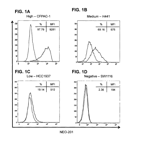
[16] FIGs. 1A-1D: Flow cytometry of NE0-201 binding to human carcinoma cell
lines. Representative human carcinoma cell lines with various levels of NEO-
201 antigen
expression, (FIG. 1A) pancreatic CFPAC-1 (high), (FIG. 1B) NSCLC H441
(medium), (FIG.
1C) breast HCC1937 (low), and (FIG. 1D) colon SW1116 (negative). Results are
expressed
as % NEO-201 positive and mean fluorescence intensity (MFI) for each cell
line. Red, NE0-
7
CA 03083467 2020-05-25
WO 2019/090134 PCT/US2018/059039
201-stained cells; black, unstained cells. NEO-201 positivity was defined as %
positive >
10%.
[17] FIGs. 2A-2C: IHC staining of human tumor samples by NE0-201. (FIG. 2A)
Representative NEO-201 staining from adjacent normal and malignant tissues
from colon,
pancreas, stomach, and lung samples. All images were obtained at 100X. (FIG.
2B)
Quantification of NEO-201 positive staining from the human tumor microarray
samples from
various carcinoma tissues. (FIG. 2C) Quantification of NEO-201 positive
staining from
human tumor microarray samples from normal tissue adjacent to tumor tissue. n
¨ number of
samples.
[18] FIGs. 3A-3C: NE0-201 mediates ADCC and CDC against human tumor cell
lines. (FIG. 3A) ADCC activity using CFPAC-1 or ASPC-1 cells as target cells.
Cells were
treated with 10 g/mL of NEO-201 or human IgG1 (negative control). Purified NK
cells
from two healthy donors were used as effector cells at the indicated E:T
ratios. *, statistically
significant (p < 0.05) by T-test. (FIG. 3B) ADCC assay using CFPAC-1 cells
treated with
increasing doses of NEO-201. NK cells isolated from a healthy donor were used
as effector
cells at an E:T ratio of 12.5:1. The graph depicts the fold increase in %
specific lysis of
NE0-201-treated tumor cells versus that of control cells treated with 10 g/mL
human IgGl.
*, statistically significant (p < 0.05) by T-test. (FIG. 3C) CDC assay using
ASPC-1 cells
treated with the indicated doses of NEO-201 for the indicated durations.
*statistically
significant (p < 0.05) by T-test.
[19] FIGs. 4A-4D: Antitumor efficacy of NEO-201 in CFPAC-1 tumor xenografts.
(FIG. 4A) Tumor volume measurements for the CFPAC-1 xenografts from each
treatment
group at various time points. Mice (n = 10 animals/group) were dosed
intraperitoneally with
saline solution, human IgG1 (250 g), or NEO-201 (100 and 250 jig) on days 13,
17, and 20
post tumor cell implantation. Mice were also dosed intraperitoneally with ¨1.0
x 107 IL-2-
activated human PBMCs on days 14, 18, and 21 as a source of immune effector
cells. (FIG.
4B) Quantification of the number of mice still bearing palpable tumors on day
36. (FIG. 4C)
Representative image of NE0-201-treated versus saline-treated tumor-bearing
mice. (FIG.
4D) Body weight measurements of the tumor-bearing mice at various time points
during the
study.
[20] FIGs. 5A-5B: NE0-201 biodistribution in CFPAC-1 xenograft-bearing mice.
Measurement of normalized radioactivity from the indicated tissues of CFPAC-1
tumor-
bearing female (FIG. 5A) and male (FIG. 5B) mice dosed intravenously with
radiolabeled
CA 03083467 2020-05-25
WO 2019/090134 PCT/US2018/059039
NEO-201. n = 4 animals/time point. Day 1, 2, 4, and 7 represents the amount of
time
between radiolabeled antibody injection and necropsy.
[21] FIGs. 6A-6C: Body weight and neutrophil counts from cynomolgus monkeys
treated with NE0-201. (FIG. 6A) Percent change in body weight relative to
baseline (day -
1) measured for monkeys at 7 days and 14 days after a receiving a single dose
of NEO-201 at
the indicated dose levels. n = 4 animals per group (2 females, 2 males). (FIG.
6B) Percent
change in neutrophil levels relative to baseline (day -7) from the blood of
monkeys treated
with a single dose of NEO-201 at the indicated dose levels. n = 4 animals per
group (2
females, 2 males). (FIG. 6C) p values for neutrophil levels versus 0 mg/kg
controls for each
dosage and time point. *statistically significant (p < 0.05) by T-test.
[22] FIGS. 7A-7C: haNK ADCC assay using NEO-201 (4hr). Target cells = 3000
cells/well. (FIG. 6A-6B) Percentage specific lysis of H520 lung carcinoma
(FIG. 6A) or
0V90 ovarian carcinoma (FIG. 6B) cells treated with NEO-201 (upper line,
square symbols)
or IgG1 negative control (lower line, round symbols) at 4 hours as a function
of
Effector:Target (E:T) ratio. E:T ratios were 6.25:1, 12.5:1, or 25:1. mAb
concentration was
g/mL. Values shown are mean +/- SD of 3 replicates. Asterisk (*) indicates
statistical
significance vs. IgG negative control (p<0.01, 2-tailed t-test). (FIG. 6C)
Percentage specific
lysis of lung (H520, HCC827), breast (ZR-75-1), and ovarian (0V90) carcinomas
cells
treated with NEO-201 (right, lightly grey bars) or negative control IgG (left,
solid black bars)
at four hours at a constant E:T ratio of 25:1. mAb concentration was
101.1g/mL. Values
shown are mean +/- SD of 3 replicates. Asterisk (*) indicates statistical
significance vs. IgG
negative control (p<0.01, 2-tailed t-test).
[23] FIG. 8: Treatment with ALT-803 enhance ADCC activity mediated by NE0-
201. NK cells isolated from two normal donors were treated with ALT-803
(25ng/m1) or
medium control for 48 hours and used as effector cells in a 4h non radioactive
ADCC assay
using Celigo Imaging cytometer. CF-PAC1 (human pancreatic cancer cell line)
cells were
stained with calcein AM and used as targets at 3,000 cells/well. Results are
expressed in %
specific lysis (SE).
[24] FIG. 9: Treatment with ALT-803 enhanced the expression of TIM-3 and NKG2D
on human NK cells. Purified human NK cells from a normal donor were cultured
for 48
hours with or without ALT-803 (25ng/m1). Results are expressed in % of
positive cells
(MFI).
[25] FIG. 10: Treatment with ALT-803 enhanced the expression of TIM-3 and
NKG2D on human NK cells. Purified human NK cells from another normal donor
were
9
CA 03083467 2020-05-25
WO 2019/090134 PCT/US2018/059039
cultured for 48 hours with or without ALT-803 (25ng/m1). Results are expressed
in % of
positive cells (MFI).
[26] FIG. 11: Treatment with ALT-803 enhance ADCC activity mediated by low
concentrations NEO-201. NK cells isolated from a normal donor (ND#6) were
treated with
ALT-803 (25ng/m1) or medium control for 48 hours and used as effector cells in
a 4h non
radioactive ADCC assay using Celigo Imaging cytometer. NEO-201 was used at
three
different concentrations (lOug/ml, lag/ml, and 0.1]..tg/m1). CF-PAC (human
pancreatic
cancer cell line) cells were stained with calcein AM and used as targets at
3,000 cells/well.
E:T = 25:1. Results are expressed in % specific lysis (SE). * Statistically
significant (p
<0.01).
[27] FIG. 12: Treatment with ALT-803 enhances ADCC activity of a normal donor
(ND#8) with minimal ADCC activity mediated by and NEO-201 and the activity can
be
blocked by anti-CD16 and anti-TIM-3 antibody. NK cells isolated from a normal
donor
with minimal ADCC activity and were treated with ALT-803= (25ng/m1) or medium
control
for 48 hours and used as effector cells in a 4h non radioactive ADCC assay
using Celigo
Imaging cytorneter. Anti-CD16 and anti-TIM-3 were used at a concentrations of
30iLtg/m1
and 15 g/ml. NK cells were pretreated with anti-CD16 or anti-TIM-3 for 2 hour
prior to the
addition of NEO-201 and effector cells. CF-PAC (human pancreatic cancer cell
line) cells
were stained with calcein AM and used as targets at 3,000 cells/well. NEO-201
was used at a
concentration of 1 Oug/ml. E:T = 25:1. Results are expressed in % specific
lysis (SE).
*Statistically significant (p <0.01) compared to no ALT-803 treatment.
#Statistically
significant (p <0.01) compared to no anti-CD16 and anti-TIM-3 treatment.
[28] FIG. 13: NK-92 killing assay using NEO-201 (16hr). Target tumor cells
(ASPC-1,
BxPC-3, CFPAC-1, or L5174T) were seeded at 3000 cells/well. The cells were
then treated
with 10 ug/mL of either human IgG1 isotype control antibody or NEO-201, and
then the
natural killer (NK) cell line NK-92 was added at effector-to-target (E:T)
ratios of 1.5625:1,
3.125:1, 6.25:1, and 12.5:1. After 16hr incubation at 37 C, cell viability was
quantified using
the Celigo Imaging Cytometer and GraphPad Prism 7 software. Live target cells
(calcein
AM+/PI-) were counted for each well, and specific lysis was calculated.
Results are shown
graphically and tabulated below for each tumor cell type. *statistically
significant (p < 0.05).
DETAILED DESCRIPTION
CA 03083467 2020-05-25
WO 2019/090134 PCT/US2018/059039
[29] In one aspect, the disclosure provides a method of killing carcinoma
cells comprising
administering an effective amount of a NEO-201 antibody to a patient in need
thereof.
[30] In one aspect, the disclosure provides a method of treating a carcinoma,
comprising
administering an effective amount of a NEO-201 antibody to a patient in need
thereof.
[31] In one aspect, the disclosure provides a method of preventing the
recurrence of a
carcinoma, comprising administering an effective amount of a NEO-201 antibody
to a patient
in need thereof.
[32] In one aspect, the disclosure provides a method of decreasing the tumor
burden in a
patient having a carcinoma, comprising administering an effective amount of a
NEO-201
antibody to a patient in need thereof.
[33] Said antibody may mediate complement mediated cytotoxicity (CDC), thereby
killing
carcinoma cells in said patient.
[34] Said patient may be natural killer ("NK")-depleted prior to or at the
time of said
administering. Said patient may be severely NK-depleted prior to or at the
time of said
administering. Said patient may have NK cell deficiency (NKD), such as CNKD
(e.g.,CNKD1, CNKD2), or FNKD (e.g., FNKD1). Said patient may be NK-depleted or
severely NK-depleted as a result of another therapy, e.g., a cancer therapy,
such as
chemotherapy or radiotherapy. Said patient may been treated with one or more
proteasome
inhibitors (e.g., Bortezomib, MG132), Histone deacetylase, inhibitors (e.g.,
valproic acid,
Trichostatin A, Suberoylanilide-hydroxamic acid (SAH), Sodium butyrate),
genotoxic agents
(e.g., doxorubicin, melphalan, cisplatin, Ara-C, aphidicolin, mitomycin,
methotrexate,
etoposide), GSK inhibitors (e.g., LiC1, BIO, SB21), BET inhibitors (e.g.,
JQ1), HSP90
inhibitors (e.g., radicicola), 17-AAG), microtubule assembly inhibitors (e.g.,
vincristinc,
cytochalasin D, nocodazole, docetaxel), and/or immunomodulatory drugs (e.g.,
lenalidomide).
[35] The method may include, prior to or at the time of said administering,
determining
whether said patient is NK-depleted.
[36] The method may include, prior to or at the time of said administering,
determining
whether said patient is severely NK-depleted.
[37] In said method, prior to or at the time of said administering, NK cells
may comprise
less than 5% of the peripheral blood mononuclear cells (PBMCs) in said
individual.
[38] In said method, prior to or at the time of said administering, NK cells
may comprise
less than 3% of the peripheral blood mononuclear cells (PBMCs) in said
individual.
11
CA 03083467 2020-05-25
WO 2019/090134 PCT/US2018/059039
[39] In said method, prior to or at the time of said administering, less than
70% of PBMC
NK cells in said patient may be CD56dimCD16+ NK cells.
[40] In said method, prior to or at the time of said administering, less than
50% of PBMC
NK cells in said patient may be CD56dimCD16+ NK cells.
[41] Said NEO-201 antibody may comprise at least one, two, three, four, five,
or all six of
the CDR sequences contained in SEQ ID NO: 28 and SEQ ID NO: 29.
[42] Said NEO-201 antibody may comprise a variable heavy chain sequence having
at
least 90% identity to SEQ ID NO: 38.
[43] Said NEO-201 antibody may comprise a variable light chain sequence having
at least
90% identity to SEQ ID NO: 39.
[44] Said NEO-201 antibody may comprise a variable heavy chain sequence having
at
least 90% identity to SEQ ID NO: 38 and a variable light chain sequence having
at least 90%
identity to SEQ ID NO: 39.
[45] Said NEO-201 antibody may comprise a heavy chain sequence having at least
90%
identity to amino acids 20-470 of SEQ ID NO: 28 and a light chain sequence
having at least
90% identity to amino acids 20-233 of SEQ ID NO: 29.
[46] Said NEO-201 antibody may comprise all six of the CDR sequences contained
in
SEQ ID NO: 28 and SEQ ID NO: 29.
[47] Said NEO-201 antibody may comprise a human IgG1 constant domain.
[48] Said NEO-201 antibody may be humanized.
[49] Said NEO-201 antibody may be conjugated to another moiety.
[50] Said NEO-201 antibody may be conjugated to another cytotoxic moiety,
label,
radioactive moiety, or affinity tag.
[51] Said method may further comprise administering to the patient an
effective amount of
a cytokine agonist to potentiate or stimulate killing of cells of said
carcinoma. Said cytokine
agonist may comprise interleukin-2 (IL-2), interleukin 21 (IL-21), ALT-803, IL-
15 inhibitors,
checkpoint inhibitors, anti-PD1, anti-PDL1, anti-CTLA-4, anti-41BB, anti-0X40,
anti-Tim-3,
or a combination thereof.
[52] Said method may further comprise administering to said patient an
effective amount
of a complement-regulatory protein (CRP) antagonist to potentiate or stimulate
killing of
cells of said carcinoma. Said CRP antagonist may antagonize one or more of
CD46, CD55,
or CD59. Said CRP antagonist may comprise an antibody or antigen-binding
fragment
thereof.
[53] Said cytokine agonist may comprise an IL-15 agonist or an 1L-15
superagonist.
12
CA 03083467 2020-05-25
WO 2019/090134 PCT/US2018/059039
[54] Said cytokine agonist may comprise a complex consisting of an IL-15
mutant (IL-
15N72D) bound to an IL-15 receptor a/IgG1 Pc fusion protein, such as ALT-803.
[55] The effective dosage of said NEO-201 antibody may be reduced compared to
treatment with the NEO-201 antibody alone without said cytokine agonist.
[56] Said carcinoma may comprise colon cancer. Said carcinoma may comprise
pancreatic
cancer. Said carcinoma may comprise ovarian cancer. Said carcinoma may
comprise stomach
cancer. Said carcinoma may comprise lung cancer. Said carcinoma may comprise
breast
cancer. Said carcinoma may comprise uterine cancer.
[57] In another embodiment, the disclosure provides a method of killing
carcinoma cells
comprising administering an effective amount of a NEO-201 antibody to a
patient in need
thereof, wherein said pkient is natural killer ("NK")-depleted prior to or at
the time of said
administering. Said NK-depletion may comprise the patient having less than 5%
or less than
3% of the peripheral blood mononuclear cells (PBMCs) being NK cells in a
sample derived
from the patient, e.g., in a blood sample. Alternatively or in addition, prior
to or at the time
of said administering, less than 70% (or optionally less than 50%) of PBMC NK
cells in said
patient may be CD56dimCD16+ NK cells.
[58] In another embodiment, the disclosure provides a method of treating a
carcinoma,
comprising administering an effective amount of a NEO-201 antibody to a
patient in need
thereof, wherein said patient is natural killer ("NK")-depleted prior to or at
the time of said
administering.
[59] In another embodiment, the disclosure provides a method of preventing the
recurrence
of a carcinoma, comprising administering an effective amount of a NEO-201
antibody to a
patient in need thereof; wherein said patient is natural killer ("NK")-
depleted prior to or at the
time of said administering.
[60] In another embodiment, the disclosure provides a method of decreasing the
tumor
burden in a patient having a carcinoma, comprising administering an effective
amount of a
NEO-201 antibody to a patient in need thereof, wherein said patient is natural
killer ("NK")-
depleted prior to or at the time of said administering.
[61] In the foregoing methods, said antibody may mediate CDC, thereby, thereby
killing
carcinoma cells in said patient, e.g., notwithstanding the absence of
effective ADCC due to
the patient being NK-depleted. Said patient may be severely NK-depleted at the
time of said
administering. Optionally, the method further comprises determining whether
said patient is
NK-depleted or severely NK-deleted, e.g., at the time of said administering or
within a period
prior to said administering, such as within 1 or 2 weeks prior. NK-depleted or
severely NK-
13
CA 03083467 2020-05-25
WO 2019/090134 PCT/US2018/059039
depleted status may also be inferred from the patient's history, such as the
prior or concurrent
use of another therapy that depletes NK cells. For example said patient have
undergone or be
concurrently undergoing cancer therapy, such as radiotherapy or chemotherapy.
Said cancer
therapy may include administration of one or more one or more proteasome
inhibitors (e.g.,
Bortezomib, MG132), Histone deacetylase inhibitors (e.g., valproic acid,
Trichostatin A,
Suberoylanilide-hydroxamic acid (SAH), Sodium butyrate), genotoxic agents
(e.g.,
doxorubicin, melphalan, cisplatin, Ara-C, aphidicolin, mitomycin,
methotrexate, etoposide),
GSK inhibitors (e.g., LiC1, 1310, SB21), BET inhibitors (e.g., JQ1), HSP90
inhibitors (e.g.,
radici'cola), 17-AAG), microtubule assembly inhibitors (e.g., vincristine,
cytochalasin D,
nocodazole, docetaxel), and/or immunomodulatory drugs (e.g., lenalidomide).
[62] Said patient may have NK cell deficiency (NKD), such as CNKD (e.g.,CNKD1,
CN1CD2), or FNKD (e.g., FNI(D1).
[63] In a preferred embodiment of the invention which may be used with any of
the
foregoing or following embodiments, said NEO-201 antibody may comprise at
least one,
two, three, four, five, or all six of the CDR sequences contained in SEQ ID
NO: 28 and SEQ
ID NO: 29.
[64] In a preferred embodiment of the invention which may be used with any of
the
foregoing or following embodiments, said NEO-201 antibody may comprise a
variable heavy
chain sequence having at least 80%, at least 85%, at least 90% or most
preferably at least
95% identity to SEQ ID NO: 38. Said variable heavy chain having said
percentage sequence
identity may comprise all 3 of the CDR sequences contained in SEQ ID NO: 38.
[65] In a preferred embodiment of the invention which may be used with any of
the
foregoing or following embodiments, said NEO-201 antibody may comprise a
variable light
chain sequence having at least 80%, at least 85%, at least 90% or most
preferably at least
95% identity to identity to SEQ ID NO: 39. Said variable light chain may
comprise all 3 of
the CDR sequences contained in SEQ ID NO: 39.
[66] In a preferred embodiment of the invention which may be used with any of
the
foregoing or following embodiments, said NEO-201 antibody may comprise a
variable heavy
chain sequence having at least 80%, at least 85%, at least 90% or most
preferably at least
95% identity to SEQ ID NO: 38 and a variable light chain sequence having at
least 80%, at
least 85%, at least 90% or most preferably at least 95% identity to identity
to to SEQ ID NO:
39. Said variable light chain may comprise all 3 of the CDR sequences
contained in SEQ ID
NO: 39, and said variable heavy chain having said percentage sequence identity
may
comprise all 3 of the CDR sequences contained in SEQ ID NO: 38.
14
CA 03083467 2020-05-25
WO 2019/090134 PCT/US2018/059039
[67] In a preferred embodiment of the invention which may be used with any of
the
foregoing or following embodiments, said NEO-201 antibody may comprise a heavy
chain
sequence having at least 80%, at least 85%, at least 90% or most preferably at
least 95%
identity to amino acids 20-470 of SEQ ID NO: 28 and a light chain sequence
having at least
80%, at least 85%, at least 90% or most preferably at least 95% identity to
amino acids 20-
233 of SEQ ID NO: 29. Said light chain may comprise all 3 of the CDR sequences
contained
in SEQ ID NO: 29, and said heavy chain having said percentage sequence
identity may
comprise all 3 of the CDR sequences contained in SEQ ID NO: 28.
[68] In a preferred embodiment of the invention which may be used with any of
the
foregoing or following embodiments, said NEO-201 antibody may comprise the
heavy chain
variable region sequence contained in SEQ ID NO: 28 and the light chain
variable region
sequence contained in SEQ ID NO: 29.
[69] In a preferred embodiment of the invention which may be used with any of
the
foregoing or following embodiments, said NEO-201 antibody may comprise a heavy
chain
sequence containing amino acids 20-470 of SEQ ID NO: 28 and a light chain
sequence
containing amino acids 20-233 of SEQ ID NO: 29.
[70] In a preferred embodiment of the invention which may be used with any of
the
foregoing or following embodiments, said NEO-201 antibody comprises a human
IgG1
constant domain.
[71] In a preferred embodiment of the invention which may be used with any of
the
foregoing or following embodiments, said NEO-201 antibody may be humanized.
[72] In a preferred embodiment of the invention which may be used with any of
the
foregoing or following embodiments, said NEO-201 antibody may be conjugated to
another
moiety, such as another cytotoxic moiety, label, radioactive moiety, or
affinity tag.
[73] In a preferred embodiment of the invention which may be used with any of
the
foregoing or following embodiments, said method may further comprise
administering to the
patient an effective amount of a cytokine agonist to potentiate or stimulate
killing of cells of
said carcinoma. Said cytokine agonist may comprise interleukin-2 (IL-2),
interleukin 21 (IL-
21), ALT-803, IL-15 inhibitors, checkpoint inhibitors, anti-PD1, anti-PDL1,
anti-CTLA-4,
anti-41BB, anti-0X40, anti-Tim-3, or a combination thereof.
[74] In a preferred embodiment of the invention which may be used with any of
the
foregoing or following embodiments, said method may further comprise
administering to said
patient an effective amount of a complement-regulatory protein (CRP)
antagonist to
potentiate or stimulate killing of cells of said carcinoma. Said CRP
antagonist may
CA 03083467 2020-05-25
WO 2019/090134 PCT/US2018/059039
antagonize one or more of CD46, CD55, or CD59. Said CRP antagonist may
comprise an
antibody or antigen-binding fragment thereof. Said cytokine agonist may
comprise an IL-15
agonist or an IL-15 superagonist. Said cytokine agonist may comprises complex
consisting of
an IL-15 mutant (IL-15N72D) bound to an IL-15 receptor a/IgG1 Fe fusion
protein. Said
cytokine agonist may comprise ALT-803.
[75] In a preferred embodiment of the invention which may be used with any of
the
foregoing or following embodiments, the effective dosage of said NEO-201
antibody is
reduced compared to treatment with the NEO-201 antibody alone without said
cytokine
agonist.
[76] In a preferred embodiment of the invention which may be used with any of
the
foregoing or following embodiments, said cancer may express the NEO-201
antigen. Said
expression of NEO-201 antigen may be determined by detecting the NEO-201
antigen in a
sample of said cancer. Said detecting may be performed by techniques including
but not
limited to histological staining, flow cytometry, RT-PCR, dot blotting,
Western blotting,
Northern Blotting, and other techniques known in the art. In the case of a
recurrent or
metastatic cancer, expression of NEO-201 antigen may also be inferred by the
expression of
NEO-201 in the primary cancer, or by responsiveness of the primary cancer to
NEO-201
antibody therapy.
[77] In a preferred embodiment of the invention which may be used with any of
the
foregoing or following embodiments, said cancer may comprise colon cancer.
[78] In a preferred embodiment of the invention which may be used with any of
the
foregoing or following embodiments, said cancer may comprise pancreatic
cancer.
[79] In a preferred embodiment of the invention which may be used with any of
the
foregoing or following embodiments, said cancer may comprise ovarian cancer.
[80] In a preferred embodiment of the invention which may be used with any of
the
foregoing or following embodiments, said cancer may comprise stomach cancer.
[81] In a preferred embodiment of the invention which may be used with any of
the
foregoing or following embodiments, said cancer may comprise lung cancer.
[82] In a preferred embodiment of the invention which may be used with any of
the
foregoing or following embodiments, said cancer may comprise breast cancer.
[83] In a preferred embodiment of the invention which may be used with any of
the
foregoing or following embodiments, said cancer may comprise uterine cancer.
[84] DEFINITIONS
16
CA 03083467 2020-05-25
WO 2019/090134 PCT/US2018/059039
[85] Unless defined otherwise, all technical and scientific terms used herein
have the same
meaning as thotse commonly understood by one of ordinary skill in the art to
which this
invention belongs. Although methods and materials similar or equivalent to
those described
herein may be used in the invention or testing of the present invention,
suitable methods and
materials are described herein. The materials, methods and examples are
illustrative only,
and are not intended to be limiting.
[86] As used in the description herein and throughout the claims that follow,
the meaning
of "a," "an," and "the" includes plural reference unless the context clearly
dictates otherwise.
[87] "Amino acid," as used herein refers broadly to naturally occurring and
synthetic
amino acids, as well as amino acid analogs and amino acid mimetics that
function in a
manner similar to the naturally occurring amino acids. Naturally occurring
amino acids are
those encoded by the genetic code, as well as those amino acids that are later
modified, e.g.,
hydroxyproline, y-carboxyglutamate, and 0-phosphoserine. Amino acid analogs
refers to
compounds that have the same basic chemical structure as a naturally occurring
amino acid,
i.e., an a carbon that is bound to a hydrogen, a carboxyl group, an amino
group, and an R
group, e.g., homoserine, norleucine, methionine sulfoxide, methionine methyl
sulfonium.
Such analogs have modified R groups (e.g., norleucine) or modified peptide
backbones, but
retain the same basic chemical structure as a naturally occurring amino acid.
Amino acid
mimetics refers to chemical compounds that have a structure that is different
from the general
chemical structure of an amino acid, but that functions in a manner similar to
a naturally
occurring amino acid.
[88] The terms "NK-depleted" or "natural killer-depleted" as used herein refer
to a patient
having low natural killer (NK) cell levels relative to the normal range. NK
cells are a
cytotoxic innate immune lymphocyte. Typically, NK cells comprise 5-20% of the
peripheral
blood mononuclear cells (PBMCs) in a healthy individual. A patient having NK
cells
comprising less than 5% of the PMBCs is referred to as NK-depleted.
Additionally, a patient
is referred to as severely NK-cell depleted if NK cells comprising less than
3% of the
PMBCs. Additionally, in normal individuals, up to 90% of PBMC NK cells are
CD56dimCD16+ NK cells, and these are considered the most cytotoxic subset. If
less than
70% of PBMC NK cells are CD56dimCD16 NK cells, then the patient is referred
to as NK-
depleted. Additionally, if less than 50% of PBMC NK cells are CD56dimCD16+ NK
cells,
then the patient is referred to as severely NK-depleted. A given patient may
be referred to as
NK-depleted or severely NK-depleted based on meeting either or both of these
individual
criteria. Generally speaking, a patient's status as NK-depleted or severely NK-
depleted is
17
CA 03083467 2020-05-25
WO 2019/090134 PCT/US2018/059039
determined by testing a sample taken from the patient, e.g., a blood sample,
e.g., a sample
obtained and tested within one or two weeks prior. A patient's status as NK-
depleted or
severely NK-depleted may also be inferred from a disease diagnosis and/or a
course of
treatment that is associated with such depletion of NK cells.
[89] NK-depleted also includes subjects having an NK cell deficiency (NKD).
Exemplary
NKD conditions include Classical NKD (CNKD), characterized by an absence of NK
cells
and their function among peripheral blood lymphocytes; Functional NKD (FNKD),
characterized by presence of NK cells within peripheral blood lymphocytes,
having defective
NK cell activity. In both CNKD and FNKD the NK cell abnormality is a major
immunological deficit, which results in inadequate ADCC responses. CNKD and
FNKD can
be further subdivided based on patient characteristics such as the identity of
causative gene(s)
and other patient characteristics. CNKD includes CNKD subtype 1 (CNKD1), which
is
autosomal dominant and is associated with defects in the GATA2 gene, and CNKD
subtype 2
(CNKD2), which is autosomal recessive and is associated with defects in the
MCM4 gene.
FNKD includes FNKD1, which is autosomal recessive and is associated with
defects in the
FCCR3A gene.
[90] "Antibody," as used herein, refers broadly to any polypeptide chain-
containing
molecular structure with a specific shape that fits to and recognizes an
epitope, where one or
more non-covalent binding interactions stabilize the complex between the
molecular structure
and the epitope. The archetypal antibody molecule is the immunoglobulin, and
all types of
immunoglobulins, IgG, IgM, IgA, IgE, IgD, from all sources, e.g., human,
rodent, rabbit,
cow, sheep, pig, dog, chicken, are considered to be "antibodies." Antibodies
include but are
not limited to chimeric antibodies, human antibodies and other non-human
mammalian
antibodies, humanized antibodies, single chain antibodies (scFvs),
camelbodies, nanobodies,
IgNAR (single-chain antibodies derived from sharks), small-modular
immunopharmaceuticals (SMIPs), and antibody fragments (e.g., Fabs, Fab',
F(ab')2.)
Numerous antibody coding sequences have been described; and others may be
raised by
methods well-known in the art. See Streltsov, et aL (2005) Protein Sci.
14(11): 2901-9;
Greenberg, et aL (1995) Nature 374(6518): 168-173; Nuttall, et aL (2001) Mol
Immunol.
38(4): 313-26; Hamers-Casterman, etal. (1993) Nature 363(6428): 446-8; Gill,
et aL (2006)
Curr Opin Biotechnol. 17(6): 653-8.
[91] "NEO-201 antibody" refers to an antibody containing the heavy and light
chains of
SEQ ID NOs: 28 and 29 or the variable regions optionally together with the
constant regions
contained therein, as well as fragments and variants thereof. Such variants
include sequences
18
CA 03083467 2020-05-25
WO 2019/090134 PCT/US2018/059039
containing one, two, three, four, five or preferably all six of the CDR
sequences contained in
SEQ ID NO: 28 and SEQ ID NO: 29, i.e., the heavy chain CDR1 of SEQ ID NO: 32,
the
heavy chain CDR2 of SEQ ID NO: 33, the heavy chain CDR3 of SEQ ID NO: 34, the
light
chain CDR1 of SEQ ID NO: 35, the light chain CDR2 of SEQ ID NO: 36, and the
light chain
CDR3 of SEQ ID NO: 37. Said antibody may be humanized. Said antibody may be
expressed containing one or more leader sequences, which may be removed during
expression and/or processing and secretion of the antibody. Said antibody may
be presented
in a monovalent, bivalent, or higher multivalent format, including without
limitation a
bispecific or multispecific antibody containing said NEO-201 antibody sequence
and a
binding fragment of a different antibody. Typically said antibody specifically
binds to
carcinoma cells and competes for binding to carcinoma cells with an antibody
comprising the
variable heavy chain of SEQ ID NO: 38 and variable light chain of SEQ ID NO:
39, or
comprising the heavy chain of SEQ ID NO: 28 and light chain of ,SEQ ID NO: 29.
One or
more of those CDR sequences contained in SEQ ID NO: 28 and/or SEQ ID NO: 29
may be
substituted with a variant sequence, such as the light chain CDR1 of SEQ ID
NO: 1 or 4;
light chain CDR2 of SEQ ID NO: 2 or 5; light chain CDR3 of SEQ ID NO: 3 or 6;
heavy
chain CDR1 of SEQ ID NO: 7; heavy chain CDR2 of SEQ ID NO: 8,10, 30, or 31;
heavy
chain CDR3 of SEQ ID NO: 9 or 11; or SEQ ID NOs: 30-31. The light chain may
comprise
the CDRs contained in the light chain sequence of SEQ ID NO: 14, 16, 17, 18,
19, 20, 21, or
29. The heavy chain may comprise the CDRs contained in the heavy chain
sequence of SEQ
ID NO: 15, 22, 23, 24, 25, 26, 27, or 29. Said antibody may comprise a
variable heavy chain
sequence having at least 75%, 80%, 85%, 90%, 95%, 96%, 97%, 98%, or 99%
identity to
SEQ ID NO: 38, and/or a variable light chain sequence having at least 75%,
80%, 85%, 90%,
95%, 96%, 97%, 98%, or 99% identity to SEQ ID NO: 39, optionally wherein said
heavy
and/or light chain sequence contains one, two, three, four, five or preferably
all six of the
CDR sequences contained in SEQ ID NO: 28 and SEQ ID NO: 29, i.e., the heavy
chain
CDR1 of SEQ ID NO: 32, the heavy chain CDR2 of SEQ ID NO: 33, the heavy chain
CDR3
of SEQ ID NO: 34, the light chain CDR1 of SEQ ID NO: 35, the light chain CDR2
of SEQ
ID NO: 36, and the light chain CDR3 of SEQ ID NO: 37. Said antibody may be
conjugated
to another moiety, such as a cytotoxic moiety, radioactive moiety, label, or
purification tag.
[92] "Antigen," as used herein, refers broadly to a molecule or a portion of a
molecule
capable of being bound by an antibody which is additionally capable of
inducing an animal to
produce an antibody capable of binding to an epitope of that antigen. An
antigen may have
one epitope, or have more than one epitope. The specific reaction referred to
herein indicates
19
CA 03083467 2020-05-25
WO 2019/090134 PCT/US2018/059039
that the antigen will react, in a highly selective manner, with its
corresponding antibody and
not with the multitude of other antibodies which may be evoked by other
antigens. Antigens
may be tumor specific (e.g., expressed by neoplastic cells of pancreatic and
colon
carcinoma.)
[93] "Cancer," as used herein, refers broadly to any neoplastic disease
(whether invasive or
metastatic) characterized by abnormal and uncontrolled cell division causing
malignant
growth or tumor.
[94] "Chimeric antibody," as used herein, refers broadly to an antibody
molecule in which
the constant region, or a portion thereof, is altered, replaced or exchanged
so that the antigen
binding site (variable region) is linked to a constant region of a different
or altered class,
effector function and/or species, or an entirely different molecule which
confers new
properties to the chimeric antibody, e.g., an enzyme, toxin, hormone, growth
factor, drug; or
the variable region, or a portion thereof, is altered, replaced or exchanged
with a variable
region having a different or altered antigen specificity.
[95] "Conservatively modified variants," as used herein, applies to both amino
acid and
nucleic acid sequences, and with respect to particular nucleic acid sequences,
refers broadly
to conservatively modified variants refers to those nucleic acids which encode
identical or
essentially identical amino acid sequences, or where the nucleic acid does not
encode an
amino acid sequence, to essentially identical sequences. Because of the
degeneracy of the
genetic code, a large number of functionally identical nucleic acids encode
any given protein.
Such nucleic acid variations are "silent variations," which are one species of
conservatively
modified variations. Every nucleic acid sequence herein which encodes a
polypeptide also
describes every possible silent variation of the nucleic acid. One of skill
will recognize that
each codon in a nucleic acid (except AUG, which is ordinarily the only codon
for methionine,
and TGG, which is ordinarily the only codon for tryptophan) may be modified to
yield a
functionally identical molecule.
[96] "Complementarity determining region," "hypervariable region," or "CDR,"
as used
herein, refers broadly to one or more of the hyper-variable or complementarily
determining
regions (CDRs) found in the variable regions of light or heavy chains of an
antibody. See
Kabat, et al. (1987) "Sequences of Proteins of Immunological Interest"
National Institutes of
Health, Bethesda, MD. These expressions include the hypervariable regions as
defined by
Kabat, et aL (1983) "Sequences of Proteins of Immunological Interest" U.S.
Dept. of Health
and Human Services or the hypervariable loops in 3-dimensional structures of
antibodies.
Chothia and Lesk (1987) J Mol. Biol. 196: 901-917. The CDRs in each chain are
held in
CA 03083467 2020-05-25
WO 2019/090134 PCT/US2018/059039
close proximity by framework regions and, with the CDRs from the other chain,
contribute to
the formation of the antigen binding site. Within the CDRs there are select
amino acids that
have been described as the selectivity determining regions (SDRs) which
represent the
critical contact residues used by the CDR in the antibody-antigen interaction.
Kashmiri
(2005) Methods 36: 25-34.
[97] "Control amount," as used herein, refers broadly to a marker can be any
amount or a
range of amounts to be compared against a test amount of a marker. For
example, a control
amount of a marker may be the amount of a marker in a patient with a
particular disease or
condition or a person without such a disease or condition. A control amount
can be either in
absolute amount (e.g., microgram/ml) or a relative amount (e.g., relative
intensity of signals).
[98] "Differentially present," as used herein, refers broadly to differences
in the quantity or
quality of a marker present in a sample taken from patients having a disease
or condition as
compared to a comparable sample taken from patients who do not have one of the
diseases or
conditions. For example, a nucleic acid fragment may optionally be
differentially present
between the two samples if the amount of the nucleic acid fragment in one
sample is
significantly different from the amount of the nucleic acid fragment in the
other sample, for
example as measured by hybridization and/or NAT-based assays. A polypeptide is
differentially present between the two samples if the amount of the
polypeptide in one sample
is significantly different from the amount of the polypeptide in the other
sample. It should be
noted that if the marker is detectable in one sample and not detectable in the
other, then such
a marker may be considered to be differentially present. Optionally, a
relatively low amount
of up-regulation may serve as the marker.
[99] "Diagnostic," as used herein, refers broadly to identifying the presence
or nature of a
pathologic condition. Diagnostic methods differ in their sensitivity and
specificity. The
"sensitivity" of a diagnostic assay is the percentage of diseased individuals
who test positive
(percent of "true positives"). Diseased individuals not detected by the assay
are "false
negatives." Subjects who are not diseased and who test negative in the assay
are termed "true
negatives." The "specificity" of a diagnostic assay is 1 minus the false
positive rate, where
the "false positive" rate is defined as the proportion of those without the
disease who test
positive. While a particular diagnostic method may not provide a definitive
diagnosis of a
condition, it suffices if the method provides a positive indication that aids
in diagnosis.
[100] "Diagnosing," as used herein refers broadly to classifying a disease or
a symptom,
determining a severity of the disease, monitoring disease progression,
forecasting an outcome
of a disease and/or prospects of recovery. The term "detecting" may also
optionally
21
CA 03083467 2020-05-25
WO 2019/090134 PCT/US2018/059039
encompass any of the foregoing. Diagnosis of a disease according to the
present invention
may, in some embodiments, be affected by determining a level of a
polynucleotide or a
polypeptide of the present invention in a biological sample obtained from the
subject,
wherein the level determined can be correlated with predisposition to, or
presence or absence
of the disease. It should be noted that a "biological sample obtained from the
subject" may
also optionally comprise a sample that has not been physically removed from
the subject.
[101] "Effective amount," as used herein, refers broadly to the amount of a
compound,
antibody, antigen, or cells that, when administered to a patient for treating
a disease, is
sufficient to effect such treatment for the disease. The effective amount may
be an amount
effective for prophylaxis, and/or an amount effective for prevention. The
effective amount
may be an amount effective to reduce, an amount effective to prevent the
incidence of
signs/symptoms, to reduce the severity of the incidence of signs/symptoms, to
eliminate the
incidence of signs/symptoms, to slow the development of the incidence of
signs/symptoms, to
prevent the development of the incidence of signs/symptoms, and/or effect
prophylaxis of the
incidence of signs/symptoms. The "effective amount" may vary depending on the
disease
and its severity and the age, weight, medical history, susceptibility, and pre-
existing
conditions, of the patient to be treated. The term "effective amount" is
synonymous with
"therapeutically effective amount" for purposes of this invention. ,
[102] "Expression vector," as used herein, refers broadly to any recombinant
expression
system for the purpose of expressing a nucleic acid sequence of the invention
in vitro or in
vivo, constitutively or inducibly, in any cell, including prokaryotic, yeast,
fungal, plant, insect
or mammalian cell. The term includes linear or circular expression systems.
The term
includes expression systems that remain episomal or integrate into the host
cell genome. The
expression systems can have the ability to self-replicate or not, i.e., drive
only transient
expression in a cell. The term includes recombinant expression cassettes which
contain only
the minimum elements needed for transcription of the recombinant nucleic acid.
[103] "Framework region" or "FR," as used herein, refers broadly to one or
more of the
framework regions within the variable regions of the light and heavy chains of
an antibody.
See Kabat, et al. (1987) "Sequences of Proteins of Immunological Interest,"
National
Institutes of Health, Bethesda, MD. These expressions include those amino acid
sequence
regions interposed between the CDRs within the variable regions of the light
and heavy
chains of an antibody.
[104] "Heterologous," as used herein, refers broadly to portions of a nucleic
acid indicates
that the nucleic acid comprises two or more subsequences that are not found in
the same
22
CA 03083467 2020-05-25
WO 2019/090134 PCT/US2018/059039
relationship to each other in nature. For instance, the nucleic acid is
typically recombinantly
produced, having two or more sequences from unrelated genes arranged to make a
new
functional nucleic acid, e.g., a promoter from one source and a coding region
from another
source. Similarly, a heterologous protein indicates that the protein comprises
two or more
subsequences that are not found in the same relationship to each other in
nature (e.g., a fusion
protein).
[105] "High affinity," as used herein, refers broadly to an antibody having a
KD of at least
1 0-8 M, more preferably at least 10-9 M and even more preferably at least 10-
1 M for a target
antigen. However, "high affinity" binding can vary for other antibody
isotypes. For
example, "high affinity" binding for an igM isotype refers to an antibody
having a KD of at
least 10-7 M, more preferably at least 10-8 M.
[106] "Homology," as used herein, refers broadly to a degree of similarity
between a
nucleic acid sequence and a reference nucleic acid sequence or between a
polypeptide
sequence and a reference polypeptide sequence. Homology may be partial or
complete.
Complete homology indicates that the nucleic acid or amino acid sequences are
identical. A
partially homologous nucleic acid or amino acid sequence is one that is not
identical to the
reference nucleic acid or amino acid sequence. The degree of homology can be
determined
by sequence comparison. The term "sequence identity" may be used
interchangeably with
"homology."
[107] "Host cell," as used herein, refers broadly to a cell that contains an
expression vector
and supports the replication or expression of the expression vector. Host
cells may be
prokaryotic cells such as E. coil, or eukaryotic cells such as yeast, insect
(e.g., SF9),
amphibian, or mammalian cells such as CHO, HeLa, HEK-293, e.g., cultured
cells, explants,
and cells in vivo.
[108] "Hybridization," as used herein, refers broadly to the physical
interaction of
complementary (including partially complementary) polynucleotide strands by
the formation
of hydrogen bonds between complementary nucleotides when the strands are
arranged
antiparallel to each other.
[109] "K-assoc" or "Ka", as used herein, refers broadly to the association
rate of a particular
antibody-antigen interaction, whereas the term "Kdiss" or "Kd," as used
herein, refers to the
dissociation rate of a particular antibody-antigen interaction. The term "KD",
as used herein,
is intended to refer to the dissociation constant, which is obtained from the
ratio of Kd to Ka
(i.e., Kd/Ka) and is expressed as a molar concentration (M). KD values for
antibodies can be
determined using methods well established in the art.
23
CA 03083467 2020-05-25
WO 2019/090134 PCT/US2018/059039
[110] "Immunoassay," as used herein, refers broadly to an assay that uses an
antibody to
specifically bind an antigen. The immunoassay may be characterized by the use
of specific
binding properties of a particular antibody to isolate, target, and/or
quantify the antigen.
[111] "Isolated," as used herein, refers broadly to material removed from its
original
environment in which it naturally occurs, and thus is altered by the hand of
man from its
natural environment. Isolated material may be, for example, exogenous nucleic
acid included
in a vector system, exogenous nucleic acid contained within a host cell, or
any material which
has been removed from its original environment and thus altered by the hand of
man (e.g.,
"isolated antibody").
[112] "Label" or a "detectable moiety" as used herein, refers broadly to a
composition
detectable by spectroscopic, photochemical, biochemical, immunochemical,
chemical, or
other physical means.
[113] "Low stringency," "medium stringency," "high stringency," or "very high
stringency
conditions," as used herein, refers broadly to conditions for nucleic acid
hybridization and
washing. Guidance for performing hybridization reactions can be found in
Ausubel, et al.
(2002) Short Protocols in Molecular Biology (5th Ed.) John Wiley & Sons, NY.
Exemplary
specific hybridization conditions include but are not limited to: (1) low
stringency
hybridization conditions in 6X sodium chloride/sodium citrate (SSC) at about
45 C, followed
by two washes in 0.2XSSC, 0.1% SDS at least at 50 C (the temperature of the
washes can be
increased to 55 C for low stringency conditions); (2) medium stringency
hybridization
conditions in 6XSSC at about 45 C, followed by one or more washes in 0.2XSSC,
0.1% SDS
at 60 C; (3) high stringency hybridization conditions in 6XSSC at about 45 C,
followed by
one or more washes in 0.2XSSC, 0.1% SDS at 65 C; and (4) very high stringency
hybridization conditions are 0.5M sodium phosphate, 7% SDS at 65 C, followed
by one or
more washes at 0.2XSSC, 1% SDS at 65 C.
[114] "Mammal," as used herein, refers broadly to any and all warm-blooded
vertebrate
animals of the class Mammalia, including humans, characterized by a covering
of hair on the
skin and, in the female, milk-producing mammary glands for nourishing the
young.
Examples of mammals include but are not limited to alpacas, armadillos,
capybaras, cats,
camels, chimpanzees, chinchillas, cattle, dogs, goats, gorillas, hamsters,
horses, humans,
lemurs, llamas, mice, non-human primates, pigs, rats, sheep, shrews,
squirrels, and tapirs.
Mammals include but are not limited to bovine, canine, equine, feline, murine,
ovine,
porcine, primate, and rodent species. Mammal also includes any and all those
listed on the
24
CA 03083467 2020-05-25
WO 2019/090134 PCT/US2018/059039
Mammal Species of the World maintained by the National Museum of Natural
History,
Smithsonian Institution in Washington DC.
[115] "Nucleic acid" or "nucleic acid sequence," as used herein, refers
broadly to a deoxy-
ribonucleotide or tibonucleotide oligonucleotide in either single- or double-
stranded form.
The term encompasses nucleic acids, i.e., oligonucleotides, containing known
analogs of
natural nucleotides. The term also encompasses nucleic-acid-like structures
with synthetic
backbones. Unless otherwise indicated, a particular nucleic acid sequence also
implicitly
encompasses conservatively modified variants thereof (e.g., degenerate codon
substitutions)
and complementary sequences, as well as the sequence explicitly indicated. The
term nucleic
acid is used interchangeably with gene, cDNA, mRNA, oligonucleotide, and
polynucleotide.
[116] "Operatively linked", as used herein, refers broadly to when two DNA
fragments are
joined such that the amino acid sequences encoded by the two DNA fragments
remain in-
frame.
[117] "Paratope," as used herein, refers broadly to the part of an antibody
which recognizes
an antigen (e.g., the antigen-binding site of an antibody.) Paratopes may be a
small region
(e.g., 15-22 amino acids) of the antibody's Fv region and may contain parts of
the antibody's
heavy and light chains. See Goldsby, et al. Antigens (Chapter 3) Immunology
(5th Ed.) New
York: W.H. Freeman and Company, pages 57-75.
[118] "Patient," as used herein, refers broadly to any animal who is in need
of treatment
either to alleviate a disease state or to prevent the occurrence or
reoccurrence of a disease
state. Also, "Patient" as used herein, refers broadly to any animal who has
risk factors, a
history of disease, susceptibility, symptoms, signs, was previously diagnosed,
is at risk for, or
is a member of a patient population for a disease. The patient may be a
clinical patient such
as a human or a veterinary patient such as a companion, domesticated,
livestock, exotic, or
zoo animal. The term "subject" may be used interchangeably with the trm
"patient".
[1191 "Polypeptide," "peptide" and "protein," are used interchangeably and
refer broadly to
a polymer of amino acid residues. The terms apply to amino acid polymers in
which one or
more amino acid residue is an analog or mimetic of a corresponding naturally
occurring
amino acid, as well as to naturally occurring amino acid polymers. The terms
apply to amino
acid polymers in which one or more amino acid residue is an artificial
chemical mimetic of a
corresponding naturally occurring amino acid, as well as to naturally
occurring amino acid
polymers and non-naturally occurring amino acid polymer. Polypeptides can be
modified,
e.g., by the addition of carbohydrate residues to form glycoproteins. The
terms
"polypeptide," "peptide" and "protein" include glycoproteins, as well as non-
glycoproteins.
CA 03083467 2020-05-25
WO 2019/090134 PCT/US2018/059039
[120] "Promoter," as used herein, refers broadly to an array of nucleic acid
sequences that
direct transcription of a nucleic acid. As used herein, a promoter includes
necessary nucleic
acid sequences near the start site of transcription, such as, in the case of a
polymerase II type
promoter, a TATA element. A promoter also optionally includes distal enhancer
or repressor
elements, which can be located as much as several thousand base pairs from the
start site of
transcription. A "constitutive" promoter is a promoter that is active under
most
environmental and developmental conditions. An "inducible" promoter is a
promoter that is
active under environmental or developmental regulation.
[121] "Prophylactically effective amount," as used herein, refers broadly to
the amount of a
compound that, when administered to a patient for prophylaxis of a disease or
prevention of
the reoccurrence of a disease, is sufficient to effect such prophylaxis for
the disease or
reoccurrence. The prophylactically effective amount may be an amount effective
to prevent
the incidence of signs and/or symptoms. The "prophylactically effective
amount" may vary
depending on the disease and its severity and the age, weight, medical
history, predisposition
to conditions, preexisting conditions, of the patient to be treated.
[122] "Prophylaxis," as used herein, refers broadly to a course of therapy
where signs
and/or symptoms are not present in the patient, are in remission, or were
previously present in
a patient. Prophylaxis includes preventing disease occurring subsequent to
treatment of a
disease in a patient. Further, prevention includes treating patients who may
potentially
develop the disease, especially patients who are susceptible to the disease
(e.g., members of a
patent population, those with risk factors, or at risk for developing the
disease).
[123] "Recombinant" as used herein, refers broadly with reference to a
product, e.g., to a
cell, or nucleic acid, protein, or vector, indicates that the cell, nucleic
acid, protein or vector,
has been modified by the introduction of a heterologous nucleic acid or
protein or the
alteration of a native nucleic acid or protein, or that the cell is derived
from a cell so
modified. Thus, for example, recombinant cells express genes that are not
found within the
native (non-recombinant) form of the cell or express native genes that are
otherwise
abnormally expressed, under expressed or not expressed at all.
[124] "Specifically (or selectively) binds" to an antibody or "specifically
(or selectively)
immunoreactive with," or "specifically interacts or binds," as used herein,
refers broadly to a
protein or peptide (or other epitope), refers, in some embodiments, to a
binding reaction that
is determinative of the presence of the protein in a heterogeneous population
of proteins and
other biologics. For example, under designated immunoassay conditions, the
specified
antibodies bind to a particular protein at least two times greater than the
background (non-
26
CA 03083467 2020-05-25
WO 2019/090134 PCT/US2018/059039
specific signal) and do not substantially bind in a significant amount to
other proteins present
in the sample. Typically a specific or selective reaction will be at least
twice background
signal or noise and more typically more than about 10 to 100 times background.
[125] "Specifically hybridizable" and "complementary" as used herein, refer
broadly to a
nucleic acid can form hydrogen bond(s) with another nucleic acid sequence by
either
traditional Watson-Crick or other non-traditional types. The binding free
energy for a nucleic
acid molecule with its complementary sequence is sufficient to allow the
relevant function of
the nucleic acid to proceed, e.g., RNAi activity. Determination of binding
free energies for
nucleic acid molecules is well known in the art. See, e.g., Turner, et al.
(1987) CSH Symp.
Quant. Biol. LII: 123-33; Frier, et al. (1986) PNAS 83: 9373-77; Turner, et
al. (1987) J. Am.
Chem. Soc. 109: 3783-85. A percent complementarity indicates the percentage of
contiguous residues in a nucleic acid molecule that can form hydrogen bonds
(e.g., Watson-
Crick base pairing) with a second nucleic acid sequence (e.g., about at least
5, 6, 7, 8, 9,10
out of 10 being about at least 50%, 60%, 70%, 80%, 90%, and 100%
complementary,
inclusive). "Perfectly complementary" or 100% complementarity refers broadly
all of the
contiguous residues of a nucleic acid sequence hydrogen bonding with the same
number of
contiguous residues in a second nucleic acid sequence. "Substantial
complementarity" refers
to polynucleotide strands exhibiting about at least 90% complementarity,
excluding regions
of the polynucleotide strands, such as overhangs, that are selected so as to
be
noncomplementary. Specific binding requires a sufficient degree of
complementarity to
avoid non-specific binding of the oligomeric compound to non-target sequences
under
conditions in which specific binding is desired, i.e., under physiological
conditions in the
case of in vivo assays or therapeutic treatment, or in the case of in vitro
assays, under
conditions in which the assays are performed. The non-target sequences
typically may differ
by at least 5 nucleotides.
[126] "Signs" of disease, as used herein, refers broadly to any abnormality
indicative of
disease, discoverable on examination of the patient; an objective indication
of disease, in
contrast to a symptom, which is a subjective indication of disease.
[127] "Solid support," "support," and "substrate," as used herein, refers
broadly to any
material that provides a solid or semi-solid structure with which another
material can be
attached including but not limited to smooth supports (e.g., metal, glass,
plastic, silicon, and
ceramic surfaces) as well as textured and porous materials.
[128] "Subjects" as used herein, refers broadly to anyone suitable to be
treated according to
the present invention include, but are not limited to, avian and mammalian
subjects, and are
27
CA 03083467 2020-05-25
WO 2019/090134 PCT/US2018/059039
preferably mammalian. Mammals of the present invention include, but are not
limited to,
canines, felines, bovines, caprines, equines, ovines, porcines, rodents (e.g.,
rats and mice),
lagomorphs, primates, humans. Any mammalian subject in need of being treated
according
to the present invention is suitable. Human subjects of both genders and at
any stage of
development (i.e., neonate, infant, juvenile, adolescent, adult) can be
treated according to the
present invention. The present invention may also be carried out on animal
subjects,
particularly mammalian subjects such as mice, rats, dogs, cats, cattle, goats,
sheep, and horses
for veterinary purposes, and for drug screening and drug development purposes.
"Subjects"
is used interchangeably with "patients."
[129] "Symptoms" of disease as used herein, refers.broadly to any morbid
phenomenon or
departure from the normal in structure, function, or sensation, experienced by
the patient and
indicative of disease.
[130] "Therapy," "therapeutic," "treating," or "treatment", as used herein,
refers broadly to
treating a disease, arresting, or reducing the development of the disease or
its clinical
symptoms, and/or relieving the disease, causing regression of the disease or
its clinical
symptoms. Therapy encompasses prophylaxis, treatment, remedy, reduction,
alleviation,
and/or providing relief from a disease, signs, and/or symptoms of a disease.
Therapy
encompasses an alleviation of signs and/or symptoms in patients with ongoing
disease signs
and/or symptoms (e.g., tumor growth, metastasis). Therapy also encompasses
"prophylaxis".
The term "reduced", for purpose of therapy, refers broadly to the clinical
significant
reduction in signs and/or symptoms. Therapy includes treating relapses or
recurrent signs
and/or symptoms (e.g., tumor growth, metastasis). Therapy encompasses but is
not limited to
precluding the appearance of signs and/or symptoms anytime as well as reducing
existing
signs and/or symptoms and eliminating existing signs and/or symptoms. Therapy
includes
treating chronic disease ("maintenance") and acute disease. For example,
treatment includes
treating or preventing relapses or the recurrence of signs and/or symptoms
(e.g., tumor
growth, metastasis).
[131] "Variable region" or "VR," as used herein, refers broadly to the domains
within each
pair of light and heavy chains in an antibody that are involved directly in
binding the
antibody to the antigen. Each heavy chain has at one end a variable domain
(VH) followed by
a number of constant domains. Each light chain has a variable domain (VL) at
one end and a
constant domain at its other end; the constant domain of the light chain is
aligned with the
first constant domain of the heavy chain, and the light chain variable domain
is aligned with
the variable domain of the heavy chain.
28
CA 03083467 2020-05-25
WO 2019/090134 PCT/US2018/059039
[132] "Vector," as used herein, refers broadly to a plasmid, cosmid, phagemid,
phage DNA,
or other DNA molecule which is able to replicate autonomously in a host cell,
and which is
characterized by one or a small number of restriction endonuclease recognition
sites at which
such DNA sequences may be cut in a determinable fashion without loss of an
essential
biological function of the vector, and into which DNA may be inserted in order
to bring about
its replication and cloning. The vector may further contain a marker suitable
for use in the
identification of cells transformed with the vector.
[133] The techniques and procedures are generally performed according to
conventional
methods well known in the art and as described in various general and more
specific
references that are cited and discussed throughout the present specification.
See, e.g.,
Sambrook, et al. (2001) Molec. Cloning: Lab. Manual [3rd Ed] Cold Spring
Harbor
Laboratory Press. Standard techniques may be used for recombinant DNA,
oligonucleotide
synthesis, and tissue culture, and transformation (e.g., electroporation,
lipofection).
Enzymatic reactions and purification techniques may be performed according to
manufacturer's specifications or as commonly accomplished in the art or as
described herein.
The nomenclatures utilized in connection with, and the laboratory procedures
and techniques
of, analytical chemistry, synthetic organic chemistry, and medicinal and
pharmaceutical
chemistry described herein are those well known and commonly used in the art.
Standard
techniques may be used for chemical syntheses, chemical analyses,
pharmaceutical
preparation, formulation, and delivery, and treatment of patients.
[134] EXAMPLES
[135] The invention now being generally described, it will be more readily
understood by
reference to the following examples, which are included merely for purposes of
illustration of
certain aspects and embodiments of the present invention, and are not intended
to limit the
invention.
[136] EXAMPLE 1
[137] NE0-201 binds to various human carcinoma cell lines
[138] Flow cytometry analysis was used to profile a panel of human carcinoma
cell lines for
NEO-201 binding. The staining profile is summarized in Table 1, and
representative
histograms from cell lines with high, medium, low, and negative staining is
shown in FIGs.
1A-C. Assessment of the binding activity of NEO-201 revealed that 3/6 (50%)
colon cancer
cell lines and 4/5 (80%) pancreatic cancer cell lines were highly positive.
When non-small
cell lung carcinoma (NSCLC) cell lines of various histological subtypes were
profiled, it was
determined that 3/5 (60%) of adenocarcinoma cell lines reacted with NEO-201,
while only
29
CA 03083467 2020-05-25
WO 2019/090134 PCT/US2018/059039
1/4 (25%) of squarnous cell carcinoma cell lines were found to be positive.
Screening of
breast cancer cell lines was also conducted. Of the cell lines that expressed
either the
estrogen receptor (ER) or the progesterone receptor (PR), whether alone or in
combination
with HER2, 2/4 (50%) stained positively for NEO-201. Of the HER2+ cell lines,
whether
alone or in combination with ER or PR, 3/4 (75%) were recognized by NEO-201.
However,
NEO-201 staining was found at low levels on only 1/4 (25%) of triple-negative
breast cancer
cell lines. In total, 15/30 (50%) of tested tumor cell lines were recognized
by NEO-201.
These data indicate that NEO-201 is reactive against a broad range of in vitro
cultured tumor
cell lines, and show that distinct differences in antibody reactivity can
occur based upon
tumor subtype.
[139] EXAMPLE 2
[140] NE0-201 tissue staining is highly tumor-specific
[141] Immunohistochemistry was used to investigate NEO-201 reactivity from
human
tumor samples using tissue microarrays representing dozens of samples for each
cancer type.
As shown in FIG. 2A, immunoreactivity 7,829,678 with NEO-201 was completely
absent
from normal colon, pancreas, and lung tissues, but was highly positive in the
tumor tissues
from these organs. Strikingly, staining was found only on the tumor cells, as
the surrounding
stromal cells were not stained (FIG. 2A). IHC staining and of the microarray
samples
determined that NEO-201 was highly reactive against colon cancer (72%),
pancreatic cancer
(80%), stomach cancer (71%), lung cancer (61%), breast cancer (55%), and
uterine cancer
(54%). Additionally, a sizeable minority of ovarian cancer (26%) samples also
exhibited
positive staining, but no staining was observed in prostate cancer tissues
(FIG. 2B). Overall,
258/345 (74.7%) of sampled tumor tissues stained positively for NEO-201.
Importantly,
NEO-201 reactivity was almost entirely absent from normal healthy tissues
(Table 2) and
from the normal tumor-adjacent tissues with the exception of some uterine and
ovarian
sample (FIG. 2C). However, the number of tissues in this set of uterine and
ovarian tissues
was limited (5 and 9 samples, respectively). Altogether, these data indicate
that NEO-201
recognizes tumor tissues from a wide variety of carcinomas and is highly tumor-
specific.
[142] EXAMPLE 3
[143] NEO-201 mediates ADCC and CDC to kill tumor cells
[144] As a humanized IgG1 antibody, NEO-201 is theorized to be capable of
mediating
ADCC to kill tumor cells that express the NEO-201 antigen. To investigate this
potential
mechanism of action, ADCC assays utilizing human natural killer (NK) cells
isolated from
PBMCs from two different healthy donors were performed on cell lines highly
positive for
CA 03083467 2020-05-25
WO 2019/090134 PCT/US2018/059039
NEO-201 staining (CFPAC-1 and ASPC-1). Treatment with NEO-201 was observed to
enhance the killing of both CFPAC-1 and ASPC-1 cells to levels 2 to 6-fold
greater than the
killing of control IgGl-treated tumor cells (FIG. 3A). Titration assays were
also conducted,
and revealed that NEO-201 retains the ability to significantly induce ADCC at
doses as low
as 0.1 g/mL (FIG. 3B).
[145] CDC is a complex cascade of proteolytic cleavages that culminates in the
activation
of the membrane attack complex that lyses antibody-bound target cells. Certain
human IgG1
antibodies are capable of mediating CDC, however, CDC is dependent on the
antigen
specificity of the antibody. CDC assays revealed that NEO-201 induces
complement-
mediated lysis of ASPC-1 cells in a manner that was dependent upon both mAb
dose and
incubation time (FIG. 3C). Altogether, these data demonstrate that NEO-201
effectively
engages innate immune effector mechanisms to specifically lyse antibody-bound
tumor cells
in vitro.
[146] EXAMPLE 4
[147] NEO-201 reduces the growth of tumor xenografts alone' and in combination
with
human PBMC effector cells
[148] To determine the potential antitumor efficacy of NEO-201, CFPAC-1 cells
were
grown as tumor xenografts in immunocompromised NU/NU nude mice. These cells
were
chosen based upon their high expression level of NEO-201 antigen and high
sensitivity to
NE0-201-mediated ADCC. Once the CFPAC-1 tumors had grown to approximately
100mm3 in size, tumor-bearing mice were injected three times with saline, 250
mg human
IgGl, 100 jig NEO-201, or 250 14g NEO-201 followed by three injections of 1.0
x 107 IL-2-
activated (200 U/mL) human PBMCs to function as ADCC-mediating effector cells.
As
shown in FIG. 4A, NEO-201 + PBMCs induced a substantial reduction in tumor
growth at
both dose levels compared to either the saline + PBMCs or human IgG + PBMCs
control
groups. Whereas no mice from the control groups were tumor-free on day 36, 1
of 10 (10%)
and 4 of 10 (40%) mice had no palpable tumor remaining from the NEO-201 1001Ag
+
PBMCs and the NEO-201 250pg + PBMCs groups, respectively (FIG. 4B). In
addition,
another group of mice were dosed with NEO-201 without the addition of human
PBMCs, and
a significant reduction in tumor growth relative to the control groups was
observed (FIG. 4A,
C). Importantly, monitoring of the body weights of the tumor-bearing mice
revealed no
weight reduction in any of the treatment groups (FIG. 4D). Collectively, these
results
indicate that NEO-201 is capable of substantially reducing tumor growth though
both ADCC
and non-ADCC mechanisms (such as CDC) without inducing significant toxicity in
mice.
31
CA 03083467 2020-05-25
WO 2019/090134 PCT/US2018/059039
[149] EXAMPLE 5
[150] NE0-201 localizes at the xenograft tumor site
[151] Biodistribution studies were conducted utilizing radiolabeled NEO-201 in
female and
male NU/NU nude mice with established CFPAC-1 xenograft tumors. These mice
were
injected intravenously with the radiolabeled antibody, and then blood, organs,
and tumors
were harvested for analysis at various time points post-injection. Low levels
of radioactivity
were found in the pancreas, spleen, kidney, liver, stomach, intestines, and
lungs in both male
and female mice at all time points (FIG. 5A, B). However, normalized uptake of
radioactivity was substantially higher in tumors versus all other tissues at
all time points, with
tumor radioactivity progressively rising to levels 20-30 times higher than
those of the blood
by day 7 (FIG. 5A, B). Quantitatively similar results were obtained for both
female and male
mice. These results indicate that NEO-201 preferentially localizes to
malignant tissue that
expresses the target antigen, and does not accumulate in normal tissues.
[152] EXAMPLE 6
[153] NE0-201 pharmacokinetics and toxicity evaluation in non-human primates
[154] A single-dose study was conducted in purpose-bred cynomolgus monkeys to
determine NEO-201 pharmacokinetics and associated toxicity. Cynomolgus monkeys
were
selected because this species is closely related to humans both
phylogenetically and
physiologically, and is a species commonly used for nonclinical toxicity
evaluations. Male
and female animals received a single intravenous infusion of NEO-201 diluted
in saline at
dose levels of 5 mg/kg, 20 mg/kg, and 49 mg/kg, which was the highest
achievable dose per
infusion volume. Blood samples were drawn in all animals pre-injection and at
various time
points post-injection up to 14 days, and serum preparations were assessed for
NEO-201 levels
by ELISA. As depicted in Table 3, quantifiable and dose-dependent serum
concentrations of
NEO-201 were observed through the last collection time point (14 days post-
dose). As
expected for an intravenous administration, Tmax values peaked by 10 mm for
the majority
of the animals from all groups (10/12, 83%), with the exception of one male
and one female
animal each from the 5 mg/kg group. Over the dose range evaluated, peak (Cmax)
exposure
was dose proportional; total (AUC) exposure was greater than dose proportional
at the lowest
doses and approximately proportional from 20mg/kg to 49mg/kg. Differences in
exposure at
the lowest dose were attributed to an approximately 2-fold greater mean
clearance (CL) and
lesser volume of distribution (Vz). Mean half-life (HL) was 167 (20mg/kg) or
170
(49mg/kg) hours at the higher doses, approximately 3.7-fold greater than at
the 5mg/kg dose
(46.2hr). Sex-differences were not observed.
32
CA 03083467 2020-05-25
WO 2019/090134 PCT/US2018/059039
[155] Observations and examinations to determine toxicity over the course of
the 14 day
study included 1) periodic clinical evaluations; 2) measurement of food
consumption and
body weight; and 3) urine and blood evaluations, including urinalysis,
hematology,
coagulation tests, serum chemistry, and toxicokinetics. As shown in FIG. 6A,
none of the
dose level groups experienced a change in body weight >3% from their pre-
injection weight,
and no individual monkeys experienced a change >7%. Food consumption remained
unchanged for all but two animals in the 5mg/kg dose group who had low
consumption on
day 11 only. There were no significant changes from baseline (before NEO-201
injection)
through day 15 in any of the serum chemistry, urinalysis, or coagulation tests
(see Materials
and Methods for details). The main laboratory change in blood counts was a
decrease in
neutrophil counts relative to baseline (FIG. 6B). The decreases were of
varying magnitudes,
ranging from mild to marked, and a clear dose-response was not evident. For
the majority of
animals this was a transient finding, as improvements were typically noted by
day 8 (FIG.
6B). By day 15, neutrophil counts were observed to recover nearly totally or
partially for the
mg/kg group or the 20 mg/kg and 49 mg/kg groups, respectively (FIG. 6B). The
recovery
of neutrophil counts by day 15 is reflected in the statistical comparison to
the 0 mg/kg
animals, which were significantly different at day 2 for all 3 dosage levels
(p<0.05) but not
significantly different at days 8 and 15 for two out of three dosage groups
(p>0.05) (FIG.
6C).
[1561 Example 7
[157] Materials and Methods
[158] Cell lines and culture
[159] The following human carcinoma cell lines were obtained from the American
Type
Culture Collection (Manassas, VA): colon (COLO 205, HT-29, LS174T, SW1116,
SW1463,
SW480, SW620), pancreas (ASPC-1, CFPAC-1, PANC-1), breast (AU-565, BT-474, BT-
549, HCC1500, HCC1937, HCC38, MDA-MB-231, MDA-MB-468, SK-BR-3, T-47D, ZR-
75-1), and lung (CALU-1, H1703, H226, H441, H520, H522, H596, HCC4006, HCC827,
SK-LU-1). All cell cultures were maintained in RPM' 1640, DMEM, or IMDM
culture
medium (Corning, Coming, NY) as designated by the provider for propagation and
maintenance. Culture medium was supplemented with 10% USA-sourced and heat-
inactivated HyClone fetal bovine serum defined (GE Healthcare Life Sciences,
Issaquah,
WA, USA), 100 U/mL penicillin, 100 pg/mL streptomycin (Corning Life Science,
Manassas,
VA, USA). PBMCs from healthy volunteer donors were obtained from the National
33
CA 03083467 2020-05-25
WO 2019/090134 PCT/US2018/059039
Institutes of Health Clinical Center Blood Bank (NCT00001846) under the
appropriate
Institutional Review Board approval and informed consent.
[160] Generation of the humanized NE0-201 monoclonal antibody
[161] The Hollinshead colon cancer specific vaccine was used as the
immunogenic material
to generate monoclonal antibodies in mice. The method for the preparation of
tumor-
associated proteins and peptides has been previously described (Hollinshead,
U54810781,
1989). In brief, cancer tissue was minced and used to generate a single cell
suspension that
was then subjected to hypotonic saline membrane extraction, a series of
centrifugation steps,
and followed with low frequency sonication. The resulting membrane-extracted
proteins
were fractionated on Sephadex G-200 resin or by electrophoretic methods, then
concentrated
and quantitated (Hollinshead et al, 1970; Hollinshead et al., 1972;
Hollinshead et al., 1985).
The TAA preparation was admixed with complete Freund's adjuvant and injected
subcutaneously in BALB/c mice. This was followed by 3 booster injections in
incomplete
Freund's adjuvant, separated by 2-3 weeks. Mouse serum was tested by ELISA for
antibody
responses against the immunizing antigen and mice with potent responses were
used to
generate immortalized hybridoma cells by fiising the mouse B cells from the
spleen with the
SP2/0-Ag14 myeloma cell line and selecting cells that grew and produced mouse
immunoglobulins (IgGs). From these mouse IgGs, the murine 16C3 clone (ml 6C3)
was
chosen based upon reactivity with colon tumor cell membrane extract derived
from LS174T
or HT-29 cells as determined by ELISA. The cDNAs encoding the heavy and light
chain
IgG1 were determined from RNA isolated from hybridoma clone 16C3 E12 and shown
to be
unique (Bristol & Kantor, US7829678, 2010). The m16C3 protein sequence was
humanized
as hl 6C3 and designated NEO-201. Humanization was performed in silica by
replacing
mouse sequences outside the complementarity-determining regions (CDRs) of the
Fab region
of both heavy and light chain proteins with human Fab sequences, and retaining
the three
mouse CDR sequences from each chain. The Fe regions of the heavy and light
chains were
selected from human IgG1 isotype used in other humanized approved mAb
products. The
amino acid sequence was back-translated to DNA, which was optimized for
protein
expression in CHO cells. The DNA for heavy and light chain hl6C3 was then
synthesized
chemically, cloned into mammalian expression plasmids, and transfected into
mammalian
cell lines (HEK293T and CHO). Several stable CHO cell lines expressing
recombinant
hl6C3 were derived and banked. Purified recombinant h16C3 was retested in
studies which
verified that the humanized 16C3 antibody had similar characteristics as the
original m16C3
antibody (Bristol & Kantor, US7829678, 2010).
34
CA 03083467 2020-05-25
WO 2019/090134 PCT/US2018/059039
[162] The NE0-201 antibody sequences used in these examples are contained in
the
following illustration:
H160-Abb* Heavy Chain:
MCAVSCIIFFLVATATGVHS/QVQLVQSGAEVKI(PGASVIOISCKASGYTFTIWA
MHWVRQAPGQRLEWNIGLISTYSGDTKYNQNFQGRVTMTVDKSASTAYMELS
SLR SEDTAVY YCARG DYSGSRYWFAY WOQUILVTVS SIASTKGPS VIA' iA PSS
STSGMAALCiCINKD'YFPEPVTVSWNSGALTSCIVHTFPAVLQSSGLYSLSSVVTV
PS SSLGIQTY leN VNI-11(PSNTKVDKKVEPKSCDKIFITCPPCPAPELLGC1PSVFLFP
PKPKDTL MIS RIPE VTCVVVDVS HEDPEVICFNWYVDGVEVENA.KTKPREEQYN
STYRY VS VLTVLHQ DWLNOKEYKCKVSNK ALPAPIEKTIS KAKOQPREPQVYTL
PP SRDELTKN QVSLICLVKCIFYPSDIAVEWESNGQPENNYKTTPPVLDSDG SFI'L
YSKIANDKSR.WQQGNNTSCSVMHEALI.INTIYTQKSLSLSPerK (SEQ. ID NO:28)
H16C3-Abb* Light Chain:
NAGVPTQUILWLTVVVVRC/DIQNITQSPSSLSAS V GDRVTITCOASEN1YGALN
WYQRKPGK.SPKLLIYGASNLATGMPSRFSGSGSGTDYTFTISSLQPED1ATYYC
(X)VLSSPYFFGGC.iTKLEIKRirVAAPSVFIFPPSDEQLKSCiTASVVCILNNFYPRE
AKVQWK.VDNALQSGNSQES VTEQDS K DSTYSLSSTUILSKAD YEKHKVYACEV
THQUSSPVIKSFNRGEC (SEQ ID NO:29)
[163] The boundaries between the expression leader sequence, variable region,
and constant
region is delimited by a forward slash ("/") in each sequence, and CDR
sequences are shown
in bold, underlined text. The antibody sequences used included the variable
and constant
regions shown. These include the heavy chain CDR1 of SEQ ID NO: 32, the heavy
chain
CDR2 of SEQ ID NO: 33, the heavy chain CDR3 of SEQ ID NO: 34, the light chain
CDR1
of SEQ ID NO: 35, the light chain CDR2 of SEQ ID NO: 36, and the light chain
CDR3 of
SEQ ID NO: 37.
[164] Flow cytometty
[165] Binding of NEO-201 to human carcinoma cell lines was analyzed by flow
cytometry.
Cells (1.0 x 10⁶) were incubated with 1 L per test of LIVE/DEAD Fixable Aqua
(Thermo
Fisher Scientific, Waltham, MA, USA) in 1X phosphate buffered saline (PBS) for
30 min at
4 C to accomplish live versus dead cell discrimination. Cells were then
centrifuged, washed
twice with cold PBS, and then stained with Pacific Blue-conjugated NEO-201
antibody
(BioLegend, San Diego, CA) in 1X PBS + 1% BSA (Teknova, Hollister, CA, USA)
for 30
minutes at 4 C. After staining, cells were washed twice with cold PBS and
examined using
a FACSVerse flow cytometer (BD Biosciences, San Jose, CA, USA). Analysis of
cellular
fluorescence was performed using BD FACSuite software (BD Biosciences, San
Jose, CA,
CA 03083467 2020-05-25
WO 2019/090134 PCT/US2018/059039
USA). Staining values > 10% positive were considered positive for NE0-201
expression.
Positive cell lines were ranked according to their quantified expression level
(% positive x
MFI), and then sorted into groups of low (<200), medium (200-1000), and high
(<1000)
expression.
[166] Immunohistochemistry (IHC)
[167] Tissue microarrays for colon samples (C0808, C0951) were obtained from
US
Biomax (Rockville, MD), and AccuMax tissue microarrays for colon (A303(I)),
pancreas
(A207(II), A307), stomach (A209), lung (A206(V), A306), breast (A202(VI),
A712), uterus
(A212), ovary (A212, A213(II)), prostate (A302(IV)), and various normal
(A103(VII))
samples were obtained from Accurate Chemical and Scientific Corporation
(Westbury, NY).
NEO-201 was biotinylated using the Biotin Protein Labeling Kit (Roche, Basel,
Switzerland)
as per manufacturer's instructions. Slides were baked at 60 C for 20min,
deparaffinized with
xylene, and rehydrated with a graded ethanol series. Slides were then
subjected to peroxide
blocking using Peroxidazed I solution (Biocare Medical, Concord, CA) for 2
min, avidin
blocking using avidin solution (Biocare Medical, Concord, CA) for 10 min,
biotin blocking
using biotin solution (Biocare Medica, Concord, CA) for 10 min, and protein
blocking using
CAS-Block histochemical reagent (Thermo Fisher Scientific, Waltham, MA) for 10
min.
Slides were then incubated at room temperature with negative control
biotinylated human
IgG1 kappa (Ancell, Bayport, MN) or biotinylated NEO-201 at 10 1.1g/mL diluted
in 1X PBS
for 2 hr. Detection was enabled with Dako streptavidin-HRP conjugate (Agilent
Technologies, Santa Clara, CA) at 1:300 for 30 min, incubation with DAB
peroxidase
substrate (Thermo Fisher Scientific, Waltham, MA) for 1-3 min, and
counterstaining with
hematoxylin. Each microarray tissue spot was evaluated by light microscopy for
cell staining
intensity using the following scale: 0 (negative), (equivocal), 1+ (weak),
2+ (moderate), 3+
(strong). A tissue spot was recorded as positive if it contained cells stained
with intensity
>+1.
[168] Antibody-dependent cellular cytotoxicity (ADCC) assay
[169] ADCC assays were performed using a modification of a previously
described
procedure (Boyerinas et al., 2015). Negative selection of NK cells from normal
human donor
PBMCs was performed using the EasySep Human NK Cell Isolation Kit (StemCell
Technologies, Vancouver, BC, Canada) according to the manufacturer's protocol.
Purified
NK cells were incubated overnight in RPMI-1640 medium supplemented with L-
glutamine,
10% FBS, and antibiotics. On the day of the assay, target cells (CFPAC-1, ASPC-
1) were
labeled with 10 uM Calcein AM cell-permeant dye (Termo Fisher Scientific,
Waltham, MA,
36
CA 03083467 2020-05-25
WO 2019/090134 PCT/US2018/059039
USA) for 30 min and then seeded in triplicate at 3.0 x 10³ cells/well into
black-walled flat-
bottom 96-well culture plates (#655090 Greiner bio-one, Germany). Tumor cells
were then
treated with 10 pg/mL of human IgG1 isotype control antibody (Thermo Fisher
Scientific,
Waltham, MA, USA) or NEO-201 unless otherwise indicated, and then NK cells
were added
at effector-to-target (E:T) ratios of 12.5:1 and 25:1. After 4hr incubation at
37 C, 10 1.1g/mL
the propidium iodide (Thermo Fisher Scientific, Waltham, MA, USA) was added to
each well
and the plate was imaged and analyzed using the Celigo Imaging Cytometer
(Nexcelom
Bioscence LLC, Lawrence, MA, USA). Live target cells (calcein AM+/PI-) were
counted for
each well, and specific ADCC lysis was calculated as follows: % specific lysis
= 100 ¨
[(average live target countexperimental / average live target counteontroi) x
100].
[170] Complement-dependent cytotoxicity (CDC) assay
[171] CDC assays were performed using a modification of a previously described
procedure
(Konishi et al., 2008). ASPC-1 target cells were labeled with Calcein AM as
described above
and seeded at 5.0 x 10³ cells/well into black-walled 96-well plates. Cells
were then treated
with 0.5 or 5.0 p.g/mL NEO-201 for 15 mm at 37 C to opsonize the cells, and
then purified
rabbit complement (MP Biomedicals, Santa Ana, CA) was added to each well at a
1:8
dilution. After incubation at 37 C for 30, 60, or 120 min, propidium iodide
was added, plates
were imaged and analyzed using the Celigo Imaging Cytometer, and specific
lysis was
calculated as described above for ADCC activity.
[172] Xenograft antitumor assay
[173] Tumors were established in 6-week old female athymic NU/NU nude mice
(Charles
River Laboratories International, Wilmington, MA) by implanting a suspension
of cultured
tumor cells in 1X PBS subcutaneously in the right flank of the mice. Once
tumors reached
¨100 mm3 in size, mice were sorted by tumor volume and randomized into 5
groups (n = 10
animals). Mice were then injected intraperitoneally with vehicle alone (saline
solution),
human IgG1 (250 gg), or NEO-201 (100 and 250 p,g) on days 13, 17, and 20 post
implantation. Mice also received intraperitoneal injection of approximately
1.0 x 10⁷ human
PBMCs activated with IL-2 (200 U/mL treated overnight in culture) on days 14,
18, and 21 as
a source of immune effector cells. One group of mice was treated similarly
with NEO-201
but did not receive human PBMCs. Tumors were measured with a digital calipers
every 2-3
days, and tumor volumes were calculated according to the formula (width2 x
length)/2 =
mm3, where width was the shorter of the two measurements. Mice were also
weighed weekly
as a gross measure of general health. Mice with tumor volumes >2000 mm3 were
sacrificed
according to IACUC guidelines.
37
CA 03083467 2020-05-25
WO 2019/090134 PCT/US2018/059039
[174] Biodistribution analysis
[175] The biodistribution study was evaluated in tumor-bearing mice using
radiolabeled
NEO-201 (by Comparative Biosciences, Sunnyvale, CA) using a procedure
described
previously (Patel et al., 2013). Briefly, male and female athymic NU/NU nude
mice (Charles
River Laboratories International, Wilmington, MA) were injected subcutaneously
in the flank
with a 2004 suspension of 4.0 x 10⁶ CFPAC-1 cells in 1X PBS. On day 14 after
engraftment, mice were injected intravenously with 20 El of 125I-labeled NEO-
201 and then
necropsied after I, 2, 4, or 7 days. Blood, tumor tissue, and internal organs
(lungs, kidneys,
liver, spleen, pancreas, intestines, and stomach) were harvested at each time
point (n = 4
animals), all tissues were weighed, and radioactivity in tissues was measured
using a gamma
counter. Data for each mouse was first calculated as cpm/mg tissue, and then
tissue cpm
values were normalized relative to blood cpm values.
[176] Single-dose toxicity study in cynomolgus monkeys
[177] A single-dose toxicity study was conducted in purpose-bred cynomolgus
monkeys to
test NEO-201 for pharmacokinetics and toxicity after a single dose of NEO-201.
The
duration of the study was 15 days from dose administration, with an additional
14 days
quarantine prior to dose administration to acclimate the monkeys to the study
room. Eight
male and eight female animals (2 animals/sex/group) were dosed by slow
intravenous
infusion (approximately 30 mm 5 min infusion) of NEO-201 diluted in saline
solution
using an infusion pump and plastic disposable syringe with a catheter
extension tubing at
dose levels of 0 mg/kg, 5 mg/kg, 20 mg/kg, and 49 mg/kg, which was the highest
attainable
concentration of antibody. Blood samples were drawn in all animals that
received NEO-201
at the following time points: pre-dose, 10 minutes, 1, 2, 4, 6, 24, 48, 72,
96, 168, and 336
hours. Serum was prepared from the blood samples for pharmacokinetic and
toxicology
analysis. Whole blood was used for cellular analysis. NEO-201 levels in the
serum were
measured by ELISA using the Human Therapeutic IgGI ELISA kit (Cayman Chemical,
Ann
Arbor, MI) as per the manufacturer's instructions.
[178] Laboratory tests included hematology and coagulation (baseline (BL), day
2, 8, 15):
CBC and differential, activated partial thromboplastin time, fibrinogen and
prothrombin time;
serum chemistry (BL, day 2, 8, 15): albumin, alkaline phosphatase, ALT, AST,
total
bilirubin, calcium, total cholesterol, creatine kinase, creatinine, glucose,
inorganic
phosphorus, total protein, triglyceride, sodium, potassium, chloride,
globulin,
albumin/globulin ratio, BUN; urinalysis (BL, day 15): color, clarity, glucose,
ketones, occult
blood, protein, bilirubin, nitrites, pH, urobilinogen, leukocytes, volume,
specific gravity;
38
CA 03083467 2020-05-25
WO 2019/090134 PCT/US2018/059039
bioanalytical analysis (using ELISA) - (BL, 10 minutes, 1, 2, 4, 6, hours, 24,
48, 72, 96, 168,
and 336 hours) from Groups 2 through 4 using Phoenix WinNonlin version 6.1
software
(Certara USA, Princeton, NJ). Animal body weight measurements were recorded
(BL, 7, and
14), and neutrophil counts were assessed (BL, day 2, 8, 15).
[179] Statistical Analysis
[180] Data were analyzed using GraphPad Prism (GraphPad Software, La Jolla,
CA).
Comparisons between two groups were conducted by T-test, and p < 0.05 was
considered
statistically significant. Graphs depict the mean SD from one representative
experiment
performed in triplicate.
[181] Example 8
[182] ALT-803 enhances ADCC mediated by NE0-201
[183] ALT-803 is a novel IL-15 superagonist complex consisting of an 1L-15
mutant (IL-
15N72D) bound to an 1L-15 receptor a/IgG1 Fe fusion protein. This example
tests the ability
of ALT-803 to modulate ADCC by NEO-201.
[184] Methods
[185] NK cells were isolated from normal donors and were treated with ALT-803
at
different concentrations for 48h prior to be used as effector cells, and human
carcinoma cell
lines expressing the NEO-201 antigen were utilized as targets in an in vitro
non-radioactive
ADCC assay. The ability of ALT-803 to affect the phenotype of NK cells and to
modulate
NK cells gene expression was evaluated by flow cytometry and by using the
Nanostring
analysis respectively.
[186] Results
[187] Treatment with ALT-803 significantly enhanced the ADCC activity mediated
by
NEO-201 against NEO-201 positive carcinoma cells (FIG. 8, FIG. 11). The effect
of ALT-
803 was dose-dependent and achieved statistical significance at all doses
tested compared to
vehicle control treatment. Treatment of NK cells with ALT-803 enhanced ADCC
activity
also from donors with minimal ADCC activity and lowered the effective dose of
NEO-201
required to initiate the ADCC response compared to untreated NK cells (FIG.
12). Moreover,
ADCC activity could be blocked by using anti-CD16 and anti-TIM3 blocking
antibodies
(FIG. 12).
[188] Phenotypic analysis of NK cells treated with 25 ng/ml of ALT-803 for 48h
demonstrated that ALT-803 enhanced the expression of TIM3 and NKG2D and the
mean
fluorescence intensity (MFI) of granzyme B and CD107a in CD16/CD56 positive NK
cells
(FIG. 9).
39
CA 03083467 2020-05-25
WO 2019/090134 PCT/US2018/059039
[189] Nanostring analysis of human NK cells treated with ALT-803 at different
concentrations for 48h showed that ALT-803 was able to modulate mRNA
expression of 62
genes (1.6 10g2 fold change compared to vehicle control was considered
significant).
[190] ALT-803 treatment up-regulated the mRNA expression of 43 genes,
including NK
activating receptors, factors involved in the NK cytotoxicity, cytokines and
their receptors,
and down-regulated the mRNA expression of 19 genes, including NK inhibiting
receptors
and factors involved in the activation of apoptosis.
[191] Thus, ALT-803 enhances ADCC activity mediated by NEO-201 against human
carcinoma cells. The enhancement of the ADCC activity may be in part due to
the increase in
the expression of TIM3, NKG2D, granzyme B, and CD107a positive NK cells, as
well as to
the modulation of transcripts that are involved in the NK activation and
cytotoxicity.
[192] In summary, treatment of NK cells isolated from normal donors with ALT-
803 can
enhance the ADCC activity mediated by NEO-201. Phenotypic analysis of ALT-803
treated
NK cell isolated from normal donors demonstrated that the ALT-803 can enhance
the
expression of TIM-3 and NKG2D on CD16/CD56 positive NK cells. Treatment of
normal
NK cells with ALT-803 can also increase the MFI of granzyme B in CD16/CD56
positive
NK cells. Treatment of normal NK cells with ALT-803 can also increase the MFI
of CD107a
in CD16/CD56 positive NK cells in one of the two donor tested. TIM-3 is an
inducible
human NK cell receptor that enhances interferon gamma production. It is also a
maturation
marker. The enhancement of ADCC activity mediated by NE0-201 after treatment
with
ALT-803 may be in part due to the increased in the expression of TIM-3
positive, NKG2D
positive granzyme B positive and CD107a positive NK cells, though this theory
is not
intended to be limiting. Treatment of NK cells with ALT-803 can enhance the
ADCC
activity mediated by lower concentrations of NE0-201. Lower concentrations of
Mabs can
be used to mediate ADCC activity when NK cells were treated with ALT-803 and
can
generate equivalent levels of cytotoxicity as compare to NK cells without ALT-
803 treatment
using higher concentration of NEO-201. This result suggests that smaller dose
of Mabs may
be used in combination with ALT-803 in clinical trials for treatment of
cancers.
[193] Example 9
[194] NE0-201 enhances NK cell-dependent killing of tumor cells through
blockade of the
inhibitory CEACAM5/CEACAM1 immune checkpoint pathway
[195] Immunotherapy using checkpoint blockade antibodies that target effector
cell
inhibitory receptors, like PD-1 and CTLA-4, have elicited some dramatic and
durable
responses in several tumor types. Carcinoembryonic antigen-related cell
adhesion molecule 1
CA 03083467 2020-05-25
WO 2019/090134 PCT/US2018/059039
(CEACAM1) is a cell-surface protein expressed by immune cells and tumor cells,
and it can
inhibit T cell function similar to PD-1 and CTLA-4. CEACAM1 is also a potent
inhibitor of
natural killer (NK) cell function; binding between CEACAM1 on NK cells and
CEACAM1
or CEACAM5 on tumor cells inhibits activation signaling by NKG2D, which
prevents NK
cell cytolysis and permits tumor cells to evade NK killing.
[196] NEO-201 binds to members of the CEACAM family, and can activate innate
immune
mechanisms such as antibody-dependent cellular cytotoxicity (ADCC) and
complement-
dependent cytotoxicity (CDC) to kill tumor cells. This investigation was
designed determine
whether NEO-201 blocks the CEACAM1 inhibitory pathway to restore antitumor
functionality to NK cells.
[197] Methods
[198] In vitro assays using human tumor cell lines were conducted to identify
CEACAM
family members bound by NEO-201. Functional assays were conducted to assess
the ability
of NEO-201 to potentiate the in vitro killing of tumor cells by the NK cell
line NK-92, which
expresses CEACAM1 and lacks CD16 and the ability to mediate ADCC.
[199] Killing assays were performed using a modification of a previously
described
procedure (David et al., 2017). Briefly, target cells derived from pancreatic
(ASPC-1, BxPC-
3, CFPAC-1) and colon (LS174T) carcinomas were labeled with 10 1AM Calcein AM
cell-
permeant dye (Thermo Fisher Scientific, Waltham, MA, USA) for 30 min and then
seeded in
triplicate at 3.0 x 10³ cells/well into black-walled flat-bottom 96-well
culture plates. Tumor
cells were then treated with 10 gg/mL of either human IgG1 isotype control
antibody
(Thermo Fisher Scientific, Waltham, MA, USA) or NEO-201, and then the natural
killer
(NK) cell line NK-92 was added at effector-to-target (E:T) ratios of 1.5625:1,
3.125:1,
6.25:1, and 12.5:1. After 16hr incubation at 37 C, propidium iodide (PI;
Thermo Fisher
Scientific, Waltham, MA, USA) was added to each well at a final concentration
of 1.67
gg/mL, and the plate was centrifuged, imaged using the Celigo Imaging
Cytometer
(Nexcelom Bioscence LLC, Lawrence, MA, USA), and analyzed using GraphPad Prism
7
software (GraphPad Software, La Jolla, CA). Live target cells (calcein AM+/PI-
) were
counted for each well, and specific lysis was calculated as follows: %
specific lysis 100 ¨
[(average live target count experimental / average live target count control)
X 100].
[200] Results
[201] NEO-201 was found to react with distinct variants of CEACAM5 and
CEACAM6,
but not with CEACAM1 or CEACAM8. Expression profiling revealed that various
NE0-
201+ cell lines cells expressed differing levels of the native forms of
CEACAM5/6 vs. the
43.
CA 03083467 2020-05-25
WO 2019/090134 PCT/US2018/059039
NE0-201-reactive variant forms of these molecules. Functionally, NE0-201
treatment
augmented the cytolytic activity of NK-92 cells against NEO-201+ tumor cells
that expressed
CEACAM5, but not against NEO-201+ cells that only expressed CEACAM6 (FIG. 13).
[202] Conclusions
[203] NEO-201 reacts with a tumor-associated variant of CEACAM5/6, and can
block the
interaction between tumor cell CEACAM5 and NK cell CEACAM1 to reverse CEACAM1-
dependent inhibition of NK cytotoxicity.
42
CA 03083467 2020-05-25
WO 2019/090134 PCT/US2018/059039
Abbreviations
[204] Antibody-dependent cellular cytotoxicity (ADCC), area under plasma
concentration-
time curve from time 0 to infinity (AUCinf), dose-normalized area under the
plasma
concentration-time curve from time 0 to infinity (AUCinf/D), baseline (BL),
complement-
dependent cytotoxicity (CDC), clearance (CL), maximum observed plasma
concentration
(Cmax), dose-normalized measured maximum plasma concentration (Cmax/D),
estrogen
receptor (ER), half-life (HL), immunohistochemistry (IHC), natural killer
(NK), non-small
cell lung cancer (NSCLC), peripheral blood mononuclear cells (PBMC),
progesterone
receptor (PR), tumor-associated antigen (TAA), time of maximum observed plasma
concentration (Tmax), volume of distribution (Vz).
References
,
[205] Each document cited herein, including each one in the following list, is
hereby
incorporated by reference in its entirety.
1. Ferlay 5, Soerjomataram I, Dikshit R, Eser S, Mathers C, Rebelo M, Parkin
DM,
Forman D, Bray F. Cancer incidence and mortality worldwide: sources, methods
and
major patterns in GLOBOCAN 2012. Int J Cancer. 2015 Mar 1; 136(5):E359-86.
2. Bodey B, Siegel SE, Kaiser HE. Human cancer detection and immunotherapy
with
conjugated and non-conjugated monoclonal antibodies. Anticancer Res. 1996 Mar-
Apr; 16(2):661-74.
3. Mittal D, Gubin MM, Schreiber RD, Smyth MJ. New insights into cancer
immunoediting and its three component phases--elimination, equilibrium and
escape.
Curr Opin Immunol. 2014 Apr;27:16-25. doi: 10.1016/j.coi.2014.01.004.
4. Dunn GP, Old LI, Schreiber RD. The three Es of cancer immunoediting. Annu
Rev
Immunol. 2004; 22:329-60.
5. Carter P. Improving the efficacy of antibody-based cancer therapies. Nat
Rev Cancer.
2001 Nov; 1(2):118-29.
6. Hodge JW, Greiner JW, Tsang KY, Sabzevari H, Kudo-Saito C, Grosenbach DW,
Gulley JL, Arlen PM, Marshall JL, Panicali D, Schlom J. Costimulatory
molecules as
adjuvants for immunotherapy. Front Biosci. 2006 Jan 1;11: 788-803.
43
CA 03083467 2020-05-25
WO 2019/090134 PCT/US2018/059039
7. Vergati M IC, Huen NY, Schlom J, Tsang KY. Strategies for cancer vaccine
development. J Biomed Biotechnol. 2010;2010(596432).
8. Gabitzsch ES TK, Palena C, David JM, Fantini M, Kwilas A, Rice AE, Latchman
Y,
Hodge JW, Gulley JL, Madan RA, Heery CR, Balint JP Jr, Jones FR, Schlom S. The
generation and analyses of a novel combination of recombinant adenovirus
vaccines
targeting three tumor antigens as an immunotherapeutic. Oncotarget.
2015;6(31):31344-59.
9. Topalian SL, Weiner GJ, Pardoll DM. Cancer immunotherapy comes of age. J
Clin
Oncol. 2011 Dec 20;29(36):4828-36.
10. Hollinshead A, Glew D, Bunnag B, Gold P, Herberman R. Skin-reactive
soluble
antigen from intestinal cancer-cell-membranes and relationship to
carcinoembryonic
antigens. Lancet. 1970; 1(7658):1191-1195.
11. Hollinshead AC, McWright CG, Alford TGD, Gold P, Herbeman RB. Separation
of
skin reactive intestinal cancer antigen from the carcino embryonic antigen of
Gold.
Science. 1972; 177(4052):887-889.
12. Hollinshead A, Elias EG, Arlen M, Buda B, Mosley M, Scherrer J. Specific
active
immunotherapy in patients with adenocarcinoma of the colon utilizing tumor-
associated antigens (TAA). A phase I clinical trial. Cancer. 1985; 56(3):480-
489.
13. Hollinshead AC. Methods of preparing epitopes of tumor associated
antigens.
U54810781. 1989.
14. Bristol JA, Kantor JA. Recombinant monoclonal antibodies and corresponding
antigens for colon and pancreatic cancers. US7829678. 2010.
15. Hollinshead A. Active specific immunotherapy and immunochemotherapy in the
treatment of lung and colon cancer. Semin Surg Oncol. 1991 Jul-Aug;7(4):199-
210.
16. Luka J, Arlen PM, Bristol A. Development of a serum biomarker assay that
differentiates tumor-associated MUC5AC (NPC-1C ANTIGEN) from normal
MUC5AC. S Biomed Biotechnol. 2011;2011:934757. doi: 10.1155/2011/934757.
Epub 2010 Dec 16. PubMed PMID: 21197415
17. Patel SP, Bristol A, Sane 0, Wang XP, Dubeykovskiy A, Arlen PM, Morse MA.
Anti-tumor activity of a novel monoclonal antibody, NPC-1C, optimized for
recognition of tumor antigen MUC5AC variant in preclinical models. Cancer
Immunol Immunother. 2013 Jun;62(6):1011-9.
18. Beg MS, Azad NS, Patel SP, Torrealba J, Mavroukakis S, Beatson MA, Wang
XP,
Arlen PM, Morse MA. A phase 1 dose-escalation study of NE0-102 in patients
with
44
CA 03083467 2020-05-25
WO 2019/090134 PCT/US2018/059039
refractory colon and pancreatic cancer. Cancer Chemother Pharmacol. 2016
Sep;78(3):577-84.
19. Kim RD, Arlen PM, Tsang KY, Mavroukakis SA, Zaki A, Cui K, Azad NS, Tan
Jr.
BR, Poplin E, Morse MA, Beg MS. Ensituximab (E) in patients (pts) with
refractory
metastatic colorectal cancer (mCRC): Results of a phase 1/2 clinical trial. J
Clin
Oncol 35, 2017 (suppl; abstr 3081).
20. Zeligs K, Arlen PM, Tsang K, Hernandez L, Fantini M, Annunziata CM.
Abstract
3025: Preclinical characterization of a novel monoclonal antibody targeting a
neo-
antigen expressed in ovarian and GI malignancies. Cancer Res July 1 2017 (77)
(13
Supplement) 3025.
21. Seidel UJ, Schlegel P, Lang P. Natural killer cell mediated antibody-
dependent
cellular cytotoxicity in tumor immunotherapy with therapeutic antibodies.
Front
Imrnunol. 2013 Mar 27; 4:76.
22. Petricevic B, Laengle J, Singer 3, Sachet M, Fazekas J, Steger G, Bartsch
R, Jensen-
Jarolim E, Bergmann M. Trastuzumab mediates antibody-dependent cell-mediated
cytotoxicity and phagocytosis to the same extent in both adjuvant and
metastatic
HER2/neu breast cancer patients. J Transl Med. 2013 Dec 12; 11:307.
23. Dall'Ozzo S, Tartas S, Paintaud G, Cartron G, Colombat P, Bardos P, Watier
H,
Thibault G. Rituximab-dependent cytotoxicity by natural killer cells:
influence of
FCGR3A polymorphism on the concentration-effect relationship. Cancer Res. 2004
Jul 1;64(13):4664-9.
24. Levy EM, Sycz G, Arriaga JM, Barrio MM, von Euw EM, Morales SB, Gonzalez
M,
Mordoh J, Bianchini M. Cetuximab-mediated cellular cytotoxicity is inhibited
by
HLA-E membrane expression in colon cancer cells. Innate Immun. 2009
Apr; 15(2):91-100.
25. Kawaguchi Y, Kono K, Mimura K, Sugai H, Akaike H, Fujii H. Cetuximab
induce
antibody-dependent cellular cytotoxicity against EGFR-expressing esophageal
squamous cell carcinoma. Int J Cancer. 2007 Feb 15;120 (4):781-7.
26. Lopez-Albaitero A, Lee SC, Morgan S, Grandis JR, Gooding WE, Ferrone S,
Ferris
RL. Role of polymorphic Fe gamma receptor Ina and EGFR expression level in
cetuximab mediated, NK cell dependent in vitro cytotoxicity of head and neck
squamous cell carcinoma cells. Cancer Immunol Immunother. 2009 Nov;58
(11):1853-64. doi: 10.1007/s00262-009-0697-4.
CA 03083467 2020-05-25
WO 2019/090134
PCT/US2018/059039
27. Boyerinas B, Jochems C, Fantini M, Heery CR, Gulley JL, Tsang KY, Schlom
J.
Antibody-Dependent Cellular Cytotoxicity Activity of a Novel Anti-PD-L1
Antibody
Avelumab (MSB0010718C) on Human Tumor Cells. Cancer Immunol Res. 2015
Oct;3(10):1148-57.
28. Meyer S, Leusen JH, Boross P. Regulation of complement and modulation of
its
activity in monoclonal antibody therapy of cancer. MAbs. 2014;6(5):1133-44.
29. Strome SE, Sausville EA, Mann D. A mechanistic perspective of monoclonal
antibodies in cancer therapy beyond target-related effects. Oncologist. 2007
Sep;12(9):1084-95.
30. Hayes J, Frostell A, Karlsson R, Miller S, Millan-Martin S. Pauers M,
Reuss F,
Cosgrave E, Anneren C, Davey GP, Rudd PM. Identification of Fc gamma receptor
glycoforms that produce differential binding kinetics for rituximab. Mol Cell
Proteomics. 2017 Jun 2. pii: mcp.M117.066944. doi: 10.1074/mcp.M117.066944.
[Epub ahead of print] '
31. Koene HR, Kleijer M, Algra J, Roos D, von dem Borne AE, de Haas M. Fe
gammaRIIIa-158V/F polymorphism influences the binding of IgG by natural killer
cell Fe gammaRIIIa, independently of the Fe gammaRIIIa-48L/R/H phenotype.
Blood. 1997 Aug 1;90(3):1109-14. PubMed PMID: 9242542.
32. Wu J, Edberg JC, Redecha PB, Bansal V, Guyre PM, Coleman K, Salmon JE,
Kimberly RP. A novel polymorphism of FcgammaRIIIa (CD16) alters receptor
function and predisposes to autoimmune disease. J Clin Invest. 1997 Sep
1;100(5):1059-70. PubMed PMID: 9276722
33. Musolino A, Naldi N, Bortesi B, Pezzuolo D, Capelletti M, Missale G,
Laccabue D,
Zerbini A, Camisa R, Bisagni G, Neri TM, Ardizzoni A. Immunoglobulin G
fragment
C receptor polymorphisms and clinical efficacy of trastuzumab-based therapy in
patients with HER-2/neu-positive metastatic breast cancer. J Clin Oncol. 2008
Apr
10;26(11):1789-96.
34. Hank JA, Robinson RR, Surfus J, Mueller BM, Reisfeld RA, Cheung NK, Sondel
PM. Augmentation of antibody dependent cell mediated cytotoxicity following in
vivo therapy with recombinant interleukin 2. Cancer Res. 1990 Sep
1;50(17):5234-9.
. 35. Watanabe M, Kono K, Kawaguchi Y, Mizukarni Y, Mimura K,
Maruyama T, Fujii H.
Interleukin-21 can efficiently restore impaired antibody-dependent cell-
mediated
cytotoxicity in patients with oesophageal squamous cell carcinoma. Br J
Cancer. 2010
Feb 2;102(3):520-9.
46
CA 03083467 2020-05-25
WO 2019/090134 PCT/US2018/059039
36. Han KP, Zhu X, Liu B, Jeng E, Kong L, Yovandich JL, Vyas VV, Marcus WD,
Chavaillaz PA, Romero CA, Rhode PR, Wong HC. IL-15:IL-15 receptor alpha
superagonist complex: high-level co-expression in recombinant mammalian cells,
purification and characterization. Cytokine. 2011 Dec;56(3):804-10.
37. Gomes-Giacoia E, Miyake M, Goodison S, Sriharan A, Zhang G, You L, Egan
JO,
Rhode PR, Parker AS, Chai KX, Wong HC, Rosser CJ. Intravesical ALT-803 and
BCG treatment reduces tumor burden in a carcinogen induced bladder cancer rat
model; a role for cytokine production and NK cell expansion. PLoS One. 2014
Jun
4;9(6):e96705.
38. Mathios D, Park CK, Marcus WD, Alter S, Rhode PR, Jeng EK, Wong HC,
Pardo11
DM, Lim M. Therapeutic administration of IL-15 superagonist complex ALT-803
leads to long-term survival and durable antitumor immune response in a murine
glioblastoma model. Jut J Cancer. 2016 Jan 1;138(1):187-94.
39. Rhode PR, Egan JO, Xu W, Hong H, Webb GM, Chen X, Liu B, Zhu X, Wen J, You
L, Kong L, Edwards AC, Han K, Shi S, Alter S, Sacha JB, Jeng EK, Cai W, Wong
HC. Comparison of the Superagonist Complex, ALT-803, to IL15 as Cancer
Immunotherapeutics in Animal Models. Cancer Immunol Res. 2016 Jan;4(1):49-60.
40. Kim PS, Kwilas AR, Xu W, Alter S, Jeng EK, Wong HC, Schlom J, Hodge JW. IL-
15
superagonist/IL-15RaSushi-Fc fusion complex (IL-15SA/IL-15RaSu-Fc; ALT-803)
markedly enhances specific subpopulations of NK and memory CD8+ T cells, and
mediates potent anti-tumor activity against murine breast and colon
carcinomas.
Oncotarget. 2016 Mar 29;7(13):16130-45.
41. Felices M, Chu S, Kodal B, Bendzick L, Ryan C, Lenvik AJ, Boylan KLM, Wong
HC, Skubitz APN, Miller JS, Geller MA. IL-15 super-agonist (ALT-803) enhances
natural killer (NK) cell function against ovarian cancer. Gynecol Oncol. 2017
Jun;145(3):453-461.
42. Rosario M, Liu B, Kong L, Collins LI, Schneider SE, Chen X, Han K, Jeng
EK,
Rhode PR, Leong JW, Schappe T, Jewell BA, Keppel CR, Shah K, Hess B, Romee R,
Piwnica-Worms DR, Cashen AF, Bartlett NL, Wong HC, Fehniger TA. The IL-15-
Based ALT-803 Complex Enhances FcyRIIIa-Triggered NK Cell Responses and In
Vivo Clearance of B Cell Lymphomas. Clin Cancer Res. 2016 Feb 1;22(3):596-608.
43. Seya T, Matsumoto M, Hara T, Hatanaka M, Masaoka T, Akedo H. Distribution
of
C3-step regulatory proteins of the complement system, CD35 (CR1), CD46 (MCP),
47
CA 03083467 2020-05-25
WO 2019/090134 PCT/US2018/059039
and CD55 (DAF), in hematological malignancies. Leuk Lymphoma. 1994 Feb;12(5-
6):395-400.
44. Niehans GA, Cherwitz DL, Staley NA, Knapp DJ, Dalmasso AR Human carcinomas
variably express the complement inhibitory proteins CD46 (membrane cofactor
protein), CD55 (decay-accelerating factor), and CD59 (protectin). Am J Pathol.
1996
Jul;149(1):129-42.
45. Donin N, Jurianz K, Ziporen L, Schultz S, Kirschfink M, Fishelson Z.
Complement
resistance of human carcinoma cells depends on membrane regulatory proteins,
protein kinases and sialic acid. Clin Exp Immunol. 2003 Feb;131(2):254-63.
46. Hsu YF, Ajona D, Corrales L, Lopez-Picazo JM, Gurpide A, Montuenga LM, Pio
R.
Complement activation mediates cetuximab inhibition of non-small cell lung
cancer
tumor growth in vivo. Mol Cancer. 2010 Jun 7;9:139.
47. Konishi E, Kitai Y, Kondo T. Utilization of complement-dependent
cytotoxicity to
measure low levels of antibodies: application to nonstructural protein 1 in a
model of
Japanese encephalitis virus. Clin Vaccine immunol. 2008 Jan;15(1):88-94.
48. David JM, Dominguez C, McCampbell KK, Gulley JL, Schlom J, Palena C. A
novel
bifunctional anti-PD-L1/TGF-011 Trap fusion protein (M7824) efficiently
reverts
mesenchymalization of human lung cancer cells. OncoImmunology. 2017 Jul
13;6(10):e1349589.
48
CA 03083467 2020-05-25
WO 2019/090134
PCT/US2018/059039
Table 1: Flow cytometry analysis of NEO-201 binding to cultured tumor cell
lines derived
from various types of solid tumors. The percentage of positive cells and mean
fluorescence
. intensity (MFI) values are detailed for each cell line. NEO-201 positive
cell lines appear in
bold text. NEO-201 positivity was defined as % positive >10%.
CELL LINE TUMOR TYPE % POSITIVE MFI
COLO 205 Colon 10.33 245
HT-29 Colon 38.40 352
LS174T Colon 46.46 345
SW1116 Colon 2.36 194
SW1463 Colon 1.23 278
SW480 Colon 1.70 575
ASPC-1 Pancreatic 79.26 8927
BxPC-3 Pancreatic 97.25 2584
CAPAN-2 Pancreatic 29.69 327
CFPAC-1 Pancreatic 97.79 9281
PANC-1 Pancreatic 3.29 289
H441 NSCLC (adenocarcinoma) 69.16 675
H522 NSCLC (adenocarcinoma) 1.38 238
11CC4006 NSCLC (adenocarcinoma) 99.27 9899
11CC827 NSCLC (adenocarcinoma) 77.46 692
SK-LU-1 NSCLC (adenocarcinoma) 1.77 685
CALU-1 NSCLC (squamous) 4.22 571
H1703 NSCLC (squamous) 4.16 111
H226 NSCLC (squamous) 4.83 209
11520 NSCLC (squamous) 61.78 443
AU-565 Breast (HER2+) 50.04 227
BT-474 Breast (PR+/HER2+) 68.79 591
I
HCC1500 Breast (ER+/PR+) 1.53 597
SK-BR-3 Breast (HERZ+) 1.61 329
T-47D Breast (ER+/PR+) 8.00 161
ZR-75-1 Breast (ER+/PR+/HER2+) 68.80 550
BT-549 Breast (ER-/PR-/HER2-) 1.47 477
11CC1937 Breast (ER-/PR-/11ER2-) 19.14 510
HCC38 Breast (ER-/PR-/HER2-) 2.15 226
MDA-MB-468 Breast (ER-/PR-/HER2-) 6.33 344
49
CA 03083467 2020-05-25
WO 2019/090134
PCT/US2018/059039
,
Table 2: IHC profile of NEO-201 staining of normal human microarray tissues.
TISSUE TYPE POSITIVE/TOTAL TISSUE TYPE
POSITIVE/TOTAL
Cerebral Cortex 0/2 Spleen 0/2
Cerebellum 0/2 Lymph node 0/2
Basal Ganglia 0/2 Tonsil 0/2
Hippocampus 0/2 Thymus 0/2
Spinal Cord 0/2 Paratoid gland 0/2
Heart 0/2 Skeletal muscle 0/2
Lung 0/2 Ureter 0/2
Bronchus 0/2 Exocervix 2/2, weak
Tongue 2/2, weak Endocervix 0/2
Esophagus 0/2 Pro-endometrium 0/2
Stomach 0/2 S ec-endometrium 0/2
Breast 0/2 Myometrium 0/2
Liver 0/2 Umbilical cord 0/2
Prostate 0/2 Soft Tissue 0/2
Testis 0/2 Placenta:amnion 0/2
Ovary 0/2 Placenta; chorionvilli 0/2
Fallopian Tube 0/2 Placenta; basal plate 0/2
CA 03083467 2020-05-25
WO 2019/090134 PCT/US2018/059039
Table 3: Pharmacokinetic results of single-dose NEO-201 administration in
cynomolgus
monkeys. Eight male and eight female animals (2 animals/sex/group) were
injected
intravenously with 0 mg/kg (saline solution) or 5 mg/kg, 20 mg/kg, or 49 mg/kg
of NEO-201.
Blood samples were drawn in all animals that received NEO-201 at various time
points (pre-
dose, 10 min, 1, 2, 4, 6, 24, 48, 72, 96, 168, and 336 hr post dose), and
pharmacokinetic
measurements from serum preparations were obtained by ELISA. Values in the
table
represent the average from the 2 animals/sex/group (M, F) or from all 4
animals (All).
Abbreviations: area under plasma concentration-time curve from time 0 to
infinity (AUCinf);
dose-normalized area under the plasma concentration-time curve from time 0 to
infinity
(AUCinf/D); clearance (CL); maximum observed plasma concentration (Cmax); dose-
normalized measured maximum plasma concentration (Cmax/D); half-life (HL);
time of
maximum observed plasma concentration (Tmax); volume of distribution (Vz).
Dose Sex HL Tmax Cmax Cmax/D AUCinf AUCinf/D CL
Vz
Level (hr) (hr) (pg/mL) (Itg/mL /mg) (hrxpig/mL) (hrxpg/mUmg) (mL/hr)
(mL)
M 58.5 0.584 135 10.4 8,210 640 1.67
137
5 mg/kg F 34.0 0.584 142 12.4 8,230 720 1.41 69.8
All 46.2 0.584 138 11.4 8,220 680 1.54
103
M 176 0.167 639 12.3 77,600 1,500
0.669 171
20 mg/kg F 158 0.167 518 10.1 62,700 1,230 0.823 187
All 167 0.167 579 11.2 70,100 1,360
0.746 179
M 122 0.167 1,460 11.6 126,000 1,000
1.00 _174
49 mg/kg F 219 0.167 1,470 11.9 187,000 1,520
0.658 208
All 170 0.167 1,470 11.8 157,000 1,260
0.830 191
51