Note: Descriptions are shown in the official language in which they were submitted.
CA 03084315 2020-06-02
WO 2019/118362
PCT/US2018/064775
YEAST DISPLAY OF PROTEINS IN THE PERIPLASMIC SPACE
CROSS-REFERENCE TO RELATED APPLICATION
[0001] This application claims priority to U.S. Provisional Application No.
62/597,388 filed on
December 11, 2017, the content of which is incorporated herein by reference in
its entirety.
STATEMENT REGARDING FEDERALLY SPONSORED RESEARCH
[0002] This invention was made with government support under Grant No. 1747391
awarded by
the National Science Foundation. The government has certain rights in the
invention.
TECHNICAL FIELD
[0003] The disclosure relates to cell display and methods of high-throughput
screening of protein
libraries. In particular, the disclosure relates to methods for displaying
proteins in the periplasmic
space of yeast and the use of such methods for screening protein libraries for
specific binding or
functional characteristics.
BACKGROUND
[0004] Molecular display technology has proven invaluable for the discovery,
production, and
optimization of proteins and peptides for a variety of biotechnological and
biomedical
applications. Various approaches including phage display (Smith (1985) Science
228:1315-
1317), mRNA (Wilson et al. (2001) Proc. Natl. Acad. Sci. USA 98:3750-3755) and
DNA display
(Yonezawa et al. (2003) Nucleic Acids Res. 31:e118), ribosome display (Hanes &
Pluckthun
(1997) Proc. Natl. Acad. Sci. USA 94:4937-4942), eukaryotic virus display
(Bupp & Roth (2002)
Mol. Ther. 5:329-335; Muller et al. (2003) Nat. Biotechnol. 21:1040-1046),
bacterial display (Lu
et al. (1995) Biotechnology13:366-372), and yeast display (Boder & Wittrup
(1997) Nat.
Biotechnol. 15:553-557) have been developed to screen combinatorial libraries
of recombinant
proteins for desired characteristics. Such display technologies have been
widely used in protein
engineering to identify proteins having improved stability and desired binding
affinities and
enzymatic activities, and have found use in various applications, including
directed evolution,
affinity maturation, therapeutic protein and antibody engineering, biofuel
production, adsorption
of environmental pollutants, epitope mapping, and study of protein-protein
interactions.
[0005] In particular, yeast display has been used to display a wide variety of
prokaryotic and
eukaryotic proteins (Cherf et al. (2015) Methods Mol. Biol. 1319:155-175).
Expression in yeast
cells provides the advantage of allowing proper folding and glycosylation of
eukaryotic proteins.
-1-
CA 03084315 2020-06-02
WO 2019/118362
PCT/US2018/064775
In conventional yeast display, recombinant proteins are displayed on the
surface of yeast cells by
fusion to a cell wall protein. Although Saccharomyces cerevisiae has been the
most commonly
used species for cell surface display, other yeast species, including Pichia,
Candida, and
Yarrowia strains have found use for some applications (Tanaka et al. (2012)
Appl. Microbiol.
Biotechnol. 95(3):577-591, Buerth et al. (2016) Appl. Microbiol. Biotechnol.
100(16):6981-
6990, Madzak (2015) Appl. Microbiol. Biotechnol. 99(11):4559-4577).
[0006] There remains a need for improved methods that more effectively display
proteins,
particularly for high-throughput screening of protein-protein interactions
with membrane
proteins.
SUMMARY
[0007] The present disclosure relates to high-throughput screening of protein
libraries for
specific binding or functional characteristics by displaying proteins in the
periplasmic space of
yeast cells.
[0008] In one aspect, the invention includes a yeast periplasmic display
library comprising a
plurality of yeast host cells, wherein each yeast host cell comprises: a) a
protein variant for
display in the yeast host cell periplasmic space, wherein the displayed
protein variant is different
in each yeast host cell such that the plurality of yeast host cells displays a
plurality of protein
variants; b) a periplasm anchor protein, wherein the periplasm anchor protein
is linked to the
protein variant such that the protein variant is displayed in the periplasmic
space; and c) a target
membrane protein of interest, wherein the membrane protein of interest is
located in the yeast
host cell plasma membrane and accessible to the protein variant displayed in
the yeast host cell
periplasmic space. The yeast host cells may be haploid or diploid.
[0009] In certain embodiments, the protein variant and the periplasm anchor
protein are
covalently linked together in a fusion protein. In other embodiments, the
protein variant and the
periplasm anchor protein are noncovalently linked together by molecular
binding interactions in
a complex. In other embodiments, the protein variant and the periplasm anchor
protein are
linced by a linked by a non-peptidic bond in a complex. In some embodiments,
the non-peptidic
bond is a disulfide bond.
[0010] In certain embodiments, the periplasm anchor protein comprises a signal
sequence that
directs transport of the fusion protein to the yeast host cell periplasm,
plasma membrane, or cell
wall such that the fused protein variant is displayed in the periplasm. An
exemplary signal
sequence that can be used is the prepro-alpha-factor signal sequence.
-2-
CA 03084315 2020-06-02
WO 2019/118362
PCT/US2018/064775
[0011] In certain embodiments, the periplasm anchor protein comprises a
membrane-spanning
transmembrane domain that projects the fused protein variant into the
periplasm.
[0012] In certain embodiments, the periplasm anchor protein comprises a cell-
membrane
associated protein domain that localizes to an external face of the cell
membrane such that the
displayed protein variant is projected into the periplasm. In certain
embodiments, the cell-
membrane associated protein domain is a glycosylphosphatidylinositol (GPI)-
plasma membrane
anchoring domain. For example, the GPI-plasma membrane anchoring domain may be
a yapsin
GPI plasma membrane anchoring domain such as, but not limited to, a YPS1,
YPS2, YPS3,
YPS4, YPS5, YPS6, or YPS7 yapsin GPI plasma membrane anchoring domain.
[0013] In certain embodiments, the periplasm anchor protein is a protein that
binds to an inner
face of the cell wall such that the displayed protein variant is projected
into the periplasm.
[0014] In certain embodiments, the periplasm anchor protein comprises a signal
sequence that
directs transport of the fusion protein to the yeast host cell periplasm, and
the periplasm anchor
protein is sufficiently large that the fusion protein is retained in the
periplasm.
[0015] In certain embodiments, the anchor protein is a component of a
periplasmic protein
complex that is sufficiently large that formation of the complex in the
periplasm results in
retention of the fusion protein in the periplasm.
[0016] In another embodiment, the fusion protein further comprises a tag.
[0017] In certain embodiments, the protein variants are antibodies, antibody
mimetics, aptamers,
antigens, enzymes, receptors, hormones, substrates, agonists, antagonists, or
ligands.
[0018] In certain embodiments, the protein variants are antibodies selected
from the group
consisting of monoclonal antibodies, chimeric antibodies, nanobodies,
recombinant fragments of
antibodies, Fab fragments, Fab' fragments, F(ab1)2 fragments, F, fragments,
and scFv fragments.
[0019] In certain embodiments, each yeast host cell in the yeast periplasmic
display library
further comprises a target protein of interest that is expressed in a location
accessible to the
displayed protein variant (e.g., in close enough proximity for the displayed
protein variant to
bind to the target protein of interest). For example, the target protein of
interest may be located
in the yeast host cell plasma membrane or periplasm. The target protein of
interest can be, for
example, a membrane protein, a receptor, an ion channel, or a transporter. In
one embodiment,
the target protein of interest is a G-protein coupled receptor (GPCR).
-3-
CA 03084315 2020-06-02
WO 2019/118362
PCT/US2018/064775
[0020] In certain embodiments, each yeast host cell further comprises a
reporter system for
detecting a response of the target protein of interest to a protein-protein
interaction with the
displayed protein variant. In certain embodiments, the displayed protein
variant is an antagonist
of the target protein of interest, and the response is a decrease in activity
of the target protein of
interest upon binding of the antagonist to the target protein of interest,
wherein the reporter
system detects the decrease in activity of the target protein of interest upon
binding of the
antagonist to the target protein of interest. In other embodiments, the
displayed protein variant is
an agonist of the target protein of interest, and the response is an increase
in activity of the target
protein of interest upon binding of the agonist to the target protein of
interest, wherein the
reporter system detects the increase in activity of the target protein of
interest upon binding of the
agonist to the target protein of interest.
[0021] In certain embodiments, activation of the target protein of interest
increases growth of the
yeast host cells. In this case, the yeast periplasmic display library may be
screened for an agonist
of the target protein of interest by culturing at least a subset of the yeast
host cells of the yeast
periplasmic display library in a media, wherein growth of a yeast host cell in
the media indicates
that the protein variant displayed in the yeast host cell is an agonist of the
target protein of
interest.
[0022] In other embodiments, activation of the target protein of interest
decreases growth of the
yeast host cells. In this case, the yeast periplasmic display library may be
screened for an
antagonist of the target protein of interest by culturing at least a subset of
the yeast host cells of
the yeast periplasmic display library in a media, wherein growth of a yeast
host cell in the media
indicates that the protein variant displayed in the yeast host cell is an
antagonist of the target
protein of interest.
[0023] In another embodiment, the invention includes a yeast periplasmic
display library
comprising a plurality of yeast host cells, wherein each yeast host cell
comprises: a) a fusion
protein comprising a periplasm anchor protein fused to an antibody for display
in the yeast host
cell periplasmic space, wherein the displayed antibody is different in each
yeast host cell such
that the plurality of yeast host cells displays a plurality of antibodies; and
b) a target membrane
protein of interest, wherein the membrane protein of interest is located in
the yeast host cell
plasma membrane and accessible to the antibody displayed in the yeast host
cell periplasmic
space.
-4-
CA 03084315 2020-06-02
WO 2019/118362
PCT/US2018/064775
[0024] The target membrane protein of interest may be, for example, a
receptor, an ion channel,
and a transporter. In some embodiments, the target membrane protein of
interest comprises a
mutation that increases or decreases its activity.
[0025] Antibodies that may be displayed with the target membrane protein of
interest may
include, but are not limited to, monoclonal antibodies, chimeric antibodies,
humanized
antibodies, nanobodies, recombinant fragments of antibodies, Fab fragments,
Fab' fragments,
F(ab1)2 fragments, F, fragments, and scFv fragments.
[0026] In certain embodiments, the yeast periplasmic display library further
comprises a reporter
system comprising a reporter gene operably linked to an inducible promoter
that is activated
when the target membrane protein of interest is activated to allow detection
of increases or
decreases in activity of the target membrane protein of interest upon binding
of the antibody to
the target membrane protein of interest. For example, the reporter gene may be
a nutritional
marker (e.g., HIS3, HIS7, ARG6, LEU2, URA3, and TRP1), antibiotic resistance
marker (e.g.,
confers resistance to an antibiotic such as geneticin (e.g., aphAl), zeocin
(e.g., ble), hygromycin
B, nourseothricin, or bialaphos), fluorescent marker (e.g., of a green
fluorescent protein, a red
fluorescent protein, a blue fluorescent protein, a cyan fluorescent protein, a
yellow fluorescent
protein, and an orange fluorescent protein), bioluminescent marker (e.g.,
luciferase or aequorin),
or counter-selectable marker (e.g., CAN1, URA3, MET15, TRP1, and TK). In
certain
embodiments, the reporter gene is a selectable marker such that increases in
activity of the target
membrane protein of interest upon binding of the antibody to the target
membrane protein of
interest are detectable by growth of the yeast host cells on a positive
selection media. In other
embodiments, the reporter gene is a counter-selectable marker such that
decreases in activity of
the target membrane protein of interest upon binding of the antibody to the
target membrane
protein of interest are detectable by growth of the yeast host cells on media
comprising a
counterselection agent.
[0027] In certain embodiments, the target membrane protein of interest is a G-
protein coupled
receptor (GPCR), for example, an exogenous GPCR such as a mammalian GPCR
(e.g., from
human or nonhuman primate, rodent, laboratory animal, livestock). In certain
embodiments, the
mammalian GPCR is a human GPCR selected from the group consisting of CXCR4,
CXCR5,
SSTR2, MOR, AVPR2, FPR2/ALX, ADORA2A, CHRM3, CGRP2, CCR2, CCR4, CCR5,
CHRM4, PAC, b2AR, CXCR2, CYSLTR2, KSHV vGPCR, PKR1, PKR2, CB', CB2, A3AR,
and AT1R.
-5-
CA 03084315 2020-06-02
WO 2019/118362
PCT/US2018/064775
[0028] In certain embodiments, the yeast periplasmic display library further
comprises an
engineered Ga subunit capable of being activated by the GPCR, wherein the
activated
engineered Ga subunit is capable of activating a detectable pheromone response
in the yeast host
cell.
[0029] In certain embodiments, the engineered Ga subunit is a chimeric G
protein alpha (Ga)
subunit comprising an N-terminal domain of a yeast Ga subunit and a C-terminal
domain of an
exogenous Ga subunit. For example, the yeast Ga subunit may belong to a Gai,
Gaq, Gas, or
Gao family G protein. In the chimeric Ga subunit, at least five C-terminal
residues of a yeast Ga
subunit may be replaced with corresponding C-terminal residues of a mammalian
Ga subunit
such that the chimeric Ga subunit is capable of being activated by a mammalian
GPCR. In some
embodiments, at least 20 C-terminal residues of the yeast Ga subunit are
replaced with
corresponding C-terminal residues of the mammalian Ga subunit such that the
chimeric Ga
subunit is capable of being activated by the mammalian GPCR. In another
embodiment, the
chimeric Ga subunit comprises at least 41 N-terminal residues of the yeast Ga
subunit.
[0030] Exemplary mammalian Ga subunits include G alpha-S, G alpha-I, G alpha-
0, G alpha-T,
G alpha-Z, G alpha-Q, G alpha-11, G alpha-12, G alpha-13, and transducin.
[0031] In some embodiments, the target GPCR of interest has constitutive
ligand-independent
activity. In other embodiments, a ligand is added to activate the target GPCR
of interest.
[0032] In certain embodiments, the yeast host cell is a haploid or diploid
yeast host cell. In
certain embodiments, the yeast host cell is a Afarl, Asst2, Aste14, Aste3 or
Amat strain. A Amat
strain may comprise, for example, a deleted or inactivated MATa locus or a
deleted or
inactivated MATa locus.
[0033] In another embodiment, the yeast host cell further comprises a modified
CLN3 protein
comprising a C-terminal truncation that increases abundance of CLN3 in the
yeast host cell
compared to a wild-type CLN3 protein. For example, the modified CLN3 protein
may retain at
least N-terminal amino acids 1-387 or 1-408 of the wild-type CLN3 protein, or
any number of N-
terminal amino acids within these ranges, such as 1-388, 1-389, 1-390, 1-391,
1-392, 1-393, 1-
394, 1-395, 1-396, 1-397, 1-398, 1-399, 1-400, 1-401, 1-402, 1-403, 1-404, 1-
405, 1-406, 1-407,
or 1-408, wherein the C-terminal truncation comprises a deletion of all or
some of the remaining
residues of the wild-type CLN3 protein.
-6-
CA 03084315 2020-06-02
WO 2019/118362
PCT/US2018/064775
[0034] In another embodiment, the yeast host cell is a FAR1 strain for
selection of antibody
antagonists of a GPCR.
[0035] In another embodiment, the yeast host cell is a Marl strain comprising
a pheromone-
inducible PRM1 promoter operably linked to a reporter gene for selection of
antibody agonists of
a GPCR.
[0036] In another aspect, the invention provides a yeast periplasmic display
library comprising a
plurality of yeast host cells, wherein each yeast host cell comprises: a) an
antibody for display in
the yeast host cell periplasmic space, wherein the displayed antibody is
different in each yeast
host cell such that the plurality of yeast host cells displays a plurality of
antibodies, wherein the
antibody is linked to a signal sequence that directs transport of the antibody
to the yeast host cell
periplasm, plasma membrane or cell wall, such that the antibody is displayed
in the yeast host
cell periplasmic space; and b) a target membrane protein of interest, wherein
the membrane
protein of interest is located in the yeast host cell plasma membrane and
accessible to the
antibody displayed in the yeast host cell periplasmic space.
[0037] In another aspect, the invention includes a method of making a yeast
periplasmic display
library described herein, the method comprising: a) providing a plurality of
recombinant
polynucleotides encoding fusion proteins, wherein each recombinant
polynucleotide encodes a
different fusion protein comprising the periplasm anchor protein fused to a
different antibody for
display; b) transfecting the plurality of yeast host cells with the plurality
of recombinant
polynucleotides encoding the fusion proteins; c) transfecting the plurality of
yeast host cells with
a recombinant polynucleotide encoding the target membrane protein of interest;
and d) culturing
the plurality of yeast host cells under conditions that permit expression of
the fusion proteins and
the target membrane protein of interest, wherein each yeast host cell displays
a different antibody
in the periplasmic space and the target membrane protein of interest localizes
to the plasma
membrane (i.e., where it is accessible to binding by the displayed antibody).
In certain
embodiments, the recombinant polynucleotides encoding the fusion proteins or
the recombinant
polynucleotide encoding the target membrane protein of interest are provided
by expression
vectors. In other embodiments, the recombinant polynucleotides encoding the
fusion proteins or
the target membrane protein of interest are integrated into the yeast host
cell genome at a target
locus.
[0038] In another aspect, the invention provides a method of making the yeast
periplasmic
display library, the method comprising: a) providing a first plurality of
recombinant
-7-
CA 03084315 2020-06-02
WO 2019/118362
PCT/US2018/064775
polynucleotides encoding the antibodies for display in the yeast host cell
periplasmic space,
wherein the displayed antibody is different in each yeast host cell such that
the plurality of yeast
host cells displays a plurality of antibodies; b) providing a second
recombinant polynucleotide
encoding the periplasm anchor protein, wherein the periplasm anchor protein is
linked to the
antibody such that the antibody is displayed in the periplasmic space; c)
transfecting the plurality
of yeast host cells with the first plurality of recombinant polynucleotides
and the second
recombinant polynucleotide; d) transfecting the plurality of yeast host cells
with a recombinant
polynucleotide encoding the target membrane protein of interest; and e)
culturing the plurality of
yeast host cells under conditions that permit expression of the antibodies,
the periplasm anchor
protein and the target membrane protein of interest, wherein each yeast host
cell displays a
different antibody in the periplasmic space and the target membrane protein of
interest localizes
to the plasma membrane, such that the yeast periplasmic display library is
produced.
[0039] Expression of the fusion proteins and the target membrane protein of
interest will
generally depend on the presence of a promoter, which may be included in a
vector or at a
chromosomal locus in which the recombinant polynucleotides are integrated. The
promoter may
be a constitutive or an inducible promoter. In certain embodiments, each
recombinant
polynucleotide comprises a promoter operably linked to a polynucleotide
encoding a fusion
protein or a target membrane protein of interest. The recombinant
polynucleotide may be
provided by a vector comprising the promoter. In other embodiments, a
chromosomal target
locus comprises a promoter that becomes operably linked to a polynucleotide
encoding a fusion
protein or a target membrane protein of interest that integrates at a
chromosomal target locus.
[0040] In another embodiment, the method further comprises introducing into
the plurality of
yeast host cells a recombinant polynucleotide encoding an engineered Ga
subunit capable of
being activated by the GPCR, wherein the activated engineered Ga subunit is
capable of
activating a detectable pheromone response in the yeast host cell.
[0041] In another embodiment, the invention includes a periplasm-targeting
expression vector
comprising: a) a polynucleotide encoding a signal peptide; b) a cloning site
suitable for in-frame
insertion of a polynucleotide encoding a protein variant after the
polynucleotide encoding the
signal peptide; c) a polynucleotide encoding a glycophosphatidylinositol (GPI)
plasma
membrane anchoring domain, positioned such that the vector is capable of
producing a fusion
protein comprising the signal peptide and the protein variant fused to the GPI
plasma membrane
anchoring domain; and d) a promoter operably linked to sequences encoding the
fusion protein.
In one embodiment, the signal peptide comprises a prepro-alpha-factor signal
sequence. In
-8-
CA 03084315 2020-06-02
WO 2019/118362
PCT/US2018/064775
another embodiment, the cloning site comprises one or more restriction sites.
In certain
embodiments, the GPI plasma membrane anchoring domain is a yapsin GPI plasma
membrane
anchoring domain such as, but not limited to, a YPS1, YPS2, YPS3, YPS4, YPS5,
YPS6, or
YPS7 yapsin GPI plasma membrane anchoring domain. In another embodiment, the
periplasm-
targeting expression vector further comprises a polynucleotide encoding a
linker, wherein said
polynucleotide encoding the linker is positioned in between the cloning site
and the
polynucleotide encoding the GPI plasma membrane anchoring domain. The linker
may further
comprise a tag. In another embodiment, the periplasm-targeting expression
vector further
comprises a selectable marker.
[0042] In another aspect, the invention includes a method of making a yeast
periplasmic display
library described herein, the method comprising: a) providing a plurality of
recombinant
polynucleotides encoding antibody variants, wherein each recombinant
polynucleotide encodes a
different antibody variant; b) transfecting the plurality of yeast host cells
with a periplasm-
targeting expression vector described herein c) transfecting the plurality of
yeast host cells with
the plurality of recombinant polynucleotides encoding the antibody variants,
wherein in each
yeast host cell, a recombinant polynucleotide encoding an antibody variant is
integrated into the
cloning site of the periplasm-targeting expression vector by homologous
recombination to allow
expression of a fusion protein comprising a periplasm anchor protein fused to
an antibody variant
for display; c) transfecting the plurality of yeast host cells with a
recombinant polynucleotide
encoding the target membrane protein of interest; and d) culturing the
plurality of yeast host cells
under conditions that permit expression of the fusion proteins and the target
membrane protein of
interest, wherein each yeast host cell displays a different antibody in the
periplasmic space and
the target membrane protein of interest localizes to the plasma membrane
(i.e., where it is
accessible to binding by the displayed antibody). In another embodiment, the
method further
comprises introducing into the plurality of yeast host cells a recombinant
polynucleotide
encoding an engineered Ga subunit capable of being activated by the GPCR,
wherein the
activated engineered Ga subunit is capable of activating a detectable
pheromone response in the
yeast host cell.
[0043] In another aspect, the invention includes a method of screening a yeast
periplasmic
display library comprising a reporter system, as described herein, for an
antibody that modulates
activity of the target membrane protein of interest, the method comprising
culturing at least a
subset of the yeast host cells of a yeast periplasmic display library
described herein in a selection
media; and detecting expression of the reporter gene, wherein increased
expression of a reporter
gene indicates that the antibody increases activity of target membrane protein
of interest and
-9-
CA 03084315 2020-06-02
WO 2019/118362 PCT/US2018/064775
decreased expression of the reporter gene indicates that the antibody
decreases activity of the
target membrane protein of interest.
[0044] Exemplary reporter genes include a nutritional marker (e.g., HIS3,
HIS7, ARG6, LEU2,
URA3, and TRP1), an antibiotic resistance marker (e.g., confers resistance to
an antibiotic such
as geneticin (aphAl), zeocin (ble), hygromycin B, nourseothricin, and
bialaphos), a fluorescent
marker (e.g., of a green fluorescent protein, a red fluorescent protein, a
blue fluorescent protein, a
cyan fluorescent protein, a yellow fluorescent protein, and an orange
fluorescent protein),
bioluminescent marker (e.g., luciferase or aequorin), and a counter-selectable
marker (e.g.,
CAN1, URA3, MET15, TRP 1, and TK).
[0045] In another embodiment, the method further comprises positive selection
for expression of
a nutritional marker, wherein growth of the yeast host cells in a nutrient-
deficient selection media
indicates the target membrane protein of interest is activated.
[0046] In another embodiment, the method further comprises positive selection
for expression of
an antibiotic resistance marker, wherein growth of the yeast host cells in a
selection media
comprising an antibiotic indicates the target membrane protein of interest is
activated.
[0047] In another embodiment, the method further comprises positive selection
for expression of
a fluorescent marker, wherein detection of fluorescence emitted by the yeast
host cells indicates
the target membrane protein of interest is activated.
[0048] In another embodiment, the method further comprises positive selection
for expression of
a bioluminescent marker, wherein detection of bioluminescence emitted by the
yeast host cells
indicates the target membrane protein of interest is activated.
[0049] In another embodiment, the method further comprises negative selection
for expression of
the counter-selectable marker, wherein decreases in activity of the target
membrane protein of
interest upon binding of the displayed antibody to the target membrane protein
of interest are
detectable by growth of the yeast host cells in a media comprising an agent
that selects against
cells expressing the counter-selectable marker.
[0050] In another embodiment, the invention includes a method of screening a
yeast periplasmic
display library for an antibody that modulates the activity of a target GPCR
of interest, the
method comprising culturing at least a subset of the yeast host cells of the
yeast periplasmic
display library in a media, wherein detection of activation or inhibition of
the pheromone
response in at least one yeast host cell compared to a control yeast host cell
not having an
-10-
CA 03084315 2020-06-02
WO 2019/118362
PCT/US2018/064775
antibody displayed in the periplasmic space indicates that the displayed
antibody in said at least
one yeast host cell binds to and modulates the activity of the GPCR. In some
embodiments, the
method further comprises contacting the human GPCR with a ligand. In other
embodiments, the
GPCR has constitutive ligand-independent activity.
[0051] In certain embodiments, the yeast host cell comprises an engineered Ga
subunit capable
of being activated by the GPCR, wherein the activated engineered Ga subunit is
capable of
activating a detectable pheromone response in the yeast host cell. In certain
embodiments, the
engineered Ga subunit is a chimeric G protein alpha (Ga) subunit comprising an
N-terminal
domain of a yeast Ga subunit and a C-terminal domain of an exogenous Ga
subunit. For
example, the yeast Ga subunit may belong to a Gai, Gaq, Gas, or Gao family G
protein. In the
chimeric Ga subunit, at least five C-terminal residues of a yeast Ga subunit
may be replaced with
corresponding C-terminal residues of a mammalian Ga subunit such that the
chimeric Ga subunit
is capable of being activated by a mammalian GPCR. In some embodiments, at
least 20 C-
terminal residues of the yeast Ga subunit are replaced with corresponding C-
terminal residues of
the mammalian Ga subunit such that the chimeric Ga subunit is capable of being
activated by the
mammalian GPCR. In another embodiment, the chimeric Ga subunit comprises at
least 41 N-
terminal residues of the yeast Ga subunit. Exemplary mammalian Ga subunits
include G alpha-
S, G alpha-I, G alpha-0, G alpha-T, G alpha-Z, G alpha-Q, G alpha-11, G alpha-
12, G alpha-13,
and transducin.
[0052] In certain embodiments, the yeast host cell is a FAR1 strain, wherein
inhibition of the
pheromone response by an antibody acting as an antagonist that binds to an
inhibits the GPCR in
the yeast host cell results in cessation of cell cycle arrest and growth of
the yeast host cell. In
other embodiments, the yeast host cell is a Afarl strain comprising a
pheromone-inducible PRM1
promoter operably linked to a reporter gene, wherein activation of the
pheromone response by an
antibody acting as an agonist that binds to and activates the GPCR in the
yeast host cell results in
increased expression of the reporter gene.
[0053] In another aspect, the invention provides a yeast host cell comprising:
a) an antibody for
display in the yeast host cell periplasmic space, b) a periplasm anchor
protein, wherein the
periplasm anchor protein is linked to the antibody such that the antibody is
displayed in the
periplasmic space; and c) a target membrane protein of interest, wherein the
membrane protein of
interest is located in the yeast host cell plasma membrane and accessible to
the antibody
displayed in the yeast host cell periplasmic space.
-11-
CA 03084315 2020-06-02
WO 2019/118362
PCT/US2018/064775
[0054] In another aspect, the invention provides an antibody linked to a a
periplasm anchor
protein. In some embodiments, the the antibody is produced in a yeast host
cell, the antibody is
localized to the yeast host cell periplasmic space. In some embodiments, the
antibody and the
periplasm anchor protein are noncovalently linked together by molecular
binding interactions in
a complex or are linked by a covalent non-peptidic bond in a complex. In some
embodiments, the
non-peptidic bond is a disulfide bond. In some embodiments, the antibody and
the periplasm
anchor protein are covalently linked together in a fusion protein.
[0055] In another aspect, the invention provides a method of localizing an
antibody to a yeast
host cell periplasmic space comprising linking the antibody to a periplasm
anchor protein such
that the antibody is localized to the periplasmic space. In some embodiments,
the antibody and
the periplasm anchor protein are noncovalently linked together by molecular
binding interactions
in a complex or are linked by a covalent non-peptidic bond in a complex. In
some embodiments,
the non-peptidic bond is a disulfide bond. In some embodiments, the antibody
and the periplasm
anchor protein are covalently linked together in a fusion protein.
[0056] In another aspect, the invention includes a kit comprising a yeast
periplasmic display
library described herein and instructions for screening a plurality of protein
variants for their
ability to bind and/or modulate activity of a target protein of interest.
[0057] These and other embodiments of the subject invention will readily occur
to those of skill
in the art in view of the disclosure herein.
BRIEF DESCRIPTION OF THE DRAWINGS
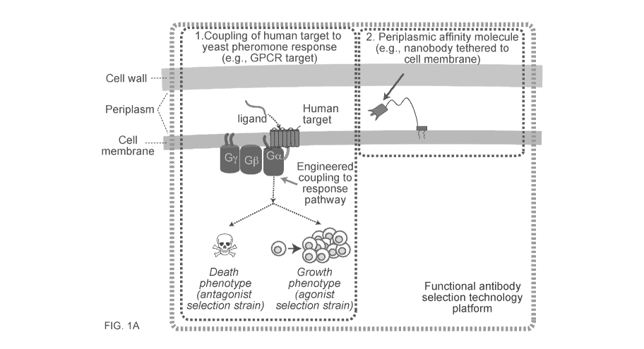
[0058] FIGS. 1A-1D show novel yeast cell display method for screening for
antibodies that
modulate the function of GPCRs. FIG. lA shows the unique combination of 1)
functional human
GPCR-yeast coupling to 2) affinity molecule secretion and 3) affinity molecule
localization,
together in a high-throughput, highly engineerable yeast cellular platform.
Functional, properly
folded GPCR yields ScFvs (which can easily be converted to IgG antibodies) or
nanobodies that
are more likely to function as therapeutics in the human organismal context.
FIG. 1B shows use
of an "antagonist selection strain" to find antagonists. FIG. 1C shows use of
an "agonist selection
strain" to find agonists. By altering the logic of reporters and selectable
markers coupled to the
pheromone response system output, the platform can be used to select for
agonists or antagonists.
FIG. 1D shows direct functional screening yields therapeutic antibody
candidates that would
normally be missed in traditional screening, which could yield novel binding
modes and
functional modulation of GPCR targets. Because of the ease of genetic
engineering in yeast, we
-12-
CA 03084315 2020-06-02
WO 2019/118362
PCT/US2018/064775
can both adjust antibody and GPCR expression levels, and tune selectable and
screenable
reporters to be very sensitive. Both enable us to find low-affinity but
functional candidates,
which can easily be affinity matured later.
[0059] FIG. 2 shows method of reducing background/false positive in "halo
assays". 107 cells of
the parental strain (left) and the current platform strain (NIY326, right)
were plated on agar
media. A filter paper disc was placed onto the plate and spotted with 3 ill of
1 mM alpha factor.
A zone of no-growth in response to ligand (the desired phenotypic response)
was observed in
both, but in the parental strain (Left), suppressor mutants arise and grow
into colonies in the
presence of pheromone (colonies in halo region). In platform strain NI326
(Right), we have
reduced the background rate to ¨10-7, as demonstrated in the clear halo zone
and lack of
background suppressor mutations that would act as false positives in an
antagonist selection.
[0060] FIG. 3 shows affinity molecule targeting vector structure and concept.
We cloned the
affinity molecule downstream of a secretion signal and upstream of a linker
and extracellular
membrane-anchoring domain from GPI. When expressed in cells, the protein is
secreted into the
extracellular space, and then the GPI domain is processed to leave a domain
with a GPI that
binds to the membrane, which tethers the affinity molecules to this cell and
leaves it free to
interact with the target GPCR on its extracellular face.
[0061] FIGS. 4A and 4B show verification of affinity molecule
expression/targeting vector. FIG.
4A shows that if an expressed anti-GFP nanobody properly folds and localizes,
a GFP applied
from the outside of the cell (after cell wall digestion) should label the cell
membrane. FIG. 4B
shows images of yeast expressing an anti-GFP nanobody using our targeting
vector, after cell
wall digestion and applying purified GFP protein indicate GFP binding at the
membrane. No
fluorescence was observed in control cells (data not shown) Left, brightfield;
Right, GFP
channel.
[0062] FIGS. 5A and 5B show verification of the plasmid dependence of alpha
factor resistant
clones. FIG. 5A shows a schematic of the strategy. FIG. 5B shows an example of
a "candidate"
clone that exhibited alpha factor-resistant growth as analyzed by a halo
assay, and then showed
no resistance after forcing the plasmid to drop.
[0063] FIG. 6 shows a workflow schematic.
-13-
CA 03084315 2020-06-02
WO 2019/118362
PCT/US2018/064775
[0064] FIG. 7 shows the impact on growth rate of yeast cells by activation of
the cannabinoid
receptor type 2 (CB2 receptor) using VH1-1 domain agonists displayed in the
periplasmic space in
various ways.
DETAILED DESCRIPTION OF THE INVENTION
[0065] A plethora of therapeutic targets in such diseases as cancer and
inflammation involve cell
membrane-associated proteins. However, many cell membrane-associated proteins
with the
greatest therapeutic potential for high-impact diseases are difficult to drug.
Although small
molecules affecting the function of these proteins are easily found, they are
often non-specific.
Unlike small molecules, antibodies and related affinity molecules (e.g.,
nanobodies and ScFvs
and Fabs), are an appealing therapeutic class due to their potentially
superior specificity,
functional diversity, and pharmacological properties. Additionally, antibodies
can better interact
with extracellular domains and loops, which can modulate the structure (and
thus function) of
cell membrane-associated proteins, such as GPCRs, in more sophisticated ways
than small
molecules. However, there is to date not a single approved GPCR antibody
therapeutic in the
United States, and only one worldwide, in Japan.
[0066] Current yeast or phage display workflows identify antibodies that
tightly bind but often
do not affect the function of cell membrane-associated proteins, such as
GPCRs. The antigens
used are often fragments that do not represent the functional protein
accessible to the antibody in
vivo, or are heterogeneously structured full-length protein preparations. The
workflow also
overlooks a tremendous fraction of total functional diversity, because most
antibodies are never
functionally assayed. What is needed is a high-throughput platform to directly
select for
antibodies that modulate the function of cell membrane-associated proteins,
such as GPCRs.
[0067] It is much less straightforward to develop antibodies that alter the
function of cell
membrane-associated proteins, such as GPCRs (Jo 2015, Hutchings 2010). This is
due primarily
to the following issues with many current solutions: 1) The antigens used are
lacking. Antigens
derived from extracellular peptides or fragments may be good for developing
antibodies for
Western blots, but do not structurally represent therapeutically relevant
targets. Further,
homogenously, functionally folded full-length protein in lipids or detergents
can be hard to
prepare in sufficient amounts for immunization, phage display, or yeast
display. 2) Antibodies
selected for their high affinity are mostly non-functional; they bind to
regions in the protein that
do not affect function. 3) Workflows lose significant antibody diversity¨and
therefore
functionality¨in selected antibodies. By first selecting for antibodies that
bind tightly and
discarding the rest, huge amounts of functional diversity are lost. Mammalian
cell systems have
-14-
CA 03084315 2020-06-02
WO 2019/118362
PCT/US2018/064775
been created to functionally screen antibody candidate subsets in an autocrine
fashion (Zhang
2014), which partially addresses issue 2, but due to transformation
efficiencies (-104) and limited
engineerability of selectable/screenable readouts, they are limited to screens
of small subsets of
candidates.
[0068] Our innovation includes combining cell membrane-associated protein-to-
yeast
pheromone response coupling and expressing affinity molecules that act in cis
in the same cell in
a high-throughput platform. This enables direct and high-throughput functional
selection of
affinity molecules in the yeast periplasmic space. Antibodies and related
affinity molecules are
large molecules with complex folding patterns that must be maintained to
retain binding activity.
While unstructured, short peptides may be able to be localized to the yeast
periplasmic space, it
was not previously known that antibodies and related affinity molecules could
be displayed in the
yeast periplasmic space and retain binding activity.
[0069] The practice of the present invention will employ, unless otherwise
indicated,
conventional methods of pharmacology, chemistry, biochemistry, recombinant DNA
techniques
and immunology, within the skill of the art. Such techniques are explained
fully in the literature.
See, e.g., High Throughput Screening: Methods and Protocols (Methods in
Molecular Biology,
W.P. Janzen ed., Humana Press, 31d edition, 2016); G Protein-Coupled
Receptors: Structure,
Signaling, and Physiology (S. Siehler and G. Milligan eds., Cambridge
University Press, 2010);
Handbook of Experimental Immunology,Vols. I-TV (D.M. Weir and C.C. Blackwell
eds.,
Blackwell Scientific Publications); A.L. Lehninger, Biochemistry (Worth
Publishers, Inc.,
current addition); Sambrook, et al., Molecular Cloning: A Laboratory Manual
(31d Edition,
2001); Methods In Enzymology (S. Colowick and N. Kaplan eds., Academic Press,
Inc.).
[0070] All publications, patents and patent applications cited herein, whether
supra or infra, are
hereby incorporated by reference in their entireties.
I. DEFINITIONS
[0071] In describing the present invention, the following terms will be
employed, and are
intended to be defined as indicated below.
[0072] It must be noted that, as used in this specification and the appended
claims, the singular
forms "a," "an" and "the" include plural referents unless the content clearly
dictates otherwise.
Thus, for example, reference to "a cell" includes a mixture of two or more
cells, and the like.
-15-
CA 03084315 2020-06-02
WO 2019/118362
PCT/US2018/064775
[0073] The term "about," particularly in reference to a given quantity, is
meant to encompass
deviations of plus or minus five percent.
[0074] The term "about," particularly in reference to a given quantity,
encompasses and
describes the given quantity itself
[0075] The terms "polypeptide" and "protein" refer to a polymer of amino acid
residues and are
not limited to a minimum length. Thus, peptides, oligopeptides, dimers,
multimers, and the like,
are included within the definition. Both full length proteins and fragments
thereof are
encompassed by the definition. The terms also include post expression
modifications of the
polypeptide, for example, glycosylation, acetylation, phosphorylation,
hydroxylation, and the
like. Furthermore, for purposes of the present invention, a "polypeptide"
refers to a protein
which includes modifications, such as deletions, additions and substitutions
to the native
sequence. These modifications may be deliberate, as through site directed
mutagenesis, or may
be accidental, such as through mutations of hosts which produce the proteins
or errors due to
PCR amplification.
[0076] The term "antibody" encompasses monoclonal antibodies as well as hybrid
antibodies,
altered antibodies, chimeric antibodies, and humanized antibodies. The term
antibody includes:
hybrid (chimeric) antibody molecules (see, for example, Winter et al. (1991)
Nature 349:293-
299; and U.S. Pat. No. 4,816,567); F(ab1)2 and F(ab) fragments; F, molecules
(noncovalent
heterodimers, see, for example, Inbar et al. (1972) Proc Natl Acad Sci USA
69:2659-2662; and
Ehrlich et al. (1980) Biochem 19:4091-4096); single-chain Fv molecules (scFv)
(see, e.g., Huston
et al. (1988) Proc Natl Acad Sci USA 85:5879-5883); nanobodies or single-
domain antibodies
(sdAb) (see, e.g., Wang et al. (2016) Int J Nanomedicine 11:3287-3303, Vincke
et al. (2012)
Methods Mol Biol 911:15-26; dimeric and trimeric antibody fragment constructs;
minibodies
(see, e.g., Pack et al. (1992) Biochem 31:1579-1584; Cumber et al. (1992) J
Immunology
149B:120-126); humanized antibody molecules (see, e.g., Riechmann et al.
(1988) Nature
332:323-327; Verhoeyan et al. (1988) Science 239:1534-1536; and U.K. Patent
Publication No.
GB 2,276,169, published 21 Sep. 1994); and, any functional fragments obtained
from such
molecules, wherein such fragments retain specific-binding properties of the
parent antibody
molecule.
[0077] The phrase "specifically (or selectively) binds" with reference to
binding of an antibody
to an antigen (e.g., GPCR) refers to a binding reaction that is determinative
of the presence of the
antigen in a heterogeneous population of proteins and other biologics. Thus,
under designated
-16-
CA 03084315 2020-06-02
WO 2019/118362
PCT/US2018/064775
immunoassay conditions, the specified antibodies bind to a particular antigen
at least two times
the background and do not substantially bind in a significant amount to other
antigens present in
the sample. Specific binding to an antigen under such conditions may require
an antibody that is
selected for its specificity for a particular antigen. For example, antibodies
raised to an antigen
from specific species such as rat, mouse, or human can be selected to obtain
only those
antibodies that are specifically immunoreactive with the antigen and not with
other proteins,
except for polymorphic variants and alleles. This selection may be achieved by
subtracting out
antibodies that cross-react with molecules from other species. A variety of
immunoassay formats
may be used to select antibodies specifically immunoreactive with a particular
antigen. For
example, solid-phase ELISA immunoassays are routinely used to select
antibodies specifically
immunoreactive with a protein (see, e.g., Harlow & Lane. Antibodies, A
Laboratory Manual
(1988), for a description of immunoassay formats and conditions that can be
used to determine
specific immunoreactivity). Typically, a specific or selective reaction will
be at least twice
background signal or noise and more typically more than 10 to 100 times
background.
[0078] A protein is said to "interact" with another protein if it binds
specifically (e.g., in a lock-
and-key type mechanism), non-specifically or in some combination of specific
and non-specific
binding. A first protein "interacts preferentially" with a second protein if
it binds (non-
specifically and/or specifically) to the second protein with greater affinity
and/or greater
specificity than it binds to other proteins. The term "affinity" refers to the
strength of binding
and can be expressed quantitatively as a dissociation constant (Kd). It is to
be understood that
specific binding does not necessarily require interaction between specific
amino acid residues
and/or motifs of each protein. For example, in certain embodiments, a first
protein may interact
preferentially with a second protein but, nonetheless, may be capable of
binding other
polypeptides at a weak, yet detectable, level (e.g., 10% or less of the
binding shown to the
polypeptide of interest). Typically, weak binding, or background binding, is
readily discernible
from the preferential interaction with the compound or polypeptide of
interest, e.g., by use of
appropriate controls.
[0079] As used herein, the term "binding pair" refers to first and second
molecules that
specifically bind to each other. "Specific binding" of the first member of the
binding pair to the
second member of the binding pair in a sample is evidenced by the binding of
the first member to
the second member, or vice versa, with greater affinity and specificity than
to other components
in the sample. The binding between the members of the binding pair is
typically noncovalent.
Examples include antigen-antibody, receptor-hormone, receptor-ligand, receptor-
agonist, and
receptor-antagonist binding pairs.
-17-
CA 03084315 2020-06-02
WO 2019/118362
PCT/US2018/064775
[0080] As used herein, the term "ligand" refers to a molecule that binds to
another molecule, e.g.,
an antigen binding to an antibody, a hormone, agonist, or antagonist binding
to a receptor, a
neurotransmitter binding to an ion channel, or a substrate, inhibitor, or
allosteric effector binding
to an enzyme and includes natural and synthetic biomolecules, such as
proteins, polypeptides,
peptides, nucleic acid molecules, carbohydrates, sugars, lipids, lipoproteins,
small molecules,
natural and synthetic organic and inorganic materials, synthetic polymers,
aptamers, and the
like.
[0081] The term "polynucleotide," as known in the art, generally refers to a
nucleic acid
molecule. A "polynucleotide" can include both double- and single-stranded
sequences and refers
to, but is not limited to, prokaryotic sequences, eukaryotic mRNA, cDNA from
viral, prokaryotic
or eukaryotic mRNA, genomic RNA and DNA sequences from viral (e.g. RNA and DNA
viruses
and retroviruses), prokaryotic DNA or eukaryotic (e.g., mammalian) DNA, and
especially
synthetic DNA sequences. The term also captures sequences that include any of
the known base
analogs of DNA and RNA, and includes modifications such as deletions,
additions and
substitutions (generally conservative in nature), to the native sequence.
These modifications may
be deliberate, as through site-directed mutagenesis, or may be accidental,
such as through
mutations of hosts including polynucleotides encoding variant polypeptides for
display.
Modifications of polynucleotides may have any number of effects including, for
example,
facilitating expression of the polypeptide product in a host cell.
[0082] A polynucleotide can encode a biologically active protein or
polypeptide. Depending on
the nature of the polypeptide encoded by the polynucleotide, a polynucleotide
can include as
little as 10 nucleotides, e.g., where the polynucleotide encodes an antigen or
epitope. Typically,
the polynucleotide encodes peptides of at least 10, 11, 12, 13, 14, 15, 16,
17, 18, 19, 20, 21, 22,
23, 24, 25, 30 or even more amino acids.
[0083] The terms "variant," "analog" and "mutein" refer to biologically active
derivatives of the
reference molecule that retain desired activity (e.g., efficient polypeptide
display) as described
herein. In general, the terms "variant" and "analog" refer to compounds having
a native
polypeptide sequence and structure with one or more amino acid additions,
substitutions and/or
deletions relative to the native molecule, as long as the modifications do not
destroy biological
activity and which are "substantially homologous" to the reference molecule as
defined below. In
general, the amino acid sequences of such analogs will have a high degree of
sequence homology
to the reference sequence, e.g., amino acid sequence homology of more than
50%, generally
more than 60%-70%, even more particularly 80%-85% or more, such as at least
90%-95% or
-18-
CA 03084315 2020-06-02
WO 2019/118362
PCT/US2018/064775
more, when the two sequences are aligned. Often, the analogs will include the
same number of
amino acids but will include substitutions, as explained herein. The term
"mutein" further
includes polypeptides having one or more amino acid-like molecules including
but not limited to
compounds comprising only amino and/or imino molecules, polypeptides
containing one or more
analogs of an amino acid (including, for example, unnatural amino acids,
etc.), polypeptides with
substituted linkages, as well as other modifications known in the art, both
naturally occurring and
non-naturally occurring (e.g., synthetic), cyclized, branched molecules and
the like. The term
also includes molecules comprising one or more N-substituted glycine residues
(a "peptoid") and
other synthetic amino acids or peptides. (See, e.g., U.S. Pat. Nos. 5,831,005;
5,877,278; and
5,977,301; Nguyen et al., Chem. Biol. (2000) 7:463-473; and Simon et al.,
Proc. Natl. Acad. Sci.
USA (1992) 89:9367-9371 for descriptions of peptoids). Methods for making
polypeptide
analogs and muteins are known in the art and are described further below.
[0084] Analogs generally include substitutions that are conservative in
nature, i.e., those
substitutions that take place within a family of amino acids that are related
in their side chains.
Specifically, amino acids are generally divided into four families: (1)
acidic¨aspartate and
glutamate; (2) basic--lysine, arginine, histidine; (3) non-polar--alanine,
valine, leucine,
isoleucine, proline, phenylalanine, methionine, tryptophan; and (4) uncharged
polar--glycine,
asparagine, glutamine, cysteine, serine threonine, tyrosine. Phenylalanine,
tryptophan, and
tyrosine are sometimes classified as aromatic amino acids. For example, it is
reasonably
predictable that an isolated replacement of leucine with isoleucine or valine,
an aspartate with a
glutamate, a threonine with a serine, or a similar conservative replacement of
an amino acid with
a structurally related amino acid, will not have a major effect on the
biological activity. For
example, the polypeptide of interest may include up to about 5-10 conservative
or non-
conservative amino acid substitutions, or even up to about 15-25 conservative
or non-
conservative amino acid substitutions, or any integer between 5-25, so long as
the desired
function of the molecule remains intact. One of skill in the art may readily
determine regions of
the molecule of interest that can tolerate change by reference to Hopp/Woods
and Kyte-Doolittle
plots, well known in the art.
[0085] "Recombinant" as used herein to describe a nucleic acid molecule means
a polynucleotide
of genomic, cDNA, viral, semisynthetic, or synthetic origin which, by virtue
of its origin or
manipulation is not associated with all or a portion of the polynucleotide
with which it is
associated in nature. The term "recombinant" as used with respect to a
protein, polypeptide, or
peptide means a polypeptide produced by expression of a recombinant
polynucleotide. In
general, the gene of interest is cloned and then expressed in transformed
organisms, as described
-19-
CA 03084315 2020-06-02
WO 2019/118362
PCT/US2018/064775
further below. The host organism expresses the foreign gene to produce the
protein under
expression conditions.
[0086] A "polynucleotide coding sequence" or a sequence that "encodes" a
selected polypeptide,
is a nucleic acid molecule that is transcribed (in the case of DNA) and
translated (in the case of
mRNA) into a polypeptide in vivo when placed under the control of appropriate
regulatory
sequences (or "control elements"). The boundaries of the coding sequence are
determined by a
start codon at the 5' (amino) terminus and a translation stop codon at the 3'
(carboxy) terminus.
A transcription termination sequence may be located 3' to the coding sequence.
Typical "control
elements," include, but are not limited to, transcription regulators, such as
promoters,
transcription enhancer elements, transcription termination signals, and
polyadenylation
sequences; and translation regulators, such as sequences for optimization of
initiation of
translation, e.g., Shine-Dalgarno (ribosome binding site) sequences, Kozak
sequences (i.e.,
sequences for the optimization of translation, located, for example, 5' to the
coding sequence),
leader sequences (heterologous or native), translation initiation codon (e.g.,
ATG), and
translation termination sequences. Promoters can include inducible promoters
(where expression
of a polynucleotide sequence operably linked to the promoter is induced by an
analyte, cofactor,
regulatory protein, etc.), repressible promoters (where expression of a
polynucleotide sequence
operably linked to the promoter is induced by an analyte, cofactor, regulatory
protein, etc.), and
constitutive promoters.
[0087] "Operably linked" refers to an arrangement of elements wherein the
components so
described are configured so as to perform their usual function. Thus, a given
promoter operably
linked to a coding sequence is capable of effecting the expression of the
coding sequence when
the proper enzymes are present. The promoter need not be contiguous with the
coding sequence,
so long as it functions to direct the expression thereof Thus, for example,
intervening
untranslated yet transcribed sequences can be present between the promoter
sequence and the
coding sequence and the promoter sequence can still be considered "operably
linked" to the
coding sequence.
[0088] By "fragment" is intended a molecule consisting of only a part of the
intact full-length
sequence and structure. The fragment can include a C-terminal deletion an N-
terminal deletion,
and/or an internal deletion of the peptide. Active fragments of a particular
protein or peptide will
generally include at least about 5-10 contiguous amino acid residues of the
full-length molecule,
preferably at least about 15-25 contiguous amino acid residues of the full-
length molecule, and
most preferably at least about 20-50 or more contiguous amino acid residues of
the full-length
-20-
CA 03084315 2020-06-02
WO 2019/118362
PCT/US2018/064775
molecule, or any integer between 5 amino acids and the full-length sequence,
provided that the
fragment in question retains biological activity.
[0089] "Substantially purified" generally refers to isolation of a substance
(compound,
polynucleotide, protein, polypeptide, polypeptide composition) such that the
substance comprises
the majority percent of the sample in which it resides. Typically in a sample,
a substantially
purified component comprises 50%, preferably 80%-85%, more preferably 90-95%
of the
sample. Techniques for purifying polynucleotides and polypeptides of interest
are well-known in
the art and include, for example, ion-exchange chromatography, affinity
chromatography and
sedimentation according to density.
[0090] By "isolated" is meant, when referring to a polypeptide, that the
indicated molecule is
separate and discrete from the whole organism with which the molecule is found
in nature or is
present in the substantial absence of other biological macro-molecules of the
same type. The
term "isolated" with respect to a polynucleotide is a nucleic acid molecule
devoid, in whole or
part, of sequences normally associated with it in nature; or a sequence, as it
exists in nature, but
having heterologous sequences in association therewith; or a molecule
disassociated from the
chromosome.
[0091] "Homology" refers to the percent identity between two polynucleotide or
two polypeptide
molecules. Two nucleic acid, or two polypeptide sequences are "substantially
homologous" to
each other when the sequences exhibit at least about 50%, preferably at least
about 75%, more
preferably at least about 80%-85%, preferably at least about 90%, and most
preferably at least
about 95%-98% sequence identity over a defined length of the molecules. As
used herein,
substantially homologous also refers to sequences showing complete identity to
the specified
sequence.
[0092] In general, "identity" refers to an exact nucleotide-to-nucleotide or
amino acid-to-amino
acid correspondence of two polynucleotides or polypeptide sequences,
respectively. Percent
identity can be determined by a direct comparison of the sequence information
between two
molecules (the reference sequence and a sequence with unknown % identity to
the reference
sequence) by aligning the sequences, counting the exact number of matches
between the two
aligned sequences, dividing by the length of the reference sequence, and
multiplying the result by
100. Readily available computer programs can be used to aid in the analysis,
such as ALIGN,
Dayhoff, M.O. in Atlas of Protein Sequence and Structure M.O. Dayhoff ed., 5
Suppl.
3:353-358, National biomedical Research Foundation, Washington, DC, which
adapts the local
-21-
CA 03084315 2020-06-02
WO 2019/118362
PCT/US2018/064775
homology algorithm of Smith and Waterman Advances in App!. Math. 2:482-489,
1981 for
peptide analysis. Programs for determining nucleotide sequence identity are
available in the
Wisconsin Sequence Analysis Package, Version 8 (available from Genetics
Computer Group,
Madison, WI) for example, the BESTFIT, FASTA and GAP programs, which also rely
on the
Smith and Waterman algorithm. These programs are readily utilized with the
default parameters
recommended by the manufacturer and described in the Wisconsin Sequence
Analysis Package
referred to above. For example, percent identity of a particular nucleotide
sequence to a
reference sequence can be determined using the homology algorithm of Smith and
Waterman
with a default scoring table and a gap penalty of six nucleotide positions.
[0093] Another method of establishing percent identity in the context of the
present invention is
to use the MPSRCH package of programs copyrighted by the University of
Edinburgh,
developed by John F. Collins and Shane S. Sturrok, and distributed by
IntelliGenetics, Inc.
(Mountain View, CA). From this suite of packages, the Smith-Waterman algorithm
can be
employed where default parameters are used for the scoring table (for example,
gap open penalty
of 12, gap extension penalty of one, and a gap of six). From the data
generated the "Match"
value reflects "sequence identity." Other suitable programs for calculating
the percent identity or
similarity between sequences are generally known in the art, for example,
another alignment
program is BLAST, used with default parameters. For example, BLASTN and BLASTP
can be
used using the following default parameters: genetic code = standard; filter =
none; strand =
both; cutoff = 60; expect = 10; Matrix = BLOSUM62; Descriptions = 50
sequences; sort by =
HIGH SCORE; Databases = non-redundant, GenBank + EMBL + DDBJ + PDB + GenBank
CDS translations + Swiss protein + Spupdate + PIR. Details of these programs
are readily
available.
[0094] Alternatively, homology can be determined by hybridization of
polynucleotides under
conditions which form stable duplexes between homologous regions, followed by
digestion with
single-stranded-specific nuclease(s), and size determination of the digested
fragments. DNA
sequences that are substantially homologous can be identified in a Southern
hybridization
experiment under, for example, stringent conditions, as defined for that
particular system.
Defining appropriate hybridization conditions is within the skill of the art.
See, e.g., Sambrook
et al., supra; DNA Cloning, supra; Nucleic Acid Hybridization, supra.
[0095] "Expression cassette" or "expression construct" refers to an assembly
which is capable of
directing the expression of the sequence(s) or gene(s) of interest. An
expression cassette
generally includes control elements, as described above, such as a promoter
which is operably
-22-
CA 03084315 2020-06-02
WO 2019/118362
PCT/US2018/064775
linked to (so as to direct transcription of) the sequence(s) or gene(s) of
interest, and often
includes a polyadenylation sequence as well. Within certain embodiments of the
invention, the
expression cassette described herein may be contained within a plasmid or
viral vector construct.
In addition to the components of the expression cassette, the construct may
also include, one or
more selectable markers, a signal which allows the construct to exist as
single stranded DNA
(e.g., a M13 origin of replication), at least one multiple cloning site, and
an origin of replication
(e.g., autonomously replicating sequence in yeast).
[0096] The term "transfection" is used to refer to the uptake of foreign DNA
by a cell. A cell has
been "transfected" when exogenous DNA has been introduced inside the cell
membrane. A
number of transfection techniques are generally known in the art. See, e.g.,
Graham et al. (1973)
Virology, 52:456, Sambrook et al. (2001) Molecular Cloning, a laboratory
manual, 3rd edition,
Cold Spring Harbor Laboratories, New York, Davis et al. (1995) Basic Methods
in Molecular
Biology, 2nd edition, McGraw-Hill, and Chu et al. (1981) Gene 13:197. Such
techniques can be
used to introduce one or more exogenous DNA moieties into suitable host cells.
The term refers
to both stable and transient uptake of the genetic material, and includes
uptake of peptide- or
antibody-linked DNAs.
[0097] A "vector" is capable of transferring nucleic acid sequences to target
cells (e.g., viral
vectors, non-viral vectors, particulate carriers, and liposomes). Typically,
"vector construct,"
"expression vector," and "gene transfer vector," mean any nucleic acid
construct capable of
directing the expression of a nucleic acid of interest and which can transfer
nucleic acid
sequences to target cells. Thus, the term includes cloning and expression
vehicles, as well as
plasmid and viral vectors.
[0098] The term "transformation" refers to the insertion of an exogenous
polynucleotide into a
host cell, irrespective of the method used for the insertion. For example,
direct uptake,
transduction or f-mating are included. The exogenous polynucleotide may be
maintained as a
non-integrated vector, for example, a plasmid, or alternatively, may be
integrated into the host
genome.
[0099] "Recombinant host cells", "host cells," "cells", "cell lines," "cell
cultures," and other such
terms denoting microorganisms or eukaryotic cell lines cultured as unicellular
entities refer to
cells which can be, or have been, used as recipients for recombinant vector or
other transferred
DNA, and include the original progeny of the original cell which has been
transfected.
-23-
CA 03084315 2020-06-02
WO 2019/118362
PCT/US2018/064775
[0100] A "coding sequence" or a sequence which "encodes" a selected
polypeptide, is a nucleic
acid molecule which is transcribed (in the case of DNA) and translated (in the
case of mRNA)
into a polypeptide in vivo when placed under the control of appropriate
regulatory sequences (or
"control elements"). The boundaries of the coding sequence can be determined
by a start codon
at the 5' (amino) terminus and a translation stop codon at the 3' (carboxy)
terminus. A coding
sequence can include, but is not limited to, cDNA from viral, prokaryotic or
eukaryotic mRNA,
genomic DNA sequences from viral or prokaryotic DNA, and even synthetic DNA
sequences. A
transcription termination sequence may be located 3' to the coding sequence.
[0101] Typical "control elements," include, but are not limited to,
transcription promoters,
transcription enhancer elements, transcription termination signals,
polyadenylation sequences
(located 3' to the translation stop codon), sequences for optimization of
initiation of translation
(located 5' to the coding sequence), and translation termination sequences.
[0102] The terms "label" and "detectable label" refer to a molecule capable of
detection,
including, but not limited to, radioactive isotopes, stable (non-radioactive)
heavy isotopes,
fluorescers, chemiluminescers, enzymes, enzyme substrates, enzyme cofactors,
enzyme
inhibitors, chromophores, dyes, metal ions, metal sols, ligands (e.g., biotin
or haptens) and the
like. The term "fluorescer" refers to a substance or a portion thereof that is
capable of exhibiting
fluorescence in the detectable range. Particular examples of labels that may
be used with the
invention include, but are not limited to radiolabels (e.g., 3H, 1251, 35S,
'4C, or 32P), stable (non-
radioactive) heavy isotopes (e.g., '3C or '5N), phycoerythrin, fluorescein, 7-
nitrobenzo-2-oxa-1,3-
diazole (NBD), YPet, CyPet, Cascade blue, allophycocyanin, Alexa dyes (e.g.,
Alexa 350, Alexa
430, Alexa 488, Alexa 532, Alexa 546, Alexa 555, Alexa 594, Alexa 647, Alexa
660, Alexa 680,
and Alexa 750), Atto dyes (e.g., Atto 488, Atto 532, Atto 550, Atto 565, Atto
590, Atto 610, Atto
620, Atto 635, Atto 647, Atto 655, and Atto 680), cyanine dyes (e.g., Cy3,
Cy5, and Cy7), TYE
563, TYE 665, TYE 705, TEX 615, JOE, TET, HEX, TAMRA, ROX, rhodamine, dansyl,
umbelliferone, Texas red, luminol, acradimum esters, biotin or other
streptavidin-binding
proteins, magnetic beads, electron dense reagents, green fluorescent protein
(GFP), enhanced
green fluorescent protein (EGFP), yellow fluorescent protein (YFP), enhanced
yellow fluorescent
protein (EYFP), blue fluorescent protein (BFP), red fluorescent protein (RFP),
TagRFP, Dronpa,
Padron, mApple, mCherry, rsCherry, rsCherryRev, firefly luciferase, Renilla
luciferase,
NADPH, beta-galactosidase, horseradish peroxidase, glucose oxidase, alkaline
phosphatase,
chloramphenical acetyl transferase, and urease. Enzyme tags are used with
their cognate
substrate. As with many of the standard procedures associated with the
practice of the invention,
skilled artisans will be aware of additional labels that can be used.
-24-
CA 03084315 2020-06-02
WO 2019/118362
PCT/US2018/064775
MODES OF CARRYING OUT THE INVENTION
[0103] Before describing the present invention in detail, it is to be
understood that this invention
is not limited to particular formulations or process parameters as such may,
of course, vary. It is
also to be understood that the terminology used herein is for the purpose of
describing particular
embodiments of the invention only, and is not intended to be limiting.
[0104] Although a number of methods and materials similar or equivalent to
those described
herein can be used in the practice of the present invention, the preferred
materials and methods
are described herein.
[0105] The present invention is based on the development of methods for
displaying
recombinant proteins in the periplasmic space of yeast cells. In particular,
recombinant proteins
are linked to a cell membrane-spanning transmembrane domain, a cell-membrane
associated
protein domain that is on the external face of the yeast cell membrane, a
protein that binds to the
inner face of the yeast cell wall, or a periplasmic protein in order to
display proteins in the yeast
periplasmic space. Recombinant proteins can also be targeted to the periplasm
by linking the
recombinant protein to a secretion signal. In addition, a target protein of
interest can be
coexpressed in yeast such that it is localized to the plasma membrane or
periplasmic space and
accessible to displayed proteins. In particular embodiments, the inventors
have used their method
of yeast periplasmic display to screen for antibodies that bind to and
modulate the function of
human GPCRs (see Examples). Antibodies displayed in the periplasmic space of a
yeast cell are
in sufficient proximity to bind to a GPCR expressed in the cell membrane. The
inventors have
further developed a method for high-throughput screening of GPCRs for
antagonists and agonists
using periplasmic display by coupling human GPCRs to the yeast pheromone
response pathway.
[0106] In order to further an understanding of the invention, a more detailed
discussion is
provided below regarding yeast periplasmic display and methods of using it for
high-throughput
screening of protein libraries.
A. Periplasmic Display of Recombinant Proteins in Yeast
[0107] In one aspect, the invention relates to the display of a protein in the
periplasmic space of a
yeast host cell. In some embodiments, the yeast host cell comprises a protein
for display in the
yeast periplasmic space, a periplasm anchor protein linked to the protein to
be displayed in the
periplasmic space, and a target membrane protein of interest located in the
periplasmic space of
the yeast host cell. In some embodiments, the yeast host cell can be used to
determine if the
protein to be displayed in the periplasmic space specifically binds to or
affects the function of the
-25-
CA 03084315 2020-06-02
WO 2019/118362
PCT/US2018/064775
target membrane protein of interest. The protein to be displayed in the
periplasmic space of the
yeast host cell can be prepared by linking a recombinant protein to a
periplasm anchor protein
that localizes the recombinant protein to the periplasmic space of a yeast
cell. Linkage can be
covalent or noncovalent. For example, a recombinant protein may be linked
covalently to a
periplasm anchor protein in a fusion protein. Alternatively, a recombinant
protein variant may
form a complex with a periplasmic anchor protein, wherein the recombinant
protein and the
periplasmic anchor protein are linked noncovalently by molecular binding
interactions in the
complex. Alternatively, a recombinant protein variant and the periplasmic
anchor protein are
linked covalently by a non-peptidic bond in a complex. In some embodiments,
the non-peptidic
bond is a disulfide bond. A protein to be displayed in the periplasmic space
of the yeast host cell
can also be prepared by linking the protein to be displayed to a secretion
signal. In some
embodiments, the genus of the yeast host cell is selected from the group
consisting of
Saccharomyces, Candida, Pichia, Kluyveromyces, and Yarrowia. In some
embodiments, the
genus of the yeast host cells is Saccharomyces. In some embodiments, the
species of the yeast
host cell is Saccharomyces cerevisiae.
[0108] In another aspect, the invention relates to an antibody linked to a
periplasm anchor
protein. In some embodiments, the antibody linked to a periplasm anchor
protein further
comprises an additional modification, moiety or interacting protein. In some
embodiments, the
additional modification is a post-translational modification. In some
embodiments, the moiety is
an affinity tag, epitope, label or the like. In some embodiments, the antibody
is localized to a
yeast host cell periplasmic space. In some embodiments, when the antibody is
produced in or
introduced to a yeast host cell, the antibody is localized to a yeast host
cell periplasmic space. In
some embodiments, the antibody is linked to a periplasm anchor protein such
that the antibody is
localized to the periplasmic space. Linkage of the antibody to the periplasm
anchor protein can
be covalent or noncovalent. For example, an antibody may be linked covalently
to a periplasm
anchor protein in a fusion protein. Alternatively, an antibody may form a
complex with a
periplasmic anchor protein, wherein the antibody and the periplasmic anchor
protein are linked
noncovalently by molecular binding interactions in the complex. Alternatively,
an antibody and
the periplasmic anchor protein are linked covalently by a non-peptidic bond in
a complex. In
some embodiments, the non-peptidic bond is a disulfide bond. Also provided are
yeast host cells
comprising an antibody as described herein. In some embodiments, the genus of
the yeast host
cell is selected from the group consisting of Saccharomyces, Candida, Pichia,
Kluyveromyces,
and Yarrowia. In some embodiments, the genus of the yeast host cells is
Saccharomyces. In
some embodiments, the species of the yeast host cell is Saccharomyces
cerevisiae .
-26-
CA 03084315 2020-06-02
WO 2019/118362
PCT/US2018/064775
[0109] In another aspect, the invention relates to methods of localizing an
antibody to a yeast
host cell periplasmic space comprising linking the antibody to a periplasm
anchor protein such
that the antibody is localized to the periplasmic space. In some embodiments,
the antibody is
linked to a periplasm anchor protein such that the antibody is localized to
the periplasmic space.
Linkage of the antibody to the periplasm anchor protein can be covalent or
noncovalent. For
example, an antibody may be linked covalently to a periplasm anchor protein in
a fusion protein.
Alternatively, an antibody may form a complex with a periplasmic anchor
protein, wherein the
antibody and the periplasmic anchor protein are linked noncovalently by
molecular binding
interactions in the complex. Alternatively, an antibody and the periplasmic
anchor protein are
linked covalently by a non-peptidic bond in a complex. In some embodiments,
the non-peptidic
bond is a disulfide bond. In some embodiments, the genus of the yeast host
cell is selected from
the group consisting of Saccharomyces, Candida, Pichia, Kluyveromyces, and
Yarrowia. In
some embodiments, the genus of the yeast host cells is Saccharomyces. In some
embodiments,
the species of the yeast host cell is Saccharomyces cerevisiae.
[0110] In another aspect, the invention relates to methods of high-throughput
screening of
protein libraries for specific binding or functional characteristics by
displaying proteins in the
periplasmic space of yeast. A yeast periplasmic display library can be
prepared by linking
recombinant proteins to a periplasm anchor protein that localizes recombinant
proteins to the
periplasmic space of a yeast cell. Linkage can be covalent or noncovalent. For
example, a
recombinant protein may be linked covalently to a periplasm anchor protein in
a fusion protein.
Alternatively, a recombinant protein variant may form a complex with a
periplasmic anchor
protein, wherein the recombinant protein and the periplasmic anchor protein
are linked
noncovalently by molecular binding interactions in the complex. Alternatively,
a recombinant
protein variant and the periplasmic anchor protein are linked covalently by a
non-peptidic bond
in a complex. In some embodiments, the non-peptidic bond is a disulfide bond.
A yeast
periplasmic display library can also be prepared by linking recombinant
proteins to secretion
signals. In some embodiments, the genus of the yeast host cell is selected
from the group
consisting of Saccharomyces, Candida, Pichia, Kluyveromyces, and Yarrowia. In
some
embodiments, the genus of the yeast host cells is Saccharomyces. In some
embodiments, the
species of the yeast host cell is Saccharomyces cerevisiae .
[0111] Localization to the periplasmic space can be accomplished in a variety
of ways. In
certain embodiments, the recombinant protein is localized to the periplasm by
linking the
recombinant protein to a secretion signal that results in the recombinant
protein being secreted
into the extracellular space. In certain embodiments, a periplasm anchor
protein comprises a
-27-
CA 03084315 2020-06-02
WO 2019/118362
PCT/US2018/064775
signal sequence that directs transport of the periplasm anchor protein to the
yeast host cell
periplasm, plasma membrane, or cell wall such that a linked recombinant
protein is displayed in
the periplasm. For example, the periplasm anchor protein may comprise a signal
sequence that
directs transport of the periplasm anchor protein and the linked recombinant
protein to the yeast
host cell periplasm. Preferably, the periplasm anchor protein is sufficiently
large that the
periplasm anchor protein and the linked recombinant protein are retained in
the periplasm.
Alternatively, the periplasm anchor protein may be a component of a
periplasmic protein
complex that is sufficiently large that formation of the complex in the
periplasm results in
retention in the periplasm. In other embodiments, the periplasm anchor protein
comprises a
membrane-spanning transmembrane domain or a membrane-associated protein domain
that
localizes to an external face of the cell membrane such that the linked
recombinant protein is
projected into the periplasm. For example, a glycosylphosphatidylinositol
(GPI)-anchoring
domain that localizes to the plasma membrane can be used for this purpose. In
one embodiment,
the GPI-plasma membrane anchoring domain is a yapsin GPI plasma membrane
anchoring
domain such as, but not limited to, a YPS1, YPS2, YPS3, YPS4, YPS5, YPS6, or
YPS7 yapsin
GPI plasma membrane anchoring domain. In another embodiment, the periplasm
anchor protein
is a protein that binds to an inner face of the cell wall such that the
displayed protein variant is
projected into the periplasm. In certain embodiments, the periplasm anchor
protein and the
recombinant protein are covalently linked in a fusion protein variant and the
periplasm anchor
protein comprises a signal sequence that directs transport of the fusion
protein to the yeast host
cell periplasm, plasma membrane, or cell wall such that the fused protein
variant is displayed in
the periplasm. For example, the periplasm anchor protein may comprise a signal
sequence that
directs transport of the fusion protein to the yeast host cell periplasm.
Preferably, the periplasm
anchor protein is sufficiently large that the fusion protein is retained in
the periplasm.
Alternatively, the periplasm anchor protein may be a component of a
periplasmic protein
complex that is sufficiently large that formation of the complex in the
periplasm results in
retention of the fusion protein in the periplasm. In other embodiments, the
periplasm anchor
protein comprises a membrane-spanning transmembrane domain or a membrane-
associated
protein domain that localizes to an external face of the cell membrane such
that the fused protein
variant is projected into the periplasm. For example, a
glycosylphosphatidylinositol (GPI)-
anchoring domain that localizes to the plasma membrane can be used for this
purpose. In one
embodiment, the GPI-plasma membrane anchoring domain is a yapsin GPI plasma
membrane
anchoring domain such as, but not limited to, a YPS1, YPS2, YPS3, YPS4, YPS5,
YPS6, or
YPS7 yapsin GPI plasma membrane anchoring domain. In another embodiment, the
periplasm
anchor protein is a protein that binds to an inner face of the cell wall such
that the displayed
-28-
CA 03084315 2020-06-02
WO 2019/118362
PCT/US2018/064775
protein variant is projected into the periplasm. In certain embodiments, the
periplasm anchor
protein is a fragment of a full-length protein that retains the ability to be
localized to the
periplasm.
[0112] Any type of protein may be displayed in the periplasmic space of a
yeast cell,
including, but not limited to antibodies, antibody mimetics, aptamers,
antigens, enzymes,
receptors, transporters, ion channels, hormones, substrates, agonists,
antagonists, or ligands. The
yeast periplasmic display library presents a plurality of such proteins, which
can be screened for
binding and/or biological activity in the presence of a target molecule of
interest. If located
extracellularly, the target molecule must be able to penetrate the yeast cell
wall to reach the
displayed proteins for screening. In certain embodiments, a target protein of
interest is
coexpressed with a displayed protein variant in a yeast cell at a location
accessible to the
displayed protein variant (e.g., in close enough proximity for the displayed
protein variant to
bind to and/or modulate the activity of the target protein of interest). For
example, the target
protein of interest may be localized to the yeast host cell plasma membrane or
periplasm near
where the protein variant is displayed. In particular, this method is
applicable to receptors, ion
channels, transporters, and other membrane proteins which localize to the
plasma membrane.
Thus, yeast periplasmic display can be used to present proteins to a target
membrane protein in
an environment substantially similar to its native environment.
[0113] Any polypeptides included in a fusion construct, including the
periplasm anchor protein
and displayed protein variant may be connected directly to each other by
peptide bonds or may
be separated by intervening amino acid sequences or linkers. Linker amino acid
sequences will
typically be short, e.g., 20 or fewer amino acids (i.e., 20, 19, 18, 17, 16,
15, 14, 13, 12, 11, 10, 9,
8, 7, 6, 5, 4, 3, 2, or 1). Examples include short peptide sequences which
facilitate cloning, poly-
glycine linkers (Glyn where n = 2, 3, 4, 5, 6, 7, 8, 9, 10 or more), histidine
tags (Hisi, where n = 3,
4, 5, 6, 7, 8, 9, 10 or more), linkers composed of glycine and serine
residues, wherein n = 1, 2, 3,
4, 5, 6, 7, 8, 9, 10, 11, 12, 13, 14, 15 or more), GSAT, SEG, and Z-EGFR
linkers. Linkers may
include restriction sites, which aid cloning and manipulation. Other suitable
linker amino acid
sequences will be apparent to those skilled in the art. (See e.g., Argos
(1990) J. Mol. Biol.
211(4):943-958; Crasto et al. (2000) Protein Eng. 13:309-312; George et al.
(2002) Protein Eng.
15:871-879; Arai et al. (2001) Protein Eng. 14:529-532; and the Registry of
Standard Biological
Parts (partsregistry.org/Protein_domains/Linker).
[0114] Optionally, a tag may be included in fusion constructs. Tags that can
be used in the
practice of the invention include, but are not limited to a His-tag, a Strep-
tag, a TAP-tag, an 5-
-29-
CA 03084315 2020-06-02
WO 2019/118362
PCT/US2018/064775
tag, an SBP-tag, an Arg-tag, a calmodulin-binding peptide tag, a cellulose-
binding domain tag, a
DsbA tag, a c-myc tag, a glutathione S-transferase tag, a FLAG tag, a HAT-tag,
a maltose-
binding protein tag, a NusA tag, and a thioredoxin tag.
B. Polynucleotides Encoding Periplasm-Anchored Protein Variants and Target
Proteins and Library Construction
[0115] Polynucleotides encoding periplasm-anchored protein variants and target
proteins of
interest can be produced in any number of ways, all of which are well known in
the art. For
example, polynucleotides can be generated using recombinant techniques, well
known in the art.
One of skill in the art can readily determining nucleotide sequences that
encode the desired
proteins using standard methodology and the teachings herein.
[0116] Oligonucleotide probes can be devised based on known gene sequences and
used to
probe genomic or cDNA libraries. The polynucleotides with desired sequences
can then be
further isolated using standard techniques and, e.g., restriction enzymes
employed to truncate a
gene at desired portions of the full-length sequence. Similarly,
polynucleotides with sequences of
interest can be isolated directly from cells and tissues containing the same,
using known
techniques, such as phenol extraction and the sequence further manipulated to
produce desired
protein variants. See, e.g., Sambrook et al., supra, for a description of
techniques used to obtain
and isolate DNA.
[0117] The sequences encoding protein variants can also be produced
synthetically, for
example, based on known sequences. The nucleotide sequence can be designed
with the
appropriate codons for the particular amino acid sequence desired. The
complete sequence is
generally assembled from overlapping oligonucleotides prepared by standard
methods and
assembled into a complete coding sequence. See, e.g., Edge (1981) Nature
292:756; Nambair et
al. (1984) Science 223:1299; Jay et al. (1984) J. Biol. Chem. 259:6311;
Stemmer et al. (1995)
Gene 164:49-53.
[0118] Recombinant techniques are readily used to clone sequences encoding
protein variants
(e.g., antibodies) useful in the claimed invention that can then be
mutagenized in vitro by the
replacement of the appropriate base pair(s) to result in the codon for the
desired amino acid. Such
a change can include as little as one base pair, effecting a change in a
single amino acid, or can
encompass several base pair changes. Alternatively, the mutations can be
effected using a
mismatched primer that hybridizes to the parent nucleotide sequence (generally
cDNA
corresponding to the RNA sequence), at a temperature below the melting
temperature of the
-30-
CA 03084315 2020-06-02
WO 2019/118362
PCT/US2018/064775
mismatched duplex. The primer can be made specific by keeping primer length
and base
composition within relatively narrow limits and by keeping the mutant base
centrally located.
See, e.g., Innis et al, (1990) PCR Applications: Protocols for Functional
Genomics; Zoller and
Smith, Methods Enzymol. (1983) 100:468. Primer extension is effected using DNA
polymerase,
the product cloned and clones containing the mutated DNA, derived by
segregation of the primer
extended strand, selected. Selection can be accomplished using the mutant
primer as a
hybridization probe. The technique is also applicable for generating multiple
point mutations.
See, e.g., Dalbie-McFarland et al. Proc. Natl. Acad. Sci. USA (1982) 79:6409.
[0119] The diversity of a display library will depend on the method of
mutagenesis that is
used. Cassette mutagenesis can be used to quickly generate a large number of
mutations by
insertion of mutagenic cassettes into a nucleic acid (see, e.g., Worrall
(1994) Methods Mol. Biol.
30:199-210, Kegler-Ebo et al. (1994) Nucleic Acids Research. 22 (9):1593-
1599). In addition,
random mutagenesis can also be used to generate large numbers of variants for
a display library.
Suitable methods of random mutagenesis include, but are not limited to, error-
prone PCR, rolling
circle error-prone PCR, chemical mutagenesis, mutagenesis in a mutator strain
with deficient
DNA repair pathways, insertion mutagenesis using a transposon system, or DNA
shuffling (see,
e.g., McCullum et al. (2010) Methods Mol. Biol. 634:103-109, Fujii et al.
(2014) Methods Mol.
Biol. 1179:23-29, Muteeb (2010) Methods Mol. Biol. 634:411-419, Bose (2016)
Methods Mol.
Biol. 1373:111-115, Labrou (2010) Curr Protein Pept Sci. 11(1):91-100, Wilson
et al. (2011)
Methods Mol. Biol. 765:359-371). Such methods can be used to efficiently
generate a large
number of variants with modifications to a parent nucleic acid molecule. In
some embodiments,
a DNA library is constructed containing at least 106, preferably at least 108,
and more preferably
at least 1010 variants with unique sequences, using methods known in the art.
[0120] In some embodiments, antibody libraries are constructed for yeast
periplasmic display
by cloning natural antibodies from B lymphocytes obtained from blood donors.
Nucleic acids
encoding antibody light and heavy chains or fragments thereof containing
variable domain
complementarity-determining regions (e.g., Fab) can be amplified by PCR and
cloned into
vectors. ScFy antibodies can be generated by cloning into a vector that
connects the light and
heavy chains via a linker in one open reading frame. The blood donor can be of
any species. In
some embodiments, human blood donors are used for generation of a library of
human
antibodies. In other embodiments, camelid blood donors are used for generation
of a library of
camelid antibodies. Camelid antibodies may be derived, for example, from
Dromedary camels,
bactrian camels, llamas and alpacas. Such camelids produce a unique type of
antibody that lacks
a light chain. These heavy-chain antibodies (HCAbs) or variable domain
fragments thereof (e.g.,
-31-
CA 03084315 2020-06-02
WO 2019/118362
PCT/US2018/064775
single-domain antibodies or nanobodies) can be used to construct an antibody
library (see, e.g.,
Vincke etal. (2012) Methods Mol. Biol. 911:15-26, Krah etal. (2016)
Immunopharmacol.
Immunotoxicol. 38(1):21-8; herein incorporated by reference).
[0121] Once coding sequences for protein variants (e.g., antibodies) have been
isolated and/or
synthesized, they can be cloned into any suitable vector or replicon for
expression in yeast. A
"vector" is a composition of matter which can be used to deliver a nucleic
acid of interest to the
interior of a cell. Numerous vectors are known in the art including, but not
limited to, linear
polynucleotides, polynucleotides associated with ionic or amphiphilic
compounds, plasmids, and
viruses. Thus, the term "vector" includes an autonomously replicating plasmid
or a virus. An
expression construct can be replicated in a living cell, or it can be made
synthetically. For
purposes of this application, the terms "expression construct," "expression
vector," and "vector,"
are used interchangeably to demonstrate the application of the invention in a
general, illustrative
sense, and are not intended to limit the invention.
[0122] In certain embodiments, the nucleic acid encoding a polynucleotide of
interest is under
transcriptional control of a promoter. A "promoter" refers to a DNA sequence
recognized by the
synthetic machinery of the cell, or introduced synthetic machinery, required
to initiate the
specific transcription of a gene. The term promoter will be used here to refer
to a group of
transcriptional control modules that are clustered around the initiation site
for RNA polymerase I,
II, or III. Enhancer elements may be used in association with the promoter to
increase expression
levels of the constructs. In certain embodiments, an expression vector
comprises a promoter
"operably linked" to a polynucleotide encoding a fusion protein or target
protein of interest. The
phrase "operably linked" or "under transcriptional control" as used herein
means that the
promoter is in the correct location and orientation in relation to a
polynucleotide to control the
initiation of transcription by RNA polymerase and expression of the fusion
protein or target
protein of interest.
[0123] Typically, transcription terminator/polyadenylation signals will also
be present in the
expression construct. Examples of such sequences include, but are not limited
to, those derived
from SV40, as described in Sambrook et al., supra, as well as a bovine growth
hormone
terminator sequence (see, e.g., U.S. Patent No. 5,122,458). Additionally, 5'-
UTR sequences can
be placed adjacent to the coding sequence in order to enhance expression of
the same. Such
sequences may include UTRs comprising an internal ribosome entry site (IRES).
-32-
CA 03084315 2020-06-02
WO 2019/118362
PCT/US2018/064775
[0124] Inclusion of an IRES permits the translation of one or more open
reading frames from a
vector. The IRES element attracts a eukaryotic ribosomal translation
initiation complex and
promotes translation initiation. See, e.g., Kaufman et al., Nuc. Acids Res.
(1991) 19:4485-4490;
Gurtu et al., Biochem. Biophys. Res. Comm. (1996) 229:295-298; Rees et al.,
BioTechniques
(1996) 20:102-110; Kobayashi et al., BioTechniques (1996) 21:399-402; and
Mosser et al.,
BioTechniques (1997 22 150-161. A multitude of IRES sequences are known and
include
sequences derived from a wide variety of viruses, such as from leader
sequences of
picornaviruses such as the encephalomyocarditis virus (EMCV) UTR (Jang et al.
J. Virol. (1989)
63:1651-1660), the polio leader sequence, the hepatitis A virus leader, the
hepatitis C virus IRES,
human rhinovirus type 2 IRES (Dobrikova et al., Proc. Natl. Acad. Sci. (2003)
100(25):15125-
15130), an IRES element from the foot and mouth disease virus (Ramesh et al.,
Nucl. Acid Res.
(1996) 24:2697-2700), a giardiavirus IRES (Garlapati et al., J. Biol. Chem.
(2004) 279(5):3389-
3397), and the like. A variety of nonviral IRES sequences will also find use
herein, including,
but not limited to IRES sequences from yeast, as well as the human angiotensin
II type 1 receptor
IRES (Martinet al., Mol. Cell Endocrinol. (2003) 212:51-61), fibroblast growth
factor IRESs
(FGF-1 IRES and FGF-2 IRES, Martineau et al. (2004)Mol. Cell. Biol.
24(17):7622-7635),
vascular endothelial growth factor IRES (Baranick et al. (2008) Proc. Natl.
Acad. Sci. USA.
105(12):4733-4738, Stein et al. (1998)Mol. Cell. Biol. 18(6):3112-3119, Bert
et al. (2006) RNA
12(6):1074-1083), and insulin-like growth factor 2 IRES (Pedersen et al.
(2002) Biochem. J.
363(Pt 1):37-44). These elements are readily commercially available in
plasmids sold, e.g., by
Clontech (Mountain View, CA), Invivogen (San Diego, CA), Addgene (Cambridge,
MA) and
GeneCopoeia (Rockville, MD). See also IRESite: The database of experimentally
verified IRES
structures (iresite.org). An IRES sequence may be included in a vector, for
example, to express a
fusion protein comprising a periplasm anchor protein fused to a protein
variant for display in
combination with a target protein of interest from an expression cassette.
[0125] Alternatively, a polynucleotide encoding a viral T2A peptide can be
used to allow
production of multiple protein products from a single vector. 2A linker
peptides are inserted
between the coding sequences in the multicistronic construct. The 2A peptide,
which is self-
cleaving, allows co-expressed proteins from the multicistronic construct to be
produced at
equimolar levels. 2A peptides from various viruses may be used, including, but
not limited to 2A
peptides derived from the foot-and-mouth disease virus, equine rhinitis A
virus, Thosea asigna
virus and porcine teschovirus-1. See, e.g., Kim et al. (2011) PLoS One
6(4):e18556, Trichas et
al. (2008) BMC Biol. 6:40, Provost et al. (2007) Genesis 45(10):625-629,
Furler et al. (2001)
Gene Ther. 8(11):864-873; herein incorporated by reference in their
entireties.
-33-
CA 03084315 2020-06-02
WO 2019/118362
PCT/US2018/064775
[0126] In certain embodiments, the expression construct comprises a plasmid
suitable for
transforming a yeast cell. Yeast expression plasmids typically contain a yeast-
specific origin of
replication (ORI) and nutritional selection markers (e.g., HIS3, URA3, LYS2,
LEU2, TRP1,
MET15, ura4+, leul+, ade6+), antibiotic selection markers (e.g., aphAl or
ble), fluorescent
markers (e.g., mCherry, green fluorescent protein), bioluminescent markers
(e.g., luciferase), or
other markers for selection of transformed yeast cells. The yeast plasmid may
further contain
components to allow shuttling between a bacterial host (e.g., E. coil) and
yeast cells. A number
of different types of yeast plasmids are available including yeast integrating
plasmids (YIp),
which lack an ORI and are integrated into host chromosomes by homologous
recombination;
yeast replicating plasmids (YRp), which contain an autonomously replicating
sequence (ARS)
and can replicate independently; yeast centromere plasmids (YCp), which are
low copy vectors
containing a part of an ARS and part of a centromere sequence (CEN); and yeast
episomal
plasmids (YEp), which are high copy number plasmids comprising a fragment from
a 2 micron
circle (a natural yeast plasmid) that allows for 50 or more copies to be
stably propagated per cell.
[0127] Inclusion of regulatory sequences may also be desirable, which allow
for regulation of
expression of the protein sequences relative to the growth of the host cell.
Such regulatory
sequences are known to those of skill in the art, and examples include those
which cause the
expression of a gene to be turned on or off in response to a chemical or
physical stimulus,
including the presence of a regulatory compound. For example, a pheromone-
inducible
promoter, such as a PRM1 or FUS2 promoter can be used to make transcription
dependent on
activation of the pheromone signaling pathway. The control sequences and other
regulatory
sequences may be ligated to the coding sequence prior to insertion into a
vector. Alternatively,
the coding sequence can be cloned directly into an expression vector that
already contains the
control sequences and an appropriate restriction site.
[0128] In some cases, it may be necessary to modify the coding sequence so
that it may be
attached to the control sequences with the appropriate orientation; i.e., to
maintain the proper
reading frame. Mutants or analogs may be prepared by the deletion of a portion
of the sequence
encoding the protein, by insertion of a sequence, and/or by substitution of
one or more
nucleotides within the sequence. Techniques for modifying nucleotide
sequences, such as site-
directed mutagenesis, are well known to those skilled in the art. See, e.g.,
Sambrook et al., supra;
DNA Cloning, Vols. I and II, supra; Nucleic Acid Hybridization, supra.
[0129] In one embodiment, recombinant polynucleotides encoding protein
variants are cloned
into a periplasm-targeting expression vector comprising: a) a polynucleotide
encoding a signal
-34-
CA 03084315 2020-06-02
WO 2019/118362
PCT/US2018/064775
peptide; b) a cloning site suitable for in-frame insertion of a polynucleotide
encoding a protein
variant after the polynucleotide encoding the signal peptide; c) a
polynucleotide encoding a
glycophosphatidylinositol (GPI) plasma membrane anchoring domain, positioned
such that the
vector is capable of producing a fusion protein comprising the signal peptide
and the protein
variant fused to the GPI plasma membrane anchoring domain; and d) a promoter
operably linked
to sequences encoding the fusion protein. In certain embodiments, the GPI
plasma membrane
anchoring domain is a yapsin GPI plasma membrane anchoring domain such as, but
not limited
to, a YPS1, YPS2, YPS3, YPS4, YPS5, YPS6, or YPS7 yapsin GPI plasma membrane
anchoring
domain. The periplasm-targeting expression vector may further comprise a
polynucleotide
encoding a linker positioned in between the cloning site and the
polynucleotide encoding the GPI
plasma membrane anchoring domain. The cloning site may comprise one or more
restriction
sites. The periplasm-targeting expression vector may further comprise a
selectable marker.
[0130] In some embodiments, an affinity tag, epitope, label, or the like, is
added to the protein
variant to allow measurement of the total display level in yeast cells. As
used herein, the term
"affinity tag" refers to a biomolecule, such as a polypeptide segment, that
can be attached to a
second biomolecule to provide for purification or detection of the second
biomolecule or provide
sites for attachment of the second biomolecule to a substrate. Examples of
affinity tags include a
poly-histidine tract, protein A (Nilsson et al. (1985) EMBO J. 4:1075; Nilsson
et al. (1991)
Methods Enzymol. 198:3, glutathione S transferase (Smith and Johnson (1988)
Gene 67:31),
Glu-Glu affinity tag (Grussenmeyer et al., (1985) PNAS USA 82:7952), substance
P, FLAG
peptide (Hopp et al. (1988) Biotechnology 6:1204), streptavidin binding
peptide, or other
antigenic epitope or binding domain, and the like, (Ford et al. (1991) Protein
Expression and
Purification 2:950), all of which are herein incorporated by reference. As
used herein, a "label" is
a molecule or atom which can be conjugated to a biomolecule to render the
biomolecule or a
form of the biomolecule, such as a conjugate, detectable or measurable.
Examples of labels
include fluorescent agents, bioluminescent proteins, photoactive agents,
radioisotopes,
paramagnetic ions, chelators, and the like.
[0131] An expression vector is used to transform an appropriate yeast host
cell. A number of
yeast hosts are known in the art, including but not limited to, Saccharomyces
arboricolus,
Saccharomyces bayanus, Saccharomyces boulardii, Saccharomyces bulderi,
Saccharomyces
cariocanus, Saccharomyces cariocus, Saccharomyces cerevisiae, Saccharomyces
chevalieri,
Saccharomyces dairenensis, Saccharomyces elhpsoideus, Saccharomyces eubayanus,
Saccharomyces exiguus, Saccharomyces florentinus, Saccharomyces fragilis,
Saccharomyces
kluyveri, Saccharomyces kudriavzevii, Saccharomyces martiniae, Saccharomyces
mikatae,
-35-
CA 03084315 2020-06-02
WO 2019/118362
PCT/US2018/064775
Saccharomyces monacensis , Saccharomyces norbensis, Saccharomyces paradoxus,
Saccharomyces pastor/anus, Saccharomyces spencerorum, Saccharomyces
turicensis,
Saccharomyces unisporus, Saccharomyces uvarum, Saccharomyces zonatus, Candida
alb/ cans,
Candida ascalaphidarum, Candida amphixiae, Candida antarctica, Candida argen
tea, Candida
atlantica, Candida atmosphaerica, Candida auris, Candida blattae, Candida
bromeliacearum,
Candida carpophila, Candida carvajalis, Candida cerambycidarum, Candida
chauliodes,
Candida corydali, Candida dosseyi, Candida dubliniensis, Candida ergatensis,
Candida fructus,
Candida glabrata, Candida fermentati, Candida guilliermondii, Candida
haemulonii, Candida
humilis, Candida insectamens, Candida insectorum, Candida intermedia, Candida
jeffresii,
Candida ker, Candida keroseneae, Candida krusei, Candida lusitaniae, Candida
lyxosophila,
Candida maltosa, Candida marina, Candida membranifaciens , Candida mogii,
Candida
oleophila, Candida oregonensis, Candida parapsilosis, Candida quercitrusa,
Candida rugosa,
Candida sake, Candida shehatea, Candida temnochilae, Candida tenuis, Candida
theae,
Candida tolerans, Candida trop/cal/s, Candida tsuchiyae , Candida
sinolaborantium, Candida
so/ac, Candida subhashii, Candida viswanathii, Candida utilis, Candida
ubatubensis, Candida
zemplinina, Pichia farinosa, Pichia anomala, Pichia heed//, Pichia
guilliermondii , Pichia
kluyveri, Pichia membranifaciens , Pichia norvegensis, Pichia ohmeri , Pichia
pastor/s, Pichia
methanol/ca, Pichia subpelliculosa, Kluyveromyces aestuarii, Kluyveromyces
africanus,
Kluyveromyces bacillisporus , Kluyveromyces blattae, Kluyveromyces
dobzhanskii,
Kluyveromyces hub eiensis, Kluyveromyces lactis, Kluyveromyces lodderae,
Kluyveromyces
marxianus, Kluyveromyces nonfermentans, Kluyveromyces piceae, Kluyveromyces
sinensis,
Kluyveromyces thermotolerans, Kluyveromyces waltii, Kluyveromyces wickerhamii,
Kluyveromyces yarrow//, Yarrow /a bubula, Yarrowia deformans , Yarrowia
hpolytica, Yarrowia
porcina, and Yarrowia yakushimensis, which will find use with the present
expression
constructs. In some embodiments, the yeast is a Saccharomyces species. In some
embodiments,
the yeast is Saccharomyces cerevisiae .
[0132] In order to effect expression of sense or antisense gene constructs,
the expression
construct must be delivered into a yeast cell. This delivery may be
accomplished in vitro using
laboratory procedures for transforming yeast cells well-known in the art, such
as spheroplast
transformation, alkaline ion treatment (e.g., Cs+ or Li), electroporation,
trans-kingdom
conjugation, electroporation, and biolistic and glass bead methods (see, e.g.,
Kawai et al. (2010)
Bioeng. Bugs. 1(6):395-403, Gietz et al. (1995) Yeast 11(4):355-360, Gietz et
al. (2007) Nat.
Protoc. 2(1):38-41, Hinnen et al. (1978) Proc. Natl. Acad. Sci. USA 75:1929-
1933, Avery et al.
(1995) Mol. Med. 1(4):344-365, Ito et al. (1983) J. Bacteriol. 153:163-168,
Johnston et al. (1988)
-36-
CA 03084315 2020-06-02
WO 2019/118362
PCT/US2018/064775
Science 240:1538-1541, Dohmen etal. (1991) Yeast 7(7):691-246, Hayama etal.
(2002) J.
Biosci. Bioeng. 94(2):166-171, and Wang et al. (2001) Crit. Rev. Biotechnol.
21(3):177-218;
herein incorporated by reference).
[0133] Once an expression construct has been delivered into the cell, the
nucleic acid encoding
the gene of interest may be positioned and expressed at different sites. In
certain embodiments,
the nucleic acid encoding the gene may be stably integrated into the genome of
the cell via
homologous recombination. This integration may be in the cognate location and
orientation
(gene replacement), within a gene (gene disruption), or in a random, non-
specific location (gene
augmentation). Integration of a construct at a target locus that disrupts a
gene may be acceptable
as long as the gene disruption does not interfere with cell growth or
screening of the yeast
periplasmic display library (e.g., avoid disruption of pheromone response if
used in screening).
In yet further embodiments, the nucleic acid may be stably maintained in the
cell as a separate,
episomal segment of DNA. Such nucleic acid segments or "episomes" encode
sequences
sufficient to permit maintenance and replication independent of or in
synchronization with the
host cell cycle. How the expression construct is delivered to a cell and where
in the cell the
nucleic acid remains is dependent on the type of expression construct
employed.
[0134] In certain embodiments, the expression construct may simply consist of
naked
recombinant DNA or plasmids. Transfer of the construct may be performed by any
of the
methods mentioned above which physically or chemically permeabilize the cell
membrane.
[0135] In still another embodiment, a naked DNA expression construct may be
transferred into
cells by particle bombardment. This method depends on the ability to
accelerate DNA-coated
microprojectiles to a high velocity allowing them to pierce yeast cell walls
and membranes and
enter cells without killing them (Armaleo et al. (1990) Curr. Genet. 17(2):97-
103). Several
devices for accelerating small particles have been developed. One such device
relies on a high
voltage discharge to generate an electrical current, which in turn provides
the motive force (Yang
et al. (1990) Proc. Natl. Acad. Sci. USA 87:9568-9572). The microprojectiles
may consist of
biologically inert substances, such as tungsten or gold beads.
[0136] In some embodiments, a collection of linear DNA molecules encoding
protein variants
are generated. Rather than cloning the linear DNA molecules into a vector
prior to
transformation, the yeast cells are transformed with an empty vector together
with the collection
of linear DNA molecules encoding the protein variants, which subsequently
integrate into the
vector in vivo, e.g., by homologous recombination in the yeast host cells.
-37-
CA 03084315 2020-06-02
WO 2019/118362
PCT/US2018/064775
C. Kits
[0137] A kit may include a yeast periplasmic display library, as described
herein, or agents for
preparing a yeast periplasmic display library, such as suitable vectors for
cloning nucleic acids
encoding protein variants for production of protein variants linked to a
periplasm anchor protein,
yeast cells, transfection agents, media suitable for growing yeast cells,
agents for positive and
negative selection of cells, and other reagents that are required.
Instructions (e.g., written, CD-
ROM, DVD, Blu-ray, flash drive, digital download, etc.) for production and/or
screening of a
yeast periplasmic display library as described herein usually will be included
in the kit.
[0138] A kit may include a yeast periplasmic display library, as described
herein, or agents for
preparing a yeast periplasmic display library, such as suitable vectors for
cloning nucleic acids
encoding protein variants for production of fusions with a periplasm anchor
protein, yeast cells,
transfection agents, media suitable for growing yeast cells, agents for
positive and negative
selection of cells, and other reagents that are required. Instructions (e.g.,
written, CD-ROM,
DVD, Blu-ray, flash drive, digital download, etc.) for production and/or
screening of a yeast
periplasmic display library as described herein usually will be included in
the kit.
[0139] In one embodiment, the kit comprises a periplasm-targeting expression
vector
comprising: a) a polynucleotide encoding a signal peptide; b) a cloning site
suitable for in-frame
insertion of a polynucleotide encoding a protein variant (e.g., antibody)
after the polynucleotide
encoding the signal peptide; c) a polynucleotide encoding a
glycophosphatidylinositol (GPI)
plasma membrane anchoring domain, positioned such that the vector is capable
of producing a
fusion protein comprising the signal peptide and the protein variant fused to
the GPI plasma
membrane anchoring domain; and d) a promoter operably linked to sequences
encoding the
fusion protein. In another embodiment, the signal peptide comprises a prepro-
alpha-factor signal
sequence. In certain embodiments, the GPI plasma membrane anchoring domain is
a yap sin GPI
plasma membrane anchoring domain such as, but not limited to, a YPS1, YPS2,
YPS3, YPS4,
YPS5, YPS6, or YPS7 yapsin GPI plasma membrane anchoring domain. In another
embodiment, the periplasm-targeting expression vector further comprises a
polynucleotide
encoding a linker, wherein said polynucleotide encoding the linker is
positioned in between the
cloning site and the polynucleotide encoding the GPI plasma membrane anchoring
domain. The
linker may further comprise a tag. In another embodiment, the periplasm-
targeting expression
vector further comprises a selectable marker. In another embodiment, the
cloning site comprises
one or more restriction sites.
-38-
CA 03084315 2020-06-02
WO 2019/118362
PCT/US2018/064775
[0140] In another embodiment, the kit includes a yeast periplasmic display
library comprising
a plurality of yeast host cells, each yeast host cell comprising: a) a fusion
protein comprising a
periplasm anchor protein fused to a protein variant (e.g., antibody) for
display in the yeast host
cell periplasmic space, wherein the displayed protein variant is different in
each yeast host cell
such that the plurality of yeast host cells displays a plurality of protein
variants; b) a target G-
protein coupled receptor (GPCR) of interest, wherein the target GPCR of
interest is located in the
yeast host cell plasma membrane and accessible to the protein variant
displayed in the yeast host
cell periplasmic space; and c) an engineered Ga subunit capable of being
activated by the GPCR,
wherein the activated engineered Ga subunit is capable of activating a
detectable pheromone
response in the yeast host cell. In certain embodiments, the engineered Ga
subunit is a chimeric
G protein alpha (Ga) subunit comprising an N-terminal domain of a yeast Ga
subunit and a C-
terminal domain of an exogenous Ga subunit. For example, the yeast Ga subunit
may belong to
a Gai, Gaq, Gas, or Gao family G protein. In the chimeric Ga subunit, at least
five C-terminal
residues of a yeast Ga subunit may be replaced with corresponding C-terminal
residues of a
mammalian Ga subunit such that the chimeric Ga subunit is capable of being
activated by a
mammalian GPCR. In some embodiments, at least 20 C-terminal residues of the
yeast Ga
subunit are replaced with corresponding C-terminal residues of the mammalian
Ga subunit such
that the chimeric Ga subunit is capable of being activated by the mammalian
GPCR. In another
embodiment, the chimeric Ga subunit comprises at least 41 N-terminal residues
of the yeast Ga
subunit. In certain embodiments, the mammalian GPCR is a mouse GPCR. In
certain
embodiments, the mammalian GPCR is a human GPCR selected from the group
consisting of
CXCR4, CXCR5, SSTR2, MOR, AVPR2, FPR2/ALX, ADORA2A, CHRM3, CGRP2, CCR2,
CCR4, CCR5, CHRM4, PAC1, b2AR, CXCR2, CYSLTR2, KSHV vGPCR, PKR1, PKR2, CB',
CB2, A3AR, and AT1R.
[0141] In another embodiment, the kit includes a yeast periplasmic display
library comprising:
a plurality of yeast host cells, each yeast host cell comprising a fusion
protein comprising a
periplasm anchor protein fused to a protein variant for display, wherein the
periplasm anchor
protein is sufficiently large that the fusion protein is retained in the
periplasm, and the displayed
protein variant is different in each yeast host cell such that the plurality
of yeast host cells
displays a plurality of protein variants.
[0142] In another embodiment, the kit includes a yeast periplasmic display
library comprising:
a plurality of yeast host cells, each yeast host cell comprising a fusion
protein comprising a
protein variant for display linked to a target membrane protein of interest,
wherein the displayed
-39-
CA 03084315 2020-06-02
WO 2019/118362
PCT/US2018/064775
protein variant is different in each yeast host cell such that the plurality
of yeast host cells
displays a plurality of protein variants with the target membrane protein of
interest.
D. Applications
[0143] The present invention may be broadly applied in screening for proteins
that perform
useful or desired functions including binding, catalysis, assembly, transport,
and the like. For
example, yeast periplasmic display can be used in identifying agonists and
antagonists for
receptors, engineering therapeutic proteins and antibodies, identifying
protein-protein
interactions, and epitope mapping.
[0144] Yeast periplasmic display is particularly well-suited for screening a
protein library for
candidates that bind to and/or modulate the function of a target protein that
is a membrane
protein, such as a receptor, an ion channel, or a transporter. Localization of
a protein to the
membrane makes it accessible to the displayed protein variants in the
periplasmic space (e.g., in
close enough proximity for a displayed protein variant to bind to the target
protein of interest).
[0145] In certain embodiments, activation of the target protein of interest
increases growth of
the yeast host cells. In this case, the yeast periplasmic display library may
be screened for an
agonist of the target protein of interest by culturing at least a subset of
the yeast host cells of the
yeast periplasmic display library in a media, wherein growth of a yeast host
cell in the media
indicates that the protein variant displayed in the yeast host cell is an
agonist of the target protein
of interest.
[0146] In other embodiments, activation of the target protein of interest
decreases growth of
the yeast host cells. In this case, the yeast periplasmic display library may
be screened for an
antagonist of the target protein of interest by culturing at least a subset of
the yeast host cells of
the yeast periplasmic display library in a media, wherein growth of a yeast
host cell in the media
indicates that the protein variant displayed in the yeast host cell is an
antagonist of the target
protein of interest.
[0147] In certain embodiments, each yeast host cell further comprises a
reporter system
comprising a reporter gene operably linked to an inducible promoter that is
activated when the
target protein of interest is activated to allow detection of increases or
decreases in activity of the
target protein of interest upon binding of the displayed protein variant to
the target protein of
interest. For example, the reporter gene may be a nutritional marker (e.g.,
HIS3, HIS7, ARG6,
LEU2, URA3, and TRP1), antibiotic resistance marker (e.g., confers resistance
to an antibiotic
such as geneticin (e.g., aphAl), zeocin (e.g., ble), hygromycin B,
nourseothricin, or bialaphos),
-40-
CA 03084315 2020-06-02
WO 2019/118362 PCT/US2018/064775
fluorescent marker (e.g., a green fluorescent protein, a red fluorescent
protein, a blue fluorescent
protein, a cyan fluorescent protein, a yellow fluorescent protein, and an
orange fluorescent
protein), bioluminescent marker (e.g., luciferase and aequorin), or counter-
selectable marker
(e.g., CAN1, URA3, MET15, TRP1, and TK).
[0148] For example, positive selection (i.e., selection for the activation of
expression of the
reporter gene) can be used to detect increases in activity of the target
membrane protein of
interest upon binding of the displayed protein variant to the target membrane
protein of interest.
Expression of a nutritional marker can be detected, for example, by growth of
the yeast host cells
in a nutrient-deficient selection media. Expression of an antibiotic
resistance marker can be
detected, for example, by growth of the yeast host cells in a selection media
comprising an
antibiotic. Expression of a fluorescent marker can be detected, for example,
by fluorescence
emitted by the yeast host cells. Expression of a bioluminescent marker can be
detected, for
example, by bioluminescence emitted by the yeast host cells.
[0149] Alternatively, counterselection (i.e., growth-based selection for the
loss of expression of
the reporter gene) can be used to detect decreases in activity of the target
membrane protein of
interest upon binding of the displayed protein variant to the target membrane
protein of interest.
A counter-selectable marker may kill cells by inducing apoptosis, converting a
nontoxic drug to a
toxic compound, or transporting a toxic molecule into a cell. Counterselection
can be performed
by culturing the yeast host cells in a media comprising an agent that
selectively kills cells
expressing the counter-selectable marker. Exemplary counter selectable markers
include CAN1
(counterselection with canavanine), URA3 (counterselection with 5-fluoro-
orotic acid (5-F0A)),
MET15 (counterselection with methylmercury), TRP1 (counterselection with 5-
fluoroanthranilic
acid (5-FAA)), and human Herpes virus thymidine kinase TK (counterselection
with floxuridine
(FUDR)).
[0150] In particular, a yeast periplasmic display library may be used for
screening for
antibodies that bind to and modulate the function of a GPCR. In some
embodiments, a GPCR in
the yeast host cell is replaced with a mammalian GPCR, e.g., human GPCR. In
some
embodiments, the yeast host cell expresses a mammalian GPCR, e.g., human GPCR,
and the
endogenous yeast GPCR. In some embodiments, antagonists and agonists are
identified using a
reporter system that couples the response of a GPCR to binding of a displayed
antibody to levels
of yeast pheromone secretion (see Examples). For this purpose, the yeast host
cell can be
genetically modified to express an engineered Ga subunit capable of being
activated by the
GPCR, wherein the activated engineered Ga subunit is capable of activating a
detectable
-41-
CA 03084315 2020-06-02
WO 2019/118362
PCT/US2018/064775
pheromone response in the yeast host cell. In certain embodiments, the
engineered Ga subunit is
a chimeric G protein alpha (Ga) subunit comprising an N-terminal domain of a
yeast Ga subunit
and a C-terminal domain of an exogenous Ga subunit. For example, the yeast Ga
subunit may
belong to a Gai, Gaq, Gas, or Gao family G protein. In the chimeric Ga
subunit, at least five C-
terminal residues of a yeast Ga subunit may be replaced with corresponding C-
terminal residues
of a mammalian Ga subunit such that the chimeric Ga subunit is capable of
being activated by a
mammalian GPCR. In some embodiments, at least 20 C-terminal residues of the
yeast Ga
subunit are replaced with corresponding C-terminal residues of the mammalian
Ga subunit such
that the chimeric Ga subunit is capable of being activated by the mammalian
GPCR. In another
embodiment, the chimeric Ga subunit comprises at least 41 N-terminal residues
of the yeast Ga
subunit. Exemplary mammalian Ga subunits include G alpha-S, G alpha-I, G alpha-
0, G alpha-
T, G alpha-Z, G alpha-Q, G alpha-11, G alpha-12, G alpha-13, and transducin.
[0151] In particular, Afarl, Asst2, Aste14, Aste3 or Amat yeast strains are
useful in screening
for antagonists and agonists of GPCRs. A Amat strain may comprise, for
example, a deleted or
inactivated MATa locus or a deleted or inactivated MATa locus. The yeast host
cell may further
comprise a modified CLN3 protein comprising a C-terminal truncation that
increases abundance
of CLN3 in the yeast host cell compared to a wild-type CLN3 protein. For
example, the
modified CLN3 protein may retain at least N-terminal amino acids 1-387 or 1-
408 of the wild-
type CLN3 protein, or any number of N-terminal amino acids within these
ranges, such as 1-388,
1-389, 1-390, 1-391, 1-392, 1-393, 1-394, 1-395, 1-396, 1-397, 1-398, 1-399, 1-
400, 1-401, 1-
402, 1-403, 1-404, 1-405, 1-406, 1-407, or 1-408, wherein the C-terminal
truncation comprises a
deletion of all or some of the remaining residues of the wild-type CLN3
protein. The yeast host
cells used to prepare a periplasmic display library may be haploid or diploid.
Exemplary yeast
strains designed for use in antagonist and agonist selection are described in
Examples 2 and 5,
respectively.
[0152] In certain embodiments, the yeast host cell is a FAR1 strain, wherein
inhibition of the
pheromone response by an antibody acting as an antagonist that binds to an
inhibits the GPCR in
the yeast host cell results in cessation of cell cycle arrest and growth of
the yeast host cell. In
other embodiments, the yeast host cell is a Afarl strain comprising a
pheromone-inducible PRM1
promoter operably linked to a reporter gene, wherein activation of the
pheromone response by an
antibody acting as an agonist that binds to and activates the GPCR in the
yeast host cell results in
increased expression of the reporter gene.
-42-
CA 03084315 2020-06-02
WO 2019/118362
PCT/US2018/064775
[0153] Any type of GPCR from any species may be screened using periplasmic
display as
described herein. In some embodiments, the target GPCR of interest is a
mammalian GPCR
(e.g., from human or nonhuman primate, rodent, laboratory animal, livestock).
For example, the
mammalian GPCR may be a human GPCR (e.g., CXCR4, CXCR5, SSTR2, MOR, AVPR2,
FPR2/ALX, ADORA2A, CHRM3, CGRP2, CCR2, CCR4, CCR5, CHRM4, PAC, b2AR,
CXCR2, CYSLTR2, KSHV vGPCR, PKR1, PKR2, CB1, CB2, A3AR, and AT1R). The target
GPCR of interest may have constitutive ligand-independent activity.
Alternatively, a ligand may
be added to activate the target GPCR of interest during screening for agonists
or antagonists. In
some embodiments, the yeast host cell expresses the target GPCR of interest,
e.g., human GPCR
of interest, and the endogenous yeast GPCR.
[0154] In some embodiments, the protein variants are antibodies. Any type of
antibody may
be screened using yeast periplasmic display by the methods described herein,
including
monoclonal antibodies, hybrid antibodies, altered antibodies, chimeric
antibodies and,
humanized antibodies, as well as: hybrid (chimeric) antibody molecules (see,
for example,
Winter et al. (1991) Nature 349:293-299; and U.S. Pat. No. 4,816,567); F(ab1)2
and F(ab)
fragments; F, molecules (noncovalent heterodimers, see, for example, Inbar et
al. (1972) Proc
Natl Acad Sci USA 69:2659-2662; and Ehrlich et al. (1980) Biochem 19:4091-
4096); single-
chain Fv molecules (sFv) (see, e.g., Huston et al. (1988) Proc Natl Acad Sci
USA 85:5879-5883);
nanobodies or single-domain antibodies (sdAb) (see, e.g., Wang et al. (2016)
Int J Nanomedicine
11:3287-3303, Vincke et al. (2012) Methods Mol Biol 911:15-26; dimeric and
trimeric antibody
fragment constructs; minibodies (see, e.g., Pack et al. (1992) Biochem 31:1579-
1584; Cumber et
al. (1992) J Immunology 149B:120-126); humanized antibody molecules (see,
e.g., Riechmann et
al. (1988) Nature 332:323-327; Verhoeyan et al. (1988) Science 239:1534-1536;
and U.K. Patent
Publication No. GB 2,276,169, published 21 Sep. 1994); and, any functional
fragments obtained
from such molecules, wherein such fragments retain specific-binding properties
of the parent
antibody molecule.
[0155] In other embodiments, the protein variants are aptamers. Aptamers may
be isolated
from a combinatorial library and improved by directed mutation or repeated
rounds of
mutagenesis and selection. For a description of methods of producing aptamers,
see, e.g.,
Aptamers: Tools for Nanotherapy and Molecular Imaging (R.N. Veedu ed., Pan
Stanford, 2016),
Nucleic Acid and Peptide Aptamers: Methods and Protocols (Methods in Molecular
Biology, G.
Mayer ed., Humana Press, 2009), Aptamers Selected by Cell-SELEX for
Theranostics (W. Tan,
X. Fang eds., Springer, 2015), Cox et al. (2001) Bioorg. Med. Chem. 9(10):2525-
2531; Cox et al.
(2002) Nucleic Acids Res. 30(20): e108, Kenan et al. (1999) Methods Mol. Biol.
118:217-231;
-43-
CA 03084315 2020-06-02
WO 2019/118362
PCT/US2018/064775
Platella etal. (2016) Biochim. Biophys. Acta Nov 16 pii: S0304-4165(16)30447-
0, and Lyu etal.
(2016) Theranostics 6(9):1440-1452; herein incorporated by reference in their
entireties.
[0156] In yet other embodiments, the protein variants are antibody mimetics.
Any type of
antibody mimetic may be used, including, but not limited to, affibody
molecules (Nygren (2008)
FEBS J. 275 (11):2668-2676), affilins (Ebersbach et al. (2007) J. Mol. Biol.
372 (1):172-185),
affimers (Johnson etal. (2012) Anal. Chem. 84 (15):6553-6560), affitins
(Krehenbrink et al.
(2008) J. Mol. Biol. 383 (5):1058-1068), alphabodies (Desmet etal. (2014)
Nature
Communications 5:5237), anticalins (Skerra (2008) FEBS J. 275 (11):2677-2683),
avimers
(Silverman et al. (2005) Nat. Biotechnol. 23 (12):1556-1561), darpins (Stumpp
et al. (2008) Drug
Discov. Today 13 (15-16):695-701), fynomers (Grabulovski etal. (2007) J. Biol.
Chem. 282
(5):3196-3204), and monobodies (Koide et al. (2007) Methods Mol. Biol. 352:95-
109).
[0157] In addition, directed evolution with multiple rounds of mutagenesis and
screening by
yeast periplasmic display may be performed to enrich libraries for protein
variants (e.g.,
antibodies) that bind with high affinity to a target protein of interest.
Directed evolution may be
particularly useful for improving the binding characteristics of candidates
with desired functional
activities but weak binding affinities.
III. Exemplary Embodiments
[0158] Among the embodiments provided herein are:
1. A yeast periplasmic display library comprising a plurality of yeast host
cells, wherein each
yeast host cell comprises:
a) an antibody for display in the yeast host cell periplasmic space, wherein
the displayed
antibody is different in each yeast host cell such that the plurality of yeast
host cells displays a
plurality of antibodies;
b) a periplasm anchor protein, wherein the periplasm anchor protein is linked
to the antibody
such that the antibody is displayed in the periplasmic space; and
c) a target membrane protein of interest, wherein the membrane protein of
interest is located
in the yeast host cell plasma membrane and accessible to the antibody
displayed in the yeast host
cell periplasmic space.
2. The yeast periplasmic display library of embodiment 1, wherein the antibody
and the
periplasm anchor protein are noncovalently linked together by molecular
binding interactions in
a complex or are linked by a covalent non-peptidic bond in a complex.
3. The yeast periplasmic display library of embodiment 1, wherein the antibody
and the
periplasm anchor protein are covalently linked together in a fusion protein.
-44-
CA 03084315 2020-06-02
WO 2019/118362
PCT/US2018/064775
4. The yeast periplasmic display library of any one of embodiments 1-3,
wherein the periplasm
anchor protein further comprises a signal sequence that directs transport of
the periplasm anchor
protein to the yeast host cell periplasm, plasma membrane, or cell wall such
that the antibody is
displayed in the periplasm.
5. The yeast periplasmic display library of any one of embodiments 1-3,
wherein the periplasm
anchor protein comprises a membrane-spanning transmembrane domain or a
membrane
associated protein domain that projects the antibody into the periplasm.
6. The yeast periplasmic display library of embodiment 5, wherein the membrane
associated
protein domain is a glycosylphosphatidylinositol (GPI)-plasma membrane
anchoring domain.
7. The yeast periplasmic display library of embodiment 6, wherein the GPI-
plasma membrane
anchoring domain is a yapsin GPI plasma membrane anchoring domain.
8. The yeast periplasmic display library of embodiment 7, wherein the yapsin
GPI plasma
membrane anchoring domain is a YPS1, YPS2, YPS3, YPS4, YPS5, YPS6, or YPS7
yapsin GPI
plasma membrane anchoring domain.
9. The yeast periplasmic display library of embodiment 2, wherein the
periplasm anchor protein
is a protein that binds to an inner face of the cell wall such that the
antibody is projected into the
periplasm.
10. The yeast periplasmic display library of embodiment 3, wherein the
periplasm anchor
protein is a protein that binds to an inner face of the cell wall that
projects the fusion protein into
the periplasm.
11. The yeast periplasmic display library of embodiment 2, wherein the
periplasm anchor
protein is sufficiently large such that the periplasm anchor protein and
linked antibody are
retained in the periplasm.
12. The yeast periplasmic display library of embodiment 3, wherein the
periplasm anchor
protein is sufficiently large that the fusion protein is retained in the
periplasm.
13. A yeast periplasmic display library comprising a plurality of yeast host
cells, wherein each
yeast host cell comprises:
a) an antibody for display in the yeast host cell periplasmic space, wherein
the displayed
antibody is different in each yeast host cell such that the plurality of yeast
host cells displays a
plurality of antibodies, wherein the antibody is linked to a signal sequence
that directs transport
of the antibody to the yeast host cell periplasm, plasma membrane or cell
wall, such that the
antibody is displayed in the yeast host cell periplasmic space; and
b) a target membrane protein of interest, wherein the membrane protein of
interest is
located in the yeast host cell plasma membrane and accessible to the antibody
displayed in the
yeast host cell periplasmic space.
-45-
CA 03084315 2020-06-02
WO 2019/118362
PCT/US2018/064775
14. The yeast periplasmic display library of any one of embodiments 1-13,
further comprising a
reporter system comprising a reporter gene operably linked to an inducible
promoter that is
activated when the target membrane protein of interest is activated to allow
detection of increases
or decreases in activity of the target membrane protein of interest upon
binding of the antibody to
the target membrane protein of interest.
15. The yeast periplasmic display library of embodiment 14, wherein the
reporter gene is a
nutritional marker, antibiotic resistance marker, fluorescent marker,
bioluminescent marker, or
counter-selectable marker.
16. The yeast periplasmic display library of embodiment 15, wherein the
nutritional marker is
selected from the group consisting of HIS3, HIS7, ARG6, LEU2, URA3, and TRP1.
17. The yeast periplasmic display library of embodiment 15, wherein the
antibiotic resistance
marker confers resistance to an antibiotic selected from the group consisting
of geneticin, zeocin,
hygromycin B, nourseothricin, and bialaphos.
18. The yeast periplasmic display library of embodiment 15, wherein the
fluorescent marker is
selected from the group consisting of a green fluorescent protein, a red
fluorescent protein, a blue
fluorescent protein, a cyan fluorescent protein, a yellow fluorescent protein,
and an orange
fluorescent protein.
19. The yeast periplasmic display library of embodiment 15, wherein the
bioluminescent marker
is luciferase or aequorin.
20. The yeast periplasmic display library of embodiment 15, wherein the
counter-selectable
marker is selected from the group consisting of CAN1, URA3, MET15, TRP1, and
TK.
21. The yeast periplasmic display library of embodiment 14, wherein the
reporter gene is a
selectable marker such that said increases in activity of the target membrane
protein of interest
upon binding of the antibody to the target membrane protein of interest are
detectable by growth
of the yeast host cells on a positive selection media.
22. The yeast periplasmic display library of embodiment 14, wherein the
reporter gene is a
counter-selectable marker such that said decreases in activity of the target
membrane protein of
interest upon binding of the antibody to the target membrane protein of
interest are detectable by
growth of the yeast host cells on a negative selection media.
23. The yeast periplasmic display library of any one of embodiments 1-22,
wherein the target
membrane protein of interest is selected from the group consisting of a
receptor, an ion channel,
and a transporter.
24. The yeast periplasmic display library of embodiment 23, wherein the
receptor is a G-protein
coupled receptor (GPCR).
-46-
CA 03084315 2020-06-02
WO 2019/118362
PCT/US2018/064775
25. The yeast periplasmic display library of embodiment 24, wherein the GPCR
is an exogenous
GPCR.
26. The yeast periplasmic display library of embodiment 25, wherein the yeast
host cells further
comprise an endogenous GPCR.
27. The yeast periplasmic display library of embodiment 25 or 26, further
comprising an
engineered Ga subunit capable of being activated by the exogenous GPCR,
wherein the activated
engineered Ga subunit is capable of activating a detectable pheromone response
in the yeast host
cell.
28. The yeast periplasmic display library of embodiment 27, wherein the
engineered Ga subunit
is a chimeric G protein alpha (Ga) subunit comprising an N-terminal domain of
a yeast Ga
subunit and a C-terminal domain of an exogenous Ga subunit.
29. The yeast periplasmic display library of embodiment 28, wherein the yeast
Ga subunit
belongs to a Gai, Gaq, Gas, or Gao family G protein.
30. The yeast periplasmic display library of any one of embodiments 25-29,
wherein the
exogenous GPCR is a mammalian GPCR.
31. The yeast periplasmic display library of embodiment 30, wherein at least
five C-terminal
residues of the yeast Ga subunit are replaced with corresponding C-terminal
residues of a
mammalian Ga subunit such that the chimeric Ga subunit is capable of being
activated by the
mammalian GPCR.
32. The yeast periplasmic display library of embodiment 31, wherein at least
20 C-terminal
residues of the yeast Ga subunit are replaced with corresponding C-terminal
residues of the
mammalian Ga subunit such that the chimeric Ga subunit is capable of being
activated by the
mammalian GPCR.
33. The yeast periplasmic display library of embodiment 31 or 32, wherein the
mammalian Ga
subunit is selected from the group consisting of G alpha-S, G alpha-I, G alpha-
0, G alpha-T, G
alpha-Z, G alpha-Q, G alpha-11, G alpha-12, G alpha-13, and transducin.
34. The yeast periplasmic display library of any one of embodiments 28-33,
wherein the
chimeric Ga subunit comprises at least 41 N-terminal residues of the yeast Ga
subunit.
35. The yeast periplasmic display library of any one of embodiments 30-34,
wherein the
mammalian GPCR is a human GPCR.
36. The yeast periplasmic display library of embodiment 35, wherein the human
GPCR is
selected from the group consisting of CXCR4, CXCR5, SSTR2, MOR, AVPR2,
FPR2/ALX,
ADORA2A, CHRM3, CGRP2, CCR2, CCR4, CCR5, CHRM4, PAC1, b2AR, CXCR2,
CYSLTR2, KSHV vGPCR, PKR1, PKR2, CB1, CB2, A3AR, and AT1R.
-47-
CA 03084315 2020-06-02
WO 2019/118362
PCT/US2018/064775
37. The yeast periplasmic display library of any one of embodiments 24-36,
wherein the GPCR
target membrane protein of interest has constitutive ligand-independent
activity.
38. The yeast periplasmic display library of any one of embodiments 24-37,
wherein the yeast
host cell is a FAR1 strain for selection of antibody antagonists of the GPCR
target membrane
protein of interest.
39. The yeast periplasmic display library of any one of embodiments 24-36,
wherein the yeast
host cell is a Afarl strain comprising a pheromone-inducible PRM1 promoter
operably linked to
a reporter gene for selection of antibody agonists of the GPCR target membrane
protein of
interest.
40. The yeast periplasmic display library of any one of embodiments 1-39,
wherein the
antibodies are selected from the group consisting of monoclonal antibodies,
chimeric antibodies,
humanized antibodies, nanobodies, recombinant fragments of antibodies, Fab
fragments, Fab'
fragments, F(ab1)2 fragments, F, fragments, and scFv fragments.
41. The yeast periplasmic display library of any one of embodiments 1-40,
wherein the target
membrane protein of interest comprises a mutation that increases or decreases
its activity.
42. The yeast periplasmic display library of any one of embodiments 1-40,
wherein the yeast
host cell is a Afarl, Asst2, Aste14, Aste3, or Amat strain.
43. The yeast periplasmic display library of embodiment 42, wherein the yeast
host cell is a
Amat strain comprising a deleted or inactivated MATa locus or a deleted or
inactivated MATa
locus.
44. The yeast periplasmic display library of any one of embodiments 1-43,
wherein the yeast
host cell further comprises a modified CLN3 protein comprising a C-terminal
truncation that
increases abundance of CLN3 in the yeast host cell compared to a wild-type
CLN3 protein.
45. The yeast periplasmic display library of embodiment 44, wherein the
modified CLN3 protein
retains at least N-terminal amino acids 1-387 of the wild-type CLN3 protein.
46. The yeast periplasmic display library of embodiment 44, wherein the
modified CLN3 protein
retains at least N-terminal amino acids 1-408 of the wild-type CLN3 protein.
47. The yeast periplasmic display library of any one of embodiments 1-46,
wherein the yeast
host cell is a haploid or diploid yeast host cell.
48. A method of making the yeast periplasmic display library of embodiment 1,
the method
comprising:
a) providing a first plurality of recombinant polynucleotides encoding the
antibodies for
display in the yeast host cell periplasmic space, wherein the displayed
antibody is different in
each yeast host cell such that the plurality of yeast host cells displays a
plurality of antibodies;
-48-
CA 03084315 2020-06-02
WO 2019/118362
PCT/US2018/064775
b) providing a second recombinant polynucleotide encoding the periplasm anchor
protein,
wherein the periplasm anchor protein is linked to the antibody such that the
antibody is displayed
in the periplasmic space;
c) transfecting the plurality of yeast host cells with the first plurality of
recombinant
polynucleotides and the second recombinant polynucleotide;
d) transfecting the plurality of yeast host cells with a recombinant
polynucleotide
encoding the target membrane protein of interest; and
e) culturing the plurality of yeast host cells under conditions that permit
expression of the
antibodies, the periplasm anchor protein and the target membrane protein of
interest, wherein
each yeast host cell displays a different antibody in the periplasmic space
and the target
membrane protein of interest localizes to the plasma membrane, such that the
yeast periplasmic
display library of embodiment 1 is produced.
49. The method of embodiment 48, wherein the recombinant polynucleotides
encoding the
antibodies or the recombinant polynucleotide encoding the periplasm anchor
protein or the target
membrane protein of interest are provided by expression vectors.
50. The method of embodiment 48, wherein the recombinant polynucleotides
encoding the
antibodies or the recombinant polynucleotide encoding the periplasm anchor
protein or the target
membrane protein of interest are integrated into the yeast host cell genome at
a target locus.
51. A method of making the yeast periplasmic display library of embodiment 3,
the method
comprising:
a) providing a plurality of recombinant polynucleotides encoding fusion
proteins, wherein
each recombinant polynucleotide encodes a different fusion protein comprising
the periplasm
anchor protein linked to a different antibody for display;
b) transfecting the plurality of yeast host cells with the plurality of
recombinant
polynucleotides encoding the fusion proteins;
c) transfecting the plurality of yeast host cells with a recombinant
polynucleotide encoding
the target membrane protein of interest; and
d) culturing the plurality of yeast host cells under conditions that permit
expression of the
fusion proteins and the target membrane protein of interest, wherein each
yeast host cell displays
a different antibody in the periplasmic space and the target membrane protein
of interest localizes
to the plasma membrane, such that the yeast periplasmic display library of
embodiment 3 is
produced.
52. The method of embodiment 51, wherein the recombinant polynucleotides
encoding the
fusion proteins or the recombinant polynucleotide encoding the target membrane
protein of
interest are provided by expression vectors.
-49-
CA 03084315 2020-06-02
WO 2019/118362
PCT/US2018/064775
53. The method of embodiment 51, wherein the recombinant polynucleotides
encoding the
fusion proteins or the target membrane protein of interest are integrated into
the yeast host cell
genome at a target locus.
54. The method of any one of embodiment 48-53, wherein the target membrane
protein of
interest is selected from the group consisting of a receptor, an ion channel,
and a transporter.
55. The method of embodiment 54, wherein the receptor is a G-protein coupled
receptor
(GPCR).
56. The method of any one of embodiments 48-55, further comprising introducing
into the
plurality of yeast host cells a recombinant polynucleotide encoding an
engineered Ga subunit
capable of being activated by the GPCR, wherein the activated engineered Ga
subunit is capable
of activating a detectable pheromone response in the yeast host cell.
57. The method of embodiment 56, wherein the engineered Ga subunit is a
chimeric G protein
alpha (Ga) subunit comprising an N-terminal domain of a yeast Ga subunit and a
C-terminal
domain of an exogenous Ga subunit.
58. The method of embodiment 57, wherein the yeast Ga subunit belongs to a
Gai, Gaq, Gas, or
Gao family G protein.
59. The method of embodiment 57 or 58, wherein the exogenous Ga subunit is a
mammalian Ga
subunit.
60. The method of embodiment 59, wherein at least five C-terminal residues of
the yeast Ga
subunit are replaced with corresponding C-terminal residues of a mammalian Ga
subunit such
that the chimeric Ga subunit is capable of being activated by the mammalian
GPCR.
61. The method of embodiment 60, wherein at least 20 C-terminal residues of
the yeast Ga
subunit are replaced with corresponding C-terminal residues of the mammalian
Ga subunit such
that the chimeric Ga subunit is capable of being activated by the mammalian
GPCR.
62. The method of any one of embodiments 55-61, wherein the yeast host cell is
a FAR1 strain
for selection of antibody antagonists of the target GPCR of interest.
63. The method of any one of embodiments 55-61, wherein the yeast host cell is
a Afarl strain
comprising a pheromone-inducible PRM1 promoter operably linked to a reporter
gene for
selection of antibody agonists of the GPCR.
64. A method of screening the yeast periplasmic display library of embodiment
14 for an
antibody that modulates activity of the target membrane protein of interest,
the method
comprising culturing at least a subset of the yeast host cells of the yeast
periplasmic display
library of embodiment 14 in a selection media; and detecting expression of the
reporter gene,
wherein increased expression of the reporter gene indicates that the antibody
increases activity of
-50-
CA 03084315 2020-06-02
WO 2019/118362
PCT/US2018/064775
target membrane protein of interest and decreased expression of the reporter
gene indicates that
the antibody decreases activity of the target membrane protein of interest.
65. The method of embodiment 64, wherein the reporter gene is a nutritional
marker, antibiotic
resistance marker, fluorescent marker, bioluminescent marker, or a counter-
selectable marker.
66. The method of embodiment 65, further comprising performing positive
selection for
expression of the nutritional marker, wherein growth of the yeast host cells
in a nutrient-deficient
selection media indicates the target membrane protein of interest is
activated.
67. The method of embodiment 66, wherein the nutritional marker is HIS3, HIS7,
ARG6, LEU2,
URA3, and TRP1.
68. The method of embodiment 65, further comprising performing positive
selection for
expression of the antibiotic resistance marker, wherein growth of the yeast
host cells in a
selection media comprising an antibiotic indicates the target membrane protein
of interest is
activated.
69. The method of embodiment 68, wherein the antibiotic resistance marker
confers resistance to
an antibiotic selected from the group consisting of geneticin, zeocin,
hygromycin B,
nourseothricin, and bialaphos.
70. The method of embodiment 65, further comprising performing positive
selection for
expression of the fluorescent marker, wherein detection of fluorescence
emitted by the yeast host
cells indicates the target membrane protein of interest is activated.
71. The method of embodiment 70, wherein the fluorescent marker is selected
from the group
consisting of a green fluorescent protein, a red fluorescent protein, a blue
fluorescent protein, a
cyan fluorescent protein, a yellow fluorescent protein, and an orange
fluorescent protein.
72. The method of embodiment 65, further comprising performing positive
selection for
expression of the bioluminescent marker, wherein detection of bioluminescence
emitted by the
yeast host cells indicates the target membrane protein of interest is
activated.
73. The method of embodiment 72, wherein the bioluminescent marker is
luciferase or aequorin.
74. The method of embodiment 65, further comprising performing negative
selection for
expression of the counter-selectable marker, wherein decreases in activity of
the target
membrane protein of interest upon binding of the displayed antibody to the
target membrane
protein of interest are detectable by growth of the yeast host cells in a
media comprising an agent
that selects against cells expressing the counter-selectable marker.
75. The method of embodiment 74, wherein the counter-selectable marker is
selected from the
group consisting of CAN1, URA3, MET15, TRP1, and TK.
76. A method of screening the yeast periplasmic display library of embodiment
27 for an
antibody that modulates the activity of the target GPCR of interest, the
method comprising
-51-
CA 03084315 2020-06-02
WO 2019/118362
PCT/US2018/064775
culturing at least a subset of the yeast host cells of the yeast periplasmic
display library of
embodiment 27 in a media, wherein detection of activation or inhibition of the
pheromone
response in at least one yeast host cell compared to a control yeast host cell
not having an
antibody displayed in the periplasmic space indicates that the displayed
antibody in said at least
one yeast host cell binds to and modulates the activity of the GPCR.
77. The method of embodiment 76, wherein the target GPCR of interest is a
human GPCR.
78. The method of embodiment 77, further comprising contacting the human GPCR
with a
ligand.
79. The method of embodiment 78, wherein the GPCR has constitutive ligand-
independent
activity.
80. The method of any one of embodiments 76-79, wherein the yeast host cell is
a FAR1 strain,
wherein inhibition of the pheromone response by an antibody acting as an
antagonist that binds
to an inhibits the GPCR in the yeast host cell results in cessation of cell
cycle arrest and growth
of the yeast host cell.
81. The method of any one of embodiments 76-79, wherein the yeast host cell is
a Afarl strain
comprising a pheromone-inducible PRM1 promoter operably linked to a reporter
gene, wherein
activation of the pheromone response by an antibody acting as an agonist that
binds to and
activates the GPCR in the yeast host cell results in increased expression of
the reporter gene.
82. The method of embodiment 73, wherein the reporter gene is a nutritional
marker, antibiotic
resistance marker, fluorescent marker, bioluminescent marker, or a counter-
selectable marker.
83. The method of any one of embodiments 1-82, wherein the genus of the yeast
host cells is
selected from the group consisting of Saccharomyces, Candida, Pichia,
Kluyveromyces, and
Yarrowia.
84. The method of embodiment 83, wherein the genus of the yeast host cells is
Saccharomyces.
85. The method of embodiment 84, wherein the species of the Saccharomyces is
Saccharomyces
cerevisiae .
86. A yeast host cell comprising:
a) an antibody for display in the yeast host cell periplasmic space,
b) a periplasm anchor protein, wherein the periplasm anchor protein is linked
to the
antibody such that the antibody is displayed in the periplasmic space; and
c) a target membrane protein of interest, wherein the membrane protein of
interest is
located in the yeast host cell plasma membrane and accessible to the antibody
displayed in the
yeast host cell periplasmic space.
-52-
CA 03084315 2020-06-02
WO 2019/118362
PCT/US2018/064775
87. The yeast host cell of embodiment 86, wherein the antibody and the
periplasm anchor
protein are noncovalently linked together by molecular binding interactions in
a complex or are
linked by a covalent non-peptidic bond in a complex.
88. The yeast host cell of embodiment 86, wherein the antibody and the
periplasm anchor
protein are covalently linked together in a fusion protein.
89. The yeast host cell of any one of embodiments 86-88, wherein the periplasm
anchor protein
further comprises a signal sequence that directs transport of the periplasm
anchor protein to the
yeast host cell periplasm, plasma membrane, or cell wall such that the
antibody is displayed in
the periplasm.
90. The yeast host cell of any one of embodiments 86-88, wherein the periplasm
anchor protein
comprises a membrane-spanning transmembrane domain or a membrane associated
protein
domain that projects the antibody into the periplasm.
91. The yeast host cell of any one of embodiments 86-88, wherein the periplasm
anchor protein
is a protein that binds to an inner face of the cell wall such that the
antibody is projected into the
periplasm.
92. The yeast host cell of any one of embodiments 86-88, wherein the periplasm
anchor protein
is sufficiently large such that the periplasm anchor protein and linked
antibody are retained in the
periplasm.
93. The yeast host cell of any one of embodiments 86-92, wherein the target
membrane protein
of interest is selected from the group consisting of a receptor, an ion
channel, and a transporter.
94. The yeast host cell of embodiment 93, wherein the receptor is a G-protein
coupled receptor
(GPCR).
95. The yeast host cell of any one of embodiments 86-94, further comprising
introducing into
the yeast host cell a recombinant polynucleotide encoding an engineered Ga
subunit capable of
being activated by the GPCR, wherein the activated engineered Ga subunit is
capable of
activating a detectable pheromone response in the yeast host cell.
96. The yeast host cell of embodiment 95, wherein the engineered Ga subunit is
a chimeric G
protein alpha (Ga) subunit comprising an N-terminal domain of a yeast Ga
subunit and a C-
terminal domain of an exogenous Ga subunit.
97. The yeast host cell of embodiment 96, wherein the yeast Ga subunit belongs
to a Gai, Gaq,
Gas, or Gao family G protein.
98. The yeast host cell of embodiment 96 or 97, wherein the exogenous Ga
subunit is a
mammalian Ga subunit.
-53-
CA 03084315 2020-06-02
WO 2019/118362
PCT/US2018/064775
99. The yeast host cell of embodiment 98, wherein at least five C-terminal
residues of the yeast
Ga subunit are replaced with corresponding C-terminal residues of a mammalian
Ga subunit
such that the chimeric Ga subunit is capable of being activated by the
mammalian GPCR.
100. The yeast host cell of embodiment 99, wherein at least 20 C-terminal
residues of the yeast
Ga subunit are replaced with corresponding C-terminal residues of the
mammalian Ga subunit
such that the chimeric Ga subunit is capable of being activated by the
mammalian GPCR.
101. The yeast host cell of any one of embodiments 86-100, wherein the yeast
host cell is a
FAR1 strain for selection of antibody antagonists of the target GPCR of
interest.
102. The yeast host cell of any one of embodiments 86-101, wherein the yeast
host cell is a
Afarl strain comprising a pheromone-inducible PRM1 promoter operably linked to
a reporter
gene for selection of antibody agonists of the GPCR.
103. The yeast host cell of any one of embodiments 86-101, wherein the genus
of the yeast host
cell is selected from the group consisting of Saccharomyces, Candida, Pichia,
Kluyveromyces,
and Yarrow ia .
104. The yeast host cell of embodiment103, wherein the genus of the yeast host
cells is
Saccharomyces.
105. The yeast host cell of embodiment104, wherein the species of the
Saccharomyces is
Saccharomyces cerevisiae
106. An antibody linked to a a periplasm anchor protein.
107. The antibody of embodiment106, wherein the antibody is localized to a
yeast host cell
periplasmic space.
108. The antibody of embodiment106, wherein when the antibody is produced in a
yeast host
cell, the antibody is localized to the yeast host cell periplasmic space.
109. The antibody of any one of embodiments 106-108, wherein the antibody and
the periplasm
anchor protein are noncovalently linked together by molecular binding
interactions in a complex
or are linked by a covalent non-peptidic bond in a complex.
110. The antibody of any one of embodiments 106-108, wherein the antibody and
the periplasm
anchor protein are covalently linked together in a fusion protein.
111. The antibody of any one of embodiments 107-110, wherein the periplasm
anchor protein
further comprises a signal sequence that directs transport of the periplasm
anchor protein to the
yeast host cell periplasm, plasma membrane, or cell wall such that the
antibody is displayed in
the periplasm.
112. The antibody of any one of embodiments 107-110, wherein the periplasm
anchor protein
comprises a membrane-spanning transmembrane domain or a membrane associated
protein
domain that projects the antibody into the periplasm.
-54-
CA 03084315 2020-06-02
WO 2019/118362
PCT/US2018/064775
113. The antibody of embodiment 112, wherein the membrane associated protein
domain is a
glycosylphosphatidylinositol (GPI)-plasma membrane anchoring domain.
114. The antibody of any one of embodiments 107-110, wherein the periplasm
anchor protein is
a protein that binds to an inner face of the cell wall such that the antibody
is projected into the
periplasm.
115. The antibody of any one of embodiments 107-110, wherein the periplasm
anchor protein is
sufficiently large such that the periplasm anchor protein and linked antibody
are retained in the
periplasm.
116. The antibody of any one of embodiments 106-115, wherein the antibody is
selected from
the group consisting of a monoclonal antibody, a chimeric antibody, a
humanized antibody, a
nanobody, a recombinant fragment of an antibody, a Fab fragment, a Fab'
fragment, a F(ab1)2
fragment, an F, fragment, and a scFv fragment.
117. The antibody of any one of embodiments 107-116, wherein the genus of the
yeast host cell
is selected from the group consisting of Saccharomyces, Candida, Pichia,
Kluyveromyces, and
Yarrow ia .
118. The antibody of embodiment 117, wherein the genus of the yeast host cells
is
Saccharomyces.
119. The antibody of embodiment 118, wherein the species of the Saccharomyces
is
Saccharomyces cerevisiae.
120. A yeast host cell comprising the antibody of any one of embodiments 106-
119.
121. A method of localizing an antibody to a yeast host cell periplasmic space
comprising
linking the antibody to a periplasm anchor protein such that the antibody is
localized to the
periplasmic space.
122. The method of embodiment121, wherein the antibody and the periplasm
anchor protein are
noncovalently linked together by molecular binding interactions in a complex
or are linked by a
covalent non-peptidic bond in a complex.
123. The method of embodiment122, wherein the antibody and the periplasm
anchor protein are
covalently linked together in a fusion protein.
124. The method of any one of embodiments 121-123, wherein the periplasm
anchor protein
further comprises a signal sequence that directs transport of the periplasm
anchor protein to the
yeast host cell periplasm, plasma membrane, or cell wall such that the
antibody is displayed in
the periplasm.
125. The method of any one of embodiments 121-123, wherein the periplasm
anchor protein
comprises a membrane-spanning transmembrane domain or a membrane associated
protein
domain that projects the antibody into the periplasm.
-55-
CA 03084315 2020-06-02
WO 2019/118362
PCT/US2018/064775
126. The method of embodiment 125, wherein the membrane associated protein
domain is a
glycosylphosphatidylinositol (GPI)-plasma membrane anchoring domain.
127. The method of any one of embodiments 121-123, wherein the periplasm
anchor protein is a
protein that binds to an inner face of the cell wall such that the antibody is
projected into the
periplasm.
128. The method of any one of embodiments 120-123, wherein the periplasm
anchor protein is
sufficiently large such that the periplasm anchor protein and linked antibody
are retained in the
periplasm.
129. The method of any one of embodiments 121-128, wherein the antibody is
selected from the
group consisting of a monoclonal antibody, a chimeric antibody, a humanized
antibody, a
nanobody, a recombinant fragment of an antibody, a Fab fragment, a Fab'
fragment, a F(ab1)2
fragment, an F, fragment, and a scFv fragment.
130. The method of any one of embodiments 121-129, wherein the genus of the
yeast host cell is
selected from the group consisting of Saccharomyces, Candida, Pichia,
Kluyveromyces, and
Yarrowia.
131. The method of embodiment 130, wherein the genus of the yeast host cells
is
Saccharomyces.
132. The method of embodiment 131, wherein the species of the Saccharomyces is
Saccharomyces cerevisiae.
133. A yeast periplasmic display library comprising a plurality of yeast host
cells, wherein each
yeast host cell comprises:
a) an antibody for display in the yeast host cell periplasmic space, wherein
the displayed
antibody is different in each yeast host cell such that the plurality of yeast
host cells
displays a plurality of antibodies;
b) a periplasm anchor protein, wherein the periplasm anchor protein is linked
to the antibody
such that the antibody is displayed in the periplasmic space; and
c) a target membrane protein of interest, wherein the membrane protein of
interest is located
in the yeast host cell plasma membrane and accessible to the antibody
displayed in the
yeast host cell periplasmic space.
134. The yeast periplasmic display library of embodiment 133, wherein the
antibody and the
periplasm anchor protein are covalently linked together in a fusion protein.
135. The yeast periplasmic display library of embodiment 133, wherein the
antibody and the
periplasm anchor protein are noncovalently linked together by molecular
binding interactions.
136. The yeast periplasmic display library of embodiment 133, further
comprising a reporter
system comprising a reporter gene operably linked to an inducible promoter
that is activated
-56-
CA 03084315 2020-06-02
WO 2019/118362
PCT/US2018/064775
when the target membrane protein of interest is activated to allow detection
of increases or
decreases in activity of the target membrane protein of interest upon binding
of the antibody to
the target membrane protein of interest.
137. The yeast periplasmic display library of embodiment 136, wherein the
reporter gene is a
nutritional marker, antibiotic resistance marker, fluorescent marker,
bioluminescent marker, or
counter-selectable marker.
138. The yeast periplasmic display library of embodiment 137, wherein the
nutritional marker is
selected from the group consisting of HIS3, HIS7, ARG6, LEU2, URA3, and TRP1.
139. The yeast periplasmic display library of embodiment 137, wherein the
antibiotic resistance
marker confers resistance to an antibiotic selected from the group consisting
of geneticin, zeocin,
hygromycin B, nourseothricin, and bialaphos.
140. The yeast periplasmic display library of embodiment 137, wherein the
fluorescent marker is
selected from the group consisting of a green fluorescent protein, a red
fluorescent protein, a blue
fluorescent protein, a cyan fluorescent protein, a yellow fluorescent protein,
and an orange
fluorescent protein.
141. The yeast periplasmic display library of embodiment 137, wherein the
bioluminescent
marker is luciferase or aequorin.
142. The yeast periplasmic display library of embodiment 137, wherein the
counter-selectable
marker is selected from the group consisting of CAN1, URA3, MET15, TRP1, and
TK.
143. The yeast periplasmic display library of embodiment 136, wherein the
reporter gene is a
selectable marker such that said increases in activity of the target membrane
protein of interest
upon binding of the antibody to the target membrane protein of interest are
detectable by growth
of the yeast host cells on a positive selection media.
144. The yeast periplasmic display library of embodiment 136, wherein the
reporter gene is a
counter-selectable marker such that said decreases in activity of the target
membrane protein of
interest upon binding of the antibody to the target membrane protein of
interest are detectable by
growth of the yeast host cells on a negative selection media.
145. The yeast periplasmic display library of embodiment 133, wherein the
target membrane
protein of interest is selected from the group consisting of a receptor, an
ion channel, and a
transporter.
146. The yeast periplasmic display library of embodiment 145, wherein the
receptor is a G-
protein coupled receptor (GPCR).
147. The yeast periplasmic display library of embodiment 146, wherein the GPCR
is an
exogenous GPCR.
-57-
CA 03084315 2020-06-02
WO 2019/118362
PCT/US2018/064775
148. The yeast periplasmic display library of embodiment 147, further
comprising an engineered
Ga subunit capable of being activated by the exogenous GPCR, wherein the
activated engineered
Ga subunit is capable of activating a detectable pheromone response in the
yeast host cell.
149. The yeast periplasmic display library of embodiment 148, wherein the
engineered Ga
subunit is a chimeric G protein alpha (Ga) subunit comprising an N-terminal
domain of a yeast
Ga subunit and a C-terminal domain of an exogenous Ga subunit.
150. The yeast periplasmic display library of embodiment 149, wherein the
yeast Ga subunit
belongs to a Gai, Gaq, Gas, or Gao family G protein.
151. The yeast periplasmic display library of embodiment 149, wherein the
exogenous GPCR is
a mammalian GPCR.
152. The yeast periplasmic display library of embodiment 151, wherein at least
five C-terminal
residues of the yeast Ga subunit are replaced with corresponding C-terminal
residues of a
mammalian Ga subunit such that the chimeric Ga subunit is capable of being
activated by the
mammalian GPCR.
153. The yeast periplasmic display library of embodiment 152, wherein at least
20 C-terminal
residues of the yeast Ga subunit are replaced with corresponding C-terminal
residues of the
mammalian Ga subunit such that the chimeric Ga subunit is capable of being
activated by the
mammalian GPCR.
154. The yeast periplasmic display library of embodiment 151, wherein the
mammalian Ga
subunit is selected from the group consisting of G alpha-S, G alpha-I, G alpha-
0, G alpha-T, G
alpha-Z, G alpha-Q, G alpha-11, G alpha-12, G alpha-13, and transducin.
155. The yeast periplasmic display library of embodiment 149, wherein the
chimeric Ga subunit
comprises at least 41 N-terminal residues of the yeast Ga subunit.
156. The yeast periplasmic display library of embodiment 151, wherein the
mammalian GPCR is
a human GPCR.
157. The yeast periplasmic display library of embodiment 156, wherein the
human GPCR is
selected from the group consisting of CXCR4, b2AR, CXCR2, CYSLTR2, KSHV vGPCR,
PKR1, PKR2, CB2, A3AR, and AT1R.
158. The yeast periplasmic display library of embodiment 146, wherein the GPCR
has
constitutive ligand-independent activity.
159. The yeast periplasmic display library of embodiment 146, wherein the
yeast host cell is a
FAR1 strain for selection of antibody antagonists of the GPCR.
160. The yeast periplasmic display library of embodiment 146, wherein the
yeast host cell is a
Afarl strain comprising a pheromone-inducible PRM1 promoter operably linked to
a reporter
gene for selection of antibody agonists of the GPCR.
-58-
CA 03084315 2020-06-02
WO 2019/118362
PCT/US2018/064775
161. The yeast periplasmic display library of embodiment 133, wherein the
antibodies are
selected from the group consisting of monoclonal antibodies, chimeric
antibodies, humanized
antibodies, nanobodies, recombinant fragments of antibodies, Fab fragments,
Fab' fragments,
F(ab1)2 fragments, F, fragments, and scFv fragments.
162. The yeast periplasmic display library of embodiment 133, wherein the
target membrane
protein of interest comprises a mutation that increases or decreases its
activity.
163. The yeast periplasmic display library of embodiment 133, wherein the
yeast host cell is a
Afarl, Asst2, Aste14, Aste3, or Amat strain.
164. The yeast periplasmic display library of embodiment 163 wherein the Amat
strain comprises
a deleted or inactivated MATa locus or a deleted or inactivated MATa locus.
165. The yeast periplasmic display library of embodiment 133, wherein the
yeast host cell further
comprises a modified CLN3 protein comprising a C-terminal truncation that
increases abundance
of CLN3 in the yeast host cell compared to a wild-type CLN3 protein.
166. The yeast periplasmic display library of embodiment 165, wherein the
modified
CLN3 protein retains at least N-terminal amino acids 1-387 of the wild-type
CLN3 protein.
167. The yeast periplasmic display library of embodiment 166, wherein the
modified
CLN3 protein retains at least N-terminal amino acids 1-408 of the wild-type
CLN3 protein.
168. The yeast periplasmic display library of embodiment 133, wherein the
yeast host cell is a
haploid or diploid yeast host cell.
169. The yeast periplasmic display library of embodiment 133, wherein the
periplasm anchor
protein further comprises a signal sequence that directs transport of the
fusion protein to the yeast
host cell periplasm, plasma membrane, or cell wall such that the fused protein
variant is
displayed in the periplasm.
170. The yeast periplasmic display library of embodiment 133, wherein the
periplasm anchor
protein comprises a membrane-spanning transmembrane domain or a membrane
associated
protein domain that projects the fused protein variant into the periplasm.
171. The yeast periplasmic display library of embodiment 170, wherein the
membrane associated
protein domain is a glycosylphosphatidylinositol (GPI)-plasma membrane
anchoring domain.
172. The yeast periplasmic display library of embodiment 171, wherein the GPI-
plasma
membrane anchoring domain is a yapsin GPI plasma membrane anchoring domain.
173. The yeast periplasmic display library of embodiment 172, wherein the
yapsin GPI plasma
membrane anchoring domain is a YPS1, YPS2, YPS3, YPS4, YPS5, YPS6, or YPS7
yapsin GPI
plasma membrane anchoring domain.
-59-
CA 03084315 2020-06-02
WO 2019/118362
PCT/US2018/064775
174. The yeast periplasmic display library of embodiment 133, wherein the
periplasm anchor
protein is a protein that binds to an inner face of the cell wall such that
the displayed protein
variant is projected into the periplasm.
175. The yeast periplasmic display library of embodiment 133, wherein the
periplasm anchor
protein is sufficiently large that the fusion protein is retained in the
periplasm,
176. A method of making the yeast periplasmic display library of embodiment
134, the method
comprising:
a) providing a plurality of recombinant polynucleotides encoding fusion
proteins, wherein
each recombinant polynucleotide encodes a different fusion protein comprising
the
periplasm anchor protein linked to a different antibody for display;
b) transfecting the plurality of yeast host cells with the plurality of
recombinant
polynucleotides encoding the fusion proteins;
c) transfecting the plurality of yeast host cells with a recombinant
polynucleotide encoding
the target membrane protein of interest; and
d) culturing the plurality of yeast host cells under conditions that permit
expression of the
fusion proteins and the target membrane protein of interest, wherein each
yeast host cell
displays a different antibody in the periplasmic space and the target membrane
protein of
interest localizes to the plasma membrane, such that the yeast periplasmic
display library
of embodiment 134 is produced.
177. The method of embodiment 176, wherein the recombinant polynucleotides
encoding the
fusion proteins or the recombinant polynucleotide encoding the target membrane
protein of
interest are provided by expression vectors.
178. The method of embodiment 176, wherein the recombinant polynucleotides
encoding the
fusion proteins or the target membrane protein of interest are integrated into
the yeast host cell
genome at a target locus.
179. The method of embodiment 176, wherein the target membrane protein of
interest is selected
from the group consisting of a receptor, an ion channel, and a transporter.
180. The method of embodiment 179, wherein the receptor is a G-protein coupled
receptor
(GPCR).
181. The method of embodiment 180, further comprising introducing into the
plurality of yeast
host cells a recombinant polynucleotide encoding an engineered Ga subunit
capable of being
activated by the GPCR, wherein the activated engineered Ga subunit is capable
of activating a
detectable pheromone response in the yeast host cell.
-60-
CA 03084315 2020-06-02
WO 2019/118362
PCT/US2018/064775
182. The method of embodiment 181, wherein the engineered Ga subunit is a
chimeric G protein
alpha (Ga) subunit comprising an N-terminal domain of a yeast Ga subunit and a
C-terminal
domain of an exogenous Ga subunit.
183. The method of embodiment 182, wherein the yeast Ga subunit belongs to a
Gai, Gaq, Gas,
or Gao family G protein.
184. The method of embodiment 182, wherein the exogenous Ga subunit is a
mammalian Ga
subunit.
185. The method of embodiment 184, wherein at least five C-terminal residues
of the yeast Ga
subunit are replaced with corresponding C-terminal residues of a mammalian Ga
subunit such
that the chimeric Ga subunit is capable of being activated by the mammalian
GPCR.
186. The method of embodiment 185, wherein at least 20 C-terminal residues of
the yeast Ga
subunit are replaced with corresponding C-terminal residues of the mammalian
Ga subunit such
that the chimeric Ga subunit is capable of being activated by the mammalian
GPCR.
187. The method of embodiment 181, wherein the yeast host cell is a FAR1
strain for selection
of antibody antagonists of the target GPCR of interest.
188. The method of embodiment 181, wherein the yeast host cell is a Afar'
strain comprising a
pheromone-inducible PRM1 promoter operably linked to a reporter gene for
selection of
antibody agonists of the GPCR.
189. A method of screening the yeast periplasmic display library of embodiment
136 for an
antibody that modulates activity of the target membrane protein of interest,
the method
comprising culturing at least a subset of the yeast host cells of the yeast
periplasmic display
library of embodiment 136 in a selection media; and detecting expression of
the reporter gene,
wherein increased expression of the reporter gene indicates that the antibody
increases activity of
target membrane protein of interest and decreased expression of the reporter
gene indicates that
the antibody decreases activity of the target membrane protein of interest.
190. The method of embodiment 189, wherein the reporter gene is a nutritional
marker, antibiotic
resistance marker, fluorescent marker, bioluminescent marker, or a counter-
selectable marker.
191. The method of embodiment 190, further comprising performing positive
selection for
expression of the nutritional marker, wherein growth of the yeast host cells
in a nutrient-deficient
selection media indicates the target membrane protein of interest is
activated.
192. The method of embodiment 191, wherein the nutritional marker is HIS3,
HIS7, ARG6,
LEU2, URA3, and TRP1.
193. The method of embodiment 190, further comprising performing positive
selection for
expression of the antibiotic resistance marker, wherein growth of the yeast
host cells in a
-61-
CA 03084315 2020-06-02
WO 2019/118362
PCT/US2018/064775
selection media comprising an antibiotic indicates the target membrane protein
of interest is
activated.
194. The method of embodiment 193, wherein the antibiotic resistance marker
confers resistance
to an antibiotic selected from the group consisting of geneticin, zeocin,
hygromycin B,
nourseothricin, and bialaphos.
195. The method of embodiment 190, further comprising performing positive
selection for
expression of the fluorescent marker, wherein detection of fluorescence
emitted by the yeast host
cells indicates the target membrane protein of interest is activated.
196. The method of embodiment 195, wherein the fluorescent marker is selected
from the group
consisting of a green fluorescent protein, a red fluorescent protein, a blue
fluorescent protein, a
cyan fluorescent protein, a yellow fluorescent protein, and an orange
fluorescent protein.
197. The method of embodiment 190, further comprising performing positive
selection for
expression of the bioluminescent marker, wherein detection of bioluminescence
emitted by the
yeast host cells indicates the target membrane protein of interest is
activated.
198. The method of embodiment 197, wherein the bioluminescent marker is
luciferase or
aequorin.
199. The method of embodiment 190, further comprising performing negative
selection for
expression of the counter-selectable marker, wherein decreases in activity of
the target
membrane protein of interest upon binding of the displayed antibody to the
target membrane
protein of interest are detectable by growth of the yeast host cells in a
media comprising an agent
that selects against cells expressing the counter-selectable marker.
200. The method of embodiment 199, wherein the counter-selectable marker is
selected from the
group consisting of CAN1, URA3, MET15, TRP1, and TK.
201. A method of screening the yeast periplasmic display library of embodiment
148 for an
antibody that modulates the activity of the target GPCR of interest, the
method comprising
culturing at least a subset of the yeast host cells of the yeast periplasmic
display library of
embodiment 148 in a media, wherein detection of activation or inhibition of
the pheromone
response in at least one yeast host cell compared to a control yeast host cell
not having an
antibody displayed in the periplasmic space indicates that the displayed
antibody in said at least
one yeast host cell binds to and modulates the activity of the GPCR.
202. The method of embodiment 201, further comprising contacting the human
GPCR with a
ligand.
203. The method of embodiment 201, wherein the GPCR has constitutive ligand-
independent
activity.
-62-
CA 03084315 2020-06-02
WO 2019/118362
PCT/US2018/064775
204. The method of embodiment 201, wherein the yeast host cell is a FAR1
strain, wherein
inhibition of the pheromone response by an antibody acting as an antagonist
that binds to an
inhibits the GPCR in the yeast host cell results in cessation of cell cycle
arrest and growth of the
yeast host cell.
205. The method of embodiment 201, wherein the yeast host cell is a Afarl
strain comprising a
pheromone-inducible PRM1 promoter operably linked to a reporter gene, wherein
activation of
the pheromone response by an antibody acting as an agonist that binds to and
activates the GPCR
in the yeast host cell results in increased expression of the reporter gene.
206. The method of embodiment 205, wherein the reporter gene is a nutritional
marker, antibiotic
resistance marker, fluorescent marker, bioluminescent marker, or a counter-
selectable marker.
IV. Experimental
[0159] The invention will be more fully understood by reference to the
following examples of
specific embodiments for carrying out the present invention. The examples are
offered for
illustrative purposes only, and are not intended to limit the scope of the
present invention in any
way. Various modifications or changes in light thereof will be suggested to
persons skilled in the
art and are to be included within the spirit and purview of this application
and scope of the
appended claims.
[0160] Efforts have been made to ensure accuracy with respect to numbers used
(e.g., amounts,
temperatures, etc.), but some experimental error and deviation should, of
course, be allowed for.
Example 1
Yeast Display for Selection of Antibodies that Modulate GPCR Function
Overview
[0161] A plethora of therapeutic targets in such diseases as cancer and
inflammation involve
G-protein coupled receptors (GPCRs). However, many GPCRs with the greatest
therapeutic
potential for high-impact diseases are difficult to drug. Although small
molecules affecting
GPCR function are easily found, they are often non-specific due to structural
similarity between
GPCR ligand-binding pockets, potentially causing significant off-target side
effects. Unlike small
molecules, antibodies and related affinity molecules (e.g., nanobodies and
ScFvs and Fabs), are
an appealing therapeutic class due to their potentially superior specificity,
functional diversity,
and pharmacological properties. Additionally, antibodies can better interact
with extracellular
-63-
CA 03084315 2020-06-02
WO 2019/118362
PCT/US2018/064775
domains and loops, which can modulate the structure (and thus function) of
GPCRs in more
sophisticated ways than small molecules. However, there is to date not a
single approved GPCR
antibody therapeutic in the United States, and only one worldwide, in Japan.
[0162] Current yeast or phage display workflows identify antibodies that
tightly bind but often
do not affect the function of GPCRs. The antigens used are often GPCR
fragments that do not
represent the functional GPCR accessible to the antibody in vivo, or are
heterogeneously
structured full-length protein preparations. The workflow also overlooks a
tremendous fraction of
total functional diversity, because most antibodies are never functionally
assayed. What is
needed is a high-throughput platform to directly select for antibodies that
modulate GPCR
function.
[0163] However, it is much less straightforward to develop antibodies that
alter the function of
GPCRs (Jo 2015, Hutchings 2010). This is due primarily to the following issues
with many
current solutions: 1) The antigens used are lacking. Antigens derived from
extracellular GPCR
peptides or fragments may be good for developing antibodies for Western blots,
but do not
structurally represent therapeutically relevant targets. Further,
homogenously, functionally folded
full-length protein in lipids or detergents can be hard to prepare in
sufficient amounts for
immunization, phage display, or yeast display. 2) Antibodies selected for
their high affinity are
mostly non-functional; they bind to regions in the GPCR that do not affect
function. 3)
Workflows lose significant antibody diversity¨and therefore functionality¨in
selected
antibodies. By first selecting for antibodies that bind tightly and discarding
the rest, huge
amounts of functional diversity are lost. Mammalian cell systems have been
created to
functionally screen antibody candidate subsets in an autocrine fashion (Zhang
2014), which
partially addresses issue 2, but due to transformation efficiencies (-104) and
limited
engineerability of selectable/screenable readouts, they are limited to screens
of small subsets of
candidates.
[0164] Our innovation includes combining GPCR-to-yeast pheromone response
coupling and
expressing affinity molecules that act in cis in the same cell in a high-
throughput platform. This
enables direct and high-throughput functional selection of affinity molecules
(FIG. 1). To do
this, we combine two strategies that are optimal for yeast:
1. Functional expression of a human GPCR, coupled to yeast pheromone response
readouts
-64-
CA 03084315 2020-06-02
WO 2019/118362
PCT/US2018/064775
[0165] Yeast are the system of choice for expressing full length, functional
GPCRs from
humans for in vitro biochemical and structural studies. Remarkably, yeast have
also been used
successfully to functionally couple human GPCRs to the yeast pheromone
response pathway and,
depending on the readout, screen/select for small molecule, peptide, or
protein ligands that
functionally interact with a GPCR (King 1990, Brown 2000, Erlenbach 2001,
Minic 2005,
Dowell 2009, Dong 2010, Liu 2016). The most "universal" method of coupling is
to modify the
yeast Ga subunit, Gpal, to bind to a human GPCR and transduce signals by
transplanting the 5
C-terminal residues of the cognate human Ga to replace the native 5 C-terminal
amino acids in
Gpal to make a "Ga transplant" (Conklin 1993, Brown 2000, Erlenbach 2001).
Other methods
include complete replacement of Gpal with the full-length cognate Ga of the
human receptor
(King 1990), or construction of a more complex Gpal/Ga chimera, with different
portions of
each combined into a hybrid human/yeast Ga (Klein 1998, Price 1995).
2. Yeast display of affinity molecules- antibodies, nanobodies, and ScFvs
[0166] There is also substantial knowledge about expressing and secreting
affinity molecules
like IgGs, ScFvs, Fabs, and nanobodies in yeast (Horwitz 1988, Hamilton 2006,
Jeong 2011).
Multiple antibody clinical candidates were developed by yeast display-based
companies like
Adimab. Many academic labs and companies have refined methods to express these
molecules
and "display" them on yeast by fusing the affinity molecules to proteins that
are covalently
attached to the extracellular surface of the cell wall, e.g., Agal and Aga2
(Boder 1997,
Bidlingmaier 2015). Libraries of complexities of 109 have been routinely
developed in yeast,
which is sufficient for normal binding studies and more than adequate for our
functional
selection platform (for reasons described in FIG. 1).
[0167] In addition, we endeavored to tether secreted affinity molecules not to
the cell wall
facing outward, as in the case of conventional yeast display, but rather
tethered to the
extracellular face of the cell membrane. In this way, the affinity molecule
can functionally
interact in cis with GPCRs in the membrane. We tested four different domains,
and determined
GPI-anchoring domain of YPS1 (Frieman 2003) to be the best, based on assessing
membrane
fluorescence of cells expressing GFP fusions to the tested domains.
-65-
CA 03084315 2020-06-02
WO 2019/118362
PCT/US2018/064775
Example 2
Low-Background Yeast Strain
[0168] We created a yeast strain that, when treated with a GPCR ligand, does
not grow, and is
not prone to frequently occurring mutations that would allow growth. We aimed
for a 10-7
background/false positive rate.
[0169] Haploid yeast of mating type "a" ("MATa" cells) undergo cell cycle
arrest when the
mating pheromone, alpha factor (a factor) activates its cognate GPCR receptor
Ste2. This
growth-arrest phenotype can be used in selection of Ste2 antagonists.
Unfortunately, spontaneous
"pheromone resistant" mutants arise at a staggering rate. The background or
"false positive" rate
in our parental haploid strain is ¨10-4, i.e., ¨100 colonies grow when 106
cells are spread in a-
factor plates. To reduce the background, we engineered a pheromone-responsive
diploid base
strain (Herskowitz 1989). False positives caused by loss-of-function mutations
appear much less
frequently in diploids. However, normal diploid yeast carry both MATa and
MATalpha genes;
a/alpha cells do not express Ste2 nor respond to pheromones. We constructed a
diploid that
behaves like a MATa haploid by deleting the entire MATalpha locus from its
genome. The
MATa/Amatalpha diploid had a ¨100-fold lower background rate.
[0170] Almost none of the "false positives" in the diploid strain responded to
pheromone at all.
Presumably, they carried gain-of-function mutations that inactivated the
signaling cascade. We
thus developed a selectable marker that is only active in cells with a
functioning pheromone
response pathway. This marker depends on the pathway's basal level of
signaling for its
expression¨this signaling does not require pheromone or the pheromone receptor
and is instead
dependent on stochastic "baseline" activation of the cognate G-protein (Hagen
1991; Oehlen
1995). This low basal signaling is not enough to trigger cell cycle arrest;
that response requires
activation of the GPCR Ste2 by a-factor. Some constitutively-expressed yeast
genes depend on
the basal activity of the pheromone response pathway to be expressed. We
constructed our
selectable marker by placing the promoter of one such gene, MFA1, driving the
expression of
HI53, a gene required for cells to synthesize its own histidine. (Daniel
2006). The P(MFA1)-
HI53 construct confers growth to cells in media lacking histidine (H- media)
only if they can
signal from the G-proteins down to the pheromone response transcription factor
Ste12. The
engineered diploid strain (NIY326) carrying P(MFA1)-HI53 and plated in H-
media with a-
factor had a background rate of 10-7.
-66-
CA 03084315 2020-06-02
WO 2019/118362 PCT/US2018/064775
Table 1. Summary of strain development to reduce background rate. Background
is
colonies formed per 107 cells plated on pheromone-containing agar
Background
Strain
rate
Parent haploid 10-4
Engineered 10-6
diploid
Engineered
diploid +pathway
function 10-7
confirmation
cassette
Example 3
Construction of a Periplasm-Localized Nanobody Library
[0171] We constructed a set of periplasm-targeting expression vectors driving
expression of a
chimera with an N-terminal secretion signal, MFalpha PrePro (Brake 1984),
followed by an 18-
amino-acid linker containing a single FLAG epitope (DYKDDDDK) and ending in
the YPS1
glycophosphatidylinositol (GPI) anchoring domain. The PrePro signal targets
the protein for
translocation into the endoplasmic reticulum and secretion, and is later
cleaved off, while
processing of the YPS1 GPI domain in the ER results in an N-terminal GPI
anchor that retains
the chimera tethered to the plasma membrane (Frieman 2003). Restriction sites
immediately after
the MFalphaPrePro coding sequence allow cloning of affinity molecules (e.g.,
for creation of a
nanobody cDNA library). The vectors carry the selectable marker URA3, which
allows for
positive selection in uracil-deficient media and also for counterselection
(see below).
[0172] We confirmed that these expression vectors localized a nanobody to the
extracellular
face of the membrane by cloning an anti-GFP nanobody (Kirchhoffer 2009). When
we digested
the cell walls of these cells and applied GFP extracellularly, we observed
green fluorescence
coinciding with their cell membranes (FIG. 4), which confirmed the periplasmic
localization of
the anti-GFP nanobody.
[0173] We next constructed a nanobody library. We used as a source, a library
of nanobodies
with a 106 clone titer, previously cloned in an E. coil vector (Salema 2013).
We amplified the
nanobody coding sequences by PCR, and cloned them into a plasma-membrane
targeting vector
by yeast homologous recombination, using the low background platform strain
NIY326 (van
Leeuwen 2015). We obtained ¨1x10⁶ independent colonies on uracil and histidine
deficient (U-
-67-
CA 03084315 2020-06-02
WO 2019/118362
PCT/US2018/064775
/H-) agar media. We scraped the colonies with cryogenic storage media and
stored the slurry in
aliquots at -80 C (Library 001).
Example 4
Selection of Nanobodies that Act as 5te2 Autocrine Antagonists
A. Selection of Pheromone-Resistant Clones from Nanobody Library:
[0174] We selected for pheromone-resistant clones from the Library 001
described in Example
3. We plated 108 cells onto several U-/H- plates with a-factor and incubated
them for 5 days. We
analyzed a random sample of 90 colonies out of approximately 300 that grew. We
next
determined if their ability to grow on a-factor was plasmid-dependent. We
selected clones that
had lost the plasmid spontaneously by plating them onto media containing 5-
F0A, which is toxic
to cells expressing URA3. We then tested clones that grew before 5-FOA
selection, but did not
grow afterward. We performed halo assays and found that 12 clones out of 90
initially isolated
lost their ability to grow on a-factor-containing agar media after 5-FOA
selection in halo assays
(FIG. 5).
B. Specificity and Site of Action Tests:
[0175] To test whether the candidate nanobody affinity molecules are specific
(i.e., require the
target receptor Ste2 to block the pheromone response), we express the
candidates in a MATalpha
strain. MATalpha cells express the a-factor receptor Ste3 and do not express
Ste2. Ste3 is a
GPCR only distantly related to Ste2, and its ligand, the a-factor pheromone,
is a glycosylated
peptide entirely different from a-factor. For both, the pheromone signaling
cascades of MATa
and MATalpha downstream of the receptor are identical. Both MATa and MATalpha
cells arrest
their cell cycle and activate pheromone-inducible genes in response to their
cognate pheromone.
Our MATa and MATalpha tester cells carry a pheromone-inducible transcriptional
reporter,
P(PRM1)-YFP that can be used to test pathway function in the absence and
presence of the
antagonist candidates. We thus assess specificity by comparing the effect of
the antagonist
candidates by measuring cell cycle arrest and induction of YFP in MATa and
MATalpha cells
exposed to their cognate pheromones. We advance candidates if they act as
antagonists solely in
the MATa (Ste2) strain.
-68-
CA 03084315 2020-06-02
WO 2019/118362
PCT/US2018/064775
C. Assess Site of Action by Applying Purified Nanobody Extracellularly
[0176] Though unlikely, GPCR antagonism could result from intracellular
binding and
disruption of Ste2 localization to the plasma membrane. Since any "binder"
antibody could
potentially act in this way, we are testing the ability of the candidates to
modulate GPCR
function when added exogenously.
[0177] We express and purify the nanobody proteins, apply them to cells in the
presence of
pheromone, and use growth in pheromone and reporter induction assays to
measure their effect
on 5te2 (and 5te3) function. We express the candidates in bacteria with a C-
terminal 6xHis tag,
using vector pET28b and BL21(DE3) cells, and purify them using non-denaturing
6His affinity
purification (Bornhorst 2000). Due to their small size (15 kDa), nanobodies
are able to diffuse
through the yeast cell wall (Ries 2012). In addition to this functional test,
we label the
recombinant nanobodies with a fluorescent dye (Alexa 488, compatible with GFP
wavelengths;
Kit #A20181, Thermo-Fisher Scientific) and test their ability to stain the
plasma membrane of
5te2-expressing cells and not control 5te3-expressing cells. The
immunofluorescence
experiments also allow us to compare the staining of fixed cells with and
without their cell walls
(which can be easily removed with a lyticase enzymatic treatment), and thus
confirm that the
nanobodies can diffuse effectively through the cell wall. Finally,
immunofluorescence is used to
confirm that antagonist candidates directly bind the receptor rather than the
ligand to exert their
effects.
Example 5
Agonist Selection Strains
[0178] We construct agonist selection strains that require 5te2 stimulation to
grow. In a version
of our platform strain lacking the P(MFA1)-HI53 marker, we disable the
pheromone-induced
cell cycle arrest function by deleting both genomic copies of FAR1 using a
CRISPR-Cas9
approach (Horwitz 2015). Next, we replace the PRM1 open reading frame in one
of the PRM1
alleles in this diploid strain with a HI53 ORF, creating a P(PRM1)-HI53
selectable-marker. We
have incorporated P(PRM1)-HI53 in other strains and observed no significant
"leakiness", i.e.,
these cells grow on H-plates only in the presence of a-factor. We create MATa
and MATa
versions of this strain for the specificity tests. As expected, the false
positive rate of P(PRM1)-
HI53 strains is much lower than for the antagonist selection strain because
mutations that turn on
the pheromone cascade are rarer than those that turn it off (Brown 2000). In
case we observe an
-69-
CA 03084315 2020-06-02
WO 2019/118362
PCT/US2018/064775
abundance of weak "growers" in H- plates when plating cells expressing the
nanobody library,
we are using the minimal, empirically determined concentration of the His3
inhibitor 3-
aminotriazole that blocks their growth due to leakiness of HIS3 expression (a
standard strategy
for yeast HIS3 selection applications).
101791 Similar to the antagonist screens, we select yeast clones expressing
candidate agonist
nanobodies for 5te2 based on growth in H- plates. We test whether growth is
plasmid/expression
dependent and confirm specificity by testing hits in the 5te3 agonist
selection strain (using the
P(PRM1)-HI53 and P(PRM1)-YFP reporter). Also, as above, we produce the
nanobodies in
bacteria and add them directly to cells to test their effectiveness.
Example 6
Coupling Human GPCRs to the Yeast Pheromone Pathway
A. Gpal-Transplant Panel
[0180] A widely used method for coupling human GPCRs to the yeast pheromone
pathway is
to modify the yeast Ga, Gpal, such that its 5 C-terminal amino acids are
changed to those of a
human Ga, generating a "GPA1 transplant" (Brown 2000, Erlenbach 2001). For
each receptor,
the suitable Ga is often found empirically (Dowell 2009). In most cases,
coupling is achieved
with transplants for either Gai, Gaq, Gas or Gao (Dong 2010). Using a CRISPR
approach
(Horwitz 2015), we are creating a panel of diploid agonist and antagonist GPAl-
transplant
strains for these 4 Ga transplant strains.
B. Test Human GPCRs in our System
[0181] In most cases, human GPCRs express best in yeast from a genome-
integrated construct
driven by a moderate promoter like P(ACT1) (Shiroishi 2012, Schutz 2016).
Often, the well-
expressed GPCRs are chimeras with an N-terminal cleavable secretory signal
(typically the
MFalpha PrePro), followed by a FLAG epitope tag for immunodetection, and
sometimes a C-
terminal GFP. We have constructed vectors with these features, and can be
modified easily on a
case-by-case basis. We clone the receptors in Table 2 into these vectors and
test their expression
level and plasma membrane localization by fluorescence microscopy and/or anti-
FLAG
immunofluorescence.
-70-
CA 03084315 2020-06-02
WO 2019/118362
PCT/US2018/064775
[0182] We transform the transplant strains with the GPCR-expressing
constructs. For each
GPCR, and test the strength of their coupling to the pheromone response using
the P(PRM1)-
HIS3 and P(PRM1)-YFP reporters in the agonist selection strain.
Table 2. Candidate human therapeutic GPCR targets for coupling and
antagonist/agonist
discovery
Selection Ligand/
Target Indication Collaborator Yeast Nb/ScFv
Type Constitutive mutant
Cancer (11, SDF-1 cytokine, small
40), Handel (29), molecule agonist (79),
CXCR4 Antagonist (73) (36, 40)
inflammation Gutkind (76) and constitutive mutant
(72) (79)
Asthma (48),
b2AR Kobilka (66) Agonist N/A (agonist
selection) (17) (45, 54)
COPD (2)
lmmuno-
Handel (18), IL-8 cytokine, constitutive
CXCR2 oncology (75,
Gutkind (24) Antagonist
mutant (53) (53)
37)
Oncology N/A (constitutively active)
CYSLTR2 Gutkind (35) Antagonist
(70) (70)
KSHV N/A (constitutively active)
Oncology (9) Gutkind (47) Antagonist
vGPCR (3)
Oncology/
EG-VEGF, BV8, peptide
PKR1 Angiogenesis Ferrara (63) Antagonist
(50, 10)
(33)
Oncology/
PKR2 Angiogenesis Ferrara Antagonist EG-VEGF, BV8 (50)
(33)
Immune
suppression
(57), RA
CB2 Agonist N/A (agonist selection)
(25),
IBD/Crohn's
(44)
RA (22),
A3AR asthma (58), Agonist N/A (agonist selection)
psoriasis (78)
Cardiac
Angiotensin II, small
AT1R disease (68), Antagonist (51)
molecule (27)
diabetes (68)
Example 7
Screening for Antibodies that Modulate GPCR Function
A. Submit Selected GPCRs to Antibody Selection Process
[0183] We follow an approach similar to our previous work with Ste2, except
for the
following. We are using a diversified human ScFy library (guaranteed 108-109
diversity, Oak
Biosciences). We clone this library in the same affinity molecule vector as
before, expressing the
-71-
CA 03084315 2020-06-02
WO 2019/118362
PCT/US2018/064775
ScFvs in the plasma membrane as YPS1 GPI-anchored chimeras. For antagonist
selection,
signaling from the coupled receptors is not expected to be strong enough to
trigger cell cycle
arrest. We therefore are using a P(FUS2)-CAN1 counter-selectable marker
(Erlenbach 2001).
P(FUS2) is a pheromone inducible promoter, like P(PRM1). CAN1 encodes a plasma
membrane
transporter for arginine and also for the toxic arginine-analog canavanine.
Cells carrying
P(FUS2)-CAN1 cannot grow in canavanine plates if they carry an activated human
GPCR
coupled to the pheromone response. This phenotype enables us to select for
antagonistic
antibodies for human GPCRs in canavanine plates. For agonist selection, we use
the P(PRM1)-
HIS3 marker as before. After selecting candidates, as above, we isolate
plasmid-free derivatives
of these clones to test the plasmid-dependency of their pheromone-blocking
phenotypes.
B. Test Specificity and Site of Action of Candidate Antibodies
1) Test on other GPCRs
[0184] We validate the specificity of the plasmid-dependent candidates by
transforming the
plasmids in strains expressing other receptors. In this case, we test them on
the MATa (Ste2) and
MATa (Ste3) strains as well as in strains expressing at least 2 other human
GPCRs coupled to
the pheromone response via a GPA1 transplant (i.e., to check that the agonist
or antagonist does
not act on other GPCRs).
2) Test by applying purified nanobody protein extracellularly
[0185] While nanobodies (15 kDa) can diffuse through the yeast cell wall (Ries
2012), larger
ScFvs (27 kDa) might be significantly constrained. We perform
immunofluorescence with
labeled candidate ScFvs in yeast, with and without digesting the cell wall.
Example 8
Testing the Impact of anti-CB2 VHH Domain Agonist Presentation on Growth Rate
[0186] To determine the effect of agonist presentation on growth rate of yeast
cells, we
constructed yeast strains expressing a human GPCR protein, the human
cannabinoid receptor
type 2 (CB2 receptor), and transformed them with either empty plasmid (no WET)
or with
agonist expression plasmids in which the single-domain VHH antibody Ab101 is
presented in
different ways. We tested four different agonist VHH expression plasmids in
which the VHH
domain is presented by 1) a short linker connected to a Ypsl plasma membrane
anchor 2) a long
linker connected to a Ypsl plasma membrane anchor 3) an N-terminal fusion
connected to the
-72-
CA 03084315 2020-06-02
WO 2019/118362
PCT/US2018/064775
soluble periplasmid enzyme Suc2 and 4) direct secretion of untagged VHEI into
the periplasm.
From two to five independent clones were grown to saturation in the absence of
selection.
Saturated cells were used to start cultures with technical duplicates in a 96-
well microtiter plate
for automated absorbance measurements (0D630) in a plate reader. Two culture
media
formulations were used. The first culture media formulation requires no
expression from the
pheromone response reporter (no selection). The second culture media
formulation was the same
as the first, but lacks one amino acid that can be produced only as a result
of pheromone response
reporter expression (selection). Absorbance measurements were taken every five
minutes for 48
hours. For each technical replicate, a max growth rate was extracted from raw
absorbance
measurements computationally. All data generated were graphed, and the median
growth rate is
indicated by a horizonal bar (FIG. 7). The cartoons below the graph depict
each presentation
modality (FIG. 7).
[0187] Each of the four agonist VHEI expression plasmids increased the growth
rate in the
second culture media formulation compared to cells transformed with empty
plasmid (no VHH).
This demonstrates that various ways of presenting the agonist in the periplasm
can be used to
activate the activity of the GPCR protein CB2 receptor. In particular, we
demonstrated that the
CB2 receptor can be activated by VHEI domain antibodies that are covalently
connected to a
plasma membrane anchor protein, whether through a long or a short linker. We
also
demonstrated that functional VHEI domain agonist antibodies can be localized
to the periplasm
by fusing the antibody to a periplasmic protein, 5uc2, that is sufficiently
large such that the
fusion protein is retained in the periplasm. 5uc2 forms oligomers comprising
multiple 5uc2
proteins linked by non-covalent interactions and this multimerization is
required for retention of
5uc2 in the periplasm. Therefore, this condition also demonstrates retention
in the periplasm
partly through non-covalent interactions between the antibody and the anchor
protein. Finally,
this experiment demonstrates that direct secretion of untagged VHEI domain
antibodies into the
periplasm can activate the CB2 receptor.
[0188] Although preferred embodiments of the subject invention have been
described in some
detail, it is understood that obvious variations can be made without departing
from the spirit and
the scope of the invention as described herein.
-73-