Note: Descriptions are shown in the official language in which they were submitted.
CA 03084985 2020-06-05
WO 2019/113266 PCT/US2018/064158
USE OF MI R101 OR MIR128 IN THE TREATMENT OF SEIZURE DISORDERS
Cross-Reference To Related Application
This application claims benefit of and priority to U.S. Provisional
Application No.
62/595,255, filed December 6, 2017, which is incorporated by reference in its
entirety.
Technical Field
Treatment of seizure disorders using micro RNAs MIR101 or M1R128.
Background
Seizure disorders typically involve abnormal nerve cell activity in the brain,
causing
seizures which may be manifested by periods of unusual behavior, sensations,
convulsions,
diminished consciousness and sometimes loss of consciousness. Seizures can be
a symptom
of many different disorders that can affect the brain. Epilepsy is a seizure
disorder
characterized by recurrent seizures. See, e.g., Blume et al., Epilepsia. 2001;
42:1212-1218.
Epileptic seizures are usually marked by abnormal electrical discharges in the
brain and
typically manifested by sudden brief episodes of altered or diminished
consciousness,
involuntary movements, or convulsions.
Seizures can be categorized as focal seizures (also referred to as partial
seizures) and
generalized seizures. Focal seizures affect only one side of the brain, while
generalized
seizures affect both sides of the brain. Specific types of focal seizures
include simple focal
seizures, complex focal seizures, and secondarily generalized seizures. Simple
focal seizures
can be restricted or focused on a particular lobe (e.g., temporal lobe,
frontal lobe, parietal
lobe, or occipital lobe). Complex focal seizures generally affect a larger
part of one
hemisphere than simple focal seizures, but commonly originate in the temporal
lobe or the
frontal lobe. When a focal seizure spreads from one side (hemisphere) to both
sides of the
brain, the seizure is referred to as a secondarily generalized seizure.
Specific types of
generalized seizures include absences (also referred to as petit mal
seizures), tonic seizures,
atonic seizures, myoclonic seizures, tonic clonic seizures (also referred to
as grand mal
seizures), and clonic seizures.
Examples of seizure disorders include epilepsy, epilepsy with generalized
tonic-clonic
seizures, epilepsy with myoclonic absences, frontal lobe epilepsy, temporal
lobe epilepsy,
Landau-Kleffner Syndrome, Rasmussen' s syndrome, Dravet syndrome, Doose
syndrome,
CDKL5 disorder, infantile spasms (West syndrome), juvenile myoclonic epilepsy
(JME),
1
CA 03084985 2020-06-05
WO 2019/113266 PCT/US2018/064158
vaccine-related encephalopathy, intractable childhood epilepsy (ICE), Lennox-
Gastaut
syndrome (LGS), Rett syndrome, Ohtahara syndrome, CDKL5 disorder, childhood
absence
epilepsy, essential tremor, acute repetitive seizures, benign rolandic
epilepsy, status
epilepticus, refractory status epilepticus, super-refractory status
epilepticus (SRSE), PCDH19
pediatric epilepsy, focal cortical dysplasia, and increased seizure activity
or breakthrough
seizures (also called serial or cluster seizures). Seizure disorders can be
associated with a
sodium channel protein type 1 subunit alpha (Scnla)-related disorder.
Brain tumors of all types can be associated with seizure disorders. Certain
tumors are
associated with a greater frequency of seizures. For example, gangliogliomas
are slow
growing benign tumors which may occur in the spinal cord and/or temporal
lobes.
Gangliogliomas are composed of both neoplastic glial and ganglion cells which
are
disorganized, variably cellular, and non-infiltrative. Gangliogliomas are
commonly
associated with seizures. Gliomas are brain tumors that develop from glial
cells in the brain.
Gliomas are classified into four grades (I, II, III and IV), and the treatment
and prognosis
depend upon the tumor grade. Low grade gliomas originate from two different
types of brain
cells: astrocytes and oligodendrocytes. Low grade gliomas are classified as a
grade 2 tumor
making them the slowest growing type of glioma. Between 60 and 85 percent of
people with
low-grade glioma may experience a seizure. High grade gliomas (grade 3 or 4)
are fast
growing gliomas that typically present a poor prognosis. Grade 3 gliomas
include anaplastic
astrocytoma, anaplastic oligodendroglioma, anaplastic oligoastrocytoma, and
anaplastic
ependymoma. Glioblastomas are grade 4 gliomas. Seizures occur in more than
half of
patients with grade III gliomas and about one-quarter of patients with grade
IV gliomas.
Meningiomas are tumors that arise from the meninges 0 the membranes
surrounding the
brain and spinal cord. Although not technically located in the brain,
meningiomas may
compress or squeeze the adjacent brain, nerves and vessels. Meningioma is the
most common
type of tumor that forms in the head. Most meningiomas are slow growing.
Seizures are
associated with meningiomas.
Focal cortical dysplasia is a malformation of cortical development, which is a
common cause of medically refractory epilepsy in the pediatric population and
a common
etiology of medically intractable seizures in adults. Focal cortical dysplasia
(FCD) has been
classified into three types and further sub-types. Type I is typically
associated with temporal
lobes 0 malformation presenting with abnormal cortical lamination as a result
of abnormal
radial migration and maturation of neurons (FCD Type Ia) or disruption of
typical 6-layered
tangential composition of the cortex with immature neurons (FCD Type lb) or
both
2
CA 03084985 2020-06-05
WO 2019/113266 PCT/US2018/064158
architectural abnormalities, radial and tangential cortical lamination (FCD
Type Ic). Type II
is commonly found in frontal lobes 0 malformation resulting from disrupted
cortical
lamination and specific cytological abnormalities, Type Ha - dysmorphic
neurons (without
balloon cells) and Type IIb - dysmorphic neurons and balloon cells. Type III 0
malformation
connected with different cortical dislamination and cytological abnormalities
with main
lesion within the same area/lobe. Type Ma 0 in the temporal lobe, cortical
dislayering with
hippocampal atrophy, Illb Eadjacent to glial or glioneuronal tumors (DNET,
ganglioglioma),
Mc 0 adjacent to vascular malformations (as hemangiomas, arteriovenous
malformations,
telangiectasias, etc), Ind 0 acquired at early age (trauma, ischemia or
perinatal hemorrhage,
infectious or inflammatory diseases). See, Kabat and Krol, Pol J Radiol, 2012,
77(2) 35-43.
FCD may involve any part of the brain, may vary in size and location and may
be multifocal.
Seizures are the main symptom of FCD, sometimes associated with mental
retardation,
particularly with early seizure onset. Symptoms can appear at any age, mostly
in childhood,
but also can occur in adults. Seizures associated with FCD can be drug-
resistant.
Hemartomas are a mostly benign, focal malformation that resembles a neoplasm
in
the tissue of its origin. They are composed of tissue elements normally found
at that site, but
grow in a disorganized manner. Hemartomas can originate in the brain. Tuberous
Sclerosis
Complex (TSC) is a genetic seizure disorder characterized by hamartomatous
growth in
various organs. Patients who have this disorder can exhibit a high rate of
epilepsy and
cognitive problems resulting from multiple lesions in the brain. TSC lesions
(corticol tubers)
typically contain dysmorphic neurons, brightly eosinophilic giant cells and
white matter
alterations. Seizures associated with TSC can be intractable. Tuber cinereum
hamartoma
(also known as hypothalamic hamartoma) is a benign tumor in which a
disorganized
collection of neurons and glia accumulate at the tuber cinereum of the
hypothalamus.
Symptoms include gelastic seizures, a disorder characterized by spells of
involuntary laughter
with interval irritability and depressed mood.
Medications used to treat seizure disorders can be referred to as anti-
epileptic drugs
(EAED 0. The treatment of recurrent seizures predominantly centers on the
utilization of at
least one AED, with possible adjunctive use of a second or even third agent in
the case of
monotherapeutic failure. See, Tolman and Faulkner, Ther Clin Risk Manag. 2011;
7: 367 0
375. However, approximately 30%D40% of epileptic patients have inadequate
seizure control
with just one AED, and require the use of adjunctive agents. Id. A subset of
this group will
have regular and persistent seizure activity despite reasonable doses of
multiple AEDs. These
seizures are considered refractory to treatment. Id. Accordingly, there
remains a need for
3
CA 03084985 2020-06-05
WO 2019/113266 PCT/US2018/064158
improved and/or additional therapies for treating seizure disorders.
MicroRNAs (miRNAs) are short (20-24 nt) non-coding RNAs that are involved in
post-transcriptional regulation of gene expression in multicellular organisms
by affecting
both the stability and translation of mRNAs. miRNAs are transcribed by RNA
polymerase II
as part of capped and polyadenylated primary transcripts (pri-miRNAs) that can
be either
protein-coding or non-coding. pri-miRNAs are processed into ¨70 nt hairpin
structures
known as precursors (pre-miRNAs). Pre-miRNAs are transported from the nucleus
to the
cytoplasm, where they are processed into ¨22 bp double stranded RNAs by the
RISC loading
complex. The mature miRNA is incorporated into a RNA-induced silencing complex
(RISC),
which recognizes target mRNAs through imperfect base pairing with the miRNA
and most
commonly results in translational inhibition or destabilization of the target
mRNA.
MicroRNA 101 (also referred to as MIR101, miR101, miR-101 or miRNA-101) has
been identified in connection with inhibition of expression and function of
EZH2 in cancer
cell lines. See, Varambally et al., Science 2008, 322: 1695-1699. There are
two miR-101
isoforms: miR- 101-1 and miR-101-2 in humans and miR-101a and miR-101b in
mice. See,
Huang et al., Journal of Biological Chemistry, 2017, 292, 16420-16439. All of
the miR-101
isoforms have the same mature sequence, with the exception of miR-101b, which
has one
base difference. Id. The mature sequence of human miR101 is
UACAGUACUGUGAUAACUGAA [SEQ ID NO:1]. Lippi et al., Neuron 2016, 92(6),
1337-1351, indicates that miR-101 regulates multiple post-natal developmental
programs in
parallel to constrain excitatory activity in adult rodents. Lippi et al.,
identified miR-101a and
miR-101b as being highly expressed on post-natal day 12 from RNA sequencing of
the
mouse hippocampus. Lippi et al. posit that transient miR-101 inhibition in
early life produces
hyperexcitable networks in the adult. Although miR-101 inhibition led to the
appearance of
spontaneous high-frequency burst discharges that resembled spontaneous seizure-
like events,
Lippi et al. conclude that the network does not exhibit full epileptic
phenotype.
MicroRNA 128 (also referred to as MIR128, miR-128 or miRNA-128) is encoded
by two separate genes, miR-128-1 and miR-128-2, on mouse chromosomes 1 and 9
or human
chromosomes 2 and 3, respectively. Tan, et al. Science 2013, 342(6163):1254-
1258.
MicroRNA 128-2 (also referred to as MIR128-2 or miR128-2) is one of the most
abundant
and highest enriched miRNA in the adult mouse and human brain. Id. The mature
sequence
of miR128 is GGGGGCCGAUACACUGUACGAGA [SEQ ID NO:2]. In mice, germline
miR-128-2 deficiency results in an 80% reduction of miR-128 expression in the
forebrain,
whereas ablation of the miR-128-1 gene eliminates only 20% of miR-128. Id. Tan
et al.
4
CA 03084985 2020-06-05
WO 2019/113266 PCT/US2018/064158
determined that in mice, a reduction of miR-128 expression in postnatal
neurons causes
increased motor activity and fatal epilepsy. Overexpression of miR-128
attenuates neuronal
responsiveness, suppresses motor activity and alleviates motor abnormalities
associated with
Parkinson' sElike disease and seizures in mice. Id.
The blood brain barrier (BBB) prevents many compounds in the blood stream from
entering the tissues and fluids of the brain. The BBB is formed by brain-
specific endothelial
cells and supported by the cells of the neurovascular unit to limit the
passage of polar
molecules or large molecules such as proteins and peptides into or out of the
brain
interstitium. However, the BBB also prevents many therapeutic compounds from
entering the
brain which can interfere with effective treatment of brain conditions and
diseases.
One method of assisting transport of therapeutic drugs through the BBB
involves
delivering ultrasound energy to the BBB which d)pens up Othe BBB and
interferes with the
ability of the BBB to prevent transport of therapeutic agents into the brain.
See, e.g., US
Patent No. 5,752,515, which is directed to image guided ultrasound delivery of
compounds
through the BBB. In one aspect, the change induced in the central nervous
system (CNS)
tissues and/or fluids by ultrasound is by heating or cavitation. Such heating
or cavitation may
present a drawback since it may cause damage to tissues and potentially
degrade the
compounds being delivered for therapeutic benefit. Ultrasound also causes
degradation of
organic compounds. See, e.g., Bremner et al., Current Organic Chemistry,
15(2): 168-177
(2011) (EBremner et al. 0. According to Bremner et al., when aqueous solutions
are irradiated
with ultrasound, the H-0 bond in water is homolytically cleaved to form
hydroxyl radicals
and hydrogen atoms. This process is the result of cavitation, whereby very
high temperatures
and pressures are generated within an imploding bubble. Id. Accordingly, use
of ultrasound in
an attempt to open the BBB to cause or increase delivery of therapeutic
compounds to the
brain could degrade them and interfere with or prevent therapeutic treatment.
Summary
A method of treating a seizure disorder in a patient in need thereof is
provided which
includes delivering to the patient an effective amount of a composition that
increases the level
of microRNA-101 molecules in brain cells of the patient. A method of treating
a seizure
disorder in a patient in need thereof is provided which includes delivering to
the patient an
effective amount of a composition that increases the level of microRNA-128
molecules in
brain cells of the patient. A method of treating a seizure disorder in a
patient in need thereof
is provided which includes administering a vector encoding microRNA-101, pri-
miR101 or
CA 03084985 2020-06-05
WO 2019/113266 PCT/US2018/064158
pre-miR101 to the patient. A method of treating a seizure disorder in a
patient in need thereof
is provided which includes administering a vector encoding microRNA-128, pri-
miR128 or
pre-miR128 to the patient. In embodiments, increased levels of microRNA-101 or
microRNA-128 cause improvement in one or more symptoms of the seizure
disorder.
In embodiments, a vector encoding microRNA-101, pri-miR101 or pre-miR101,
causes increased levels of microRNA-101 in a patient with a seizure disorder
and is
associated with reduced symptoms of the seizure disorder. In embodiments, a
vector
encoding microRNA-128, pri-miR128 or pre-miR128 causes increased levels of
microRNA-
128 in a patient with a seizure disorder and is associated with reduced
symptoms of the
seizure disorder.
In embodiments, a vector including nucleic acid encoding microRNA-101, pri-
miR101 or pre-miR101, includes a promoter operatively linked to the nucleic
acid encoding
microRNA-101, pri-miR101 or pre-miR101. In embodiments, the vector includes a
woodchuck post-transcriptional regulatory element (WPRE). In embodiments, the
vector
includes a bovine growth hormone polyadenylation sequence (BGHpA). In
embodiments, the
vector includes a fluorescence reporter cassette. In embodiments, the vector
is an adeno-
associated virus. In embodiments, the vector is a lentivirus. In embodiments,
a vector
including nucleic acid encoding microRNA-128, pri-miR128 or pre-miR128,
includes a
promoter operatively linked to the nucleic acid encoding microRNA-128, pri-
miR128 or pre-
miR128. In embodiments, the nucleic acid encoding microRNA-128 is microRNA-128-
2. In
embodiments, the vector includes a woodchuck post-transcriptional regulatory
element
(WPRE). In embodiments, the vector includes a bovine growth hormone
polyadenylation
sequence (BGHpA). In embodiments, the vector includes a fluorescence reporter
cassette. In
embodiments, the vector is an adeno-associated virus. In embodiments, the
vector is a
lentivirus. In embodiments, the vector is pAM/CBA-miR101-1-WPRE-BGHpA. In
embodiments, the vector is pAM/CBA-miR128-2-WPRE-BGHpA.
In embodiments, the vector is delivered to a target location in the patient' s
brain. In
embodiments, the target location is the frontal lobe, the temporal lobe, the
occipital lobe or
the parietal lobe. In embodiments, the route of administration of the vector
is oral, buccal,
sublingual, rectal, topical, intranasal, vaginal or parenteral. In
embodiments, the vector is
administered directly to the target location.
In embodiments, the seizure disorder is characterized by focal seizures. In
embodiments,
the seizure disorder is focal cortical dysplasia. In embodiments, the seizure
disorder is epilepsy,
epilepsy with generalized tonic-clonic seizures, epilepsy with myoclonic
absences, frontal lobe
6
CA 03084985 2020-06-05
WO 2019/113266 PCT/US2018/064158
epilepsy, temporal lobe epilepsy, occipital lobe epilepsy, parietal lobe
epilepsy, Landau-Kleffner
Syndrome, Rasmussen' s syndrome, Dravet syndrome, Doose syndrome, CDKL5
disorder,
infantile spasms (West syndrome), juvenile myoclonic epilepsy (JME), vaccine-
related
encephalopathy, intractable childhood epilepsy (ICE), Lennox-Gastaut syndrome
(LGS), Rett
syndrome, Ohtahara syndrome, CDKL5 disorder, childhood absence epilepsy,
essential tremor,
acute repetitive seizures, benign rolandic epilepsy, status epilepticus,
refractory status epilepticus,
super-refractory status epilepticus (SRSE), PCDH19 pediatric epilepsy, brain
tumor induced
seizures, hamartoma induced seizures, drug withdrawal induced seizures,
alcohol withdrawal
induced seizures, increased seizure activity or breakthrough seizures.
In embodiments, ultrasound is applied to a target location in the patient' s
brain to
enhance permeability of the patient' s blood brain barrier at a target
location, wherein
microRNA-101 or microRNA-128 is delivered to the target location.
Brief Description of the Drawings
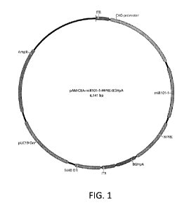
FIG. 1 is a plasmid map of pAM/CBA-miR101-1-WPRE-BGHpA.
FIGs. 2A, 2B, 2C and 2D depict the nucleotide sequence of pAM/CBA-miR101-1-
WPRE-BGHpA [SEQ ID NO:3].
FIG. 3 is a plasmid map of pCMV-MIR101-1.
FIG. 4 is a plasmid map of pAM CBA-pl-WPRE-BGHpA.
FIG. 5 is a plasmid map of pAM/CBA-miR128-2-WPRE-BGHpA.
FIGs. 6A, 6B, 6C and 6D depict the nucleotide sequence of pAM/CBA-miR128-2-
WPRE-BGHpA [SEQ ID NO:4].
FIG. 7 is a plasmid map of pCMV-MIR128-2.
FIG. 8 depicts the amino acid sequence of AAVRec3 [SEQ ID NO:5].
Detailed Description
Described herein are methods and compositions for treating a seizure disorder
which
include administering microRNA-101, pri-miR101 or pre-miR101, to a patient
having a
seizure disorder. Also described herein are methods and compositions for
treating a seizure
disorder which include administering microRNA-128, pri-miR128 or pre-miR128,
to a
patient having a seizure disorder. In embodiments, vectors encoding microRNA-
101, pri-
miR101 or pre-miR101 are provided. In embodiments, vectors encoding microRNA-
101, pri-
miR101 or pre-miR101are administered to a patient having a seizure disorder
wherein the
patient exhibits improvement in one or more symptoms of the seizure disorder.
In
7
CA 03084985 2020-06-05
WO 2019/113266 PCT/US2018/064158
embodiments, vectors encoding microRNA-128, pri-miR128 or pre-miR128 are
provided. In
embodiments, vectors encoding microRNA-128, pri-miR128 or pre-miR128 are
administered
to a patient having a seizure disorder wherein the patient exhibits
improvement in one or
more symptoms of the seizure disorder. In embodiments, ultrasound is applied
to a target
location in the patient' s brain to enhance permeability of the patient's
blood brain barrier at a
target location, wherein microRNA-101 or microRNA-128 is delivered to the
target location.
MicroRNA-101, pri-miR101, pre-miR101, microRNA-128, pri-miR128, and/or pre-
miR128, are collectively referred to herein as microRNA or microRNAs.
Administration to a
patient of microRNA-101, pri-miR101, pre-miR101, microRNA-128, pri-miR128,
and/or
pre-miR128, is collectively referred to herein as microRNA treatment. MicroRNA
treatment
increases the level of respective active microRNA molecules in a cell. The
increase can come
about by directly providing the microRNA to a cell, or may come about by
indirectly
providing microRNA to cell, such as through a vector. The microRNA may include
a RNA or
DNA molecule that also includes additional sequences. Increases in the level
of respective
active microRNA molecules in brain cells of a patient are associated with an
improvement in
one or more symptoms of a seizure disorder.
One or more pri-miRNA(s) can be used in the compositions and methods described
herein. Any suitable form of a pri-mRNA can be used. The pri-mRNA(s) can be
processed
intracellularly and act to gain function for the miRNA, e.g., converted into
pre-mRNA(s) and
then the mature form. Alternatively, the miRNA may initially be a miRNA
precursor. In
embodiments, the compositions and methods include pre-miRNA, which is subject
to
cleavage by an RNAse III type double stranded endonuclease called Dicer,
resulting in an
imperfect miRNA:miRNA* duplex that is about 20-25 nucleotides in size. This
duplex
contains the mature miRNA strand and its opposite complementary miRNA* strand.
One or
more pre-miRNA(s) can be used in the compositions and methods described
herein. The pre-
miRNA may act to gain function for the miRNA. Any suitable form of a pre-miRNA
can be
used. It is also contemplated that the miRNA of the compositions and methods
described
herein may be mature miRNA.
The microRNAs can be delivered to cells in non-expression vector or expression
vector modalities. Expression vector and vector are used interchangeably
herein. In
embodiments, microRNA may be isolated or purified prior to use in a subsequent
step.
MicroRNAs may be isolated or purified prior to introduction into a cell.
"Introduction" into a
cell includes known methods of transfection, transduction, infection and other
methods for
introducing an expression vector or a heterologous nucleic acid into a cell. A
template nucleic
8
CA 03084985 2020-06-05
WO 2019/113266 PCT/US2018/064158
acid or amplification primer may be isolated or purified prior to it being
transcribed or
amplified. Isolation or purification can be performed by a number of methods
known to those
of skill in the art with respect to nucleic acids. The delivery of the
microRNA may occur
through several forms, such as through encapsulation of a chemically modified
or through an
unmodified RNA moiety within a viral or non-viral delivery vessel. Non-
expression vector
delivery modalities include nanoparticles, microparticles, liposomes,
polymers, microspheres,
etc., which may be targeted to brain cells. The microRNA can also be delivered
as a plasmid
or minivector based expression system where it can then be expressed and
processed by the
RNAi machinery in cells to form a mature microRNA.
Nucleic acid constructs for miRNA expression may be produced recombinantly.
Such
expression vectors are provided herein. Expression vectors are a carrier
nucleic acid into
which a nucleic acid sequence can be inserted for introduction into a cell
where it can be
replicated. Expression vectors include plasmids, cosmids, recombinant viruses,
such as
adeno-associated virus (AAV), adenoviruses, retroviruses, poxviruses, and
other known
viruses in the art (bacteriophage, animal viruses, and plant viruses), and
artificial
chromosomes (e.g., YACs). A person of ordinary skill in the art is well
equipped to construct
expression vectors through standard recombinant techniques. In embodiments, an
expression
vector having an microRNA is delivered to cells of a patient. The nucleic acid
molecules are
delivered to the cells of a patient in a form in which they can be taken up
and are
advantageously expressed so that therapeutically effective levels can be
achieved.
Any suitable expression vector known to those skilled in the art may be
utilized to
deliver microRNA(s) herein to a target location in the brain. Upon such
delivery, neurons in
the target locations are transfected with microRNA(s), thereby increasing
levels of those
microRNA(s) in the brain of the patient. Transducing viral (e.g., retroviral,
adenoviral,
lentiviral and adeno-associated viral) vectors can be used for somatic cell
gene therapy,
especially because of their high efficiency of infection and stable
integration and expression.
In embodiments, the expression vector may be a stable integrating vector or a
stable
nonintegrating vector. Examples of suitable vectors are lentiviruses and adeno-
associated
viruses (AAV). Lentiviruses are a subclass of retroviruses. Lentiviruses can
integrate into the
genome of non-dividing cells such as neurons. Lentiviruses are characterized
by high-
efficiency infection, long-term stable expression of transgenes and low
immunogenicity. In
embodiments, lentiviral vectors may be utilized to deliver microRNA(s) to the
brain.
AAV is a defective parvovirus known to infect many cell types and is
nonpathogenic
to humans. AAV can infect both dividing and non-dividing cells. In
embodiments, AAV
9
CA 03084985 2020-06-05
WO 2019/113266 PCT/US2018/064158
vectors may be utilized herein to deliver microRNA(s) to the brain. Any of the
known adeno-
associated viruses (AAV) may be utilized herein, e.g., AAV1, AAV2, AAV4, AAV5,
AAV8,
AAV9 and AAVRec3 may be utilized in connection with neurons. Additional
suitable AAV
serotypes have been developed through pseudotyping, i.e., mixing the capsid
and genome
from different viral serotypes. Accordingly, e.g., AAV2/7 indicates a virus
containing the
genome of serotype 2 packaged in the capsid from serotype 7. Other examples
are AAV2/5,
AAV2/8, AAV2/9, etc. Hybrid AAV capsid serotypes red, rec2, rec3 and rec4 were
generated by shuffling the fragments of capsid sequences that matched in all
three non-
human primate AAV serotypes cy5, rh20 and rh39, with AAV8. See, Charbel et
al., PLoS
One. 2013 Apr 9;8(4):e60361. The terms rec3AAV and AAVRec3 may be used
interchangeably herein. The amino acid sequence of AAVRec3 is depicted in FIG.
8. Self
-
complementary adeno-associated virus (scAAV) may also be utilized as vectors.
Whereas
AAV packages a single strand of DNA and requires the process of second-strand
synthesis,
scAAV packages both strands which anneal together to form double stranded DNA.
By
skipping second strand synthesis scAAV allows for rapid expression in the
cell.
Suitable vectors may be constructed by those having ordinary skill in the art
using
known techniques. Suitable vectors can be chosen or constructed, containing,
in addition to
microRNA(s), appropriate regulatory sequences, including promoter sequences,
terminator
fragments, polyadenylation sequences, marker genes and other sequences as
appropriate. Those skilled in the art are familiar with appropriate regulatory
sequences,
including promoter sequences, terminator fragments, polyadenylation sequences,
marker
genes and other suitable sequences.
Expression vectors herein include appropriate sequences operably linked to the
coding
sequence or ORF to promote its expression in a targeted host cell. "Operably
linked"
sequences include both expression control sequences such as promoters that are
contiguous
with the coding sequences and expression control sequences that act in trans
or distally to
control the expression of the desired product.
Typically, the vector includes a promoter to facilitate expression of the
microRNA(s)
within a target cell. The promoter may be selected from a number of
constitutive or inducible
promoters that can drive expression of the selected transgene in the brain.
Examples of
constitutive promoters include CMV immediate early enhancer/chicken beta-actin
(CBA)
promoter-exon 1-intron 1 element, RSV LTR promoter/enhancer, the 5V40
promoter, the
CMV promoter, dihydrofolate reductase (DHFR) promoter, and the phosphoglycerol
kinase
(PGK) promoter.
CA 03084985 2020-06-05
WO 2019/113266 PCT/US2018/064158
Specificity can be achieved by regional and cell-type specific expression of
the
receptor exclusively, e.g., using a tissue or region specific promoter. Virus
gene promoter
elements may help dictate the type of cells that express microRNA(s). Some
promoters are
nonspecific (e.g., CAG, a synthetic promoter), while others are neuronal-
specific. The CAG
promoter is a strong synthetic promoter that can be used to drive high levels
of expression.
The CAG promoter consists of 1) a cytomegalovirus (CMV) early enhancer
element, 2) the
promoter, the first exon and the first intron of the chicken beta-actin gene,
and 3) the splice
acceptor of the rabbit beta-globin gene. In embodiments the promoter is the
CAG promoter.
Neuronal specific promoters include (e.g., synapsin; hSyn), or preferential to
specific neuron
types, e.g., dynorphin, encephalin, GFAP (Cilial fibrillar), acidic protein)
which is preferential
to astrocytes, or CaMKIIa, which is preferential to cortical glutamatergic
cells but can also
target subcortical GABAergic cells. In embodiments, the promoter is the
CamkIIa (alpha
CaM kinase II gene) promoter, which may drive expression in the forebrain.
Other neuronal
cell type-specific promoters include the NSE promoter, tyrosine hydroxylase
promoter,
myelin basic protein promoter, glial fibrillary acidic protein promoter, and
neurofilaments
gene (heavy, medium, light) promoters.
Expression control sequences may also include appropriate transcription
initiation,
termination, and enhancer sequences; efficient RNA processing signals such as
splicing and
polyadenylation signals; sequences that stabilize cytoplasmic mRNA; sequences
that enhance
translation efficiency (e.g., Kozak consensus sequence); sequences that
enhance nucleic acid
or protein stability; and when desired, sequences that enhance product
processing and/or
secretion. Many varied expression control sequences, including native and non-
native,
constitutive, inducible and/or tissue-specific, are known in the art and may
be utilized herein
depending upon the type of expression desired.
In addition to promoters, expression control sequences for eukaryotic cells
typically
include an enhancer, such as one derived from an immunoglobulin gene, 5V40,
CMV, etc.,
and a polyadenylation sequence which may include splice donor and acceptor
sites. The
polyadenylation sequence generally is inserted 3 Jo the coding sequence and 5
Jo the 3 EITR
sequence. Illustrative examples of polyA signals that can be used in a vector
herein include
polyA sequence (e.g., AATAAA, ATTAAA, or AGTAAA), a bovine growth hormone
polyA
sequence (BGHpA), a rabbit beta-globin polyA sequence (rBgpA), or another
suitable
heterologous or endogenous polyA sequence known in the art.
Regulatory sequences useful herein may also contain an intron, such as one
located
between the promoter/enhancer sequence and the coding sequence. One useful
intron
11
CA 03084985 2020-06-05
WO 2019/113266 PCT/US2018/064158
sequence is derived from SV40, and is referred to as the SV40 T intron
sequence. Another
includes the woodchuck hepatitis virus post-transcriptional element (WPRE).
WPRE is a
DNA sequence that, when transcribed, creates a tertiary structure that
enhances expression.
Vectors herein may contain reporter genes, e.g., those which encode
fluorophores. A
fluorophore is a fluorescent compound that can re-emit light upon excitation,
usually at
specific frequencies. They can be used as a tag or marker which can be
attached to, e.g., a
protein to allow the protein to be located. Many suitable fluorophores are
known in the art.
They may be categorized by the color they emit, e.g., blue, cyan, green,
yellow, orange, red
and others. For example, mCherry, mRasberry, mTomato and mRuby are red
fluorophore
proteins; citrine, venus, and EYFP are yellow fluorophore proteins. Green
fluorescent protein
(GFP) is a commonly used fluorophore.
In embodiments, the expression vector is pAM/CBA-miR101-1-WPRE-BGHpA. A
plasmid map of pAM/CBA-miR101-1-WPRE-BGHpA is depicted in FIG. 1. The nucleic
acid sequence [SEQ ID NO:3] is shown in FIGs. 2A-2D. TABLE I annotates pAM/CBA-
miR101-1-WPRE-BGHpA.
TABLE I
Name Type Minimum Maximum Length Direction
AmpR CDS 4353 5213 861 reverse
pUC19 On rep origin 3440 4227 788 reverse
5V40 On rep origin 3019 3354 336 reverse
ITR LTR 2836 3018 183 forward
BGHpA polyA signal 2558 2826 269 forward
WPRE misc feature 1947 2539 593 forward
miR101-1 misc feature 1209 1877 669 forward
CAG-promoter promoter 190 1125 936 forward
uukaryotic
ITR LTR 1 183 183 forward
To construct pAM/CBA-miR101-1-WPRE-BGHpA, EcoRI LI1EcoRV fragments from
the pCMV-MIR101-1 plasmid (772 bp) (5C400013), commercially available from
OriGene
Technologies, Inc., 9620 Medical Center Dr., Suite 200, Rockville, MD 20850,
is inserted
into pAM CBA-pl-WPRE-BGHpA vector cut with EcoRI+EcoRV. A plasmid map of
pCMV-MIR101-1 is depicted in FIG. 3. A plasmid map of pAM CBA-pl-WPRE-BGHpA is
depicted in FIG. 4.
In embodiments, the expression vector is pAM/CBA-miR128-2-WPRE-BGHpA. A
plasmid map of pAM/CBA-miR128-2-WPRE-BGHpA is depicted in FIG. 5. The nucleic
12
CA 03084985 2020-06-05
WO 2019/113266 PCT/US2018/064158
acid sequence [SEQ ID NO:4] is shown in FIGs. 6A-6D. TABLE II annotates
pAM/CBA-
miR128-2-WPRE-BGHpA.
TABLE II
Name Type Minimum Maximum Length Direction
Kan/Neo CDS 5072 5866 795 reverse
IRES regulatory 4471 5056 586 reverse
tGFP CDS 3772 4470 699 reverse
ColE1 rep origin 2680 3352 673 forward
polyA polyA signal 1819 2404 586 forward
Editing History 1027 1026 0 none
Deletion
miR128-2 misc feature 1027 1678 652 forward
CMV promoter promoter uukaryotic 200 926 727 forward
To construct pAM/CBA-miR128-2-WPRE-BGHpA, EcoRI LI1EcoRV fragments from
the pCMV-MIR128-2 plasmid (755 bp) (5C400112), commercially available from
OriGene
Technologies, Inc., 9620 Medical Center Dr., Suite 200, Rockville, MD 20850,
is inserted
into pAM CBA-pl-WPRE-BGHpA vector cut with EcoRI+EcoRV. A plasmid map of
pCMV-MIR128-2 is depicted in FIG. 7. A plasmid map of pAM CBA-pl-WPRE-BGHpA is
depicted in FIG. 4.
The microRNAs described herein, whether delivered by expression vector or by
non-
expression vector modalities, are used to treat seizure disorders. Seizure
disorders, including
those involving complex partial seizures, e.g., temporal lobe epilepsy (TLE)
may be one of
the most refractory forms of epilepsy. In certain instances, one temporal lobe
may be defined
as the site of seizure origin (the epileptogenic region) and the medial
temporal lobe including
the anterior hippocampus may be targeted in accordance with the methods
herein. Seizure
disorders can result from an imbalance of excitation to inhibition. The
antagonism of
excitation and enhancing of inhibition can provide improvement in at least one
symptom of
the seizure disorder.
Examples of seizure disorders include epilepsy, epilepsy with generalized
tonic-clonic
seizures, epilepsy with myoclonic absences, frontal lobe epilepsy, temporal
lobe epilepsy,
Landau-Kleffner Syndrome, Rasmussen' s syndrome, Dravet syndrome, Doose
syndrome,
CDKL5 disorder, infantile spasms (West syndrome), juvenile myoclonic epilepsy
(JME),
vaccine-related encephalopathy, intractable childhood epilepsy (ICE), Lennox-
Gastaut
syndrome (LGS), Rett syndrome, Ohtahara syndrome, CDKL5 disorder, childhood
absence
13
CA 03084985 2020-06-05
WO 2019/113266 PCT/US2018/064158
epilepsy, essential tremor, acute repetitive seizures, benign rolandic
epilepsy, status
epilepticus, refractory status epilepticus, super-refractory status
epilepticus (SRSE), PCDH19
pediatric epilepsy, drug withdrawal induced seizures, alcohol withdrawal
induced seizures,
increased seizure activity or breakthrough seizures (also called serial or
cluster seizures). In
embodiments, the seizure disorder is associated with a sodium channel protein
type 1 subunit
alpha (Scnla)-related disorder. In embodiments, the seizure disorder is
characterized by focal
seizures. In embodiments, the seizure disorder is focal cortical dysplasia. In
embodiments, the
FCD is Type I FCD. In embodiments, the FCD is Type Ia FCD. In embodiments, the
FCD is
Type lb FCD. In embodiments, the FCD is Type Ic FCD. In embodiments, the FCD
is Type
II FCD. In embodiments, the FCD is Type ha FCD. In embodiments, the FCD is
Type Ilb
FCD. In embodiments, the FCD is Type III FCD. In embodiments, the FCD is Type
Ma
FCD. In embodiments, the FCD is Type Mb FCD. In embodiments, the FCD is Type
Mc
FCD. In embodiments, the seizure disorder is associated with a brain tumor,
i.e., brain tumor
induced seizures, such as a ganglioglioma, a glioma - low grade and high
grade, including
anaplastic astrocytoma, anaplastic oligodendroglioma, anaplastic
oligoastrocytoma, and
anaplastic ependymoma, a glioblastoma, or a meningioma. In embodiments, the
seizure
disorder is associated with brain hamartomas, i.e., hamartoma induced
seizures, such as
Tuberous Sclerosis Complex (TSC) or Tuber Cinereum Hamartoma.
In embodiments, the seizure disorder is status epilepticus (SE). SE is
characterized by
an epileptic seizure of greater than five minutes or more than one seizure
within a five-minute
period without the person returning to normal between them. SE can be a
dangerous
condition that can lead to mortality if treatment is delayed. SE can be
convulsive, with a
regular pattern of contraction and extension of the arms and legs, or non-
convulsive, with a
change in a person level of consciousness of relatively long duration but
without large scale
bending and extension of the limbs due to seizure activity. Convulsive SE
(CSE) may be
further classified into (a) tonic LIilonic SE, (b) tonic SE, (c) clonic SE and
(d) myoclonic SE.
Non-convulsive SE (NC SE) is characterized by abnormal mental status,
unresponsiveness,
ocular motor abnormalities, persistent electrographic seizures, and possible
response to
anti convul sants.
Symptoms of a seizure disorder may include, but are not limited to, episodes
involving ataxia, gait impairment, speech impairment, vocalization, impaired
cognition,
abnormal motor activity, clinical seizure, subclinical seizure, hypotonia,
hypertonia, drooling,
mouthing behavior, aura, repetitive movements, laughing, and unusual
sensations. In
embodiments, the methods and compositions provided may reduce or prevent one
or more
14
CA 03084985 2020-06-05
WO 2019/113266 PCT/US2018/064158
different types of seizures. Generally, a seizure can include repetitive
movements, unusual
sensations, and combinations thereof. Seizures can be categorized as focal
seizures (also
referred to as partial seizures) and generalized seizures. Focal seizures
affect only one side of
the brain, while generalized seizures affect both sides of the brain. Specific
types of focal
seizures include simple focal seizures, complex focal seizures, and
secondarily generalized
seizures. Simple focal seizures can be restricted or focused on a particular
lobe (e.g., temporal
lobe, frontal lobe, parietal lobe, or occipital lobe). Complex focal seizures
generally affect a
larger part of one hemisphere than simple focal seizures, but commonly
originate in the
temporal lobe or the frontal lobe. When a focal seizure spreads from one side
(hemisphere) to
both sides of the brain, the seizure is referred to as a secondarily
generalized seizure. Specific
types of generalized seizures include absences (also referred to as petit mal
seizures), tonic
seizures, atonic seizures, myoclonic seizures, tonic clonic seizures (also
referred to as grand
mal seizures), and clonic seizures. Methods of treatment herein can include
providing
improvement in one or more of the foregoing symptoms.
Once a determination has been made of the location or of a suspected location
of
abnormal electrical impulses associated with a seizure disorder in a patient,
targeted
treatment in accordance with the present disclosure can be implemented.
Methods of
determining the location of abnormal electrical activity in the brain are well-
known in the art.
Although any area exhibiting abnormal electricity in the brain can be targeted
for treatment
herein, areas of the brain which are known to be associated with seizure
disorders and which
can receive targeted treatment include, but are not limited to, the temporal
lobe, the frontal
lobe, the occipital lobe and the parietal lobe. For example, the temporal
lobes can be a
common site of localized epileptic seizures. In certain instances, seizures
beginning in the
temporal lobes can extend to other parts of the brain. In embodiments,
specific areas of the
temporal lobe which can be targeted for treatment include structures of the
limbic system
such as the hippocampus, auditory-vestibular cortex, the medial temporal lobe,
and the
amygdala. In embodiments, specific areas of the occipital lobe can also be
targeted, e.g., the
primary visual cortex. In embodiments, specific areas of the parietal lobe can
be targeted,
e.g., the lateral postcentral gyms. In embodiments, the location of the
primary somatosensory
cortex which can be targeted. In embodiments, specific areas of the frontal
lobe can be
targeted, e.g., the motor cortex, the olfactory-gustatory cortex. In
embodiments, large areas of
the brain which have been identified as exhibiting abnormal electrical
activity can be
targeted. In certain instances, manifestations of seizure disorders can begin
within certain
areas of the brain and spread to others. For example, manifestations of
seizure disorders can
CA 03084985 2020-06-05
WO 2019/113266 PCT/US2018/064158
begin within the hippocampus or its surrounding structures. In embodiments,
areas
determined to be the site of origin of the abnormal electrical activity can be
targeted.
Methods for administering materials directly to target locations within the
brain are
well-known. For example, a hole, e.g., Burr hole, can be drilled into the
skull and an
appropriately sized needle may be used to deliver a vector or non-vector
vehicle to a target
location. In embodiments, a portion of the skull may be removed to expose the
dura matter
(craniotomy) at or near a target location and a vector or non-vector vehicle
can be
administered directly to the target location. In embodiments, a vector or non-
vector vehicle is
injected intracranially using stereotaxic coordinates, a micropipette and an
automated pump
for precise delivery of the vector or non-vector vehicle to the desired area
with minimal
damage to the surrounding tissue. In embodiments, a micropump may be utilized
to deliver
pharmaceutical compositions containing a vector or non-vector vehicle
containing the
microRNA(s) to target areas in the brain. The compositions can be delivered
immediately or
over an extended period of time, e.g., over 1, 2, 3, 4, 5, 6, 7, 8, 9, 10 or
more minutes. After
vector delivery to a target location in the brain a sufficient amount of time
may be allowed to
pass to allow expression of the microRNA(s) at the target location.
In embodiments, vectors or nonvector delivery vehicles herein can be
administered
systemically. Systemic delivery includes oral, buccal, sublingual, rectal,
topical, intranasal,
vaginal and parenteral modes of administration. Examples of parenteral modes
of
administration include intravenous, intraperitoneal, intramuscular and
subcutaneous modes of
administration. In embodiments, vectors or nonvector delivery vehicles will
circulate until
they contact the target location(s) in the brain where they deliver the
microRNA(s) or cause
the microRNA(s) to be expressed and act, e.g., to aid in network formation
and/or modulate
neuronal signaling networks.
The microRNA(s) is used in an amount effective against a seizure disorder in
patients.
The dosage of the active ingredient depends upon the age, weight, and
individual condition of
the patient, the individual pharmacokinetic data, and the mode of
administration. In the case
of an individual human having a bodyweight of about 70 kg the daily dose
administered of a
microRNA can be from 0.01 mg/kg bodyweight to 100 mg/kg bodyweight, e.g., from
0.1
mg/kg bodyweight to 50 mg/kg bodyweight, from 1 mg/kg to 20 mg/kg bodyweight
administered as a single dose or as several doses. The microRNA(s) can be used
alone or in
combination with other AED drugs.
16
CA 03084985 2020-06-05
WO 2019/113266 PCT/US2018/064158
In embodiments, treatment with ultrasound is used to enhance delivery of the
microRNA(s) to target locations in the brain by disrupting the blood brain
barrier. Use of
focused ultrasound energy herein disrupts the BBB without adversely affecting
the vector,
non-vector delivery vehicle, the microRNA(s), and/or brain tissue itself This
may be
considered surprising in view of potential damage to organic compounds and
tissues by
ultrasound energy. Use of ultrasound energy herein can increase the speed of
delivery of
vectors, non-vector delivery vehicles, and/or the microRNA(s) to target
locations in the brain,
reduce side effects which may be associated with delivery of vectors non-
vector delivery
vehicles, and/or the microRNA(s) to target locations in the brain, reduce
dosage amounts
while concentrating vectors, non-vector delivery vehicles, and/or the
microRNA(s) at a target
location and can allow controlled release of the amount of vectors, non-vector
delivery
vehicles, and/or the microRNA(s) at a target location.
In accordance with the present disclosure, in embodiments, ultrasound energy
assists
and/or propels penetration of the vector carrying the microRNA(s) to target
locations in the
brain. In embodiments, ultrasound energy is used to make the blood brain
barrier permeable
to vectors, non-vector delivery vehicles, and/or the microRNA(s) herein.
Accordingly, in
embodiments, ultrasound energy can be applied to a target location prior to
administration of
the vector, non-vector delivery vehicles, and/or the microRNA(s). In
embodiments, vectors,
non-vector delivery vehicles, and/or the microRNA(s) herein can be
administered to a target
area in the brain simultaneously with administration of ultrasound energy. In
embodiments,
vectors, non-vector delivery vehicles, and/or the microRNA(s), herein can be
administered to
a target area in the brain before administration of ultrasound energy.
As mentioned previously, vectors, non-vector delivery vehicles, and/or the
microRNA(s) herein can be administered systemically. In this manner vectors,
non-vector
delivery vehicles, and/or the microRNA(s) circulating in the blood stream are
delivered to a
target location in the brain through a portion of the BBB disrupted by
ultrasonic energy. In
embodiments, vectors, non-vector delivery vehicles, and/or the microRNA(s)
herein can be
administered systemically after ultrasound energy treatment of the target
location and the
vectors, non-vector delivery vehicles, and/or the microRNA(s) penetrate the
disrupted BBB
to become situated at the target location. In embodiments, vectors, non-vector
delivery
vehicles, and/or the microRNA(s),herein can be administered directly to a
target location in
the brain. In embodiments, vectors, non-vector delivery vehicles, and/or the
microRNA(s)
herein can be administered directly to a target location in the brain after
ultrasound energy
treatment of the target location to become situated at the target location. In
embodiments,
17
CA 03084985 2020-06-05
WO 2019/113266 PCT/US2018/064158
vectors, non-vector delivery vehicles, and/or the microRNA(s) herein can be
administered
directly to a target location in the brain without ultrasound treatment.
In embodiments, ultrasound energy can be administered to a target area by
removing a
portion of the skull (craniotomy) to expose the dura matter at or near a
target location and
delivering the ultrasound energy at or below the exposed dura matter. In
embodiments,
ultrasound energy can be administered to a target location through the skull,
eliminating the
need for surgery associated with delivery of ultrasound energy to a target
location. Methods
for delivering ultrasound energy through the skull are known in the art. See,
e.g., US Pat. No.
5,752,515 and US Publication No. 2009/0005711, both of which are hereby
incorporated by
reference in their respective entireties. See also, Hynynen et al., NeuroImage
24 (2005) 12-
120.
In embodiments, ultrasound energy can be applied to a target location in the
brain at
frequencies ranging from about 20 kHz to about 5 MHz, and with sonication
duration ranging
from 100 nanoseconds to 1 minute. In embodiments, ultrasound energy can be
applied to a
target location in the brain at frequencies ranging from about 20 kHz to about
10 MHz,
sonication duration ranging from about 100 nanoseconds to about 30 minutes,
with
continuous wave or burst mode operation, where the burst mode repetition
varies from about
0.01 Hz to about 1 MHz. In embodiments, ultrasound energy can be applied to a
target
location in the brain at frequencies ranging from about 200 kHz to about 10
MHz, and with
sonication duration ranging from about 100 milliseconds to about 30 minutes.
In
embodiments, ultrasound energy can be applied to a target location in the
brain at frequencies
ranging from about 250 kHz to about 10 MHz, and with sonication duration
ranging from
about 0.10 microseconds to about 30 minutes. In embodiments, ultrasound energy
can be
applied to a target location in the brain at a frequency of about 1.525 MHz.
In embodiments,
ultrasound energy can be applied to a target location in the brain at a
frequency of about
0.69MHz. In embodiments, pressure amplitudes generated by ultrasound energy
can be about
0.5 to about 2.7 MPa. In embodiments, pressure amplitudes generated by
ultrasound energy
can be about 0.8 to about 1 MPa. In embodiments, ultrasound energy is applied
to a target
location in the brain at a focal region sized in accord with the volume of
tissue and/or fluids
to which a vector, non-vector delivery vehicle, and/or the microRNA(s) herein
is to be
delivered, e.g., from about 0.1 mm3 to about 5 cm3.
In embodiments, the target location and access thereto is confirmed by
introducing a
contrast agent into the patient prior to, during or after application of
ultrasound energy to the
target location, allowing sufficient time for the contrast agent to permeate
the BBB, and
18
CA 03084985 2020-06-05
WO 2019/113266 PCT/US2018/064158
determining whether the contrast agent is present at the target location.
Contrast agents are
well-known and include, e.g., iodine-based compounds, barium-based compounds
and
lanthanide based compounds. Iodine-based agents include, e.g., iohexol,
iopromide,
iodixanol, iosimenol, ioxaglate, iothalamate and iopamidol. Barium-based
compounds
include barium sulfate. Lanthanide-based compounds include, e.g., gadolinium-
based
chelates such as gadoversetamide, gadopentetate dimeglumine, gadobutrol,
gadobenate
dimeglumine, gadoterate meglumine, and gadoxetate disodium. Detection
modalities include
2-dimensional X-ray radiography, X-ray computed tomography and magnetic
resonance
imaging which are well-known techniques that may be utilized to confirm the
presence or
absence of contrast agent in a target location.
In accordance with the present disclosure, microRNA treatment provides
improvement in one or more symptoms of a seizure disorder for more than 1 hour
after
administration to the patient. In embodiments, microRNA treatment provides
improvement in
one or more symptoms of the disorder for more than 2 hours after
administration to the
patient. In embodiments, microRNA treatment provides improvement in one or
more
symptoms of the disorder for more than 3 hours after administration to the
patient. In
embodiments, microRNA treatment provides improvement in one or more symptoms
of the
disorder for more than 4 hours after administration to the patient. In
embodiments,
microRNA treatment provides improvement in one or more symptoms of the
disorder for
more than 6 hours after administration to the patient. In embodiments,
microRNA treatment
provides improvement in one or more symptoms of the disorder for more than 8,
10, 12, 14,
16, 18, 20, 22 or 24 hours after administration to the patient. In
embodiments, improvement
in at least one symptom for 12 hours after administration to the patient is
provided in
accordance with the present disclosure. In embodiments, microRNA treatment
provides
improvement of next day functioning of the patient. For example, the microRNA
may
provide improvement in one or more symptoms of the disorder for more than
about, e.g., 2
hours, 4 hours, 6 hours, 8 hours, 10 hours, 12 hours, 14 hours, 16 hours, 18
hours, 20 hours,
22 hours or 24 hours after administration and waking from a night of sleep.
In embodiments, provided herein are methods of treating a seizure disorder
including
administering to a patient in need thereof microRNA(s) after a warning sign of
an impending
seizure is detected to reduce or prevent seizure activity.
In embodiments, the methods described herein are effective to reduce, delay,
or
prevent one or more other clinical symptoms of a seizure disorder. For
example, the effect, in
a patient of microRNA treatment in a target location of the brain, whose
delivery is optionally
19
CA 03084985 2020-06-05
WO 2019/113266 PCT/US2018/064158
enhanced by ultrasound energy on a particular symptom, pharmacologic, or
physiologic
indicator can be compared to an untreated patient, or the condition of the
patient prior to
treatment. In embodiments, the symptom, pharmacologic, and/or physiologic
indicator is
measured in a patient prior to treatment, and again one or more times after
treatment is
initiated. In embodiments, the control is a reference level, or average
determined based on
measuring the symptom, pharmacologic, or physiologic indicator in one or more
patients that
do not have the disease or condition to be treated (e.g., healthy patients).
In embodiments,
the amount of miR-101 and/or miR-128 in brain tissue prior to treatment is
compared to the
amount of miR-101 and/or miR-128 in brain tissue after treatment. In
embodiments, the
effect of the treatment is compared to a conventional treatment that is within
the purview of
those skilled in the art.
Effective treatment of a seizure disorder (e.g., intractable focal seizures,
focal cortical
dysplasia, acute repetitive seizure, status epilepticus, etc.) herein may be
established by
showing reduction in the frequency or severity of symptoms (e.g., more than
10%, 20%, 30%
40% or 50%) after a period of time compared with baseline. For example, after
a baseline
period of 1 month, the patients having microRNA treatment may be randomly
allocated a
placebo as add-on therapy to standard therapies, during a double-blind period
of 2 months.
Primary outcome measurements may include the percentage of responders on a
microRNA
and on placebo, defined as having experienced at least a 10% to 50% reduction
of symptoms
during the second month of the double-blind period compared with baseline.
In embodiments, pharmaceutical compositions containing vectors, non-vector
delivery vehicles, and/or the microRNA(s) may be provided with conventional
release or
modified release profiles. Pharmaceutical compositions may be prepared using a
pharmaceutically acceptable LIarrierLI1 composed of materials that are
considered safe and
effective. The Darrierp includes all components present in the pharmaceutical
formulation
other than the active substance or ingredients. Examples of active substances
include
microRNA(s), expression vectors containing microRNA(s) and AEDs. The term
Darrier
includes, but is not limited to, diluents, binders, lubricants, disintegrants,
fillers, and coating
compositions. Those with skill in the art are familiar with such
pharmaceutical carriers and
methods of compounding pharmaceutical compositions using such carriers.
In embodiments, pharmaceutical compositions containing vectors, non-vector
delivery vehicles, and/or the microRNA(s) are suitable for parenteral
administration,
including, e.g., intramuscular (i.m.), intravenous (iv.), subcutaneous (s.c.),
intraperitoneal
(i.p.), or intrathecal (it.). Parenteral compositions must be sterile for
administration by
CA 03084985 2020-06-05
WO 2019/113266 PCT/US2018/064158
injection, infusion or implantation into the body and may be packaged in
either single-dose or
multi-dose containers. In embodiments, liquid pharmaceutical compositions for
parenteral
administration to a patient include an active substance, e.g., vectors, non-
vector delivery
vehicles, and/or the microRNA(s), in any of the respective amounts described
above. In
embodiments, the pharmaceutical compositions for parenteral administration are
formulated
as a total volume of about, e.g., 0.1 ml, 0.25 ml, 0.5 ml, 0.75 ml, 1 ml, 1.25
ml, 1.5 ml, 1.75
ml, 2 ml, 2.25 ml, 2.5 ml, 2.75 ml, 3 ml, 3.25 ml, 3.5 ml, 3.75 ml, 4 m1,4.25
ml, 4.5 ml, 4.75
ml, 5 ml, 10 ml, 20 ml, 25 ml, 50 ml, 100 ml, 200 ml, 250 ml, or 500 ml. In
embodiments,
the volume of pharmaceutical compositions containing expression vectors are
microliter
amounts. For example, 0.1 microliters to 10 or more microliters can be
injected. For example,
0.1, 0.2, 0.3, 0.4, 0.5, 0.6, 0.7, 0.8, 0.9, 1.0, 1.25, 1.5, 1.75, 2.0, 2.25,
2.5, 2.75, 3.0, 3.25, 3.5,
3.75, 4.0, 4.25, 4.5, 4.75, 5.0, 5.25, 5.5, 5.75, 6.0, 6.25, 6.5, 6.75, 7.0,
8.25, 8.5, 8.75, 9.0,
9.25, 9.5, 9.75, or 10 microliters. In embodiments, the compositions are
contained in a
micropipette, a bag, a glass vial, a plastic vial, or a bottle.
In embodiments, pharmaceutical compositions for parenteral administration
include
respective amounts described above. In embodiments, pharmaceutical
compositions for
parenteral administration include about 0.0001 mg to about 500 mg active
substance, e.g.,
vectors, non-vector delivery vehicles, and/or the microRNA(s). In embodiments,
pharmaceutical compositions for parenteral administration to a patient include
an active
substance, e.g., vectors, non-vector delivery vehicles, and/or the
microRNA(s), at a respective
concentration of about 0.001 mg/ml to about 500 mg/ml. In embodiments, the
pharmaceutical
composition for parenteral administration includes an active substance at a
respective
concentration of, e.g., about 0.005 mg/ml to about 50 mg/ml, about 0.01 mg/ml
to about 50
mg/ml, about 0.1 mg/ml to about 10 mg/ml, about 0.05 mg/ml to about 25 mg/ml,
about 0.05
mg/ml to about 10 mg/ml, about 0.05 mg/ml to about 5 mg/ml, or about 0.05
mg/ml to about
1 mg/ml. In embodiments, the pharmaceutical composition for parenteral
administration
includes an active substance at a respective concentration of, e.g., about
0.05 mg/ml to about
15 mg/ml, about 0.5 mg/ml to about 10 mg/ml, about 0.25 mg/ml to about 5
mg/ml, about 0.5
mg/ml to about 7 mg/ml, about 1 mg/ml to about 10 mg/ml, about 5 mg/ml to
about 10
mg/ml, or about 5 mg/ml to about 15 mg/ml.
In embodiments, a pharmaceutical composition for parenteral administration is
provided wherein the pharmaceutical composition is stable for at least six
months. In
embodiments, the pharmaceutical compositions for parenteral administration
exhibit no more
than about 5% decrease in active substance for at least, e.g., 3 months or 6
months. In
21
CA 03084985 2020-06-05
WO 2019/113266 PCT/US2018/064158
embodiments, the amount of vector or non-vector vehicle, degrades at no more
than about,
e.g., 2.5%, 1%, 0.5% or 0.1%. In embodiments, the degradation is less than
about, e.g., 5%,
2.5%, 1%, 0.5%, 0.25%, 0.1%, for at least six months.
In embodiments, pharmaceutical compositions for parenteral administration are
provided wherein the pharmaceutical composition remains soluble. In
embodiments,
pharmaceutical compositions for parenteral administration are provided that
are stable,
soluble, local site compatible and/or ready-to-use. In embodiments, the
pharmaceutical
compositions herein are ready-to-use for direct administration to a patient in
need thereof
The pharmaceutical compositions for parenteral administration provided herein
may
include one or more excipients, e.g., solvents, solubility enhancers,
suspending agents,
buffering agents, isotonicity agents, stabilizers or antimicrobial
preservatives. When used, the
excipients of the parenteral compositions will not adversely affect the
stability,
bioavailability, safety, and/or efficacy of a vector, non-vector delivery
vehicle, and/or the
microRNA(s), used in the composition. Thus, parenteral compositions are
provided wherein
there is no incompatibility between any of the components of the dosage form.
In embodiments, parenteral compositions including vectors, non-vector delivery
vehicles, and/or the microRNA(s) include a stabilizing amount of at least one
excipient. For
example, excipients may be selected from the group consisting of buffering
agents,
solubilizing agents, tonicity agents, antioxidants, chelating agents,
antimicrobial agents, and
preservative. One skilled in the art will appreciate that an excipient may
have more than one
function and be classified in one or more defined group.
In embodiments, parenteral compositions include a vector, non-vector delivery
vehicle, and/or the microRNA(s) and an excipient wherein the excipient is
present at a weight
percent (w/v) of less than about, e.g., 10%, 5%, 2.5%, 1%, or 0.5%. In
embodiments, the
excipient is present at a weight percent between about, e.g., 1.0% to 10%, 10%
to 25%, 15%
to 35%, 0.5% to 5%, 0.001% to 1%, 0.01% to 1%, 0.1% to 1%, or 0.5% to 1%. In
embodiments, the excipient is present at a weight percent between about, e.g.,
0.001% to 1%,
0.01% to 1%, 1.0% to 5%, 10% to 15%, or 1% to 15%.
In embodiments, parenteral compositions may be administered as needed, e.g.,
once,
twice, three, four, five, six or more times daily, or continuously depending
on the patient' s
needs.
In embodiments, parenteral compositions of an active substance are provided,
wherein
the pH of the composition is between about 4.0 to about 8Ø In embodiments,
the pH of the
compositions is between, e.g., about 5.0 to about 8.0, about 6.0 to about 8.0,
about 6.5 to
22
CA 03084985 2020-06-05
WO 2019/113266 PCT/US2018/064158
about 8Ø In embodiments, the pH of the compositions is between, e.g., about
6.5 to about
7.5, about 7.0 to about 7.8, about 7.2 to about 7.8, or about 7.3 to about
7.6. In embodiments,
the pH of the aqueous solution is, e.g., about 6.8, about 7.0, about 7.2,
about 7.4, about 7.6,
about 7.7, about 7.8, about 8.0, about 8.2, about 8.4, or about 8.6.
Unless defined otherwise, all technical and scientific terms used herein have
the same
meanings as commonly understood by one of skill in the art to which the
disclosure herein
belongs.
The term "about" or "approximately" as used herein means within an acceptable
error
range for the particular value as determined by one of ordinary skill in the
art, which will
depend in part on how the value is measured or determined, i.e., the
limitations of the
measurement system. For example, "about" can mean within 3 or more than 3
standard
deviations, per the practice in the art. Alternatively, "about" can mean a
range of up to 20%,
up to 10%, up to 5%, and/or up to 1% of a given value. Alternatively,
particularly with
respect to biological systems or processes, the term can mean within an order
of magnitude,
preferably within 5-fold, and more preferably within 2-fold, of a value.
Improvement LIII refers to the treatment of seizure disorders such as focal
epilepsy,
intractable focal epilepsy, focal cortical dysplasia, epilepsy, epilepsy with
generalized tonic-
clonic seizures, epilepsy with myoclonic absences, frontal lobe epilepsy,
temporal lobe
epilepsy, Landau-Kleffner Syndrome, Rasmussen' s syndrome, Dravet syndrome,
Doose
syndrome, CDKL5 disorder, infantile spasms (West syndrome), juvenile myoclonic
epilepsy
(JME), vaccine-related encephalopathy, intractable childhood epilepsy (ICE),
Lennox-
Gastaut syndrome (LGS), Rett syndrome, Ohtahara syndrome, CDKL5 disorder,
childhood
absence epilepsy, essential tremor, acute repetitive seizures, benign rolandic
epilepsy, status
epilepticus, refractory status epilepticus, super-refractory status
epilepticus (SRSE), PCDH19
pediatric epilepsy, brain tumor induced seizures, hamartoma induced seizures,
drug
withdrawal induced seizures, alcohol withdrawal induced seizures, increased
seizure activity
or breakthrough seizures (also called serial or cluster seizures), measured
relative to at least
one symptom of the foregoing disorders.
Improvement in next day functioningOor JATherein there is improvement in next
day
functioning LII refers to improvement after waking from an overnight sleep
period wherein the
beneficial effect of administration of microRNA therapy to a patient applies
to at least one
symptom of a syndrome or disorder herein and is discernable, either
subjectively by a patient
or objectively by an observer, for a period of time, e.g., 2 hours, 3 hours, 4
hours, 5 hours, 6
hours, 12 hours, 24 hours, etc. after waking.
23
CA 03084985 2020-06-05
WO 2019/113266 PCT/US2018/064158
"Treating", "treatment" or L1reatLIII can refer to the following: alleviating
or delaying
the appearance of clinical symptoms of a disease or condition in a patient
that may be
afflicted with or predisposed to the disease or condition, but does not yet
experience or
display clinical or subclinical symptoms of the disease or condition. In
certain embodiments,
LIIreatingLI AreatO or AreatmentO may refer to preventing the appearance of
clinical
symptoms of a disease or condition in a patient that may be afflicted with or
predisposed to
the disease or condition, but does not yet experience or display clinical or
subclinical
symptoms of the disease or condition. "Treating", AreatO or "treatment" also
refers to
inhibiting the disease or condition, e.g., arresting or reducing its
development or at least one
clinical or subclinical symptom thereof. "Treating", Areat LII or "treatment"
further refers to
relieving the disease or condition, e.g., causing regression of the disease or
condition or at
least one of its clinical or subclinical symptoms. The benefit to a patient to
be treated may be
statistically significant, mathematically significant, or at least perceptible
to the patient and/or
the physician. Nonetheless, prophylactic (preventive) treatment and
therapeutic (curative)
treatment are two separate embodiments of the disclosure herein.
"Pharmaceutically acceptable" refers to molecular entities and compositions
that are
"generally regarded as safe", e.g., that are physiologically tolerable and do
not typically
produce an allergic or similar untoward reaction, such as gastric upset and
the like, when
administered to a human. In embodiments, this term refers to molecular
entities and
compositions approved by a regulatory agency of the federal or a state
government, as the
GRAS list under section 204(s) and 409 of the Federal Food, Drug and Cosmetic
Act, that is
subject to premarket review and approval by the FDA or similar lists, the U.S.
Pharmacopeia
or another generally recognized pharmacopeia for use in animals, and more
particularly in
humans.
rFffective amount LII or Aherapeutically effective amount LII can mean a
dosage
sufficient to alleviate one or more symptoms of a syndrome, disorder, disease,
or condition
being treated, or to otherwise provide a desired pharmacological and/or
physiologic effect.
rFffective amount LII or Aherapeutically effective amount LII may be used
interchangeably
herein.
Xo-administered withLI Eildministered in combination withLI
combination of or
Eildministered along withp may be used interchangeably and mean that two or
more agents
are administered in the course of therapy. The agents may be administered
together at the
same time or separately in spaced apart intervals. The agents may be
administered in a single
dosage form or in separate dosage forms.
24
CA 03084985 2020-06-05
WO 2019/113266 PCT/US2018/064158
Tatient in need thereof0 may include individuals, e.g., mammals such as
humans,
canines, felines, porcines, rodents, etc., that have been diagnosed with a
seizure disorder such
as epilepsy, epilepsy with generalized tonic-clonic seizures, epilepsy with
myoclonic
absences, focal epilepsy, intractable focal epilepsy, focal cortical
dysplasia, frontal lobe
epilepsy, temporal lobe epilepsy, Landau-Kleffner Syndrome, Rasmussen' s
syndrome,
Dravet syndrome, Doose syndrome, CDKL5 disorder, infantile spasms (West
syndrome),
juvenile myoclonic epilepsy (JME), vaccine-related encephalopathy, intractable
childhood
epilepsy (ICE), Lennox-Gastaut syndrome (LGS), Rett syndrome, Ohtahara
syndrome,
CDKL5 disorder, childhood absence epilepsy, essential tremor, acute repetitive
seizures,
benign rolandic epilepsy, status epilepticus, refractory status epilepticus,
super-refractory
status epilepticus (SRSE), PCDH19 pediatric epilepsy, brain tumor induced
seizures,
hamartoma induced seizures, drug withdrawal induced seizures, alcohol
withdrawal induced
seizures, increased seizure activity or breakthrough seizures (also called
serial or cluster
seizures). Seizure disorders can be associated with a sodium channel protein
type 1 subunit
alpha (Scnla)-related disorder. The methods may be provided to any individual
including,
e.g., wherein the patient is a neonate, infant, a pediatric patient (6 months
to 12 years), an
adolescent patient (age 12-18 years) or an adult (over 18 years). Patients
include mammals.
EProdrugp refers to a pharmacological substance (drug) that is administered to
a
patient in an inactive (or significantly less active) form. Once administered,
the prodrug is
metabolized in the body (in vivo) into a compound having the desired
pharmacological
activity.
EAnalogpand EDerivativeDmay be used interchangeably and refer to a compound
that
possesses the same core as the parent compound, but may differ from the parent
compound in
bond order, the absence or presence of one or more atoms and/or groups of
atoms, and
combinations thereof Enantiomers are examples of derivatives. The derivative
can differ
from the parent compound, for example, in one or more substituents present on
the core,
which may include one or more atoms, functional groups, or substructures. In
general, a
derivative can be imagined to be formed, at least theoretically, from the
parent compound via
chemical and/or physical processes.
The term 4tharmaceutically acceptable salt LIJ as used herein, refers to
derivatives of
the compounds defined herein, wherein the parent compound is modified by
making acid or
base salts thereof. Examples of pharmaceutically acceptable salts include, but
are not limited
to, nontoxic base addition salts with inorganic bases. Suitable inorganic
bases such as alkali
and alkaline earth metal bases include metallic cations such as sodium,
potassium,
CA 03084985 2020-06-05
WO 2019/113266 PCT/US2018/064158
magnesium, calcium and the like. The pharmaceutically acceptable salts can be
synthesized
from the parent compound by conventional chemical methods.
EXAMPLES
The examples provided herein are included solely for augmenting the disclosure
herein and should not be considered to be limiting in any respect.
Example I
Hu-pri-miR sequences as described below are cloned into a AAV pAM-plasmid
backbone. Each plasmid will be verified by sequencing and restriction digests.
1. pAM-CBA-hu-pri-miR128-2-WPRE-bGH
2. pAM-CBA-hu-pri-miR101-WPRE-bGH
3. pAM-CBA-sc-hu-pri-miR128-2-GFP-WPRE-bGH
Plasmids expressing a GFP reporter facilitate analysis of in vivo spread of
miR
expression following infusion into the mouse brain.
Plasmids 1-3 above and pAM-empty plasmid will be packaged into AAVRec3 vectors
(-300 tL AAV vector; >1 x 10¹² vg/mL) and purified using standard iodixanol
purification
methods. AAV vector stock purity will be confirmed by standard SDS-PAGE and
Coomassie
staining methods. AAV vector titers will be determined by quantitative RT-PCR.
Study 1: Optimization of AAV vector dose
AAV miR-128-2-GFP and AAV miR128-2 vector will be injected into the mouse
brain at
two vector doses (n=4 animals per vector and at 2 doses) (miR128-2 only vector
included in
the event there are synergistic toxic effects with GFP). Brains will be taken
at 4 weeks post-
vector infusion for analysis of transgene expression (for GFP) and spread and
any evidence of
toxicity (GFAP, Cdl lb, NeuN IHC, Fluorojade) using standardized lab methods.
Study 2: Optimization of AAV vector dose
AAV miR-101-1-GFP and AAV miR101-1 vector will be injected into the mouse
brain at
two vector doses (n=4 animals per vector and at 2 doses) (miR101-1 only vector
included in
the event there are synergistic toxic effects with GFP). Brains will be taken
at 4 weeks post-
vector infusion for analysis of transgene expression (for GFP) and spread and
any evidence of
toxicity (GFAP, Cdl lb, NeuN IHC, Fluorojade) using standardized lab methods.
26
CA 03084985 2020-06-05
WO 2019/113266 PCT/US2018/064158
Study 3:
The following treatment groups will be generated. Brains will be taken at 3-4
weeks post-
vector infusion for analyses.
Treatment groups:
AAV-miR128-2
AAV-miR101
AAV-miR128-2/miR101
Animal group sizes: n=8 per vector.
Analyses:
= Toxicity - NeuN IHC, Fluorojade staining.
= Effect on expression of target gene expression determined by RT-
qPCR/digital drop PCR.
Western blot and/or IHC methods will be performed where possible if antibodies
to these
targets are available.
= miR-128-2 targets
o slc6a1 (EAAT3), TARPP, Pea15a, ERKI/2 activation Eli striatal samples.
= miR-101 targets
o NKCC I, Ank2, Kifl a (presynaptic), Abcal, Ndrg2 (glial), xCT, PMCA2
Example 2
Prospective Assessment of Efficacy of MicroRNA Treatment In Mouse Seizure
Model
On day one, 50 mice will be injected with kainic acid (KA, 200 ng/nl) in the
hippocampus under isoflurane anesthesia. On day 21, AAVRec3- pAM/CBA-miR101-1-
WPRE-BGHpA (4.66 E+13 genome copies/nil) will be injected in the sclerotic
hippocampus
(500 n1). In 25 IHKA control mice AAVRec3- pAM CBA-pl-WPRE-BGHpA control
vector
will be injected. At the same time a bipolar recording electrode will be
implanted in the
hippocampus. On days 25, 27, 30, 32 and 35, mice will be selected for
evaluation. About
20% to 25 % of the animals (5 to 6 mice/group) will be examined. EEG will
continuously be
recorded and recorded signals will be further processed to quantify the number
of seizures.
Mice will be evaluated to determine which dosages provide prolonged seizure
suppression
without side effects. An endpoint is at least 8 hours seizure suppression.
Example 3
Prospective Assessment of Efficacy of MicroRNA Treatment In Mouse Seizure
Model
On day one, 50 mice will be injected with kainic acid (KA, 200 ng/nl) in the
hippocampus under isoflurane anesthesia. On day 21, AAVRec3- pAM/CBA-miR128-2-
27
CA 03084985 2020-06-05
WO 2019/113266 PCT/US2018/064158
WPRE-BGHpA (4.66 E+13 genome copies/nil) will be injected in the sclerotic
hippocampus
(500 n1). In 25 IHKA control mice AAVRec3- pAM CBA-pl-WPRE-BGHpA control
vector
will be injected. At the same time a bipolar recording electrode will be
implanted in the
hippocampus. On days 25, 27, 30, 32 and 35, mice will be selected for
evaluation. About
20% to 25 % of the animals (5 to 6 mice/group) will be examined. EEG will
continuously be
recorded and recorded signals will be further processed to quantify the number
of seizures.
Mice will be evaluated to determine which dosages provide prolonged seizure
suppression
without side effects. An endpoint is at least 8 hours seizure suppression.
Example 4
Prospective Assessment of Efficacy of Ultrasound Enhanced MicroRNA Treatment
In Mouse
Seizure Model
On day one, 50 mice will be injected with kainic acid (KA, 200 ng/nl) in the
hippocampus under 2% isoflurane anesthesia. On day 21, AAVRec3- pAM/CBA-miR101-
1-
WPRE-BGHpA (4.66 E+13 genome copies/nil) will be injected in the sclerotic
hippocampus
(500 n1). In 25 IHKA control mice AAVRec3- pAM CBA-pl-WPRE-BGHpA control
vector
will be injected.
Prior to AAVRec3-pAM/CBA-miR101-1-WPRE-BGHpA administration, ultrasound
energy will be applied to the BBB proximate to the hippocampal locus of the
modified
receptors. Each mouse will be anesthetized using 2% isoflurane and placed
prone with its
head immobilized by a stereotaxic apparatus that includes a mouse head holder,
ear bars, and
a gas anesthesia mask. The mouse hair will be removed using an electric
trimmer and a
depilatory cream. A degassed water-filled container sealed at the bottom with
a thin,
acoustically and optically transparent plastic wrap will be placed on top of
the mouse head.
Ultrasound coupling gel will also be used to eliminate any remaining impedance
mismatch.
Ultrasound waves will be generated by a single-element spherical segment
focused
ultrasound transducer (center frequency: 1.525 MHz, focal depth: 90 mm,
radius: 30 mm,
available, e.g., from Riverside Research Institute, New York, N.Y., USA). A
pulse-echo
diagnostic transducer (center frequency: 7.5 MHz, focal length: 60 mm) will be
aligned
through a central, circular hole (radius 11.2 mm) of the focused ultrasound
transducer so that
the foci of the two transducers fully overlap. A cone filled with degassed and
distilled water
will be mounted onto the transducer system with the water contained in the
cone by an
acoustically transparent polyurethane membrane cap. The transducer system will
be attached
28
CA 03084985 2020-06-05
WO 2019/113266 PCT/US2018/064158
to a computer-controlled, three-dimensional positioning system (e.g.,
available from Velmex
Inc., Lachine, QC, CAN). The focused ultrasound transducer will be connected
to a matching
circuit and driven by a computer-controlled function generator and a 50-dB
power amplifier.
The pulse-echo transducer will be driven by a pulser-receiver system connected
to a digitizer
in a personal computer.
The focused ultrasound transducer will be submerged in the degassed water-
filled
container with its beam axis perpendicular to the surface of the skull. The
focus of the
transducer will be positioned inside the mouse brain using, e.g., a grid-
positioning method.
The beam axis of the transducer will be aligned such that the focal point is
placed 3 mm
beneath the top of the parietal bone of the skull. In this placement, the
focus of the focused
ultrasound beam will overlap with the left hippocampus and the left posterior
cerebral artery
(PCA). The right hippocampus will not be targeted and can be used as a
control.
A 25 ul bolus of ultrasound contrast agents constituting of microbubbles (mean
diameter: 3.0-4.5 um, concentration: 5.0-8.0 x 10' bubbles per ml) will be
injected into the
tail vein 1-4 minutes prior to sonication. Pulsed focused ultrasound (pulse
rate: 10 Hz, pulse
duration: 20 ms, duty cycle: 20%) will then be applied at 0.64 MPa peak-to-
peak in a series
of two bursts consisting of 30 s of sonication at a single location (i.e., the
hippocampus).
Between each burst, a 30-s interval will be allowed for any residual heat
between pulses to
dissipate. The focused ultrasound sonication procedure can be performed one or
more times
in each mouse brain.
Following BBB opening, a bipolar recording electrode will be implanted in the
hippocampus. Mice will be evaluated to determine which dosages provide
prolonged seizure
suppression without side effects. An endpoint is at least 8 hours seizure
suppression.
Example 5
Prospective Assessment of Efficacy of Ultrasound Enhanced MicroRNA Treatment
In Mouse
Seizure Model
On day one, 50 mice will be injected with kainic acid (KA, 200 ng/nl) in the
hippocampus under 2% isoflurane anesthesia. On day 21, AAVRec3-pAM/CBA-miR128-
2-
WPRE-BGHpA (4.66 E+13 genome copies/nil) will be injected in the sclerotic
hippocampus
(500 n1). In 25 IHKA control mice AAVRec3- pAM CBA-pl-WPRE-BGHpA control
vector
will be injected.
29
CA 03084985 2020-06-05
WO 2019/113266 PCT/US2018/064158
Prior to AAVRec3-pAM/CBA-miR128-2-WPRE-BGHpA administration, ultrasound
energy will be applied to the BBB proximate to the hippocampal locus of the
modified
receptors. Each mouse will be anesthetized using 2% isoflurane and placed
prone with its
head immobilized by a stereotaxic apparatus that includes a mouse head holder,
ear bars, and
a gas anesthesia mask. The mouse hair will be removed using an electric
trimmer and a
depilatory cream. A degassed water-filled container sealed at the bottom with
a thin,
acoustically and optically transparent plastic wrap will be placed on top of
the mouse head.
Ultrasound coupling gel will also be used to eliminate any remaining impedance
mismatch.
Ultrasound waves will be generated by a single-element spherical segment
focused
ultrasound transducer (center frequency: 1.525 MHz, focal depth: 90 mm,
radius: 30 mm,
available, e.g., from Riverside Research Institute, New York, N.Y., USA). A
pulse-echo
diagnostic transducer (center frequency: 7.5 MHz, focal length: 60 mm) will be
aligned
through a central, circular hole (radius 11.2 mm) of the focused ultrasound
transducer so that
the foci of the two transducers fully overlap. A cone filled with degassed and
distilled water
will be mounted onto the transducer system with the water contained in the
cone by an
acoustically transparent polyurethane membrane cap. The transducer system will
be attached
to a computer-controlled, three-dimensional positioning system (e.g.,
available from Velmex
Inc., Lachine, QC, CAN). The focused ultrasound transducer will be connected
to a matching
circuit and driven by a computer-controlled function generator and a 50-dB
power amplifier.
The pulse-echo transducer will be driven by a pulser-receiver system connected
to a digitizer
in a personal computer.
The focused ultrasound transducer will be submerged in the degassed water-
filled
container with its beam axis perpendicular to the surface of the skull. The
focus of the
transducer will be positioned inside the mouse brain using, e.g., a grid-
positioning method.
The beam axis of the transducer will be aligned such that the focal point is
placed 3 mm
beneath the top of the parietal bone of the skull. In this placement, the
focus of the focused
ultrasound beam will overlap with the left hippocampus and the left posterior
cerebral artery
(PCA). The right hippocampus will not be targeted and can be used as a
control.
A 25 11.1 bolus of ultrasound contrast agents constituting of microbubbles
(mean
diameter: 3.0-4.5 p.m, concentration: 5.0-8.0 x 10' bubbles per ml) will be
injected into the
tail vein 1-4 minutes prior to sonication. Pulsed focused ultrasound (pulse
rate: 10 Hz, pulse
duration: 20 ms, duty cycle: 20%) will then be applied at 0.64 MPa peak-to-
peak in a series
of two bursts consisting of 30 s of sonication at a single location (i.e., the
hippocampus).
Between each burst, a 30-s interval will be allowed for any residual heat
between pulses to
CA 03084985 2020-06-05
WO 2019/113266 PCT/US2018/064158
dissipate. The focused ultrasound sonication procedure can be performed one or
more times
in each mouse brain.
Following BBB opening, a bipolar recording electrode will be implanted in the
hippocampus. Mice will be evaluated to determine which dosages provide
prolonged seizure
suppression without side effects. An endpoint is at least 8 hours seizure
suppression.
Example 6
Prospective Assessment of Efficacy of Ultrasound Enhanced MicroRNA Treatment
In Mouse
Seizure Model
On day one, 50 mice will be injected with kainic acid (KA, 200 ng/nl) in the
hippocampus under 2% isoflurane anesthesia. On day 21, AAV 9 - pAM/CBA-miR101-
1-
WPRE-BGHpA (4.66 E+13 genome copies/nil) will be injected in the sclerotic
hippocampus
(500 n1). In 25 IHKA control mice AAV 9 - pAM CBA-pl-WPRE-BGHpA control vector
will be injected. EEG will continuously be recorded and recorded signals will
be further
processed to quantify the number of seizures.
Prior to AAV 9 - pAM/CBA-miR101-1-WPRE-BGHpA administration, ultrasound
energy will be applied to the BBB proximate to the hippocampal locus of the
modified
receptors. Each mouse will be anesthetized using 2% isoflurane and placed
prone with its
head immobilized by a stereotaxic apparatus that includes a mouse head holder,
ear bars, and
a gas anesthesia mask. The mouse hair will be removed using an electric
trimmer and a
depilatory cream. A degassed water-filled container sealed at the bottom with
a thin,
acoustically and optically transparent plastic wrap will be placed on top of
the mouse head.
Ultrasound coupling gel will also be used to eliminate any remaining impedance
mismatch.
Ultrasound waves will be generated by a single-element spherical segment
focused
ultrasound transducer (center frequency: 1.525 MHz, focal depth: 90 mm,
radius: 30 mm,
available, e.g., from Riverside Research Institute, New York, N.Y., USA). A
pulse-echo
diagnostic transducer (center frequency: 7.5 MHz, focal length: 60 mm) will be
aligned
through a central, circular hole (radius 11.2 mm) of the focused ultrasound
transducer so that
the foci of the two transducers fully overlap. A cone filled with degassed and
distilled water
will be mounted onto the transducer system with the water contained in the
cone by an
acoustically transparent polyurethane membrane cap. The transducer system will
be attached
to a computer-controlled, three-dimensional positioning system (e.g.,
available from Velmex
Inc., Lachine, QC, CAN). The focused ultrasound transducer will be connected
to a matching
31
CA 03084985 2020-06-05
WO 2019/113266 PCT/US2018/064158
circuit and driven by a computer-controlled function generator and a 50-dB
power amplifier.
The pulse-echo transducer will be driven by a pulser-receiver system connected
to a digitizer
in a personal computer.
The focused ultrasound transducer will be submerged in the degassed water-
filled
container with its beam axis perpendicular to the surface of the skull. The
focus of the
transducer will be positioned inside the mouse brain using, e.g., a grid-
positioning method.
The beam axis of the transducer will be aligned such that the focal point is
placed 3 mm
beneath the top of the parietal bone of the skull. In this placement, the
focus of the focused
ultrasound beam will overlap with the left hippocampus and the left posterior
cerebral artery
(PCA). The right hippocampus will not be targeted and can be used as a
control.
A 25 11.1 bolus of ultrasound contrast agents constituting of microbubbles
(mean
diameter: 3.0-4.5 p.m, concentration: 5.0-8.0 x 10' bubbles per ml) will be
injected into the
tail vein 1-4 minutes prior to sonication. Pulsed focused ultrasound (pulse
rate: 10 Hz, pulse
duration: 20 ms, duty cycle: 20%) will then be applied at 0.64 MPa peak-to-
peak in a series
of two bursts consisting of 30 s of sonication at a single location (i.e., the
hippocampus).
Between each burst, a 30-s interval will be allowed for any residual heat
between pulses to
dissipate. The focused ultrasound sonication procedure can be performed one or
more times
in each mouse brain.
Following BBB opening, a bipolar recording electrode will be implanted in the
hippocampus. An Mill contrast agent, e.g., gadolinium, will be administered.
The contrast
agent will be used to determine whether the BBB has been opened by the focused
ultrasound
treatment. The agent will be observed by use of TI- and T2-weighted MRI scans
using a 9.4
T system. The mice will be placed in a plastic tube with a 3.8-cm diameter
birdcage coil
attached and were inserted vertically into the magnet. Approximately 15
minutes after
sonication, but before MRI contrast agent injection, a TI-weighted spin-echo
Mill scan will
be obtained (TRITE: 246.1 ms/10 ms; BW: 50,505.1 Hz; matrix size:
256×256; FOV:
1.92×1.92 cm; slice thickness: 0.6 mm: NEX: 10, 15 and 45). Once the
first scan is
completed, 0.5 mL of MRI contrast agent gadolinium is administered
intraperitoneally via a
catheter to depict BBB opening. Intraperitoneal injection allows for the slow
uptake of the
Mill contrast agent into the bloodstream. After injection of the MM contrast
agent, a series
of six alternating Ti-weighted and T2-weighted fast spin-echo image scans
(TRITE: 4000
ms/9.2 ms; rapid acquisition with relaxation enhancement: 16; FOV:
1.92×1.92 cm;
matrix size: 256×256; number of slices: 10; slice thickness: 0.6 mm;
slice gap: 0.1 mm;
NEX: 10, 15 and 45) are taken for each mouse.
32
CA 03084985 2020-06-05
WO 2019/113266 PCT/US2018/064158
Mice will be evaluated to determine which dosages provide prolonged seizure
suppression without side effects. An endpoint is at least 8 hours seizure
suppression.
It should be understood that the examples and embodiments provided herein are
exemplary examples embodiments. Those skilled in the art will envision various
modifications of the examples and embodiments that are consistent with the
scope of the
disclosure herein. Such modifications are intended to be encompassed by the
claims.
33