Note: Descriptions are shown in the official language in which they were submitted.
CA 03085316 2020-06-09
WO 2019/118387
PCT/US2018/064826
METHODS, SYSTEMS, APPARATUSES, AND COMPUTER PROGRAM
PRODUCTS FOR EXTENDING THE FIELD OF VIEW OF A SENSOR
AND OBTAINING A SYNTHETIC RADIAGRAPH
[0001] CROSS REFERENCE TO RELATED APPLICATIONS
[0002] This application claims benefit of provisional applications 62/597,189,
filed on December 11, 2017 and 62/640,267, filed on March 8, 2018 which are
incorporated herein by reference in their entirety.
[0003] FIELD
[0004] The present application relates generally to obtaining intraoral images
in
a dental environment, and, more particularly, to a method, system, apparatus,
and
computer program product for using an invalidity matrix, iterative
reconstruction
and reprojection to generate from a three-dimensional (3D) reconstructed
volume
a two-dimensional (2D) image with image artifacts removed wherein the 3D
reconstructed volume is based on a plurality of projection images. Herein, the
field-of-view of an x-ray sensor/detector is extended to generate a two-
dimensional (2D) image that has a greater area than the area of the
sensor/detector. The two-dimensional image is generated based on images taken
at different x-ray source positions, along with an iterative reconstruction
algorithm in combination with a reprojection algorithm that minimizes
geometric
distortion while maximizing field-of-view. Also discussed is the generation of
a
synthetic radiograph with noise comparable to a standard (non-synthetic)
radiograph to allow for a two-dimensional radiograph that does not include
marker particles.
[0005] BACKGROUND
[0006] X-ray radiography can be performed by positioning an x-ray source on
one side of an object (e.g., a patient or a portion thereof) and causing the x-
ray
source to emit x-rays through the object and toward an x-ray detector located
on
the other side of the object. As the x-rays pass through the object from the x-
ray
source, their energies are absorbed to varying degrees depending on the
composition of the object, and x-rays arriving at the x-ray detector form a
two-
1
CA 03085316 2020-06-09
WO 2019/118387
PCT/US2018/064826
dimensional x-ray image (also known as a radiograph) based on the cumulative
absorption through the object.
[0007] Intraoral radiography is a technique in which an imaging sensor is
placed inside the mouth of a patient and an x-ray source outside the mouth is
used
to irradiate the sensor with x-rays. The x-ray attenuation of hard tissues in
the
mouth results in a clinical image being formed on the sensor. Intraoral x-ray
images provide a high level of detail of the tooth, bone, and supporting
tissues.
They also allow dentists to find cavities, examine tooth roots, evaluate the
condition of the bony area around the tooth, determine if periodontal disease
is
present or a concern, and monitor the status of developing teeth, among other
things.
[0008] First, increasing the applied x-ray dose typically improves the number
of
x-ray photons contributing to the image. Given that x-ray images are typically
dominated by Poisson noise, the signal-to-noise ratio (SNR) improves as
additional x-ray dose is applied. A minimum x-ray dose is therefore typically
required to successfully visualize a given feature of clinical interest.
Beyond that
dosage, increasing dosage does not necessarily result in significant
additional
clinical utility.
[0009] Conventional x-ray imaging, discussed above, produces a two-
dimensional image. Tomosynthesis however provides three-dimensional
information about a patient in the form of tomographic image slices
reconstructed
from x-ray images of the patient taken from multiple perspectives within a
scan
angle smaller than that of computed tomography (CT) or cone-beam computed
tomography (CBCT) (e.g., 200, compared with at least 180 in CBCT).
However, tomosynthesis is a relatively undeveloped field in dentistry.
[0010] In both traditional x-ray imaging and tomosynthesis, an intraoral
sensor/detector may be placed in a patient's mouth. For diagnostic images that
include multiple teeth or for diagnostic tasks requiring entirely capturing a
single
tooth in an image, the size of a typical intraoral sensor can be prohibitive.
A
human's intraoral cavity has limited space, and thus the physical size of the
intraoral sensor is also limited. In addition, patients may have certain
conditions
(e.g., dental tori) that restrict the use of intraoral sensors due to patient
2
CA 03085316 2020-06-09
WO 2019/118387
PCT/US2018/064826
discomfort. There have been several approaches to increasing the field-of-view
of
the intraoral sensor. Some approaches focus on physical changes to the
intraoral
sensor. For example, one approach has been to use intraoral sensors with cut-
off
corners thereby making them easier to fit into the mouth. While this may allow
for a larger intraoral sensor, this approach only marginally increases the
field of
view. Another approach has been to develop flexible intraoral sensors. This
approach, however, requires significant changes in manufacturing parameters
and
does not appreciably increase the field of view. Another approach has been to
capture and combine a series of images taken with parallel illumination.
However, the typical system geometries for intraoral imaging result in
significant
stitching artifacts with this approach, causing misalignment between
subvolumes
to be combined. Other approaches rely on reconstruction methods to increase
the
reconstructed volume. These approaches are for external (i.e., non-intraoral)
tomographic imaging systems where sample to be imaged is rotated, something
which is impossible to achieve intraorally.
[0011] Therefore, it would be desirable to have a device, method and computer
program products that could increase the effective size of a sensor to allow
for
viewing more teeth than can be seen with a standard sensor or, conversely,
obtaining a standard size intraoral image on a patient who is unable to
tolerate a
sensor of standard size.
[0012] Further, intraoral x-ray imaging is a known and commonly used
technology that is used to screen for caries and other dental pathologies.
Instead
of acquiring a single image using a stationary x-ray source, a series of
images are
taken while varying the source position in a known way. That series of images
may be used to construct an estimate of the x-ray attenuation coefficient in
the
sampled volume. Intraoral radiography is a known and familiar technology which
clinicians have considerable experience in evaluating. Therefore, providing
both
an intraoral radiograph and a dental tomosynthesis scan to a clinician will
improve diagnostic capability. This has been solved in the past by presenting
a
center projection of a tomosynthesis scan as a radiograph. However, the center
projection is not equivalent to a high dose radiograph because each projection
of
a tomosynthesis scan is typically taken at low dose. Another solution has been
3
CA 03085316 2020-06-09
WO 2019/118387
PCT/US2018/064826
attempted in the past by moving the scanned x-ray source to the center of the
scan
position and then acquiring a high dose intraoral radiograph. However, this
solution also increases the delivered dose to the patient by necessitating an
additional high dose image which is not desirable.
[0013] In the case of breast tomosynthesis, a solution to generating a single
two-
dimensional image with significantly higher signal-to-noise ratio has involved
reconstructing the tomosynthesis scan and then reprojecting the resulting
volume
to obtain a low noise mammogram by summing slices of the volume. Herein,
non-iterative reconstruction methods are used wherein projections are acquired
and filtered using a generalized Fourier filter. The filtered projection
images are
then backprojected to create a reconstructed volume. The reconstructed volume
may then be reprojected to obtain a 2D image by summing slices that make up
reconstructed volume. Filtered backprojection is a common non-iterative
reconstruction technique. Each image is filtered and backprojected through a
volume. The filter is typically chosen so that backprojections through the
volume
match the original projections. Artifacts may be minimized by smoothly
extrapolating the input images so that the extrapolated images cover the full
extent of the reconstructed volume. Unfortunately, this solution generates
image
artifacts when high contrast features move off of the field of view because
the
projection extensions are attempting to extrapolate large, high-frequency
features,
which is difficult to achieve.
[0014] Another problem with this solution is that the images taken during the
scan contain information from different, overlapping volumes. The contrast
variations are however relatively small. This method has therefore not been
previously applied to hard tissues, such as dental anatomy which has high
contrast variations or while using an intraoral scan. Dental tissues, unlike
most
breast tissues, particularly in patients with significant dental work
containing
metal, contain regions of extreme contrast variation. This contrast variation
results in large truncation artifacts in reconstructed data which manifest in
a
reprojected radiograph. Truncation artifacts appear as multiple fine parallel
lines
immediately adjacent to high-contrast interfaces or as dark shading adjacent
to
high attenuation regions. They occur as a result of variations in the number
of
4
CA 03085316 2020-06-09
WO 2019/118387
PCT/US2018/064826
projections contributing to different regions in the reconstructed data. In
addition,
unlike breast tomosynthesis, the system geometry in dental tomosynthesis is
not
accurately known and the patient does not remain effectively static during
scanning. In order to enable clinical usage at a range of positions in the
mouth, an
x-ray source may be mounted on a flexible arm. This arm is placed and aligned
manually, with the expectation of significant variation in source placement
depending on the user. In addition, the arm flexes and vibrates during the
scan
owing to the translation of the x-ray source. Second, breast tomosynthesis is
also
typically conducted with significantly larger pixel sizes and with the breast
tissue
fixed in place using an adjustable paddle. As a result, patient motion creates
much more significant artifacts for intraoral tomosynthesis than for breast
tomosynthesis. As such, it is necessary to measure the system geometry and
patient position accurately. The simplest method involves the use of marker
particles visible in the projections that can be used to determine the system
geometry. Unfortunately, the use of marker particles generates artifacts in
the
reprojected radiograph.
[0015] Therefore, it would be desirable to have a device which allows for the
provision of a low noise intraoral radiograph with features comparable to a
standard radiograph given a low-dose tomosynthesis scan.
[0016] SUMMARY
[0017] Existing limitations associated with the foregoing, as well as other
limitations, can be overcome by methods for using an invalidity matrix,
iterative
reconstruction and reprojection to generate from a three-dimensional
reconstructed volume a two-dimensional image with image artifacts removed
wherein the 3D reconstructed volume is based on a plurality of projection
images.
Herein, the plurality of projection images are processed by the iterative
reconstruction algorithm to handle image artifacts by using a smooth
deweighting
process, discussed hereinafter, driven by an invalidity matrix to remove the
image
artifacts. By choosing an appropriate reprojection surface, the 3D
reconstructed
volume can be reprojected to get a final two-dimensional image with image
artifacts removed wherein the final two-dimensional image has a greater area
than the area of the sensor or wherein the final two-dimensional image is a
CA 03085316 2020-06-09
WO 2019/118387
PCT/US2018/064826
synthetic two-dimensional radiograph with noise comparable that of a standard
(non-synthetic) radiograph, Systems, apparatuses, and computer programs that
operate in accordance with the methods also overcome the existing limitations.
[0018] According to an example embodiment herein, a method for generating a
two-dimensional image from a 3D reconstructed volume based on a plurality of
projection images comprises acquiring projections through an object to create
projected images, calibrating the acquired projected images, estimating a
geometry of the tomosynthesis system, determining an invalidity matrix for
each
acquired projection image, removing contributions of marker particles to the
acquired projection images, constructing a starting volume for reconstruction,
performing an iteration process for iteratively updating the starting volume,
and
reprojecting a final reconstructed volume to obtain a final two-dimensional
image.
[0019] In one example embodiment herein, the acquiring includes performing a
tomosynthesis scan including taking a number of projections at various
locations
over a scan angle. In an embodiment herein, the number of projections is 41.
In
another embodiment herein the scan angle is from a starting angle of-2O to
finishing angle of 20' and a central projection occurs at the 0' angle.
[0020] In another example embodiment herein, the calibration procedure
includes converting gray level values of pixels of projection images of a
calibration phantom into an estimation of material thickness of the phantom.
This
can be utilized in an estimation of the material thickness of the
object/dental
anatomy.
[0021] In a further example embodiment herein, estimating the geometry of the
tomosynthesis system includes using marker particles to determine the position
of
the dental anatomy in relation to the x-ray source.
[0022] In an example embodiment herein, determining an invalidity matrix
includes identifying any invalid regions (e.g. projection edge, marker
particles) in
a binary mask and calculating the distance inside the invalid regions
(positive)
and the distance outside the invalid regions (negative) depending on whether
the
pixel in question is invalid. For example, starting with a binary definition
of valid
and invalid the distance of a pixel from its nearest valid pixel can be
measured.
6
CA 03085316 2020-06-09
WO 2019/118387
PCT/US2018/064826
This can, for example, be zero if the pixel itself is a valid pixel. The
distance of
that pixel from the nearest invalid pixel can also be measured. This can, for
example, be zero if the pixel itself is invalid. These numbers may be combined
to
obtain a value for the pixel and the process repeated to obtain a value for
each
pixel of the selected projection, creating an invalidity matrix for said
selected
projection. The invalidity matrix enables the determination of the
contribution to
the reconstructed volume by each pixel in an acquired projection image during
a
volume update process of the iterative reconstruction.
[0023] In another example embodiment herein, removing contributions of
marker particles to the acquired projection images includes subtracting
portions
of the image representing marker particles to create blank regions and
interpolating the blank regions with fake data such as regions of the image
close
to the blank regions.
[0024] In yet another example embodiment herein, constructing a starting
volume for reconstruction comprises constructing a starting volume for a first
volume update process wherein said starting volume is a blank or empty volume.
[0025] In yet another example embodiment herein, performing an iteration
process comprises iteratively updating a volume beginning with a starting
volume
in which the update is based on all acquired projections and the invalidity
matrix
for each projection such that image artifacts are removed. This process is
further
based on a smooth deweighting of pixels, driven by the invalidity matrix such
that potentially problematic pixels contribute less to the volume to be
updated
than non-problematic pixels do. In yet another example embodiment herein,
performing an iteration process further comprises testing against a
termination
criteria and repeating the iteration process if the termination criteria is
not met.
[0026] In another example embodiment herein, reprojecting a final
reconstructed
volume includes determining a reprojection surface such that the field of view
of
a sensor if maximized. In yet another example embodiment herein, reprojecting
a
final reconstructed volume includes determining a reprojection surface such
that a
synthetic radiograph is obtained.
[0027] The method may be useful for increasing the effective size of a sensor
to
allow for viewing more teeth than can be seen with a standard sensor or for
7
CA 03085316 2020-06-09
WO 2019/118387
PCT/US2018/064826
obtaining a standard size intraoral image on a patient who is unable to
tolerate a
sensor of standard size. The method may also be useful for obtaining a
synthetic
(non-standard) radiograph having a higher signal-to-noise ratio that that of
any
single projection image in a tomosynthesis scan. This can, for example,
replace a
standard, high dose, two-dimensional radiographic image taken separately by a
dentist for analysis without the need to expose a patient to additional x-ray
radiation after a tomosynthesis scan.
[0028] Further features and advantages, as well as the structure and operation
of
various embodiments herein, are described in detail below with reference to
the
accompanying drawings.
[0029] BRIEF DESCRIPTION OF THE DRAWINGS
[0030] Example embodiments will become more fully understood from the
detailed description given herein below and the accompanying drawings, wherein
like elements are represented by like reference characters, which are given by
way of illustration only and thus are not limitative of the example
embodiments
herein and wherein:
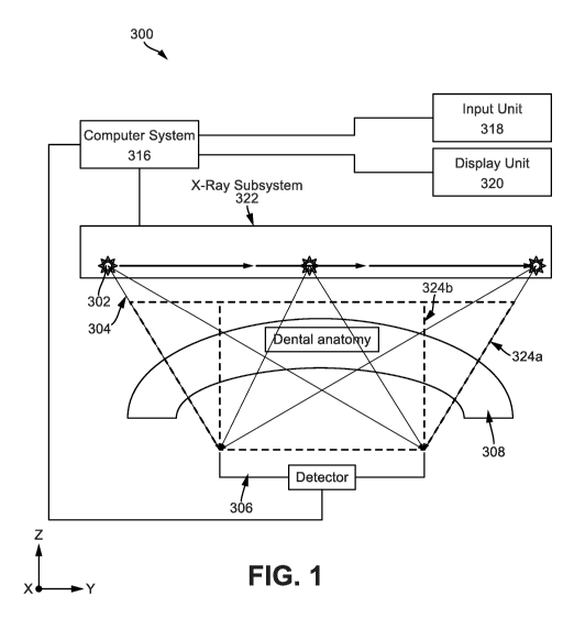
[0031] Fig. 1 illustrates a system illustrating how different portions of the
anatomy are imaged by different projections.
[0032] Fig. 2 illustrates a block diagram of an example computer system of the
tomosynthesis system of Fig. 1.
[0033] Fig. 3 is a flowchart illustrating the overall operation of a system
using
iterative reconstruction.
[0034] Fig. 4 shows a representation of how a reconstructed anatomy is
reprojected onto a reprojection surface.
[0035] Fig. 5 illustrates how discontinuities from variation of support are
generated in non-iterative reconstructions.
[0036] Fig. 6 illustrates how discontinuities from variation of support are
generated in non-iterative reconstructions.
[0037] Fig. 7 illustrates the use of an invalidity matrix in an iterative
reconstruction.
[0038] Fig. 8 illustrates the use of an invalidity matrix in an iterative
reconstruction.
8
CA 03085316 2020-06-09
WO 2019/118387
PCT/US2018/064826
[0039] Fig. 9 illustrates a grayscale representation of the invalidity matrix
and
the corresponding selected input projection.
[0040] Fig. 10 illustrates an x-ray image of the maxillary anterior region of
a
patient taken with a size 1 sensor.
[0041] Fig. 11 illustrates an image of same maxillary anterior region of Fig.
10
with the image having an extended field of view according to an embodiment
herein.
[0042] Different ones of the Figures may have at least some reference numerals
that are the same in order to identify the same components, although a
detailed
description of each such component may not be provided below with respect to
each Figure.
[0043] DETAILED DESCRIPTION
[0044] In accordance with example aspects described herein, methods, systems,
apparatuses, and computer programs are provided for generating a two-
dimensional image from a three-dimensional reconstructed volume based on a
plurality of projection images.
[0045] X-ray System
[0046] Fig. 1 illustrates a block diagram of an intraoral x-ray system 300 for
obtaining an intraoral images, and which is constructed and operated in
accordance with at least one example embodiment herein. An x-ray detector 306
and an X-ray subsystem 322 are electrically connected to the computer system
316. The X-ray subsystem 322 comprises an X-ray source 302. The computer
system 316 is electrically coupled to a user display unit 320 and a user input
unit
318 with the user display unit 320 being an output and/or input user
interface. As
an x-ray source 302 moves from right to left, projections are taken and
projection
images of the dental anatomy 308 are formed on detector 306 for each
projection,
which images are collected by the computer system 316 for processing. The
system 300 can be operated to obtain the one or more images of the dental
anatomy 308 of interest, which may further include one or more sub-object (not
shown). For example, the dental anatomy 300 may be a tooth (or teeth) and
surrounding dentition of a patient, and the sub-object may be a root structure
within the tooth.
9
CA 03085316 2020-06-09
WO 2019/118387
PCT/US2018/064826
10047] The system 300 includes an x-ray detector 306 and an x-ray subsystem
322, both of which, including subcomponents thereof, are electrically coupled
to
the computer system 316. In one example embodiment herein, the x-ray
subsystem 322 hangs from a ceiling or from a wall-mounted mechanical arm (not
shown), so as to be freely positioned relative to the dental anatomy 308. The
x-
ray subsystem 322 further includes an x-ray source 302 which may be mounted
on a motorized stage (not shown).
[0048] The x-ray detector 306 is positioned on one side of the object 50 and
the
receiving surface of the x-ray detector 306 extends in an x-y plane in a
Cartesian
coordinate system. The x-ray detector 306 can be a small intraoral x-ray
sensor
that includes, for example, a complementary metal-oxide semiconductor (CMOS)
digital detector array of pixels, a charge-coupled device (CCD) digital
detector
array of pixels, or the like. In an example embodiment herein, the size of the
x-
ray detector 306 varies according to the type of patient as well as the volume
of
space in the buccal cavity available to be occupied by the x-ray detector. In
an
embodiment, small x-ray detectors 306 may be used by the system to obtain
images with larger size than the size of the x-ray detector 306 by employing
the
processes discussed hereinafter. The x-ray detector 306 may also be one of a
standard size employed in the dental industry. Examples of the standard dental
sizes include a "Size-2" detector, which is approximately 27 x 37 mm in size
and
is typically used on adult patients, a "Size-1" detector, which is
approximately 21
x 31 mm in size and is typically used on patients that are smaller than Size-2
adult patients, and a "Size-0" detector, which is approximately 20 x 26 mm in
size and is typically used on pediatric patients. In a further example
embodiment
herein, each pixel of the x-ray detector 306 has a pixel width of 15 gm, and
correspondingly, the Size-2 detector has approximately 4 million pixels in a
1700
x 2400 pixel array, the Size-1 detector has approximately 2.7 million pixels
in a
1300 x 2000 pixel array, and the Size-0 detector has approximately 1.9 million
pixels in a 1200 x 1600 pixel array. The color resolution of the x-ray
detector
306 may be, in one example embodiment herein, a 12-bit grayscale resolution.
Other examples include an 8-bit grayscale resolution, a 14-bit grayscale
resolution, and a 16-bit grayscale resolution.
CA 03085316 2020-06-09
WO 2019/118387
PCT/US2018/064826
[0049] The x-ray source 302 is positioned on an opposite side of the dental
anatomy from the x-ray detector 306. The x-ray source 302 emits x-rays 10
which pass through the dental anatomy 308 and are detected by the x-ray
detector
306. The x-ray source 302 is oriented so as to emit x-rays 304 towards the
receiving surface of the x-ray detector 306 in at least a z-axis direction of
the
Cartesian coordinate system as shown in Fig. 1, where the z-axis is orthogonal
to
the x-y plane associated with the receiving surface of the x-ray detector 306.
[0050] In one embodiment as shown in Fig. 1, the x-ray system is a
tomosynthesis x-ray system wherein the x-ray source 302 can project x-rays 304
while positioned at each of multiple different locations within a scan angle
328
(shown in Fig. 4) where a 00 position in the scan angle 328 corresponds to the
position for emitting x-rays 304 along the z-axis. In one example embodiment
herein, the user initially positions the x-ray source 302, to a predetermined
starting position relative to the dental anatomy 308. The computer system 316
then controls an on-board motor controller (not shown) to move the x-ray
source
302 via a motorized stage (not shown), based on the known starting position,
to
step through each of the different locations within the scan angle 328. The
computer system 316 can control the x-ray source 302 to cause the source 302
to
emit x-rays 304 to project x-rays at each of those locations. In an example
embodiment herein, there are 41 projections in the tomosynthesis scan and the
scan angle ranges from -20 to +20 wherein the 0 position is the position of
the
x-ray source 302 at which x-rays are projected in the z-axis direction towards
the
x-ray detector 306 as shown in Fig. 4 (source position #21), and wherein
source
position #21 is the central source position 336, and wherein source position
#41
is the last source position 338 in an example tomosynthesis system with 41
projections. The x-rays 304 may converge substantially at a focal spot 314.
The
focal spot 314 may however be located such that part of the x-rays projected
from
the outer limits of the scan angle 328, the outer limits corresponding to, for
example, source position #1 and source position # 41 miss the x-ray detector
306.
The steps discussed hereinafter ensure, among other things, that contributions
to a
reconstructed volume by such x-rays that miss the detector (and thus detector
pixels close to the edge of the detector 306) and by x-rays that hit marker
11
CA 03085316 2020-06-09
WO 2019/118387
PCT/US2018/064826
particles (and thus pixels corresponding to marker particles in the projected
image) are minimized.
[0051] In one example embodiment, the x-ray detector 306 may be an indirect
type of detector (e.g., a scintillator x-ray detector) that first converts x-
rays 304
into an optical image and then converts the optical image into the electrical
signals, and in another example embodiment, the x-ray detector 306 may be a
direct type of detector (e.g., a semiconductor x-ray detector) that converts x-
rays
304 directly into the electrical signals. The computer system 316 processes
the
electrical signals to form a two-dimensional projection images which are
processed to a reconstructed volume 310 and then to a final two-dimensional
image of the dental anatomy. In one example embodiment herein, the image size
of the two-dimensional projection image corresponds to the dimensions and the
number of pixels of the x-ray detector 306. However the image size of the
final
two-dimensional image may be larger than the image size (the dimensions and
the number of pixels) of the projection image and/or x-ray detector.
[0052] The system 300 may collect a plurality of projection images, as
described
above, by first positioning the x-ray source 302 at different angles,
including at
least the 00 position, and emitting x-rays 304 at each of those different
angles
through the dental anatomy 308 towards the x-ray detector 306.
[0053] Computer System for X-ray Imaging
[0054] Having described a system 300 for generating a two-dimensional image
from a three-dimensional reconstructed volume based on a plurality of
projection
images, reference will now be made to Fig. 2, which shows a block diagram of a
computer system 600 that may be employed in accordance with at least some of
the example embodiments herein. Although various embodiments are described
herein in terms of this exemplary computer system 600, after reading this
description, it will become apparent to a person skilled in the relevant
art(s) how
to implement the disclosure using other computer systems and/or architectures.
[0055] In one example embodiment herein, at least some components of the
computer system 600 (such as all those components, or all besides component
628) can form or be included in the computer system 316 of Fig. 1. The
computer system 600 includes at least one computer processor 622. The
12
CA 03085316 2020-06-09
WO 2019/118387
PCT/US2018/064826
computer processor 622 may include, for example, a central processing unit, a
multiple processing unit, an application-specific integrated circuit ("ASIC"),
a
field programmable gate array ("FPGA"), or the like. The processor 622 is
connected to a communication infrastructure 624 (e.g., a communications bus, a
cross-over bar device, or a network).
[0056] The computer system 600 also includes a display interface (or other
output interface) 626 that forwards video graphics, text, and other data from
the
communication infrastructure 624 (or from a frame buffer (not shown)) for
display on a display unit 628 (which, in one example embodiment, can form or
be
included in the display unit 320 of Fig. 1). For example, the display
interface 626
can include a video card with a graphics processing unit.
[0057] The computer system 600 also includes an input unit 630 that can be
used
by a user of the computer system 600 to send information to the computer
processor 622. In one example embodiment herein, the input unit 630 can form
or be included in the input unit 318 of Fig. 1. The input unit 630 may include
a
keyboard device and/or a mouse device or other input device. In one example,
the display unit 628, the input unit 630, and the computer processor 622 may
collectively form a user interface.
[0058] In yet another embodiment that may include a touch screen, the input
unit
630 and the display unit 628 may be combined, or may represent a same user
interface. In such an embodiment, a user touching the display unit 628 can
cause
corresponding signals to be sent from the display unit 628 to the display
interface
626, which can forward those signals to a processor such as processor 622. In
an
example embodiment herein, a system with a wall-mounted mechanical arm (not
shown) may have a module attached to a wall wherein the module includes a
processor 622 and on board electronics for controlling the x-ray source 304, a
motorized stage (not shown) and communicating with the x-ray detector 306.
Processor 622 can be configured to perform part (or all) of any of the
procedures
described herein. For example, one or more steps of the procedure illustrated
in
Fig. 3 can be stored on a non-transitory storage device in the form of
computer-
readable program instructions. To execute a procedure, the processor 622 loads
13
CA 03085316 2020-06-09
WO 2019/118387
PCT/US2018/064826
the appropriate instructions, as stored on storage device, into memory 632,
and
then executes the loaded instructions.
[0059] Moreover, the computer system 600 may comprise a main memory 632,
which may be a random access memory ("RAM"), and also may include a
secondary memory 634. The secondary memory 634 may include, for example, a
hard disk drive 636 and/or a removable-storage drive 638 (e.g., a floppy disk
drive, a magnetic tape drive, an optical disk drive, a flash memory drive, and
the
like). The removable-storage drive 638 reads from and/or writes to a removable
storage unit 640 in a well-known manner. The removable storage unit 640 may
be, for example, a floppy disk, a magnetic tape, an optical disk, a flash
memory
device, and the like, which is written to and read from by the removable-
storage
drive 638. The removable storage unit 640 may include a non-transitory
computer-readable storage medium storing computer-executable software
instructions and/or data.
[0060] In further alternative embodiments, the secondary memory 634 may
include other computer-readable media storing computer-executable programs or
other instructions to be loaded into the computer system 600. Such devices may
include a removable storage unit 644 and an interface 642 (e.g., a program
cartridge and a cartridge interface similar to those used with video game
systems); a removable memory chip (e.g., an erasable programmable read-only
memory ("EPROM") or a programmable read-only memory ("PROM")) and an
associated memory socket; and other removable storage units 644 and interfaces
642 that allow software and data to be transferred from the removable storage
unit 644 to other parts of the computer system 600.
[0061] The computer system 600 also may include a communications interface
646 that enables software and data to be transferred between the computer
system
600 and external devices. Such an interface may include a modem, a network
interface (e.g., an Ethernet card or an IEEE 802.11 wireless LAN interface), a
communications port (e.g., a Universal Serial Bus ("USB") port or a FireWire
port), a Personal Computer Memory Card International Association ("PCMCIA")
interface, and the like. Software and data transferred via the communications
interface 646 may be in the form of signals, which may be electronic,
14
CA 03085316 2020-06-09
WO 2019/118387
PCT/US2018/064826
electromagnetic, optical or another type of signal that is capable of being
transmitted and/or received by the communications interface 646. Signals are
provided to the communications interface 646 via a communications path 648
(e.g., a channel). The communications path 648 carries signals and may be
implemented using wire or cable, fiber optics, a telephone line, a cellular
link, a
radio-frequency ("RF") link, or the like. The communications interface 646 may
be used to transfer software or data or other information between the computer
system 600 and a remote server or cloud-based storage (not shown).
[0062] One or more computer programs or computer control logic may be stored
in the main memory 632 and/or the secondary memory 634. The computer
programs may also be received via the communications interface 646. The
computer programs include computer-executable instructions which, when
executed by the computer processor 622, cause the computer system 600 to
perform the processes as described herein and shown in Figs. 3 - 9.
Accordingly, the computer programs may control the computer system 316 and
other components (e.g., the x-ray detector 306 and the x-ray source 302) of
the
intraoral tomosynthesis system.
[0063] In another embodiment, the software may be stored in a non-transitory
computer-readable storage medium and loaded into the main memory 632 and/or
the secondary memory 634 of the computer system 600 using the removable-
storage drive 638, the hard disk drive 636, and/or the communications
interface
646. Control logic (software), when executed by the processor 622, causes the
computer system 600, and more generally the intraoral tomosynthesis system, to
perform the processes described herein.
[0064] Lastly, in another example embodiment hardware components such as
ASICs, FPGAs, and the like, may be used to carry out the functionality
described
herein. Implementation of such a hardware arrangement so as to perform the
functions described herein will be apparent to persons skilled in the relevant
art(s) in view of this description.
[0065] Method for generating a two-dimensional image from a three-
dimensional reconstructed volume based on a plurality of projection images.
CA 03085316 2020-06-09
WO 2019/118387
PCT/US2018/064826
[0066] Having described the computer system 316 of Fig. 2, the intraoral
tomosynthesis x-ray system 300 will now be further described in conjunction
with Fig. 3, which shows a flow diagram of a process according to an example
embodiment herein for using an invalidity matrix, a reconstruction process and
a
reprojection process to generate a two-dimensional image from a three-
dimensional reconstructed volume based on a plurality of projection images
[0067] In Step S202 the intraoral tomosynthesis system 300 acquires a
plurality
of projection image of the dental anatomy 308 for different spatial position
of the
x-ray source during a tomosynthesis scan. For example, the x-ray source 302 is
moved by a motorized stage (not shown) and control circuitry to different
positions within the scan angle 328, and the computer system 316 controls the
x-
ray source 302 to emit x-rays 304 at each position. In one example embodiment
herein, x-ray source 302 is scanned, by moving the x-ray source from -200 at
source position #1, 334 where a first projection 330 is made to obtain a first
projection image, through 00 at source position #21, 336 where a central
projection is made to obtain a central projection image, to -20 at source
position
#41, 338 where a final projection is made to obtain a final projection image.
In an
embodiment herein 41 projections are made in a single tomosynthesis scan in
evenly distributed increments of 1 to provide 41 scan angles, including one
at
the 0 position, although this example is not limiting. It can be seen that in
some
projections, for example in the first projection 330, not all individual x-
rays 332
of that first projection hit the detector 306.
[0068] X-rays 304 that pass through the dental anatomy 308 are attenuated by
the dental anatomy 308 before being projected onto the x-ray detector 306. The
x-ray detector 306 converts the x-rays 110 into electrical signals and
provides the
electrical signals to the computer system 316. The computer system 316
processes the electrical signals collected at each scan angle position to
acquire the
plurality of projection images, each image comprising an array of pixels. The
image acquired with the x-ray source 302 at the 00 position is also referred
to
herein as a central projection image. The computer system 316 then performs in
Step S204 a calibration of the acquired projection images by converting gray
16
CA 03085316 2020-06-09
WO 2019/118387
PCT/US2018/064826
level values of the projection images into material thickness based on an
earlier
projection using a phantom calibration object of known dimensions.
[0069] In Step S206, an associated system geometry is estimated by using
marker particles in the tomosynthesis scan to determine the position of the
dental
anatomy in relation to the X-ray source. An invalidity matrix, discussed
hereinafter, may then be determined in Step S208 for each acquired projection
image to determine the contribution of pixels the acquired image to a
reconstructed volume during an update Step S222 of the iterations S236
discussed hereinafter. Contribution of marker particles to the projection
images
can be identified and removed in Step S210 such that their further
contribution to
a volume to be reconstructed 310 is limited. Removal of said marker particle
contributions from the projection images can be achieved by identifying
regions
in the projection images that correspond to the shape of the marker particles
and
subtracting them from the projection images. The resulting blank regions of
the
projection images can then padded by, for example, interpolating said blank
regions with data of the surrounding regions. However the padded data is
essentially fake data and this information can be further propagated to the
volume
to be reconstructed 310. A smooth deweighting process based on an invalidity
matrix of all pixels, discussed hereinafter, helps to limit this further
contribution.
Herein pixels corresponding to the fake padded data as well as pixels close to
the
edges of the detector (collectively referred to as potentially problematic
pixels)
can be weighted for each projection image such that they do not contribute to
the
volume to be reconstructed 310 as much as other pixels do.
[0070] A starting volume 324a, depicted in Fig. 1 for a first volume update
process wherein said starting volume 324a is a blank or empty volume can be
constructed in Step S212 and a projection selected in Step S214 from a
projection
list for calculating a forward projection of the starting volume in Step S216
using
the system geometry of the selected projection. This starting volume will be
iteratively updated in the volume update steps S234 and iteration steps S236
discussed hereinafter to reconstruct the irradiated dental anatomy.
17
CA 03085316 2020-06-09
WO 2019/118387
PCT/US2018/064826
[0071] In an example embodiment, the number of projections may be 41 and a
number of iterations S236 may be 5 or 6. Therefore 41 volume update steps S234
are executed in each iteration step S236 for said example embodiment.
[0072] In another embodiment herein, a first volume update step S234 for
reconstructing the irradiated dental anatomy can be started in a first
iteration
S236 using a first selected projection image wherein the volume update step
S234
is subsequently repeated for the remaining projection images during said first
iteration S236. Stored projections images may be selected in succession such
that
a selected projection image is from a projection position that located away
from
the projection position of the previously selected projection image such that
the
two projection images are substantially different from each other. For
example,
every nth projection image can be selected successively wherein n does not
divide the total number of projection images evenly. In an exemplary
embodiment, n can be 7. Alternatively projection images of projection
positions
that are furthest apart from each other in the scan angle 328 may be selected
successively.
[0073] In the first volume update Step S234, a first projection image is
selected
in Step S214. A forward projection of the starting volume is then determined
in
Step S216 using the system geometry. A difference image between the resultant
forward projection and the selected projection, which contains the padded
data, is
determined in Step S218. An update for updating the starting volume is
calculated in Step S220 by scaling said difference image according to the
invalidity matrix for the projection. The invalidity matrix is a matrix that
ensures
that the contribution, of potentially problematic pixels (pixels close to the
edge of
the detector and pixels representing padded data) to the update of the
reconstructed volume (or starting volume in the case of a first volume update
S234 of a first iteration S236) is limited. The invalidity matrix for all
projection
images can be calculated in Step S208 by identifying any invalid regions
(potentially problematic pixels) in a binary mask and calculating the distance
inside the invalid regions (positive) and the distance outside the invalid
regions
(negative) depending on whether the pixel in question itself is valid or
invalid.
For example, staring with a binary definition of valid and invalid the
distance of a
18
CA 03085316 2020-06-09
WO 2019/118387
PCT/US2018/064826
pixel from its nearest valid pixel can be measured. This can, for example, be
zero
if the pixel itself is a valid pixel. The distance of that pixel from the
nearest
invalid pixel can also be measured. This can, for example, be zero if the
pixel
itself is invalid. These numbers may be combined to obtain a value for each
pixel
of a projection image and the process repeated to obtain a value for each
pixel of
the selected projection, creating an invalidity matrix for said selected
projection
image as shown in Fig. 9 wherein potentially problematic pixels of the
projection
image include portions of the image representing marker particles 520 and
edges
of the image 522. In the adjacent image, a representation of the invalidity
matrix
is shown. Values corresponding to pixels closest to the potentially
problematic
pixels are shown to have a lighter color (or shorter distance) than values
corresponding to pixels furthest from the potentially problematic pixels which
have a darker color (or larger distance). It can be seen that portions of the
invalidity matrix 524, 526 corresponding to the potentially problematic pixels
will have the shortest distances since they coincide with the potentially
problematic pixels. Therefore pixels with shorter distances will contribute
less to
the reconstructed volume than pixels with larger distances will. After scaling
the
difference image according to the invalidity matrix to obtain an update, the
starting volume (blank or empty volume) is updated by backprojecting the
difference image through said starting volume to obtain a first reconstructed
volume in Step S222. The first reconstructed volume is then processed further
volume update processes S234 using subsequent selected projection images until
all projection images have been selected. If a termination criteria, discussed
hereinafter, is not met the iteration steps S236 are repeated.
The update of the volume in Step S222 may comprise a Simultaneous Algebraic
Reconstruction Technique (SART) based iterative reconstruction algorithm
wherein the volume V is updated by summing the currently estimated volume
with a backprojected volume according to the formula V ¨> V +
AWVi(B Pi(E Pi)) .
This may take inputs:
Pi, where i denotes the ith measured/selected projection P in Step S214
and P is a two-dimensional matrix corresponding to the projected image.
19
CA 03085316 2020-06-09
WO 2019/118387
PCT/US2018/064826
BPi, where i denotes the ith backprojection operator which is used to alter
the voxels in a reconstructed volume to make them consistent with the measured
projections
FPi, where i denotes the ith forward projection operator obtained in Step
S216, wherein the forward projection operator is an operator used to calculate
the
projection resulting from a volume with specific volume content
A, which is a scaling factor used to control convergence speed, described
hereinafter.
WVi, which is a volumetric weighting matrix, described hereinafter and
V, which is the currently estimated volume.
This may be accomplished by the following steps:
I. Start with an uninitialized volume V as shown in Step S212.
2. Compute an error/difference image EPi = FPi(V) ¨ Pi, as shown in
Step S218.
3. Update the volume V according to: V .-4 V + AINVi(BPi(EPi)), for
each i as in Step S222. The ordering of the update in terms of i can
be non-consecutive because it speeds convergence.
In an example embodiment herein, each iteration of an update process can be
thought of as multiplying an error term associated with the iteration by a
number.
If that number has a magnitude less than one, each iteration will reduce the
error
term and the process converges. If the number is greater than one, the error
term
increases and the process diverges. As such, the convergence factor is chosen
to
be as high as possible without exceeding a certain threshold value since fewer
iterations are better. Beyond the threshold, the iterations diverge and each
iteration becomes increasingly far from the desired volume.
In an example embodiment herein, a goal of the volume update may be to
construct a final volume such that the difference image EPi is close to zero.
Each pixel of a projection image can be represented by a three-dimensional
equivalent known as a voxel. WVi represents an "ith" volumetric weighting
matrix which determines how much weight should be given to each voxel of the
back projection "ith" error image when determining said voxel's contribution
to
the volume that is about to be updated during a volume update process S234.
This
CA 03085316 2020-06-09
WO 2019/118387
PCT/US2018/064826
allows the removal of the contributions of invalid pixels of the "ith"
projection
from the reconstructed volume.
may be obtained as follows:
An invalidity I is obtained according to the ternary/conditional notation
below such that
/i = BPK (spopi+co)/Pi) < ¨d ? 0 :
wherein d is a term indicating a distance from invalid regions where a
voxel is considered completely valid and r is a scaling factor chosen to
provide a scale based on the extent of perturbations to data based on
edge effects related to marker identification.
The ith invalidity matrix /Pimay be computed as:
'Pt = D11> DOt? DIt : ¨D01, wherein
IPt is the ith invalidity matrix and Pt is a two-dimensional matrix
corresponding to the projected image.
DIt is the distance from the nearest valid pixel for a given pixel
DOtis the distance from the nearest invalid pixel for a given pixel.
The invalidity I may then be used to calculate the weighting term for
update:
1
WV = (1 + 4)2
[0074] The above steps can be used to iteratively reconstruct the volume using
the invalidity matrix. A grayscale representation of the invalidity matrix and
the
corresponding selected input projection is shown in Fig. 9. Since the
invalidity
matrix reduces the contribution of invalid pixels to the reconstructed volume,
the
reconstructed volume may contain little to no influence by marker particles.
After
a first update of the reconstruction volume, a next non-consecutive projection
image can be selected such that it is different from the first projection and
the
process may be repeated S230 with the newly reconstructed volume being used
21
CA 03085316 2020-06-09
WO 2019/118387
PCT/US2018/064826
for a new forward projection in Step S216. It can be goal to obtain a
reduction in
the difference image with each newly selected projection until the difference
image is close to zero. The volume update process S234 is repeated until all
projections are selected. After all projections are selected S232, a second
iteration
S236 involving all projections may be started if a termination criteria is not
met.
By testing against said termination criteria in Step S224, a new iteration
S236
may be started if the termination criteria is not met S228 with the current
reconstructed volume being used for the forward projection. Alternatively the
reconstruction may be ended if the termination criteria is met. The
termination
criteria can be for example (i) the difference of forward projection and
selected
projection or a function of said difference of forward projection and selected
projection being close to zero or (ii) a fixed number of iterations steps S236
having been completed. The fixed number can, for example, be between 5 and
10. Upon meeting the termination criteria, a reprojection surface may be
calculated and the final reconstructed volume may be reprojected in Step S226
to
obtain a 2D Image with an extended field of view. The extended field of view
is
obtained as follows. Each pixel of the reprojection surface can have
properties
such as an x-position, a depth position and a direction which may be
determined
by the direction between the pixel of the reprojection surface and the
position of a
virtual focus 314. Starting at an extreme end of the reconstructed volume 310,
voxels intersected by a line determined by the pixel position and direction
may be
summed to determine the total attenuation of the pixel. This may be repeated
for
all pixels of the reprojection surface to obtain a 2D Image with an extended
field
of view. Thus, using the virtual focus 314 at negative depth (past the
position of
the detector 306 in the opposite direction of the x-ray source) that is
matched to
the opening angle of the scan angle 328 of the tomosynthesis scan, the
reconstructed dental anatomy 310 may be projected onto the reprojection
surface
312 to obtain an image that includes a larger area than that available for a
single
detector system with no extended field of view. The larger area is illustrated
as
the surface of the effective detector 326 in the x-y plane of Fig. 4. Fig. 4
also
shows a representation of how the reconstructed volume 310 is reprojected onto
the reprojection surface 312 wherein the reprojection surface 312 is for
example a
22
CA 03085316 2020-06-09
WO 2019/118387
PCT/US2018/064826
semi-circular surface containing pixels. The reprojection surface can also be
dynamically constructed to match the geometry of the dental anatomy. A
dynamically obtained surface that matches the geometry of the dental anatomy
can be based on the reconstructed volume 310.
[0075] Fig. 5 and 6 illustrate how discontinuities from variations in support
are generated in non-iterative reconstructions, support being a representation
of the number of projections that contribute to a given voxel. Voxels 402 and
418 are adjacent voxels In Fig. 5, rays 406, 408 and 410 of three different
projections pass through voxel 402 of volume 404 and result in all projections
contributing to voxel 402. In Fig. 6, rays 412, 414 and 416 of three different
projections pass through voxel 418 of volume 404 but only projections
corresponding to rays 414 and 412 contribute to voxel 418. The projection
corresponding to ray 416 does not contribute to voxel 418 during
reconstruction, because ray 416 is not incident on detector 306 and as such
ray
416 does not contribute to the formation of any pixels of the projection
image.
Each pixel of the projection image has a three-dimensional equivalent known
as a voxel. A difference in the number of projections contributing to adjacent
voxels 402 and 418 in Figs 5 and 6 will generate discontinuity in the
reconstructed volume.
[0076] Figs. 7 and 8 illustrate the use of an invalidity matrix in an
iterative
reconstruction to reduce or eliminate discontinuities in reconstructed
volumes.
Voxels 502 and 518 are adjacent voxels. In Fig. 7, three different projections
corresponding to rays 506, 508 and 510 respectively all contribute to voxel
502 since the rays are all incident on the detector 306. Projections
corresponding to rays 506 and 508 however contribute more to voxel 502 than
the projection of ray 510 does. This is because contributions of pixels close
to
the detector edge are deweighted according to the invalidity matrix during an
update step of the iterative reconstruction process since not all rays
corresponding to projections that are incident close to the edges of the
detector fall on the detector.
[0077] In Fig. 8, a projection of ray 516 misses contribution to voxel 518.
Projections of rays 512 and 514 however contribute to voxel 518. A difference
23
CA 03085316 2020-06-09
WO 2019/118387
PCT/US2018/064826
in the number of projections contributing to adjacent voxels does not generate
discontinuity here because the contribution of near edge pixels to the
reconstruction is near zero according to the invalidity matrix. As can be seen
in Fig. 8, pixels of projections corresponding to rays 512 and 514 are
weighted during an update step of the iterative reconstruction according to
the
invalidity matrix to contribute more to voxel 518 while the contribution of
pixels of the projection of ray 516 is near zero. This results in a
reconstructed
three-dimensional volume 310 that is more representative of the irradiated
dental anatomy 308 in preparation for the reprojection step of Step S226
discussed above.
[0078] Further, a synthetic radiograph having a higher signal-to-noise ratio
that
that of any single projection image in a tomosynthesis scan may be obtained by
the above processes wherein the reconstruction done is smaller and a flat
plane
reprojection surface having the same size as the detector is selected for
reprojection. For a smaller reconstruction, a smaller starting volume 324b may
be
chosen wherein said smaller starting volume 324b may be a blank or empty
volume the length in the X-Y plane of which matches the length of the detector
306 in said X-Y plane as shown in FIG. 1. For starting volume 324b, fewer
projections incident at the edges of the detector will be used in the
iterative
reconstruction process compared to the number of projections incident at the
edges of the detector that are used when employing a bigger volume 324a. In
consequence, artifacts in the reconstruction are reduced. Further a flat plane
reprojection surface having the same length as the length of detector in the X-
Y
plane can be used for reprojection using the processes described above and
shown in Fig. 3. Herein, a virtual focus at negative depth is not used. Rather
x-
rays are projected through the reconstructed volume programmatically by
applying a forward projection operator of the central source position to the
reconstructed volume to obtain a 2D image with the same field of view as that
of
the detector. This results in a 2D image which has much noise removed and thus
possesses a much higher signal-to-noise ratio than that of any single
projection
image in the low dose tomosynthesis scan while showing more features of the
scanned dental anatomy than a single center projection shows. This is helpful
in a
24
CA 03085316 2020-06-09
WO 2019/118387
PCT/US2018/064826
setting where both an intraoral radiograph and a dental tomosynthesis scan is
needed to provide by a dentist. In an embodiment herein, such an image may be
obtained primarily to allow the presentation of a 2D radiograph without marker
particles using a 3D tomosynthesis scan and eliminating the need to take a
separate high dose radiograph for use by a dentist.
[0079] Fig. 10 and 11 illustrate images of the maxillary anterior region of a
patient taken with a size 1 sensor. In Fig 10 a system without the extended
field
of view described herein is used to take a single high dose radiograph and
results
in an image having the same field of view as the sensor used. Fig. 11 shows
that a
larger field of view can be obtained at a low dose with a tomosynthesis x-ray
system having the extended field of view described herein.
[0080] The general operation of the x-ray system according to the disclosure
may be as follows. A dentist may, for example, note that a patient has a
painful
torus behind said patient's left molars. In addition, the patient may have
extensive
tooth decay that the dentist may like to image using an intraoral scan prior
to
assessing the need for a bridge. The dentist may therefore use a size 1 sensor
oriented vertically to form an image using the system disclosed herein, said
image being somewhat larger than the image of a size 2 sensor oriented
horizontally using convention x-ray systems. Therefore a dentist may use a
smaller, easier to fit, sensor to obtain an image with similar or larger size
than
that obtained from a larger sensor that doesn't fit in a given patient's
mouth.
Without this approach, the most expeditious approach would be to take several
images while shifting the sensor manually and stitching them together by eye.
This would invariably complicate the dentist's understanding of the problem
since no single image may contain the entirety of the problem region.
Moreover,
if the primary goal of a dentist is to obtain a standard 2D radiograph with a
high
signal to noise ratio than that of any single projection image in a low dose
tomosynthesis scan without taking an additional high dose radiograph, a device
according to the disclosure wherein the reprojection surface is a flat plane
may be
similarly used to produce such a 2D image.
[0081] In view of the foregoing description, it can be appreciated that the
example embodiments described herein provide systems, methods, apparatuses,
CA 03085316 2020-06-09
WO 2019/118387
PCT/US2018/064826
and computer programs products for for using an invalidity matrix, iterative
reconstruction and reprojection to generate from a three-dimensional
reconstructed volume a two-dimensional image with image artifacts removed
wherein the 3D reconstructed volume is based on a plurality of projection
images
[0001] Unless otherwise defined, all technical and scientific terms used
herein
have the same meaning as commonly understood by one of ordinary skill in the
art to which this invention belongs. Although methods and materials similar to
or
equivalent to those described herein can be used in the practice or testing of
the
disclosure, suitable methods and materials are described above. All
publications,
patent applications, patents, and other references mentioned herein are
incorporated by reference in their entirety to the extent allowed by
applicable law
and regulations. The disclosure may be embodied in other specific forms
without
departing from the spirit or essential attributes thereof, and it is therefore
desired
that the present embodiment be considered in all respects as illustrative and
not
restrictive. Any headings utilized within the description are for convenience
only
and have no legal or limiting effect.
26