Note: Descriptions are shown in the official language in which they were submitted.
- 1 -
DEVICES FOR TRANSDERMAL DRUG DELIVERY
This is a divisional of Canadian Patent Application No. 2,810,800 filed
September 13,
2011.
DESCRIPTION
Technical field
The invention relates to the field of transdermal delivery of drugs into the
body of a
patient. In particular, it relates to the delivery of drugs through pores
previously
created in the skin.
For the sake of brevity, the term "drugs" is used in this specification to
refer to any
biologically active substances that may need to be introduced into the body of
a
patient to provide a therapeutic, cosmetic or nutritional effect. The patient
may be
human or a non-human animal. "Transderrnal" refers to delivery through the
skin of
the patient or through any other accessible surface tissue such as the cornea
or the
inside of the mouth cavity.
Background of the invention
Methods have been described for enhancing skin permeation of drugs by using a
device that gradually eases microneedles into contact with the skin, for
example by
forming an array of microneedles directly on a roller or, as described in
international
patent application WO 2008/125798, by forming an array of microneedles on a
patch
secured to a belt that travels over a set of rollers. This method has been
demonstrated
to be superior to simply pressing a flat array of microneedles against the
skin. That is
because less insertion force is required and because, given that the array of
needles is
inserted row by row, the reproducibility of the dose is also increased
independently of
the operator.
The main barrier to delivery of drugs through the skin is the stratum corneum,
which
is a tough outer layer of dead skin cells. The microneedles may be hollow to
provide
a channel for delivery of a fluid drug through the stratum corneum or they may
be
CA 3085890 2020-07-03
- 2 -
solid and simply coated with the drug for delivery. Alternatively, a device
comprising
solid microneedles may be used to disrupt the stratum corneum and/or to create
pores
through it in order to enhance its permeability to a drug that is subsequently
applied to the
surface of the skin, for example in the form of a gel or in a patch. However,
because the
needles only perforate a small proportion of the surface area of skin being
treated, a
majority of the subsequently applied drug formulation does not enter the pores
but remains
on the surface of the skin. This is contrary to the requirements of most
bodies governing
drug registration that minimal drug should be applied, and that minimal excess
should be
present after application. It is also wasteful of a potentially expensive
product.
Summary of the invention
The invention provides a transdermal drug delivery device comprising:
means comprising a number of needles for piercing a patient's skin to form a
number of pores in the skin;
a number of carriers for loading with a drug to be delivered; and
means, operable while the needles remain in the skin, for applying each
carrier to
one of said number of pores to deliver the drug to said one of said number of
pores;
wherein each of the carriers is an elongate element that is introduced into
the pore
alongside the corresponding needle, between the needle and the skin.
Each of the carriers may have a blunt tip.
Preferably, a plurality of the needles are configured in a first array and
wherein a plurality
of the carriers are configured in a second array having the same layout as the
first array.
CA 3085890 2021-12-01
- 2a -
The invention further provides a transdermal drug delivery device comprising:
means comprising a number of needles for piercing a patient's skin to form a
number of pores in the skin;
a number of carriers for loading with a drug to be delivered;
a channel associated with each carrier;
a first insert part that defines a first portion of each channel and a second
insert part
that defines a second portion of each channel, the first and second insert
parts being capable
of relative movement between a rest configuration in which the first and
second portions
of the channel are not aligned and an operating configuration in which the
first and second
portions of the channel are aligned; and
means, operable while the needles remain in the skin and while the first and
second
insert parts are in the operating configuration, for sliding each carrier
along the associated
channel to apply the carrier to one of said number of pores to deliver the
drug to said one
of said number of pores.
Each of the carriers may be an elongate element having a tip that can be
loaded with the
drug for delivery.
Preferably, the drug loaded on the tip of each carrier and contained within
the associated
channel by the tip of the carrier.
In the rest configuration, the first portion of the channel may be closed by
an opposing wall
of the second insert part.
In accordance with the invention, the number of carriers is preferably equal
to the number
of pores.
CA 3085890 2021-12-01
- 2b -
By delivering the drug only to the locations of the previously formed pores, a
controlled
quantity of the drug formulation can be delivered to precisely where it can
travel through
the pores to penetrate the stratum corneum and be taken up by the body. There
will be
minimal wastage of drug left on the surface of the skin and inaccessible to
the body. The
drug may be delivered to the mouths of the pores, especially if it is in a
fluid state.
Preferably the drug is positioned by the carriers directly inside the pores,
beneath the
stratum corneum, from where it can be diffused and dispersed through the body
like other
transdermally delivered treatments. This is suitable for drug formulations in
various states,
including solid (powdered or particulate) drugs.
The simplest means for forming pores in the skin comprises a plurality of
needles. A
common mechanism can then be used to position the needles and the drug
carriers to ensure
a consistent alignment between them. However, other mechanical or non-
mechanical
means may be used for forming pores in predetermined locations on the skin,
for example
laser ablation.
=
CA 3085890 2021-12-01
- 3 -
The drawings
Figures 1 a and lb illustrate two steps in the process of delivering drugs to
a patient
using a device according to a first embodiment of the invention.
Figures 2a and 2b illustrate two steps in the process of delivering drugs to a
patient
using a device according to a second embodiment of the invention.
Figures 3a to 3d illustrate four steps in the process of delivering drugs to a
patient
using a device according to a third embodiment of the invention.
Figures 4a to 4c illustrate three steps in the process of delivering drugs to
a patient
using a device according to a fourth embodiment of the invention.
Figures la and lb are schematic views of a transdermal drug delivery device
comprising a frame 2 that is held in a fixed position on a treatment area of
the skin 4
of a patient. An axle 6 extends between opposite sides of the frame 2 in an
orientation
generally parallel to the surface of the skin 4. The axle carries a block 8
with an array
of microneedles 10 on one face and an array of microstructures 12 on the
opposite
face. The two arrays 10,12 share an identical layout. In the side view of
Figure 1,
only a single row of microneedles 10 and a single row of microstructures 12
are
visible but in practice each array will extend over a two-dimensional surface
of the
block 8.
The microneedles 10 may be formed using any suitable method such as moulding
or
micro-machining. They may have a diameter in the range from a few tens of
micrometres to more than a millimetre; and a length typically a few times
greater than
their width. The length is preferably sufficient to penetrate the stratum
corneum of
the skin but not great enough for the needles to reach the nerve endings that
are
deeper in the skin.
The microstructures 12 are preferably elongate elements such as rods or posts.
Unlike
conventional microneedles, these are formed to have blunt tips. The tips are
preferably flat, i.e. generally planar and perpendicular to the long axes of
the
microstructures 12. The microstructures 12, like the microneedles 10, may be
CA 3085890 2020-07-03
- 4 -
produced from any suitable type of plastic, ceramic or metal, and should be of
a
dimension and shape that allows the insertion of the drug directly into the
pore created
by the microneedle. To achieve this the microstructure may either remain above
the
stratum corneum, acting purely to force the drug through the pore, or it may
be shaped
such that it is able to penetrate the pore already created by the microneedle
thus
delivering the drug directly to a deeper region within the skin. The
microstructure
may thus be smaller than the pore created by the microneedle to allow it to be
inserted
into the skin as it forces the drug through, or it may be angled or bevelled
such that
although the microstructure may be larger than the pore created, or larger
than the
needles used to create the pores, it will still be able to penetrate the pore
and push the
drug deeper into the skin.
A drug in any suitable formulation may be loaded onto the tips of the
microstructures
12. If the formulation is in solid form, one or more particles 14 of it may be
attached
to the tips by using an adhesive in which the particles are insoluble. The
particles 14
may alternatively be attached through electrostatic attraction to avoid the
use of any
adhesive that may cause degradation or weakening of the particles during
storage.
Static charge will be concentrated at the tips of the carriers and may
encourage the
particles 14 to attach there. Metallic based particles may be loaded on to the
tips of
the microstructures using magnetic attraction.
Each end of the axle 6 of the drug delivery device is mounted in a mechanism
16 that
permits the axle 6 to move towards and away from the skin 4 of the patient.
The
movement towards the skin may be effected by manual pressure, as shown by the
pair
of double-headed arrows 18 in Figs. la and lb. The movement may be guided by a
slot (not shown); and return springs (not shown) may be provided to retract
the axle 6
away from the skin 4 to its rest position. It will be understood that the
drawing is
purely schematic. In practice, a cover would have to be provided to prevent
accidental contact by the user with the upwardly facing microneedles 10 or
microstructures 12. A mechanism could be provided to ensure that the axle 6
remains
level. The movement of the axle 6 towards and away from the skin could be
CA 3085890 2020-07-03
- 5 -
automated instead of being effected manually; and means could be provided to
regulate the force of impact with the skin 4.
This "stamping" action is first carried out with the microneedles 10 facing
the skin 4,
as shown in Fig. I a to pierce the stratum corneum and create an array of
pores 20,
shown in Fig. lb. The block 2 is then rotated through 1800 about the axle 6,
as shown
by a curved arrow 22, so that the microstructures 12 are now facing the skin 4
and
perfectly aligned with the pre-formed pores 20. The same stamping action is
then
repeated so that the microstructures 12 enter the respective pores 20 and each
deposits
its load of drug 14 at a predetermined depth within the pore 20.
The block 8 is not limited to a single array of microneedles 10 and a single
array of
microstructures 180 apart. There could be multiple such arrays angularly
distributed
about the surface of a cylinder or the faces of a prism. Indexing means would
then be
provided for turning the block 8 manually or automatically through an
appropriate
angle to ensure the precise orientation of the block 8 with the desired type
of array
facing the skin 4. In this manner more than one type of drug could be
successively
delivered into a single set of pores 20.
The arrays of microneedles 10 and microstructures 12 could be formed as
separate
patches for attachment to the surface of the block 8, provided they can be
positioned
sufficiently accurately. Alternatively, the arrays could be provided
alternately along
the surface of a belt (not shown) that is wrapped around a cylindrical block
8, with
means for rotating the cylinder to advance the belt through a predetermined
distance
and bring the next array to the correct position.
Figures 2a and 2b are schematic views of an alternative transdermal drug
delivery
device, in which the means for exchanging the arrays of microneedles 10 and
microstructures 12 involves rotation about an axle 30 that is generally
perpendicular
to the skin 4, as shown by the curved arrow 32. Means (not shown in detail)
are
provided to permit the left hand array (as viewed in the drawings) to come
into
contact with the skin 4 and then be withdrawn, in a similar stamping action to
that
CA 3085890 2020-07-03
- 6 -
previously described, as indicated by the double-headed arrows 34. First the
array of
microneedles 10 are pushed into the skin 4 and retracted to form pores 36.
Then the device is
rotated about its axle 30 to bring the array of microstructures 12 carrying
the drug particles 14
into coincidence with the pores 36 and the stamping action is repeated to
deliver the drug into
the pores 36.
The axle 30 may be constructed so as to be capable of compression
telescopically to bring the
left hand array into contact with the skin, with a return spring (not shown)
to return the array
to its rest position. If the skin is flat, such an arrangement would tend to
bring the right hand
array simultaneously into contact with a different area of the skin, which
must be avoided.
Means could be provided to shield the skin in that area or the device could be
configured so
that the right hand array is higher than the left hand array, for example by
angling the axle 30
slightly away from the vertical while keeping the active array on the left
parallel to the surface
of the skin.
Not only rotational motion is capable of exchanging the positions of the
arrays. A device in
accordance with the invention could allow the array of microneedles 10 to
slide out of position
and the array of microstructures 12 to slide into position, preferably both in
a single
movement.
Figures 3a to 3d show a different embodiment of drug delivery device according
to the
invention, in which the microstructures 40 introduce the drug 42 into the
pores 44 while the
microneedles 46 that formed the pores 44 are still in place. Figures 3a to 3d
show a single
needle, which will typically but not exclusively form part of an array of
identical needles. Fig.
3a shows a microneedle 46 poised above the skin 4 of a patient. A
microstructure 40 has
particles of a drug formulation 42 loaded onto its flat tip 48. (It is not
necessary that the drug
be in particulate or even solid form.) The microstructure 40 is in a retracted
position so that
the microneedle 46 can penetrate the surface of the skin 4 without the drug 42
coming into
contact with the skin, as shown in Figure 3b. The microneedle 46 forms a pore
44 through
the outer layer of the skin 4.
CA 3085890 2021-12-01
- 7 -
Next the microstructure 40 advances from the retracted position shown in Fig.
3b to
the deployed position shown in Fig. 3c, travelling along the surface of the
needle 46
and entering the pore 44 that has been formed by the needle. The drug 42 can
thereby
be deposited into the pore 44 from the blunt tip 48 of the microstructure at a
suitable
depth to be absorbed by the patient. As shown in Fig. 3d, the microneedle 46
and the
microstructure 40 can then both be withdrawn from the skin, which quickly
closes up
the pore, leaving the embedded drug 42.
In the embodiment of Figures 3a to 3d, the microneedles need not be
cylindrical/conical but can have a flattened cross-section or be generally
wedge-
shaped to present one or more flat side faces. This provides a larger surface
against
which the microstructure 40 can bear. The side face of the microneedles may
also be
provided with at least one longitudinal groove, which provides space within
the pore
44 for the drug to be accommodated and encourages the drug to flow down the
groove
deeper into the skin.
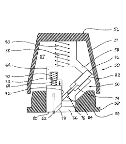
Figures 4a to 4c show schematically a cross-section through a hand-held device
according to a further embodiment of the invention. The device comprises an
outer
case 50 that is formed from two parts, namely an upper part 51 and a lower
part 52.
The upper and lower parts 51,52 engage one another so as to be capable of
relative
sliding movement in the vertical direction (as illustrated). That movement is
regulated by compression springs, as will be discussed below. A generally flat
lower
face 54 of the lower part 52 is intended to rest against the skin of a patient
during use
of the device. An upper face 56 of the upper part 51 is intended to be pressed
by the
hand of a user and may be generally flat or curved so as to provide additional
comfort
or control. The upper and lower parts 51,52 of the case form a cavity 57
between
them. A cam surface 58 projects from an interior wall of the upper part 51
into the
cavity 57.
A central opening in the lower face 54 of the case is shaped so that it can
receive a
disposable insert 60. Means such as a catch or clip (not shown) hold the
insert in
position in the device during use and can then be manually released to allow
the insert
CA 3085890 2020-07-03
=
-8-
60 to be removed and replaced. When in position in the device, the insert 60
lies
entirely within the boundary of the case, except that a row of needles 62 is
mounted in
the insert and the tips of the needles project just below the lower face 54 so
that they
can penetrate the outer layer of a patient's skin. (The row of needles 62
extends
perpendicularly to the plane of Figures 4a-4c so only one of them is visible.
If the
drug to be delivered is sufficiently potent, it may be that only a single
needle is
required, instead of a row of them.)
The insert 60 is formed from two parts, namely an upper part 64 and a lower
part 66.
The upper and lower parts 64,66 engage one another so as to be capable of
relative
sliding movement in the vertical direction (as illustrated). A peg 68 on an
upper
surface of the lower part 66 of the insert is aligned with a recess 70 in a
lower surface
of the upper part 64 of the insert. The peg 68 and the recess 70 provide
seating for a
first compression spring 72. (It will be understood that the positions of the
peg 68 and
the recess 70 could be exchanged or that other means of seating the spring 72
could be
employed.)
A set of angled channels 74 is formed in the insert 60. There is one channel
74
corresponding to each of the needles 62. Each channel 74 comprises an upper
channel 76 formed in the upper part 64 of the insert and a lower channel 78
formed in
the lower part 66 of the insert. Each lower channel 78 intersects the lower
face 80 of
the insert 60 at approximately the point where the corresponding needle 62
emerges.
Located in each upper channel 76 is an elongated drug carrier 82. A tip 84 of
the
carrier occupies substantially the whole cross-section of the channel 76. An
upper
end 86 of the carrier projects from the insert 60 into the cavity 57 of the
upper case
51. As shown, the upper end 86 may be wider than the tip to provide a surface
to
which a force can readily be applied. Under the influence of that force, the
carrier 82
can slide along the upper channel 76.
A peg 88 on an upper surface of the upper part 64 of the insert provides
seating for a
second compression spring 90, which acts between the insert 60 and an interior
wall
of the upper part 51 of the outer case 50. The second spring 90 may be seated
in a
CA 3085890 2020-07-03
- 9 -
recess (not shown) in the case and it will be understood that the positions of
the peg
88 and the optional recess could be exchanged or that other means of seating
the
spring 90 could be employed.
Figure 4a shows the device in its rest configuration, when the first and
second
compression springs 72,90 are maximally extended. The upper part 51 of the
outer
case 50 is spaced from the lower part 52. The upper part 64 of the insert 60
is spaced
from the lower part 66 and as a result the upper portion 76 of each channel 74
is not
aligned with the lower portion 78. The carrier 82 is partially withdrawn from
the
upper channel 76 so that its upper end 86 projects into the cavity 57 and is
close to or
in engagement with the cam surface 58. A dose of drug (not shown) is loaded in
each
of the upper channels 76 and is contained by the tip 84 of the drug carrier 82
at its
upper end and by an opposing wall 92 of the lower part 66 of the insert so
that the
drug cannot escape from the insert 60 during transport and storage.
To use the device and deliver the drug to a patient, manual pressure is
applied to the
top face 56 of the outer case 50. This causes the needles 62 to penetrate the
outer
surface of the patient's skin and form pores through which the drug may enter.
The
first compression spring 72 is weaker than the second compression spring 90 so
that
as the manual pressure continues it is the first compression spring 72 that is
compressed first, as shown in Figure 4b. This brings the upper and lower parts
64,66
of the insert 60 into contact and causes the upper and lower portions 76,78 of
the
angled channels 74 to align with one another, releasing the drug that has been
contained in the upper channel 76 to flow into the lower channel 78.
As additional pressure is applied to the top face 56 of the outer case 50, the
engagement between the upper and lower parts 64,66 of the insert 60 prevents
further
compression of the first compression spring 72 so the second compression
spring 90
then begins to be compressed. This allows the upper part 51 of the outer case
50 to
move towards the lower part 52. In so doing, the cam surface 58 begins to act
against
the upper ends 86 of the drug carriers 82 and forces each carrier 82 to slide
along its
upper channel 74 and to expel the drug therefrom towards the lower channel 78
in the
CA 3085890 2020-07-03
- 10 -
manner of a piston. The lower end of the lower channel 78 is now aligned with
the
pore that has been formed by the needle 62 so that the drug is delivered
directly to the
pore, where it can be taken through the patient's skin.
Figure 4c shows the configuration when the second spring 90 has been maximally
compressed, the drug carrier 82 has been pushed fully into the channel 74 and
the
upper part 51 of the outer case 50 has come into engagement with the lower
part. A
catch (not shown) may be provided to maintain the device in this fully
compressed
configuration after use so that it is obvious that it has been used and no
attempt will
be made to re-use it until the insert 60 has been replaced with a new dose of
drug.
It will be noted that in this embodiment of the invention the drug does not
have to be
adhered to the tip 84 of the carrier because the location of the drug is
controlled by the
channel 74. Thus the drug can optionally be in fluid form. Also, the tip 84 of
the
carrier does not necessarily have to approach the associated pore in the skin
very
closely, provided that it pushes the drug sufficiently far ahead of it to
reach the pore.
Although the needles 62 are shown as fixed to the insert 60 and permanently
extending from its bottom face 80, means (not shown) may be provided for
shielding
the tips of needles 62 or for keeping them retracted inside the insert 60
until the
device is ready for use. The retracted needles may then be either extended
manually
or extended automatically when pressure is applied to the top face 56 of the
outer case
50. For example, the needles could be mounted on the upper part 64 of the
insert 60
and run through guides in the lower part 66. Then, as the two parts 64,66 move
together in changing from the configuration of Fig. 4a to that of Fig. 4b, the
needles
will move along the guides until their tips project from the bottom face 80.
CA 3085890 2020-07-03