Note: Descriptions are shown in the official language in which they were submitted.
CA 03086203 2020-06-18
WO 2019/126369 PCT/US2018/066574
1
TOTAL MESORECTAL EXCISION SURGICAL SIMULATOR
Cross-reference to Related Applications
[0001] This application claims the benefit of U.S. Provisional patent
application no. 62/607,476, filed
on December 19, 2017, the entire disclosure of which is hereby incorporated by
reference as if set in full
herein.
Background
[0002] This application is generally related to surgical training systems and
methods, and, in particular
to, simulated tissue structures and models for teaching, practicing and
evaluating various surgical
techniques and procedures related to but not limited to total mesorectal
excision procedures and
techniques.
[0003] Medical students as well as experienced doctors learning new surgical
techniques must
undergo extensive training before, they are qualified to perform surgery on
human patients. The training
must teach proper techniques employing various medical devices for cutting,
penetrating, clamping,
grasping, stapling, cauterizing and suturing a variety of tissue types. The
range of possibilities that a
trainee may encounter is great. For example, different organs and patient
anatomies and diseases are
presented. The thickness and consistency of the various tissue layers will
also vary from one part of the
body to the next and from one patient to another. Different procedures demand
different skills.
Furthermore, the trainee must practice techniques in various anatomical
environs that are influenced by
factors such as the size and condition of the patient, the adjacent anatomical
landscape and the types of
targeted tissues and whether they are readily accessible or relatively
inaccessible.
[0004] Various teaching aids, trainers, simulators and model organs are
available for one or more
aspects of surgical training. However, there is a need for models or simulated
tissue elements that are
CA 03086203 2020-06-18
WO 2019/126369 PCT/US2018/066574
2
likely to be encountered in and that can be used for practicing endoscopic and
laparoscopic, minimally
invasive, transluminal surgical procedures. In laparoscopic surgery, a trocar
or cannula is inserted to
access a body cavity and to create a channel for the insertion of a camera
such as a laparoscope. The
camera provides a live video feed capturing images that are then displayed to
the surgeon on one or
more monitors. At least one additional small incision is made through which
another trocar/cannula is
inserted to create a pathway through which surgical instruments can be passed
for performing
procedures observed on the monitor. The targeted tissue location such as the
abdomen is typically
enlarged by delivering carbon dioxide gas to insufflate the body cavity and
create a working space large
enough to accommodate the scope and instruments used by the surgeon. The
insufflation pressure in the
tissue cavity is maintained by using specialized trocars. Laparoscopic surgery
offers several advantages
when compared with an open procedure but requires an increased level of skill
as the target tissue is not
directly observed by the clinician. The target tissue is observed on monitors
displaying a portion of the
surgical site that is accessed through a small opening. Therefore, clinicians
need to practice visually
determining tissue planes, three-dimensional depth perception on a two-
dimensional viewing screen,
hand-to-hand transfer of instruments, suturing, precision cutting and tissue
and instrument manipulation.
Typically
[0005] One procedure is a total mesorectal excision (TME) for the treatment of
late stage colorectal
cancer in which the entire mesorectal envelope and a portion of the rectum are
removed. This procedure
has shown reduced local recurrence rates and improved oncologic outcomes for
patients. The TME
procedure can be performed using a combination of minimally invasive
techniques, including
laparoscopic and transanal approaches. Currently, there is an unmet need of
educational tools for
surgeons to utilize while developing and practicing skills relevant to the TME
procedure.
CA 03086203 2020-06-18
WO 2019/126369 PCT/US2018/066574
3
Summary
[0006] In accordance with various embodiments of the present invention, a TME
surgical simulator is
provided. The surgical simulator comprises a frame having a proximal opening
and a distal opening and
a simulated tissue layer connected and covering the proximal opening. In
various embodiments, the
surgical simulator further comprises a simulated organ assembly extending
through the distal opening.
[0007] In various embodiments, a surgical simulator comprises a frame having a
proximal portion
defining a simulated abdominal cavity and a distal portion defining a
simulated pelvic cavity and a
simulated parietal peritoneum layer connected to the proximal portion. In
various embodiments, the
surgical simulator further comprises a simulated aorta disposed within the
simulated abdominal cavity
and a simulated prostate disposed within the simulated pelvic cavity.
[0008] In various embodiments, a surgical simulator comprises a simulated
parietal peritoneum layer
and a simulated mesorectum and mesentery layer connected to the simulated
parietal peritoneum layer
and together forming a envelope there between. In various embodiments, the
surgical simulator further
comprises a simulated fatty fill disposed within the envelope.
[0009] In various embodiments, a surgical simulator comprises a frame having a
proximal portion
defining a simulated abdominal cavity and a distal portion defining a
simulated pelvic cavity, a
simulated endopelvic fascia layer disposed within the simulated pelvic cavity
and a simulated pelvic
floor layer attached to the simulated endopelvic fascia layer and the distal
portion of the frame. In
various embodiments, the surgical simulator further comprises a simulated
mesorectum layer attached to
the simulated endopelvic fascia layer, the simulated mesorectum layer and the
simulated endopelvic
fascia layer defining a simulated dissection plane therebetween.
[00010] In various embodiments, a surgical simulator comprises a frame having
a proximal portion
defining a simulated abdominal cavity and a distal portion defining a
simulated pelvic cavity, a
CA 03086203 2020-06-18
WO 2019/126369 PCT/US2018/066574
4
simulated visceral peritoneum layer disposed within the simulated abdominal
cavity and a simulated
peritoneum or parietal peritoneum attached to the simulated visceral
peritoneum layer and the proximal
portion of the frame. In various embodiments, the surgical simulator further
comprises a simulated
mesentery layer attached to the simulated Toldt's/endopelvic fascia layer, the
simulated mesentery layer
and the simulated To1dt' s/endopelvic fascia layer defining a simulated
dissection plane there between.
[00011] Many of the attendant features of the present invention will be more
readily appreciated as the
same becomes better understood by reference to the foregoing and following
description and considered
in connection with the accompanying drawings.
Brief Description of the Drawings
[00012] The present inventions may be understood by reference to the following
description, taken in
connection with the accompanying drawings in which the reference numerals
designate like parts
throughout the figures thereof.
[00013] FIG. 1 is perspective view of a surgical simulator or model in
accordance with various
embodiments of the present invention.
[00014] FIG. 2 is a side view of a surgical simulator in accordance with
various embodiments of the
present invention.
[00015] FIG. 3 is a top view of a surgical simulator in accordance with
various embodiments of the
present invention.
[00016] FIGS. 4A-4B are front cross-sectional semi-schematic diagrams of a
surgical simulator in
accordance with various embodiments of the present invention taken along the
line A-A.
[00017] FIG. 5A is a perspective view of portions of a surgical simulator in
accordance with various
embodiments of the present invention.
CA 03086203 2020-06-18
WO 2019/126369 PCT/US2018/066574
[00018] FIG. 5B is an exploded view of portions of a surgical simulator in
accordance with various
embodiments of the present invention.
[00019] FIG. 6 is a perspective view of portions of a surgical simulator in
accordance with various
embodiments of the present invention.
[00020] FIG. 7 is a side view of a surgical simulator in accordance with
various embodiments of the
present invention.
[00021] FIGS. 8A-8B are front cross-sectional semi-schematic diagrams of a
surgical simulator in
accordance with various embodiments of the present invention taken along the
line B-B.
[00022] FIG. 9 is a top view of a surgical simulator in accordance with
various embodiments of the
present invention.
[00023] FIGS. 10A-10B are side cross-sectional semi-schematic diagrams of a
surgical simulator in
accordance with various embodiments of the present invention taken along the
curved line C-C.
[00024] FIGS. 11A-11B are front cross-sectional semi-schematic diagrams of
various configurations of
a surgical simulator in accordance with various embodiments of the present
invention.
[00025] FIGS. 12A-12B are front cross-sectional semi-schematic diagrams of
various configurations of
a surgical simulator in accordance with various embodiments of the present
invention.
[00026] FIG. 13 is a front cross-sectional semi-schematic diagram of a
configuration of a surgical
simulator in accordance with various embodiments of the present invention.
[00027] FIG. 14 is a perspective view of a frame of a surgical simulator in
accordance with various
embodiments of the present invention.
[00028] FIG. 15 is a front view of a frame of a surgical simulator in
accordance with various
embodiments of the present invention.
CA 03086203 2020-06-18
WO 2019/126369 PCT/US2018/066574
6
[00029] FIG. 16 is a perspective cross-sectional view of a frame of a surgical
simulator in accordance
with various embodiments of the present invention.
[00030] FIG. 17 is a top view of a frame of a surgical simulator in accordance
with various
embodiments of the present invention.
[00031] FIG. 18 is a perspective view of a frame of a surgical simulator in
accordance with various
embodiments of the present invention.
[00032] FIG. 19 is a perspective cross-sectional view of a frame of a surgical
simulator in accordance
with various embodiments of the present invention.
[00033] FIG. 20 is a side view of a frame of a surgical simulator in
accordance with various
embodiments of the present invention.
Detailed Description
[00034] Generally, a TME surgical simulator or model is provided to assist in
surgical skill training and
simulation. The TME simulator comprises various simulated organ and/or tissue
structure assemblies
attached to each other and positioned within a rigid frame. The simulated
organ structure assemblies
provide different simulated tissue planes and in particular, distinguishable
simulated dissection planes.
Various features of the assemblies further provide added tactile and/or visual
feedback along with
difficulties and challenges to simulate and further assist in training and
assessment of a simulated TME
surgical procedure. For example, varying sizes and/or attachment of various
simulated organ structures
and/or varying various compositions and/or toughness of such structures and
assemblies are provided.
[00035] During a TME procedure, the fatty mesorectal envelope circumferencing
the rectum is
removed along with a portion of the rectum. This procedure requires ligation
of the relevant blood
supply as well as circumferential mobilization of the rectum and mesorectum
from the pelvic cavity and
CA 03086203 2020-06-18
WO 2019/126369 PCT/US2018/066574
7
surrounding structures. Once the mesorectal specimen is removed, the
mesorectum is inspected and
graded; ranging from incomplete to complete dissection of this tissue. This is
determined by visibility of
the rectal lumen where segments of the mesorectum have been dissected into
erroneously. Grading of
the excised specimen can be used to assess surgical performance as well as
local recurrence risk.
[00036] A TME procedure, as such, requires a high level of technical skill and
understanding of the
pelvic anatomy. The TME surgical simulator in various embodiments mimics a
portion of the pelvic
anatomy and thus can meet the clinical need in providing learning tools for
surgeons to use during their
education. The TME surgical simulator in accordance with various embodiments
allows for the
execution and/or completion of a simulated TME procedure from a laparoscopic
and/or transanal
approach. In accordance with various embodiments, for compatibility with both
approaches and
realistic identification of the relevant anatomical landmarks, the TME
surgical simulator comprises at
least one or more of the following simulated organ structures, tissue,
vasculature and/or materials: colon,
mesentery, mesorectum, Toldt' s fascia, visceral peritoneum (retroperitoneum),
endopelvic fascia,
prostate, ureter, gonadal vessels, seminal vesicles, muscle layers, pelvic
floor, IMA, IMV and aorta. The
simulated materials, tissue layers and/or organ structures, in various
embodiments, are also assembled in
such a way that allows the end user, e.g., the surgeon, to perform the
simulated procedure such that it
provides realistic tactile feedback similar to that which one would encounter
during the non-simulated
procedure. This, among other things, allows the user to train on technical
skill development associated
with the identification of relevant anatomy and tissue handling. In accordance
with various
embodiments, the simulated materials can be confined within a simulated
pelvis, which provides
realistic limitations of the pelvic workspace including, for example, limited
visualization occurring
during dissection down the curved pelvic floor during a TME procedure. The
simulated materials in
CA 03086203 2020-06-18
WO 2019/126369 PCT/US2018/066574
8
various embodiments allows for the removal of the simulated mesorectum
specimen which upon
removal can be graded, for example, on a scale of 1-3 ranging from incomplete
to complete dissection.
[00037] The TME surgical simulator according to various embodiments of the
present invention is used
to simulate a total mesorectal excision procedure. To provide a realistic
procedural training
environment, the TME surgical simulator or model provides laparoscopic and
transanal access for the
surgical procedure simulation. The surgical simulator provides simulated
materials to represent the
various anatomical landmarks and planes, as well as materials to simulate
dissection, which provides
notable visual and tactile feedback useful to understand the anatomy and
tissue handling in order to
better understand the complications that can be encountered during a surgical
procedure. Additionally,
the TME surgical simulator provides materials to simulate the mesorectum that
allow the mesorectum to
be mobilized, removed, and graded. Portions of the simulator, such as the
mesorectum, are provided
such that it is sufficiently fragile and thus able to be damaged during the
simulated procedure. This in
turn allows the simulated mesorectum and the like to be assessed and/or graded
similar to a surgical
procedure. In various embodiments, the confinement of portions of the surgical
simulator in a simulated
pelvis presents the realistic challenge of working within a confined space.
Additionally, in various
embodiments, the simulated pelvis has a curve integrated within its shape to
simulate the curve of the
sacrum, which presents a visualization challenge during the surgical
procedure. The TME surgical
simulator in various embodiments can be used within a laparoscopic trainer and
with laparoscopic
instruments for a simulated procedure. The TME surgical simulator, in
accordance with various
embodiments, relevant and realistic anatomy for the performance of each
surgical step of the TME
procedure.
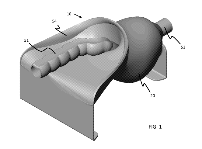
[00038] Referring to FIGS. 1-3 and 14-20, the TME surgical simulator 10
comprises a simulated pelvic
frame or base 20. The frame 20 in various embodiments is the hardest or most
rigid portion of the
CA 03086203 2020-06-18
WO 2019/126369 PCT/US2018/066574
9
simulator 10. The frame provides a platform raising and/or suspending
simulated organ or tissue
structures 100 secured to the frame. The frame has a proximal end and a distal
end in which the
proximal end has enlarged opening 21 relative to a restricted opening 22 at
its distal end. In various
embodiments, the frame is sized and shaped to fit within the confines of a
laparoscopic trainer having
specific and limited access points or channels to access the TME surgical
simulator and/or not air tight
or able to seal in insufflation gas.
[00039] A proximal portion of the simulator 10 is a simulated abdominal cavity
or portions thereof and
a distal portion of the simulator is a simulated pelvic cavity or portions
thereof. As such, the proximal
portion of the frame is set higher or above the distal portion of the frame.
Similarly, simulated organ
and/or tissue structures in the proximal portion are different from those in
the distal portion of the
simulator 10. However, some common organ and/or tissue structures extend from
the proximal portion
to the distal portion. In the illustrated embodiment, at or near the proximal
end of the frame extends a
support or leg 23 and at or near the distal end of the frame extends a support
or leg 24. The leg 23
extends from a proximal portion of frame floor 25 and leg 24 extend from a
distal portion of the frame
floor 25 with the proximal portion of the frame floor being higher or
positioned above the distal portion
of the frame floor. The frame floor 25 extends into sidewalls 26 and at a
distal portion extends to a roof
or top 27. As such, in various embodiments, the distal portion of the frame
provides a confined curved
cavity or enclosure and at a distal end provides a generally circular opening
and the proximal portion of
the frame provides an enlarged curved cavity with an enlarged opening. The
frame floor 25 in various
embodiments starting at the proximal end of the frame extends laterally,
curves down and back up at the
distal end of the frame. In the illustrated embodiment, the legs and frame
floor are integrated or formed
as a single monolithic structure and in various embodiments are separate
components, connected
together to form a simulated working space for the simulator 10. In various
embodiments, the frame is
CA 03086203 2020-06-18
WO 2019/126369 PCT/US2018/066574
formed plastic or a similar rigid material and in other various embodiments
includes plastic sheets cut to
shape and attached together, e.g., with mechanical fasteners, to form the
frame. Various simulated
organ and tissue structures, assemblies or subassemblies are secured and/or
supported by the frame.
[00040] Referring now also to FIGS. 4A-10B, the simulator 10 comprises a
simulated pelvic floor
assembly, a simulated Toldt's/endopelvic fascia assembly, a colon/rectum
assembly and a
mesentery/mesorectum assembly. The simulated pelvic floor assembly comprises
simulated Gonadal
vessels 32, simulated aorta 33 and simulated nerves 34 with a layer of fibrous
material, such as batting,
attaching or otherwise placing the simulated material between or intermingling
with the fibrous material
and a simulated pelvic floor 31. In various embodiments, simulated pelvic
floor 31, simulated Gonadal
vessels 32, simulated aorta 33 and simulated nerves 34 are all made of
silicone but are colored or
otherwise distinguished from each other visually and/or tactilely. Similarly,
the simulated vessel
structures have a different thickness or length to further distinguish the
structures, such as the simulated
aorta 33 being thicker than the simulated nerves 34 and the simulated gonadal
vessels 32 and/or the
simulated gonadal vessels being longer than the simulated nerves 34.
[00041] In accordance with various embodiments, the simulated Toldt' s or
endopelvic fascia assembly
comprises a simulated Toldt's fascia / visceral peritoneum / endopelvic fascia
sheet or layer 41
("Toldt's/endopelvic fascia") and simulated ureters 42 and in various
embodiments simulated blood
vessels attached to the simulated ureters. The simulated Toldt's/endopelvic
fascia sheet in various
embodiments is made from silicone and fibrous material, e.g., batting,
attached or cured thereto. The
simulated ureters and associated vessels are also made of silicone and are
attached to the simulated
Toldt' s/endopelvic fascia sheet and in various embodiments adhered or
attached to a proximal portion of
the sheet and/or on a smooth or non-fibrous side of the sheet. In various
embodiments, simulated
CA 03086203 2020-06-18
WO 2019/126369 PCT/US2018/066574
11
ureters, vessels and Toldt's/endopelvic fascia sheet are colored or otherwise
distinguished from each
other visually, e.g., colored, and/or tactilely, e.g., having different
dimensions, thickness and/or length.
[00042] In the illustrated embodiment, the simulated Gonadal vessels 32,
simulated aorta 33 and
simulated nerves 34 are adhered to the simulated pelvic floor 31 which is
attached to the frame and a
distal portion of the simulated pelvic floor away from the simulated aorta 33
is cut, folded and shaped
also attached to the frame. The simulated Gonadal vessels 32, simulated aorta
33 and/or simulated
nerves 34 are also only accessible and/or visible at or near the proximal
portion of the TME surgical
simulator while the simulated pelvic floor extends along or throughout the
entire length or interior of the
frame. The simulated ureters 42 are also only accessible and/or visible at or
near the proximal portion of
the TME surgical simulator and thereby further mimicking or closely
representing portions of abdominal
cavity in the TME surgical simulator. The Toldt's/endopelvic fascia sheet is
adhered or otherwise
attached to the pelvic floor placing the simulated ureters and vessel there
between. Furthermore, a distal
portion of the Toldt's/endopelvic fascia sheet is shaped, formed and/or
attached to itself to provide a
generally enclosed or circular or oval enclosure at the distal portion of the
TME surgical simulator and
thereby also further mimicking or more closely representing portions of the
pelvic cavity in the TME
surgical simulator.
[00043] In accordance with various embodiments, at a distal portion of the TME
surgical simulator, a
simulated prostate 43, simulated bladder 44 and simulated seminal vesicles 45
are also provided and
attached to the simulated endopelvic sheet 41. As provided in the illustrated
embodiment, the simulated
bladder 44 is positioned above and somewhat around the simulated prostate 43
with the simulated
prostate 43 having a simulated urethra 46 extending therefrom. The simulated
seminal vesicles 45 are
positioned adjacent or next to the simulated prostate 43 and in various
embodiments, the simulated
prostate 43, simulated bladder 44, simulated seminal vesicles 45 and/or
simulated urethra 46 are
CA 03086203 2020-06-18
WO 2019/126369 PCT/US2018/066574
12
disposed between the simulated pelvic floor 31 and the simulated endopelvic
fascia sheet 41. In various
embodiments, although not illustrated, the ureters attach to the bladder
towards the proximal end of the
surgical simulator. In various embodiments, the simulated pelvic floor 31
covers the simulated
endopelvic fascia sheet 41 to form or define a cavity in which the simulated
prostate 43, simulated
bladder 44, simulated seminal vesicles 45 and/or simulated urethra 46 are
captured or enclosed therein.
In accordance with various embodiments, the simulated endopelvic fascia sheet
41 is adhered to the
simulated pelvic floor 31, which is then placed under an outward tension or
away from the interior of the
frame but still within the confines of frame. In various embodiments, the
simulated pelvic floor is
stretched or pulled towards the interior of frame after being adhered to the
frame to ultimately provide or
assist in providing the outward tension for the simulated pelvic floor.
[00044] In various embodiments, the simulated bladder 44 is adhered to the
simulated pelvic floor 31 at
the pubis, and the simulated prostate 43 is adhered to the simulated
endopelvic fascia sheet 41, anterior
to the simulated rectum 53. The simulated seminal vesicles 45 are adhered only
to the simulated
prostate 43 and the simulated bladder 44. In various embodiments, the
simulated prostate, simulated
bladder, simulated seminal vesicles, simulated urethra and/or
Toldt's/endopelvic fascia sheet are colored
or otherwise distinguished from each other visually, e.g., colored, and/or
tactilely, e.g., having different
dimensions, thickness and/or length. In various embodiments, the simulated
prostate, simulated bladder,
simulated seminal vesicles and/or simulated urethra are all made of silicone.
[00045] In accordance with various embodiments, the colon/rectum assembly
comprises a simulated
colon 51 attached to a simulated rectum 53. The simulated colon is contoured
or has curves, bumps or
other surface features thereby distinguishing the simulated colon from the
substantially smooth and
tubular simulated rectum. The transition from the bumpy colon to the smooth
rectum forms the recto-
sigmoid junction, a visual landmark during surgery and provided in various
embodiments of the surgical
CA 03086203 2020-06-18
WO 2019/126369 PCT/US2018/066574
13
simulator. In various embodiments, a simulated parietal peritoneum 54 has a
proximal end or portion
attached to the simulated colon 51 and a distal end or portion attached to the
simulated rectum 53.
Additionally, the simulated parietal peritoneum 54 is attached to the
Toldt's/endopelvic fascia sheet with
portions of the mesentery / mesorectum assembly 55 disposed therebetween.
[00046] In various embodiments, a sheet of the same material used to create
the simulated
Toldt' s/endopelvic fascia sheet 41 is cut and attached to itself and forms
the simulated mesentery 57. In
various embodiments, a simulated inferior mesenteric artery (IMA) 52 is
attached to the simulated colon
51, e.g., at the proximal portion of the simulated colon and thus is only
accessible and/or visible at the
proximal portion of the TME surgical simulator.
[00047] In various embodiments, the mesentery/mesorectum assembly comprises a
simulated
mesentery 57 and a simulated mesorectum 58 and a simulated IMA 52 and inferior
mesenteric vein
(IMV) 56 is attached to the simulated mesentery 57. The simulated mesentery is
attached to or
integrated into the simulated parietal peritoneum 54 where the two interfaces.
In various embodiments,
the simulated mesentery 57 and simulated mesorectum 58 are a single monolithic
structure with the
simulated mesentery 57 being at the proximal portion of the TME surgical
simulator and the simulated
mesorectum 58 being at the distal portion of the TME surgical simulator. In
various embodiments, a
simulated IMA 52, IMV 56 and mesentery 57 are positioned at or in the proximal
portion of the TME
surgical simulator and thus is only accessible and/or visible at the proximal
portion of the TME surgical
simulator. In various embodiments, the simulated IMA and/or IMV are all made
of silicone.
[00048] In accordance with various embodiments of the present invention, the
first step of a simulated
TME procedure using a laparoscopic approach is entry through the simulated
parietal peritoneum. The
root of the mesentery is a common point of entry and can be recognized by a
color difference between
the mesenteric fat and posterior abdominal wall. This entry point can also be
recognized by observing
CA 03086203 2020-06-18
WO 2019/126369 PCT/US2018/066574
14
movement in the anatomical layers when the sigmoid colon is moved with
laparoscopic instruments, as
well as by the identification of the bulge of the inferior mesenteric artery
(IMA) through the mesentery
and peritoneum. The identification of this anatomical feature can provide a
necessary starting point for
a TME procedure. Referring to the TME surgical simulator, the simulated
parietal peritoneum 54 is
adhered or otherwise attached to the simulated visceral peritoneum more
securely than the simulated
mesentery 57 to simulate realistic tenting and dissection. The simulated colon
51 in various
embodiments is tubular and/or made of silicone. The simulated colon is adhered
or otherwise attached
on top of the simulated parietal peritoneum to simulate the descending colon.
In various embodiments,
the sheet thickness is increased or decreased and may be made with a lower or
higher durometer
material to change the elasticity and structure of the simulated mesentery.
The simulated mesentery 57
in various embodiments is molded out of a conductive material to enable the
user to use laparoscopic
energy equipment to make incisions. The simulated mesentery is assembled such
that it is adhered to
the simulated Toldt's/endopelvic fascia layer 41 up to the simulated pelvic
brim. As such, this allows
abdominal dissection to be differentiated from the lower pelvic dissection
that is performed from the
laparoscopic approach to mobilize the rectum.
[00049] Once through the simulated parietal peritoneum 54, the surgeon can
dissect through the
simulated avascular plane, e.g., the simulated Toldt's/endopelvic fascia layer
41, to find the simulated
IMA 52, IMV 56, aorta 33 and/or ureters 42. Ureter identification can be
important to avoid surgical
complications and further reinforce training and the effects of the
simulation. Once the simulated
structures are identified, the simulated IMA 52 can be skeletonized, ligated
and divided close to the
simulated aorta. In various embodiments, these simulated organ and/vascular
structures, e.g., the
simulated IMA, IMV, aorta and/or ureter, can be cut, stapled, sutured and tied
to simulate ligation and
division. The simulated ureters and aorta allow for the identification of the
simulated IMA 52 and IMV
CA 03086203 2020-06-18
WO 2019/126369 PCT/US2018/066574
56 during the simulated TME procedure. In various embodiments, the simulated
vasculature varies in
thickness and can be hollow or hollow and fluid filled to simulate bleeding.
One or more of the
simulated vasculatures may also disposed within the surgical simulator without
adhesion or minimal
attachment, e.g., loosely, directly adhered or otherwise attached to a
silicone structure and/or any
combination thereof. Dissection is continued laterally toward the simulated
abdominal sidewall
mobilizing the simulated mesentery.
[00050] In various embodiments, the simulated avascular plane, e.g., the
simulated Toldt's/endopelvic
fascia layer, comprises a fibrous batting layer made of polyester fiberfill
(polyfil). The tactile feedback
of the fibrous layer, e.g., fibers within the batting, provides sufficient
resistance to allow blunt dissection
using laparoscopic instruments to be utilized. This dissection within the
fibers allows the simulated
vasculature within the layer to be skeletonized. Below or adjacent to the
simulated Toldt' s fascia is a
thin layer relative to the simulated Toldt's fascia that represents the layer
entering the retroperitoneum or
visceral peritoneum. The thinness of or fragility of this layer
(retroperitoneum/visceral peritoneum) and
by extension the simulated Toldt's/endopelvic fascia layer 41 simulates the
ease in which this plane can
accidently be entered, which is a challenge encountered during the surgical
procedure and provided or
simulated by the TME surgical simulator. In various embodiments, the simulated
Toldt' s fascia is a
fibrous layer or filling, e.g., batting, representing or simulating connective
tissue and is disposed next to
and/or attached to the simulated retroperitoneum or visceral peritoneum, e.g.,
one or more silicone
sheets. The simulated Toldt' s fascia and/or the simulated retroperitoneum or
visceral peritoneum are
disposed next to and/or attached to the simulated parietal peritoneum 54.
[00051] In various embodiments, the simulated Tole s/endopelvic fascia layer
41 is a composite layer
41, for example, having fibrous material and silicone, including the simulated
Toldt's fascia layer along
with the simulated visceral peritoneum or retroperitoneum layer with the
simulated endopelvic fascia
CA 03086203 2020-06-18
WO 2019/126369 PCT/US2018/066574
16
layer being a continuation of the combined simulated Toldt's fascia layer and
the simulated visceral
peritoneum or retroperitoneum layer. As such, together, in various
embodiments, the simulated Toldt's
fascia layer and the simulated retroperitoneum or visceral peritoneum layer
form a composite layer, e.g.,
fibrous material and silicone, and the simulated endopelvic fascia layer is an
extension of this composite
layer, being a continuation of the combined simulated Toldt's fascia layer and
the simulated
retroperitoneum or visceral peritoneum layer. In various embodiments, the
simulated Toldt' s fascia
layer and simulated visceral peritoneum or retroperitoneum layer are disposed
in the simulated
abdominal cavity, and thus so named, while the simulated endopelvic fascia
layer is disposed in the
simulated pelvic cavity and thus so named differently. As such, reference to
the simulated endopelvic
fascia layer may be used interchangeability throughout the description with
the simulated
Toldt' s/endopelvic fascia layer and vice versa. Similarly, the combined or
composite layer of the
simulated Toldt's fascia and simulated retroperitoneum or visceral peritoneum
may be used
interchangeability throughout the description with the simulated endopelvic
fascia layer and simulated
Toldt' s/endopelvic fascia layer and vice versa.
[00052] In various embodiments, the retroperitoneum layer or portions thereof
is yellow or otherwise
discernible via color or the like to further distinguish or highlight the
retroperitoneum layer. Within the
simulated retroperitoneum layer, there are additional fibers or fibrous
material, e.g., a batting layer
(simulated Toldt's fascia), that contains the simulated aorta, nerves, and
gonadal vessels. The simulated
ureters and nerves are adhered or otherwise attached to the simulated
retroperitoneum 41 while the
simulated aorta 33 and gonadal vessels 32 are adhered or otherwise attached to
the simulated pelvic
floor. In various embodiments, the simulated pelvic floor 31 is a thin sheet
molded out of pink or
blood/flesh colored silicone. The simulated nerves 34, ureters 42 and gonadal
vessels 32, in accordance
with various embodiments, are adhered or otherwise attached to the respective
simulated sheets or layers
CA 03086203 2020-06-18
WO 2019/126369 PCT/US2018/066574
17
at an angle, slant or similar orientations such that the simulated vasculature
pairs are closer to each other
at the proximal end than at the distal end of the respective simulated sheets
or layers. As such, in the
simulated lower pelvic region, when the simulated ureters and gonadal vessels
are wrapped around the
simulated mesorectum 58, the simulated vasculature meet at the location of the
simulated bladder 44 and
prostate 43.
[00053] To further simulate the appearance of the abdominal cavity under the
simulated
retroperitoneum, in various embodiments, an additional pink silicone layer
making up the simulated
pelvic floor is placed under the simulated aorta and batting layer of the
simulated retroperitoneum. This
allows the visualization of the color of the simulated abdominal cavity when
the simulated
retroperitoneum layer is encountered.
[00054] In accordance with various embodiments, the silicone and fibrous
layers that make up the
simulated mesentery, Toldt's fascia, endopelvic fascia, and/or retroperitoneum
are adhered using
silicone. Silicone adhesive or alternative adhesives such as cyanoacrylate
adhesives and rubber cement
in various embodiments are used to adhere the layers together. When using
silicone layers, the silicone
can be readily masked within the silicone layer and application quantities can
be easily controlled with
syringes and sponge like materials, such as polyurethane foam. It also creates
a strong silicone to
silicone bond while also adhering to the fibrous layers, if any is present.
The silicone to silicone bonds
between the silicone mesentery and the fibrous material and between the
fibrous material and the
silicone retroperitoneum layer allow the dissection to be contained within the
fibrous material, e.g., the
batting layer. Similarly, in various embodiments, silicone to silicone bonds
between the silicone
retroperitoneum layer and the fibrous layer and between the fibrous layer and
a red silicone layer allow
the dissection to be contained within the fibrous material, e.g., the batting
fibrous layer.
CA 03086203 2020-06-18
WO 2019/126369 PCT/US2018/066574
18
[00055] In accordance with various embodiments, to simulate the difference in
dissection within the
simulated Toldt's/endopelvic fascia layer 41, the amount of adhesive,
silicone, and/or pressure are used
and varied. Additionally, various durometers of silicone are used to change
the way in which two parts
made of silicone adhere together and tear or pull apart. This simulates
different techniques of blunt
dissection useful in different areas of the anatomy. Within the
Toldt's/endopelvic fascia sheet 41, in
accordance with various embodiments, more adhesive is used relative to the
simulated retroperitoneum
layer. As such, this provides tactile feedback of the more difficult
dissection within the correct plane,
the Toldt's/endopelvic fascia sheet, versus the easier, looser dissection of
the wrong plane, into the
retroperitoneum. It should be noted that staying within the correct dissection
layer between the
Toldt's/endopelvic fascia layer 41 and the mesentery/mesorectum layer 57, 58
avoids surgical
complications, as the dissection within simulated retroperitoneum layer and
pelvic floor 31 leads to
simulated anatomical structures, within the simulated lower pelvis, that can
be damaged during the
simulated surgical procedure.
[00056] In accordance with various embodiments, the fibers or fibrous material
attached or
incorporated into the layers, e.g., within Toldt's/endopelvic fascia layer 41
and/or the retroperitoneum
sheet, can include other low tear strength materials. These materials can be
or also include but are not
limited to gel like materials and soft conductive materials on which
electrosurgical energy can be used.
Using fibrous materials, such as batting and the like, also enhances the
simulation providing a visual
appearance of the fibers, which is observed in the simulated surgical
procedure.
[00057] In various other embodiments, the fibrous or fiber like material is
not included or otherwise
integrated into the various simulated layers or sheets, thus creating direct
contact between sheets or
layers of material within the surgical simulator. In such embodiments,
distinction between the various
layers is indicated by color versus varying textures and materials. These
various embodiments or
CA 03086203 2020-06-18
WO 2019/126369 PCT/US2018/066574
19
combinations thereof however can create added difficulty to the simulated
procedure performed in the
simulated TME surgical simulator due to unrealistic visual feedback.
[00058] In accordance with various embodiments, the molding or forming of
silicone sheets including
fibrous material, e.g., batting, to extend among or from or appear between the
simulated tissue layers
creates a plane of dissection in which a user must remain while completing
simulated dissection down
the simulated pelvis similar to fully simulate dissection. This further allows
for an enhanced and/or
realistic margin for error. Additionally, in various embodiments, the fibrous
silicone composite material
provides similar tactile and visual feedback during the simulated procedure
and can be manipulated
during manufacturing to change the level of difficulty of the simulated
procedure.
[00059] In accordance with various embodiments, simulated dissection of
Toldt's/endopelvic fascia
layer 41 is continued medial to lateral towards the left sidewall of the frame
in order to mobilize the
simulated left, descending colon and sigmoid colon so that the simulated
dissection within the simulated
lower pelvis can occur. The descending and sigmoid colon are then freed from
the sidewall of the frame
by dividing the white line of Toldt. In accordance with various embodiments,
the adhesion line along
the left sidewall resembles the white line created by the junction of two
tissue planes.
[00060] Once the simulated descending colon and sigmoid colon are mobilized,
posterior dissection
into the simulated pelvis continues. Within the surgical simulator, in various
embodiments, the
simulated sigmoid colon passes through the silicone layer that covers the
frame simulating the pubis. At
the pelvic brim, a specific aspect of the TME procedure is that the surgeon
remains in the correct
dissection plane between the simulated colon/mesorectum and the simulated
Toldt's/endopelvic fascia
layer 41, an area between two tissue planes known as the holy plane 71. During
this simulated posterior
dissection, the surgeon can recognize landmarks, such as simulated nerves 34,
to ensure they are on the
right path of dissection.
CA 03086203 2020-06-18
WO 2019/126369 PCT/US2018/066574
[00061] Since it is the same set of layers in the surgical simulator that
comprise the circumferential
dissection layers around the mesorectum and colon at the posterior end, the
same adhesion properties
between the layers apply. As such, dissection through the holy plane is more
difficult or notable when
compared to the dissection through the wrong plane. Furthermore, in various
embodiments, adjusting
the amount of fibrous material can make the simulated procedure more
challenging. In various
embodiments, the added fibrous material or batting strengthens the thin
silicone sheets it is added to and
eases manufacturing when adhering the simulated layers together. The simulated
Toldt's/endopelvic
fascia layer 41 continues posteriorly within the TME surgical simulator to
make up the outer boundary
of the holy plane. Circumferential dissection around the simulated mesorectum
and colon is simulated
through the dissection of the fibrous material simulating the holy plane. At
the posterior side of the
colon 51, within the retroperitoneal space is a pair of silicone molded thin
branched structures to
simulate nerves, a landmark for the TME procedure. In various embodiments, the
simulated nerves 34
are colored, e.g., white, in order to be visualized through the thin yellow
silicone layer of the simulated
retroperitoneum. The placement of the nerves within this simulated
retroperitoneal space reflects the
anatomical placement of the nerves. Dissection within the retroperitoneal
space within the surgical
simulator would allow the simulated nerves 34 to be encountered in this plane,
which would be
indicative of the dissection in the wrong plane.
[00062] In accordance with various embodiments, in the TME surgical simulator,
the Toldt's
fascia/retroperitoneum layer and the endopelvic fascia layer are all
integrated and/or one in the same. In
accordance with various embodiments, in the TME surgical simulator, three main
simulated tissue
planes or layers are provided, the simulated mesentery/mesorectum layer, the
Toldt' s fascia/endopelvic
fascia/retroperitoneum layer and the pelvic floor/sidewall/peritoneum layer.
These tissue planes should
not be confused with the two major planes of dissection, the holy plane 71,
which occurs between
CA 03086203 2020-06-18
WO 2019/126369 PCT/US2018/066574
21
mesentery/mesorectum 57, 58 and Toldt's/visceral peritoneum/endopelvic fascia
layer 41
("Toldt's/endopelvic fascia") and the wrong plane 73, which occurs between
Toldt's/visceral
peritoneum/endopelvic fascia ("Toldt's/endopelvic fascia") and the pelvic
sidewall layers.
[00063] Dissection into the wrong plane can cause complications, such as
issues with the presacral
veins or penetration of the mesorectal envelope resulting in an incomplete
mesorectal dissection. By
providing recognizable landmarks, such as nerves, bladder, prostate and
seminal vesicles, the surgical
simulator ensures or assists the surgeon or user to not dissect into the wrong
plane. In accordance with
various embodiments, within the simulated TME surgical simulator, the
simulated nerves 34 are
positioned on the posterior side of the mesorectum and colon and dissection
within the plane containing
the nerves is indicative of dissection within the wrong plane. Additionally,
in various embodiments, the
looser tactile feedback of and easier dissection through the simulated layers
is a second indicator of
dissection within the wrong plane. Continuation of the dissection within this
plane to the anterior side of
the colon, towards the simulated pubis will lead to simulated structures of
the prostate 43, seminal
vesicles 45, and bladder 44. The simulated prostate 43 and seminal vesicles 45
in various embodiments
are cast using silicone or urethane foam and colored, e.g., pigmented blue and
white, respectively, to
enhance the simulation or correspond with the anatomical structures they
represent. A simulated bladder
44, in various embodiments, is made of silicone and the simulated seminal
vesicles are placed on either
side of the simulated bladder. In various embodiments, the simulated prostate
comprises a silicone
molded simulated urethra 46 resting underneath or extending there through.
These simulated structures
in various embodiments are disposed within the frame such that they reflect
their anatomical position
relative to the simulation.
[00064] In various embodiments, adhesives such as silicone adhesives or
cyanoacrylate adhesives can
be used to adhere the simulated components together. Additionally, in various
embodiments, gel,
CA 03086203 2020-06-18
WO 2019/126369 PCT/US2018/066574
22
rubber, foam, and urethane materials can be used in the simulated components.
In various
embodiments, silicone, silicone foams, and urethane foams have desirable
material properties to
simulate the visual appearance and tactile feel, shape, and structure of the
simulated components. The
simulated anatomical structures, in accordance to various embodiments, during
assembly, are adhered
on top of the layer that make up the retroperitoneum at the posterior end and
are surrounded on either
side as well on top by fibrous material, e.g., batting, that makes up the
retroperitoneal layer on the
posterior end. Entrance into this wrong plane within the surgical simulator
would be the result of
dissection through the thin yellow layer that makes up the retroperitoneum on
the posterior end.
[00065] In various embodiments, specific or particular adhesion or otherwise
attachment patterns are
used along the simulated mesentery and simulated organ structures to ensure
relevant simulated
anatomical interfaces are provided. Variations to the way in which anterior
dissection can be simulated,
in accordance with various embodiments, includes the usage of silicone, foam
and/or fibrous material to
create thicker or thinner planes of dissection to decrease and/or increase the
level of difficulty of the
simulated procedure respectively. Furthermore, assembly of these materials can
vary in relative distance
from other simulated planes and layers to provide a varying working space
during the simulated TME
procedure. In accordance with various embodiments, the TME surgical simulator
creates a challenging
surgical environment in which room for error is provided, encouraging the
development of manual
dexterity and the anatomical and surgical knowledge required to successfully
perform and assess the
performance of the simulated TME procedure. Moreover, strategic placement of
attachment points or
areas creates visual and tactile feedback during a simulated anterior
dissection.
[00066] In accordance with various embodiments, simulated tissue structures
can be molded out of
conductive materials, which utilize visual indicators such as color as opposed
to utilizing visual
indicators of texture. Thus, dissection in the wrong plane can be identified
by changes of color upon
CA 03086203 2020-06-18
WO 2019/126369 PCT/US2018/066574
23
entrance into the various simulated tissue layers. In various embodiments, the
simulated dissection in
the wrong plane involves incorporation of structural supports in the surgical
simulator's frame to widen
the lateral sides of the colon/rectum assembly. This can further simulate the
easier dissection
encountered in the wrong plane of dissection, and can also serve as providing
additional simulated
insufflation, i.e., providing the surgeon with a larger simulated surgical
work space. These structural
supports in various embodiments are made of hard plastics or soft materials
such as silicone, which
create strong adhesion points with other simulated tissue structures.
[00067] The TME surgical simulator in accordance with various embodiments
provides adequate visual
and tactile feedback to the surgeon during the simulated procedure. Moreover,
the TME surgical
simulator can be altered to adjust the difficulty of simulated dissection
provided by the simulator. To
meet clinical needs, the level of difficulty of the simulated TME procedure
can be adjusted, for example,
by increasing or decreasing the amounts of fibrous tissue, adhesive and/or
silicone material.
[00068] Simulated dissection from the laparoscopic approach is performed to
create a circumferential
dissection around the mesorectum. This dissection is performed within the holy
plane to free the fatty
mesorectal envelope from surrounding structures. Circumferential mobilization
of the mesorectum is
continued to the pelvic floor where the rectum is divided at its distal end.
Due to the curve of the
sacrum, visualization of the dissection from the laparoscopic end is limited;
therefore, a transanal
approach can be used to create the circumferential dissection around the
mesorectum. The dissection
from the transanal end and laparoscopic end can allow for the complete
mobilization of the colon. The
colon is then transected proximally before the specimen is removed. After
removal, the specimen is
evaluated for tears in the mesorectal envelope and for any exposure of the
rectal wall. A complete
dissection will exhibit a smooth surface with the entire outer envelope and
contained volume of filling of
the mesorectum intact.
CA 03086203 2020-06-18
WO 2019/126369 PCT/US2018/066574
24
[00069] In various embodiments, the simulated mesorectum structure is made of
soft silicone and gel
like materials. The posterior side 64 of a balloon or envelope formed from the
simulated mesorectum
structure is larger than the anterior side 63 and/or both the proximal and
distal end taper, with a more
obvious taper at the distal end. The simulated mesorectum structure, in
various embodiments, includes
an outer membrane, making up the balloon, with the balloon or envelope
simulating the fascia propria
surrounding a fatty envelope. This membrane in various embodiments is a thin,
fragile balloon layer and
is made up of a silicone and fibrous material composite to allow additional
silicone layers to be adhered
circumferentially using silicone as adhesive. The mesorectum fatty fill in
various embodiments is gel-
like, soft and/or fluid enough that it can partially escape or exit from the
outer membrane upon a
puncture or cut and yet still have the ability to maintain its shape within
the simulated mesorectum
balloon envelope in such a way that it simulates the anatomical mesorectum
once it has been punctured.
Furthermore, the simulated specimen is fragile enough to be accidentally
breached by laparoscopic
scissors, graspers, or dissectors during the simulated division of the distal
end of the colon and
mesorectum for removal of the specimen. In various embodiments, due to the
consistency and/or
amount of the fatty fill filling the simulated mesorectum/mesentery, the
excised simulated mesorectum
specimen will have visual voids or lumps indicative of where materials have
leaked out. The fibrous
material creates structure within the gel material such that it prevents an
unrealistic ejection of the gel
from any given puncture site. Therefore, the simulated specimen can be graded
and assessed. Grading
of a simulated specimen can be assigned on a 1-3 scale where 3 is a complete
dissection, where the
simulated mesorectum is intact without any punctures and/or tears, 2 is an
incomplete dissection with
minimal tears and/or punctures and 1 is an incomplete dissection with
punctures, tears and exposure of
the rectal wall.
CA 03086203 2020-06-18
WO 2019/126369 PCT/US2018/066574
[00070] In various embodiments, the simulated mesorectum structure is filled
with a fatty fill 61, such
as soft or dense foam, silicone, conductive materials, gelatin and various
gels such as Kraton. The
simulated specimen may have the thin outer membrane, or the outer membrane may
be made of a gel,
conductive material, or foam or if the simulated mesorectal structure has the
strength to hold its shape,
the simulated specimen may have no outer layer.
[00071] In various embodiments, the simulated mesorectum may not simulate
tissue properties but
rather is graded by color patterns and markings made on the simulated specimen
during the simulated
dissection. Such embodiments can include a molded component, which resembles
the shape of the
mesorectum made of foam, silicone or another soft material in a color. This
molded component can be
tightly wrapped or coated with a silicone sheet in a contrasting color. In
various embodiments, if the
simulated specimen is punctured during simulated dissection, exposure of the
component color can
indicate a tear or puncture.
[00072] The simulated mesorectum in accordance with various embodiments
provides the user when
performing the simulated TME procedure, realistic tactile and visual feedback
along with an ability to
assess their surgical technique. Furthermore, the simulated mesorectum can be
graded upon removal
similarly to that of a specimen removed from a patient. Furthermore, the
simulated mesorectum in
various embodiments is made with an outer silicone sheet, which enables
adhesion to the other silicone
and fibrous components within the TME surgical simulator.
[00073] According to various embodiments, anterior dissection of the
mesorectum is simulated by
creating various planes made of thin silicone sheets and fibrous material,
e.g., batting. The simulated
mesorectum and simulated mesentery 57, 58 are a continuous structure and are
distinguished from the
surrounding tissue by color and texture. The sheets that make up the envelope
of the mesorectum and
mesentery in various embodiments are similar in color to the surrounding
tissue. In accordance with
CA 03086203 2020-06-18
WO 2019/126369 PCT/US2018/066574
26
various embodiments, the mesentery and mesorectum are filled with a fatty fill
61, e.g., a yellow gel
substance that simulates fat. The presence of this yellow gel behind the
silicone envelope that forms the
outer boundary of the mesentery and mesorectum gives these structures their
color. Simulated dissection
in the correct planes is identified by simulated anatomical landmarks such as
the bladder, seminal
vesicles, prostate and ureter. These simulated organ structures in various
embodiments are made of
silicone and foam materials. The simulated mesorectum 58, mesentery 57,
bladder 44 and ureters 42 are
assembled in such a way that allows the surgeon to identify these useful
landmarks in order to complete
the simulated TME procedure. In various embodiments, circumferential
dissection of the mesorectum
within the correct plane of dissection, the holy plane, is simulated by
creating a cylinder made of a thin
silicone sheet and batting composite. This thin silicone sheet has been
previously described as the
simulated retroperitoneum in which a thin layer of fibrous material, such as
batting, has been adhered to
it to simulate the holy plane of dissection. In various embodiments, the
simulated mesorectum and the
thin sheet of silicone creating the simulated retroperitoneum, which is named
the wrong plane in the
lower pelvis, are distinguished by color and texture as well as the simulated
holy plane, which lies in
between and divides into two, indicating the correct path of simulated
dissection circumferentially
around the simulated mesorectum. Location of landmarks such as nerves,
ureters, and gonadal vessels,
are useful for this dissection aspect the simulated TME procedure and are
identified as being below the
retroperitoneum. The inclusion of simulated nerves 34 and ureters 42 adhered
or otherwise attached to
the posterior of the simulated Toldt's/endopelvic fascia layer 41 allow
surgeons to confirm they are
dissecting in the correct plane.
[00074] The simulated Toldt's / endopelvic fascia layer 41 forms a tissue
plane around the mesorectum
58 and in various embodiments with strategic placement of adhesive, silicone
and/or other similar
attachments ensures proper visualization of relevant simulated landmarks. In
various embodiments,
CA 03086203 2020-06-18
WO 2019/126369 PCT/US2018/066574
27
variations to the way in which circumferential dissection is simulated
includes the usage of more or less
silicone, foam or batting material to create thicker or thinner planes to
decrease or increase the level of
difficulty of the simulated procedure respectively. The addition of adhesive,
more silicone adhesion
and/or similar types of attachments of the simulated Toldt's/endopelvic fascia
layer 41 to the simulated
mesorectum 58 can also be used to create a more challenging circumferential
dissection through the holy
plane. In various embodiments, the difficulty of dissection in the holy and
wrong planes of dissection
can also be adjusted by altering the amount of pressure used to glue the
planes together. Additionally,
the difficulty of dissection in the holy and wrong planes of dissection can be
adjusted by changing the
size of the tissue planes relative to the next outer plane. For example, by
making the overall surface size
of the Toldt's/visceral peritoneum/endopelvic fascia ("Toldt's/endopelvic
fascia") layer smaller relative
to the pelvic floor layer, the dissection through the wrong plane of
dissection that exists between
Toldt's/endopelvic fascia and pelvic sidewall becomes easier, because there is
more outward stretch or
tension in the Toldt's/endopelvic fascia layer, and thus more force pulling
that layer inward as the user
dissects into the pelvis.
[00075] By varying the amount and/or type of fibrous material and/or
attachment, e.g., silicone
adhesive, pressure or the like, and/or any combination thereof, the difficulty
of dissection in the holy
and/or wrong plane can be controlled and/or varied. It should be noted however
that such adjustments
or variations may distract or alter the tactile and/or visual feedback of the
surgical simulator. As such, a
balancing of fibrous material and/or attachments can be required or adjusted
to appropriately provide the
desired dissection difficulties or challenges and visual/tactile feedback. It
should also be noted that the
depicted holy and/or wrong planes are exaggerated in size, shape, uniformity
and/or openness, to ease
depiction and readability of the description. Fibrous material, adhesive and
the like, for example, would
CA 03086203 2020-06-18
WO 2019/126369 PCT/US2018/066574
28
extend or otherwise occupy all or portions of the dissection planes. The
dissection planes become
pronounced or otherwise separated during simulated dissection of the surgical
simulator.
[00076] In various embodiments, use of a conductive material for the simulated
mesorectum 58 and
Toldt' s/endopelvic fascia layer 41 can allow for the use of energy during
this segment of the simulated
TME procedure. Furthermore, assembly of these materials could vary in a way
such that the relative
distance from other simulated planes and tissues will provide a larger or
tighter workspace during the
simulated TME procedure. Furthermore, the outward tension that results from
stretching and attachment
of the various layer or assemblies simulates the effects of insufflation on
dissection in the pelvic cavity.
Due to insufflation being used in the real procedure, when the surgeon
performs circumferential
dissection around the mesorectum within the pelvic cavity, the pressure from
the insufflation gas tends
to pull the tissue planes apart as the surgeon makes cuts and bluntly dissects
within the dissection plane.
[00077] Additionally, in this surgical simulator the adhesion of the silicone
simulated mesorectum to
the simulated silicone visceral peritoneum layer via the batting layer
provides room for puncturing and
tearing of the simulated mesorectum that cannot be achieved with usage of
varying materials that do not
adhere to silicone.
[00078] In various embodiments, there is a pronounced shape of the simulated
mesorectum 58 where it
is curved and more voluminous on the posterior side following the curve of the
sacrum while remaining
thinner on the anterior side towards the pubis. In various embodiments, the
transanal approach for the
simulated TME procedure can be used to mobilize the rectum and mesorectum in
the low pelvis region.
In this approach, the surgeon uses a transanal access system, platform and/or
channel to access the
rectum, create a purse string and occlude the rectum. A circumferential
incision is made around the
purse string to gain entry into the holy plane. Entry into the holy plane from
the transanal approach
requires a great deal of skill and anatomical knowledge. In various
embodiments, a simulated transanal
CA 03086203 2020-06-18
WO 2019/126369 PCT/US2018/066574
29
adapter is used to gain entry into the simulated rectum. The transanal adapter
allows for the surgical
simulator the interface to a simulated laparoscopic trainer. The adapter
contains a rigid component that
locks into the top and bottom torso of the trainer. The rigid component can be
made of urethane or
plastic. Molded into the adapter is a soft silicone layer that contains a
small opening that simulates the
anus in which the access platform can interface with. Variations in materials
to simulate the anus and
flesh around the adapter can include soft rubber like materials or rigid
materials. The use of the soft
materials allows the flexibility and ability to manipulate access instruments
within the simulated orifice.
In various embodiments, the use of silicone allows a strong silicone to
silicone bond to be used when
interfacing with additional silicone components found within the simulated TME
surgical simulator.
[00079] In accordance with various embodiments, at the distal end of the
simulated TME surgical
simulator, the simulated rectum 53 extends beyond the frame such that it can
be placed around the
access channel that is penetrated through the orifice of the adapter described
above. The diameter of the
rectum, in various embodiments, is smaller than the access channel to allow
the rectum to be stretched
around the channel and remain secure. In various embodiments, the simulated
rectum can be directly
adhered or otherwise directly attached to the silicone portion of the adapter.
As such, the access channel
would be inserted through both the adapter and colon simultaneously, which
could be challenging for a
user due to the difference in size that allows the tight interface fit. The
extension of the rectum at the
distal end beyond the frame of the surgical simulator and beyond the
circumferential dissection layers
provide sufficient length for the access channel to remain in place. In
various embodiments, the frame
and/or the dissection layers can extend to be adjacent with the adapter. This
in turn would cause the
surgical simulator at the distal end to become stiffer and less mobile, which
is unrealistic to the anatomy.
[00080] Once the access channel is placed, the inner lumen of the simulated
colon is encountered, and
the laparoscopic instruments can be used to create a purse string. In various
embodiments, mesh can be
CA 03086203 2020-06-18
WO 2019/126369 PCT/US2018/066574
embedded within the simulated rectum 53 to increase or enhance its tear
strength enough to withstand
forces generated by suturing. In various embodiments, additional material is
used to simulate the
rectum, enhancing its strength to hold a suture without the use of mesh and/or
in combination with the
mesh. The additional materials can include silicone, Kraton, other rubber like
materials, and/or
combinations thereof. Once a purse string is created, the inner lumen of the
simulated colon is occluded,
exposing the circumferential dissection layers around the rectum.
[00081] In accordance with various embodiments, at the distal (anal) end, the
rectum is followed by the
mesorectum fatty fill 61, which is encased within a silicone envelope, and the
outermost layer of the
mesorectal silicone balloon or envelope is adhered or otherwise attached to
the Toldt's/endopelvic fascia
sheet or layer. The plane of dissection between the envelope of the mesorectum
and endopelvic fascia
represents the holy plane. The simulated endopelvic fascia sheet 41, in
various embodiments, is a thin
yellow layer that is called the retroperitoneum in the pelvic cavity during
the simulated laparoscopic
approach. Between the endopelvic fascia and pelvic sidewall is another layer
of fibrous material, which
creates or represents the wrong plane of dissection. Around this fibrous layer
is a pink silicone layer to
visually represent the pelvic floor/pelvic sidewall. The layers are adhered or
otherwise attached
together, such that upon placement of the purse string, the simulated rectum
and mesorectum cinch in
such a way that access to the correct (holy) and incorrect (wrong) planes 71,
73 for simulated dissection
are allowed. These planes of dissection occur between the mesorectum and
endopelvic fascia and
between the endopelvic fascia and pelvic sidewall. During simulated
circumferential dissection of the
simulated mesorectum from the transanal entry, the wrong plane 73 can easily
be entered due to the
cinching of a culmination of the surgical simulator's simulated planar tissue
layers at the distal end of
the mesorectum. This creates a confined work environment thereby simulating
that, which could be
found in a patient. The wrong plane may be entered from the transanal approach
and entry into the
CA 03086203 2020-06-18
WO 2019/126369 PCT/US2018/066574
31
wrong plane can be visualized by color and texture change, as well as the
tactile feedback between the
ease of the dissection between the planes. The holy plane has a more difficult
and/or tactile response in
comparison to the dissection through the wrong plane. In various embodiments,
this haptic feedback
response is simulated through the variation in adhesion or attachments between
the fibrous layers
extending into or between the planes.
[00082] In accordance with various embodiments, entry via the transanal
approach may vary in the
shape of the simulated pelvis structure, which can be altered to create a
smaller or larger work
environment and/or changing the proximity of the simulated tissue layers, and
landmarks can also alter
the level of difficulty of the simulated TME procedure. In various
embodiments, simulated entry via the
transanal approach may further involve incorporation of additional structural
supports in the frame to
enable improved tissue structure interface and simulated insufflation,
providing the surgeon a greater
amount of simulated surgical workspace. These structural supports, in various
embodiments, may be
made of hard plastics or soft materials such as silicone, which create strong
adhesion points with other
simulated tissue structures, made of silicone. The simulated transanal entry
of the TME surgical
simulator in accordance with various embodiments provides adequate challenge
in completion of the
simulated TME procedure by creating a confined work environment that resembles
the confined space
found within a pelvic cavity, and allows room for entrance into the wrong
plane, which is a learning
objective for a TME procedure. Moreover, the simulated rectum 53 holds the
purse string long enough
for the surgeon to occlude the rectum and enter the holy plane. In various
embodiments, the simulated
mesorectum at the transanal end of the TME surgical simulator is tapered to
resemble or simulate the
taper of the mesorectum in a patient. In various embodiments, the simulated
mesorectum 58 does not
have a taper at the transanal end but resembles or simulates the bulge of the
mesorectum as found in a
patient. In a similar variation, the mesorectum can encapsulate the rectum at
the same thickness from
CA 03086203 2020-06-18
WO 2019/126369 PCT/US2018/066574
32
end to end of the fatty envelope. In various embodiments, the taper at the
transanal end of the
mesorectum allows access into the holy plane within the tight workspace of the
transanal end of the
surgical simulator, while also providing the ability for the user to enter the
wrong plane if they are too
aggressive in their dissection.
[00083] The narrow pelvis in male patients can pose a challenge for surgeons
to reach the pelvic floor
from a laparoscopic approach after complete circumferential dissection of the
mesorectum. As such, in
various embodiments, the TME surgical simulator provides a simulated pelvis
structure, e.g., the
frame/housing 20, 20', that supports the simulated tissue structures and
creates a confined work
environment, which simulates the natural curves of the pelvis. In various
embodiments, as shown in
FIGS. 18-20, the frame 20' comprises plastic sheets 29 cut, folded and
assembled and in various
embodiments in such a way that they resemble the deep sacrum curve of the
pelvis. This curve creates
limited visualization during dissection, thus provides another simulated
feature in the TME surgical
simulator. The plastic sheets, in various embodiments, are interfaced with
thick silicone sheets through
apertures in the frame sheets. These silicone sheets do not serve as
anatomical features, but rather are
used to secure simulated tissue structures, e.g., silicone tissue components,
to the plastic frame
components. In various embodiments, silicone is used to adhere the silicone
tissue structures to the
silicone sheets interfaced with the frame components forming durable silicone
bonds that enhance or
strengthen the TME surgical simulator. Similarly, simulated tissue structures,
in various embodiments,
are also adhered or otherwise attached to domed fixtures 28 that are then
fastened or otherwise attached
to the frame providing tension for the simulated tissue structures. Outward
extension, stretch and/or
tension, e.g., circumferential tension, of the simulated tissue can further
simulate insufflation provided
during laparoscopic procedures.
CA 03086203 2020-06-18
WO 2019/126369 PCT/US2018/066574
33
[00084] During surgery, insufflation gas stretches out the tissue planes
within the patient. Thus, when
the surgeon makes cuts in the tissue, the tension created by the insufflation
gas makes the tissue planes
pull away from each other. This is especially evident as the surgeon moves
from the abdominal cavity to
the pelvic cavity. This is also part of the reason why it is so easy to enter
the wrong plane of dissection,
and to continue in the wrong plane as dissection is continued into the pelvis.
As such, in various
embodiments, simulated tissue structures within the TME surgical simulator,
especially within the pelvic
cavity, are placed under circumferential tension as a way to simulate the
behavior of tissue planes that
are under tension as a result of insufflation.
[00085] In various embodiments, the frame is made of thin plastic sheets that
adds flexibility and
enables formation of the curvature of the simulated pelvis without the need of
additional bends, cuts,
sheets and fasteners. Additionally, in various embodiments, the silicone
sheets used for assembly may
be adhered to the frame with adhesive and/or fasteners. In accordance with
various embodiments, the
frame provides a confined and challenging work environment including limited
visibility during
posterior dissection through the holy plane. Moreover, in various embodiments,
the frame creates
supports that can be utilized to provide simulated insufflation to the
surgical simulator as well as
manipulate the level of difficulty of the surgical simulator by widening or
narrowing the work
environment. In various embodiments, the modularity of having the frame be
assembled by at least two
pieces or components facilitates the assembly of the TME surgical simulator.
[00086] In various embodiments, the TME surgical simulator final assembly
begins with the
colon/rectum/mesentery/mesorectum assembly, which is the most interior
assembly in the TME surgical
simulator. Toldt's/endopelvic fascia assembly is attached around the
mesorectum/rectum assembly,
then the prostate assembly is added, and then the pelvic floor is attached
to/around the toldt's/visceral
peritoneum/endopelvic fascia. After the soft layers have been adhered or
otherwise attached in place
CA 03086203 2020-06-18
WO 2019/126369 PCT/US2018/066574
34
around the mesorectum, plastic/silicone components that interface the soft
subassembly to the frame are
attached in place to the final soft subassembly. The silicone subassembly is
such that its outer surface is
slightly smaller than the inner surface created when then plastic frame
components are finally
assembled. This size mismatch between the layers in the silicone subassembly
and plastic frame results
in circumferential tension of all tissue layers within the pelvic cavity of
the surgical simulator. In
various embodiments, this circumferential tension is the feature of the
surgical simulator that provides
simulated insufflation further enhancing the realism of the simulation.
[00087] In accordance with various embodiments, the TME surgical simulator
assembly begins with a
fully assembled frame 20' or a single monolithic frame 20. The pelvic
floor/sidewall assembly adhered
or otherwise attached into the frame. The Toldt' s/visceral
peritoneum/endopelvic fascia
("Toldt's/endopelvic fascia") layer is adhered or otherwise attached to the
pelvic floor/sidewall and the
mesorectum/mesentery/colon/rectum assembly is adhered or otherwise attached
within the
Toldt' s/endopelvic fascia layer 41. When attached in such a fashion, each
successive layer is under
outward tension. This outward tension causes the layers or subassemblies to
pull away from each other
when dissection occurs between the layers, which create a realistic simulation
of what occurs in a patient
during laparoscopic dissection due to tension created by the insufflation gas.
[00088] In accordance with various embodiments, outward circumferential
tension is achieved when
the frame is formed with the simulated assemblies, e.g., the simulated
mesorectum 58 or endopelvic
fascia layers 41, already adhered to one another, with portions of the frame
attached to the outermost
layer of the simulated assemblies. The frame being larger than the simulated
assemblies or layers causes
the simulated layers to stretch as the frame is assembled or formed. This
stretching that occurs during
frame assembly provides this outward tension on the attached simulated tissue
assemblies. This outward
tension simulates insufflation without the use of insufflation gases or a
sealed enclosure.
CA 03086203 2020-06-18
WO 2019/126369 PCT/US2018/066574
[00089] As a result of this pre-created or initially created outward tension,
as the simulated dissection
occurs, the corresponding simulated assemblies or layers tend to separate
simulating the effects of
insufflation. For example, during the simulated dissection, the outer layer or
layers being attached to the
frame remains fixed or tends to move outward or away from the inner layer or
layers due to the release
of the weight of the inner layer, the retraction or tendency of the material
to move towards the frame
and/or combination thereof. The inner layer, being separated from the outer
layer, is released from the
outward tension and thus moves or tends to move inward or away from the outer
layer. The weight of
the inner layer, the retraction or tendency of the material to return to a non-
tension or pre-stretch state
and/or a combination thereof can further cause the inner layer to move away
from the outer layer.
[00090] In various embodiments, the pre-created outward tension is formed as
each simulated assembly
or layer is formed. For example, each simulated assembly is subjected to
outward tension as it is
attached to the frame or the preceding simulated layer between it and the
frame. As such, each
simulated assembly is pre-stretched and then attached to the preceding
simulated layer or frame. Thus,
tension is added to the simulated assembly or layers as these layers are added
to the surgical simulator,
resulting in the layers being less stiff, destructive when separated and/or
overly adhered or attached to
the other simulated layers or frame. In this method, glue and pressure are
applied to the layers while they
are pre-stretched, although glue and pressure quantities can be controlled
independently of the layer size,
and all characteristics, along with batting quantity, must be in balance to
achieve the desired dissection
qualities.
[00091] In various embodiments, the fibrous material can be thicker or thinner
to affect dissection and
overall tactile feel of the surgical simulator. The simulated assembly, e.g.,
the endopelvic fascia layer,
includes wet silicone layers, one or more layers providing a waterproof
barrier and one or more layers
providing a bond with the fibrous material, and can also be made thicker.
Making the silicone layer or
CA 03086203 2020-06-18
WO 2019/126369 PCT/US2018/066574
36
layers thicker can make simulated dissection easier, as it becomes more
difficult to damage or mishandle
the simulated assembly with surgical instruments. Additionally, making the
silicone thicker can
increase the outward tension compared to a thinner layer of the same size and
shape.
[00092] In reference to FIGS. 11A-11B, a before stretching/tension
configuration is depicted in FIG.
11A and an after configuration is depicted in FIG. 11B, to illustrate an
exemplary configuration in which
the simulated endopelvic fascia 41 is not or minimally stretched or otherwise
tensioned and the
simulated mesorectum 58 is stretched considerably or otherwise tensioned
relative to the simulated
endopelvic fascia 41 in accordance with various embodiments of the present
invention. In such a
configuration, dissection in the holy plane 71 is easier than in the wrong
plane 73 as the simulated
mesorectum 58 tends to separate from the simulated endopelvic layer 41, as the
mesorectum tends to
return to its pre-stretched or non-stretched form. Easier holy plane
dissection is less challenging or less
difficult for a training surgeon as they will more likely dissect in the
"correct" holy plane instead of the
wrong plane.
[00093] In reference to FIGS. 12A-12B, a before stretching/tension
configuration is depicted in FIG.
12A and an after configuration is depicted in FIG. 12B, to illustrate an
exemplary configuration in which
the simulated mesorectum is not or minimally stretched or otherwise tensioned
and the simulated
endopelvic fascia 41 is stretched considerably or otherwise tensioned relative
to the simulated
mesorectum 58. In such a configuration, dissection in the wrong plane 73 is
easier than in the holy
plane 71 as the simulated mesorectum 58 would not tend to separate from the
simulated endopelvic layer
41, the mesorectum having no or minimal tendency to bias to move inward or
away from the endopelvic
fascia layer 41. On the other hand, the simulated endopelvic layer would tend
to separate from the
simulated pelvic floor or towards the simulated mesorectum 58, as the
simulated endopelvic layer 41
will tend to return to its pre-stretched or non-stretched form. Easier wrong
plane dissection is more
CA 03086203 2020-06-18
WO 2019/126369 PCT/US2018/066574
37
challenging or more difficult for a training surgeon as they may dissect in
the wrong plane instead of the
"correct" holy plane.
[00094] In reference to FIG. 13, a neutral or no/minimal stretching/tension
configuration is depicted to
illustrate an exemplary configuration in which the size of the simulated
mesorectum and endopelvic
fascia layers 58, 41 match the size of the frame and layer thickness of
previous simulated layers so that
they are adhered or otherwise attached in place without being placed under
outward tension, stretching
or the like. In this situation, in accordance with various embodiments, the
difficulty of dissection is
controlled or adjusted only by the fibrous material, pressure,
adhesive/attachments and/or combinations
thereof, as the layers are not subjected to outward tension and thus would not
tend or be pre-conditioned
to separate from each other. In various embodiments, as shown, for example, in
FIG. 8B, the simulated
endopelvic fascia and mesorectum layers 41, 58 are stretched or otherwise
tensioned so that there is a
simulated insufflation effect within both the holy and wrong planes 71, 73. In
various embodiments, as
such, the simulated endopelvic fascia layer 41 is stretched or otherwise
tensioned more than the
simulated mesorectum 58 so that the wrong plane dissection is slightly easier,
but the simulated
mesorectum is still stretched as well.
[00095] In various embodiments, the mesorectum fatty fill 61, e.g., a pre-made
non-liquid fatty fill, is
injected or otherwise introduced into the mesorectum/mesentery as one of the
final steps. As a result,
the fatty fill does not negatively affect the process of adhering or otherwise
attaching the simulated
tissue layers together and, in particular, the attachment between the
mesorectum and endopelvic fascia
can be enhanced to accurately simulate the holy plane of dissection between
these two layers.
Furthermore, in various embodiments, the outer layer of the mesorectum can be
made very thin and
fragile, e.g., made of a thin and fragile sheet of silicone, which enhances
the realism of the TME surgical
simulator and its ability to be used for assessment purposes.
CA 03086203 2020-06-18
WO 2019/126369 PCT/US2018/066574
38
[00096] In various embodiments, the simulated fatty fill comprises sodium
polyacrylate, opaque agar
gel, water and/or any combination thereof. It should be noted that gelatin and
agar gels can be used for
mesorectum fatty fill. However, gelatin melts at a low temperature (e.g.,
about 350) thereby limiting its
usefulness and agar gel exhibits syneresis when the gel is broken, which
causes an unrealistic leaking of
water from the mesorectum when the mesorectum is damaged.
[00097] It should be noted that various drawings are provided in a semi-
schematic fashion to ease
identifying and description of the TME surgical simulator. As such, the TME
surgical simulator
provided herein may intentionally not be made so uniform in places and/or the
spacing between
simulated structures as the drawings may suggest.
[00098] In various embodiments, a surgical simulator comprises a simulated
parietal peritoneum layer,
a simulated mesorectum and mesentery layer connected to the simulated parietal
peritoneum layer and
together forming a envelope there between. In various embodiments, the
surgical simulator further
comprises a simulated fatty fill disposed within the envelope. In various
embodiments, the simulated
fatty fill comprises sodium polyacrylate, agar gel, and/or a gel. In various
embodiments, the simulated
mesorectum and mesentery layer is puncturable and/or made of a thin
puncturable silicone sheet.
[00099] In various embodiments, a surgical simulator comprises a frame having
a proximal portion
defining a simulated abdominal cavity and a distal portion defining a
simulated pelvic cavity. In various
embodiments, a simulated endopelvic fascia layer is disposed within the
simulated pelvic cavity, a
simulated pelvic floor layer is attached to the simulated endopelvic fascia
layer and the distal portion of
the frame. In various embodiments, a simulated mesorectum layer is attached to
the simulated
endopelvic fascia layer. In various embodiments, the simulated mesorectum
layer and the simulated
endopelvic fascia layer define a simulated dissection plane therebetween.
CA 03086203 2020-06-18
WO 2019/126369 PCT/US2018/066574
39
[000100] In various embodiments, the simulated endopelvic fascia layer, the
simulated pelvic floor
layer, the simulated mesorectum layer and/or any combination thereof are
placed under circumferential
tension, stretched, pre-stretched and/or pre-tensioned.
In various embodiments, the simulated
endopelvic fascia layer is larger than the simulated pelvic floor layer. In
various embodiments, the
simulated endopelvic fascia layer is thicker and/or longer than the simulated
pelvic floor layer.
[000101] In various embodiments, a surgical simulator comprises a frame having
a proximal portion
defining a simulated abdominal cavity and a distal portion defining a
simulated pelvic cavity. In various
embodiments, a simulated visceral peritoneum layer is disposed within the
simulated abdominal cavity.
In various embodiments, a simulated parietal peritoneum attached to the
simulated visceral peritoneum
layer and the proximal portion of the frame. In various embodiments, a
simulated mesentery layer is
attached to the simulated visceral peritoneum layer. In various embodiments,
the simulated mesentery
layer and the simulated visceral peritoneum layer define a simulated
dissection plane therebetween.
[000102] In various embodiments, a surgical simulator comprises a simulated
endopelvic fascia layer
and a simulated mesorectum layer with at least one of the simulated endopelvic
fascia layer and a
simulated mesorectum layer are placed under circumferential tension,
stretched, pre-stretched and/or
pre-tensioned. In various embodiments, a surgical simulator comprises a
simulated mesorectum layer
and a simulated parietal peritoneum layer connected to each other and forming
there between an
envelope and a simulated fatty fill is disposed within the envelope. In
various embodiments, a surgical
simulator comprises a simulated endopelvic fascia layer and a simulated
mesorectum layer, the
simulated endopelvic fascia layer is attached to the simulated mesorectum
layer to define a simulated
dissection plane there between. In various embodiments, a surgical simulator
comprises a simulated
visceral peritoneum layer and a simulated mesentery layer, the simulated
visceral peritoneum layer is
attached to the simulated mesentery layer to define a simulated dissection
plane there between. In
CA 03086203 2020-06-18
WO 2019/126369 PCT/US2018/066574
various embodiments, a surgical simulator comprises a simulated pelvic floor
layer and a simulated
mesorectum layer, the simulated pelvic floor layer is attached to the
simulated mesorectum layer to
define a simulated dissection plane there between. In various embodiments, a
surgical simulator
comprises a simulated pelvic layer and a simulated mesentery layer, the
simulated pelvic floor layer is
attached to the simulated mesentery layer to define a simulated dissection
plane there between. In
various embodiments, a surgical simulator comprises a first composite silicone
sheet connected to a
second composite silicone sheet. In various embodiments, at least one of the
first or second composite
silicone sheets are placed under circumferential tension, stretched, pre-
stretched and/or pre-tensioned.
In various embodiments, a surgical simulator comprises a silicone sheet
connected to a composite
silicone sheet to define a simulated dissection plane there between. In
various embodiments, a surgical
simulator comprises a first composite silicone sheet connected to a second
composite silicone sheet to
define a simulated dissection plane there between. In various embodiments, a
surgical simulator
comprises a silicone sheet connected to a composite silicone sheet to form an
envelope therebetween
and a simulated fatty fill is disposed within the envelope. In various
embodiments, a surgical simulator
comprising a silicone envelope and a gel comprising at least one of sodium
polyacrylate and opaque
agar. In various embodiments, the simulated fascia layer is the simulated
endopelvic fascia layer, the
simulated Toldt's fascia/visceral peritoneum layer, the simulated Toldt's
fascia, the Toldt's/endopelvic
fascia layer and/or any combination thereof. In various embodiments, the
surgical simulator does not
include a frame or base and/or in various embodiments includes one or more
simulated structures,
components and the like without a frame or the like.
[000103] The above description is provided to enable any person skilled in the
art to make and use the
present invention and perform the methods described herein and sets forth the
best modes contemplated
by the inventors of carrying out their inventions. Various modifications,
however, will remain apparent
CA 03086203 2020-06-18
WO 2019/126369 PCT/US2018/066574
41
to those skilled in the art. It is contemplated that these modifications are
within the scope of the present
disclosure. Different embodiments or aspects of such embodiments may be shown
in various figures
and described throughout the specification. However, it should be noted that
although shown or
described separately each embodiment and aspects thereof may be combined with
one or more of the
other embodiments and aspects thereof unless expressly stated otherwise. It is
merely for easing
readability of the specification that each combination is not expressly set
forth.
[000104] Although the present invention has been described in certain specific
aspects, many additional
modifications and variations would be apparent to those skilled in the art. It
is therefore to be understood
that the present invention may be practiced otherwise than specifically
described, including various
changes in the size, shape and materials, without departing from the scope and
spirit of the present
invention. Thus, embodiments of the present invention should be considered in
all respects as illustrative
and not restrictive.