Note: Descriptions are shown in the official language in which they were submitted.
CA 03086482 2020-06-19
WO 2019/126115 PCT/US2018/066151
GAUGE FOR VERIFICATION OF 3-D IMAGES
Field of the Invention
The invention relates to a device and methods to improve the accuracy of 3-D
images of 3-D
objects, particularly to dental or medical imaging. More particularly, the
invention relates to improving
the accuracy of 3-D images wherein a plurality of images are made and stitched
together to represent a
larger area. A verification gauge is provided with a visual and imigable scale
(that is a scale that is able
to be scanned) to correct discrepancies between the plurality of scans. At
least one second scan is made
including scanning the gauge, and the second scan is used to correct the
discrepancies to the digital
image made from the stitched first scans.
Many surgical procedures concern the temporary or permanent insertion, into
the soft or bony
tissue of a patient, of prosthetic and other artificial devices that are
required to fit the anatomy of the
patient to a very high degree of precision and accuracy. One such application
concerns implant
dentistry, in the course of which one or more often metallic implant anchors
are surgically placed within
the jawbone of a patient, to receive and support prosthetic components
designed to simulate and
replace one or more natural teeth lost by the patient. It is well known that,
to be wholly successful,
implant procedures must adhere to very strict placement, orientation and
sizing requirements
determined by existing bone structure and dentition, whereby the prosthetic
components to be fitted
onto surgically-placed implant anchors must preferably be designed, shaped and
sized specifically to
conform to the precise anatomical geometry of the patient, including the
location, shape and size of
adjoining teeth, and must transition to the precise orientation of the
principal axis of the supporting
implant anchor with a high degree of accuracy.
Conventional methods for meeting these rigorous requirements provide for the
creation of a
model of the patient's jaw and dentition, the making of said model comprising
the taking of a so-called
"impression" of the patient's dentition, using a malleable substance placed
over and around the teeth in
the patient's mouth comprising the entire dental arch. Where the placement of
implants and restorative
components is a factor, typically this impression is taken following the
surgical insertion of the implant
anchors. Typically, reference components called impression copings are affixed
to the external extremity
of the inserted implant anchors, and serve to reference the location and
angular orientation of the
anchors. Subsequently, a model made from a mold based on said impression will
incorporate so-called
"analog" anchors to model the anchors in the patient's jaw, and prosthetic
devices for said anchors will
be designed and manufactured based on the geometry of the model created as
described.
In actual practice the conventional procedure described above is fraught with
numerous
difficulties and shortcomings. It has proven impossible for dental
practitioners to make dental
impressions, and thus models, that are consistently free of dimensional and
positional errors; so
rigorous are the geometrical requirements involved in such applications that
even a sub-millimeter
dimensioning error, or a 1 or 2 degree orientation error, will result in
prosthetic placements that give
rise to unacceptable stresses and conditions.
1
CA 03086482 2020-06-19
WO 2019/126115 PCT/US2018/066151
In the dental arts, efforts have been made to employ image-based modeling
techniques to
address these well-known problems of conventional implant dentistry
procedures. In these efforts,
images are taken of the patient's mouth, and a three-dimensional model of the
pertinent regions is
recreated using so-called three-dimensional image processing techniques and
software. In the art of 3-
D imaging, it is often the case that a larger image is made by combining or
stitching together overlapping
images or "scans." An algorithm is often used to register identifiable points
on the object to be scanned
and or the surrounding area. By such registration, a plurality of scans can be
stitched together to
produce a digital representation of a larger area. For example, in the area of
dentistry, a scanning
device can be inserted into the oral cavity and a number of scan images can be
obtained. Identifiable
points in the oral cavity can be used to register the different scan images,
such as points on a tooth or a
dental appliance of some kind. While any dental appliance can be used, a
useful one is commonly
known as a scan flag.
It has been found that soft tissue, including the gums are not good choices
for registering the
different scans with respect to each other. Being soft, such surfaces can be
easily moved such that a
point I on scan may not be in the exact same location in a second scan. Also,
minor movement of the
patient during the scan can cause even fixed points in hard tissue to be
slightly off between scan
procedures. Hence, the stitched scan images are not always precisely located,
meaning that the
resulting stitched digital representation of the target is not suitably
rendered. In some procedures such
as in dental implant procedures, this is a particular concern as precise
location of an implant is
important for a number of reasons. This includes for example, the position of
the restorative placed
upon the implant and the avoidance of nerves and the like when an implant is
placed. In short, it is
advantageous for the resulting digital image to be as precise as possible so
that subsequent procedures
can be based upon such images with confidence.
SUMMARY OF THE INVENTION
According to the invention, a gauge to verify the accuracy of an image of at
least a
portion of the oral cavity which is formed from a plurality of intra-oral
scans of the oral cavity of
a patient. The gauge includes a bar of adjustable length and removably
affixable between two
points in the oral cavity. The bar has a first and a second end, and is
scanable by the intra-oral
scan. By "scanable" it is meant that it shows on a standard image scan. As
such it may be made
of a metal, plastic or other material that is subject to being scanned and
showing up as a
scanned image.
The relative position and/or measurement of the distance between the two
points is
thereby verified by the visual indicia and digitally by the scan of the bar.
The bar is affixable at
at least one of its ends to for example, the patient's dentition, a dental
appliance, a dental
implant component including the implant, its abutment, healing caps and the
like without
limitation. As used herein dental implant and dental implant component will be
used
interchangeably to refer to all such components.
2
CA 03086482 2020-06-19
WO 2019/126115 PCT/US2018/066151
In addition, a method of improving the accuracy of a digital image the oral
cavity of a
patient includes making a plurality of first intraoral scans of the oral
cavity. The data from the
first intraoral scans is used to construct a digital image of at least a
portion of the oral cavity. A
gauge is provided having a bar of adjustable length and removably affixable
between two
points in the oral cavity. The bar has a first and a second end, such that it
is removably secured
at its first end to one of the points in the oral cavity and the second end of
the bar to the other
point in the oral cavity. At least one second intraoral scan of the oral
cavity is made, including
scanning at least a portion of the bar. The data from the second scan is used
to make
correction adjustments to the digital image.
DESCRIPTION OF THE DRAWINGS
FIG. 1 is a perspective view of a gauge according to the present invention.
FIG. 2 is a perspective view of the gauge of FIG. 1 shown in use in the oral
cavity of a patient.
DETAILED DESCRIPTION
There is provided according to the present invention and as shown on the
drawings, a gauge 10
made of a material as described hereinabove. According to the invention, gauge
10 is used to verify the
accuracy of an image of at least a portion of the oral cavity which is formed
from a plurality of intra-oral
or other scans of the oral cavity of a patient. The gauge includes a bar 11 of
adjustable length and
removably affixable between two points 12, 13 in the oral cavity. The 11 bar
has a first and a second
end 14 and 15 respectively, and is scanable by an intra-oral scanner. The bar
11 preferably has a scale
such as Vernier scale 21, or other suitable indicia, to show relative position
or distance between ends
14, 15 and hence, the same between points 12, 13. The measurement of the
distance between the two
points 12, 13 is thereby verified by the visual indicia 21 and digitally by
the scan of the bar 11. The bar
11 is affixable at at least one of its ends 14, 15 to for example, the
patient's dentition, a dental
appliance, a dental implant component including the implant 30 or its abutment
31, healing cap and the
like without limitation. As used herein dental implant and dental implant
component will be used
interchangeably to refer to all such components.
Use of the present invention in inventive methods will be described in the
following examples.
Reference is made to 10FLO scan flags available from Dentsply Sirona, Inc. of
York PA.
Method One:
This method is a complete intraoral scan with the scan flags placed in their
respective implants.
Here, a single scan would detect the gingival soft tissue along the maxillary
or mandibular arch, along
with the necessary features of the scan flag to enable detection of the
implant locations and
orientations from the resulting 3D scan file. In figure 1, the term 10FLO is
used as a representation of a
DENTSPLY SIRONA specific scan flag, but any generic scan flag can also be used
in this method.
3
CA 03086482 2020-06-19
WO 2019/126115 PCT/US2018/066151
Method Two:
Method two begins with an initial scan of the edentulous space to capture the
soft tissue and
the general shape of the arch. No scan flags are present in this initial scan.
Subsequent scans are
conducted with the scan flags placed into the implants. These scans may cover
the entire clinically
relevant area (as in the prior scan), or it may capture just the scan flags
and the immediately
surrounding edentulous space (e.g. within a 2-10mm radius of the scan flag).
The auxiliary scan is
processed using a detection algorithm to detect the location of the scan flags
within the scan data, and
thereby determine the implant location and orientation (Figure 2).
The implant locations obtained from the subsequent scan must be aligned and
merged to the
dental anatomy obtained in the initial scan. This alignment can take place
before or after 10FLO
detection, though if alignment occurs after 10FLO detection, the detected
locations must be
transformed according to the alignment results.
Method One-A
This method is a complete intraoral scan where the scan flags and/or the
digital verification
gauges are placed in the oral cavity. The single scan would detect the
gingival soft tissue along the
mandibular arch, along with the necessary indicators to measure the critical
features of an implant
orientation. In this embodiment, the gauge can either be used in conjunction
with existing scan flags, or
extra implant identifying features can be integrated into the gauge that
replicate the function of a scan
flag. If the gauge and the scan flags are to be used together, the two devices
would attach either
actively or passively with each other and the location to the implant will be
preserved though the
coupling to the two devices. Information from each of the scans can be used to
stitch together an
accurate representation of the underlying edentulous space.
In this method, the single scan can be repeated multiple times depending on
the number of
implants in the edentulous restoration. For example, if there are 4 implants,
one to six total individual
scans may be required. This can be from one implant to implant location, or up
to every possible
implant to implant combination. For example, if the implants are number 1, 2,
3, and 4, the possible
combinations include: 1-2, 1-3, 1-4, 2-3, 2-4, and 3-4. Not all multiples may
be required, and only the
implant to implant distances representing the longest lengthwise distance may
be necessary. The
maximum critical number of scans be quantified as, [(n*(n-1)]/2, where n
represents the number of
implants in a single arch. (Figure 3)
Method One-B
This method begins with an initial scan of the edentulous space to capture the
soft tissue and
the general shape of the arch. The second scan would capture the critical
implant features and
distances. This can be a combination of using existing scan flags and the
digital verification gauge
4
CA 03086482 2020-06-19
WO 2019/126115 PCT/US2018/066151
together, or an embodiment of the gauge with integrated scan flag features as
also described
previously.
The scan, in this scenario can also be repeated multiple times (as depicted in
Figure 4) according
to the protocol as defined above in the "one scan method" section.
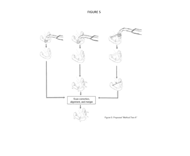
Method Two-A:
The three scan method also begins with an initial scan of the edentulous
space. The second
scan is taken with the scan flag in place, and can be repeated for each
implant location. The third scan
will feature the digital verification gauge, capturing the implant to implant
location. This scan can also
be repeated up to the maximum number of unique implant to implant positions as
described above (as
depicted in Figure 5).
Additional image method:
The scan methods described previously only utilize a single scanning source,
in this case an intra
oral scanning system. However, the digital verification gauge can also be used
with auxiliary inputs,
either in a form of an optical camera or manual recording of the gauge system
in place. In using an
optical camera, various photogrammetry techniques can be utilized to
automatically detect key
distances from a two-dimensional image. The optical image can also be
interpreted manually as part of
the restoration workflow.
Technical Details:
The use of the digital verification gauge during an edentulous scan accomplish
two primary
tasks. The first is that the gauge can incorporate defining features along the
length of the device to aid in
the scanning and stitching of the multiple images required to create a full
three-dimensional scan.
These defining features can be protruding shapes, space out a different
distance intervals, and be
between 50 microns to 1 centimeter in length.
The second task of the gauge is to serve as a calibration device and allow for
additional data points for
post processing of the raw scan file. The post processing can be completed
either in real time during the
scan or at a later point after the scan, either locally or offsite at a remote
location or server. Methods of
secondary analysis include a scale factor to augment the original scan based
on the measurements
gathered from the gauge and also fixing localized stitching errors captured in
the frame scan.
The scale factor method is highlighted in figure 6, where known distances from
the gages can be
used to scale and "calibrate" the scan in a non-uniform manner. Here, the
entire scan file is augmented
in each of the known orientations from the various scans with the gage in
place. As error can be
introduced in a non-linear method, the augmentation of the scan can also vary
along each axis.
Alternatively, scans with the gage in place can be used to better inform the
frame by frame
image stitching process used by the scanning systems. Each image, or series of
images, captured by the
scanner is overlaid on top of the prior using a best fit algorithm. If not
enough unique detail is present in
CA 03086482 2020-06-19
WO 2019/126115 PCT/US2018/066151
each scan, additional error may be introduced during the stitching process.
The known accuracy
provided by the gages can help better inform these stitching algorithms to
produce a final scan result
that better represents the physical scanning surface. This essentially acts as
a real-time offset or
coefficient to modify the process during scanning.
It will be appreciated that a gauge and methods of using the gauge as
described herein
provide a valuable contribution to the art of verifying and improving the
accuracy of 3D scans,
in particular those used in dental arts. Alternatives to the invention as
described are within the
scope of the invention and will only be limited by the attached claims.
6