Note: Descriptions are shown in the official language in which they were submitted.
CA 03086538 2020-06-19
WO 2019/118989 PCT/US2018/066095
OPTICAL READER FOR ANALYTE TESTING
CROSS REFERENCE TO RELATED APPLICATIONS
[0001] This patent application claims priority to U.S. Provisional
Patent Application No.
62/599,671, filed on December 15, 2017 (titled "POLARIZATION MAINTAINING
OPTICAL
PATH FOR ENSURING READER TO READER CONSISTENCY") and U.S. Provisional
Patent Application No. 62/599,674, field on December 15, 2017 (titled "CLAMP
DESIGN FOR
PRECISION ALIGNMENT OF THE CARTRIDGE"), each of which is herein incorporated
by
reference in its entirety
[0002] This patent application may also be related to U.S. Patent
Application No.
16/040,506, filed on July 19, 2018, titled ("CARTRIDGES FOR ORAL FLUID
ANALYSIS
AND METHODS OF USE"), which claims priority to U.S. Provisional Patent
Application No.
62,534,394, titled "ORAL FLUID ANALYZING SYSTEMS AND METHODS" and filed July
19, 2017.
INCORPORATION BY REFERENCE
[0003] All publications and patent applications mentioned in this
specification are herein
incorporated by reference in their entirety to the same extent as if each
individual publication or
patent application was specifically and individually indicated to be
incorporated by reference.
FIELD
[0004] Embodiments of the invention relate generally to analyte
collection and testing
systems and methods, and more particularly to disposable oral fluid collection
and testing
systems and methods. Described herein are methods and apparatuses to achieve
significant
improvements in the detection of fluorescence signals in the reader.
BACKGROUND
[0005] Detection of analytes, particularly for drugs of abuse, is
important in various
workplace drug testing settings, such as for pilots, professional athletes,
and law enforcement,
and to detect driving under the influence of drugs (DUID). Detection of these
analytes in oral
fluid, i.e. saliva, provides a more convenient method of sample collection
than collection of
blood or urine.
[0006] Conventionally, the collected samples are sent to a certified
testing laboratory for
analysis. However, sending the samples to the lab and then waiting for the lab
to process and
- 1 -
CA 03086538 2020-06-19
WO 2019/118989
PCT/US2018/066095
testing the sample and then report the results can take a significant amount
of time, typically at
least days. In many situations, it would be desirable to have testing results
at the point of testing
instead of waiting days for results from the lab. This would allow, for
example, an airline to
prevent pilots under the influence of drugs to fly a plane, thereby improving
safety.
[0007] Tools for reading a sample-containing cartridge (e.g., a "reader")
may utilize a
detection scheme utilizing an optical sensor for reading the cartridge.
SUMMARY OF THE DISCLOSURE
[0008] Described herein are methods and apparatuses for how to achieve
robust optical
performance of a sample cartridge reader with the use of polarization
maintaining components in
the optical path. The inherent nature of the optical coupling to almost any
photonic chip is highly
polarization dependent in addition to the propagation through waveguides and
also evanescent
coupling of light to molecules, including biological molecules. Thus by fixing
the polarization
state of all four excitation channels to the optical transverse-magnetic (TM)
polarization as
described herein, we may achieve optimal and reliable performance of our
system.
[0009] This embodiment of the invention utilizes polarization-
maintaining optical
components in the optical path of the reader hardware. The excitation laser
may be pigtailed with
a polarization maintaining fiber and the scan head fiber is also polarization
maintaining. With
electromagnetic (EM) simulations of the photonic chip, we obtain the optimal
polarization state
(TM) to use with this photonic chip architecture. This leads to a stable and
repeatable known
polarization state of light exiting the scan head and coupling into the
photonic chip.
[0010] Also described herein are methods and apparatuses for use with
analyte holding
cartridges.
[0011] For example, described herein are optical reader devices for
reading a photonic chip
of a removable cartridge. An optical reader device may include: a cartridge
holder comprising a
slot extending into the reader, the slot having a height and a width; a scan
head, wherein the scan
head comprises a first plurality of optical fiber ends that are optically
connected to one or more
laser light sources and a second plurality of fiber ends that are optically
connected to a plurality
of detectors; a scan head actuator configured to move the scan head relative
to the cartridge
holder; a plurality of valves on the cartridge holder that are configured to
couple with valve
openings in the removable cartridge; a pump membrane actuator on the cartridge
holder that is
configured to apply force to a membrane pump of the removable cartridge,
wherein the pump
membrane actuator is configured to hold a plurality of extended positions to
deflect or relax
deflection of the membrane pump; and a controller configured to coordinate
movement of the
scan head, illumination of the one or more laser light sources, detection by
the plurality of
- 2 -
CA 03086538 2020-06-19
WO 2019/118989
PCT/US2018/066095
detectors, opening and closing of the plurality of valves and positioning of
the pump membrane
actuator when the removable cartridge is inserted into the cartridge holder.
[0012] An optical reader device for reading a photonic chip of a
removable cartridge may
include: a reader housing including a cartridge interface comprising an
opening into the reader
housing; a cartridge holder comprising a slot extending into the reader from
the cartridge
interface, the slot having a height and a width; a scan head within the reader
housing, wherein
the scan head comprises a first plurality of optical fiber ends that are
optically connected to one
or more laser light sources and a second plurality of fiber ends that are
optically connected to a
plurality of detectors, further wherein the optical fiber ends within the
first and second plurality
of optical fiber ends are arranged in a line; a scan head actuator configured
to move the scan
head relative to the cartridge holder; a plurality of valves on the cartridge
holder that are
configured to couple with valve openings in the removable cartridge; a pump
membrane actuator
comprising a rocker arm having a rounded end, wherein the pump membrane
actuator is
configured to apply force to a membrane pump of the removable cartridge,
wherein the pump
membrane actuator is configured to hold a plurality of extended positions to
deflect or relax
deflection of the membrane pump; and a controller configured to coordinate
movement of the
scan head, illumination of the one or more laser light sources, detection by
the plurality of
detectors, opening and closing of the plurality of valves and positioning of
the pump membrane
actuator when the removable cartridge is inserted into the cartridge
interface.
[0013] In any of these variations, the device may also include one or more
(e.g., a plurality
of) fluid sensors in communication with the controller and configured to
optically detect fluid
within one or more regions of the removable cartridge. The controller may be
configured to
clamp the cartridge holder when the removable cartridge is inserted in to the
cartridge holder.
For example, the cartridge holder may include a movable/lockable base that may
be driven to
clamp the cartridge in position.
[0014] The scan head may include a linear array of the first plurality
of optical fiber ends and
the second plurality of optical fiber ends. A fiber mount holder may hold the
ends of the fibers
(and in some variations a lens or lensing material, filter, etc.) to the scan
head.
[0015] Each valve of the plurality of valves may comprise a seal
configured to be moved
relative to the cartridge holder to open or close a valve opening in the
removable cartridge when
the removable cartridge is held within the cartridge holder. The seal may
block or unblock an
opening (valve opening) on a cartridge.
[0016] The pump membrane actuator may be any appropriate actuator (e.g.,
mechanical,
electromechanical, pneumatic, etc.), for example, the pump membrane actuator
may comprise an
- 3 -
CA 03086538 2020-06-19
WO 2019/118989
PCT/US2018/066095
arm and a driver. The pump membrane actuator may comprise a rounded, ball-
shaped end. In
some variations the pump membrane actuator comprises a rocker arm that is
motor driven.
[0017] Any of these device may include a second actuator, such as a
blister pack arm on the
cartridge holder, that is configured to apply force to a blister pack of the
removable cartridge
(e.g., to open/break the blister pack).
[0018] Any of these devices may include a temperature sensor on the
cartridge holder and a
heater on the cartridge holder, wherein the controller is further configured
to regulate a
temperature of a removable cartridge held in the cartridge holder.
[0019] In any of these variations, the reader may be configured for
precise control of the
alignment of the cartridge relative to the scan head. For example, described
herein are optical
reader devices for reading a photonic chip of a removable cartridge that
include: a cartridge
holder comprising a slot extending in a z-axis into the reader, the slot
having a height in the y-
axis direction and a width in the x-axis direction, a ball plunger on one side
of the slot, the ball
plunger biased to extend into the slot in the x-axis direction, configured to
drive the removable
cartridge against a reference surface in the z-axis and a reference surface in
the x-axis; a movable
clamp base configured to apply force in the y-axis to secure the removable
cartridge within the
slot and to drive the removable cartridge against a reference surface in the y-
axis; a scan head
configured to move relative to the cartridge holder, wherein the scan head
comprises a first
plurality of fiber ends optically connected to one or more laser light sources
and a second
plurality of fiber ends optically connected to a plurality of detectors; and a
controller configured
to coordinate movement of the movable clamp, movement of the scan head,
illumination of the
one or more laser light sources and detection by the plurality of detectors.
[0020] For example, an optical reader device for reading a photonic chip
of a removable
cartridge may include: a reader housing including a cartridge interface
comprising an opening
into the reader housing; a cartridge holder comprising a slot extending in a z-
axis into the reader,
the slot having a height in the y-axis direction and a width in the x-axis
direction, a ball plunger
on one side of the slot, the ball plunger biased to extend into the slot in
the x-axis direction,
configured to drive the removable cartridge against a reference surface in the
z-axis and a
reference surface in the x-axis; a movable clamp base configured to apply
force in the y-axis to
secure the removable cartridge within the slot and to drive the removable
cartridge against a
reference surface in the y-axis; a scan head configured to move relative to
the cartridge holder,
wherein the scan head comprises a first plurality of fiber ends optically
connected to one or more
laser light sources and a second plurality of fiber ends optically connected
to a plurality of
detectors; a pump membrane actuator configured to apply force to a membrane
pump of the
removable cartridge, wherein the pump membrane actuator is configured to hold
a plurality of
- 4 -
CA 03086538 2020-06-19
WO 2019/118989
PCT/US2018/066095
extended positions to deflect or relax deflection of the membrane pump; and a
controller
configured to coordinate movement of the movable clamp, movement of the scan
head,
illumination of the one or more laser light sources and detection by the
plurality of detectors.
[0021] In some variations the reference surface in the z-axis is a pin
extending into the
cartridge slot.
[0022] Any of these devices may further include a scan head actuator
configured to move the
scan head relative to the cartridge holder. The scan head actuator may be a
motor.
[0023] As mentioned above, any of these devices may include one or a
plurality of valves on
the cartridge holder that are configured to couple with valve openings in the
removable cartridge.
The devices may include a pump membrane actuator on the cartridge holder that
is configured to
apply force to a membrane pump of the removable cartridge, wherein the pump
membrane
actuator is configured to hold a plurality of extended positions to deflect or
relax deflection of
the membrane pump. For example, the pump membrane actuator may comprise an arm
and a
driver. Any of the pump membrane actuators may have an end that contacts the
pump
membrane that is configured to uniformly apply force (or to distribute the
force) to the pump
membrane to prevent damaging it. In some variations the pump membrane actuator
comprises a
rounded, ball-shaped end. In some variations the pump membrane actuator
comprises a rocker
arm that is motor driven.
[0024] As mentioned, any of these devices may include a plurality of
fluid sensors in
communication with the controller and configured to optically detect fluid
within one or more
regions of the removable cartridge. The controller may be configured to clamp
the movable
clamp base to apply force in the y-axis to secure the removable cartridge when
the removable
cartridge is inserted in to the cartridge holder.
[0025] The scan head may comprise a linear array of the first plurality
of optical fiber ends
and the second plurality of optical fiber ends, as described above.
[0026] Any of these devices may include a second actuator (similar to
the pump membrane
actuator) that is configured to rupture one or more blister packs on the
cartridge. For example,
any of these apparatuses may include a blister pack arm on the cartridge
holder configured to
apply force to a blister pack of the removable cartridge.
[0027] The devices described herein may include one or more temperature
sensors on the
cartridge holder and/or a heater on the cartridge holder, wherein the
controller is further
configured to regulate a temperature of a removable cartridge held in the
cartridge holder.
[0028] Any of the optical reader devices described herein may be part of
a system that may
further include one or more cartridges as described herein. Any of the reader
variations
- 5 -
CA 03086538 2020-06-19
WO 2019/118989
PCT/US2018/066095
described herein may be used with any of these cartridges to form a system;
one or more
additional components (outputs, displays, user interface software, etc.) may
also be included.
[0029] Also described herein are devices (e.g., optical reader devices)
for reading a photonic
chip of a removable cartridge that are configured to control the polarization
of the energy applied
so that it matches the inherent polarization of the photonic chip. For example
described herein
are optical reader devices comprising: a scan head; a plurality of laser
sources each configured to
emit light having a TM polarization; a first plurality of optical fibers,
wherein each laser source
is coupled to one optical fiber of the first plurality of optical fibers,
further wherein the first
plurality of optical fibers are polarization maintaining single-mode fibers; a
plurality of optical
sensors; a second plurality of optical fibers, wherein each optical sensor is
coupled to one optical
fiber of the second plurality of optical fibers, further wherein the second
plurality of optical
fibers are multimode fibers; wherein each of the first plurality of optical
fibers and the second
plurality of optical fibers terminates on the scan head so that an end of each
optical fiber of the
first and second pluralities of optical fibers are arranged in a line facing a
gap; and a cartridge
holder configured to receive the removable cartridge so that the photonic chip
is aligned with a
polarization axis formed by the scan head so that an end of the photonic chip
comprising a
plurality of waveguides faces the gap, across from the scan head, wherein the
device is
configured to maintain the polarization of the polarization axis in a
transverse-magnetic (TM)
polarization.
[0030] The laser sources may comprise one or more diode lasers. The second
plurality of
fibers may contain at least twice as many optical fibers as the first
plurality of fibers (e.g., there
may be two optical fibers in the first plurality and four optical fibers in
the second plurality, there
may be four optical fibers in the first plurality and nine optical fibers in
the second plurality,
etc.).
[0031] The controller may be configured to control alignment of the scan
head relative to the
cartridge. For example, the cartridge holder may be configured to clamp the
cartridge to prevent
it from moving. In some variations, the cartridge holder may be configured to
bias the cartridge
in a direction that is normal to a major plane of the cartridge (e.g., in a y-
axis direction) against a
reference surface to prevent movement of the cartridge as one or more
actuators apply force to
the cartridge to drive fluid through the cartridge. This may reduce or
eliminate misalignment of
the chip relative to the scan head during operation. In any of these
variations, the controller may
be configured to adjust the position of the scan head during operation of the
device by actuating
a scan head actuator to align the ends of the optical fibers with waveguides
of the photonic chip
when the cartridge is in the cartridge holder.
- 6 -
CA 03086538 2020-06-19
WO 2019/118989
PCT/US2018/066095
[0032] Also described herein are methods of operation of any of the
devices and systems
described. For example, a method of reading optical signals from a photonic
chip of a
removable cartridge head in an optical reader may include: aligning a scan
head of the optical
reader with the chip so that the chip and a laser source, a plurality of
fibers, and an optical sensor
of the scan head are aligned along a polarization axis with the chip;
maintaining a polarization of
the polarization axis in a transverse-magnetic (TM) polarization; emitting one
or more beams of
light from the laser, through the plurality of fibers and into an edge of the
photonic chip in the
TM polarization; and detecting, in the optical sensor, TM polarized light from
one or more
waveguides within the chip when the one or more beams of light interact with
an analyte
molecule on the chip.
[0033] Any of these methods may include inserting a cartridge containing
the chip into the
optical reader.
[0034] Emitting may comprise emitting a plurality of concurrent beams of
TM polarized
light from the scan head, into the edge of the photonic chip.
[0035] Maintaining the polarization of the polarization axis comprises
maintaining the
polarization of the plurality of fibers (e.g., the first and/or second
plurality of fibers).
[0036] The method may also comprise polarizing light emitted from the
scan to the edge of
the chip in a polarizer.
[0037] The methods described herein may also include adjusting the
alignment of the scan
head while emitting and/or detecting to maintain the TM polarization.
[0038] Any of the methods described herein may include inserting the
cartridge into the
reader device. Any of these methods may include clamping the cartridge and/or
aligning the
cartridge within the cartridge holder (e.g., clamp). For example, the
cartridge may be inserted so
that a ball plunger rides against a wall of the cartridge until it reaches a
seating edge of the
cartridge, when a back surface (e.g., a z-face) of the cartridge contacts a
reference surface. The
ball plunger may drive the cartridge against two or more seating surfaces,
including a seating
surface in the x-axis and the back (z-face) seating surface(s). Once seated,
the controller may
then lock the cartridge into position by clamping a y-face seating surface
(e.g., a bottom of the
cartridge holder) against the cartridge. The controller may control alignment
of the scan head
with the photonics chip, as described herein. The controller may coordinate
fluid control of the
cartridge and testing (applying light and detecting signals). For example, the
controller may
coordinate one or more of: puncturing one or more blister pack, moving the
control solutions,
moving the sample, dissolving reagents (e.g., labeled antibodies for one or
more targets, e.g.,
drugs of addition) into a control solution, dissolving reagents into the
sample, mixing the control
solution, mixing the sample, moving the control solution into one or more test
wells in the
- 7 -
CA 03086538 2020-06-19
WO 2019/118989
PCT/US2018/066095
photonics chip, emitting light through all or some of the first plurality of
fibers, detecting
evanescent signals from the photonics chips from one or more of the second
plurality of fibers,
moving the sample into the one or more test wells of the photonics chip, and
detecting
evanescent signals from the photonics chips from one or more of the second
plurality of fibers.
In some variations the method may include testing the control solution in the
same well as the
sample solution. The controller may coordinate any or all of these steps and
may repeat any of
these steps.
BRIEF DESCRIPTION OF THE DRAWINGS
[0039] The novel features of the invention are set forth with particularity
in the claims that
follow. A better understanding of the features and advantages of the present
invention will be
obtained by reference to the following detailed description that sets forth
illustrative
embodiments, in which the principles of the invention are utilized, and the
accompanying
drawings, of which:
[0040] FIG. 1 is a top view of an embodiment of a disposable device that
includes an
integrated oral fluid collection device and cartridge for processing and
testing the collected
sample.
[0041] FIG. 2 is an exploded view of the components of the disposable
device shown in FIG.
1.
[0042] FIGS. 3A and 3B illustrate a top perspective view and bottom
perspective view,
respectively, of a bottom part of the cartridge attached to the saliva
collection device.
[0043] FIG. 4A illustrates a top view of the bottom part of the
cartridge attached to the saliva
collection device.
[0044] FIG. 4B illustrates a perspective view of the bottom part of the
cartridge attached to a
cross-sectional view of the saliva collection device.
[0045] FIG. 4C illustrates a close up view of a piercing element using
to puncture a blister
pack.
[0046] FIGS. 4D and 4E show front perspective and back perspectives,
respectively of an
example of a distal end of a saliva collection system, showing the collection
body and two swab
pistons extending distally from the collection.
[0047] FIGS. 5A and 5B illustrate a top perspective view and a bottom
perspective view,
respectively, of a top part of the cartridge.
[0048] FIGS. 5C and 5D show a top and bottom, respectively, or an
example of a cartridge
body.
- 8 -
CA 03086538 2020-06-19
WO 2019/118989
PCT/US2018/066095
[0049] FIGS. 6A and 6B illustrate a perspective view and a cross-
sectional view,
respectively, of a cap for the saliva collection device.
[0050] FIG. 6C shows a section through a top of an example of a saliva
collection system
(with the front side removed).
[0051] FIG. 6D shows a perspective view of the exemplary saliva collection
system top of
FIG. 6C.
[0052] FIG. 7 illustrates a schematic of the parts of the fluidic
circuit in the assembled
cartridge.
[0053] FIGS. 8A and 8B illustrate an embodiment of an optical chip for
analyte testing.
[0054] FIG. 9A illustrates a side cross-sectional view of a portion of the
cartridge and optical
chip in alignment with an optical scan head of a reader device.
[0055] FIG. 9B is another view of an end face of a cartridge showing a
ledge or lip region
protecting the optical chip.
[0056] FIG. 10 illustrates the insertion of the cartridge into a reader
for testing.
[0057] FIGS. 11A, 11B, and 11C illustrate another embodiment of a fluid
collection device.
[0058] FIGS. 12A illustrates another embodiment of a fluid collection
device. FIG. 12B
illustrates an example of how a fluid collection device such as the one shown
in FIG. 12A can be
manufactured.
[0059] FIGS. 13A, 13B, 13C, 13D, and 13E show an example of an assay
useful for
detecting an analyte, such as an analyte from a bodily fluid. FIG. 13A shows a
control sample
containing a detectably labeled binding agent (antibody). FIG. 13B shows a
test sample reacted
with a detectably labeled binding agent (antibody). FIG. 13C shows antigen
attached to a sensing
site as it appears prior to passing a control sample, such as the control
sample shown in FIG 13A,
across it. FIG. 13D shows antigen on a sensing site and a detectably labeled
binding agent
(antibody) from a control samples, conjugated to an antigen. FIG. 13E show a
sensing site, such
as the site shown in FIG. 13D, after flowing a detectably labeled sample
across the sensing site.
[0060] FIG. 14A illustrates kinetics of analyte and antibody binding
over time. FIG. 14B
shows the kinetics over time of free, unbound antibody binding to antigen,
such as antigen
attached to a sensing well.
[0061] FIG. 15A and 15B shows results of an assay signal distribution as
described herein
for detecting the presence of marijuana (THC; tetrahydrocannabinol) in a
sample.
[0062] FIGS. 16A and 16B shows results from a multiplex assay as
described herein for
detecting cocaine (COC), marijuana (THC; tetrahydrocannabinol) and
benzodiazepine (BZO)
from a bodily fluid sample.
- 9 -
CA 03086538 2020-06-19
WO 2019/118989
PCT/US2018/066095
[0063] FIGS. 17A-17C show results from a multiplex assay as described
herein for detecting
cocaine (COC-M), fentanyl (FEN), morphine (MOR) and benzodiazepine (BZ0-0)
from a
bodily fluid sample.
[0064] FIG. 18A and 18B show part of a microfluidic circuit with a
serpentine mixer useful
for mixing a sample and a plurality of dried beads containing a binding agent.
[0065] FIGS. 19A-19B illustrate a method of operating the cartridge
(including an integrated
saliva collection system) to test a subject's saliva, as described herein,
including both local (e.g.,
immediate) testing with a reader similar to that shown in FIGS. 21A-21B, and
confirmation
testing.
[0066] FIGS. 20A shows a partial schematic of an exemplary fluidic circuit
for the cartridge
(which may include a saliva collection system), similar that shown in FIG. 7.
FIG. 20B is a
legend illustrating component part of the partial schematic.
[0067] FIGS. 20C-20N illustrate one example of method of operating an
exemplary cartridge
for testing a subject's saliva for one or more drugs.
[0068] FIGS. 21A-21B show an example of a reader for reading a cartridge
and
automatically performing the method of operating the exemplary cartridge as
described herein.
FIG. 21A is a front perspective view. FIG. 21B is a front view.
[0069] FIG. 21C is a schematic of one example of a reader as described
herein.
[0070] FIG. 21D shows a front perspective view of one example of a
reader (the reader
components may be housed within a housing, not shown), including a cartridge
inserted into the
reader.
[0071] FIG. 21E shows the reader of FIG. 21D, without the cartridge,
showing the slot of the
cartridge holder.
[0072] FIG. 21F is a back perspective view of the reader device shown in
FIG. 21D.
[0073] FIG. 21G is a side view of the reader device example shown in FIG.
21D.
[0074] FIG. 21H is a back view of the reader device example shown in
FIG. 21D.
[0075] FIG. 211 is a front view of the reader device example shown in
FIG. 21D.
[0076] FIG. 21J is a top view of the reader device example shown in FIG.
21D.
[0077] FIG. 21K is a back view of the reader device example shown in
FIG. 21D.
[0078] FIG. 21L is a side perspective view of an example of a reader
device, showing the
gap between the scan head assembly and the back of a cartridge when the
cartridge is held in the
cartridge holder.
[0079] FIG. 21M is a closer view of the reader device, showing the back
of a cartridge (held
in the cartridge holder), the gap, and the scan head assembly.
- 10 -
CA 03086538 2020-06-19
WO 2019/118989
PCT/US2018/066095
[0080] FIGS. 21N is another view of the close-up shown in FIG. 21M, with
the cover and
fiber guide of the scan assembly removed, showing a portion of the scan head
where the plurality
of fibers connected to the light sources (laser diodes) and sensors
(photodetectors) end, across
the alignment gap from the photonics ship of the cartridge when a cartridge is
held by the
cartridge holder.
[0081] FIG. 22 shows an example of an electric field mode profile for
photonic chip
waveguides.
[0082] FIG. 23 shows input coupling efficiency for TE and TM modes.
[0083] FIG. 24 shows normalized electric field of TE modes at the 3-
layer / 4-layer (wet)
interface.
[0084] FIG. 25 shows normalized electric field of TM modes at the 3-
layer / 4-layer (wet)
interface.
[0085] FIG. 26A shows the results of an optical jump experiment for TE
and TM
polarization modes.
[0086] FIG. 26B is an example of one variation of a schematic of an optical
reader as
described herein.
[0087] FIG. 26C is an example of a variation of a schematic for an
optical reader as
described herein.
[0088] FIG. 27 is an example of a cartridge as described herein.
[0089] FIG. 28 is a partial cut-away view of one variations of a cartridge
holder portion of
reader apparatus.
[0090] FIG. 29 is an illustration of cartridge holder showing a
cartridge within it.
[0091] FIG. 30 is another example of a portion of a cartridge holder
including a cartridge.
[0092] FIGS. 31 and 32 show examples illustrating a portion of a
cartridge holder including
a ball plunger. FIG. 31 is a top plan view while FIG. 32 is a top perspective
view.
[0093] FIG. 33 schematically illustrates one example of a portion of a
reader.
[0094] FIG. 34 is an enlarged view of FIG. 33.
[0095] FIG. 35 is a top view of a portion of the reader.
[0096] FIG. 36 is an example of a cartridge holder portion of a reader.
[0097] FIGS. 37 and 38 show linear drive actuator(s) coupled to the
cartridge holder portion
of a reader. FIG. 37 is a side view, while 38 is a slightly enlarged view.
[0098] FIG. 39 is illustrates one portion of a cartridge holder of a
reader, showing thermal
regulation (and feedback) that may be helpful
[0099] FIGS. 40, 41 and 42 illustrate specific features of an exemplary
cartridge that may be
used in any of the apparatuses described herein.
- 11 -
CA 03086538 2020-06-19
WO 2019/118989
PCT/US2018/066095
DETAILED DESCRIPTION
[0100] In general, described herein are reader apparatuses (device and
systems) and
methods for reading one or more analyte from a photonics chip of a cartridge.
These apparatuses
may be configured to receive one or more cartridges that include a photonics
chip.
[0101] In general, the methods and apparatuses described herein may be
used for the
detection of an analyte (e.g., drug, biomarker, protein, etc.) from a bodily
fluid. The examples
provided below are directed primarily to detection of an analyte (or multiple
analytes) from a
saliva sample, and in particular to the detection of one or more drugs of
abuse. However, it
should be understood that these methods and apparatuses may apply as well to
other bodily
fluids and other analytes.
[0102] For example, described herein are apparatuses, including optical
readers, that may be
configured to process a cartridge used for saliva collection so that the
saliva sample(s) may be
prepared for testing to detect one or more analytes. Processing may include
regulating the
microfluidics (e.g., combining, mixing, incubating, etc., including in
particular, detection.
Analytes may be detected by applying a sensing optical wavelength to detect a
florescent marker
in conjunction with a photonics chip in the cartridge. The reader may control
the application of
the prepared fluid sample onto the photonics chip, and may read out one or
more signal(s) to
detect and/or quantify signal.
[0103] The optical readers described herein may be used with one or more
cartridges that can
concurrently collect two samples (one for acute or immediate testing and one
for later validation
of the acute testing). For example, these cartridges may automatically and
accurately process
(e.g., dilute) the saliva sample for processing; the optical reader
apparatuses described herein
may regulate the processing of the fluid sample for detection of one or more
analyte. The
cartridge may include a cap that is pre-loaded with one or more solution
(e.g., a dilution fluid
and/or a preservation solution). The cartridge may be configured so that
attaching the cap
exposes the saliva sample(s) to the appropriate solution, keeping the
different samples isolated
from each other, and may precisely mix and dispense the saliva sample with the
dilution sample
in a predictable manner. The cartridge may be configured so that the act of
snapping the cap onto
the body of the cartridge provide the mechanical energy for dispensing the
dilution fluid, mixing
it with the saliva sample, and dispensing the diluted and mixed saliva
dilution into a diluted
sample reservoir ("diluted sample cavity") where it can be further processed.
[0104] Any of these cartridges may include one or more fluidic circuits
that are configured to
processes, in conjunction with a reader, the diluted sample. The cartridge may
include, in
communication with the fluidic circuit or part of the fluidic circuit, a chip
(an optical chip, also
- 12 -
CA 03086538 2020-06-19
WO 2019/118989
PCT/US2018/066095
referred to as a photonic chip) that includes one or more waveguides along
with detection
chemistry that may allow detection via evanescent field detection of the
presence and/or amount
of an analyte. The cartridge may be self-contained, and may include a pump
(e.g., a diaphragm,
elastomeric membrane, etc.) that may be driven by a driver (e.g., piston, rod,
etc.) to push and
pull fluid within the microfluidic circuit. The cartridge may also include a
plurality of vents
(opening) to atmosphere that may be opened/closed by the reader to control
fluidic movement
(including metering, mixing, sampling, etc.) within the cartridge.
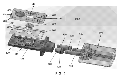
[0105] FIGS. 1 and 2 illustrate an embodiment of a disposable device
1000 for collecting,
processing, and testing an oral fluid/saliva sample from a subject. After a
sample has been
collected, the disposable device 1000, which may be a cartridge, can be
inserted into a reader for
analyzing the sample. FIG. 1 illustrates the disposable device in an assembled
state, while FIG.
2 illustrates an exploded view of the disposable device 1000. In one
embodiment, the disposable
device 1000 is constructed as an assembly of a bottom part (cartridge bottom)
100, a top part
(cartridge top) 200 and a channel sealing layer 900. In one preferred
embodiment, the sealing
layer 900 is a double sided adhesive tape with appropriate cut-outs 902 for
fluid
conduits/channels that form a fluidic circuit 120. The three parts come
together to form a
sandwich structure with the sealing layer 900 in between bottom and top parts
100, 200. In one
preferred embodiment, the top and bottom parts 100, 200 are held together by
the double sided
adhesive tape.
[0106] The sealing layer 900 can be made from a rubber or plastic sheet and
held between
the top and bottom part by screws, clips, rivets, bolts, or other fastening
mechanisms that can be
used to compress the bottom part 100 with the bottom part 200. The tightening
force applied by
the screws or other fastening mechanism squeezes the rubber or plastic sheet,
which functions
like a gasket, and provides sealing between fluid channels.
[0107] The sealing layer 900 can be made from a rubber sheet and held
between the top and
bottom part by means of heat staking or mechanical staking between the top and
bottom parts.
The stakes are designed to provide a mechanical force which squeezes the
rubber sheet and
provides sealing between fluid channels.
[0108] The bottom and top parts 100, 200 may be connected to each other
by applying liquid
adhesive in a pattern required by the fluid channels. The adhesive can also
provide sealing
between fluid channels.
[0109] In some embodiments, the sealing layer 900 can be a combination
of the features
described above, such as a rubber or plastic layer with adhesives.
[0110] In some embodiments, the cartridge top 200 and cartridge bottom
100 may be hard
plastic parts that when assembled form the fluid conduits. The plastic parts
may be manufactured
- 13 -
CA 03086538 2020-06-19
WO 2019/118989
PCT/US2018/066095
by machining or injection molding or vacuum forming or any other appropriate
plastic
manufacturing techniques.
[0111] The cartridge top 200 can have an elastomeric membrane 400
covering a cut-out in
the hard plastic part. The elastomeric membrane 400 may be attached, such as
by being glued, to
the cartridge top 200. The elastomeric membrane 400 may be molded over the
hard plastic top
200 by means of over-molding or two-shot injection molding process. The
elastomeric
membrane 400 and the cavity formed by the cut-out can be in fluid
communication with the
fluidic channels and can function as a pump that drives fluid through the
fluidic channels.
[0112] FIG. 5C shows another example of the cartridge top (shown from a
bottom view,
FIG. 5D shows a top view). In this example, the elastomeric diaphragm 400
(pump) is exposed
on one side to allow access by the reader piston (not shown). The cartridge
top may also include
a waste region 207 (waste well) and may include calibration regions (e.g., z-
location region 588).
The cartridge top also includes an opening 489 for the blister pack. FIG. 5D
also shown a sample
inlet (e.g., which may be part of the diluted sample cavity/reservoir 201. The
cartridge body may
be made of any appropriate material, for example, a clear, transparent,
medical grade
polycarbonate (PC) and/or (e.g., overmolded with) a medical grade,
thermoplastic elastomer
(TPE), Shore 40A.
[0113] For example, as shown in FIGS. 1, 2, 5A, and 5B, an elastomeric
membrane 400 can
cover a cavity 203 in the cartridge top 200 to create a pumping well. The
elastomeric membrane
400 may be pushed upon by an actuator in a reader for the disposable device
1000. As the
membrane 400 is depressed into the cavity 203 it pushes the air out of the
cavity 203 and into the
fluid channel. The column of air pushed into the fluid channel in turn moves a
slug of liquid in
the fluid channel.
[0114] Reversing the direction of motion of the actuator releases the
stretched membrane
400 which, owing to its elastic nature tries to return to its original shape
and thus tracking the
actuator as it moves. As the membrane 400 moves back to its original shape, it
creates a suction
in the pumping cavity 203. This suction allows movement of slug of liquid
within the fluid
channel in a direction opposite to the previous motion. Thus, the action of
pushing on the
membrane 400 and releasing it in a controlled manner allows bi-directional
control over the
movement of fluid within the fluid channel. As further described below
particularly with respect
to FIG. 7, a unique aspect of the disclosed device is the multi-channel
management of fluid
columns/slugs in the fluidic channels using a single on-board pumping
mechanism in
combination with vents placed at strategic locations.
[0115] Returning to FIGS. 1 and 2, a blister pack 300, can be assembled
within the
disposable device 1000. The blister pack 300 may contain buffer solution
(e.g., control solution)
- 14 -
CA 03086538 2020-06-19
WO 2019/118989
PCT/US2018/066095
and/or reagents used as part of the testing protocol. The blister pack 300 may
be stuck directly to
a sealing layer 900 made of double sided adhesive tape. The blister pack 300
may be affixed to
the cartridge bottom 100 or sealing layer 900 by means of an additional double
sided adhesive
tape placed on the blister pack 300. The blister pack may be glued to the
cartridge bottom 100 or
.. sealing layer 900 by means of a liquid adhesive.
[0116] The blister pack 300 can be installed within the disposable
device 1000 such that it is
very close to, proximate to, or adjacent to a piercing mechanism 112, which
may be an integral
part of the disposable device 1000. As shown in FIGS. 2, 4A, and 4B, in one
embodiment the
piercing mechanism 112 is a sharp pointed feature within the molded cartridge
bottom 100.
Alternatively, the piercing mechanism 112 may be a sharp needle that is glued
onto the cartridge
bottom 100. The needle may be made from metal or plastic. The piecing
mechanism 112 may be
press fit or insert molded into the cartridge bottom 100. The piercing
mechanism 112 can be
positioned in a depression within the molded cartridge bottom 100 such that
the blister pack 300
is positioned above the piercing mechanism 112. The cartridge top 200 may have
an opening
113 that provides access to the blister pack 300 and allows an actuator of the
reader to push the
blister pack 300 into the piercing mechanism and thereby release the contents
of the blister pack
into the fluid channels.
[0117] As shown in FIGS. 1, 2, and 5B, the disposable device 1000 can
also include a
sensing element 3000 in fluid communication with the fluidic circuit 120. The
sensing element
3000 may be a photonic chip which is placed within a cavity 204 in the
cartridge top 200. The
sensing element 3000 may be held in place by being sandwiched between the
cartridge top 200
and cartridge bottom 100 and can be held together by means of an adhesive
sealing layer 900, for
example.
[0118] In one embodiment as shown in FIGS. 1 and 2, the disposable
device 1000 has an
integrated collection device and cartridge. The cartridge includes primarily
the cartridge bottom
100, the cartridge top 200, and the associated components as described herein.
The collection
device includes primarily a pair of collection swabs 610, 620 and a cap 500
and associated
components as further described herein.
[0119] As shown in FIGS. 1, 2, 3A, and 3B, first and second swabs 610,
620 can be held
firmly within first and second swab holders (e.g., swab pistons) 710, 720
respectively. The swab
holder may alternatively be referred to as swab pistons. The swabs 610, 620
may be held within
swab holder 710, 720 by means of press fit. Alternatively, the swabs 610, 620
may also be glued
to the swab holders 710, 720. The saliva collection swabs 610, 620 may be made
of an
absorbent material, such as a sintered porous polymer with an open cell foam
structure similar to
one used in wicks. Other materials that can be used include polyurethane foam
or cellulose fiber.
- 15 -
CA 03086538 2020-06-19
WO 2019/118989
PCT/US2018/066095
At least one of the saliva collection swabs 610, 620 may have an embedded
indicator, such as a
colored dye indicator, which changes color upon contact with oral fluids thus
indicating
completion of the saliva collection. A saliva stimulant configured to
stimulate saliva production
from a subject may be included on first and/or second collection swabs 610,
620 or otherwise
administered to a subject. Since confirmatory testing by the certified lab
typically uses
traditional testing systems and protocols, a larger amount of saliva may be
collected for the
confirmatory sample, such as about 2, 3, or 4 times the amount as compared for
the rapid test
sample. Therefore, in some embodiments, the indicator is included with the
confirmatory saliva
collection swab 620. In one embodiment the rapid test saliva collection swab
610 is designed to
be a hollow shell. The amount of oral fluid collected can be controlled by the
size of the
collection swabs 610, 620 and the position of the indicator on and/or within
the swabs. FIGS.
11A, 11B, and 11C show another embodiment of a collection swab. First
collection swabs 610'
may have a structure including a plurality of capillary channels 614. (A
second collection swab
as used herein may have a generally similar structure as a first collection
swab with the most
common difference a matter of size or dimensions). Upon placing first
capillary collection swab
610' in the mouth of the subject (e.g., under the subject's tongue), capillary
channels 614 absorb
the oral fluid by capillary action and collect only as much as the channel
volume allows them.
[0120] FIGS. 4D and 4E illustrate one example of a collection body 455.
The collection
body may be flanged outwards and may mate with cartridge (not visible in FIG.
4) body. In
some variations the collection body may be the same or integral with the
cartridge body. In FIG.
4D the collection body includes a connector 457 (a female portion of a snap
fit in this example)
for connecting to the cap. A pair of swab pistons 710 extend distally from the
collection body.
Each swab piston includes an internal channel 458 configured to wick saliva
from an open distal
end of the first swab piston. For example the channel may hold a porous
material and/or
capillaries. The swab pistons may each also include a seal (e.g., plunger
seal) 459. FIG. 4E
shows an internal view of the collection body, showing a connection within the
body for fluidic
connection to the cartridge portion (e.g., the diluted sample cavity in the
cartridge). In this
example, the collection body includes a male lure 462 connection for
connecting to the cartridge.
[0121] The collection body and/or swab pistons may be made of any
appropriate material,
for example, a clear, transparent, medical grade polycarbonate (PC) and/or
(e.g., overmolded
with) a medical grade, thermoplastic elastomer (TPE), Shore 40A.
[0122] As mentioned, the wicking material within the swab piston, which
may be referred to
as the swab, may be porous material and/or it may be constructed by putting a
number of
capillaries 616 together in a bundle with a sheath 612 around them to hold
them together or for
protection. Such capillaries may be curved or otherwise shaped, but in general
will be straight.
- 16 -
CA 03086538 2020-06-19
WO 2019/118989
PCT/US2018/066095
Alternately a swab may be constructed using a multi-lumen capillary with the
requisite number
of lumens. The capillaries may be made of glass or plastic material or
otherwise manufactured or
treated to minimize binding of substances of interest to prevent their loss
prior to assay. A swab
may be relatively rigid or may be flexible to aid in placement. A swab may
have a flat end(s) or
may have one or more shaped end 618 as shown in FIG. 11A which may allow easy
access to
saliva for capillary suction upon placing the swab in the mouth (e.g., under
the tongue). An
entire swab or swab holder may be shaped to aid in collection and/or handling.
Such a shaped
end or shaped swab may be flattened, rounded, tapered or so on. Although the
capillaries or
channel may all be the same length, in some examples, some capillaries or some
channels may
be shorter than others. For example, capillaries on one side of a taper may be
shorter than
capillaries on the other side of the taper. Likewise, a swab with a single
channel in a hollow shell
or a porous material may have different dimensions on different parts, and one
longitudinal part
of a channel, shell or single material swab may be longer than another part
(e.g., 1% -50%
longer). FIG. 11C shows a cross section through a swab showing one example of
placement of
capillaries.
[0123] FIGS. 12A and 12B illustrate swab 610" with a plurality of
channels configured to
collect a bodily fluid. FIG. 12A shows a perspective view and FIG. 12B shows a
front view of
the three sections of a swab before and after joining the sections. A swab may
be made in a
sandwich construction whereby two or more halves or parts of a swab come
together to create
capillary channels. Each half or part may be made of a material with channels
cut out as shown
in FIG. 12B. In some embodiments, an opening (channel) is cut out of one half
or part, and the
floor or roof of the channel is supplied by another half of part of the swab.
In FIG. 12B, top
section 622 of swab 610' houses top channels 628 while middle section 624 of
swab 610'
provides floors 632 for top channels 628 when the top section 622 and middle
section 624 of the
swab are adjoined. Top 622 of swab 610' also provides roof 634 for middle
channels 630
provided by middle section 624. Similarly, middle section 624 and bottom
section 626 also form
channels. In some embodiments, a cut out channel is half a channel and two
half channels come
together to create a complete capillary channel(s) (as could be seen if top
section 622 and middle
section 624 were offset from one another. Channels may be any shape that
collects or transports
the body fluid, such as circular, rectangular, rounded rectangles and so on.
Halves or parts may
be plastic and the plastic parts may be manufactured by machining or injection
molding or
vacuum forming or any other appropriate plastic manufacturing techniques. The
plastic parts
may be joined together by pressure sensitive adhesive or liquid adhesive or by
ultrasonic welding
or any other plastic joining techniques known in the art.
- 17 -
CA 03086538 2020-06-19
WO 2019/118989
PCT/US2018/066095
[0124] A swab may have at least 2, at least 3, at least 4, at least 5,
at least 10, at least 15, at
least 20, or at least 30 channels. In some preferred embodiments, a swab may
have between 14
and 22 channels, such as about 18 capillary channels. Capillary channel(s) of
a swab may have a
length between 1 mm and 10 cm and in general will have length between 5 mm and
50 mm (5
cm). In some embodiments, a capillary, a capillary channel, a hollow shell or
a porous material
has a length of from 5 mm to 40 mm, such as approximately 25 mm (from 10 mm to
25 mm).
Each capillary channel or lumen may have a diameter between 0.05 mm and 5 mm,
such as
between 0.1 mm and 1.5 mm (e.g., between 0.3 mm and 0.8 mm.) In general, a
length of
capillary selected is less than the capillary head for the selected diameter.
That is, for a selected
capillary channel diameter, the length of oral fluid pulled into the channel
due to capillary action
against gravity is greater than the selected length of the capillary channel
to ensure consistent
collection volume.
Saliva collection
[0125] In some examples, a pair of saliva samples are collected
simultaneously by placing
the saliva collection swabs 610, 620 in the mouth of the test subject. The
saliva collection swabs
610, 620 may be sized, shaped, and designed ergonomically to be placed under
the tongue on
either side of the tongue. This may enhance the salivation of the test subject
and allow for
improved collection efficiency. In some examples, a saliva collection swab may
be configured
for increasing saliva production, such as allowing or encouraging biting or
chewing or may
contain a component configured to increase saliva production such as a
chemical or odorant. In
some examples, components for increasing saliva production may be separate
from a collection
device, such as a separate vial containing an odorant, etc. In some examples,
a single saliva
sample may be collected such as a single sample in which part of the sample is
used for rapid test
analysis and another part used for confirmatory testing. In some examples, two
or more saliva
samples may be separately collected (e.g., using two or more separate
collection devices).
[0126] One of the saliva collection swabs 610 is used for the rapid test
performed within the
cartridge portion of the disposable device 1000, while the saliva sample
collected by the other
swab 620 may be used for testing by a certified forensic lab for confirmatory
testing and/or can
also be used for storage as forensic evidence.
[0127] Once the saliva / oral fluid is successfully collected by the saliva
collection swabs
610, 620, the user applies the collection device cap 500 over the oral fluid
collection end, i.e. the
saliva collection swabs 610, 620, of the disposable device 1000.
[0128] As shown in FIGS. 6A and 6B, the collection device cap 500 has
two cavities 501,
502 to receive the saliva collection swabs 610, 620. In some embodiments, the
disposable device
- 18 -
CA 03086538 2020-06-19
WO 2019/118989
PCT/US2018/066095
can have more than 2 collection swabs, such as 3, 4, or 5 swabs, and the
device cap 500 can have
a matching number of cavities. The rapid test cavity 501 is filled with a
known amount of
dilution buffer solution used for dilution of the rapid test saliva sample
collected by swab 610.
The dilution buffer solution may be constituted of 5% bovine serum albumin
(BSA) in phosphate
buffered saline (PBS), for example. Other concentrations of BSA or other
protein can be used,
such as between 2-10%. In addition, other proteins may be used in the dilution
buffer solution,
such as non-fat dry milk, and other buffers can be used, such as tris-buffered
saline (TBS). The
confirmatory test cavity 502 is filled with a preservation solution used to
preserve the
confirmatory sample collected by swab 620 so that the confirmatory sample can
be sent to a
certified lab for confirmatory testing. The preservation solution may include
a buffer.
[0129] The two cavities 501, 502 filled with dilution and preservation
fluids respectively
may be sealed by means of a foil cover 510 or other removable or pierceable
sealing mechanism,
such as a lid or cap. The primary purpose of the foil cover 510 is to contain
the dilution and
preservation fluids within the collection device cap 500. The foil cover 510
is designed to have
very low vapor permeability to prevent or greatly reduce any ingress of water
vapor and any
evaporation of the fluids within the cavities 501, 502. The foil cover may be
a heat sealable foil
with a typical multi-laminate construction of a layer of aluminum foil for
reduced vapor
permeability, and a polymer layer (for example polypropylene) for heat seal
ability.
[0130] Upon connecting the collection device cap 500 with the cartridge
of the disposable
device 1000, the collection swabs 610, 620 pierce through the foil seal 510
within the cap 500
and move into the cavities 501, 502. The action of closing the collection
device cap 500
generally x initiates the sequence for dilution of the saliva sample for rapid
testing.
[0131] FIGS. 6C-6D show additional examples of a cap. FIG. 6C shows a
section view
(bisecting the cap in the long axis) shown in the inside of the cap. In this
example, the frangible
cover (shown as a foil seal 698) enclosed the fluid held within the tubes of
the cap. For example,
the first tube 678 includes a dilution buffer (rapid test buffer) 679, while
the second tube 668
includes a preservation solution (lab test buffer) 669. FIG. 6D shows an
external view of this
variation of a cap, showing connector (e.g., a male snap-fit connector 699)
that may click and
lock onto the collection body, as described. In some variations the connector
is configured to
snap on with a force sufficient to drive fluid from the tubes in the top,
through the swab piston,
mixing, and dispensing into the diluted sample cavity.
[0132] As shown in FIGS. 1 and 2, the swab holder 710 is sized, shaped,
and designed to act
as a plunger within the cavity 501 of the collection device cap 500. The 0-
ring 730 fitted onto
the swab holder 710 provides a fluid seal between the swab holder 710 and
cavity 501 during the
- 19 -
CA 03086538 2020-06-19
WO 2019/118989
PCT/US2018/066095
plunging action, which ensures that the displaced dilution buffer solution is
forced through the
collection swab 610 to mix with the collected saliva sample.
[0133] The 0-ring may be an over-molded elastomeric lip type feature to
provide the sealing
function. The elastomer can be silicone, thermoplastic elastomer (TPE) or any
other elastomeric
material that does not cause any contamination of saliva/ oral fluid sample by
means of chemical
reaction or leaching chemicals or absorption of analyte.
[0134] As shown in FIGS. 1, 2, 4B, 5A, and 5B, the swab holder 710 has a
fluid pathway
111 connecting the back end of the porous saliva collection swab 610 to a
diluted sample cavity
201 within the cartridge. The diluted sample cavity 201 holds the diluted
sample within the
.. cartridge for further use in the rapid test.
[0135] As the cap 500 is closed, the swab holder 710 performs a plunging
action. The
plunging action pushes upon the dilution buffer fluid within the cavity 501.
As the cavity is
sealed by the 0-ring 730, the dilution buffer within the cavity 501 is forced
through the porous
saliva collection swab 610 and into the diluted sample cavity 201 within the
cartridge through
the fluid pathway 111. As the dilution buffer moves through the saliva
collection swab 610, it
mixes with the saliva sample contained within the porous swab 610.
[0136] A dilution factor can be defined as:
[0137] Dilution Factor (DF)=(Plunged Volume)/(Volume of Saliva)
[0138] The volume of saliva collected depends on the porosity or open
space of the saliva
swab material and the solid volume of the saliva swab 610, and if used, the
location of the fluid
indicator on the swab. In general, for a given shape, size and material the
maximum or desired
volume of saliva collected by the swab 610 is generally fixed. For example,
the volume of saliva
collected depends on the overall dimensions. For example, the capillary volume
within saliva
swab 610 is: Capillary Volume = No. of Capillaries x Length of Capillary x
Cross-section Area
of Capillary. The volume of saliva obtained by a swab may be between 3.0 X 10-
5 mls to 3 mls.
In some particular examples, the volume of saliva obtained by a swab is
between 0.01 mls and
1.0 ml (e.g., between 0.1 mls and 1.0 mls).
[0139] The amount of fluid pushed through the swab is equal to the
volume plunged by the
swab holder 710. The dilution factor therefore is dependent only on geometry
and material
selected. Thus the device disclosed can achieve a very consistent dilution
factor. Any variability
in the dilution factor is directly controlled by the manufacturing tolerances
of the swab 610, and
the swab holder 710. The dilution factor may also be measured and calculated
by including a
known quantity or concentration of a substance in the dilution buffer which is
then combined
with the saliva sample and tested along with the analyte of interest. The
dilution factor can be
- 20 -
CA 03086538 2020-06-19
WO 2019/118989
PCT/US2018/066095
equal to the known concentration of the substance in the dilution buffer
divided by tested
concentration of the substance after combination with the saliva sample.
[0140] The diluted sample pushed through the swab 610 is collected in
the diluted sample
cavity 201 within the cartridge. The cavity 201 can be provided with a
capillary stop valve 101
to prevent the sample from moving into the fluidic circuit by capillary
action.
[0141] The collection device cap 500 may then be connected to the
cartridge by mechanical
means. The mechanical connecting means may be a snap fit mechanism to hold the
cap in place.
Additionally, the mechanical connection can be a single use snap fit that can
be designed in a
manner such that it cannot be opened without permanently damaging the snap fit
mechanism
thus preventing any possibility of tampering.
[0142] Once the cap 500 is placed firmly, the disposable device 1000 may
be inserted into a
reader 1002 for automated testing as shown in FIG. 10.
[0143] The reader module 1002 receives the disposable device 1000 and
clamps it in place.
As the detection system is an optical sensing system, the disposable device
1000 needs to be
accurately located within the cartridge and/or accurately aligned with the
optical sensing
mechanism in the reader 1002. For this purpose, the disposable device 1000 has
two features that
ensure accurate alignment of the device within the reader module.
[0144] Any of the apparatuses (e.g., readers) described herein may
include a z-alignment
feature. With the disposable device 1000 clamped within the reader module, as
shown in FIG.
9A, the front face 3102 of the photonic chip may be excited by an optical
element within the
reader. The optical element within the reader may also sense the photonic
information emitted
from the photonic chip.
[0145] A Z-gap 1006 can be defined as the distance between the front
face 3102 of the
photonic chip 3000 and the sensing element 1004 within the reader module. This
Z-gap is
.. helpful for accurate excitation and sensing of the photonic chip 3000 as
the intensity of light
transferred between the chip and the sensing element may vary with the square
of the Z-gap.
[0146] As shown in FIGS. 1,2, 5A, 5B, and 10, upon insertion of the
disposable device 1000
within a reader 1002, the face 212 of the cut-out feature 208 butts against a
dowel pin 1008
present in the reader 1002. The face 3102 then becomes a reference face for
location of all fluidic
features and the chip cavity 204 that holds the photonic chip 3000.
[0147] With a pre-designed reference face 212 engaging with a pin 1008
in the reader
module 1002, the Z-gap 1006 can be accurately controlled and any cartridge-to-
cartridge
variation of the Z-gap 1006 can be kept within a controlled narrow band.
[0148] Z-gap variability may dependent on the tolerance stack-up of
features within the
disposable device 1000 and may be controlled by the manufacturing process.
- 21 -
CA 03086538 2020-06-19
WO 2019/118989
PCT/US2018/066095
[0149] Any of the apparatuses described herein may include an optical
sealing feature. As
shown in FIGS. 8A, 8B, and 9 (showing examples of photonic chips that may be
used in a
cartridge as described herein), the sensing method may involve a laser
illumination of the
photonic chip 3000 by means of an optical scan head 1004 within the reader.
The scan head 1004
.. shines a laser which is received by an optical waveguide 3103 within the
chip 3000. In FIG. 8A,
there are four excitation-receiving waveguides 3121 that intersect with eight
detection (or
emission) waveguides 3123 (an additional loopback waveguide is also included);
a well is
located at each intersection. The light irradiates the sensing wells 3101
within the chip 3000.
These wells have coated reagents, such as antigens of the analytes being
tested, which bind with
the binding agents (fluorophore conjugated antibodies) added to the sample.
The analyte/sensing
wells (also called sensing sites) can be pre-conditioned with antigens. An
antigen can be bound
to a sensing well using any type of tether, such as BSA, another antibody,
etc. In some
embodiments, the amount of bound antigen in the sensing well 3101 is much
greater, such as on
the order of at least 10, 100, or 1000 times (e.g. mole per mole) the amount
of fluorophore
conjugated antibody that is added to the control sample and optionally also
the saliva sample.
This ensures that the antibodies from the control sample only uses up a very
small fraction of the
antigen, which can essentially or approximately considered to be an infinite
amount relative to
the amount of antibody, which means that there is sufficient amount of free
antigen to process
the saliva sample without washing the sensing wells 3101 to remove the
antibody bound to the
antigen. The saliva sample may generate a higher fluorescent intensity due to
control antibodies
left in the well, but this offset can be accounted for, subtracted out, or
ignored by measuring the
slope of the fluorescent intensity as a function of time. FIG. 13D shows
sensing wells 818 with
attached antigen 822, for example a drug attached to a sensing well via tether
820 such as a BSA
(bovine serum albumin) attachment molecule. Detectably labeled antibody from
the control
sample has attached to antigen (see the far right of FIG. 13D). Upon the
addition of a reacted
sample (e.g., a diluted bodily fluid sample incubated with a detectably
labeled antibody),
unbound antibody will bind to available antigen (see the far left of FIG. 13E)
and increase in
signal intensity of the sample can be measured over time. As indicated above,
FIG. 14B shows
the kinetics over time of free, unbound antibody binding to antigen, such as
antigen attached to a
sensing well. The slope is determined by the diffusion coefficient of the
unbound antibody in
contacting and binding to the antigen (drug) bound to the well. The top part
of FIG. 14A shows
the equilibrium between analyte found in a sample binding to antibody (thus
preventing such
antibody from binding to antigen in a sensing well). The bottom part of FIG.
14A shows the
equilibrium between detectably labeled antibody and antigen in a sensing well.
- 22 -
CA 03086538 2020-06-19
WO 2019/118989
PCT/US2018/066095
[0150] The sample metering well 102 may include lyophilized beads
having antibodies
conjugated with fluorophores that absorb the incoming laser light and then re-
emit at a known
wavelength. The re-emitted light from the fluorophores is recoupled into
another set of
waveguides 3103 which direct the light from the fluorophores back to the front
face 3102 of the
chip 3000. The re-emitted light by the fluorophores received within the
waveguides 3103 is
measured by the optical scan head 1004 and is the true measure within the
system.
[0151] This re-emitted light from the fluorophores can also couple
optically to the fluid
(sample or control) in contact with the photonic chip 3000. Such light can
then be dispersed into
the medium and reach the front face 3102 of the cartridge and can also be
picked up by the scan
head 1004 along with the light within the sensing waveguides 3103 of the chip.
This light may
become a major source of error in measurement if not dealt with.
[0152] Two key pathways of this 'optical leakage' were identified: (1)
the transmission of
light through the material of the cartridge bottom 100, and (2) the
transmission of light through
the double sided adhesive tape 900. To address the optical leakage, the
cartridge bottom 100 is
made from an opaque material (preferably black polycarbonate).
[0153] As shown in FIG. 9A, to block the optical leakage through the
double sided adhesive
900, a ledge feature 110 or lip may be provided at the front end of the
cartridge bottom 100. The
double sided adhesive 900 is placed behind the ledge 110 such that the ledge
110 is between the
double sided adhesive 900 and the optical scan head 1004. The height of the
ledge 110 is
designed such that the double sided adhesive 900 is completely recessed post
compression within
the sandwich structure of the assembled disposable device 1000. FIG. 9B
illustrates another
view of a distal end region of a cartridge portion that may integrated with a
saliva collection
system, the end including a ledge or lip region 110.
[0154] Thus the front edge of the cartridge bottom 1000 becomes
entirely opaque and
.. provides proper optical sealing and may eliminates a major source of error
in measurements.
[0155] FIG. 7 is a schematic that illustrates how fluid is transported
through the fluid
channels in the cartridge using a pump 400 and a series of strategically
placed vents V1, V2, and
V3 and capillary stops 101, 104, and 108. Vent V1 is positioned downstream of
the waste well
207. Vent V2 is positioned upstream the sample metering well 102 and
downstream the diluted
sample cavity 201, i.e. between the sample well 102 and the diluted sample
cavity 201. Vent V3
vents and leads to the diluted sample cavity 201. A first capillary stop 101
is located just
downstream the diluted sample cavity 201. A second capillary stop 104 is
located downstream
of the mixer 103 for the sample metering well 102 and upstream of the chip
3000. A third
capillary stop 108 is located downstream the mixer 107 for the control
metering well 106 and
upstream the chip 3000. The diluted sample is received in a chamber (diluted
sample cavity) 201
- 23 -
CA 03086538 2020-06-19
WO 2019/118989
PCT/US2018/066095
and is retained within the chamber by means of a capillary stop 101. The
capillary stops prevent
the fluid from advancing through the fluid channels by capillary action.
Advancing past the
capillary stops generally requires application of the pump. The disposable
device has three vent
holes V1, V2, and V3. Upon insertion of the disposable device in the reader,
the reader
establishes establish fluidic connection with the vent holes. The vent holes
are in fluidic
connection with valves within the reader. These valves allow the reader to
open or close the
vents as required.
[0156] The valves may be solenoid operated plunger type valves or pinch
valves or air
operated piston valves, for example.
[0157] At the start of the test and/or initialization sequence, the vent
valve V1 is open to
atmosphere and thus allows venting of the waste channel 114. At the same time,
vents V2 and
V3 are kept in closed position thus sealing off all other channels.
[0158] The pump membrane 400 is pushed down to remove air from the
pumping chamber.
With vent V1 in open position and V2, V3 in closed position, the air escapes
through V1 without
affecting the sample contained within the diluted sample cavity 201. This
primes the pump 400
for a suction operation. Next, vent V3 is opened and V1, V2 are closed. This
allows the pump to
move fluid in the diluted sample cavity 201. The pump actuator in the reader
gradually releases
the pump membrane 400 thereby creating suction in the fluid channels. Due the
suction, the
diluted sample moves past the capillary stop 101 and into the sample metering
well 102. A fluid
sensor FS1 positioned at the end of the sample metering well 102 senses the
presence of fluid
(sample) in its view field and the control unit of the reader stops the
movement of the pump
actuator and the pump membrane 400 and thus stopping the movement of diluted
sample in the
sample metering well 102 after it has filled the sampled metering well 102.
[0159] Fluid sensors FS1 and FS2 may be non-contact optical reflectance
or transmission
type sensors as part of the reader.
[0160] Next, vent V2 is opened and V1, V3 are closed. The pump actuator
then further
releases the pump membrane 400 to further pull the diluted sample into the
mixing chamber 103.
At this time, air is pulled into the cartridge through the vent V2, which
'cleaves' off a slug of the
diluted liquid sample present in the sample fluid channel. The air thus
isolates a slug of diluted
saliva sample of a known volume within the sample metering well 102, thereby
providing a
controlled and metered volume of sample for testing.
[0161] Additionally, the sample metering well 102 may contain solid
reagents that modify
the diluted saliva sample as a part of the assay for analyte detection within
the saliva sample. In
one embodiment, these reagents are in the form of a freeze dried/ lyophilised
bead(s) that may
include antibodies conjugated with a fluorophore and sugars or other
stabilizers for stability. The
- 24 -
CA 03086538 2020-06-19
WO 2019/118989
PCT/US2018/066095
bead(s) may be placed within the sample metering well 102 of the cartridge
during assembly of
the disposable device. The reagents may be in the form of multiple small
pellets or powder form
for improved dissolution. The surface of the sample metering well 102 may be
spray coated with
the reagents to allow better distribution of the dissolved regent within the
slug of diluted saliva
sample.
[0162] The lyophilised bead or other material containing the reagent
dissolves upon contact
with the diluted saliva sample. Owing to the low diffusivity of proteins
within saliva, the
dissolved reagents typically create a high concentration zone within the slug
of saliva sample.
For accurate testing, the reagents need to be uniformly dissolved within the
entire volume of
metered sample.
[0163] Uniform distribution of reagents within the saliva sample is
achieved by passing the
saliva sample through a mixing chamber 103.
Mixer Operation
[0164] The mixing chamber 103 is a passive microfluidic mixer which
improves the
concentration distribution of the dissolved reagents within the metered slug
of the diluted
sample.
[0165] In the disposable device disclosed herein, the mixing chamber 103
achieves mixing
by manipulating the fluid flow to enhance the chaotic advection.
[0166] In one preferred embodiment the mixer 103 is a serpentine channel
which utilizes the
variation of speed of fluid around the bends of the sample fluid channel. This
difference in speed
of fluid between the inside and outside radius of the bend of the serpentine
channel creates
advection within the cross section of flow. As the fluid moves along the
alternating bends of the
serpentine channel, the chaotic advection increases and thus enhances mixing.
In some
embodiments, the fluidic channels, and in particular one or more serpentine
channels have an
inner diameter of at least 50 um, at least 100 um, or at least 500 um. Such
channels may be
readily formed using less expensive molding techniques and/or may allow better
mixing,
particularly during the back and forth movement and movement around any curves
in the
channels.
[0167] The pump actuation continues to release the pump membrane to pull
the metered
sample into the mixing chamber 103 and then stops. To reduce the length of
channel required for
mixing, a multi pass approach may be applied. The pump actuation is reversed
and the pump
membrane 400 is pushed down to move the metered saliva sample back into the
sample metering
well 102. The pump actuation is again reversed to pull the sample back into
the mixing chamber
103. This process can be repeated multiple times to increase the mixing. FIGS.
18A and 18B
- 25 -
CA 03086538 2020-06-19
WO 2019/118989
PCT/US2018/066095
shows concentration maps for a fluidic circuit with a serpentine channel and a
sample well. FIG.
18A shows a simplified view and FIG. 18B shows an expanded view of a fluidic
circuit 120 with
a serpentine channel and sample well 102 for mixing a sample. Sample well 102
contains beads
800 with reagent, e.g. detectably labeled antibody. Diluted bodily fluid
enters sample well 102
from diluted sample cavity 201, diluting and dissolving beads 800, forming
metered sample. The
scales on the right indicates reagent concentration (e.g., detectably labeled
antibody) in different
shades. The highest concentration is in the beads as shown by the dark color.
As fluid moves
along the alternating bends of the serpentine channel and back and forth
between the serpentine
channels and even into the sample well, reagent concentration becomes more
consistent. In one
embodiment, a relatively uniform distribution was achieved within 3-7 passes
of the sample
through the mixing chamber 103.
[0168] In a Split and Recombine (SAR) configuration, the fluid channel
splits into two or
more separate channels and then recombine into a single channel, or a 3-
Dimensional Serpentine
configuration with cross ridges.
[0169] For microfluidic flow, the Reynolds number is typically <1 and
hence, diffusion is the
dominant mode for mixing of fluids. Typically, assay reagents are small
proteins and have low
diffusivity in saliva. In addition, diffusion is a very slow process which
makes it difficult to mix
fluids at microfluidic scales.
[0170] Microfluidic mixing schemes can be either "active", where an
external energy or
force is applied to perturb the sample species (e.g., a mixing paddle, etc.),
or "passive", where
the contact area and contact time of the species samples are increased through
specially-designed
microchannel configurations.
[0171] For a disposable device, active mixing introduces many problems
including
complicated fabrication, increased cost etc. Passive micromixers contain no
moving parts and
require no energy input other than the pressure head used to drive the fluid
flows at a constant
rate. Due to the laminar characteristics of micro-scaled flows (Reynolds <1),
mixing in passive
micromixers relies predominantly on chaotic advection.
[0172] After the mixing step the sample is held within the mixing
chamber 103. The
capillary stop 104 at the exit of the mixing chamber prevents any movement of
sample past the
capillary stop 104 due to capillary action.
[0173] The vent V1 is then opened and V2, V3 are closed. At this point
the blister actuator
within the reader pushes down on the blister pack 300. The actuator pushes
down on the blister
pack 300 in controlled steps till the blister bursts and releases the control
fluid out of the blister
pack 300 and into the control fluid channel.
- 26 -
CA 03086538 2020-06-19
WO 2019/118989
PCT/US2018/066095
[0174] The blister actuator pushes further on to the blister pack 300 to
push the control fluid
into the control metering well 106. A fluid sensor FS2 positioned at the end
of the control
metering well 106 senses the presence of the control fluid in its view field
when the control
metering well 106 has been filled and the control unit of the reader stops the
movement of the
blister actuator and thus stopping the movement of control fluid in the
control metering well 106.
[0175] The pump actuator then pushes down on the pump membrane 400.
Since the pump is
located upstream the control metering well 106, this pushes air into the
control fluid channel
which 'cleaves' off a slug of the control fluid present in the control fluid
channel and control
metering well 106. The air thus isolates a slug of control fluid of a known
volume within the
control metering 106, thereby providing a controlled and metered volume of
control fluid for
measurements.
[0176] Additionally, the control metering well 106 may contain solid
reagents that modify
the control fluid as a part of the assay measurements/ testing. In the
preferred embodiment, these
reagents are in the form of a freeze dried/ lyophilised bead(s). The bead(s)
may be placed within
the control metering well 106 of the cartridge during assembly of the
disposable device.
[0177] The reagents may be in the form of multiple small pellets or
powder form such as for
improved dissolution. A control reagent may include one or a plurality of
types of control
reagents. Such reagents may be in a single bead, pellet, powder or other form,
or may be in a
plurality of beads, pellets, powders or other forms or a combination (e.g.,
one control reagent in a
bead, another control reagent in a powder, etc.). A control reagent may be an
antibody or other
molecule configured to bind to a substance of interest (e.g., a drug, a legal
substance, an illegal
substance, a metabolite of such substances and so on). Two or more control
reagents may be
used to assay a single substance such as by using a first control reagent to
detect a substance of
interest and using a second control reagent to detect a metabolite (or
different epitope or part) of
a substance of interest.
[0178] The surface of the control metering well 106 may be spray coated
with the reagents to
allow better distribution of the dissolved regent within the slug of control
fluid.
[0179] The lyophilized bead containing the reagent dissolves upon
contact with the control
fluid. For accurate testing, the reagents need to be uniformly dissolved
within the entire volume
of the metered control fluid.
[0180] Uniform distribution of reagents within the control fluid may be
achieved by passing
the control fluid through a mixing chamber 107. The mixing method is the same
as described for
the diluted saliva sample.
[0181] After the mixing step in some examples the control fluid may be
held within the
mixing chamber 107. The capillary stop 108 at the exit of the mixing chamber
107 prevents any
- 27 -
CA 03086538 2020-06-19
WO 2019/118989
PCT/US2018/066095
movement of the control fluid past the capillary stop 108 due to capillary
action. In other
examples, the control fluid may be moved out of mixing chamber 107 immediately
after mixing
and into chip channel 109 for assay.
[0182] At this point, at least the sample fluid or both the sample and
control fluids are held
stationary within the respective mixing chambers for a fixed duration
(typically 5-10 minutes).
This allows for antibodies to bind with the analyte in the sample. FIG. 14A
illustrates the
kinetics of antibody binding with analyte in the sample during the sample
incubation phase.
[0183] After incubation of the sample and control fluids, the pump
actuator pushes down on
the pump membrane 400 to move the control fluid out of the mixing chamber 107
and into the
chip channel 109. The pump actuator pushes down on the membrane 400 a known
amount which
in turn moves the control fluid a known distance within the chip channel 109.
The control fluid is
stopped at a point in the chip channel 109 such that the control fluid covers
the entire sensing
area of the chip 3000. At this point optical measurements are made to sense
the analyte reaction
within the control fluid.
[0184] Post-measurement, the entire metered volume of control fluid is
pushed further into
the waste well 207. The selected chip channel and pump volume ensures that the
entire chip
channel 109 is empty after pushing the control fluid into the waste well 207.
[0185] Next, vent V2 is opened and V1, V3 are closed. The pump actuator
then moves in
reverse direction to release the pump membrane 400 and create suction within
the sample fluid
channel. This moves the incubated sample out of the mixing chamber 103 and
into the chip
channel 109. The fluid is moved a known amount such that the metered volume of
the incubated
sample covers the entire sensing area of the chip 3000. Optical measurements
are made to sense
the analyte reaction within the saliva sample.
[0186] Upon completion of measurements, the pump is released completely.
This moves the
saliva sample out of the chip channel 109 and into the control fluid channel
which now functions
as a secondary waste well. Since many tests only require the detection of a
threshold amount of
the analyte such as a drug, a single control sample having the analyte at the
threshold
concentration is sufficient to establish whether the saliva sample has a
concentration of analyte
that is greater than, less than, or equal to the threshold concentration. A
readout to a user in such
a case may indicate "Pass" or "Not detected" or "Fail" or "Detected or "Error"
or the like. If an
absolute concentration of the analyte is desired instead, multiple blister
packs having varying
concentrations of the analyte of interest can be added to the cartridge and
tested to construct a
calibration curve.
- 28 -
CA 03086538 2020-06-19
WO 2019/118989
PCT/US2018/066095
[0187] The reader may coordinate and control (e.g., using a controller
comprising one or
more processors) the operation of the vents (e.g., opening, closing), the
pressure (increase,
release, hold) on the pump (pumping membrane), the scan head, etc.
[0188] As indicated above, included herein is a method for analyzing a
bodily fluid from a
.. subject. A bodily fluid may be analyzed for detecting for one or more than
one substances of
interest (analytes), such as 2, 3, 4, 5, or more than 5 substances of
interest. The method may
include the steps obtaining or having obtained a bodily fluid sample from a
subject, the sample
suspected of containing a first analyte. Although any bodily fluid (or
biofluid) such as blood,
breast milk, plasma, sweat, tears, urine, etc., may be used, in general the
method uses an oral
fluid such as a saliva sample that may readily be obtained non-invasively and
without requiring
any special facilities such as a lab or bathroom. Such a fluid may be readily
obtained from a
subject by a person having no medical training and no or very little special
training. FIGS. 13A-
13E show how a method for analyzing a body fluid from a subject for a
substance of interest.
[0189] A method as described herein may include the steps of mixing the
bodily fluid sample
with a first detection reagent comprising a first aliquot of a first binding
agent. In general a first
binding agent will include or contain or will bind to a detectable label. A
first detection reagent
may include a plurality of binding agents (second, third, fourth, etc.). A
detectable label
associated with a binding agent may include a label detectable by a reader
using a laser and
evanescent sensing. One or more than one types of detectable labels may be
used. For example,
detection of each of a plurality of analytes may use different detectable
labels such that each
analyte may be analyzed. In some examples, two or more analytes may use the
same label. For
example, a binding agent for two different opioids may use the same label such
that a bodily
sample can be determined to have more than an acceptable amount of "opioid".
In some
examples, a first (second, third, etc.) binding agent is a detectably labeled
antibody configured to
bind a substance of interest (first analyte, second analyte, third analyte,
etc.) in the bodily sample
to generate a sample mixture. A label may be a fluorophore attached to or
configured to be
attached to an antibody. A method as described herein may include a step of
incubating the
sample mixture under conditions configured to bind first analyte (second
analyte, third analyte,
etc.) to the first binding agent (detectably labeled antibody; second binding
agent, third binding
agent, etc.) to generate a reacted sample from the subject wherein first
(second, third, etc.)
detectably labeled antibody that is not bound to first analyte (second, third)
has an available
epitope. The amount of antibody may be in excess of analyte. In other words,
only some of the
available antibody may be bound to analyte. A method as described herein may
include
providing a first control sample comprising a first control aliquot of first
(second, third, etc.)
.. binding agent. A binding agent may be one or more detectably labeled
antibodies wherein the
- 29 -
CA 03086538 2020-06-19
WO 2019/118989
PCT/US2018/066095
antibodies are not bound to an antigen or analytes and have an available
epitope. Such a first
control aliquot may include a plurality of antibodies, which may be initially
be found in a test
device as non-aqueous or lyophilized or dried as beads, pellets, sprays, etc.
and may be located in
control metering well 106 as described elsewhere herein and may be
reconstituted using solution
from blister pack 105. A non-aqueous or lyophilized or dried beads, coating,
pellets, sprays, etc.
may contain a single binding agent or may contain a plurality of binding
agents. For example, a
single dried bead, coating, pellet, spray may contain just 1 binding agent or
may contain 2, 3, 4,
5, or more binding agents. Alternatively, a system as described herein may
include a plurality of
dried beads, coatings, pellets, or sprays and such each one may include only a
single binding
agent or only a subset of binding agents. A particular delivery form for
binding agent(s) may be
chosen for cost or ease of manufacturability, ease or speed of reconstitution
or so on. A first
control sample may include a one or more than one detectably labeled binding
agents. A method
for analyzing a bodily fluid as described herein may include the step of
providing at least one
analyte sensing site having a supply of first antigen (second antigen, third
antigen, etc.) attached
thereto. At least one analyte sensing site may include 1 or more (2, 3, 4, 5,
10, 20 or more or
anything between these numbers) of analyte sensing sites such as analyte
sensing sites 3103
shown in FIG. 8B. A method for analyzing a bodily fluid as described herein
may include the
steps of passing the first control sample over the at least one sensing site
to thereby conjugate
first binding agent (detectably labeled antibody) to the first (second, third,
etc.) antigen in the at
least one sensing site and thereby activate a first (second, third, etc.)
detectable control signal.
[0190] A method for analyzing a bodily fluid from a subject may also
include the step of
after the passing the first control sample step, measuring over time
detectable signal from the at
least one sensing site to generate a first set of measurements. Such
measurements may be taken
over time from the same at least one sensing site. As shown in FIG. 8B and
described in detail
elsewhere herein, detectable signals from a plurality of such sites may be
collected in a single
waveguide 3101 (a sensing waveguide). In a particular example, detectable
signals (optical
radiation) from between 6 and 10 analyte sensing sites are collected into a
single sensing
waveguide and assayed. Detectable signals (optical radiation) for each
detectable signal
(fluorophore) may be collected over time, measured and plotted on an X-Y graph
to obtain a
slope based on signal intensity vs time. As discussed in more detail below,
the slope of the
control graph may be compared with the slope of signal intensity vs time for a
bodily sample
such as handled as described herein to calculate an amount of analyte present
in the bodily
sample. Although only one binding agent may be present, in other cases a
plurality of different
binding agents (antibodies) may be present in an aliquot of a single reagent
or in a single control
- 30 -
CA 03086538 2020-06-19
WO 2019/118989
PCT/US2018/066095
metering well each with a different detectable label. In general, a separate
control graph is
generated for each detectable signal (for each antibody).
[0191] A method for analyzing a bodily fluid from a subject may also
include the step of
passing the reacted sample from the subject over the at least one analyte
sensing site and
conjugating reacted sample antibody having the available epitope to first
antigen in the at least
one sensing site and thereby activating a first detectable sample signal from
the at least one
sensing site; after the passing the reacted sample step, measuring over time
detectable signal
from the at least one sensing site to generate a second set of measurements;
and comparing the
second set of measurements to the first set of measurements to thereby
determine a level of first
-- analyte in the bodily fluid; wherein first reacted sample does not
substantially bind to the first
antigen in the at least one analyte sensing site if first analyte is bound
thereto. In some
examples, a sample of bodily fluid is diluted prior to the mixing or
incubating with a binding
agent. A bodily fluid, especially an oral fluid such as saliva, may be
relatively viscous and
diluting the sample prior to analysis may make it easier to handle and assay.
[0192] This may conclude the rapid test and the cartridge can be removed
from the reader
module. The disposable device 1000 may then be packaged in a sealed container
to be sent out to
a forensic or other lab for confirmatory testing. The sealed container may be
a sealable bag such
as a Ziplock bag or a standard evidence bag used by the law enforcement
agencies, for example.
In addition to using a standard evidence bag, chain of custody can be
maintained and
documented by use of barcodes or other identifiers which can be attached to
the swabs and/or
other parts of the system.
[0193] Assays as described herein may be especially useful for detecting
a substance of
interest and especially for detecting a substance that may alter cognition and
affect a subject's
actions or behavior (e.g., a drug, a drug of abuse, a legal substance, an
illegal substance, a
metabolite of such substances and so on). Substances of interest may be
detected directly or a
form of a substance, such as a metabolite, may be detected. In some examples,
a single substance
of interest may be detected using the systems described herein and in other
examples, a plurality
of different substances may be detected using a multiplex assay. In some
examples, a single
substance of interest may be detected using two assays in a system, For
example, or more control
reagents may be used to assay a single substance such as by using a first
control reagent to detect
a first substance and using a second control reagent to detect a metabolite
(or different epitope or
different part) of the same substance.
[0194] Substances that may be analyzed using the systems described
herein include
cannabinoids, depressants, hallucinogens, muscle relaxants, narcotics, sleep
aids, and stimulants.
Substances that may be analyzed using the systems described herein include 11-
Hydroxy-A9-
- 31 -
CA 03086538 2020-06-19
WO 2019/118989
PCT/US2018/066095
tetrahydrocannabinol (11-0H-THC, 11-hydroxy-THC, or 11-nor-delta-9-THC-COOH),
11-nor-
9-carboxy-THC (THC-COOH), amphetamine, another cannabinoid, a barbiturate,
benzodiazepine, benzoylecgonine, buprenorphine, cocaine, d-Amphetamine (AMP),
ecstasy
(MDMA), ethyl alcohol, fentanyl, heroin, heroin metabolite, hydrocodone,
lysergic acid
diethylamide (LDS), mescaline, methadone, methadone metabolite, methaqualone,
morphine, an
opiate, oxazepam, oxycodone, phencyclidine, synthetic cannabinoid,
tetrahydrocannabinol (THC
cannabinoid), and so forth.
[0195] In some particular examples, one or more than one or all of the
following are sensed
using the systems described herein: amphetamine, benzodiazepine, cocaine,
marijuana,
methamphetamine, and opiates. In a particular example, at least three of
benzodiazepine,
cocaine, fentanyl, and marijuana (THC) are sensed.
Examples
[0196] Example 1 FIG. 15A and 15B shows results of an assay signal
distribution as
described herein for detecting marijuana (THC; tetrahydrocannabinol) in a
sample. An assay is a
balance between specificity and sensitivity: calling true negatives (TN;
calling a result that was
actually negative negative), false negatives (FN; calling a result negative
when it was actually
positive), false positives (FP; calling a result positive when it was actually
negative) and true
positives (calling a results positive when it was actually positive). FIG. 15A
shows a graph of
probability for (from L to R) true positives, false positives, false
negatives, and true negatives
using the systems and assays described herein. A threshold value of about 0.7X
provides a
balance between minimizing both false negatives and false positives (see the
point at which these
two curves overlap) and maximizing true negatives and true positives. Other
threshold values
could also or instead be chosen to increase/improve either specificity or
sensitivity. FIG. 15B
shows a graph of error rate vs signal threshold. At a threshold around 0.7
(0.72) the error rate
from false positives (the curve starting high on the left side of the graft)
and the error rate from
false negatives (the curve starting low on the left side of the graft) are
both less than 6%. This
graph assumes that the 30 measurement of the samples are normally distributed.
[0197] Example 2 is shown in FIGS. 16A and 16B. These figures show an
example of error
rate results from a multiplex assay as described herein for detecting cocaine
(COC), marijuana
(THC; tetrahydrocannabinol) and benzodiazepine (BZO). FIG. 16A shows error
rates for false
positives (the bars on the left side of the graph; left of 0%)) and false
negatives (the bars on the
right side of the graph; right of 0%). Error rates are less than 10% for the
analytes tested cocaine
(COC), marijuana (THC; tetrahydrocannabinol) and benzodiazepine (BZO). False
positive and
false negative error rates for cocaine are around or less than 4% and 6%
respectively for cocaine;
- 32 -
CA 03086538 2020-06-19
WO 2019/118989
PCT/US2018/066095
around or less than 6% and 4% respectively for THC, and around or less than 2%
and 2%
respectively for benzodiazepine (BZO) at 0.6X. Other threshold values could be
chosen to
minimize either false positives or false negatives.
[0198] Example 3 is shown in FIGS. 17A-17C. These figures show results
from a multiplex
assay using the systems and methods described herein including dried beads
containing reagents
and a serpentine mixer for detecting cocaine (COC-M), fentanyl (FEN), morphine
(MOR) and
benzodiazepine (BZ0-0). Errors are less than 10% and less than 4% in an FIG.
17A shows error
rates for false positives (the bars on the left side of the graph; left of 0%)
and false negatives (the
bars on the right side of the graph right of 0%). Error rates are less than
10% for the analytes
tested. False positive and false negative error rates for morphine are around
or less than 1% and
0%, around or less than 0% and 0% respectively for cocaine, and around or less
than 1% and 0%
respectively for fentanyl, and around or less than 4% and 4% for
benzodiazepine (BZ0-0), with
false positives at 0.5X and false negatives at 1.5X. Other threshold values
could be chosen to
minimize either false positives or false negatives. FIG. 17B shows assay
signal distribution for
the fentanyl (FEN) assay shown in FIG. 17A for fentanyl at 0.5X and 1.5X. FIG.
17C shows a
graph of the probability (Y-axis) of an assay signal distribution for the
benzodiazepine (BZ0-0)
for the assay shown in FIG. 17A.
[0199] FIGS. 19A-19B show (with individual illustrations) one example of
a method of
operation of an apparatus as described herein for sampling saliva. In this
example, the cartridge,
including a saliva collection system (also referred to as a saliva collection
sub-systems) is
removed from a sterile packaging 1901, and includes the cartridge body
(coupled to the
collection body) and a cap. The first and second swab pistons extending from
the collection
body may then be inserted into a subject's mouth to collect saliva 1903; an
indicator
(colorimetric indicator) on the side of the device may change color to
indicate when it is full, and
saliva collection is complete 1905. The cap may then be inserted and snapped
over the first and
second swab pistons (containing the saliva sample); the action of attaching
the cap may pierce a
frangible cover within the cap and may force the one or more fluids (e.g., a
dilution fluid in one
side, corresponding to the first swab piston, and a preservation solution in
the second side
corresponding to the second swab piston) to mix with the saliva samples. The
first and second
sides may be isolated from each other (fluidically isolated) 1907. The sample
to be immediately
tested is diluted a predetermined amount and dispensed into the diluted sample
cavity within the
cartridge. The cartridge may then be inserted into a reader 1909 for
processing and reading.
[0200] FIG. 19B continues the method shown in FIG. 19A. In FIG. 19B, the
cartridge reader
may then process the fluid within the cartridge via the fluidic circuit(s), as
will be described in
greater detail in reference to FIGS. 20A-20N, below, and resulting signals may
be read out, as
- 33 -
CA 03086538 2020-06-19
WO 2019/118989
PCT/US2018/066095
described above 1911. The readout may be qualitative (e.g., above a threshold,
within a range
indicating "positive", "negative" or "inconclusive", etc. for the
presence/absence of a drug of
addiction), and/or it may be quantitative (estimating concentration values).
The output may be
presented and/or stored and/or transmitted.
[0201] The entire cartridge may then be stored and/or transmitted for
confirmation
processing, e.g., at a remote laboratory 1913, 1915. For example, the
cartridge may be sealed in
a package. The second sample (mixed with the preservation solution within the
cartridge, e.g.,
the collection sub-system portion of the cartridge) may be kept indefinitely
until confirmation
testing is desired. When retesting of the stored sample is desired, the
cartridge may be unsealed,
e.g., the tab on the collection device may be broken, and the confirmation
test performed 1917.
[0202] Any of the processing steps described herein using the
microfluidics on the cartridge
may include manipulation, e.g., by a reader, of the fluidics circuit within
the cartridge. FIGS.
20A-20N illustrate one example of fluidics circuit (similar to that shown in
FIG. 7). In FIG.
20A, the circuit is illustrated; FIG. 20B shows a legend or key that may be
helpful when
reviewing the exemplary operation described and shown schematically in FIGS.
20C-20N.
[0203] FIG. 20C illustrates the initialization step, in which the pump
(diaphragm) may be set
up so that both pushing and pulling of fluid through the device may be
allowed. In FIG. 20C, the
reader (e.g., a pump piston on the reader) may be pushed at least partway in
to deflect (e.g.,
approximately 50%) the pump diaphragm in the cartridge, as shown. In this
case, valves in the
reader keep the vents on either side of the diluted sample cavity closed, but
leave the waste vent
(downstream of the waste reservoir) open, so that only air may pass into the
channels. The swab
piston (also referred to herein as a swab plunger) in the saliva collection
portion has already
pushed diluted sample into the Diluted Sample Cavity (DSC) in the cartridge. A
cap stop may
prevent capillary movement of the sample.
[0204] In FIG. 20D, the sample may be metered (e.g., a predetermined volume
of diluted
sample) by the circuit. Once the reader has closed the waste vent valve and
opened the vent
downstream of the diluted sample cavity, the reader may then controllably
release the pump
piston so that the pump (diaphragm) applies negative pressure to pull a sample
into the sample
metering well (SMW) until a fluid sensor detects a fluid meniscus and stops
the pull by holding
the pump piston in place. The sample may be 'cleaved' (e.g., so that a bolus
of air is added to
cut off the metered sample from the diluted sample cavity), by the reader
closing the vent
downstream from the diluted sample cavity and opening the vent between the
diluted sample
cavity and the metering well ("sample well"). The pump may be allowed to pull
fluid slightly,
drawing a bolus of air behind the metered sample in the sample well, as shown
in FIG. 20E,
accurately separate a slug of metered volume of sample.
- 34 -
CA 03086538 2020-06-19
WO 2019/118989
PCT/US2018/066095
[0205] In this example, a lyophilised bead (e.g., including a
fluorescently labeled antibody to
the drug(s) to be identified) may be present in the sample well and may
dissolve in the sample.
The fluid may then be pulled into the serpentine mixer and moved back and
forth within the
mixer multiple times to achieve thorough mixing. This is illustrated in FIG.
20F. The reader
may achieve this by extending and retracting the pump piston to controllably
push and release
the pump diaphragm on the cartridge, resulting in pushing and pulling the
sample fluid within
the mixer; as illustrated above, the mixer may be a serpentine channel. Once
mixed, the fluid
may be left in the sample channel and allowed to incubate, as shown in FIG.
20G. In some
variations, the pump may be released (e.g., allowed to fully relax to a
neutral position), by
opening the vent downstream from the waste channel, and closing the vents
upstream and
downstream from the diluted sample cavity.
[0206] The control solution within the blister pack may then be
dispensed. For example, in
FIG. 20H, the blister pack is burst by applying a force (e.g., from a piston)
to push the blister
pack against the needle within the cartridge, and the control fluid is pushed
into the control
metering well (CMW) till a fluid sensor detects the meniscus and stops the
reader (e.g., a piston
for pushing the blister pack) from pushing further. In FIG. 201, the control
fluid may be metered
by the reader pushing on the (now air-filled) pump diaphragm. The pump then
pushes air into the
control channel to accurately separate a slug of metered volume of control
fluid. In this example,
a lyophilised bead (e.g., fluorescently labeled antibody) in the CMW (control
metering well)
may dissolve in the control fluid. As shown in FIG. 20J, the control solution
(fluid) may then be
pulled into the serpentine mixer and moved back and forth within the mixer
multiple times to
achieve thorough mixing, again by applying pushing force (or relaxing the
pushing force) to
allow the diaphragm to move in and out, pushing and pulling the control
solution through the
second serpentine mixing channel. The control fluid is then left in the
control channel to allow
incubation. After incubation, the control fluid is pushed into the chip
channel and data
acquisition is done, as shown in FIG. 20K. In this example, the solution may
be passed onto the
chip and evanescent signals detected as described above. Thereafter, the
control fluid may be
pushed into the waste well till the chip channel is empty, as shown in FIG.
20L, by the reader
pushing (via the pump piston) on the pump diaphragm.
[0207] Next, the sample may be pulled into the chip channel and data
acquisition done, as
shown in FIG. 20M. The vent downstream to the waste channel is closed, and the
vent between
the sample (metering) well and the diluted sample cavity may be opened, as
shown, so that
releasing the pump piston by the reader allows the pump diaphragm to apply
negative pressure to
pull the metered sample solution over the chip, allowing evanescent reading by
the chip. Finally,
the sample may be pulled into the control channel and pump chamber, as shown
in FIG. 20N.
- 35 -
CA 03086538 2020-06-19
WO 2019/118989
PCT/US2018/066095
[0208] In general, any appropriate reader may be used. A schematic of
one example of a
desktop reader is shown in FIGS. 21A and 21B. In this example, the reader may
include one or
more processors (controllers) including a memory, and control circuitry, for
controlling the
pump piston, the valves, the fluid sensors, and the optical illumination
source and optical
detector for reading from the photonics chip, as well as hardware, software
and/or firmware for
processing signals from the photonics chip. The reader may also include one or
more outputs
(displays, memory, wireless or wired transmitters, printers, removable memory,
etc.
[0209] In general, the reader apparatuses described herein (e.g., the
optical reader devices)
may include a cartridge holder for holding any of the removable cartridges
described herein, a
scan head coupled to a laser light source and an optical detector for applying
excitation light to
the photonics chip of a cartridge in the holder and detecting an emitted
signal, a microfluidics
manipulator for manipulating fluids in the cartridge (e.g., one or more valve
controls, one or
more membrane pumps, and one or more optical fluid sensors), an output for
outputting the
readings, and a controller for controlling and coordinating the operation of
the scan head, light
(e.g., laser) source(s), optical detector(s), microfluidic manipulator(s) and
output. The apparatus
may also include one or more inputs. The controller may include control
circuitry (e.g., one or
more processors, memory accessible to the one or more processors, clocks,
wireless
communications circuitry, etc.).
[0210] For example, FIG. 21C is a schematic of one example of an
apparatus as described
herein. In this example, the reader 2100 includes a cartridge holder 2151,
which may be referred
to herein as a clamp, which may hold and secure the cartridge within the
reader and align it. The
cartridge holder may include a variety of alignment surfaces (e.g., pins,
registration surfaces,
etc.) as described in greater detail below. The cartridge holder may include
or may be coupled
with one or more valves controlled by one or more valve controls 2161. The
valves may be
plunger (e.g., solenoid) and/or pinch valves that may interface the valve
openings on the
cartridge to regulate fluid flow, as described above. One or more force
applicators (e.g.,
membrane pump actuator 2163) may be included as well, to push against the
membrane pump in
the cartridge. The force applicator may be a piston, rod, or other
extendable/retractable member
that may apply force against the pump membrane by moving towards or away from
the
membrane. In some variations the force activator is a geared member (e.g.,
rod) that is
controlled to move forward or backwards to deflect or relax deflection of the
pump membrane,
as described above. In some variations, the force applicator may be a balloon
that is
inflated/deflated to deflect or relax deflection. In general, the membrane
pump actuator 2163
may apply force to increase deflection of the pump membrane of the cartridge,
to hold a
deflection of the pump membrane, and/or to relax deflection of the pump
membrane. The
- 36 -
CA 03086538 2020-06-19
WO 2019/118989
PCT/US2018/066095
membrane pump actuator may be integrated with the cartridge holder and/or it
may be separate
from the cartridge holder. In some variations the membrane pump actuator
includes an arm for
applying force to the membrane and a driver (e.g., a mechanical drive, a
pneumatic driver, an
electromagnetic driver, etc.) for driving the arm against the membrane. In
some variations the
arm may include a rounded end (e.g., a ball-shaped end, etc.) to avoid
damaging the membrane
pump. The arm may be hinged. A mechanical driver may include one or more
gears. The driver
may also include a sensor or detector for detecting the position of the arm
relative to the
cartridge and/or membrane pump, and/or for detecting the force applied by the
driver. The
detected position and/or force may be used as feedback to regulate the
pumping.
[0211] The apparatus may also include a second force applicator for
applying force to
puncture, rupture or otherwise open the blister pack. For example, the
apparatus may include a
force applicator (e.g., rod, striker, etc.) for applying force to drive a
piercing element (e.g., in or
on the cartridge) to open a blister pack. The membrane pump actuator and
blister pack force
actuator may be controlled by the controller 2150.
[0212] One or more non-contact, optical fluid sensors 2165 may be included
as part of the
cartridge holder and/or in communication with the cartridge holder. As
mentioned above, fluid
sensors 2165 may be non-contact optical reflectance or transmission type
sensors. The fluid
sensors may communicate with the controller 2150.
[0213] The reader 2100 may also include a scan head 2159 within the
reader housing. In
general, the position of the cartridge in the cartridge holder is adjustable
relative to the position
of the scan head. Typically the scan head 2159 is movable relative to the
cartridge holder,
however in some variations the cartridge holder may also be adjustable or the
scan head may be
fixed in position while the cartridge holder position is adjusted. In FIG.
21C, the scan head
position is movable relative to the cartridge holder and a scan head actuator
may adjust the
position of the scan head relative to a cartridge within the cartridge holder.
The scan head
actuator may be configured to move the scan head to adjust one or more of the
x, y or z position
of the scan head (and thus the output of the light/laser source 2155 and the
input for the optical
detector 2157) so that the scan head may optimally couple with the photonics
chip, e.g., the edge
of the photonics chip of the cartridge having access to the excitation
waveguides and emission
wave guides in the chip (see, e.g., FIGS. 8A-8B). In some variations the
pitch, yaw and/or roll
of the scan head may also be adjusted.
[0214] The light source 2155 may be part of the scan head or it may be
separate from the
scan head. In some variations the light source is a plurality of laser diodes
that each couple to
the scan head through a fiber line (e.g., as described below, a polarization
maintaining single
mode fiber). The optical detector(s) 2157 may be part of the scan head or may
be separate from
- 37 -
CA 03086538 2020-06-19
WO 2019/118989
PCT/US2018/066095
the scan head. In some variations the optical detectors may be detectors
(e.g., photodiode
detectors) that couple via a fiber to the scan head. For example, an array of
photodiode detectors
may couple to the scan head via a multimode fiber. The scan head may include a
fiber coupler,
and an end of each of the emission fibers (e.g., polarization maintaining
single mode fibers
connected to the laser diodes) and an end of each of the sensing fibers (e.g.,
the multimode fibers
each coupled to a photodiode) may be exposed in a configuration that is
complimentary to the
configuration of the emission and detection waveguides on the edge of the
photonics chip, as
shown in FIGS. 8A-8B and 9A.
[0215] The controller may also control operation of the scan head
actuator to align the scan
head (and thus the excitation source, e.g., laser source, and the optical
detector) with the
cartridge, and may coordinate the application of control fluid and then test
sample into the wells
of the photonics chip and detection of signal from control and sample.
Typically the reader and
cartridges described herein may detect both control and sample signals from
the same wells, thus
minimizing error.
[0216] The controller may also receive input from a user via one or more
inputs 2171, which
may be a keyboard, touchscreen, dial, control, buttons, and/or wireless input
from a remote
processor (e.g., smartphone, computer, laptop, tablet, etc.). The controller
may provide output
2169 to one or more screens (e.g., touchscreen, display, etc.), files, memory,
printers, etc.
[0217] FIGS. 21D-21N illustrate another example of a reader device. In
this example, a
cartridge 2144 is shown inserted into the reader device 2100. The reader
device in this example
includes a scan head assembly 2144, including a first collection of fibers
2148 that connect to a
plurality of light sources (e.g., laser diodes, not shown) via a connector
2152. A second plurality
of fibers 2154 connects on one end to the scan head and couples to a plurality
of detectors (e.g.,
photodetectors, not shown) to detect evanescent signals from the chip. The
second plurality of
fibers coupled to the detectors through a connector 2156. The fibers may be
held in a channel
2165. The scan head assembly may be moved relative to the cartridge holder by
one or more
actuators (e.g., linear actuators) 2153.
[0218] The reader device in FIG. 21D also include a holder (clamp)
assembly forming a slot
2158 into which the cartridge his inserted. The slot includes a reference
surface in the z-
direction (a pin at the back of the slot, not visible in FIG. 21D), as well as
a reference surface in
the x-direction (e.g. along the long side of the slot). The slot may include a
track, flange, lip, rim,
etc. for guiding and securing the cartridge (e.g., by mating with a
corresponding lip, ring, flange.
pin, etc.) on the cartridge. The cartridge holder (cartridge holder assembly)
may include a top
2162, which in this example forms the opening and upper and side walls, and a
bottom of base
- 38 -
CA 03086538 2020-06-19
WO 2019/118989
PCT/US2018/066095
plate 2164, which is visible in FIG. 21E, showing the same device as in FIG.
21D, without a
cartridge inserted.
[0219] This device may also include a plurality of valves 2161 and a
membrane pump
actuator 2163. In the example shown in FIG. 21D, the pump actuator is a rocker
arm that is
driven by a linear actuator 2174. The linear actuator may push or pull one end
of the rocker arm
and may lock the rocker arm in a pushed or pulled position, controlling the
deflection or
relaxation of a membrane pump of a cartridge held in the cartridge holder. As
described below,
the end of the rocker arm contacting the membrane pump may be ball-shaped (not
visible in FIG.
21D or 21E). A second actuator 2188, also shown configured as a rocker arm
that is connected
to a linear actuator 2190 and may be used to apply force to rupture or
otherwise open a fluid
container (e.g., blister pack) on the cartridge.
[0220] The reader device may also include a controller having one or
more processors (not
shown) and one or more memories. In any of these variations, the components
shown in FIGS.
21D and 21E may be covered by a housing, which may have an opening, door, etc.
for inserting
the cartridge (see, e.g., FIGS. 21A-21B).
[0221] FIG. 21F shows a back perspective view of the device of FIGS. 21D-
21E. The
cartridge holder, actuators and imaging sub-systems may be supported on a
frame 2149.
[0222] FIG. 21G shows a side view of the device of FIGS. 21D-21F,
showing the gap 2192
between the imaging sub-assembly 2196 (including the scan head, fibers, light
source, detectors),
and the holder sub-assembly 2198 (and therefore a cartridge and photonics chip
held by the
cartridge holder). As described above, the controller may move the scan head
to align the fiber
ends on the scan head for emitting and receiving light from the chip with the
waveguides on the
chip.
[0223] FIGS. 21H and 211 show back and front views, respectively, of the
same device
shown in FIGS. 21D-21G. FIGS. 21J and 21K show top and bottom views,
respectively. FIG.
21L shows another example of a side perspective view of the device of FIGS.
21D-21K, showing
the gap 2192 between the scan head and the cartridge chip when the cartridge
is held by the
cartridge holder. FIGS. 21M and 21N show an enlarged view of the gap; in FIGS.
21N the outer
portions of the scan head (e.g., an upper scan head mount 2134 and a lower
scan head mount
2136, present in FIG. 21M) removed to show the ends of the fibers 2138 held on
the gap-facing
side of the scan head.
- 39 -
CA 03086538 2020-06-19
WO 2019/118989
PCT/US2018/066095
OPTICAL PATH POLARIZATION
[0224] Any of the reader apparatuses (e.g., optical readers) described
herein may be
configured to control the polarization of the light for signal detection to a
cartridge's photonics
chip and/or received from photonics chip.
[0225] First generation hardware of the reader did not utilize any
polarization maintaining
hardware. This lead to high variability of optical characteristics from one
apparatus to another.
After ruling out mechanical variances as the root cause of the machine to
machine variability,
EM simulations were performed for the photonic chip architecture of our
system. See FIG. 1.
[0226] FIG. 22 is a graph showing field mode profiles for an optical
reader (such as the
optical readers discussed and illustrated above) in which both transverse-
electric (TE)
polarization (also referred to as S polarization) and transverse-magnetic (TM)
polarization (also
referred to a P polarization) are compared. In FIG. 22, the electric field
mode profiles of the two
orthogonal modes of the waveguide are quite different, e.g., the input
coupling efficiency is
different. The coupling efficiency to each of the two modes is given by the
mode overlap integral
with the input Gaussian beam from the scan head fiber. FIG. 23 shows a TM mode
coupling that
is significantly greater than the TE mode coupling efficiency in the tested
optical reader
apparatus. This demonstrates a large variation in reader optical
characteristics if coupled into TE
mode vs TM mode, as this would result in different amounts of light into the
system for a given
laser output.
[0227] In addition to the significant difference of input coupling
efficiency, there is also a
large difference at the well interfaces of the coupling to fluorophore, e.g.,
in the photonic chip(s)
of the cartridge(s) being read by the optical reader. The fluorophore absorbed
energy is
proportional to the square of the electric field at that point in space. FIGS.
24 and 25 show the
electric field profiles for the two orthogonal TE and TM modes in various
configurations of the
waveguide (e.g., a four layer waveguide configuration and a three-layer
waveguide
configuration). In any of the tested configurations, at the well surface where
the fluorophore will
sit (=400nm), the TM mode has a large spike 2501 and its magnitude is
significantly larger than
the value 2401 at the surface for the TE mode.
[0228] An optical experiment was used to experimentally test these
simulation results.
Using free space optics, the linear polarization state of light that is
outputted from the scan head
was controlled. Using a fixed laser power, an optical jump experiment was
performed with the
optical polarization being TE and also TM. As shown in FIG. 26A, TM
polarization had a factor
of 2.5x better signal compared to the TE mode for the same laser power.
[0229] The simulation and experimental results such as those described
above, illustrate the
surprising polarization sensitivity of the optical reader system(s) and
cartridge(s), e.g. photonic
- 40 -
CA 03086538 2020-06-19
WO 2019/118989
PCT/US2018/066095
chips. In particular, the configuration of the optical readers and cartridge
chips described herein
respond surprisingly well to the use of TM (e.g., S) polarized light for both
excitation and
detection. Thus, in any of the apparatuses described herein, the optical path
may be configured
to maintain a known polarization (e.g., TM polarization) throughout. This may
be done by the
use of polarization-maintaining fibers pigtailed onto diode lasers and also
using polarization-
maintaining fibers in the newly designed scan head in general. The
polarization axis of our
optical system may be configured to optimally interface with the photonic chip
using the TM
mode.
[0230] FIG. 26B illustrates one example of an optical reader apparatus
2600 including a
scan head 2601, a scan head actuator 2617, a controller 2619 and a cartridge
holder 2615. The
scan head may be aligned with a cartridge (e.g., the edge of a photonics chip
in the cartridge)
both to permit the one (or in some cases a plurality of parallel) excitation
beam(s) that are
emitted by the scan head to be properly centered on the edge region of the
chip so that light may
pass into the chip to enter into the one or more waveguides of the chip. As
described herein,
when the photonics chip includes a plurality of parallel waveguides that are
arranged in an array
in which excitation row are crossed by detection rows (see, e.g., FIGS. 8A and
8B), so that
evanescent transmission may be detected in the detection row(s), the
polarization of the light
may be critical and should be controlled as described herein. Thus, any of
these apparatuses may
be configured to control the polarization of the light applied and received by
the apparatus, and
particularly so that the apparatus may emit and in some variations receive, TM
polarization.
[0231] For example, in FIG. 25B, the laser light source 2603,
transmission fibers 2605 and
scan head 2601 may form a polarization axis 2609 that is configured to
maintain the TM
polarization of light transmitted by the system so as to optimally match the
polarization of a
photonics chip (e.g., including an array of intersecting waveguides) that
permit evanescent light
transmission. In some variations the scan head may include a laser light
source 2603 to which a
plurality of polarization-maintaining fibers 2605 are connected.
Alternatively, as shown in FIG.
26C, the laser source (e.g., LEDs) may be separate from the scan head and may
be coupled to the
scan head through the polarization maintaining fibers. Both the laser source
and the polarization
maintaining fibers may be configured so that they are matched in polarity
(e.g. TM polarization)
with the photonic chip. The scan head may also include or be connected to the
optical detector
2607 via a plurality of multimodal fibers 2525. In some variations the scan
head includes an
interface 2613 that holds the ends of the fibers (e.g., the polarization
maintaining fibers and/or
the multimodal fibers) in an arrangement that matches the arrangement of the
waveguides (the
emission and excitation waveguides) in the photonics chip. The optical
detector(s) (e.g.,
photodiode detector(s)) may be arranged as an array of sensors that detect
signal(s) from the
- 41 -
CA 03086538 2020-06-19
WO 2019/118989
PCT/US2018/066095
photonics chip when properly aligned, concurrent with excitation. In some
variations, any
portion of the scan head may include a polarizer (e.g., a TM polarizer) to
remove or redirect TE
polarized light so that the scan head may be positioned by moving the scan
hear relative to the
interface edge of a chip (or chips) on a cartridge held in a cartridge reader.
For example, in some
variations a polarizer may be between the optical sensor and/or the cartridge
once it is loaded in
to the cartridge structure. Alternatively or additionally, a polarizer (e.g.,
TM polarizer) may be
positioned in front of the optical sensor(s), e.g., between the optical
sensor(s) and the fibers
2605; and/or between the laser source and the plurality of fibers.
[0232] The scan head may also include one or more lenses, filters,
and/or other optical
elements. For example, each optical fiber end may terminate in a lens or
lensing element. The
ends of the fibers may be arranged in a pattern configured to match the
pattern of emission and
detection waveguides ends in the photonic chip edge. For example, the optical
fiber ends may be
arranged in a line, and individual fiber ends (and/or groups of fibers, such
as emission and
detection fibers) may be separated by the same distances as the waveguides in
the chip (see, e.g.,
FIGS. 8A-8B).
[0233] In some variations, the apparatus may be configured to adjust
the polarization of the
system before or during the assessment of analyte signals. For example, the
controller 2619 of
the apparatus may be configured to adjust the position of all of the scan head
or a portion of the
scan head (e.g., the interface 2613, also referred to as a cartridge
interface) relative to the edge of
the chip in the cartridge holder 2615. The position may be adjusted in x, y, z
and/or angle (e.g.,
pitch, yaw, and/or roll) relative to the cartridge holder.
[0234] FIG. 26C shows another example of a schematic of a portion of an
optical reader
apparatus for detecting evanescent signals from a photonic chip 2680. In FIG.
26C, the optical
reader apparatus includes a scan head 2681 that may be moved within the
optical reader relative
to the photonic chip in order to pair with the excitation-receiving waveguides
(e.g., four
excitation receiving waveguides such as those shown in FIGS. 8A-8B). The
photonic chip is
oriented so that the waveguides for both excitation and detection have ends
that are arranged
along an edge of the chip, and this configuration of waveguides has an optimal
polarization that
is TM polarized. The polarization of the chip is matched by the polarization
of both the light
source (e.g., laser diodes 2699) and the light path from the laser diodes to
the photonics chip
(including a plurality of polarization-maintaining single mode fibers 2691,
2685 and fiber
coupler(s) 2687'. Each excitation waveguide may match with a TM polarized
light path
extending from an individual diode laser (e.g., a 635 nm fiber pigtailed diode
laser) and may
couple via a polarization maintaining single mode fiber and fiber coupler to
the scan head. The
return (sensing) path may include a plurality of photodiode detectors 2697
that couple to the scan
- 42 -
CA 03086538 2020-06-19
WO 2019/118989
PCT/US2018/066095
head via a plurality of multimode fibers 2683, 2689 and fiber couplers 2689.
Each
emission/sensing waveguide may couple to an individual photodiode detector.
[0235] In some variations the angle of polarization may be matched
within +/- a few
degrees (e.g., 30 degrees, 25 degrees, 20 degrees, 15 degrees, 10 degrees 5
degrees, 2 degrees, 1
degree, etc.).
PRECISION ALIGNMENT OF THE CARTRIDGE
[0236] Any of the apparatuses described herein may also or alternatively
be configured to
control the precise alignment between the cartridge and a scan head. In
particular, a cartridge
holder may be configured to securely but releasably and repeatably holding a
cartridge so that
the edge of the photonics chip, on which a detection reaction (such as those
described above)
may be sensed. Alignment may be particularly important between the scan head
and a cartridge
held within the reader. Although the reader should allow some tolerance when
inserting a
cartridge, so that the cartridge may be easily inserted and reliably read, the
distance and
orientation between the scan head and the photonic chip may be precisely
controlled to allow
rapid and accurate reading of the cartridge. In addition, the actuation of the
fluidics in the
cartridge (e.g., valves and membrane pump) may be aligned in order to allow
the device to be
operated reliably. Described herein are methods and apparatuses for coupling a
cartridge having
fluidic components (e.g., valves, membrane pump, etc.) and one or more
photonic chips so that
these components of the cartridge are aligned with the corresponding
components of the reader.
[0237] FIGS. 27-42 illustrate examples of optical readers that include a
cartridge holder
(clamp) that is configured to hold the cartridge securely and precisely
aligned with the scan head.
In general, the apparatuses described herein may be configured to
automatically adjust the
alignment between the scan head and the cartridge, particularly an edge of one
or more photonics
chip of the cartridge. Thus, described herein are methods and apparatuses for
holding a sample-
collecting cartridge that include a clamp that is particularly configured to
permit robust use, so
that even after repeated use with different cartridges, subsequent use may
still result in precise
positioning and alignment between the scan head and the cartridge.
[0238] The cartridge may be inserted into the optical reader by
inserting into a cartridge
interface 2105 (e.g., an opening, lid, tray slot, etc.) in the reader. Once
the cartridge is inserted
into the cartridge interface it may be clamped into position. Clamping may be
automatic or
manual. In some variations the apparatus detects insertion of a cartridge and
clamps onto the
cartridge. Thus, the cartridge interface may open into a cartridge holder that
may include a
clamping interface (e.g., an opening an open clamp). In some variations, the
apparatus includes a
clamp housing (e.g., the cartridge holder includes a clamp housing) that has
one or more slots cut
- 43 -
CA 03086538 2020-06-19
WO 2019/118989
PCT/US2018/066095
into the side of the clamp housing; the cartridge may have wings or other
mating features (pins,
rails, etc.) that mate with the slots. See, e.g., FIG. 27, showing one example
of an end region of
a cartridge including at least one wing 2705 and a cut-out region for a wing
2709 on the
cartridge. When engaging with the optical reader, a cartridge such as the one
shown in FIG. 27
may be pushed forward (showing as the Z-axis, corresponding to a long axis of
the cartridge in
this example) until a reference surface 2711 on or near the front of the
cartridge (e.g., the
reference surface in the x axis 2711 and/or the reference surface in the y
axis 2713) hits a
cylindrical surface of a reference pin in the reader and stops.
[0239] The device may also include an output (e.g., display, screen,
etc., including a
touchscreen) 2169 and/or an input (e.g., in FIG. 21A-21B, a touchscreen).
[0240] In some variations, the clamp securing the cartridge includes a
ball plunger on one
side of the clamp housing which pushes against one of the wings (in the X-
axis) that extend from
the side of the cartridge. This wing has a cut out at a certain location such
that when the cartridge
is all the way in the clamp the ball plunger pushes the cartridge wall up
against two reference
surfaces (e.g., against the Z-Axis and X-Axis). The reference surface in the Z-
axis may be a pin
that is part of the clamp housing. Another reference surface may be the wall
of the top clamp
housing opposite the ball plunger. The ball plunger may keep a constant force
on the cartridge
while it is fully inserted. While the cartridge is fully inserted, the clamp
base can be raised (Y-
axis) which forces the cartridge up against the third reference surface (e.g.,
the underside of the
top of the clamp.) and may hold the cartridge precisely and securely in place
during usage.
[0241] As discussed above, there may be an optical chip in the
cartridge which is optically
scanned. The location of the front surface of this chip may be precisely
placed in the X-Y-Z-axis.
The back of the chip in the Z-Axis may be placed in contact with a reference
wall in the top front
of the cartridge.
[0242] In any of these variations, the cartridge holder (e.g., the clamp
base) is temperature
controlled. Temperature control may allow the cartridge to be maintained at a
constant
temperature and/or may allow the cartridge temperature (all or a local region
of the cartridge) to
be adjusted. For example, FIG. 39 illustrates one example of a cartridge
holder portion of a
reader apparatus that is configured to control the temperature. In FIG. 39,
the temperature may
be sensed (e.g., the temp of the cartridge) by included two or more
temperature sensors 3905 on
the cartridge holder in regions facing or in contact with the cartridge. In
some variations the
cartridge holder may also include one or more heaters 3908. The cartridge
and/or the reader may
include one or more insulating regions (e.g., thermally insulated regions),
For example, in FIG.
39, the cartridge holder includes a partition 3913 (e.g., a material cut or
region to help confine
- 44 -
CA 03086538 2020-06-19
WO 2019/118989
PCT/US2018/066095
heat from another region of the cartridge (e.g., thermally isolating the front
part of the clamp
(cartridge holder).
[0243] The cartridge holder (e.g., the clamp mechanism) may be
mechanically and/or
electrically and/or pneumatically controlled. For example, the controller in
the optical reader
may coordinate the operation of the cartridge holder, including one or more
of: sensing the
cartridge within the cartridge holder, closing/opening the clamp of the
cartridge holder,
coordinating the application of force in the x, y, and/or z direction to
retain and align the
cartridge, etc. For example, in some variations the cartridge and the optical
reader (e.g., the
cartridge holder) may be configured for pneumatic and fluidic operations.
[0244] In addition to holding the cartridge in a predetermined manner, for
alignment with
the optics of the optical reader, the reader, including the cartridge holder,
may be aligned so that
the controller may coordinate the movement of fluid (microfluidics) within the
cartridge, as
discussed above. For example, in FIG. 27, a cartridge may include a pump
membrane 2715 that
may be operated by a pump membrane actuator (e.g., a piston or other member)
in the reader to
apply positive and/or negative force (e.g., by pushing, holding, or releasing
the membrane)
within the cartridge. The cartridge may also include one or more blisters 2717
including a fluid,
as described above. The controller of the optical reader may further
coordinate the release of
fluid from the blister. A chip (e.g., photonic chip) 2721 is typically exposed
at one end. In FIG.
27, the exposed edge of the chip is located in the middle of the x-axis face
about midway through
the y-axis face.
[0245] FIG. 28 shows one example of a partial section through a
cartridge holder 2800
including a clamping portion. In this example, the cartridge holder includes a
cut out slot region
2803, 2803' on either side for engaging wings on the cartridge (extending into
the reader from
the opening in the reader in the z-axis direction, as shown in FIG. 27). The
opening formed for
the cartridge in the cartridge holder may also include at least one reference
pin 2805 in the z-
axis. A reference surface in the y-axis 2807 as well as a reference surface in
the x-axis 2809 may
assist in alignment in these directions. By choosing and configuring the
reference surfaces in the
clamp and cartridge, the stack up of tolerances may be minimized. In addition,
the ball plunger
and movable clamp base 2831 may provide precise and repeatable positioning of
the cartridge.
FIG. 28 also shows some of the regulator members 2811 (e.g., valve controls)
that may
open/close air vents in the cartridge to control fluid movement (as described
above). Additional
regulators (e.g., mechanical regulators, such as pistons, etc.) may be used to
apply force to
regions of the cartridge to move fluid through the cartridge (e.g., pushing on
the membrane(s)).
[0246] FIG. 29 illustrates an example of a cartridge showing it clamped
onto one example of
a cartridge 2922. In FIG. 29, the X and Y reference surfaces 2907, 2909 are
shown making
- 45 -
CA 03086538 2020-06-19
WO 2019/118989
PCT/US2018/066095
contact with the corresponding surfaces on the cartridge. The cartridge holder
includes a clamp
top 2911 that secures the cartridge 2922; wings on the right side 2913 and
left side 2915 of the
cartridge may engage with slots in the cartridge holder, as shown. The wings
may guide the
cartridge into the clamp but are not necessary as an alignment feature; by
themselves, the wings
may not secure the cartridge in position adequately, because they must include
tolerance for
inserting, removing and sliding the cartridge in/out of the apparatus.
[0247] In general, the clamp (e.g., cartridge holder 3000) may include
one or more alignment
pins, such as one or more ball plungers, that are keyed to provide a force in
a predetermined
direction to secure the cartridge in the cartridge holder of the reader. For
example, FIG. 30
illustrate an example of a ball plunger 3005 that engages with the cartridge
3001. The ball
plunger is biased to extend in the x direction; inserting the cartridge into
the cartridge holder
initially pushes the ball plunger back. As shown in FIG. 30, the ball plunger
3005 (an x- and z-
directed ball plunger) may rest on a corner 3003 of the cartridge when the
cartridge is fully
inserted. The force from the ball plunger is directed in the x and z
directions (though the ball
plunger moved in the x direction primarily). In this example, the ball plunger
engages with a
shoulder region of the cartridge about midway along the z-axis of the
cartridge.
[0248] FIG. 31 is similar to FIG. 30, in which the cartridge holder has
been made
transparent, showing just the ball plunger 3105 portion of the cartridge
holder, as well as a
portion of a cartridge 3100. As in FIG. 30, the ball plunger rests on a corner
3103 of the
cartridge (shoulder region) when the cartridge. As the cartridge is inserted
into the cartridge
holder, the ball plunger pushes against a side surface 3107 of the cartridge,
which forces the
cartridge against the reference wall (e.g., the y reference surface).
[0249] Similarly, as shown in FIG. 32, the cartridge 3200 (top half of a
cartridge is shown in
FIG. 32) may include a shoulder region (part of a cut out portion on the
lateral side of the
.. cartridge) that the ball plunger 3205 may engage with. The cartridge may
also include one or
more reference pins 3207 and/or walls.
[0250] As mentioned, any of these apparatuses may include one or more
actuators (e.g.,
mechanical actuators, valve actuators, etc.) for processing the sample,
including the valve
actuators 3305 and one or more mechanical actuators for applying force to the
cartridge, e.g., a
membrane on the cartridge, to move fluid. The mechanical actuator may be a
piston or, as shown
in FIG. 33, a rocker arm 3301, 3301' that is motor controlled. In FIG. 33, the
rocker arms may
be independently controlled to assist in precise metering and movement of the
fluid in the
cartridge. In FIG. 33, an optical sensor 3307 may detect when the cartridge is
inserted into the
holder, as well as one or more additional sensor components, such as a flange
3309 for home
.. positioning sensors (e.g., detecting the position of the cartridge holder
and/or cartridge. In some
- 46 -
CA 03086538 2020-06-19
WO 2019/118989
PCT/US2018/066095
variations the sensor 3307 and/or additional sensors may detect fluid within
the cartridge held in
the cartridge holder.
[0251] For example, a reader, including the cartridge holder portion of
the reader, may
include additional sensors for monitoring processes within the cartridge, in
addition to
monitoring the position of the cartridge. For example, FIG. 34 shows an
example of a pair of
optical sensors 3403 for monitoring fluid within the cartridge, including (as
discussed above) the
presence of fluid in the mixing channel, etc. FIG. 34 also shows detail of the
seals (e.g., 0-
rings) that may be included as part of the cartridge holder portion of the
reader (alternatively in
some variations, they may be part of the cartridge, or of both the cartridge
and the reader). The
.. 0-Rings provide an air tight seal between the cartridge and the clamp base.
[0252] FIG. 35 shows an overhead view of the optical sensors for fluid
monitoring similar
to the side view shown in FIG. 34. Thus, in this example, four fluid sensors
3505 are included.
[0253] FIG. 36 shows another view of a cartridge holder portion of a
reader apparatus,
without a cartridge inserted into it. In FIG. 36, the ball plunger 3603 shown
on one side of
.. cartridge-holding portion, and a pair of mechanical regulators, showing as
rocking arms that
include a tooling ball 3605 at the end of the rocking arm, to apply force to
the cartridge, such as a
pump membrane, in order to drive fluid through the cartridge. In this example,
the tooling ball
provides a spherical contact surface between itself and a pump membrane (e.g.,
pump
diaphragm) or a blister pack. The spherical shape may provide a constant
contact shape
throughout the slight arc of the tooling ball's path. The contact surface area
may change
throughout the travel because the ball may go deeper into the blister pack
and/or pump
membrane.
[0254] In some variations the pistons of the valves may be actuated by a
linear actuator
3705, as shown in FIG. 37. In this example the valves 3709 that may be
controlled by the
controller of the apparatus (not shown in FIG. 37) to regulate the flow of
fluid in the cartridge.
As shown in FIG. 38 the linear actuator may be coupled to a ball coupler
assembly 3708 that
may provide a means of moving a base plate up and down (opening and closing)
which holds the
cartridge in place during a scan. This mechanism does not bind and is not
subject to axial
alignment issues because the ball plunger and ball plunger coupler 3804 are
not rigidly coupled.
Also, the contact surfaces between the two parts are a radius and a plane,
again removing any
alignment issues. In this example, the cartridge holder includes a clamp base
that rides on linear
bearings.
[0255] FIGS. 40-42 show one example of the placement of the chip (e.g.,
photonic chip) for
use in their investigation. As shown in FIG. 41, the chip may be positioned in
a chip pocket
within the cartridge and connected to the fluid channels for washing, binding,
rinsing, etc.
- 47 -
CA 03086538 2020-06-19
WO 2019/118989
PCT/US2018/066095
[0256] Any of the methods (including user interfaces) described herein
may be implemented
as software, hardware or firmware, and may be described as a non-transitory
computer-readable
storage medium storing a set of instructions capable of being executed by a
processor (e.g.,
computer, tablet, smartphone, etc.), that when executed by the processor
causes the processor to
.. control perform any of the steps, including but not limited to: displaying,
communicating with
the user, analyzing, modifying parameters (including timing, frequency,
intensity, etc.),
determining, alerting, or the like.
[0257] When a feature or element is herein referred to as being "on"
another feature or
element, it can be directly on the other feature or element or intervening
features and/or elements
.. may also be present. In contrast, when a feature or element is referred to
as being "directly on"
another feature or element, there are no intervening features or elements
present. It will also be
understood that, when a feature or element is referred to as being
"connected", "attached" or
"coupled" to another feature or element, it can be directly connected,
attached or coupled to the
other feature or element or intervening features or elements may be present.
In contrast, when a
feature or element is referred to as being "directly connected", "directly
attached" or "directly
coupled" to another feature or element, there are no intervening features or
elements present.
Although described or shown with respect to one embodiment, the features and
elements so
described or shown can apply to other embodiments. It will also be appreciated
by those of skill
in the art that references to a structure or feature that is disposed
"adjacent" another feature may
have portions that overlap or underlie the adjacent feature.
[0258] Terminology used herein is for the purpose of describing
particular embodiments
only and is not intended to be limiting of the invention. For example, as used
herein, the singular
forms "a", "an" and "the" are intended to include the plural forms as well,
unless the context
clearly indicates otherwise. It will be further understood that the terms
"comprises" and/or
"comprising," when used in this specification, specify the presence of stated
features, steps,
operations, elements, and/or components, but do not preclude the presence or
addition of one or
more other features, steps, operations, elements, components, and/or groups
thereof. As used
herein, the term "and/or" includes any and all combinations of one or more of
the associated
listed items and may be abbreviated as "/".
[0259] Spatially relative terms, such as "under", "below", "lower", "over",
"upper" and the
like, may be used herein for ease of description to describe one element or
feature's relationship
to another element(s) or feature(s) as illustrated in the figures. It will be
understood that the
spatially relative terms are intended to encompass different orientations of
the device in use or
operation in addition to the orientation depicted in the figures. For example,
if a device in the
.. figures is inverted, elements described as "under" or "beneath" other
elements or features would
- 48 -
CA 03086538 2020-06-19
WO 2019/118989
PCT/US2018/066095
then be oriented "over" the other elements or features. Thus, the exemplary
term "under" can
encompass both an orientation of over and under. The device may be otherwise
oriented (rotated
90 degrees or at other orientations) and the spatially relative descriptors
used herein interpreted
accordingly. Similarly, the terms "upwardly", "downwardly", "vertical",
"horizontal" and the like
are used herein for the purpose of explanation only unless specifically
indicated otherwise.
[0260] Although the terms "first" and "second" may be used herein to
describe various
features/elements (including steps), these features/elements should not be
limited by these terms,
unless the context indicates otherwise. These terms may be used to distinguish
one
feature/element from another feature/element. Thus, a first feature/element
discussed below
could be termed a second feature/element, and similarly, a second
feature/element discussed
below could be termed a first feature/element without departing from the
teachings of the present
invention.
[0261] Throughout this specification and the claims which follow, unless
the context
requires otherwise, the word "comprise", and variations such as "comprises"
and "comprising"
means various components can be co-jointly employed in the methods and
articles (e.g.,
compositions and apparatuses including device and methods). For example, the
term
"comprising" will be understood to imply the inclusion of any stated elements
or steps but not
the exclusion of any other elements or steps.
[0262] In general, any of the apparatuses and methods described herein
should be understood
to be inclusive, but all or a sub-set of the components and/or steps may
alternatively be
exclusive, and may be expressed as "consisting of' or alternatively
"consisting essentially of'
the various components, steps, sub-components or sub-steps.
[0263] As used herein in the specification and claims, including as used
in the examples and
unless otherwise expressly specified, all numbers may be read as if prefaced
by the word "about"
or "approximately," even if the term does not expressly appear. The phrase
"about" or
"approximately" may be used when describing magnitude and/or position to
indicate that the
value and/or position described is within a reasonable expected range of
values and/or positions.
For example, a numeric value may have a value that is +/- 0.1% of the stated
value (or range of
values), +/- 1% of the stated value (or range of values), +/- 2% of the stated
value (or range of
values), +/- 5% of the stated value (or range of values), +/- 10% of the
stated value (or range of
values), etc. Any numerical values given herein should also be understood to
include about or
approximately that value, unless the context indicates otherwise. For example,
if the value "10"
is disclosed, then "about 10" is also disclosed. Any numerical range recited
herein is intended to
include all sub-ranges subsumed therein. It is also understood that when a
value is disclosed that
"less than or equal to" the value, "greater than or equal to the value" and
possible ranges between
- 49 -
CA 03086538 2020-06-19
WO 2019/118989
PCT/US2018/066095
values are also disclosed, as appropriately understood by the skilled artisan.
For example, if the
value "X" is disclosed the "less than or equal to X" as well as "greater than
or equal to X" (e.g.,
where X is a numerical value) is also disclosed. It is also understood that
the throughout the
application, data is provided in a number of different formats, and that this
data, represents
endpoints and starting points, and ranges for any combination of the data
points. For example, if
a particular data point "10" and a particular data point "15" are disclosed,
it is understood that
greater than, greater than or equal to, less than, less than or equal to, and
equal to 10 and 15 are
considered disclosed as well as between 10 and 15. It is also understood that
each unit between
two particular units are also disclosed. For example, if 10 and 15 are
disclosed, then 11, 12, 13,
and 14 are also disclosed.
[0264] Although various illustrative embodiments are described above,
any of a number of
changes may be made to various embodiments without departing from the scope of
the invention
as described by the claims. For example, the order in which various described
method steps are
performed may often be changed in alternative embodiments, and in other
alternative
embodiments one or more method steps may be skipped altogether. Optional
features of various
device and system embodiments may be included in some embodiments and not in
others.
Therefore, the foregoing description is provided primarily for exemplary
purposes and should
not be interpreted to limit the scope of the invention as it is set forth in
the claims.
[0265] The examples and illustrations included herein show, by way of
illustration and not of
limitation, specific embodiments in which the subject matter may be practiced.
As mentioned,
other embodiments may be utilized and derived there from, such that structural
and logical
substitutions and changes may be made without departing from the scope of this
disclosure.
Such embodiments of the inventive subject matter may be referred to herein
individually or
collectively by the term "invention" merely for convenience and without
intending to voluntarily
limit the scope of this application to any single invention or inventive
concept, if more than one
is, in fact, disclosed. Thus, although specific embodiments have been
illustrated and described
herein, any arrangement calculated to achieve the same purpose may be
substituted for the
specific embodiments shown. This disclosure is intended to cover any and all
adaptations or
variations of various embodiments. Combinations of the above embodiments, and
other
embodiments not specifically described herein, will be apparent to those of
skill in the art upon
reviewing the above description.
- 50 -