Note: Descriptions are shown in the official language in which they were submitted.
CA 03086602 2020-06-04
WO 2019/109127 PCT/AU2018/000249
1
BIOPRINTER FOR FABRICATING 3D CELL CONSTRUCTS
Technical Field
[001] The technology relates to a bioprinter for fabricating three-dimensional
(30) cell
constructs, processes for bioprinting 3D cell constructs, and bioprinted 3D
cell constructs.
Related Application
[002] This application is based on and claims priority to Australian
provisional patent
application No 2017904946 filed on 8 December 2017, the content of which is
incorporated
by reference in its entirety.
Background
[003] The workhorse of in vitro cell biology is cell culture where primary or
immortalized
cells are simply plated onto plastic or glass surfaces. A number of cellular
properties, such
as in cell proliferation, differentiation and responses towards external
stimuli, are
fundamentally different for cells in two dimensional (2D) and the 3D
environments found in
vivo. Particularly for drug development and precision medicine programs, cell
culture
conditions that better reflect the 3D animal environments, and hence would
limit the number
of failed animal experiments, would be highly advantageous.
[004] For example, in cancer cell biology, 3D models exhibit more in vivo
tumor-like
features including hypoxic regions, gradient distribution of chemical and
biological factors
and expression of pro-angiogenic and multidrug resistance proteins, compared
to 2D cell
culture models.
[005] It is for this reason that 3D multicellular models, are generally
regarded as superior
models of in vivo systems than the more popular 2D cell culture.
[006] Further, most cellular structures in multicellular biology are organised
three-
dimensionally. Numerous studies have reported the printing of cells using 3D
bioprinting
technology (reviewed in (Murphy and Atala, 2014)).
[007] There exist many commercially available 3D bioprinters, for example: 3D-
Bioplottere
by EnvisionTEC; BioScaffolder by GeSiM; Bio X by Cellink; BioFactory by
RegenHU;
BioBot 2 by BioBots. The commercially available 3D bioprinters are most
commonly based
on micro-extrusion, thermal inkjet or piezoelectric inkjet technology. The
commercially
CA 03086602 2020-06-04
WO 2019/109127 PCT/AU2018/000249
2
available 3D bioprinters most commonly utilise cartridges (e.g. Nordson
Optimum Syringe
Barrels) for loading substances into the printer. Each one of these cartridges
is often
coupled to a single printhead. Maintenance of sterility is challenging during
cartridge filling,
handling, installation and removal.
[008] The design of 30 models of organ or tissue architecture for 3D
bioprinting
applications have largely been based on:
1) noninvasive medical imaging technologies (e.g. computed tomography (CT) and
magnetic resonance imaging (MRI)) for data collection; and
2) computer-aided design and computer-aided manufacturing (CAD-CAM) tools and
mathematical modelling for information digitisation, generation of 3D-rendered
models and
generation of 2D cross-sectional images (Murphy and Atala, 2014; Horn and
Harrysson,
2012).
[009] There is a need for tools and techniques that facilitate application of
30 cell culture
models in a scalable, repeatable and cost-effective manner to drug discovery,
personalized
medicine and general cell biology.
[010] The present inventors have developed devices, systems and methods for
fabricating
in vitro 3D cell culture assays and arrays thereof.
Summary
[011] In a first aspect, the present invention provides a bioprinter for
fabricating three-
dimensional (3D) cell constructs, the bioprinter comprising:
one or more holding reservoirs for holding a fluid sample;
a printstage for holding a sample container and supporting a substrate on
which a
3D cell construct is to be printed;
a sample loading system in fluid communication with the one or more holding
reservoirs, the sample loading system configured to load a sample from a
sample container
into the one or more holding reservoirs;
a pump in fluid communication with the sample loading system, the pump
configured
to draw the sample out of a sample container and pump the sample into the one
or more
holding reservoirs; and
a droplet dispensing system in fluid communication with the one or more
reservoirs,
the droplet dispensing system configured to print sample droplets from the one
or more
reservoirs onto a substrate supported by the printstage.
CA 03086602 2020-06-04
WO 2019/109127 PCT/AU2018/000249
3
[012] In an embodiment, the bioprinter further comprises a housing
encompassing the one
or more holding reservoirs, the printstage, the holder, the sample loading
system, the pump,
and the droplet dispensing system.
[013] In an embodiment, the bioprinter further comprises an air flow system
disposed in
the housing, the air flow system configured to induce a laminar air flow
within the housing.
[014] In an embodiment, the air flow system comprises a fan to induce the
laminar air flow
in the housing
[015] In an embodiment, the air flow system comprises at least one air filter.
[016] In an embodiment, the sample loading system comprises a needle for
insertion into
a sample container, the pump configured to draw fluid through the needle when
the needle
is inserted into the sample container.
[017] In an embodiment, the bioprinter further comprises a first positioning
unit coupled to
the needle, the first positioning unit configured to insert the needle into a
sample container
and withdraw the needle from the sample container.
[018] In an embodiment, the bioprinter further comprises a second positioning
unit having
a track, the second positioning unit coupled to the needle and the droplet
dispensing system
and configured to move the needle and the droplet dispensing system along the
track of the
second positioning unit.
[019] In an embodiment, the bioprinter further comprises a third positioning
unit having a
track, the third positioning unit coupled to the print stage and configured to
move the print
stage along track of the third positioning unit.
[020] In an embodiment, the track of the second positioning unit extends
substantially
perpendicularly to the track of the third positioning unit.
[021] In an embodiment, the bioprinter further comprises a plurality of
holding reservoirs,
and the sample loading system configured to load a sample from the sample
container into
any one of the plurality of reservoirs.
CA 03086602 2020-06-04
WO 2019/109127 PCT/AU2018/000249
4
[022] In an embodiment, the sample container is a tray having a plurality of
sample wells,
the sample wells configured to contain samples, and the sample loading system
is
configured to load a sample from any one of the sample wells into any one of
the holding
reservoirs.
[023] In an embodiment, the bioprinter further comprises a waste container
configured to
receive waste material from the sample loading system.
[024] In an embodiment, the waste container is provided on the tray
[025] In an embodiment, the pump is configured to draw the sample out of one
of the
holding reservoir and pump the sample out of the sample loading system.
[026] In an embodiment, the bioprinter further comprises a pressure regulator
coupled in
fluid communication to each holding reservoir to regulate the pressure in each
holding
reservoir.
[027] In an embodiment, the bioprinter further comprises a selector valve in
fluid
communication with the pump, the sample loading system, each holding
reservoir, and the
pressure regulator, the selector valve configured to selectively couple the
pump in fluid
communication to the sample loading system and each holding reservoir.
[028] In an embodiment, the pressure regulator is removably coupled in fluid
communication to a compressed air supply
[029] In a second aspect, the present invention provides a method of
fabricating a three-
dimensional cell construct comprising depositing droplets of one or more
samples using the
bioprinter of the first aspect.
[030] In a third aspect, the present invention provides a method of
fabricating a three-
dimensional cell construct, the method comprising:
providing a bioprinter of the first aspect;
providing a substrate to the printstage;
providing a sample container to printstage, the sample container comprising a
sample;
CA 03086602 2020-06-04
WO 2019/109127 PCT/AU2018/000249
loading a sample into one of the holding reservoirs by the sample loading
system;
and
printing the sample onto the substrate from the holding reservoir using the
droplet
dispensing system to form the three-dimensional cell construct.
[031] There is also disclosed a bioprinter for fabricating 3D cell constructs,
the bioprinter
comprising:
a sample loading system for loading a sample from a sample container into a
holding reservoir;
a selector valve in fluid communication with the holding reservoir for
directing the
sample into the holding reservoir;
a droplet dispensing system in fluid communication with the holding reservoir,
the
droplet dispensing system adapted to print sample droplets from the holding
reservoir onto
a substrate;
a control system to control operation of the sample loading system, the
selector
valve and the droplet dispensing system;
a laminar air flow system; and
a housing encompassing the sample loading system, the selector valve, the
droplet
dispensing system and the laminar air flow system.
[032] In an embodiment, the sample loading system comprises a plurality of
sample
containers and a plurality of holding reservoirs for holding a sample from
each container.
[033] In an embodiment the plurality of sample containers are housed in a
removable
sample tray.
[034] In one embodiment, the removable sample tray comprises 10 positions for
holding
the sample containers in an array.
[035] In an embodiment, the sample containers are vials having a cap and
septum.
[036] In an embodiment, the removable sample tray further includes a waste
container for
receiving waste from flushing the sample loading system.
[037] In an embodiment, the removable sample tray further includes a cleaning
container
for cleaning the sample loading system, the selector valve, and droplet
dispensing system.
CA 03086602 2020-06-04
WO 2019/109127 PCT/AU2018/000249
6
[038] In an embodiment, the sample loading system comprises a needle for
insertion into
the sample container, a pump operably coupled to the needle for transferring
the sample in
the sample container to the holding reservoir.
[039] In an embodiment, the pump is a positive displacement pump.
[040] In an embodiment, the pump is a peristaltic, diaphragm or syringe pump.
[041] In an embodiment, the pump is operably reversible for resuspension of a
sample in
a container.
[042] In an embodiment, the sample loading system further comprises a first
positioning
unit operably coupled to the needle, the first positioning unit for
positioning the needle into
puncturing-engagement with the sample container and out of puncturing-
engagement with
the sample container.
[043] In an embodiment, the sample in the sample container can be cell
suspension,
water, ethanol, bio-ink, activator, cleaning solution, flushing fluid, cell
culture media, or drug
dispersed in solution.
[044] In an embodiment, the sample in the sample container is sterile.
[045] In an embodiment, the sample loading system further comprises a second
positioning unit operably coupled to the needle, the second positioning unit
for positioning
the needle in two-dimensional space.
[046] In an embodiment, the holding reservoir is an elongate tubing.
[047] In an embodiment, the elongate tubing is coiled and encased in a
chamber.
[048] In an embodiment, the holding reservoir is formed of a spool of flexible
tubing.
[049] In an embodiment, the flexible tubing is made from
Polytetrafluoroethylene (PTFE)
tubing.
CA 03086602 2020-06-04
WO 2019/109127 PCT/AU2018/000249
7
[050] In an embodiment, the droplet dispensing system comprises at least one
printhead
operably coupled to the plurality of holding reservoirs and adapted to
dispense sample
droplets onto the substrate from each holding reservoir.
[051] In an embodiment, the at least one printhead is an array of valves.
[052] In an embodiment, each valve is a micro-solenoid valve.
[053] In an embodiment, the samples are stored in the holding reservoirs
upstream of the
micro-solenoid valves.
[054] In an embodiment, each holding reservoir has a respective printhead.
[055] In an embodiment, each holding reservoir is coupled to a pressure
regulator.
[056] In an embodiment, a compressed air supply is coupled to the regulator
manifold.
[057] In an embodiment, each micro-solenoid valve is coupled to each pressure
regulator.
[058] In an embodiment, the droplet dispensing system includes a plurality of
pressure
regulators in a regulator manifold, at least one check valve, wherein the
compressed air
supply is operably coupled to each pressure regulator in the regulator
manifold.
[059] In an embodiment, the pressure regulator is coupled to the selector
valve.
[060] In an embodiment, the sample is taken from the sample container into the
holding
reservoir using the sample loading system, and taken from the holding
reservoir into the
printhead via operation of the droplet dispensing system, with the pressure
regulator of the
droplet dispensing system and the selector valve of the sample loading system
operatively
working to move the sample from the holding reservoir to the printhead.
[061] In an embodiment, the droplet dispensing system further comprises a
printstage for
supporting the substrate.
[062] In an embodiment, the substrate is a multi-well plate.
CA 03086602 2020-06-04
WO 2019/109127 PCT/AU2018/000249
8
[063] In an embodiment, the droplet dispensing system further comprises a
third
positioning unit operably coupled to the printstage, the third positioning
unit for positioning
the printstage in two-dimensional space.
[064] In an embodiment, the control system records the identity of a sample in
a sample
container from a user input.
[065] In an embodiment, the control system comprises a non-transitory computer
readable
medium including programmed instructions for operating the bioprinter.
[066] In an embodiment, the non-transitory computer readable medium is located
separately from the bioprinter and is operatively connectable to the
bioprinter.
[067] In an embodiment, the laminar flow system comprises a fan for drawing
air into the
housing, an air inlet for the air to flow into, filters and an air outlet.
[068] In an embodiment, the fan is a centrifugal fan.
[069] In an embodiment, the fan draws air into the front of the housing from
underneath
the printstage, around the sample loading system and through one or more
filters and out of
the housing.
[070] In an embodiment, the fan draws air through a front access door of the
bioprinter
housing.
[071] In an embodiment, the laminar flow system comprises two filters, one for
exhaust
air, one for receiving air towards the printstage.
[072] In an embodiment, each filter is high efficiency particulate air (FIEPA)
filter.
[073] In an embodiment, each filter receives about 50% of the airflow.
[074] In an embodiment, the housing contains a hinged door to allow access to
the interior
of the bioprinter by a user.
CA 03086602 2020-06-04
WO 2019/109127 PCT/AU2018/000249
9
[075] In an embodiment, the removable sample tray is loadable into the
bioprinter via the
door.
[076] In an embodiment, the removable sample tray has a lid.
[077] In an embodiment, the housing has a recess to receive the sample tray
lid and a lid
for the multi-well plate.
[078] In an embodiment, the sample container is loadable into the removable
sample tray
inside the bioprinter.
[079] In a second aspect, there is provided a method of fabricating at least
one three-
dimensional cell construct by depositing a plurality of droplets of samples
using a bioprinter
according to the first aspect.
[080] Throughout this specification, unless the context requires otherwise,
the word
"comprise", or variations such as "comprises" or "comprising", will be
understood to imply
the inclusion of a stated element, integer or step, or group of elements,
integers or steps,
but not the exclusion of any other element, integer or step, or group of
elements, integers or
steps.
[081] Any discussion of documents, acts, materials, devices, articles or the
like which has
been included in the present specification is solely for the purpose of
providing a context for
the present invention. It is not to be taken as an admission that any or all
of these matters
form part of the prior art base or were common general knowledge in the field
relevant to
the present invention as it existed before the priority date of each claim of
this specification.
[082] In order that the present invention may be more clearly understood,
preferred
embodiments will be described with reference to the following drawings and
examples.
Brief Description of the Drawings
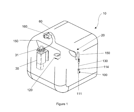
[083] Figure 1 is rear perspective view of a bioprinter for fabricating 3D
cell constructs,
illustrating a sample loading system;
[084] Figure 2 is a front perspective view of the sample loading system of
Figure 1;
CA 03086602 2020-06-04
WO 2019/109127 PCT/AU2018/000249
[085] Figure 3 is a front perspective view of the sample loading system,
illustrating a
plurality of sample containers each containing a sample;
[086] Figure 4 is rear perspective view of the sample loading system of Figure
3;
[087] Figure 5 is a perspective view of a removable sample tray used in the
sample
loading system;
[088] Figure 6 is a front perspective view of the bioprinter with a laminar
air flow system
attached;
[089] Figure 7 is a rear perspective view of the bioprinter showing only the
laminar air flow
system;
[090] Figure 8 is rear perspective view of the bioprinter showing only the
laminar air flow
system of Figure 7;
[091] Figure 9 and Figure 10 show the bioprinter with clear panels
illustrating the air flow
path of the laminar air flow system;
[092] Figure 11 is a top perspective view of the bioprinter with the
positioning units for the
droplet dispensing system and sample loading system illustrated;
[093] Figure 12 is side perspective view of the bioprinter with a plurality of
holding
reservoirs and the compressed air supply system;
[094] Figure 13 is a front perspective view of the bioprinter of Figure 11
with the laminar
air flow system installed;
[095] Figure 14 is a rear perspective view of the bioprinter of Figure 13;
[096] Figure 15 is a rear perspective view of the bioprinter with a single
holding reservoir;
[097] Figure 16 is front perspective view of an assembled bioprinter;
[098] Figure 17 is a rear perspective view of the bioprinter of Figure 16;
CA 03086602 2020-06-04
WO 2019/109127 PCT/AU2018/000249
11
[099] Figure 18 is a flow chart schematic of the components bioprinter;
[0100] Figure 19 is a side view of each component of the bioprinter showing
the air flow
path between the components of the laminar air flow system;
[0101] Figure 20 is a flow chart schematic of the bioprinter operatively
associated to a
control system computer;
[0102] Figure 21 is an exemplary graphical user interface of the control
system software
implemented on the computer showing the 3D cell construct design;
[0103] Figure 22 is an exemplary graphical user interface of the control
system software
implemented on the computer showing printing of the 3D cell construct in a
multi-well plate;
and
[0104] Figure 23 is a flow chart schematic of designing the 3D cell construct
integrated with
the bioprinter.
Description of Embodiments
[0105] As illustrated in the drawings, there is disclosed herein a bioprinter
10 for fabricating
3D cell constructs. The bioprinter 10 comprises a sample loading system 20 for
loading a
sample 100 from a sample container 110 into a holding reservoir 120; a
selector valve 30 in
fluid communication with the holding reservoir 120 for directing the sample
100 into the
holding reservoir 120; a droplet dispensing system 25 in fluid communication
with the
holding reservoir 120, the droplet dispensing system 25 adapted to print
sample droplets
101 from the holding reservoir 120 onto a substrate 125; a control system 40
to control
operation of the sample loading system 20, the selector valve 30, and the
droplet
dispensing system 40; a laminar air flow system 50; and a housing 60
encompassing the
sample loading system 20, the selector valve 30, the droplet dispensing system
40 and the
laminar air flow system 50.
Sample loading system
[0106] Referring to Figures 1 to 4, the sample loading system 20 is adapted to
access
samples 100 contained in one or more sample containers 110 and is in fluid
communication
with one or more holding reservoirs 120. The holding reservoirs 120 are
configured to hold
samples 100 from the sample containers 110. The sample containers 110 can be a
CA 03086602 2020-06-04
WO 2019/109127 PCT/AU2018/000249
12
standard chromatography vial 111 having a cap 112 comprising a rubber septum
113 used
in laboratories for storage and transport of samples. The vials 111 are
typically
manufactured from glass, plastic or any suitable material capable of
maintaining a sterile
environment within the vial 111. The vials 111 are made in multiple sizes to
accommodate
various samples 100, typically smaller vials storing approximately 5 ml and
larger vials
storing approximately 10 ml. The sample loading system 20 may comprise one
sample
container 110, or a plurality of sample containers 110, depending on the 3D
cell construct to
be printed by the bioprinter 10.
[0107] Each holding reservoir 120 is adapted to store a sample 100 received
from one of
the sample containers 110. Each holding reservoir 120 is manufactured from
elongate
tubing 122 wrapped inside a reservoir housing 121. The elongate tubing 122 may
be coiled
and encased in the housing 121. The elongate tubing 122 is a spool of flexible
tubing 122.
In certain embodiments, the flexible tubing 122 is made from
Polytetrafluoroethylene
(PTFE) tubing, or other suitable material such as fluorinated ethylene
propylene (FEP),
ethyltrifluoroethylene (ETFE), polyether ether ketone (PEEK), silicone,
thermoplastic
elastomer (TPE) or stainless steel. In alternative embodiments, each holding
reservoir 120
is a container with an inlet, an outlet, and a storage cavity for storing a
sample 100. The
bioprinter 10 can include one or more holding reservoirs 120, corresponding to
one or more
individual and differing samples 100.
[0108] The sample 100 in each sample container 110 can be a cell suspension,
water,
ethanol, bio-ink, activator, cleaning solution, flushing fluid, cell culture
media, or drug
dispersed in solution, which are described in detail below. The sample 100
stored in the
sample container 110 may or may not be sterile.
[0109] The sample loading system 20 comprises at least one needle 130 that is
insertable
into each sample vial 111 and is in fluid communication with the one or more
holding
reservoirs 120. In the embodiment depicted in the drawings, there is a single
needle 130.
The needle 130 is 50mm long, bevel tip, 16 gauge, made from stainless steel.
The needle
130 may be operatively associated with the sample loading system 20 for
removing a
sample 100 from each vial 111. The needle 130 is movable in the z-direction to
insert the
needle 130 into a vial 111 from above by a first positioning unit 140. The
first positioning
unit 140 is a miniature electric linear actuator 140a with a stroke of 60mm
driven by a lead
screw 140b coupled to a stepper motor 140c, illustrated in Figure 3 and Figure
4.
CA 03086602 2020-06-04
WO 2019/109127 PCT/AU2018/000249
13
[0110] The bioprinter 10 includes the flow selector valve 30 that directs a
sample 100 taken
from a sample container 110 to a holding reservoir 120. This is so that each
sample 100
taken from each vial 111 is held in a separate holding reservoir 120 to
separate each
sample 100 to maintain sterile conditions.
[0111] The sample loading system 20 and droplet dispensing system 25 of the
bioprinter 10
are connected by tubing 150. The tubing 150 is selected from a number of
different
materials, diameters and lengths, based on its desired location and functional
requirements.
The tubing 150 connecting the sample loading system 20 to each holding
reservoir 120 is
2.16mm inner diameter and 3.175mm outer diameter PTFE tubing. The elongate
tubing 122
in each holding reservoir 120 is 1/8" PTFE tubing.
[0112] To prime each holding reservoir 120 with a sample 100, the sample 100
is moved
from a vial 111 to, and through, the selector valve 30 using a pump 160. The
pump 160 may
be a positive displacement pump such as a peristaltic, diaphragm or syringe
pump. The
pump 160 is connected to the selector valve 30 which comprises a suitable
channel 31 for
directing the sample 100 into the suitable (and isolated) holding reservoir
120. The valve 30
is a low pressure flow-through selector valve 30 manufactured by VICI Valco
Instruments
Co Inc. The flow selector valve 30 comprises many channels 31, such as 4, 6,
8, 10, 12 or
16 channels. The flow selector valve 30 has a common inlet connected to the
pump 160
and needle 130. When a channel 31 is selected, the selected channel 31 is
fluidically
connected to the pump 160 and needle 130. When a channel 31 is not selected,
that
channel 31 is fluidically connected to pressurised air from an air pressure
regulator 171 in
the pressure regulator manifold 170. The pressure in each holding reservoir
120 or channel
31 is independently set by a respective regulator 171 in the regulator
manifold 170. Each
regulator 171 in the regulator manifold 170 is connected to the selector valve
30 using 4mm
Nylon tubing and 1/8" PTFE tubing. The number of pressure regulators 171 in
the regulator
manifold 170 is equivalent to the number of holding reservoirs 120 or channels
31. This
allows the pressure to be set independently for each valve 252 of the droplet
dispensing
system 25, which means the bioprinter 10 may support a wide range of fluid
viscosities in
each valve 252.
[0113] The pressure regulators 171 in the regulator manifold 170 of the sample
loading
system 20 independently control pressure feeding into each channel 31 of the
selector
valve 30 and the holding reservoirs 120. The pressure regulator manifold 170
is operatively
connected to a compressed air supply inlet 180 and a static pressure reservoir
(not shown).
CA 03086602 2020-06-04
WO 2019/109127 PCT/AU2018/000249
14
The bioprinter 10 includes an air filter 190 within the housing 60 for
filtering the air from the
compressed air supply inlet 180. The pump 160 is operably coupled to the
needle 130 for
transferring a sample 100 in a sample container 110 to a holding reservoir
120. The pump
160 may be operably reversible for resuspension of the sample 100 in the
sample container
110.
[0114] A seal 114 is formed by a rubber septa 113 of each sample container 110
and upon
operation of the first positioning unit 140 is punctured using the needle 130
driven by the
first positioning unit 140. The first positioning unit 140 is operated by a
control system 40
and robotic linear actuators 140a. The control system 40 positions the first
positioning unit
140 by moving a stepper motor 140c on the linear actuator 140a by a desired
number of
steps. The first positioning unit 140 is operably coupled to the needle 130
for positioning the
needle 130 into puncturing-engagement with a sample container 110 and out of
puncturing-
engagement with the sample container 110. The channel 31 is selected on the
selector
valve 30, the micro-solenoid valve 252 is opened and the pump 160 is turned on
to move
the sample 100 from the sample vial 111, through the needle 130, tubing 150,
pump 160,
selector valve 30, and into a holding reservoir 120. The pump 160 is then
turned off and the
micro-solenoid valve 252 is closed. The channel 31 is deselected on the flow
selector valve
30. The pressure is then set by the respective regulator 171 of the regulator
manifold 170
and the micro-solenoid valve 252 is fired repeatedly until all air is out of
the tubing line 150
and the sample 100 is fired from the holding reservoir 120. The above process
is repeated
to prime each holding reservoir 120 that is to be used.
[0115] The sample loading system 20 further comprises a second positioning
unit 141
operably coupled to the needle 130 and the printhead 250 of the droplet
dispensing unit 25,
the second positioning unit 141 for positioning the needle 130 and the
printhead 250 in two-
dimensional space above the sample containers 110 and the substrate 125. The
second
positioning unit 141 is configured to move the needle 130 and the printhead
250 along a
track 142. The second positioning unit 141 may be a 3-axis motion control
stage unit. The
second positioning unit 141 is a belt driven linear actuator 141a with a
stroke of 300mm.
The belt 141b is a toothed belt and driven by a stepper motor 141c.
[0116] To print sample droplets 101, one or more holding reservoirs 120 and
micro-
solenoid valves 252 are primed as described above, and sample droplets 101 are
fired from
one or more nozzles 253 of a printhead 250 of the droplet dispensing system
25, and
CA 03086602 2020-06-04
WO 2019/109127 PCT/AU2018/000249
deposited on the substrate 125 in a predetermined manner controlled by
computer-
controlled software 40. This droplet dispensing system 25 is described in more
detail below.
[0117] To clean the bioprinter 10, detergent can be moved from sample vials
111 using the
needle 130 to and through the tubing lines 150, selector valve 30, the holding
reservoir 120,
using the sample loading system 20 as described above. Detergent is ejected
from the
nozzles 253 of the droplet dispensing system 25 into a waste container 205.
This process is
repeated for other cleaning chemicals, such as 70% ethanol and water. The
cleaning of the
bioprinter 10 is finished when all water has been flushed through the lines
and only air is
being ejected from the nozzles 253 of the droplet dispensing system 25,
described in detail
below.
[0118] To resuspend samples in the vials 111, the needle 130 can be moved
towards the
respective sample vial 111 using the first and second positioning units 140
and 141 until the
needle 130 punctures the sample vial septa 113 and engages the sample 100. The
cell-
containing sample 100 in a sample vial 111 is moved from the sample vial 111,
through the
needle 130 and tubing 150 using the peristaltic pump 160. The cell-containing
sample 100
is moved in the opposite direction (i.e. towards the sample vial 111) using
the peristaltic
pump 160 in reverse. This process of moving the cell-containing sample from
and towards
the vial 111 via the needle 130 and tubing can be repeated as desired.
Sample Tray
[0119] The sample containers 110 may be housed in a removable sample tray 200
that
may also be sterile. It is envisaged that up to 14 vials 111 can be housed in
the removable
sample tray 200, but any suitable number of vials 111 can be housed in the
removable
sample tray 200. The removable sample tray 200 has one or more sample
container
housings 201 adapted to store sample containers 110 of differing size, such as
vials 111
and waste containers 205. The removable sample tray 200 as shown in Figure 5
has a lid
202 and a tray 203. The removable sample tray 200 may be manufactured from
plastic or
other suitable material. The removable sample tray 200 is loadable into a
recess 129 f the
printstage 128 of the bioprinter 10 via the hinged door 210 as shown in
Figures 13 and 16.
In alternative embodiments, the sample containers 110 are loadable into the
removable
sample tray 200. The removable sample tray 200 provides a substantially
sterile
environment for storing the vials 111, as well as each vial 111 being sterile.
CA 03086602 2020-06-04
WO 2019/109127 PCT/AU2018/000249
16
[0120] It is also envisaged that instead of the removable sample tray 200, the
sample
container housings 201 may be integral with printstage 128. In this case, each
sample
container 110 would be loadable into the printstage 128 via the hinged door
210 of the
bioprinter 10.
[0121] The removable sample tray 200 housing the sample containers 110 may
further
include the waste container 205 for receiving waste when flushing the sample
loading
system 20. The removable tray 200 may further include a cleaning container 204
for
cleaning the sample loading system 20, the selector valve 30, and droplet
dispensing
system 25.
Droplet dispensing system
[0122] Referring to Figures 11 to 15, the droplet dispensing system 25
includes the
printhead 250 operably coupled to the plurality of holding reservoirs 120 and
adapted to
dispense sample droplets 101 onto the substrate 125 from each holding
reservoir 120. The
at least one printhead 250 may be an array of valves 251. The array of valves
251 may
comprise a plurality of micro-solenoid valves 252. The micro-solenoid valves
252 may be
VHS Series Solenoid Valves manufactured by The Lee Company. Each micro-
solenoid
valve 252 includes a nozzle 253 with an orifice diameter of 0.003", 0.005" or
0.007". Each
micro-solenoid valve 252 is opened by applying a spike and hold voltage across
the
solenoid coil. The spike voltage is 24V and the hold voltage is 5V. The
duration of the spike
voltage is between 0.2 and 0.5 ms. When the voltage is switched off the valve
252 returns
to the closed position.
[0123] Each nozzle 253 may be a jeweled orifice dispensing nozzle 253
controlled by a
microcontroller, namely the control system 40. The samples 100 are stored in
the holding
reservoirs 120 upstream of the micro-solenoid valves 252 and nozzle 253. The
internal
diameter of the jeweled orifice nozzles 253 can be between 127 and 254 pm
depending on
the fluid viscosity and the desired droplet volume of the sample droplet 101.
[0124] The droplet dispensing system 25 includes the compressed air supply
inlet 180,
operably coupled to the pressure regulator manifold 170 via air filter 190.
The air moves the
samples 100 around the sample loading system 20 and droplet dispensing system
25, so as
to be dispensed via the nozzles 253 of the printhead 250. The desired sample
droplet 101
volume can also be adjusted using the backpressure set by the pressure
regulators 171 of
the regulator manifold 170 and the open time of the respective valves 252 open
time.
CA 03086602 2020-06-04
WO 2019/109127 PCT/AU2018/000249
17
Typically, the backpressure is set to a pressure between 1 and 60 psi, the
valve 251 open
time is 0.3 ms or greater and the droplet volume is between 1 and 500 nl.
[0125] The droplet dispensing system 25 may further comprise a third
positioning unit 300
operably coupled to the printstage 128, the third positioning unit 300 for
positioning the
printstage 128 in two-dimensional space. The third positioning unit 300 is
configured to
move the printstage 128 along a track 301. The track 142 extends
perpendicularly to the
track 301. Referring to Figure 13, the printstage 128 supports the substrate
125 and has a
recess 129 that is configured to removably receive the sample tray 200. The 3-
axis motion
control stage is capable of accurately positioning the droplet dispensing
system at a
resolution of 10 pm in all three (X, Y and Z) dimensions.
[0126] In a sterile environment, each of the activator, bio-ink and bio-ink or
cell-ink
containing cells (ie, samples 100) are slowly loaded into the appropriate
holding reservoir
120 using the sample loading system 20 to avoid the generation of small
bubbles.
[0127] The bioprinter is equipped with a power supply in the form of a 24V DC
power
supply (not shown).
[0128] The compressed air supply inlet 180 is supplied from an air compressor
(not shown).
The air compressor can supply an air pressure between 3 and 10 bar. The
compressed air
can be supplied from a common compressed air line that is common in research
laboratories.
[0129] Tubing 150 within the droplet dispensing system 25 is 40mm 1/16" Teflon
tubing.
This tubing 150 connects the holding reservoirs 120 to the array of valves 251
of the
printhead 250.
[0130] The sample loading system 20 can automatically load samples 100 into
the holding
reservoirs 120 for printing. The system has several advantages over current
state of the art
bioprinting systems. Firstly, bio-inks can be stored in easy to handle sample
containers 110
such as glass or plastic vials 111. These samples containers 110 are easily
sterilized before
filling with bio-ink samples. End users, such as biologists, deposit their
cells inside the
appropriate vial 111 using the usual methods, for example a pipette.
Depositing cells inside
the bio-ink vials 111 can be carried out inside a bio-safety cabinet to ensure
samples 100
CA 03086602 2020-06-04
WO 2019/109127 PCT/AU2018/000249
18
are not contaminated. After depositing cells, the vials 111 can be placed
inside the
bioprinter 10 in the appropriate location.
[0131] The sample loading system 20 allows bio-inks and bio-inks containing
cells to be
loaded from individual vials 111 sealed with rubber septa 113. This is
achieved using the
needle 130 that is positioned using the z-axis linear actuator 140. The needle
130 is
fluidically connected to the positive displacement pump 160. When the tip of
the needle 130
pierces the septa 113 and is positioned inside the vial 111, the pump 160 is
engaged and
fluid sample 100 is pumped out of the vial 111.
[0132] The printhead 250 may comprise multiple electronic pressure regulators
171 that are
individually adjustable for printing a large range of viscosities, droplet
sizes etc, based on
user input, the sample construct, and/or the desired cell construct. The
electronic pressure
regulators 171 are operably connected to the array of valves 251.
[0133] The bank of pressure regulators 171 (it is envisaged that the
bioprinter 10 may
include 10, as there are up to 10 holding reservoirs 120) is contained in the
pressure
regulator manifold 170. The function of the manifold 170 is to distribute
pressurised air from
the external air compressor connected to the supply inlet 180 to each of the
regulators 171.
In Figure 11 and Figure 12, only a single regulator 171 and one side of the
manifold 170 is
shown. In Figure 14 and Figure 15, a bank of ten regulators 171 is shown.
[0134] An exemplary embodiment of the sample loading system of the bioprinter
10
comprises the steps for loading samples 100 into the holding reservoir 120 as
follows:
1. Move Selector Valve 30 to selected channel 31;
2. Open micro-solenoid valve 252;
3. Position needle 130 above vial 111 using x-axis and y-axis actuators;
4. Lower needle 130 into vial 111 piercing the vial septum 113 using z-axis
actuator;
5. Engage peristaltic pump 160;
6. Pump fluid from vial 111 through selector valve 30 into tubing holding
reservoir 120;
7. Stop pump 160 when fluid reaches nozzle of micro-solenoid valve 252; and
8. Close micro-solenoid valve 252.
CA 03086602 2020-06-04
WO 2019/109127 PCT/AU2018/000249
19
[0135] Another exemplary embodiment of the sample loading system 20 of the
bioprinter
10, in particular the steps for cleaning and sterilising comprises the
following steps:
1. Position micro-solenoid valve 252 over waste spittoon or vial;
2. Empty any fluid remaining after print job into waste container 205;
3. Move Selector Valve 30 to selected channel for cleaning;
4. Position needle 130 above vial 111 containing ethanol using x-axis and y-
axis actuators;
5. Lower needle 130 into vial 111 piercing the vial septum using z-axis
actuator;
6. Open micro-solenoid valve 252;
7. Engage peristaltic pump;
8. Pump ethanol from vial 111 through selector valve 30 and open micro-
solenoid valve 252;
9. Stop pump when sufficient ethanol has passed through open micro-solenoid
valve 252;
10. Close micro-solenoid valve 252;
11. Repeat process with detergent; and
12. Repeat the process with water.
[0136] The bioprinter 10 is adapted to print onto many kinds of substrate 125,
such as
micro-well plates and Petri dishes. Referring to Figure 20, the substrate 125
can be heated
to 37 C to assist cell proliferation using a temperature control unit 280.
Both the
temperature control units 280 regulate the temperature inside the bioprinter
10, based on
the need of the 3D cell construct conditions necessary for optimal growth
conditions. The
temperature control units 280 are adjustable to between 36 C and 38 C to
regulate the
temperature of the printhead 250, the substrate 125 disposed on the printstage
128, and/or
the interior of the bioprinter 10.
[0137] The substrate 125 that is disposed on and supported by the printstage
may be a
multi-well plate 126. The substrate 125 may be biocompatible consumables used
to enclose
and culture the printed cellular structure. These substrates may include:
= Microtitre plate of different well configurations (6, 12, 24, 48, 96 and
384-well);
= Microtitre plate with coverslip bottom of different well configurations
(6, 12, 24, 48,96
and 384-well);
= Coverslips and microscope slides;
= Fluorodish of various sizes; and
= Chamber slides of different chamber configurations (1, 2, 4, 8 and 16).
CA 03086602 2020-06-04
WO 2019/109127 PCT/AU2018/000249
[0138] To clean the tubing lines 150, array of valves 251 and the nozzles 253,
detergent
can be moved from sample vials 111 to, and through the valves 252 and the
nozzles 253
using the droplet dispensing system 25 as described above. Detergent is
ejected from the
nozzles 253 into the waste container 205. This process is repeated for other
cleaning
chemicals, such as 70% ethanol and water. The cleaning of the tubing lines 150
and the
printhead 250 is finished when all water has been flushed through the lines
150, the array of
valves 251 and the nozzles 253 and only air is being ejected from the nozzles
253.
Laminar Flow System
[0139] Referring to Figures 6 to 10, the bioprinter 10 further includes a
laminar flow system
50 as illustrated in Figures 6 to 10. Sterility and operator safety is a major
concern in 3D
bioprinting applications. It is normally achieved by locating the bioprinter
inside a biosafety
cabinet or clean room. Typically, biosafety cabinets and clean rooms are
regarded as
precious and expensive space in a tissue culture lab. Therefore, there is a
need for
solutions to minimise use of bio-safety cabinet and clean room space in 3D bio-
printing
applications.
[0140] The integrated laminar flow system 50, integrated into the bio-printer
10, provides
the sterile environment for bio-printing of cells and 3D tissue culture models
without
requiring biosafety cabinet or clean room facilities. Furthermore, the
operator is protected
using directional airflow to draw air from the outside environment through the
front access.
[0141] The laminar air flow system 50 includes a chamber or enclosure with a
metallic
frame 500, for example stainless steel, and is provided with a metallic grate
at the base to
allow contaminated airflow to be drawn into to the electrically powered blower
fan or
centrifugal fan 510. The contaminated air is pumped through a duct inlet 520
using the fan
510 and into a positive pressure chamber 535 consisting of two High-Efficiency
Particulate
Arresting (HEPA) filters 525 and 530. The HEPA filters 525 and 530 may remove
at least
99% of particles from the contaminated air flow. One HEPA filter 525 acts as
an exhaust to
the external environment and the other HEPA filter 530 recycles the air flow
to the sterile
chamber 535. It is envisaged that each filter will take approximately 50% of
the airflow.
Figure 21 illustrates the air flow into the bioprinter 10, through the duct
inlet 520 via the
blower fan 510, through the HEPA filters 525 and 530 and either exhausted out
or recycled.
The airflow from the recycle HEPA filter 525 to the sterile chamber 535
provides
unidirectional downward airflow to the sterile chamber 535 with a typical
velocity of 0.45m/s.
CA 03086602 2020-06-04
WO 2019/109127 PCT/AU2018/000249
21
This airflow provides a uniform clean airflow over the bio-printed sample
droplet 101
significantly reducing the risk of particle contamination in the sample 100.
[0142] During a bio-printing operation, the front access hinged door 210 must
be closed to
reduce the risk of particle contamination. Therefore, the blower fan 510 flow
rate can be
reduced by decreasing the blower rpm. The reduced airflow in the sterile
chamber 535
reduces the effect of dehydration on the printhead 250 and sample 100. In
addition, it
reduces the effect of the airflow disturbing sample droplets 101 during their
flight from the
printhead 250 to the printing substrate 125.
Control software
[0143] The bioprinter 10 is controlled via custom software developed for
printing biological
assays. The control system 40 comprises the control software that includes a
non-transitory
computer readable medium having the programmed instructions for operating the
bioprinter
10. The non-transitory computer readable medium is located separately from the
bioprinter
and is operatively connectable to the bioprinter 10.
[0144] The software includes a graphical user interface (GUI) as illustrated
in Figure 21 and
Figure 22. Through the GUI, the end user can select different assay printing
routines and
change the assay parameters, such as droplet spacing and droplet volume. The
user can
also manually control the spatial position of the droplet dispensing system
and create a
custom pattern of droplets. Additional features of the software include
routines for cleaning,
priming and purging of the droplet dispensing system.
[0145] Bioprinting requires a 3D model of the object to be printed. For tissue
engineering
applications, this is typically created using engineering tools such as
CAD/CAM software.
These tools are expensive and have a high degree of complexity, forcing
scientists to spend
time and resources learning engineering tools. For 3D tissue culture
applications, the
complexity of the structure to be printed is lower. There is need for a simple
and intuitive
method to create 3D structures for bio-printing in 3D tissue culture
applications.
[0146] The software provided with the bioprinter 10 provides a method to
design each layer
of the 3D structure to be printed. In an embodiment, a grid is provided for
the user to draw a
pattern for each layer of the structure. The material to be printed can be
defined as a
mixture of multiple materials that are dispensed from different nozzles 253 in
the printhead
CA 03086602 2020-06-04
WO 2019/109127 PCT/AU2018/000249
22
250. For example, hydrogel can be defined a droplet of bio-ink mixed with a
droplet of
activator.
[0147] Typical substrates used in biology labs for tissue culture are multi-
well plates such
as 6, 12, 24, 48, 96 and 384-well plates. In an embodiment, an interface 670
is provided to
print a previously defined 3D structures inside each well on a multi-well
plate. The user
firstly selects a well or arrays of wells and then selects the print routine
to be printed in
those wells.
[0148] The custom software provides the user interface for the user to input
where in the
array the user would like to bioprint a layer of the 3D cell construct. A
print preview button
671 is provided with the software prior to printing to allow the user to
visualise where the
cells are being printed and what the construct will look like. A feature of
the software is that
it can control the bioprinter droplet size to change how the cell construct is
printed. The
intention behind the layering of the cell construct is to mimic how biologists
use z stack
layering in a microscope.
[0149] Generally, the bioprinter 10 will print 20-25 layers when building the
3D cell
construct, but the number of layers is controlled using the control system and
associated
control software.
[0150] The positioning unit 141 coupled to the printhead 250, controlled by
computer-
controlled software, spatially-positioned the valves 251 and nozzles 253
during each
ejection of sample droplets 101 of bio-ink, activator, cells, cell-ink, or
combinations thereof.
The computer-controlled spatial-positioning of the solenoid valves and
nozzles, and
computer-controlled droplet ejection from the valves 251 and nozzles 253
facilitate the
generation of the 3D tissue construct.
[0151] To generate an array of 3D tissue constructs, the process of generating
3D tissue
constructs was repeated at multiple locations on the substrate 125.
[0152] The control system records the identity of each sample 100 in the
sample containers
110 by either user input or automatic recording. The intention is to know
which sample
containers 110 contain which sample 100, so that during printing, when the
holding
reservoirs 120 are storing their respective samples, the requisite sample is
printed to the
desired location.
CA 03086602 2020-06-04
WO 2019/109127 PCT/AU2018/000249
23
Biobrinter housing
[0153] The bioprinter 10 comprises a housing 60 encompassing the sample
loading system
20, the droplet dispensing system 25 and the laminar air flow system 50. The
housing 60 is
assembled from numerous panels made from steel, aluminum and stainless steel
and
assembled using screw fasteners 600. The housing 60 further includes the
hinged door 210
in the front face, to allow access to the sterile chamber 535 of the
bioprinter 10 by a user.
The removable sample tray 200 is loadable into the bioprinter 10 via the door
210. The front
panel and hinged door 210 can be made from glass or clear plastic. Figure 16
and Figure
17 illustrate the bioprinter assembly 10.
Method
[0154] In operation, the bioprinter 10 has the following steps for
transferring the sample
from the sample container to the holding reservoir ready to be utilised by the
printhead for
bioprinting 3D cell constructs. To prime the printer holding reservoirs 120
and solenoid
valves with fluid, fluid is moved from sample containers to, and through, the
solenoid valves
using the sample loading system. The suitable channel is selected on the flow
selector
valve. The seal of a sample vial is punctured using the needle 130, the
solenoid valve is
opened. The peristaltic pump is turned on to move the desired amount of fluid
from the
sample container, through the needle, tubing, pump, tubing, flow selector
valve, and into the
holding reservoir. The peristaltic pump is turned off and the solenoid valve
is closed. The
suitable channel is deselected on the flow selector valve. The pressure is set
by the
regulator 171 and the solenoid valve 252 is fired repeatedly until all air is
out of the line and
droplet/s bio-ink, activator, cells, cell-ink, or combinations thereof are
fired from the nozzle
253. The above process is repeated for each printer fluid reservoir and
solenoid valve that
is used.
[0155] Steps for loading bio-ink into holding reservoir:
1. Move Selector Valve to selected channel;
2. Open solenoid valve;
3. Position needle above vial using x-axis and y-axis actuators;
4. Lower needle into vial piercing the vial septum using z-axis actuator;
5. Engage peristaltic pump;
6. Pump fluid from vial through selector valve into tubing holding
reservoir;
7. Stop pump when fluid reached nozzle of solenoid valve; and
8. Close solenoid valve.
CA 03086602 2020-06-04
WO 2019/109127 PCT/AU2018/000249
24
[0156] To print bio-ink, activator, cells, cell-ink, or combinations thereof
from the solenoid
valves, printer fluid reservoirs and solenoid valves are primed as described
above, droplets
of bio-ink, activator, cells, cell-ink, or combinations are fired from the
nozzles and deposited
on the substrate in a predetermined manner controlled by computer-controlled
software.
[0157] A positioning unit is coupled to the printhead 250, controlled by
computer-controlled
software, spatially-positioned the solenoid valves and nozzles during each
ejection of
droplets of bio-ink, activator, cells, cell-ink, or combinations thereof. The
computer-
controlled spatial-positioning of the solenoid valves and nozzles, and
computer-controlled
droplet ejection from the solenoid valves and nozzles facilitate the
generation of the 3D
tissue construct.
[0158] To generate an array of 30 tissue constructs, the process of generating
3D tissue
constructs is repeated at multiple locations on the substrate.
Bio-Ink
[0159] In the present specification, bio-ink is defined as an aqueous solution
of one or more
types of macromolecule in which cells may be suspended or housed. Upon
activation or
crosslinking, it creates a hydrogel structure having its physical and chemical
properties
defined by chemical and physical composition of the bio-ink. Macromolecules
are defined
as an array of both synthetic and natural polymers, proteins and peptides.
Macromolecules
may be in their native state or chemically modified with amine or thiol-
reactive
functionalities.
[0160] Synthetic macromolecules may include:
= Polysaccharides, such as polymers containing fructose, sucrose or glucose
functionalities;
= Non-ionic polymers, such as poly(ethylene glycol) (PEG),
poly(hydroxyethyl
methacrylate (PHEMA), poly(E-caprolactone) (PCL), poly(vinyl alcohol) (PVA),
poly(vinylpyrrolidone) (PVP), poly(NIPAAM) and poly(propylene fumarate) (PPF)
and
derivatives;
= Polyelectrolytes ¨ polymers that carry either positive or negative
charge, amphoteric
as well as zwitterionic polymer;
= Polypeptides ¨ a single linear chain of many amino acids (a minimum of 2
amino
acids), held together by amide bonds; and
= Nucleobase containing synthetic polymers ¨ polymers with nucleobase
(adenine,
thymine, guanine or cytosine) repeating units.
CA 03086602 2020-06-04
WO 2019/109127 PCT/AU2018/000249
[0161] Natural macromolecules may include:
= Polysaccharides, such as alginate, chitosan, gellan gum, hyaluronic acid,
agarose
and glycosaminoglycan;
= Proteins, such as gelatin, fibrin and collagen;
= DNA and Oligonucleotides, such as single stranded DNA (ssDNA), double
stranded
DNA (dsDNA) DNAzymes and Aptamers; and
= Basement membrane extracts.
[0162] Amine-reactive functionalities may include: aldehyde, epoxy, N-
hydroxysuccinimide
(NHS) and 2-vinyl-4,4-dimethylazIactone (VDM).
[0163] Thiol-reactive functionalities may include: alkenes, alkynes, azides,
halogens and
cyanates.
[0164] The bio-ink used and found suitable was alginate (at 2 w/vc/o)
dissolved in calcium
free DMEM supplemented with 10 v/v(Yo FCS, L-glutamine and sodium pyruvate.
[0165] Bio-ink with dispersed SK-N-BE(2) neuroblastoma cells is referred to as
bio-ink
containing cells.
Activator
[0166] Activator is an aqueous solution comprising of either small molecules
or
macromolecules which interact with the bio-ink to form a hydrogel structure.
The
composition of the activator can be altered to control the physical properties
of the resulting
hydrogel. The type of activator used is highly dependent on the macromolecules
used as
well as the intended crosslinking process.
[0167] Activators can be selected from:
= Inorganic salts such as calcium carbonate, calcium chloride, sodium
chloride,
magnesium sulphate. sodium hydroxide and barium chloride;
= Photoinitiators such as 2,2-dimethoxy-2-phenylacetophenone (DMPA) and
lrgacure;
= Polyelectrolytes ¨ polymers that carry an opposite charge to the
macromolecules in
the bio-ink. It could be cationic, anionic, amphoteric and zwitterionic;
= Polypeptides ¨ a single linear chain of many amino acids (a minimum of 2
amino
acids), held together by amide bonds;
= DNA linker ¨ macromolecules carrying nucleotides or DNA sequences which
complement those present on the bio-ink's macromolecules; and
CA 03086602 2020-06-04
WO 2019/109127 PCT/AU2018/000249
26
= Natural or synthetic macromolecules carrying amine or thiol groups,
either natively
or through chemical modifications.
[0168] The activator used for the alginate bio-ink was calcium chloride at 4
w/V/0 dissolved
in MilliQ water.
Crosslinking or Gelation
[0169] This is the process whereby individual macromolecular chains are linked
together by
the activator to form a hydrogel. The crosslinking process can be classified
to either
chemical or physical crosslinking. Physical crosslinking or non-covalent
crosslinking may
include:
= Ionic crosslinking ¨ crosslinking via the interaction of the opposite
charges present in
the macromolecule and the activator. The activator may include charged
oligomers,
ionic salt and ionic molecule;
= Hydrogen bonds ¨ crosslinking via the electrostatic attractions of polar
molecules. In
this case, the macromolecule and the activator are carrying polar
functionalities;
= Temperature crosslinking ¨ crosslinking via the rearrangement of the
macromolecular chains as a response to change in temperature (heating or
cooling);
and
= Hydrophobic interaction or van der VVaals force.
[0170] Chemical or covalent crosslinking involves chemical reactions between
the
macromolecule and the activator. The type of reactions may include:
= Photocrosslinking whereby the crosslinking reaction is promoted by UV or
light
irradiation;
= Michael-type addition reaction between thiols and vinyl-carrying
macromolecules in
aqueous media;
= Schiff base reaction between amino and aldehyde groups;
= DieIs-alder reaction;
= Click chemistry;
= Aminolysis reaction to active ester group; and
= Enzyme crosslinking.
CA 03086602 2020-06-04
WO 2019/109127 PCT/AU2018/000249
27
[0171] Examples of other bio-ink and activator combinations are set out in the
Table below:
Bio-Ink Activator
Positively charged polyelectrolyte (e.g. Negatively charged polyelectrolyte
(e.g.
poly(trimethylammonium) or poly(sulfonate), poly(carboxylic acid)
poly(guanidium)
Fluorenylmethoxycarbonyl (Fmoc) Phosphate buffer solution
polypeptide Cell culture medium
Thiol-reactive macromolecules (e.g. Photoinitiator and/or thiol-containing
PEG-diacrylate, hyaluronic acid macromolecules (e.g. bis-thiol-PEG)
maleimide) Thiol-containing polypeptides (e.g. bis-
cysteine functionalised peptide)
Amine-reactive macromolecules (e.g. Amine-containing polypeptides (e.g. bis-
PEG-co-Poly(benzaldehyde), aldehyde- amine functionalised peptide, gelatin,
alginate collagen)
Charged polysaccharides(e.g. alginate Inorganic salts (e.g. calcium
chloride,
and gellan gum) barium chloride).
Macromolecules containing nucleobase Macromolecules containing the
(e.g. Adenine) corresponding nucleobase pair (e.g.
Thymine)
Cell-Ink
[0172] Cell-ink is an aqueous solution of one or more type of molecules or
macromolecules
in which cells are to be and remain evenly suspended throughout the 3D bio-
printing
process. The concentration of the cell-ink is optimised to prevent cells from
settling but still
maintains high cell viability.
[0173] Cell-link can be selected from:
= Small molecules such as glycerol; and
= Macromolecules such as FicollTM, dextran, alginate, gellan gum,
methylcellulose and
poly(vinylpyrrolidone) (PVP).
CA 03086602 2020-06-04
WO 2019/109127 PCT/AU2018/000249
28
[0174] Ficoll TM is a neutral, highly branched, high-mass, hydrophilic
polysaccharide which
dissolves readily in aqueous solutions. Ficoll TM radii range from 2-7 nnn and
is prepared by
reaction of the polysaccharide with epichlorohydrin. FicollTM is a registered
trademark
owned by GE Healthcare companies.
[0175] The cell-ink used was Ficoll TM 400 (at 10 w/v%) dissolved in PBS.
[0176] Cell-ink with dispersed SK-N-BE(2) neuroblastoma cells is referred to
as cell-ink
containing cells.
[0177] Gellan gum is a water-soluble anionic polysaccharide produced by the
bacterium
Sphingomonas elodea (formerly Pseudomonas elodea).
Cell-Culture Solutions
[0178] Cell-culture solutions are liquids that come into contact with the
cultured cells and
are suitable for various cell-related works. The preparation process includes
careful analysis
of the salt and pH balance, incorporation of only biocompatible molecules and
sterilisation.
[0179] Some of the cell culture solutions include:
= Cell culture medium such as Dulbecco's Modified Eagle Medium (DMEM),
Minimum
Essential Media (MEM), Iscove's Modified Dulbecco's Medium (IMDM), Media 199,
Ham's F10, Ham's F12, McCoy's 5A and Roswell Park Memorial Institute (RPM!)
medium;
= Growth supplements such as foetal calf serum (FCS), epidermal growth
factor
(EGF), basic fibroblast growth factor (bFBF), fibroblast growth factor (FBF),
endothelial cell growth factor (ECGF), insulin-like growth factor 1 (IGF-1)
and
platelet-derived growth factor (PDGF);
= Biological buffers such as PBS, HEPES and CHES;
= Chelating and stabilizing solutions; and
= Sterilized MilliQ water.
Cell-Culture Conditions
[0180] Cells and the 3D tissue culture models can be incubated, cultured and
maintained
using standard cell culture techniques. The 3D tissue culture models
comprising the cells
encapsulated in the hydrogel mold can be incubated under conditions to allow
or maintain
cell growth or spheroid formation. Incubation is typically carried out at
about 37 C with a
CO2 level of 5% for at least 24 hours for most animal and human cell lines. It
will be
appreciated that incubation can be carried out at any suitable conditions,
temperature and
CA 03086602 2020-06-04
WO 2019/109127 PCT/AU2018/000249
29
time duration that allows growth, maintenance or spheroid formation of the
type of cell or
cells in the hydrogel mold.
Utility Solutions
[0181] Utility solutions are defined as the solutions which do not come into
contact with the
cells but are used to clean and sterilise all printer surfaces exposed to the
cells. These
solutions may include:
= Ethanol at the correct concentration;
= Sterile MilliQ water;
= Cell culture medium;
= Detergent; and
= Hydrogen peroxide solution (2 w/V3/0 maximum concentration).
Preparation of Bio-Ink
[0182] Initially, bio-ink is prepared by mixing the right type and amount of
macromolecules
in the appropriate cell-culture solution. After achieving homogeneity, the
blank bio-ink is
sterilised via both UV irradiation and filtration (0.22 pm filter). The bio-
ink is then kept at 4 C
until further usage.
Preparation of Cells
[0183] Harvest cells by washing with PBS. Aspirate PBS. Add trypsin and
incubate at 37 C
to dissociate cells from flask surface. Add tissue culture media to collect
dissociated cells
into a tube. Centrifuge cells, aspirate supernatant and resuspend pellet in
fresh media.
Perform cell count by mixing equal volumes of cell suspension and trypan blue
stain.
Perform calculation to determine the cell concentration. Desired numbers of
cells then can
be added to bio-ink, cell-ink or added to cell culture solutions.
Preparation of Activators
[0184] The correct type and amount of molecules were dissolved in the
appropriate cell-
culture solution. The resulting solution was sterilised via UV irradiation and
filtration prior to
use.
Preparation of Cell-Ink
[0185] The correct type and amount of molecules were dissolved in the
appropriate cell-
culture solution. After achieving homogeneity, the resulting solution was
sterilised via UV
irradiation and filtration prior to use. The cell-ink was then kept at room
temperature until
further use.
CA 03086602 2020-06-04
WO 2019/109127 PCT/AU2018/000249
Cell Harvesting
[0186] Cultured cells of interest at certain confluency are harvested by
following the already
established protocols. To make up the bio-ink or cell-ink containing cells,
harvested cells
are resuspended at the correct cell concentration to give 250 million cells/ml
concentration
in 200 pl of bio-ink or cell-ink. The resulting cell pellets are then
redispersed in the correct
volume of bio-ink or cell-ink. The bio-ink or cell-ink containing cells is
then ready for use in
the 3D bio-printer.
Printing of Hydrogel Mold
[0187] The hydrogel mold can be printed using a drop-on-drop process whereby a
droplet
of bio-ink and a droplet of activator were deposited on top of each other to
produce a
hydrogel. This process can be repeated and used to form 3D hydrogel structures
by
building up layers of hydrogel.
Cell Types
[0188] 30 tissue culture models such as spheroids can be prepared from any
suitable cell
type including adherent cells such as mammalian liver cells, gastrointestinal
cells,
pancreatic cells, kidney cells, lung cells, tracheal cells, vascular cells,
skeletal muscle cells,
cardiac cells, skin cells, smooth muscle cells, connective tissue cells,
corneal cells,
genitourinary cells, breast cells, reproductive cells, endothelial cells,
epithelial cells,
fibroblast, neural cells, Schwann cells, adipose cells, bone cells, bone
marrow cells,
cartilage cells, pericytes, mesothelial cells, cells derived from endocrine
tissue, stromal
cells, stem cells, progenitor cells, lymph cells, blood cells, endoderm-
derived cells,
ectoderm-derived cells, mesoderm-derived cells, or combinations thereof.
[0189] Additional cell types may include other eukaryotic cells (e.g. chinese
hamster ovary),
bacteria (e.g. helicobacter pylori), fungi (e.g. Penicillium chrysogenum) and
yeast (e.g.
saccharomyces cerevisiae).
[0190] The cell line SK-N-BE(2) (neuroblastoma cells) has been used
successfully in the
process to produce 3D tissue culture models under a range of conditions. It
will be
appreciated that other cell lines would be expected to perform as required in
30 tissue
models produced by the process developed. Other cell lines used include DAOY
(human
medulloblastoma cancer cells), H460 (human non-small lung cancer) and p53R127H
(human pancreatic cancer cells). Other cell lines that may be suitable are
listed on 088 and
089.
CA 03086602 2020-06-04
WO 2019/109127 PCT/AU2018/000249
31
[0191] 30 bio-printing technology was developed to produce high density 30
tissue culture
models encapsulated in a hydrogel mold via drop-on-demand techniques.
Specifically, a 3D
printing technology was used to print biocompatible hydrogel molds using a bio-
ink and
activator that are constructed in a layer-by-layer manner to fabricate a
variety of 3D
structures. During the fabrication of the hydrogel molds, high cell density
droplets can be
included into the hydrogel mold.
[0192] It will be appreciated by persons skilled in the art that numerous
variations and/or
modifications may be made to the invention as shown in the specific
embodiments without
departing from the spirit or scope of the invention as broadly described. The
present
embodiments are, therefore, to be considered in all respects as illustrative
and not
restrictive.
References
[0193] Murphy, S. and Atala, A. (2014). 3D bioprinting of tissues and organs.
Nature
Biotechnol, 32(8), pp 773-785.
[0194] Horn, T. and Harrysson, 0. (2012). Overview of current additive
manufacturing
technologies and selected applications. Sci. Prog, 95, pp 255-282.