Note: Descriptions are shown in the official language in which they were submitted.
CA 03087259 2020-06-26
,
WO 2019/136300 PCT/US2019/012415
ANTI-MCT1 ANTIBODIES AND USES THEREOF
RELATED APPLICATIONS
[1] This application claims priority to United States Provisional No.
62/613,447 filed on
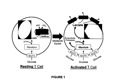
January 4, 2018, United States Provisional No. 62/684,870 filed on June 14,
2018, and
United States Provisional No. 62/736,025 filed on September 25, 2018 and
United States
Provisional No. 62/773,630 filed on November 30, 2018. The contents of each of
these
provisional applications are incorporated by reference in its entirety herein.
SEQUENCE LISTING
[2] The sequence listing in the file named "43260.4213,txt" having a size
of xxxxxx bytes
that was created January 4, 2019, is hereby incorporated by reference in its
entirety.
FIELD OF THE INVENTION
[3] This invention generally pertains to anti-MCT1 antibodies and antigen-
binding
fragments thereof, e.g., humanized, chimeric, and human antibodies and antigen-
binding
fragments thereof, e.g., antagonistic anti-MCT1 antibodies and antigen-binding
fragments
thereof, and compositions containing such antibodies and antigen-binding
fragments
thereof. Such antibodies and antigen-binding fragments include those which
specifically
bind to MCT1, e.g., MCT1 expressed on the surface of endogenous MCT1
expressing human
cells or recombinant cells engineered to express MCT1 and which antagonize one
or more
functions associated with MCT1, e.g., its ability to promote lactate
transport. The invention
also relates to fusion or multispecific proteins comprising one or more anti-
MCT1 antibody
binding sequences, e.g., multispecific and bispecific antibodies. The
invention further relates
to therapeutic and diagnostic uses for such antibodies, antigen-binding
fragments, fusion
and multispecific polypeptides, and compositions containing same. The
invention
specifically relates to the use of these antibodies and antigen-binding
fragments thereof as
prophylactics or therapeutics, e.g., for the treatment of autoimmunity,
inflammation,
allergy, transplant, GVHD, cancer and other conditions wherein suppression of
MCT1
activity and/or increased TR1 cell numbers/activity and/or decreased
numbers/activity of T
effector cells are therapeutically desirable.
1
CA 03087259 2020-06-26
WO 2019/136300 PCT/US2019/012415
BACKGROUND OF THE INVENTION
[4] The monocarboxylate nutrient transporter SLC16A1 (MCT1) is a multipass
transmembrane protein responsible for the facilitated transport of critical
metabolites,
including products of glycolysis. MCT1 is a member of one of the largest
family of surface
membrane proteins, known as solute channel proteins (SLCs), whose functions
involve the
transport across membranes of critical cellular nutrients, metabolites, ions,
hormones and
lipids. MCT1 belongs to the SLC16 family of transporters, five of which have
been shown to
transport monocarboxylates, such as pyruvate, lactate and ketones (REF. 34-36)
in a
facilitated, pH dependent and bidirectional manner. SLC16A1 (MCT1), SLC16A7
(MCT2),
SLC16A8 (MCT3) and SLC16A3 (MCT4) have all been shown to transport
monocarboxylates
with Km in the 1-40 mM range (REF. 37). MCT1, MCT3 and MCT4 are co-expressed
with the
Ig-domain containing surface protein CD147 (Basigin), which in many cells is
critical for
proper cell surface expression (REF. 38, 37). Besides these MCTs, other
lactate transporters
include the recently characterized SLC16A11 (REF. 39) and sodium-dependent
SLC5A8 and
SLC5Al2 (REF. 40), AQP9 (REF. 41, 42) as well as SLC4A1 (Band 3) expressed on
red blood
cells. Thus, nine independent proteins can control and regulate the transport
of lactate into,
between, and out of cells throughout the body. MCT1 is especially relevant to
the transport
of lactate in T and B cells (REF. 43).
[5] Immune cells undergo shifts in their metabolic demand throughout
growth, and
require specific metabolic states for employing their effector functions. The
blocking of
glycolysis in inflammatory disease models has shown efficacy (REF. 53). For
example, the
development of lupus in disease-prone mice is prevented when lymphocytes were
blocked
from using the glycolytic pathway following activation (REF. 53). Indeed, the
lack of IFNy
production in these models is consistent with previous reports that have shown
glycolysis is
required for the production of1FNy (REF. 54). Blocking the export of lactate
reduces flux
through the glycolytic pathway (REF. 55) and, by altering Myc, can shift T
cells away from
effector functions (REF. 56). Inhibition of MCT1 function blocks effector T
cell activity in
several animal models of disease, including collagen-induced arthritis,
allograft rejection and
GVHD (REF. 45, 47, 50, 57-59).
2
CA 03087259 2020-06-26
WO 2019/136300 PCT/US2019/012415
[6] However, the ubiquity of these pathways in non-immune cells and the
lack of
immune-specific targets have prevented therapeutic intervention. Given the
broad
expression of MCTs across many tissues, small molecule approaches that hit
multiple MCTs
pose particular challenges including tissue toxicities. For example, AZ3965 is
a small
molecule that binds to MCT1 and MCT2 (REF. 45, 46). This MCT1/2 small molecule
inhibitor
had potential applications in the treatment of autoimmune disease/transplant
(REF. 47), but
promiscuous binding resulted in toxicities to the retina, heart and testis in
preclinical models
(REF. 48, 85).
[7] Adult humans deficient in MCT1 are healthy (REF. 49, 68). Individuals
with
homozygous MCT1 loss-of-function (LOF) mutations were identified only under
stress
(infection, starvation) due to alterations in ketone utilization and
metabolism. Infants
presented with ketone utilization defects and, sometimes, exercise
intolerance. These
various symptoms disappeared as they aged, possibly due to growth of skeletal
muscle mass
during adolescence. Heterozygous family members of individuals with homozygous
MCT1
mutations had no history of ketoacidosis, suggesting that LOF mutations cause
ketoacidosis
only in conjunction with additional genetic/environmental factors (REF. 68).
Outside the
immune system, MCT1 is expressed in multiple organs, including skeletal
muscle, kidney,
liver, testis, heart and brain along with other MCTs. The absence of broad
toxicity in
individuals with MCT1 mutations is likely due to the vast redundancy of MCTs.
For example,
MCT1, MCT2 & MCT4 are all expressed in the retina (REF. 69), and no retinal
defects were
observed in MCT1-deficient individuals suggesting functional redundancy. At
this time, no
overt immune deficiencies have been observed in MCT1-deficient individuals.
Additionally,
MCT1-deficient humans do not present with any RBC dysfunction.
[8] There are metabolic differences between cancerous and normal cells: in
particular,
tumor cells rely upon a high rate of aerobic glycolysis rather than oxidative
phosphorylation
to produce energy for maintenance of cellular functions. Indeed, cancer cells
have up to a
60-fold enhanced rate of glycolysis relative to normal cells, even with
sufficient oxygen. This
dependence upon glycolysis, and its consequences, is termed "the Warburg
effect" (REF. 94,
95). Malignant cells are highly anabolic and require very high levels of
nutrients, ATP, and
building blocks to synthesize components needed for their growth and survival.
Use of the
glycolytic pathway provides ATP but also drives production of lactate, which
is produced
3
CA 03087259 2020-06-26
WO 2019/136300 PCT/US2019/012415
from pyruvate at the end of the glycolytic pathway. Massive lactate production
by the
tumor cell requires an efficient means for its consumption or elimination, to
prevent
intracellular acidification of the cancer cell.
[9] One of the ways by which lactate homeostasis is maintained is via the
monocarboxylate transporters. Expression profiling studies have established
that most
aggressive tumor types express markedly elevated levels of MCT1, MCT4 or both
(REF. 96).
The expression of MCT1 and MCT4 is regulated by two major oncogenic
transcription
factors, MYC and hypoxia inducible factor-I a (H IF-la), respectively (REF.
96, 97) that direct
marked increases in the production of key proteins that support aerobic
glycolysis, including
amino acid transporters and enzymes involved in the catabolism of glutamine
and glucose
(REF. 98). Malignancies having MYC involvement and hypoxic tumors are
generally resistant
to current frontline therapies, with high rates of treatment failure, relapse
and high patient
mortality (REF. 99, 100). Importantly, inhibition of MCT1 can kill tumor cells
ex vivo and
provoke tumor regression in vivo, and their potency is augmented by agents
such as
metformin that force a glycolytic phenotype upon the cancer cell (REF. 96,
100).
[10] MCT1 is normally expressed at very low levels in pancreatic islets and
in beta-cells in
particular (REF. 101, 102). This likely explains the very slow uptake of
lactate by these cells.
A hallmark of exercise-induced hyperinsulinism (EIHI) is inappropriate insulin
secretion
following vigorous physical activity, which leads to hypoglycemia (REF. 103).
EIHI has been
associated with elevated expression of MCT1 in beta-cells and transgenic mice
engineered
to overexpress MCT1 in part displayed many of the hallmarks of EIHI (REF.
104).
[11] As described above, various small molecule MCT inhibitors have been
developed,
but many of these small molecule inhibitors lack specificity for MCT1, thereby
leading to off-
target toxicities. In spite of these drawbacks, small molecule MCT1 inhibitors
have been
shown to disable tumor cell metabolism, proliferation and survival, and impair
tumorigenic
potential in vivo in tumors highly expressing MCT1 (REF. 96). Antitumor
effects of such small
molecule MCT1 inhibitors are augmented by co-administration of the biguanide
metformin,
which is thought to further enhance the reliance of tumor cells upon aerobic
glycolysis and
thus increase the demand to MCT1 -mediated efflux of lactate (REF. 96).
However,
heretofore no antibodies which bind to surface expressed MCT1 have been
reported, e.g.,
those which bind to MCT1 expressed on the surface of endogenous or engineered
MCT1
4
CA 03087259 2020-06-26
WO 2019/136300 PCT/US2019/012415
expressing human or non-human cells. Moreover to the best of Applicants'
knowledge no
functional antibodies have been reported in the literature, i.e., those which
bind to MCT1
and thereby antagonize, inhibit or block the effects of MCT1.
SUMMARY OF THE INVENTION
[12] For the first time this invention provides antibodies and antigen-
binding fragments
thereof that specifically bind to human MCT1 expressed on the surface of
endogenous or
recombinant MCT1 expressing cells, e.g., human cells which antibodies moreover
are
functional, i.e., such antibodies antagonize MCT1 related functions.
[13] More specifically the invention provides novel antibodies and antigen-
binding
fragments thereof that specifically bind to human MCT1 which antagonize MCT1
related
functions such as inhibiting MCT1-mediated lactate transport.
[14] The invention further provides MCT1-binding fusion proteins and MCT1-
binding
multispecific polypeptides which comprise one or more MCT1 binding antibody
variable
domains and optionally other moieties, e.g., another polypeptide such as
another antigen
binding variable domain, cytokine, or a receptor.
[15] The invention further provides an isolated antibody or antigen-binding
fragment
thereof that binds to one or more residues comprised in an extracellular
domain or region
of human or non-human MCT1.
[16] The invention further provides an isolated antibody or antigen-binding
fragment
thereof that binds to human or non-human MCTI which antagonizes, inhibits or
blocks one
or more MCT1-related functions, e.g., in vitro and/or in vivo.
[17] The invention further provides an isolated antibody or antigen-binding
fragment that
binds to a non-human MCT1, e.g., rodent such as mouse or rat MCT1, which
optionally
antagonizes, inhibits or blocks one or more MCT1-related functions, e.g., in
vitro and/or in
vivo, e.g., which optionally further binds to human MCT1.
[18] The invention further provides an isolated anti-MCT1 antibody or
antigen-binding
fragment thereof that competes for binding to human or non-human MCT1 as any
one of
anti-human MCT1 antibodies Ab1-Ab95.
CA 03087259 2020-06-26
WO 2019/136300 PCT/US2019/012415
[19] The invention further provides isolated anti-MCT1 antibodies or
antigen-binding
fragments thereof that bind to the same or overlapping epitope on human MCT1
as any one
of anti-human MCT1 antibodies Ab1-Ab95.
[20] The invention further provides isolated anti-MCT1 antibodies or
antigen-binding
fragments thereof that bind to an epitope on human MCT1 selected from the
following:
(i) one which comprises one or more of residues T41, E46, 5285, S286, Y287,
K289, H292, Y293, K297, G417,147, and D418;
(ii) one which comprises least three residues wherein at least one, two, or
all
three of said residues comprise a residue selected from T41, E46, S285,
5286, Y287, K289, H292, Y293, G417,147 and D418;
(iii) one which comprises three residues wherein three residues wherein at
least one, two, or all three of said residues comprise T41, E46, 5285, 5286,
Y287, K289, H292, Y293, G417, 147 and D418;
(iv) one which comprises three to six residues wherein one, two, three,
four,
five or six of said residues comprise T41, E46, 5285, 5286, Y287, K289,
H292, Y293, G417,147 and D418;
(v) one which comprises at least one, two or all three of residues 141,
S285
and 5286;
(vi) one which comprises T41;
,
(vii) one which comprises 5286;
(viii) one which comprises 5285;
(ix) one which comprises H292;
(x) one which comprises residues T41, 5285, 5286, Y287, G417 and D418;
(xi) one which comprises residues141, 5285 and S286;
(xii) one which comprises residues-141,147, 5285, 5286, G417 and D418,
(xiii) one which comprises residues E46, K289, and H292;
(xiv) one which comprises residues K297, Y293 and H292;
6
CA 03087259 2020-06-26
WO 2019/136300 PCT/US2019/012415
(xv) one which comprises one or more of the corresponding residues
of a non-
human MCT1, e.g., selected from rodent (e.g., mouse, rat, guinea pig),
rabbit, chicken, non-human primate (e.g., cynomolgus monkey, chimp,
orangutan), bovine, ovine, canine, and feline;
wherein optionally the residues present in said epitope are identified by use
of
alanine scanning.
[21] The invention further provides isolated anti-MCT1 antibodies or
antigen-binding
fragments thereof that bind to an epitope on human MCT1 selected according to
claim 7,
wherein said antibody or antigen-binding fragment further interacts with one
or more of
the following residues:
(i) one or more of residues P37,140, K45, E48, and 155 (loop 1);
(ii) residue Q111 (loop 2);
(iii) residue Q166 (loop 3);
(iv) one or more of residues L284, E296, S298 (loop 4);
(v) residue Y353 (loop 5);
(vi) one or both of residues Y419, T422 (loop 6); and/or
(vii) any combination of the foregoing.
[22] The invention further provides isolated anti-MCT1 antibodies or
antigen-binding
fragments thereof that bind to an epitope on non-human MCT1 which non-human
MCT1 is
optionally selected from rodent (e.g., mouse, rat, guinea pig), rabbit, avian
(e.g., chicken,
turkey, goose), non-human primate (e.g., cynomolgus monkey, chimp, orangutan),
bovine,
ovine, canine, feline wherein optionally said epitope on non-human MCT1
comprises one or
more of the corresponding residues in the non-human MCT1 as one or more of
141, S285,
5286, Y287, G417,147 and D418 of human MCT1, e.g., which antagonize, inhibit
or block one
or more of the activity(ies) of said non-human MCT1, e.g., in vitro and/or in
vivo.
[23] The invention further provides isolated anti-MCT1 antibodies or
antigen-binding
fragments thereof that are human, humanized, non-human primate, primatized,
chicken,
rodent or chimeric.
7
CA 03087259 2020-06-26
WO 2019/136300 PCT/US2019/012415
[24] The invention further provides isolated anti-MCT1 antibodies or
antigen-binding
fragments thereof that inhibit human MCT1-mediated lactate transport, e.g., in
vitro and/or
in vivo.
[25] The invention further provides isolated anti-MCT1 antibodies or
antigen-binding
fragments thereof that bind to endogenous MCT1-expressing cells and/or binds
to
recombinant or engineered MCT1-expressing cells, e.g., human MCT1 expressing
293 cells.
[26] The invention further provides isolated anti-MCT1 antibodies or
antigen-binding
fragments thereof wherein the antibody or antigen-binding fragment thereof is
selected
from the group consisting of: a human or humanized monoclonal antibody;
monospecific
antibody; polyspecific antibody; a multispecific antibody-like polypeptide, a
humanized
antibody; a human or humanized tetrameric antibody; a human or humanized
tetravalent
antibody; a human or humanized multispecific antibody; a single chain
antibody; a domain-
specific antibody; a single domain antibody; a domain-deleted antibody; an
scFc fusion
protein; a chimeric antibody; a synthetic antibody; a recombinant antibody; a
hybrid
antibody; multispecific antibody, bispecific antibody, ByTE, a mutated
antibody; CDR-grafted
antibodies; an antibody fragment; an Fab; an F(a13)2; an Fab' fragment; an Fv
fragment; a
single-chain Fv (scFv) fragment; an Fd fragment; a dAb fragment; diabodies; a
nanobody; a
bivalent nanobody; a VHH antibody; and a minibody.
[27] The invention further provides isolated anti-MCT1 antibodies or
antigen-binding
fragments thereof which comprise humanized antibodies or antigen-binding
fragments
thereof.
[28] The invention further provides isolated anti-MCT1 antibodies or
antigen-binding
fragments thereof which comprises at least 1, 2, 3, 4, 5 or all 6 CDRs of any
of anti-MCT1
antibodies Ab1-Ab95, wherein optionally said CDRs are defined according to
Kabat or
according to Chothia and Lesk, or an isolated antibody or antigen-binding
fragment thereof
which competes for binding with MCT1 or which binds the same epitope with any
of anti-
MCT1 antibodies Ab1-Ab95 or an affinity-matured variant of any of the
foregoing.
[29] The invention further provides isolated anti-MCT1 antibodies or
antigen-binding
fragments thereof that are humanized which comprise the same CDRs of any of
anti-MCT1
8
CA 03087259 2020-06-26
WO 2019/136300 PCT/US2019/012415
antibodies Ab1-Ab95, wherein optionally said CDRs are defined according to
Kabat or
according to Chothia and Lesk.
[30] The invention further provides isolated anti-MCT1 antibodies or
antigen-binding
fragments thereof that comprise the same VH polypeptide as is comprised in an
anti-MCT1
antibody selected from Ab1-Ab95 or a humanized variant thereof.
[31] The invention further provides isolated anti-MCT1 antibodies or
antigen-binding
fragments thereof that comprise the same VL polypeptide as is comprised in an
anti-MCT1
antibody selected from Ab1-Ab95 or a humanized variant thereof.
[32] The invention further provides isolated anti-MCT1 antibodies or
antigen-binding
fragments thereof that comprise a VH polypeptide and a VL polypeptide which
are identical
to those comprised in an anti-MCT1 antibody selected from Ab1-Ab95 or a
humanized
variant thereof.
[33] The invention further provides isolated anti-MCT1 antibodies or
antigen-binding
fragments thereof which comprise a variable heavy polypeptide and/or a
variable light chain
polypeptide respectively possessing at least 80, 90, 95, 96, 97, 98, 99 or 100
% sequence
identity to a variable heavy polypeptide and/or a variable light chain
polypeptide contained
in any of anti-MCT1 antibodies Ab1-Ab95.
[34] The invention further provides isolated anti-MCT1 antibodies or
antigen-binding
fragments thereof which comprise the VH CDR1, 2 and 3 polypeptides
respectively having
the amino acid sequences of SEQ ID NO: 4-6 and the VL CDR1, 2 and 3
polypeptides
respectively having the amino acid sequences of SEQ ID NO: 7-9.
[35] The invention further provides isolated anti-MCT1 antibodies or
antigen-binding
fragments thereof that which is a humanized anti-MCT1 antibody or antigen
binding
fragment derived from any of Ab1-Ab95, optionally containing the same CDRs as
any of Ab1-
Ab95, wherein optionally said CDRs are defined according to Kabat or according
to Chothia
and Lesk.
[36] The invention further provides affinity-matured anti-MCT1 antibodies
or antigen
binding fragments derived from any of Ab1-Ab95, wherein at most 1, 2, 3, 4, 5,
6, 7, 8, 9, 10,
11, 12 or 13 CDR residues are mutated relative to the CDR residues which are
comprised in
the 6 CDR polypeptides of any one of Ab1-Ab95, wherein optionally said
affinity-matured
9
CA 03087259 2020-06-26
WO 2019/136300 PCT/US2019/012415
anti-MCT1 antibody binds to human MCT1 with at least the same or greater
affinity as the
anti-MCT1 antibody from which it is derived and/or the affinity-matured
antibody or antigen
binding fragment antagonizes human MCT1, e.g., in vitro and/or in vivo,
wherein optionally
said CDRs are defined according to Kabat or according to Chothia and Lesk
optionally
wherein at most 1, 2, 3, 4, 5, 6 or 7 CDR residues are mutated relative to the
CDR
polypeptides of any one of Ab1-Ab95 or at most 1, 2, 3 or 4 CDR residues are
mutated
relative to the CDR polypeptides of any one of Ab1-Ab95 or at most 1 or 2 CDR
residues are
mutated relative to the CDR polypeptides of any one of Ab1-Ab95.
[37] The invention further provides an anti-human MCT1 antibody or antigen
binding
fragment according to any of the foregoing, which further binds to a non-human
MCT1,
optionally rodent, rabbit, chicken or non-human primate MCT1.
[38] The invention further provides anti-MCT1 antibodies comprising the VH and
VL
polypeptides of SEQ ID NO: 2 and 3; SEQ ID NO: 12 and 13; SEQ ID NO: 14 and
15; SEQ ID
NO: 16 and 17; or one comprising the VL and/or VH polypeptides of any of one
of antibodies
Ab5-Ab95, or comprising humanized or affinity-matured variants of the VL
and/or VH
polypeptides of any of one of antibodies Ab5-Ab95.
[39] The invention further provides anti-MCT1 antibodies or antigen binding
fragments
comprising a variable heavy chain polypeptide or heavy chain polypeptide
having an amino
acid sequence selected from SEQ ID NO: 2, 12, 14, 16, 19-32, 45, 47, 49, 51,
53, 55, 57, 59,
61, 63, 65, 67, 69, 71, 73, 75, 77, 79, 81, 83, 85, 87, 89, 91, 93, 95, 97,
99, 101, 103, 105, 107,
109, 111, 113, 115, 117, 119, 121, 123, 125, 127, 129, 131, 133, 135, 137,
139, 141, 143,
145, 147, 149, 151, 153 and 155: and a variable light chain polypeptide or
light chain
polypeptide having an amino acid sequence selected from SEQ ID NO: 3, 13, 15,
17, 33-44,
46, 48, 50, 52, 54, 56, 58, 60, 62, 64, 66, 68, 70, 72, 74, 76, 78, 80, 82,
84, 86, 88, 90, 92, 94,
96, 98, 100, 102, 104, 106, 108, 110, 112, 114, 116, 118, 120, 122, 124, 126,
128, 130, 132,
134, 136, 138, 140, 142, 144, 146, 148, 150, 152, 154 and 156.
[40] The invention further provides anti-MCT1 antibodies or antigen binding
fragments
comprising a variable heavy chain polypeptide and a variable light chain
polypeptide having
an amino acid sequence respectively selected from the following: SEQ ID NO: 2
and 3; SEQ
ID NO: 12 and 13; SEQ ID NO: 14 and 15; SEQ ID NO: 16 and 17; SEQ ID NO: 45
and 46; SEQ
ID NO: 47 and 48; SEQ ID NO: 49 and 50; SEQ ID NO: 51 and 52; SEQ ID NO: 53
and 54; SEQ
CA 03087259 2020-06-26
WO 2019/136300 PCT/US2019/012415
ID NO: 55 and 56; SEQ ID NO: 57 and 58; SEQ ID NO: 59 and 60; SEQ ID NO: 61
and 62; SEQ
ID NO: 63 and 64; SEQ ID NO: 65 and 66; SEQ ID NO: 67 and 68; SEQ ID NO: 69
and 70; SEQ
ID NO: 71 and 72; SEQ ID NO: 73 and 74; SEQ ID NO: 75 and 76; SEQ ID NO: 77
and 78; SEQ
ID NO: 79 and 80; SEQ ID NO: 81 and 82; SEQ ID NO: 83 and 84; SEQ ID NO: 85
and 86; SEQ
ID NO: 87 and 88; SEQ ID NO: 89 and 90; SEQ ID NO: 91 and 92; SEQ ID NO: 93
and 94; SEQ
ID NO: 95 and 96; SEQ ID NO: 97 and 98; SEQ ID NO: 99 and 100; SEQ ID NO: 101
and 102;
SEQ ID NO: 103 and 104; SEQ ID NO: 105 and 106; SEQ ID NO: 107 and 108; SEQ ID
NO: 109
and 110; SEQ ID NO: 111 and 112; SEQ ID NO: 113 and 114; SEQ ID NO: 115 and
116; SEQ ID
NO: 117 and 118; SEQ ID NO: 119 and 120; SEQ ID NO: 121 and 122; SEQ ID NO:
123 and
124; SEQ ID NO: 125 and 126; SEQ ID NO: 127 and 128; SEQ ID NO: 129 and 130;
SEQ ID NO:
131 and 132; SEQ ID NO: 133 and 134; SEQ ID NO: 135 and 136; SEQ ID NO: 137
and 138;
SEQ ID NO: 139 and 140; SEQ ID NO: 141 and 142; SEQ ID NO: 143 and 144; SEQ ID
NO: 145
and 146; SEQ ID NO: 147 and 148; SEQ ID NO: 149 and 150; SEQ ID NO: 151 and
152; SEQ ID
NO: 153 and 154 and SEQ ID NO: 155 and 156.
[41] The invention further provides humanized and/or affinity matured anti-
MCT1
antibodies or antigen-binding fragments according to any of the foregoing
embodiments
which comprise a VL polypeptide having an amino acid sequence selected from
those of SEQ
ID NO: 3, 13, 15, 17 and 33-44 or that of any of antibodies Ab5-Ab60.
[42] The invention further provides humanized anti-MCT1 antibodies or antigen-
binding
fragments according to any of the foregoing embodiments which comprise a VH
polypeptide
having an amino acid sequence selected from those of SEQ ID NO: 2, 12, 14, 16
and 19-32 or
that of any of antibodies Ab5-Ab60.
[43] The invention further provides humanized anti-MCT1 antibodies or
antigen-binding
fragments according to any of the foregoing which comprise a VL polypeptide
having an
amino acid sequence selected from those of SEQ ID NO: 13, 15, 17 and 33-44 and
a VH
polypeptide having an amino acid sequence selected from those of SEQ ID NO:
12, 14, 16
and 19-32 or that of any of antibodies Ab5-Ab60.
[44] The invention further provides humanized anti-MCT1 antibodies or
antigen-binding
fragments according to any of the foregoing which comprise a VL polypeptide
having a
sequence having at least 80, 85, 90, 95, 96, 97, 98 or 99% sequence identity
to any of SEQ ID
NO: 3, 13, 15, 17, 33-44 or to a VL polypeptide comprised in any of antibodies
Ab5-Ab95.
11
CA 03087259 2020-06-26
WO 2019/136300 PCT/US2019/012415
[45] The invention further provides humanized anti-MCT1 antibodies or
antigen-binding
fragments according to any of the foregoing which comprise a VH polypeptide
having a
sequence having at least 80, 85, 90, 95, 96, 97, 98, 99% or 100 % sequence
identity to any of
SEQ ID NO: 2, 12, 14, 16, 19-32 or to a VH polypeptide comprised in any of
antibodies Ab5-
Ab95.
[46] The invention further provides humanized anti-MCT1 antibodies or
antigen-binding
fragment according to any of the foregoing which comprise a VL polypeptide
having a
sequence possessing at least 80, 85, 90, 95, 96, 97, 98 or 99% sequence
identity to any of
SEQ ID NO: 3, 13, 15, 17, 33-44 or to a VL polypeptide comprised in any of
antibodies Ab5-
Ab95 and/or a VH polypeptide having a sequence having at least 90, 95, 96, 97,
98, 99% or
100 % sequence identity to the VH polypeptide of SEQ ID NO: 2, 12, 14, 16, 19-
32 or to a VH
polypeptide comprised in any of antibodies Ab5-Ab95.
[47] The invention further provides a humanized anti-MCT1 antibody or
antigen-binding
fragment according to any of the foregoing, wherein the heavy chain CDR3
sequence
comprises 18, 19, 20, 21, 22, 23 or 24 amino acid residues.
[48] The invention further provides a humanized anti-MCT1 antibody or
antigen-binding
fragment according to any of the foregoing, wherein the heavy chain CDR3
sequence
comprises 21, 22, 23 or 24 amino acid residues.
[49] The invention further provides an isolated anti-MCT1 human or antigen-
binding
fragment according to any of the foregoing, wherein the heavy chain CDR3
sequence is
identical to SEQ ID NO:6 or differs therefrom by at most 5, 4, 3, 2 or 1
residues, optionally
wherein said differences if present comprise conservative amino acid
substitutions or
comprise substituting amino acids which are prevalent at the same position in
the heavy
chain CDR3 of human or rodent antibodies comprising a CDR3 of the same length.
[50] The invention further provides an isolated anti-MCT1 human or humanized
antibody
or antigen-binding fragment thereof according to of any of the foregoing which
competes
for binding to MCT1 with a reference antibody, wherein the reference antibody
is selected
from Ab1-Ab95.
12
CA 03087259 2020-06-26
WO 2019/136300 PCT/US2019/012415
[51] The invention further provides anti-human MCT1 antibodies or antigen-
binding
fragments thereof comprising the same variable heavy and/or variable light CDR
polypeptides as an anti-human MCT1 antibody selected from Ab1-Ab95.
[52] The invention further provides anti-MCT1 antibodies comprising the
variable heavy
and/or light polypeptides of an antibody selected from Ab1-Ab95.
[53] The invention further provides anti-MCT1 human or humanized antibodies
or
antigen-binding fragments thereof according to of any of the foregoing, which
comprises
heavy and/or light chain constant regions, optionally human IgG1, IgG2, IgG3
or IgG4 heavy
and/or light chain constant regions which constant region(s) optionally are
mutated to
impair or enhance at least one effector function, e.g., wherein said effector
functions
include FcR binding, complement binding, ADCC function, FcRN binding, and
glycosylation.
[54] The invention further provides anti-MCT1 antibodies or antigen-binding
fragment
thereof according to of any of the foregoing, wherein the CDRs of the antibody
or antigen-
binding fragment thereof form a similar three-dimensional antibody structure
similar or the
same as those of Ab1, as indicated by the positions of the alpha carbons in
corresponding
CDRs differing by an average root-mean-squared deviation (RMSD) of less than
2.0 A, less
than 1.0 A, or less than 0.5 A, as determined via structural alignment.
[55] The invention further provides humanized antibodies or antigen-binding
fragments
thereof comprising the variable heavy chain CDR sequences of Abl (SEQ ID NOS:
4, 5, 6) and
the variable light chain CDR sequences of Abl (SEQ ID NOS: 7, 8, 9).
[56] The invention further provides anti-MCT1 antibodies or antigen-binding
fragment
thereof comprising a VH domain having at least 80%, at least 85%, at least
90%, at least
95%, at least 98%, at least 99%, or 100% identity to the amino acid sequence
of the VH
domain of MCT1 Abl (SEQ ID NO: 2); and comprising a VL domain having at least
80%, at
least 85%, at least 90%, at least 95%, at least 98%, at least 99%, or 100%
identity to the
amino add sequence of the VL domain of MCT1 Ab1 (SEQ ID NO: 3).
[57] The invention further provides anti-MCT1 antibodies or antigen-binding
fragment
thereof according to any of the foregoing embodiments which comprises human
constant
domains, optionally IgG1, IgG2, IgG3 or IgG4, further optionally modified to
enhance at least
13
CA 03087259 2020-06-26
WO 2019/136300 PCT/US2019/012415
one Fc effector function selected from glycosylation, FcR binding, FcRN
binding,
complement binding, and ADCC function.
[58] The invention further provides anti-MCT1 antibodies or antigen-binding
fragment
thereof according to any of the foregoing embodiments which comprises human
IgG1
constant regions, optionally modified to decrease FcR binding and/or
complement binding,
further optionally comprising E269R and/or K322A mutations and/or said human
IgG1
constant regions comprise the amino acid sequence of SEQ ID NO:18.
[59] The invention further provides fusion polypeptides, chimeric antigen
receptors
(CARs), multispecific antigen binding polypeptides or multispecific or
bispecific antibody
polypeptides comprising at least one anti-MCT1 antibody or antigen binding
fragment
according to any of the foregoing.
[60] The invention further provides an anti-MCT1 antibody or fusion
polypeptide,
chimeric antigen receptor (CAR), multispecific antigen binding polypeptide or
multispecific
or bispecific antibody polypeptide of any of the foregoing embodiments which
decreases T
effector cell activity and/or numbers of T effector cells, e.g., CD3+, CD4+ or
CD8+ T effector
cells.
[61] The invention further provides anti-MCT1 antibodies or fusion
polypeptides,
chimeric antigen receptors (CARs), multispecific antigen binding polypeptides
or
multispecific or bispecific antibody polypeptides of any of the foregoing
embodiments which
increases the activity and/or numbers of Tr1 cells.
[62] The invention further provides anti-MCT1 antibodies or fusion
polypeptides,
chimeric antigen receptor (CARs), multispecific antigen binding polypeptide or
multispecific
or bispecific antibody polypeptide of any of the foregoing embodiments which
decreases T
effector cell activity and/or numbers of T effector cells, e.g., CD3+, CD4+ or
CD8+ T effector
cells and further which increases the activity and/or numbers of Tr1 cells.
[63] The invention further provides cells which express at least one anti-
MCT1 antibody
or antigen binding fragment, fusion polypeptide, chimeric antigen receptor
(CAR),
multispecific antigen binding polypeptide or multispecific or bispecific
antibody polypeptide
according to any of the foregoing, e.g., human, non-human mammalian, yeast,
bacterial,
14
CA 03087259 2020-06-26
WO 2019/136300 PCT/US2019/012415
amphibian, plant, insect or reptile cells or a human cell, optionally a human
immune cell,
e.g., a T cell. NK cell, monocyte, T regulatory cell, or macrophage.
[64] The invention further provides anti-idiotypic antibodies produced
against an anti-
MCT1 antibody or antigen-binding fragment thereof according to of any of the
foregoing,
optionally which is human, humanized and/or affinity matured.
[65] The invention further provides anti-anti-idiotypic antibodies produced
against an
anti-idiotypic antibody as above-described which optionally binds MCT1 and
further
optionally blocks or antagonizes one or more MCT1 activities.
[66] The invention further provides fusion proteins which comprise an anti-
MCT1
antibody or antigen-binding fragment thereof according to of any of the
foregoing or the VH
CDR3 polypeptide of SEQ ID NO: 6 or a variant possessing at least 80% sequence
identity
therewith, which is directly or indirectly linked to another polypeptide,
e.g., an antibody
polypeptide or antibody domain, serum albumin, human or other primate serum
albumin,
adnectin, an affibody, a DARPin, an anticalin, glycol (PEG), monomethoxy PEG
(mPEG), an
XTEN molecule, an rPEG molecule or fragment or variant of any of the
foregoing, e.g.,
wherein the antibody polypeptide or domain comprises an Fc polypeptide or
fragment
thereof, e.g., a human IgG1, IgG2, IgG3 or IgG4 Fc region or fragment thereof.
[67] The invention further provides anti-MCT1 antibodies or antigen-binding
fragments
thereof or fusion polypeptides, chimeric antigen receptors (CARs),
multispecific antigen
binding polypeptides or multispecific or bispecific antibody polypeptides
according to any of
the foregoing, or a cell which expresses any of the foregoing, which elicits
one or more of
the following properties upon binding to MCT1 on the surface of a cell, e.g.,
an activated T
cell or B cell, further optionally a human cell:
(i) inhibits the transport of lactate;
= (ii) inhibits the transport of bromopyruvate;
(iii) inhibits the transport of one or more of monocarboxylates,
pyruvate,
branched-chain oxo acids derived from leucine, valine and isoleucine,
ketone bodies, acetoacetate, beta-hydroxybutyrate, acetate, lactic acid,
cellular nutrients, metabolites, ions, hormones, lipids, and ketones;
CA 03087259 2020-06-26
WO 2019/136300 PCT/US2019/012415
(iv) inhibits the proliferation of CD3/CD28 stimulated T cells;
(v) inhibits the proliferation of the activated T cell or B cell;
(vi) inhibits the production of one or more inflammatory cytokines;
(vii) decreases the activity and/or numbers of T effector cells, e.g.,
CD3+, CD4+
and/or CD8+ effector T cells;
(viii) increases the proportion or activity of regulatory T (Treg) cells;
(ix) inhibits allogeneic activation in a mixed lymphocyte reaction;
(x) or a combination of any of the foregoing.
[68] The invention further provides anti-MCT1 antibodies or antigen-binding
fragments
thereof or fusion polypeptides, chimeric antigen receptors (CARs),
multispecific antigen
binding polypeptides or multispecific or bispecific antibody polypeptides
according to any of
the foregoing, or a cell which expresses any of the foregoing, e.g., according
to any of the
foregoing embodiments, which inhibits the production of one or more
inflammatory
cytokines upon binding to MCT1.
[69] The invention further provides anti-MCT1 antibodies or antigen-binding
fragments
thereof or fusion polypeptides, chimeric antigen receptor (CARs),
multispecific antigen
binding polypeptides or multispecific or bispecific antibody polypeptides
according to any of
the foregoing embodiments, or a cell which expresses any of the foregoing,
wherein at least
one of the one or more inflammatory cytokines is selected from FGF2, FLT-3L,
Fractilkine, G-
CSF, GM-CSF, GRO, IFNa2, IFNy, IL-3, IL-5, 1L-9, IL-10, IL-12p40, IL-12p70, IL-
13, IL-17a, IP-10,
MCP-1, MDC, MIP-la, MIP-lb, sCD401_, TNFa, and TN 93.
[70] The invention further provides anti-MCT1 antibodies or antigen-binding
fragments
thereof or fusion polypeptides, chimeric antigen receptors (CARs),
multispecific antigen
binding polypeptides or multispecific or bispecific antibody polypeptides
according to any of
the foregoing, or a cell which expresses any of the foregoing, wherein at
least one of the
one or more inflammatory cytokines is selected from IFNy, GM-CSF, TNFa, IL-10,
and IL-6.
[71] The invention further provides anti-MCT1 antibodies or antigen-binding
fragments
thereof or fusion polypeptides, chimeric antigen receptors (CARs),
multispecific antigen
binding polypeptides or multispecific or bispecific antibody polypeptides
according to any of
16
CA 03087259 2020-06-26
WO 2019/136300
PCT/US2019/012415
the foregoing embodiments, or a cell which expresses any of the foregoing,
which inhibits
MCT1-mediated lactate transport in activated T cells with a Kd of less than
100 nM, less
than 50 nM, or less than 10 nM as measured via a lactate FLIPR assay.
[72] The
invention further provides anti-MCT1 antibodies or antigen-binding fragments
thereof or fusion polypeptides, chimeric antigen receptors (CARs),
multispecific antigen
binding polypeptides or multispecific or bispecific antibody polypeptides
according to any of
the foregoing, or a cell which expresses any of the foregoing, which does not:
(i) bind to MCT2, MCT3, MCT4, and/or CD147 as measured via flow
cytometry;
(ii) inhibit MCT2, MCT3, and/or MCT4 transport;
(iii) inhibit the production of IL-2;
(iv) inhibit lactate transport in monocytes;
(v) inhibit the proliferation of naïve, resting, and/or regulatory T cells;
(vi) inhibit lactate transport in RBCs;
(vii) alter the expression of one or more T cell activation markers,
optionally
selected from CD25, CD54, CD69, CD95, CD98, CD147, CD154, CD278,
CD279, and HLA-DR/DP/DQ.
[73] The
invention further provides anti-MCT1 antibodies or antigen-binding fragments
thereof or fusion polypeptides, chimeric antigen receptors (CARs),
multispecific antigen
binding polypeptides or multispecific or bispecific antibody polypeptides
according to any of
the foregoing, or a cell which expresses any of the foregoing, which comprises
a human
IgG1, IgG2, IgG3, or IgG4 Fc region, optionally an Fc region that has been
modified to alter at
least one of effector function, half-life, proteolysis, or glycosylation,
wherein optionally the
Fc region contains one or more mutations that alters or eliminates N- and/or 0-
glycosylation.
[74] The
invention further provides anti-MCT1 antibodies or antigen-binding fragments
thereof or fusion polypeptides, chimeric antigen receptors (CARs),
multispecific antigen
binding polypeptides or multispecific or bispecific antibody polypeptides
according to any of
17
CA 03087259 2020-06-26
WO 2019/136300
PCT/US2019/012415
the foregoing, or a cell which expresses any of the foregoing, which binds to
human MCT1
with an affinity (KD) of less than 100 nM, less than 50 nM, or less than 10
nM.
[75] The
invention further provides anti-MCT1 antibodies or antigen-binding fragments
thereof or fusion polypeptides, chimeric antigen receptors (CARs),
multispecific antigen
binding polypeptides or multispecific or bispecific antibody polypeptides
according to any of
the foregoing, or a cell which expresses any of the foregoing according to any
of the
foregoing embodiments, which additionally has one or more of the following
modifications:
(i) is conjugated to a cytotoxic agent;
(ii) is comprised in a bispecific antibody;
(iii) is comprised in a multispecific antigen-binding protein;
(iv) is conjugated to a label; and
(v) is conjugated to another therapeutic agent, optionally an
immunosuppressive agent or a chemotherapeutic agent.
[76] The
invention further provides anti-MCT1 antibodies or antigen-binding fragments
thereof or fusion polypeptides, chimeric antigen receptors (CARs),
multispecific antigen
binding polypeptides or multispecific or bispecific antibody polypeptides
according to any of
the foregoing, or a cell which expresses any of the foregoing, wherein the
label is a
chemiluminescent label, a paramagnetic label, an MRI contrast agent, a
fluorescent label, a
bioluminescent label, or a radioactive label or the cytotoxic agent is a
moiety that inhibits
DNA, RNA, or protein synthesis; a radionuclide; or a ribosomal inhibiting
protein.
[77] The
invention further provides anti-MCT1 antibodies or antigen-binding fragments
thereof or fusion polypeptides, chimeric antigen receptors (CARs),
multispecific antigen
binding polypeptides or multispecific or bispecific antibody polypeptides
according to any of
the foregoing, or a cell which expresses any of the foregoing according to any
of the
foregoing, which is suitable for treating a human subject having an
autoimmune,
inflammatory, or allergic condition; metabolic disorder (e.g., diabetes),
polycystic kidney
disease (ADPKD), cancer; transplant recipient or EIH1 or any other condition
wherein
decreased T effector cell numbers and/or activity, e.g., CD3+ T cells, CD4+ T
cells and/or
18
CA 03087259 2020-06-26
WO 2019/136300 PCT/US2019/012415
CD8 T cells and/or increased Tr1 or T suppressor cell activity and/or numbers
is
therapeutically desirable,
[78] The invention further provides anti-idiotypic antibodies or antigen-
binding fragments
thereof produced against an anti-MCT1 antibody or antigen-binding fragment
thereof
according to any of the foregoing, which optionally neutralizes one or more
biological
effects of the anti-MCT1 antibody or antigen-binding fragment thereof to which
it binds.
[79] The invention further provides anti-anti-idiotypic antibodies or
antigen-binding
fragments thereof produced against an anti-idiotypic antibody or antigen-
binding fragment
thereof according to the foregoing, optionally wherein the anti-anti-idiotypic
antibody or
antigen-binding fragment thereof neutralizes the anti-idiotypic antibody or
antigen-binding
fragment thereof to which it binds.
[80] The invention further provides methods of using the above-described
anti-idiotypic
antibody to monitor the in vivo levels of said anti-MCT1 antibody or antigen-
binding
fragment thereof in a subject or to neutralize the in vivo effects of said
anti-MCT1 antibody
or antigen-binding fragment thereof in a subject.
[81] The invention further provides polynucleotides encoding the anti-MCT1
antibody or
antigen-binding fragment thereof or fusion polypeptide, chimeric antigen
receptor (CAR),
multispecific antigen binding polypeptide or multispecific or bispecific
antibody polypeptide
or anti-anti-MCT1 antibody or antigen-binding fragment or anti-anti-MCT1
antibody or
antigen-binding fragment according to any of the foregoing, expression vectors
containing
same and host cells comprising said polynucleotides or expression vectors
optionally a
human immune cell, e.g., a T cell, B cell, or an NK cell.
[82] The invention further provides pharmaceutical or diagnostic
compositions
comprising an effective amount of the anti-MCT1 antibody or antigen-binding
fragment
thereof or fusion polypeptide, chimeric antigen receptor (CAR), multispecific
antigen binding
polypeptide or multispecific or bispecific antibody polypeptide or anti-anti-
MCT1 antibody
or antigen-binding fragment or anti-anti-MCT1 antibody or antigen-binding
fragment
according to any one of the foregoing or a cell which expresses any of the
foregoing, e.g.,
which are suitable for use in human or non-human therapy or prophylaxis.
19
CA 03087259 2020-06-26
WO 2019/136300 PCT/US2019/012415
[83] The invention further provides methods of producing an isolated anti-
MCT1 antibody
or antigen-binding fragment thereof comprising culturing a host cell as above-
described
under conditions that allow expression of the antibody or antigen-binding
fragment thereof;
and recovering the antibody or antigen-binding fragment thereof from the
culture medium
or host cell.
[84] The invention further provides pharmaceutical compositions comprising
a
pharmaceutically effective amount of an isolated anti-MCT1 antibody or antigen-
binding
fragment thereof, anti-idiotypic antibody, fusion polypeptide, chimeric
antigen receptor
(CAR), multispecific antigen binding polypeptide or nnultispecific or
bispecific antibody
polypeptide or a cell which expresses any of the foregoing, e.g., those
comprising a
pharmaceutical diluent, carrier, or excipient and optionally which may
comprise another
therapeutic agent, e.g., a mitochondrial inhibitor and/or a biguanide and/or
another
Monocarboxylate transporter (MCI inhibitor), e.g., a SLC16A1, SLC16A2,
SLC16A3, SLC16A4,
SLC16A5, SLC16A6, SLC16A7, SLC16A8, SLC16A9, SLC16A10, SLC16A11, SLC16Al2,
SLC16A13, or SLC16A14 inhibitor or a MC-11, MCT2, MCT3, MCT4, MCT5, MCT6,
MCT7,
MCT8, MCT9 or MCT10 inhibitor wherein said inhibitor may inhibit one or more
of the
foregoing transporters and further said inhibitor optionally comprises a small
molecule,
RNAi, antibody, antibody fragment or a fusion protein or wherein said other
active agent is
selected from Metformin, Phenformin, Alexidine, Bisbiguanide, Buformim,
Chlorohexidine,
Chlorproguanil, Phenyibiguanide, Polyaminopropyl biguanide, Polyhexanide,
Moroxydine,
Glipizide, Glyburide, Repaglinide, Saxagliptin, Sitagliptin, Pyrvinum Pamoate,
Proguanil,
Doxycycline, Atovaquone, Canagliflozin, Glitazones (e.g. Troglitazone ,
Pioglitazone,
Rosiglitazone), Tigecycline, Thiazolides (e.g., Nitazoxanide), Salicylanilides
(e.g. Closantel,
Oxyclozanide, Niclosamide), Perhexiline, Propronolol, Fenofibrate, Miconazole,
Nefazodone,
Pentamidine, Hydrocortisone, Metaiodobenzylguanidine, Lonidamine, alpha
tocopheryl
succinate (primary form of Vitamin E), Carbonic anhydrase, ME344 (MEI Pharma),
HIF1a
inhibitors (e.g. Chrysin, Chetomin, Dimethy-bisphenol A, BAY84-2243), SR13800,
Dimethyloxaloylglycine (DMOG), carbonilcyanide p-
triflouromethoxyphenylhydrazone
(FCCP), carbon ilcyanide m-cholorophenylhydrazone (CCCP), Antimycin A,
Oligomycin,
Salinomycin, Dinitrophenol, Rotenone, Phenformin, Tyrphostin 9, Atpenin A5,
Berberine,
Azide, Cyanide, Nitrous oxide, Arsenic trioxide, Pyrvinium, Canagliflozin,
Rosiglitazone,
CA 03087259 2020-06-26
WO 2019/136300 PCT/US2019/012415
Amobarbital, Honokiol, Arctigenin, Caffeic acid phenyl ester, Perhenazine,
Triflouroperazine,
Methylglyoxal and combinations comprising any of the foregoing.
[85] The invention further provides methods for inhibiting the activity
and/or numbers of
T effector cells, e.g., CD3+, CD4+ and/or CD8+ T effector cells in a subject
in need thereof
comprising administering to the subject a therapeutically or prophylactically
effective
amount of an anti-MCT1 antibody or antigen-binding fragment thereof or fusion
polypeptide, chimeric antigen receptor (CAR), multispecific antigen binding
polypeptide or
multispecific or bispecific antibody polypeptide according to any of the
foregoing or a cell
which expresses at least one of the foregoing or a pharmaceutical composition
containing a
therapeutically or prophylactically effective amount of any of the foregoing.
[86] The invention further provides methods for increasing the activity
and/or numbers
of T suppressor or Tr1 cells in a subject in need thereof comprising
administering to the
subject a therapeutically or prophylactically effective amount of an anti-MCT1
antibody or
antigen-binding fragment thereof or fusion polypeptide, chimeric antigen
receptor (CAR),
multispecific antigen binding polypeptide or multispecific or bispecific
antibody polypeptide
according to any of the foregoing or a cell which expresses at least one of
the foregoing or a
pharmaceutical composition containing a therapeutically or prophylactically
effective
amount of any of the foregoing.
[87] The invention further provides methods for inhibiting the activity
and/or numbers of
T effector cells, e.g., CD3+, CD4+ and/or CD8+ T effector cells and increasing
the activity
and/or numbers of T suppressor or Tni. cells in a subject in need thereof
comprising
administering to the subject a therapeutically or prophylactically effective
amount of an
anti-MCT1 antibody or antigen-binding fragment thereof or fusion polypeptide,
chimeric
antigen receptor (CAR), multispecific antigen binding polypeptide or
multispecific or
bispecific antibody polypeptide according to any of the foregoing or a cell
which expresses
at least one of the foregoing or a pharmaceutical composition containing a
therapeutically
or prophylactically effective amount of any of the foregoing, e.g., wherein
the subject has an
autoimmune condition, allergic condition, inflammatory condition, metabolic
disorder,
cancer, transplant recipient, cell therapy recipient, ElHI condition,
polycystic kidney disease
(ADPKD) characterized by increased T effector cell activity, e.g., CD3+, CD4+
or CD8+ and/or
decreased T suppressor or Tr1 activity and/or decreased T suppressor or Trl
cell numbers.
21
CA 03087259 2020-06-26
WO 2019/136300 PCT/US2019/012415
[88] The invention further provides methods for preventing or treating an
autoimmune
condition, allergic condition, inflammatory condition, metabolic disorder,
cancer, transplant
recipient, cell therapy recipient, EIHI condition, polycystic kidney disease
(ADPKD), or
symptoms associated with any of said conditions comprising administering to a
subject in
need thereof a therapeutically or prophylactically effective amount of an anti-
MCT1
antibody or antigen-binding fragment thereof or fusion polypeptide, chimeric
antigen
receptor (CAR), multispecific antigen binding polypeptide or multispecific or
bispecific
antibody polypeptide according to any of the foregoing or a cell which
expresses at least
one of the foregoing or a pharmaceutical composition containing a
therapeutically or
prophylactically effective amount of any of the foregoing, e.g., wherein the
autoimmune
condition, allergic condition, inflammatory condition, metabolic disorder,
cancer, transplant
recipient, cell therapy recipient, EIHI condition, polycystic kidney disease
(ADPKD)
characterized by increased T effector cell activity, e.g., CD3+, CD4+ or CD8+
and/or
decreased T suppressor or Tr1 activity and/or decreased T suppressor or Tr1
cell numbers or
optionally wherein the metabolic disorder comprises Danon disease, diabetes
mellitus,
Duarte galactosemia, MDP syndrome, metabolic myopathy,
methylenetetrahydrofolate
reductase deficiency, Winchester syndrome, salicylate sensitivity, X-linked
hypophosphatemia, alcoholic ketoacidosis, alcohol flush reaction, Alpha-
aminoadipic and
alpha-ketoadipic aciduria, High anion gap metabolic acidosis, gout, refeeding
syndrome,
Exercise-associated hyponatremia, pancreatitis, pancreatitis, and Metab-L or
optionally
wherein the condition is mediated at least in part by activated T cells or B
cells and/or MCT1
expressing cells.
[89] The invention further provides methods according to any of the foregoing,
wherein
administration of the antibody or antigen-binding fragment thereof or fusion
protein has
one or more of the following effects:
(i) inhibits lactate transport in activated T cells or B cells;
(ii) inhibits the transport of bromopyruvate toxin in activated T cells or
B
cells;
(iii) inhibits the proliferation of CD3/CD28 stimulated T cells;
(iv) inhibits the proliferation of activated T cells;
22
CA 03087259 2020-06-26
WO 2019/136300 PCT/US2019/012415
(v) inhibits the production and/or secretion of one or more inflammatory
cytokines;
.
(vi) does not inhibit the production and/or secretion of IL-2;
(vii) increases the production of urine ketones;
(viii) increases survival time;
(ix) decreases graft rejection;
(x) increases the proportion or activity of regulatory T (Treg) cells;
(xi) increases the proportion of CD4+ T cells that are Tregs;
(xii) decreases the proportion of IgG1+ B cells;
(xiii) decreases the proportion of germinal center B cells;
(xiv) does not inhibit lactate transport in human RBCs;
(xv) decreases T cell activation; and
(xvi) decreases cytotoxic T cell activity.
[90] The invention further provides methods according to any of the
foregoing, which are
used to treat or prevent at least one of lupus, graft rejection, graft versus
host disease
(GVHD), type 1 or 2 diabetes, or obesity.
[91] The invention further provides methods according to any of the
foregoing, wherein
treatment efficacy is monitored via the measurement of urine ketones, an
increase in the
number of TR1 cells, reduced or increased expression of a biomarker selected
from an
inflammatory cytokine, IFNy, GM-CSF, INFa, IL-10, IL-6, IL-2, TIGIT, PD1,
granzyme B, by a
decrease in the number of effector T cells and/or hCD3+ cells, suppression of
PMBC
proliferation or a combination of any of the foregoing.
[92] The invention further provides methods of assessing the therapeutic
efficacy of an
anti-MCT1 antagonist antibody which comprises detecting its effect in vitro or
in vivo on
any of the foregoing: urine ketones, the number of TR1 cells, the expression
of a biomarker
selected from an inflammatory cytokine, IFNy, GM-CSF, TNFa, IL-10, IL-6, IL-2,
TIGIT, PD1,
granzyme B, a decrease in the number of effector T cells and/or hCD3+ cells,
suppression of
PMBC proliferation or a combination of any of the foregoing.
23
CA 03087259 2020-06-26
WO 2019/136300 PCT/US2019/012415
[93] The invention further provides methods according to any of the
foregoing, for
treating, or preventing a recurrence of, cancer comprising administering to a
subject in need
thereof a therapeutically or prophylactically effective amount of an anti-MCT1
antibody or
antigen-binding fragment thereof or fusion polypeptide, chimeric antigen
receptor (CAR),
multispecific antigen binding polypeptide or multispecific or bispecific
antibody polypeptide
according to any of the foregoing or a cell which expresses at least one of
the foregoing or a
pharmaceutical composition containing a therapeutically or prophylactically
effective
amount of any of the foregoing, e.g., wherein the tumor cells express MCT1 or
the subject is
a mammal or the subject is a mammal selected from human, non-human primate or
a
rodent.
[94] The invention further provides methods for inhibiting, or reducing the
activity of,
activated T cells or B cells, comprising contacting said activated cells with
of an anti-MC-11
antibody or antigen-binding fragment thereof or fusion polypeptide, chimeric
antigen
receptor (CAR), multispecific antigen binding polypeptide or multispecific or
bispecific
antibody polypeptide or a cell which expresses at least one of the foregoing
according to
any of the foregoing.
[95] The invention further provides methods according to any of the
foregoing, wherein
an anti-MCT1 antibody or antigen-binding fragment thereof or fusion
polypeptide, chimeric
antigen receptor (CAR), multispecific antigen binding polypeptide or
multispecific or
bispecific antibody polypeptide or a cell which expresses at least one of the
foregoing
according to any of the foregoing is administered as a monotherapy.
[96] The invention further provides methods according to any of the foregoing,
wherein
an anti-MCT1 antibody or antigen-binding fragment thereof or fusion
polypeptide, chimeric
antigen receptor (CAR), multispecific antigen binding polypeptide or
multispecific or
bispecific antibody polypeptide or a cell which expresses at least one of the
foregoing
according to any of the foregoing is administered in combination with a second
therapeutic
agent e.g., wherein the therapeutic agent is selected from an
immunosuppressive drug, a
chemotherapeutic agent, biguanide, e.g., metformin or another anti-diabetic
agent, or an
anti-inflammatory agent or said other therapeutic agent is a mitochondrial
inhibitor and/or
a biguanide or said other therapeutic agent is selected from Metformin,
Phenformin,
Alexidine, Bisbiguanide, Buformim, Chlorohexidine, Chlorproguanil,
Phenylbiguanide,
24
CA 03087259 2020-06-26
WO 2019/136300 PCT/US2019/012415
Polyaminopropyl biguanide, Polyhexanide, Moroxydine, Glipizide, Glyburide,
Repaglinide,
Saxagliptin, Sitagliptin, Pyrvinum Pamoate, Proguanil, Doxycycline,
Atovaquone,
Canagliflozin, Glitazones (e.g. Troglitazone Pioglitazone, Rosiglitazone),
Tigecycline,
Thiazolides (e.g., Nitazoxanide), Salicylanilides (e.g. Closantel,
Oxyclozanide, Niclosamide),
Perhexiline, Propronolol, Fenofibrate, Miconazole, Nefazodone, Pentamidine,
Hydrocortisone, Metaiodobenzylguanidine, Lonidamine, alpha tocopheryl
succinate
(primary form of Vitamin E), Carbonic anhydrase, ME344 (MEI Pharma), HIF1a
inhibitors
(e.g. Chrysin, Chetomin, Dim ethy-bisphenol A, BAY84-2243), SR13800,
Dimethyloxaloylglycine (DMOG), carbonilcyanide p-
triflouromethoxyphenylhydrazone
(FCCP), carbonilcyanide m-cholorophenylhydrazone (CCCP), Antimycin A,
Oligomycin,
Salinomycin, Din itrophenol, Rotenone, Phenformin, Tyrphostin 9, Atpenin A5,
Berberine,
Azide, Cyanide, Nitrous oxide, Arsenic trioxide, Pyrvinium, Canagliflozin,
Rosiglitazone,
Amobarbital, Honokiol, Arctigenin, Caffeic acid phenyl ester, Perhenazine,
Triflouroperazine,
Methylglyoxal and combinations comprising any of the foregoing.
[97] The invention further provides methods according to any of the foregoing,
wherein
the anti-MCT1 antibody, antigen-binding fragment thereof, fusion protein, or
pharmaceutical composition is administered enterally, parenterally, or
topically.
[98] The invention further provides methods for monitoring the efficacy of
treatment
with an antibody or antigen-binding fragment thereof or fusion protein that
binds to MCT1
and reduces MCT1-mediated lactate transport comprising measuring the level of
urine
ketones.
[99] The invention further provides methods for diagnosing a condition
selected from an
autoimmune, inflammatory, or allergic condition; a cancer; EI1-11; polycystic
kidney disease
(ADPKD); diabetes or other metabolic disorder, and/or a condition associated
with
upregulation of MCT1, said method comprising:
(i) isolating the cells responsible for mediating the condition;
(ii) contacting said cells with an anti-MCT1 antibody or antigen-binding
fragment thereof or MCT1-binding fusion protein; and
(iii) detecting the level of anti-MCT1 antibody or antigen-binding fragment
or
MCT1-binding fusion protein thereof bound to said cells.
CA 03087259 2020-06-26
WO 2019/136300 PCT/US2019/012415
[100] The invention further provides treatment and detection methods as above-
described
wherein the condition is an autoimmune, inflammatory, transplant, GVHD,
metabolic
disorder (e.g., diabetes), EIHI; polycystic kidney disease (ADPKD); or
allergic condition, e.g.,
wherein the condition is an autoimmune, inflammatory, transplant, GVHD,
metabolic
disorder (e.g., diabetes), polycystic kidney disease (ADPKD), or allergic
condition, and the
cells are activated T cells or B cells or the condition is cancer and the
cells are tumor cells or
the condition is EIHI and the cells are beta cells.
[101] The invention further provides treatment and detection methods as above-
described
wherein the anti-MCT1 antibody or antigen-binding fragment thereof or MCT1-
binding
fusion protein comprises one or more of the following:
(i) competes with an anti-MCT1 antibody selected from any of Ab1-Ab95 or
another anti-MCT1 antibody comprising the same CDRs as any of the
foregoing an anti-MCT1 antibodies;
(ii) comprises the same CDRs as an anti-human MCT1 antibody selected from
Ab1-Ab95;
(iii) comprises an affinity-matured or humanized variant of an anti-human
MCT1 antibody selected from Ab1-Ab95;
(iv) competes with an antibody comprising a VH domain having at least 80%,
at least 85%, at least 90%, at least 95%, at least 98%, at least 99%, or
100% identity to the amino acid sequence of the VH domain of MCT1 Ab1
(SEQ ID NO: 2) or with any of Ab1-Ab59; and comprising a VL domain
having at least 80%, at least 85%, at least 90%, at least 95%, at least 98%,
at least 99%, or 100% identity to the amino acid sequence of the VL
domain of MCT1 Ab1 (SEQ ID NO: 3) or with any of Ab2-Ab95;
(v) comprises the heavy chain CDR sequences of MCT1 Abl (SEQ ID NOS: 4,
5, 6) and the light chain CDR sequences of MCT1 Ab1 (SEQ ID NOS: 7, 8, 9)
or those of any of Ab2-Ab95;
(vi) competes with an antibody comprising or itself comprises a VH domain
having at least 80%, at least 85%, at least 90%, at least 95%, at least 98%,
at least 99%, or 100% identity to the amino acid sequence of the VH
26
CA 03087259 2020-06-26
WO 2019/136300 PCT/US2019/012415
domain of MCT1 Abl (SEQ ID NO: 2) or with any of Ab2-Ab60; and
comprises a V1 domain having at least 80%, at least 85%, at least 90%, at
least 95%, at least 98%, at least 99%, or 100% identity to the amino acid
sequence of the VL domain of MCT1 Abl (SEQ ID NO: 3) or with any of
Ab2-Ab60;
(vii) competes with an antibody comprising or itself comprises a VH domain
having at least 80%, at least 85%, at least 90%, at least 95%, at least 98%,
at least 99%, or 100% identity to the amino acid sequence of the VIA
domain selected from those of SEQ ID NO: 2, 12, 14, 16, 19-32 or with any
of Ab5-Ab60; and/or
(viii) competes with an antibody comprising or itself comprises a 1/1
domain
having at least 80%, at least 85%, at least 90%, at least 95%, at least 98%,
at least 99%, or 100% identity to the amino acid sequence of the VH
domain selected from those of SEQ ID NO: 13, 15, 17 or 33-44 or with any
of Ab5-Ab60; and/or
(ix) comprises at least one peptide comprising a sequence identical to SEQ
ID
NO:6 or comprising a sequence which differs therefrom by at most 5, 4, 3,
2, or 1 residues, wherein said peptide is directly or indirectly linked to
another polypeptide, e.g., an antibody polypeptide or antibody domain,
serum albumin, human or other primate serum albumin, adnectin, an
affibody, a DARPin, an anticalin, glycol (PEG), monomethoxy PEG (mPEG),
an XTEN molecule, an rPEG molecule or fragment or variant of any of the
foregoing.
27
CA 03087259 2020-06-26
WO 2019/136300 PCT/US2019/012415
[102] The invention methods of detecting the expression of MCT1, optionally
functional
MCT1, by a cell comprising determining whether any of the anti-MCTI antibodies
according
to any of the foregoing embodiments which bind to MCT1 expressed by said cell,
e.g.,
wherein the cell is human or non-human, e.g., wherein the cell is obtained
from a patient
having or suspected of comprising an autoimmune condition, allergic condition,
inflammatory condition, metabolic disorder, cancer, transplant recipient, cell
therapy
recipient, EIHI condition, polycystic kidney disease (ADPKD) or wherein the
detection
method is used to diagnose or monitor a disease or disease prognosis using a
cell sample
obtained from a patient having or suspected of comprising an autoimmune
condition,
allergic condition, inflammatory condition, metabolic disorder, cancer,
transplant recipient,
cell therapy recipient, HMI condition, polycystic kidney disease (ADPKD)
characterized by
cells which comprise aberrant (increased) MCT1 expression or activity.
[103] In some embodiments, the antibody or antigen-binding fragment thereof is
selected
from the group consisting of: a monoclonal antibody; a monospecific antibody;
a
polyspecific antibody; a humanized antibody; a tetrameric antibody; a
tetravalent antibody;
a multispecific antibody; a single chain antibody; a domain-specific antibody;
a single
domain antibody; a domain-deleted antibody; an scFc fusion protein; a chimeric
antibody; a
synthetic antibody; a recombinant antibody; a hybrid antibody; a mutated
antibody; CDR-
grafted antibodies; an antibody fragment; an Fab; an F(abl2; an Fab' fragment;
an Fv
fragment; a single-chain Fv (scFv) fragment; an Fd fragment; a dAb fragment;
multiple
specific antibodies, diabodies; ByTEs, bivalent antibodies, a nanobody; a
bivalent nanobody;
a shark variable IgNAR domain; a VHH antibody; a camelid antibody; and a
minibody.
[104] In some embodiments, the antibody or antigen-binding fragment thereof is
a
human, humanized, or chimeric antibody or antigen-binding fragment thereof.
[105] In some embodiments, the antibody or antigen-binding fragment thereof is
an anti-
MCT1 antibody which competes or which binds to the same or overlapping epitope
as any
of the antibodies which are identified as Ab1-Ab95 herein, wherein such
antibody or antigen
binding fragment optionally antagonizes one or more MCT1 associated functions,
e.g., it
inhibits MCT1-mediated lactate transport.
[106] In some embodiments, the antibody or antigen-binding fragment thereof is
an anti-
MCT1 antibody which comprises at least 1, 2, 3, 4, 5 or all 6 CDRs as any of
the anti-MCT1
28
CA 03087259 2020-06-26
WO 2019/136300
PCT/US2019/012415
antibodies which are identified as Ab1-Ab95 herein, wherein such antibody or
antigen
binding fragment optionally antagonizes one or more MCT1 associated functions,
e.g., it
inhibits MCT1-mediated lactate transport.
[107] In some embodiments, the antibody or antigen-binding fragment thereof is
an anti-
MCT1 antibody or antigen-binding fragment which comprises a humanized,
chimeric, scFv,
or affinity-matured derivative of any of the anti-MCT1 antibodies which are
identified as
Ab1-Ab95 herein, wherein such antibody or antigen binding fragment optionally
antagonizes one or more MCT1 associated functions, e.g., it inhibits MCT1-
mediated lactate
transport.
[108] In some embodiments, the antibody or antigen-binding fragment thereof is
a fusion
polypeptide or multispecific polypeptide which comprises at least one anti-
MCT1 antigen
binding domain which comprises the same CDRs or heavy and/or light variable
regions as
any of the anti-MCT1 antibodies which are identified as Ab1-Ab95 herein,
wherein such
fusion polypeptide or multispecific polypeptide optionally antagonizes one or
more MCT1
associated functions, e.g., it inhibits MCT1-mediated lactate transport.
[109] In some embodiments the anti-MCT1 antibody or antigen binding fragment
will
comprise a heavy chain CDR3 sequence comprises 19, 20, 21, 22, 23 or 24 amino
acid
residues. In some embodiments, the heavy chain CDR3 sequence comprises 21, 22
or 23
amino acid residues. In some embodiments, the heavy chain CDR3 sequence is
identical to
SEQ ID NO:6 or differs therefrom by at most 5, 4, 3, 2 or 1 residues. In some
embodiments,
said substitutions if present comprise conservative amino acid substitutions
or comprise
substituting amino acids which are prevalent at the same position in the heavy
chain CDR3
of human or rodent antibodies.
[110] In some embodiments, the antibody or antigen-binding fragment thereof
competes
for binding to MCT1 with a reference antibody, wherein the reference antibody
comprises:
i. the heavy chain CDR sequences of MCT1 Abl (SEQ ID NOS: 4, 5, 6), and the
light chain CDR sequences of MCT1 Ab1 (SEQ ID NOS: 7, 8, 9); or
ii. a VH domain having at least 80%, at least 85%, at least 90%, at least
95%, at
least 98%, at least 99%, or 100% identity to the amino acid sequence of the VH
domain of MCT1 Abl(SEQ ID NO: 2); and comprising a VL domain having at
29
CA 03087259 2020-06-26
WO 2019/136300 PCT/US2019/012415
least 80%, at least 85%, at least 90%, at least 95%, at least 98%, at least
99%,
or 100% identity to the amino acid sequence of the VL domain of MCT1
Ab1(SEQ ID NO: 3).
[111] In some embodiments, the antibody or antigen-binding fragment thereof
comprises
the heavy chain CDR sequences of MCT1 Abl (SEQ ID NOS: 4, 5, 6) and the light
chain CDR
sequences of MCT1 Ab1(SEQ ID NOS: 7, 8, 9).
[112] In some embodiments, the antibody or antigen-binding fragment thereof
comprises
a VH domain having at least 80%, at least 85%, at least 90%, at least 95%, at
least 98%, at
least 99%, or 100% identity to the amino acid sequence of the VH domain of
MCT1 Ab1(SEQ
ID NO: 2); and comprises a VL domain having at least 80%, at least 85%, at
least 90%, at
least 95%, at least 98%, at least 99%, or 100% identity to the amino acid
sequence of the VL
domain of MCT1 Ab1(SEQ ID NO: 3).
[113] In some embodiments, the antibody or antigen-binding fragment thereof
comprises
a VH domain comprising the same CDRs as comprised in the VH domain of any of
anti-MCT1
antibodies identified herein as Ab1-Ab95 and/or comprises a VL domain
comprising the
same CDRs as the VH domain of any of anti-MCT1 antibodies identified herein as
Ab1-Ab83.
[114] In some embodiments, the antibody or antigen-binding fragment thereof
comprises
a VH domain having at least 80%, at least 85%, at least 90%, at least 95%, at
least 98%, at
least 99%, or 100% identity to the amino acid sequence of the VH domain of any
of anti-
MCT1 antibodies identified herein as Ab1-Ab95 and/or comprises a VL domain
having at
least 80%, at least 85%, at least 90%, at least 95%, at least 98%, at least
99%, or 100%
identity to the to the amino acid sequence of the VH domain of any of anti-
MCT1 antibodies
identified herein as Ab1-Ab83.
[115] In some embodiments the anti-MCT1 antibody or antigen binding fragment
will bind
to one or more of the following residues of the epitope bound by anti-MCT1
antibodies
according to the invention, i.e., any of Ab1-Ab95, optionally wherein the
residues which .
constitute the epitope are identified by alanine scanning.
[116] In some embodiments, the CDRs of the anti-MCT1 antibody or antigen-
binding
fragment thereof will have a similar three-dimensional structure to those of
MCT1 Abl, as
indicated by the positions of the alpha carbons in corresponding CDRs
differing by an
CA 03087259 2020-06-26
WO 2019/136300 PCT/US2019/012415
average root-mean-squared deviation (RMSD) of less than 2.0 A, less than 1.0
A, or less than
0.5 A, as determined via structural alignment as shown in Figure 21.
[117] The invention additionally provides an isolated anti-MCT1 antibody or
antigen-
binding fragment thereof comprising a variable heavy chain polypeptide or
heavy chain
polypeptide having an amino acid sequence selected from SEQ ID NO: 2, 12, 14,
16, 19-32,
45, 47, 49, 51, 53, 55, 57, 59, 61, 63, 65, 67, 69, 71, 73, 75, 77, 79, 81,
83, 85, 87, 89, 91, 93,
95, 97, 99, 101, 103, 105, 107, 109, 111, 113, 115, 117, 119, 121, 123, 125,
127, 129, 131,
133, 135, 137, 139, 141, 143, 145, 147, 149, 151, 153 and 155, and a variable
light chain
polypeptide or light chain polypeptide having an amino acid sequence selected
from SEQ ID
NO: 3, 13, 15, 17, 33-44, 46, 48, 50, 52, 54, 56, 58, 60, 62, 64, 66, 68, 70,
72, 74, 76, 78, 80,
82, 84, 86, 88, 90, 92, 94, 96, 98, 100, 102, 104, 106, 108, 110, 112, 114,
116, 118, 120, 122,
124, 126, 128, 130, 132, 134, 136, 138, 150, 152, 144, 146, 148, 150, 152, 154
and 156.
[118] The invention specifically provides an isolated anti-MCT1 antibody or
antigen-
binding fragment thereof comprising a variable heavy chain polypeptide and
variable light
chain polypeptide having an amino acid sequence respectively selected from the
following:
SEQ ID NO: 2 and 3; SEQ ID NO: 12 and 13; SEQ ID NO: 14 and 15; SEQ ID NO: 16
and 17; SEQ
ID NO: 45 and 46; SEQ ID NO: 47 and 48; SEQ ID NO: 49 and 50; SEQ ID NO: 51
and 52; SEQ
ID NO: 53 and 54; SEQ ID NO: 55 and 56; SEQ ID NO: 57 and 58; SEQ ID NO: 59
and 60; SEQ
ID NO: 61 and 62; SEQ ID NO: 63 and 64; SEQ ID NO: 65 and 66; SEQ ID NO: 67
and 68; SEQ
ID NO: 69 and 70; SEQ ID NO: 71 and 72; SEQ ID NO: 73 and 74; SEQ ID NO: 75
and 76; SEQ
ID NO: 77 and 78; SEQ ID NO: 79 and 80; SEQ ID NO: 81 and 82; SEQ ID NO: 83
and 84; SEQ
ID NO: 85 and 86; SEQ ID NO: 87 and 88; SEQ ID NO: 89 and 90; SEQ ID NO: 91
and 92; SEQ
ID NO: 93 and 94; SEQ ID NO: 95 and 96; SEQ ID NO: 97 and 98; SEQ ID NO: 99
and 100; SEQ .
ID NO: 101 and 102; SEQ ID NO: 103 and 104; SEQ ID NO: 105 and 106; SEQ ID NO:
107 and
108; SEQ ID NO: 109 and 110; SEQ ID NO: 111 and 112; SEQ ID NO: 113 and 114;
SEQ ID NO:
115 and 116; SEQ ID NO: 117 and 118; SEQ ID NO: 119 and 120; SEQ ID NO: 121
and 122;
SEQ ID NO: 123 and 124; SEQ ID NO: 125 and 126; SEQ ID NO: 127 and 128; SEQ ID
NO: 129
and 130; SEQ ID NO: 131 and 132; SEQ ID NO: 133 and 134; SEQ ID NO: 135 and
136; SEQ ID
NO: 137 and 138; SEQ ID NO: 139 and 140; SEQ ID NO: 141 and 142; SEQ ID NO:
143 and
144; SEQ ID NO: 145 and 146; SEQ ID NO: 147 and 148; SEQ ID NO: 149 and 150;
SEQ ID NO:
151 and 152; SEQ ID NO: 153 and 154 and SEQ ID NO: 155 and 156.
31
CA 03087259 2020-06-26
WO 2019/136300 PCT/US2019/012415
[119] The invention further provides an isolated antibody or antigen-binding
fragment
thereof comprising a VH domain having at least 80%, at least 85%, at least
90%, at least
95%, at least 98%, at least 99%, or 100% identity to the amino acid sequence
of the VH
domain of MCT1 Abl (SEQ ID NO: 2); and comprising a VL domain having at least
80%, at
least 85%, at least 90%, at least 95%, at least 98%, at least 99%, or 100%
identity to the
amino acid sequence of the VL domain of MCT1 Abl (SEQ ID NO: 3).
[120] The invention also provides a fusion protein which comprises at least
one peptide
comprising a sequence identical to SEQ ID NO:6 or comprising a sequence which
differs
therefrom by at most 5, 4, 3, 2, or 1 residues, wherein said peptide is
directly or indirectly
linked to another polypeptide, e.g., an antibody polypeptide or antibody
domain, serum
albumin, human or other primate serum albumin, adnectin, an affibody, a
DARPin, an
anticalin, glycol (PEG), monomethoxy PEG (mPEG), an XTEN molecule, an rPEG
molecule or
fragment or variant of any of the foregoing. In some embodiments, the antibody
polypeptide or domain comprises an Fc polypeptide or fragment thereof, e.g., a
human
IgGl, IgG2, IgG3 or IgG4 Fc region or fragment thereof. In some embodiments,
said
substitutions if present comprise conservative amino acid substitutions or
comprise
substituting amino acids which are prevalent at the same position in the heavy
chain CDR3
of human or rodent antibodies.
[121] In some embodiments, the antibody or antigen-binding fragment thereof or
fusion
protein has one or more of the following properties upon binding to MCT1 on
the surface of
an activated T cell or B cell:
i. inhibits the transport of lactate;
ii. inhibits the transport of bromopyruvate;
iii. inhibits the transport of one or more of monocarboxylates, pyruvate,
branched-chain oxo acids derived from leucine, valine and isoleucine, ketone
bodies, acetoacetate, beta-hydroxybutyrate, acetate, lactic acid, cellular
nutrients, metabolites, ions, hormones, lipids, and ketones;
iv. inhibits the proliferation of CD3/CD28 stimulated T cells;
v. inhibits the proliferation of the activated T cell or B cell;
32
CA 03087259 2020-06-26
WO 2019/136300 PCT/US2019/012415
vi. inhibits the production of one or more inflammatory cytokines;
vii. increases the proportion or activity of regulatory T (Treg) cells; and
viii. inhibits allogeneic activation in a mixed lymphocyte reaction.
[122] In some embodiments, the antibody or antigen-binding fragment thereof or
fusion
protein inhibits the production of one or more inflammatory cytokines upon
binding to
MCT1. In some embodiments, at least one of the one or more cytokines, e.g.,
inflammatory
cytokines wherein such cytokines may include any of the following: FGF2, FLT-
3L, Fractilkine,
G-CSF, GM-CSF, GRO, IFNa2,1FNy,IL-3, IL-5, 1L-9, IL-10, IL-12p40, IL-12p70, IL-
13, IL-17a, IP-
10, MCP-1, MDC, MIP-la, MIP-1b, sCD4OL, TNFa, and TNF13. In some embodiments,
at least
one of the one or more inflammatory cytokines is selected from IFNy, GM-CSF,
TNFa, IL-10,
and IL-6.
[123] In some embodiments, the antibody or antigen-binding fragment thereof or
fusion
protein inhibits MCT1-mediated lactate transport in activated T cells with a
Kd of less than
100 nM, less than 50 nM, or less than 10 nM as measured via a lactate FLIPR
assay.
[124] In some embodiments, the antibody or antigen-binding fragment thereof or
fusion
protein does not:
i. bind to MCT2, MCT3, MCT4, and/or CD147 as measured via flow
cytonnetry;
H. inhibit MCT2, MCT3, and/or MCT4 transport;
iii. inhibit the production of IL-2;
iv. inhibit lactate transport in monocytes;
v. inhibit the proliferation of naïve, resting, and/or regulatory T cells;
vi. inhibit lactate transport in RBCs;
vii. alter the expression of one or more T cell activation markers,
optionally
selected from CD25, CD54, CD69, CD95, CD98, CD147, CD154, CD278, CD279,
and HLA-DR/DP/DQ.
[125] In some embodiments, the antibody or antigen-binding fragment thereof or
fusion
protein comprises a human IgGl, IgG2, IgG3, or IgG4 Fc region.
33
CA 03087259 2020-06-26
WO 2019/136300
PCT/US2019/012415
[126] In some embodiments, the antibody or antigen-binding fragment thereof or
fusion
protein comprises an Fc region that has been modified to alter at least one of
effector
function, half-life, proteolysis, or glycosylation, wherein optionally the Fc
region contains
one or more mutations that alters or eliminates N- and/or 0-glycosylation.
[127] In some embodiments, the antibody or antigen-binding fragment thereof or
fusion
protein binds MCT1 with an affinity (KD) of less than 100 nM, less than 50 nM,
or less than
nM.
[128] In some embodiments, the antibody or antigen-binding fragment thereof or
fusion
protein additionally has one or more of the following modifications:
i. is conjugated to a cytotoxic agent;
ii. is comprised in a bispecific antibody;
iii. is comprised in a multispecific antigen-binding protein;
iv. is conjugated to a label; and
v. is conjugated to another therapeutic agent, optionally an
immunosuppressive
agent or a chemotherapeutic agent.
[129] In some embodiments, the label is a chemiluminescent label, a
paramagnetic label,
an MRI contrast agent, a fluorescent label, a bioluminescent label, or a
radioactive label.
[130] In some embodiments, the cytotoxic agent is a moiety that inhibits DNA,
RNA, or
protein synthesis; a radionuclide; or a ribosomal inhibiting protein.
[131] In some embodiments, the antibody or antigen-binding fragment thereof or
fusion
protein is suitable for treating a human subject having an autoimmune,
inflammatory, or
allergic condition; cancer; or EIHI.
[132] The invention also provides an anti-idiotypic antibody or antigen-
binding fragment
thereof produced against an anti-MCT1 antibody or antigen-binding fragment
thereof
according to any of the preceding embodiments, which optionally neutralizes
one or more
biological effects of the anti-MCT1 antibody or antigen-binding fragment
thereof to which it
binds. The invention further provides an anti-anti-idiotypic antibody or
antigen-binding
fragment thereof produced against the anti-idiotypic antibody or antigen-
binding fragment
thereof, optionally wherein the anti-anti-idiotypic antibody or antigen-
binding fragment
34
CA 03087259 2020-06-26
WO 2019/136300 PCT/US2019/012415
thereof neutralizes the anti-idiotypic antibody or antigen-binding fragment
thereof to which
it binds.
[133] Further, in some embodiments, the invention concerns a method of using
the anti-
idiotypic antibody to monitor the in vivo levels of said anti-MCT1 antibody or
antigen-
binding fragment thereof in a subject or to neutralize the in vivo effects of
said anti-MCT1
antibody or antigen-binding fragment thereof in a subject.
[134] The invention also provides an isolated polynucleotide encoding the anti-
MCT1
antibody or antigen-binding fragment thereof or fusion protein according to
any of the
foregoing embodiments. Additionally provided are expression vectors comprising
such
polynucleotides. The invention also provides a host cell comprising the
expression vector.
The invention further relates to a method of producing an isolated anti-MCT1
antibody or
antigen-binding fragment thereof comprising culturing the host cell under
conditions that
allow expression of the antibody or antigen-binding fragment thereof; and
recovering the
antibody or antigen-binding fragment thereof from the culture medium or host
cell.
[135] The invention further provides a pharmaceutical composition comprising a
pharmaceutically effective amount of an isolated anti-MCT1 antibody or antigen-
binding
fragment thereof or fusion protein or an isolated cell which expresses same
according to
any of the foregoing embodiments which may further comprise a pharmaceutical
diluent,
carrier, or excipient.
[136] Also provided herein is a method for treating or preventing an
autoimmurie, allergic,
or inflammatory condition comprising administering to a subject in need
thereof a
therapeutically or prophylactically effective amount of an anti-MCT1 antibody,
or antigen-
binding fragment thereof, or fusion protein according to any of the foregoing
embodiments
or a pharmaceutical composition as described above.
[137] In some embodiments, the condition is mediated at least in part by
activated T cells
or B cells.
[138] In some embodiments, administration of the anti-MCT1 antibody or antigen-
binding
fragment thereof or fusion protein has one or more of the following effects:
i. inhibits lactate transport in activated T cells or B cells;
CA 03087259 2020-06-26
WO 2019/136300 PCT/US2019/012415
ii. inhibits the transport of bromopyruvate toxin in activated T cells or B
cells;
iii. inhibits the proliferation of CD3/CD28 stimulated T cells;
iv, inhibits the proliferation of activated T cells;
v. inhibits the production and/or secretion of one or more inflammatory
cytokines;
vi. does not inhibit the production and/or secretion of IL-2;
vii. increases the production of urine ketones;
viii. increases survival time;
ix. decreases graft rejection;
x. increases the proportion or activity of regulatory T (Treg) cells;
xi. increases the proportion of CD4+ T cells that are Tregs;
xii. decreases the proportion of IgG1+ B cells;
xiii. decreases the proportion of germinal center B cells;
xiv. does not inhibit lactate transport in human RBCs;
xv. decreases T cell activation; and
xvi. decreases cytotoxic T cell activity.
[139] In some embodiments, the method is used to treat or prevent lupus.
[140] In some embodiments, the method is used to treat or prevent graft
rejection.
[141] In some embodiments, the method is used to treat or prevent graft versus
host
disease (GVHD).
[142] In some embodiments, the method is used to treat or prevent diabetes.
[143] In some embodiments, the method is used to treat or prevent obesity.
[144] In some embodiments, treatment efficacy is monitored via the measurement
of
urine ketones.
[145] The invention further provides a method for treating, or preventing a
recurrence of,
cancer comprising administering to a subject in need thereof a therapeutically
or
36
CA 03087259 2020-06-26
WO 2019/136300 PCT/US2019/012415
prophylactically effective amount of an anti-MCT1 antibody, or antigen-binding
fragment
thereof, or fusion protein according to any one of the foregoing embodiments
or a
pharmaceutical composition according to the foregoing embodiments.
[146] In some embodiments, the tumor cells express MCT1.
[147] In some embodiments, the subject is a mammal. In some embodiments, the
mammal is a human. In some embodiments, the mammal is a non-human primate. In
some
embodiments, the mammal is a rodent.
[148] The invention also provides a method for inhibiting, or reducing the
activity of,
activated T cells or B cells, comprising contacting said activated cells with
an anti-MCT1
antibody, or antigen-binding fragment thereof, or fusion protein according to
any one of the
foregoing embodiments.
[149] In some embodiments, the anti-MCT1 antibody or antigen-binding fragment
thereof
or fusion protein is administered as a monotherapy.
[150] In some embodiments, the anti-MCT1 antibody or antigen-binding fragment
thereof
or fusion protein is administered in combination with a second therapeutic
agent.
[151] In some embodiments, the therapeutic agent is selected from an
immunosuppressive drug or a chemotherapeutic agent.
[152] In some embodiments, the anti-MCT1 antibody, antigen-binding fragment
thereof,
fusion protein, or pharmaceutical composition is administered enterally,
parenterally, or
topically.
[153] The invention additionally provides a method for monitoring the efficacy
of
treatment with an antibody or antigen-binding fragment thereof or fusion
protein that binds
to MCT1 and reduces MCT1-mediated lactate transport comprising measuring the
level of
urine ketones.
[154] In a further aspect, the invention provides a method for diagnosing a
condition
selected from an autoimmune, inflammatory, or allergic condition; a cancer;
EIHI; and a
condition associated with upregulation of MCT1, said method comprising:
i. isolating the cells responsible for mediating the condition;
37
CA 03087259 2020-06-26
WO 2019/136300 PCT/US2019/012415
ii. contacting said cells with an anti-MCT1 antibody or antigen-binding
fragment
thereof or MCT1 binding fusion protein; and
iii. detecting the level of anti-MCT1 antibody or antigen-binding fragment
or
MCT1 binding fusion protein thereof bound to said cells.
[155] in some embodiments, the condition is an autoimmune, inflammatory, or
allergic
condition, and the cells are activated T cells or B cells.
[156] In some embodiments, the condition is cancer, and the cells are tumor
cells.
[157] In some embodiments, the condition is EH, and the cells are beta cells.
[158] In some embodiments, the anti-MCT1 antibody or antigen-binding fragment
thereof
or MCT1 binding fusion protein:
i. competes with an antibody comprising the heavy chain CDR sequences of
MCT1 Abl (SEQ ID NOS: 4, 5, 6) and the light chain CDR sequences of MCT1
Ab1 (SEQ ID NOS: 7, 8, 9) or an anti-MCT1 antibody selected from any of Ab1-
Ab95 ;
ii. competes with an antibody comprising a VH domain having at least 80%,
at
least 85%, at least 90%, at least 95%, at least 98%, at least 99%, or 100%
identity to the amino acid sequence of the VH domain of MCT1 Ab1 (SEQ ID
NO: 2) or to the VH domain of any of Ab1-Ab95 ; and further comprising a VL
domain having at least 80%, at least 85%, at least 90%, at least 95%, at least
98%, at least 99%, or 100% identity to the amino acid sequence of the VL
domain of MCT1 Abl (SEQ ID NO: 3) or the VL domain of any of Ab1-Ab95 ;
iii. comprises the heavy chain CDR sequences of MCT1 Ab1 (SEQ ID NOS: 4, 5,
6)
and the light chain CDR sequences of MCT1 Ab1 (SEQ ID NOS: 7, 8, 9);
iv. comprises a VH domain having at least 80%, at least 85%, at least 90%,
at least
95%, at least 98%, at least 99%, or 100% identity to the amino acid sequence
of the 1/[i domain of MCT1 Ab1 (SEQ ID NO: 2); and comprises a VL domain
having at least 80%, at least 85%, at least 90%, at least 95%, at least 98%,
at
least 99%, or 100% identity to the amino acid sequence of the VL domain of
MCT1 Ab1 (SEQ ID NO: 3); or
38
CA 03087259 2020-06-26
WO 2019/136300 PCT/US2019/012415
V. comprises at least one peptide comprising a sequence identical to
SEQ ID
NO:6 or comprising a sequence which differs therefrom by at most 5, 4, 3, 2,
or 1 residues, wherein said peptide is directly or indirectly linked to
another
polypeptide, e.g., an antibody polypeptide or antibody domain, serum
albumin, human or other primate serum albumin, adnectin, an affibody, a
DARPin, an anticalin, glycol (PEG), monomethoxy PEG (mPEG), an XTEN
molecule, an rPEG molecule or fragment or variant of any of the foregoing.
DETAILED DESCRIPTION OF THE SEVERAL VIEWS OF THE DRAWINGS
[159] Figure 1 illustrates that the metabolic states of leukocytes are
associated with
distinct immunological properties (REF. 33). Resting, memory and Treg cells
are dependent
on oxidative phosphorylation (Oxphos) (left), whereas effector T cell
proliferation and
effector function are largely dependent on glycolysis after antigen activation
(right).
[160] Figure 2 shows that CD3/CD28 activation induces higher MCT1 and MCT4
expression
and that the IC50 values for inhibition of proliferation by AZ3965 do not
change in the
presence of high (Donor 1) or low (Donor 2) expression levels of MCT4 (S =
stimulated for 3
days; NS = not stimulated; BSG = Basigin/CD147). From left to right for Donor
1, the bars
correspond to unstimulated expression of MCT2, MCT4, and BSG, followed by
stimulated
expression of MCT1, MCT2, MCT4, and BSG. Note: Donor 1 had no expression of
MCT1 in
unstimulated cells. From left to right for Donor 2, the bars correspond to
unstimulated
expression of MCT1, MCT2, MCT4, and BSG, followed by stimulated expression of
MCT1,
MCT2, MCT4, and BSG.
[161] Figure 3 contains the results of a lactate FLIPR assay with AZ3965.
AZ3965 inhibits
lactate transport in human CD4 + T cells (CD4), CD8 + T cells (CD8), B-cell
lymphoma cells
(Daudi), and peripheral blood mononuclear cells (PBMC), but not in monocytes
(Mono).
From top to bottom at 100 nM AZ3965, the curves correspond to Daudi, CD4, CD8,
PBMC,
and Mono.
[162] Figure 4 contains the results of a human T cell proliferation assay with
a small
molecule MCT1 inhibitor. MCT1 inhibition leads to inhibition of T cell
proliferation with an
IC50 of 0.54 nM.
39
CA 03087259 2020-06-26
WO 2019/136300 PCT/US2019/012415
[163] Figure 5 contains the results of a human mixed lymphocyte reaction (MLR)
assay
with a small molecule MCT1 inhibitor. T cell proliferation in this MLR assay
was inhibited
with an IC50 of 1.34 nM.
[164] Figure 6 shows the inhibition of T cell cytokine secretion in vitro
following AZ3965
administration. T cells were CD3/CD28 activated for 5 days prior to drug
administration. Red
areas of the figure (higher expression) have been outlined in a black dotted
line. All other
areas are blue (lower expression). Intensity of shading also indicates
expression. AZ3965
inhibits secretion of 1FNy, GM-CSF, TNFa, IL-10, and IL-6, but not IL-2.
[165] Figure 7A-J show the expression of various T cell surface markers on
activated T cells
following 4 days of treatment with 100 nM small molecule MCT1 inhibitor or no
treatment,
as compared with an unstained control. In each panel, the antibody non-
staining control is
the left most peak. In each panel, there is no significant difference in
staining between the
treated and untreated conditions. The untreated condition is the slightly
taller curve for all
panels except FIG. 7H, where the treated condition curve is slightly taller.
The results shown
here are for the following cell surface makers: CD25 (FIG. 7A); CD54 (FIG.
713); CD69 (FIG.
7C); CD95 (FIG. 7D); CD98 (FIG. 7E); CD147 (FIG. 7F); CD154 (FIG. 7G); CO278
(FIG. 7H);
CD279 (FIG. 71); and HLA-DR, DP, DQ (FIG. 7J).
[166] Figure 8 shows the results of a xeno-GVHD assay with AZ3965. AZ3965
blocks GVHD
morbidity until drug withdrawal and outperforms a JAK inhibitor.
[167] Figure 9 shows a dose-dependent increase in the frequency of tissue
Tregs for the
xeno-GVHD experiment (FIG. 8) during the AZ3965 dosing period.
[168] Figure 10 shows the effects of an MCT1 small molecule inhibitor on graft
rejection.
Both visually and in a graft analysis on day 10, 25 mg/kg compound
administration 2x/day
reduced graft rejection.
[169] Figure 11A-B show that AZ3965 administration reduces IgG1 B cell and
germinal
center B cell proportions in mice exposed to sheep RBC. FIG. 11A shows a
decrease in IgG1 B
cells with 2.5 mpk administration of AZ3965 and FIG. 118 shows a decrease in
germinal
center B cells with the same dosage.
[170] Figure 12A-D show the cross-reactivity of MCT1 Abl as assessed via flow
cytometry
measurements of binding to MCT1 on the surface of different species' PBMCs.
FIG. 12A
CA 03087259 2020-06-26
WO 2019/136300 PCT/US2019/012415
shows that MCT1 Ab1 binds to MCT1 on the surface of human PBMCs, and that it
binds to
an even greater extent to stimulated cells. FIG. 12B shows that MCT1 Ab1 binds
to
cynomolgus MCT1. FIG. 12C shows that MCT1 Ab1 binds to rabbit MCT1. FIG. 12D
shows
that MCT1 Ab1 does not bind to rat MCT1.
[171] Figure 13A-B show that MCI]. Ab1 binds to activated T cells. MCT1 Abl
does not
stain naive cells (FIG. 13A), but stains the surface of CD3/CD28 activated
cells on day 3 (FIG.
13B). The only staining in FIG. 13A corresponds to nucleus staining,
confirming the presence
of the naive T cells.
[172] Figure 14 shows that MCT1 Ab1 inhibits MCT1 transport of lactate in
activated T cells
in vitro. The rat Ig control and buffer control curves show no change compared
to control,
while MCT1 Ab1 resulted in decreased lactate transport compared to the contra
with a Kd
of 7,6 nM (bottom curve on the right hand side).
[173] Figure 15 shows that MCT1 Ab1 inhibits transport of bromopyruvate toxin
as
measured by MCT1 Ab1 protection from cell death using ATPlite (Kd = 1.2 nM).
[174] Figure 16 contains the results of a T cell proliferation assay, in which
MCT1 Ab1
inhibited T cell proliferation with an EC50 of 1.3 nM.
[175] Figure 17 shows that MCI]. Ab1 inhibited allogeneic activation in a dose
dependent
fashion in a human mixed lymphocyte reaction.
[176] Figure 18A-B show MCT1 expression on the surface of RBCs from five
different
species. In FIG. 18A, MCT1 Ab1 staining of purified cynomolgus RBCs (right)
shows
expression of MCT1 on the plasma membrane, in contrast to purified human RBCs
(20
donors, left) which lack expression. In the left panel, the secondary Ab only
condition, the
control condition, and the MCI]. Ab1 stained condition all show no staining of
MCT1. In FIG.
1813, staining shows MCT1 expression on the surface of rabbit RBCs (left), but
none on the
surface of rat (middle) or beagle (right) RBCs.
[177] Figure 19 shows that human RBCs do not require MCT1 for lactate
transport. Neither
MCT1 Ab1 nor AZ3965 inhibition of MCT1 blocked lactate transport in purified
human RBCs
using FL1PR based transport assays (REF. 1, 2). None = no inhibitor.
41
CA 03087259 2020-06-26
WO 2019/136300 PCT/US2019/012415
[178] Figure 20 contains the results of flow cytometry analysis of lupus B
cells. Exemplary
MCT1 staining of B cell populations from one healthy and two lupus patients
indicates
significantly increased MCT1 staining for the lupus patients.
[179] Figure 21 contains a graphic rendering of the crystal structure of the
MCT1 Ab1 Fab,
deposited with this application as 43260_4200-MCT1_Abl.pdb. In the image, the
VH CDR3
can be seen to extend beyond the rest of the antigen binding surface.
[180] Figure 22 schematically shows that while MCT1 is involved in various
functions there
are redundant pathways which avoid toxicity outside the lymph system but that
MCT1 has a
sole transporter pathway in the lymphoid system (e.g., B, T cells).
[181] Figure 23 shows that cynomolgus red blood cells (RBCs) express high
levels of MCT1.
[182] Figure 24 contains experiments indicating that cynos tolerate repeated
dosing of an
anti-MCT1 antibody (Abl) at 50mpk.
[183] Figure 25 contains PK data observed in cynos which suggest that there is
good
binding of the administered anti-MCT1 antibody (Ab1) and the results further
indicate that
at Ab1 dose rates > 5mpk that the RBC sink is saturated.
[184] Figure 26 contains experiments evaluating target tissues (muscle, testis
and eye) in
tamoxifen-inducible MCT1 knockout mouse.
[185] Figure 27 shows that the MCT1 knockout mice animals had smaller testes
and a
microscopic finding indicating some spermatid degeneration.
[186] Figure 28 shows that the MCT1 KO phenotype confers robust tamoxifen-
inducible
knockdown of MCT1 expression in various target tissues which were assayed,
i.e.. thymus,
spleen, lymph nodes, tests and retina, relative to expression of a control
housekeeper gene
(HPRT).
[187] Figure 29 shows phenotypic changes in the testis observed in the
knockout mice. As
shown spermatid degeneration was observed in testis of all MCT1 knockout mice
(Lack of
late-stage spermatids and spermatocytes, decreased tubular cellularity,
vacuolation, and
cell debris).
[188] Figure 30 further compares the histology of testes in WT and MCT1 KO
mice and
shows increased spermatid degeneration in the knockout mice relative to the
wild-type.
42
CA 03087259 2020-06-26
WO 2019/136300 PCT/US2019/012415
[189] Figure 31 summarizes binding and functional results comparing different
commercially available anti-MCT1 antibodies. The Figure contains MFI (TOP,
flow cytometry,
cell binding of live cells) and Bromopyruvate functional assay results
(Bottom, RLU) using all
anti-MCT1 antibodies (Mabs and Polyclonal) sold by Abcam. (The catalogue
numbers are
listed in the figure).
[190] Figure 32 contains experiments results which detected the antagonist
activity of
different anti-MCT1 antibodies disclosed herein, i.e., INX420, INX444,
INX356,INX352, and
INX453 based on their relative ability to block MCT1 transporter activity in a
bromopyruvate assay.
[191] Figure 33 contains an alignment of the variable heavy regions of
different anti-MCT1
antibodies disclosed herein, i.e., INX420, INX444, INX356, INX352 and 1NX453.
[192] Figure 34 contains an alignment of the variable light regions of
different anti-MCT1
antibodies disclosed herein, i.e., INX420, INX444, INX356, INX352 and INX453.
[193] Figure 35A and B respectively show the binding of anti-MCT1 disclosed
herein to
MCT1 + 293 cells and their relative functionality in bromopyruvate toxin
transport assays.
[194] Figure 36A-D contains experimental data which compare two anti-MCT1
antibodies
disclosed herein, i.e. INX310 and INX420 with respect to their relative
abilities to inhibit the
proliferation of CD4+ and CDS+ T cells.
[195] Figure 37A-D contains experimental data which show that an anti-MCT1
antibody
disclosed herein, i.e. INX420, increases the frequency of PD1+ TIG1T+ cells in
vitro
comparably to a small molecule MCT1 inhibitor compound..
[196] Figure 38A-B contains experimental data which show that of PD1+ TIGIT
Tr1 cells
suppress the proliferation of PMBCs.
[197] Figure 39 contains experimental data which show that blocking IL-10
signaling with
an IL-10 antagonist (anti-IL-10RB) does not impact the suppression of PMBC
proliferation by
an anti-MCT1 antibody disclosed herein, i.e. 1NX420.
[198] Figure 40 contains experimental data which show treatment of xeno-GvHD
animals
with anti-MCT1 antibodies disclosed herein, i.e., INX420 and 1NX310, resulted
in significant
43
CA 03087259 2020-06-26
WO 2019/136300 PCT/US2019/012415
decreases in the number of CD3+, CD4+ and CD8+ effector T cells and increases
in Trl cells
compared to xeno-GvHD animals treated with a control antibody.
[199] Figure 41A-C contains experimental data which show treatment of xeno-
GvHD
animals with anti-MCT1 antibodies disclosed herein, i.e., INX420, INX413 and
INX310,
resulted in significant decreases in the number of CD3+, CD4+ and CD8+
effector T cells
compared to xeno-GvHD animals treated with a control antibody.
[200] Figure 42: contains experiment results showing that the administration
of anti-MCT1
antibodies, i.e., INX420 and INX310, in the xeno-GvHD animal model resulted in
increased
survival, long-term protection and tolerance induction compared to animals
treated with
control antibody.
[201] Figure 43 contains biomarker expression data showing that TIGIT and PD1
are
expressed on a substantial (75%) of human T cells in the xeno-GvHD animal
model and
comprise putative biomarkers of Tr1 cells.
[202] Figure 44 contains biomarker expression data showing that TIGIT and PD1
comprise
putative biomarkers of Tr1 cells and that putative Tnl. cells which express
these markers
suppress the proliferation of T effector cells.
[203] Figure 45 contains biomarker expression data showing that Trl cells
express high
levels of Granzyme B and do not express FOXP3 or Blimp1.
[204] Figure 46A-C contains experimental results showing that NSG mice treated
with an
anti-MCT1 antibody (INX420) comprise reduced numbers of hCD3+ T effector cells
compared
to animals treated with control antibody.
[205] Figure 47A-B contains experimental results showing that Trl cells
suppress the
proliferation of hCD3+ T effector cells and PMBCs after CD23/CO28 stimulation.
[206] Figure 48 schematically depicts the kinetics of Tr1 generation in the
xeno-GvHD
animal model and shows that treatment with an anti-MCT1 antibody suppresses
proliferation in the effector phase.
[207] Figure 49A-B contains experimental data relating to ex vivo culture of
Tr1 cells with
various antibodies, cytokines and ligands. The observed results indicate that
anti-TIGIT and
PVR ligands did not enhance survival. By contrast treatment with IL-2, IL-17
and IL-15
44
CA 03087259 2020-06-26
WO 2019/136300 PCT/US2019/012415
increased ex vivo survival of Tr1 cells substantially (up to about 75%
survival) in a dose
dependent manner.
[208] Figure 50A-B contains experimental data showing the effects of the small
molecule
MCT1 inhibitor on ketosis after 8-24 hours of starvation conditions based on
blood ketone
and glucose levels.
[209] Figure 51A-B contains experimental data showing the small molecule MCT1
inhibitor does not trigger ketoacidosis after 24 hours of starvation and
elicits only a minimal
reduction in pH (about 0.05) compared to starvation in the absence of the
small molecule
MCT1 inhibitor.
[210] Figure 52 shows the residues of human MCT1 which constitute the
predicted
epitope bound by 4 exemplary anti-human MCT1 antibodies according to the
invention as
determined by ala nine scanning. The results show that the epitope bound by
all 4 antibodies
comprises the same extracellular region of human MCT1 and substantially the
same
residues of human MCT1.
[211] Figures 53 and 54 further map the specific residues of human MCT1 which
constitute
the predicted epitope bound by 4 anti-human MCT1 antibodies according to the
invention
as determined by alanine scanning. Again these results show that the epitope
bound by all 4
antibodies comprises the same extracellular region of human MCT1 and
substantially the
same residues of human MCT1.
[212] Figure 55 contains experimental results which indicate an anti-human
MCT1
antibody according to the invention which further binds mouse MCT1 protects
mouse
MCT1-expressing transfectants from the toxic effects of bromopyruvate.
DETAILED DESCRIPTION OF THE INVENTION
[213] The present invention relates to antibodies and antigen-binding
fragments thereof
that bind to monocarboxylate transporter 1 ("MCT1"), nucleic acids encoding
said anti-
MCT1 antibodies and antigen-binding fragments thereof, compositions comprising
said anti-
MCT1 antibodies and antigen-binding fragments thereof, and methods of using
said anti-
MCT1 antibodies, antibody fragments, and compositions in diagnostics and
therapy.
CA 03087259 2020-06-26
WO 2019/136300 PCT/US2019/012415
Antibody target: MCT1
[214] MCT1 is a multipass transmembrane protein responsible for the
facilitated transport
of critical metabolites, including products of glycolysis. The subject
application provides
novel anti-MCT1 antibodies, particularly anti-human MCT1 antibodies including
those
comprising the same CDRS as any of the antibodies identified in this
application as Ab1-
Ab83. Prior to the present invention, no anti-MCT1 antibodies or antibody
fragments that
block the function of MCT1 have been reported.
[215] The binding of an anti-MCT1 antibody or antibody fragment to MCT1
according to
the invention will reduce, suppress, diminish, or otherwise inhibit at least
one of the
functions of MCT1. As it pertains to immunity, this binding and inhibition of
MCT1 may then
have at least one suppressive effect on autoimmunity, e.g., activated T cells,
B cells, and/or
inflammatory cytokine expression. Importantly, MCT1 is the only
immunologically relevant
lactate transporter expressed on T and B cells. The anti-MCT1 antibodies of
the invention
particularly target activated T cells due to a shift to glycolysis during
effector T cell
activation, thus providing an innovative and powerful opportunity for
controlling
autoimmune, inflammatory, and allergic conditions. As demonstrated in the
Examples, the
anti-MCT1 antibodies of the invention provide selective inhibition of
lymphocyte
metabolism and an attractive safety profile, especially in light of the data
on MCT1-
deficiency in humans. The blocking of lymphocyte glycolysis in inflammatory
disease
models, e.g., models of lupus in disease-prone mice, prevents IFNy production
in these
models and provides further proof that the inventive antibodies that block
lymphocyte
glycolysis in a safe and effective way have powerful potential as
immunoregulatory drugs.
[216] Anti-MCT1 antibodies that block or inhibit the functions of MCT1 may be
used to
reduce autoimmunity. In particular, these antibodies may be used to suppress
undesired
human immune responses such as autoimmune, allergic, lupus, GVHD, sepsis or
undesirable
inflammatory immune responses.
[217] MCT1 expression has also been implicated in cancers, due to the
particular energy
requirements and dependence on glycolysis of tumor cells. The inventive
antibodies and
antigen-binding fragments thereof are therefore suitable for cancer treatment.
Overexpression of MCT1 in beta cells is also an underlying cause of exercise-
induced
46
CA 03087259 2020-06-26
WO 2019/136300 PCT/US2019/012415
hyperinsulinism (EIHI), such that the antibodies of the invention may also be
applied to the
treatment of EIHI.
[218] Notably, while small molecule inhibitors of MCI proteins have been
associated with
toxicities in the retina, heart and testes in preclinical models, humans
deficient in MCT1
have no toxicities in any of these organs (REF. 49 and Examples), which
supports the strong
safety profile of MCT1-specific antibodies of the invention. In addition, MCT1-
deficient
individuals have been shown to be healthy, and these conclusions are supported
by the data
within the examples, showing that MCT1 is not involved in human RBC lactate
transport.
[219] Human MCT1 has the following amino acid sequence (SEQ ID NO:1),
deposited in the
UniProt database with identifier P53985-1:
SEQ ID NO:1
[220] MPPAVGGPVGYIPPDGGWGWAVVIGAFISIGFSYAFPKSITVFFKEIEGIFHATTSEVSWISSIML
AVMYGGGPISSILVNKYGSRIVMIVGGCLSGCGLIAASFCNTVQQLYVCIGVIGGLGLAFNLNPALTMIGK
YFYKRRPLANGLAMAGSPVFLCTLAPLNQVFFGIFGWRGSFLILGGLLLNCCVAGALMRPIGPKPTKAGK
DKSKASLEKAGKSGVKKDLHDANTDLIGRHPKQEKRSVFQTINQFLDLTLFTHRGFLLYLSGNVIMFFGLF
APLVFLSSYGKSQHYSSEKSAFLLSILAFVDMVARPSMGLVANTKPIRPRIQYFFAASVVANGVCHMLAPL
.
STTYVGFCVYAGFFGFAFGWLSSVLFETLMDLVGPQRFSSAVGLVTIVECCPVLLGPPLLGRLNDMYGDYK
YTYWACGVVLIISGIYLFIGMGINYRLLAKEQKANEQKKESKEEETSIDVAGKPNEVTKAAESPDQKDTDG
GPKEEESPV
Binding to MCT1 and inhibition of MCT1 function
[221] An anti-MCT1 antibody of the invention can have any suitable affinity
and/or avidity
for MCT1. Affinity refers to the strength of binding of an anti-MCT1 antibody
or other
antigen-binding protein to an epitope or antigenic determinant. Typically,
affinity is
measured in terms of a dissociation constant Kd defined as [Ab]x[Ag]/[Ab-Ag]
where [Ab-Ag]
is the molar concentration of the antibody-antigen complex, [Ab] is the molar
concentration
of the unbound antibody and [Ag] is the molar concentration of the unbound
antigen. The
affinity constant Ka is defined by 1/Kd. Suitable methods for determining
binding peptide
specificity and affinity by competitive inhibition, equilibrium dialysis, and
the like can be
found in, e.g., Harlow, et al., Antibodies: A Laboratory Manual, Cold Spring
Harbor
Laboratory Press, Cold Spring Harbor, N.Y., 1988); Colligan et al., eds.,
Current Protocols in
47
CA 03087259 2020-06-26
WO 2019/136300 PCT/US2019/012415
Immunology, Greene Publishing Assoc. and Wiley Interscience, N.Y., (1992,
1993), and
Muller, Meth. Enzymol. 92:589-601 (1983).
[222] Affinity can be determined by any of the methods described elsewhere
herein or
their known equivalents in the art. An example of one method that can be used
to
determine affinity is provided in Scatchard analysis of Munson & Pollard,
Anal. Biochem.
107:220 (1980). Binding affinity also may be determined by KIN EXA,
equilibrium methods
(e.g. enzyme-linked immunoabsorbent assay (ELISA) or radioimmunoassay (RIA))
or kinetics
analysis (e.g. BlAcorem analysis).
[223] In yet another embodiment of the invention, anti-MCT1 antibodies and
antigen
binding fragments, e.g., human, humanized or chimerized anti-MCT1 antibodies
or antibody
fragments, may bind to MCT1 with a binding affinity (KD) of less than or equal
to 5x10⁻⁵ M,
10-5 M, 5x10⁻⁶ M, 10-6 M, 5x10⁻⁷ M, 10 M, 5x10⁻⁸ M, 10-8M, 5x10⁻⁹ NA, 10-9M,
5x10⁻¹⁰,
5x10-11, 10-11 M, 5x10-12, 10-12 M, 5x10-13 M, or 10-13 M, e.g., as determined
by ELISA, bio-
layer interferometry ("BLI"), KINEXA or surface plasmon resonance at 25 or 37
C. Typically,
an anti-MCT1 antibody provided by the invention has an affinity for MCT1 in
the range of
about 10-4 to about 10-12 M (e.g., about 10-7 to about 1040 M). The term
immunoreact
herein typically refers to binding of an anti-MCT1 antibody to MCT1 with an
affinity lower
than about 10-4 M. For example, in a particular aspect, the invention provides
an anti-MCT1
antibody that has a binding affinity (KD) of about 7x10⁻⁹ M or less with
respect to MCT1, as
determined by, e.g., KIN EXA.
[224] Additionally, the anti-MCT1 antibodies and antigen binding fragments,
e.g., human,
humanized or chimerized anti-MCT1 antibodies or antibody fragments, of the
invention may
include anti-MCT1 antibodies or antibody fragments which bind to MCT1 with an
off-rate
(koff) of less than or equal to 5x10 -4s-1, 10-4 s-1, 5x10⁻⁵ S-1, or 10-5 s-1.
[225] Avidity refers to the overall strength of the total interactions between
a binding
protein and antigen (e.g., the total strength of interactions between an anti-
MCT1 antibody
and a MCT1). Affinity is the strength of the total noncovalent interactions
between a single
antigen-binding site on an antibody or other binding peptide and a single
epitope or
antigenic determinant. Avidity typically is governed by three major factors:
the intrinsic
affinity of the binding protein for the epitope(s) or antigenic determinant(s)
to which it
binds, the valence of the antibody or binding protein and antigen (e.g., an
anti-MCT1
48
CA 03087259 2020-06-26
WO 2019/136300 PCT/US2019/012415
antibody with a valency of three, four, or more will typically exhibit higher
levels of avidity
for an antigen than a bivalent antibody and a bivalent antibody can will have
a higher avidity
for an antigen than a univalent antibody, especially where there are repeated
epitopes in
the antigen), and/or the geometric arrangement of the interacting components.
Avidity
typically is measured by the same type of techniques used to assess affinity.
[226] Anti-MCT1 antibodies can be characterized on the basis of their ability
to bind to
MCT1 and thereby inhibit one or more functions of MCT1. Such anti-MCT1
antibodies may
be used directly as therapeutic agents in a native form. Inhibitory anti-MCT1
antibodies may
partially or fully inhibit the various functions of MCT1, such as the
transport of
monocarboxylates, ions, and various other molecules, e.g. toxins. In a
particular
embodiment, the antibodies of the invention inhibit the MCT1-mediated
transport of
lactate. Inhibition can be measured by any suitable method. In one aspect,
inhibition is
reflected in that the inhibiting anti-MCT1 antibody causes an least about 20%,
e.g., at least
about 30%, at least about 40%, at least about 50%, at least about 60%, at
least about 75% or
more (e.g., about 25-100%) decrease in MCT1-mediated lactate transport. The
percentage
decrease in this aspect can be determined when considering anti-MCT1
antibodies effect on
lactate transport in comparison with controls, e.g,, in comparison with the
results of lactate
transport assays from cells that do not express MCT1 or cells not blocked by
the antibody.
Production of anti-MCT1 antibodies
[227] Anti-MCI]. monoclonal antibodies (mAbs) and antigen-binding fragments
according
to the present invention potentially can be produced by different methods such
as
monoclonal antibody methodology e.g., the standard somatic cell hybridization
technique of
Kohler and Milstein (1975) Nature 256:495. Also other techniques for producing
monoclonal
antibody potentially can be employed e.g., viral or oncogenic transformation
of B
lymphocytes.
[228] A preferred animal system for preparing hybridomas is the murine system.
Hybridoma production in the mouse is a very well-established procedure.
Immunization
protocols and techniques for isolation of immunized splenocytes for fusion are
known in the
art. Fusion partners (e.g., murine myeloma cells) and fusion procedures are
also known.
Chimeric or humanized antibodies of the present invention can be prepared
based on the
sequence of a murine monoclonal antibody prepared as described above. DNA
encoding the
49
CA 03087259 2020-06-26
WO 2019/136300 PCT/US2019/012415
heavy and light chain immunoglobulins can be obtained from the murine
hybridoma of
interest and engineered to contain non-murine (e.g., human) immunoglobulin
sequences
using standard molecular biology techniques. For example, to create a chimeric
antibody,
the murine variable regions can be linked to human constant regions using
methods known
in the art (see e.g., U.S. Pat. No. 4,816,567 to Cabilly et al.). To create a
humanized antibody,
the murine CDR regions can be inserted into a human framework using methods
known in
the art (see e.g., U.S. Pat. No. 5,225,539 to Winter and U.S. Pat. Nos.
5,530,101; 5,585,089;
5,693,762 and 6,180,370 to Queen et al.).
[229] According to at least some embodiments of the invention, the antibodies
are human
monoclonal antibodies. Such human monoclonal antibodies directed against MCT1
can be
generated using transgenic or transchromosomic mice carrying parts of the
human immune
system rather than the mouse system, e.g., HuMAb Mouser", KM Mouse' (see e.g.,
Lonberg, et al. (1994) Nature 368(6474): 856-859). Accordingly, the mice
exhibit reduced
expression of mouse IgM or K and in response to immunization, the introduced
human
heavy and light chain transgenes undergo class switching and somatic mutation
to generate
high affinity human IgG K monoclonal (Lonberg, N. et al. (1994), supra;
reviewed in Lonberg,
N. (1994) Handbook of Experimental Pharmacology 113:49-101; Lonberg, N. and
Huszar, D.
(1995) Intern. Rev. Immunol. 13: 65-93, and Harding, F. and Lonberg, N. (1995)
Ann. N. Y.
Acad. Sci. 764:536-546). In another embodiment, human antibodies according to
at least
some embodiments of the invention can be raised using a mouse that carries
human
immunoglobulin sequences on transgenes and transchomosomes, such as a mouse
that
carries a human heavy chain transgene and a human light chain transchromosome.
Such
mice, referred to herein as "KM mice , are described in detail in PCT
Publication WO
02/43478 to Ishida et al.
[230] Still further, alternative transgenic animal systems expressing human
immunoglobulin genes are available in the art and can be used to raise anti-
MCT1
antibodies according to at least some embodiments of the invention. For
example, an
alternative transgenic system referred to as the Xenomouse (Abgenix, Inc.) can
be used;
such mice are described in, for example, U.S. Pat. Nos. 5,939,598; 6,075,181;
6,114,598; 6,
150,584 and 6,162,963 to Kucherlapati et al.
CA 03087259 2020-06-26
WO 2019/136300
PCT/US2019/012415
[231] Moreover, alternative transchromosomic animal systems expressing human
immunoglobulin genes are available in the art and can be used to raise anti-
MCT1
antibodies according to at least some embodiments of the invention, For
example, mice
carrying both a human heavy chain transchromosome and a human light chain
transchromosome, referred to as "IC mice" can be used; such mice are described
in
Tomizuka et al. (2000) Proc. Natl. Acad Sc!. USA 97:722-727'. Furthermore,
cows carrying
human heavy and light chain transchromosomes have been described in the art
(Kuroiwa et
al. (2002) Nature Biotechnology 20:889-894) and can be used to raise anti-MCT1
antibodies
according to at least some embodiments of the invention.
[232] Human monoclonal antibodies according to at least some embodiments of
the
invention can also be prepared using phage display methods for screening
libraries of
human immunoglobulin genes. Such phage display methods for isolating human
antibodies
are established in the art. See for example: U.S, Pat, Nos, 5,223,409;
5,403,484; and
5,571,698 to Ladner et al.; U.S. Pat. Nos. 5,427,908 and 5,580,717 to Dower et
al.; U.S. Pat.
Nos. 5,969,108 and 6,172,197 to McCafferty et al.; and U.S. Pat. Nos.
5,885,793; 6,521,404;
6,544,731; 6,555,313; 6,582,915 and 6,593,081 to Griffiths et al.
[233] Human monoclonal antibodies according to at least some embodiments of
the
= invention can also be prepared using SCID mice into which human immune
cells have been
reconstituted such that a human antibody response can be generated upon
immunization.
Such mice are described in, for example, U.S. Pat. Nos. 5,476,996 and
5,698,767 to Wilson et
al.
[234] In some embodiments human Ig mice are used to raise human anti-MCT1
antibodies
according to the invention, e.g., by immunizing such mice with a purified or
enriched
preparation of MCT1 antigen and/or recombinant MCT1, or MCT1 fusion protein,
as
described by Lonberg, N. et al. (1994) Nature 368(6474): 856-859; Fishwild, D.
et al. (1996)
Nature Biotechnology 14: 845-851; and PCT Publication WO 98/24884 and WO
01/14424.
Preferably, the mice will be 6-16 weeks of age upon the first infusion. For
example, a
purified or recombinant preparation (dose ranging from.5-5001.1g) of MCT1
antigen can be
used to immunize the human Ig mice intraperitoneally.
[235] In general, transgenic mice respond when initially immunized
intraperitoneally (IP)
with antigen in complete Freund's adjuvant, followed by every other week IP
immunizations
51
CA 03087259 2020-06-26
WO 2019/136300 PCT/US2019/012415
(up to a total of 6) with antigen in incomplete Freund's adjuvant. However,
adjuvants other
than Freund's are also found to be effective. In addition, whole cells in the
absence of
adjuvant are found to be highly immunogenic. The immune response can be
monitored over
the course of the immunization protocol with plasma samples being obtained by
retroorbital bleeds. The plasma can be screened by ELISA, and mice with
sufficient titers of
anti-MCT1 human immunoglobulin can be used for fusions. Mice can be boosted
intravenously with antigen 3 days before sacrifice and removal of the spleen.
It is expected
that 2-3 fusions for each immunization may need to be performed. Between 6 and
24 mice
are typically immunized for each antigen.
[236] In certain embodiments, hybridomas producing a human monoclonal anti-
MCT1
antibody according to the invention may be generated using splenocytes and/or
lymph node
cells from immunized mice which are isolated and fused to an appropriate
immortalized cell
line, such as a mouse myeloma cell line. The resulting hybridomas can be
screened for the
production of antigen-specific antibodies.
[237] In certain embodiments, an anti-MCT1 antibody according to the invention
can be
produced in a host cell transfectoma using, for example, a combination of
recombinant DNA
techniques and gene transfection methods as is well known in the art (e.g.,
Morrison, S.
(1985) Science 229: 1202). For example, to express the antibodies, or antibody
fragments
thereof, DNAs encoding partial or full-length light and heavy chains, can be
obtained by
standard molecular biology techniques (e.g., PCR amplification or cDNA cloning
using a
hybridoma that expresses the antibody of interest) and the DNAs can be
inserted into
expression vectors such that the genes are operatively linked to
transcriptional and
translational control sequences. In this context, the term "operatively
linked" is intended to
mean that an antibody gene is ligated into a vector such that transcriptional
and
translational control sequences within the vector serve their intended
function of regulating
the transcription and translation of the antibody gene. The expression vector
and
expression control sequences are chosen to be compatible with the expression
host cell
used. The antibody light chain gene and the antibody heavy chain gene can be
inserted into
separate vector or, more typically, both genes are inserted into the same
expression vector.
The antibody genes are inserted into the expression vector by standard methods
(e.g.,
ligation of complementary restriction sites on the antibody gene fragment and
vector, or
52
CA 03087259 2020-06-26
WO 2019/136300 PCT/US2019/012415
blunt end ligation if no restriction sites are present). The light and heavy
chain variable
regions of the antibodies described herein can be used to create full-length
antibody genes
of any antibody isotype by inserting them into expression vectors already
encoding heavy
chain constant and light chain constant regions of the desired isotype such
that the VH
segment is operatively linked to the CH segments within the vector and the VL
segment is
operatively linked to the CL segment within the vector. Additionally or
alternatively, the
recombinant expression vector can encode a signal peptide that facilitates
secretion of the
antibody chain from a host cell. The antibody chain gene can be cloned into
the vector such
that the signal peptide is linked in-frame to the amino terminus of the
antibody chain gene.
The signal peptide can be an immunoglobulin signal peptide or a heterologous
signal
peptide (i.e., a signal peptide from a non-immunoglobulin protein).
[238] In some instances antagonistic anti-MCT1 antibodies may be obtained by
immunizing animals, e.g., a non-human mammal, non-human primate, avian or
amphibian;
e.g., a cynomolgus monkey, rodent, rabbit, guinea pig, bovine, equine, canine,
feline,
chicken, frog, with virus-like particles (VLPS) which express on their surface
an intact MCT1
protein, MCT1 fragment, MCT1 fusion protein or MCT1 multimer and optionally
another
adjuvant. The use of VLPs which express an antigen as immunogens in order to
generate a
cellular or humoral (antibody) immune response to an antigen expressed on the
surface of
the VLP is known in the art. (See e.g., U.S. Patent No's 10,138,277;
10,130,696; 10,125,175;
10,086,056; 10,080,796; 10,072,058; 10,046,026; 10,040,830; 9,969,986;
9,957,300;
9,833,504; 9,803,189 ; 9,637,532 ; 9,566,327 ; 9,617,321; 9,585,954 9,518,096;
9,517,261;
9,381,239; 9,481,875 ; 9213027; 9,296,792; 9,216,229; 8,980,275; 8,889,144;
8,852,604;
8728985; 8,691,209; 8,680,244; 8,574,590; 8,529,906 8,324,149; 8,377,691;
8,158,130;
7,959,928; 7,875,450; 7,641,896; 7,494,656; 7,479,280; 7,320,793; 7,264,810;
7229624;
7,138,252; 6,991,795; 6,964,769; 6,534,064 and 5,667,782 among others, which
patents are
herein incorporated by reference in their entirety).
Expression of anti-MCT1 antibodies
[239] A suitable host cell generally includes any cell wherein the subject
anti-MCT1
antibodies and antigen-binding fragments thereof can be produced recombinantly
using
techniques and materials readily available. For example, the anti-MCT1
antibodies and
antigen binding fragments thereof of the present invention can be produced in
genetically
53
CA 03087259 2020-06-26
WO 2019/136300 PCT/US2019/012415
engineered host cells according to conventional techniques. Suitable host
cells are those cell
types that can be transformed or transfected with exogenous DNA and grown in
culture,
and include bacteria, fungal cells (e.g., yeast), and cultured higher
eukaryotic cells (including
cultured cells of multicellular organisms), particularly cultured mammalian
cells, e.g., human
or non-human mammalian cells. In an exemplary embodiment these antibodies may
be
expressed in CHO cells or HEK-293 cells. Techniques for manipulating cloned
DNA molecules
and introducing exogenous DNA into a variety of host cells are disclosed by
Sambrook et al.,
Molecular Cloning: A Laboratory Manual, 2nd ed., Cold Spring Harbor, N.Y. :
Cold Spring
Harbor Laboratory Press (1989), and Current Protocols in Molecular Biology,
Ausubel et al,
editors, New York, NY: Green and Wiley and Sons (1993).
[240] In some exemplary embodiments the antibodies may be expressed in mating
competent yeast, e.g., any haploid, diploid, or tetraploid yeast that can be
grown in culture.
Yeast useful in fermentation expression methods may exist in a haploid,
diploid, or other
polyploid form. The cells of a given ploidy may, under appropriate conditions,
proliferate for
an indefinite number of generations in that form. Diploid cells can also
sporulate to form
haploid cells. Sequential mating can result in tetraploid strains through
further mating or
fusion of diploid strains. The present invention contemplates the use of
haploid yeast, as
well as diploid or other polyploid yeast cells produced, for example, by
mating or
spheroplast fusion. By way of example, such yeast may include members of the
Saccharomycetaceae family, which includes the genera Arxiozyma;
Ascobotryozyma;
Citeromyces; Debaryomyces; Dekkera; Eremothecium; lssatchenkia; Kazachstania;
Kluyveromyces; Kodamaea; Lodderomyces; Pachysolen; Pichia; Saccharomyces;
Saturnispora; Tetrapisispora; Torulaspora; Williopsis; and Zygosaccharomyces.
Other types
of yeast potentially useful in the invention include Yarrowia; Rhodosporidium;
Candida;
Hansenula; Filobasium; Sporidiobolus; Bullera; Leucosporidium and
Filobasidella.
[241] The polypeptide coding sequence of interest is operably linked to
transcriptional and
translational regulatory sequences that provide for expression of the
polypeptide in the
desired host cells, e.g., yeast or mammalian cells. These vector components
may include,
but are not limited to, one or more of the following: an enhancer element, a
promoter, and
a transcription termination sequence. Sequences for the secretion of the
polypeptide may
also be included, e.g. a signal sequence, and the like. An origin of
replication, e.g., a yeast
54
CA 03087259 2020-06-26
WO 2019/136300 PCT/US2019/012415
origin of replication, is optional, as expression vectors are often integrated
into the host cell
genome. In one embodiment of the invention, the polypeptide of interest is
operably linked,
or fused, to sequences providing for optimized secretion of the polypeptide
from yeast
diploid cells.
[242] Promoters are untranslated sequences located upstream (5') to the start
codon of a
structural gene (generally within about 100 to 1000 bp) that control the
transcription and
translation of particular nucleic acid sequences to which they are operably
linked. Such
promoters fall into several classes: inducible, constitutive, and repressible
promoters (that
increase levels of transcription in response to absence of a repressor).
Inducible promoters
may initiate increased levels of transcription from DNA under their control in
response to
some change in culture conditions, e.g., the presence or absence of a nutrient
or a change in
temperature. The promoter fragment may also serve as the site for homologous
recombination and integration of the expression vector into the same site in
the host cell,
e.g., yeast or mammalian cell genome; alternatively, a selectable marker may
be used as the
site for homologous recombination.
[243] The anti-MCT1 antibody polypeptides of interest may be produced
recombinantly
not only directly, but also as a fusion polypeptide with a heterologous
polypeptide, e.g. a
signal sequence or other polypeptide having a specific cleavage site at the N-
terminus of the
mature protein or polypeptide. In general, the signal sequence may be a
component of the
vector, or it may be a part of the polypeptide coding sequence that is
inserted into the
vector. The heterologous signal sequence selected e.g., is one that is
recognized and
processed through one of the standard pathways available within the host cell,
e.g., a
mammalian cell, an insect cell, or a yeast cell. Additionally, these signal
peptide sequences
may be engineered to provide for enhanced secretion in expression systems.
Secretion
signals of interest also include mammalian and yeast signal sequences, which
may be
heterologous to the protein being secreted, or may be a native sequence for
the protein
being secreted. Signal sequences include pre-peptide sequences, and in some
instances may
include propeptide sequences. Many such signal sequences are known in the art,
including
the signal sequences found on immunoglobulin chains, e.g., K28 prep rotoxin
sequence,
PHA-E, FACE, human MCP-1, human serum albumin signal sequences, human Ig heavy
chain,
CA 03087259 2020-06-26
WO 2019/136300 PCT/US2019/012415
human ig light chain, and the like. For example, see Hashimoto et. al, Protein
Eng., 11 (2):75
(1998); and Kobayashi et. al,, Therapeutic Apheresis, 2(4):257 (1998).
[244] Transcription may be increased by inserting a transcriptional activator
sequence into
the vector. These activators are cis-acting elements of DNA, usually about
from 10 to 300
bp, which act on a promoter to increase its transcription. Transcriptional
enhancers are
relatively orientation and position independent, having been found 5' and 3'
to the
transcription unit, within an intron, as well as within the coding sequence
itself. The
enhancer may be spliced into the expression vector at a position 5' or 3' to
the coding
sequence, but is e.g., located at a site 5' from the promoter.
[245] Expression vectors used in eukaryotic host cells may also contain
sequences
necessary for the termination of transcription and for stabilizing the mRNA.
Such sequences
are commonly available from 3' to the translation termination codon, in
untranslated
regions of eukaryotic or viral DNAs or cDNAs. These regions contain nucleotide
segments
transcribed as polyadenylated fragments in the untranslated portion of the
mRNA.
[246] Construction of suitable vectors containing one or more of the above-
listed
components employs standard ligation techniques or PCR/recombination methods.
Isolated
plasmids or DNA fragments are cleaved, tailored, and re-ligated in the form
desired to
generate the plasmids required or via recombination methods. For analysis to
confirm
correct sequences in plasmids constructed, the ligation mixtures are used to
transform host
cells, and successful transformants selected by antibiotic resistance (e.g.
ampicillin or
Zeocin) where appropriate. Plasmids from the transformants are prepared,
analyzed by
restriction endonuclease digestion, and/or sequenced.
[247] As an alternative to restriction and ligation of fragments,
recombination methods
based on specific attachment ("att") sites and recombination enzymes may be
used to insert
DNA sequences into a vector. Such methods are described, for example, by
Landy, Ann. Rev.
Biochem., 58: 913-949 (1989); and are known to those of skill in the art. Such
methods
utilize intermolecular DNA recombination that is mediated by a mixture of
lambda and E.
coli -encoded recombination proteins. Recombination occurs between att sites
on the
interacting DNA molecules. For a description of att sites see Weisberg and
Landy, Site-
Specific Recombination in Phage Lambda, in Lambda II, p. 21 1-250, Cold Spring
Harbor, NY:
Cold Spring Harbor Press (1983). The DNA segments flanking the recombination
sites are
56
CA 03087259 2020-06-26
WO 2019/136300 PCT/US2019/012415
switched, such that after recombination, the att sites are hybrid sequences
comprised of
sequences donated by each parental vector. The recombination can occur between
DNAs of
any topology.
[248] Att sites may be introduced into a sequence of interest by ligating the
sequence of
interest into an appropriate vector; generating a PCR product containing att B
sites through
the use of specific primers; generating a cDNA library cloned into an
appropriate vector
containing att sites; and the like.
[249] Folding, as used herein, refers to the three-dimensional structure of
polypeptides
and proteins, where interactions between amino acid residues act to stabilize
the structure.
While non-covalent interactions are important in determining structure,
usually the proteins
of interest will have intra- and/or intermolecular covalent disulfide bonds
formed by two
cysteine residues. For naturally occurring proteins and polypeptides or
derivatives and
variants thereof, the proper folding is typically the arrangement that results
in optimal
biological activity, and can conveniently be monitored by assays for activity,
e.g. ligand
binding, enzymatic activity, etc.
[250] In some instances, for example where the desired product is of synthetic
origin,
assays based on biological activity will be less meaningful. The proper
folding of such
molecules may be determined on the basis of physical properties, energetic
considerations,
modeling studies, and the like.
[251] The expression host may be further modified by the introduction of
sequences
encoding one or more enzymes that enhance folding and disulfide bond
formation, i.e.
foldases, chaperon ins, protein disulfide isomerases, etc. Such sequences may
be
constitutively or inducibly expressed in the yeast host cell, using vectors,
markers, etc. as
known in the art. Preferably the sequences, including transcriptional
regulatory elements
sufficient for the desired partem of expression, are stably integrated in the
host cell genome
through a targeted methodology.
[252] For example, the eukaryotic protein disulfide isomerase ("PDI") is not
only an
efficient catalyst of protein cysteine oxidation and disulfide bond
isomerization, but also
exhibits chaperone activity. Co-expression of PDI can facilitate the
production of active
proteins having multiple disulfide bonds. Also of interest is the expression
of
57
CA 03087259 2020-06-26
WO 2019/136300 PCT/US2019/012415
immunoglobulin heavy chain binding protein ("BIP"); cyclophilin; and the like.
In one
embodiment of the invention, each of the haploid parental strains expresses a
distinct
folding enzyme, e.g. one strain may express BIP, and the other strain may
express PDI or
combinations thereof.
[253] Cultured mammalian cells are also preferred exemplary hosts for
production of the
disclosed anti-MCT1 antibodies and antigen binding fragments thereof. As
mentioned, CHO
cells are particularly suitable for expression of antibodies. Many procedures
are known in
the art for manufacturing monoclonal antibodies in mammalian cells. (See,
Galfre, G. and
Milstein, C, Methods Enzym. , 73:3-46, 1981; Basalp et al., Turk. J. Sic!, 24:
189-196, 2000;
Wurm, P.M., Nat. Biotechnol , 22: 1393-1398, 2004; and Li et al., mAbs,
2(5):466-477, 2010).
As mentioned in further detail infra, common host cell lines employed in
mammalian
monoclonal antibody manufacturing schemes include, but are not limited to,
human
embryonic retinoblast cell line PER.C6 (Crucell N.V., Leiden, The
Netherlands), NSO murine
myeloma cells (Medical Research Council, London, UK), CV1 monkey kidney cell
line, 293
human embryonic kidney cell line, BHK baby hamster kidney cell line, VERO
African green
monkey kidney cell line, human cervical carcinoma cell line HELA, MDCK canine
kidney cells,
BRL buffalo rat liver cells, W138 human lung cells, HepG2 human liver cells,
MMT mouse
mammary tumor cells, TRI cells, MRC5 cells, Fs4 cells, myeloma or lymphoma
cells, or
Chinese Hamster (Cricetulus griseus) Ovary (CHO) cells, and the like. Many
different
subclones or sub-cell lines of CHO cells known in the art that are useful and
optimized for
production of recombinant monoclonal antibodies, such as the DP12 (CHO KI dhfr-
) cell line.
NSO cells are a non-Ig secreting, non-light chain-synthesizing subclone of NS-
1 cells that are
resistant to azaguanine. Other Chinese Hamster and CHO cells are commercially
available
(from ATCC, etc.), including CHO-DXB11 (CHO-DUKX), CHO-pr03, CHO-DG44, CHO 1-
15, CHO
DP-12, Lec2, M1WT3, Lec8, pgsA-745, and the like, all of which are genetically
altered to
optimize the cell line for various parameters. Monoclonal antibodies are
commonly
manufactured using a batch fed method whereby the monoclonal antibody chains
are
expressed in a mammalian cell line and secreted into the tissue culture medium
in a
bioreactor. Medium (or feed) is continuously supplied to the bioreactor to
maximize
recombinant protein expression. Recombinant monoclonal antibody is then
purified from
the collected media. In some circumstances, additional steps are needed to
reassemble the
58
CA 03087259 2020-06-26
WO 2019/136300 PCT/US2019/012415
antibodies through reduction of disulfide bonds, etc. Such production methods
can be
scaled to be as large as 10,000 L in a single batch or more. It is now routine
to obtain as
much as 20 pg/cell/day through the use of such cell lines and methodologies,
providing
titers as high as 10 g/L or more, amounting to 15 to 100 kg from bioreactors
of 10 kL to 25
kL. (Li et al, 2010). Various details of this production methodology,
including cloning of the
polynucleotides encoding the antibodies into expression vectors, transfecting
cells with
these expression vectors, selecting for transfected cells, and expressing and
purifying the
recombinant monoclonal antibodies from these cells are provided below.
[254] For recombinant production of an anti-MCT1 antibody or antigen binding
fragment
in mammalian cells, nucleic acids encoding the antibody or fragment thereof
are generally
inserted into a replicable vector for further cloning (amplification of the
DNA) or for
expression. DNA encoding the antibody is readily isolated or synthesized using
conventional
procedures (e.g., by using oligonucleotide probes that are capable of binding
specifically to
DNAs encoding the heavy and light chains of the antibody). The vector
components
generally include, but are not limited to, one or more of the following: a
signal sequence, an
origin of replication, one or more marker genes, an enhancer element, a
promoter, and a
transcription termination sequence. Selection of promoters, terminators,
selectable
markers, vectors, and other elements is a matter of routine design within the
level of
ordinary skill in the art. Many such elements are known in the art and are
available through
commercial suppliers.
[255] The antibodies of this invention may be produced recombinantly not only
directly,
but also as a fusion polypeptide with a heterologous polypeptide, which is
e.g., a signal
sequence or other polypeptide having a specific cleavage site at the N-
terminus of the
mature protein or polypeptide. The homologous or heterologous signal sequence
selected
e.g., is one that is recognized and processed (i.e., cleaved by a signal
peptidase) by the host
cell. In mammalian cell expression, mammalian signal sequences as well as
viral secretory
leaders, for example, the herpes simplex gD signal, are available.
[256] Such expression vectors and cloning vectors will generally contain a
nucleic acid
sequence that enables the vector to replicate in one or more selected host
cells. Typically, in
cloning vectors this sequence is one that enables the vector to replicate
independently of
the host chromosomal DNA, and includes origins of replication or autonomously
replicating
59
CA 03087259 2020-06-26
WO 2019/136300 PCT/US2019/012415
sequences. Such sequences are well known for a variety of bacteria, yeast, and
viruses, e.g.,
the origin of replication from the plasmid pBR322 is suitable for most Gram-
negative
bacteria, the 2mu plasmid origin is suitable for yeast, and various viral
origins (Simian Virus
40 ("SV40"), polyoma, adenovirus, vesicular stomatitis virus ("VSV"), or
bovine
papillomavirus ("BPV") are useful for cloning vectors in mammalian cells.
Generally, the
origin of replication component is not needed for mammalian expression vectors
(the SV40
origin may typically be used only because it contains the early promoter).
[257] These vectors will also typically contain a selection gene, also termed
a selectable
marker. Typical selection genes encode proteins that (a) confer resistance to
antibiotics or
other toxins, e.g., ampicillin, neomycin, methotrexate, or tetracycline, (b)
complement
auxotrophic deficiencies, or (c) supply critical nutrients not available from
complex media,
e.g., the gene encoding D-alanine racemase for Bacilli.
[258] One example of a selection scheme utilizes a drug to arrest growth of a
host cell.
Drug selection is generally used to select for cultured mammalian cells into
which foreign
DNA has been inserted. Such cells are commonly referred to as "transfectants".
Cells that
have been cultured in the presence of the selective agent and are able to pass
the gene of
interest to their progeny are referred to as "stable transfectants." Examples
of such
dominant selection use the drugs neomycin, mycophenolic acid, and hygromycin.
An
exemplary selectable marker is a gene encoding resistance to the antibiotic
neomycin.
Selection is carried out in the presence of a neomycin-type drug, such as G-
418 or the like.
Those cells that are successfully transformed with a heterologous gene produce
a protein
conferring drug resistance and thus survive the selection regimen.
[259] Selection systems can also be used to increase the expression level of
the gene of
interest, a process referred to as "amplification." Amplification of
transfectants typically
occurs by culturing the cells in the presence of a low level of the selective
agent and then
increasing the amount of selective agent to select for cells that produce high
levels of the
products of the introduced genes. Exemplary suitable selectable markers for
mammalian
cells are those that enable the identification of cells competent to take up
the antibody
nucleic acid, such as dihydrofolate reductase ("DHFR"), thymidine kinase,
metallothionein-I
and -II, e.g., primate metallothionein genes, adenosine dearninase, ornithine
decarboxylase,
etc.
CA 03087259 2020-06-26
WO 2019/136300 PCT/US2019/012415
[260] For example, an amplifiable selectable marker for mammalian cells is
dihydrofolate
reductase, which confers resistance to methotrexate. Other drug resistance
genes (e.g.
hygromycin resistance, multi-drug resistance, puromycin acetyltransferase) can
also be
used. Cells transformed with the DHFR selection gene are first identified by
culturing all of
the transformants in a culture medium that contains methotrexate ("MTX"), a
competitive
antagonist of DHFR. An appropriate host cell when wild-type DHFR is employed
is the
Chinese hamster ovary ("CHO") cell line deficient in DHFR activity.
[261] Alternatively, host cells (particularly wild-type hosts that contain
endogenous DHFR)
transformed or co-transformed with DNA sequences encoding antibody, wild-type
DHFR
protein, and another selectable marker such as aminoglycoside 3'-
phosphotransferase
("APH") can be selected by cell growth in medium containing a selection agent
for the
selectable marker such as an aminoglycosidic antibiotic, e.g., kanamycin,
neomycin, or G-
418. See U.S. Patent No. 4,965,199.
[262] These vectors may comprise an enhancer sequence that facilitates
transcription of a
DNA encoding the antibody. Many enhancer sequences are known from mammalian
genes
(for example, globin, elastase, albumin, alpha-fetoprotein, and insulin). A
frequently used
enhancer is one derived from a eukaryotic cell virus. Examples thereof include
the SV40
enhancer on the late side of the replication origin (bp 100-270), the
cytomegalovirus early
promoter enhancer, the polyoma enhancer on the late side of the replication
origin, and
adenovirus enhancers (See, also Yaniv, Nature, 297: 17-18, 1982, on enhancing
elements for
activation of eukaryotic promoters). The enhancer may be spliced into the
vector at a
position 5' or 3' to the antibody-encoding sequence, but is e.g., located at a
site 5' from the
promoter.
[263] Expression and cloning vectors will also generally comprise a promoter
that is
recognized by the host organism and is operably linked to the antibody nucleic
acid.
Promoter sequences are known for eukaryotes. Virtually all eukaryotic genes
have an AT-
rich region located approximately 25 to 30 bases upstream from the site where
transcription is initiated. Another sequence found 70 to 80 bases upstream
from the start of
transcription of many genes is a CNCAAT region where N may be any nucleotide.
At the 3'
end of most eukaryotic genes is an AATAAA sequence that may be the signal for
addition of
61
CA 03087259 2020-06-26
WO 2019/136300 PCT/US2019/012415
the poly A tail to the 3' end of the coding sequence. All of these sequences
are suitably
inserted into eukaryotic expression vectors.
[264] Antibody transcription from vectors in mammalian host cells is
controlled, for
example, by promoters obtained from the genomes of viruses such as polyoma
virus,
fowl pox virus, adenovirus (such as Adenovirus 2), BPV, avian sarcoma virus,
cytomegalovirus, a retrovirus, hepatitis-B virus, and most e.g., SV40, from
heterologous
mammalian promoters, e.g., the actin promoter or an immunoglobulin promoter,
from
heat-shock promoters, provided such promoters are compatible with the host
cell systems.
[265] The early and late promoters of the SV40 virus are conveniently obtained
as an SV40
restriction fragment that also contains the SV40 viral origin of replication.
The immediate
early promoter of the human cytomegalovirus is conveniently obtained as a
Hindi!' E
restriction fragment. A system for expressing DNA in mammalian hosts using the
BPV as a
vector is disclosed in U.S. Patent No. 4,419,446. A modification of this
system is described in
U.S. Patent No. 4,601,978. See also Reyes et al., Nature, 297:598-601 (1982)
on expression
of human beta-interferon cDNA in mouse cells under the control of a thymidine
kinase
promoter from herpes simplex virus. Alternatively, the rous sarcoma virus long
terminal
repeat can be used as the promoter.
[266] Strong transcription promoters can be used, such as promoters from SV40,
cytomegalovirus, or myeloproliferative sarcoma virus. See, e.g., U.S. Patent
No. 4,956,288
and U.S. Patent Publication No. 20030103986. Other suitable promoters include
those from
metallothionein genes (U.S. Patent Nos. 4,579,821 and 4,601,978) and the
adenovirus major
late promoter. Expression vectors for use in mammalian cells include pZP-1,
pZP-9, and
pZMP21, which have been deposited with the American Type Culture Collection,
10801
University Blvd., Manassas, VA. USA under accession numbers 98669, 98668, and
PTA-5266,
respectively, and derivatives of these vectors.
[267] Expression vectors used in eukaryotic host cells (yeast, fungus, insect,
plant, animal,
human, or a nucleated cell from other multicellular organism) will also
generally contain
sequences necessary for the termination of transcription and for stabilizing
the mRNA. Such
sequences are commonly available from the 5' and, occasionally 3',
untranslated regions of
eukaryotic or viral DNAs or cDNAs. These regions contain nucleotide segments
transcribed
as polyadenylated fragments in the untranslated portion of the mRNA encoding
the
62
CA 03087259 2020-06-26
WO 2019/136300 PCT/US2019/012415
antibody. One useful transcription termination component is the bovine growth
hormone
polyadenylation region. See WO 94/11026 and the expression vector disclosed
therein.
[268] Suitable host cells for cloning or expressing the subject antibodies
include
prokaryote, yeast, or higher eukaryote cells described above. However,
interest has been
greatest in vertebrate cells, and propagation of vertebrate cells in culture
has become a
routine procedure. Examples of useful mammalian host cell lines are monkey
kidney CV1
line transformed by SV40 (COS-1 (ATCC No. CRL 1650); and COS-7, ATCC CRL
1651); human '
embryonic kidney line (293 or 293 cells subcloned for growth in suspension
culture, (ATCC
No. CRL 1573; Graham et al, J. Gen. Viral, 36:59-72 (1977)); baby hamster
kidney cells (BHK,
ATCC CCL 10, ATCC No. CRL 1632; BHK 570, ATCC No. CRL 10314); CHO cells (CHO-
K1, ATCC
No. CCL 61; CHO-DG44, Urlaub et al, Proc. Natl. Acad. Sc!. USA, 77:4216-4220
(1980));
mouse Sertoli cells (TM4, Mather, Biol. Reprod., 23:243-251 (1980)); monkey
kidney cells
(CV1 ATCC CCL 70); African green monkey kidney cells (VERO-76, ATCC CRL-
1587); human
cervical carcinoma cells (HELA, ATCC CCL 2); canine kidney cells (MOCK, ATCC
CCL 34);
buffalo rat liver cells (BRL 3A, ATCC CRL 1442); human lung cells (W138, ATCC
CCL 75);
human liver cells (Hep G2, HB 8065); mouse mammary tumor (MMT 060562, ATCC
CCL51);
TRI cells (Mather et al., Annals N. Y. Acad. Sci., 383:44-68 (1982)); MRC 5
cells; FS4 cells; and
a human hepatoma line (Rep G2). Additional suitable cell lines are known in
the art and
available from public depositories such as the American Type Culture
Collection, Manassas,
VA.
[269] Host cells are transformed with the above-described expression or
cloning vectors
for antibody production and cultured in conventional nutrient media modified
as
appropriate for inducing promoters, selecting transformants, or amplifying the
genes
encoding the desired sequences as discussed supra.
[270] The mammalian host cells used to produce the antibody of this invention
may be
cultured in a variety of media. Commercially available media such as Ham's F10
(Sigma-
Aldrich Corporation, St. Louis, MO), Minimal Essential Medium (("MEM" (Sigma-
Aldrich
Corporation, St. Louis, MO), Roswell Park Memorial Institute- 1640 medium
("RPM 1-1640",
Sigma-Aldrich Corporation, St. Louis, MO), and Dulbecco's Modified Eagle's
Medium
(("DMEM" Sigma-Aldrich Corporation, St. Louis, MO) are suitable for culturing
the host cells.
In addition, any of the media described in Ham et al., Meth. Era., 58:44
(1979); Barnes et al.,
63
CA 03087259 2020-06-26
WO 2019/136300 PCT/US2019/012415
Anal. Biochem., 102:255 (1980); U.S. Patent Nos. 4,767,704; 4,657,866;
4,927,762;
4,560,655; or 5,122,469; WO 90/03430; WO 87/00195; or U.S. Patent Reexam No.
30,985,
can be used as culture media for the host cells. Any of these media may be
supplemented as
necessary with hormones and/or other growth factors (such as insulin,
transferrin, or
epidermal growth factor), salts (such as sodium chloride, calcium, magnesium,
and
phosphate), buffers (such as HEPES), nucleotides (such as adenosine and
thymidine),
antibiotics (such as Gentamycin drug), trace elements (defined as inorganic
compounds
usually present at final concentrations in the micromolar range), and glucose
or an
equivalent energy source. Any other necessary supplements may also be included
at
appropriate concentrations that would be known to those skilled in the art.
The culture
conditions, such as temperature, pH, and the like, are those previously used
with the host
cell selected for expression, and will be apparent to the ordinarily skilled
artisan. Methods of
development and optimization of media and culture conditions are known in the
art. (See,
Gronerneyer et al, Bioengineering, 1(4): 188-212, 2014).
[271] After culture conditions are optimized and a preferred cell line clone
is selected,
these cells are cultured (either adherent cells or suspension cultures) most
typically in a
batch-fed process in a bioreactor (many models are commercially available)
that involves
continuously feeding the cell culture with medium and feed, optimized for the
particular cell
line chosen and selected for this purpose. (See, Butler, M., AppL Microbial.
Biotechnol ,
68:283-291, 2005; and Kelley, B., mAb, l(5):443-452, 2009). Perfusion systems
are also
available in which media and feed are continuously supplied to the culture
while the same
volume of media is being withdrawn from the bioreactor. (Wurm, 2004).
Synthetic media,
also commercially available, are available for growing cells in a batch-fed
culture, avoiding
the possibility of contamination from outside sources, such as with the use of
animal
components, such as bovine serum albumin, etc. However, animal-component-free
hydrolysates are commercially available to help boost cell density, culture
viability and
productivity. (Li et al., 2010). Many studies have been performed in an effort
to optimize cell
culture media, including careful attention to head space available in roller
bottles, redox
potentials during growth and expression phases, presence of reducing agents to
maintain
disulfide bonds during production, etc. (See, for instance, Hutterer et al.,
mAbs, 5(4):608-
613, 2013; and Mullan et al, BMC Proceed., 5(Suppl 8):P110, 2011). Various
methodologies
64
CA 03087259 2020-06-26
WO 2019/136300 PCT/US2019/012415
have been developed to address the possibility of harmful oxidation during
recombinant
monoclonal antibody production. (See, for example, U.S. Patent No. 8,574,869).
Cultured
cells may be grown by feeding nutrients continuously or as separately
administered
amounts. Often various process parameters such as cell concentration, pH,
temperature,
CO2, d02, osmolality, amount of metabolites such as glucose, lactate,
glutamine and
glutamate, and the like, are monitored by the use of probes during the cell
growth either
on-line by direct connection to calibrated analyzers or off-line by
intervention of operators.
The culturing step also typically involves ensuring that the cells growing in
culture maintain
the transfected recombinant genes by any means known in the art for cell
selection.
[2721 Following fermentation, i.e., upon reaching maximum cell growth and
recombinant
protein expression, the culturing step is typically followed by a harvesting
step, whereby the
cells are separated from the medium and a harvested cell culture media is
thereby obtained.
(See, Liu et al, mAbs, 2(5):480-499, 2010). Typically various purification
steps, involving
column chromatography and the like, follow culturing to separate the
recombinant
monoclonal antibody from cell components and cell culture media components.
The exact
purification steps needed for this phase of the production of recombinant
monoclonal
antibodies depends on the site of expression of the proteins, i.e., in the
cytosol of the cells
themselves, or the more commonly preferred route of protein excreted into the
cell culture
medium. Various cell components may be separated using techniques known in the
art such
as differential centrifugation techniques, gravity-based cell settling, and/or
size exclusion
chromatograph/filtration techniques that can include tangential flow micro-
filtration or
depth filtration. (See, Pollock et al, Blotechnol. Bieeng., 110:206-219, 2013,
and Liu et al,
2010). Centrifugation of cell components may be achieved on a large scale by
use of
continuous disk stack centrifuges followed by clarification using depth and
membrane
filters. (See, Kelley, 2009). Most often, after clarification, the recombinant
protein is further
purified by Protein A chromatography due to the high affinity of Protein A for
the Fc domain
of antibodies, and typically occurs using a low pH/acidification elution step
(typically the
acidification step is combined with a precautionary virus inactivation step).
Flocculation
and/or precipitation steps using acidic or cationic polyelectrolytes may also
be employed to
separate animal cells in suspension cultures from soluble proteins. (Liu et
al, mAbs, 2(5):480-
499, 2010). Lastly, anion- and cation-exchange chromatography, hydrophobic
interaction
CA 03087259 2020-06-26
WO 2019/136300 PCT/US2019/012415
chromatograph ("HIC"), hydrophobic charge induction chromatograph (HCIC),
hydroxyapatite chromatography using ceramic hydroxyapatite (Ca5(PO4)30H)2, and
combinations of these techniques are typically used to polish the solution of
recombinant
monoclonal antibody. Final formulation and concentration of the desired
monoclonal
antibody may be achieved by use of ultracentrifugation techniques.
Purification yields are
typically 70 to 80%. (Kelley, 2009).
Anti-Idiotypic Antibodies
[273] Another aspect of the invention is directed to anti-idiotypic antibodies
and anti-anti-
idiotypic antibodies. An anti-idiotypic antibody is an antibody that
recognizes determinants
of another antibody (a target antibody). Generally, the anti-idiotypic
antibody recognizes
determinants of the antigen-binding site of the target antibody. Typically,
the target
antibody is a monoclonal antibody. An anti-idiotypic antibody is generally
prepared by
immunizing an animal (particularly, mice) of the same species and genetic type
as the
source of the target monoclonal antibody, with the target monoclonal antibody.
The
immunized animal mounts an immune response to the idiotypic determinants of
the target
monoclonal antibody and produces antibodies against the idiotypic determinants
of the
target monoclonal antibody. Antibody-producing cells, such as splenic cells,
of the
immunized animal may be used to generate anti-idiotypic monoclonal antibodies.
Furthermore, an anti-idiotypic antibody may also be used to immunize animals
to produce
anti-anti-idiotypic antibodies. These immunized animals may be used to
generate anti-anti-
idiotypic monoclonal antibodies using standard techniques. The anti-anti-
idiotypic
antibodies may bind to the same epitope as the original, target monoclonal
antibody used
to prepare the anti-idiotypic antibody. The anti-anti-idiotypic antibodies
represent other
monoclonal antibodies with the same antigen specificity as the original target
monoclonal
antibody.
[274] If the binding of the anti-idiotypic antibody with the target antibody
is inhibited by
the relevant antigen of the target antibody, and if the anti-idiotypic
antibody induces an
antibody response with the same specificity as the target antibody, it mimics
the antigen of
the target antibody. Such an anti-idiotypic antibody is an "internal image
anti-idiotypic" and
is capable of inducing an antibody response as if it were the original
antigen. (Bona and
66
CA 03087259 2020-06-26
WO 2019/136300 PCT/US2019/012415
Kohler, Anti-ldiotypic Antibodies And Internal Image, in Monoclonal And Anti-
ldiotypic
Antibodies: Probes For Receptor Structure And Function, Venter J. C., Frasser,
C. M.,
Lindstrom, J. (Eds.), Alan R. Liss, N. Y., 1984. pp 141-149). Vaccines
incorporating internal
image anti-idiotype antibodies have been shown to induce protective responses
against
viruses, bacteria, and parasites (Kennedy et al., (1986) Science, 232:220-223;
McNamara et
al. (1985) Science 226:1325-1326). Internal image anti-idiotypic antibodies
have also been
shown to induce immunity to tumor related antigens (Raychauhuri el al.
(1986)J. Immunol.
137:1743-1749; Raychauhuri et al. (1987)J. Immunol. 139:3902-3910;
Bhattacharya-
Chatterjee et al. (1987).1. Immunol. 139:1354-1360; Bhattacharya-Chatterjee et
al. (1988) J.
Immunol. 141:1398-1403; Herlyn, D. et al. (1989) Intern. Rev. Immunol. 4:347-
357; Chen, Z.-
J et al. (1990) Cell Imm. Immunother. Cancer 351-359; Herlyn, D. et al.
(1991)/n Vivo 5:615-
624; Furuya et al. (1992) Anticancer Res. 12:27-32; Mittelman A. et al. (1992)
Proc. Natl.
Acad. Sc,, USA 89:466-470; Durrant, L. G. et al. (1994) Cancer Res. 54:4837-
4840;
Mittelman, A. et al. (1994) Cancer Res 54:415-421; Schmitt, H. et al. (1994)
Hybridoma
13:389-396; Chakrobarty, M. et al. (1995)1. Immunother. 18:95-103;
Chakrobarty, M. et al.
(1995) Cancer Res. 55:1525-1530; Foon, K. A. et al. (1995) Clin. Cancer Res.
1:1205-1294;
Herlyn, D, et al. (1995) Hybridoma 14:159-166; Sclebusch, H. et al. (1995)
Hybridoma
14:167-174; Herlyn, D. et al. (1996) Cancer Immunol Immunother. 43:65-76).
[275] Anti-idiotypic antibodies for MCT1 may be prepared, for example, by
immunizing an
animal, such as a mouse, with an immunogenic amount of a composition
comprising MCT1
or immunogenic portions thereof, containing at least one antigenic epitope of
MCT1. The
composition may also contain a suitable adjuvant, and any carrier necessary to
provide
immunogenicity. Monoclonal antibodies recognizing MCT1 may be prepared from
the cells
of the immunized animal as described above. A monoclonal antibody recognizing
an epitope
of MCT1 is then selected and used to prepare a composition comprising an
immunogenic
amount of the anti-MCT1 monoclonal antibody. Typically, a 25 to 200 pg dose of
purified
MCT1 monoclonal would be sufficient in a suitable adjuvant.
[276] Animals may be immunized 2-6 times at 14 to 30 day intervals between
doses.
Typically, animals are immunized by any suitable route of administration, such
as
intraperitoneal, subcutaneous, intravenous, or a combination of these. Anti-
idiotypic
antibody production may be monitored during the immunization period using
standard
67
CA 03087259 2020-06-26
WO 2019/136300 PCT/US2019/012415
immunoassay methods. Animals with suitable titers of antibodies reactive with
the target
monoclonal antibodies may be re-immunized with the monoclonal antibody used as
the
immunogen three days before harvesting the antibody producing cells.
Preferably, spleen
cells are used, although other antibody producing cells may be selected.
Antibody-producing
cells are harvested and fused with myeloma cells to produce hybridomas, as
described
above, and suitable anti-idiotypic antibody-producing cells are selected.
[277] Anti-anti-idiotypic antibodies are produced by another round of
immunization and
hybridoma production by using the anti-idiotypic monoclonal antibody as the
immunogen.
Competition, epitope mapping, and structural similarity
[278] The identification of one or more antibodies that bind(s) to
substantially or
essentially the same epitope as the monoclonal antibodies described herein can
be readily
determined using alanine scanning. Additionally, any one of a variety of
immunological
screening assays in which antibody competition can be assessed. A number of
such assays
are routinely practiced and well known in the art (see, e.g., U.S. Patent No.
5,660,827,
issued Aug. 26, 1997, which is specifically incorporated herein by reference).
It will be
understood that actually determining the epitope to which an antibody
described herein
binds is not in any way required to identify an antibody that binds to the
same or
substantially the same or overlapping epitope as the monoclonal antibody
described herein.
[279] For example, where the test antibodies to be examined are obtained from
different
source animals, or are even of a different Ig isotype, a simple competition
assay may be
employed in which the control antibody is mixed with the test antibody and
then applied to
a sample containing MCT1. Protocols based upon ELISAs, radioimmunoassays,
Western
blotting, and the use of BIACORE (GE Healthcare Life Sciences, Marlborough,
MA) analysis
are suitable for use in such simple competition studies.
[280] In certain embodiments, the control anti-MCT1 antibody is pre-mixed with
varying
amounts of the test antibody (e.g., in ratios of about 1:1, 1:2, 1:10, or
about 1:100) for a
period of time prior to applying to the MCT1 antigen sample. In other
embodiments, the
control and varying amounts of test antibody can simply be added separately
and admixed
during exposure to the MCT1 antigen sample. As long as bound antibodies can be
68
CA 03087259 2020-06-26
WO 2019/136300 PCT/US2019/012415
distinguished from free antibodies (e.g., by using separation or washing
techniques to
eliminate unbound antibodies) and control antibody from the test antibody
(e.g., by using
species specific or isotype specific secondary antibodies or by specifically
labeling the
control antibody with a detectable label) it can be determined if the test
antibody reduces
the binding of the control antibody to the MCT1 antigen, indicating that the
test antibody
recognizes substantially the same epitope as the control anti-MCT1 antibody.
The binding of
the (labeled) control antibody in the presence of a completely irrelevant
antibody (that does
not bind MCT1) can serve as the control high value. The control low value can
be obtained
by incubating the labeled control antibody with the same but unlabeled control
antibody,
where competition would occur and reduce binding of the labeled antibody. In a
test assay,
a significant reduction in labeled antibody reactivity in the presence of a
test antibody is
indicative of a test antibody that recognizes substantially the same epitope,
i.e., one that
competes with the labeled control antibody. For example, any test antibody
that reduces
the binding of the control antibody to MCT1 by at least about 50%, such as at
least about
60%, or more e.g., at least about 70% (e.g., about 65-100%), at any ratio of
test antibody
between about 1 : 1 or 1 : 10 and about 1 : 100 is considered to be an
antibody that binds to
substantially the same or overlapping epitope or determinant as the control
antibody.
[281] Preferably, such test antibody will reduce the binding of the control
antibody to
MCT1 antigen e.g., at least about 50%, at least about 60%, at least about 80%,
or at least
about 90% (e.g., about 95%) of the binding of the control antibody observed in
the absence
of the test antibody.
[282] A simple competition assay in which a test antibody is applied at
saturating
concentration to a surface onto which MCT1 (or a portion thereof) is
immobilized also may
be advantageously employed. The surface in the simple competition assay is
e.g., a
BIACORE (GE Healthcare Life Sciences, Marlborough, MA) chip (or other media
suitable for
surface plasmon resonance ("SPR") analysis). The binding of a control antibody
that binds
MCT1 to the MCT1-coated surface is measured. This binding to the MCT1-
containing surface
of the control antibody alone is compared with the binding of the control
antibody in the
presence of a test antibody. A significant reduction in binding to the MCT1-
containing
surface by the control antibody in the presence of a test antibody indicates
that the test
antibody recognizes substantially the same epitope as the control antibody
such that the
69
CA 03087259 2020-06-26
WO 2019/136300 PCT/US2019/012415
test antibody "competes" with the control antibody. Any test antibody that
reduces the
binding of control antibody by at least about 20% or more, at least about 40%,
at least
about 50%, at least about 70%, or more, can be considered to be an antibody
that binds to
substantially the same epitope or determinant as the control antibody.
Preferably, such test
antibody will reduce the binding of the control antibody to MCT1 by at least
about 50%
(e.g.) at least about 60%, at least about 70%, or more). It will be
appreciated that the order
of control and test antibodies can be reversed; i.e. the control antibody can
be first bound
to the surface and then the test antibody is brought into contact with the
surface thereafter
in a competition assay. Alternatively, the antibody having greater affinity
for MCT1 antigen
is bound to the MCT1-containing surface first, as it will be expected that the
decrease in
binding seen for the second antibody (assuming the antibodies are competing)
will be of
greater magnitude. Further examples of such assays are provided in e.g.,
Saunal and
Regenmortel, J. Immunol. Methods, 183:33-41 (1995), the disclosure of which is
incorporated herein by reference.
[283] In addition, whether an antibody binds the same or overlapping
epitope(s) on MCT1
as another antibody or the epitope bound by a test antibody may in particular
be
determined using a Western-blot based assay. In this assay a library of
peptides
corresponding to the antigen bound by the antibody, the MCT1 protein, is made,
that
comprise overlapping portions of the protein, typically 10-25, 10-20, or 10-15
amino acids
long. These different overlapping amino acid peptides encompassing the MCT1
sequence
are synthesized and covalently bound to a PEPSPOTSTm nitrocellulose membrane
(JPT
Peptide Technologies) Berlin, Germany). Blots are then prepared and probed
according to
the manufacturer's recommendations.
[284] Essentially, the immunoblot assay then detects by fluorometric means
what peptides
in the library bind to the test antibody and thereby can identify what
residues on the
antigen, i.e., MCT1, interact with the test antibody. (See U.S. Patent No.
7,935,340,
incorporated by reference herein).
[285] Various epitope mapping techniques are known in the art. By way of
example, X-ray
co-crystallography of the antigen and antibody; NMR; SPR (e.g., at 25 or 37
C); array-based
oligo-peptide scanning (or "pepscan analysis"); site-directed mutagenesis
(e.g., alanine
scanning); mutagenesis mapping; hydrogen-deuterium exchange; phage display;
and limited
CA 03087259 2020-06-26
WO 2019/136300 PCT/US2019/012415
proteolysis are all epitope mapping techniques that are well known in the art
(See, e.g.,
Epitope Mapping Protocols: Second Edition, Methods in Molecular Biology,
editors Mike
Schutkowski and Ulrich Reineke, 2nd Ed., New York, NY: Humana Press (2009),
and Epitope
Mapping Protocols, Methods in Molecular Biology, editor Glenn Morris, 1st Ed.)
New York,
NY: Humana Press (1996), both of which are herein incorporated by referenced
in their
entirety).
[286] The identification of one or more antibodies that bind(s) to
substantially or
essentially the same epitope as the monoclonal antibodies described herein,
e.g., MCT1 Ab1
or a variant thereof, can be readily determined using any one of variety of
immunological
screening assays in which antibody competition can be assessed. A number of
such assays
are routinely practiced and well known in the art (see, e.g., U.S. Patent No.
5,660,827, issued
Aug. 26, 1997, which is incorporated herein by reference). It will be
understood that
determining the epitope to which an antibody described herein binds is not in
any way
required to identify an antibody that binds to the same or substantially the
same epitope as
the monoclonal antibody described herein.
[287] For example, where the test antibodies to be examined are obtained from
different
source animals, or are even of a different Ig isotype, a simple competition
assay may be
employed in which the control antibody (e.g., MCT1 Ab1 or any of Ab1-Ab95 or a
fragment
or variant of any of the foregoin antibodies, for example) is mixed with the
test antibody
and then applied to a sample containing MCT1, which is known to be bound by
MCT1 Ab1
and to any of Ab1-Ab95 . Protocols based upon ELISAs, radioimmunoassays,
Western
blotting, and BIACORE (GE Healthcare Life Sciences, Marlborough, MA) analysis
(as
described in the Examples section herein) are suitable for use in such simple
competition
studies.
[288] In certain embodiments, the method comprises pre-mixing the control
antibody with
varying amounts of the test antibody (e.g., in ratios of about 1 : 1, 1 : 2, 1
: 10, or about 1 :
100) for a period of time prior to applying to the MCT1 antigen sample. In
other
embodiments, the control and varying amounts of test antibody can be added
separately
and admixed during exposure to the MCT1 antigen sample. As long as bound
antibodies can
be distinguished from free antibodies (e.g., by using separation or washing
techniques to
eliminate unbound antibodies) and control antibody from the test antibody
(e.g., by using
71
CA 03087259 2020-06-26
WO 2019/136300 PCT/US2019/012415
species specific or isotype specific secondary antibodies or by specifically
labelling the
control antibody with a detectable label), the method can be used to determine
that the
test antibody reduces the binding of the control antibody to the MCT1 antigen,
indicating
that the test antibody recognizes substantially the same epitope as the
control antibody
(e.g., MCT1 Ab1 or any of Ab1-Ab95 ). The binding of the (labeled) control
antibody in the
presence of a completely irrelevant antibody (that does not bind MCT1) can
serve as the
control high value. The control low value can be obtained by incubating the
labeled control
antibody with the same but unlabeled control antibody, where competition would
occur
and reduce binding of the labeled antibody. In a test assay, a significant
reduction in' labeled
antibody reactivity in the presence of a test antibody is indicative of a test
antibody that
recognizes substantially the same epitope, i.e., one that competes with the
labeled control
antibody. For example, any test antibody that reduces the binding of MCT1 Ab1
to MCT1 by
at least about 50%, such as at least about 60%, or more e.g., at least about
70% (e.g., about
65-100%), at any ratio of control MCT1 Abl:test antibody between about 1 : 1
or 1: 10 and
about 1 : 100 is considered to be an antibody that binds to substantially the
same epitope or
determinant as MCT1 Abl or any of Ab1-Ab95 . Preferably, such test antibody
will reduce
the binding of MCT1 Abl to MCT1 to at least about 50%, at least about 60%, at
least about
80% or at least about 90% (e.g., about 95%) of the binding of MCT1 Abl
observed in the
absence of the test antibody. These methods can be adapted to identify and/or
evaluate
antibodies that compete with other control antibodies.
[289] A simple competition assay in which a test antibody is applied at
saturating
concentration to a surface onto which MCT1 is immobilized also may be
advantageously
employed. The surface in the simple competition assay is e.g., of a media
suitable for
OCTET and/or PROTEON . The binding of a control antibody (e.g., MCT1 Ab1 or
any of Ab2-
Ab95) to the MCT1-coated surface is measured. This binding to the MCT1-
containing surface
of the control antibody alone is compared with the binding of the control
antibody in the
presence of a test antibody. A significant reduction in binding to the MCT1-
containing
surface by the control antibody in the presence of a test antibody indicates
that the test
antibody recognizes substantially the same epitope as the control antibody
such that the
test antibody "competes" with the control antibody. Any test antibody that
reduces the
binding of control antibody (such as MCT1 Abl) to MCT1 by at least about 20%
or more, at
72
CA 03087259 2020-06-26
WO 2019/136300 PCT/US2019/012415
least about 40%, at least about 50%, at least about 70%, or more, can be
considered to be
an antibody that binds to substantially the same epitope or determinant as the
control
antibody (e.g., MCT1 Abl). Preferably, such test antibody will reduce the
binding of the
control antibody (e.g., MCT1 Abl) to the MCT1 antigen by at least about 50%
(e.g., at least
about 60%, at least about 70%, or more). It will be appreciated that the order
of control and
test antibodies can be reversed; i.e. the control antibody can be first bound
to the surface
and then the test antibody is brought into contact with the surface thereafter
in a
competition assay. Preferably, the antibody having higher affinity for MCT1 is
bound to the
MCT1-containing surface first, as it will be expected that the decrease in
binding seen for
the second antibody (assuming the antibodies are competing) will be of greater
magnitude.
Further examples of such assays are provided in, e.g., Sauna! and Regenmortel,
J. lmmunol.
Methods, 183:33-41 (1989), the disclosure of which is incorporated herein by
reference.
[290] Determination of whether an antibody, antigen binding fragment thereof,
or
antibody derivative binds within one of the epitope regions defined above can
be carried
out in ways known to the person skilled in the art. In another example of such
mapping/characterization methods, an epitope region for an anti-MCT1 antibody
may be
determined by epitope "footprinting" using chemical modification of the
exposed
amines/carboxyls in the MCT1 protein. One specific example of such a foot-
printing
technique is the use of hydrogen-deuterium exchange detected by mass
spectrometry
("HXMS''), wherein a hydrogen/deuterium exchange of receptor and ligand
protein amide
protons, binding, and back exchange occurs, wherein the backbone amide groups
participating in protein binding are protected from back exchange and
therefore will remain
deuterated. Relevant regions can be identified at this point by peptic
proteolysis, fast
microbore high-performance liquid chromatography separation, and/or
electrospray
ionization mass spectrometry (See, e.g., Ehring H., Analytical Biochemistry,
267(2):252-259
(1999) and Engen, J. R. & Smith, D. L., Anal. Chem., 73:256A-265A (2001)).
Another example
of a suitable epitope identification technique is nuclear magnetic resonance
epitope
mapping ("NMR"), where typically the position of the signals in two-
dimensional NMR
spectres of the free antigen and the antigen complexed with the antigen
binding peptide,
such as an antibody, are compared. The antigen typically is selectively
isotopically labeled
with 15N so that only signals corresponding to the antigen and no signals from
the antigen
73
,
CA 03087259 2020-06-26
WO 2019/136300 PCT/US2019/012415
binding peptide are seen in the NMR-spectrum. Antigen signals originating from
amino acids
involved in the interaction with the antigen binding peptide typically will
shift position in the
spectres of the complex compared to the spectres of the free antigen, and the
amino acids
involved in the binding can be identified that way. See, e.g., Ernst Schering
Res. Found.
Workshop, (44): 149-67 (2004); Huang et al, J. Mol. Biol, 281(l):61-67 (1998);
and Saito and
Patterson, Methods, 9(3):516-24 (1996). Epitope mapping/characterization also
can be
performed using mass spectrometry ("MS") methods (See, e.g., Downard, J. Mass
Spectrom.
, 35(4):493-503 (2000) and Kiselar and Downard, Anal. Chem., 71(9): 1792-801
(1999)).
[291] Protease digestion techniques also can be useful in the context of
epitope mapping
and identification. Antigenic determinant-relevant regions/sequences can be
determined by
protease digestion, e.g. by using trypsin in a ratio of about 1 :50 to MCT1
overnight ("o/n")
digestion at 37 C and pH 7-8, followed by mass spectrometry ("MS") analysis
for peptide
identification. The peptides protected from trypsin cleavage by the anti-MCT1
antibody can
subsequently be identified by comparison of samples subjected to trypsin
digestion and
samples incubated with antibody and then subjected to digestion by e.g.
trypsin (thereby
revealing a footprint for the antibody). Other enzymes like chyrnotrypsin or
pepsin can be
used in similar epitope characterization methods. Moreover, enzymatic
digestion can
provide a quick method for analyzing whether a potential antigenic determinant
sequence is
within a region of MCT1 in the context of a MCT1 -binding polypeptide. If the
polypeptide is
not surface exposed, it is most likely not relevant in terms of
immunogenicity/antigenicity
(See, e.g., Manca, Ann. 1st. Super. Sanita., 27(1): 15-9 (1991) for a
discussion of similar
techniques).
[292] Site-directed mutagenesis is another technique useful for
characterization of a
binding epitope. For example, in "alanine-scanning" site-directed mutagenesis
(also known
as alanine scanning, alanine scanning mutagenesis, alanine scanning mutations,
combinatorial alanine scanning, or creation of alanine point mutations, for
example), each
residue within a protein segment is replaced with an alanine residue (or
another residue
such as valine where alanine is present in the wild-type sequence) through
such
methodologies as direct peptide or protein synthesis, site-directed
mutagenesis, the
GENEARTTm Mutagenesis Service (Thermo Fisher Scientific, Waltham, MA U.S.A.)
or shotgun
mutagenesis, for example. A series of single point mutants of the molecule is
thereby
74
CA 03087259 2020-06-26
WO 2019/136300 PCT/US2019/012415
generated using this technique; the number of mutants generated is equivalent
to the
number of residues in the molecule, each residue being replaced, one at a
time, by a single
alanine residue. Ala nine is generally used to replace native (wild-type)
residues because of
its non-bulky, chemically inert, methyl functional group that can mimic the
secondary
structure preferences that many other amino acids may possess. Subsequently,
the effects
replacing a native residue with an alanine has on binding affinity of an
alanine scanning
mutant and its binding partner can be measured using such methods as, but not
limited to,
SPR binding experiments. If a mutation leads to a significant reduction in
binding affinity, it
is most likely that the mutated residue is involved in binding. Monoclonal
antibodies specific
for structural epitopes (i.e., antibodies that do not bind the unfolded
protein) can be used as
a positive control for binding affinity experiments to verify that the alanine-
replacement
does not influence the overall tertiary structure of the protein (as changes
to the overall fold
of the protein may indirectly affect binding and thereby produce a false
positive result). See,
e.g., Clackson and Wells, Science, 267:383-386 (1995); Weiss et al, Proc.
Natl. Acad. Sci. USA,
97(16):8950-8954 (2000); and Wells, Proc. Natl. Acad. Sc!. USA, 93: 1-6
(1996).
[293] Electron microscopy can also be used for epitope "footprinting". For
example, Wang
et al., Nature, 355:275-278 (1992) used coordinated application of
cryoelectron microscopy,
three-dimensional image reconstruction, and X-ray crystallography to determine
the
physical footprint of a Fab-fragment on the capsid surface of native cowpea
mosaic virus.
[294] Other forms of "label-free" assay for epitope evaluation include SPR
(sold
commercially as the BIACORE system, GE Healthcare Life Sciences, Marlborough,
MA) and
reflectometric interference spectroscopy ("RifS") (See, e.g., Fagerstam et al,
Journal of
Molecular Recognition, 3:208-14 (1990); Nice et al, J. Chromatogr., 646: 159-
168 (1993);
Leipert et al, Angew. Chem. Int. Ed., 37:3308-3311 (1998); Kroger et al,
Biosensors and
Bioelectronics, 17:937-944 (2002)).
[295] In some embodiments, an anti-MCT1 antibody of the invention may have the
same
or similar structure to another anti-MCT1 antibody. In a preferred embodiment,
an anti-
MCT1 antibody of the invention has a similar structure to MCT1 Abl or that of
any of Ab1-
Ab95 . Structural similarity may be assessed via a structural alignment of
three dimensional
protein structures attained through x-ray crystallography, NMR, or other known
methods. A
similar structure may be determined through an analysis of the difference in
positions
CA 03087259 2020-06-26
WO 2019/136300 PCT/US2019/012415
between the C alpha carbons in the CDRs of the two proteins being compared.
Generally, an
average RMSD of less than 5 A, less than 4 A, less than 3 A, less than 2 A,
less than 1 A, or
less than 0.5 A in one or more of the CDRs is indicative of a similar protein
structure. Thus, in
one embodiment, an anti-MCT1 antibody of the invention has CDRs which adopt
the same
structure as those of MCT1 Ab1 with an average RMSD of less than 0.5 A in a
structural
alignment.
[296] In another embodiment, an anti-MCT1 antibody of the invention may be
similar to
MCT1 Abl in protein surface physicochemical properties. In a particular
embodiment, the
antibody has the same surface charge as MCT1 Ab1 or that of any of Ab1-Ab95 in
the
binding surface of the antibody. In another embodiment, it has the same
electrostatic
potential and/or hydrophobicity.
Exemplary anti-MCT1 antibodies, antibody fragments, and fusion proteins
[297] In one embodiment, an antibody of the invention comprises the heavy
chain and
light chain CDRs of MCT]. Abl. In one embodiment, an antibody of the invention
comprises
the heavy chain CDRs of SEQ ID NO:4, 5, 6 and the light chain CDRs of SEQ ID
NO:7, 8, 9.
[298] in one embodiment, an antibody or antibody fragment of the invention
comprises
the VH domain and the VL domain of MCT1 Abl or that of any of Ab2-Ab95. In one
embodiment, an antibody or antibody fragment of the invention comprises a VH
domain
having at least 80%, at least 85%, at least 90%, at least 95%, at least 98%,
at least 99%, or
100% identity to the amino acid sequence of SEQ ID NO:2 or to the VH domain
any of Ab2-
Ab95. In one embodiment, an antibody or antibody fragment of the invention
comprises a
VL domain having at least 80%, at least 85%, at least 90%, at least 95%, at
least 98%, at least
99%, or 100% identity to the amino acid sequence of SEQ ID NO:3 or to the VL
domain any of
Ab1-Ab95 . In one embodiment, an antibody or antibody fragment of the
invention
comprises a VH domain having at least 80%, at least 85%, at least 90%, at
least 95%, at least
98%, at least 99%, or 100% identity to the amino acid sequence of SEQ ID NO:2
and a VL
domain having at least 80%, at least 85%, at least 90%, at least 95%, at least
98%, at least
99%, or 100% identity to the amino acid sequence of SEQ ID NO:3.
76
CA 03087259 2020-06-26
WO 2019/136300 PCT/US2019/012415
[299] In one embodiment, an antibody or antibody fragment of the invention
comprises a
VH domain having at least 80%, at least 85%, at least 90%, at least 95%, at
least 98%, at least
99%, or 100% identity to the amino acid sequence of the VH domain of any of
Ab2-Ab95 and
a VL domain an amino acid sequence having at least 80%, at least 85%, at least
90%, at least
95%, at least 98%, at least 99%, or 100% identity to the amino acid sequence
of the VL
domain Ab1-Ab95 , preferably wherein these homologous VH and VL domains
correspond to
those of the same antibody, i.e., one of Ab2-Ab95.
[300] In one embodiment, a fusion protein of the invention comprises the heavy
chain
CDR3 of MCT1 Abl (SEQ ID NO:6), or a variant thereof. In one embodiment, the
fusion
protein comprises a peptide having at least 80%, at least 85%, at least 90%,
at least 95%, at
least 98%, at least 99%, or 100% identity to the amino acid sequence of SEQ ID
NO:6. In
particular, as the heavy chain CDR3 of MCT1 Abl is longer than most CDRs and
clearly
extends beyond the plane of the antigen-binding surface on MCT1 Ab1, it is
contemplated
that a fusion protein comprising a peptide with this sequence (SEQ ID NO:6),
or a variant
thereof, could retain one or more functions or binding capabilities of MCT1
Ab1.
Further modifications
Antibody conjugates
[301] in some embodiments, the present invention features antibody-drug
conjugates
(ADCs), consisting of an antibody (or antibody fragment such as a single-chain
variable
fragment (scFv) linked to a payload drug (often cytotoxic). The antibody
causes the ADC to
bind to the target cancer cells. Often the ADC is then internalized by the
cell and the drug is
released into the cell. Because of the targeting, the side effects are lower
and give a wider
therapeutic window. Hydrophilic linkers (e.g., PEG4Mal) help prevent the drug
being
pumped out of resistant cancer cells through MDR (multiple drug resistance)
transporters.
[302] In another aspect, the present invention features immunoconjugates
comprising an
anti-MCT1 antibody, or a fragment thereof, conjugated to a therapeutic agent,
such as a
cytotoxin, a drug (e.g., an imrnunosuppressant) or a radiotoxin. Such
conjugates are referred
to herein as "immunoconjugates". mmunoconjugates that include one or more
cytotoxins
are referred to as "immunotoxins." A cytotoxin or cytotoxic agent includes any
agent that is
77
CA 03087259 2020-06-26
WO 2019/136300 PCT/US2019/012415
detrimental to (e.g., kills) cells. Examples include Taxol, cytochalasin B,
gramicidin D,
ethidium bromide, emetine, mitomycin, etoposide, teniposide, vincristine,
vinblastine,
colchicine, doxorubicin, daunorubicin, dihydroxy anthracin dione,
mitoxantrone,
mithramycin, actinomycin D, 1-dehydrotestosterone, glucocorticoids, procaine,
tetracaine,
lidocaine, propranolol, and puromycin and analogs or homologs thereof.
Therapeutic agents
also include, for example, antimetabolites (e.g., methotrexate, 6-
mercaptopurine, 6-
thioguanine, cytarabine, 5-fluorouracil decarbazine), alkylating agents (e.g.,
mechlorethamine, thiotepa chlorambucil, melphalan, carmustine (BSNU) and
lomustine
(CCNU), cyclophosphamide, busulfan, dibromomannitol, streptozotocin, mitomycin
C, and
cis-dichlorodiamine platinum (II) (DDP) cisplatin), anthracyclines (e.g.,
daunorubicin
(formerly daunomycin) and doxorubicin), antibiotics (e.g., dactinomycin
(formerly
actinomycin), bleomycin, mithramycin, and anthramycin (AMC)), and anti-mitotic
agents
(e.g., vincristine and vinblastine).
[303] Other examples of therapeutic cytotoxins that can be conjugated to an
antibody
according to at least some embodiments of the invention include duocarmycins,
calicheamicin, maytansines and auristatins, and derivatives thereof. An
example of a
calicheamicin antibody conjugate is commercially available (Mylotarem Wyeth).
[304] Cytotoxins can be conjugated to antibodies according to at least some
embodiments
of the invention using linker technology available in the art. Examples of
linker types that
have been used to conjugate a cytotoxin to an antibody include, but are not
limited to,
hydrazones, thioethers, esters, disulfides and peptide-containing linkers. A
linker can be
chosen that is, for example, susceptible to cleavage by low pH within the
lysosomal
compartment or susceptible to cleavage by proteases, such as proteases
preferentially
expressed in tumor tissue such as cathepsins (e.g., cathepsins B, C, D). For
further discussion
of types of cytotoxirts, linkers and methods for conjugating therapeutic
agents to antibodies,
see also Saito, G. et al. (2003) Adv. Drug Deliv. Rev. 55: 199-215; Trail, P.
A. et al. (2003)
Cancer Immunol. Immunother. 52:328-337; Payne, G. (2003) Cancer Cell 3:207-
212; Allen, T.
M. (2002) Nat. Rev. Cancer 2:750-763; Pastan, I. and Kreitman, R. J. (2002)
Curr. Opin.
Investig. Drugs 3: 1089-1091; Senter, P. D. and Springer, C. J. (2001) Adv.
Drug Del/v. Rev.
53:247-264.
78
CA 03087259 2020-06-26
WO 2019/136300 PCT/US2019/012415
[305] Antibodies of the present invention also can be conjugated to a
radioactive isotope
to generate cytotoxic radiopharnnaceuticals, also referred to as
radioimmunoconjugates.
Examples of radioactive isotopes that can be conjugated to antibodies for use
diagnostically
or therapeutically include, but are not limited to, iodine 131, indium 111,
yttrium 90 and
lutetium 177. Methods for preparing radioimmunoconjugates are established in
the art.
Radioimmunoconjugates are commercially available, including Zevalin
(BiogenIDEC) and
Bexxar . (Corixa Pharmaceuticals), and similar methods can be used to prepare
radioimmunoconjugates using the antibodies according to at least some
embodiments of
the invention.
[306] The anti-human MCT1 antibodies and conjugates containing according to at
least
some embodiments of the invention can be used to modify a given biological
response, and
the drug moiety is not to be construed as limited to classical chemical
therapeutic agents.
For example, the drug moiety may be a protein or polypeptide possessing a
desired
biological activity. Such proteins may include, for example, an enzymatically
active toxin, or
active fragment thereof, such as abrin, ricin A, pseudomonas exotoxin, or
diphtheria toxin; a
protein such as tumor necrosis factor or interferon-y; or, biological response
modifiers such
as, for example, lymphokines, interleukin- 1 ("IL-1"), interleukin-2 ("IL-2"),
interleukin-6 ("IL-
6"), granulocyte macrophage colony stimulating factor ("GM-CSF"), granulocyte
colony
stimulating factor ("G-CSF"), or other growth factors.
[307] Techniques for conjugating such therapeutic moiety to antibodies are
well known,
see, e.g., Arnon et al., "Monoclonal Antibodies For Immunotargeting Of Drugs
In Cancer
Therapy", in Monoclonal Antibodies And Cancer Therapy, Reisfeld et al. (eds.),
pp. 243-56
(Alan R. Liss, Inc. 1985); Hellstrom et al., "Antibodies For Drug Delivery",
in Controlled Drug
Delivery (2nd Ed.), Robinson et al. (eds.), pp. 623-53 (Marcel Dekker, Inc.
1987); Thorpe,
"Carriers Of Cytotoxic Agents In Cancer Therapy: A Review", in Monoclonal
Antibodies '84:
Biological And Clinical Applications, Pinchera et al. (eds.), pp. 475-506
(1985); "Analysis,
Results, And Future Prospective Of The Therapeutic Use Of Radiolabeled
Antibody In Cancer
Therapy", in Monoclonal Antibodies For Cancer Detection And Therapy, Baldwin
et al. (eds.),
pp. 303-16 (Academic Press 1985), and Thorpe et al., "The Preparation And
Cytotoxic
Properties Of Antibody-Toxin Conjugates", lmmunol. Rev., 62: 119-58 (1982).
79
CA 03087259 2020-06-26
WO 2019/136300 PCT/US2019/012415
Modifications to the constant regions, Fc domain, and post-translational
modifications
[308] In addition or as an alternative to modifications made within the
framework or CDR
regions, antibodies according to at least some embodiments of the invention
may be
engineered to include modifications within the Fc region, typically to alter
one or more
functional properties of the antibody, such as serum half-life, complement
fixation, Fc
receptor binding, and/or antigen-dependent cellular cytotoxicity. Furthermore,
an antibody
according to at least some embodiments of the invention may be chemically
modified (e.g.,
one or more chemical moieties can be attached to the antibody) or be modified
to alter its
glycosylation, again to alter one or more functional properties of the
antibody. Such
embodiments are described further below. The numbering of residues in the Fc
region is
that of the EU index of Kabat.
[309] In one embodiment, the hinge region of CHI is modified such that the
number of
cysteine residues in the hinge region is altered, e.g., increased or
decreased. This approach
is described further in U.S. Pat. No. 5,677,425 by Bodmer et al. The number of
cysteine
residues in the hinge region of CHI is altered to, for example, facilitate
assembly of the light
and heavy chains or to increase or decrease the stability of the antibody.
[310] In another embodiment, the Fc hinge region of an antibody is mutated to
decrease
the biological half-life of the antibody. More specifically, one or more amino
acid mutations
are introduced into the CH2-CH3 domain interface region of the Fc-hinge
fragment such that
the antibody has impaired Staphylococcal protein A (SpA) binding relative to
native Fc-hinge
domain SpA binding. This approach is described in further detail in U.S. Pat.
No. 6,165,745
by Ward et al.
[311] In another embodiment, the antibody is modified to increase its
biological half-life.
Various approaches are possible. For example, one or more of the following
mutations can
be introduced: T2521_, T254S, and T256F, as described in U.S. Pat. No.
6,277,375 to Ward.
Alternatively, to increase the biological half-life, the antibody can be
altered within the CH1
or CL region to contain a salvage receptor binding epitope taken from two
loops of a CH2
domain of an Fc region of an IgG, as described in U.S. Pat. Nos. 5,869,046 and
6,121,022 by
Presta et al.
CA 03087259 2020-06-26
WO 2019/136300 PCT/US2019/012415
[312] In yet other embodiments, the Fc region is altered by replacing at least
one amino
acid residue with a different amino acid residue to alter the effector
functions of the
antibody. For example, one or more amino acids selected from amino acid
residues 234,
235, 236, 237, 297, 318, 320 and 322 can be replaced with a different amino
acid residue
such that the antibody has an altered affinity for an effector ligand but
retains the antigen-
binding ability of the parent antibody. The effector ligand to which affinity
is altered can be,
for example, an Fc receptor or the Cl component of complement. This approach
is
described in further detail in U.S. Pat. Nos. 5,624,821 and 5,648,260, both by
Winter et al.
[313] In some embodiments, one or more amino acids selected from amino acid
residues
329, 331 and 322 can be replaced with a different amino acid residue such that
the antibody
has altered C1ci binding and/or reduced or abolished complement dependent
cytotoxicity
(CDC). This approach is described in further detail in U.S. Pat. Nos.
6,194,551 by Idusogie et
al.
[314] In another embodiment, one or more amino acid residues within amino acid
positions 231 and 239 are altered to thereby alter the ability of the antibody
to fix
complement. This approach is described further in PCT Publication WO 94/29351
by Bodmer
et al.
[315] In yet another embodiment, the Fc region is modified to increase the
affinity of the
antibody for an Fy receptor by modifying one or more amino acids at the
following
positions: 238, 239, 248, 249, 252, 254, 255, 256, 258, 265, 267, 268, 269,
270, 272, 276,
278, 280, 283, 285, 286, 289, 290, 292, 293, 294, 295, 296, 298, 301, 303,
305, 307, 309,
312, 315, 320, 322, 324, 326, 327, 329, 330, 331, 333, 334, 335, 337, 338,
340, 360, 373,
376, 378, 382, 388, 389, 398, 414, 416, 419, 430, 434, 435, 437, 438 or 439.
This approach is
described further in PCT Publication WO 00/42072 by Presta. Moreover, the
binding sites on
human IgG1 for FcyRI, FcyRII, FcyRIII and FcRn have been mapped and variants
with
improved binding have been described (see Shields, R. L. et al. (2001)J. Biol.
Chem.
276:6591-6604). Specific mutations at positions 256, 290, 298, 333, 334 and
339 are shown
to improve binding to FcyRIII. Additionally, the following combination mutants
are shown to
improve FcyRlIl binding: T256A/S298A, S298A/E333A, S298A/K224A and
5298A/E333A/K334A. Furthermore, mutations such as M252Y/S254T/T256E or
81
CA 03087259 2020-06-26
WO 2019/136300 PCT/US2019/012415
M428L/N4345 improve binding to FcRn and increase antibody circulation half-
life (see Chan
CA and Carter PJ (2010) Nature Rev Immunol 10:301-316).
[316] In still another embodiment, the antibody can be modified to abrogate in
vivo Fab
arm exchange. Specifically, this process involves the exchange of IgG4 half-
molecules (one
heavy chain plus one light chain) between other IgG4 antibodies that
effectively results in bi
specific antibodies which are functionally monovalent. Mutations to the hinge
region and
constant domains of the heavy chain can abrogate this exchange (see Aalberse,
RC,
Schuurman J., 2002, immunology 105:9-19).
[317] In still another embodiment, the glycosylation of an antibody is
modified. For
example, an aglycosylated antibody can be made (i.e., the antibody lacks
glycosylation).
Glycosylation can be altered to, for example, increase the affinity of the
antibody for
antigen. Such carbohydrate modifications can be accomplished by, for example,
altering one
or more sites of glycosylation within the antibody sequence. For example, one
or more
amino acid substitutions can be made that result in elimination of one or more
variable
region framework glycosylation sites to thereby eliminate glycosylation at
that site. Such
aglyclosylation may increase the affinity of the antibody for antigen. Such an
approach is
described in further detail in U.S. Pat. Nos. 5,714,350 and 6,350,861 by Co et
al.
[318] Additionally or alternatively, an antibody can be made that has an
altered type of
glycosylation, such as a hypofucosylated antibody having reduced amounts of
fucosyl
residues or an antibody having increased bisecting GIcNac structures. Such
altered
glycosylation patterns have been demonstrated to increase the ADCC ability of
antibodies.
Such carbohydrate modifications can be accomplished by, for example,
expressing the
antibody in a host cell with altered glycosylation machinery. Cells with
altered glycosylation
machinery have been described in the art and can be used as host cells in
which to express
recombinant antibodies according to at least some embodiments of the invention
to
thereby produce an antibody with altered glycosylation. For example, the cell
lines Ms704,
Ms705, and Ms709 lack the fucosyltransferase gene, FUT8 (a (1,6)
fucosyltransferase), such
that antibodies expressed in the Ms704, Ms705, and Ms709 cell lines lack
fucose on their
carbohydrates. The Ms704, Ms705, and Ms709 FUT8 cell lines are created by the
targeted
disruption of the FUT8 gene in CHO/DG44 cells using two replacement vectors
(see U.S.
Patent Publication No. 20040110704 by Yamane et al. and Yamane-Ohnuki et al.
(2004)
82
CA 03087259 2020-06-26
WO 2019/136300 PCT/US2019/012415
Biotechnol Bioeng 87:614-22). As another example, EP 1,176,195 by Hanai et al.
describes a
cell line with a functionally disrupted FUT8 gene, which encodes a fucosyl
transferase, such
that antibodies expressed in such a cell line exhibit hypofucosylation by
reducing or
eliminating the a 1,6 bond-related enzyme. Hanai et al, also describe cell
lines which have a
low enzyme activity for adding fucose to the N-acetylglucosamine that binds to
the Fc region
of the antibody or does not have the enzyme activity, for example the rat
myeloma cell line
YB2/0 (ATCC CRL 1662). PCT Publication WO 03/035835 by Presta describes a
variant CHO
cell line, Lec13 cells, with reduced ability to attach fucose to Asn(297)-
linked carbohydrates,
also resulting in hypofucosylation of antibodies expressed in that host cell
(see also Shields,
R. L. et al. (2002) Biol. Chem. 277:26733-26740). PCT Publication WO 99/54342
by Umana et
al. describes cell lines engineered to express glycoprotein-modifying glycosyl
transferases
(e.g., P(1,4)-N-acetylglucosanninyltransferase III (GnTIII)) such that
antibodies expressed in
the engineered cell lines exhibit increased bisecting GIcNac structures which
results in
increased ADCC activity of the antibodies (see also Umana et al. (1999) Nat.
Biotech. 17:
176-180). Alternatively, the fucose residues of the antibody may be cleaved
off using a
fucosidase enzyme. For example, the fucosidase -L-fucosidase removes fucosyl
residues
from antibodies (Tarentino, A. L. et al. (1975) Biochem. 14:5516-23).
[319] Another modification of the antibodies herein that is contemplated by
the invention
is pegylation or the addition of other water soluble moieties, typically
polymers, e.g., in
order to enhance half-life. An antibody can be pegylated to, for example,
increase the
biological (e.g., serum) half-life of the antibody. To pegylate an antibody,
the antibody, or
fragment thereof, typically is reacted with polyethylene glycol (PEG), such as
a reactive ester
or aldehyde derivative of PEG, under conditions in which one or more PEG
groups become
attached to the antibody or antibody fragment. Preferably, the pegylation is
carried out via
an acylation reaction or an alkylation reaction with a reactive PEG molecule
(or an
analogous reactive water-soluble polymer). As used herein, the term
"polyethylene glycol"
is intended to encompass any of the forms of PEG that have been used to
derivatize other
proteins, such as mono (Ci-Cio) alkoxy- or aryloxy-polyethylene glycol or
polyethylene
glycol-maleimide. In certain embodiments, the antibody to be pegylated is an
aglycosylated
antibody. Methods for pegylating proteins are known in the art and can be
applied to the
83 ,
CA 03087259 2020-06-26
WO 2019/136300 PCT/US2019/012415
antibodies according to at least some embodiments of the invention. See for
example, EP 0
154 316 by Nishimura et al. and EP 0 401 384 by lshikawa et al.
Nucleic acid molecules
[320] The invention further provides nucleic acids which encode an anti-MCT1
antibody
according to the invention, or a fragment or conjugate thereof. The nucleic
acids may be
present in whole cells, in a cell lysate, or in a partially purified or
substantially pure form. A
nucleic acid is "isolated" or "rendered substantially pure" when purified away
from other
cellular components or other contaminants, e.g., other cellular nucleic acids
or proteins, by
standard techniques, including alkaline/SDS treatment, CsCI banding, column
chromatography, agarose gel electrophoresis and others well known in the art.
See Ausubel,
et al. (2011) Current Protocols in Molecular Biology, John Wiley & Sons, Inc,
A nucleic acid
according to at least some embodiments of the invention can be, for example,
DNA or RNA
and may or may not contain intronic sequences. In a preferred embodiment, the
nucleic
acid is a cDNA molecule.
[321] Nucleic acids according to at least some embodiments of the invention
can be
obtained using standard molecular biology techniques. For antibodies expressed
by
hybridomas (e.g., hybridomas prepared from transgenic mice carrying human
immunoglobulin genes as described further below), cDNAs encoding the light and
heavy
chains of the antibody made by the hybridoma can be obtained by standard PCR
amplification or cDNA cloning techniques. For antibodies obtained from an
immunoglobulin
gene library (e.g., using phage display techniques), nucleic acid encoding the
antibody can
be recovered from the library.
[322] Once DNA fragments encoding VH and VL segments are obtained, these DNA
fragments can be further manipulated by standard recombinant DNA techniques,
for
example to convert the variable region genes to full-length antibody chain
genes, to Fab
fragment genes or to an scFv gene. In these manipulations, a VL- or VH-
encoding DNA
fragment is operatively linked to another DNA fragment encoding another
protein, such as
an antibody constant region or a flexible linker. As previously defined,
"operatively linked"
means that that the two DNA fragments are joined such that the amino acid
sequences
encoded by the two DNA fragments remain in-frame.
84
CA 03087259 2020-06-26
WO 2019/136300 PCT/US2019/012415
[3231 The isolated DNA encoding the VH region can be converted to a full-
length heavy
chain gene by operatively linking the VH -encoding DNA to another DNA molecule
encoding
heavy chain constant regions (CH I, CH2 and CH3). The sequences of human heavy
chain
constant region genes are known in the art (see e.g., Kabat, E. A., el al.
(1991) Sequences of
Proteins of Immunological Interest, Fifth Edition, U.S. Department of Health
and Human
Services, Nil-1 Publication No. 91-3242) and DNA fragments encompassing these
regions can
be obtained by standard PCR amplification. The heavy chain constant region can
be an lgG1,
IgG2, IgG3, IgG4, IgA, IgE, IgM or 1gD constant region, but most e.g., is an
IgGI, IgG2 or IgG4
constant region. For a Fab fragment heavy chain gene, the WI-encoding DNA can
be
operatively linked to another DNA molecule encoding only the heavy chain CH1
constant
region.
[324] The isolated DNA encoding the VL region can be converted to a full-
length light chain
gene (as well as a Fab light chain gene) by operatively linking the VL-
encoding DNA to
another DNA molecule encoding the light chain constant region, CL-The
sequences of
human light chain constant region genes are known in the art (see e.g., Kabat,
E. A., et al.
(1991) Sequences of Proteins of Immunological Interest, Fifth Edition, U.S.
Department of
Health and Human Services, NIH Publication No. 91-3242) and DNA fragments
encompassing these regions can be obtained by standard PCR amplification. The
light chain
constant region can be a kappa (K) or lambda (X) constant region, but most
e.g., is a K
constant region.
[325] To create an scFv gene, the VH- and V1-encoding DNA fragments are
operatively
linked to another fragment encoding a flexible linker, e.g., encoding the
amino acid
sequence (Gly4-Ser)3, such that the VH and VL sequences can be expressed as a
contiguous
single-chain protein, with the VL and VH regions joined by the flexible linker
(see e.g., Bird et
al. (1988) Science 242:423-426; Huston et al. (1988) Proc. Natl. Acad, Sci.,
USA 85:5879-
5883; McCafferty et al., (1990) Nature 348:552-554).
Vectors
[326] The present invention also provides vectors in which a DNA of the
present invention
is inserted. Vectors derived from retroviruses are suitable tools to achieve
long-term gene
transfer since they allow for genetic stability and high expression, in
addition to having a
CA 03087259 2020-06-26
WO 2019/136300 PCT/US2019/012415
flexible genome. Furthermore, clinical experience with retroviral vectors
provides guidance
for optimizing efficacy and safety in their use.
[327] In brief summary, the expression of natural or synthetic nucleic acids
encoding
antibodies or antigen-binding fragments thereof is typically achieved by
operably linking a
nucleic acid encoding the antibody or antigen-binding fragment thereof, or
portions thereof,
to a promoter, and incorporating the construct into an expression vector. The
vectors can
be suitable for replication and integration in eukaryotes. Typical cloning
vectors contain
transcription and translation terminators, initiation sequences, and promoters
useful for
regulation of the expression of the desired nucleic acid sequence.
[328] The nucleic acid can be cloned into a number of types of vectors. For
example, the
nucleic acid can be cloned into a vector including, but not limited to a
plasmid, a phagemid,
a phage derivative, an animal virus, and a cosmid. Vectors of particular
interest include
expression vectors, replication vectors, probe generation vectors, and
sequencing vectors.
[329] Further, the expression vector may be provided to a cell in the form of
a viral vector.
Viral vector technology is well known in the art and is described, for
example, in Sambrook
et al. (2001, Molecular Cloning: A Laboratory Manual, Cold Spring Harbor
Laboratory, New
York), and in other virology and molecular biology manuals. Viruses, which are
useful as
vectors include, but are not limited to, retroviruses, gamma retroviruses,
adenoviruses,
adeno-associated viruses, herpes viruses, and lentiviruses. In general, a
suitable vector
contains an origin of replication functional in at least one organism, a
promoter sequence,
convenient restriction endonuclease sites, and one or more selectable markers,
(e.g., WO
01/96584; WO 01/29058; and U.S. Pat. No. 6,326,193).
[330] A number of viral based systems have been developed for gene transfer
into
mammalian cells. For example, retroviruses provide a convenient platform for
gene delivery
systems. A selected gene can be inserted into a vector and packaged in
retroviral particles
using techniques known in the art. The recombinant virus can then be isolated
and delivered
to cells of the subject either in vivo or ex vivo. A number of retroviral
systems are known in
the art. In some embodiments, adenovirus vectors are used. A number of
adenovirus
vectors are known in the art. In one embodiment, retrovirus vectors are used.
86
CA 03087259 2020-06-26
WO 2019/136300 PCT/US2019/012415
[331] Additional promoter elements, e.g., enhancers, regulate the frequency of
transcriptional initiation. Typically, these are located in the region 30-110
bp upstream of
the start site, although a number of promoters have recently been shown to
contain
functional elements downstream of the start site as well. The spacing between
promoter
elements frequently is flexible, so that promoter function is preserved when
elements are
inverted or moved relative to one another. In the thymidine kinase (tk)
promoter, the
spacing between promoter elements can be increased to 50 bp apart before
activity begins
to decline. Depending on the promoter, it appears that individual elements can
function
either cooperatively or independently to activate transcription.
[332] Various promoter sequences may be used, including, but not limited to
the
immediate early cytomegalovirus (CMV) promoter, Elongation Growth Factor-1a
(EF-1a),
simian virus 40 (SV40) early promoter, mouse mammary tumor virus (MMTV), human
immunodeficiency virus (HIV) long terminal repeat (LIR) promoter, MoMuLV
promoter, an
avian leukemia virus promoter, an Epstein-Barr virus immediate early promoter,
a Rous
sarcoma virus promoter, as well as human gene promoters such as, but not
limited to, the
actin promoter, the myosin promoter, the hemoglobin promoter, and the creatine
kinase
promoter. Further, the invention should not be limited to the use of
constitutive promoters.
Inducible promoters are also contemplated as part of the invention. The use of
an inducible
promoter provides a molecular switch capable of turning on expression of the
polynucleotide sequence which it is operatively linked when such expression is
desired, or
turning off the expression when expression is not desired. Examples of
inducible promoters
include, but are not limited to a metallothionein promoter, a glucocorticoid
promoter, a
progesterone promoter, and a tetracycline promoter.
[333] In order to assess the expression of an antibody, antigen-binding
fragment of an
antibody, or a portion thereof, the expression vector to be introduced into a
cell can also
contain either a selectable marker gene or a reporter gene or both to
facilitate identification
and selection of expressing cells from the population of cells sought to be
transfected or
infected through viral vectors. In other aspects, the selectable marker may be
carried on a
separate piece of DNA and used in a co-transfection procedure. Both selectable
markers and
reporter genes may be flanked with appropriate regulatory sequences to enable
expression
87
CA 03087259 2020-06-26
WO 2019/136300 PCT/US2019/012415
in the host cells. Useful selectable markers include, for example, antibiotic-
resistance genes,
such as neo and the like.
[334] Reporter genes are used for identifying potentially transfected cells
and for
evaluating the functionality of regulatory sequences. In general, a reporter
gene is a gene
that is not present in or expressed by the recipient organism or tissue and
that encodes a
polypeptide whose expression is manifested by some easily detectable property,
e.g.,
enzymatic activity. Expression of the reporter gene is assayed at a suitable
time after the
DNA has been introduced into the recipient cells. Suitable reporter genes may
include genes
encoding luciferase, beta-galactosidase, chloramphenicol acetyl transferase,
secreted
alkaline phosphatase, or the green fluorescent protein gene (e.g., Ui-Tei et
al., 2000 FEBS
Letters 479: 79-82). Suitable expression systems are well known and may be
prepared using
known techniques or obtained commercially. In general, the construct with the
minimal 5'
flanking region showing the highest level of expression of reporter gene is
identified as the
promoter. Such promoter regions may be linked to a reporter gene and used to
evaluate
agents for the ability to modulate promoter-driven transcription.
Transduction
[335] Methods of introducing and expressing genes into a cell are known in the
art. In the
context of an expression vector, the vector can be readily introduced into a
host cell, e.g.,
mammalian, bacterial, yeast, or insect cell by any method in the art. For
example, the
expression vector can be transferred into a host cell by physical, chemical,
or biological
means.
[336] Physical methods for introducing a polynucleotide into a host cell
include calcium
phosphate precipitation, lipofection, particle bombardment, microinjection,
electroporation, and the like. Methods for producing cells comprising vectors
and/or
exogenous nucleic acids are well-known in the art. See, for example, Sambrook
et al. (2001,
Molecular Cloning: A Laboratory Manual, Cold Spring Harbor Laboratory, New
York). A
preferred method for the introduction of a polynucleotide into a host cell is
calcium
phosphate transfection.
[337] Biological methods for introducing a polynucleotide of interest into a
host cell
include the use of DNA and RNA vectors. Viral vectors, and especially
retroviral vectors,
88
CA 03087259 2020-06-26
WO 2019/136300 PCT/US2019/012415
have become the most widely used method for inserting genes into mammalian,
e.g.,
human cells. Other viral vectors can be derived from lentivirus, poxviruses,
herpes simplex
virus I, adenoviruses and adeno-associated viruses, and the like. See, for
example, U.S. Pat.
Nos. 5,350,674 and 5,585,362.
[338] Chemical means for introducing a polynucleotide into a host cell include
colloidal
dispersion systems, such as macromolecule complexes, nanocapsules,
microspheres, beads,
and lipid-based systems including oil-in-water emulsions, micelles, mixed
micelles, and
liposomes. An exemplary colloidal system for use as a delivery vehicle in
vitro and in vivo is a
liposome (e.g., an artificial membrane vesicle).
[339] In the case where a non-viral delivery system is utilized, an exemplary
delivery
vehicle is a liposome. The use of lipid formulations is contemplated for the
introduction of
the nucleic acids into a host cell (in vitro, ex vivo or in vivo). In another
aspect, the nucleic
acid may be associated with a lipid. The nucleic acid associated with a lipid
may be
encapsulated in the aqueous interior of a liposome, interspersed within the
lipid bilayer of a
liposome, attached to a liposome via a linking molecule that is associated
with both the
liposome and the oligonucleotide, entrapped in a liposome, complexed with a
liposome,
dispersed in a solution containing a lipid, mixed with a lipid, combined with
a lipid,
contained as a suspension in a lipid, contained or complexed with a micelle,
or otherwise
associated with a lipid. Lipid, lipid/DNA or lipid/expression vector
associated compositions
are not limited to any particular structure in solution. For example, they may
be present in a
bilayer structure, as micelles, or with a "collapsed" structure. They may also
simply be
interspersed in a solution, possibly forming aggregates that are not uniform
in size or shape.
Lipids are fatty substances which may be naturally occurring or synthetic
lipids. For example,
lipids include the fatty droplets that naturally occur in the cytoplasm as
well as the class of
compounds which contain long-chain aliphatic hydrocarbons and their
derivatives, such as
fatty acids, alcohols, amines, amino alcohols, and aldehydes.
[340] Lipids suitable for use can be obtained from commercial sources. For
example,
dimyristyl phosphatidylcholine ("DMPC") can be obtained from Sigma, St. Louis,
Mo.; dicetyl
phosphate ("DCP") can be obtained from K & K Laboratories (Plainview, N.Y.);
cholesterol
("Choi") can be obtained from Calbiochem-Behring; dimyristyl
phosphatidylglycerol
("DMPG") and other lipids may be obtained from Avanti Polar Lipids, Inc.
(Birmingham, Ala.).
89
CA 03087259 2020-06-26
WO 2019/136300 PCT/US2019/012415
Stock solutions of lipids in chloroform or chloroform/methanol can be stored
at about -20
degrees Celsius. Chloroform is used as the only solvent since it is more
readily evaporated
than methanol. "Liposome" is a generic term encompassing a variety of single
and
multilamellar lipid vehicles formed by the generation of enclosed lipid
bilayers or
aggregates. Liposomes can be characterized as having vesicular structures with
a
phospholipid bilayer membrane and an inner aqueous medium. Multilamellar
liposomes
have multiple lipid layers separated by aqueous medium. They form
spontaneously when
phosphofipids are suspended in an excess of aqueous solution. The lipid
components
undergo self-rearrangement before the formation of closed structures and
entrap water
and dissolved solutes between the lipid bilayers (Ghosh et al., 1991
Glycobiology 5: 505-10).
However, compositions that have different structures in solution than the
normal vesicular
structure are also encompassed. For example, the lipids may assume a micellar
structure or
merely exist as nonuniform aggregates of lipid molecules. Also contemplated
are
fipofectamine-nucleic acid complexes.
[341] Regardless of the method used to introduce exogenous nucleic acids into
a host cell
or otherwise expose a cell to the inhibitor of the present invention, in order
to confirm the
presence of the recombinant DNA sequence in the host cell, a variety of assays
may be
performed. Such assays include, for example, "molecular biological" assays
well known to
those of skill in the art, such as Southern and Northern blotting, RT-PCR and
PCR;
"biochemical" assays, such as detecting the presence or absence of a
particular peptide,
e.g., by immunological means (ELISAs and Western blots) or by assays described
herein to
identify agents falling within the scope of the invention.
Therapeutic applications
[342] Isolated anti-MCT1 antibodies or antigen-binding fragments thereof
obtained
through the above methods, or compositions containing the same, can be used as
a
medicament in the treatment of a disease, disorder, or condition in a subject.
In some
embodiments, such a medicament can be used for treating an autoimmune,
inflammatory,
or allergic condition. In some embodiments, the medicament can be used for the
treatment
of cancer. In some embodiments, the medicament can be used for the treatment
of EIHI.
CA 03087259 2020-06-26
WO 2019/136300 PCT/US2019/012415
Subject
[343] The subject referred to herein may be any living subject. in a preferred
embodiment,
the subject is a mammal. The mammal referred to herein can be any mammal. As
used
herein, the term ''mammal" refers to any mammal, including, but not limited
to, mammals
of the order Rodentia, such as mice and hamsters, and mammals of the order
Logomorpha,
such as rabbits. The mammals may be from the order Carnivora, including
Felines (cats) and
Canines (dogs). The mammals may be from the order Artiodactyla, including
Bovines (cows)
and Swines (pigs) or of the order Perssodactyla, including Equines (horses).
The mammals
may be of the order Primates, Ceboids, or Simoids (monkeys) or of the order
Anthropoids
(humans and apes)
[344] In some embodiments, the subject, to whom the antibodies, antibody
fragments, or
compositions are administered is a primate, such as a human. In some
embodiments, the
primate is a monkey or an ape. The subject can be male or female and can be
any suitable
age, including infant, juvenile, adolescent, adult, and geriatric subjects. In
some examples,
the patient or subject is a validated animal model for disease, antibody
therapy, and/or for
assessing toxic outcomes.
[345] In some embodiments, the subject has persistent or relapsed disease,
e.g., following
treatment with another immunotherapy and/or other therapy, including
chemotherapy,
radiation, and/or hematopoietic stem cell transplantation (HSCT), e.g.,
allogenic HSCT. In
some embodiments, the administration effectively treats the subject despite
the subject
having become resistant to another therapy. In some embodiments, the subject
has not
relapsed but is determined to be at risk for relapse, such as at a high risk
of relapse, and
thus the compound or composition is administered prophylactically, e.g., to
reduce the
likelihood of or prevent relapse.
[346] In some embodiments, the methods include administration of anti-MCT1
antibodies,
antibody fragments, or compositions containing to a subject, tissue, or cell.
The subject to
be treated, or from whom the tissue or cell is derived, may be one having, at
risk for, or
suspected of having a disease, condition or disorder associated with the
expression of
MCT1. In some embodiments, the antibodies, antibody fragments, or compositions
are
administered to a subject having the particular disease or condition to be
treated. In some
embodiments, antibodies, antibody fragments, or compositions are administered
to the
91
CA 03087259 2020-06-26
WO 2019/136300 PCT/US2019/012415
subject, such as a subject having or at risk for the disease or condition. In
some aspects, the
methods thereby treat, e.g., ameliorate one or more symptom of the disease or
condition,
such as by lessening the proportion of activated T cells or B cells mediating
an autoimmune
disorder.
Functional activity and/or assessment
[347] Inhibiting MCT1 may be used to downregulate autoimmune responses.
Downregulation can be in the form of inhibiting or blocking an autoimmune
response
already in progress, or may involve preventing the induction of an autoimmune
response.
The functions of activated immune cells can be inhibited by inhibiting MCT1-
mediated
lactate transport. For example, MCT1 Ab1 may bind to MCT1 which is expressed
and
immunologically relevant on activated T cells and B cells, thereby
downmodulating the
autoimmune response mediated by these cells. As disclosed herein, other anti-
MCT1
antibodies can be identified by, e.g., their ability to inhibit activated T
cell activity or
proliferation and/or based on their immunosuppressive effects in vitro or in
inflammatory,
allergic or autoimmune disease models.
[348] A number of art-recognized readouts of cell activation can be employed
to measure,
e.g., cell proliferation or effector function (e.g., antibody production,
cytokine production,
phagocytosis) in the presence of the anti-MCT1 antibody or antigen-binding
fragment
thereof. The ability of a test antibody to inhibit MCT1 can be readily
determined by
measuring the ability of the antibody to effect a decrease in proliferation or
effector
function being measured. Accordingly, the ability of a test antibody to be
immunosuppressive and to block autoimmune activation can be determined by
measuring
cytokine production and/or proliferation at different concentrations of
antigen. In some
embodiments, the production or secretion of inflammatory cytokines may be used
to
monitor the efficacy of the inventive treatment methods.
[349] In some embodiments, the efficacy of treatment with the inventive
antibodies may
be measured by the detection of urine ketones. In particular, since MCT1 Abl
does not
cross-react with rodent MCT1, in vivo results in mouse studies may suggest
that ketonuria
could be induced directly from human leukocytes, since these are the only
target cells in
NSG mice. In addition, some of the observed immunomodulatory effects of MCT1
inhibition
92
CA 03087259 2020-06-26
WO 2019/136300 PCT/US2019/012415
may result indirectly from the generation of ketones as studies have shown
increased blood
levels of ketones can suppress the inflammasome (REF. 67).
[350] In some aspects the efficacy of treatment with the inventive antibodies
is measured
by assessing clinical outcome. For the treatment of autoimmune, inflammatory,
or allergic
conditions, treatment efficacy may be measured by the improvement of the
condition. For
example, decreased symptoms of lupus, improved survival in GVHD, reduced graft
rejection,
decreased autoantibody concentration, etc. In the case of the treatment of
cancer, this
could include reduction in tumor burden or load, stabilization of tumor,
progression free
survival, or overall survival. In the case of treatment of EIHI, such clinical
outcome may
include the reduction of hypoglycemia following physical activity.
Downregulation of Immune Responses
[351] MCT1 inhibition may be used to downregulate immune responses.
Downregulation
can be in the form of inhibiting or blocking an immune response already in
progress, or may
involve preventing the induction of an immune response. The functions of
activated
immune cells can be inhibited by downregulating immune cell responses or by
inducing
specific anergy in immune cells, or both. For example, anti-MCT1 antibodies
may bind to
MCT1 on activated T cells and thereby downmodulate the immune response. This
antibody
may be monospecific or multispecific, e.g., it may comprise a bispecific
antibody such as a
BiTE. For example, such an antibody can comprise an MCT1 antigen binding
moiety and
another antigen binding moiety, e.g., which targets a cell surface receptor on
an immune
cell, e.g., an activated T cell or B cell. Such an antibody, in addition to
comprising an MCT1
antigen binding site, may comprise a binding site which binds to a B cell
antigen receptor, a
T cell antigen receptor, or an Fc or other receptor, in order to target the
molecule to a
specific cell population. Selection of this second antigen for the bispecific
antibody provides
flexibility in selection of cell population to be targeted. As disclosed
herein other human
MCT1 binding antibodies can be identified by their ability to inhibit T cell
or B cell activity or
proliferation and/or based on their immunosuppressive effects in vitro or in
inflammatory,
allergic or autoimmune disease models.
[352] Tolerance may be induced against specific antigens by co-administering
an antigen
with an anti-MCT1 antibody according to the invention. For example, tolerance
may be
induced to specific polypeptides. Immune responses to allergens or foreign
polypeptides to
93
CA 03087259 2020-06-26
WO 2019/136300 PCT/US2019/012415
which an immune response is undesirable can be inhibited. For example,
patients that
receive Factor VIII frequently generate antibodies against this clotting
factor. Co-
administration of an anti-MCT1 antibody according to the invention with
recombinant factor
VIII may suppress this undesired immune response.
[353] An anti-MCT1 antibody according to the invention may be used in
combination with
another agent that blocks the activity of costimulatory receptors on an immune
cell or
which agonizes the activity of an immunosuppressive receptor or ligand
expressed on
immune cells in order to downmodulate immune responses. Exemplary molecules
include:
PD-1, PDL-1 agonists, soluble forms of CTLA-4, anti-B7-I antibodies, anti-B7-2
antibodies,
antagonistic antibodies targeting one or more of LAG-3, TIM-3, BTLA, B7-H4,
B7H3, et al.
and/or agonistic antibodies targeting one or more of CD40, CD137, 0X40, GITR,
CD27, CD28,
ICOS, or VISTA or combinations thereof. These moieties can be combined in a
single
composition or compound, e.g., a bispecific antibody containing an anti-MCT1
antibody
according to the invention and further comprising an immune agonist antibody
or it may
comprise a fusion polypeptide containing an anti-MCT1 antibody according to
the invention
which is fused to another immunosuppressive polypeptide or other active agent.
Alternatively these moieties may be administered as separate or discrete
entities
(simultaneously or sequentially) in the same or different compositions to
downregulate
immune cell mediated immune responses in a subject.
[354] Examples of specific immmunoinhibitory molecules that may be combined
with anti-
MCT1 antibodies according to the invention include antibodies that block a
costimulatory
signal (e.g., against CD28 or ICOS), antibodies that activate an inhibitory
signal via CTLA4,
and/or antibodies against other immune cell markers (e.g., against CD40, CD40
ligand, or
cytokines), fusion proteins (e.g., CTLA4-Fc or PD-l-Fc), and immunosuppressive
drugs (e.g.)
rapamycin, cyclosporine A, or FK506).
[355] In a further embodiment, bispecific antibodies containing anti-MCT1
antibodies
according to the invention are useful for targeting a specific cell
population, e.g., using a
marker found only on a certain type of cell, e.g., activated T cells or B
lymphocytes.
Downregulating immune responses by blocking MCT1 is useful in downmodulating
the
immune response, e.g., in situations of tissue, skin and organ
transplantation, in graft-
versus-host disease (GVHD), or allergies, or in autoimmune and inflammatory
diseases such
94
CA 03087259 2020-06-26
WO 2019/136300 PCT/US2019/012415
as systemic lupus erythematosus, IBD, RA, psoriasis and multiple sclerosis.
For example,
blockage of MCT1 function results in reduced tissue destruction in tissue
transplantation.
Typically, in tissue transplants, rejection of the transplant is initiated
through its recognition
as foreign by immune cells, followed by an immune reaction that destroys the
transplant.
The administration of a molecule which inhibits MCT1 on immune cells alone or
in
conjunction with another downmodulatory agent prior to or at the time of
transplantation
can inhibit the generation of a costimulatory signal. Moreover, blocking MCT1
may also be
sufficient to energize the immune cells, thereby inducing tolerance in a
subject.
[356] To achieve sufficient immunosuppression or tolerance in some diseases or
in some
subjects, it may be necessary to block the costimulatory function of other
molecules. For
example, it may be desirable to block the function of B7-1 and B7-2 by
administering a
soluble form of a combination of peptides having an activity of each of these
antigens or
blocking antibodies against these antigens (separately or together in a single
composition)
prior to or at the time of transplantation. Alternatively, it may be desirable
to block MCT1
and to further inhibit a costimulatory activity of B7-1 and/or B7-2.
[357] The subject anti-MCT1 antibodies are especially useful in treating
autoimmune
disease. Many autoimmune disorders are the result of inappropriate activation
of immune
cells that are reactive against self-tissue and which promote the production
of cytokines and
autoantibodies involved in the pathology of the diseases. Preventing the
activation of
autoreactive immune cells may reduce or eliminate disease symptoms.
Administration of
the subject anti-MCT1 antibodies may induce antigen-specific tolerance of
autoreactive
immune cells which could lead to long-term relief from the disease.
Additionally, co-
administration of agents which block costimulation of immune cells by
disrupting receptor-
ligand interactions of B7 molecules with costimulatory receptors may be useful
in inhibiting
immune cell activation to prevent production of autoantibodies or cytokines
which may be
involved in the disease process.
[358] Downregulation of an immune response via the subject anti-MCT1
antibodies may
also be useful in treating an autoimmune attack of autologous tissues. Thus,
conditions that
are caused or exacerbated by autoimmune attack (e.g., heart disease,
myocardial infarction
or atherosclerosis) may be ameliorated or improved by inhibiting MCT1. It is
therefore
within the scope of the invention to modulate conditions exacerbated by
autoimmune
CA 03087259 2020-06-26
WO 2019/136300 PCT/US2019/012415
attack, such as autoimmune disorders (as well as conditions such as heart
disease,
myocardial infarction, and atherosclerosis) by inhibiting MCT1 using the
subject anti-human
MCT1 antibodies.
[359] As mentioned previously, the efficacy of anti-MCT1 antibodies according
to the
invention for preventing or alleviating autoimmune and inflammatory disorders
can be
determined using a number of well-characterized animal models of human
autoimmune and
inflammatory diseases. Examples include murine experimental autoimmune
encephalitis,
systemic lupus erythematosus in MRL/IprApr mice or NZB hybrid mice, murine
autoimmune
collagen arthritis, diabetes mellitus in NOD mice and BB rats, and murine
experimental
myasthenia gravis. See Paul ed., Fundamental Immunology, Raven Press, New
York, 1989,
pages 840-856.
[360] Inhibition of immune cell activation is further useful therapeutically
in the treatment
of allergies and allergic reactions, e.g., by inhibiting IgE production. The
subject anti-MCT1
antibodies can be administered to an allergic subject to inhibit immune cell-
mediated
allergic responses in the subject. Inhibition of MCT1 can be accompanied by
exposure to
allergen in conjunction with appropriate MHC molecules. Allergic reactions can
be systemic
or local in nature, depending on the route of entry of the allergen and the
pattern of
deposition of IgE on mast cells or basophils. Thus, immune cell-mediated
allergic responses
can be inhibited locally or systemically by administration of the subject anti-
human MCT1
antibodies.
Treatment of autoimmune, inflammatory, or allergic conditions
[361] Antibodies, antibody fragments, and pharmaceutical compositions
according to the
invention may be used to inhibit activated T cells or B cells and to treat
conditions where
this is therapeutically desirable, such as autoimmunity, allergy, or
inflammatory conditions.
These compositions will comprise an amount of an antibody or antibody fragment
according
to the invention effective to suppress B cell activity or T cell activation or
proliferation or
cytokine expression in a subject in need thereof. Such autoimmune,
inflammatory and
allergic conditions include, for example, arthritic conditions such as
rheumatoid arthritis
(RA), psoriatic arthritis, psoriasis, scleroderma, multiple sclerosis, lupus,
IBD, ITP, diabetes,
GvHD, sarcoidosis, allergic asthma, and hepatitis-associated hepatotoxicity.
These
antibodies may also be used for inhibiting unwanted T cell immune responses
against
96
CA 03087259 2020-06-26
WO 2019/136300 PCT/US2019/012415
transplanted cells, tissues or organs, such as tissue grafts, CAR-T cell or
gene therapy
constructs or cells containing and the like.
[362] Specific conditions wherein the inventive antibodies may be used alone
or in
association with other therapeutics, especially other immunosuppressant
molecules include
acquired immune deficiency syndrome (AIDS), acquired splenic atrophy, acute
anterior
uveitis, Acute Disseminated Encephalomyelitis (AD EM), acute gouty arthritis,
acute
necrotizing hemorrhagic leukoencephalitis, acute or chronic sinusitis, acute
purulent
meningitis (or other central nervous system inflammatory disorders), acute
serious
inflammation, Addison's disease, adrenalitis, adult onset diabetes mellitus
(Type II diabetes),
adult-onset idiopathic hypoparathyroidism (A011-1), Agammaglobulinemia,
agranulocytosis,
vasculitides, including vasculitis, optionally, large vessel vasculitis,
optionally, polymyalgia
rheumatica and giant cell (Takayasu's) arthritis, allergic conditions,
allergic contact
dermatitis, allergic dermatitis, allergic granulomatous angiitis, allergic
hypersensitivity
disorders, allergic neuritis, allergic reaction, alopecia areata, alopecia
total's, Alport's
syndrome, alveolitis, optionally allergic alveolitis or fibrosing alveolitis,
Alzheimer's disease,
amyloidosis, amylotrophic lateral sclerosis (ALS; Lou Gehrig's disease), an
eosinophil-related
disorder, optionally eosinophilia, anaphylaxis, ankylosing spondylitis,
angiectasis, antibody-
mediated nephritis, Anti-GBM/Anti-TBM nephritis, antigen-antibody complex-
mediated
diseases, antiglomerular basement membrane disease, anti-phospholipid antibody
syndrome, antiphospholipid syndrome (APS), aphthae, aphthous stomatitis,
aplastic anemia,
arrhythmia, arteriosclerosis, arteriosclerotic disorders, arthritis,
optionally rheumatoid
arthritis such as acute arthritis, or chronic rheumatoid arthritis, arthritis
chronica
progrediente, arthritis deformans, ascariasis, aspergilloma, granulomas
containing
eosinophils, aspergillosis, aspermiogenese, asthma, optionally asthma
bronchiale, bronchial
asthma, or auto-immune asthma, ataxia telangiectasia, ataxic sclerosis,
atherosclerosis,
autism, autoimmune angioedema, autoimmune aplastic anemia, autoimmune atrophic
gastritis, autoimmune diabetes, autoimmune disease of the testis and ovary
including
autoimmune orchitis and oophoritis, autoimmune disorders associated with
collagen
disease, autoimmune dysautonomia, autoimmune ear disease, optionally
autoimmune
inner ear disease (AGED), autoimmune endocrine diseases including thyroiditis
such as
autoimmune thyroiditis, autoimmune enteropathy syndrome, autoimmune gonadal
failure,
97
CA 03087259 2020-06-26
WO 2019/136300 PCT/US2019/012415
autoimmune hearing loss, autoimmune hemolysis, Autoimmune hepatitis,
autoimmune
hepatological disorder, autoimmune hyperlipidemia, autoimmune
immunodeficiency,
autoimmune inner ear disease (AIED), autoimmune myocarditis, autoimmune
neutropenia,
autoimmune pancreatitis, autoimmune polyendocrinopathies, autoimmune
polyglandular
syndrome type I, autoimmune retinopathy, autoimmune thrombocytopenic purpura
(ATP),
autoimmune thyroid disease, autoimmune urticaria, autoimmune-mediated
gastrointestinal
diseases, Axonal & neuronal neuropathies, Balo disease, Behcet's disease,
benign familial
and ischemia-reperfusion injury, benign lymphocytic angiitis, Berger's disease
(IgA
nephropathy), bird-fancier's lung, blindness, Boeck's disease, bronchiolitis
obliterans (non-
transplant) vs NSIP, bronchitis, bronchopneumonic aspergillosis, Bruton's
syndrome, bullous
pemphigoid, Caplan's syndrome, Cardiomyopathy, cardiovascular ischemia,
Castleman's
syndrome, Celiac disease, celiac sprue (gluten enteropathy), cerebral
degeneration, cerebral
ischemia, and disease accompanying vascularization, Chagas disease,
channelopathies,
optionally epilepsy, channelopathies of the CNS, chorioretinitis, choroiditis,
an autoimmune
hematological disorder, chronic active hepatitis or autoimmune chronic active
hepatitis,
chronic contact dermatitis, chronic eosinophilic pneumonia, chronic fatigue
syndrome,
chronic hepatitis, chronic hypersensitivity pneumonitis, chronic inflammatory
arthritis,
Chronic inflammatory demyelinating polyneuropathy (CIDP), chronic intractable
inflammation, chronic mucocutaneous candidiasis, chronic neuropathy,
optionally IgM
polyneuropathies or IgM-mediated neuropathy, chronic obstructive airway
disease, chronic
pulmonary inflammatory disease, Chronic recurrent multifocal osteomyelitis
(CRMO),
chronic thyroiditis (Hashimoto's thyroiditis) or subacute thyroiditis, Churg-
Strauss
syndrome, cicatricial pemphigoid/benign mucosa! pemphigoid, CNS inflammatory
disorders,
CNS vasculitis, Coeliac disease, Cogan's syndrome, cold agglutinin disease,
colitis polyposa,
colitis such as ulcerative colitis, colitis ulcerosa, collagenous colitis,
conditions involving
infiltration of T cells and chronic inflammatory responses, congenital heart
block, congenital
rubella infection, Coombs positive anemia, coronary artery disease, Coxsackie
myocarditis,
CREST syndrome (calcinosis, Raynaud's phenomenon), Crohn's disease,
cryoglobulinemia,
Cushing's syndrome, cyclitis, optionally chronic cyclitis, heterochronic
cyclitis, iridocyclitis, or
Fuchs cyclitis, cystic fibrosis, cytokine-induced toxicity, deafness,
degenerative arthritis,
demyelinating diseases, optionally autoimmune demyelinating diseases,
demyelinating
neuropathies, dengue, dermatitis herpetiformis and atopic dermatitis,
dermatitis including
98
CA 03087259 2020-06-26
WO 2019/136300 PCT/US2019/012415
contact dermatitis, dermatomyositis, dermatoses with acute inflammatory
components,
Devic's disease (neuromyelitis optica), diabetic large-artery disorder,
diabetic nephropathy,
diabetic retinopathy, Diamond Blackfan anemia, diffuse interstitial pulmonary
fibrosis,
dilated cardiomyopathy, discoid lupus, diseases involving leukocyte
diapedesis, Dressler's
syndrome, Dupuytren's contracture, echovirus infection, eczema including
allergic or atopic
eczema, encephalitis such as Rasmussen's encephalitis and limbic and/or
brainstem
encephalitis, encephalomyelitis, optionally allergic encephalomyelitis or
encephalomyelitis
allergica and experimental allergic encephalomyelitis (EAE), end arterial
hyperplasia,
endocarditis, endocrine ophthalmopathy, endometriosis, endomyocardial
fibrosis,
enophthalmia phacoanaphylactica, endophthalmitis, enteritis allergica,
eosinophilia-myalgia
syndrome, eosinophilic fascitis, epidemic keratoconjunctivitis, epidermolysis
bullosa
acquisita (EBA), episclera, episcleritis, Epstein-Barr virus infection,
erythema elevatum et
diutinum, erythema multiforme, erythema nodosum leprosum, erythema nodosum,
erythroblastosis fetalis, esophageal dysmotility, Essential mixed
cryoglobulinemia, ethmoid,
Evan's syndrome, Experimental Allergic Encephalomyelitis (EAE), Factor VIII
deficiency,
farmer's lung, febris rheumatica, Felty's syndrome, fibromyalgia, fibrosing
alveolitis,
filariasis, focal segmental glomerulosclerosis (FSGS), food poisoning,
frontal, gastric atrophy,
giant cell arthritis (temporal arthritis), giant cell hepatitis, giant cell
polymyalgia,
glomerulonephritides, glomerulonephritis (GN) with and without nephrotic
syndrome such
as chronic or acute glomerulonephritis (e.g., primary GN), Goodpasture's
syndrome, gouty
arthritis, granulocyte transfusion-associated syndromes, granulomatosis
including
lymphomatoid granulomatosis, granulomatosis with polyangiitis (GPA),
granulomatous
uveitis, Grave's disease, Guillain-Barre syndrome, guttate psoriasis,
hemoglobinuria
paroxysmatica, Hamman-Rich's disease, Hashimoto's disease, Hashimoto's
encephalitis,
Hashimoto's thyroiditis, hemochromatosis, hemolytic anemia or immune hemolytic
anemia
including autoimmune hemolytic anemia (AIHA), hemolytic anemia, hemophilia A,
Henoch-
Schonlein purpura, Herpes gestationis, human immunodeficiency virus (HIV)
infection,
hyperalgesia, hypogammaglobulinemia, hypogonadism, hypoparathyroidisrn,
idiopathic
diabetes insipidus, idiopathic facial paralysis, idiopathic hypothyroidism,
idiopathic IgA
nephropathy, idiopathic membranous GN,or idiopathic membranous nephropathy,
idiopathic nephritic syndrome, idiopathic pulmonary fibrosis, idiopathic
sprue, Idiopathic
thrombocytopenic purpura (ITP), IgA nephropathy, IgE-mediated diseases,
optionally
99
CA 03087259 2020-06-26
WO 2019/136300 PCT/US2019/012415
anaphylaxis and allergic or atopic rhinitis, IgG4-related sclerosing disease,
ileitis regionalis,
immune complex nephritis, immune responses associated with acute and delayed
hypersensitivity mediated by cytokines and T-Iymphocytes, immune-mediated GN,
immunoregulatory lipoproteins, including adult or acute respiratory distress
syndrome
(ARDS), Inclusion body myositis, infectious arthritis, infertility due to
antispermatozoan
antibodies, inflammation of all or part of the uvea, inflammatory bowel
disease (IBD)
inflammatory hyperproliferative skin diseases, inflammatory myopathy, insulin-
dependent
diabetes (type 1), insulitis, Interstitial cystitis, interstitial lung
disease, interstitial lung
fibrosis, iritis, ischemic re-perfusion disorder, joint inflammation, Juvenile
arthritis, juvenile
dermatomyositis, juvenile diabetes, juvenile onset (Type I) diabetes mellitus,
including
pediatric insulin-dependent diabetes mellitus (IDDM), juvenile-onset
rheumatoid arthritis,
Kawasaki syndrome, keratoconjunctivitis sicca, kypanosomiasis, Lambert-Eaton
syndrome,
leishmaniasis, leprosy, leucopenia, leukocyte adhesion deficiency,
Leukocytoclastic
vasculitis, leukopenia, lichen plan us, lichen sclerosus, ligneous
conjunctivitis, linear IgA
dermatosis, Linear IgA disease (LAD), Loffler's syndrome, lupoid hepatitis,
lupus (including
nephritis, cerebritis, pediatric, non-renal, extra-renal, discoid, alopecia),
Lupus (SLE), lupus
erythematosus disseminatus, Lyme arthritis, Lyme disease, lymphoid
interstitial
pneumonitis, malaria, male and female autoimmune infertility, maxillary,
medium vessel
vasculitis (including Kawasaki's disease and polyarteritis nodosa), membrano-
or
membranous proliferative GN (MPGN), including Type land Type II, and rapidly
progressive
GN, membranous GN (membranous nephropathy), Meniere's disease, meningitis,
microscopic colitis, microscopic polyangiitis, migraine, minimal change
nephropathy, Mixed
connective tissue disease (MCTD), mononucleosis infectiosa, Mooren's ulcer,
Mucha-
Habermann disease, multifocal motor neuropathy, multiple endocrine failure,
multiple
organ injury syndrome such as those secondary to septicemia, trauma or
hemorrhage,
multiple organ injury syndrome, multiple sclerosis (MS) such as spino-optical
MS, multiple
sclerosis, mumps, muscular disorders, myasthenia gravis such as thymoma-
associated
myasthenia gravis, myasthenia gravis, myocarditis, myositis, narcolepsy,
necrotizing
enterocolitis, and transmural colitis, and autoimmune inflammatory bowel
disease,
necrotizing, cutaneous, or hypersensitivity vasculitis, neonatal lupus
syndrome (NLE),
nephrosis, nephrotic syndrome, neurological disease, neuromyelitis optica
(Devic's),
neuromyelitis optica, neuromyotonia, neutropenia, non-cancerous lymphocytosis,
100
CA 03087259 2020-06-26
WO 2019/136300 PCT/US2019/012415
nongranulomatous uveitis, non-malignant thymoma, ocular and orbital
inflammatory
disorders, ocular cicatricial pemphigoid, oophoritis, ophthalmia symphatica,
opsoclonus
myoclonus syndrome (OMS), opsoclonus or opsoclonus myoclonus syndrome (OMS),
and
sensory neuropathy, optic neuritis, orchitis granulomatosa, osteoarthritis,
palindromic
rheumatism, pancreatitis, pancytopenia, PANDAS (Pediatric Autoimmune
Neuropsychiatric
Disorders Associated with Streptococcus), paraneoplastic cerebellar
degeneration,
paraneoplastic syndrome, paraneoplastic syndromes, including neurologic
paraneoplastic
syndromes, optionally Lambert-Eaton myasthenic syndrome or Eaton-Lambert
syndrome,
parasitic diseases such as Leishmania, paroxysmal nocturnal hemoglobinuria
(PNH), Parry
Romberg syndrome, pars planitis (peripheral uveitis), Parsonnage-Turner
syndrome,
parvovirus infection, pemphigoid such as pemphigoid bullous and skin
pemphigoid,
pemphigus (including pemphigus vulgaris), pemphigus erythematosus, pemphigus
foliaceus,
pemphigus mucus-membrane pemphigoid, pemphigus, peptic ulcer, periodic
paralysis,
peripheral neuropathy, perivenous encephalomyelitis, pernicious anemia (anemia
perniciosa), pernicious anemia, phacoa ntigenic uveitis, pneumonocirrhosis,
POEMS
syndrome, polyarteritis nodosa, Type I, II, & Ill, polyarthritis chronica
primaria,
polychondritis (e.g., refractory or relapsed polychondritis), polyendocrine
autoimmune
disease, polyendocrine failure, polyglandular syndromes, optionally autoimmune
polyglandular syndromes (or polyglandular endocrinopathy syndromes),
polymyalgia
rheumatica, polymyositis, polymyositis/dermatomyositis, polyneuropathies,
polyradiculitis
acuta, post-cardiotomy syndrome, posterior uveitis, or autoimmune uveitis,
postmyocardial
infarction syndrome, postpericardiotomy syndrome, post-streptococcal
nephritis, post-
vaccination syndromes, presenile dementia, primary biliary cirrhosis, primary
hypothyroidism, primary idiopathic myxedema, primary lymphocytosis, which
includes
monoclonal B cell lymphocytosis, optionally benign monoclonal gammopathy and
monoclonal gammopathy of undetermined significance, MGUS, primary myxedema,
primary
progressive MS (PPMS), and relapsing remitting MS (RRMS), primary sclerosing
cholangitis
progesterone dermatitis, progressive systemic sclerosis, proliferative
arthritis, psoriasis such
as plaque psoriasis, psoriasis, psoriatic arthritis, pulmonary alveolar
proteinosis, pulmonary
infiltration eosinophilia, pure red cell anemia or aplasia (PRCA), pure red
cell aplasia,
purulent or nonpurulent sinusitis, pustular psoriasis and psoriasis of the
nails, pyelitis,
pyoderma gangrenosum, Quervain's thyroiditis, Raynaud's phenomenon, reactive
arthritis,
101
CA 03087259 2020-06-26
WO 2019/136300 PCT/US2019/012415
recurrent abortion, reduction in blood pressure response, reflex sympathetic
dystrophy,
refractory sprue, Reiter's disease or syndrome, relapsing polychondritis,
reperfusion injury
of myocardial or other tissues, reperfusion injury, respiratory distress
syndrome, restless
legs syndrome, retinal autoimmunity, retroperitoneal fibrosis, Reynaud's
syndrome,
rheumatic diseases, rheumatic fever, rheumatism, rheumatoid arthritis,
rheumatoid
spondylitis, rubella virus infection, Sampter's syndrome, sarcoidosis,
schistosomiasis,
Schmidt syndrome, SCID and Epstein-Barr virus-associated diseases, sclera,
scleritis,
sclerodactyl, scleroderma, optionally systemic scleroderma, sclerosing
cholangitis, sclerosis
disseminata, sclerosis such as systemic sclerosis, sensoneural hearing loss,
seronegative
spondyloarthritides, Sheehan's syndrome, Shulman's syndrome, silicosis,
Sji5gren's
syndrome, sperm & testicular autoimmunity, sphenoid sinusitis, Stevens-Johnson
syndrome,
stiff-man (or stiff-person) syndrome, subacute bacterial endocarditis (SBE),
subacute
cutaneous lupus erythematosus, sudden hearing loss, Susac's syndrome,
Sydenham's
chorea, sympathetic ophthalmia, systemic lupus erythematosus (SLE) or systemic
lupus
erythematodes, cutaneous SLE, systemic necrotizing vasculitis, ANCA-associated
vasculitis,
optionally Churg-Strauss vasculitis or syndrome (CSS), tabes dorsalis,
Takayasu's arteritis,
telangiectasia, temporal arteritis/Giant cell arteritis, thromboangiitis
ubiterans,
thrombocytopenia, including thrombotic thrombocytopenic purpura (UP) and
autoimmune
or immune -mediated thrombocytopenia such as idiopathic thrombocytopenic
purpura (ITP)
including chronic or acute ITP, thrombocytopenic purpura (UP), thyrotoxicosis,
tissue injury,
Tolosa-Hunt syndrome, toxic epidermal necrolysis, toxic-shock syndrome,
transfusion
reaction, transient hypogammaglobulinemia of infancy, transverse myelitis,
traverse
myelitis, tropical pulmonary eosinophilia, tuberculosis, ulcerative colitis,
undifferentiated
connective tissue disease (UCTD), urticaria, optionally chronic allergic
urticaria and chronic
idiopathic urticaria, including chronic autoimmune urticaria, uveitis,
anterior uveitis,
uveoretinitis, valvulitis, vascular dysfunction, vasculitis, vertebral
arthritis, vesiculobullous
dermatosis, vitiligo, Wegener's granulomatosis (Granulomatosis with
Polyangiitis (GPA)),
Wiskott-Aldrich syndrome, or x-linked hyper IgM syndrome.
[363] It should be understood that the disease conditions identified herein
are intended to
be exemplary and not exhaustive.
102
CA 03087259 2020-06-26
WO 2019/136300 PCT/US2019/012415
[364] According to at least some embodiments, anti-MCT1 antibodies, fragments,
conjugates thereof or a pharmaceutical composition comprising same, as
described herein,
which function to decrease MCT1-mediated transport of lactose, may be used for
treating
an immune system related disease.
[365] Optionally, the immune system related condition comprises an immune
related
condition, autoimmune diseases as recited herein, lupus, transplant rejection
and graft
versus host disease and/or for blocking activated T cells and B cells, immune
related
diseases as recited herein and/or for immunotherapy (inhibiting immune
stimulation).
[366] Optionally the immune condition is selected from autoimmune disease,
transplant
rejection, inflammatory disease, allergic condition or graft versus host
disease. In a
particular embodiment, the anti-MCT1 antibodies of the invention may be used
to treat
lupus. In one embodiment, the anti-MCT1 antibodies of the invention may be
used to treat
graft versus host disease (GVHD). In another embodiment, the anti-MCT1
antibodies of the
invention may be used to treat graft rejection. In yet another embodiment, the
anti-MCT1
antibodies of the invention may be used to treat type I diabetes. In one
embodiment, the
anti-MCT1 antibodies of the invention may be used to treat type II diabetes.
In another
embodiment, the anti-MCT1 antibodies of the invention may be used to treat
obesity.
[367] In a particular embodiment, MCT1 Abl may be used to treat lupus. In one
embodiment, MCT1 Ab1 may be used to treat graft versus host disease (GVHD). In
another
embodiment, MCT1 Abl may be used to treat graft rejection. In yet another
embodiment,
MCT1 Ab1 may be used to treat type I diabetes. In one embodiment, MCT1 Ab1 may
be
used to treat type II diabetes. In another embodiment, MCT1 Ab1 may be used to
treat
obesity. Equally, in each of these embodiments, a variant or fusion protein
comprising one
or more CDRs of MCT1 Ab1 may be used.
[368] Optionally the treatment is combined with another moiety useful for
treating an
immune related condition, e.g., metformin.
[369] Thus, treatment of systemic lupus erythematosus, using the subject
antibodies may
be combined with, for example, any known therapeutic agent or method for
treating for
systemic lupus erythematosus, optionally as described herein. Likewise,
treatment of GVHD,
using the subject antibodies may be combined with, for example, any known
therapeutic
103
CA 03087259 2020-06-26
WO 2019/136300 PCT/US2019/012415
agent or method for treating GVHD, optionally as described herein. Treatment
of multiple
sclerosis using the agents according to at least some embodiments of the
present invention
may be combined with, for example, any known therapeutic agent or method for
treating
multiple sclerosis, optionally as described herein. Similarly, treatment of
rheumatoid
arthritis or other arthritic condition, using the subject antibodies may be
combined with, for
example, any known therapeutic agent or method for treating rheumatoid
arthritis,
optionally as described herein. Additionally, treatment of type 1 diabetes
using the subject
antibodies may be combined with, for example, any known therapeutic agent or
method for
treating type 1 diabetes, optionally as described herein. Treatment of
psoriasis using the
subject antibodies may be combined with, for example, any known therapeutic
agent or
method for treating psoriasis, optionally as described herein.
[370] In the above-described therapies, e.g., a subject with one of the
aforementioned or
other autoimmune or inflammatory conditions will be administered an anti-MCT1
antibody
disclosed herein or antigen-binding fragment according to the invention, which
antibody
suppresses activated T cells and/or B cells and/or the production of
proinflammatory
cytokines which are involved in the disease pathology, thereby preventing or
ameliorating
the disease symptoms and potentially resulting in prolonged disease remission,
e.g.,
because of the induction of Tregs which elicit T cell tolerance or prolonged
immunosuppression.
[371] The therapeutic agents and/or a pharmaceutical composition comprising
same, as
recited herein, according to at least some embodiments of the invention, may
be
administered as the sole active ingredient or together with other drugs in
immunomodulating regimens or other anti-inflammatory agents e.g. for the
treatment or
prevention of alio- or xenograft acute or chronic rejection or inflammatory or
autoimmune
disorders, or to induce tolerance.
Treatment of cancer
[372] Cancers that may be treated include tumors that are not vascularized, or
not yet
substantially vascularized, as well as vascularized tumors. The cancers may
comprise non-
solid tumors (such as hematological tumors, for example, leukemias and
lymphomas) or
may comprise solid tumors. Types of cancers to be treated with the antibodies
of the
invention include, but are not limited to, carcinoma, blastoma, and sarcoma,
and certain
104
CA 03087259 2020-06-26
WO 2019/136300 PCT/US2019/012415
leukemia or lymphoid malignancies, benign and malignant tumors, and
malignancies e.g.,
sarcomas, carcinomas, and melanomas. Adult tumors/cancers and pediatric
tumors/cancers
are also included.
[373] Hematologic cancers are cancers of the blood or bone marrow. Examples of
hematological (or hematogenous) cancers include leukemias, including acute
leukemias
(such as acute lymphocytic leukemia, acute myelocytic leukemia, acute
myelogenous
leukemia and myeloblastic, promyelocytic, myelomonocytic, monocytic and
erythroleukemia), chronic leukemias (such as chronic myelocytic (granulocytic)
leukemia,
chronic myelogenous leukemia, and chronic lymphocytic leukemia), polycythemia
vera,
lymphoma, Hodgkin's disease, non-Hodgkin's lymphoma (indolent and high grade
forms),
multiple myeloma, Waldenstrom's macroglobulinemia, heavy chain disease,
myelodysplastic
syndrome, hairy cell leukemia and myelodysplasia.
[374] Solid tumors are abnormal masses of tissue that usually do not contain
cysts or liquid
areas. Solid tumors can be benign or malignant. Different types of solid
tumors are named
for the type of cells that form them (such as sarcomas, carcinomas, and
lymphomas).
Examples of solid tumors, such as sarcomas and carcinomas, include
fibrosarcoma,
myxosarconia, liposarcoma, chondrosarcoma, osteosarcoma, and other sarcomas,
synovioma, mesothelioma, Ewing's tumor, leiomyosarcoma, rhabdomyosarcoma,
colon
carcinoma, lymphoid malignancy, pancreatic cancer, breast cancer, lung
cancers, ovarian
cancer, prostate cancer, hepatocellular carcinoma, squamous cell carcinoma,
basal cell
carcinoma, adenocarcinoma, sweat gland carcinoma, medullary thyroid carcinoma,
papillary
thyroid carcinoma, pheochromocytomas sebaceous gland carcinoma, papillary
carcinoma,
papillary adenocarcinomas, medullary carcinoma, bronchogenic carcinoma, renal
cell
carcinoma, hepatoma, bile duct carcinoma, choriocarcinoma, Wilms' tumor,
cervical cancer,
testicular tumor, seminoma, bladder carcinoma, melanoma, and CNS tumors (such
as a
glioma (such as brainstem glioma and mixed gliomas), glioblastoma (also known
as
glioblastoma multiforme) astrocytoma, CNS lymphoma, germinoma,
medulloblastoma,
Schwannoma craniopharyngioma, ependymoma, pinealoma, hemangioblastoma,
acoustic
neuroma, oligodendroglioma, menangioma, neuroblastoma, retinoblastoma and
brain
metastases).
105
CA 03087259 2020-06-26
WO 2019/136300 PCT/US2019/012415
[375] Preferably, the antibodies of the invention are used to treat a cancer
wherein the
tumorous cells are positive for expression of MCT1. In general, MCT1 positive
tumor cells
may be identified via known methods. For example, MCT1 expression on tumor
cells may be
identified via immunofluorescence or flow cytometry using the antibodies of
the invention.
Alternatively, MCI]. expression may be measured functionally through the
observation of
inhibition by the inventive antibodies against target cells.
[376] A biopsy is the removal of tissue and/or cells from an individual. Such
removal may
be to collect tissue and/or cells from the individual in order to perform
experimentation on
the removed tissue and/or cells. This experimentation may include experiments
to
determine if the individual has and/or is suffering from a certain condition
or disease-state.
The condition or disease may be, e.g., cancer. With respect to detecting the
presence of
MCT1 expressing tumor cells in a host, the sample comprising cells of the host
can be a
sample comprising whole cells, lysates thereof, or a fraction of the whole
cell lysates, e.g., a
nuclear or cytoplasmic fraction, a whole protein fraction, or a nucleic acid
fraction. If the
sample comprises whole cells, the cells can be any cells of the host, e.g.,
the cells of any
organ or tissue, including blood cells or endothelial cells.
Treatment of other MCT1-associated conditions, e.g. EIHI
[377] The antibodies and antibody fragments of the invention may also be used
to treat,
prevent, or diagnose any other conditions, disorders, or diseases involving
the expression of
MCT]. in healthy or diseased cells. For example, the invention also
contemplates a method
of treating or preventing EIHI in a subject, the method of which comprises
administering
antibodies or antibody fragments according to the invention.
Modes of administration
[378] The compositions of the present invention may be administered in a
number of ways
depending upon whether local or systemic treatment is desired.
[379] In general, administration may be topical, parenteral, or enteral.
[380] The compositions of the invention are typically suitable for parenteral
administration. As used herein, "parenteral administration" of a
pharmaceutical
composition includes any route of administration characterized by physical
breaching of a
tissue of a subject and administration of the pharmaceutical composition
through the
106
CA 03087259 2020-06-26
WO 2019/136300 PCT/US2019/012415
breach in the tissue, thus generally resulting in the direct administration
into the blood
stream, into muscle, or into an internal organ. Parenteral administration thus
includes, but
is not limited to, administration of a pharmaceutical composition by injection
of the
composition, by application of the composition through a surgical incision, by
application of
the composition through a tissue-penetrating non-surgical wound, and the like.
In
particular, parenteral administration is contemplated to include, but is not
limited to,
subcutaneous, intraperitoneal, intramuscular, intrasternal, intravenous,
intraarterial,
intrathecal, intraventricular, intraurethral, intracranial, intratumoral,
intrasynovial injection
or infusions; and kidney dialytic infusion techniques. In a preferred
embodiment, parenteral
administration of the compositions of the present invention comprises
subcutaneous or
intraperitoneal administration.
[381] Formulations of a pharmaceutical composition suitable for parenteral
administration
typically generally comprise the active ingredient combined with a
pharmaceutically
acceptable carrier, such as sterile water or sterile isotonic saline. Such
formulations may be
prepared, packaged, or sold in a form suitable for bolus administration or for
continuous
administration. Injectable formulations may be prepared, packaged, or sold in
unit dosage
form, such as in ampoules or in multi-dose containers containing a
preservative.
Formulations for parenteral administration include, but are not limited to,
suspensions,
solutions, emulsions in oily or aqueous vehicles, pastes, and the like. Such
formulations may
further comprise one or more additional ingredients including, but not limited
to,
suspending, stabilizing, or dispersing agents. In one embodiment of a
formulation for
parenteral administration, the active ingredient is provided in dry (i.e.
powder or granular)
form for reconstitution with a suitable vehicle (e.g, sterile pyrogen-free
water) prior to
parenteral administration of the reconstituted composition. Parenteral
formulations also
include aqueous solutions which may contain excipients such as salts,
carbohydrates and
buffering agents (e.g., to a pH of from 3 to 9), but, for some applications,
they may be more
suitably formulated as a sterile non-aqueous solution or as a dried form to be
used in
conjunction with a suitable vehicle such as sterile, pyrogen-free water.
Exemplary parenteral
administration forms include solutions or suspensions in sterile aqueous
solutions, for
example, aqueous propylene glycol or dextrose solutions. Such dosage forms can
be suitably
buffered, if desired. Other parentally-administrable formulations which are
useful include
107
CA 03087259 2020-06-26
WO 2019/136300 PCT/US2019/012415
those which comprise the active ingredient in microcrystalline form, or in a
liposomal
preparation. Formulations for parenteral administration may be formulated to
be
immediate and/or modified release. Modified release formulations include
delayed-,
sustained-, pulsed-, controlled-, targeted and programmed release.
[382] The terms "oral", "enteral", "enterally", "orally", "non-parenteral",
"non-
parenterally", and the like, refer to administration of a compound or
composition to an
individual by a route or mode along the alimentary canal. Examples of "oral"
routes of
administration of a composition include, without limitation, swallowing liquid
or solid forms
of a composition from the mouth, administration of a composition through a
nasojejunal or
gastrostomy tube, intraduodenal administration of a composition, and rectal
administration,
e.g., using suppositories for the lower intestinal tract of the alimentary
canal.
[383] Preferably, the formulated composition comprising isolated anti-MCT1
antibodies or
antibody fragments is suitable for administration via injection.
[384] Pharmaceutical compositions and formulations for topical administration
may
include transdermal patches, ointments, lotions, creams, gels, drops,
suppositories, sprays,
liquids, semi-solids, monophasic compositions, multiphasic compositions (e.g.,
oil-in-water,
water-in-oil), foams, microsponges, liposomes, nanoemulsions, aerosol foams,
polymers,
fullerenes, and powders. Conventional pharmaceutical carriers, aqueous, powder
or oily
bases, thickeners and the like may be necessary or desirable.
[385] Compositions and formulations for oral administration include powders or
granules,
suspensions or solutions in water or non-aqueous media, capsules, sachets or
tablets.
Thickeners, flavoring agents, diluents, emulsifiers, dispersing aids or
binders may be
desirable.
[386] Compositions and formulations for parenteral, intrathecal, or
intraventricular
administration may include sterile aqueous solutions that may also contain
buffers, diluents
and other suitable additives such as, but not limited to, penetration
enhancers, carder
compounds and other pharmaceutically acceptable carriers or excipients.
[387] Pharmaceutical compositions of the present invention include, but are
not limited
to, solutions, emulsions, and liposome-containing formulations. These
compositions may be
108
CA 03087259 2020-06-26
WO 2019/136300 PCT/US2019/012415
generated from a variety of components that include, but are not limited to,
preformed
liquids, self-emulsifying solids and self-emulsifying semisolids.
[388] The pharmaceutical compositions of the present invention, which may
conveniently
be presented in unit dosage form, may be prepared according to conventional
techniques
well known in the pharmaceutical industry. Such techniques include the step of
bringing into
association the active ingredients with the pharmaceutical carrier(s) or
excipient(s). In
general the formulations are prepared by uniformly and intimately bringing
into association
the active ingredients with liquid carriers or finely divided solid carriers
or both, and then, if
necessary, shaping the product.
[389] The compositions of the present invention may be formulated into any of
many
possible dosage forms such as, but not limited to, tablets, capsules, liquid
syrups, soft gels,
suppositories, aerosols, and enemas. The compositions of the present invention
may also be
formulated as suspensions in aqueous, non-aqueous or mixed media. Aqueous
suspensions
may further contain substances that increase the viscosity of the suspension
including, for
example, sodium carboxymethylcellulose, sorbitol and/or dextran. The
suspension may also
contain stabilizers.
[390] In one embodiment of the present invention, the pharmaceutical
compositions may
be formulated and used as foams. Pharmaceutical foams include formulations
such as, but
not limited to, emulsions, microemulsions, creams, jellies and liposomes.
While basically
similar in nature these formulations vary in the components and the
consistency of the final
product. Agents that enhance uptake of oligonucleotides at the cellular level
may also be
added to the pharmaceutical and other compositions of the present invention.
For example,
cationic lipids, such as lipofectin (U.S. Pat. No. 5,705,188), cationic
glycerol derivatives, and
polycationic molecules, such as polylysine (WO 97/30731), also enhance the
cellular uptake
of oligonucleotides.
[391] The compositions of the present invention may additionally contain other
adjunct
components conventionally found in pharmaceutical compositions. Thus, for
example, the
compositions may contain additional, compatible, pharmaceutically-active
materials such
as, for example, antipruritics, astringents, local anesthetics or anti-
inflammatory agents, or
may contain additional materials useful in physically formulating various
dosage forms of
the compositions of the present invention, such as dyes, flavoring agents,
preservatives,
109
CA 03087259 2020-06-26
WO 2019/136300 PCT/US2019/012415
antioxidants, pacifiers, thickening agents and stabilizers. However, such
materials, when
added, should not unduly interfere with the biological activities of the
components of the
compositions of the present invention. The formulations can be sterilized and,
if desired,
mixed with auxiliary agents, e.g., lubricants, preservatives, stabilizers,
wetting agents,
emulsifiers, salts for influencing osmotic pressure, buffers, colorings,
flavorings and/or
aromatic substances and the like which do not deleteriously interact with the
nucleic acid(s)
of the formulation.
[392] Formulations comprising anti-MCT1 antibodies or antigen-binding
fragments thereof
may include pharmaceutically acceptable excipient(s). Excipients included in
the
formulations will have different purposes depending, for example, on the
antibody and the
mode of administration. Examples of generally used excipients include, without
limitation:
saline, buffered saline, dextrose, water-for- infection, glycerol, ethanol,
and combinations
thereof, stabilizing agents, solubilizing agents and surfactants, buffers and
preservatives,
tonicity agents, bulking agents, and lubricating agents. The formulations
comprising anti-
MCT1 antibodies will typically have been prepared and cultured in the absence
of any non-
human components, such as animal serum (e.g., bovine serum albumin).
[393] The formulation or composition may also contain more than one active
ingredient
useful for the particular indication, disease, or condition being treated with
the binding
molecules or cells, e.g., those with activities complementary to the binding
molecule or cell,
where the respective activities do not adversely affect one another. Such
active ingredients
are suitably present in combination in amounts that are effective for the
purpose intended.
Thus, in some embodiments, the pharmaceutical composition further includes
other
pharmaceutically active agents or drugs, such as chemotherapeutic agents,
e.g.,
asparaginase, busulfan, carboplatin, cisplatin, daunorubicin, doxorubicin,
fluorouracil,
gemcitabine, hydroxyurea, methotrexate, paclitaxel, rituximab, vinblastine,
vincristine, etc.
In some embodiments, the pharmaceutically active agents or drugs may comprise
immune
checkpoint inhibitors, e.g., drugs that target PD-1, PD-L1, PD-L2, LAG3,
CTLA4, KIR, CD244,
B7-H3, B7-H4, BTLA, HVEM, GAL9, TIM3, and/or A2aR. Examples of these
inhibitors include,
but are not limited to, pidilizumab, nivolumab, pembrolizumab, atezolizumab,
MDX-1105,
BMS-936559, MEDI4736, MPDL3280A, MSB0010718C, tremelimumab, and ipilimumab,
which may be administered alone or in combination with other agents, e.g., GM-
CSF.
110
CA 03087259 2020-06-26
WO 2019/136300 PCT/US2019/012415
[394] The antibodies may be combined with other therapeutics which may be
administered in the same or different compositions, at the same or different
time and in
either order. For example, the inventive antibodies may be administered in a
therapeutic
regimen that includes the administration of a PD-1 or PD-L1 agonist, CTLA4-Ig,
a cytokine, a
cytokine agonist or antagonist, or another receptor agonist or antagonist.
[395] The pharmaceutical composition in some aspects can employ time-released,
delayed
release, and sustained release delivery systems such that the delivery of the
composition
occurs prior to, and with sufficient time to cause, sensitization of the site
to be treated.
Many types of release delivery systems are available and known. Such systems
can avoid
repeated administrations of the composition, thereby increasing convenience to
the subject
and the physician.
Dosing
[396] The pharmaceutical composition in some embodiments contains the anti-
MCT1
antibodies or antibody fragments in amounts effective to treat or prevent the
disease or
condition, such as a therapeutically effective or prophylactically effective
amount.
Therapeutic or prophylactic efficacy in some embodiments is monitored by
periodic
assessment of treated subjects. For repeated administrations over several days
or longer,
depending on the condition, the treatment is repeated until a desired
suppression of
disease symptoms occurs. However, other dosage regimens may be useful and can
be
determined. The desired dosage can be delivered by a single bolus
administration of the
composition, by multiple bolus administrations of the composition, or by
continuous
infusion administration of the composition.
[397] The antibodies or antibody fragments can be administrated in one or more
doses. In
some embodiments, said effective amount of antibodies can be administrated as
a single
dose. In some embodiments, said effective amount of antibodies can be
administrated as
more than one dose over a period time. Timing of administration is within the
judgment of
managing physician and depends on the clinical condition of the patient. While
individual
needs vary, determination of optimal ranges of effective amounts of a given
antibody for a
particular disease or conditions is within the skill of the art. An effective
amount means an
amount which provides a therapeutic or prophylactic benefit. The dosage
administrated will
be dependent upon the age, health and weight of the recipient, kind of
concurrent
111
CA 03087259 2020-06-26
WO 2019/136300 PCT/US2019/012415
treatment, if any, frequency of treatment and the nature of the effect
desired. In some
embodiments, an effective amount of antibodies or composition comprising those
antibodies are administrated parenterally. In some embodiments, administration
can be an
intravenous administration. In some embodiments, administration can be
directly done by
injection within a disease site.
[398] For purposes of the invention, the amount or dose of the inventive
antibodies
administered should be sufficient to effect a therapeutic or prophylactic
response in the
subject or animal over a reasonable time frame. For example, the dose of the
inventive
antibody should be sufficient to bind to antigen, or detect, treat or prevent
disease in a
period of from about 2 hours or longer, e.g., about 12 to about 24 or more
hours, from the
time of administration. In certain embodiments, the time period could be even
longer. The
dose will be determined by the efficacy of the particular antibody and the
condition of the
animal (e.g., human), as well as the body weight of the animal (e.g., human)
to be treated.
[399] The amount of active ingredient which can be combined with a carrier
material to
produce a single dosage form will vary depending upon the subject being
treated, and the
particular mode of administration. The amount of active ingredient which can
be combined
with a carrier material to produce a single dosage form will generally be that
amount of the
composition which produces a therapeutic effect. Generally, out of one hundred
percent,
this amount will range from about 0.01 per cent to about ninety-nine percent
of active
ingredient, e.g., from about 0.1 per cent to about 70 percent, most e.g., from
about 1
percent to about 30 percent of active ingredient in combination with a
pharmaceutically
acceptable carrier.
[400] Dosage regimens are adjusted to provide the optimum desired response
(e.g., a
therapeutic response). For example, a single bolus may be administered,
several divided
doses may be administered over time or the dose may be proportionally reduced
or
increased as indicated by the exigencies of the therapeutic situation. It is
especially
advantageous to formulate parenteral compositions in dosage unit form for ease
of
administration and uniformity of dosage. Dosage unit form as used herein
refers to
physically discrete units suited as unitary dosages for the subjects to be
treated; each unit
contains a predetermined quantity of active compound calculated to produce the
desired
therapeutic effect in association with the required pharmaceutical carrier.
The specification
112
CA 03087259 2020-06-26
WO 2019/136300 PCT/US2019/012415
for the dosage unit forms according to at least some embodiments of the
present invention
are dictated by and directly dependent on (a) the unique characteristics of
the active
compound and the particular therapeutic effect to be achieved, and (b) the
limitations
inherent in the art of compounding such an active compound for the treatment
of
sensitivity in individuals.
[401] For administration of the anti-MCT1 antibody, or antigen-binding
fragment thereof,
disclosed herein, the dosage ranges from about 0.0001 to 100 mg/kg, and more
usually 0.01
to 5 mg/kg, of the host body weight. For example dosages can be 0.3 mg/kg body
weight, 1
mg/kg body weight, 3 mg/kg body weight, 5 mg/kg body weight or 10 mg/kg body
weight or
within the range of 1-10 mg/kg. An exemplary treatment regime entails
administration once
per week, once every two weeks, once every three weeks, once every four weeks,
once a
month, once every 3 months or once every three to 6 months. Preferred dosage
regimens
for an antibody disclosed herein according to at least some embodiments of the
present
invention include 1 mg/kg body weight or 3 mg/kg body weight via intravenous
administration, with the antibody disclosed herein being given using one of
the following
dosing schedules: (i) every four weeks for six dosages, then every three
months; (ii) every
three weeks; (iii) 3 mg/kg body weight once followed by 1 mg/kg body weight
every three
weeks.
[402] In some methods, two or more monoclonal antibodies with different
binding
specificities are administered simultaneously in which case the dosage of each
antibody
disclosed herein administered falls within the ranges indicated. Antibody
disclosed herein is
usually administered on multiple occasions. Intervals between single dosages
can be, for
example, daily, weekly, monthly, every three months or yearly. Intervals can
also be
irregular as indicated by measuring blood levels of antibody to the target
antigen in the
patient. In some methods, dosage is adjusted to achieve a plasma antibody
concentration of
about 1-1000 rig/m1 and in some methods about 25-300 pg/ml.
[403] Alternatively, a therapeutic agent can be administered as a sustained
release
formulation, in which case less frequent administration is required. Dosage
and frequency
vary depending on the half-life of the therapeutic agent in the patient. In
general, human
antibodies show the longest half-life, followed by humanized antibodies,
chimeric
antibodies, and nonhuman antibodies. The half-life for fusion proteins may
vary widely. The
113
CA 03087259 2020-06-26
WO 2019/136300 PCT/US2019/012415
dosage and frequency of administration can vary depending on whether the
treatment is
prophylactic or therapeutic. In prophylactic applications, a relatively low
dosage is
administered at relatively infrequent intervals over a long period of time.
Some patients
continue to receive treatment for the rest of their lives. In therapeutic
applications, a
relatively high dosage at relatively short intervals is sometimes required
until progression of
the disease is reduced or terminated, and e.g., until the patient shows
partial or complete
amelioration of symptoms of disease. Thereafter, the patient can be
administered a
prophylactic regime.
[404] Actual dosage levels of the active ingredients in the pharmaceutical
compositions of
the present invention may be varied so as to obtain an amount of the active
ingredient
which is effective to achieve the desired therapeutic response for a
particular patient,
composition, and mode of administration, without being toxic to the patient.
The selected
dosage level will depend upon a variety of pharmacokinetic factors including
the activity of
the particular compositions of the present invention employed, or the ester,
salt or amide
thereof, the route of administration, the time of administration, the rate of
excretion of the
particular compound being employed, the duration of the treatment, other
drugs,
compounds and/or materials used in combination with the particular
compositions
employed, the age, sex, weight, condition, general health and prior medical
history of the
patient being treated, and like factors well known in the medical arts.
[405] In some embodiments, the antibodies are administered as part of a
combination
treatment, such as simultaneously with or sequentially with, in any order,
another
therapeutic intervention, such as another antibody or engineered cell or
receptor or agent,
such as a cytotoxic or therapeutic agent. The antibodies in some embodiments
are co-
administered with one or more additional therapeutic agents or in connection
with another
therapeutic intervention, either simultaneously or sequentially in any order.
In some
contexts, the antibodies are co-administered with another therapy sufficiently
close in time
such that the antibodies enhance the effect of one or more additional
therapeutic agents, or
vice versa. In some embodiments, the antibodies are administered prior to the
one or more
additional therapeutic agents. In some embodiments, the antibodies are
administered after
to the one or more additional therapeutic agents.
114
CA 03087259 2020-06-26
WO 2019/136300 PCT/US2019/012415
Variations
[406] Included in the scope of the invention are functional portions of the
inventive
antibodies described herein. The term "functional portion" when used in
reference to an
antibody refers to any part or fragment of the antibody of the invention,
which part or
fragment retains the biological activity of the antibody of which it is a part
(the parent
antibody). Functional portions encompass, for example, those parts of an
antibody that
retain the ability to recognize target cells, or detect, treat, or prevent a
disease, to a similar
extent, the same extent, or to a higher extent, as the parent antibody. In
reference to the
parent antibody, the functional portion can comprise, for instance, about 10%,
25%, 30%,
50%, 68%, 80%, 90%, 95%, or more, of the parent antibody.
[407] The functional portion can comprise additional amino acids at the amino
or carboxy
terminus of the portion, or at both termini, which additional amino acids are
not found in
the amino acid sequence of the parent antibody. Desirably, the additional
amino acids do
not interfere with the biological function of the functional portion, e.g.,
recognize target
cells, detect cancer, treat or prevent cancer, etc. More desirably, the
additional amino acids
enhance the biological activity, as compared to the biological activity of the
parent antibody.
[408] Included in the scope of the invention are functional variants of the
inventive
antibodies described herein. The term "functional variant" as used herein
refers to an
antibody, polypeptide, or protein having substantial or significant sequence
identity or
similarity to a parent antibody, which functional variant retains the
biological activity of the
antibody of which it is a variant. Functional variants encompass, for example,
those variants
of the antibody described herein (the parent antibody) that retain the ability
to recognize
target cells to a similar extent, the same extent, or to a higher extent, as
the parent
antibody. In reference to the parent antibody, the functional variant can, for
instance, be at
least about 30%, 50%, 75%, 80%, 90%, 98% or more identical in amino acid
sequence to the
parent antibody.
[409] A functional variant can, for example, comprise the amino acid sequence
of the
parent antibody with at least one conservative amino acid substitution.
Alternatively or
additionally, the functional variants can comprise the amino acid sequence of
the parent
antibody with at least one non-conservative amino acid substitution. In this
case, it is
preferable for the non-conservative amino acid substitution to not interfere
with or inhibit
115
CA 03087259 2020-06-26
WO 2019/136300 PCT/US2019/012415
the biological activity of the functional variant. The non-conservative amino
acid
substitution may enhance the biological activity of the functional variant,
such that the
biological activity of the functional variant is increased as compared to the
parent antibody.
[410] Amino acid substitutions of the inventive antibodies are e.g.,
conservative amino
acid substitutions. Conservative amino acid substitutions are known in the
art, and include
amino acid substitutions in which one amino acid having certain physical
and/or chemical
properties is exchanged for another amino acid that has the same or similar
chemical or
physical properties. For instance, the conservative amino acid substitution
can be an
acidic/negatively charged polar amino acid substituted for another
acidic/negatively
charged polar amino acid (e.g., Asp or GILA), an amino acid with a nonpolar
side chain
substituted for another amino acid with a nonpolar side chain (e.g., Ala, Gly,
Val, Ile, Leu,
Met, Phe, Pro, Trp, Cys, Val, etc.), a basic/positively charged polar amino
acid substituted for
another basic/positively charged polar amino acid (e.g. Lys, His, Arg, etc.),
an uncharged
amino acid with a polar side chain substituted for another uncharged amino
acid with a
polar side chain (e.g., Asn, Gln, Ser, Thr, Tyr, etc.), an amino acid with a
beta-branched side-
chain substituted for another amino acid with a beta-branched side-chain
(e.g., lie, Thr, and
Val), an amino acid with an aromatic side-chain substituted for another amino
acid with an
aromatic side chain (e.g., His, Phe, Trp, and Tyr), etc.
[411] Also, amino acids may be added or removed from the sequence based on
vector
design.
[412] The antibody can consist essentially of the specified amino acid
sequence or
sequences described herein, such that other components, e.g., other amino
acids, do not
materially change the biological activity of the functional variant.
[413] The antibodies of embodiments of the invention (including functional
portions and
functional variants) can be of any length, i.e., can comprise any number of
amino acids,
provided that the antibodies (or functional portions or functional variants
thereof) retain
their biological activity, e.g., the ability to specifically bind to antigen,
detect diseased cells
in a mammal, or treat or prevent disease in a mammal, etc. For example, the
antibody can
be about 50 to about 5000 amino acids long, such as 50, 70, 75, 100, 125, 150,
175, 200,
300, 400, 500, 600, 700, 800, 900, 1000 or more amino acids in length.
116
CA 03087259 2020-06-26
WO 2019/136300 PCT/US2019/012415
[414] The antibodies of embodiments of the invention (including functional
portions and
functional variants of the invention) can comprise synthetic amino acids in
place of one or
more naturally-occurring amino acids. Such synthetic amino acids are known in
the art, and
include, for example, aminocyclohexane carboxylic acid, norleucine, a-amino n-
decanoic
acid, homoserine, S-acetylaminomethyl-cysteine, trans-3- and trans-4-
hydroxyproline, 4-
aminophenylalanine, 4-nitrophenylalanine, 4-chlorophenylalanine, 4-
carboxyphenylalanine,
P-phenylserine P-hydroxyphenylalanine, phenylglycine, a-naphthylalanine,
cyclohexylalanine, cyclohexylglycine, indoline-2-carboxylic acid, 1,2,3,4-
tetrahydroisoquinoline-3-carboxylic acid, aminomalonic acid, aminomalonic acid
monoamide, N'-benzyl-N'-methyl-lysine, N',N'-dibenzyl-lysine, 6-hydroxylysine,
ornithine, a-
aminocyclopentane carboxylic acid, a-aminocyclohexane carboxylic acid, a-
aminocycloheptane carboxylic acid, a-(2-amino-2-norbornane)-carboxylic acid,
a,y-
diaminobutyric acid, a,[3-diaminopropionic acid, homophenylalanine, and a-tert-
butylglycine.
[415] The antibodies of embodiments of the invention (including functional
portions and
functional variants) can be glycosylated, amidated, carboxylated,
phosphorylated, esterified,
N-acylated, cyclized via, e.g., a disulfide bridge, or converted into an acid
addition salt
and/or optionally dimerized or polymerized, or conjugated.
[416] The antibodies of embodiments of the invention (including functional
portions and
functional variants thereof) can be obtained by methods known in the art. The
antibodies
may be made by any suitable method of making polypeptides or proteins.
Suitable methods
of de nova synthesizing polypeptides and proteins are described in references,
such as Chan
et al., Fmoc Solid Phase Peptide Synthesis, Oxford University Press, Oxford,
United Kingdom,
2000; Peptide and Protein Drug Analysis, ed. Reid, R., Marcel Dekker, Inc.,
2000; Epitope
Mapping, ed. Westwood et al., Oxford University Press, Oxford, United Kingdom,
2001; and
U.S. Pat. No. 5,449,752. Also, polypeptides and proteins can be recombinantly
produced
using the nucleic acids described herein using standard recombinant methods.
See, for
instance, Sambrook et al., Molecular Cloning: A Laboratory Manual, 3rd ed.,
Cold Spring
Harbor Press, Cold Spring Harbor, N.Y. 2001; and Ausubel et al., Current
Protocols in
Molecular Biology, Greene Publishing Associates and John Wiley & Sons, N Y,
1994. Further,
some of the antibodies of the invention (including functional portions and
functional
117
CA 03087259 2020-06-26
WO 2019/136300 PCT/US2019/012415
variants thereof) can be isolated and/or purified from a source, such as a
plant, a bacterium,
an insect, a mammal, e.g., a rat, a human, etc. Methods of isolation and
purification are
well-known in the art. Alternatively, the antibodies described herein
(including functional
portions and functional variants thereof) can be commercially synthesized by
companies. In
this respect, the inventive antibodies can be synthetic, recombinant,
isolated, and/or
purified.
[417] Antibodies having VH and VL sequences disclosed herein may be used to
create new
variant antibodies by modifying the VH and/or VL sequences, or the constant
region(s)
attached thereto. Thus, the structural features of a variant antibody of the
invention are
used to create structurally related variant antibodies that retain at least
one functional
property of the antibodies of the invention, such as binding to MCT1. For
example, one or
more CDR regions of one anti-MCT1 variant antibody, e.g., one of Ab1-Ab95 or
mutations
thereof, may be combined recombinantly with known framework regions and/or
other CDRs
to create additional, recombinantly-engineered, anti-MCT1 antibodies (e.g.,
antibodies
which bind to MCT1) of the invention, as discussed herein. The starting
material for the
engineering method may be one or more of the VH and/or VL sequences provided
herein, or
one or more CDR regions thereof. To create the engineered antibody, it is not
necessary to
actually prepare (i.e., express as a protein) an antibody having one or more
of the VH and/or
VL sequences provided herein, or one or more CDR regions thereof. Rather, the
information
contained in the sequence(s) is used as the starting material to create a
"second generation"
sequence(s) derived from the original sequence(s) and then the "second
generation"
sequence(s) is prepared and expressed as a protein. Standard molecular biology
techniques
may be used to prepare and express altered antibody sequence.
[418] The antibody encoded by the altered antibody sequence(s) may retain one,
some or
all of the functional properties of the anti-MCT1 antibodies produced by
methods and with
sequences provided herein, which functional properties include binding to
variant MCT1 or
variant MCT1 conjugate with a specific KD level or less and/or modulating
immune cell
activity, and/or selectively binding to desired target cells such as, for
example, active T cells
or B cells. The functional properties of the altered antibodies may be
assessed using
standard assays available in the art and/or described herein.
118
CA 03087259 2020-06-26
WO 2019/136300 PCT/US2019/012415
[419] Mutations may be introduced randomly or selectively along all or part of
an anti-
MCT1 antibody coding sequence and the resulting modified anti-MCT1 antibodies
may be
screened for binding activity and/or other desired functional properties.
DEFINITIONS
[420] Unless defined otherwise, all technical and scientific terms used herein
have the
same meaning as those commonly understood by one of ordinary skill in the art
to which
this invention belongs. Although methods and materials similar or equivalent
to those
described herein may be used in the invention or testing of the present
invention, suitable
methods and materials are described herein. The materials, methods and
examples are
illustrative only, and are not intended to be limiting. The nomenclatures
utilized in
connection with, and the laboratory procedures and techniques of, analytical
chemistry,
synthetic organic chemistry, and medicinal and pharmaceutical chemistry
described herein
are those well-known and commonly used in the art. Standard techniques may be
used for
chemical syntheses, chemical analyses, pharmaceutical preparation,
formulation, and
delivery, and treatment of patients.
[421] As used herein, a "5' cap" (also termed an RNA cap, an RNA 7-
methylguanosine cap
or an RNA m7G cap) is a modified guanine nucleotide that has been added to the
"front" or
5' end of a eukaryotic messenger RNA shortly after the start of transcription.
The 5' cap
consists of a terminal group which is linked to the first transcribed
nucleotide. Its presence is
critical for recognition by the ribosome and protection from RNases. Cap
addition is coupled
to transcription, and occurs co-transcriptionally, such that each influences
the other. Shortly
after the start of transcription, the 5' end of the mRNA being synthesized is
bound by a cap-
synthesizing complex associated with RNA polymerase. This enzymatic complex
catalyzes
the chemical reactions that are required for mRNA capping. Synthesis proceeds
as a multi-
step biochemical reaction. The capping moiety can be modified to modulate
functionality of
mRNA such as its stability or efficiency of translation.
[422] As used in the description herein and throughout the claims that follow,
the meaning
of "a," "an," and "the" includes plural reference unless the context clearly
dictates
otherwise.
119
CA 03087259 2020-06-26
WO 2019/136300 PCT/US2019/012415
[423] "Allergic disease," as used herein, refers broadly to a disease
involving allergic
reactions. More specifically, an "allergic disease" is defined as a disease
for which an
allergen is identified, where there is a strong correlation between exposure
to that allergen
and the onset of pathological change, and where that pathological change has
been proven
to have an immunological mechanism. Herein, an immunological mechanism means
that
leukocytes show an immune response to allergen stimulation.
[424] The term "allogeneic" or "donor-derived" refers to any material derived
from a
different animal of the same species as the individual to whom the material is
introduced.
Two or more individuals are said to be allogeneic to one another when the
genes at one or
more loci are not identical. In some aspects, allogeneic material from
individuals of the
same species may be sufficiently unlike genetically to interact antigenically.
[425] "Amino acid," as used herein refers broadly to naturally occurring and
synthetic
amino acids, as well as amino acid analogs and amino acid mimetics that
function in a
manner similar to the naturally occurring amino acids. Naturally occurring
amino acids are
those encoded by the genetic code, as well as those amino acids that are later
modified
(e.g., hydroxyproline, y -carboxyglutamate, and 0-phosphoserine). Amino acid
analogs
refers to compounds that have the same basic chemical structure as a naturally
occurring
amino acid (i. e., a carbon that is bound to a hydrogen, a carboxyl group, an
amino group),
and an R group (e.g., homoserine, norleucine, methionine sulfoxide, methionine
methyl
sulfonium.) Analogs may have modified R groups (e.g., norleucine) or modified
peptide
backbones, but retain the same basic chemical structure as a naturally
occurring amino acid.
Amino acid mimetics refers to chemical compounds that have a structure that is
different
from the general chemical structure of an amino acid, but that functions in a
manner similar
to a naturally occurring amino acid.
[426] The term "antibody," as used herein, refers to an immunoglobulin
molecule which
specifically binds with an antigen. In one aspect, the antigen is MCT1.
Antibodies can be
intact immunoglobulins derived from natural sources or from recombinant
sources and can
be immunoreactive portions of intact immunoglobulins. The term is used in the
broadest
sense and includes polyclonal and monoclonal antibodies, including intact
antibodies and
functional (antigen-binding) antibody fragments, including fragment antigen
binding (Fab)
fragments, F(abl)2 fragments, Fab' fragments, Fv fragments, recombinant IgG
(rIgG)
120
CA 03087259 2020-06-26
WO 2019/136300 PCT/US2019/012415
fragments, single chain antibody fragments, including single chain variable
fragments (scFv),
ByTEs, multispecific antibody polypeptides, diabodies, and single domain
antibodies (e.g.,
sdAb, sdFv, nanobody) fragments. The term encompasses genetically engineered
and/or
otherwise modified forms of immunoglobulins, such as intrabodies, peptibodies,
chimeric
antibodies, fully human antibodies, humanized antibodies, and heteroconjugate
antibodies,
multispecific, e.g., bispecific antibodies, diabodies, triabodies, and
tetrabodies, tandem di-
scFv, and tandem tri-scFv. Unless otherwise stated, the term "antibody" should
further be
understood to encompass functional antibody fragments thereof. The term also
encompasses intact or full-length antibodies, including antibodies of any
class or sub-class,
including IgG and sub-classes thereof, 1gM, IgE, lgA, and 1gD.
[427] The term "antigen-binding fragment" or "antibody fragment" refers to a
portion of
an intact antibody and refers to the antigenic determining variable regions of
an intact
antibody. Examples of antibody fragments include, but are not limited to,
fragment antigen
binding (Fab) fragments, F(ab')2 fragments, Fab' fragments, Fv fragments,
recombinant 1gG
(r1gG) fragments, single chain antibody fragments, including single chain
variable fragments
(scFv), single domain antibodies (e.g., sdAb, sdFv, nanobody) fragments,
diabodies, and
multispecific antibodies formed from antibody fragments. Furthermore, although
the two
domains of the Fv fragment, VL and VH, are coded for by separate genes, they
can be joined,
using recombinant methods, by a synthetic linker that enables them to be made
as a single
protein chain in which the VL and VH regions pair to form monovalent molecules
known as
single chain Fv (scFv). See e.g., Bird, et al. (1988) Science 242: 423-426;
Huston, et al. (1988)
Proc Natl. Acad. Sc!. USA 85: 5879-5883; and Osbourn, et al. (1998) Nat.
Biotechnol. 16: 778.
Single chain antibodies are also intended to be encompassed within the term
"antigen-
binding portion" of an antibody. Any VH and 1/1 sequences of specific scFv can
be linked to
human innmunoglobulin constant region cDNA or genomic sequences, in order to
generate
expression vectors encoding complete IgG molecules or other isotypes. VH and
VL can also
be used in the generation of Fab, Fv, or other fragments of immunoglobulins
using either
protein chemistry or recombinant DNA technology. Other forms of single chain
antibodies,
such as diabodies are also encompassed. Diabodies are bivalent, bispecific
antibodies in
which VH and VL domains are expressed on a single polypeptide chain, but using
a linker that
is too short to allow for pairing between the two domains on the same chain,
thereby
121
CA 03087259 2020-06-26
WO 2019/136300 PCT/US2019/012415
forcing the domains to pair with complementary domains of another chain and
creating two
antigen-binding sites. See e.g. Holliger, et al. (1993) Proc Natl. Acad. Sc!.
USA 90: 6444-6448;
Poljak, et al. (1994) Structure 2: 1121-1123. Still further, an antibody or
antigen-binding
portion thereof (antigen-binding fragment, antibody fragment, antibody
portion) may be
part of a larger immunoadhesion molecules, formed by covalent or noncovalent
association
of the antibody or antibody portion with one or more other proteins or
peptides. Examples
of immunoadhesion molecules include use of the streptavidin core region to
make a
tetrameric say molecule (Kipriyanov, et al. (1995) Hum. Antibodies Hybridomas
6: 93-101)
and use of a cysteine residue, a marker peptide and a C-terminal polyhistidine
tag to make
bivalent and biotinylated scR, molecules. Kipriyanov, et al. (1994) Mol.
lmmunol. 31: 1047-
1058, Antibody portions, such as Fab and F(a131)2 fragments, can be prepared
from whole
antibodies using conventional techniques, such as papain or pepsin digestion,
respectively,
of whole antibodies. Moreover, antibodies, antibody portions and
immunoadhesion
molecules can be obtained using standard recombinant DNA techniques, as
described
herein. Antibodies may be polyclonal, monoclonal, xenogeneic, allogeneic,
syngeneic, or
modified forms thereof, e.g., humanized, chimeric, bispecific or multispecific
antibodies.
[428] An "antibody heavy chain," as used herein, refers to the larger of the
two types of
polypeptide chains present in all antibody molecules in their naturally
occurring
conformations.
[429] An "antibody light chain," as used herein, refers to the smaller of the
two types of
polypeptide chains present in all antibody molecules in their naturally
occurring
conformations. Kappa and lambda light chains refer to the two major antibody
light chain
isotypes. By the term "synthetic antibody" as used herein, is meant an
antibody which is
generated using recombinant DNA technology, such as, for example, an antibody
expressed
by a bacteriophage as described herein. The term should also be construed to
mean an
antibody which has been generated by the synthesis of a DNA molecule encoding
the
antibody and which DNA molecule expresses an antibody protein, or an amino
acid
sequence specifying the antibody, wherein the DNA or amino acid sequence has
been
obtained using synthetic DNA or amino acid sequence technology which is
available and well
known in the art.
122
CA 03087259 2020-06-26
WO 2019/136300 PCT/US2019/012415
[4301 The term "antigen" or "Ag" refers to a molecule that provokes an immune
response,
e.g., an autoantigen in the case of (humoral) autoimmunity or an alloantigen
in the case of
transplant or an allergen in the case of an allergic condition. This immune
response may
involve either antibody production, or the activation of specific
immunologically-competent
cells, or both. The skilled artisan will understand that any macromolecule,
including virtually
all proteins or peptides, can serve as an antigen. Furthermore, antigens can
be derived from
recombinant or genomic DNA. A skilled artisan will understand that any DNA,
which
comprises a nucleotide sequences or a partial nucleotide sequence encoding a
protein that
elicits an immune response therefore encodes an "antigen" as that term is used
herein.
Furthermore, one skilled in the art will understand that an antigen need not
be encoded
solely by a full length nucleotide sequence of a gene. It is readily apparent
that the present
invention includes, but is not limited to, the use of partial nucleotide
sequences of more
than one gene and that these nucleotide sequences are arranged in various
combinations to
encode polypeptides that elicit the desired immune response. Moreover, a
skilled artisan
will understand that an antigen need not be encoded by a "gene" at all. It is
readily apparent
that an antigen can be generated, synthesized, or can be derived from a
biological sample,
or might be macromolecule besides a polypeptide. Such a biological sample can
include, but
is not limited to a tissue sample, a tumor sample, a cell or a fluid with
other biological
components. In one aspect, the antigen is MCT1.
[431] "Autoimmunity" or "autoimmune disease or condition," as used herein,
refers
broadly to a disease or disorder arising from and directed against an
individual's own tissues
or a co-segregate or manifestation thereof or resulting condition therefrom,
and includes.
Herein autoimmune conditions include inflammatory or allergic conditions,
e.g., chronic
diseases characterized by a host immune reaction against self-antigens
potentially
associated with tissue destruction such as rheumatoid arthritis.
[432] The term "autologous" refers to any material derived from the same
individual to
whom it is later to be re-introduced.
[433] "AZ3965" is used herein to refer collectively to AZ3965 and its
analogues with the
same binding affinity, PK and MCT1/2 selectivity. (REF. 50)
[434] The term "bind" refers to an attractive interaction between two
molecules that
results in a stable association in which the molecules are in close proximity
to each other.
123
CA 03087259 2020-06-26
WO 2019/136300 PCT/US2019/012415
The result of molecular binding is sometimes the formation of a molecular
complex in which
the attractive forces holding the components together are generally non-
covalent, and thus
are normally energetically weaker than covalent bonds.
[435] "Cancer," as used herein, refers broadly to any neoplastic disease
(whether invasive
or metastatic) characterized by abnormal and uncontrolled cell division
causing malignant
growth or tumor (e.g., unregulated cell growth). The term "cancer" or
"cancerous" as used
herein should be understood to encompass any neoplastic disease (whether
invasive, non-
invasive or metastatic) which is characterized by abnormal and uncontrolled
cell division
causing malignant growth or tumor, non-limiting examples of which are
described herein.
This includes any physiological condition in mammals that is typically
characterized by
unregulated cell growth. Examples of cancer include but are not limited to,
carcinoma,
lymphoma, blastoma, sarcoma, and leukemia. More particular examples of such
cancers
include squamous cell cancer, lung cancer (including small-cell lung cancer,
non-small cell
lung cancer, adenocarcinoma of the lung, and squamous carcinoma of the lung),
cancer of
the peritoneum, hepatocellular cancer, gastric or stomach cancer (including
gastrointestinal
cancer), pancreatic cancer, glioblastoma, cervical cancer, ovarian cancer,
liver cancer,
bladder cancer, hepatoma, breast cancer, colon cancer, colorectal cancer,
endometrial or
uterine carcinoma, salivary gland carcinoma, kidney or renal cancer, liver
cancer, prostate
cancer, vulval cancer, thyroid cancer, hepatic carcinoma and various types of
head and neck
cancer, as well as B-cell lymphoma (including low grade/follicular non-
Hodgkin's lymphoma
(NHL); small lymphocytic (SL) NHL; intermediate grade/follicular NHL;
intermediate grade
diffuse NHL; high grade immunoblastic NHL; high grade lymphoblastic NHL; high
grade small
non-cleaved cell NHL; bulky disease NHL; mantle cell lymphoma; AIDS-related
lymphoma;
and Waldenstrom's Macroglobulinemia); chronic lyrnphocytic leukemia (CLL);
acute
lymphoblastic leukemia (ALL); Hairy cell leukemia; chronic myeloblasts
leukemia; multiple
myeloma and post-transplant lymphoproliferative disorder (PTLD). Other cancers
amenable
for treatment by the present invention include, but are not limited to,
carcinoma,
lymphoma, blastoma, sarcoma, and leukemia or lymphoid malignancies. The
cancerous
conditions amenable for treatment of the invention include cancers that
express MM..
[436] "Complementarity determining region," "hypervariable region," or "CDR,"
as used
herein, refers broadly to one or more of the hyper-variable or complementarily
determining
124
CA 03087259 2020-06-26
WO 2019/136300 PCT/US2019/012415
regions (CDRs) found in the variable regions of light or heavy chains of an
antibody. See
Kabat, et al. (1987) Sequences of Proteins of Immunological Interest National
Institutes of
Health, Bethesda, Md. These expressions include the hypervariable regions as
defined by
Kabat, et al. (1983) Sequences of Proteins of Immunological Interest, U. S.
Dept. of Health
and Human Services or the hypervariable loops in 3-dimensional structures of
antibodies.
Chothia and Lesk (1987)J. Mal. Biol. 196: 901-917. The CDRs in each chain are
held in close
proximity by framework regions and, with the CDRs from the other chain,
contribute to the
formation of the antigen-binding site. Within the CDRs there are select amino
acids that
have been described as the selectivity determining regions (SDRs) which
represent the
critical contact residues used by the CDR in the antibody-antigen interaction.
(Kashmiri
Methods 36: 25-34(2005)).
[437] The term "compete", as used herein with regard to an antibody, means
that a first
antibody, or an antigen binding fragment (or portion) thereof, binds to an
epitope in a
manner sufficiently similar to the binding of a second antibody, or an antigen
binding
portion thereof, such that the result of binding of the first antibody with
its cognate epitope
is detectably decreased in the presence of the second antibody compared to the
binding of
the first antibody in the absence of the second antibody. The alternative,
where the binding
of the second antibody to its epitope is also detectably decreased in the
presence of the first
antibody, can, but need not be the case. That is, a first antibody can inhibit
the binding of a
second antibody to its epitope without that second antibody inhibiting the
binding of the
first antibody to its respective epitope. However, where each antibody
detectably inhibits
the binding of the other antibody with its cognate epitope or ligand, whether
to the same,
greater, or lesser extent, the antibodies are said to "cross-compete" with
each other for
binding of their respective epitope(s). Both competing and cross-competing
antibodies are
encompassed by the invention. Regardless of the mechanism by which such
competition or
cross-competition occurs (e.g., steric hindrance, conformational change, or
binding to a
common epitope, or portion thereof), the skilled artisan would appreciate,
based upon the
teachings provided herein, that such competing and/or cross-competing
antibodies are
encompassed and can be useful for the methods disclosed herein. In some
embodiments,
the antibody of the invention may compete or cross-compete with MCT1 Ab1 for
binding to
MCT1.
125
CA 03087259 2020-06-26
WO 2019/136300 PCT/US2019/012415
[438] The terms "complementarity determining region," and "CDR," synonymous
with
"hypervariable region" or "HVR," are known in the art to refer to non-
contiguous sequences
of amino acids within antibody variable regions, which confer antigen
specificity and/or
binding affinity. In general, there are three CDRs in each heavy chain
variable region (CDR-
H1, CDR-H2, CDR-H3) and three CDRs in each light chain variable region (CDR-
L1, CDR-L2,
CDR-L3). "Framework regions" and "FR" are known in the art to refer to the non-
CDR
portions of the variable regions of the heavy and light chains. In general,
there are four FRs
in each full-length heavy chain variable region (FR-H1, FR-H2, FR-H3, and FR-
H4), and four
FRs in each full-length light chain variable region (FR-L1, FR-L2, FR-L3, and
FR-L4).
[439] The term "cytokines" refers to a broad category of small proteins that
are involved in
cell signaling. Generally, their release has some effect on the behavior of
cells around them.
Cytokines may be involved in autocrine signalling, paracrine signalling and/or
endocrine
signalling as immunomodulating agents. Cytokines include chemokines,
interferons,
interleukins, lymphokines, and tumour necrosis factors. Cytokines are produced
by a broad
range of cells, including immune cells like macrophages, B lymphocytes, T
lymphocytes and
mast cells, as well as endothelial cells, fibroblasts, and various stromal
cells. "Chemokines"
are a family of cytokines generally involved in mediating chemotaxis.
[440] The phrase "disease associated with expression of MCT1" includes, but is
not limited
to, a disease associated with expression of MCT1 or condition associated with
cells which
express MCT1 including, e.g., autoimmune diseases such as lupus; or a
cancerous or
noncancerous indication associated with cells which express MCT1.
[441] The term "EC50" as used herein refers to the dose of a test compound,
e.g., anti-
MCT1 antibody or antigen-binding fragment thereof, which produces 50% of its
maximum
response or effect in an assay.
[442] An "effective amount" or "an amount effective to treat" refers to a dose
that is
adequate to prevent or treat a disease, condition, or disorder in an
individual. Amounts
effective for a therapeutic or prophylactic use will depend on, for example,
the stage and
severity of the disease or disorder being treated, the age, weight, and
general state of
health of the patient, and the judgment of the prescribing physician. The size
of the dose
will also be determined by the active selected, method of administration,
timing and
frequency of administration, the existence, nature, and extent of any adverse
side-effects
126
CA 03087259 2020-06-26
WO 2019/136300 PCT/US2019/012415
that might accompany the administration of a particular active, and the
desired
physiological effect. It will be appreciated by one of skill in the art that
various diseases or
disorders could require prolonged treatment involving multiple
administrations, perhaps
using the inventive antibodies in each or various rounds of administration.
[443] An ''epitope" or "binding site" is an area or region on an antigen to
which an antigen-
binding peptide (such as an antibody) specifically binds. A protein epitope
may comprise
amino acid residues directly involved in the binding (also called
immunodominant
component of the epitope) and other amino acid residues, which are not
directly involved in
the binding, such as amino acid residues that are effectively blocked by the
specifically
antigen binding peptide (in other words, the amino acid residue is within the
"footprint" of
the specifically antigen binding peptide). The term epitope herein includes
both types of
amino acid binding sites in any particular region of MCT1 that specifically
binds to an anti-
MCT1 antibody. MCT1 may comprise a number of different epitopes, which may
include,
without limitation, (1) linear peptide antigenic determinants, (2)
conformational antigenic
determinants that consist of one or more noncontiguous amino acids located
near each
other in a mature MCT1 conformation; and (3) post-translational antigenic
determinants
that consist, either in whole or part, of molecular structures covalently
attached to a MCT1
protein such as carbohydrate groups. In particular, the term "epitope"
includes the specific
residues in a protein or peptide, e.g., MCT1, which are involved in the
binding of an
antibody to such protein or peptide as determined by known and accepted
methods such as
alanine scanning techniques. Such methods are exemplified herein..
[444] An "expression vector" herein refers to DNA vectors containing elements
that
facilitate manipulation for the expression of a foreign protein within the
target host cell,
e.g., a bacterial, insect, yeast, plant, amphibian, reptile, avian, or
mammalian cell, and most
typically a yeast or mammalian cell, e.g., a CHO cell. Conveniently,
manipulation of
sequences and production of DNA for transformation is first performed in a
bacterial host,
e.g. E. coli, and usually vectors will include sequences to facilitate such
manipulations,
including a bacterial origin of replication and appropriate bacterial
selection marker.
Selection markers encode proteins necessary for the survival or growth of
transformed host
cells grown in a selective culture medium. Host cells not transformed with the
vector
containing the selection gene will not survive in the culture medium. Typical
selection genes
127
CA 03087259 2020-06-26
WO 2019/136300 PCT/US2019/012415
encode proteins that (a) confer resistance to antibiotics or other toxins, (b)
complement
auxotrophic deficiencies, or (c) supply critical nutrients not available from
complex media.
Exemplary vectors and methods for transformation of yeast are described, for
example, in
Burke, D., Dawson, D., & Stearns, T., Methods in yeast genetics: a Cold Spring
Harbor
Laboratory course manual, Plainview, NY: Cold Spring Harbor Laboratory Press
(2000).
Expression vectors for use in the methods of the invention may include yeast
or mammalian
specific sequences, including a selectable auxotrophic or drug marker for
identifying
transformed host strains. A drug marker may further be used to amplify copy
number of the
vector in a yeast host cell.
[445] The terms "express" and "produce" are used synonymously herein, and
refer to the
biosynthesis of a gene product. These terms encompass the transcription of a
gene into
RNA. These terms also encompass translation of RNA into one or more
polypeptides, and
further encompass all naturally occurring post-transcriptional and post-
translational
modifications. The expression/production of an antibody or antigen-binding
fragment can
be within the cytoplasm of the cell, and/or into the extracellular milieu such
as the growth
medium of a cell culture.
[446] The terms "Fc receptor" and "FcR" describe a receptor that binds to the
Fc region of
an antibody. The preferred FcR is a native sequence human FcR. Moreover, a
preferred FcR
is one that binds an IgG antibody (a gamma receptor) and includes receptors of
the FcyRI,
FcyRII, and FcyRill subclasses, including allelic variants and alternatively
spliced forms of
these receptors. FcyRI1 receptors include FcyRIIA (an "activating receptor")
and FcyRIIB (an
"inhibiting receptor"), which have similar amino acid sequences that differ
primarily in the
cytoplasmic domains thereof. FcRs are reviewed in Ravetch and Kinet, Ann. Rev.
Immunol.,
9:457-92 (1991); Capel et al., lmmunomethods, 4:25-34 (1994); and de Haas et
al, J. Lab.
Clin. Med., 126:330-41 (1995). "FcR" also includes the neonatal receptor,
FcRn, which is
responsible for the transfer of maternal IgGs to the fetus (Guyer et al, J.
Immunol., 117:587
(1976); and Kim et al., J. Immunol., 24:249 (1994)), and which primarily
functions to
modulate and/or extend the half-life of antibodies in circulation. To the
extent that the
disclosed anti-MCT1 antibodies are aglycosylated, as a result of the
expression system
and/or sequence, the subject antibodies are expected to bind FcRn receptors,
but not to
bind (or to minimally bind) Fcy receptors.
128
CA 03087259 2020-06-26
WO 2019/136300 PCT/US2019/012415
[447] The term "Fc region" is used to define a C-terminal region of an
immunoglobulin
heavy chain. The "Fc region" may be a native sequence Fc region or a variant
Fc region.
Although the boundaries of the Fc region of an immunoglobulin heavy chain
might vary, the
human IgG heavy chain Fc region is usually defined to stretch from an amino
acid residue at
position Cys226, or from Pro230, to the carboxyl-terminus thereof. The
numbering of the
residues in the Fc region is that of the EU index as in Kabat. Kabat et al,
Sequences of
Proteins of Immunological Interest, 5th edition, Bethesda, MD: U.S. Dept. of
Health and
Human Services, Public Health Service, National Institutes of Health (1991).
The Fc region of
an immunoglobulin generally comprises two constant domains, CH2 and CH3.
[448] The expressions "framework region" or "FR" refer to one or more of the
framework
regions within the variable regions of the light and heavy chains of an
antibody (See Kabat et
al, Sequences of Proteins of Immunological Interest, 4th edition, Bethesda,
MD: U.S. Dept. of
Health and Human Services, Public Health Service, National Institutes of
Health (1987)).
These expressions include those amino acid sequence regions interposed between
the CDRs
within the variable regions of the light and heavy chains of an antibody.
[449] A "functional Fc region" possesses at least one effector function of a
native sequence
Fc region. Exemplary "effector functions" include C1q binding; complement
dependent
cytotoxicity ("CDC"); Fc receptor binding; antibody-dependent cell-mediated
cytotoxicity
("ADCC"); phagocytosis; down-regulation of cell surface receptors (e.g. B cell
receptor
("BCR'')), etc. Such effector functions generally require the Fc region to be
combined with a
binding domain (e.g. an antibody variable domain) and can be assessed using
various assays
known in the art for evaluating such antibody effector functions. A "native
sequence Fc
region" comprises an amino acid sequence identical to the amino acid sequence
of an Fc
region found in nature. A "variant Fc region" comprises an amino acid sequence
that differs
from that of a native sequence Fc region by virtue of at least one amino acid
modification,
yet retains at least one effector function of the native sequence Fc region.
Preferably, the
variant Fc region has at least one amino acid substitution compared to a
native sequence Fc
region or to the Fc region of a parent polypeptide, e.g. from about one to
about ten amino
acid substitutions, and e.g., from about one to about five amino acid
substitutions in a
native sequence Fc region or in the Fc region of the parent polypeptide. The
variant Fc
region herein will e.g., possess at least about 80% sequence identity with a
native sequence
129
CA 03087259 2020-06-26
WO 2019/136300 PCT/US2019/012415
Fc region and/or with an Fe region of a parent polypeptide, and most e.g., at
least about
90% sequence identity therewith, more e.g., at least about 95%, at least about
96%, at least
about 97%, at least about 98%, or at least about 99% sequence identity
therewith.
[450] "Graft versus Host Disease" (GVHD): as used herein refers to a common
complication
of allogeneic bone marrow transplantation or hematopoietic stem cells
transplantation in
which functional immune cells in the transplanted marrow recognize the
recipient as
"foreign" and produce an immune response to the host tissue. According to the
1959
Billingham Criteria, there are three criteria must be met in order for GVHD to
occur: 1)
Administration of an immunocompetent graft, with viable and functional immune
cells; 2)
the recipient is immunologically histoincompatible; 3) The recipient is
immunocomprornised
and therefore cannot destroy or inactivate the transplanted cells. Clinically,
graft-versus-
host-disease is divided into acute and chronic forms. The acute or fulminant
form of the
disease (aGVHD) is normally observed within the first 100 days post-
transplant, and is a
major challenge to the effectiveness of transplants owing to the associated
morbidity and
mortality. The chronic form of graft -versus-host-disease (cGVHD) normally
occurs after 100
days. The appearance of moderate to severe cases of cGVHD adversely influences
long-term
survival. After bone marrow transplantation, T cells present in the graft,
either as
contaminants or intentionally introduced into the host, attack the tissues of
the transplant
recipient after perceiving host tissues as antigenically foreign. The T cells
produce an excess
of cytokines, including TNFa and interferon-gamma (IFNy). A wide range of host
antigens
can initiate graft-versus-host-disease, among them the human leukocyte
antigens (HLAs).
However, graft-versus-host disease can occur even when M LA-identical siblings
are the
donors. Classically, acute graft-versus-host-disease is characterized by
selective damage to
the liver, skin and mucosa, and the gastrointestinal tract. Additional studies
show that that
graft-versus-host-disease targets organs including the immune system (such as
the bone
marrow and the thymus) itself, and the lungs in the form of idiopathic
pneumonitis. Chronic
graft-versus-host-disease also attacks the above organs, but over its long-
term course can
also cause damage to the connective tissue and exocrine glands.
[451] "Host cell," as used herein, refers broadly to a cell into which a
nucleic acid molecule
of the invention, such as a recombinant expression vector of the invention,
has been
introduced. Host cells may be prokaryotic cells (e.g., E. coli), or eukaryotic
cells such as
130
CA 03087259 2020-06-26
WO 2019/136300 PCT/US2019/012415
yeast, insect (e.g., SF9), amphibian, or mammalian cells such as CHO, HeLa,
HEK-293, e.g.,
cultured cells, explants, and cells in vivo. The terms "host cell" and
"recombinant host cell"
are used interchangeably herein. It should be understood that such terms refer
not only to
the particular subject cell but to the progeny or potential progeny of such a
cell. Because
certain modifications may occur in succeeding generations due to either
mutation or
environmental influences, progeny may not, in fact, be identical to the parent
cell, but are
still included within the scope of the term as used herein.
[452] As used herein, "human antibody" means an antibody having an amino acid
sequence corresponding to that of an antibody produced by a human and/or which
has
been made using any of the techniques for making human antibodies known to
those skilled
in the art or disclosed herein. This definition of a human antibody includes
antibodies
comprising at least one human heavy chain polypeptide or at least one human
light chain
polypeptide. One such example is an antibody comprising murine light chain and
human
heavy chain polypeptides. Human antibodies can be produced using various
techniques
known in the art. In one embodiment, the human antibody is selected from a
phage library,
where that phage library expresses human antibodies (Vaughan et al., Nature
Biotechnology, 14:309-314, 1996; Sheets et al., Proc. Natl. Acad. Sci. (USA)
95:6157-6162,
1998; Hoogenboom and Winter, J. WI. Biol., 227:381, 1991; Marks et al., J.
Mol. Biol.,
222:581, 1991). Human antibodies can also be made by immunization of animals
into which
human immunoglobulin loci have been transgenically introduced in place of the
endogenous
loci, e.g., mice in which the endogenous immunoglobulin genes have been
partially or
completely inactivated. This approach is described in U.S. Pat. Nos.
5,545,807; 5,545,806;
5,569,825; 5,625,126; 5,633,425; and 5,661,016. Alternatively, the human
antibody may be
prepared by immortalizing human B lymphocytes that produce an antibody
directed against
a target antigen (such B lymphocytes may be recovered from an individual or
from single
cell cloning of the cDNA, or may have been immunized in vitro). See, e.g.,
Cole et al.
Monoclonal Antibodies and Cancer Therapy, Alan R. Liss, p. 77, 1985; Boerner
et al., J.
Immunol., 147 (1):86-95, 1991; and U.S. Pat. No. 5,750,373.
[453] "Human monoclonal antibody" refers to antibodies displaying a single
binding
specificity which have variable regions in which both the framework and CDR
regions are
derived from human germline immunoglobulin sequences. In one embodiment, the
human
131
CA 03087259 2020-06-26
WO 2019/136300 PCT/US2019/012415
monoclonal antibodies are produced by a hybridoma which includes a B cell
obtained from a
transgenic nonhuman animal, e.g., a transgenic mouse, having a genome
comprising a
human heavy chain transgene and a light chain transgene fused to an
immortalized cell. This
includes fully human monoclonal antibodies and conjugates and variants
thereof, e.g.,
which are bound to effector agents such as therapeutics or diagnostic agents.
[454] "Humanized antibody," as used herein, broadly includes antibodies made
by a non-
human cell having variable and constant regions which have been altered to
more closely
resemble antibodies that would be made by a human cell. For example, by
altering the non-
human antibody amino acid sequence to incorporate amino acids found in human
germline
immunoglobulin sequences. The humanized antibodies of the invention may
include amino
acid residues not encoded by human germline immunoglobulin sequences (e.g.,
mutations
introduced by random or site-specific mutagenesis in vitro or by somatic
mutation in vivo),
for example in the CDRs. The term "humanized antibody", as used herein, also
includes
antibodies in which CDR sequences derived from the germline of another
mammalian
species, such as a mouse, have been grafted onto human framework sequences.
The term
"humanized antibody", as used herein, also includes affinity-matured
antibodies which are
both humanized and affinity-matured, e.g., in order to enhance the binding of
the antibody
to MCT1 or another target antigen.
[455] The term "IC50" as used herein refers to the dose of a test compound,
e.g., anti-MCT1
antibody or antigen-binding fragment thereof, which produces 50% inhibition in
a
biochemical assay.
[456] "Inflammatory disorders", "inflammatory conditions" and/or
"inflammation", used
interchangeably herein, refers broadly to chronic or acute inflammatory
diseases, and
expressly includes inflammatory autoimmune diseases and inflammatory allergic
conditions.
These conditions include by way of example inflammatory abnormalities
characterized by
dysregulated immune response to harmful stimuli, such as pathogens, damaged
cells, or
irritants. Inflammatory disorders underlie a vast variety of human diseases.
Non-immune
diseases with etiological origins in inflammatory processes include cancer,
atherosclerosis,
and ischemic heart disease. Examples of disorders associated with inflammation
include:
chronic prostatitis, glomerulonephritis, hypersensitivities, pelvic
inflammatory disease,
reperfusion injury, sarcoidosis, vasculitis, interstitial cystitis,
normocomplementemic
132
CA 03087259 2020-06-26
WO 2019/136300 PCT/US2019/012415
urticarial vasculitis, pericarditis, myositis, anti-synthetase syndrome,
scleritis, macrophage
activation syndrome, Behget's Syndrome, PAPA Syndrome, Blau's Syndrome, gout,
adult and
juvenile Still's disease, cryropyrinopathy, Muckle- Wells syndrome, familial
cold-induced
auto-inflammatory syndrome, neonatal onset multisystemic inflammatory disease,
familial
Mediterranean fever, chronic infantile neurologic, cutaneous and articular
syndrome,
systemic juvenile idiopathic arthritis, hyper IgD syndrome, Schnitzler's
syndrome, TNF
receptor-associated periodic syndrome (TRAPSP), gingivitis, periodontitis,
hepatitis,
cirrhosis, pancreatitis, myocarditis, vasculitis, gastritis, gout, gouty
arthritis, and
inflammatory skin disorders, selected from the group consisting of psoriasis,
atopic
dermatitis, eczema, rosacea, urticaria, and acne.
[457] The term "inhibitor" as used herein refers to a compound that binds to a
target and
renders it biologically inactive or less active. In a particular embodiment,
the compound is
an anti-MCT1 antibody or antigen-binding fragment thereof. In some
embodiments, the
inhibitory effect of the compound is measured via inhibition of MCT1-mediated
lactate
transport.
[458] An "isolated" biological component (such as an isolated antibody or cell
or vector or
protein or nucleic acid) refers to a component that has been substantially
separated or
purified away from its environment or other biological components in the cell
of the
organism in which the component naturally occurs, for instance, other
chromosomal and
extra-chromosomal DNA and RNA, proteins, and organelles. Nucleic acids and
proteins that
have been "isolated" include nucleic acids and proteins purified by standard
purification
methods. The term also embraces nucleic acids and proteins prepared by
recombinant
technology as well as chemical synthesis. An isolated nucleic acid or protein
can exist in
substantially purified form, or can exist in a non-native environment such as,
for example, a
host cell.
[459] "Isolated antibody", as used herein, is intended to refer to an antibody
that is
substantially free of other antibodies having different antigenic
specificities (e.g., an isolated
antibody that specifically binds MCT1 is substantially free of antibodies that
specifically bind
antigens other than MCT1). Moreover, an isolated antibody may be substantially
free of
other cellular material and/or chemicals.
133
CA 03087259 2020-06-26
WO 2019/136300 PCT/US2019/012415
[460] "Label" or a "detectable moiety" as used herein, refers broadly to a
composition
detectable by spectroscopic, photochemical, biochemical, immunochemical,
chemical, or
other physical means.
[461] "Lupus", as used herein, is intended to include all types of lupus.
There are 4 types of
lupus which are discussed below. "Lupus-like condition ", as used herein, is
intended to
include inflammatory conditions with symptoms similar to lupus such as kidney
inflammation, increased proteinuria, and splenomegaly. "Systemic Lupus
Erythematosus" or
("SLE") the most common form of lupus which can be mild or severe and can
affect major
organ systems. This is the condition most people associate with "lupus". It is
an autoimmune
condition of unknown cause that may result in inflammation of the kidneys-
called lupus
nephritis¨ which can affect the body's ability to filter waste from the blood,
and or if severe
may result in kidney damage requiring dialysis or kidney transplant. Also SLE
may result in
an increase in blood pressure in the lungs¨ called pulmonary hypertension-can
cause
difficulty breathing. Further SLE may cause Inflammation of the nervous system
and brain
which can cause memory problems, confusion, headaches, and strokes. Further
SLE may
result in inflammation in the brain's blood vessels which can cause high
fevers, seizures, and
behavioral changes. Also SLE may result in hardening of the arteries or
coronary artery
disease¨ the buildup of deposits on coronary artery walls¨ can lead to a heart
attack.
"Skin Lupus" herein refers to lupus conditions that only affect the skin.
There are three types
of lupus that affect the skin chronic cutaneous lupus erythematosus (CCLE)
(also known as
Discoid Lupus Erythematosus PLED, subacute cutaneous lupus erythematosus
(SCLE), and
tumid lupus. Cutaneous Lupus Erythematosus or Discoid Lupus Erythematosus can
cause
many types of rashes and lesions (sores), the most common-called discoid rash¨
is raised,
scaly and red, but not itchy. Areas of rash appear like disks, or circles.
Another common
example of cutaneous lupus is a rash over the cheeks and across the bridge of
the nose,
known as the butterfly rash. Other rashes or sores may appear on the face,
neck, or scalp
(areas of the skin that are exposed to sunlight or fluorescent light), or in
the mouth, nose, or
vagina. Hair loss and changes in the pigment, or color, of the skin are also
symptoms of
cutaneous lupus. Approximately 10 percent of people who have cutaneous lupus
will
develop systemic lupus. However, it is likely that these people already had
systemic lupus,
with the skin rash as their main symptom. "Drug-induced Lupus Erythematosus"
is a
134
CA 03087259 2020-06-26
WO 2019/136300 PCT/US2019/012415
condition caused by certain drugs which can cause lupus-like symptoms in
people who do
not have SLE. Generally, this form of lupus is temporary and usually subsides
within months
of the time that the medication is stopped. Medications known to induce lupus-
like
symptoms include the blood pressure medications hydralazine and methyldopa, a
heart
medication called procainamide, and a drug called D-penicillamine, which is
used in cases of
metal poisoning. Other causes of drug-induced lupus include minocycline (used
to treat
acne), isoniazid¨ a treatment for tuberculosis and anti-TN F (used to treat
rheumatoid
arthritis). The symptoms of drug-induced lupus are similar to those of
systemic lupus,
however unlike SLE but it rarely affects major organs. Neonatal lupus is not a
true form of
lupus. It is a rare condition that affects infants of women who have lupus and
is caused by
antibodies from the mother acting upon the infant in the womb. At birth, the
infant may
have a skin rash, liver problems, or low blood cell counts but these symptoms
generally
disappear completely after several months with no lasting effects. Some
infants with
neonatal lupus can also have a serious heart defect.
[462] "MCT1" is a proton-coupled monocarboxylate transporter. MCT1 is a
multipass
transmembrane protein responsible for the facilitated transport of critical
metabolites,
including products of glycolysis. It catalyzes the rapid transport across the
plasma
membrane of many monocarboxylates such as lactate, pyruvate, branched-chain
oxo acids
derived from leucine, valine and isoleucine, and the ketone bodies
acetoacetate, beta-
hydroxybutyrate and acetate. Depending on the tissue and on circumstances,
MCT1
mediates the import or export of lactic acid and ketone bodies. MCT1 is a
member of one of
the largest family of surface membrane proteins, known as solute channel
proteins (SLCs),
whose functions involve the transport across membranes of critical cellular
nutrients,
metabolites, ions, hormones and lipids. MCT1 belongs to the SLC16 family of
transporters,
five of which have been shown to transport monocarboxylates, such as pyruvate,
lactate
and ketones in a facilitated, pH dependent and bidirectional manner. MCT1 may
also be
referred to by any of the following names: monocarboxylate transporter 1,
SLC16A1, HHF7,
MCT, MCT1, MCT1D, solute carrier family 16 member 1. In humans, it is encoded
by the
SLC16A1 gene.
[463] "MCT2" is a proton-coupled monocarboxylate transporter. It catalyzes the
rapid
transport across the plasma membrane of many monocarboxylates such as lactate,
135
CA 03087259 2020-06-26
WO 2019/136300 PCT/US2019/012415
pyruvate, branched-chain oxo acids derived from leucine; valine and
isoleucine, and the
ketone bodies acetoacetate, beta-hydroxybutyrate and acetate. It also
functions as high-
affinity pyruvate transporter. MCT2 may also be referred to by any of the
following names:
monocarboxylate transporter 2, SLC16A7, MCT2, solute carrier family 16 member
7. In
humans, it is encoded by the SLC16A7 gene.
[464] "MCT3" is a proton-coupled monocarboxylate transporter. It catalyzes the
rapid
transport across the plasma membrane of many monocarboxylates such as lactate,
pyruvate, branched-chain oxo acids derived from leucine, valine and
isoleucine, and the
ketone bodies acetoacetate, beta-hydroxybutyrate and acetate. It also
functions as high-
affinity pyruvate transporter. Expression of MCT3 is confined to the retinal
pigment
epithelium and choroid plexus epithelia, where it is located on the basal
membrane in
contrast to MCT1 which is found on the apical membrane. MCT3 may also be
referred to by
any of the following names: monocarboxylate transporter 3, SLC16A8, MCT3,
REMP, solute
carrier family 16 member 8. In humans, it is encoded by the SLC16A8 gene.
[465] "MCT4" is a proton-coupled monocarboxylate transporter. MCT4 may also be
referred to by any of the following names: monocarboxylate transporter 4,
SLC16A3, MCT 3,
MCI 4, MCT-3, MCT-4, MCT3, MCT4, solute carrier family 16 member 3. In humans,
it is
encoded by the SLC16A3 gene.
[466] "Multispecific antibody" or "multispecific antigen-binding protein"
refers to a
polypeptide or antibody with 2 or more antigen binding regions. This includes
bispecific
antibodies. These antigen binding regions may bind to different antigens or to
different
epitopes of the same antigen.
[467] The term "nucleic acid" and "polynucleotide" refer to RNA or DNA that is
linear or
branched, single or double stranded, or a hybrid thereof. The term also
encompasses
RNA/DNA hybrids. The following are non-limiting examples of polynucleotides: a
gene or
gene fragment, exons, introns, mRNA, tRNA, rRNA, ribozymes, cDNA, recombinant
polynucleotides, branched polynucleotides, plasmids, vectors, isolated DNA of
any
sequence, isolated RNA of any sequence, nucleic acid probes and primers. A
polynucleotide
may comprise modified nucleotides, such as methylated nucleotides and
nucleotide
analogs, uracil, other sugars and linking groups such as fluororibose and
thiolate, and
nucleotide branches. The sequence of nucleotides may be further modified after
136
CA 03087259 2020-06-26
WO 2019/136300 PCT/US2019/012415
polymerization, such as by conjugation, with a labeling component. Other types
of
modifications included in this definition are caps, substitution of one or
more of the
naturally occurring nucleotides with an analog, and introduction of means for
attaching the
polynucleotide to proteins, metal ions, labeling components, other
polynucleotides or solid
support. The polynucleotides can be obtained by chemical synthesis or derived
from a
microorganism. The term "gene" is used broadly to refer to any segment of
polynucleotide
associated with a biological function. Thus, genes include introns and exons
as in genomic
sequence, or just the coding sequences as in cDNAs and/or the regulatory
sequences
required for their expression. For example, gene also refers to a nucleic acid
fragment that
expresses mRNA or functional RNA, or encodes a specific protein, and which
includes
regulatory sequences.
[468] Nucleic acids are "operably linked" when placed into a functional
relationship with
another nucleic acid sequence. For example, DNA for a signal sequence is
operably linked to
DNA for a polypeptide if it is expressed as a preprotein that participates in
the secretion of
the polypeptide; a promoter or enhancer is operably linked to a coding
sequence if it affects
the transcription of the sequence. Generally, "operably linked" means that the
DNA
sequences being linked are contiguous, and, in the case of a secretory leader,
contiguous
and in reading frame. However, enhancers do not have to be contiguous. Linking
is
accomplished by ligation at convenient restriction sites or alternatively via
a
PCR/recombination method familiar to those skilled in the art (GATEWAY11
Technology;
Invitrogen, Carlsbad California). If such sites do not exist, the synthetic
oligonucleotide
adapters or linkers are used in accordance with conventional practice.
[469] A "pharmaceutically acceptable carrier" or "excipient" refers to
compounds or
materials conventionally used in pharmaceutical compositions during
formulation and/or to
permit storage.
[470] ''Polypeptide," "peptide" and "protein," are used interchangeably and
refer broadly
to a polymer of amino acid residues s of any length, regardless of
modification (e.g.,
phosphorylation or glycosylation). The terms apply to amino acid polymers in
which one or
more amino acid residue is an analog or mimetic of a corresponding naturally
occurring
amino acid, as well as to naturally occurring amino acid polymers. The terms
apply to amino
acid polymers in which one or more amino acid residue is an artificial
chemical mimetic of a
137
CA 03087259 2020-06-26
WO 2019/136300 PCT/US2019/012415
corresponding naturally occurring amino acid, as well as to naturally
occurring amino acid
polymers and non-naturally occurring amino acid polymer. Polypeptides can be
modified,
e.g., by the addition of carbohydrate residues to form glycoproteins. The
terms
"polypeptide," "peptide" and "protein" expressly include glycoproteins, as
well as non-
glycoproteins.
[471] The term "promoter", as used herein, is defined as a DNA sequence
recognized by
the synthetic machinery of the cell, or introduced synthetic machinery,
required to initiate
the specific transcription of a polynucleotide sequence.
[472] "Prophylactically effective amount," as used herein, refers broadly to
the amount of
a compound that, when administered to a patient for prophylaxis of a disease
or prevention
of the reoccurrence of a disease, is sufficient to effect such prophylaxis for
the disease or
reoccurrence. The prophylactically effective amount may be an amount effective
to prevent
the incidence of signs and/or symptoms. The "prophylactically effective
amount" may vary
depending on the disease and its severity and the age, weight, medical
history,
predisposition to conditions, preexisting conditions, of the patient to be
treated.
[4731 "Recombinant" as used herein, refers broadly to a product, e.g., to a
cell, or nucleic
acid, protein, or vector, indicates that the cell, nucleic acid, protein or
vector, has been
modified by the introduction of a heterologous nucleic acid or protein or the
alteration of a
native nucleic acid or protein, or that the cell is derived from a cell so
modified. Thus, for
example, recombinant cells express genes that are not found within the native
(non-
recombinant) form of the cell or express native genes that are otherwise
abnormally
expressed, under expressed or not expressed at all.
[474] The term "recombinant human antibody", as used herein, includes all
human
antibodies that are prepared, expressed, created or isolated by recombinant
means, such as
(a) antibodies isolated from an animal (e.g., a mouse) that is transgenic or
transchromosomal for human immunoglobulin genes or a hybridoma prepared
therefrom
(described further below), (b) antibodies isolated from a host cell
transformed to express
the human antibody, e.g., from a transfectoma, (c) antibodies isolated from a
recombinant,
combinatorial human antibody library, and (d) antibodies prepared, expressed,
created or
isolated by any other means that involve splicing of human immunoglobulin gene
sequences
to other DNA sequences. Such recombinant human antibodies have variable
regions in
138
CA 03087259 2020-06-26
WO 2019/136300 PCT/US2019/012415
which the framework and CDR regions are derived from human germline
immunoglobulin
sequences. I n certain embodiments, however, such recombinant human antibodies
can be
subjected to in vitro mutagenesis (or, when an animal transgenic for human Ig
sequences is
used, in vivo somatic mutagenesis) and thus the amino acid sequences of the VH
and VL
regions of the recombinant antibodies are sequences that, while derived from
and related
to human germline VH and VL sequences, may not naturally exist within the
human antibody
germline repertoire in vivo
[475] A "selectable marker" herein refers to a gene or gene fragment that
confers a
growth phenotype (physical growth characteristic) to a cell receiving that
gene as, for
example through a transformation event. The selectable marker allows that cell
to survive
and grow in a selective growth medium under conditions in which cells that do
not receive
that selectable marker gene cannot grow. Selectable marker genes generally
fall into several
types, including positive selectable marker genes such as a gene that confers
on a cell
resistance to an antibiotic or other drug, temperature when two temperature
sensitive
("ts") mutants are crossed or a ts mutant is transformed; negative selectable
marker genes
such as a biosynthetic gene that confers on a cell the ability to grow in a
medium without a
specific nutrient needed by all cells that do not have that biosynthetic gene,
or a
mutagenized biosynthetic gene that confers on a cell inability to grow by
cells that do not
have the wild type gene; and the like. Suitable markers include but are not
limited to: ZEO;
G418; LYS3; MET1; MET3a; ADE1; ADE3; URA3; and the like.
[476] "Subject" or "patient" or "individual" in the context of therapy or
diagnosis herein
includes any human or nonhuman animal. The term " nonhuman animal" includes
all
vertebrates, e.g., mammals and non-mammals, such as nonhuman primates, sheep,
dogs,
cats, horses, cows, chickens, amphibians, reptiles, etc., i.e., anyone
suitable to be treated
according to the present invention include, but are not limited to, avian and
mammalian
subjects, and are e.g., mammalian. Any mammalian subject in need of being
treated
according to the present invention is suitable. Human subjects of both genders
and at any
stage of development (i. e., neonate, infant, juvenile, adolescent, and adult)
can be treated
according to the present invention. The present invention may also be carried
out on animal
subjects, particularly mammalian subjects such as mice, rats, dogs, cats,
cattle, goats, sheep,
139
CA 03087259 2020-06-26
WO 2019/136300 PCT/US2019/012415
and horses for veterinary purposes, and for drug screening and drug
development purposes.
"Subjects" is used interchangeably with "individuals" and "patients."
[477] The phrase that an antibody (e.g., first antibody) binds "substantially"
or "at least
partially" the same epitope as another antibody (e.g., second antibody) means
that the
epitope binding site for the first antibody comprises at least 10%, 20%, 30%,
40%, 50%, 60%,
70%, 80%, 90%, or more of the amino acid residues on the antigen that
constitutes the
epitope binding site of the second antibody. Also, that a first antibody binds
substantially or
partially the same or overlapping epitope as a second antibody means that the
first and
second antibodies compete in binding to the antigen, as described above. Thus,
the term
"binds to substantially the same epitope or determinant as" a monoclonal
antibody means
that an antibody "competes" with the antibody. The phrase "binds to the same
or
overlapping epitope or determinant as" an antibody of interest means that an
antibody
"competes" with said antibody of interest for at least one, (e.g., at least 2,
at least 3, at least
4, at least 5) or all residues on MCT1 to which said antibody of interest
specifically binds.
The identification of one or more antibodies that bind(s) to substantially or
essentially the
same epitope as the monoclonal antibodies described herein can be readily
determined
using alanine scanning. Additionally, any one of variety of immunological
screening assays in
which antibody competition can be assessed. A number of such assays are
routinely
practiced and well known in the art (see, e.g., U.S. Patent No. 5,660,827,
issued Aug. 26,
1997, which is specifically incorporated herein by reference). It will be
understood that
actually determining the epitope to which an antibody described herein binds
is not in any
way required to identify an antibody that binds to the same or substantially
the same or
overlapping epitope as the monoclonal antibody described herein.
[478] The term "transfected" or "transformed" or "transduced" refers to a
process by
which exogenous nucleic acid is transferred or introduced into the host cell.
A "transfected"
or "transformed" or "transduced" cell is one which has been transfected,
transformed or
transduced with exogenous nucleic acid. The cell includes the primary subject
cell and its
progeny.
[479] "Therapy," "therapeutic," "treating," or "treatment", as used herein,
refers broadly
to treating a disease, arresting, or reducing the development of the disease
or its clinical
symptoms, and/or relieving the disease, causing regression of the disease or
its clinical
140
CA 03087259 2020-06-26
WO 2019/136300 PCT/US2019/012415
symptoms. Therapy encompasses prophylaxis, treatment, remedy, reduction,
alleviation,
and/or providing relief from a disease, signs, and/or symptoms of a disease.
Therapy
encompasses an alleviation of signs and/or symptoms in patients with ongoing
disease signs
and/or symptoms (e.g., inflammation, pain). Therapy also encompasses
"prophylaxis". The
term "reduced", for purpose of therapy, refers broadly to the clinical
significant reduction in
signs and/or symptoms. Therapy includes treating relapses or recurrent signs
and/or
symptoms (e.g., inflammation, pain). Therapy encompasses but is not limited to
precluding
the appearance of signs and/or symptoms anytime as well as reducing existing
signs and/or
symptoms and eliminating existing signs and/or symptoms. Therapy includes
treating
chronic disease ("maintenance") and acute disease. For example, treatment
includes
treating or preventing relapses or the recurrence of signs and/or symptoms
(e.g.,
inflammation, pain).
[480] "Treg cell" (sometimes also referred to as suppressor T cells or
inducible Treg cells or
iTregs) as used herein refers to a subpopulation of T cells which modulate the
immune
system and maintain tolerance to self-antigens and can abrogate autoimmune
diseases.
Foxp3+ CD4+CD25+ regulatory T cells (Tregs) are critical in maintaining
peripheral tolerance
under normal conditions.
[481] The term "Tr cell" herein refers to a specific type or population of
regulatory T cells,
i.e., Type 1 regulatory T cells (Tr) which comprise CD4+ Foxpr cells that
express high levels
of 1L-10, which generally are characterized in the scientific literature based
on their
expression of CD49b and LAG-3. These cells are further characterized by their
ability to
secrete IL-10, TGF-13, and granzyme (Gz) B, in the absence of IL-4 and IL-17.
The chief
mechanisms by which Tr1 cells reportedly control immune responses comprise the
secretion
of IL-10 and TGF-I3 and killing of myeloid cells via GzB. Tr1 cells, were
first observed in
peripheral blood of patients who developed tolerance after HLA-mismatched
fetal liver
hematopoietic stem cell transplantation, have been reported to modulate
inflammatory and
effector T cell responses in several immune-mediated diseases. These cells may
be
generated and expanded in vitro in an Ag-specific manner which has led to
their being
evaluated for potential clinical use in cell therapy in treating patients with
autoimmune
conditions such as type 1 diabetes and multiple sclerosis.
141
CA 03087259 2020-06-26
WO 2019/136300 PCT/US2019/012415
[482] "Variable region" or "VR," as used herein, refers broadly to the domains
within each
pair of light and heavy chains in an antibody that are involved directly in
binding the
antibody to the antigen. Each heavy chain has at one end a variable domain
(VH) followed by
a number of constant domains. Each light chain has a variable domain (VI) at
one end and a
constant domain at its other end; the constant domain of the light chain is
aligned with the
first constant domain of the heavy chain, and the light chain variable domain
is aligned with
the variable domain of the heavy chain.
[483] A "vector" is a replicon, such as a plasmid, phage, cosmid, or virus in
which a nucleic
acid segment may be operably inserted so as to bring about the replication or
expression of
the segment. The vector may contain one or more additional sequences such as,
but not
limited to, regulatory sequences (e.g., promoter, enhancer), a selection
marker, and a
polyadenylation signal. Vectors for transforming a wide variety of host cells
are well known
to those of skill in the art. They include, but are not limited to, plasmids,
phagemids,
cosmids, baculoviruses, bacmids, bacterial artificial chromosomes (BACs),
yeast artificial
chromosomes (YACs), as well as other bacterial, yeast and viral vectors. The
vectors
described herein may be integrated into the host genome or maintained
independently in
the cell or nucleus.
[484] The term "xenogeneic" refers to a graft derived from an animal of a
different
species.
[485] Having described the invention the following examples are provided to
further
demonstrate the invention and its inherent advantages. The following examples
are offered
to illustrate, but not to limit, the claimed invention.
EXAMPLES
Example 1: Differential Expression of MCTs on T cells
[486] Materials and Methods
[487] The SM compound AZ3965 (MedChem Express, NJ) and its related analogues,
which
are commercially available, were used to help reveal the unique biology of the
MCT1
142
CA 03087259 2020-06-26
WO 2019/136300 PCT/US2019/012415
pathway in immune cells. For clarity, we will refer to AZ396 and its analogues
with the same
binding affinity, PK and MCT1/2 selectivity collectively as "AZ3965".
[488] MCT1, MCT2, MCT4, and BSG (C1D147) expression were measured in
unstimulated
and stimulated leukocytes from two different donors. For the "stimulated"
condition, cells
were CD3/CO28 activated for 3 days. Stimulated cells were tested for
inhibition of
proliferation by AZ3965.
[489] Results
[490] MCT1 facilitates the transfer of metabolites, including the products of
glycolysis,
which is more important in activated T/B cells (FIG. 1). The expression levels
of MCT1,
MCT2, MCT4, and BSG (CD147) for two donors are shown in FIG. 2, demonstrating
that
activated T cells upregu late MCT1 (also FIG. 139) but not MCT2; neither
resting nor
activated T cells express MCT2 at high levels; and that across individuals,
MCT4 expression
in activated T cells is variable. The AZ3965 inhibition assay (results
indicated on FIG.2) shows
that the IC50 for suppression of T cell proliferation in individuals with high
MCT4 expression
(0.59 nM) vs. low MCT4 expression (0.43 nM) is indistinguishable. These
results
demonstrate that MCT4 does not significantly contribute to lactate transport
in activated T
cells and that MCT1-specific targeting will inhibit T cell functions even in
the presence of
MCT4.
[491] Additional data shows that mouse MCT4-deficient T cells are identical to
WT T cells
following activation with CD3/CD28.
Example 2: Viability of Targeting MCT1 . In vitro and In vivo for
Infiammation/Autoimmune
Disorders Verified Using AZ3965 Inhibitor
[492] In vitro
[493] Effects on lactate transport. A lactate FLIPR assay was used to show
that AZ3965
inhibits lactate transport in human T cells (both CD4+ and CD8+), B cell
lymphoma (Daudi),
and PBMC, but not in monocytes (FIG. 3). AZ3965 inhibited lactate transport by
up to 80% in
affected cells, but did not affect transport in monocytes which is important
for protecting
innate immune responses in treated individuals.
143
CA 03087259 2020-06-26
WO 2019/136300 PCT/US2019/012415
[494] Human T cell proliferation. In a human T cell proliferation assay, MCT1
inhibitor
administration reduced T cell proliferation with an IC50 of 0.54 nM (FIG. 4).
[495] Human mixed lymphocyte reaction (MLR). In a human MLR assay, MCT1
inhibitor
administration reduced T cell proliferation with an IC50 of 1.34 nM (FIG. 5).
[496] T cell cytokine secretion. T cells were CD3/CD28 activated for 5 days in
vitro.
Subsequent AZ3965 administration inhibited secretion of the following
cytokines: IFNy, GM-
CSF, TNFa, IL-10 and IL-6 (FIG. 6).
[497] Activation markers. CD3/CD28 activated T cells were treated for 4 days
with 100 nM
small molecule MCT1 inhibitor or were untreated for 4 days (untreated
control). These
conditions were compared to a negative (antibody non-staining) control. Over
200 CD
markers were assessed via flow cytometric staining. MCT1 inhibition does not
prevent T cell
expression of cell surface markers (e.g., CD25, CD44, CD69, CD4, CD8, LFA,
Class I/II, etc.;
see FIG. 7A-J) as observed by flow cytometric staining following TCR
stimulation, with the
exception of slight increases in expression of surface PD1 and CTLA4. AZ3965
treatment of
lymphocytes also has no impact on cell viability.
In vivo
[498] GVHD suppression and Treg frequency increase. Human PBMCs were
transferred to
immune-deficient NSG mice in a murine model of GVHD. AZ3965 administration
prolonged
mouse survival during xeno-GVHD in a manner superior to the JAK inhibitor CP-
690550 and
reduced GVHD morbidity until drug withdrawal (FIG. 8). On day 20 of this xeno-
GVHD
experiment, an AZ3965 dose-dependent increase in the percent of CD4+ T cells
that were
regulatory T (Treg) cells was observed from 2 mg/kg (2.5% Tregs) to 50 mg/kg
(10%) (FIG. 9).
In this model, Tregs typically do not survive long after transfer into
lymphopenic
environments partly due to the inflammatory micro-environment (REF. 60-62). In
another
GVHD experiment, AZ3965 attenuated mouse GVHD (BALB/c -> C57BL/6) as measured
by
CFSE-labeled T cell proliferation.
[499] Graft rejection. In a mouse allograft assay, 25 mg/kg compound
administration
reduced graft rejection (FIG. 10).
[500] Inhibition of B cell IgG1 responses. AZ3965 administration (2.5 mpk/day)
also
inhibited B cell immunoglobulin production, as measured via IgG1 responses to
sheep RBC
144
CA 03087259 2020-06-26
WO 2019/136300 PCT/US2019/012415
(FIG. 11A). This administration also reduced the proportion of germinal center
B cells by
approximately 30% (FIG. 11B).
[501] Increase in urine ketones. Consistent with loss of MCT1 in humans (REF.
49), mice
dosed with AZ3965 showed measurable, but not adverse, increases in urine
ketones without
associated ketoacidosis.
[502] Conclusions
[503] These studies illustrate the efficacy of MCT1 inhibition in reducing
both T and B cell
responses, a feature important for therapeutic targeting in autoimmune
diseases such as
lupus. Thus, MCT1 is a viable drug target for controlling inflammation, with
inhibition
showing no effect on innate immunity, but profound effects on adaptive/humoral
immunity.
Example 3: Development and Binding Characterization of Anti-Human MCT1
Antibody
[504] MCT1 Abl mAb selection. MCT1 Ab1 is a rat anti-human MCT1 monoclonal
antibody
that was selected following cell-based rodent immunizations and binding
screens using
MCT1 expressing and MCT1 knockout (KO) cell lines.
[505] Binding affinity and MCT1 cross-reactivity. Kinetic Exclusion Assay
(KinExA) analysis
revealed that MCT1 Ab1 binds human MCT1 with a Kd of 6.3 nM. MCT1 Ab1 is also
highly
cross-reactive with cynomolgus (cyno) and rabbit MCT1, but not with rodent
MCT1 (FIG.
12A-D).
[506] Binding specificity. HEK-293 WT cells only express MCT1/CD147 and no
other MCTs
as measured by RT-PCR. To measure the binding specificity of an antibody of
the invention,
the HEK-23 MCT1/CD147 double KO cell line may be used as a negative control.
Furthermore, this double KO cell line was engineered to express individual
transporters
(MCT1, MCT2, MCT3, MCT4, CD147). Using flow cytometry, these engineered cell
lines may
be measured for expression of each protein via detection of Flag-tagged
proteins and for
anti-MCT1 antibody binding via surface staining.
[507] MCT1 Ab1 binding to activated T cells. MCT1 Abl bound specifically to
MCT1 and
confirmed increased cell surface expression on human CD3/CD28 activated T
cells on day 3
(FIG. 13B), but showed low to no staining on resting naive T cells (FIG. 13A).
This binding
145
CA 03087259 2020-06-26
WO 2019/136300 PCT/US2019/012415
confirms the expression data presented in FIG. 2 and confirms the prediction
based on
mRNA analysis.
Conclusions
[508] MCT1 Ab1 is a highly specific rat anti-human MCT1 antibody.
Example 4: In vitro Characterization of MCT1 Inhibition by Anti-MCT1 Antibody
[509] Inhibition of lactate transport. Cell-based lactate transport assays
using FLIPR Tetra
and the pH sensitive dye, BCECF (References 63-65) proved that MCT1 Ab1 can
block lactate
transport (Kd = 7.6 nM) in a dose-dependent fashion in activated T cells (FIG.
14).
[510] Inhibition of bromopyruvate toxicity. Since MCT1 is the sole transporter
necessary for
the in vitro efficacy of the anti-cancer toxin bromopyruvate (Reference 66), a
second cell-
based functional assay was developed to measure the in vitro killing of cells
using this toxin
at a concentration of 150 M. With this assay, a dose-dependent inhibition of
bromopyruvate toxicity was observed, as measured by protection from cell death
using
ATPlite (Kd = 1.2 nM) (F1G. 15).
[511] Inhibition of T cell proliferation, inflammatory cytokine production,
and allogeneic
activation. MCT1 Ab1 inhibited T cell proliferation in CD3/CD28 stimulated
cultures with an
EC50 of 1.3 nM (FIG. 16). An anti-MCT1 antibody or antibody fragment of the
invention may
also be tested for its ability to inhibit the production of inflammatory
cytokines, relative to
controls, in stimulated T cells on day 3 post-stimulation. MCT1 Ab1As with
CD3/CD28
activation, MCT1 Ab1 inhibited allogeneic activation by 50-60% in a human
mixed
lymphocyte reaction (see, e.g., FIG. 17).
Example 5: In vivo lmmunoregulatory Effects of Anti-MCT1 Antibody
Administration
[512] Protection from lethal GVHD. 3-week xeno-GVHD studies (using human PBMC -
> NSG
mice) may be conducted with once/week drug or control administration and n = 8
mice per
group. Protection from lethal GVHD may be observed daily over the entire
testing period
MCT1 Ab1for various doses of anti-MCT1 antibody. T cell populations indicated
by absolute
lymphocyte counts (ALC) and inflammatory cytokines may be measured on day 14,
after two
doses of MCT1 Ablanti-MCT1 antibody or control have been administered.
Reductions in
146
CA 03087259 2020-06-26
WO 2019/136300 PCT/US2019/012415
blood CD4+ T cell expansion and reductions in inflammatory cytokines are
observed, if these
data indicate a high potency at a low dosage, then in some embodiments, MCT1
Ablthe
anti-MCT1 antibody or antibody fragment may be administered subcutaneously as
a
therapeutic for autoimmune disease.
[513] Increase in urine ketones. Ketonuria may be measured on day 4 of the
xeno-GVHD
studies for three experiments, and may be analyzed for dose dependency. MCT1
AblSuch a
drug-induced increase in ketones may provide a pharmacodynamic (PD) biomarker
that is
proximal to on-target MCT1 inhibition for use in clinical studies.
[514] Furthermore, preliminary metabolomics findings show an improved
generation of
ATP and NADH, along with increased oxidative metabolism and viability in MCT1
Ab1-
treated human T cells.
Example 6: Safety of Targeting Human MCT1 with Anti-MCT1 Specific Antibodies
[515] MCT1 is not expressed on human RBCs. MCT1 Ab1 was used to stain cyno
RBCs and
human RBCs (20 donors), with a control condition and a secondary antibody only
condition.
The results clearly show that MCT1 is not expressed on human RBCs, in stark
contrast to
cyno RBCs that do express MCT1 at high levels (FIG. 18A). MCT1 is also
expressed on rabbit
RBCs, but not rat or beagle RBCs (FIG. 188).
[516] MCT1 Ab1 and AZ3965 do not affect human RBC lactate transport. MCT1 Abl
and
AZ3965 were used to inhibit MCT1 lactate transport in purified human RBCs
using FL1PR
based transport assays (REF. 1, 2) in the presence of 10 mM lactate. The
levels of lactate
transport were compared to a no lactate control condition and a no inhibitor
condition in
the presence of lactate. The results indicate that lactate transport in human
RBCs is
unaffected by AZ3965 or MCT1 Ab1 treatment (FIG. 19), confirming that neither
MCT1 nor
MCT2 is necessary for lactate transport in RBCs.
[517] Conditional MCT1 KO mouse strain confirms limited toxicity of MCT1
inhibition. To
evaluate toxicological concerns, a conditional MCI! KO mouse strain was
developed. These
mice were postnatally induced to delete both MCT1 alleles using tamoxifen in
all tissues,
and no serious adverse findings were found 4 months after deletion. Spermatid
degeneration was observed prior to spermatozoa formation, but this loss was
deemed
reversible as this stage of spermiogenesis is glycolytic (REF. 82) and is in
fact the target for a
147
CA 03087259 2020-06-26
WO 2019/136300 PCT/US2019/012415
new class of reversible contraceptives (REF. 83, 84). Immunologically, the
mice showed no
changes in immune compartment cellularity supporting normal hematopoiesis.
However,
consistent with the impact observed on lymphocyte proliferation and activation
by SM and
mAb inhibitors, mice made conditionally deficient for MCT1 in all tissues
showed significant
reductions in antigen-specific immune responses as measured by OTII (OVA-
specific
transgenic TCR) T cell transfer studies with little impact on T cell memory.
Therefore, the
limited toxicity concerns raised in these studies and in MCT1-deficient
individuals (REF. 49)
provide proof that a specific anti-MCT1 mAb would have powerful
immunoregulatory
activities with no or limited toxicities.
Conclusions
[518] Targeting MCT1 in humans with anti-MCT1 mAbs is safe. Existing data
strongly
indicate a good safety profile. Adult humans deficient in MCT1 are healthy
(REF. 49, 68); no
overt immune deficiencies have been observed in MCT1-deficient individuals;
and adult
MCT1-deficient humans are furthermore not neurologically impaired (REF. 49),
suggesting a
lack of effects in the human brain following loss of MCT1. The absence of
broad toxicity in
individuals with MCT1 mutations is likely due to the vast redundancy of MCTs.
[519] In addition, our data confirm that MCT1 is not the major lactate
transporter on
human red blood cells (RBCs), and MCT1-deficient humans do not present with
any RBC
dysfunction.
Example 7: Treatment of Lupus via B Cell Inhibition with Anti-MCT1 Antibodies
[520] Greatly increased expression of MCT1 on plasma cells in lupus patients.
Plasma cells
from lupus patients and healthy patients were stained with MCT1 Ab1 and
measured via
flow cytometry. FIG. 20 shows exemplary flow cytometry data for healthy B cell
versus lupus
B cell MCT1 expression, revealing much increased MCT1 expression for diseased
cells.
Conclusions
[521] With regard to B cells ¨ key adaptive immune cells involved in the
pathogenesis of
lupus ¨the results show that MCT1 is much more highly expressed on plasma
cells in lupus
148
CA 03087259 2020-06-26
WO 2019/136300 PCT/US2019/012415
patients (FIG. 20). Thus, anti-MCT1 antibodies not only target effector cell
metabolism, but
have the potential to do so in all pathogenic lymphocytes of lupus patients.
Example 8: Humanization and Selection of Anti-MCT1 Antibodies
Humanization
[522] The anti-MCT1 antibody MCT1 Ab1 is a rat/human chimera. Humanization of
MCT1
Ab1 is performed in concert with immunogenicity testing (a.k.a.
"deimmunization") (REF.
87). Humanization and deimmunization are combined, thereby retaining function,
affinity,
and specificity, while delivering low immunogenicity profiles. The removal of
T cell epitopes
minimizes the risk of immunogenicity and therefore allows patients to receive
an entire
course of treatment. The approach combines careful analysis of the binding
domains,
selection of appropriate human sequence segments, and the application of in
silico tools, to
generate proposed humanized antibody sequences, producing a panel of humanized
antibodies. Three antibodies are selected based on affinity.
[523] Evaluation of the three mAbs includes an immunogenicity assessment using
EpiScreen" technology which uses a time course dendritic cell:T cell co-
culture assay with
blood samples from >20 healthy volunteer donors. Immunogenicity, expressed as
% of
positive responders, is benchmarked against a database for various clinical
grade biologics
with known clinical immunogenicity. The target is <10% positive responders.
[524] The three mAbs along with MCT1 Abl as control are converted to whole IgG
format.
A "silent" Fc domain is selected on an IgGl. Adding known antibody dependent
cell
mediated cytotoxicity (ADCC) silencing mutations, such as ala/ala, to the Fc
domain reduces
potential toxicity while retaining MCT1 anti-inflammatory efficacy. These mAbs
are
expressed and purified at a scale of 200 mg each, and this material is used to
select a single
lead candidate.
Affinity measurements
[525] A cell-based assay is used with CD147-/- HEK-293 cells engineered to
express MCT1
(but no other MCTs), and affinity is measured using Sapidyne's immunosensor-
based
kinetic-exclusion analysis (KinExa). An MCT1 cDNA is also introduced in CD147-
/- MCT1-/-
cells with and without CD147 to estimate the effects of this partner protein
on MCT1 Abl
binding affinity since CD147 is known to influence MCT1 surface expression
(REF. 88).
149
CA 03087259 2020-06-26
WO 2019/136300 PCT/US2019/012415
Functional testing in vitro
[526] MAbs are ranked using a canonical T cell activation assay. CD4+ T cells
are isolated by
negative selection from human PBMC, and then incubated with each of the three
humanized MCT1 mAbs or an isotype control for 30 minutes on ice. The T cells
and antibody
are placed on anti-CD3/CD28 coated 96-well flat-bottom plates and cultured for
72 hours,
after which supernatant is collected for analysis of cytokine production by
Luminex.
Separately, tritiated thymidine (3H) is added to the culture for 8 hours to
measure
proliferation by 3H incorporation.
[527] The mAbs are tested in three independent experiments using unique donors
to
confirm activity. Each antibody is tested at half-log dilutions (0.01 -> 30
pg/m1), and IC50
values are calculated to determine which is the most potent (highest efficacy
at lowest
concentration).
Nonhuman primate (NHP) cross-reactivity
[528] An identical assay to the human T cell activation assay is used to
screen for the
retention of functional activity in a relevant tox species, cynomolgus monkeys
(cyno),
through use of the anti-CD3 clone 5P34, and CD28 which drives potent T cell
proliferation in
cyno. Whole blood from cyno is obtained from World Wide Primates (Florida),
and T cells
are isolated through negative selection. The T cells are incubated with
antibody and
cultured on CD3 coated plates for 72 hours. Cytokine production is analyzed
with a non-
human primate (NHP)-specific Luminex assay, and proliferation measured by 3H
incorporation. IC50 scores are compared to human.
Functional testing in vivo
[529] Xeno-GVHD is a systemic disease mediated by the adoptive transfer of
xenogeneic
human T cells into an irradiated mouse host. MCT1 Abl is tested at various
doses to
determine a decrease in T cell expansion and reduce cytokine levels in the NSG
model of
xeno-GVHD. Each of the three mAbs is additionally tested, along with MCT1 Abl
and a
control IgG1 to confirm in vivo functionality. Eight mice per group are used
in two replicate
experiments, where 10, 3, 1 or 0.3 mpk of each antibody is administered at the
time of
human PBMC transfer, as well as at days 2 and 4 post transfer. At day 14, mice
are bled, and
absolute lymphocyte counts (ALC) and cytokine levels are determined by flow
cytornetry
150
CA 03087259 2020-06-26
WO 2019/136300 PCT/US2019/012415
and Luminex analysis, respectively. The body weight of each mouse is tracked,
and any
mouse that loses more than 20% of its initial body weight is sacrificed.
Kaplan-Meier curves
are generated for each experiment with a statistical log-rank test comparing
each anti-MCT1
antibody to the control.
Further modifications
[530] Humanization as described often maintains binding, specificity, and
potency without
increasing immunogenicity. To further improve these features, back mutations
around the
CDRs may be introduced to increase binding and potency. Alternatively,
antibodies may be
humanized by maintaining sequences near the CDRs and eliminating by mutation
any
predicted immunogenic T cell epitopes in the variable domains. FcRn-binding
mutations may
be introduced to improve antibody half-life.
Characteristics of humanized antibodies
[531] Some humanized antibodies of the invention have:
a. MCT1-specific binding as indicated by binding to HEK-293 cells that only
express MCT1.
b. Cross-reactivity with cynos at >90% of potency as with human T cells in the
in
vitro CD3/CO28 assay.
c. Immunogenicity of <10% positive responders among the >20 healthy
volunteer donors.
d. Confirmation of in vivo potency in the xeno-GVHD model.
Conclusion
[532] The humanized mAb anti-MCT1 Ab4 is selected by meeting the above
criteria and by
ranking IC50 values using in vitro CD3/CD28 assays, with anti-MCT1 Ab4 having
high potency
and low variability (within and between experiments). The humanized variable
heavy and
variable light sequences of humanized anti-MCT1 antibody Ab4 as well as Ab3
and Ab2 (all
derived from ab1) is contained in the Sequence Listing which precedes the
claims.
151
CA 03087259 2020-06-26
WO 2019/136300 PCT/US2019/012415
Example 9: Affinity Maturation
[533] The humanized antibody MCT1 Ab4 is affinity matured by using phage
display
technology.
[534] The antibody is converted to a single chain Fv (scFv) format (either
soluble or linked
to M13 phage) and tested for binding to the MCT1 + HEK-293 cell line used
during
humanization to ensure the variable domains are compatible with the selected
format, and
establish a baseline. To achieve this scFv format, genes encoding the variable
heavy (VH) and
light (1/1) domains are linked via a 15-amino acid linker (REF. 89). Then,
specific amino acids
within the CDRs of the starting antibody are identified and targeted for
randomized
mutagenesis. In addition, specific framework residues may be deliberately or
randomly
mutated. The resulting mutants are used to generate an scFv phage display
library (with
approximately 1x10⁸ members) presented on the surface of M13 phage. Three
rounds of
selection using the MCT1 + cell line are performed by reducing antigen
concentrations in
each round to identify affinity-matured scFvs.
[535] Affinity-matured scFvs are sequenced and <10 unique scFvs are selected
and scaled
up for soluble expression and IMAC purification. Three are selected based on
affinity and
converted to silent IgG format.
[536] The three are ranked by (a) IC50 potency in the in vitro T cell assay
and (b) in vivo
function in the xeno-GVHD model. The ranking incorporates both potency and
variability,
with an ideal candidate having high potency and low variability. The best
ranked mAb is
designated MCT1 Ab5.01, the others as MCT1 Ab5.02 and MCT1 Ab5.03.
[537] Additional rounds of affinity maturation may be performed. Sequences of
exemplary
humanized, affinity matured variants of Abl, i.e., Ab5-Ab60 may be found in
the Sequence
Listing which precedes the claims herein.
Conclusions
[538] Affinity-matured humanized antibodies of the invention may have a
potency of <2
mg/kg for optimized subcutaneous administration. Xeno-GVHD data for MCT1 Ab1an
antibody of interest may be used to determine efficacy at low doses.
152
CA 03087259 2020-06-26
WO 2019/136300 PCT/US2019/012415
Example 10: Physicochemical Assessment of Anti-MCT1 Antibodies
[539] MCT1 Ab5.01 is assessed for suitable robustness, solubility and
stability. It is
particularly tested for (a) physicochemical stability at elevated temperature,
(b) solubility,
and (c) physical and low pH stresses seen in a typical manufacturing process
setting.
[540] Physicochemical stability assessment is performed in four formulations
of different
buffers, pH and excipients. Each of the formulations is stressed at an
elevated temperature
(40QC) for up to 4 weeks, and then assessed for (a) the propensity to
aggregate into dimers
or high molecular weight species (by SEC-HPLC, cGE, and absorbance), and (b)
any potential
degradation by isomerization, deamidation and/or oxidation (as observed by
changes in
charge variants by ICE).
[541] To evaluate suitability for subcutaneous administration, MCT1 Ab5.01 is
prepared at
150 mg/mL in two separate formulations. These samples are analyzed with the
same test
panel used for the 4-week stability assessment followed by analytical
assessments as above.
[542] A stress study using physical and chemical means of forced degradation
assesses
MCT1 Ab5.01 susceptibility to degradation after multiple freeze thaws,
agitation and low pH
conditions. The low pH study mimics conditions typically used during antibody
manufacturing for inactivation of potential viruses.
[543] Further, 2.5 grams of purified antibody are manufactured using a CHO-
DG44 DHFR
mini-pool. The material is analyzed for purity by SDS-PAGE, SEC-HPLC,
endotoxin by LAL and
binding by flow cytometry.
[544] In some embodiments, the antibodies of the invention may have:
a. Minimal (<10%) aggregation, loss of purity and change in charge variants
during 4-week stability study
b. Minimal (<5%) change of same characteristics in forced degradation stress
studies
c. Solubility of >100 mg/mL
153
CA 03087259 2020-06-26
WO 2019/136300 PCT/US2019/012415
Example 11: Cell Line Development
[545] A high-expressing cell line is developed to enable cGMP commercial
manufacture of
MCT1 Ab5.01 using Chinese Hamster Ovary (CHO) cell lines.
[546] Sequences are generated for codon optimization, gene synthesis and
insertion into
expression vectors. A total of six different codon optimized variants are
prepared and
confirmed by pilot protein production (< 1 mg). The host cell line (CHO-M) is
transfected
using the six antibody variant sequences, and stable pools generated. One of
the stable
pools is selected and re-transfected to enhance cell line productivity. After
two cloning
steps, the 10-12 highest-titer clones are expanded and cryopreserved as a
Research Cell
Bank (RCB). Further assessment of the clones is performed in fed-batch
cultures, and the
top three clones are selected based on titer and productivity. To confirm
clone stability, a
phenotypic stability study is performed by continuous passaging of the cell
lines for up to 60
generations, where antibody production and productivity are monitored. The
highest titer
clone is selected after confirming in vitro potency and in vivo function as
described above.
To confirm product quality, purified antibodies are assessed for aggregation
and
fragmentation (by SEC-HPLC), and for charge heterogeneity (by icIEF). Peptide
mapping
using RP-UPLC MS/MS is performed on the highest expressor to confirm expected
amino
acid sequence.
[547] In some embodiments, a clone of the invention may produce at least 1
g/L. The top
clones may be re-transfected to increase the copy number of the mAb gene.
Example 12: Biomarker Discovery & Disease Association
Pharmacodynamic (PD) biomarker discovery
[548] Ketones may be used as a PD biomarker. These metabolites are (a) easily
measured
in urine or blood, (b) may be induced by anti-MCT1 antibody administration and
(c) play a
plausible role in the mechanism of action (MOA), showing significant
immunomodulator
functions on their own (REF. 67). MC-11 Ab5.01 is further used in. mn vitro
T/B cell assays
(both cell types involved in lupus) and in vivo assays to expand on PD
biomarkers.
154
CA 03087259 2020-06-26
WO 2019/136300 PCT/US2019/012415
[549] Metabolites. Metabolomics studies are used to assess the relative
concentration
profile of approximately 12,000 small-molecule entities, which include
endogenous
compounds, xenobiotics and their metabolites, as well as fully quantitative
measurements
of more than 1,100 lipid species. Plasma and human cells are isolated from the
xeno-GVHD
model (MCT1 Ab5.01 and control treated), along with cells from in vitro T and
B cell assays.
For xeno-GVHD, experiments are run with 10 different healthy donors, 1 dosage,
and
various time points for blood collection. For in vitro studies, human T and B
cells from 10
healthy volunteers (stimulated with anti-CD3/CD28 or CD4OL/IL4 respectively)
or 10 lupus
patients (without further stimulation) are treated with MCT1 Ab5.01 or
control. Analyses
are conducted with mass spectrometry-based metabolomics using global
metabolomic and
lipidomic technology to identify and measure the analytes present in each
sample.
Biochemical change analysis includes metabolic pathway analysis to indicate
additional
MOA in each assay, and novel metabolites are deconvoluted using follow-on MS
analysis.
[550] Cytokines. Data from Luminex studies may be used to compare the chimeric
MCT1
Ab1 and control-treated leukocytes to yield several cytokines as putative
biomarkers,
including e.g., IFNy and 11_10. MCT1 Ab5.01 is also used to determine cytokine
biomarkers.
Differences are compared between treated and untreated cell populations from
the xeno-
GVHD model, and in vitro T and B cell assays. Xeno-GVHD experiments are run
with 5
different donors, animals are treated with at least 2 MCT1 Ab5.01 dosages, and
plasma is
collected at 4 different time points. Correlation with clinical endpoints
(graft acceptance or
delay of rejection) is scored and additional cytokine biomarkers identified.
[551] Transcripts. Transcriptomics by RNA-seq are used to identify
differentially expressed
genes and pathways that are linked to MCT1 Ab5.01 treatment on cells in the
xeno-GVHD
model (10 healthy donors) and in vitro T and B cell assays using healthy
volunteers
(stimulated with anti-CD3/CD28 or CD4OL/IL-4 respectively) or lupus patients
(without
further stimulation). Cell pellets are subjected to RNA isolation, poly(A)-
enriched library
preparation, and paired-end sequencing on an Illumina instrument. Raw data are
delivered
in fastq format, and bioinformatics are performed using publicly available
pipelines for
differential expression (STAR aligner and DESeq2 from R/Bioconductor).
Differentially
expressed transcripts are validated across both in vivo and in vitro systems
as described
below.
155
CA 03087259 2020-06-26
WO 2019/136300 PCT/US2019/012415
[552] Human T and T/B cell assays. In addition to the T cell and PBMC assays
described
above, effects on T and B cells are measured in a co-culture system. T cells
are isolated by
negative selection from human PBMC and co-cultured for 5 days with CD19-
purified B cells
on anti-CD3/CD28 coated 96-well flat-bottom plates. Supernatants are collected
to measure
Ig production (IgM and total 1gG) as well as B cell activation markers (CD80,
CD83, CD86,
class II MHC and intracellular Ig). An MCT1 mAb or an isotype control is added
at a range of
concentrations (0.1 to 10 u.g/mL) at the initiation of culture. To measure
direct effects on B
cell activation, CD19-purified B cells are cultured with an agonistic CD4OL
(100 ng/ml
megaCD40L, Enzo Life Sciences), IL-2 (50 U/mL) and IL-4 (400 U/mL). B cell
proliferation,
activation and Ig production are assessed over a 5-day period. MCT1 Ab5.01 or
control is
added at the initiation of culture.
PD biomarker selection and confirmation
[553] Urine ketones may be a strong candidate biomarker. Other specimens are
also
examined (serum/plasma/cells), and analyzed to identity changing molecular
components
(e.g., acetone, acetoacidic acid, I3-hydroxybutyric acid, and/or broader
classes, such as
cyclic, saturated or unsaturated ketones). This is accomplished in the
metabolomics section
with potentially corresponding (or new) gene/protein changes found by RNA-seq
and/or
flow or PhosFlow cytometry. PD biomarkers are chosen based on correlation to
pathological
endpoints. Data from xeno-GVHD may also be confirmed using additional models
such as
NSG-SGM3 mice (human stem cell reconstituted) and/or human-MCT1 knock in
mouse.
[554] SGM3 mice (stem cell reconstituted). NOD/scid/IL2 receptor gamma
knockout mice
(NSG) are the standard mouse strain for engraftment of human blood cells,
particularly
long-term engraftment using CD34+ hematopoietic stem cells. This engraftment
generates
large numbers of human lymphocytes in the blood with much smaller numbers of
myeloid
cells. Recently a group of human cytokine genes has been incorporated into
this model,
(steel factor, GM-CSF and IL-3 a.k.a "SGM3"). The SGM3 model supports both
high levels of
lymphocytes and also high levels of human myeloid cells, providing more
complete
engraftment of human blood cells.
156
CA 03087259 2020-06-26
WO 2019/136300 PCT/US2019/012415
[555] Human MCT1 knock in (KI) mice. A KI/K0 mouse model is generated where
human
SLC16A1 (MCT1) cDNA is knocked in at exon 1, with a termination stop
preventing
expression of the mouse gene, thus creating a KO of the mouse SLC16A1 gene.
This model
provides a rodent strain that allows MCT1 Ab5.01 to bind the endogenous MCT1
target. This
mouse strain is used to perform additional lupus-related studies such as the
transfer of CD8
depleted splenocytes from MCT1 KI mice into (B6 x DBA)F1 mice that approximate
many of
the phenotypes observed in human lupus (B cell activation, anti-ds DNA
antibodies,
glomerulonephritis, interferon-a gene signatures) (REF. 90, 91). In some
embodiments, anti-
MCT1 mAbs of the invention may suppress many of these lupus-like phenotypes.
Human lupus disease association of MCT1 healthy control and patient samples
[556] Data in mice and humans suggest that MCT1 expression is increased at
sites of
chronic inflammation. For example, cell surface expression of MCT1 by human
plasma cells
in the peripheral blood of lupus patients is dramatically increased compared
to healthy
donors (FIG. 20). MCT1 expression is studied in cells from lupus patients
using MCT1 Ab5.01
and lineage analysis. Studies are performed on at least 3 healthy volunteers
and 3 lupus
patients.
[557] Determine MCT1 expression in healthy and lupus cells. To determine the
constitutive
expression of MCT1 in blood leukocytes from healthy donors and from lupus
patients,
various immune populations, including T cells, B cells and NK cells, are
characterized
through flow staining for MCT1 (MCT1 Ab5.01), CD45, CD16, CD56, CD14, CD138,
CD8,
CD19, CD4 and CD3. Anti-Ki67 and Cell Trace Violet are used here and below for
cell
proliferation.
[558] Measure MCT1Ab5.01 inhibition of T/Bcell proliferation, To determine
whether
MCT1 Ab5.01 inhibits T and B cell proliferation, purified T or B cells are
stimulated with anti-
0O3/CD28 beads + IL-2, or megaCD4OL + IL-4 + IL-2, respectively. Various cell
sub-
populations are identified using CD3, CD4, CD8, CCR7, CD45RA, CD127 and CD25
for T cells
and CD19, CD20, CD38, CD27, IgD, and IgG for B cells. MCT1 is detected using
commercial
antibodies that bind intracellular epitopes on MCT1 (these antibodies do not
compete with
MCT1 Ab5.01).
157
CA 03087259 2020-06-26
WO 2019/136300 PCT/US2019/012415
[559] MCT1 Ab5.01 inhibition of lymphocyte proliferation in lupus. MCT1 Ab5.01
inhibition
of lymphocyte proliferation is performed using PBMCs from lupus patients,
Various T and B
cell populations are identified through staining for MCT1 (commercial), CD3,
CD4, CD8,
CCR7, CD45RA, CD127, CD25 and CD56 or separately CD19, CD20, CD38, CD27 and
IgG.
[560] Given the data presented in FIG. 20, MCT1 expression may be correlated
to disease
severity, type and/or stage of progression. Additional patients are evaluated
and additional
cell types are studied.
Example 13: Non-GLP Tox/PK/PD in Cynomolgus Monkeys.
[561] Manufacture of test material. 6 grams of MCT1 Ab5.01 are produced and
material is
analyzed for purity (SDS-PAGE, SEC-HPLC) and endotoxin levels (LAL).
[562] Non-GLP Tox/PK/PD. A 4-phase study in cynos is performed, as summarized
in Table
1, including a dose escalation target mediated drug disposition (TMDD) study
followed by a
repeat dose toxicity, a single dose PK and a PD/TDAR study. Bio-distribution
and tissue
analysis of the testis and retina may be performed.
Table 1. NHP Study Design
#
Study # Repeat Animals
Study Dose (mpk)
# doses dose
a y
Dose escalation
1 1, 10, 100 - TBD 2 -
(TMDD)
2 Repeat dose 1 2 weekly 3 -
(Toxicity) 20 4 weekly 2 2
3 Single dose (PK) TBD 1 - 3 -
4 PD (KLH TDAR) TBD 1 - 6 6
[563] Study 1 - Dose escalating evaluation of target mediated drug disposition
(TMDD).
Although MCT1 is not present on human red blood cells (RBCs), it is present on
the RBCs of
cynos (FIG. 18A). Therefore, the first cyno study is performed on 2 animals to
determine the
dose needed to overcome the RBC sink, and estimate accurate PK and toxicity in
the
absence of RBC¨a large TMDD target in cyno but not human blood, In NHP studies
where
antibodies bind RBC it is not uncommon to cause a transient anemia (REF. 92).
During this
158
CA 03087259 2020-06-26
WO 2019/136300 PCT/US2019/012415
anemic state, the animals are re-dosed and the serum levels of MCT1 Ab5.01 are
compared
to predicted PK for a typical antibody. Cynos receive increasing doses of MCT1
Ab5.01 and
immunophenotyping/receptor occupancy analysis will suggest the best dose to
remove all
MCT1-binding RBCs in subsequent cyno studies. Time points for this analysis
are chosen
based on earlier studies (REF. 92). This allows for the evaluation of doses
that approximate
the binding of MCT1 Ab5.01 to leukocytes in humans.
[564] TMDD assessment identifies a dose that allows for reasonable, achievable
dosing in
cynos for additional studies. Due to the unusual expression of MCT1 on cyno
but not human
RBC, an assessment of any TMDD in the RBC compartment of cynos is first
performed prior
to performing toxicity and PK studies. This is achieved by measuring serum
levels of MCT1
Ab5.01 and comparing these values to predicted PK for a typical antibody while
monitoring
anemia.
[565] Study 2 - Toxicity. For toxicity testing, a pilot 2-week study is
performed on 3 animals
at a low dose, estimated to be 1 mpk which is higher than the minimum
anticipated
biological effect level (MABEL) for this drug. Following this, a >10-fold
higher dose is
examined on 4 animals to measure toxicity. Larger doses may be chosen, such as
50 mpk, if
formulation allows. Bleeding schedule: Day 1 pre-dose, 10 min, 1 and 24-hour
post-dose,
immediately prior to next dose and at release or necropsy. Clinical
measurements and
health observations are conducted daily and summarized weekly. Hematology,
coagulation,
serum chemistries, insulin, biomarkers and receptor occupancy are evaluated at
standard
time points. Animals treated at higher doses are necropsied, and tissues
analyzed using
histopathology.
[566] A NOAEL (no observed adverse effect level) is determined for MCT1 Ab5.01
in cynos.
The NOAEL is based on standard toxicological criteria or, if it is not
observed in the toxicity
study, the highest formulated dose level serves as the NOAEL. In some
embodiments, an
anti-MCT1 antibody according to the invention creates no significant toxicity
and does not
stimulate significant inflammatory cytokine release.
[567] Study 3 - PK. To determine the clearance of MCT1 Ab5.01, blood samples
are
collected from 3 cynos after 1 dose for PK analysis at Day 1 pre-dose, 10 min,
1, 24 and 168
hours, and 3 and 4-week post-dose. Serum PK is determined by ELISA, and the
data
examined for linearity, Cmax, AUC, CL and Vd and terminal t1/2 determination.
Anti-drug
159
CA 03087259 2020-06-26
WO 2019/136300 PCT/US2019/012415
antibody (ADA) response is also assessed. Anti-MCT1 Ab5.01 antibody tools
necessary for
sandwich ELISA ADA assays are produced following hyperirnmunization of cynos
(CRL) using
MCT1 Ab5.01 as described (REF. 93). Serum from pre-dose and 4-week post-dose
are
compared using a qualified ADA assay.
[568] An antibody of the invention may have a normal PK for a human IgG of
approximately 20 days. If the PK is shorter, known FcRn binding mutations are
explored to
improve mAb half-life,
[569] Study 4 - PD/TDAR. To evaluate the immune modulating effects of MCT1
Ab5.01, a T
cell dependent antibody response (TDAR) is performed at two dose levels. A
total of 8
animals (2 males, 2 females at each dose) receive a single dose of MCT1 Ab5.01
intravenously. An additional 4 animals (2 males, 2 females) receive a negative
control.
Animals are immunized with KLH and MCT1 Ab5.01 on day 0. Blood samples are
collected
prior to study initiation and on Days 7, 10, 14, 21 and 28, and analyzed for
anti-KLH titers by
ELISA. In some embodiments, these titers are inhibited between 25-90% by an
anti-MCT1
antibody of the invention.
[570] An antibody of the invention may have potency that supports subcutaneous
(SC)
administration. The NSG model with human leukocytes shows effect at 1 mpk and
the target
for SC is mpk. MCT1 Ab1 had a MABEL of ¨1 mpk in xeno-GVHD. The TDAR is used
to
provide a more accurate MABEL for MCT1 Ab5.01 in humans.
Example 14: Pre-clinical and Clinical Program Planning
[571] Experiments described above provide an extensive data set on MOA,
efficacy and
safety.
[572] The development of therapeutics using anti-MCT1 antibodies, including
those for
lupus, is based on compiled data on effects of MCT1 Ab5.01 in non-human
primates and in
human tissue. A Phase 1 single ascending dose trial in healthy volunteers and
a multiple
ascending dose trial in lupus patients will be performed. A second study plan
will include a
multiple-dose placebo-controlled randomized component to assess the clinical
efficacy of
treatment in lupus patients with active (non-renal) systemic disease.
160
CA 03087259 2020-06-26
WO 2019/136300 PCT/US2019/012415
Example 15: in vitro MCT1 function assay: Bromopyruvate sensitivity
[573] HEK293T cells are pre-treated with anti-MCT1 antibody or small molecular
MCT1
inhibitor at 37 C for 1 hour. Cells are then incubated with a cytotoxic
reagent 3-
bromopyruvate (3-BrPy) at concentrations that rand from 25 to 500 piM for 2 to
6 hours.
ATP from dying cells will be quantified using a commercial viability kit
(ATPlite,
PerkinElmer) in a 96-well plate and viability measured using luminescence.
Reduction of ATP
production indicates functionality of the antibody. A positive control
antibody is the mouse
or chimeric antibody before humanization. A negative control cell line is
MCT1/CD147
double knockout 293T cells.
[574] Using this assay functional, i.e., antagonistic anti-MCT1 antibodies may
be identified.
Example 16: In Vivo studies in Non-Human Primates Corroborate Therapeutic
Efficacy and
Safety of anti-MCT1 antibodies
[575] Humans who do not express MCI]. (null mutants) reportedly exhibit no
major
toxicities. The only known abnormalities associated with no expression of MCT1
comprise
Induced ketoacidosis which is observed only in pre-adolescent patients and not
in older
subjects. No overt immune phenotypes have been reported. Moreover, based on
MCT1's
effects on immunity it is theorized by the inventors that these subjects may
even have some
protection from developing autoimmune diseases or autoimmunity.
[576] In order to further corroborate the safety and efficacy of anti-MCT1
antibodies for
human therapy we administered dosages of 50 mpk of anti-human MCT1 antibodies
to
cynomolgus monkeys. As disclosed in this example and corroborated by the
figure
referenced herein no toxicity was observed after 30 days.
[577] As shown in Figure 22 while MCT1 is involved in various functions there
are
redundant pathways which avoid toxicity outside the lymph system. By contrast
MCT1 has a
sole transporter pathway in the lymphoid system (B, T cells) which permits the
efficacy of
the subject antagonistic anti-MCT1 antibodies for blocking this transporter
pathway and its
associated activities in the lymphoid system.
161
CA 03087259 2020-06-26
WO 2019/136300 PCT/US2019/012415
[578] As shown in Figure 23 cynomolgus red blood cells (RBCs) express high
levels of
MCT1. Based thereon we tested the effects of antagonistic anti-MCT1 antibodies
in cyno
monkeys and in particular looked at any effects on RBCs after anti-MCT1
antibody dosing.
Also, we determined whether cynos could tolerate a therapeutic effective
dosage of the
antibody.
[579] As evidenced by the results in Figure 24 cynos tolerate repeated dosing
of Abl at
50mpk and while there is an initial reduction of RBC mass after dosing this
resolves after a
short time. These results indicate that antagonistic anti-MCT1 antibodies
should be safe
and effective in primates.
[580] As further shown in Figure 25, the PK data which was observed in cynos,
albeit
preliminary, further indicates that there was sufficient exposure of the anti-
MCT1
antibodies and the results indicate that at Ab1 dose rates > 5mpk the RBC sink
is saturated.
[581] Moreover, it was further observed that 30 days after administration of
anti-MCT1
antibodies no significant in-life toxicity was observed with good exposure,
specifically after 4
weekly doses of antagonistic anti-MCT1 antibodies (Ab1) administered at 50
mpk. In
particular no adverse histological findings were seen in all of the organs
(Heart, muscle,
testis and eye) we assessed using H&E.
Example 17: Mouse conditional KO toxicological assessment
[582] In order to further assess the potential safety and efficacy of
antagonistic anti-MCT1
antibodies as therapeutics we studied the effects of conditional knock-out of
MCT1 in mice.
[583] As shown in Figure 26 we evaluated target tissues (muscle, testis and
eye) in
tamoxifen-inducible MCT1 knockout mouse. All of the organs we studied (except
the testis)
were found to be normal with no genotype- associated changes. As shown in
Figure 27 the
MCT1 knockout mice animals had smaller testes and a microscopic finding
indicating some
spermatid degeneration.
[584] As further shown in Figure 28 the MCTI KO phenotype confers robust
tamoxifen-
inducible knockdown of MCT1 expression in various target tissues which were
assayed, i.e.,
thymus, spleen, lymph nodes, tests and retina, relative to expression of a
control
housekeeper gene (HPRT). Figure 29 further shows the phenotypic changes in the
testis
observed in the knockout mice. As shown spermatid degeneration was observed in
testis of
162
CA 03087259 2020-06-26
WO 2019/136300 PCT/US2019/012415
all MCT1 knockout mice (Lack of late-stage spermatids and spermatocytes,
decreased
tubular cellularity, vacuolation, and cell debris). Figure 30 further compares
the histology of
testes in WT and MCT1 KO mice and shows increased spermatid degeneration in
the
knockout mice relative to the wild-type.
Example 18: Previously Reported Anti-MCT1 antibodies show No Antagonistic
Activity
[585] There are a number of commercially available antibodies which
purportedly bind to
MCT1. Based on Applicant's screening of these antibodies none bind to cell
surface-
expressed MCT1 and moreover to the best of the inventors' knowledge none of
these
commercially available anti-MCT1 antibodies modulate or block the effects of
MCT1.
[586] Figure 31 summarizes these results with different commercially available
anti-MCT1
antibodies. The Figure contains MFI (TOP, flow cytometry, cell binding of live
cells) and
Bromopyruvate functional assay results (Bottom, RLU) using all commercially
available
Abcam anti-MCT1 antibodies (Mabs and Polyclonal). (The catalogue numbers are
listed in
the figure).
[587] As can be seen from these results these commercially available anti-MCT1
antibodies
do not bind to MCT1 expressing cells and as a result elicit no effect on MCT1-
related
activities. By contrast the inventive anti-MCT1 antibodies in these same
assays bind to MCT1
cell-surface expressed MCT1 (on different cells) and potently block MCT1's
transporter
function (i.e., its ability to transport bromopyruvate). Similar results (not
shown) have been
observed for every other commercially available anti-MCT1 antibody which has
been tested
to date by the inventors.
Example 19: Humanization of Exemplary Anti-MCT1 Antibody (Ab1)
[588] The variable heavy and light chain polypeptides of the rat anti-MCT1
antibody used
in the foregoing example (Ab1 or INX310) were humanized using known methods in
order to
provide humanized anti-MCT1 antibodies for human therapy. Exemplary humanized
heavy
and light chains are shown below. In the depicted sequences the variable heavy
or light
chain polypeptides are underlined and the constant regions associated
therewith (IgG1
constant regions) are in bold type. The exemplary sequences comprise a Fc-
silent human
IgGl/kappa backbone (human IgG1) (Uniprot P01857) modified to contain
mutations which
eliminate Clq and FcR binding (E269R/K322A mutations). The variable regions
are
163
CA 03087259 2020-06-26
WO 2019/136300 PCT/US2019/012415
underlined and the constant regions are in bold type. The signal sequences are
not shown in
the depicted exemplary humanized light and heavy chain sequences.
Humanized Heavy Chains
>aMCT1_Humanized_VH1._hIgGl_INXsilent_HC
QVQLQESGPGLVKPSETLSLTCTVSGFSLTNYHLQWIRQPPGKGLEWIGFIRSSGNTEYN
PSLKSRVTISRDTSKNQVSLKLSSVTAADTAVYYCARNSWYFIGTYYSPGYYVMDAWGQGT
MVTVSSASTKGPSVFPLAPSSKSTSGGTAALGCLVKDYFPEPVTVSWNSGALTSGVHTFPAVLQSSGLYSL
SSVVTVPSSSLGTQTYICNVNHKPSNTKVDKKVEPKSCDKTHTCPPCPAPELLGGPSVFLFPPKPKDTLMIS
RTPEVTCVVVDVSHROPEVKFNWYVDGVEVHNAKTKPREEQYNSTYRVVSVL7VLHQDWLNGKEYKCA
VSNKALPAPIEKTISKAKGQPREPQVYTLPPSRDELTKNQVSLTCLVKGFYPSDIAVEWESNGQPENNYKT
TPPVLDSDGSFFLYSKLTVDKSRWQQGNVFSCSVMHEALHNHYTQKSLSLSPGK
>aMCT1_Humanized_VH2_hIgG1_INXsilent_HC
QVQLKESGPGLVKPSETLSLICTVSGFSLTNYHLQWVRCIPPGKGLEWIGFIRSSGNTEYN
PSLKSRVTISRDTSKNQVSLKLSSVTAADTAVYYCARNSWYHGTYYSPGYYVMDAWGQGT
MVTVSSASTKGPSVFPLAPSSKSTSGGTAALGCLVKDYFPEPVTVSWNSGALTSGVHTFPAVLQSSGLYSL
SSVVTVPSSSLGTQTYICNVNHKPSNTKVDKKVEPKSCDKTHTCPPCPAPELLGGPSVFLFPPKPKDTLMIS
RTPEVTCVVVDVSHRDPEVKFNWYVDGVEVHNAKTKPREEQYNSTYRVVSVLTVLHQDWLNGKEYKCA
VSNKALPAPIEKTISKAKGQPREPQVYTLPPSRDELTKNQVSLTCLVKGFYPSDIAVEWESNGQPENNYKT
TPPVLDSDGSFFLYSKLTVDKSRWQQGNVFSCSVMHEALHNHYTQKSLSLSPGK
>aMCT1_Humanized_VH3_111gGIUNXsilent_HC
QVQLQESGPGLVKPSQTLSLTCTVSGFSLTNYHLQWIRQPPGKGLEWIGFIRSSGNTEYN
PSLKSRVTISRDTSKNQVSLKLSSVTAADTAVYYCARNSWYHGTYYSPGYYVMDAWGQGT
LVTVSSASTKGPSVFPLAPSSKSTSGGTAALGCLVKDYFPEPVTVSWNSGALTSGVHTFP
AVLQSSGLYSLSSVVTVPSSSLGTQTYICNVNHKPSNTKVDKKVEPKSCDKTHTCPPCPA
PELLGGPSVFLFPPKPKDTLMISRTPEVTCVVVDVSHRDPEVKFNWYVDGVEVHNAKTKP
REEQYNSTYRVVSVLTVLHQDWLNGKEYKCAVSNKALPAPIEKT1SKAKGQPREPQVYTL
PPSRDELTKNQVSLTCLVKGFYPSDIAVEWESNGQPENNYKTTPPVLDSDGSFFLYSKLT
VDKSRWQQGNVFSCSVMHEALHNHYTQKSLSLSPGK
>aMCT1._Humanized_VH4
QVQLKESGPGLVKPSQTLSLICTVSGFSLTNYHLQWVRQPPGKGLEWIGFIRSSGNTEYN
PSLKSRVTISRDTSKNQVSLKLSSVTAADTAVYYCARNSWYHGTYYSPGYYVMDAWGQGT
LVTVSSASTKGPSVFPLAPSSKSTSGGTAALGCLVKDYFPEPVWSWNSGALTSGVHTFP
AVLQSSGLYSLSSVVTVPSSSLGTQTYICNVNHKPSNTKVDKKVEPKSCDKTHTCPPCPA
PELLGGPSVFLFPPKPKDTLMISRTPEVTCVVVDVSHRDPEVKFNWYVDGVEVHNAKTKP
REEQYNSTYRVVSVLTVLHQDWLNGKEYKCAVSNKALPAPIEKTISKAKGQPREPQVYTL
PPSRDELTKNQVSLTCLVKGFYPSDIAVEWESNGQPENNYKTTPPVLDSDGSFFLYSKLT
VDKSRWQQGNVFSCSVMHEALHNHYTQKSLSLSPGK
>aMCTIJIumanized_VH_AmbCons_hIgGIJNXsilent_HC
QVQLQESGPGLVQPTQTLSITCTVSGFSLTNYHLQWVRQTPGKGLEWMGFIRSSGNTEYN
SEFKSRLSISRDTSKNQVFLKMNSLKTEDTGVYYCARNSWYHGTYYSPGYYVMDAWGQGT
TVTVSSASTKGPSVFPLAPSSKSTSGGTAALGCLVKDYFPEPVTVSWNSGALTSGVHTFP
164
CA 03087259 2020-06-26
WO 2019/136300 PCT/US2019/012415
AVLQSSGLYSLSSVVTVPSSSLGTQTYICNVNHKPSNTKVDKKVEPKSCDKTHTCPPCPA
PELLGGPSVFLFPPKPKDTLM1SRTPEVTCVVVDVSHRDPEVKFNWYVDGVEVHNAKTKP
REEQYNSTYRVVSVLTVLHQDWLNGKEYKCAVSNKALPAP1EKTISKAKGQPREPQVYTL
PPSRDELTKNQVSLTCLVKGFYPSD1AVEWESNGQPENNYKTTPPVLDSDGSFFLYSKLT
VDKSRWQQGNVFSCSVMHEALHNHYTQKSLSLSPGK
>aMCI1_Humanized_VH_AmbMod_hIgG1_INXsilent_HC
QVQLQESGPGLVKPSQTLSLTCTVSGFSLTNYHLQWVRQPPGKGLEWMGFIRSSGNTEYN
SEFKSRLSISRDTSKNQVYLQMNSLKTEDTAVYYCARNSWYHGTYYSPGYYVMDAWGQGT
TVTVSSASTKGPSVFPLAPSSKSTSGGTAALGCL VKDYFPEPVTVSWNSGALTSGVHTFP
AVLQSSGLYSLSSVVTVPSSSLGTQTYICNVNHKPSNTKVDKKVEPKSCDKTHTCPPCPA
PELLGGPSVFLFPPKPKDTLM1SRTPEVTCVVVDVSHRDPEVKFNWYVDGVEVHNAKTKP
REEQYNSTYRVVSVLTVLHQDWLNGKEYKCAVSNKALPAPIEKTISKAKGQPREPQVYTL
PPSRDELTKNQVSLTCLVKGFYPSDIAVEWESNGQPENNYKTTPPVLDSDGSFFLYSKLT
VDKSRWQQGNVFSCSVMHEALHNHYTQKSLSLSPGK
>aMCTl_Humanized_VH_AmbAgg_hIgG1 _INXsilent_HC
QVQLQESGPGLVKPSQTLSLICTVSGFSLINYHLQWVRQPPGKGLEWMGFIRSSGNTEYN
SEFKSRLTISKDTSKNQVYLQMNSLKTEDTAVYYCARNSWYHGTYYSPGYYVMDAWGQGT
TVTVSSASTKGPSVFPIAPSSKSTSGGTAALGCLVKDYFPEPVTVSWNSGALTSGVHTFP
AVLQSSGLYSLSSVVTVPSSSLGTQTYICNVNHKPSNTKVDKKVEPKSCDKTHTCPPCPA
PELLGGPSVFLFPPKPKDTLMISRTPEVTCVVVDVSHRDPEVKFNWYVDGVEVHNAKTKP
REEQYNSTYRVVSVLTVLHQDWLNGKEYKCAVSNKALPAPIEKTISKAKGQPREPQVYTL
PPSRDELTKNQVSLTCLVKGFYPSD1AVEWESNGQPENNYKTTPPVLDSDGSFFLYSKLT
VDKSRWQQGNVFSCSVMHEALHNHYTQKSLSLSPGK
Humanized Light Chains
>aMCI1_Humanized_VL1_hKappa_LC
DIQMTQSPSSLSASVGDRVTITCRGSQNINNYLAWFQQKPGKTPKLLIYNRHNLQSGVPSRFSGSGSGTD
FTLTISSLQPEDVATYYCYQYSDGYTFGQGTKLEIKRTVAAPSVFIFPPSDEQLKSGTASVVCLLNNFYPREA
KVQWKVDNALQSGNSQESVTEQDSKDSTYSLSSTLTLSKADYEKHKVYACEVTHQGLSSPVTKSFNRGEC
>aMCTl_Humanized_VII_IiKappa_LC
DIQMTQSPSSLSASVGDRVTITCRGSQNINNYLAWFQQKPGKTPKLLIYNRHNLQSGVPSRFSGSGSGTD
YTLTISSLQPEDVATYYCYQYSDGYTFGQGTKLEIKRTVAAPSVFIFPPSDEQLKSGTASVVCLLNNFYPREA
KVQWKVDNALQSGNSQESVTEQDSKDSTYSLSSTLTLSKADYEKHKVYACEVTHQGLSSPVTKSFNRGEC
>aMCT1_,Humanized_VL3_hKappa_LC
DIQMTQSPSSLSASVGDKVTITCRGSQNINNYLAWFQQKPGKTPALLIYNRHNLQSGVPSRFRGSGSGT
DFTLTISSLQPEDVATYYCYQYSDGYTFGPGTKVDIKRTVAAPSVFIFPPSDEQLKSGTASVVCUNNFYPRE
AKVQWKVDIVALQSGNSQESVTEQDSKDSTYSLSSTLTLSKADYEKHKVYACEVTHQGLSSPVTKSFNRGE
C
>aMCT1_Humanized_VL4_hKappa_LC
DIQMTQSPSSLSASVGDKVTITCRGSQNINNYLAWFQQKPGKTPALLIYNRHNLQSGVPSRFRGSGSGT
DYTLTISSLQPEDVATYYCYQYSDGYTFGPGTKVDIKRTVAAPSVFIFPPSDEQLKSGTASVVCLLNNFYPRE
165
CA 03087259 2020-06-26
WO 2019/136300 PCT/US2019/012415
AKVQWKVDNALQSGNSQESVTEQDSKDSTYSLSSTLTLSKADYEKHKVYACEVTHQGLSSPVTKSFNRGE
>aMCT1_Humanized_VL_AmbCons_hKappa_LC
NIQMTQSPSLLSASVGDRVTLSCKGSQNINNYLAWFQQKFGETPKWYNRHNLQTGIPSRFSGSGSGTD
YTLTINSLQPEDVATYFCYQYSDGYTFGGGTKVEIKRTVAAPSVFIFPPSDEQLKSGTASVVCaNNFYPRE
AKVQWKVDNALQSGNSQESVTEQDSKDSTYSLSSTLTLSKADYEKHKVYACEVTHQGLSSPVTKSFNRGE
>aMCT1 _Humanized_VL_AmbMod_hKappa_LC
NIQMTQSPSLLSASVGDRVTISCKGSQNINNYLAWFQQKFGETPKLLIYNRHNLQTGIPS
RFSGSGSGTDYTLTISSLQPEDVATYFCYQYSDGYTFGGGTKVEIKRTVAAPSVFIFPPSDEQLKSGTASVV
CLLNNFYPREAKVQWKVDNALQSGNSQESVTEQDSKDSTYSLSSTLTLSKADYEKHKVYACEVTHQGLSS
PVTKSFNRGEC
>aMCT1_Humanized_VL_AmbAgg_hKappa_LC
NIQMTQSPSLLSASVGDRVTISCKGSQNINNYLAWFQQKFGQPFKLLIYNRHNLQTGIPSRFSGSGSGTD
YTLTISSLOPEDVATYYCYQYSDGYTFGGGTKVEIKRTVAAPSVFIFPPSDEQLKSGTASVVCLLNNFYPREA
KVQWKVDNALQSGNSQESVTEQDSKDSTYSLSSTLTLSKADYEKHKVYACEVTHQGLSSPVTKSFNRGEC
[589] Exemplary humanized anti-MCT1 antibodies according to the invention are
further
set forth below. The exemplary humanized antibodies comprise a common light
chain. The
bolded residues in the sequences are predicted CDRs (identifiied using IMGT
DomainGapAlign).
Rat Anti-MCT1 antibody (Ab1 or INX310)
> Rat Anti-MCT1 Ab _VH
QVQLKATGPGLVQPTQTLSITCTVSGFSLTNYHLQWVRQTPGKGLEWMGFIRSSGNTEYN
SEFKSRLSISRDTSKNQVFLKMNSLKTDDIGVYYCARNSWYHGTYYSPGYYVMDAWGQGA
SVTVSS
> Rat Anti-NICT1 Ab VL
NIHLTQSPSLLSASVGDRVTLSCKGSQNINNYLAWFQQKFGETPKLLIYNRHNLQTGIPS
RFSGSGSGTDYTLT1NSLOPEDVATYFCYQYSDGYTFGAGTKLELK
Humanized Anti-MCT1 antibody 1 (Ab2 or INX352)
> Humanized Anti-MCT1 antibody l_VH
QVQLKESGPGLVKPSQTLSLTCTVSGFSLTNYHLQWVRQPPGKGLEWIGFIRSSGNTEYN
PSLKSRVTISRDTSKNQVSLKLSSVTAADTAVYYCARNSWYHGTYYSPGYYVMDAWGQGT
LVTVSS
> Humanized Anti-MCT1 antibody 1, 2 and 3 _VL
DIQMTQSPSSLSASVGDKVTITCRGSQNINNYLAWFQQKPGKTPALLIYNRHNLQSGVPS
166
CA 03087259 2020-06-26
WO 2019/136300 PCT/US2019/012415
RFRGSGSGTDYTLTISSLCIPEDVATYYCYQYSDGYTFGPGTKVDIK
Humanized Anti-MCT1 antibody 2 (Ab3 or INX356)
> Humanized Anti-MCT1 antibody 2_VH
QVQLQESGPGLVKPSETLSLICTVSGFSLTNYHLQWIRCIPPGKGLEWIGF1RSSGNTEYN
PSLKSRVTISRDTSKNQVSLKLSSVTAADTAVYYCARNSWYHGTYYSPGYYVMDAWGQGT
MVTVSS
> Humanized Anti-MCT1 antibody 1, 2 and 3 _VL
DIQMTQSPSSLSASVGDKVTITCRGSQNINNYLAWFQQKPGKTPALLIYNRHNLQSGVPS
RFRGSGSGTDYTLTISSLQPEDVATYYCYQYSDGYTFGPGTKVDIK
Humanized Anti-MCT1 antibody 3 (AM or INX364)
> Humanized Anti-MCT1 antibody 3 _VH
QVQLQESGPGLVKPSQTLSLTCTVSGFSLTNYHLQWIRQPPGKGLEWIGFIRSSGNTEYN
PSLKSRVTISRDTSKNQVSLKLSSVTAADTAVYYCARNSWYHGTYYSPGYYVMDAWGQGT
LVTVSS
> Humanized Anti-MCT1 antibody 1, 2 and 3 _VL
DIQMTQSPSSLSASVGDKVTITCRGSQNINNYLAWFQQKPGKTPALLIYNRHNLQSGVPS
RFRGSGSGTDYILTISSLOPEDVATYYCYQYSDGYTFGPGTKVDIK
Silent IgG1 (constant)
= E269R/K322A
AgG1_INX_Silent
ASTKGPSVFPLAPSSKSTSGGTAALGCLVKDYFPEPVTVSWNSGALTSGVHTFPAVLQSS
GLYSLSSVVTVPSSSLGTQTYICNVNHKPSNTKVDKKVEPKSCDKTHTCPPCPAPELLGG
PSVFLFPPKPKDTLMISRTPEVTCVVVDVSHRDPEVKFNWYVDGVEVHNAKTKPREEQYN
STYRVVSVLTVLHQDWLNGKEYKCAVSNKALPAPIEKTISKAKGQPREPQVYTLPPSRDE
LTKNQVSLTCLVKGFYPSD1AVEWESNGQPENNYKTTPPVLDSDGSFFLYSKLTVDKSRW
QQGNVFSCSVMHEALHNHYTQKSLSLSPGK
Example 20: Affinity-Matured, Humanized Anti-MCT1 Antibodies
[590] The variable heavy and light chain polypeptides of the rat anti-MCT1
antibody
disclosed in the foregoing examples (Ab1 or INX310) were humanized and
affinity-matured
in order to provide humanized, affinity-matured anti-MCT1 antibodies suitable
for human
therapy. These antibodies bind to human MCT1 with high affinity and should be
substantially non-immunogenic in human subjects. The VH and VI_ sequences of
these
humanized and affinity-matured anti-MCT1 antibodies (Ab5-Ab60) are contained
in the
Sequence Listing which immediately precedes the claims of this application. As
with Abl
167
CA 03087259 2020-06-26
WO 2019/136300 PCT/US2019/012415
(INX310) these antibodies may be used to antagonize the effects of MCT1 in
vitro or in vivo
and based on their increased affinity they should be more potent than Ab1
(INX310). Figure
32 contains experimental results comparing the antagonistic asctivity of
different anti-MCT1
antibodies according to the invention in bromopyruvate functional assays,
i.e., INX420,
INX356, INX364, INX444 and INX453.
[591] Figure 33 and Figure 34 contain alignments comparing the sequences of
the variable
heavy and variable light regions of different anti-MCT1 antibodies disclosed
herein.
Particularly these figures respectively align the VH and VL sequences of Ab1
(INX310), 3
humanized antibodies derived therefrom, i.e., Ab2 (INX352), Ab3 (INX356) and
Ab4 (INX364)
to humanized, affinity matured anti-MCT1 antibodies which were derived from
Abl, i.e,,
Ab23 (INX420), Ab47 (INX444) and Ab56 (INX453). The boxed regions in these
alignments
show the sequences differences in the framework residues, The CDRs are in bold
type and
show the CDR changes in these affinity, matured antibodies compared to the
CDRs of the
parental antibody Ab1 and humanized variants thereof, i.e., Ab2 (INX352), Ab3
(INX356)
and Ab4 (INX364).
Example 21: Isolation of Other High Affinity, Functional Anti-MCT1 Antibodies
[592] Additional anti-human MCT1 antibodies were produced in chickens.
Chickens were
immunized with recombinant cells that express human MCT1 proteins on their
surface in
order to potentially elicit the production of functional anti-human MCT1
binding antibodies.
Serum was obtained from these animals and screened anti-MCT1 binding
antibodies. The
nucleic acids encoding said antibodies were then cloned and expressed in host
cells. Such
methods have resulted in the isolation of over a 100 putative human MCT1-
binding
antibodies including the anti-human MCT1 antibodies Ab61 through Ab95 having
the
sequences contained in the Sequence Listing which precedes the claims.
[593] These antibodies were further screened in order to identify those which
specifically
bound to MCT1-expressing 293 cells. Figure 35A shows the binding of anti-MCT1
antibodies
to MCT+ 293 cells some of whose sequences are contained in the Sequence
Listing which
precedes the claims. These antibodies are identified as anti-MCT1 antibodies
Ab61 through
Ab95 in the Sequence Listing as well as being identified by alternative
nomenclature ("LM-
XXX" or "MCT" designation) by which some are identified in Figure 35A and
Figure 35B. It
168
CA 03087259 2020-06-26
WO 2019/136300 PCT/US2019/012415
can be seen from the binding results in Figure 35A that many of these
antibodies bind with
comparable affinity to MCT1- expressing 293 cells as Ab1 (INX310).
[594] The same anti-MCT1 antibodies which were demonstrated to specifically
bind to
human MCT1 expressed on the surface of 293 cells were further screened in
functional
assays which screen for those MCT1 binding antibodies which block or
antagonize the
effects of MCT1 in the bromopyruvate toxin transport assay previously
described. As
further shown in Figure 35B these functional screening methods demonstrated
that many of
these anti-human MCT1 antibodies were functional in this assay, i.e., they
provided
protection from cell death as measured by ATP-lite. These additional anti-
human MCT1
antibodies possess sequence diversity compared to the sequences of Ab1 and
humanized
and affinity matured variants thereof derived therefrom which are identified
herein as Ab2-
Ab60, i.e. none of these additional anti-MCT1 antibodies comprise the same
CDRs as Ab1-
Ab60.
[595] The sequences for these 35 other anti-human MCT1 antibodies which are
referred to
as Ab61-Ab95 may be found in the Sequence Listing which precedes the claims of
this
application. The Sequence Listing contains the amino acid sequences for the
heavy and light
CDRs, variable heavy and light chain polypeptides, heavy and light chain
polypeptides and
further contains the sequences of nucleic acids which encode each of these 35
anti-human
MCT1 antibodies. Based on their comparable binding affinity to human MCT1 as
Abl and
their functional activity in the bromopyruvate toxin assay it is expected that
many of these
antibodies may be used to develop other therapeutic anti-MCT1 antibodies,
e.g., by
humanization and/or affinity maturation.
[596] The resultant antibodies will bind to human MCT1 with high affinity and
further
should be substantially non-immunogenic in human subjects. As with Abl
(1NX310) and
humanized or affinity-matured variants derived therefrom (Ab2-Ab60), humanized
and/or
affinity matured anti-MCT1 antibodies derived from Ab61-Ab95 potentially may
be used to
antagonize the effects of MCT1 in vitro or in vivo and potentially may be used
in the
treatment of diseases such as inflammatory, autoimmune, and allergic
conditions, cancer,
transplant and GVHD and other conditions wherein increased TR1 cells and/or
decreased T
effector cells, or decreased MCT1 activity is therapeutically desirable.
169
CA 03087259 2020-06-26
WO 2019/136300 PCT/US2019/012415
[597] Moreover it is contemplated different combinations of humanized or
humanized
affinity matured heavy and light polypeptides disclosed herein may be combined
to produce
other functional (antagonistic) anti-MCT1 antibodies. Also any of the
exemplary humanized
or humanized affinity matured heavy and light polypeptides disclosed herein
may be further
humanized or other humanized anti-MCT1 antibodies containing other humanized
variable
heavy and light chain polypeptides may be derived from Ab1 (INX310) or any of
Ab2-Ab95
by known humanization methods in order to obtain other humanized anti-MCT1
antibodies
suitable for human therapy. Also these humanized sequences may further be
affinity
matured in order to obtain anti-MCT1 antibodies having increased binding
affinity. Further
these humanized or affinity matured antibody polypeptides may be incorporated
into
multispecific binding polypeptides which can be of different formats such as
bispecific
antibodies, BsAbs, Dual Variable Domain ¨IgG (DVD-Ig) diabodies among other
well-known
multispecific antibody formats.
[598] These humanized heavy and light polypeptides may further be associated
with
different human IgG constant domains, e.g., human IgGl, IgG2, IgG3 and IgG4
constant
domains or domains or fragments thereof. These constant regions if desired may
be
modified to impair or enhance at least one effector function such as FcR
binding, e.g., FcyR
(IgG), FcERI (IgE), FcaRI (IgA), Fc1.111 (IgM) and Fc5R (IgD) binding,
complement binding, ADCC
activity, CDC activity, FcRN binding, and the like. Exemplary "effector
functions" include but
are not limited to, Clq binding; complement dependent cytotoxicity (CDC); Fc
receptor
binding; antibody-dependent cell-mediated cytotoxicity (ADCC); phagocytosis;
down-
regulation of cell surface receptors (e.g., B cell receptor; BCR), and the
like. Such effector
functions generally require the Fc region to be combined with a binding domain
(e.g., an
antibody variable domain) and can be assessed using various assays known in
the art for
evaluating such antibody effector functions.
[599] The exemplified humanized and humanized affinity matured sequences are
intended
to be exemplary as other humanization or affinity maturation methods may be
used to
derive alternative humanized heavy and light polypeptides derived from Ab1 or
other anti-
MCT1 antibodies disclosed herein which may be used to produce humanized anti-
MCT1
antibodies for use in human or animal therapy. The invention in particular
embraces any
anti-MCT1 antibody comprising the same CDRs as any of Ab1-Ab95.
170
CA 03087259 2020-06-26
WO 2019/136300 PCT/US2019/012415
Example 22: Potency of Different Anti-MCT1 Antibodies According to the
Invention
[600] The potency of two anti-MCT1 antibodies according to the invention,
i.e., INX420
and INX310 were compared in assays which determined the effect of such
antibodies on the
proliferation of CD4+ and CD8+ T cells at different antibody concentrations.
As shown in
Figure 36A-D both antibodies inhibited the proliferation of CD4+ and CD8+ T
cells. Of these
two antibodies the affinity matured antibody INX420 more potently inhibited
the
proliferation of CD4+ and CD8+ T cells and possesses single digit nM potency
in these
functional assays.
[601] These results demonstrate that this anti-MCT1 antibody, which was
derived by
affinity maturation of Ab1, like the parental antibody Abl, potently
suppresses the
proliferation of CD4+ and CD8+ T cells comparably to another anti-MCT1
antibody according
to the invention (Abl).
Example 23: In vitro Effects of Anti-MCT1 Antibodies on In Cells
[602] Anti-MCT1 antibodies according to the invention were further evaluated
in in vitro
assays to assess their effects on Tr1 cells. The methods used to generate Tr1
cells and
assays using Trl cells are described below.
In Vitro Generation of Trl Cells and Tr1 Functional Assays
[603] Tr1 cells are generated in vitro using CD3/CD28 stimulation (+INX420) of
fresh total
hPBMC's and these cells tested in vitro in functional assays with anti-MCT1
antibodies using
methods and materials set forth below.
REAGENTS
1. 96 well tissue culture flat-bottom plates (Falcon, Catalog No, 353072)
2. 50 ml_ reagent reservoirs (Costar, Catalog No. 4870)
1 PBS (Corning, Catalog No. 21-040-CV)
4. Media - RPM! 1640 (HyClone, Catalog No. 5H30096.01). 10% human serum, lx
Penicillin/ Streptomycin/ Glutamine, 10mM HEPES
5. Media for Jurkat - RPMI 1640 (HyClone, Catalog No. SH30096.01). 10% FBS, lx
Penicillin/ Streptomycin/ Glutaine, 10mM Hepes
6. Anti-CD3 ¨ Clone OKT3 (Bio X Cell. Catalog No. 13E0001-2)
7. Anti-CD28 ¨ Clone 15E8 (Miltenyi Biotec, Catalog No. 130-093-375)
8. Apheresis cone blood
9. Histopaque 1077
10. 1NX420 lot 17069-8129269
171
CA 03087259 2020-06-26
WO 2019/136300 PCT/US2019/012415
11. Versene lx (Gibco, Catalog No.15040-066)
Reagent Stock concentration Final concentration (1x)
a-CD3 (OKT3) 5.46 mg/ml, lot 5480/1215 1 ug/ml
a-CD28 6.16 mg/ml 2 ug/ml
INX420 = 10.03 mg/ml 10 ug/ml
Day -1/0: Coat plates with anti-CD3
1. Dilute stock OKT3 to lug/m1 in PBS
2. Add 100 ul of 1 ug/ml OKT3 to each well of 96 well flat-bottom plate
3. Incubate overnight at 4 C or lh at 37C
Day 0: stimulation of fresh human PBMCs
1. Prepare fresh PBMC from core blood
= In sterile conditions transfer blood to a 50mlfalcon and dilute with PBS
to 30m1
= Slowly layer 13m1 of Histopaque 1077 under the diluted blood
= Centrifuge at 850xg for 20min at RT with mild acceleration (1/5 or 3/9)
and brake off
= Collect the mononuclear cells from the plasma/ficoll interface and
resuspend in 50m1
of PBS, centrifuge at 400xg for 5min
= Count cells (1:10 dilution) and prepare hPBMC at 200K/100u1(2*106/m1)
2. Wash OKT3-coated plates 2X in RPM!
= Remove PBS from plates
= Immediately add 200 ul of RPM! to wells
= Remove media and add 200 ul of RPM1to wells
3. Take OKT3 coated plate and remove remaining RPMI. Ensure that any remaining
media
is removed by blotting plate on sterile gauze covered paper towels
4. Prepare all reagents and mAbs at 2x in RPM I, add 100u1 per well
5. Add cells at 200k in 100u1 and incubate at 37 C/5% CO2 for up to 7 days.
If cells are cultured longer than 7 days replenish culture with 20u1 of media
containing 10x
of mAb (CD28, INX420)
Trl phenotyping
[604] Tr1 cells derived from sera of animals treated with anti-MCT1 antibodies
according
to the invention are phenotyped as follows:
Materials and Methods:
-100 of total blood was used for analysis
172
CA 03087259 2020-06-26
WO 2019/136300 PCT/US2019/012415
- blood was stained using a Stain-1 wash protocol to allow absolute cell count
with Ab panel
below followed by FACS lysis buffer ix (BD Bioscience, #349202); whole blood
was
incubated with a 10x antibody (shown below) and after 30 min, the blood was
lysed in a
large volume of lx lysis buffer (at [east 6 times) following manufacturer
instructions; or
blood was first lysed with ACK (10 min), washed in PBS and stained with
antibody mix,
exactly as described in a table below.
Flow panel below (for blood ALC)
FACS
Antibody Dilution Vendor, cat#
Volume (u1)
Miltenyi Biotec, 130-092-
Fc-gamma block 575; Fisher Scientific
1:200 each 5 + 5
mouse+human (eBioscience), 50-112-
9053
hLAG3-BV421 1:200 Biolegend, 369314 5
hCD45-BV510 1:400 Biolegend, 304036 2.5
hCD3-FITC 1:100 Biolegend, 317306 10
CD49b-PE 1:50 Biolegend, 359308 20
mCD45-PercpCy5.5* 1:400 Biolegend, 103132 2.5
hCD45-RO-PECy7 1:200 Biolegend, 304230 5
hCD8-APC 1:100 Biolegend, 344722 10
hCD4-APCCy7 1:100 Biolegend, 300518 _ 10
PBS/2%FBS/1mM
FACS NA 1000 final
EDTA
FACS (for human Trl phenotyping)
Antibody Dilution Vendor, cat#
Volume (u1)
Miltenyi Biotec, 130-092-
Fc-gamma block mouse 575; Fisher Scientific
1:200 each 5 + 5
+ human (eBioscience), 50-112-
9053
hPD1-BV421 1:100 Biolegend, 329919 10
hCD45-BV510 1:400 Biolegend, 304036 2.5
hCD3-FITC 1:100 Biolegend, 317306 10
hTIGIT-PE 1:100 Biolegend, 372703 10
hCD62L-PercpCy5.5 1:200 Biolegend, 304824 5
hCD45-RO-PECy7 1:200 Biolegend, 304230 5
hCD8-APC 1:100 Biolegend, 344722 10
hCD4-APCCy7 1:100 Biolegend, 300518 10
FACS PBS/2%FBS/1mM NA 1000
final
173
CA 03087259 2020-06-26
WO 2019/136300 PCT/US2019/012415
E DTA
[605] The flow antibodies set forth below were additionally used for surface
characterization of markers expressed on the surface of putative Tr]. cells.
Bioledend 342304 PE anti-human CD66a/c/e Antibody
_
Bioledend 339106 PE anti-human CD355 (CRTAM) Antibody
Brilliant Violet 51OTM anti-human CD195
Bioledend 359128 (CCR5) Antibody
Bioledend 339938
PE anti-human CD161 Antibody (aka KLRB1)
Brilliant Violet 421TM anti-human/mouse/rat
Biolegend 313524 CD278 (ICOS) Antibody
Brilliant Violet 421TM anti-human CD226
Biolegend 338332 (DNAM-1) Antibody
Brilliant Violet 4210m anti-human CD28
Biolegend 302930 Antibody
Biolegend 349906 PE anti-human CD152 (CTLA-4)
Antibody
Brilliant Violet 421rm anti-human CD39
Biolegend 328214 Antibody
Biolegend 345005 PE anti-human CD366 (Tim-3) Antibody
[606] The flow antibodies set forth below were additionally used for
intracellular
characterization of markers expressed intracellularly by putative Tri cells.
Alexa Fluor 700 Mouse anti-Human
BD Biosciences 560213 Granzyme B Clone GB11 (APC)
BD Biosciences 565002
Alexa Fluor 647 Rat Anti-HUMAN Blimp-1
Brilliant Violet 421TM anti-human FOXP3
Biolegend 320124 Antibody
=
174
CA 03087259 2020-06-26
WO 2019/136300 PCT/US2019/012415
Day 7: Collect cells for FACS and suppression assay
1. Collect cells from 200u1 of culture media (pool wells)
2. Dissociate leftover cells from plates by adding 150u1 of sterile Versene
per well,
incubate for 10 min at 37 C, combine with previously collected media
3. Stain cells in 50u1 of antibody mix (Panel for Trl purity check) for 30min
at RT with
shaking (400rpm) or proceed to suppression assay
In vitro assays using generated Tr1 cells
[607] The effects of anti-MCT1 antibodies according to the invention may be
evaluated in
in vitro assays using Tin l cells generated as above-described in assays,
e.g., assays set forth
below.
1. % viability
2. number and % of TIG1T+PD1+ Cells
3. Suppression assay with human CD3+ (or TIGIT+PD1+) cells
4. Day 6/7: suppression of proliferation of fresh human PBMCs or T cells
(including
Jurkat)
[608] Exemplary reagents and materials which may be used in said assays are
described
below.
REAGENTS and materials
= CD3/CD28 Dynabeads (Life Technologies, cat #11131D)
= Cell Trace Violet (Invitrogen #C34557)
= Responder cells: PBMC, T cells, Jurkat cells
Reagent Stock concentration Final concentration (1x)
Cell Trace Violet 5 mM 5 uM
Dynabeads 2.5 ul/well
RESULTS
[609] As shown by the results in Figure 37A-D in vitro treatment of PMBCs with
an
exemplary anti-MCT1 antibody, 1NX420, after CD3/CD28 stimulation resulted in a
substantial increase in the number of PD1+ TIGIT+ cells. Also the observed
results were
comparable to those elicited by the small molecule MCT1 inhibitor in the same
in vitro
assay.
175
CA 03087259 2020-06-26
WO 2019/136300 PCT/US2019/012415
[610] As additionally shown by the results in Figure 38 these in vitro
experiments further
revealed that PD1+ TIGIIT + Tr1 cells which were produced as a result of
treatment with the
same exemplary anti-MCT1 antibody, INX420, potently suppress the proliferation
of PMBCs.
[611] As further shown by the in vitro experimental results in Figure 39 which
experiments
evaluated the proliferation of PMBCs in the presence of an anti-MCT1 antibody
and IL-10
antagonists it was demonstrated that blocking IL-10 signaling with an IL-10
antagonist (e.g.,
anti-IL10RB) did not interfere with Tr-mediated suppression of PMBC
proliferation which
resulted from treatment with the anti-MCT1 antibody.
[612] The foregoing experimental results are clinically significant because
defects in Tr1
cell frequency/function and the number thereof have been demonstrated in a
number of
autoimmune and inflammatory diseases (in preclinical and clinical models) to
indicate that
IL-10-producing Tr1 cells are relevant for disease protection and that drugs
which result in
Tr1 cell boosting in vivo have potential application in treating/preventing T
cell-mediated
diseases, e.g., autoimmune and inflammatory conditions, and allogeneic
transplant.
Example 24: Effect of Anti-MCT1 Antibodies in Xeno-GvHD Assay
[613] Exemplary anti-MCT1 antibodies according to the invention, i.e., INX420,
INX413 and
INX310 were further evaluated in an in vivo model of GVHD, i.e., the xeno-GvHD
model.
Xeno-GvHD model:
[614] In this model of GvHD animals male NSG mice are treated with sub-lethal
irradiation
(250 rad), and afterward these mice receive 2.5x10⁶ of fresh human PBMC (day
0). First dose
of anti-human MCT1 is combined with cells and injected i.v. The follow up
treatment
schedule is weekly (day 7, 14 and 21, IP). Both anti-MCT1 (INX420 or other)
and human IgG1
control are dosed at 10 mg/Kg (or as specified).
[615] For re-challenge experiments previously treated with anti-MCT1 (days
0,7,14,21)
NSG mice receive a second dose of 2.5x10⁶ of previously frozen human PBMC of
the same
donor (day 42). Survival and weight loss of these mice is monitored with no
additional
antibody treatments and compared to a new cohort of NSG mice receiving same
donor
PBMCs.
Absolute lymphocyte count (ALC).
176
CA 03087259 2020-06-26
WO 2019/136300
PCT/US2019/012415
[616] 100u1 of total blood is used for analysis: blood was stained using a
Stain-1 wash
protocol to allow absolute cell count with Ab panel below followed by FACS
lysis buffer 1x
(BD Bioscience, #349202); whole blood was incubated with a 10x antibody (shown
below)
and after 30 min, the blood was lysed in a large volume of lx lysis buffer (at
least 6 times)
following manufacturer instructions; or blood was first lysed with ACK (10
min), washed in
PBS and stained with antibody mix, exactly as described in the table below.
Ex vivo Suppression Experiments
[617] Humanized NSG mice (treated with anti-MCT1) were sacrificed on d67 or
other day
specified, single cells suspensions were prepared from spleen, followed by
bead-based
enrichment of hCD3+ cells. Isolated cells were plated with or without fresh
human PBMC of
different donor in classical anti-CD3/anti-CD28 stimulation (dynabeads, Life
Technologies,
#11131D) conditions for 72-96 hours, followed by pulsing with tritiated
thymidine for 16
hours to assess proliferation.
TABLE: ANTIBODY MIX
Recombinant Human IL-15
Biolegend 570304
(carrier-free)
Recombinant Human IL-7
Biolegend 581904
(carrier-free)
Recombinant Human IL2
Biolegend 589104
(carrier-free)
Ultra-LEAFTM Purified anti-human
Biolegend 372720 TIGIT (VSTM3)
Antibody
PE anti-human CD112 (Nectin-2)
Biolegend 337410 Antibody
PE anti-human CD155 (PVR)
Biolegend 337508 Antibody
[618] Alternatively, suppression of fresh PBMC proliferation was measured as
diminished
dilution of Cell Trace' Violet dye. To this aim, cell labeling of responder
PBMCs was done
with Cell Trace"' Violet Cell Proliferation Kit, for flow cytometry (C34557,
Thermo Fisher).
Responder PBMC were then co-cultured with different amounts of Trl cells for
96 hours.
177
CA 03087259 2020-06-26
WO 2019/136300 PCT/US2019/012415
Tr1 cells were defined as TIGIT+PD1+ cells, isolated using magnetic beads
technology
(Miltenyi Biotech).
Ex vivo Trl Survival
[619] Humanized NSG mice (treated with anti-MCT1) were sacrificed on d32 or
other day
specified, single cells suspensions were prepared from spleens, followed by
bead-based
enrichment of hCD3+ cells. Isolated cells were plated with the analytes (see
above table) to
access In survival:
RESULTS
[620] As shown by the results in Figure 40A-D and Figure 41A-C these
experiments
revealed that treatment with exemplary anti-MCT1 antibodies according to the
invention,
i.e., INX420,INX413 and INX310, in the xeno-GvHD model resulted in a
significant decrease
in the number of CD3+, CD4+ and CD8+ effector I cells compared to NSG mice
treated with a
control antibody. Further both of these anti-MCT1 antibodies elicited a
significant increase
in the number of Tr1 cells.
[621] Further as shown by the experimental results in Figure 42A-B these same
exemplary
tested anti-MCT1 antibodies further elicited long-term protection and
tolerance in the xeno-
GvHD model when these animals were re-challenged at day 42 with donor PMBC's
from the
same donor.
[622] As further shown by the biomarker binding results contained in Figure 43
and Figure
44 TIG1T and PD1 are putative biomarkers of Tr1 cells as these biomarkers are
expressed on
over 75% of human T cells in the xeno-GvHD model. As further shown by the
experimental
results in Figure 45 Trl cells express high levels of Granzyme B but do not
express FOXP3 or
Blimp1.
[623] The experiments in Figure 46A-C further demonstrated that at day 14
these NSG
mice contain many effector cells and further shows that the proliferation of
hCD3+ cells is
suppressed by an exemplary anti-MCT1 antibody (1NX420). The experimental
results in
Figure 47A-B also show that Trl cells potently suppress the proliferation of
hCD3+ cells and
total PMBC's.
[624] Figure 48 shows schematically the kinetics of Tr1 cell generation in the
xeno-GvHD
178
CA 03087259 2020-06-26
WO 2019/136300 PCT/US2019/012415
model. Specifically the figure shows that anti-MCT1 antibodies reduce the T
effector phase.
Figure 49A-B shows ex vivo Trl survival factors. Figure 496 further shows that
the killing of
target cells is not the mechanism by which Tr1 cells elicit such suppression
and that Trl cells
survive upon co-culture with target cells but die if individually cultured.
The experimental
results shown in Figure 49A reveal that anti-TIG1T and PVR ligands do not
improve ex vivo
Tr1 survival. By contrast as further shown in Figure 49A IL-2, IL-7 and IL-15
increased ex vivo
Trl survival in a dose-dependent manner with the rate of Tr1 cell survival
increasing up to
about 75%.
Example 25: Effect of Small Molecule MCT1 Antagonist on Ketosis
[625] Experiments were further conducted to assess the effects of MCT1
inhibitors on
safety based on their effects on ketosis. As shown by the experimental results
in Figure 50A-
B the small molecule MCT1 inhibitor (SMi) potentiated ketosis triggered by
starvation at 8-
24 hours; that SMi- driven ketosis proceeds hypoglycemia upon starvation and
that SMi
treatment potentiates starvation-driven ketosis and hypoglycemia.
[626] By contrast the experimental results in Figure 51A-B showed that
starvation at 24
hours in the presence and absence of the SMi did not trigger ketoacidosis.
Rather the
inventors only observed a slight starvation-dependent pH reduction (from 7.3
to 7.1) and a
slight additional reduction (about 0.05) only at high doses of SMi.
Example 26: Epitope Characterization of Functional Anti-MCT1 Antibodies by
Alanine
Scanning Experiments
[627] Alanine scanning experiments were further conducted in order to identify
the MCT1
residues that constitute the epitope or epitopes bound by functional anti-MCT1
antibodies
of the present invention. Specifically the epitope bound by 4 exemplary
functional anti-
human MCT1 antibodies (INX444, INX420, LM183 and LM186) were visualized on a
model
structure of the target protein MCT1. These 4 antibodies were selected to be
representative of those identified herein. In particular 2 of these antibodies
are affinity
matured variants of mouse anti-human MCT1 antibody INX310 (Abl) and the other
2 are
chicken anti-human MCT1 antibodies. All 4 of these antibodies are functional,
i.e., all block
human MCT1 function.
179
CA 03087259 2020-06-26
WO 2019/136300 PCT/US2019/012415
[628] Binding of each test Fab to each mutant clone in a constructed alanine
scanning
library was determined, in duplicate, by high-throughput flow cytometry. For
each point,
background fluorescence was subtracted from the raw data, which were then
normalized to
Fab reactivity with WT target protein. For each mutant clone, the mean binding
value was
plotted as a function of expression (represented by control reactivity). To
identify
preliminary primary critical clones, a threshold of >70% WT binding to control
MAb or Fab
and <15% WT binding to test Fabs was applied. Secondary clones that did not
meet the set
thresholds but whose decreased binding activity and proximity to critical
residues further
suggested that the mutated residue may be part of the antibody.
[629] The Table below contains the identified critical residues for binding of
Fab(s) derived
from all 4 tested anti-human MCT1 antibodies to the target (human MCT1
protein). Critical
residues are those where mutation thereof gave the lowest reactivities with
specific
antibodies. Validated critical residues represent amino acids whose side
chains make the
highest energetic contributions to the antibody-epitope interaction (Bogan,
A.A. and Thorn,
K.S. (1998). "Anatomy of hot spots in protein interfaces". J. Mol. Biol. 280,
1-9.; Lo Conte, L.,
Chothia, C., and Janin, J. (1999). The atomic structure of protein-protein
recognition sites. J.
MoL Biol. 285, 2177-2198., 1999); therefore, these residues are likely the
major energetic
contributors to the binding epitope.
Antibody Name Residues
1NX444 141, 5285, 5286, Y287,
G417, 0418
INX420 141, S285, S286
INX420 350 mM NaCl 141, 147, S285, S286, G417,
0418
LM183 E46, K289, H292
LM186 K297, Y293, H292
[630] The Table below further contains the mean binding reactivities (and
ranges) for the
identified critical residues that constitute the MCT1 epitope for these same 4
antibodies.
Critical residues for Fab binding (outlined in red) were residues whose
mutations were
negative for binding to test Fabs, but positive for binding to control Fabs.
Additional
secondary residues (outlined in blue) were identified that did not meet the
threshold
,
180
CA 03087259 2020-06-26
WO 2019/136300 PCT/US2019/012415
guidelines, but whose decreased binding activity and proximity to critical
residues suggested
that they may be part of the antibody epitope.
Binding Reactivity ( % WT)
INX420 Fab HS
Mutation INX444 Fab INX420 Fab HS 350 mM NaCI LM183 MAb 1N1186 MAb
741A 4.1(0) 1 40.7(1) 4.2(8) 135.6 (29) 138.1(6)
I47A 101.3 (12) 85.2 (10) 14.8 (20) 107.1 (19)
104.3 (18)
5285A 4,6(1) I-314 (9) 7.7(31) 104.3 (5)
93.8 (20)
5286A 0 (0) 4.0 (0) -4.7(5) 114.4 (4)
131.9 (11)
Y287A 22.7(4) 77.3 (12) 35.6 (12) 96.0 (40)
86.2 (32)
G417A 18.5(6) 51.6(7) 18.6 (26) 96.2 (19) 112.4 (23)
D418A 15.4 (0) 74.0 (4) 13.8 (10) 85.3 (26) 95.2
(28)
[631] Critical residues and secondary residues involved in the binding of
these 4 exemplary
functional anti-human MCT1 antibodies (1NX444, INX420, LM183 and LM186) were
further
visualized on a model structure of the target MCT1 protein. Figure 52 shows
the residues
that comprise the predicted anti-MCT1 epitope for these 4 different antibodies
as identified
by ala nine scanning. It can be seen that the residues which constitute the
epitope for all 4 of
these antibodies are comprised in the same extracellular region of human MCT1
which
would suggest that many or all of the functional anti-human MCT1 antibodies
disclosed
herein likely bind to the same or overlapping epitope on human MCT1. Figure 53
and Figure
54 further map the specific human MCT1 residues bound by the 4 tested
exemplary anti-
MCT1 antibodies.
[632] Based on the epitope analysis the invention at least embraces any
isolated antibody
or antigen-binding fragment thereof that binds to an epitope on human MCT1
selected from
the following:
(x) one which comprises one or more of residues T41, E46, S285, S286, Y287,
K289, H292, Y293, K297, G417,147, and D418;
(xi) one which comprises least three residues wherein at least one, two, or
all
three of said residues comprise a residue selected from 141, E46, 5285,
5286, Y287, K289, H292, Y293, G417,147 and D418;
181
CA 03087259 2020-06-26
WO 2019/136300 PCT/US2019/012415
(xii) one which comprises three residues wherein three residues wherein at
least one, two, or all three of said residues comprise 141, E46, 5285, 5286,
Y287, K289, H292, Y293, G417,147 and D418;
(xiii) one which comprises three to six residues wherein one, two, three,
four,
five or six of said residues comprise T41, E46, 5285, S286, Y287, K289,
H292, Y293, G417,147 and D418;
(xiv) one which comprises at least one, two or all three of residues T41, S285
and S286;
(xv) one which comprises 141;
(xvi) one which comprises 5286;
(xvii) one which comprises S285;
(xviii) one which comprises H292;
(xix) one which comprises residues T41, S285, S286, Y287, G417 and D418;
(xx) one which comprises residues T41, S285 and 5286;
(xxi) one which comprises residues T41, 147, 5285, S286, G417 and D418,
(xxii) one which comprises residues E46, K289, and H292;
(xxiii) one which comprises residues K297, Y293 and H292;
(xxiv) one which comprises one or more of the corresponding residues of a non-
human MCT1 selected from rodent (e.g., mouse) rat, guinea pig), rabbit,
chicken, non-human primate (e.g., cynomolgus monkey, chimp,
orangutan), bovine, ovine, canine, feline;
wherein optionally the residues present in said epitope are identified by use
of
ala nine scanning.
[633] Based on the same epitope analysis the invention at least further
embraces any
isolated antibody or antigen-binding fragment thereof that binds to an epitope
on human
MCT1 as above-described that may further comprise any of the following human
MCT1
residues which are comprised in loop 1-6 of human MCT1 or the corresponding
residues
comprised in the loop 1-6 regions of a non-human MCT1, e.g., a rodent or non-
human
primate MCT1:
182
CA 03087259 2020-06-26
WO 2019/136300 PCT/US2019/012415
(1) one or more of residues P37,I40, K45, E48, and T55 (loop 1);
(ii) residue 0111 (loop 2);
(iii) residue 0166 (loop 3);
(iv) one or more of residues L284, E296, S298 (loop 4);
(v) residue Y353 (loop 5);
(vi) one or both of residues Y419, T422 (loop 6); and/or
(vii) any combination of the foregoing.
Example 27: Binding of Anti-Human MCT1 Antibody (INX444) to Mouse MCT1
[634] At least functional one anti-human MCT1 antibody (INX444) disclosed
herein also
binds to mouse MCT1. This would further indicate that the region in human MCT1
or
epitope or residues with which the subject anti-human MCT1 antibodies interact
on the
human MCT1 protein is likely conserved in MCT1 proteins of different species,
e.g., human
and murines and likely other species such as non-human primates.
[635] Moreover this anti-human MCT1 antibody which binds to mouse MCT1 was
further
demonstrated to protect mouse MCT1- expressing transfectants from the toxic
effects of
bromopyruvate. Specifically as shown in Figure 55 this same antibody when
tested at 2
different Ab concentrations (low 10 ug/ml; high, 100 ug/mi) protected
transfectants
expressing mouse MCT1 from the toxic effects of bromopyruvate analogously to
the positive
control is AZ3965 (small molecule MCT1 inhibitor, green).
[636] By contrast 2 other tested functional anti-human MCT1 antibodies, i.e.,
INX420 and
INX438 in the same experiments did not block mouse MCT1 function (baseline).
(Note that
the media alone control does not reach zero because at 150 uM bromopyruvate
because
the transfectant cells are not completely killed at this bromopyruvate
concentration).
[637] This result further corroborates that the functional epitope or residues
with which
the subject anti-human MCT1 antibodies interact on the human MCT1 protein is
likely
conserved in MCT1 proteins of different species which would suggest that the
subject anti-
human MCT1 antibodies may be used in competitive binding assays to screen for
other
functional anti-MCT1 antibodies, i.e., those which antagonize, inhibit or
block one or more
183
CA 03087259 2020-06-26
WO 2019/136300
PCT/US2019/012415
activities of human MCT1 or those which antagonize, inhibit or block one or
more activities
of orthologs thereof, e.g., rodent or non-human primate MCT1 proteins.
184
CA 03087259 2020-06-26
WO 2019/136300 PCT/US2019/012415
REFERENCES
References in this list are incorporated by reference and are cited by
reference number in
the specification above.
1. Carpenter L, Halestrap AP. The kinetics, substrate and inhibitor
specificity of the
lactate transporter of Ehrlich-Lettre tumour cells studied with the
intracellular pH indicator
BCECF. The Biochemical Journal. 1994;304 ( Pt 3):751-60. Epub 1994/12/15.
PubMed PMID:
7818477; PMCID: PMC1137398.
2. Jackson VN, Halestrap AP. The kinetics, substrate, and inhibitor
specificity of the
monocarboxylate (lactate) transporter of rat liver cells determined using the
fluorescent
intracellular pH indicator, 21,7'-bis(carboxyethyl)-5(6)-carboxyfluorescein.
The Journal of
Biological Chemistry. 1996;271(2):861-8. Epub 1996/01/12. PubMed PMID:
8557697.
3. Araki K, Myers DK. Aerobic Glycolysis of X-Irradiated Thymocytes. Can J
Biochem
Physiol. 1963;41:2157-69. Epub 1963/10/01. PubMed PMID: 14083980.
4. Ardawi MS, Newsholme EA. Glutamine metabolism in lymphocytes of the rat.
The
Biochemical Journal. 1983;212(3):835-42. Epub 1983/06/15. PubMed PMID:
6882397;
PMCID: PMC1153161.
5. Frauwirth KA, Riley JL, Harris MH, Parry RV, Rathmell JC, Plas DR,
Elstrom RL, June
CH, Thompson CB. The CO28 signaling pathway regulates glucose metabolism.
Immunity.
2002;16(6):769-77. PubMed PMID: 12121659.
6. Pearce EJ, Everts B. Dendritic cell metabolism, Nature Reviews
Immunology.
2015;15(1):18-29. doi: 10.1038/nri3771. PubMed PMID: 25534620; PMCID:
PMC4495583.
7. Takeshima Y, Iwasaki Y, Okamura T, Fujio K, Yamamoto K. The metabolic
regulation
in immune cells and pathogenesis of systemic lupus erythematosus approximately
toward
new therapeutic applications approximately. Nihon Rinsho Meneki Gakkai Kaishi.
2017;40(1):12-20. Epub 2017/05/26. doi: 10.2177/jsci.40.12. PubMed PMID:
28539549.
8. Pucino V. Bombardieri M, Pitzalis C, Mauro C. Lactate at the crossroads
of
metabolism, inflammation, and autoimmunity. European Journal of Immunology.
2017;47(1):14-21. Epub 2016/11/25. doi: 10.1002/eji.201646477. PubMed PMID:
27883186.
185
CA 03087259 2020-06-26
WO 2019/136300 PCT/US2019/012415
9. Mak TW, Grusdat M, Duncan GS, Dostert C, Nonnenmacher Y, Cox M, Binsfeld
C, Hao
Z, Brustle A, Itsumi M, Jager C, Chen Y, Pinkenburg 0, Camara B, 011ert M,
Bindslev-Jensen C,
Vasiliou V, Gorrini C, Lang PA, Lohoff M, Harris IS, Hiller K, Brenner D.
Glutathione Primes T
Cell Metabolism for Inflammation. Immunity. 2017;46(4):675-89. Epub
2017/04/20. doi:
10.1016/j.imr muni.2017.03.019. PubMed PMID: 28423341.
10. Ma EH, Bantug G, Griss T, Condotta S, Johnson RM, Samborska B, Mainolfi
N, Sun i V,
Guak H, Balmer ML, Verway MJ, Raissi IC, Tsui H, Boukhaled G, Henriques da
Costa S, Frezza
C, Krawczyk CM, Friedman A, Manfredi M, Richer MJ, Hess C, Jones RG. Serine Is
an Essential
Metabolite for Effector T Cell Expansion. Cell Metab. 2017;25(4:345-57. Epub
2017/01/24.
doi: 10.1016/j.cmet.2016.12.011. PubMed PMID: 28111214.
11. Jellusova J, Cato MH, Apgar JR, Ramezani-Rad P, Leung CR, Chen C,
Richardson AD,
Conner EM, Benschop RJ, Woodgett JR, Rickert RC. Gsk3 is a metabolic
checkpoint regulator
in B cells. Nature Immunology, 201748(3):303-12. Epub 2017/01/24. doi:
10.1038/ni.3664.
PubMed PMID: 28114292; PMCID: PMC5310963.
12. Gnanaprakasam JNR, Sherman JW, Wang R. MYC and HIF in shaping immune
response and immune metabolism. Cytokine Growth Factor Rev. 2017;35:63-70.
Epub
2017/04/02. doi: 10.1016/j.cytogfr.2017.03.004. PubMed PMID: 28363691.
13. Mah AY, Cooper MA. Metabolic Regulation of Natural Killer Cell IFN-
gamma
Production. Critical reviews in immunology. 2016;36(4131-47. Epub 2016/12/03.
doi:
10.1615/CritRevimmuno/.2016017387. PubMed PMID: 27910764; PMCID: PMC5335907.
14. Loftus RM, Finlay DK. Immunometabolism: Cellular Metabolism Turns
Immune
Regulator. The Journal of Biological Chemistry. 2016;291(1):1-10. Epub
2015/11/05. doi:
10.1074/jbc.R115.693903. PubMed PMID: 26534957; PMCID: PMC4697146.
15. Keating SE, Zaiatz-Bittencourt V, Loftus RM, Keane C, Brennan K, Finlay
DK, Gardiner
CM. Metabolic Reprogramming Supports IFN-gamma Production by CD56bright NK
Cells.
Journal of Immunology. 2016;196(6):2552-60. Epub 2016/02/14. doi:
10.4049/jimmuno1.1501783. PubMed PMID: 26873994.
16. Cretenet G, Clerc I, Matias M, Loisel 5, Craveiro M, Oburoglu L, Kinet
S, Mongellaz C,
Dardalhon V, Taylor N. Cell surface Glutl levels distinguish human CD4 and COB
T
186
CA 03087259 2020-06-26
WO 2019/136300
PCT/US2019/012415
lymphocyte subsets with distinct effector functions. Sci Rep. 2016;6:24129.
Epub
2016/04/14. doi: 10.1038/srep24129. PubMed PMID: 27067254; PMCID: PMC4828702.
17. Adamia N, Jorjoliani L, Khachapuridze D, Katamadze N, Chkuaseli N.
Allergic Diseases
and Asthma in Adolescents. Georgian Med News. 2015(243):58-62. PubMed PMID:
26087732.
18. Yang Z, Matteson EL, GoronzyJJ, Weyand CM. T-cell metabolism in
autoimmune
disease. Arthritis Research & Therapy. 2015;17:29, Epub 2015/04/19. doi:
10.1186/s13075-
015-0542-4. PubMed PMID: 25890351; PMCID: PMC4324046.
19. Poll izzi KN, Patel CH, Sun IH, Oh MH, Waickman AT, Wen J, Delgoffe GM,
Powell JD.
mTORC1 and mTORC2 selectively regulate CD8(+) T cell differentiation. The
Journal of
Clinical Investigation. 2015;125(5):2090-108. Epub 2015/04/22. doi:
10.1172/JCI77746.
PubMed PMID: 25893604; PMCID: PMC4463194.
20. Medzhitov R. Bringing Warburg to lymphocytes. Nature Reviews
Immunology.
2015;15(10):598. Epub 2015/09/26. doi: 10.1038/nri3918. PubMed PMID: 26403193.
21. Chen H, Yang T, Zhu L, Zhao Y. Cellular metabolism on 1-cell
development and
function. International Reviews of Immunology. 2015;34(1):19-33, Epub
2014/04/09. doi:
10.3109/08830185.2014.902452. PubMed PMID: 24708060.
22. Matarese G, Colamatteo A, De Rosa V. Metabolic fuelling of proper T
cell functions.
Immunology Letters. 2014;161(2):174-8. Epub 2013/12/25. doi:
10.1016/j.imlet.2013.12.012. PubMed PMID: 24365064.
23. Kugelberg E. Dendritic cells: TLR agonists trigger rapid metabolic
changes. Nature
Reviews Immunology. 2014;14(4):209. Epub 2014/03/26. doi: 10.1038/nri3652.
PubMed
PMID: 24662378.
24. Green DR, Rathmell J. Sweet nothings: sensing of sugar metabolites
controls T cell
function. Cell Metab. 201348(1):7-8. Epub 2013/07/05. doi:
10.1016/j.cmet.2013.06.009.
PubMed PMID: 23823473; PMCID: PMC3749232.
25. Wang R, Green DR. Metabolic checkpoints in activated T cells. Nature
Immunology.
2012;13(10):907-15. Epub 2012/09/20. doi: 10.1038/ni.2386. PubMed PMID:
22990888.
187
CA 03087259 2020-06-26
WO 2019/136300 PCT/US2019/012415
26. Marko At, Miller RA, Kelman A, Frauwirth KA. Induction of glucose
metabolism in
stimulated T lymphocytes is regulated by mitogen-activated protein kinase
signaling. PloS
One. 2010;5(11):e15425. Epub 2010/11/19. doi: 10.1371/journal.pone.0015425.
PubMed
PMID: 21085672; PMCID: PMC2978105.
27. Jacobs SR, Michalek RD, Rathmell JC. IL-7 is essential for homeostatic
control of T cell
metabolism in vivo. Journal of Immunology. 2010;184(7):3461-9. Epub
2010/03/03. doi:
10.4049/jimmuno1.0902593. PubMed PMID: 20194717; PMCID: PMC2980949.
28. Maciver NJ, Jacobs SR, Wieman HL, Wofford JA, Coloff JL, Rathmell JC.
Glucose
metabolism in lymphocytes is a regulated process with significant effects on
immune cell
function and survival. Journal of Leukocyte Biology. 2008;84(4):949-57. Epub
2008/06/26.
doi: 10.1189/j1b.0108024. PubMed PMID: 18577716; PMCID: PMC2638731.
29. Fox CJ, Hammerman PS, Thompson CB. Fuel feeds function: energy
metabolism and
the 1-cell response. Nature Reviews Immunology. 2005;5(11):844-52. Epub
2005/10/22. doi:
10.1038/nri1710. PubMed PMID: 16239903.
30. Cham CM, Gajewski IF. Glucose availability regulates IFN-gamma
production and
p7056 kinase activation in CD8+ effector T cells. Journal of Immunology,
2005;174(8):4670-
7. Epub 2005/04/09. PubMed PMID: 15814691.
31. Rathmell JC, Elstrom RL, Cinalli RM, Thompson CB. Activated Akt
promotes increased
resting T cell size, CD28-independent T cell growth, and development of
autoimmunity and
lymphoma. European Journal of Immunology. 2003;33(8):2223-32. Epub 2003/07/29.
doi:
10.1002/eji.200324048. PubMed PMID: 12884297.
32. Brand K, Netzker R, Aulwurm U, Hermfisse U, Fabian D, Weigert C,
Schaefer D,
Hamm-Kuenzelmann B. Control of thymocyte proliferation via redox-regulated
expression of
glycolytic genes. Redox Rep. 2000;5(1):52-4. Epub 2000/07/25. doi:
10.1179/rer.2000.5.1.52. PubMed PMID: 10905547.
33. Finlay DK. Regulation of glucose metabolism in T cells: new insight
into the role of
Phosphoinositide 3-kinases. Frontiers in Immunology. 2012;3:247. Epub
2012/08/15. doi:
10.3389/fimmu.2012.00247. PubMed PMID: 22891069; PMCID: PMC3413010.
188
CA 03087259 2020-06-26
WO 2019/136300 PCT/US2019/012415
34. Halestrap AP, Meredith D. The SLC16 gene family-from monocarboxylate
transporters (MCTs) to aromatic amino acid transporters and beyond. Pflugers
Arch.
2004;447(0:619-28. doi: 10.1007/s00424-003-1067-2. PubMed PMID: 12739169.
35. Halestrap AP. Monocarboxylic acid transport. Compr Physiol.
2013;3(4):1611-43. doi:
10.1002/cphy,c130008. PubMed PMID: 24265240.
36, Halestrap AP. The SLC16 gene family - structure, role and regulation in
health and
disease. Mol Aspects Med. 2013;34(2-3):337-49. doi: 10.1016/j.mam.2012.05.003.
PubMed
PMID: 23506875.
37. Halestrap AP. The monocarboxylate transporter family--Structure and
functional
characterization. IUBMB Life. 2012;64(1):1-9. doi: 10.1002/iub.573. PubMed
PMID:
22131303.
38. Halestrap AP, Wilson MC. The monocarboxylate transporter family--role
and
regulation. IUBMB Life. 2012;64(4109-19. doi: 10.1002/iub.572. PubMed PMID:
22162139.
39. Rusu V, Hoch E, MercaderJM, Tenen DE, Gymrek M, Hartigan CR, DeRan M,
von
Grotthuss M, Fontanillas P, Spooner A, Guzman G, Deik AA, Pierce KA, Dennis C,
Clish CB,
Carr SA, Wagner BK, Schenone M, Ng MCY, Chen BH, Consortium M, Consortium STD,
Centeno-Cruz F, Zerrweck C, Orozco L, Altshuler DM, Schreiber SL, Florez JC,
Jacobs SBR,
Lander ES. Type 2 Diabetes Variants Disrupt Function of SLC16A11 through Two
Distinct
Mechanisms. Cell. 2017;170(1):199-212 e20. Epub 2017/07/01. doi:
10.1016/j.ce11.2017.06.011. PubMed PMID: 28666119.
40. Frank H, Groger N, Diener M, Becker C, Braun T, Boettger T. Lactaturia
and loss of
sodium-dependent lactate uptake in the colon of SLC5A8-deficient mice. The
Journal of
Biological Chemistry. 2008;283(36):24729-37. doi: 10.1074/jbc.M802681200.
PubMed
PMID: 18562324; PMCID: PMC3259809.
41. Lewis IA, Campanella ME, Markley JL, Low PS. Role of band 3 in
regulating metabolic
flux of red blood cells. Proceedings of the National Academy of Sciences of
the United States
of America. 2009;106(44):18515-20. Epub 2009/10/23. doi:
10.1073/pnas.0905999106.
PubMed PMID: 19846781; PMCID: PMC2773988.
189
CA 03087259 2020-06-26
WO 2019/136300 PCT/US2019/012415
42. Akashi A, Miki A, Kanamori A, Nakamura M. Aquaporin 9 expression is
required for l-
lactate to maintain retinal neuronal survival. Neuroscience Letters.
2015;589:185-90. Epub
2015/02/01. doi: 10.1016/j.neulet.2015.01.063. PubMed PMID: 25637697.
43. Fischer K, Hoffmann P, Voelkl S, Meidenbauer N, Ammer J, Edinger M,
Gottfried E,
Schwarz S, Rothe G, Hoves S, Renner K, Timischl B, Mackensen A, Kunz-Schughart
L,
And reesen R, Krause SW, Kreutz M. Inhibitory effect of tumor cell-derived
lactic acid on
human T cells. Blood. 2007;109(9):3812-9. Epub 2007/01/27. doi: 10.1182/blood-
2006-07-
035972. PubMed PMID: 17255361.
44. Michne WE, Schroeder JD, Guiles JW, Treasurywala AM, Weigelt CA,
Stansberry MF,
McAvoy E, Shah CR, Baine Y, Sawutz DG, et al. Novel inhibitors of the nuclear
factor of
activated T cells (NFAT)-mediated transcription of beta-galactosidase:
potential
immunosuppressive and antiinflammatory agents. _I Med Chem. 1995;38(14):2557-
69. Epub
1995/07/07. PubMed PMID: 7629796.
45. Pahlman C, Qi Z, Murray CM, Ferguson D, Bundick RV, Donald DK, Ekberg
H.
Immunosuppressive properties of a series of novel inhibitors of the
monocarboxylate
transporter MCT-1. Transpl int. 2013;26(1):22-9. doi: 10.1111/j.1432-
2277.2012.01579.x.
PubMed PMID: 23137339.
46. Ovens MJ, Davies Al, Wilson MC, Murray CM, Halestrap AP. AR-C155858 is
a potent
inhibitor of monocarboxylate transporters MCT1 and MCT2 that binds to an
intracellular site
involving transmembrane helices 7-10. The Biochemical Journal. 2010;425(3):523-
30. doi:
10.1042/BJ20091515. PubMed PMID: 19929853; PMCID: PMC2811425.
47. Bueno V, Binet I, Steger U, Bundick R, Ferguson D, Murray C, Donald D,
Wood K. The
specific monocarboxylate transporter (MCT1) inhibitor, AR-C117977, a novel
immunosuppressant, prolongs allograft survival in the mouse. Transplantation.
2007;84(9):1204-7. doi: 10.1097/01.tp.0000287543.91765.41. PubMed PMID:
17998878.
48. Murray C. Targeting MCT1: Targeting MCT1:Role in immunosuppression.
Nat. Chem
Biol., 1(7):371-6 2009.
49. van Hasselt PM, Ferdinandusse S, Monroe GR, Ruiter JP, Turkenburg M,
Geerlings
MJ, Duran K, Harakalova M, van der Zwaag B, Monavari AA, Okur I, Sharrard MJ,
Cleary M,
O'Connell N, Walker V, Rubio-Gozalbo ME, de Vries MC, Visser G, Houwen RH, van
der
190
CA 03087259 2020-06-26
WO 2019/136300 PCT/US2019/012415
Smagt JJ, Verhoeven-Duif NM, Wanders RJ, van Haaften G. Monocarboxylate
transporter 1
deficiency and ketone utilization. The New England Journal of Medicine.
2014;371(20):1900-
7. Epub 2014/11/13. doi: 10.1056/NEJMoa1407778. PubMed PMID: 25390740.
50. Guile SD, Bantick JR, Cheshire DR, Cooper ME, Davis AM, Donald DK,
Evans R,
Eyssade C, Ferguson DD, Hill S. Hutchinson R, Ingall AH, Kingston LP, Martin
I, Martin BP,
Mohammed RT, Murray C, Perry MW, Reynolds RH, Thorne PV, Wilkinson DJ,
Withnall J.
Potent blockers of the monocarboxylate transporter MCT1: novel
immunomodulatory
compounds. Bioorg Med Chem Lett. 2006;16(42260-5. doi:
10.1016/j.bmc1.2006.01.024.
PubMed PMID: 16455256.
51. Kim Y, Choi JW, Lee JH, Kim YS. Expression of lactate/HH symporters
MCT1 and
MCT4 and their chaperone CD147 predicts tumor progression in clear cell renal
cell
carcinoma: immunohistochemical and The Cancer Genome Atlas data analyses.
Human
Pathology. 2015;46(1):104-12. doi: 10.1016/j.humpath.2014.09.013. PubMed PMID:
25456395.
52. Hong CS, Graham NA, Gu W, Espindola Camacho C, Mah V. Maresh EL, Alavi
M,
Bagryanova L, Krotee PA, Gardner BK, Behbahan IS, Horvath S, Chia D,
Mellinghoff IK,
Hurvitz SA, Dubinett SM, Critchlow SE, Kurdistani SK, Goodglick L, Braes D,
Graeber TG,
Christofk HR. MCT1 Modulates Cancer Cell Pyruvate Export and Growth of Tumors
that Co-
express MCT1 and MCT4. Cell Reports. 2016;14(7):1590-601. Epub 2016/02/16.
doi:
10.1016/j.celrep.2016.01.057. PubMed PMID: 26876179; PMCID: PMC4816454.
53. Yin Y, Choi SC, Xu Z, Perry DJ, Seay H, Croker BP, Sobel ES, Brusko TM,
Morel L.
Normalization of CD4+ T cell metabolism reverses lupus. Sc! Trans! Med.
2015;7(274):274ra18. Epub 2015/02/13. doi: 10.1126/scitranslmed.aaa0835.
PubMed
PMID: 25673763; PMCID: PMC5292723.
54. Buck MD, O'Sullivan D, Pearce EL. T cell metabolism drives immunity.
The Journal of
Experimental Medicine. 2015;212(9):1345-60. Epub 2015/08/12. doi:
10.1084/jem.20151159. PubMed PMID: 26261266; PMCID: PMC4548052.
55. Doherty JR, Yang C, Scott KE, Cameron MD, Fallahi M, Li W, Hall MA,
Amelio AL,
Mishra JK, Li F, Tortosa M, Genau HM, Rounbehler RJ, Lu Y, Dang CV, Kumar KG,
Butler AA,
Bannister TD, Hooper AT, Unsal-Kacmaz K, Roush WR, Cleveland IL. Blocking
lactate export
191
CA 03087259 2020-06-26
WO 2019/136300 PCT/US2019/012415
by inhibiting the Myc target MCT1 Disables glycolysis and glutathione
synthesis. Cancer
Research. 2014;74(3):908-20. Epub 2013/11/29. doi: 10.1158/0008-5472.CAN-13-
2034.
PubMed PMID: 24285728; PMCID: PMC3946415.
56. Wang R, Dillon CP, Shi LZ, Milasta S, Carter R, Finkelstein D,
McCormick LL, Fitzgerald
P, Chi H, MungerJ, Green DR. The transcription factor Myc controls metabolic
reprogramming upon T lymphocyte activation. Immunity. 2011;35(4871-82. Epub
2011/12/27. doi: 10.1016/j.immuni.2011.09.021. PubMed PMID: 22195744; PMCID:
PMC3248798.
57. Cho KS, Yamada T, Wynn C, Behanna HA, Hong IC, Manaves V, Nakanishi T,
Hirose J,
Abe Y, Jiang H, Tamura K, Salta Y. Mechanism analysis of long-term graft
survival by
monocarboxylate transporter-1 inhibition. Transplantation. 2010;90(12):1299-
306. Epub
2010/11/16. doi: 10.1097/TP.0b013e3181ff8818. PubMed PMID: 21076380.
58. Ekberg H, Qi Z, Pahlman C, Veress B, Bundick RV, Craggs RI, Holness E,
Edwards 5,
Murray CM, Ferguson D, Kerry PJ, Wilson E, Donald DK. The specific
monocarboxylate
transporter-1 (MCT-1) inhibitor, AR-C117977, induces donor-specific
suppression, reducing
acute and chronic allograft rejection in the rat. Transplantation.
2007;84(9):1191-9. doi:
10.1097/01.tp.0000287541.53389.be. PubMed PMID: 17998876.
59. Durrbach A, Francois H. Intracellular lactate flux: a new regulator of
the allogenic
immune response. TranspOnt. 2013;26(1):20-1. Epub 2012/12/15. doi:
10.1111/tri.12035.
PubMed PMID: 23237578.
60. Rubtsov YP, Niec RE, Josefowicz S, Li L, Darce J, Mathis D, Benoist C,
Rudensky AY.
Stability of the regulatory T cell lineage in vivo. Science.
2010;329(5999):1667-71. Epub
2010/10/12. doi: 10.1126/science.1191996. PubMed PMID: 20929851; PMCID:
PMC4262151.
61. Sakaguchi S, Vignali DA, Rudensky AY, Niec RE, Waldmann H. The
plasticity and
stability of regulatory T cells. Nature Reviews Immunology. 2013;13(6):461-7.
Epub
2013/05/18. doi: 10.1038/nri3464. PubMed PMID: 23681097.
62. Hoeppli RE, Wu D, Cook L, Levings MK. The environment of regulatory T
cell biology:
cytokines, metabolites, and the microbiome. Frontiers in Immunology.
2015;6:61. Epub
192
CA 03087259 2020-06-26
WO 2019/136300
PCT/US2019/012415
2015/03/06. doi: 10.3389/firnmu.2015.00061. PubMed PMID: 25741338; PMCID:
PMC4332351.
63. Vetter I. Development and optimization of FLIPR high throughput calcium
assays for
ion channels and GPCRs. Advances in Experimental Medicine and Biology.
2012;740:45-82.
Epub 2012/03/29. doi: 10.1007/978-94-007-2888-2_3. PubMed PMID: 22453938.
64. Manning Fox JE, Meredith D, Halestrap AP. Characterisation of human
monocarboxylate transporter 4 substantiates its role in lactic acid efflux
from skeletal
muscle. The Journal of Physiology. 2000;529 Pt 2:285-93. Epub 2000/12/02.
PubMed PMID:
11101640; PMCID: PMC2270204.
65. Murray CM, Hutchinson R, Bantick JR, Belfield GP, Benjamin AD, Brazma
0, Bundick
RV, Cook ID, Craggs RI, Edwards S, Evans LR, Harrison R, Holness E, Jackson
AP, Jackson CG,
Kingston LP, Perry MW, Ross AR, Rugman PA, Sidhu SS, Sullivan M, Taylor-
Fishwick DA,
Walker PC, Whitehead YM, Wilkinson DJ, Wright A, Donald DK. Monocarboxylate
transporter MCT1 is a target for immunosuppression. Nat Chem Biol.
2005;1(7):371-6.
PubMed PMID: 16370372.
66. Birsoy K, Wang T, Possemato R, Yilmaz OH, Koch CE, Chen WW, Hutchins
AW,
Gultekin Y, Peterson TR, Carette JE, Brummelkamp TR, Clish CB, Sabatini DM.
MCT1-
mediated transport of a toxic molecule is an effective strategy for targeting
gIycolytic
tumors. Nature Genetics. 2013;45(1):104-8. doi: 10.1038/ng.2471. PubMed PMID:
23202129; PMCID: 3530647.
67. Youm YH, Nguyen KY, Grant RW, Goldberg EL, Bodogai M, Kim D, D'Agostino
D,
Planaysky N, Lupfer C, Kanneganti TD, Kang S, Horvath TL, Fahmy TM, Crawford
PA, Biragyn
A, Alnemri E, Dixit VD. The ketone metabolite beta-hydroxybutyrate blocks
NLRP3
inflammasome-mediated inflammatory disease. Nature Medicine. 2015;21(3):263-9.
Epub
2015/02/17. doi: 10.1038/nm.3804. PubMed PMID: 25686106; PMCID: PMC4352123.
68. Balasubramaniam S, Lewis B, Greed L, Meili D, Flier A, Yamamoto R,
Bilic K, Till C,
Sass JO. Heterozygous Monocarboxylate Transporter 1 (MCT1, SLC16A1) Deficiency
as a
Cause of Recurrent Ketoacidosis. JIMD Rep. 2016;29:33-8. Epub 2015/11/27. doi:
10.1007/8904_2015_519. PubMed PMID: 26608392; PMCID: PMC5059203.
193
CA 03087259 2020-06-26
WO 2019/136300 PCT/US2019/012415
69. Philp NJ, Ochrietor JD, Rudoy C, Muramatsu T, Linser PJ. Loss of MCT1,
MCT3, and
MCT4 expression in the retinal pigment epithelium and neural retina of the
5A11/basigin-
null mouse. Investigative Ophthalmology & Visual Science. 2003;44(3):1305-11.
Epub
2003/02/26. PubMed PMID: 12601063.
70. Vaihkonen LK, Poso AR. Interindividual variation in total and carrier-
mediated lactate
influx into red blood cells. The American Journal of Physiology. 1998;274(4 Pt
2):R1025-30.
Epub 1998/05/12. PubMed PMID: 9575965.
71. Merezhinskaya N, Fishbein WN, Davis JI, FoellnnerJW. Mutations in MCT1
cDNA in
patients with symptomatic deficiency in lactate transport. Muscle & Nerve.
2000;23(1):90-7.
Epub 1999/12/11. PubMed PMID: 10590411.
72. Koho NM, Vaihkonen LK, Peso AR. Lactate transport in red blood cells by
monocarboxylate transporters. Equine Veterinary Journal Supplement.
2002(34):555-9. Epub
2002/10/31. doi: 10.1111/j.2042-3306.2002.tb05482.x. PubMed PMID: 12405750.
73. , Koho NM, Raekallio M, Kuusela E, VuolleJ, Poso AR. Lactate transport
in canine red
blood cells. Am J Vet Res. 2008;69(8):1091-6. Epub 2008/08/05. doi:
10.2460/ajw.69.8.1091.
PubMed PMID: 18672976.
74. Koho NM, Hyyppa S, Poso AR. Monocarboxylate transporters (MCT) as
lactate
carriers in equine muscle and red blood cells. Equine Veterinary Journal
Supplement.
2006(36):354-8. Epub 2007/04/04. doi: 10.1111/j.2042-3306.2006.tb05568.x.
PubMed
PMID: 17402447.
75. Deuticke B. Monocarboxylate transport in red blood cells: kinetics and
chemical
modification. Methods Enzymol. 1989;173:300-29. Epub 1989/01/01. PubMed PMID:
2674614.
76. Dubinsky WP, Racker E. The mechanism of lactate transport in human
erythrocytes.
The Journal of Membrane Biology. 1978;44(1):25-36. Epub 1978/12/08. PubMed
PMID:
32398.
77. Pattillo RE, Gladden LB. Red blood cell lactate transport in sickle
disease and sickle
cell trait. Journal of Applied Physiology. 2005;99(3):822-7. Epub 2005/05/14.
doi:
10.1152/japplphysio1.00235.2005. PubMed PMID: 15890755.
194
CA 03087259 2020-06-26
WO 2019/136300 PCT/US2019/012415
78. Poole RC, Cranmer SL, Holdup DW, Halestrap AP. Inhibition of L-lactate
transport and
band 3-mediated anion transport in erythrocytes by the novel
stilbenedisulphonate
N,N,N',Nr-tetrabenzy1-4,4'-diaminostilbene-2,21-disulpho nat e (TBenzDS).
Biochimica et
Biophysica Acta. 1991;1070(1):69-76. Epub 1991/11/18. PubMed PMID: 1751540.
79. Lengacher S, Nehiri-Sitayeb T, Steiner N, Carneiro L, Favrod C,
Preitner F, Thorens B,
Stehle JC, Dix L, Pra long F, Magistretti Pi, Pellerin L. Resistance to diet-
induced obesity and
associated metabolic perturbations in haploinsufficient monocarboxylate
transporter 1
mice. PloS One. 2013;8(14e82505. Epub 2013/12/25. doi:
10.1371/journal.pone.0082505.
PubMed PMID: 24367518; PMCID: PMC3867350.
80. Lee Y, Morrison BM, Li Y, Lengacher S, Farah MH, Hoffman PN, Liu Y,
Tsingalia A, Jin
L, Zhang PW, Pellerin L, Magistretti Pi, Rothstein JD. Oligodendroglia
metabolically support
axons and contribute to neurodegeneration. Nature. 2012;487(7408):443-8. Epub
2012/07/18. doi: 10.1038/nature11314. PubMed PMID: 22801498; PMCID:
PMC3408792.
81. Morrison BM, Tsingalia A, Vidensky 5, Lee V. Jin L, Farah MH, Lengacher
5, Magistretti
Pi, Pellerin L, Rothstein JD. Deficiency in monocarboxylate transporter 1
(MCT1) in mice
delays regeneration of peripheral nerves following sciatic nerve crush.
Experimental
Neurology. 2015;263:325-38. doi: 10.1016/j.expneuro1.2014.10.018. PubMed PMID:
25447940.
82. Van W. Male infertility caused by spermiogenic defects: lessons from
gene
knockouts. Mol Cell Endocrinol. 2009;306(1-424-32. Epub 2009/06/02. doi:
10.1016/j.mce.2009.03.003. PubMed PMID: 19481682; PMCID: PMC5438260.
83. Murdoch F, Goldberg E. Male contraception: another Holy Grail. Bioorg
Med Chem
Lett 2014;24(4419-24. Epub Epub 2013 Dec 7. doi: doi:
10.1016/j.bmc1.2013.12.004.
84. Sexton JZ, Danshina PV, Lamson DR, Hughes M, House AJ, Yeh LA, O'Brien
DA,
Williams KP. Development and Implementation of a High Throughput Screen for
the Human
Sperm-Specific lsoform of Glyceraldehyde 3-Phosphate Dehydrogenase (GAPDHS).
Curr
Chem Genomics. 2011;5:30-41. Epub 2011/07/16. doi:
10.2174/1875397301105010030.
PubMed PMID: 21760877; PMCID: PMC3134944.
85. Halford SER, Jones P, Wedge S. Hirschberg S, Katugampola S, Veal G,
Payne G, Bacon
C, Potter S, Griffin M, Chenard-Poirier M, Petrides G, Holder G, Keun HC,
Banerji U, Plummer
195
CA 03087259 2020-06-26
WO 2019/136300 PCT/US2019/012415
ER. A first-in-human first-in-class (FIC) trial of the monocarboxylate
transporter 1 (MCT1)
inhibitor AZD3965 in patients with advanced solid tumours. Journal of Clinical
Oncology.
2017;35(15_suppl):2516-. doi: 10.1200/JC0.2017.35.15_supp1.2516.
86. Smith AJ. New horizons in therapeutic antibody discovery: opportunities
and
challenges versus small-molecule therapeutics. J Biomol Screen. 2015;20(4):437-
53. Epub
2014/12/17. doi: 10.1177/1087057114562544. PubMed PMID: 25512329.
87, Jones TD, Crompton U, Carr FJ, Baker MP. Deimmunization of Monoclonal
Antibodies. In: Dimitrov AS, editor. Therapeutic Antibodies: Methods and
Protocols. Totowa,
NJ: Humana Press; 2009. p. 405-23.
88. Kirk P. Wilson MC, Heddle C, Brown MH, Barclay AN, Halestrap AP. CD147
is tightly
associated with lactate transporters MCT1 and MCT4 and facilitates their cell
surface
expression. The EMBO Journal. 200049(15):3896-904. doi:
10.1093/emboj/19.15.3896,
PubMed PMID: 10921872; PMCID: 306613.
89. Toleikis L, Frenzel A. Cloning single-chain antibody fragments (ScFv)
from hyrbidoma
cells. Methods in Molecular Biology. 2012;907:59-71. Epub 2012/08/22. doi:
10.1007/978-1-
61779-974-7_3. PubMed PMID: 22907345.
90. Gleichmann E, Van Elven EH, Van der Veen JP. A systemic lupus
erythematosus (SLE)-
like disease in mice induced by abnormal T-B cell cooperation. Preferential
formation of
autoantibodies characteristic of SLE. European journal of Immunology.
1982;12(2):152-9.
Epub 1982/02/01. doi: 10.1002/eji.1830120210. PubMed PMID: 6978818.
91. Chu YW, Gress RE. Murine models of chronic graft-versus-host disease:
insights and
unresolved issues. Biol Blood Marrow Transplant. 2008;14(4):365-78. Epub
2008/03/18. doi:
10.1016/j.bbmt.2007.12.002. PubMed PMID: 18342778; PMCID: PMC2376050,
92. Liu J, Wang L, Zhao F, Tseng S, Narayanan C, Shura L, Willingham S,
Howard M,
Prohaska S, Volkmer J, Chao M, Weissman IL, Majeti R. Pre-Clinical Development
of a
Humanized Anti-CD47 Antibody with Anti-Cancer Therapeutic Potential. PloS One.
2015;10(9):e0137345. doi: 10.1371/journal.pone.0137345. PubMed PMID: 26390038;
PMCID: PMC4577081.
196
CA 03087259 2020-06-26
WO 2019/136300 PCT/US2019/012415
93. Kelley M, Ahene AB, Gorovits B, Kamerud J, King LE, McIntosh T, Yang J.
Theoretical
considerations and practical approaches to address the effect of anti-drug
antibody (ADA)
on quantification of biotherapeutics in circulation. AAPS J. 2013;15(3):646-
58. Epub
2013/04/02. doi: 10.1208/s12248-013-9468-4. PubMed PMID: 23543601; PMCID:
PMC3691419.
94. Warburg, 0. On the origin of cancer cells. Science 1956, 723, 309-314.
95. Koppenol, W. H.; Bounds, P. L; Dang, C. V. Otto Warburg's contributions
to current
concepts of cancer metabolism. Nature Rev. Cancer 2011, 77, 325-327.
96. Doherty, J.R.; Yang, C; Scott, K.; Cameron M.D.; Fallahi, M.; Li, W;
Hall, M.A.; Amelio,
A.L.; Mishra, J.K.; Li, F; Tortosa, M.; Genau, H.M.; Rounbehler, R.J.; Yungi,
L.; Dang, C.V.;
Kumar, K.G.; Butler, A.A.; Bannister, T.D.; Hooper, A.T.; Unsal-Kacmaz, K.;
Roush, W.R.; and
Cleveland, J.L. Blocking lactate export by inhibiting the myc target MCT1
disables glycolysis
and glutathione synthesis. Cancer Res. 2014, 74, 908-920.
97. Ullah, M. S.; Davies, A. J.; Halestrap, A. P. The plasma membrane
lactate transporter
MCT4, but not MCT1 , is up-regulated by hypoxia through a HIF-la-dependent
mechanism.
J. Biol. Chem. 2006, 287, 9030-9037.
98. Dang, C. V. The interplay between MYC and HIF in the Warburg effect.
Ernst Schering
Found Symp. Proc. 2007, 35-53.
99. Vaupel, P.; Mayer, A. Hypoxia in cancer: significance and impact on
clinical outcome.
Cancer Metastasis Rey. 2007, 26, 225-239.
100. Kizaka-Kondoh, S.; Inoue, M.; Harada, H.; Hiraoka, M. Tumor hypoxia: a
target for
selective cancer therapy. Cancer Sci. 2003, 94, 1021 -1028.
101. Otonkoski, T; Jiao, H; Kaminen-Ahola, N; et al. Physical exercise-
induced
hypoglycemia caused by failed silencing of monocarboxylate transporter 1 in
pancreatic
beta cells. Am I Hum Genet 2007; 87, 467-474.
102. Zhao, C; Wilson, M. C; Schuit, F,; Halestrap, A. P.; Rutter, G.A.
Expression and
distribution of lactate/monocarboxylate transporter isoforms in pancreatic
islets and the
exocrine pancreas. Diabetes 2001; 50, 361-366.
197
CA 03087259 2020-06-26
WO 2019/136300 PCT/US2019/012415
103. Otonkoski, T.; Kaminen, N,; Ustinov, J,; et al. Physical exercise-
induced
hyperinsulinemic hypoglycemia is an autosomal-dominant trait characterized by
abnormal
pyruvate-induced insulin release. Diabetes 2003; 52, 199-204.
104. Pullen T. J,; Sylow, L,; Sun, G.; Halestrap, A. P.; Richter, E. A.;
Rutter, G. A.
Overexpression of Monocarboxylate Transporter-1 (S1c16a1 ) in Mouse Pancreatic
beta-Cells
Leads to Relative Hyperinsulinism During Exercise. Diabetes 2012, 61, 1719-
1725.
105. Roncarolo MG, Yssel H, Touraine JL, Betuel H, De Vries JE, Spits H.
Autoreactive T cell
clones specific for class I and class II HLA antigens isolated from a human
chimera. _I Exp
Med. 1988 May 1;167(5):1523-1534
106. Bacchetta R, Bigler M, Touraine JL, Parkman R, Tovo PA, Abrams J, de Waal
Malefyt
R, de Vries JE, Roncarolo MG. High levels of interleukin 10 production in vivo
are associated
with tolerance in SCID patients transplanted with HLA mismatched hematopoietic
stem
cells. The Journal of Experimental Medicine. 1994;179(2):493-502.
107. Winfried Barchet, Jeffrey D. Price, Marina Cella, Marco Colonna, Sandra
K. MacMillan, J.
Perren Cobb, Paul A. Thompson, Kenneth M. Murphy, John P. Atkinson, and
Claudia
Kernper. Complement-induced regulatory T cells suppress T-cell responses but
allow for
dendritic-cell maturation. Blood. 2006 Feb 15; 107(4): 1497-1504.
198
CA 03087259 2020-06-26
WO 2019/136300 PCT/US2019/012415
MCT1 AND ANTI-MCT1 ANTIBODY SEQUENCES
Human MCT1 amino acid sequence
SEQ ID NO:1
MPPAVGGPVGYTPPDGGWGWAVVIGAFISIGFSYAFPKSITVFFKEIEGIFHATTSEVSWISSIMLAVMY
GGGPISSILVNKYGSRIVMIVGGCLSGCGLIAASFCNTVQQLYVCIGVIGGLGLAFNLNPALTMIGKYFYKR
RPLANGLAMAGSPVFLCTLAPLNQVFFGIFGWRGSFL1LGGLLLNCCVAGALMRPIGPKPTKAGKDKSKA
SLEKAGKSGVKKDLHDANTDLIGRHPKQEKRSVFQTINQFLDLTLFTHRGFLLYLSGNVIMFFGLFAPLVFL
SSYGKSQHYSSEKSAFLLSILAFVDMVARPSMGLVANTKPIRPRIQYFFAASVVANGVCHMLAPLSTTYVG
FCVYAGFFGFAFGWLSSVLFETLMDLVGPQRFSSAVGLVTIVECCPVLLGPPLLGRLNDMYGDYKYTYWA
CGVVLIISGIYLFIGMGINYRLLAKEQKANEQKKESKEEETSIDVAGKPNEVTKAAESPDQKDTDGGPKEEE
SPV
MCT1Ab1 (INX310) VH
SEQ ID NO:2
QVQLKATGPGLVQPTQTLSITCTVSGFSLTNYHLQWVRQTPGKGLEWMGFIRSSGNTEYNSEFKSRLSIS
RDTSKNQVFLKMNSLKTDDTGVYYCARNSWYHGTYYSPGYYVMDAWGQGASVTVSS
MCT1Abl (INX310) VL
SEQ ID NO:3
NIHLTQSPSLLSASVGDRVTLSCKGSQNINNYLAWFQQKFGETPKLLIYNRHNLQTGIPSRFSGSGSGTDY
TLTINSLQPEDVATYFCYQYSDGYTFGAGTKLELK
MCT1 Ab1 (INX310) VH CDR1
SEQ ID NO:4
GFSLTNYH
MCT1Ab1 (INX310) VH CDR2
SEQ ID NO:5
IRSSGNT
MCT1 Ab1 (INX310) VH CDR3
SEQ ID NO:6
ARNSWYHGTYYSPGYYVMDAWG
MCT1 Ab1 (1NX310) VL CDR1
SEQ ID NO:7
QNINNY
MCT1 Ab1 (INX310) VL CDR2
SEQ ID NO:8
NRH
199
CA 03087259 2020-06-26
WO 2019/136300
PCT/US2019/012415
MCT1 Ab1 (INX310) VL CDR3
SEQ ID NO:9
YQYSDGYT
Abl (INX310)
>INX310_VH
SEQ ID NO: 10
QVQLKATGPGLVQPTQTLSITCTVSGFSLTNYHLQWVRQTPGKGLEWMGFIRSSGNTEYN
SEFKSRLSISRDTSKNQVFLKMNSLKTD DTGVYYCARNSWYHGTYYSPGYYVMDAWGQGA
SVTVSS
>INX310_VL
SEQ ID NO: 11
NIHLTQSPSLLSASVGDRVTLSCKGSQNINNYLAWFQQKFGETPKLLIYNRHNLQTGIPS
RFSGSGSGTDYTLTINSLCIPEDVATYFCYQYSDGYTFGAGTKLELK
Ab2 (INX352)
>I NX352_VH
SEQ ID NO: 12
QVQLKESGPGLVKPSQTLSLTCTVSGFSLTNYHLQWVRQPPGKG LEWIGFIRSSGNTEYN
PSLKSRVTISRDTSKN QVSLKLSSVTAADTAVYYCARNSWYHGTYYSPGYYVMDAWGQGT
LVTVSS
>INX3521INX3561INX364_VL
SEQ ID NO: 13
DIQMTQSPSSLSASVGDKVTITCRGSQNINNYLAWFQQKPGKTPALLIYNRHNLQSGVPS
RFRGSGSGTDYTLTISSLQPEDVATYYCYQYSDGYTFGPGTKVDIK
Ab3 (INX356)
>INX356_VH
SEQ ID NO: 14
QVQLQESGPGLVKPSETLSLICTVSGFSLTNYHLQWIROPPGKGLEWIGFIRSSGNTEYN
PSLKSRVTISRDTSKN QVSLKLSSVTAADTAVYYCARNSWYHGTYYSPGYYVMDAWGQGT
MVTVSS
>INX3521INX3561INX364_VL
SEQ ID NO: 15
200
CA 03087259 2020-06-26
WO 2019/136300 PCT/US2019/012415
DIQMTQSPSSLSASVGDKVTITCRGSQNINNYLAWFQQKPGKTPALLIYNRHNLQSGVPS
RFRGSGSGTDYTLTISSLOPEDVATYYCYQYSDGYTFGPGTKVDIK
Ab4 (INX364)
>INX364 VH
SEQ ID NO: 16
QVQLQESGPGLVKPSQTLSLTCTVSGFSLTNYHLQWIRQPPGKGLEWIGFIRSSGNTEYN
PSLKSRVTISRDTSKNQVSLKLSSVTAADTAVYYCARNSWYHGTYYSPGYVVMDAWGQGT
LVTVSS
>INX352 I INX356 I INX364_VL
SEQ ID NO: 17
DI QMTQSPSSLSASVGDKVTITCRGSQNINNYLAWFQQKPGKTPALLIYNRHN LQSGVPS
RFRGSGSGTDYTLTISSLOPEDVATYYCYQYSDGYTFGPGTKVDIK
silent IgG1 (constant)
E269R/K322A
>IgG1 _I NX_Silent
SEQ ID NO: 18
ASTKGPSVFPLAPSSKSTSGGTAALGCLVKDYFPEPVTVSWNSGALTSGVHTFPAVLOSS
GLYSLSSVVTVPSSSLGTQTYICNVN H KPSNTKVDKKVEPKSCD KTHTCPPCPAPELLGG
PSVFLFP PKP KDTLMISRTPEVTCVVVDVSH RD PEVKFNWYVDGVEVH NAKTKP REEQYN
STYRVVSVLTVLH QDWLNGKEYKCAVSNKALPAPIEKTISKAKGQPREPQVYTLPPSRDE
LTKNQVSLTCLVKGFYPSDIAVEWESNGQPEN NYKTTPPVLDSDGSFFLYSKLTVD KSRW
QQGNVFSCSVMHEALHNHYTQKSLSLSPGK
HUMANIZED Abl (INX310) VARIABLE HEAVY CHAIN POLYPEPTIDE
SEQ ID NO: 19
QVQLQESG PG LVKPSETLSLTCTVSG FSLTNYH LQWIRQPPGKGLEWIG Fl RSSG NTEYN
PSLKSRVTISRD
TSKNQVSLKLSSVTAADTAVYYCARNSWYHGTYYSPGYYVMDAWGQGT
HUMANIZED Abl (INX310) HEAVY CHAIN POLYPEPTIDE
aMCTI__HumanizeclyHl_hIgGliNXsilent_HC
SEQ ID NO: 20
QVQLQESGPGLVKPSETLSLTCTVSGFSLTNYHLQWIRQPPGKGLEWIGFIRSSGNTEYNPSLKSRVTISRD
TSKNQVSLKLSSVTAADTAVYYCARNSWYHGTYYSPGYYVMDAWGQGTMVTVSSASTKGPSVFPLAPS
SKSTSGGTAALGCLVKDYFPEPVTVSWNSGALTSGVHTFPAVLQSSGLYSLSSVVTVPSSSLGTQTYICNV
NHKPSNTKVDKKVEPKSCENTHTCPPCPAPELLGGPSVFLFPPKPKDTLMISRTPEVTCVVVDVSHRDPEV
KFNWYVDGVEVHNAKTKPREEQYNSTYRVVSVLTVLHQDWLNGKEYKCAVSNKALPAP1EKTISKAKGQ
201
CA 03087259 2020-06-26
WO 2019/136300 PCT/US2019/012415
PREPQVYTLPPSRDELTKNQVSLTCLVKGFYPSDIAVEWESNGQPENNYKTTPPVLDSDGSFFLYSKLTVD
KSRWQQGNVFSCSVMHEALHNHYTQKSLSLSPGK
HUMANIZED Abl (INX310) VARIABLE HEAVY CHAIN POLYPEPTIDE
SEQ ID NO: 21
QVQLKESGPGLVKPSETLSLTCTVSGFSLTNYHLQWVROPPGKGLEWIGFIRSSGNTEYNPSLKSRVTISR
DTSKNQVSLKLSSVTAADTAVYYCARNSWYHGTYYSPGYYVMDAWGQGTMVTVSS
HUMANIZED Abl (INX310) HEAVY CHAIN POLYPEPTIDE
aMCT1_Humanized_VH2_hIgGl_INXsilent_HC
SEQ ID NO: 22
QVQLKESG PG LVKPSETLSLICTVSG FSLTNYH LQWVRQP PG KG LE WIG Fl RSSG NTEYN
PS LKS RVTIS RDTSKN QVS LKLSSVTAADTAVYYCAR N SWYH GTYYS PGYYVM DAWG
QGTMVTVSSAS
TKGPSVFPLAPSSKSTSGGTAALGCLVKDYFPEPVTVSWNSGALTSGVHTFPAVLQSSGLYSLSSVVTVPSS
SLGTQTYICNVNHKPSNTKVDKKVEPKSCDKTHTCPPCPAPELLGGPSVFLFPPKPKDTLMISRTPEVTCV
VVDVSHRDPEVKFNWYVDGVEVHNAKTKPREEQYNSTYRVVSVLTVLHQDWLNGKEYKCAVSNKALP
APIEKTISKAKGQPREPQVYTLPPSRDELTKNQVSLTCLVKGFYPSDIAVEWESNGQPENNYKTTPPVLDS
DGSFFLYSKLTVDKSRWQQGNVFSCSVMHEALHNHYTQKSLSLSPGK
HUMANIZED Abl (INX310) VARIABLE HEAVY CHAIN POLYPEPTIDE
SEQ ID NO: 23
QVQLQESG PG LVKPSQTLSLTCTVSG FSLTNYH LQWI RQPPG KG LEWIG Fl RSSG NTEYN
PS LKS RVTIS RDTSKN QVS LKLSSVTAA DTAVYYCAR N SWYH GTYYS PGYYVM DAWG QGTLVTVSS
HUMANIZED Abl (INX310) HEAVY CHAIN POLYPEPTIDE
aMCT1_Humanized_VH3_hIgGIJNXsilent_HC
SEQ ID NO: 24
QVQLQESG PG LVKPSQTLSLTCTVSG FSLTNYH LQWI RQPPGKG LEWIG Fl RSSG NTEYN
PSLKSRVTISRDTSKNQVSLKLSSVTAADTAVYYCARNSWYHGTYYSPGYYVMDAWGQGTLVTVSSAST
KGPSVFPLAPSSKSTSGGTAALGCLVKDYFPEPVTVSWNSGALTSGVHTFPAVLQSSGLYSLSSVVTVPSSS
LGTQTYICNVNHKPSNTKVDKKVEPKSCDKTHTCPPCPAPELLGGPSVFLFPPKPKDTLMISRTPEVTCVV
VDVSHRDPEVKFNWYVDGVEVHNAKTKPREEQYNSTYRVVSVLTVLHQDWLNGKEYKCAVSNKALPA
PIEKTISKAKGQPREPQVYTL
PPSRDELTKNQVSLTCLVKGFYPSDIAVEWESNGQPENNYKTTPPVLDSDGSFFLYSKLT
VDKSRWQQGNVFSCSVMHEALHNHYTQKSLSLSPGK
HUMANIZED Abl (INX310) VARIABLE HEAVY CHAIN POLYPEPTIDE
SEQ ID NO: 25
QVQLKESGPGLVKPSQTLSLTCTVSGFSLTNYHLQWVRQPPGKG LEWIGFIRSSGNTEYNPSLKSRVTISR
DTSKNQVSLKLSSVTAADTAVYYCARNSWYEIGTYYSPGYYVMDAWGQGTLVTVSS
202
CA 03087259 2020-06-26
WO 2019/136300 PCT/US2019/012415
HUMANIZED Abl (INX310) HEAVY CHAIN POLYPEPTIDE
a MCTl_H urn anized_VH4_h IgG 1_1 NXsilent_HC
SEQ ID NO: 26
QVQLKESGPGLVKPSQTLSLTCTVSGFSLTNYHLQWVRQPPGKGLEWIGFIRSSGNTEYNPSLKSRVTISR
DTSKN QVSLKLSSVTAADTAVYYCARNSWYHGTYYSPGYYVMDAWG QGTLVTVSSASTKGPSVFPLAP
SSKSTSGGTAALGCLVKDYFPEPVTVSWNSGALTSGVHTFPAVLQSSGLYSLSSVVTVPSSSLGTQTYICNV
NHKPSNTKVDKKVEPKSCDKTHTCPPCPAPELLGGPSVFLFPPKPKDTLMISRTPEVTCVVVDVSHRDPEV
KFNWYVDGVEVHNAKTKPREEQYNSTYRVVSVLTVLHQDWLNGKEYKCAVSNKALPAPIEKTISKAKGQ
PREP QVYTL
PPSRDELTKNQVSLTCLVKGFYPSDIAVEWESNGQPENNYKTTPPVLDSDGSFFLYSKLT
VDKSRWQQGNVFSCSVMHEALHNHYTQKSLSLSPGK
HUMANIZED Abl (INX310) VARIABLE HEAVY CHAIN POLYPEPTIDE
SEQ ID NO: 27
QVQLQESGPGLVQPTQTLSITCTVSGFSLTNYHLQWVRQTPGKGLEWMGFIRSSGNTEYNSEFKSRLSIS
RDTSKNQVFLKMNSLKTEDTGVYYCARNSWYHGTYYSPGYYVM DAWGQGTTVTVSS
HUMANIZED Abl (INX310) HEAVY CHAIN POLYPEPTIDE
>aMCTl_Humanized_VH_AmbCons_hIgGl_INXsilent_HC
SEQ ID NO: 28
QVQLQESGPGLVQPIQTLSITCTVSGFSLTNYHLQWVRQTPGKGLEWMGFIRSSGNTEYNSEFKSRLSIS
RDTSKNQVFLKMNSLKTEDTGVYYCARNSWYHGTYYSPGYYVMDAWGQGTEVRISSASTKGPSVFPL
APSSKSTSGGTAALGCLVKDYFPEPVTVSWNSGALTSGVHTFPAVLQSSGLYSLSSVVTVPSSSLGTQTYIC
NVNHKPSNTKVDKKVEPKSCDKTHTCPPCPAPELLGGPSVFLFPPKPKDTLMISRTPEVTCVVVDVSHRD
PEVKFNWYVDGVEVHNAKTKPREEQYNSTYRVVSVLTVLHQDWLNGKEYKCAVSNKALPAPIEKTISKA
KGQPREPQVYTL
PPSRDELTKNQVSLTCLVKGFYPSDIAVEWESNGQPENNYKTTPPVLDSDGSFFLYSKLT
VDKSRWQQGNVFSCSVMHEALHNHYTQKSLSLSPGK
HUMANIZED Abl (INX310) VARIABLE HEAVY CHAIN POLYPEPTIDE
SEQ ID NO:29
QVQLQESGPGLVKPSQTLSLTCTVSGFSLTNYHLQWVRQPPGKGLEWMGFIRSSGNTEYNSEFKSRLSIS
RDTSKNQVYLQMNSLKTEDTAVYYCARNSWYHGTYYSPGYYVMDAWGQGTTVTVSS
HUMANIZED Abl (INX310) HEAVY CHAIN POLYPEPTIDE
aMCIl_Humanized_VH_AmbMod_hIgGl_INXsilent_HC
SEQ ID NO: 30
QVQLQESG PG LVKPSQTLSLTCTVSG FSLTNYH LQWVRQPPG KG LEWMG Fl RSSG
NTEYNSEFKSRLSIS
203
CA 03087259 2020-06-26
WO 2019/136300 PCT/US2019/012415
RDTSKNQVYLQMNSLKTEDTAVYYCARNSWYHGTYYSPGYYVMDAWGQGTTVTVSSASTKGPSVFPL
APSSKSTSGGTAALGCLVKDYFPEPVTVSWNSGALTSGVHTFPAVLQSSGLYSLSSVVTVPSSSLGTQTYIC
NVNHKPSNTKVDKKVEPKSCDKTHTCPPCPAPELLGGPSVFLFPPKPKDTLMISRTPEVTCVVVDVSHRD
PEVKFNWYVDGVEVHNAKTKPREEQYNSTYRVVSVLTVLHQDWLNGKEYKCAVSNKALPAPIEKTISKA
KGQPREPQVYTL
PPSRDELTKNQVSLTCLVKGFYPSDIAVEWESNGQPENNYKTTPPVLDSDGSFFLYSKLT
VDKSRWQQGNVFSCSVMHEALHNHYTQKSLSLSPGK
HUMANIZED Abl (INX310) VARIABLE HEAVY CHAIN POLYPEPTIDE
SEQ ID NO: 31
QVQLQESG PG LVKPSQTLSLTCTVSG FSLTNYH LQWVRCIPPG KG LEWMG Fl RSSG
NTEYNSEFKSRLTIS
KDTSKNQVYLQM NSLKTEDTAVYYCARNSWYHGTYYSPGYYVMDAWGQGTTVTVSS
HUMANIZED Abl (INX310) HEAVY CHAIN POLYPEPTIDE
aMCTl_Humanized_VH_AmbAgg_hIgGl_INXsilent_FIC
SEQ ID NO: 32
QVQLQESGPGLVKPSQTLSLICTVSGFSLTNYH LQWVRQPPG KG LEWMG FIRSSGNTEYNSEFKSRLTIS
KDTSKNQVYLQMNSLKTEDTAVYYCARNSWYHGTYYSPGYYVM DAWGQGTTVTVSSASTKGPSVFPL
APSSKSTSGGTAALGCLVKDYFPEPVTVSWNSGALTSGVHTFPAVLQSSGLYSLSSVVTVPSSSLGTQTYIC
NVNHKPSNTKVDKKVEPKSCDKTHTCPPCPAPELLGGPSVFLFPPKPKDTLMISRTPEVTCVVVDVSHRD
PEVKFNWYVDGVEVHNAKTKPREEQYNSTYRVVSVLTVLHQDWLNGKEYKCAVSNKALPAPIEKTISKA
KGQPREPQVYTL
PPSRDELTKNQVSLTaVKGFYPSDIAVEWESNGQPENNYKTTPPVLDSDGSFFLYSKLT
VDKSRWQQGNVFSCSVMHEALHNHYTQKSLSLSPGK
HUMANIZED Abl (INX310) VARIABLE LIGHT CHAIN POLYPEPTIDE
SEQ ID NO: 33
DI QMTQSPSSLSASVG D RVTITCRGSQN IN NYLAWFQQKPG KTPKLLIYN RH N LQSGVPS
RFSGSGSGTDFTLTISSLQPEDVATYYCYQYSDGYTFGQGTKLEIK
HUMANIZED Abl (INX310) LIGHT CHAIN POLYPEPTIDE
aMCTl_H uman ized_VLI. _11 Ka ppa_LC
SEQ ID NO: 34
,
DIQMTQSPSSLSASVG DRVTITCRGSQNI NNYLAWFQQKPGKTPKLLIYNRHN LQSGVPS
RFSGSGSGTDFILTISSLUPEDVATYYCYQYSDGYTFGQGTKLEIKRTVAAPSVFIFPPSDEQLKSGTASVVC
LLNNFYPREAKVQWKVDNALQSGNSQESVTEQDSKDSTYSLSSTLTLSKADYEKHKVYACEVTHQGLSSP
VTKSFNRGEC
HUMANIZED Abl (INX310) VARIABLE LIGHT CHAIN POLYPEPTIDE
SEQ ID NO: 35
204
CA 03087259 2020-06-26
WO 2019/136300 PCT/US2019/012415
D IQMTQSPSSLSASVGDKVTITCRGSQN IN NYLAWFQQKPG KTPALLIYN RH NLQSGVPS
RFRGSGSGTDFTLTISSLOYEDVATYYCYQYSDGYTFGPGTKVDIK
HUMANIZED Abi (INX310) LIGHT CHAIN POLYPEPTIDE
a MCTl_H u manized_VL3_h Ka ppa_LC
SEQ ID NO: 36
DIQMTQSPSSLSASVG D KVTITCRGSQN I NNYLAWFQQKPGKTPALLIYN RH N LQSGVPS
RFRGSGSGTDFTLTISSLQPEDVATYYCYQYSDGYTFGPGTKVDIKRTVAAPSVFIFPPSDEQLKSGTASVV
CLLNNFYPREAKVQWKVDNALQSGNSQESVTEQDSKDSTYSLSSTLTLSKADYEKHKVYACEVTHQGLSS
PVTKSFNRGEC
HUMANIZED Abi (INX310) VARIABLE LIGHT CHAIN POLYPEPTIDE
SEQ ID NO:37
D IQMTQSPSSLSASVG D KVTITCRGSQN IN NYLAWFQQKPG KTPALLIYN RH N LQSGVPS
RFRGSGSGTDYTLTISSLCIPEDVATYYCYQYSDGYTFGPGTKVDIK
HUMANIZED Abi (INX310) LIGHT CHAIN POLYPEPTIDE
>a MCTl_H umanizeckyLk.h Kap pa_LC
SEQ ID NO:38
D IQMTQSPSSLSASVG D KVTITCRGSQN IN NYLAWFQQKPG KTPALLIYN RH NLQSGVPS
RFRGSGSGTDYTLTISSLQPEDVATYYCYQYSDGYTFGPGTKVDIKRTVAAPSVFIFPPS
DEQLKSGTASVVCLLNNFYPREAKVQWKVDNALQSGNSQESVTEQDSKDSTYSLSSTLTL
SKADYEKHKVYACEVTHQGLSSPVTKSFNRGEC
HUMANIZED Abi (INX310) VARIABLE LIGHT CHAIN POLYPEPTIDE
SEQ ID NO: 39
N I QMTQSPSLLSASVG DRVTLSCKGSQN I N NYLAWFQQKFGETPKLLIYN RH N LQTG I PS
RFSGSGSGTDYTLTI NSLCIPEDVATYFCYQYSDGYTFGGGTKVE 1K
HUMANIZED Abi (INX310) LIGHT CHAIN POLYPEPTIDE
aMCTl_Humanized_VL_AmbCons_hKappa_LC
SEQ ID NO: 40
N IQMTQSPSLLSASVG D RVTLSCKGSQN I N NYLAWFQQKFG ETPKLLIYN RH N LQTG IPS
RFSGSGSGTDYTLTI NS LQP E DVATYF CYQYSD GYTFGGGTKVE I
KRTVAAPSVFIFPPSDEQLKSGTASVV
CLLNNFYPREAKVQWKVDNALQSGNSQESVTEQDSKDSTYSLSSTLTLSKADYEKHKVYACEVTHQGLSS
PVTKSFNRGEC
HUMANIZED Abi (INX310) VARIABLE LIGHT CHAIN POLYPEPTIDE
SEQ ID NO: 41
N IQMTQSPSLLSASVGDRVTISCKGSQN IN NYLAWFQQKFG ETPKWYN RH N LQTG I PS
RFSGSGSGTDYTLTISSLCIPEDVATYFCYQYSDGYTFGGGTKVEIK
205
CA 03087259 2020-06-26
WO 2019/136300 PCT/US2019/012415
HUMANIZED Abl (INX310) LIGHT CHAIN POLYPEPTIDE
aMCTl_Humanized_VL_AmbMod_hKappa_LC
SEQ ID NO: 42
NIQMTQSPSLLSASVGDRVTISCKGSQNINNYLAWFQQKFGETPKWYNRHNLQTGIPS
RFSGSGSGTDYTLTISSLOPEDVATYFCYQYSDGYTFGGGTKVEIKRTVAAPSVFIFPPSDEQLKSGTASVV
CLLNNFYPREAKVQWKVDNALQSGNSQESVTEQDSKDSTYSLSSTLTLSKADYEKHKVYACEVTHQGLSS
PVTKSFNRGEC
HUMANIZED Abi (INX310) VARIABLE LIGHT CHAIN POLYPEPTIDE
SEQ ID NO: 43
NIQMTQSPSLLSASVGDRVTISCKGSQNINNYLAWFQQKFGQPPKWYNRHNLQTGIPS
RFSGSGSGTDYTLTISSLOPEDVATYYCYQYSDGYTFGGGTKVEIK
HUMANIZED Abl (INX310) LIGHT CHAIN POLYPEPTIDE
a MCTl_Humanized_VL_AmbAgg_hKappa_LC
SEQ ID NO: 44
NIQMTQSPSLLSASVGDRVTISCKGSQNINNYLAWFQQKFGQPPKLLIYNRHNLQTGIPS
RFSGSGSGTDYTLTISSLOPEDVATYYCYQYSDGYTFGGGTKVEIKRTVAAPSVFIFPPSDEQLKSGTASVV
CLLNNFYPREAKVQWKVDNALQSGNSQESVTEQDSKDSTYSLSSTLTLSKADYEKHKVYACEVTHQGLSS
PVTKSFNRGEC
Ab5 (INX402)
>VH SEQ ID NO:45
QVQLQESGPGLVKPSETLSLTCTVSGFSLTNYHLQWIRQPPGKGLEWIGFIRSSGNTEYNPSLKSRVTISRD
TSKNQVSLKLSSVTAADTAVYYCARNSWYHGTYYSPGYYVMDAWGQGTMVIVSS
>LC SEQ ID NO: 46
DIQMTQSPSSLSASVGDKVTITCRGSQNINNYLAWFQQKPGKTPALLIYN RH NLQSGVPSRFRGSGSGT
DYTLTISSLOPEDVATYYCYQYSDGYTFGPGTKVDIK
Ab6 (INX403)
>VH SEQ ID NO: 47
QVQLKESGPGLVKPSQTLSLTCTVSGFSLTNYHLQWVRQPPGKGLEWIGFIRSSGNTEYNPSLKSRVTISR
DTSKNQVSLKLSSVTAADTAVYYCARNSWYHGTYYSPGYYVMDAWGQGTLVTVSS
>VL SEQ ID NO: 48
206
CA 03087259 2020-06-26
WO 2019/136300 PCT/US2019/012415
DIQMTQSPSSLSASVGDKVTITCRGSQNINNYLAWFQQKPGKTPALLIYNRHNLQSGVPSRFRGSGSGT
DYTLTISSLQPEDVATYYCYQYSDGYTFGPGTKVDIK
Ab7 (INX404)
>VH SEQ ID NO: 49
QVQLKESGPG LVKPSQTLSLTCTVSGFSLTNYH LQWVRQPPGKGLEWIGFI RSSGNTEYN PSLKSRVTISR
DTSKNQVSLKLSSVTAADTAVYYCARNSWRHGTWYSPGYYVMDAWGQGTLVTVSS
>VL SEQ ID NO:50
DIQMTQSPSSLSASVGDKVTITCRGSQNINNYLAWFQQKPGKTPALLIYNRHNLQSGVPSRFRGSGSGT
DYTLTISSLCIPEDVATYYCYQYSDGYTFGPGTKVDIK
Ab8 (INX405)
>VH SEQ ID NO:51
QVQLKESGPGLVKPSQTLSLTCTVSGFSLTNYHLQWVRQPPGKGLEWIGFIRSSGNTEYNPSLKSRVTISR
DTSKNQVSLKLSSVTAADTAVYYCARNRFVHGTWYSPGYYVMDAWGQGTLVTVSS
>VL SEQ ID NO:52
DIQMTQSPSSLSASVGDKVTITCRGSQNINNYLAWFQQKPGKTPALLIYNRHNLQSGVPSRFRGSGSGT
DYTLTISSLQP EDVATYYCYQYSDGYTFGPGTKVDIK
Ab9 (INX406)
>VH SEQ ID NO:53
QVQLKESGPG LVKPSQTLSLICTVSGFSLTNYHLQWVRQPPGKGLEWIGFI RSSGNTEYNPSLKSRVTISR
DTSKNQVSLKLSSVTAADTAVYYCARN KWIHGTWYSPGYYVMDAWGQGTLVTVSS
>VL SEQ ID NO:54
DIQMTQSPSSLSASVGDKVTITCRGSQNINNYLAWFQQKPGKTPALLIYNRHNLQSGVPSRFRGSGSGT
DYTLTISSLQPEDVATYYCYQYSDGYTFGPGTKVDIK
Ab10 (INX407)
>VH SEQ ID NO:55
QVQLKESGPG LVKPSQTLSLTCTVSGFSLTNYHLQWVRQPPG KG LEWIGFI RSSGNTEYNPSLKSRVTISR
DTSKNQVSLKLSSVTAADTAVYYCARNSWYHGTYYSPGYYMMDAWGQGTLVTVSS
>VL SEQ ID NO:56
207
CA 03087259 2020-06-26
WO 2019/136300 PCT/US2019/012415
DIQMTQSPSSLSASVGDKVTITCRGSQNINNYLAWFQQKPGKTPALLIYNRHNLQSGVPSRFRGSGSGT
DYTLTISSLQPEDVATYYCYQYSDGYTFGPGTKVDIK
Ab11 (INX408)
>VH SEQ ID NO:57
QVQLKESGPGLVKPSQTLSLTCTVSGFSLTNYHLQWVRQPPGKGLEWIGFIRSSGNTEYNPSLKSRVTISR
DTSKNQVSLKLSSVTAADTAVYYCARNSWYHGTYYSPGYYLMDAWGQGTLVTVSS
,
>VL SEQ ID NO: 58
DIQMTQSPSSLSASVGDKVTITCRGSQNINNYLAWFQQKPGKTPALLIYNRHNLQSGVPSRFRGSGSGT
DYTLTISSLQPEDVATYYCYQYSDGYTFGPGTKVDIK
Ab12 (INX409)
>VH SEQ ID NO: 59
QVQLKESGPGLVKPSQTLSLTCTVSGFSLTNYHLQWVRQPPGKGLEWIGFIRSSGNTEYNPSLKSRVTISR
DTSKNQVSLKLSSVTAADTAVYYCARNRWIHGTWYSPGYYVMDAWGQGTLVTVSS
>VL SEQ ID NO:60
DIQMTQSPSSLSASVGDKVTITCRGSQNINNYLAWFQQKPGKTPALLIYNRHNLQSGVPSRFRGSGSGT
DYTLTISSLOPEDVATYYCYQYSDGYTFGPGTKVDIK
Ab13 (INX410)
>VH SEQ ID NO:61
QVQLKESGPGLVKPSQTLSLTCTVSGFSLTNYHLQWVRQPPGKGLEWIGFIRSSGNTEYNPSLKSRVTISR
DTSKNQVSLKLSSVTAADTAVYYCARNSWYHGTYWSPGYYVM DAWGQGTLVTVSS
>VL SEQ ID NO:62
DIQMTQSPSSLSASVGDKVTITCRGSQNINNYLAWFQQKPGKTPALLIYNRHNLQSGVPSRFRGSGSGT
DYTLTISSLOYEDVATYYCYQYSDGYTFGPGTKVDIK
Ab14 (INX411)
>VH SEQ ID NO:63
QVQLQESGPGLVKPSETLSLICTVSGFSLTNYHLQWIROPPGKGLEWIGFIRSSGNTEYNPSLKSRVTISRD
TSKNQVSLKLSSVTAADTAVYYCARNRWVHGTWYSPGYYVMDAWGQGTMVIVSS
208
CA 03087259 2020-06-26
WO 2019/136300 PCT/US2019/012415
VL SEQ ID NO:64
DIQMTQSPSSLSASVGDKVTITCRGSQNINNYLAWFQQKPGKTPAWYN RH NLQSGVPSRFRGSGSGT
DYTLTISSLQPEDVATYYCYQYSDGYTFGPGTKVDIK
Ab15 (INX412)
>VH SEQ ID NO:65
QVQLQESGPGLVKPSETLSLICTVSGFSLTNYHLQWIRQPPGKGLEWIGFIRSSGNTEYNPSLKSRVTISRD
TSKNQVSLKLSSVTAADTAVYYCARNRWVQGWWYSPGYYVMDAWGQGTMVTVSS
>VL SEQ ID NO:66
DIQMTQSPSSLSASVGDKVTITCRGSQNINNYLAWFQQKPGKTPALLIYNRHNLQSGVPSRFRGSGSGT
DYTLTISSLOPEDVATYYCYQYSDGYTFGPGTKVDIK
Ab16 (INX413)
>VH SEQ ID NO:67
QVQLKESGPGLVKPSQTLSLTCTVSGFSLTNYH LQWVRQPPGKGLEWIGFIRSSGNTEYNPSLKSRVTISR
DTSKNQVSLKLSSVTAADTAVYYCARNSWYHGTYFSPGYYLMDAWGQGTLVTVSS
>VL SEQ ID NO: 68
DIQMTQSPSSLSASVGDKVTITCRGSQNINNYLAWFQQKPGKTPALLIYNRHNLQSGVPSRFRGSGSGT
DYTLTISSLQPEDVATYYCYQYSDGYTFGPGTKVDIK
Ab17 (INX414)
>VH SEQ ID NO: 69
QVQLKESGPGLVKPSQTLSLICTVSGFSLTNYHLQWVROPPGKGLEWIGFIRSSGNTEYNPSLKSRVTISR
DTSKNQVSLKLSSVTAADTAVYYCARKRWVHGTWYSPGYYVMDAWGQGTLVTVSS
>VL SEQ ID NO:70
DIQMTQSPSSLSASVGDKVTITCRGSQNINNYLAWFQQKPGKTPALLIYNRHNLQSGVPSRFRGSGSGT
DYTLTISSLQPEDVATYYCYQYSDGYTFGPGTKVDIK
Ab18 (INX415)
>VH SEQ ID NO:71
209
CA 03087259 2020-06-26
WO 2019/136300 PCT/US2019/012415
QVQLKESG PG LVKPSQTLSLTCTVSGFSLTNYH LQWVRQPPGKGLE WIG Fl RSSGNTEYNPSLKSRVTISR
DTSKNQVSLKLSSVTAADTAVYYCARNRWM HGTWYSPGYYVMDAWGQGTLVTVSS
>VL SEQ ID NO:72
DIQMTQSPSSLSASVGDKVTITCRGSQN INNYLAWFQQKPGKTPALLIYN RH NLQSGVPSRFRGSGSGT
DYTLTISSLQPEDVATYYCYQYSDGYTFGPGTKVDIK
Ab19 (INX416)
>VH SEQ ID NO:73
e
QVQLKESG PG LVKPSQTLSLTCTVSGFSLTNYH LQWVRCIPPGKG LEWIGFIRSSGNTEYNPSLKSRVTISR
DTSKN QVSLKLSSVTAADTAVYYCARERWVHGTYYSPGYYVM DAWGQGTLVTVSS
>VL SEQ ID NO:74
DI QMTQSPSSLSASVGDKVTITCRGSQN INNYLAWFQQKPGKTPALLIYN RH NLQSGVPSRFRGSGSGT
DYTLTISSLQPEDVATYYCYQYSDGYTFGPGTKVDIK
Ab20 (INX417)
>VH SEQ ID NO:75
QVQLKESG PG LVKPSQTLSLTCTVSGFSLTNYH LQWVRQP PG KG LEWIGFIRSSGNTEYNPSLKSRVTISR
DTSKNQVSLKLSSVTAADTAVYYCARNRWVQGTYYSPGYYVMDAWGQGTLVTVSS
>VL SEQ ID NO:76
DIQMTQSPSSLSASVG DKVTITCRGSQN IN NYLAWFQQKPGKTPALLIYNRH N LQSGVPSRFRGSGSGT
DYTLTISSLOPEDVATYYCYQYSDGYTFGPGTKVDIK
Ab21 (INX418)
>VH SEQ ID NO:77
QVQLKESG PGLVKPSQTLSLTCTVSGFSLTNYH LQWVRQPPGKGLEWIG FIRSSG NTEYNPSLKSRVTISR
DTSKNQVSLKLSSVTAADTAVYYCARN RWVHGTWYSPGYYVMDAWGQGTLVTVSS
>VL SEQ ID NO:78
DIQMTQSPSSLSASVGDKVTITCRGSQNINNYLAWFQQKPGKTPALLIYNRHNLQSGVPSRFRGSGSGT
DYTLTISSLQP EDVATYYCYQYSDGYTFG PGTKVD IK
Ab22 (I NX419)
>VH SEQ ID NO: 79
210
CA 03087259 2020-06-26
WO 2019/136300 PCT/US2019/012415
QVCILQESG PG LVKPSETLSLTCTVSG FSLTNY H LQW I RQP PG KG LEW IG F I RSSG NTEYN
PSLKSRVTISRD
TS KN QVS LKLSSVTAADTAVYYCAR N RWVQGSYYSPGYYVM DAWG QGTM VTVSS
>VL SEQ ID NO:80
DIQMTQSPSSLSASVG DKVTITCRGSQN IN NYLAWFQQKPG KTPALLIYN RH N LQSGVPSRFRGSGSGT
DYTLTI SS LQP E DVATYYCYQYS D GYTFG PGTKVD 1K
Ab23 (NX420)
>VH SEQ ID NO:81
QVQLKESG PG LVK PSQTLSI2TCTVSG FSLTN YH LQWVRQP PG KG LE WIG F I RSSG NTEYN
PSLKSRVTISR
DTSKNQVSLKLSSVTAADTAVYYCARN RFVHGTWYSPGYYLM DAWG QGTLVTVSS
>VL SEQ ID NO:82
DIQMTQSPSSLSASVGD KVTITCRGSQN IN NYLAWFQQKPG KTPALLIYN RH N LQSGVPSRFRGSGSGT
DYTLTI SS LQP E DVATYYCYQYS D GYTFG PGTKVD I K
Ab24 (INX421)
>VI-I SEQ ID NO:83
QVQLK ESG PG LVKPSQTLSLTCTVSG FSLTN YH LQWVRQP PG KG LE WIG Fl
RSSGNTEYNPSLKSRVTISR
DTS KN QVS LK LSSVTAADTAVYYCAR N RW I H GTWYS PGYYLM DAWG QGTLVTVSS
>VL SEQ ID NO:84
DIQMTQSPSSLSASVG DKVTITCRGSQN IN NYLAWFQQKPG KTPALLIYN RH N LQSGVPSRFRGSGSGT
DYTLTI SS LQP E DVATYYCYQYS D GYTFG PGTKVD 1K
Ab25 (INX422)
>VH SEQ ID NO:85
QVQLQESG PG LVKPSETLSLTCTVSG FSLTNYH LQW I RQP PG KG LEWIG Fl RSSG NTEYN
PSLKSRVTISRD
TS KN QVSLKLSSVTAADTAVYYCARN RWVHGTWYSPGYYLM DAWG QGTMVTVSS
>VL SEQ ID NO:86
D IQMTQSPSSLSASVG 0 KVTITCRGSQN IN NYLAWFQQKPG KTPALLIYN RH N LQSGVPSRFRGSGSGT
DYTLTISSLQP EDVATYYCYQYSDGYTFG PGTKVD 1K
Ab26 (INX423)
>VH SEQ ID NO:87
211
CA 03087259 2020-06-26
WO 2019/136300 PCT/US2019/012415
QVQLQESGPGLVKPSETLSLTCTVSGFSLTNYHLQWIRQPPGKGLEWIGFIRSSGNTEYNPSLKSRVTISRD
TSKNQVSLKLSSVTAADTAVYYCARNRWVQGWWYSPGYYLM DAWGQGTMVTVSS
>VL SEQ ID NO:88
D IQMTQSPSSLSASVGDKVTITCRGSQNINNYLAWFQQKPGKTPALLIYNRH NLQSGVPSRFRGSGSGT
DYTLTISSLQPEDVATYYCYQYSDGYTFGPGTKVDIK
Ab27 (INX424)
>VH SEQ ID NO: 89
QVQLKESGPG LVKPSQTLSLTCTVSGFSLTNYHLQWVRQP PGKGLEWIG Fl RSSGNTEYN PSLKSRVTISR
DTSKNQVSLKLSSVTAADTAVYYCARNRWM HGTWYSPGYYLMDAWGQGTLVTVSS
>VL SEQ ID NO:90
DIQMTQSPSSLSASVGDKVTITCRGSQNINNYLAWFQQKPGKTPALLIYNRHNLQSGVPSRFRGSGSGT
DYTLTISSLQPEDVATYYCYQYSDGYTFGPGTKVDIK
Ab28 (INX425)
>VH SEQ ID NO:91
QVQLKESG PG LVKPSQTLSLTCTVSG FSLTNYH LQWVRQPPGKGLEWIG FIRSSG NTEYNPSLKSRVTISR
DTSKNQVSLKLSSVTAADTAVYYCARERWVHGTYFSPGYYLMDAWGQGTLVTVSS
>VL SEQ ID NO:92
DIQMTQSPSSLSASVGD KVTITCRGSQNINNYLAWFQQKPGKTPALLIYN RH NLQSGVPSRFRGSGSGT
DYTLTISSLOYEDVATYYCYQYSDGYTFGPGTKVDIK
Ab29 (INX426)
>VH SEQ ID NO:93
QVQLKESGPGLVKPSQTLSLTCTVSGFSLTNYHLQWVRQPPGKGLEWIGFIRSSGNTEYNPSLKSRVTISR
DTSKNQVSLKLSSVTAADTAVYYCARNRWVHGTWYSPGYYLM DAWGQGTLVTVSS
>VL SEQ ID NO:94
DIQMTQSPSSLSASVGDKVTITCRGSQNINNYLAWFQQKPGKTPALLIYNRHNLQSGVPSRFRGSGSGT
DYTLTISSLQPEDVATYYCYQYSDGYTFGPGTKVDIK
Ab30 (INX427)
>VH SEQ ID NO:95
212
CA 03087259 2020-06-26
WO 2019/136300 PCT/US2019/012415
QVQLQESGPGLVKPSETLSLTCTVSGFSLTNYHLQWIRQPPGKGLEWIGFIRSSGNTEYNPSLKSRVTISRD
TSKNQVSLKLSSVTAADTAVYYCARNRWVQGSYYSPGYYLMDAWGQGTMVIVSS
>VL SEQ ID NO:96
DIQMTQSPSSLSASVGDKVTITCRGSQN IN NYLAWFQQKPGKTPALLIYNRH NLQSGVPSRFRGSGSGT
DYTLTISSLQPEDVATYYCYQYSDGYTFGPGTKVDIK
Ab31 (INX428)
>VH SEQ ID NO:97
QVQLKESG PGLVKPSQTLSLTCTVSGFSLTNYHLQWVRQP PG KG LE WIG Fl RSSGNTEYN
PSLKSRVTISR
DTSKNQVSLKLSSVTAADTAVYYCARNSWYHGTYYSPGYYVMDAWGQGTLVTVSS
>VL SEQ ID NO: 98
EIVLTQSPDSLAVSLGERATINCKSSQSVFYNSYNRNYLAWYQQKPGQPPKWYWASTRESGVPDRFSGS
GSGTDFTLTISSLQAEDVAVYYCQQYYSTPSFTFGPGTKVDIK
Ab32 (INX429)
>VH SEQ ID NO: 99
QVQLKESGPGLVKPSQTLSLTCTVSGFSLTNYHLQWVRQPPGKGLEWIGFIRSSGNTEYNPSLKSRVTISR
DTSKNQVSLKLSSVTAADTAVYYCARNSWYHGTYYSPGYYVMDAWGQGTLVTVSS
>VL SEQ ID NO:100
AIQMTQSPSSLSASVGDRVTITCRASQGISNYLAWFQQKPGKAPKSLIYAASSLQSGVPSRFSGSGSGTDF
TLTISSLQPEDFATYYCQQSDSPPYTFGLGTKLEIK
Ab33 (INX430)
>VH SEQ ID NO:101
QVQLKESGPGLVKPSQTLSLTCTVSGFSLTNYH LQWVRQPPGKGLEWIGFIRSSGNTEYNPSLKSRVTISR
DTSKNQVSLKLSSVTAADTAVYYCARNSWYHGTYYSPGYYVMDAWGQGTLVTVSS
>VL SEQ ID NO:102
DIQLTQSPSAMSASVGDRVTITCRASQGISNYLAWYQQKPGKAPKLLIYAASTLQSGVASRFSGRGSGTD
FTLTISSLQPEDFATYYCQQSDILPYTFGQGTKVEIK
Ab34 (INX431)
>VH SEQ ID NO:103
213
CA 03087259 2020-06-26
WO 2019/136300 PCT/US2019/012415
QVQLKESG PG LVKPSQTLSLTCTVSGFSLTNYH LQWVRQP PG KG LE W IG F I RSSG NTEYN
PSLKSRVTISR
DTSKNQVSLKLSSVTAADTAVYYCARNSWYHGTYYSPGYYVMDAWGQGTLVTVSS
>VL SEQ ID NO:104
EIVLTQSPSSLSASVGDRVTITCRASQG ISNYLAWFQQKPG KAP KSLIYAASSLQSGVPSKFSGSGSGTD FT
LTI NG LQPEDFATYYCQQTDSLPYTFGQGTKLE 1K
Ab35 (INX432)
>VH SEQ ID NO:105
QVQLQESG PG LVKPSETLSLTCTVSGFSLTNYH LQWI RQP PG KG LE WIG F I RSSG NTEYN
PSLKSRVTISRD
TS K N QVS LK LSSVTAADTAVYYCAR NSWYH GTYYSPGYYVMDAWGQGTMVTVSS
>VL SEQ ID N0106
N I H LTQSPSLLSASVG D RVTLSCKGSQN 1 N NYLAW FQQK FG ETPK LLIYN RH N LQTG I
PSRFSGSGSGTDY
TLTI NS LQP E DVATYFCYQYS DG YTFGAGTKLE LK
Ab36 (INX433)
>VH SEQ ID NO:107
QVQLKESG PG LVKPSQTLSLTCTVSG FSLTNYH LQWVRQP PG KG LEWIGF I RSSG NTEYN
PSLKSRVTISR
DTSKN QVS LK LSSVTAADTAVYYCAR NSWYH GTYYS PGYYVM DAWG QGTLVTVSS
>VL SEQ ID NO:108
NI H LTQSPSLLSASVGDRVTLSCKGSQN IN NYLAWFQQKFGETPKLLIYN RH NLQTG I
PSRFSGSGSGTDY
TLTINSLQPEDVATYFCYQYSDGYTFGAGTKLELK
Ab37 (INX434)
>VH SEQ ID NO:109
QVQLKESG PG LVKPSQTLSLTCTVSG FSLTNYH LQWVRQP PG KG LE WIG Fl RSSG NTEYN
PSLKSRVTISR
DTSKN QVS LK LSSVTAADTAVYYCAR NSW R H GTWYSPGYYVM DAWGQGTLVTVSS
>VL SEQ ID NO:110
DI QLTQS PSAM SASVG D RVTITC RASQG ISN YLAWYQQKPG KAP KLLIYAASTLQSGVASRFSG
RGSGTD
FTLTISSLQP ED FATYYCQQSDILPYTFGQGTKVEIK
Ab38 (INX435)
>VH SEQ ID NO:111
QVQLKESG PG LVKPSQTLSLTCTVSG FSLTNYH LQWVRQP PG KG LE WIG Fl RSSG NTEYN
PSLKSRVTISR
DTSKN QVS LK LSSVTAADTAVYYCAR N R FVH GTWYS PG YYVM DAWGQGTLVTVSS
214
CA 03087259 2020-06-26
WO 2019/136300 PCT/US2019/012415
>VL SEQ ID NO:112
DIQLTQSPSAMSASVG DRVTITCRASQG ISNYLAWYQQKPG KAP KLLIYAASTLQSG VASRFSG RGSGTD
FTLTISSLQP ED FATYYCQQSDI LPYTFGQGTKVEIK
Ab39 (INX436)
>VH SEQ ID NO:113
QVQLKESG PG LVKPSQTLSLTCTVSG FSLTNYH LQWVRQP PG KG LE WIG Fl RSSG NTEY N
PSLKSRVTISR
DTS K N QVS LKLSSVTAADTAVYYCAR N KWIH GTWYS PGYYVM DAWGQGTLVTVSS
>VL SEQ ID NO:114
D I QLTQSPSA M SASVG D RVTITCRASQG IS N YLAWYQQK PG KAP KLLIYAASTLQSG VAS R
FSG RGSGTD
FTLTISS LOPED FATYYCQQS D I LPYTFG QGTKV E 1K
Ab40 (INX437)
>VH SEQ ID NO:115
QVQLKESG PG LVKPSQTLSLTCTVSG FSLTNYH LQWVRQP PG KG LE WIG Fl RSSGNTEYN
PSLKSRVTISR
DTSKNQVSLKLSSVTAADTAVYYCARNSWYHGTYYSPGYYM M DAWGQGTLVTVSS
>VL SEQ ID NO:116
DIQLTQSPSAMSASVGD RVTITCRASQG ISNYLAWYQQKPG KAP KLLIYAASTLQSGVASRFSG RGSGTD
FTLTISS LOP ED FATYYCQQSDILPYTFGQGTKVE 1K
Ab41 (INX438)
>VH SEQ ID NO:117
QVQLKESG PG LVKPSQTLSLTCTVSG FSLTNYH LQWVRQP PG KG LE WIG Fl RSSGNTEYN
PSLKSRVTISR
DTSKN QVSLKLSSVTAADTAVYYCARNSWYHGTYYSPGYYLM DAWGQGTLVTVSS
>VL SEQ ID NO:118
D I QLTQS PSA M SASVG D RVTITCRASQG IS N YLAWYQQKPG KAP K LLIYAASTLQSG VAS R
FSG RGSGTD
FTLTISSLQPED FATYYCQQS D I LPYTFG QGTKVE I K
Ab42 (INX439)
>VH SEQ ID NO:119
QVQLKESG PG LVKPSQTLSLTCTVSG FSLTNYH LQWVRQP PG KG LE WIG Fl RSSGNTEYN
PSLKSRVTISR
DTSKNQVSLKLSSVTAADTAVYYCARNRWIHGTWYSPGYYVMDAWGQGTLVTVSS
>VL SEQ ID NO:120
215
CA 03087259 2020-06-26
WO 2019/136300 PCT/US2019/012415
D I QLTQSPSAMSASVG D RVTITCRASQG IS N YLAWYQQKPG KAP KLLIYAASTLQSG VASR FSG
RGSGTD
FTLTI SS LOPED FATYYCQQS D I LPYTFG QGTKV E I K
Ab43 (INX440)
>VH SEQ ID NO:121
QVQLKESG PG LVKPSQTLSLTCTVSG FSLTNYH LQWVRQP PG KGLEWIG Fl RSSG NTEYN
PSLKSRVTISR
DTSKN QVS LK LSSVTAADTAVYYCAR N SWYH GTYWS PGYYVM DAWGQGTLVTVSS
>VL SEQ ID NO:122
Dl QLTQS PSAM SASVG D RVTITCRASQG IS N YLAWYQQKPG KAP KLLIYAASTLQSG VAS R FSG
RGSGTD
FTLTI SS LQP E D FATYYCQQS D I LPYTFG QGTKVE I K
Ab44 (INX441)
>VH SEQ ID NO:123
QVQLQESG PG LVKPSETLSLTCTVSGFSLTNYH LQW I RQP PG KG LE WIG F I RSSG
NTEYNPSLKSRVTISRD
TS KN QVSLKLSSVTAADTAVYYCAR N RWVHGTWYSPGYYVMDAWGQGTMVIVSS
>VL SEQ ID NO:124
D I QLTQS PSAM SASVG D RVTITCRASQG ISNYLAWYQQKPG KAP KLLIYAASTLQSG VAS R FSG
RGSGTD
FTLTISSLQP E DFATYYCQQSD I LPYTFGQGTKVE 1K
Ab45 (INX442)
>VH SEQ ID NO:125
QVQLQESG PG LVKPSETLSLTCTVSG FSLTNYH LQW I RQP PG KG LE WIG F I RSSG NTEYN
PSLKSRVTISRD
TS K N QVS LKLSSVTAADTAVYYCAR N RWVQGWWYSPGYYVM DAWGQGTMVTVSS
>VL SEQ1D NO:126
DI QLTQSPSAMSASVGDRVTITCRASQGISNYLAWYQQKPG KAP KLLIYAASTLQSGVAS R FSG RGSGTD
FTLTISSLQPE DFATYYCQQSD I LPYTFGQGTKVE 1K
Ab46 (INX443)
>VH SEQ ID NO:127
QVQLKESG PG LVKPSQTLSLTCTVSG FSLTNYH LQWVRQP PG KG LEW IG Fl RSSG NTEYN
PSLKSRVTISR
DTS KN QVS LK LSSVTAADTAVYYCA RN SWYH GTYFSPGYYLM DAWGQGTLVTVSS
>VL SEQ ID NO:128
216
CA 03087259 2020-06-26
WO 2019/136300 PCT/US2019/012415
DIQLTQSPSAMSASVGDRVTITCRASQG ISNYLAWYQQKPG KAP K LLIYAASTLQSG VASRFSG RGSGTD
FTLTI SS LQP E D FATYYCQQS D I LPYTFG QGTKVE I K
Ab47 (INX444)
>VH SEQ ID NO:129
QVQLKESG PG LVKPSQTLSLTCTVSG FSLTNYH LQWVRQP PG KG LE WIG Fl RSSG NTEYN
PSLKSRVTISR
DTS KN QVS LK LSSVTAADTAVYYCAR KRWVH GTWYS PGYYVM DAWG QGTLVTVSS
>VL SEQ ID NO:130
DI QLTQSPSAMSASVGD RVTITCRASQG ISNYLAWYQQKPG KAP KLLIYAASTLQSG VASRFSG RGSGTD
FTLTISSLQP ED FATYYCQQS D I LPYTFG QGTKVEI K
Ab48 (INX445)
>VH SEQ ID NO:131
QVQLKESG PG LVKPSQTLSLTCTVSG FSLTNYH LQWVRQP PG KG LEW IG Fl RSSG NTEYN
PSLKSRVTISR
DTSKN QVS LK LSSVTAADTAVYYCAR N RWM HGTWYSPGYYVM DAWGQGTLVTVSS
>VL SEQ ID NO:132
DIQLTQSPSAMSASVG DRVTITCRASQG ISNYLAWYQQKPG KAP KLLIYAASTLQSGVASRFSGRGSGTD
FTLTISSLQP EDFATYYCQQSD I LPYTFG QGTKVE I K
Ab49 (INX446)
>VH SEQ ID NO:133
QVQLKESG PG LVKPSQTLSLTCTVSG FSLTNYH LQWVRQP PG KG LE W IG F I RSSG NTEYN
PSLKSRVTISR
DTSKNQVSLKLSSVTAADTAVYYCARERWVHGTYYSPGYYVMDAWGQGTLVTVSS
>VL SEQ ID NO:134
DIQLTQSPSAMSASVGDRVTITCRASQG ISNYLAWYQQKPG KAP K LLIYAASTLQSG VASRFSG RGSGTD
FTLTISSLQP ED FATYYCQQSDI LPYTFGQGTKVEIK
Ab50 (INX447)
>VH SEQ ID NO:135
QVQLKESG PG LVKPSQTLSLTCTVSG FSLTNYH LQWVRQP PG KG LE WIG F I RSSG NTEYN
PSLKSRVTISR
DTSKNQVSLKLSSVTAADTAVYYCARN RWVQGTYYSPGYYVM DAWGQGTLVTVSS
>VL SEQ ID NO:136
217
CA 03087259 2020-06-26
WO 2019/136300 PCT/US2019/012415
D I QLTQSPSA M SASVG D RVTITC RASQG IS NYLAWYQQK PG KAP KLLIYAASTLQSGVASRFSG
RGSGTD
FTLTI SS LQP ED FATYYCQQS D I LPYTFGQGTKVEIK
Ab51 (INX448)
>VH SEQ ID NO:137
QVQLKESG PG LVKPSQTLSLTCTVSGFSLTNYH LQWVRQP PG KG LE WIG Fl RSSG NTEYN
PSLKSRVTISR
DTSKN QVSLKLSSVTAADTAVYYCARN RWVHGTWYSPGYYVM DAWGQGTLVTVSS
>VL SEQ ID NO:138
D I QLTQS PSAM SASVG D RVTITCRASQG IS NY LAWYQQK PG KAP KLLIYAASTLQSG VASRFSG
RGSGTD
= FTLTI SS LQP ED FATYYCQQS D I LPYTFGQGTKVE 1K
Ab52 (INX449)
>VH SEQ ID NO:139
QVQLQESG PG LVKPSETLSLTCTVSG FSLTNYH LQWIRQP PG KG LE WIG Fl RSSG NTEYN
PSLKSRVTISRD
IS KN QVS LKLSSVTAADTAVYYCAR N RWVQGSYYS PGYYVM DAWGQGTMVTVSS
>VL SEQ ID NO:140
D I QLTQS PSAM SASVG DRVTITCRASQG IS N YLAWYQQKPG KAP KLLIYAASTLQSGVAS R FSG
RGSGTD
FTLTISSLQP EDFATYYCQQSD I LPYTFGQGTKVEIK
Ab53 (INX450)
>VH SEQ ID NO:141
QVQLKESG PG LVKPSQTLSLTCTVSG FSLTNYH LQWVRQP PG KG LEW IG F I RSSG NTEYN
PSLKSRVTISR
DTSKNQVSLKLSSVTAADTAVYYCARN RFVHGTWYSPGYYLM DAWGQGTLVTVSS
>VL SEQ ID NO:142
D I QLTQS PSAM SASVG DRVTITCRASQG IS NYLAWYQQKPG KAP KLLIYAASTLQSGVAS R FSG
RGSGTD
FTLTISSLQPEDFATYYCQQSD I LPYTFGQGTKVEIK
Ab54 (INX451)
>VH SEQ ID NO:143
QVQLKESG PG LVK PSQTLSLTCTVSGFSLTNYH LQWVRQP PG KG LEWIGF I RSSG NTEYN
PSLKSRVTISR
DTSKNQVSLKLSSVTAADTAVYYCARN RW I HGTWYSPGYYLMDAWGQGTLVTVSS
>VL SEQ ID NO:144
218
CA 03087259 2020-06-26
WO 2019/136300 PCT/US2019/012415
DI QLTQSPSAMSASVG DRVTITCRASQGISNYLAWYQQKPG KAP KLLIYAASTLQSG VASRFSG RGSGTD
FTLTISSLQPE D FATYYCQQS D I LPYTFG QGTKVE I K
Ab55 (INX452)
>VH SEQ ID NO:145
QVQLQESG PG LVKPSETLSLTCTVSG FSLTNYH LQWI RQP PG KG LEW IG F I RSSG NTEYN
PSLKSRVTISRD
TSKN QVSLKLSSVTAADTAVYYCARN RWVHGTWYSPGYYLMDAWGQGTMVTVSS
>VL SEQ ID NO:146
DI QLTQSPSAMSASVGD RVTITCRASQG ISN YLAWYQQKPG KAP KLLIYAASTLQSGVASRFSG RGSGTD
FTLT1SSLQPED FATYYCQQS D I LPYTFG QGTKVE I K
Ab56 (INX453)
>VH SEQ ID NO:147
QVQLQESG PG LVKPSETLSLTCTVSG FSLTNYH LQW I RQP PG KG LE WIG F I RSSG NTEYN
PSLKSRVTISRD
TS KN QVS LKLSSVTAADTAVYYCAR N RWVQGWWYSPGYYLM DAWG QGTMVTVSS
>VL SEQ ID NO:148
D I QLTQS PSAMSASVG D RVTITCRASQG I S N YLAWYQQKPG KAP K LLIYAASTLQSGVAS RFSG
RGSGTD
FTLTISSLQPED FATYYCQQSDI LPYTFG QGTKVEIK
Ab57 (INX454)
>VH SEQ ID NO:149
QVQLKESG PG LVKPSQTLSLTCTVSG FSLTNYH LQWVRQP PG KG LE WIG Fl
RSSGNTEYNPSLKSRVTISR
DTSKN QVSLKLSSVTAADTAVYYCARN RWM HGTWYSPGYYLM DAWGQGTLVTVSS
>VL SEQ ID NO:150
DIQLTQSPSAMSASVGD RVTITCRASQG ISN YLAWYQQKPG KAP KLLIYAASTLQSGVASRFSG RGSGTD
FTLTISSLQP E D FATYYCQQSDI LPYTFG QGTKVEIK
Ab58 (INX455)
>VH SEQ ID NO:151
QVQLKESG PG LVKPSQTLSLTCTVSGFSLTNYH LQWVRQP PG KG LEW IG Fl RSSG NTEYN
PSLKSRVTISR
DTSKN QVSLKLSSVTAADTAVYYCARERWVHGTYFSPGYYLM DAWGQGTLVTVSS
>VL SEQ ID NO:152
219
CA 03087259 2020-06-26
WO 2019/136300 PCT/US2019/012415
DIQLTQSPSAMSASVGDRVTITCRASQGISNYLAWYQQKPGKAPKLLIYAASTLQSGVASRFSGRGSGTD
FTLTISSLQPEDFATYYCQQSDILPYTFGQGTKVEIK
Ab59 (INX456)
>VH SEQ ID NO:153
QVQLKESGPGLVKPSQTLSLTCTVSGFSLTNYHLQWVRQPPGKGLEWIGFIRSSGNTEYNPSLKSRVTISR
DTSKNQVSLKLSSVTAADTAVYYCARNRWVHGTWYSPGYYLMDAWGQGTLVTVSS
>VL SEQ ID NO:154
DIQLTQSPSAMSASVGDRVTITCRASQGISNYLAWYQQKPG KAPKLLIYAASTLQSGVASRFSGRGSGTD
FTLTISSLQPEDFATYYCQQSDILPYTFGQGTKVEIK
Ab60 (INX457)
>VH SEQ ID NO:155
QVQLQESGPGLVKPSETLSLICTVSGFSLTNYHLQWIRQPPGKGLEWIGFIRSSGNTEYNPSLKSRVTISRD
TSKNQVSLKLSSVTAADTAVYYCARNRWVQGSYYSPGYYLMDAWGQGTMVIVSS
>VL SEQ ID NO: 156
DIQLTQSPSAMSASVGDRVTITCRASQGISNYLAWYQQKPGKAPKLLIYAASTLQSGVASRFSGRGSGTD
FTLTISSLOPEDFATYYCQQSDILPYTFGQGTKVEIK
Ab61 (MCT1 3303 A07 or LM-183)
CDR1-HC (SEQ ID NO:161)
GFDFSNY
CDR2-HC (SEQ ID NO:162)
GDSASY
CDR3-HC (SEQ ID NO:163)
ASEGSYWYYEAGGIDT
HC Protein (SEQ ID NO:164)
AVTLDESGGGLQTPGGILSLVCKASGFDFSNYEM LWVRQAPGKGLEYVAGIGDSASYSAYGVAVKGRA
TISRDNGQSTLRLQLNGLRAEDTGTYYCTKASEGSYWYYEAGGIDTWGHGTEVIVSS
HC DNA (SEQ ID NO:165)
220
CA 03087259 2020-06-26
WO 2019/136300 PCT/US2019/012415
ACGAATTCGGCCGTGACGTTGGACGAGTCCGGGGGCGGCCTCCAGACGCCCGGAGGAACGCTCAG
CCTCGTCTGCAAGGCCTCCGGGTTCGACTTCAGCAACTACGAAATGCTCTGGGIGCGACAGGCGCCC
GGCAAGGGGCTGGAATACGTCGCTGGTATIGGCGACAGTGCTAGTTACTCAGCATACGGGGTGGC
GGTGAAGGGCCGTGCCACCATCTCGAGGGACAACGGGCAGAGCACACTGAGGCTGCAGCTGAACG
GCCTCAGGGCTGAGGACACCGGCACCTACTACTGCACCAAAGCTTCCGAGGGITCCTACTGGTATTA
TGAAGCTGGTGGTATCGACACATGGGGCCACGGGACCGAAGTCATCGTCTCCTCG
CDR1-LC (SEQ ID NO:166)
SGGGGSYG
CDR2-LC (SEQ ID NO:167)
DNDKRPS
CDR3-LC (SEQ ID NO:168)
GSAGNSGA
LC Protein (SEQ ID NO:169)
ALTQPSSVSANLGGTVKITCSGGGGSYGWYQQKSPGSAPVTVIYDN DKRPSD I PSRFSGSKSGSTATLTIT
GVRAEDEAVYYCGSAGNSGAFGAGTTLTVL
LC DNA (SEQ ID NO:170)
GCCCTGACTCAGCCGTCCTCGGIGTCAGCAAACCIGGGAGGAACCGTCAAGATCACCTGCTCCGGG
GGTGGTGGCAGCTATGGCTGGTATCAGCAGAAGICACCTGGCAGTGCCCCTGTCACTGTGATCTAT
GACAACGACAAGAGACCCTCGGACATCCCTTCACGATTCTCCGGTTCCAAGTCCGGCTCCACAGCCA
CATTAACCATCACTGGGGTTCGAGCCGAGGACGAGG CTGICTATTACTGTGG CAGTGCAGGCAATA
GTGGTGCATTTGGGGCCGGGACAACCCTGACCGTCCTr
221
CA 03087259 2020-06-26
WO 2019/136300
PCT/US2019/012415
Ab62 (LM-185 or MCT1 3303 B04-1)
CDR1-HC (SEQ ID NO:171)
GFDFSNY
_ CDR2-HC (SEQ ID NO:172)
GDSASY
CDR3-HC (SEQ ID NO:173)
ASEGSYWYYEAGGIDT
HC Protein (SEQ ID NO:174)
AVTLDESGGG LQTPGGTLSLVCKASGFDFSNYE M LWVRQAPG KG LEYVAG IG DSASYSAYGVAVKG RA
TISRD NGQSTLRLQLNGLRAE DTGTYYCTKASEGSYWYYEAGG I DTWG HGTEVIVSS
HC DNA (SEQ ID NO:175)
ACGAATTCGGCCGTGACGTTGGACGAGTCCGGGGG CGGCCTCCAGACGCCCGGAGGAACGCTCAG
CCTCGTCTGCAAGGCCTCCGGGTTCGACTTCAGCAACTACGAAATGCTCTGGGTGCGACAGGCGCCC
GGCAAGGGGCTGGAATACGTCGCTGGTATTGGCGACAGTGCTAGTTACTCAGCATACGGGGTGGC
GGTGAAGGGCCGTGCCACCATCTCGAGGGACAACGGGCAGAGCACACTGAGGCTGCAGCTGAACG
GCCTCAGGGCTGAGGACACCGGCACCTACTACTGCACCAAAGCTTCCGAGGGTTCCTACTGGTATTA
TGAAGCTGGTGGTATCGACACATGGGGCCACGGGACCGAAGICATCGTCTCCTCG
CDR1-LC (SEQ ID NO:176)
SGGSSYG
CDR2-LC (SEQ ID NO:177)
YNDKRPS
CDR3-LC (SEQ ID NO:178)
GSRDSSGADL
LC Protein (SEQ ID NO:179)
ALTQPSSVSAN LGGTVKITCSGGSSYG WFQQKSPGSALVTLIYYN DKRPSN I PSRFSGSKSGSTG I
LTISGV
QAE DEAVYYCGSRDSSGAD LFGAGTTLTVL
LC DNA (SEQ ID NO:180)
GCCCTGACTCAGCCGTCCTCGGTGTCAGCAAACCTGGGAGGAACCGTCAAGATCACCTGCTCCGGG
GGCAGCAGTTATGGCTGGTTCCAGCAGAAGTCTCCTGGCAGTGCCCTTGICACTCTGATCTATTACA
ACGACAAGAGACCCTCGAACATCCCTTCACGATTCTCCGGITCCAAATCCGGCTCCACGGGCATTTTG
222
CA 03087259 2020-06-26
WO 2019/136300 PCT/US2019/012415
ACCATCTCTGGGGTCCAAGCCGAGGACGAGGCTGTCTATTACTGIGGGAGCAGGGACAGCAGTGGT
GCTGATCTATTTGGGGCCGGGACAACCCTGACCGTCCTT
Ab63 (LM-186 or MCT1 3308 B04-2)
CDRI-HC (SEQ ID NO:181)
GFSFSSR
CDR2-HC (SEQ ID NO:182)
DNDGGYP
CDR3-HC (SEQ ID NO:183)
GAYGGGWYAASSIDA
HC Protein (SEQ ID NO:184)
AVTLDESGGGLQTPGGGLSLVCKASGFSFSSRGMFWVRQAPGKGLEYVAGIDNDGGYPNYGSAVKGR
ATISRDN RQSTVRLQLN N LRADDTGTYYCAKGAYGGGWYAASSIDAWGHGTEVIVSS
HC DNA (SEQ ID NO:185)
GTCACGAATTCGGCCGTGACGTTGGACGAGTCCGGGGGCGGCCTCCAGACGCCCGGAGGAGGGCT
CAGCCTCGTCTGCAAGGCCTCCGGGITCTCCTTCAGCAGCCGGGGCATGTTCTGGGIGCGACAGGC
ACCTGGCAAGGGGCTGGAATACGTTGCGGGTATTGATAATGATGGTGGTTACCCAAACTACGGGTC
GGCGGTGAAGGGCCGTGCCACCATCTCGAGGGACAACAGGCAGAGCACAGTGAGGCTGCAGCTGA
ACAACCICAGGGCTGACGACACCGGCACCTACTACTGCGCCAAGGGTGCTTATGGTGGTGGTTGGT
ATGCCGCTAGTAGCATCGACGCATGGGGCCACGGGACCGAAGTCATCGTCTCCTCG
CD121-LC (SEQ ID NO:186)
SGGVGQWYG
CDR2-LC (SEQ ID NO:187)
DNTNRPS
CDR3-LC (SEQ ID NO:188)
ANTYSDGN DAP
LC Protein (SEQ ID NO:189)
ALTQPSSVSANPGEAVKITCSGGVGQWYGWFQQKAPGSAPVTVIHDNTNRPSDIPSRFSGSKSGSTGTL
TITGVQAEDEAVYFCANTYSDGNDAPFGAGTTLTVL
LC DNA (SEQ ID NO:190)
223
CA 03087259 2020-06-26
WO 2019/136300 PCT/US2019/012415
GCCCTGACTCAGCCGTCCTCGGIGTCAGCAAACCCGGGAGAAGCCGTCAAGATCACCTGCAGTGGA
GGTGTCGGCCAGTGGTATGGCTGGTTCCAGCAGAAGGCACCIGGCAGTGCCCCTGTCACTGTGATC
CATGACAACACCAACAGACCCTCGGACATCCCTTCACGATTCTCCGGTTCCAAATCCGGCTCCACGG
GCACATrAACCATCACTGGGGTCCAAGCCGAGGACGAGGCTGICTATTTCTGTGCGAATACATACAG
CGACGGTAATGATGCTCCATTTGGGGCCGGGACAACCCTGACCGTCCTT
Ab64 (LM-188 or MCT1 3308 E08)
CDR1-HC (SEQ ID NO:191)
GFTFSSR
CDR2-HC (SEQ ID NO:192)
DNDGGYP
CDR3-HC (SEQ ID NO:193)
GAYGGGWYAASSIDA
HC Protein (SEQ ID NO:194)
AVILD ESGGG LQTPGGG LS LVCKASG FTFSSRG M FWVRRAPG KG LEYVAG ID N DGGYPNYGSAVKG
RA
TISRD NRQSTVRLQLN N LRAD DTGTYYCAKGAYG G G WYAASS I DAWG HGTEVIVSS
HC DNA (SEQ ID NO:195)
GTCACGAATTCGGCCGTGACGTTGGACGAGTCCGGGGGCGGCCTCCAGACGCCCGGAGGAGGGCT
CAGCCTCGTCTGCAAGGCCTCCGGGTTCACCTTCAGCAGCCGGGGCATGTTCTGGGTGCGACGGGC
ACCTGGCAAGGGGCTGGAATACGTTGCGGGTATTGATAATGATGGIGGTTACCCAAACTACGGGIC
GGCGGTGAAGGGCCGTGCCACCATCTCGAGGGACAACAGGCAGAGCACAGTGAGGCTGCAGCTGA
ACAACCTCAGGGCTGACGACACCGGCACCTACTACTGCGCCAAGGGTGCTTATGGTGGTGGTTGGI
ATGCCGCTAGTAGCATCGACGCAIGGGGCCACGGGACCGAAGTCATCGICTCCTCG
CDR1-LC (SEQ ID N0:196)
SGGVGQWYG
CDR2-LC (SEQ ID NO:197)
DNTNRPS
CDR3-LC (SEQ ID NO:198)
ANTYSDGN DAP
LC Protein (SEQ ID NO:199)
ALTQPSSVSAN PG EAVKITCSGGVGQWYGWFQQKAPGSAPVTVIYD NTN R PS D I PS R FSG SKSG
STGTL
TITGVQAED EAVYFCANTYSDG N DAP FGAGTTLTVL
224
CA 03087259 2020-06-26
WO 2019/136300 PCT/US2019/012415
LC DNA (SEQ ID NO: 200)
GCCCTGACTCAGCCGTCCTCGGTGTCAGCAAACCCGGGAGAAGCCGTCAAGATCACCTGCAGTGGA
GGTGTCGGCCAGTGGTATGGCTGGTTCCAGCAGAAGGCACCTGGCAGTGCCCCTGTCACTGTGATC
TATGACAACACCAACAGACCCTCGGACATCCCTTCACGATTCTCCGGTTCCAAATCCGGCTCCACGGG
CACATTAACCATCACTGGGGTCCAAGCCGAGGACGAGGCTGTCTATTICTGTGCGAATACATACAGC
GACGGTAATGATGCTCCATTTGGGGCCGGGACAACCCTGACCGTCCTT
Ab65 (LM-189 or MCT1 3308 G12)
CDR1-HC (SEQ ID NO: 201)
GFSFSSR
CDR2-HC (SEQ ID NO: 202)
DNDGGYP
CDR3-HC (SEQ ID NO: 203)
GAYG G G WYAASS I DA
HC Protein (SEQ ID NO: 204)
AVTLD ESGGG LQTPGGG LS LVCKASG FS FSS RG M FWVRQAPG KG LEYVAG I D N DG GYP NYG
SAVKG
AT1SRD N RQSTVRLQLN N LRAD DTGTYYCAKGAYGGG WYAASS I DAWG HGTEVIVSS
HC DNA (SEQ ID NO: 205)
GTCACGAATTCGGCCGTGACGTTGGACGAGTCCGGGGGCGGCCTCCAGACGCCCGGAGGAGGGCT
CAGCCTCGICTGCAAGGCCTCCGGGITCTCCTICAGCAGCCGGGGCATGTTCTGGGTGCGACAGGC
ACCTGGCAAGGGGCTGGAATACGTTGCGGGTATTGATAATGATGGIGGTIACCCAAACTACGGGTC
GGCGGTGAAGGGCCGTG CCACCATCTCGAGGGACAACAGGCAGAGCACAGTGAGGCTGCAGCTGA
ACAACCTCAGGGCTGACGACACCGGCACCTACTACTGCGCCAAGGGTGCTTATGGIGGIGGTTGGT
ATGCCGCTAGTAGCATCGACG CATGGGGCCACGGGACCGAAGTCATCGTCTCCTCG
CDR1-LC (SEQ ID NO: 206)
SGGGGGWYG
CDR2-LC (SEQ ID NO: 207)
DNTNRPS
CDR3-LC (SEQ ID NO: 208)
ANTDSDGNDAP
LC Protein (SEQ ID NO: 209)
225
CA 03087259 2020-06-26
WO 2019/136300 PCT/US2019/012415
ALTQPSSVSAN PG ETVKITCSGGGGGWYG WYQQKSPGSAPVTVIYD NIN RPSDIPSRFSGSKSGSTGILT
ITGVQAE DEAVYFCANTDSDG N DAP FGAGTTLTVL
LC DNA (SEQ ID NO: 210)
GCCCTGACTCAG CCGTCCTCGGTGTCAGCGAACCCGGGAGAAACCGTCAAGATCACCTGCTCCGGG
G GTG GIG G CG G CTG GTATG G CTG GTATCAG CAGAAGTCTCCTG G CAGTG CCCCTGTCACTGTG
ATC
TATGACAACACCAACAGACCCTCGGACATCCCITCACGATTCTCCGGITCCAAATCCGGCTCCACGGG
CACATTAACCATCACTGGGGTCCAAGCCGAGGACGAGGCTGTCTATTTCTGTGCGAATACAGACAGC
GACG GTAATG ATG CTCCATTTG G G G CCG G GACAACCCTGACCGTCCTT
Ab66 (LM-190 or MCT1 3308 H02)
CDR1-HC (SEQ ID NO: 211)
GFSFSSR
CDR2-HC (SEQ ID NO: 212)
DNDGGYP
CDR3-HC (SEQ ID NO: 213)
GAYGGGWYAASSI DA
HC Protein (SEQ ID NO: 214)
AVTLD ESGGGLQTPGGGLSLVCKASG FSFSSRGM FWVRQAPG KG LEYVAG I D NDGGYPNYGSAVKGR
ATIS RD N RQSTVRLQLN N LRADDTGTYYCAKGAYGGGWYAASSIDAWGHGTEVIVSS
HC DNA (SEQ ID NO: 215)
TCCG CCGTGACGTTG GACGAGTCCGGGGGCGGCCTCCAG ACG CCCGG AG GAG GG CTCAGCCTCGT
CTG CAAG G CCTCCG G GTTCTCCTTCAG CAG CCG G G G CATGTTCTG GGTG CGACAG G CACCTGG
CAA
GG G G CTG GAATACGTTG CGG GTATTGATAATGATGGTG GTTACCCAAACTACG GGTCG G CG GTGAA
GG G CCGTG CCACCATCTCGAG G GACAACAG G CAG AG CACAGTG AG G CTG CAG
CTGAACAACCTCA
GGGCTGACGACACCGGCACCTACTACTG CGCCAAG G GIG CTTATG GIG GTGGTTG GTATG CCG CTA
GTAG CATCGACGCATGGGGCCACGGGACCGAAGTCATCGTCTCCTCG
CDR1-LC (SEQ ID NO: 216)
SG GVG QWYG
CDR2-LC (SEQ ID NO: 217)
DNTKRPS
CDR3-LC (SEQ ID NO: 218)
ANTYSDG N DAP
226
CA 03087259 2020-06-26
WO 2019/136300 PCT/US2019/012415
LC Protein (SEQ ID NO: 219)
ALTQPSSVSAN LG EAVKITCSGGVGQWYGWYQQKAPGSAPVTVIYDNTKRPSN I PSRFSGSASGSTATLT
ITGVRAED EAVYFCANTYSDG N DAP FGAGTTLTVL
LC DNA (SEQ ID NO: 220)
GCCCTGACTCAGCCGTCCTCGGTGTCAG CAAACCTGGGAGAAGCCGTCAAGATCACCTGCAGTGGA
GGIGTCGGCCAGTGGTATGGCTGGTACCAGCAGAAGGCACCIGGCAGTGCCCCTGTCACTGTGATC
TATGACAACACCAAGAGACCCTCAAACATCCCTTCACGATTCTCCGGTTCCGCATCCGGCTCCACAGC
CACACTAACCATCACTG GAGTCCG AG CCG AG GACGAGG CTGTCTATTTCTGTG CG AATACATACAG C
GACGGTAATGATGCTCCATTTGGGGCCGGGACAACCCTGACCGTCCTT
Ab67 (LM493 or MCT1 333.0 A07)
CDR1-HC (SEQ ID NO: 221)
GFDFSNY
CDR2-HC (SEQ ID NO: 222)
GDSASY
CDR3-HC (SEQ ID NO: 223)
ASEGSYWYYEAGG I DT
HC Protein (SEQ ID NO: 224)
AVTLDESGGG LQTPGGTLSLVCKASGFD FSNYE M LWVRQAPG KG LEYVAG IG DSASYSAYGVAVKG RA
T1S RD N G QSTLR LQLN G LRAEDTGTYYCTKASEGSYWYYEAGG I DTWG H GTEVIVSS
HC DNA (SEQ ID NO: 225)
ACGAATTCGGCCGTGACGTTGGACGAGTCCGGGGG CGGCCTCCAGACGCCCGGAGGAACGCTCAG
CCTCGTCTGCAAGGCCTCCGGGTTCGACTTCAGCAACTACGAAATG CTCTGGGTGCGACAGGCGCCC
GGCAAGGGGCTGGAATACGTCGCTGGTATTGGCGACAGTGCTAGTTACTCAGCATACGGGGTGGC
GGTGAAGGGCCGTG CCACCATCTCGAGGGACAACGGGCAGAGCACACTGAGGCTGCAGCTGAACG
GCCTCAGGGCTGAGGACACCGGCACCTACTACTGCACCAAAGCTTCCGAGGGTTCCTACTGGTATTA
TGAAG CTG GTGGTATCGACACATGGG G CCACG G GACCGAAGTCATCGTCTCCTCG
CDR1-LC (SEQ ID NO: 226)
SGGSGSYG
CDR2-LC (SEQ ID NO: 227)
YNDKRPS
227
CA 03087259 2020-06-26
WO 2019/136300 PCT/US2019/012415
CDR3-LC (SEQ ID NO: 228)
GSAGNSGA
LC Protein (SEQ ID NO: 229)
ALTQPSSVSAN LG GTVKITCSG GSG SYG WF RQKS PGSAPVTVIYYN DKR PSD I PS
RFSGSKSGSTATLTITG
VRAEDEAVYFCGSAGNSGAFGAGTTLTVL
LC DNA (SEQ ID NO: 230)
GCCCTGACTCAGCCGTCCTCGGIGTCAGCAAACCTGGGAGGAACAGTCAAGATCACCTGCTCCGGG
GGTAGTGGCAGCTATGGCTGGTTCCGGCAGAAGICTCCIGGCAGTGCCCCTGTCACTGTGATCTATT
ACAACGACAAGAGACCCICGGACATCCCTTCACGATTCTCCGGTTCCAAATCCGGCTCCACAGCCAC
ATTAACCATCACTGGGGTCCGAGCCGAGGACGAGGCTGTCTATTICTGTGGGAGTGCAGGCAACAG
TGGIGCATTTGGGGCCGGGACAACCCTGACCGTCCTT
Ab68 (LM-194 or MCT1 3310 B07-1)
CDR1-HC (SEQ ID NO: 231)
GFDFSSY
CDR2-HC (SEQ ID NO: 232)
GDGASY
CDR3-HC (SEQ ID NO: 233)
ASEGSYWYYETGGIDT
HC Protein (SEQ ID NO: 234)
AVTLDESGGG LQTPGGTLSLVCKASG FD FSSYEM LWVRQAPG KG LAYVAG IG DGASYSAYG VAV KG
RA
TIS RD NG QSTVRLQLN N LRAEDTGTYYCAKASEGSYWYYETGG I DTWG FIGTEVIVSS
HC DNA (SEQ ID NO: 235)
ACGAATTCGGCCGTGACGTTGGACGAGTCCGGGGGCGGCCTCCAGACGCCCGGAGGAACGCTCAG
CCTCGTCTGCAAGG CCTCCGGGTTCGACTTCAGCAG CTACGAAATG CTCTGGGTGCGACAGGCGCCC
GGCAAGGGGCTGGCATACGTCGCTGGTATCGGCGACGGTGCTAGTTACTCAGCATACGGGGTGGC
GGTGAAGGGCCGAGCCACCATCTCGAGGGACAACGGGCAGAGCACAGTGAGGCTGCAGCTGAACA
ACCTCAGGGCTGAGGACACCGGCACCTACTACTGCGCCAAAGMCCGAGGGTTCCTACTGGTATTA
TGAAACTGGTGGTATCGACACATGGGGCCACGGGACCGAAGTCATCGTCTCCICG
CDR1-LC (SEQ ID NO: 236)
SGGGGSYG
CDR2-LC (SEQ ID NO: 237)
228
CA 03087259 2020-06-26
WO 2019/136300 PCT/US2019/012415
YNDKRPS
CDR3-LC (SEQ ID NO: 238)
GSAGNSGA
LC Protein (SEQ ID NO: 239)
A LTQPSSVSAN LGGTVKITCSGGGGSYGWFQQKSPGSAPVTVIYYN DKRPSD I PSRFSGSKSGSTATLTIT
GVRAEDEAVYFCGSAGNSGAFGAGTTLTVL
LC DNA (SEQ ID NO: 240)
GCCCTGACTCAGCCGTCCTCGGTGICAGCAAACCTGGGAGGAACCGTCAAGATCACCTGCTCCGGG
GGIGGTGGCAGCTATGGCTGGTICCAGCAGAAGTCTCCTGGCAGTGCCCCTGTCACTGTGATCTATT
ACAACGACAAGAGACCCTCGGACATCCCITCACGATTCTCCGGTTCCAAATCCGGCTCCACAGCCAC
ATTAACCATCACTGGGGTCCGAGCCGAGGACGAGG CIGTCTATTTCTGIGGGAGTGCAGGCAACAG
TGGTGCATTTGGGGCCGGGACAACCCTGACCGTCCTT
Ab69 (LM-195 or MCT1 3310 B07-2)
CDR1-HC (SEQ ID NO: 241)
GFDFSSY
CDR2-HC (SEQ ID NO: 242)
GDGASY
CDR3-HC (SEQ ID NO: 243)
ASEGSYWYYETGGIDT
HC Protein (SEQ ID NO: 244)
AVTLDESGGG LQTPGGTLSLVCKASG FDFSSYEM LWVRQAPG KG LAY VAG IG DGASYSAYG VAVKG RA
TISRDNGQSTLRLQLNG LRAEDTGTYYCAKASEGSYWYYETGGIDTWGHGTEVIVSS
HC DNA (SEQ ID NO: 245)
ACGAATTCGGCCGTGACGTTGGACGAGTCCGGGGGCGGCCTCCAGACGCCCGGAGGAACGCTCAG
CCTCGTCTGCAAGGCCTCCGGGTTCGACTTCAGCAG CTACGAAATGCTCTGGGTGCGACAGGCGCCC
GGCAAGGGGCTGGCATACGTCGCTGGTATCGGCGACGGTGCTAGTTACTCAGCATACGGGGTGGC
GGTGAAGGGCCGTGCCACCATCTCGAGGGACAACGGGCAGAGCACACTGAGGCTGCAGCTGAACG
GCCTCAGGGCTGAGGACACCGGCACCTACTACTGCGCCAAAGCTICCGAGGGITCCTACTGGTATTA
TGAAACTGGIGGTATCGACACATGGGGCCACGGGACCGAAGTCATCGICTCCTCG
CDR1-LC (SEQ ID NO: 246)
SGGGGSYG
'
229
CA 03087259 2020-06-26
WO 2019/136300 PCT/US2019/012415
CDR2-LC (SEQ ID NO: 247)
YN D KRPS
CDR3-LC (SEQ ID NO: 248)
GSAGNSGA
LC Protein (SEQ ID NO: 249)
ALTQPSSVSAN LGGIVKITCSGGGGSYGWFQQKSPGSAPVTVIYYN DKRPSD I PSRFSGSKSGSTATLTIT
GVRAEDEAVYFCGSAG NSGAFGAGTTLTVL
LC DNA (SEQ ID NO: 250)
GCCCTGACTCAGCCGTCCTCGGTGTCAGCAAACCTGGGAGGAACCGTCAAGATCACCTGCTCCGGG
GGIGGIGGCAGCTATGGCTGETTCCAGCAGAAGTCTCCTGGCAGTGCCCCIGTCACTGTGATCTATT
ACAACGACAAGAGACCCTCGGACATCCCTTCACGATTCTCCGGITCCAAATCCGGCTCCACAGCCAC
ATTAACCATCACTGGGGTCCGAGCCGAGGACGAGGCTGTCTATTTCTGIGGGAGTGCAGGCAACAG
TGGTGCATTTGGGGCCGGGACAACCCTGACCGTCCTT
Ab70 (LM-197 or MCT1 3310 E01)
CDR1-HC (SEQ ID NO: 251)
G F DFSSY
CDR2-HC (SEQ ID NO: 252)
GNSASY
CDR3-HC (SEQ ID NO: 253)
PSDGSYWYYEAGG I DT
HC Protein (SEQ ID NO: 254)
AVTLDESGGG LQTPGGTLSLVCKASGFDFSSYEM LWVRQAPG KG LEFVAG IG NSASYSAYGVAVKG RAT
ISRDN GQSTVRLKLN N LRAEDTGTYYCAKPSDGSYWYYEAGG I DTWGHGTEVIVSS
HC DNA (SEQ ID NO: 255)
ACGAATTCGGCCGTGACGTTGGACGAGTCCGGGGGCGGCCTCCAGACGCCCGGAGGAACGCTCAG
CCTCGTCTGCAAGGCCTCCGGGTTCGACITCAGCAG CTACGAAATGCTCTGGGTGCGACAGGCGCCC
GGCAAGGGGCTGGAATTCGTCGCTGGTATTGGCAACAGTGCTAGTTACTCAGCATACGGGGIGGCG
GTGAAGGGCCGTGCCACCATCTCGAGGGACAACGGGCAGAGCACAGTGAGGCTGAAGCTGAACAA
CCTCAGGGCTGAGGACACCGG CACCTACTACTGCG CCAAACCTTCCGATGGTTCCTACTGGTATTAT
GAAGCTGGIGGTATCGACACATGGGGCCACGGGACCGAAGTCATCGTCTCCTCG
230
CA 03087259 2020-06-26
WO 2019/136300 PCT/US2019/012415
CDR1-LC (SEQ ID NO: 256)
SGGGGSYG
CDR2-LC (SEQ ID NO: 257)
DNDKRPS
CDR3-LC (SEQ ID NO: 258)
GSAGNSGA
LC Protein (SEQ ID NO: 259)
A LTQPSSVSAN PGGTVKITCSGGGGSYGWYQQKSPGSAPVTVIYDN DKRPSD I PSRFSGSKSGSTATLTIT
GVRAEDEAVYYCGSAG NSGAFGAGTTLTVL
LC DNA (SEQ ID NO: 260)
GCCCTGACTCAGCCGICCTCGGIGTCAGCAAACCCGGGAGGAACCGTCAAGATCACCTGCTCCGGG
G GTG GTG G CAG CTATG G CTG GTATCAG CAGAAGTCACCTG G CAGTG CCCCTGTCACTGTGATCTAT
GACAACGACAAGAGACCCTCG GACATCCCTFCACGATTCTCCG GTTCCAAGTCCG G CTCCACAG CCA
CATTAACCATCACTGG G GTTCGAG CCGAG GACGAG G CTGTCTATTACTGTGG CAGTG CAG G CAATA
GTG GTG CATTTG G G G CCGG GACAACCCTGACCGTCCTT
Ab71 (LM-198 or MCT1 3310 E03)
CDR1-HC (SEQ ID NO: 261)
GFDFSNY
CDR2-HC (SEQ ID NO: 262)
G DSASY
CDR3-HC (SEQ ID NO: 263)
ASEG SYWYYEAG G I DT
HC Protein (SEQ ID NO: 264)
AVTLD ESGGGLQTPGGTLSLVCKASGFDFSNYEM LWVRQAPG KG LEY VAG IGDSASYSAYGVAVKGRA
TISRDNGQSA LR LQLNG LRAEDTGTYYCTKASEGSYWYYEAGG I DTWGHGTEVIVSS
HC DNA (SEQ ID NO: 265)
ACGAATTCGGCCGTGACGTTGGACGAGTCCGGGGGCGGCCTCCAGACGCCCGGAGGAACGCTCAG
CCTCGTCTGCAAGGCCTCCGGGTTCGACTICAGCAACTACGAAATGCTCTGGGTGCGACAGGCGCCC
GG CAAGGG G CTGG AATACGTCG CTG GTATTG G CGACAGTG CTAGTTACTCAG CATACG G G GIG G
C
GGTGAAGGGCCGTGCCACCATCTCGAGGGACAACGGGCAGAGCGCACTGAGG CTGCAGCTGAACG
231
CA 03087259 2020-06-26
WO 2019/136300 PCT/US2019/012415
GCCTCAGGGCTGAGGACACCGGCACCTACTACTGCACCAAAGCTTCCGAGGGTTCCTACTGGTATTA
TGAAGCTGGTGGTATCGACACATGGGGCCACGGGACCGAAGTCATCGTCTCCTCG
CDR1-LC (SEQ ID NO: 266)
SGGGGSYG
CDR2-LC (SEQ ID NO: 267)
YNDKRPS
CDR3-LC (SEQ ID NO: 268)
GSG DSSGG I
LC Protein (SEQ ID NO: 269)
ALTQPSSVSAN LGGTVKITCSGGGGSYGWFQQKSPGSAPVTVIYYN DKR PSDI PSRFSGSKSGSTATLTIT
GVQVD DEAVYFCGSGDSSGG I FGAGTTLTVL
LC DNA (SEQ ID NO: 270)
GCCCTGACTCAGCCGTCCTCGGIGTCAGCAAACCIGGGAGGAACCGTCAAGATCACCTGCTCCGGG
GGIGGIGGCAGCTATGGCTGGTTCCAGCAGAAGTCTCCTGGCAGTGCCCCTGICACTGTGATCTATT
ACAACGACAAGAGACCCTCGGACATCCCITCACGATTCTCCGGCTCCAAATCCGGCTCCACGGCCAC
ATTAACCATCACTGGGGTCCAAGTCGACGACGAGGCTGICTATTTCTGTGGGAGTGGAGACAGCAG
TGGTGGTATATTTGGGGCCGGGACAACCCTGACCGTCCTT
Ab72 (LM-199 or MCT1 3310 E04)
CDR1-HC (SEQ ID NO: 271)
GFDFSNY
CDR2-HC (SEQ ID NO: 272)
GDSASY
CDR3-HC (SEQ ID NO: 273)
ASEGSYWYYEAGGIDT
HC Protein (SEQ ID NO: 274)
AVTLD ESGGG LRTPGGTLSLVCKASG FD FSNYEM LWVRQAPG KG LEYVAG I G DSASYSAYGVAVKG
RAT
IS RD NG QSTLRLQLN G LRAEDTGTYYCTKASEGSYWYYEAGG I DTWG HGTEVIVSS
HC DNA (SEQ ID NO: 275)
ACGAATTCGGCCGTGACGTTGGACGAGTCCGGGGGCGGCCTCCGGACGCCCGGAGGAACGCTCAG
CCTCGTCTGCAAGGCCTCCGGGTTCGACTTCAGCAACTACGAAATGCTCTGGGTGCGACAGGCGCCC
232
CA 03087259 2020-06-26
WO 2019/136300 PCT/US2019/012415
GGCAAGGGGCTGGAATACGTCGCTGGTATTGGCGACAGTGCTAGTTACTCAGCATACGGGGTGGC
GGTGAAGGGCCGTGCCACCATCTCGAGGGACAACGGGCAGAGCACACTGAGGCTGCAGCTGAACG
GCCTCAGGGCTGAGGACACCGGCACCTACTACTGCACCAAAGCTTCCGAGGGTTCCTACTGGTATTA
TGAAGCTGGTGGTATCGACACATGGGGCCACGGGACCGAAGTCATCGTCTCCTCG
CDR1-LC (SEQ ID NO: 276)
SGGGGSYG
CDR2-LC (SEQ ID NO: 277)
YNDKRPS
CDR3-LC (SEQ ID NO: 278)
GSAGNSGA
LC Protein (SEQ ID NO: 279)
ALTQPSSVSANLGGTVKITCSGGGGSYGWFQQKSPGSAPVTVIYYNDKRPSDIPSRFSGSKSGSTGTLTIT
GVRAEDEAVYFCGSAGNSGAFGAGTTLTVL
LC DNA (SEQ ID NO: 280)
GCCCTGACTCAGCCGTCCTCGGTGICAGCAAACCTGGGAGGAACCGTCAAGATCACCTGCTCCGGG
GGTGGTGGCAGCTATGGCTGGTTCCAGCAGAAGTCTCCIGGCAGTGCCCCTGICACTGTGATCTATT
ACAACGACAAGAGACCCTCAGACATCCCTTCACGATTCTCCGGTTCCAAATCCGGCTCCACGGGCAC
ATTAACCATCACTGGGGTCCGAGCCGAGGACGAGGCTGTCTATTTCTGIGGGAGTGCAGGCAACAG
TGGTGCATTIGGGGCCGGGACAACCCTGACCGTCCTT
Ab73 (LM-201 or MCT1 3310 H09)
CDR1-HC (SEQ ID NO: 281)
GFDFSSY
CDR2-HC (SEQ ID NO: 282)
GDGASY
CDR3-HC (SEQ ID NO: 283)
ASEGSYWYYETGGIDT
HC Protein (SEQ ID NO: 284)
AVTLDESGGGLQTPGGTLSLVCKASGFDFSSYEM LWVRQAPGKGLAYVAGIGDGASYSAYGVAVKGRA
TISRDNGQSTVRLQLNNLRAEDTGTYYCAKASEGSYWYYETGGIDTWGHGTEVIVSS
HC DNA (SEQ ID NO: 285)
233
CA 03087259 2020-06-26
WO 2019/136300 PCT/US2019/012415
ACGAATTCGGCCGTGACGTTGGACGAGTCCGGGGG CGGCCTCCAGACG CCCG GAG GAACG CTCAG
CCTCGTCTGCAAGG CCTCCG G GTTCGACTTCAG CAG CTACGAAATG CTCTG G GTG CGACAGG CG CCC
GGCAAGGGGCTGGCATACGTCGCTGGTATCGGCGACGGTGCTAGTTACTCAGCATACGGGGTGGC
GGTGAAGGGCCGAGCCACCATCTCGAGGGACAACGGGCAGAGCACAGTGAGGCTGCAGCTGAACA
ACCTCAGGG CTGAGGACACCGGCACCTACTACTGCGCCAAAGCTTCCGAGGGTTCCTACTGGTATTA
TGAAACTGGTGGTATCGACACATGGGGCCACGGGACCGAAGTCATCGTCTCCTCG
CDR1-LC (SEQ ID NO: 286)
SGGGGSYG
CDR2-LC (SEQ ID NO: 287)
YNDKRPS
CDR3-LC (SEQ ID NO: 288)
GSAG NSGA
LC Protein (SEQ ID NO: 289)
ALTQPSSVSANLGETVKITCSGGGGSYGWFQQKSPGSAPVTVIYYN DKRPSDI PSRFSGSKSGSTATLTITG
VRAEDEAVYFCGSAGNSGAFGAGTTLTVL
LC DNA (SEQ ID NO: 290)
G CCCTGACTCAG CCGTCCTCG GTGTCAG CAAACCTAG GAG AAACCGTCAAGATCACCTG CTCCG G G
GGTGGTGGCAGCTATGGCTGGTTCCAGCAGAAGTCTCCTGGCAGTGCCCCTGTCACTGTGATCTATT
ACAACGACAAGAGACCCTCGGACATCCCTTCACGATTCTCCGGTTCCAAATCCGGCTCCACAGCCAC
ATTAACCATCACTG G G GTCCGAG CCGAG GACGAG G CTGTCTATTTCTGTGGGAGTG CAGG CAACAG
TGGTGCATTTGGGGCCGGGACAACCCTGACCGTCCTT
Ab74 (LM-202 or MCT1 3310 1-112)
CDR1-HC (SEQ ID NO: 291)
GFD FSSY
CDR2-HC (SEQ ID NO: 292)
G DGASY
CDR3-HC (SEQ ID NO: 293)
AS EG SYWYYETG G I DT
HC Protein (SEQ ID NO: 294)
AVTLDESGGG LQTPG GTLS LVC KASG F D FSSYE M LWVRQAP G KG LAY VAG I G DGASYSAYG
VAVKG RA
TISR D NG QSTVR LQLN N LRA EDTGTYYCA KASEGSYWYYETGG I DTWGHGTEVIVSS
234
CA 03087259 2020-06-26
WO 2019/136300 PCT/US2019/012415
HC DNA (SEQ ID NO: 295)
ACGAATTCGGCCGTGACGTTGGACGAGTCCGGGGG CGGCCTCCAGACG CCCG GAG GAACG CTCAG
CCTCGTCTG CAAG G CCTCCG G GTTCGACTTCAG CAG CTACGAAATGCTCTGGGTGCGACAGGCGCCC
GGCAAGGGGCTGG CATACGTCG CTG GTATCGG CGACG GIG CTAGTTACTCAG CATACG G G GTGG C
GGTGAAGGGCCGAGCCACCATCTCGAGGGACAACGGGCAGAGCACAGTGAGGCTGCAGCTGAACA
ACCTCAG G G CTGAGGACACCG G CACCTACTACTG CG CCAAAG CTTCCGAGGGTTCCTACTGGTATTA
TGAAACTGGTGGTATCGACACATGGGGCCACGGGACCGAAGTCATCGTCTCCTCG
CDR1-1C (SEQ ID NO: 296)
SGGGGSYG
CDR2-LC (SEQ ID NO: 297)
YND KRPS
CDR3-LC (SEQ ID NO: 298)
GSAG NSG A
LC Protein (SEQ ID NO: 299)
ALTQPSSVSAN LGGTVKITCSGGGGSYGWFQQKSPGSAPVTVIYYN DKRPSDIPSRFSGSKSGSTATLTIT
GVRA ED EAVYFCGSAGNSGAFGAGTTLTVL
LC DNA (SEQ ID NO: 300)
G CCCTGACTCAG CCGTCCTCG GTGTCAG CAAACCTG G GAG GAACCGTCAAG ATCACCTG CTCCG G G
G GTG GTGG CAG CTATG G CTG GTTCCAG CAGAAGTCTCCTG G CAGTG CCCCTGTCACTGTGATCTATT
ACAACGACAAGAGACCCTCGGACATCCCITCACGATTCTCCGGITCCAAATCCGGCTCCACAGCCAC
ATTAACCATCACTGGGGICCGAGCCGAGGACGAGGCTGTCTATTTCTGTGGGAGTGCAGGCAACAG
TGGTGCATTTGGGG CCGGGACAACCCTGACCGTCCTT
Ab75 (LM-203 or MCT1 3311 A07)
CDR1-HC (SEQ ID NO: 301)
GFDFSNY
CDR2-HC (SEQ ID NO: 302)
GDSASY
CDR3-HC (SEQ ID NO: 303)
AS EG SYWYYEAG G [DT
HC Protein (SEQ ID NO: 304)
235
CA 03087259 2020-06-26
WO 2019/136300 PCT/US2019/012415
AVTLDESGGG LQTPGGTLSLVCKASG FDFSNYE M LWVRQA PG KG LEY VAG IG DSASYSAYGVAVKG
RA
TISRD NGQSTLR LQLNG LRAEDTGTYYCTKASEGSYWYYEAGG I DTWG H GTEVIVSS
HC DNA (SEQ ID NO: 305)
ACGAATTCGGCCGTGACGTTGGACGAGTCCGGGGG CGG CCTCCAGACG CCCG GAG GAACG CTCAG
CCTCGTCTG CAAG G CCTCCG G GTTCGACTI-CAG CAACTACGAAATG CTCTG G GIG CGACAG G CG
CCC
GGCAAGGGGCTGGAATACGTCGCTGGTATTGGCGACAGTGCTAGTTACTCAGCATACGGGGTGGC
GGTGAAGGGCCGTGCCACCATCTCGAGGGACAACGGG CAGAGCACACTGAGGCTGCAGCTGAACG
GCCTCAGGGCTGAGGACACCGGCACCTACTACTGCACCAAAGCTICCGAGGGTTCCTACTGGTATTA
TGAAGCTGGTGGTATCGACACATGGGGCCACGGGACCGAAGTCATCGTCTCCTCG
CDR1-LC (SEQ ID NO: 306)
SGGSGSYG
CDR2-LC (SEQ ID NO: 307)
ANTN R PS
CDR3-LC (SEQ ID NO: 308)
GSADSTGAGM
LC Protein (SEQ ID NO: 309)
A LTQPSSVSLN LGGTVKITCSGGSGSYGWFQQKSPGSAPVTLIYANTN RPSDI PSRFSGSKSGSTNTLTITG
VQAED EAIYYCGSADSTGAGM FGAGTTLTVL
LC DNA (SEQ ID NO: 310)
GCCCTGACTCAGCCGTCCTCGGIGTCACTAAACCIGGGAGGAACCGTCAAGATCACCTGCTCCGGG
GGTAGTGGCAGCTACGGCTGGYFCCAGCAGAAGTCACCIGGCAGTGCCCCTGTCACTCTGATCTATG
CTAATACCAACAGACCCTCAGACATCCCTTCACGATTCTCCGGTTCCAAATCTGGCTCCACAAACACA
TTAACCATCACTGG G GTCCAAG CCGAGGACG AG G CTATCTATTACTGTG GGAGTG CAGACAG CACT
GGTG CTGGTATGTTTGGGG CCGGGACAACCCTGACCGTCM
Ab76 (LM-204 or MCT1 3311 B11)
CDR1-HC (SEQ ID NO: 311)
GFDFSNY
CDR2-HC (SEQ ID NO: 312)
G DSASY
CDR3-HC (SEQ ID NO: 313)
ASEGSYWYYEAGG I DT
236
CA 03087259 2020-06-26
WO 2019/136300 PCT/US2019/012415
HC Protein (SEQ ID NO: 314)
AVILD ESGGG LOTPGGTLSLVCKASG ED FSNYE M LWVRQAPG KG LEYVAG I G DSASYSAYGVAVKG
RA
TISRDNGQSTLRLOIN G LRAEDTGTYYCTKASEGSYWYYEAGG I DTWG HGTEVIVSS
HC DNA (SEQ ID NO: 315)
ACGAATTCGGCCGTGACGTTGGACGAGTCCGGGGG CG G CCTCCAGACG CCCG G AG GAACG CTCAG
CCTCGTCTGCAAGG CCTCCGGGTTCGACTTCAGCAACTACGAAATGCTCTGGGTGCGACAGGCGCCC
GG CAAGGGGCTGGAATACGTCGCTGGTATTGGCGACAGTGCTAGTTACTCAGCATACGGGGTGGC
GGTGAAGGGCCGTGCCACCATCTCGAGGGACAACGGGCAGAGCACACTGAGGCTGCAG CTGAACG
G CCTCAG G G CTGAG GACACCG G CACCTACTACTG CACCAAAG CTTCCGAG G GTTCCTA CTG
GTATTA
TGAAGCTGGTGGTATCGACACATGGGGCCACGGGACCGAAGTCATCGTCTCCTCG
CDR1-LC (SEQ ID NO: 316)
SGGGGSYG
CDR2-LC (SEQ ID NO: 317)
YND KR PS
CDR3-LC (SEQ ID NO: 318)
GSAG NSGA
LC Protein (SEQ ID NO: 319)
ALTOPSSVSAN LGGTVKITCSGGGGSYGWFQQKSPGSAPVTVIYYN DK RPSDI PSRFSGSKSGSTGTLTIT
GVRAE DEAVYFCGSAGNSGAFGAGTTLTVL
LC DNA (SEQ ID NO: 320)
G CCCTGACTCAG CCGTCCTCG GTGTCAG CAAACCTG G GAG GAACCGTCAAGATCACCTG CTCCGGG
G GIG GIG G CAG CTATG G CTG GTTCCAG CAGAAGTCTCCTG G CAGTG
CCCCTGTCACTGTGATCTATT
ACAACGACAAGAGACCCTCG GACATCCCTTCACGATTCTCCG GTTCCAAATCCG G CTCCACG G G CAC
ATTAACCATCACTG G G GTCCG AG CCGAG GACGAG G CTGTCTATTTCTGTG G GAGTG CAG G
CAACAG
TG GIG CATTTG G G G CCGGGACAACCCTGACCGTCCTT
Ab77 (LM-205 or MCT1 3311 C05)
CDR1-HC (SEQ ID NO: 321)
GFDFSNY
CDR2-HC (SEQ ID NO: 322)
GDSASY
237
CA 03087259 2020-06-26
WO 2019/136300 PCT/US2019/012415
CDR3-HC (SEQ ID NO: 323)
ASEGSYWYYEAGGIDT
HC Protein (SEQ ID NO: 324)
AVTLD ESGGG LRTPG GTLS LVCKASG F D FS NYE M LWVRQAPG KG LEYVAG IG
DSASYSAYGVAVKG RAT
IS RDNG QSTLRLQLN G LRAE DTGTYYCTKASEGSYWYYEAGG I DTWG HGTEVIVSS
HC DNA (SEQ ID NO: 325)
ACGAATTCGGCCGTGACGTIGGACGAGTCCGGGGGCGGCCTCCGGACGCCCGGAGGAACGCTCAG
CCTCGTCTGCAAGGCCTCCGGGTTCGACTTCAGCAACTACGAAATG CTCTGGGTGCGACAGGCGCCC
GGCAAGGGGCTGGAATACGTCGCTGGTATTGGCGACAGTGCTAGTTACTCAGCATACGGGGTGGC
GGTGAAGGGCCGTGCCACCATCTCGAGGGACAACGGGCAGAGCACACTGAGGCTGCAGCTGAACG
GCCTCAGGGCTGAGGACACCGGCACCTACTACTG CACCAAAGCTTCCGAGGGTTCCTACTGGTATTA
TGAAGCTGGTGGTATCGACACATGGGGCCACGGGACCGAAGTCATCGICTCCTCG
CDR1-LC (SEQ ID NO: 326)
SGGGGSYG
CDR2-LC (SEQ ID NO: 327)
YNDKRPS
CDR3-LC (SEQ ID NO: 328)
GSAGNSGA
LC Protein (SEQ ID NO: 329)
ALTQPSSVSAN LG GTVKITCSG G G G SYGWFQQKS PG SAPVTVIYY N D KRPSD I PS RFSG
SKSGSTGTLTIT
GVRAEDEAVYFCGSAGNSGAFGAGTTLTVL
LC DNA (SEQ ID NO: 330)
GCCCTGACTCAGCCGTCCTCGGTGICAGCAAACCTGGGAGGAACCGTCAAGATCACCTGCTCCGGG
GGTGGTGGCAGCTATGGCTGGTTCCAGCAGAAGTCTCCIGGCAGTGCCCCTGTCACTGTGATCTATT
ACAACGACAAGAGACCCTCAGACATCCCTTCACGATTCTCCGGTTCCAAATCCGGCTCCACGGGCAC
ATTAACCATCACTGGGGTCCGAGCCGAGGACGAGG CTGTCTATTTCTGTGGGAGTGCAGGCAACAG
TGGTGCATTTGGGGCCGGGACAACCCTGACCGTCCTT
Ab78 (LM-206 or MCT1 3311 F10)
CDR1-HC (SEQ ID NO: 331)
GFDFSNY
CDR2-HC (SEQ ID NO: 332)
238
CA 03087259 2020-06-26
WO 2019/136300 PCT/US2019/012415
GDSASY
CDR3-HC (SEQ ID NO: 333)
AS EGSYWYY EAG G I DT
HC Protein (SEQ ID NO: 334)
AVTLDESGGGLQTPGGALSLVCKASG FD FSNYEM LWVRQAPG KG LEYVAG IGDSASYSAYGVAVKG RA
TISR D NG QSTLRLQLNG LRAE DTGTYYCTKASEGSYWYYEAGG1DTWG HGTEVIVSS
HC DNA (SEQ ID NO: 335)
ACGAATTCGGCCGTGACGTTGGACGAGTCCGGGGG CGG CCTCCAGACG CCCG GAG GAG CG CTCAG
CCTCGTCTG CAAGG CCTCCG G GTTCGACTTCAG CAACTACGAAATG CTCTG G GIG CGACAG G CG
CCC
GGCAAGGGGCTGGAATACGTCGCTGGTATTGGCGACAGTGCTAGTTACTCAGCATACGGGGTGGC
GGTGAAGGGCCGTGCCACCATCTCGAGGGACAACGGGCAGAGCACACTGAGGCTGCAGCTGAACG
GCCTCAGGGCTGAGGACACCGG CACCTACTACTGCACCAAAGCTTCCGAGGGTTCCTACTGGTATTA
TGAAG CTGGIGGTATCGACACATGGGGCCACGGGACCGAAGICATCGTCTCCTCG
CDR1-LC (SEQ ID NO: 336)
SGGGGSYG
CDR2-LC (SEQ ID NO: 337)
YND KR PS
CDR3-LC (SEQ ID NO: 338)
GSAG NSGA
LC Protein (SEQ ID NO: 339)
A LTQPSSVSAN LGGTVKITCSGGGGSYGWFQQKSPGSAPVTVIYYN DKR PSD I PSRFSGSKSGSTGTLTIT
GV RAE DEAVYFCGSAG NSGAFGAGTTLTVL
LC DNA (SEQ ID NO: 340)
G CCCTGACTCAG CCGTCCTCG GTGTCAG CAAACCTGG GAG G AACCGTCAAGATCACCTG CTCCG G G
GGTGGTGGCAGCTATGG CTG GTTCCAG CAGAAGTCTCCTG G CAGTG CCCCTGTCACTGTGATCTATT
ACAACGACAAGAGACCCTCG G ACATCCCTTCACGATTCTCCGGTTCCAAATCCG G CTCCACG G G CAC
ATTAACCATCACTGGGGICCGAGCCGAGGACGAGGCTGICTATTTCTGIGGGAGTGCAGGCAACAG
TGGTGCATTTGGGGCCGGGACAACCCTGACCGTCCTT
Ab79 (LM-207 or MCT1 3311 G09)
CDR1-HC (SEQ ID NO: 341)
GFDFSNY
239
CA 03087259 2020-06-26
WO 2019/136300 PCT/US2019/012415
CDR2-HC (SEQ ID NO: 342)
G DSASY
CDR3-HC (SEQ ID NO: 343)
ASEGSYWYYEAGG I DT
HC Protein (SEQ ID NO: 344)
AVTLDESGGG LQTPGGTLSLVCKASG FD FSNYEM LWVRQAPG KG LEYVAG IG DSASYSAYGVAVKG RA
TISRD NG QSTLRLQLNG LRAEDTGTYYCTKASEGSYWYYEAGG I DTWG HGTEVIVSS
HC DNA (SEQ ID NO: 345)
ACGAATrCGGCCGTGACGTTGGACGAGTCCGGGGGCGGCCTCCAGACGCCCGGAGGAACGCTCAG
CCTCGTCTGCAAGGCCTCCGGGITCGACTICAGCAACTACGAAATGCTCTGGGTGCGACAGGCGCCC
GGCAAGGGGCTGGAATACGTCGCTGGTATTGGCGACAGTGCTAGTTACTCAGCATACGGGGTGGC
GGTGAAGGGCCGTGCCACCATCTCGAGGGACAACGGGCAGAGCACACTGAGG CTGCAGCTGAACG
G CCTCAGGGCTGAGGACACCGGCACCTACTACTGCACCAAAGCTTCCGAGGGTTCCTACTGGTATTA
TGAAGCTGGTGGTATCGACACATGGGGCCACGGGACCGAAGTCATCGTCTCCTCG
CDR1-LC (SEQ ID NO: 346)
SGGGGSYG
CDR2-LC (SEQ ID NO: 347)
YNDKRPS
CDR3-LC (SEQ ID NO: 348)
GSAGNSGA
LC Protein (SEQ ID NO: 349)
A LTQPSSVSAN LGGTVKITCSGGGGSYG WFQQKSPGSAPVTVIYYN DK RPSDI PSRFSGSKSGSTGTLTIT
GVRAED EAVYFCGSAG NSGAFGAGTTLTVL
LC DNA (SEQ ID NO: 350)
G CCCTGACTCAG CCGTCCTCG GTGTCAG CAAACCTG G GAGGAACCGTCAAGATCACCTG CTCCG G G
GGTGGTGGCAGCTATGGCTGGITCCAGCAGAAGTCTCCTGGCAGTGCCCCTGTCACTGTGATCTATT
ACAACGACAAGAGACCCTCGGACATCCCTICACGATICTCCGGTTCCAAATCCG G CTCCACGG G CAC
ATTAACCATCACTGGGGTCCGAGCCGAGGACGAGGCTGTCTATTTCTGTGGGAGTGCAGGCAACAG
TGGTGCATTTGGGG CCGGGACAACCCTGACCGTCCTT
Ab80 (LM -208 or MCT1 3312 H10)
240
CA 03087259 2020-06-26
WO 2019/136300 PCT/US2019/012415
CDR1-HC (SEQ ID NO: 351)
GFSFSSR
CDR2-HC (SEQ ID NO: 352)
DNDGGY
CDR3-HC (SEQ ID NO: 353)
GAYGGG WYAASS I DA
HC Protein (SEQ ID NO: 354)
AVTLDESGGGLQTPGGGLSLVCKASG FSFSSRG M FWVRQAPG KG LEYVAG I DN DGGYPNYGSAVKGR
ATISRDN RQSTVRLQLN N LRADDTGTYYCAKGAYGGGWYAASSIDAWG HGTEVIVSS
HC DNA (SEQ ID NO: 355)
GTCACGAATTCGGCCGTGACGTTGGACGAGTCCGGGGGCGG CCTCCAGACG CCCGGAGGAGGG CT
CAG CCTCGTCTG CAAGG CCTCCGG GITCTCCTICAG CAG CCG G GG CATGTTCTG G GIG CGACAG G
C
ACC-MG CAAG G G G CTG G AATACGTTG CG G GTATTGATAATG ATG GIGGTIACCCAAACTACGGGTC
GGCGGTGAAGGGCCGTG CCACCATCTCGAGGGACAACAGGCAGAG CACAGTGAGGCTGCAGCTGA
ACAACCTCAGGGCTGACGACACCGGCACCTACTACTG CG CCAAG G GTG CTTATG GTG GIG GTTG GI
ATGCCGCTAGTAGCATCGACGCATGGGGCCACGGGACCGAAGTCATCGTCTCCTCG
CDR1-LC (SEQ ID NO: 356)
SGGGSSSYYG
CDR2-LC (SEQ ID NO: 357)
DNNKRPS
CDR3-LC (SEQ ID NO: 358)
A NTYSDG N DAP
LC Protein (SEQ ID NO: 359)
ALTQPSSVSAKSGETVKITCSGGGSSSYYGWYQQKSPGSAPVTVIYDN N KR PS N I PSQFSGSKSGSTSTLTI
TGVQAD DEAVYFCANTYSDG N DAP FGAGTTLTVL
LC DNA (SEQ ID NO: 360)
G CCCTGACTCAG CCGTCCTCG GTGTCAG CAAAGTCAG GAG AAACCGTCAAGATCACCTG CTCCG G G
GGTGGTAG CAGCAGCTACTATGGCTGGTATCAGCAGAAGTCACCTGGCAGTGCCCCTGTCACTGTG
ATCTATGACAACAACAAGAG ACCCTCGAACATCCCTTCACAATTCTCCG GTTCCAAATCTG G CTCCAC
AAGCACATTAACCATCACTGGGGICCAAGCCGACGACGAGGCTGTCTATTICTGTGCGAATACATAC
AG CGACG GTAATGATG CTCCATTTGGGGCCGGGACAACCCTGACCGTCCTT
241
CA 03087259 2020-06-26
WO 2019/136300 PCT/US2019/012415
Ab81 (LM-209 or MCT1 3313 All)
CDR1-HC (SEQ ID NO: 361)
G FSFSSR
CDR2-HC (SEQ ID NO: 362)
DNDGGY
CDR3-HC (SEQ ID NO: 363)
GAYGGG WYAASSI DA
HC Protein (SEQ ID NO: 364)
AVTLD ESGGGLQTPGGALSLICKASG FSFSSRG M FWVRQAPG KG LEYVAG I DN DGGYP NYGSAVKG
RA
TISRDN RQSTVRLQLN N LRADDTGTYYCAKGAYGGGWYAASSI DAWGHGTEVIVSS
HC DNA (SEQ ID NO: 365)
GTCACGAATTCGGCCGTGACGTTGGACGAGTCCGGGGGCGG CCTCCAGACG CCCG GAG GAG CTCT
CAG CCTCATCTG CAAG G CCTCCG GGTTCTCCTTCAG CAG CCG G G G CATGTTCTG GGTG CGACAGG
CA
CCIGGCAAGGGGCTGGAATACGTTGCGGGTATTGATAATGATGGTGGTTACCCAAACTACGGGICG
GCGGTGAAGGGCCGTGCCACCATCTCGAGGGACAACAGGCAGAGCACAGTGAGGCTGCAGCTGAA
CAACCTCAG G G CTG ACG ACACCG G CACCTACTACTG CG CCAAGGGTG CTTATG GTG GIG
GTTGGTA
TGCCG CTAGTAGCATCGACGCATGGGGCCACGGGACCGAAGTCATCGTCTCCTCG
CDR1-LC (SEQ ID NO: 366)
SGGVGQWYG
CDR2-LC (SEQ ID NO: 367)
DNTNRPS
CDR3-LC (SEQ ID NO: 368)
ANTYSDG N DAP
LC Protein (SEQ ID NO: 369)
ALTQPSSVSAN PG EAVK ITCSGGVGQWYGWYQQKAPGSAPVTVIY DNTN RPSD I PSRFSGSKSGSTNTL
TITGVQAE DEAVYFCANTYSDG N DAP FGAGTTLTVL
LC DNA (SEQ ID NO: 370)
GCCCTGACTCAG CCGTCCTCG GTGTCAG CAAACCCG G GAGAAG CCGTCAAGATCACCTG CAGTGGA
GGTGTCGG CCAGTGGTATGGCTGGTACCAGCAGAAGGCACCTGGCAGTGCCCCTGICACTGTGATC
TATGACAACACCAACAGACCCTCG GACATCCCTTCACGATTCTCCG GTTCCAAATCCG G CTCCACAAA
242
CA 03087259 2020-06-26
WO 2019/136300 PCT/US2019/012415
CACATTAACCATCACTGGGETCCAAGCCGAGGACGAGGCTGTCTATTICTGTGCGAATACATACAGC
GACGGTAATGATGCTCCATTTGGGGCCGGGACAACCCTGACCGTCCTT
Ab82 (LM-210 or MCT1 3313 B10)
CDR1-HC (SEQ ID NO: 371)
GFSFSSR
CDR2-HC (SEQ ID NO: 372)
DNDGGY
CDR3-HC (SEQ ID NO: 373)
GAYGGGWYAASSI DA
HC Protein (SEQ ID NO: 374)
AVTLDESGGGLQTPGGGLSLVCKASGFSFSSRGM FWVRQAPGKGLEYVAGIDNDGGYPNYGSAVKGR
ATISRDNRQSTVRLQLN N LRADDTGTYYCAKGAYGGGWYAASSIDAWGHGTEVIVSS
HC DNA (SEQ ID NO: 375)
GICACGAATTCGGCCGTGACGTTGGACGAGTCCGGGGGCGGCCTCCAGACGCCCGGAGGAGGGCT
CAGCCTCGTCTGCAAGGCCTCCGGGITCTCCTTCAGCAGCCGGGGCATGTTCTGGGTGCGACAGGC
ACCTGGCAAGGGGCTGGAATACGTTGCGGGTATTGATAATGATGGIGGTTACCCAAACTACGGGTC
GGCGGTGAAGGGCCGTGCCACCATCTCGAGGGACAACAGGCAGAGCACAGTGAGGCTGCAGCTGA
ATAACCTCAGGGCTGACGACACCGGCACCTACTACTGCGCCAAGGGIGCTTATGGIGGIGGTIGGI
ATGCCGCTAGTAGCATCGACGCATGGGGCCACGGGACCGAAGTCATCGTCTCCTCG
CDR1-LC (SEQ ID NO: 376)
SGGVGQWYG
CDR2-LC (SEQ ID NO: 377)
DNANRPS
CDR3-LC (SEQ ID NO: 378)
ANTYSDGN DAP
LC Protein (SEQ ID NO: 379)
ALTQPSSVSANPGEAVKITCSGGVGQWYGWYQQKAPGSAPVTVIYDNANRPSDIPSRFSGSKSGSTGTL
TITGVQAEDEAVYFCANTYSDGNDAPFGAGTTLTVL
LC DNA (SEQ ID NO: 380)
243
CA 03087259 2020-06-26
WO 2019/136300 PCT/US2019/012415
GCCCTGACTCAGCCGTCCTCGGTGTCAGCAAACCCGGGAGAAG CCGTCAAGATCACCTGCAGTGGA
GGIGTCGGCCAGTGGTATGGCTGGTACCAGCAGAAGGCACCTGGCAGTGCCCCTGTCACTGTGATC
TATGACAACGCCAACAGACCCTCGGACATCCCITCACGATTCTCCGGITCCAAATCCGGCTCCACGG
GCACATTAACCATCACTGGGGTCCAAGCCGAGGACGAGGCTGICTATTTCTGTGCGAATACATACAG
CGACGGTAATGATGCTCCATTTGGGGCCGGGACAACCCTGACCGTCCTT
Ab83 (LM-211 or MCT1 3309 B01)
CDR1-HC (SEQ ID NO: 381)
GFSFSSR
CDR2-HC (SEQ ID NO: 382)
DNDGGY
CDR3-HC (SEQ ID NO: 383)
GAYGGGWYAASSIDA
HC Protein (SEQ ID NO: 384)
AVTLDESGGG LQTPGGALSLICKASG FSFSSRG M FWVRQA PG KG LEYVAG I DN DG GYP NYGSAVKG
RA
TISRD N RQSTVRLQLN N LRAD DTGTYYCAKGAYGGGWYAASSI DAWGHGTEVIVSS
HC DNA (SEQ ID NO: 385)
GTCACGAATTCGGCCGTGACGTTGGACGAGTCCGGGGGCGGCCTCCAGACGCCCGGAGGAGCTCT
CAGCCTCATCTGCAAGGCCTCCGGGTICTCCTTCAGCAGCCGGGGCATGTTCTGGGTGCGACAGGCA
CCTGGCAAGGGGCTGGAATACGTTGCGGGTATTGATAATGATGGTGGTTACCCAAACTACGGGICG
GCGGTGAAGGGCCGTGCCACCATCTCGAGGGACAACAGGCAGAGCACAGTGAGGCTGCAGCTGAA
CAACCICAGGGCTGACGACACCGGCACCTACTACTGCGCCAAGGGTGCTTATGGTGGTGGTTGGTA
TGCCGCTAGTAGCATCGACGCATGGGGCCACGGGACCGAAGTCATCGTCTCCTCG
CDR1-LC (SEQ ID NO: 386)
SGGVGQWYG
CDR2-LC (SEQ ID NO: 387)
DNTNRPS
CDR3-LC (SEQ ID NO: 388)
ANTYSDGN DAP
LC Protein (SEQ ID NO: 389)
ALTQPSSVSAN PG EAVKITCSG GVG QWYG WYQQKAPG SAPVTV IYD NTNRPSDIPSRFSGSKSGSTNTL
TITGVQAEDEAVYFCANTYSDG N DAP FGAGITLTVL
244
CA 03087259 2020-06-26
WO 2019/136300 PCT/US2019/012415
LC DNA (SEQ ID NO: 390)
GCCCTGACTCAGCCGTCCTCGGIGTCAGCAAACCCGGGAGAAGCCGTCAAGATCACCTGCAGTGGA
GGTGICGGCCAGTGGTATGGCTGGTACCAGCAGAAGGCACCIGGCAGTGCCCCTGTCACTGTGATC
TATGACAACACCAACAGACCCTCGGACATCCMCACGATTCTCCGGITCCAAATCCGGCTCCACAAA
CACATTAACCATCACTGGGGTCCAAGCCGAGGACGAGGCTGICTATTTCTGTGCGAATACATACAGC
GACGGTAATGATG CTCCATTTGGGGCCGGGACAACCCTGACCGTCCTT
Ab84 (LM-184 or MCT1 3303 CO3)
CDR1-HC GFDFSNY (SEQ ID NO: 391)
CDR2-HC G DSASY (SEQ ID NO: 392)
CDR3-HC ASEGSYWYYEAGGIDT (SEQ ID NO: 393)
HC Protein (SEQ ID NO: 394)
AVTLDESGGG LQTPGGTLSLVCKASG FD FSNYE M LWVRQAPG KG LEYVAG IGDSASYSAYGVAVKGRA
TISRD NGQSTLRLQLNG LRAE DTGTYYCTKASEGSYWYYEAGG I DTWG H GTEVIVSS
HC DNA (SEQ ID NO: 395)
ACGAATTCGGCCGTGACGTTGGACGAGTCCGGGGGCGGCCTCCAGACGCCCGGAGGAACGCTCAG
CCICGTCTGCAAGGCCTCCGGGITCGACTTCAGCAACTACGAAATGCTCTGGGTGCGACAGGCG CCC
GGCAAGGGGCTGGAATACGTCGCTGGTATTGGCGACAGTGCTAGTTACTCAGCATACGGGGTGGC
GGTGAAGGGCCGTGCCACCATCTCGAGGGACAACGGGCAGAGCACACTGAGG CTGCAGCTGAACG
GCCTCAGGGCTGAGGACACCGGCACCTACTACTGCACCAAAGCTTCCGAGGGTTCCTACTGGTATTA
TGAAGCTGGTGGTATCGACACATGGGGCCACGGGACCGAAGTCATCGTCTCCTCG
CDR1-LC SGGTYSYG (SEQ ID NO: 396)
CD R2-LC QNDKRPS (SEQ ID NO: 397)
CD R3-LC GSGDTTGGI (SEQ ID NO: 398)
LC Protein (SEQ ID NO: 399)
ALTQPSSVSA N PGETVKITCSGGTYSYGWFQQKSPGSAPVTVIYQN DKRPSDIPSRFSGSKSGSTGTLTIT
GVQAEDEAVYFCGSG DTTGG I FGAGTTLTVL
LC DNA (SEQ ID NO: 400)
245
CA 03087259 2020-06-26
WO 2019/136300 PCT/US2019/012415
GCCCTGACTCAGCCGTCCTCGGTGTCAGCAAACCCAGGAGAAACCGTCAAGATCACCTGCTCTGGG
GGCACCTATAGTTATGGCTGGTTCCAGCAGAAGTCTCCTGGCAGTGCCCCTGICACTGTGATCTATC
AAAACGACAAGAGACCCTCGGACATCCCTTCACGATTCTCCGGTTCCAAATCCGGCTCCACGGGCAC
ATTAACCATCACTGGGGICCAAGCCGAGGACGAGGCTGTCTATTTCTGIGGGAGTGGAGACACCAC
CGGIGGTATATTTGGGGCCGGGACAACCCTGACCGTCCTT
Ab85 (LM-187 or MCT1 3308 807)
CDR1-HC GFGFTNY (SEQ ID NO: 401)
CDR2-HC SSGGAY (SEQ ID NO: 402)
CDR3-HC APCGSWCGWGYTGVDNIDA (SEQ ID NO: 403)
HC Protein (SEQ ID NO: 404)
AVTLDESGGGLQTPGGLVSLVCKASGFGFTNYEIHWVRQAPGKGLEWVGFVSSGGAYADYAPAVKGRA
TITRDNGQSTVRLQLVN LRAEDTGTYYCTRAPCGSWCGWGYTGVDNIDAWGHGTEVIVSS
HC DNA (SEQ ID NO: 405)
GCCGTGACGTTGGACGAGTCCGGGGGCGGCCTCCAGACGCCCGGAGGACTGGICAGTCTCGTCTGC
AAGGCCTCCGGGTTCGGCTTCACCAATrATGAGATCCACTGGGIGCGACAGGCGCCCGGCAAGGGG
CTGGAGTGGGTCGGTTTIGTTAGTAGTGGTGGTGCTIACGCAGATTACGCGCCGGCGGTGAAGGGC
CGTGCCACCATCACGAGGGACAACGGGCAGAGCACAGTGAGGCTGCAGC1'GGTCAACCTCAGGGC
GGAGGACACCGGCACCTACTACTGCACCAGAGCTCCTTETGGTAGTIGGTGTGGTTGGGGTTATACT
GGTGTCGATAACATCGACGCGTGGGGCCACGGGACCGAAGTCATCGTCTCCTCG
CDR1-LC SGGGRGSHYG (SEQ ID NO: 406)
CDR2-LC ANNQRPS (SEQ ID NO: 407)
CDR3-LC GGYDSGAT (SEQ ID NO: 408)
LC Protein (SEQ ID NO: 409)
ALTQPSSVSANPGGIVKITCSGGGRGSHYGWYQQKSPGSAPVTLIYANNQRPSDIPSRFSGSESGSTATLT
ITGVQAEDEAVYFCGGYDSGATFGAGTTLTVL
LC DNA (SEQ ID NO: 410)
246
CA 03087259 2020-06-26
WO 2019/136300 PCT/US2019/012415
GCCCTGACTCAGCCGTCCTCGGTGTCAGCGAACCCAGGAGGAATCGTCAAGATCACCTGCTCCGGG
GGTGGTCGCGGCAGCCACTATGGCTGGTATCAGCAGAAGTCTCCTGGCAGTGCCCCTGTCACTCTGA
TCTATGCTAACAACCAGAGACCCTCGGACATCCCTTCGCGATTCTCCGGITCCGAATCCGGCTCCACG
GCCACATTAACCATCACTGGGGICCAAGCCGAGGACGAGGCTUCTATTTCTGTGGTGGCTACGACA
GCGGIGCTACATTTGGGGCCGGGACAACCCTGACCGTCCIT
Ab86 (LM-191 or MCT1 3310 A01)
CDR1-HC GFDFSNY (SEQ ID NO: 411)
CDR2-HC GDSASY (SEQ ID NO: 412)
CDR3-HC ASEGSYWYYEAGGIDT (SEQ ID NO: 413)
HC Protein (SEQ ID NO: 414)
AVTLDESGGG LQTPGGTLSLVCKASG FDFSNYEM LWVRQAPG KG LEY VAG IG DSASYSAYGVAVKGRA
TISRDNGQSTLRLQLNGLRAEDTGTYYCTKASEGSYWYYEAGGI DTWGHGTEVIVSS
HC DNA (SEQ ID NO: 415)
ACGAATTCGGCCGTGACGTTGGACGAGTCCGGGGGCGGCCTCCAGACGCCCGGAGGAACGCTCAG
CCTCGTCTGCAAGGCCTCCGGGITCGACTTCAGCAACTACGAAATGCTCTGGGTGCGACAGGCGCCC
GGCAAGGGGCTGGAATACGTCGCTGGTATTGG CGACAGTGCTAGTIACTCAG CATACG G G GTG G C
GGTGAAGGGCCGTGCCACCATCTCGAGGGACAACGGGCAGAGCACACTGAGGCTGCAGCTGAACG
G CCTCAGGGCTGAGGACACCGGCACCTACTACTGCACCAAAGCTTCCGAGGGTTCCTACTGGTATTA
TGAAGCTGGTGGTATCGACACATGGGGCCACGGGACCGAAGTCATCGTCTCCTCG
CDR1-LC SGSSGSYG (SEQ ID NO: 416)
CDR2-LC YNDKRPS (SEQ ID NO: 417)
CDR3-LC GSYGSTDAAI (SEQ ID NO: 418)
LC Protein (SEQ ID NO: 419)
ALTQPSSVSASPGGTVKITCSGSSGSYGWYQQKSPGSAPVTVIYYND KRPSD I PSRFSGSKSGSTGTLTITG
VQAEDEAVYFCGSYGSTDAAIFGAGTTLTVL
LC DNA (SEQ ID NO: 420)
247
CA 03087259 2020-06-26
WO 2019/136300 PCT/US2019/012415
GCCCTGACTCAGCCGTCCTCGGTGICCGCGAGCCCAGGAGGAACCGTCAAGATCACCTGCTCCGGG
AGTAGTGGCAGCTATGGCTGGTATCAGCAGAAGTCACCTGGCAGTGCCCCTGICACTGTGATCTATT
ACAACGACAAGAGACCCTCGGACATCCCTICACGATTCTCCGGTTCCAAATCCG G CTCCACG GG CAC
ATTAACCATCACTGGGGICCAAGCCGAGGACGAGGCTGTCTATTTCTGTGGGAGCTACGGCAGCAC
TGATGCTGCTATATTIGGGGCCGGGACAACCCTGACCGTCCTT
Ab87 (LM-192 or MCT1 3310 A02-1)
CDR1-HC GFDFSNY (SEQ ID NO: 421)
CDR2-HC GDSASY (SEQ ID NO: 422)
CDR3-H C ASEGSYWYYEAGG I DT (SEQ ID NO: 423)
HC Protein (SEQ ID NO: 424)
AVTLD ESGGGLQTPGGTLSLVCKASGFDFSNYEM LWVRQAPG KG LEYVAG IG DSASYSAYGVAVKG RA
TISRDNGQSTLRLQLNG LRAEDTGTYYCTKASEGSYWYYEAGG I DTWG H GTE VIVSS
HC DNA (SEQ ID NO: 425)
ACGAATTCGG CCGTGACGTTG GACGAGTCCGGGGG CGGCCTCCAGACG CCCG GAG GAACG CTCAG
CCTCGTCTG CAAGGCCTCCGGGTTCGACTTCAGCAACTACGAAATGCTCTGGGTGCGACAGGCGCCC
GGCAAGGGGCTGGAATACGTCGCTGGTATTGGCGACAGTGCTAGTTACTCAGCATACGGGGTGGC
GGTGAAGGGCCGTGCCACCATCTCGAGGGACAACGGGCAGAGCACACTGAGGCTGCAGCTGAACG
G CCTCAGGGCTGAGGACACCGGCACCTACTACTGCACCAAAGCTTCCGAGGGTTCCTACTGGTATrA
TGAAGCTGGTGGTATCGACACATGGGGCCACGGGACCGAAGTCATCGTCTCCTCG
CDR1-LC SGGYSSYG (SEQ ID NO: 426)
CDR2-LC YNAKRPS (SEQ ID NO: 427)
CDR3-LC GTADRSSTAL (SEQ ID NO: 428)
LC Protein (SEQ ID NO: 429)
ALTQPSSVSAN LGGTVKITCSGGYSSYGWYQQKSPGSAPVTLIYYNAKRPSN I PSRFSGSKSGSTATLTITG
VQAE DEAVYFCGTADRSSTALFGAGTTLTVL
LC DNA (SEQ ID NO: 430)
GCCCTGACTCAGCCGTCCTCGGIGTCAGCAAACCTGGGAGGAACCGTCAAGATCACCTGCTCCGGG
GGTTACAGCAGCTATGGCTGGTATCAGCAGAAGTCTCCIGGCAGTGCTCCTGTCACTCTGATCTATT
ACAACGCCAAGAGACCCICGAACATCCCITCACGATTCTCCGGTTCCAAATCCGGCTCCACAGCCACA
248
CA 03087259 2020-06-26
WO 2019/136300 PCT/US2019/012415
TTAACCATCACTGGGGICCAAGCCGAGGACGAGGCTGTCTATTICTGTGGGACTGCAGACAGGAGC
AGTACTGCTTTATTTGGGGCCGGGACAACCCTGACCGTCCTT
Ab88 (LM-196 or MCT1 3310 C01)
CDR1-HC GFDFSSY (SEQ ID NO: 431)
CDR2-HC GDGASY (SEQ ID NO: 432)
CD R3-HC ASEGSYWYYETGG I DT (SEQ ID NO: 433)
HC Protein (SEQ ID NO: 434)
AVTLDESGGGLQTPGGTLSLVCKASG FD FSSYEM LWVRQAPG KG LAYVAGIG DGASYSAYG VAVKG RA
TISRD NGQSTVRLQLN N LRAEDTGTYYCAKASEGSYWYYETGG I DTWG HGTEVIVSS
HC DNA (SEQ ID NO: 435)
ACGAATTCGGCCGTGACGTTGGACGAGTCCGGGGGCGGCCTCCAGACGCCCGGAGGAACGCTCAG
CCTCGTCTGCAAGGCCTCCGGGITCGACTICAGCAGCTACGAAATGCTCTGGGTGCGACAGGCGCCC
GGCAAGGGGCTGGCATACGTCGCTGGTATCGGCGACGGTGCTAGTTACTCAGCATACGGGGTGGC
GGTGAAGGGCCGAGCCACCATCTCGAGGGACAACGGGCAGAGCACAGTGAGGCTGCAGCTGAACA
ACCTCAGGGCTGAGGACACCGGCACCTACTACTGCGCCAAAGCTTCCGAGGGTTCCTACTGGTATTA
TGAAACTGGTGGTATCGACACATGGGGCCACGGGACCGAAGTCATCGTCTCCTCG
CDR1-LC SGGSGSYG (SEQ ID NO: 436)
CDR2-LC YNDKRPS (SEQ ID NO: 437)
CD R3-LC GSGDRSYDGM (SEQ ID NO: 438)
LC Protein (SEQ ID NO: 439)
ALTQPSSVSAN PG ETVE ITCSGGSGSYGWYQQKSPGSAPVTVI HY N D KRPSDI
PSRFSGSASGSTATLTIT
GVQVEDEAVYFCGSGDRSYDG M FGAGTTLTVL
LC DNA (SEQ ID NO: 440)
GCCCTGACTCAGCCGTCCTCGGTGTCAGCAAACCCGGGAGAAACCGTCGAGATCACCTGCTCCGGG
G GTAGTGG CAG CTACG G CTGGTACCAG CAGAAGTCTCCTG G CAGTG CCCCTGTCACTGTGATCCATT
ACAACGACAAGAGACCCTCGGACATCCCTTCACGATTCTCCGGITCCGCATCCGGCTCCACAGCCAC
ATTAACCATCACTGGGGICCAAGTCGAGGACGAGGCTGTCTATTTCTGTGGGAGTGGAGACAGGAG
TTATGATGGTATGTTCGGGGCCGGGACAACCCTGACCGTCCTT
249
CA 03087259 2020-06-26
WO 2019/136300 PCT/US2019/012415
Ab89 (LM-200 or MCT1 3310 F12)
CDR1-HC GFDFSNY (SEQ ID NO: 441)
CDR2-HC G DSASY. (SEQ ID NO: 442)
CDR3-HC ASEGSYWYYEAGGIDT (SEQ ID NO: 443)
HC Protein (SEQ ID NO: 444)
AVTLD ESGGGLQTPGGTLSLVCKASG FDFSNYEM LWVRQAPG KG LEYVAGIG DSASYSAYGVAVKG RA
TISRDNGQSTLRLQLNGLRAEDTGTYYCTKASEGSYWYYEAGGIDTWGHGTEVIVSS
HC DNA (SEQ ID NO: 445)
ACGAATTCGG CCGTGACGTTGGACGAGTCCGGGGGCGGCCTCCAGACG CCCGG AG GAACG CTCAG
CCTCGICTGCAAGGCCTCCGGGITCGACTTCAGCAACTACGAAATGCTCTGGGIGCGACAGGCGCCC
GGCAAGGGGCTGGAATACGTCGCTGGTATTGGCGACAGTGCTAGTTACTCAGCATACGGGGTGGC
GGTGAAGGGCCGTGCCACCATCTCGAGGGACAACGGGCAGAGCACACTGAGGCTGCAGCTGAACG
GCCTCAGGGCTGAGGACACCGGCACCTACTACTG CACCAAAGCTTCCGAGGGTTCCTACTGGTATTA
TGAAGCTGGTGGTATCGACACATGGGGCCACGGGACCGAAGTCATCGTCTCCTCG
CDR1-LC SGGSSTYG (SEQ ID NO: 446)
CD R2-LC RNDNRPS (SEQ ID NO: 447)
CDR3-LC GSADSSGAI (SEQ ID NO: 448)
LC Protein (SEQ ID NO: 449)
ALTQPSSVSAN LGGTVE ITCSGGSSTYG WYQQKS PG SAPVTV IYRN DN RPSN I PSRFSGS
KYGSTGTLTIT
GVQAED EAVYLCGSADSSGAI FGAGTTLTVL
LC DNA (SEQ ID NO: 450)
GCCCTGACTCAGCCGTCCTCGGTGTCAGCAAACCTGGGAGGAACCGTCGAGATCACCTGCTCCGGG
GGTAGCAGCACCTATGGCTGGTACCAGCAGAAGTCTCCTGGCAGTGCCCCTGICACTGTGATCTATA
GGAACGACAACAGACCCTCAAACATCCCITCACGATTCTCCGGTTCCAAATACGGCTCCACGGGCAC
ATTAACCATCACTGGGGTCCAAG CCGAGGACGAG G CTGTCTATTTATGTG G G AGTG CAGACAG CAG
TGGTGCTATATTTGGGGCCGGGACAACCCTGACCGTCCTT
Ab90 (MCT1 3308 A05)
250
CA 03087259 2020-06-26
WO 2019/136300 PCT/US2019/012415
CDR1-HC GFSFSGF (SEQ ID NO: 451)
CDR2-HC DDGGSS (SEQ ID NO: 452)
CDR3-FIC DTAACTYPCGSYVHTIDT (SEQ ID NO: 453)
HC Protein (SEQ ID NO: 454)
AVTLDESGGG LQTPGGALSLVCKASG FSFSG FSMGWVRQTPG KG LEWVAG I DDGGSSTYYGAAVKGR
ATISRD NGQSTVRLQLSNLRAEDTGIYYCARDTAACTYPCGSYVHTI DTWGHGTEVIVSS
HC DNA (SEQ ID NO: 455)
TCGG CCGTGACGTTGGACGAGTCCGGGGGCGGCCTCCAG ACG CCCG GAG GAG CG CTCAGTCTCGT
CTG CAAG G CCTCCG G GTICTCCTTCAGTG GTTTCAG CATG G GTTG G GTG CG CCAGACG CCCG G
CAA
AG G G CTG GAATG GGTCG CTG GTATTGATGATGGTGG CAGTAG CACCTACTACG G G G CGG
CGGTGA
AG G G CCGTG CCACCATCTCGAG G GACAACG G G CAGAG CACAGTGAGGCTGCAG CTGAGCAACCTC
AG G G CTGAG GACACCG G CATCTACTACTGCGCCAGAGATACTGCTGCTTGTACTTATCCITGIGGTT
CTTATGTGCATACGATAGACACATGGGGCCACGGGACCGAAGTCATCGTCTCCTCG
CDR1-LC SGGGGDYG (SEQ ID NO: 456)
CDR2-LC YSDKRPP (SEQ ID NO: 457)
CDR3-LC GGWDDTNGGI (SEQ ID NO: 458)
LC Protein (SEQ ID NO: 459)
ALTQPSSVSANLGGTVKITCSGGGGDYGWFQQKSPGSAPVTVIYYSDKRPPN I PSRFSGSLSGSTATLTIT
GVQAEDEAVYYCGGWDDTNGG I FGAGTTLTVL
LC DNA (SEQ ID NO: 460)
G CCCTGACTCAG CCGTCCTCG GTGTCAG CAAACCTGG GAG GAACCGTCAAGATCACCTG CTCCG GG
GGTGGTGG CGACTATG G CTG GTTCCAG CAGAAGTCTCCTG G CAGTGCCCCTGICACTGTGATCTATT
ACAGCGACAAGAGACCCCCGAACATCCCTTCACGATTCTCCGGTTCCCTATCCGGCTCCACAGCCACA
TTAACCATCACTGGGGTCCAAGCCGAGGACGAGGCTGTCTATTACTGTGGTGGCTGGGACGATACT
AATGGTGGTATATTTGGGGCCGGGACAACCCTGACCGTCCTT
Ab91 (MCT1 3309 D07)
CDR1-HC GFSFSSY (SEQ ID NO: 461)
251
CA 03087259 2020-06-26
WO 2019/136300 PCT/US2019/012415
CDR2-H C RSSGSS (SEQ ID NO: 462)
CDR3-H C AGCSDCWRSTPG RI DA (SEQ ID NO: 463)
HC Protein (SEQ ID NO: 464)
AVTLD ESGG G LQTPG GG LS LVC KASG FS FSSYG MGWVRQAPG KG LE F IAG I RSSG
SSTYYGAAVKG RATI
TRDN GQSTVRLQLN N LRAE DTATYYCAKAGCSDCWRSTPG RI DAWG HGTEVIVSS
HC DNA (SEQ ID NO: 465)
ACGAATTCGGCCGTGACGTTGGACGAGTCTGGGGGCGGCCTCCAGACGCCCGGAGGAGGGCTCAG
CCTCGTCTG CAAGGCCTCCGGGTTCTCCTICAGCAGTTATGGCATGGGCTGGGTGCGACAGGCGCCC
GGCAAGGGGCTGGAATTCATCGCGGGTATTAGAAGCAGTGGTAGTAGCACATACTACGGGGCGGC
GGTGAAGGGCCGTGCCACCATCACGAGGGACAACGGGCAGAGCACAGTGAGGCTGCAGCTGAACA
ACCTCAGGGCTGAGGACACCGCCACCTACTACTGCGCCAAAGCTGGTTGTAGTGATTGTTGGCGTA
GTACTCCTGGTAGGATCGACGCATGGGGCCACGGGACCGAAGTCATCGTCTCCTCG
CDR1-LC SGSSSGYGYG (SEQ ID NO: 466)
CDR2-LC TNTNRPS (SEQ ID NO: 467)
CDR3-LC GSYDSNTYLG L (SEQ ID NO: 468)
LC Protein (SEQ ID NO: 469)
A LTQPSSVSAN LGGTVEITCSGSSSGYGYG WYQQKSPGSAPVTLIYTN TN RPSD I
PSRFSGSTSGSTNTLTI
AGVQAEDEAVYYCGSYDSNTYLGLFGAGTTLTVL
LC DNA (SEQ ID NO: 470)
GCCCTGACTCAGCCGTCCTCGGIGTCAGCAAACCTGGGAGGAACCGTCGAGATCACCTGCTCCGGG
AGTAGCAGTGGCTATGGTTATGGCTGGTATCAGCAGAAGTCACCTGGCAGTGCCCCTGTCACTCTGA
TCTATACTAACACCAACAGACCCTCGGACATCCCTTCGCGATTCTCCGGTTCCACATCCGGCTCCACA
AACACATTAACGATCGCTGGGGTCCAAGCCGAGGACGAGGCTGICTATTATTGIGGGAGCTACGAC
AG CAACACTTATCTIGGICTATTTG G GG CCG G GACAACCCTGACCGTCCTT
Ab92 (MCT1 3310 A05)
CD R 1-H C G FTFSSY (SEQ ID NO: 471)
CDR2-HC SKDGGSD (SEQ ID NO: 472)
CDR3-HC GIGVGNIDA (SEQ ID NO: 473)
252
CA 03087259 2020-06-26
WO 2019/136300 PCT/US2019/012415
HC Protein (SEQ ID NO: 474)
AVTLD ESEGG LHTPGGG LSLVCKASG FTFSSYAMYW I RQA PG KG LE WVAYISKDGGSDTAYETAVKG
RA
TISRDDGQSTVRLQLNNLRAEDTATYYCARGIGVG NI DAWGHGTEVIVSS
HC DNA (SEQ ID NO: 475)
GCCGTGACGTIGGACGAGTCCGAGGGCGGCCTCCATACACCCGGAGGAGGGCTCAGCCTCGTCTGC
AAGGCCTCCGGGTTCACCTTCAGCAGTTATGCCATGTACTGGATCCGACAGGCGCCCGGCAAGGGG
CTG GAGTG G GTCG CCTATATTAG CAAGGATG GIG GTAGTGACACAG CATACGAGACAGCGGTGAA
GGGCCGTGCCACCATCTCGAGGGACGACGGGCAGAG CACAGTGAGGCTGCAGCTGAACAACCTCA
GGGCTGAGGACACCG CCACCTACTACTGCGCCAGAGGTATTGGTGTTGGTAACATCGACGCATGGG
GCCACGGGACCGAAGTCATCGTCTCCTCC
CDR1-LC SGSSSGYGYG (SEQ ID NO: 476)
CDR2-LC TNTNRPS (SEQ ID NO: 477)
CDR3-LC GSYDSNTYLGL (SEQ ID NO: 478)
LC Protein (SEQ ID NO: 479)
ALTQPSSVSANLG ETVKITCSGTSDNNYFGWYQQKSPGSAPVTVIYGN DKRPSDI PSRFSGSKSGSTATLT
ITGVQADDEAVYFCGSYDTYVN DDI FGAGTTLTVL
LC DNA (SEQ ID NO: 480)
G CCCTGAcTCAG CCGTCCTCG GTGTCAG CAAACCTG G GAGAAACCGTCAAGATCACCTG CTCCG G GA
CTAGTGACAATAACTACITTGGTTGGTATCAGCAGAAGTCTCCTGGCAGTGCCCCTGTCACGGTGAT
CTATGGCAACGACAAGAGACCCTCGGACATCCCTTCACGATTCTCCGGTTCCAAATCCGGCTCCACG
G CCACATTAACCATCACTG GG GTCCAAG CCG ACGACGAG G CTGTCTATTTCTGTG GGAG CTATGACA
CCTATGTTAATGATGATATATTIGGGGCCGGGACAACCCTGACCGTCCTA
Ab93 (MCT1 3310 Al2)
CDR1-HC GFDFSSY (SEQ ID NO: 481)
CDR2-HC YKDGGSD (SEQ ID NO: 482)
CDR3-HC GIGIGNIDA (SEQ ID NO: 483)
253
CA 03087259 2020-06-26
WO 2019/136300 PCT/US2019/012415
HC Protein (SEQ ID NO: 484)
AVTLD ESGGGLQTPGGGLSLVCKASG FD FSSYAMYWIRQAPG KG LEWVAYIYKDGGSDTAYETAVKG R
ATISRDDG QSTMRLQLN NLRAEDTATYYCARG IGIG NI DAWGHGTEVIVSS
HC DNA (SEQ ID NO: 485)
GCCGTGACGTTGGACGAGTCCGGGGGCGG CCTCCAGACG CCCGGAG GAG G G CTCAG CCTCGTCTG
CAAGG CCTCCGGGTTTGACTTCAG CAGTTACGCCATGTACTGGATCCGACAGGCG CCCGGCAAGGG
GCTGGAGIGGGICGCCTATATTTACAAGGATGGIGGTAGTGACACAGCATACGAGACAGCGGTGAA
GGGCCGTGCCACCATCTCGAGGGACGACGGGCAGAGTACGATGAGGCTGCAG CTGAACAACCTCA
G G G CTG AG GACACCG CCACCTACTACTGTG CCAGAG GTATTGGTATTG GTAACATCGACG CATGGG
GCCACGGGACCGAAGTCATCGTCTCCTCC
CDR1-LC SGNSDNNYFG (SEQ ID NO: 486)
CDR2-LC GNDKRPS (SEQ ID NO: 487)
CD R3-LC GSYDTYVN DD M (SEQ ID NO: 488)
LC Protein (SEQ ID NO: 489)
ALTQPSSVSAN PGGTVE ITCSG NSDN NYFGWFQQKSPGSAPVTVIYGN DK RPSD I PSRFSGSKSGSTATL
TITGVQADDEAVYFCGSYDTYVN DDM FGAGTTLTVL
LC DNA (SEQ ID NO: 490)
GCCCTGACTCAGCCGTCCTCGGIGTCAGCAAACCCGGGAGGAACCGTCGAGATCACCTGCTCCGGG
AATAGTGACAATAACTACTTTGGCTGGITCCAGCAGAAGTCTCCTGG CAGTGCCCCAGTCACTGTGA
TCTATGGCAACGACAAGAGACCCTCGGACATCCCTTCACGATTCTCCGGTICCAAATCCGGCTCCACG
GCCACATTAACCATCACTGGGGICCAAGCCGACGACGAGGCTGICTATTTCTGTGGGAGCTACGACA
CCTATGTCAATGATGACATGITTGGGGCCGGGACAACCCTGACCGTCCIT
Ab94 (MCT1 3311 B04)
CDR1-HC GFTFSSF (SEQ ID NO: 491)
CDR2-HC SNDGGG (SEQ ID NO: 492)
CDR3-HC GGGASSIDA (SEQ ID NO: 493)
HC Protein (SEQ ID NO: 494)
254
CA 03087259 2020-06-26
WO 2019/136300 PCT/US2019/012415
AVTLDESEGGLQTPGGTLSLVCKGSGFTFSSFNM FWVRQAPG KG LEFVAAVSNDGGGTWYATAVKGR
ATISKDN GQSTVRLQLN NLRAEDTGTYYCARGGGASSI DAWGHGTEVIVSS
HC DNA (SEQ ID NO: 495)
G CCGTGACGTTGGACGAGTCCGAG G G CG G CCTCCAGACG CCCG GAG GAACG CTCAG CCTCGTCTG C
AAGGGCTCCGGGTTCACCTTCAGCAGCTICAACATGTICTGGGTGCGACAGG CGCCCGGCAAGGGG
CTGGAATTCGTCGCTGCTGTTAGCAATGATGGTGGIGGCACATGGTACGCGACGGCGGTGAAGGGC
CGTGCCACCATCTCGAAGGACAACGGGCAGAGCACAGTGAGGCTGCAGCTGAACAACCTCAGGGCT
GAG GACACCG G CACCTACTACTG CG CCAGAGGTG GTG GTG CCAGTAGTATCGACGCATGGGGCCA
CGGGACCGAAGTCATCGTCTCCTCC
CDR1-LC SGGSGRYG (SEQ ID NO: 496)
CDR2-LC ANTKRPS (SEQ ID NO: 497)
CDR3-LC GSIDNNYVGI (SEQ ID NO: 498)
LC Protein (SEQ ID NO: 499)
ALTQPSSVSAN PG ETVKITCSGGSGRYGWYQQKSPGSAPVTVI RANTKRPSDI PSRFSGSKSGSTGTLTIT
GVQVE DEAVYFCGSI DN NYVG I FGAGTTLTVL
LC DNA (SEQ ID NO: 500)
GCCCTGAcTCAGCCGTCCTCGGTGTCAGCAAACCCAGGAGAAACCGTCAAGATCACCTGCTCCGGGG
GTAGTGGCAGGTACGGCTGGTATCAGCAGAAGTCACCTGGCAGTGCCCCTGICACTGTGATCAGGG
CTAACACCAAGAGACCCTCGGACATCCCTTCACGATTCTCCGGITCCAAATCCGGCTCCACGGGCAC
ATTAACCATCACTGGGGTCCAAGTCGAGGACGAGGCTGTCTATTTCTGIGGGAGCATAGACAACAA
CTATGTTGGTATATTTGGGGCCGGGACAACCCTGACCGTCCTA
Ab95 (MCT1 3311 B07)
CD R1-HC G FTISSY (SEQ ID NO: 501)
CDR2-HC SGSGRY (SEQ ID NO: 502)
CDR3-HC DGGGNYWNAAGGIDA (SEQ ID NO: 503)
HC Protein (SEQ ID NO: 504)
AVTLDESGGG LQTPGGILSLVCKGSG FTISSYTMQWVRQAP D KG LEYVASISGSG RYTGYGAAVKGRATI
SRD NGQSTVRLQLN N LRAEDTGTYYCAKDGGG N YWNAAGG I DAWG HGTEVIVSS
255
CA 03087259 2020-06-26
WO 2019/136300 PCT/US2019/012415
HC DNA (SEQ ID NO: 505)
GCCGTGACGTTGGACGAGTCCGGGGGCGGCCTCCAGACG CCCGGAGGAACG CTCAGCCTCGTCTG
CAAGGGCTCCGGGTTCACCATCAGCAGTTACACCATGCAGTGGGTGCGACAGGCGCCCGACAAGGG
GTTGGAATATGTCGCCAGTATTAGCGGCAGTGGTAGATACACAGGCTACGGGGCGGCGGTGAAGG
GCCGTGCCACCATCTCGAGGGACAACGGGCAGAGCACAGTGAGGCTGCAGCTGAACAACCTCAGG
GCTGAGGACACCGGCACCTACTACTGCGCCAAAGATGGIGGTGGTAATTACTGGAATGCTGCTGGI
GGTATCGACGCATGGGGCCACGGGACCGAAGTCATCGTCTCCTCC
CDR1-LC SGGSSTYG (SEQ ID NO: 506)
CDR2-LC NDDERPS (SEQ ID NO: 507)
CDR3-LC GNEDSSAGKGGI (SEQ ID NO: 508)
LC Protein (SEQ ID NO: 509)
ALTQPSSVSAN LG GTV E ITCSG G SSTYG WYQQKSPGSAP VTLIY ND DE RPS N I PS RFSGSTS
D FTGTLTITG
VQAD D EAVYFCG N EDSSAG KG G I FGAGTTLTVL
LC DNA (SEQ ID NO: 510)
GCCCTGACTCAG CCGTCCTCG GTGTCAG CGAACCTG G GAG GAACCGTCGAG ATCACCTG CTCCG GG
GGTAGCAGCACCIATGGCTGGTACCAGCAGAAGICTCCTGGCAGTGCCCCTGTCACTCTGATTTATA
ATGATGATGAGAGACCCTCGAACATCCCTICACGATICTCCGGITCCACATCCGACTTCACGGGCACA
TTAACCATCACTGGGGTCCAAGCCGACGACGAGG CTGICTATTICTGTGGGAATGAAGACAGCAGT
G CTG GTAAAG GIG G CATATTTG G G G CCGGGACAACCCTG ACCGTCCTA
256