Note: Descriptions are shown in the official language in which they were submitted.
CA 03087967 2020-07-07
WO 2019/140266
PCT/US2019/013306
PROTOCOL FOR MINIMIZING TOXICITY OF COMBINATION DOSAGES
AND IMAGING AGENT FOR VERIFICATION
Cross-Reference to Related Applications
[0001] This application claims priority from U.S. provisional application
62/617,095 filed
12 January 2018, U.S. provisional application 62/674,483 filed 21 May 2018,
U.S.
provisional application 62/711,421 filed 27 July 2018, U.S. provisional
application
62/716,788 filed 9 August 2018, U.S. provisional application 62/716,796 filed
9 August
2018, U.S. provisional application 62/700,147 filed 18 July 2018, and U.S.
provisional
application 62/711,423 filed 27 July 2018, the disclosures of which are herein
incorporated
by reference in their entirety.
Technical Field
[0002] The invention is in the field of combination treatments of solid
tumors and of
diagnostic methods that assess pharmacokinetics of administered entities,
specifically with
respect to the enhanced permeability and retention (EPR) effect exhibited when
entities of
nanometer dimensions are administered to subjects with solid tumors. More
specifically, the
invention relates to taking advantage of the EPR effect exhibited when
conjugates of
nanometer dimensions are administered to subjects with solid tumors.
Background Art
[0003] Chemotherapeutic agents that are used to treat solid tumors are
toxic to normal
tissue as well. Levels of such agents administered are limited by their
maximum tolerated
dose. When combinations of such agents are used, the toxicity of both agents
is experienced
by normal tissue which further limits effective dosage levels. This problem
has been
addressed by designing protocols that avoid simultaneous administration of
more than one
agent essentially on a trial-and-error basis which does not lead to optimal
results. Another
approach has been to utilize synergistic combinations of two or more agents
where their
synergistic ratio is maintained by controlling the pharmacokinetics using
suitable delivery
vehicles, as set forth in U.S. patents 7,850,990 and U.S. 9,271,931. Since the
drugs are acting
in synergy, lower dosage levels are effective, thus also ameliorating the
inherent toxicity of
the drugs.
1
CA 03087967 2020-07-07
WO 2019/140266
PCT/US2019/013306
[0004] Despite these approaches in the art, there remains a need for
successful design of
protocols that will minimize the toxic effect of drug combinations on normal
tissue. The
present invention solves this problem by taking advantage of the enhanced
permeability and
retention effect (EPR) of large molecules that can be used as carriers in
order to control
exposure of normal tissue to the toxic drug and, by virtue of the present
invention, assuring
that the EPR effect is shown by these conjugates.
[0005] As early as 1986, Maeda and coworkers demonstrated an EPR effect in
solid
tumors (Matsumura, Y., and Maeda, H., Cancer Res. (1986) 46:6387-6392). Later
work by
this same group confirms this effect (Maeda, H., et al., I Controlled Release
(2001)
74:47-61; Maeda, H., etal., Eur. I Pharm. Biopharm. (2009) 71:409-419).
Essentially, these
authors showed that solid tumors growing beyond the size of a few millimeters
in diameter
depend on neovasculature that differs from normal vasculature in its
architecture. While the
cutoff pore size of normal vasculature is in the range of 2-6 nm, the
neovasculature in solid
tumors has a pore cutoff range of 100-700 nm (Dreher, M. R., etal., I Natl.
Cancer Inst.
(2006) 98:335-344; Singh, Y., et al., Molecular Pharmaceutics (2012) 9:144-
155). The
larger pores in the tumor neovasculature result in leakiness that allows
macromolecules and
nanoparticles to penetrate and extravasate into the tumor and this combined
with poor
lymphatic drainage results in the EPR effect which results in accumulation of
macromolecules, conjugates or nanoparticles that is generally related to size
and flexibility of
the nanoparticle or macromolecule and exposure (i.e., t112). This has in
particular been
demonstrated for liposomal delivery as noted, for example, by Allen, T.,
etal., Science
(2004) 303:1818-1822. Useful reviews of the literature describing this effect
include
Danhier, F., et al., I Control Rel. (2010) 148:135-146 and Eshun, F. K.,
etal., Gene Ther.
(2010) 17:922-929. With various size dextrans, it has been shown that there is
an optimal
size of ¨40- to 60 kDa and tin (exposure time) that provides the most
accumulation by the
EPR effect (Dreher, M. R., etal. I Natl Cancer Inst (2006) Supra.)
[0006] In one aspect, the present invention relies on taking advantage of
the EPR effect
even for small molecules by providing conjugates to nanomolecular carriers,
especially
flexible carriers and by permitting determination of the pharmacokinetics
associated with the
EPR effect by providing an imaging agent coupled to a carrier of similar
dimensions to those
of a carrier used to deliver small molecules administered as conjugates to
nanomolecular
carriers, especially flexible carriers.
2
CA 03087967 2020-07-07
WO 2019/140266
PCT/US2019/013306
[0007] Jain, etal., have described features of the EPR effect relevant to
nanomedicine
design (Chauhan, V. P., and JaM, R. K., Nat. Mater. (2013) 12:958-962;
Chauhan, V. P., et
al., Angew. Chem. Int. Ed. Engl. (2011) 50:11417-11420; Chauhan, V. P., etal.,
Nat.
Nanotechnol. (2012) 7:383-388). Tumor vessel walls and tissue matrix exist as
a series of
inter-connected pores with variable cross-sections. Cutoff sizes only indicate
the largest
particle that penetrates, and large particles generally penetrate tumors
heterogeneously and
suboptimally compared with smaller particles. The vascular pore-size
distribution within a
single tumor can vary by orders of magnitude, with most of the pores actually
being much
smaller than the pore cutoff size. Thus, the effective vascular permeability
of small particles
does not necessarily correlate with cutoff size; smaller particles penetrate
tumors more
rapidly and uniformly than larger particles and smaller particles carrying
drugs should be
more generally effective against solid tumors than larger particles.
[0008] The shape of the nanoparticles also modifies the EPR effect
(Chauhan, V. P.,
(2011) supra). Non-spherical nanoparticles can penetrate tumors more rapidly
and
accumulate at higher levels than size-matched spheres, because of enhanced
penetration
through the pores is related to the shortest dimension of the particle. The
advantage of non-
spherical particles holds for smaller vessel-pore-sizes but is lost with
respect to large pore
sizes.
[0009] Many or most studies of nanoparticles for EPR drug delivery and
imaging utilize
larger ¨100 nm liposomes/particles containing appropriate drugs or isotopes.
As described
above, regardless of cut-off pore size these larger nanoparticles are likely
not the optimal size
for accumulation in many tumors since most will contain neovasculature with
heterogeneous
pore sizes; thus the present invention is focused on carriers with
hydrodynamic radii of less
than 50 nm.
[0010] The present invention, in some embodiments, employs linking
technologies that
are particularly favorable for preparation of conjugates designed to take
advantage of the
EPR effect. In particular, linkages that release a small molecule
chemotherapeutic agent
(drug) by beta elimination have been disclosed. See, for example, U.S. patents
8,680,315;
U.S. 9,387,254; U.S. 8,754,190; U.S. 8,946,405; and U.S. 8,703,907, and WO
2015/051307,
all incorporated herein by reference. Such linkers permit tuning of the time
of release of the
coupled drug by adjusting the acidity of a carbon-hydrogen bond positioned
beta to a suitable
leaving group.
3
CA 03087967 2020-07-07
WO 2019/140266
PCT/US2019/013306
[0011] It has also been possible to study this effect by using detectable
markers coupled
to nanoparticles. Wilks, M. Q., etal. (Bioconjug. Chem. (2015) 26:1061-1069)
reported a
30 kDa PEG-DFB-89Zr conjugate (also containing fluorescent Cy5.5). In the
mouse, it
showed an elimination t112 of 13.5 hr and high retention (-4 to 5% ID/g) in an
implanted
HT-29 tumor at 48 hr post injection. The kinetics of tumor accumulation,
clearance or
capacity were not determined. Because these nanoparticles are only about lOnm
and are
flexible, their biological distribution does not show a strong EPR effect in
tumor tissue.
However, this study shows that labeled conjugates can be thus used to
elucidate these
parameters.
[0012] Another technology useful in the method of the invention is positron
emission
tomography (PET) which offers some advantages over the use of fluorescent
label for such
studies. Current knowledge on the EPR effect in human tumors is largely based
on studies of
low-resolution single photon imaging techniques of radiolabeled liposomes c.f.
(Harrington, K. J., etal., Clin. Cancer Res. (2001) 7:243-254; Khalifa, A.
etal., Nucl. Med.
Commun. (1997) 18:17-23), which could visualize tumors but could not
quantitate the EPR
effect. The high detection sensitivity/ quantitation and spatial resolution of
PET make this
technology superior for quantitative studies of nanoparticle biodistribution.
For example, Lee
H, etal., Clin Cancer Res 23(15):4190-4202, showed that 64Cu-labeled HER2-
targeted
liposomal doxorubicin - about /over 100 nm diameter - accumulated in human
tumors and
could be quantified by PET. The range of intra- and inter-patient tumor drug
concentrations
measured was proposed to result in variable antitumor activity of these
liposomes that
included both a therapeutic and diagnostic (PET labeled) moieties, designated
herein
theranostic nanoparticles (TNP). Tumor deposition was stratified and uptake
levels were
retrospectively associated with treatment outcomes: high uptake tumors were
susceptible to
the effect of the TNPs (75% partial remission/stable disease) whereas low-
uptake tumors
(43% stable disease) were not. Brain metastases were also imaged, suggesting
their
vasculature had increased pore sizes that could make such metastasis
susceptible to TNPs.
These results indicate that a NP imaging approach may be applicable as a
predictive strategy
for personalizing nanomedicines, whereby a diagnostic procedure is performed,
and then only
patients with susceptible tumors are treated with the TNPs. In summary, these
data suggest
that it may be possible to use pretreatment imaging of NP deposition in tumors
to identify
patients most likely to benefit from treatment with closely related TNPs.
4
CA 03087967 2020-07-07
WO 2019/140266
PCT/US2019/013306
[0013] Using these tools available in the art, protocols are constructed
that ameliorate the
toxic effect of combination therapy on normal tissue.
Disclosure of the Invention
[0014] One goal of the invention is to confine the cytotoxic effect of
drugs administered
in combination to tumor tissue while sparing normal tissue to the extent
possible. In one
approach, this can be done by adjusting the dosage administration protocol so
that while a
first chemotherapeutic agent is sequestered in a solid tumor and no longer
available in the
system to exert an effect on normal tissue a second therapeutic agent is
administered so that
effectively only the toxic effects of the second drug, without supplementation
by the first, are
exerted in the system while the combined effects are exerted in the tumor. In
a second
approach, both agents are sequestered as conjugates in the solid tumor so that
higher
concentrations of both agents are experienced by tumor cells than are
experienced by normal
tissue and the agents are cleared from normal tissue while remaining in the
tumor.
[0015] Thus, in one aspect, the invention is directed to a method to
ameliorate the toxicity
to normal tissue in a subject resulting from administering to said subject a
first and second
chemotherapeutic agent in a protocol for combination therapy against a solid
tumor
employing said first and second agent, which method comprises:
administering the first agent as an agent-releasing conjugate to a carrier,
wherein the
carrier is a nanoparticle or macromolecule each with a hydrodynamic radius of
5-50 nm (i.e.,
a diameter of 10-100 nm) which conjugate exhibits enhanced permeability and
retention
(EPR) in solid tumors so as to concentrate said conjugate in the tumor and
wherein the rate of
release from the tumor of the conjugate and first agent released from the
conjugate is
substantially slower than the rate of clearance of the conjugate and released
agent from the
systemic circulation of the subject;
allowing a time period for clearance of the conjugate and released agent from
the
systemic circulation of the subject; and
after said time period, administering said second agent to the subject.
[0016] In some embodiments, an additional agent that has a non-overlapping
toxicity
with the second agent may also be administered.
[0017] In a second aspect, the invention is directed to a method to
minimize the toxic
effects on normal tissue of a subject of a first and second chemotherapeutic
agent used in
combination to treat a solid tumor in said subject which method comprises
administering both
CA 03087967 2020-07-07
WO 2019/140266 PCT/US2019/013306
said first and second agents as releasable conjugates to carriers, wherein the
carriers are
nanoparticles or macromolecules each with a hydrodynamic radius of 5-50 nm (10-
100 nm
diameter) wherein said conjugates exhibit enhanced permeability and retention
(EPR) and
effect concentration of both said conjugates in said tumor.
[0018] In some embodiments of the simultaneous administration, only the
first agent is
conjugated and the second agent is in unconjugated form.
[0019] In some instances, a third similarly conjugated or unconjugated
therapeutic agent
may be employed as well.
[0020] In connection with the foregoing methods, when the second or third
agent is
conjugated the carriers mimic those of the first agent. In any case, labeled
non-releasable
conjugates comprising carriers with the same characteristics as those used in
conjugating the
drugs can be used to monitor the uptake of the conjugates by the solid tumor.
Administering
such conjugate where the carrier is non-releasably linked to the label permits
verification
(or not) that the corresponding conjugates of drugs will exhibit an EPR
effect. The labels
used in such monitoring are preferentially those detectable by positron
emission
tomography (PET).
[0021] Thus, the present invention also offers a method to mimic the
pharmacokinetics of
a conjugate of a drug with respect to its behavior in the context of an EPR
effect in solid
tumors. By providing a suitable imaging agent with a carrier similar in size
and shape to a
carrier conjugated to a drug, the pharmacokinetics of the drug can be
predicted by monitoring
the pharmacokinetics of the imaging agent. Such diagnostic agents are also
useful in the
determining the suitability of treating patients with conjugates of
therapeutic agents.
[0022] Thus, in one aspect, the invention is directed to an imaging agent
of the
formula (1)
PEG ( _______________________ a chelator Dr, (1)
wherein PEG represents a polyethylene glycol comprising a plurality of 2-6
arms of
40-60 kD;
chelator represents a desferrioxamine or a plur-hydroxypyridinone
multidentate;
I is a radioisotope suitable for positron emission tomography(PET);
a .
- is a covalent connector;
6
CA 03087967 2020-07-07
WO 2019/140266
PCT/US2019/013306
¨ indicates sequestration of Tin the chelator; and
n is an integer of 1 up to the number of arms of said PEG.
[0023] The invention also includes hybrid conjugates of formula (2)
PEG ( chelator (-L-D), (2)
wherein PEG represents a polyethylene glycol comprising a plurality of 2-6
arms of 40-60
kD;
chelator represents a desferrioxamine or a plur-hydroxypyridinone
multidentate;
I is a radioisotope suitable for positron emission tomography(PET);
a .
- is a covalent connector;
¨ indicates sequestration of Tin the chelator;
L is a linker;
D is a therapeutic agent;
n is an integer of 1 up to the number of arms of said PEG minus x, and
xis an integer of up to the number of arms of said PEG minus n.
[0024] The use of a multi-armed PEG is advantageous in that the resulting
nanoparticle is
less flexible, and thus retained more preferentially in tumors. The imaging
agent will
optimally have a diameter of approximately 20 nm (a hydrodynamic radius of
approximately
nm). The diameter can be in the range of 10-100 nm, or 10-50 nm or 10-25 nm,
corresponding to hydrodynamic radii of 5-50, 5-25 or 5-12.5 nm.
[0025] In another aspect, the invention is directed to a method to monitor
accumulation of
the imaging agent in a tumor which method comprises administering said imaging
agent and
detecting the location of said imaging agent by PET.
[0026] In still another aspect, the invention is directed to a method to
assess the
pharmacokinetics of a drug conjugate and its accumulation in tumor which
method comprises
matching the size of a conjugate of said drug to the size of the imaging
agent, administering
said imaging agent and monitoring the accumulation of said agent in the tumor
by PET as
diagnostic of the behavior of the drug conjugate.
[0027] Thus, the invention further includes method to assess suitability of
treating a
patient with a conjugated drug based on the diagnostic agent. The dimensions
of the
diagnostic agent are matched to those of a drug conjugate intended for patient
treatment.
7
CA 03087967 2020-07-07
WO 2019/140266
PCT/US2019/013306
More broadly the diagnostic agent can simply identify patients that can be
treated taking
advantage of the EPR effect.
[0028] The invention also includes kits that include the imaging agent of
the invention
and a conjugate of a drug of similar size and shape as the imaging agent.
[0029] In another aspect, the invention is directed to a method to identify
a subject that
will likely benefit from treatment with a drug modified to exhibit the EPR
effect, which
comprises administering the imaging agent of the invention to a candidate
subject and
monitoring the distribution of the imaging agent in the subject, whereby a
subject that
accumulates said imaging agent in an undesirable tissue mass is identified as
a subject that
will benefit from such treatment. See, for example, Lee, H., etal., Clin.
Canc. Res., (2017)
23:4190-4202 (supra).
[0030] In connection with the protocols for treatment, the imaging agents
of the invention
having carriers with the same characteristics as those used in conjugating the
drugs are used
to monitor the uptake of the conjugates by the solid tumor. This permits
verification (or not)
that the corresponding conjugates of drugs will exhibit an EPR effect.
[0031] In a further aspect the invention includes a method to identify a
subject having a
tumor that will respond to treatment with an inhibitor of DNA repair which
method
comprises determining the presence or absence of a mutation in a gene that
encodes a protein
that participates in effecting DNA repair, wherein the presence of said
mutation in the subject
identifies the subject as having such a tumor.
[0032] In still another aspect the invention is directed to a hybrid
conjugate for treatment
and imaging of solid tumors which conjugate comprises a flexible carrier
wherein the carrier
is a nanoparticle or macromolecule each with a hydrodynamic radius of 5-50 nm
which
conjugate exhibits enhanced permeability and retention (EPR) in solid tumors
so as to
concentrate said conjugate in the tumor and wherein said carrier is
releaseably coupled to a
therapeutic agent and also to an imaging agent, and to a method to correlate
imaging and
treatment of a solid tumor using said hybrid conjugate. An exemplary generic
structure of
such hybrids for any drug such hybrids designated as "theranostics" is shown
in Figure 12.
Brief Description of the Drawings
[0033] Figure 1 is a graph showing the concentration of coupled SN-38 in
the form of a
conjugate to a four-armed 40 kD PEG (PLX038) in the plasma as a function of
time. Similar
8
CA 03087967 2020-07-07
WO 2019/140266
PCT/US2019/013306
results for the released SN-38 and the detoxified form of the drug, i.e., the
glucuronide
(SN-38G) are shown in the same figure. The rates are similar showing half-
lives of 50 hours
in the rat.
[0034] Figure 2 shows the effect of various concentrations of PLX038
administered to the
HT29 xenograft-bearing rat as compared to irinotecan.
[0035] Figures 3A and 3B show the concentrations of PLX038 in free SN-38 at
various
dosages in the tumor as compared to plasma.
[0036] Figure 4 is a diagram showing a hypothetical dosing schedule in
humans of a
combination of PLX038 and a second drug (e.g., a poly ADP ribose polymerase
(PARP)
inhibitor) administered systemically. PLX038 is administered on day 1; the
conjugate
accumulates in the tumor and releases the free drug (dotted line) in the
vicinity of the tumor
and both conjugate and free drug are cleared from the system (solid line).
After 2 half-lives
of systemic clearance, in this case 10 days, systemic PLX038 is reduced to
¨25% of its
original concentration, and the concentration lies below its minimal effective
(and toxic)
level. At this time the second drug is administered on an effective schedule.
[0037] Figure 5 shows C vs. t plots of SN-38 released from PLX038 in the
rat and from
PLX038A in mouse. The curve for SN-38 released from PLX038 at 3.2 nmol (200
mg)/kg in
the rat was modeled using previously determined pharmacokinetic parameters
(Santi, D. V.,
et al. , Proc. Natl. Acad. Sc!. USA (2012) 109:6211-6216).
[0038] Figures 6A-6E are maximum intensity projections (MIP) at 72h and
120h of
PEG40kDa-DFB-89Zr in mice bearing HT-29 xenografts (A) on both flanks overlaid
on a CT
scan; ex vivo biodistribution study of PEG40kDa-DFB-89Zr in mice bearing HT-29
xenografts (B) and tumor to blood ratios (C) vs time in mice bearing HT-29
tumors; 72h
MIP image (D) of PEG-(SN-38)3-DFB-89Zr in single flank tumor bearing mice and
biodistribution of PEG-(SN-38)3-DFB-89Zr (black) vs PEG-DFB-89Zr (grey) at 72h
(E).
[0039] Figures 7A-7C show the biodistribution of PEG401,Da-(DFB)-89Zr 4 in
mice bearing
tumors.
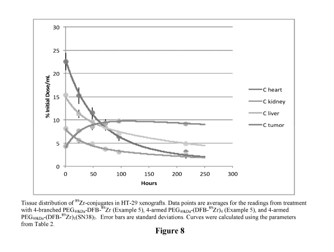
[0040] Figure 8 shows the biodistribution of various 89Zr conjugates in HT-
29 xenografts.
[0041] Figure 9 shows the biodistribution of various 89Zr conjugates in MX-
I xenografts.
[0042] Figure 10 shows the effectiveness of PEG-SN38 in tumor treatment.
9
CA 03087967 2020-07-07
WO 2019/140266
PCT/US2019/013306
[0043] Figures 11A-11C show synergy of an SN38 conjugate and separately
administered
talazoparib.
[0044] Figure 12 shows a generic hybrid drug/label conjugate theranostic.
Modes of Carrying Out the Invention
[0045] Essentially, there are two approaches to the design of protocols
that minimize the
toxic effects of combination therapies. The first approach is to ensure that a
first therapeutic
agent or drug is captured in a solid tumor to be treated by coupling the drug
to a carrier such
that the EPR effect results in substantially retaining the conjugate and
released drug in the
solid tumor, while the administered conjugate and released drug not captured
in the tumor are
more rapidly cleared from the systemic circulation, wherein the carrier is a
nanoparticle or
macromolecule each with a hydrodynamic radius of 5-50 nm preferably about 10
nm
(diameter of 10-100 nm preferably about 20 nm). Thus a substantial portion of
the
administered conjugate is retained in the tumor, as well as is the drug that
has been released
from the conjugate while the conjugate resides in the tumor. As the clearance
rate from the
systemic circulation is much greater than the clearance rate of the conjugate
and released
drug from the tumor, an effective amount of drug both in conjugated and free
form remain to
exert a cytotoxic effect on tumor cells while their concentration in the
systemic circulation
has diminished to a desired level. After two half-lives in the systemic
circulation, for
example, the level of the conjugate and free drug in circulation and in
contact with normal
tissue is reduced to 25% of the initial concentration, and this may be
sufficiently low to
ameliorate toxicity. Since the conjugate remains in the tumor to release the
agent, the agent
is able to exert its cytotoxic effect on the tumor although its concentration
in the systemic
domain is quite low, and exposure of normal tissue to the drug is therefore
also quite low.
[0046] At this point, a second drug is administered systemically and thus
the normal
tissue is exposed only to the toxic effect of the second drug while the first
drug remains out of
reach in the tumor. This minimizes the toxic effect of the combination on
normal tissue
while retaining the combined toxicities in the tumor. The second drug may be
administered
either in free form or it, too, may be administered as a conjugate with a
similar carrier or in
any other suitable form, including inclusion in delivery vehicles such as
liposomes,
nanoparticles, micelles, and the like.
[0047] In addition, a third drug that has non-overlapping toxicity with the
second drug
may be coadministered simultaneously or sequentially with said second drug.
CA 03087967 2020-07-07
WO 2019/140266
PCT/US2019/013306
[0048] Alternatively, both the first and second drug may be administered in
the form of
conjugates that are retained in the tumor by virtue of EPR either at the same
time or at
disparate times. By virtue of this retention, the major concentration of each
drug occurs in
the tumor rather than being in contact with normal tissue. Thus, the higher
dosage levels of
these drugs is experienced mainly in the tumor, and the administered
conjugates along with
released drug are rapidly cleared from the systemic circulation.
[0049] In some instances, still an additional conjugated form of an agent
may be
coadministered.
[0050] The carriers used in the method of the invention to administer at
least the first
agent in the first above-cited method and to release both the first and second
agents in the
second-noted method are carriers that are flexible in nature and have
hydrodynamic radii of
about 10 nm. Suitable macromolecule carriers include polyethylene glycols
(PEG) which
may be linear or multi-armed and have molecular weights of 10-50 kD.
Preferably, the
carriers are multi-armed PEG with molecular weights of at least 20 kD. These
characteristics
of the carriers assure that maximum advantage can be taken of the EPR effect.
Nanoparticulate carriers are also included.
[0051] Particularly useful to provide a releasable form of a conjugate of
the
chemotherapeutic agents to nanomolecular carriers are linkers that release the
agent by beta
elimination reactions such as those described in detail in the above cited
U.S. patents
8,680,315; 9,387,254; 8,754,190; 8,946,405; and 8,703,907 all incorporated
herein by
reference for their disclosures of not only the structure of useful linkers
that release the agent
by beta elimination, but also with respect to their disclosure of
nanomolecular carriers useful
in the present invention as well.
[0052] Other linkers include those cleavable by hydrolysis of esters,
carbonates, or
carbamates, by proteolysis of amides or by reduction of aromatic nitro groups
by
nitroreductase.
[0053] The subjects of the methods of the invention are typically human
subjects, but the
invention methods are also applicable in veterinary contexts including
livestock and
companion animals. The methods are also suitable for animal models useful in
the laboratory
such as rats, mice, rabbits or other model systems preparatory to designing
protocols for
human use.
11
CA 03087967 2020-07-07
WO 2019/140266
PCT/US2019/013306
[0054] With respect to the drugs useable in the combination therapy, a wide
variety of
chemotherapeutic agents is known and any combination of these may be selected
as the first
and second drug. Agents that act additively or synergistically are preferred,
for example
combination of drugs wherein each inhibits DNA repair.
[0055] Drugs that cause DNA damage, such as Topo 1 inhibitors, are
particularly useful
in treating tumors whose genome contains a mutation in a gene that normally
aids in DNA
repair. Among others, these genes include BRCA1, BRCA2, ATM which encodes
ataxia
telangiectasia mutated (ATM) kinase and ATR which encodes Rad-3 related (ATR)
kinase.
The invention includes identifying tumors that will show enhanced sensitivity
to treatment
with a Topo 1 inhibitor where the tumor-bearing subject's genome has at least
one gene that
has a mutation in BRCA1, BRCA2, ATM or ATR or other genes where mutation
prevents or
depresses the ability of the gene to enhance DNA repair. The response may be
further
enhanced by inhibiting a second enzyme involved in DNA repair, such as a PARP
inhibitor,
which then causes a synthetic lethality that is amplified because of the high
level of DNA
breaks caused by the Topo inhibitor. Thus, in using passively targeted
PEG_5N38, it is useful
to know the genetic status of the tumor, and to have an assortment choice of
inhibitors of the
DNA damage response system.
[0056] Examples of agents include:
"Signal transduction inhibitors" which interfere with or prevent signals that
cause
cancer cells to grow or divide;
"Cytotoxic agents";
"Cell cycle inhibitors" or "cell cycle control inhibitors" ¨ these interfere
with the
progress of a cell through its normal cell cycle, the life span of a cell,
from the mitosis that
gives it origin to the events following mitosis that divides it into daughter
cells;
"Checkpoint inhibitors" ¨ these interfere with the normal function of cell
cycle
checkpoints, e.g., the S/G2 checkpoint, G2/M checkpoint and Gl/S checkpoint;
"Topoisomerase Inhibitors", such as camptothecins, which interfere with
topoisomerase I or II activity, enzymes necessary for DNA replication and
transcription;
"Receptor tyrosine kinase inhibitors" ¨ these interfere with the activity of
growth
factor receptors that possess tyrosine kinase activity;
"Apoptosis inducing agents" ¨ these promote programmed cell death;
"Antimetabolites," such as gemcitabine or hydroxyurea, which closely resemble
an
essential metabolite and therefore interfere with physiological reactions
involving it;
12
CA 03087967 2020-07-07
WO 2019/140266
PCT/US2019/013306
"Telomerase inhibitors" ¨ these interfere with the activity of a telomerase,
an
enzyme that extends telomere length and extends the lifetime of the cell and
its replicative
capacity;
"Cyclin-dependent kinase inhibitors" ¨ these interfere with cyclin-dependent
kinases
that control the major steps between different phases of the cell cycle
through
phosphorylation of cell proteins such as histones, cytoskeletal proteins,
transcription factors,
tumor suppresser genes and the like;
"DNA damaging agents";
"DNA repair inhibitors";
"Anti-angiogenic agents", which interfere with the generation of new blood
vessels or
growth of existing blood vessels that occurs during tumor growth; and
"Mitochondrial poisons" which directly or indirectly disrupt mitochondrial
respiratory
chain function.
[0057] Many combinations of these for treatment of tumors are the
clinically approved.
[0058] Preferred agents that may be used in combination include DNA
damaging agents
such as carboplatin, cisplatin, cyclophosphamide, doxorubicin, daunorubicin,
epirubicin,
mitomycin C, mitoxantrone; DNA repair inhibitors including 5-fluorouracil (5-
FU) or FUDR,
gemcitabine and methotrexate; topoisomerase I inhibitors such as camptothecin,
irinotecan
and topotecan; S/G2 or G2/M checkpoint inhibitors such as bleomycin,
docetaxel,
doxorubicin, etoposi de, paclitaxel, vinblastine, vincristine, vindesine and
vinorelbine;
Gl/early S checkpoint inhibitors; G2/M checkpoint inhibitors; receptor
tyrosine kinase
inhibitors such as genistein, trastuzumab, ZD1839; cytotoxic agents; apoptosis-
inducing
agents and cell cycle control inhibitors.
[0059] Exemplary combinations are DNA damaging agents in combination with
DNA
repair inhibitors, DNA damaging agents in combination with topoisomerase I or
topoisomerase II inhibitors, topoisomerase I inhibitors in combination with
S/G2 or G2/M
checkpoint inhibitors, Gl/S checkpoint inhibitors or CDK inhibitors in
combination with
G2/M checkpoint inhibitors, receptor tyrosine kinase inhibitors in combination
with cytotoxic
agents, apoptosis-inducing agents in combination with cytotoxic agents,
apoptosis-inducing
agents in combination with cell-cycle control inhibitors, Gl/S or G2/M
checkpoint inhibitors
in combination with cytotoxic agents, topoisomerase I or II inhibitors in
combination with
DNA repair inhibitors, topoisomerase I or II inhibitors or telomerase
inhibitors in
13
CA 03087967 2020-07-07
WO 2019/140266
PCT/US2019/013306
combination with cell cycle control inhibitors, topoisomerase I inhibitors in
combination with
topoisomerase II inhibitors, and two cytotoxic agents in combination.
[0060] Exemplary specific agents include cisplatin (or carboplatin) and 5-
FU (or FUDR),
cisplatin (or carboplatin) and irinotecan, irinotecan and 5-FU (or FUDR),
vinorelbine and
cisplatin (or carboplatin), methotrexate and 5-FU (or FUDR), idarubicin and
AraC, cisplatin
(or carboplatin) and taxol, cisplatin (or carboplatin) and etoposide,
cisplatin (or carboplatin)
and topotecan, cisplatin (or carboplatin) and daunorubicin, cisplatin (or
carboplatin) and
doxorubicin, cisplatin (or carboplatin) and gemcitabine, oxaliplatin and 5-FU
(or FUDR),
gemcitabine and 5-FU (or FUDR), adriamycin and vinorelbine, taxol and
doxorubicin,
flavopiridol and doxorubicin, UCN-01 and doxorubicin, bleomycin and
trichlorperazine,
vinorelbine and edelfosine, vinorelbine and sphingosine (and sphingosine
analogues),
vinorelbine and phosphatidylserine, vinorelbine and camptothecin, cisplatin
(or carboplatin)
and sphingosine (and sphingosine analogues), sphingosine (and sphingosine
analogues) and
daunorubicin and sphingosine (and sphingosine analogues) and doxorubicin.
[0061] In one embodiment, for a first drug is a releasable conjugate of the
invention of
SN-38, a topoisomerase inhibitor, exemplary second drugs include PARP
inhibitors, mTOR
inhibitors, trabectedin, cis-platinum, oxaliplatin, fluorouracil, temozolomide
and vincristine ¨
all of which have been reported to be synergistic with SN-38.
[0062] Certain tumors are especially susceptible to treatment with PARP
inhibitors and in
these tumors, PARP inhibitors are favored as the combination drug. These are
tumors
wherein a mutation in a gene that normally is helpful in providing a protein
that aids in DNA
repair takes away this property of the gene. Such tumors are also responsive
to
topoisomerase inhibitors, such as SN38, since inhibition of topoisomerase
causes excess
DNA damage that requires DNA repair that is deficient in these tumors. These
genes include
BRCA1, BRCA2, ATM which encodes ataxia telangiectasia mutated (ATM) kinase and
ATR
which encodes Rad-3 related (ATR) kinase, among others. The invention includes
identifying tumors that have mutations in BRCA1, BRCA2, ATM or ATR or other
genes
where mutations prevent or depress the ability of the gene to enhance DNA
repair and
combining treatment with the invention 5N38 conjugates with follow up
treatment with for
example PARP inhibitors, or other inhibitors of DNA repair. Because the drug
accumulates
and remains in the tumor after it is eliminated from the rest of the system,
the toxicity of the
SN38 is confined to the tumor and the system as a whole has only to deal with
toxicity of the
PARP inhibitor.
14
CA 03087967 2020-07-07
WO 2019/140266
PCT/US2019/013306
[0063] Some of the above listed drugs to be administered as second drugs
may be
administered in combination either sequentially or simultaneously provided
their toxicities do
not overlap.
Imaging
[0064] Since the invention methods rely on the ability of the conjugates
administered for
the first agent in the first approach above and both the first and second
agents in the second
approach being subject to the EPR effect, it is important to confirm that this
is in fact the case
since tumors are heterogeneous and the particular carrier selected must be
compatible with
the pore structure of the vasculature in the solid tumor that resides in the
subject in the sense
that the EPR effect is present. Therefore, in some embodiments of the
invention method, this
is confirmed by administration either at the same time or separately of a
conjugate of a label
that is coupled non-releasably to the same carrier or a carrier with the same
characteristics as
that linked to the drug(s). While any detectable label, e.g., fluorescent
label, can be used, it is
most convenient to employ an isotope that is detectable by positron emission
tomography
(PET) scanning. The non-releasable conjugate of the isotope is then monitored
to detect
whether preferential uptake and retention by the tumor is exhibited. If so,
the method of the
invention is employed. If the tumor fails to exhibit the EPR effect with the
labeled non-
releasable conjugate, the method of the invention is contraindicated. The
isotopes thus
detectable are well known in the art as are means for coupling such isotopes
to
macromolecular carriers.
[0065] For imaging, a similar conjugate is used. As noted above, it is
advantageous to
design the imaging agent of the invention such that the diameter is
approximately are 20
nanometers and to avoid excessive flexibility. This can be accomplished by
using the multi-
armed PEG polymers in the range of 40-60kD. Although the number of arms
associated with
this polymer may range from 1-6, multi-armed PEGs of 3-5 arms, more preferably
4 arms are
focused on herein.
[0066] The value of n in formula (1) can vary from 1 to the number of arms
associated
with the polymer and it should be understood that in the compositions of the
invention the
value of n may not be the same for all of the individual imaging moieties in
the composition.
Thus, for example, for a 4 armed PEG where n is 4, or in single chain PEG
where n is 1, most
of the individual "molecules" in a given composition will contain 4 or 1 as
values of n
respectively. However, for example for 4 armed PEG, for n = 3 or n = 2,
represents an
CA 03087967 2020-07-07
WO 2019/140266
PCT/US2019/013306
average and some of the individual entities may comprise 4, some comprise 3,
some comprise
2 and some comprise 1 instances of n value
[0067] Further as to the structure of the imaging agent of Formula (1)
noted above, the
chelator represents a desferrioxamine or a multidentate chelator comprised of
a multiplicity
of hydroxypyridinones, abbreviated herein "plur-hydroxypyridinone
multidentates." A
variety of such chelators are well known in the art and are described in
detail, for example, in
Ma, M. T. et al., Dalton Trans (2015) 44:4884-4900 and by Deri, M. A., .1- Med
Chem (2014)
57:4849-4860. The description of these ligands in these documents is
specifically
incorporated herein by reference.
[0068] The covalent connector on Formula (1) may be a direct bond to the
chelator or
there may be intermediate linkers such as dipeptides or bifunctional linkers
comprising 1-20
linking atoms. Radioisotopes (I) useful in PET in the context of the present
invention are
known in the art, and particularly a subset preferred among those set forth in
Table 3 of
Smith, S. V. et al., "Production and Selection of Metal PET Radioisotopes for
Molecular
Imaging," in Radioisotopes ¨ Applications in Bio-Medical Science, Nirmal
Singh, ed.,
Chapter 10, InTech (Rijeka, Croatia), 2011, are those with suitable half-lives
such as 89Zr,
94To7 unIn, 81Rb766Ga, 640_4 62zn, 61cu or 52Fe.
[0069] To use the imaging agents of the invention as surrogates for
delivery of active
agents, i.e. drugs, the imaging agents contain carriers with the same
characteristics as those
carriers used in conjugating the drugs. These are then used to monitor the
uptake of the
conjugates by the solid tumor. This permits verification (or not) that the
corresponding
conjugates of drugs will exhibit an EPR effect.
[0070] An alternative to using separate therapeutic and imaging conjugates
employs a
hybrid conjugate of formula (2) for treatment and imaging of solid tumors
which conjugate
comprises a flexible carrier wherein the carrier is a nanoparticle or
macromolecule each with
a hydrodynamic radius of 5-50 nm which conjugate exhibits enhanced
permeability and
retention (EPR) in solid tumors so as to concentrate said conjugate in the
tumor and wherein
said carrier is releasably coupled to a therapeutic agent and also coupled to
an imaging agent.
Thus, in formula (2) as in formula (1),
PEG ( chelator I)r, (-L-D)x (2)
16
CA 03087967 2020-07-07
WO 2019/140266
PCT/US2019/013306
in some embodiments I is 89zr, 94To, 81Rb,66Ga, 64cn, 62zn, 61cu or 52Fe,
and/or the
PEG is a four armed polyethylene glycol of approximately 40 kD, and n is 1-4,
and/or the
chelator is desferrioxamine-B, and/or is a direct bond linkage.
[0071] As shown, at least one of the arms of the PEG is occupied by the
imaging agent
and at least one is occupied by the therapeutic agent. Various combinations up
to the total
number of arms of the PEG polymer are contemplated. The therapeutic agent may
be SN38
or other topoisomerase inhibitor or any other suitable agent for tumor
treatment that is
benefited by accumulation in the tumor, such as a PARP or kinase inhibitor.
[0072] The imaging agents of the invention are also useful to identify
subjects that harbor
tumors or other tissue masses that are susceptible to treatment with
therapeutic agents that
exhibit the EPR effect. Thus, the imaging agent may be administered to a
subject and
monitored to determine whether the tumor, for example, will, in fact,
preferentially take up
and retain entities of similar size.
[0073] In this application, "a", "an", and the like are intended to mean
one or more than
one unless it is clear from the context that some other meaning is intended.
In addition, the
terms "chemotherapeutic agent", "agent", and "drug" are used interchangeably.
Where
specific numerical characteristics are set forth, the number cited will
typically have error bars
of plus-or-minus 10%, preferably plus-or-minus 5% and more preferably plus-or-
minus 1%.
Thus, a range of 10-50 nm could include a range of 9-55 nm. "Hydrodynamic
radius" means
the apparent Stokes radius ¨ the radius of a hard sphere that diffuses through
solution at the
same rate as the molecule in question as measured, for example, by gel
permeation
chromatography.
[0074] The subjects of the invention are typically human, but also include
non-human
animals such as laboratory models and veterinary subjects.
[0075] All documents cited herein are hereby incorporated herein by
reference.
[0076] The following examples are offered to illustrate but not to limit
the invention.
Example 1
Administration of Conjugated SN-38
[0077] SN-38 is the topoisomerase I inhibitor that is the active drug
released from the
prodrug, irinotecan. Conjugates of SN-38 are described in WO 2015/051307. Two
different
conjugates of SN-38 were prepared: PLX038 and PLX038A. These conjugates couple
the
17
CA 03087967 2020-07-07
WO 2019/140266 PCT/US2019/013306
drug releasably to a four-armed PEG of 40 kD through a linker that effects
release by
(3-elimination. The structure of PLX038 and PLX038A is shown below wherein
"Mod" is
¨CN in PLX038, and methyl sulfonyl in PLX038A.
kicx:1õ
0 0
PEG40koa ¨C¨N =-=õ,
lkst' SN38
S.
L:rij
= CONEt,. 4
[0078] Six rats bearing HT29 tumor xenografts were injected with ¨200 mg/kg
of
PLX038 and the concentration in plasma and tumor of the conjugate and released
drug as
well as its glucuronide (SN-38G) were followed by HPLC with fluorescence
monitoring As
shown in Figure 1, the half-life of PLX038 in the systemic circulation is
about 50 hours. The
conjugate and the free drug as well as SN-38G show similar half-lives.
[0079] As shown in Figure 2, the efficacy of a non-toxic dose of 20 nmol/kg
of SN-38 in
the form of PLX038 exceeds that of a toxic gastrointestinal dose of irinotecan
control.
[0080] This is explained by the results shown in Figures 3A and 3B which
are graphs of
the levels of the conjugate PLX038 and of SN-38 that has been released from
the conjugate in
the tumor at various dosage levels. As seen in Figure 3A, at an administered
dose of
200 mg/kg the level of PLX038 in the tumor (a left bar) is roughly 8 nmol/g
while the
concentration in the plasma (shown as the right bar) is barely detectable.
Similarly, in
Figure 3B with respect to the released SN-38, at the same dosage, the left bar
shows the
concentration in the tumor as about 80 pm/g while, again, the right bar shows
that in the
circulation it is barely detectable. Indeed, as shown, at the lower dosages,
the conjugate and
free drug are not detected in the plasma, while the tumor shows significant
concentrations of
these moieties.
Example 2
Suggested Human Protocol
[0081] A dosing schedule in humans for a combination of PLX038 and a second
drug
(e.g., a PARP inhibitor) administered systemically is proposed wherein PLX038
is
administered on day 1 whereby the conjugate accumulates in the tumor and
releases the free
drug. The conjugate and the free drug are concomitantly cleared from the
system. After two
18
CA 03087967 2020-07-07
WO 2019/140266
PCT/US2019/013306
half-lives of systemic clearance or 10 days, systemic PLX038 is reduced to
¨25% of its
original concentration, which lies below its minimal effective and toxic
levels. At this time
the second drug, which is synergistic with SN-38 is administered orally for 20
days.
[0082] As shown in Figure 4, the EPR effect concentrates PLX038 in the
tumor (dotted
line), while the systemic PLX038 (solid line) is sufficiently low that any
toxic effect is only
to a second drug, which is administered as shown at 10 days. At that time, the
concentration
of the conjugate in the tumor is still above the minimum effective level but
below the toxic
level.
Example 3
Design of a Mouse Model
[0083] Because most xenograft tumor models use mice as hosts, it is
desirable to adapt
the protocols of the present invention to testing in mice. Adaptation is
needed because the
half-life of the PLX038 conjugate in the mouse is only about 24 hours, whereas
in the rat it is
about 48 hours and in humans about 6 hours. Because the more rapid elimination
of PLX038
in mice occurs before substantial amounts of the SN-38 are released, a
different conjugate of
SN-38, PLX038A that has a higher cleavage rate, is used in murine experiments.
[0084] Linker cleavage is species independent. While 32% of PLX038 is
converted to
SN-38 over one half-life of the conjugate in humans, only 12% is converted in
the rat and 6%
in the mouse. For PLX038A, the cleavage half-life is 70 hours and 26%
conversion to SN-38
over one half-life of the conjugate in the mouse occurs. This conjugate also
can be
administered intraperitoneally (IP) in mice with 100% bioavailability.
[0085] However, in mice PLX038A still has a short ti/2 of renal elimination
so a single
dose may not effect high tumor accumulation and longer exposure may be
necessary to
achieve this. A multi-dose schedule for PLX038A in the mouse that simulates a
single
effective dose of the conjugate that gives high tumor accumulation in the rat
is therefore used.
[0086] For comparison, in the rat xenograft model for colon cancer (HT-29),
a single
200 mg/kg of PLX038 produced 61% inhibition of tumor growth with no
gastrointestinal
(GI) toxicity while irinotecan control that showed near-equal tumor inhibition
showed
significant GI toxicity. There was high accumulation of PLX038 and SN-38 in
tumors
14 days post-dosing when the serum levels were undetectable. A dosing schedule
for
PLX038A in the mouse that would simulate the pharmacokinetics (PK) of PLX038
in the rat
is shown in Figure 5. Three daily decreasing doses of 152, 60 and 54 mmol/kg
effectively
19
CA 03087967 2020-07-07
WO 2019/140266
PCT/US2019/013306
simulate the rat PK profile of released SN-38 from PLX038. The "effective"
half-life of
SN-38 in the schedule is over 2 days, whereas SN-38 from irinotecan in the
mouse
is ¨2 hours. The data supporting Figure 5 are shown in Table 1.
Table 1
mouse dose schedule conj dose, mg SN-38 dose, mg SN-38 dose, nmol
dose 1 1.7 0.060 152
dose 2 0.9 0.032 80
dose 3 0.6 0.021 54
total 3.2 0.1 285.4
conj dose, SN-38 dose, SN-38 dose, AUC,
mg/kg mg/kg nmol ttM-h time over 8 nM
rat dose 200 7 3.2 11 ¨7 days
mouse total dose 128 4.5 0.285 11 ¨5 days
Example 4
Murine Testing
[0087] The ability of HT29 xenografts to accumulate conjugate using the EPR
effect is
tested by injecting mice with one dose IP of 50 nmol of 40 kD four-armed PEG
fluorescein
per 100 g (15 nmol/mouse) to obtain about 10 p.M in serum. Blood and tumor are
sampled at
various times (at 6, 24, 48 and 96 hours) and the level of fluorescein
measured. (The tumor is
excised and digested with sodium hydroxide for measurement.)
[0088] PLX038A is tested for tumor growth inhibition in a nude mouse HT29
tumor
xenograft using the three-dose schedule developed in Example 3.
[0089] The nude mouse model with HT29 xenograft is treated with the three-
dose
schedule of PLX038A developed in Example 3 and at 14 days the mice were
treated daily
with oral administration of a PARP inhibitor.
[0090] A conjugate of PARP inhibitor analogous to PLX038A is administered
daily to
nude mice bearing HT29 tumors and tested vs. daily administration of free
inhibitor.
[0091] Combinations of conjugates PLX038A and the relevant conjugate of
PARP
inhibitor are also tested concomitantly in this model.
CA 03087967 2020-07-07
WO 2019/140266
PCT/US2019/013306
Example 5
Synthesis of PEG401n.-PET isoto e es
[0092] Synthesis of PEG-desferrioxamine Conjugates
[0093] 4-branched PEG4okn coupled to DFB:
014.
rytaktif,A5cti r$
f1/4t.
0.. 13kit0
H.?0 = 0;C$iL,C4-i.AN OH 0 0'
0(OH2H:03.9 OH 0
,
mooq.c"04,4.,
1,1
= .
11..A., 0 14 tal zi
j,
t9
Hx-.ocitch, CH it
,
H
QCHCHO} "-CH
A410- (01:41.:01" 0i4
.441304C H.80 H".
[0094] A solution of 4-branched 40-kDa PEG-amine (GL4-400PA, NOF; 150 mg,
3,75
umol) and p-isothiocyanatobenzyl-desferrioxamine B (Macrocyclics; 4 mg, 5.3
umol) in
2 mL of DMSO was kept 16 hat ambient temperature, then dialyzed against water
(SpectraPor 2 membrane, 12-14 kDa cutoff) to remove unconjugated materials.
The solution
was evaporated to dryness, and the residue was dissolved in 2 mL of THF and
added slowly
with stirring to 50 mL of MTBE. The precipitated conjugate was collected and
dried to
provide the product (140 mg). A 2.4-mg sample was dissolved in 58 uL of water
to give a 1
mM solution. A 20-uL aliquot was added to 100 uL of 1 mM Fe(I11) perchlorate,
giving a
solution showing OD42snm = 0.459. Based on an extinction coefficiant of 2,300
M.1cm.1,
this indicated a DFB concentration on 1.1 mM, in good agreement with expected.
[0095] (PEG)40 coupled to [DFB=Desferrioxamine B] (DFB): PEG4okna-(DFB)4
was
prepared by reaction of PEG4.01,DANH2)4 with p-isothiocyanatobenzyl-DFB (Perk,
L. R., et al.
Eur. J. Nucl. Med. Mol. I. (2010) 37:250-259; Fischer, G., et al., Molecules
(2013)
21
CA 03087967 2020-07-07
WO 2019/140266
PCT/US2019/013306
18:6469-6490; and van de Watering, F. C., et al. Thorned. Res. Int. (2014)
2014:203601)
(macrocyclics) as follows.
4-armed PEG4okDa coupled to DFB (PEa4okDa-(DFB)4}:
DFS
C-rIi20{,ClizCHe0)õ(Ct-10.,õ,COA44
r4C;SIACI-I,A:HAICIV0,-00,0F513
[0096] A solution of 40-kDa 4-armed PEG-tetra(succinimidyl ester) (JenKem
Technologies; 100 mg, 10 umol succinimidyl ester), deferoxamine mesylate
(Sigma; 10 mg,
15 umol), N,N-diisopropylethylamine (35 uL, 200 umol), and HATU (1-[Bis(
dimethylamino
)methylene]-1H-1,2,3-triazolo [ 4,5-b ]pyridinium 3-oxide hexafluorophosphate)
(7 mg, 18
umol) in 2 mL of DMF was kept 16 h at ambient temperature, then dialyzed
against water
and methanol (SpectraPor 2 membrane, 12-14 kDa cutoff) to remove unconjugated
materials.
The solution was evaporated to dryness, and the residue was dissolved in 2 mL
of THF and
added slowly with stirring to 50 mL of MTBE to give the conjugate (84 mg). A
5.0 mg
aliquot was dissolved in 500 uL of water to give a solution 0.21 mM solution
of conjugate.
Assay for DFB content by addition to 1 mM Fe(III) perchlorate as described
above gave 0.84
1..LM DFB, indicating 4 DFB per conjugate.
Alternative Method
[0097] An alternative DFB reagent for conjugation, is prepared by acylation
of DFB with
N3-(CH2)11C0-HSE; the N3-(CH2).00-DFB is reacted with cyclooctyne-derivatized-
PEG4oka=TH2)4 by SPAAC.
0
rN
HO 0 Na 0
DFB)L-----.."=-". N3
NH õ HO-N
0 µ11
0
CIE-\IONI 0
PEG40kDa-MFC0
___________________ I.- NH HO-N
C) (1? o-OH
N-PEGaloa
NH N 0
22
CA 03087967 2020-07-07
WO 2019/140266 PCT/US2019/013306
Coupling to PET Isotopes:
[0098] Coupling to PET isotopes was performed by treatment of the PEGylated-
DFB
with 89Zr oxalate followed by purification using size-exclusion chromatography
(Perk, L. R.,
supra; and van de Watering, F. C., supra).
[0099] PEG-40kDa-(DFB)4 + 89Zr-oxalate 4 PEG4okDr(DFB-89Z04
101001 PEG4okDa-(BzI12504 is prepared by reacting the 125I- azide shown
below with a
cyclooctyne-derivatized-PEG4okr3(NH2)4 (prepared from mFCC)-HSE +
PEa4akDa(NH2)4),
which results in a clean high yield strain-promoted azide-alkyne cycloaddition
(SPAAC)
reaction. Preparation and radioiodination of the [12511 iodobenzoyl-PEG-azide
is shown
below for stable iodination of macromolecules using SPAAC.
Bu3Sn 1251
H Na1251 1101 H
N3 _____________________________________
Chlorami N-0--N3
/3
0 0
PBS/Na0H/Et0H
0
1251
PEG20kDa-(MFC0)4 F
(101 I-1 / , N¨PEG20kDa
/
/3
0
4
23
CA 03087967 2020-07-07
WO 2019/140266
PCT/US2019/013306
Example 6
Hybrid SN38/DFB Conjugates
[0101] 4-armed PEatokr.11iled to lx stable-DFB and 3x releasable-SN-38
(PEatokDA:
(sDFB)1(rSN38)31
A. Preparation of Hybrid SN38/DFB Conjugates.
C-Plat,P11*Kz0.-k4,2E,=C4C.-tyaCCI-604.,%.11'34n4.4.1.0-webx.r.lyne!, C-
CCHANS=CitS%::CW!,,NH-CO-ttac.--1c4,-5,41,2&
Et:Ikipplizit,ow-t,?,-Nit-rX.,:sirOf131,
S.
II
PELT-VH:4 ,cycLvc...amt,
AZNaz
,
)1. !
g -kr a
,
w=-= J3=
r _ õ
aziab-Lõ -8fri38
a..-162-L,DFE
Fks
+ Rt¨t-,
CYCI003'ne azge Mao.*
[0102] N-((6-azidohexyloxy)carbonyl) desferrioxamine B: A solution of 6-
azidohexyl
succinimidyl carbonate (35 mg, 120 umol) in 2 mL of acetonitrile was added to
a solution of
deferoxamine mesylate (65 mg, 100 umol) in 2 mL of 0.5 M NaHCO3. After
stirring for 16 h,
the resulting white precipitate was collected, washed with water and
acetonitrile, then dried
under vacuum to yield the product (45 mg; 62%). MS: [M+H1+ = 730.46 (calc. for
C32H60N9010 = 730.44).
[0103] Azido-linker-SN38 having a cyano modulator: prepared as described in
PCT
Publication W02015/051307.
[0104] PEthokDa-(DBC0)4: A solution of 40-kDa 4-armed PEG-tetraamine
(PTE400-PA,
NOF; 10 umol amines), dibenzocyclooctyne-N-hydroxysuccinimidyl ester (DBCO-
NHS,
ClickChemistryTools; 5 mg, 12 umol), and N,N-diisopropylethylamine (2 uL, 12
umol) in 1
mL of acetonitrile was stirred for 1 h at ambient temperature. The mixture was
evaporated to
dryness, then redissolved in 1 mL of THF and precipitated by addition of 10 mL
of MTBE.
The resulting solid was collected, washed with MTBE, and dried to provide the
product.
24
CA 03087967 2020-07-07
WO 2019/140266
PCT/US2019/013306
[0105] PEG40kDa-(sDFB)1(rSN38)3: A 1:3 mixture of stable-linker-DFB and
releasable-
linker-5N38 was coupled to PEG40kDa(DBC0)4 to yield a mixture that was
predominantly
PEG4okpa(sDFB)1(rSN38)3 and PEG4okna(rSN38)4. by HPLC analysis. These were
separated
by preparative HPLC using a Phenomenex 300A 5 um Jupiter C18 column, 21.2x150
mm,
with a 30-60% gradient of acetonitrile in water+ 0.1% TFA at 15 mL/min.
Determination of
SN38 content by UV at 360 nm (e360 = 22,400 M-lcm-1) and DFB content by assay
with
Fe(III) perchlorate as described above gave a 2.7:1 ratio of SN-38 to DFB.
B. Preparation of Additional Hybrid Drug/DFB Conjugates.
i. Alternate Preparation of (5HCO)3-PEa4o
Fmoc-OSu
104 0 - ) osu [10 HNra.-
PEG40kDa-(NH2)4 (H2N)3-PEG -NHFmoc __
jj'
MeCN DIPEA, MeCN 0 N ¨PEG -
NHFmoc DMF
H 3
H H
9*., 410 N TN -DFB
H H
1 0 N
0 N N
40 .DFB
CAN PEG -NH2
[10
0 -1(N ¨PEG -IV1 N
H 3 H_ 3 I-1 H
[0106] Step 1. (H2N)3-PEG4okDa-NifFmoc. A 25 mM solution of Fmoc-OSu (0.48
mL, 12
[Imo') in MeCN was added dropwise to a vigorously stirred solution of PEG4okpa-
(NH2)4 (406
mg, 10.0 jimol, 5 mM final concentration) in 3.5 mL of MeCN. The reaction
mixture was
stirred at ambient temperature, and after 5 min, the mixture consisted of 44%
title compound
as judged by C18 HPLC (ELSD). The reaction solution was concentrated to ¨1 mL
by rotary
evaporation. The concentrate was diluted to 6 mL with H20 (0.1% TFA) then
purified by
preparative C18 HPLC, two injections eluting with a linear gradient (35%-60%)
of MeCN in
H20 (0.1% TFA). Fractions from the first eluting Fmoc-containing peak were
analyzed by
C18 HPLC, and clean, product-containing fractions were combined and
concentrated to
dryness. After removing volatiles under high vacuum for 30 min, the residue
was dissolved in
minimal THF (-1 mL) and added dropwise to 40 mL of 0 C MTBE in a tared 50 mL
Falcon
tube. The suspension was vortexed, kept on ice for 15 min, centrifuged (3500x
g, 1 min), and
decanted. The precipitate was washed with MTBE (2x 40 mL), isolated as above,
and dried
under high vacuum to provide the title compound (96 mg, 2.2 pmol given 3 TFAs,
22%
yield) as a white powder. C18 HPLC, purity was determined by ELSD: 99.6% (RV =
9.39
mL).
CA 03087967 2020-07-07
WO 2019/140266
PCT/US2019/013306
[0107] Step 2. (Cyclooct-4-yn-/-y/oxycarbony/-NH)3-PEG4okDa-NHFmoc. A 0.15
M
solution of 0-(cyclooct-4-yn-1-y1)-0'-succinimidyl carbonate (63 !IL, 9.5
limo') in MeCN
was added dropwise to a stirred solution of (H2N)3-PEa4okDa-NHFmoc (96 mg, 2.2
gmol, 50
mg/mL final concentration; 6.7 w-nol NH2) and DIPEA (2.8 L, 16 [mop in 1.9 mL
of
MeCN. The reaction mixture was stirred at ambient temperature and monitored by
C18
HPLC. The starting material was converted to a single product peak via two
slower eluting
intermediate peaks. After 2 h, the reaction mixture was concentrated to -0.3
mL by rotary
evaporation. The concentrate was diluted with 1 mL of THF, and the solution
was added
dropwise to 40 mL of ice-cold MTBE in a tared 50 mL Falcon tube. The mixture
was kept on
ice for 15 min then centrifuged (3500x g, 1 min) and decanted. The wet solid
was washed
with ice-cold MTBE (2x 40 mL), centrifuged (3500x g, 1 min) and decanted.
Residual
volatiles were removed under high vacuum for 20 min to provide the title
compound (40 mg,
0.93 66% yield) as a white powder. To prevent decomposition, the solid was
immediately diluted with 0.78 mL of amine-free DMF. C18 HPLC, purity was
determined
by ELSD: 93.5% (RV = 9.96 mL) and a 6.5% impurity (RV = 9.78 mL).
101081 Step 3. (Cyclooct-4-yn-/-y/oxycarbony/-N11)3-PEG4okna-NH2. 4-
Methylpiperidine
(39 [IL, 5% v/v final concentration) was added to a 100 mg/mL solution of
(cyclooct-4-yn-1-
yloxycarbonyl-NH)3-PEatokDa-NHFmoc (0.78 mL, 78 mg, 1.8 iimol) in DMF. The
reaction
tube was kept at ambient temperature and monitored by C18 HPLC. After 30 min,
PEG was
precipitated by dropwise addition of the reaction solution to 40 mL of ice-
cold MTBE in a
tared 50 mL Falcon tube. The mixture was kept on ice for 15 min then
centrifuged (3500x g,
1 min) and decanted. The wet solid was washed with MTBE (2x 40 mL),
centrifuged (3500x
g, 1 min) and decanted. Residual volatiles were removed under high vacuum for
15 min to
provide the title compound (68 mg, 1.6 [imol, 89% yield) as a white powder. To
prevent
decomposition, the solid was immediately diluted with 0.68 mL of amine-free
DMF. C18
HPLC, purity was determined by ELSD: 87.0% (RV = 9.59 mL) and a 13.0% impurity
(RV =
9.43 mL).
[0109] Step 4. (Cyclooct-4-yn- 1 -yloxycarbonyl-N11)3-PEG4okDa-NHCSNH-
pheny1-4-
(NHCSNHDFB). P-isothiocyanatobenzyl-desferrioxamine B (1.8 mg, 2.4 iffnol;
Macrocyclics) was added to a 50 mg/mL solution of (cyclooct-4-yn-1-
yloxycarbonyl-NH)3-
PEatokpa-NH2 (1.36 mL, 1.6 ['mop in DMF. The reaction mixture was placed in a
37 C
water bath and monitored by C18 HPLC. After 4 h, PEG was precipitated by
dropwise
addition of the reaction solution to 40 mL of ice-cold MTBE in a tared 50 mL
Falcon tube.
26
CA 03087967 2020-07-07
WO 2019/140266
PCT/US2019/013306
The mixture was kept on ice for 15 min then centrifuged (3500x g, 2 min) and
decanted. The
wet solid was washed with MTBE (2x 40 mL), centrifuged (3500x g, 2 min) and
decanted.
Residual volatiles were removed under high vacuum for 15 min to provide the
title compound
(67 mg, 1.5 [tmol, 94% yield) as a white solid. To prevent decomposition, the
solid was
immediately diluted to 2.68 mL total volume with MeCN (2.61 mL MeCN, 25
mg/mL).
Insoluble DFB-NCS was pelleted (3500x g, 2 min), and the product-containing
MeCN
supernatant was removed. C18 HPLC, purity was determined by ELSD: 80.3% (RV =
9.59
mL) and a 19.7% shoulder (RV = 9.43 mL).
ii. Preparation of (Drug)3-PEG4okD3-DFB
a. Drug = SN38
H H
H H N _____________________________________ rit10, 9 1 'DFB
Wel 01,0 e1/411 ¨PEG rA1
N
H3 10 `-'
CONEt _ 3
S38-
101101 (SN38-L)3-PEG4okDa-NHCSNH-pheny1-4-(NHCSNH-DFB). Stable azido-SN38
(4.0 mg 5.2 [tmol, 4 mM final concentration; Santi et al., J. Med. Chem. 57:
2303-14 (2014))
was added to a 25 mg/mL solution of (cyclooct-4-yn-1-yloxycarbonyl-NH)3-
PEG4okDa-
NHCSNH-phenyl-4-(NHCSNHDFB) (1.3 mL, 0.75 iimol PEG, 2.3 mol cyclooctyne, 1.8
mM cyclooctyne final concentration) in MeCN. The reaction was placed in a 37
C water
bath and monitored by C18 HPLC. After 44 h, the reaction solution was dialyzed
against
Me0H (12-14 k MWCO). The dialysate was concentrated to dryness, and residual
volatiles
were removed under high vacuum to provide the title compound (24 mg, 0.52
!Imo', 69%
yield by mass) as white film that contained 1.4 limol of SN38 as determined by
A383 and 0.50
[tmol of DFB as determined by A490 of Fe3+-DFB. The SN38:DFB ratio was found
to be
2.8:1 using SN38 8383 = 29,100 M-lcm-1 and Fe3+-DFB 8490 = 3,000 M-lcm-1. C18
HPLC,
purity was determined by ELSD: 83.0% (RV = 9.67 mL) and a 14.6% shoulder (RV =
9.52
mL).
b. Drug = Rucaparib ¨ a PARP inhibitor
F
NH H H
N N
11 11 o Nv 'DFB
1003'NI-PEG -N1N 'DFB 0 0 0"%1 ¨PEG
H3 H H RucapadV _ 3
101111 (Rucaparib-L)3-PEG4okDa-NHCSNH-phenyl-4-(VHCSNH-DFB). A 10 mM
solution of stable azido-rucaparib (0.11 mL, 1.1 mol, 1.8 mM final
concentration; prepared
27
CA 03087967 2020-07-07
WO 2019/140266
PCT/US2019/013306
by reacting rucaparib with 6-azidohexyl succinimidyl carbonate according to
the procedures
of Santi et al., Proc. Natl. Acad. Sci. 109: 6211-16 (2012)) was added to a 25
mg/mL solution
of (cyclooct-4-yn-l-yloxycarbonyl-NH)3-PEatokpa-NHCSNH-pheny1-4-(NHCSNHDFB)
(0.50 mL, 0.29 umol PEG, 0.86 umol cyclooctyne, 1.4 mM cyclooctyne final
concentration)
in MeCN. The reaction was placed in a 37 C water bath and monitored by C18
HPLC. After
68 h, the reaction solution contained a -35:65 mixture of unmodified:PEGylated
drug-linker.
A series of the individual species of (drug)n-PEG-DFB was not observed. The
reaction
solution was concentrated by SpeedVac to 0.1 mL, diluted to 1.0 mL with H20,
and loaded
onto a PD-Midi column. Elution with H20 yielded a cloudy fraction of excluded
material that
contained both unmodified and PEGylated drug-linker. The mixture was then
dialyzed
against Me0H (12-14 k MWCO). The dialysate was concentrated to dryness, and
residual
volatiles were removed under high vacuum to provide the title compound (8.7
mg, 0.19 umol,
66% yield) as white film that contained 0.51 [unol of rucaparib as determined
by A355 and
0.19 umol of DFB as determined by A490 of Fe3+-DFB. The rucaparib:DFB ratio
was found
to be 2.7:1 using rucaparib 8355 = 13,260 M-lcm-1 (125SF68) and Fe3+-DFB 8490=
3,000 M-
1cm-1. C18 HPLC, purity was determined by ELSD: 78.5% (RV = 9.41 mL) and a
21.5%
shoulder (RV = 9.27 mL).
c. Drug = VX-970 - an ATR kinase inhibitor
N
\ 01,,,OmN3 - H H
v...9700,17t. -0 ri_pEG_H
H H N:N I
.3,m13_ is NIN-D,B )_so,
PEG 7 N _3
[0112] (VX970-L)3-PEG4okDa-NHCSNH-pheny1-4-(NHCSNH-DFB). As described above
for rucaparib, stable azido-VX970 (0.11 mL, 1.1 umol, 1.8 mM final
concentration; prepared
by reacting VX970 with 6-azidohexyl succinimidyl carbonate according to the
procedures of
Santi et al., Proc. Natl. Acad. Sci. 109: 6211-16 (2012)) was treated with a
25 mg/mL
solution of (cyclooct-4-yn-1-yloxycarbonyl-NH)3-PEGtokpa-NHCSNH-pheny1-4-
(NHCSNHDFB) (0.50 mL, 0.29 umol PEG, 0.86 umol cyclooctyne, 1.4 mM cyclooctyne
final concentration) in MeCN to provide the title compound (10 mg, 0.22 umol,
76% yield by
mass) as white film that contained 0.55 umol of VX970 as determined by A383
and 0.24 limo'
of DFB as determined by A490 of Fe3+-DFB. The VX970:DFB ratio was found to be
2.3:1
using VX970 8383 = 17,200 M-lcm-1 (127BH52) and Fe3+-DFB 8499 = 3,000 M-lcm-1.
C18
HPLC, purity was determined by ELSD: 59.2% (RV = 9.98 mL) and a 38.4% shoulder
(RV =
9.73 mL).
28
CA 03087967 2020-07-07
WO 2019/140266
PCT/US2019/013306
d. Drug = BMN673 ¨ a PARP inhibitor
H H
3 iN.DFB
N N F
'13FB N
0 010 Ã..t*N PEG1
10-0,NI-pEG-NIN 11P BMN-873
H H H 3
[0113] (BMN673-L)3-PEG4okDa-NHCSNH-pheny1-4-(NHCSNH43FB). As described
above for rucaparib, stable azido-BMN673 (0.11 mL, 1.1 gmol, 1.8 mM final
concentration;
prepared by reacting BMN673 with 6-azidohexyl succinimidyl carbonate according
to the
procedures of Santi et al., Proc. Natl. Acad. Sci. 109: 6211-16 (2012)) was
treated with a 25
mg/mL solution of (cyclooct-4-yn-1-yloxycarbonyl-NH)3-PEG4okDa-NHCSNH-phenyl-4-
(NHCSNHDFB) (0.50 mL, 0.29 mmol PEG, 0.86 jimol cyclooctyne, 1.4 mM
cyclooctyne
final concentration) in MeCN to provide the title compound (12 mg, 0.26 gmol,
91% yield by
mass) as white film that contained 0.65 iimol of BMN673 as determined by A310
and 0.20
[tmol of DFB as determined by A490 of Fe3+-DFB. The BMN673:DFB ratio was found
to be
3.3:1 using B1V1N673 8310 = 9872 M-lcm-1 (1255F39) and Fe3+-DFB 8490 = 3,000 M-
lcm-1.
C18 HPLC, purity was determined by ELSD: 69.7% (RV = 9.47 mL) and a 30.3%
shoulder
(RV = 9.32 mL).
C. Coupling to PET Isotopes
[0114] The hybrid SN38/DFB and alternative hybrid drug/DFB conjugates are
coupled to
89Zr by the methods set forth in Example 5.
Example 7
Use of PET to Detect EPR in Animal Studies
[0115] Mice bearing HT-29 human xenografts and normal mice are treated with
conjugates PEG-PET isotopes which are similar in size and shape to the drug
conjugates of
Examples 1-4. PET-imaging to measure accumulation of labeling intensity of the
tumor at
t=0, 12, 24, 48 and 96 hr is conducted in comparison with results of a similar
experiment
using PEG4o1Da-fluorescein in tumor-bearing mice (Singh, Y., supra). Sera are
counted at
these time points to determine the t112 of elimination of the PEG-isotope (the
elimination t112
Of PEGtokDa in mice is usually ¨24 hr), as well as total body radioactivity
measurements.
[0116] HT-29 Tumor bearing mice and normal control mice are treated with
¨200 uCi/mouse, and PET-imaging is performed at varying times to determine the
amount
and rates of accumulation. A signal is observable at ¨1 uCi/cc so the tumor is
easily
29
CA 03087967 2020-07-07
WO 2019/140266
PCT/US2019/013306
visualized as long as the background tissue does not accumulate the tracer. In
the same
experiment, the loss of isotope is followed as the reagent is cleared from the
body. Rates of
a) tumor accumulation of the PEG-isotope (quantitative PET imaging), b)
vascular
elimination (serum radioactivity), c) systemic elimination (whole body
radioactivity) and
d) tumor elimination (quantitative PET imaging) are thus determined.
[0117] At a time when tumor accumulation is complete, tumor-bearing mice
are treated
with varying amounts of the PEatotorisotope to determine the maximal amount of
nanoparticle that can accumulate.
[0118] Thus, in this example, PET scanning is used to simulate the behavior
of an agent
coupled to the same or similar carrier to evaluate the parameters appropriate
for the drug
administration protocol.
Example 8
PET imaging/Biodistribution of PEG4okija-DFB89Zr.
[0119] Mice bearing xenografts (n=5) were injected with ¨300 p.Ci (8.4
nmol) of
PEG4okDa-DFB-89Zr and microPET/CT images were obtained at 24 h (n=2) and 48 h
(n=2).
The %ID/g uptake (uptake of PEa4okDa-DFB-89Zr) in tumors was 15 and 20% at 24-
and 48 h,
respectively, while organs other than liver had <3% uptake. MicroPET/CT
studies showed
high accumulation of 89Zr-DFB-PEG40 in MX-1 tumors as early as 24h while
accumulation in
healthy tissue was nearly background. The imaging data corroborated the
increased
accumulation in tumor from 24 to 48h. However, there was heterogeneous uptake
in the
tumor, possibly suggesting necrosis of this rapidly growing tumor.
[0120] The experiment was repeated the slower growing HT-29 tumor. Given
the lower
tumor to blood ratios and limited clearance at early time points in MX-1
tumors (1.1 0.2 [24
h] - 1.2 0.1 [48 h]) the uptake in the HT-29 tumors was studied at 72 h and
120 h. Mice (n=
8) were injected with ¨160 p.Ci (8.4 nmol) of89Zr- DEB-PEG40 and microPET/CT
images
were obtained at 72- and 120h. Mice were euthanized at 72- and 120h for ex-
vivo
biodistribution studies. HT-29 tumors were clearly visualized on the
microPET/CT at 72h
and 120h (Fig. 6A), and biodistribution studies revealed high uptake of 20.6
2.4 and 14.4
4.5 %ID/g at 72 and 120h (Fig. 6B) with tumor/blood 2.8 + 0.4 and 5.1 1.3 at
72 and
120h, respectively (Fig. 6C). Figure 6D is an MIP image of PEG-SN-38)3-DFB89Zr
in a
single flank tumor-bearing mouse. Figure 6E shows biodistribution of PEG-(SN-
38)3-DFB-
89Zr (black) vs PEG-DFB-89Zr (grey) at 72h.
CA 03087967 2020-07-07
WO 2019/140266
PCT/US2019/013306
[0121] In an additional study, the PEG4okDa-(DFB-89Zr)4 of Example 5 was
injected into
mice bearing HT29 tumors. Five mice were used in the study and each was
injected with
250-290 pCi of the conjugate in 100 Ill saline. Two of the mice were imaged at
one hour post
injection. After 24 hours, two mice, (one that had been imaged at one hour and
an additional
mouse) were imaged and then sacrificed to perform distribution studies. At 48
hours, two
mice were imaged (one of the mice that was imaged at one hour and one
additional mouse)
and these were also sacrificed along with the remaining mouse and a
distribution study
performed.
[0122] The results of these studies are shown in Figures 7A-7C. Shown in
Figure 7A, the
label was present in the tumor at all of the times measured. As shown in
Figure 7B, the % of
the injected dose (ID) per gram of individual organs was significant in most
organs, although
bone, spleen and tumor had the highest levels. As shown in Figure 7C when
computed as the
percentage of the injected dose per organ, rather than as per gram of organ,
accumulation in
the tumor was dramatically higher, especially at 48 hours, as compared to
other organs. Only
liver showed a significant accumulation which dropped over the time period of
24-48 hours.
Thus, the imaging agent confirms that the conjugate is selectively accumulated
in the tumor
as compared to other organs.
Example 9
Additional Distribution Studies
[0123] The experiments of Example 8 were repeated using 4-branched PEG40kDa-
DFB-
89Zr (Example 5), 4-armed PEG40kDa-(DFB-89Zr)4 (Example 5), and 4-armed
PEG40kDa-(DFB-
89Zr)1(5N38)3 (Example 6) in both MX-1 and HT-29 xenografts. PET imaging was
used to
measure accumulation of89Zr in tumor, heart, liver, and kidney at 1, 24, 48,
72, 96, and 216 h
post-dose. The resulting data (expressed as decay-corrected percent of the
total dose) were
analyzed using a membrane-limited tissue distribution model according to the
methods of Li
et al., Intl. J. Nanomedicine (2012) 7: 1345-56. A compartment for the
remaining tissues was
included in order to match measured blood levels in the absence of more
specific tissue
analyses. Blood data were fit using a total clearance equal to the sum of the
diffusion
coefficients from blood into the organs (k, Table 2) and the elimination rate
constant
calculated from a plasma half-life of 20 hours.
[0124] Within experimental error, all three compounds showed the same
tissue
distribution in a specific tumor xenograft. Figure 8 shows the distribution of
89Zr in HT-29
31
CA 03087967 2020-07-07
WO 2019/140266 PCT/US2019/013306
xenografts, and Figure 9 shows the distribution of 89Zr in MX-1 xenografts.
Model
parameters are given in Table 2, where R = tissue-blood partition coefficient,
k = diffusion
coefficient, V = tissue volume, and VVF = the vascular fraction of the tissue.
Table 2
Parameters for Membrane-Limited Tissue Distribution Model
HT-29 R k (WI) V (mL) VVF k/RV
Circulation 2.8 1
Heart 0.7 0.0015 0.15 0.23 0.0143
Kidney 0.6 0.0017 0.5 0.08 0.00567
Liver 1.5 0.013 1.65 0.15 0.00523
Tumor 5 0.0095 1 0.04 0.0019
Body 1 0.03 30 0.1 0.001
MX-1
Circulation 2.8 1
Heart 0.7 0.0015 0.15 0.23 0.0143
Kidney 0.5 0.0015 0.5 0.09 0.006
Liver 1.2 0.012 1.65 0.134 0.00606
Tumor 5 0.0062 0.45 0.075 0.00276
Body 1 0.03 30 0.1 0.001
[0125] In both xenograft models, the 89Zr-conjugates were observed to
accumulate
selectively in the tumor tissue and be retained for much longer times than in
other tissues.
Example 10
Correlation of Biodistribution of Imaging Agent and Active Agent
[0126] In this example, the pharmacokinetics/biodistribution of the imaging
agent
PEG4okba-DFB89Zr is compared with that of PEG-SN-38.
32
CA 03087967 2020-07-07
WO 2019/140266
PCT/US2019/013306
[0127] SN-38 is the active metabolite of irinotecan (CPT-11) a widely used
anticancer
agent. (PEG¨SN-38) is a conjugate of 4 arm PEG40kDa with 4 equivalents of SN-
38, giving
PEG4okDa(SN-38)4 (Santi DV, et al., I ofMed. Chem. (2014) 57(6):2303-2314).
(PEG¨SN-
38 is in dose escalation in Phase 1 trials and shows a long tiar, of 6 days.)
[0128] Xenograft mice are prepared by implantation of 106 ¨ to 107 HT29
cells into the
NSG mouse flank, and maintained until the tumors are ¨200 mm3. Time vs
activity curves
from microPET/CT images, blood, tumor and main organs are used to determine
the
accumulation/elimination rates of PEG40kDa-DFB-89Zr in the tumor, the
elimination rate from
the blood and body, and the temporal activity distribution in the remainder of
the mouse.
Increasing concentrations of PEG401,Da-DFB-89Zr increase the rate of
accumulation, with no
effect on the first-order elimination from tumors.
[0129] Varying doses of the unlabeled PEG¨(SN-38)4 conjugate are injected
into
animals. From preclinical toxicology studies of PEG¨(SN-38), the dose to
provide 50%
tumor growth inhibition (TGI) in the HT-29 tumor/nude rate was 150 mg/kg. From
allometric scaling, 50% TGI in the mouse should be ¨280 mg/kg. A target dose
for
measurable growth inhibition (e.g. ¨50% TGI) is verified.
[0130] A mixture of PEG¨(SN-38)4 and PEG-(DFB-89Zr) is prepared that
suitable for
both a) achieving the therapeutic target dose, and b) monitoring tumor
uptake/elimination
kinetics of PEG-(DFB-89Zr) measured by PET over 10 days, as described above.
Tissues are
removed to quantify biodistribution, blood sampling. Total SN-38 content of
tumors is
measured by HPLC of NaOH-digested tumor and blood samples at various times
(Santi, et al.
(supra)). The PEG¨(SN-38)4/PEG-(DFB-89Zr) ratio is determined at various time
points to
verify either an identity of drug/isotope of the ratio vs time or other
relationship of tumor
uptake of two components.
[0131] The %ID/g tumor of PEG-(DFB-89Zr) that corresponds to a therapeutic
dose of
PEG¨(SN-38)4 is established. High-uptake tumors are identified that accumulate
sufficient
PEG¨(SN-38)4 to achieve a therapeutic dose.
[0132] Thus, the subjects who will benefit from an EPR effect of a
conjugated SN-38 are
identified by an initial administration of the imaging agent of the invention.
33
CA 03087967 2020-07-07
WO 2019/140266
PCT/US2019/013306
Example 11
Efficacy of PLX038A
[0133] The SN38 conjugate designated PLX038A in Example 1 and abbreviated
here as
PEG-SN38 is used in this Example.
[0134] Four groups of mice having 5 mice in each group bearing MX-1 tumor
xenographs were injected with vehicle or with a single dose of either vehicle,
137 mole/kg
irinotecan (0.137/g or ¨4 mole per mouse) or with 120 mole/kg PEG-SN38 qdx x
id
(single dose). Tumor volume was measured as a function of time. At 42 days,
the group that
received vehicle was treated with 120 umole/kg of PEG-SN28. The results are
shown in
Figure 10.
[0135] As shown, MX-1 tumor growth in the mice injected with vehicle
continued apace,
reaching 1200mm3 after 4 weeks, for the initial 42 days until the PEG-SN38 was
injected
whereupon the tumor volume declined dramatically. Dosage at time 0 with PEG-
SN38
immediately eliminated the tumor. Irinotecan, while having some effect, was
only somewhat
better than vehicle ¨ after 4 weeks these tumors reached 600mm3.
[0136] Further, for mice with untreated tumors that showed tumor growth
even as large
as 1.7cm3, a single MTD dose of PEGSN38 shrank these tumors.
[0137] These results demonstrate that PEG-SN38 is highly effective for
treating solid
tumors and that the findings with the imaging agent in Example 8 are
consistent with this
result.
Example 12
Synergistic Effect of PLX038A and PARP Inhibitor Talazoparib
(designated BMN673 or TLZ)
[0138] Preparation of murine MX-1 xenografts: The MX-1 cell line was
obtained from
Charles River Labs (Frederick, Maryland). Ovej era AA et al. Ann Clin Lab Sci
(1978) 8:50-
6. Cells were cultured in RPMI-1640, 10% FBS and 1% 2 mM L-glutamine at 37 C
in 95%
air/5% CO2 atmosphere.
[0139] Female NCr nude mice (N CrTac:NCr-Foxn/n"; ¨6-7 weeks old) from
Taconic
Bioscience (Cambridge City, Indiana) were housed at the UCSF Preclinical
Therapeutics
Core vivarium (San Francisco, California). All animal studies were carried out
in accordance
with UCSF Institutional Animal Care and Use Committee. Tumor xenografts were
34
CA 03087967 2020-07-07
WO 2019/140266
PCT/US2019/013306
established by subcutaneous injection with MX-1 tumor cells (2x10⁶ cells in
100 I of
serum free medium mixed 1:1 with Matrigel) into the right flank of female NCr
nude mice.
When tumor xenografts reached 1000-1500 mm3 in donor mice, they were resected,
cut into
even-size fragments (-2.5 x 2.5 x 2.5 mm in size), embedded in Matrigel and re-
implanted
via subcutaneous trocar implantation in receiver mice. Morton CL, Houghton PJ.
Nat
Protoc. (2007) 2:247-50.
[0140] Dosing and tumor volume measurements: Solutions of PLX038A (1.02 mM
SN38; 0.26 mM PLX038A conjugate) were prepared in pH 5 isotonic acetate and
sterile
filtered (0.2 um) before use. Solutions of BMN673 (52 M) were prepared in 10%
dimethylacetamide/5% Solutol HS15/85% 1X PBS and were sterile filtered (0.2
um) before
use.
[0141] Groups (N=4-5/group) were dosed when the group average reached 100-
200 mm3
in size. Mice received vehicle, a single dose of PLX038A (14.7 mL/kg i.p., 15
mol/kg),
daily doses of BMN673 (7.72 mL/kg p.o., 0.4 umol/kg), or a combination of
PLX038A and
BMN673 at the same doses. For groups receiving the combination, daily BMN673
dosing
began on the same day (Figure 11A) or after a 4-day delay (Figure 11B) after
dosing
PLX038A. Tumor volumes (caliper measurement: 0.5x(length x width2)) and body
weights
were measured twice weekly. When vehicle control tumors reached ¨3000 mm3 in
size, mice
were treated with the combination of a single dose of PLX038A (15 umol/kg) and
daily
BMN673 (0.4 umol/kg) combination with no delay between dosing (Figure 11A).
[0142] As shown in Figures 11A and 11B, administration of PLX038A to mice
bearing
MX-1 tumors at 15 umol/kg in combination with daily doses of Talazoparib at
0.4 umol/kg
provides a synergistic effect as compared to either of these drugs alone. This
was true
whether daily dosage with TLZ began at the same time as PLX038A or 4 days
later. A single
combination administered to control immediately reduced tumor volume (Figure
11A).
[0143] As shown in Figure 11C, event-free survival was enhanced
synergistically with
the combination vs PLX038A and TLZ individually.