Note: Descriptions are shown in the official language in which they were submitted.
CA 03088382 2020-07-13
WO 2019/143623
PCT/US2019/013678
MODULAR EXTRACORPOREAL AMBULATORY LUNG ASSIST
DEVICE
GOVERNMENTAL INTEREST
[01] This invention was made with government support under grant nos.
HL117637 and
HL135482 awarded by the National Institute of Health. The government has
certain rights in
this invention.
CROSS-REFERENCE TO RELATED APPLICATIONS
[01] This
application claims benefit of U.S. Provisional Patent Application Serial
No. 62/617,809, filed January 16, 2018, the disclosure of which is
incorporated herein by
reference.
BACKGROUND
[02] The following information is provided to assist the reader in
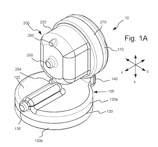
understanding
technologies disclosed below and the environment in which such technologies
may typically
be used. The terms used herein are not intended to be limited to any
particular narrow
interpretation unless clearly stated otherwise in this document. References
set forth herein
may facilitate understanding of the technologies or the background thereof The
disclosure of
all references cited herein are incorporated by reference.
[03] Lung disease, whether acute or chronic, are major healthcare problems.
The
American Lung Association reports that nearly 350,000 Americans die each year
of some
form of lung disease. Lung disease, which is responsible for one in seven
deaths, is the
number three killer of Americans. Acute lung failure and adult respiratory
distress syndrome
(ARDS) are prevalent forms of lung disease. ARDS afflicts about 150,000
Americans each
year. The associated mortality of ARDS remains between 40 and 60% despite
improvements
in critical care medicine.
[04] Most lung disease, however, is chronic. Emphysema and chronic
bronchitis, two
forms of chronic obstructive pulmonary disease (COPD), afflict over 14 million
Americans
annually. Chronic lung disease is now the 3rd leading cause of death in
America, claiming
the lives of over 400,000 annually and carrying a cost of $154 billion. As
chronic lung
1
CA 03088382 2020-07-13
WO 2019/143623
PCT/US2019/013678
disease reaches end stage, lung transplantation becomes the only choice for
effective
treatment. Lung transplantation has had a steady rise over the last 10 years
and ¨3300 lung
transplants are performed annually worldwide. The average time on the waiting
list varies
from 6 to 12 months depending on the patient's condition and institutional
expertise, and 10-
15% of patients die while on the waiting list in the US. A narrow window of
opportunity
exists for lung transplant in any patient who is sick enough to benefit from
the operation, but
healthy enough to survive months of waiting for a donor lung and then the
subsequent
surgery.
[05] Upon reaching a critical condition, mechanical ventilation and
extracorporeal
membrane oxygenation (ECMO) are the only alternatives for respiratory support
available to
bridge acute and chronic respiratory patients to lung recovery or lung
transplantation.
Mechanical ventilation (MV) may maintain adequate gas exchange for short term
support,
but longer term support can lead to ventilator induced lung injury from
barotrauma (high
pressure), volutrauma (over-distension), and biotrauma (molecular and cell
mediated
inflammation), which can further worsen the respiratory status of the patient.
ECMO is
expensive and complicated, requiring the use of an external pump and blood
circuit that have
to be supervised continuously by highly trained technicians. The confinement
of the patient in
MV and especially ECMO leads to a progressive deconditioning that is reflected
in higher
postoperative complications and earlier mortality after transplant.
Nevertheless, ECMO has
been increasingly considered as the only alternative to bridge patients to
lung transplant or
lung recovery after an acute decompensation from their disease. More recently,
with
increasing experience at active transplant centers and improvement in ECMO
technology, the
concept of "ambulatory ECMO" has gained popularity and facilitates and
expedites patient
recovery after transplantation. Success in ambulatory ECMO underscores the
importance of
maintaining patient mobility. Currently available ambulatory ECMO systems
combine
existing blood pumps and bypass oxygenators into an integrated system but
remain bulky and
cumbersome and require frequent exchange of the oxygenators for longer term
support.
[06] Recent success with paracorporeal left ventricular assist devices
(VADs) for heart
failure patients has stimulated envisioning an ambulatory pump-lung device
that can be a
bridge to lung transplant or recovery. No fully integrated ambulatory pump-
lungs are being
used clinically, however. Many portable or ambulatory systems under
development integrate
a separate blood pump and oxygenator under a single controller unit but are
cumbersome.
2
CA 03088382 2020-07-13
WO 2019/143623
PCT/US2019/013678
SUMMARY
[07] In one aspect, a system for lung assist includes a plurality of fiber
bundle sections.
Each of the fiber bundle sections includes a fiber bundle housing defining a
fiber bundle
compartment therein and a fiber bundle positioned within the fiber bundle
compartment. The
fiber bundle includes a plurality of hollow gas permeable fibers adapted or
configured to
permit diffusion of gas between blood and an interior of the plurality of
hollow gas
permeable fibers. The plurality of hollow gas permeable fibers is positioned
such that blood
flows around the plurality of hollow gas permeable fibers when flowing through
the fiber
bundle compartment. The fiber bundle of each of the plurality of fiber bundle
sections is
different in at least one property from the fiber bundle of each of the other
of the plurality of
fiber bundle sections. Thus, each fiber bundle section is unique in at least
one property of the
associated fiber bundle. The fiber bundle housing further includes a gas inlet
in fluid
connection with the fiber bundle housing and in fluid connection with inlets
of the plurality
of hollow gas permeable fibers, a gas outlet in fluid connection with the
housing and in fluid
connection with outlets of the plurality of hollow gas permeable fibers, and a
blood outlet in
fluid connection with a first end of the fiber bundle. The fiber bundle
housing also includes a
first interface.
[08] The system further includes a base section including a housing
including a
pressurizing compartment, a pressurizing mechanism within the pressurizing
compartment, a
blood inlet in fluid connection with the pressurizing compartment and a
conduit in fluid
connection with the pressurizing compartment at a first end thereof via which
pressurized
fluid exits the pressurizing compartment. The base further includes a second
interface
adapted to form a releasable, sealing connection with the first interface of
one of the plurality
of fiber bundle sections. A second end of the conduit is placed in fluid
connection with a
second end of the fiber bundle when the fiber bundle section is connected to
the base section
via the first interface and the second interface. The system may, for example,
be a
paracorporeal system.
[09] In a number of embodiments, the pressurizing mechanism includes an
impeller
rotatable within the pressurizing compartment. In a number of embodiments, the
housing of
the base section includes a pressurizing section including the pressurizing
compartment and
an interface section. The interface section includes an extending section
which extends from
3
CA 03088382 2020-07-13
WO 2019/143623
PCT/US2019/013678
the pressurizing section and the second interface. The conduit may, for
example, include a
flow channel which extends through the extending section.
[10] The plurality of hollow gas permeable fibers of each of the fiber
bundles may, for
example, extend generally perpendicular to the direction of bulk flow of blood
through the
fiber bundle from the second end of the fiber bundle to the first end of the
fiber bundle. The
plurality of hollow gas permeable fibers may, for example, include a plurality
of layers of
fiber fabric, wherein each of the plurality of layers of fiber fabric includes
hollow gas
permeable fibers. In a number of embodiments, adjacent layers of fiber fabric
are rotated
relative to each other such that the orientation of the plurality of hollow
gas permeable fibers
in adjacent layers of fiber fabric are of a different orientation.
[11] In a number of embodiments, the plurality of hollow gas permeable
fibers is
formed in at least one generally cylindrical bundle. As described above, the
generally
cylindrical bundle may be formed from a plurality of layers of fiber fabric,
wherein each of
the plurality of layers of fiber fabric includes hollow gas permeable fibers.
Once again,
adjacent layers of fiber fabric may be rotated relative to each other such
that the orientation of
the plurality of hollow gas permeable fibers in adjacent layers of fiber
fabric are of a different
orientation.
[12] In a number of embodiments, the flow channel is in fluid connection
with a
manifold formed in the extending section. The extending section may, for
example, extend
generally perpendicular to a plane of rotation of the impeller. The first
interface of each of
the fiber bundle sections may be, for example, attached to the second
interface of the base
section so that the axis of the fiber bundle of the one of the plurality of
fiber bundle sections
attached to the base section is oriented generally parallel to a plane of
rotation of the impeller.
The fiber bundle section attached to the base section may, for example, be
positioned over the
pressurizing compartment of the base section. Bulk flow of blood through the
fiber bundle
may, for example, be in a generally axial direction. In a number of
embodiments, blood is
blocked from flowing to the gas inlet and the gas outlet.
[13] The plurality of fiber bundle sections may, for example, include fiber
bundle
sections of different lengths comprising fiber bundles of different lengths
and thereby
different fiber surface areas. At least one of the plurality of fiber bundle
sections may, for
4
CA 03088382 2020-07-13
WO 2019/143623
PCT/US2019/013678
example, be configured for use with pediatric patients, and at least one of
the plurality of
fiber bundle sections may, for example, be configured for use with adult
patients.
[14] In a number of embodiments, at least one combination of one of the
plurality of
fiber bundle sections and the base section is suitable for carbon dioxide
removal in a first
range of flow rates and is suitable for oxygenation and carbon dioxide removal
in a second
range of flow rates, wherein the second range of flow rates extends to higher
flow rates.
[15] In another aspect, a method of extracorporeal lung assist to a patient
includes
providing a plurality of fiber bundle sections. As described above, each of
the fiber bundle
sections includes a fiber bundle housing defining a fiber bundle compartment
therein and a
fiber bundle positioned within the fiber bundle compartment. The fiber bundle
includes a
plurality of hollow gas permeable fibers adapted or configured to permit
diffusion of gas
between blood and an interior of the plurality of hollow gas permeable fibers.
The plurality
of hollow gas permeable fibers is positioned such that blood flows around the
plurality of
hollow gas permeable fibers when flowing through the fiber bundle compartment.
The fiber
bundle of each of the plurality of fiber bundle sections is different in at
least one property
from the fiber bundle of each of the other of the plurality of fiber bundle
sections. Thus, each
fiber bundle section is unique in at least one property of the associated
fiber bundle. The
fiber bundle housing further includes a gas inlet in fluid connection with the
fiber bundle
housing and in fluid connection with inlets of the plurality of hollow gas
permeable fibers, a
gas outlet in fluid connection with the housing and in fluid connection with
outlets of the
plurality of hollow gas permeable fibers, and a blood outlet in fluid
connection with a first
end of the fiber bundle. The fiber bundle housing also includes a first
interface. The method
further includes providing a base section including a housing including a
pressurizing
compartment, a pressurizing mechanism within the pressurizing compartment, a
blood inlet
in fluid connection with the pressurizing compartment and a conduit in fluid
connection with
the pressurizing compartment at a first end thereof via which pressurized
fluid exits the
pressurizing compartment. The base further includes a second interface adapted
to form a
releasable, sealing connection with the first interface of one of the
plurality of fiber bundle
sections. A second end of the conduit is placed in fluid connection with a
second end of the
fiber bundle when the fiber bundle section is connected to the base section
via the first
interface and the second interface. The method also includes attaching one of
the plurality of
fiber bundle sections to the base section via connection of the first
interface and the second
CA 03088382 2020-07-13
WO 2019/143623
PCT/US2019/013678
interface, wherein the fiber bundle of the one of the fiber bundle sections is
chosen for the
patient. The fiber bundle section and the base section may be characterized as
further
described above.
[16] As also described above, at least one combination of one of the
plurality of fiber
bundle sections and the base section may, for example, be suitable for carbon
dioxide
removal at in a first range of flow rates and is suitable for oxygenation and
carbon dioxide
removal in a second range of flow rates, wherein the second range of flow
rates extends to
higher flow rates.
[17] In another aspect, a system for lung assist includes a fiber bundle
section including
a fiber bundle housing defining a fiber bundle compartment therein and a fiber
bundle
positioned within the fiber bundle compartment. The fiber bundle includes a
plurality of
hollow gas permeable fibers. The plurality of hollow gas permeable fibers may,
for example,
be adapted to or configured to permit diffusion of gas between blood and an
interior of the
plurality of hollow gas permeable fibers. The plurality of hollow gas
permeable fibers is
positioned such that blood flows around the plurality of hollow gas permeable
fibers when
flowing through the fiber bundle compartment. The fiber bundle housing further
includes a
gas inlet in fluid connection with the housing and in fluid connection with
inlets of the
plurality of hollow gas permeable fibers, a gas outlet in fluid connection
with the housing and
in fluid connection with outlets of the plurality of hollow gas permeable
fibers, and a blood
outlet in fluid connection with a first end of the fiber bundle. The fiber
bundle section further
includes a first interface. The system further includes a base section
including a housing
including a pressurizing compartment, a pressurizing mechanism within the
pressurizing
compartment, a blood inlet in fluid connection with the pressurizing
compartment and a
conduit in fluid connection with the pressurizing compartment at a first end
thereof via which
pressurized fluid exits the pressurizing compartment. The base section further
includes a
second interface adapted to form a releasable, sealing connection with the
first interface of
the fiber bundle section. A second end of the conduit is placed in fluid
connection with a
second end of the fiber bundle when the fiber bundle section is connected to
the base section
via the first interface and the second interface.
[18] In still a further aspect, a method for providing lung assist includes
selecting a fiber
bundle section including a fiber bundle housing defining a fiber bundle
compartment therein
and a fiber bundle positioned within the fiber bundle compartment. As
described above, the
6
CA 03088382 2020-07-13
WO 2019/143623
PCT/US2019/013678
fiber bundle includes a plurality of hollow gas permeable fibers. The
plurality of hollow gas
permeable fibers is configured to or adapted to permit diffusion of gas
between blood and an
interior of the plurality of hollow gas permeable fibers. The plurality of
hollow gas
permeable fibers is positioned such that blood flows around the plurality of
hollow gas
permeable fibers when flowing through the fiber bundle compartment. The fiber
bundle
housing further includes a gas inlet in fluid connection with the housing and
in fluid
connection with inlets of the plurality of hollow gas permeable fibers, a gas
outlet in fluid
connection with the housing and in fluid connection with outlets of the
plurality of hollow
gas permeable fibers, a blood outlet in fluid connection with a first end of
the fiber bundle.
The fiber bundle section further includes a first interface. The method
further includes
releasably attaching the fiber bundle section to a base section including a
housing including a
pressurizing compartment, a pressurizing mechanism within the pressurizing
compartment, a
blood inlet in fluid connection with the pressurizing compartment and a
conduit in fluid
connection with the pressurizing compartment at a first end thereof via which
pressurized
fluid exits the pressurizing compartment. The base section further includes a
second interface
configured to or adapted to form a releasable, sealing connection with the
first interface of the
fiber bundle section. A second end of the conduit is placed in fluid
connection with a second
end of the fiber bundle when the fiber bundle section is attached to or
connected to the base
section via the first interface and the second interface.
[19] The present devices, systems and methods, along with the attributes
and attendant
advantages thereof, will best be appreciated and understood in view of the
following detailed
description taken in conjunction with the accompanying drawings.
BRIEF DESCRIPTION OF THE DRAWINGS
[20] Figure 1A illustrates a perspective view of an embodiment of an
extracorporeal
assist lung or paracorporeal ambulatory assist lung apparatus, device or
system hereof in
which a smaller fiber bundle section is placed in connection with the base
section of the
device.
[21] Figure 1B illustrates a perspective view of the paracorporeal
ambulatory assist lung
device of Figure 1A, in which the smaller fiber bundle section illustrated in
Figure 1A has
been removed and a larger fiber bundle section has been placed in connection
with the base
section of the device.
7
CA 03088382 2020-07-13
WO 2019/143623
PCT/US2019/013678
[22] Figure 2A illustrates a front view of the paracorporeal ambulatory
assist lung
device of Figure 1A with the larger fiber bundle section in position for
attachment to the base
section of the device and the smaller fiber bundle section positioned above
the larger fiber
bundle section.
[23] Figure 2B illustrates a side cross-sectional view of the paracorporeal
ambulatory
assist lung device of Figure 1A with the larger fiber bundle section in
position for attachment
to the base section of the device and the smaller fiber bundle section
positioned above the
larger fiber bundle section.
[24] Figure 2C illustrates a side cross-section view of the paracorporeal
ambulatory
assist lung device of Figure 1A with the larger fiber bundle connected to the
base section and
in which solid arrows indicate blood flow through the device and dashed arrows
indicate gas
flow through the device.
[25] Figure 2D illustrates a perspective, exploded view of various layers
of an
embodiment of a fiber bundle hereof wherein the orientation of the fibers in
adjacent layers is
rotated with respect to each other (wherein the fibers within individual
layers are oriented in
generally the same direction).
[26] Figure 3A illustrates a front view of the paracorporeal ambulatory
assist lung
device of Figure 1A with the smaller fiber bundle section in connection with
the base section.
[27] Figure 3B illustrates a section A-A (with reference to Figure 3A)
cross-sectional
view of the system of Figure 1A with the smaller fiber bundle section in
connection with the
base section.
[28] Figure 3C illustrates a section B-B (with reference to Figure 3A)
cross-sectional
view of the system of Figure 1A with the smaller fiber bundle section in
connection with the
base section.
[29] Figure 4A illustrates a perspective, disassembled or exploded view of
the
paracorporeal ambulatory assist lung device of Figure 1A, including the
smaller fiber bundle
section.
[30] Figure 4B illustrates a side view of the paracorporeal ambulatory
assist lung device
of Figure 1A with the larger fiber bundle connected to the base section.
8
CA 03088382 2020-07-13
WO 2019/143623
PCT/US2019/013678
[31] Figure 4C illustrates a rear view of the paracorporeal ambulatory
assist lung device
of Figure 1A with a rear panel removed to illustrates the flow path channel
from the
pressurizing section into a manifold in fluid connection with the fiber bundle
section.
[32] Figure 4D illustrates a side view of the paracorporeal ambulatory
assist lung device
hereof similar to the device of Figure 1A with a larger fiber bundle connected
to the base
section.
[33] Figure 4E illustrates a rear view of the paracorporeal ambulatory
assist lung device
of Figure 4D with a rear panel removed to illustrates the flow path channel
from the
pressurizing section into a manifold in fluid connection with the fiber bundle
section.
[34] Figure 5A illustrates a perspective, disassembled or exploded view of
the impeller
of the device of Figure 1A.
[35] Figure 5B illustrates a side, disassembled or exploded view of the
impeller of the
device of Figure 1A
[36] Figure 5C illustrates a section A-A (see Figure 5B) cross-sectional,
disassembled
or exploded view of the impeller of the device of Figure 1A.
[37] Figure 6A illustrates data from studies of volumetric oxygenation rate
(mL/min) as
a function of flow rate for a device hereof with a smaller, pediatric fiber
bundle section 200
as illustrated in Figure 1A (0.3 m2 total fiber surface area) and for a device
hereof with a
larger, adult fiber bundle section 200a as illustrated in Figure 1B (0.65 m2
total fiber surface
area).
[38] Figure 6B illustrates a normalized index of hemolysis or NIH (g/100L)
for a device
hereof with a smaller, pediatric fiber bundle section 200 as illustrated in
Figure 1A and for a
commercially available control system (that is, the LILLIPUT pediatric
oxygenator available
from Sorin Group of Modena, Italy with a CENTRIMAGO blood pump available from
Thoratec Corporation of Pleasanton, California) at a flow rate of 2.5 L/min.
[39] Figure 6C illustrates a study of pressure as a function of flow rate
for a device
hereof with a smaller, pediatric fiber bundle section 200 assuming an 18 Fr
(French) venous
cannula, a 14 Fr arterial cannula and an outflow (pulmonary artery) pressure
of 50 mmHg as
a result of pulmonary hypertension.
9
CA 03088382 2020-07-13
WO 2019/143623
PCT/US2019/013678
[40] Figure 6D illustrates a study of pressure as a function of flow rate
for a device
hereof with a larger, adult fiber bundle section 200a assuming a 27 Fr
(French) dual-lumen
cannula.
[41] Figure 6E illustrates a study of pressure as a function of flow rate
for a device
hereof with a larger, adult fiber bundle section 200a assuming a 15.5 Fr
(French) dual-lumen
cannula.
[42] Figure 6F illustrates a study of oxygen transfer rate as function of
blood flow rate
for a device hereof with a smaller, pediatric fiber bundle section 200.
[43] Figure 6G illustrates a study of oxygen transfer rate as function of
blood flow rate
for a device hereof with a larger, adult fiber bundle section 200a.
[44] Figure 6H illustrates a study of normalized CO2 removal rate as
function of blood
flow rate for a device hereof with a larger, adult fiber bundle section 200a.
[45] Figure 7A illustrates a blood flow loop used in oxygenation or oxygen
transfer rate
studies hereof
[46] Figure 7B illustrates a blood flow loop used in hemolysis studies
hereof
DETAILED DESCRIPTION
[47] It will be readily understood that the components of the embodiments,
as generally
described and illustrated in the figures herein, may be arranged and designed
in a wide
variety of different configurations in addition to the described example
embodiments. Thus,
the following more detailed description of the example embodiments, as
represented in the
figures, is not intended to limit the scope of the embodiments, as claimed,
but is merely
representative of example embodiments.
[48] Reference throughout this specification to "one embodiment" or "an
embodiment"
(or the like) means that a particular feature, structure, or characteristic
described in
connection with the embodiment is included in at least one embodiment. Thus,
the
appearance of the phrases "in one embodiment" or "in an embodiment" or the
like in various
places throughout this specification are not necessarily all referring to the
same embodiment.
CA 03088382 2020-07-13
WO 2019/143623
PCT/US2019/013678
[49] Furthermore, described features, structures, or characteristics may be
combined in
any suitable manner in one or more embodiments. In the following description,
numerous
specific details are provided to give a thorough understanding of embodiments.
One skilled
in the relevant art will recognize, however, that the various embodiments can
be practiced
without one or more of the specific details, or with other methods,
components, materials, et
cetera. In other instances, well known structures, materials, or operations
are not shown or
described in detail to avoid obfuscation.
[50] As used herein and in the appended claims, the singular forms "a,"
"an", and "the"
include plural references unless the context clearly dictates otherwise. Thus,
for example,
reference to "an impeller" includes a plurality of such impellers and
equivalents thereof
known to those skilled in the art, and so forth, and reference to "the
impeller" is a reference to
one or more such impellers and equivalents thereof known to those skilled in
the art, and so
forth. Recitation of ranges of values herein are merely intended to serve as a
shorthand
method of referring individually to each separate value falling within the
range. Unless
otherwise indicated herein, each separate value and intermediate ranges are
incorporated into
the specification as if individually recited herein. All methods described
herein can be
performed in any suitable order unless otherwise indicated herein otherwise
clearly
contraindicated by the text.
[51] As used herein in reference to device 10, the terms "axial", "axially"
or like terms
refer generally to an axis around which a component (for example, fiber bundle
300 or
impeller 400) of device 10 is formed (although not necessarily symmetrically
therearound).
The term "radial" refers generally to a direction normal to such an axis. The
terms "rear",
"rearward" or like terms refer generally to a direction along axis x of Figure
1A away from or
opposite the gas and fluid ports of device 10. The terms "front", "forward" or
like terms refer
generally to a direction along axis x toward the gas and fluid ports of device
10. The terms
"up", "upward" or like terms refer generally to a direction along axis y of
Figure 1A toward
fiber bundle section 200 and away from pressurizing section 122 of base
section 100, while
the terms "down", "downward" or like terms refer to a direction along axis y
away from fiber
bundle section 200 and toward pressurizing section 122 of base section 100.
The terms
"side", "sideways" or like terms refer to a direction orthogonal to an up or
down direction
and orthogonal to an axial direction as described above. In general, terms
related to direction
and/or orientation as set forth herein are used to describe relative positions
of the elements of
11
CA 03088382 2020-07-13
WO 2019/143623
PCT/US2019/013678
the described embodiment and are not limiting unless otherwise indicated
herein or otherwise
clear from the text hereof
[52] In a number of embodiments, extracorporeal/paracorporeal ambulatory
assist lung
system hereof provide advantages in gas transfer efficiency and
biocompatibility. The
systems hereof may, for example, be designed for either central and/or
peripheral cannulation
and respiratory support of, for example, 1-3 months duration before device
change-out may
be required. Systems hereof are, for example, amenable to patients suffering
from severe
acute respiratory failure (ARDS) to chronic patients suffering from COPD or
severe
pulmonary hypertension (PH).
Paracorporeal apparatuses, devices or systems are
extracorporeal devices/systems generally located immediately adjacent to the
body during
use. In other words, paracorporeal devices or systems are "wearable" or
ambulatory devices
or systems. The
apparatuses, devices and systems hereof are well suited for
paracorporeal/ambulatory use as well as use as generally stationary
extracorporeal use.
[53] In many ambulatory devices or system under development, a blood pump
is
connected by one or more conduits (for example, lengths of tubing) to an
oxygenator. While
a number of systems have integrated blood pumps, the blood leaving the
impeller unit of such
devices typically travels through channels before being distributed by
manifolds into the
hollow fiber bundle compartment. Recently, devices which are less cumbersome
than many
other devices under development while providing for increased ambulatory
respiratory assist
were disclosed in PCT International Patent Application Publication No.
2016/210089, the
disclosure of which is incorporated herein by reference. Such devices provide
a highly
integrated blood pump and lung, in which a pump mechanism such as an impeller
pressurizes
blood for flow through hollow gas permeable fibers (sometimes referred to
herein as a fiber
bundle). Such devices may, for example, be designed to be worn in a holster or
vest
paracorporeally. Moreover, such devices may, for example, provide for
increased average or
mean velocity through the fiber bundle as compared to other devices, which
enhances gas
exchange. The integrally formed extracorporeal systems for lung assist of PCT
International
Patent Application Publication No. 2016/210089 include an integrated housing
having a
blood flow inlet in fluid connection with a fiber bundle compartment and a
pressurizing stator
compartment.
[54] In a number of embodiments as illustrated in Figures 1A through 4,
device 10
includes a first, base or blood pressurizing section 100 (hereinafter referred
to as base
12
CA 03088382 2020-07-13
WO 2019/143623
PCT/US2019/013678
section 100) and a modular or releasably connectible second, fiber bundle or
gas exchange
section 200 (hereinafter referred to as fiber bundle section 200, 200a etc.).
In a number of
embodiments, base section 100 and fiber bundle section 200 are releasably
connectible so
that a pressurizing system and the fiber bundle are encompassed within a
relatively small
form factor. As further described below, fiber bundle section 200 may be
readily removed
and replaced with another fiber bundle section such as fiber bundle section
200a of Figure 1B
to provide a fiber bundle of, for example, a different size, a different
configuration, a
different surface treatment, a different fiber composition, etc. Device 10 may
thereby provide
for efficient and significant gas transfer rate in a number of different uses
or treatments
without inducing significant blood damage. Moreover, fiber bundle section such
as fiber
bundle section 200 or 200a may be readily replaced with another like or
identical fiber
bundles section 200 or 200a, respectively, in the case of, for example,
damage, contamination
or wear.
[55] Fiber bundle section 200 includes a housing 220 which includes a fiber
bundle
compartment 222 as, for example, illustrated in, for example, Figures 2B and
3B. Fiber
bundle compartment 222 houses a fiber bundle 300 and provides a gas pathway
designed to
uniformly perfuse the gas side of fiber bundle 300 with a sweep gas which may
be oxygen or
a gas mixture including oxygen.
[56] As, for example, illustrated in the embodiments of Figure 2A and 2B,
systems 5
hereof may include a base section 100 and a plurality of different fiber
bundle sections 200,
200a etc. Two fiber bundle sections 200 and 200a are illustrated in Figures 2A
and 2B, but
more fiber bundle sections may be provided. In a number of embodiments, fiber
bundle
sections 200 and 200a (and other fiber bundle sections hereof) are formed to
have generally
identically dimensions other than the length thereof Elements of fiber bundle
section 200a
are numbered similarly to corresponding elements of fiber bundle section 200
with the
addition of the designation "a" thereto. Fiber bundle 300a may, for example,
be formed to
have dimensions generally identically to fiber bundle 300 other than the
length thereof Upon
formation, the gas exchanging portion of fiber bundle 300 (excluding any
potting) had a
diameter of 1.75 inches (0.044 meters) and a length of 1.52 inches (0.039
meters). Fiber
bundle 300a had a diameter of 1.75 inches (0.044 meters) and a length of 3.20
inches (0.081
meters). In a number of embodiments, fiber bundle 300 had an overall surface
area for gas
exchange or total fiber surface area of 0.3 m2. Fiber bundle 300 included 96
PMP fiber
13
CA 03088382 2020-07-13
WO 2019/143623
PCT/US2019/013678
layers. Fiber bundle 300a had an overall surface area for gas exchange or
total fiber surface
area of 0.65 m2. Fiber bundle 300a included 200 PMP fiber layers.
[57] In the embodiments of Figures 1A through 4E, numerous fiber
bundle/device
properties can be changed by switching between fiber bundle sections
including, but not
limited to, fiber bundle length (and, thereby, overall surface area for gas
exchange), fiber
bundle composition (for example, fiber bundle material, surface treatment),
etc. The fiber
bundle properties can vary over a wide range to be specifically adapted for a
particular use or
function, for a particular patient group, or even for a particular patient.
[58] In the illustrated embodiment of Figures 1 through 4E, fiber bundle
section 200
and associated fiber bundle 300 were, for example, designed for pediatric use,
while fiber
bundle section 200a and associated fiber bundle 300a were designed for adult
use. In the
case of each of fiber bundle sections 200 and 200a, device 10 may, for
example, be used for
oxygenation and/or carbon dioxide removal. The integrated pump, including
impeller 400 (a
closed or enclosed impeller in a number of embodiments), draws venous blood
from a patient
via an inflow cannula (see Figure 3B) placed within a blood vessel. Blood is
pumped
through the gas-exchanging fiber bundle, which is operable to transfer oxygen
to and remove
carbon dioxide from the blood. After the blood passes through the fiber
bundle, the blood is
returned to the patient's circulatory system via an outflow cannula (see
Figure 3B). The
required levels of blood flow, pumping, and gas exchange provided by device 10
during
respiratory support depends upon patient size (for example, a pediatric
patient or an adult
patient) as well as the nature of the respiratory insufficiency. In the case
wherein carbon
dioxide removal rather than oxygenation is the primary goal, lower blood flow
rates and less
invasive cannulation strategies may be used. When carbon dioxide is the
primary goal, the
methodology is typically referred to as extracorporeal carbon dioxide removal
or ECCO2R.
[59] In a number of embodiments, all gas and fluid inlets and outlets
(collectively ports)
are oriented in generally the same directions upon assembly of device 10 by
connecting a
fiber bundle section hereof to base section 100 (see, for example, Figures 1A
through 2B). In
the illustrated embodiment, the axes of gas inlet 260, gas outlet 264, fluid
inlet 130 and fluid
outlet 250 are generally parallel (for example, within less than 20, 10 or
even 5 degrees of
being parallel). In the embodiment of Figures 1 through 4C, the inlets and
outlets are
positioned on a forward or front side of device 10. In the illustrated
embodiment, such axes
are generally coplanar (for example, within less than 20, 10 or even 5 degrees
of being
14
CA 03088382 2020-07-13
WO 2019/143623
PCT/US2019/013678
coplanar). By orientating all gas and fluid ports in generally the same
direction, connection
of tubing to such ports and wearing of device 10 hereof with attached tubing
is facilitated. As
set forth above, orientating all gas and fluid ports in generally the same
direction indicates
that the axes of each of the ports is within 20 . 10 , 5 or less of being
colinear with all other
axes.
[60] In a number of embodiments, the dimensions of device 10 were no more
than 13.2
cm (5.2 inches) in height (the y dimension in Figure 1A), no more than 11.4 cm
(4.5 inches)
in width (the z dimension in Figure 1A), and no more than 14 cm (5.5 inches)
in length (the x
dimension in Figure 1A). In a number of embodiments, the length of fiber
bundle section
varied between 6.9 cm (2.7 inches) and 11.2 cm (4.4 inches). The weight of
device 10 with
fiber bundle section 200 may, for example, be no greater than 550g, or no
greater than 500g,
while the weight of device 10 with fiber bundle section 200a may be no greater
than 620g or
nor greater than 570g. In a number of embodiments, the priming volume of
device 10 with
fiber bundle section 200 was approximately 125m1, and the priming volume of
device 10
with fiber bundle section 200a was approximately 160m1. The form factor of
device hereof
may be further reduced by increasing pumping efficiency (for example, by
further optimizing
impeller design).
[61] In that regard, a pressurizing mechanism such as a rotating element or
an
impeller 400 may be positioned within a pressurizing or pumping
(impeller/stator)
compartment 124 a housing 120 of base section 100. In the illustrated
embodiment, base
section 100 includes a first or pressurizing section 122 which houses pumping
or pressurizing
compartment 124 and a second, or interface section 140 which extends at an
angle from first
section 122 to form an interface for connection with a fiber bundle section
hereof In the
illustrated embodiment, extending section 140 extends at an angle of
approximately 90 to
the plane of rotation of impeller 400 as defined by pumping or pressurizing
compartment 124
of first section 122. Pumping or pressurizing compartment 124 was formed as an
impeller
stator/volute compartment of first section 122. In the
illustrated embodiment, first
section 122 and pumping or pressurizing compartment 124 thereof were formed
via
connection of a first or upper housing section or portion 120a and a lower or
second housing
section 120b of base housing 120. Lower or second housing section 120b of base
housing
120 (see, for example, Figure 4A) was sized to allow insertion of impeller 400
into impeller
stator/volute compartment 124 of base housing 120. Pumping
or pressurizing
CA 03088382 2020-07-13
WO 2019/143623
PCT/US2019/013678
compartment 124 houses impeller 400 and may be designed in accordance with
traditional
pump theory to maximize the pumping efficiency of impeller 400. Impeller 400
rotates
within pumping or pressurizing compartment 124 of housing 120.
[62] Fiber bundle section housing 220 and base section housing 120 are
formed from
rigid materials. In general, such rigid materials do not deform or flex
significantly under the
conditions of use. In a number of embodiments, fiber bundle section housing
220 and base
section housing 120 are formed from polymeric materials and, typically, from
the same
polymeric material. The housing sections may, for example, be formed from
extrusion.
[63] The stator section of a centrifugal pump, after flow exits the
impeller, is usually
either a diffuser or a volute. The purpose of each of these two stator types
is to efficiently
diffuse velocity energy into pressure. Diffusers are characterized by a
plurality of radially
symmetric diffusing passageways surrounding the impeller. Either a volute-
shaped or annular
collector is used in tandem with the diffuser. Volutes are characterized by
one or more scroll-
shaped diffusing passageways (one in a number of embodiments hereof),
depending on the
pump configuration. A volute hereof receives fluid being pumped by the
impeller, slowing
down the fluid's flow rate and converting kinetic energy into pressure. The
volute curves and
increases in area as it approaches the discharge port.
[64] Impeller 400 may, for example, be partially magnetically supported via
one or
more magnets positioned on or within impeller 400. Impeller 400, in the
illustrated
embodiment, is positioned within impeller volute compartment 124 such that the
net
hydrodynamic load on impeller 400 is upwards (in the orientation of the
Figures). Thus,
magnets used to support impeller 400 may exert a downward force on impeller
400. As, for
example, discussed in PCT International Publication No. W02014/085620, one or
more
magnets may be seated in one or more seating of impeller 400 and (in
cooperation with
another magnet which may be within or external to impeller volute compartment
124) is
operable to apply force offset the combined hydrodynamic and coupling magnet
forces,
thereby minimizing the axial forces applied to the bearings, and improving
overall system
durability. Top and bottom pivot bearings 412a and 412b (see Figure 4A),
respectively, may,
for example, be ultra-high-molecular-weight polyethylene (UHMWPE) pivot and
cup type
bearings housed in a stainless steel shell, which maximizes their resistance
to wear.
16
CA 03088382 2020-07-13
WO 2019/143623
PCT/US2019/013678
[65] Figure 2C illustrates fluid (blood) flow (solid arrows) and sweep gas
flow (dashed
arrows) through device 10. In that regard, a fluid such as blood is drawn into
the central
portion of impeller 400 via a fluid inlet 130 formed in base section 100 and
centrifugally spun
outwards via impeller vanes 410 (see, for example, Figure 3C and 5A) as
indicated by the
radially outward oriented solid arrows in Figure 2C. Blood is then channeled
to fiber
bundle 300 as shown in, for example, Figure 1B and 3C. As, for example,
illustrated in
Figure 3C, a channel 126 extends from impeller volute compartment 124 to a
flow
channel 142. At the point that channel 126 extends from impeller volute
compartment 124,
flow channel 142 may, for example, extend the height (that is, the vertical
dimension in the
orientation of Figure 2C) of impeller 400 to, for example, maximize washing on
the
underside of impeller 400, as this is a common area for thrombus deposition in
pivot pumps.
In the illustrated embodiment, channel 126 extends generally tangentially (for
example,
within 5 degrees of tangentially therefrom) from impeller volute compartment
to connect to
flow channel 142. In a number of embodiments, flow channel 142 had a circular
cross-
section. Channel 126 extends rearward at an angle to approximately a
centerline of
impeller 400 where channel 126 connects to flow channel 142.
[66] Flow Channel 142 may be incorporated into base housing 120 (that is,
within
extending section 140) in a manner that it does not further increase the form
factor of fiber
bundle 300 and, thereby, fiber bundle section 200. In the illustrated
embodiment (see, for
example, Figures 2B, 2C and 4C), flow channel 142 travels vertically upward
(in the
orientation of the drawings) and at an angle of approximately 90 (that is
approximately
perpendicularly or perpendicularly) to the plane of rotation of impeller 400
through extending
section 140 of base section 100 and enters a fluid/blood inlet volume or
manifold 144 portion
formed in a forward-facing portion of extending section 140 where the
fluid/blood contacts a
second or rearward surface of fiber bundle 300. Providing rounded or arced
corners/ends in
flow channel 142 may assist in, for example, reducing or minimizing hemolysis
and
thrombosis. In a number of embodiments, flow channel 142 has a round or
circular cross-
sectional shape.
[67] In a number of embodiments, the blood enters the second or rearward
end of fiber
bundle 300 from manifold 144 and passes around the hollow fibers thereof After
passing
through fiber bundle 300, blood exits system 10 via an outlet volume or
manifold 224, which
is in fluid connection with a first or forward end of fiber bundle 300 at a
first end thereof and
17
CA 03088382 2020-07-13
WO 2019/143623
PCT/US2019/013678
with a blood/fluid outlet 250 at a second end thereof The liquid/fluid flow
path may be
separated from the gas flow path through device 10 by abutment/sealing between
(i) the
periphery of the rear face of fiber bundle 300 and surface 146 and (ii) the
periphery of the
front face of fiber bundle 300 and fiber bundle housing 220.
[68] In the illustrated embodiment, fiber bundle section 200 includes an
interface 270 (a
fiber bundle section 200a includes a like interface 270a) which connects to a
cooperating
interface 170 of extending section 140 of base section 100. Each fiber bundle
section hereof
may include a like or identical interface which cooperates with interface 170
to form a sealed
connection between one of the fiber bundle sections hereof and base section
100. Fiber
bundle sections hereof are thus each readily connectible to and removable from
base
section 100 hereof for devices 10 of differing flow and/or mass exchange
properties (as well
as differing dimensions, volume and/or weight). As illustrated schematically
in the
representative embodiment of Figure 2C, fiber bundle section 200a may include
an interface
270a having a connector 272a which cooperates with a cooperating connector 172
on
interface 170 to form a sealed connection between interface 270a of fiber
bundle section 200a
and interface 170 of base section 100. Other fiber bundle sections hereof may
similarly
include like connectors to cooperate with cooperating connector 172. Connector
272a (and
like or identical connectors of other fiber bundle sections hereof) may
cooperate with
cooperating connector 172 via sliding fits, snap fits, threaded fits, Luer
lock connection fits
etc. as known in the mechanical/medical connection arts. A sealing connection
between
interface 270a and interface 170 may, for example, be facilitated by a seal
180 such as an 0-
ring, which is seated in a seating formed in forward surface 146 of the
illustrated
embodiment.
[69] The gas pathway in device 10 may, for example, be relatively simple.
Gas flows in
through a gas inlet port 260 into a channel 262 on one side of fiber bundle
300 and out
through a gas outlet port 264 in fluid connection with a channel 266 on the
other side of fiber
bundle 300. Thus, gas flow through fiber bundle 300 is in the average or bulk
direction of the
dashed arrows in Figure 2C. Channel 262 is the inlet to the gas pathway and
channel 266 is
the outlet. The sweep gas passes through 262 across (that is radially across)
the lumens of the
fibers into channel 266. Channel 262 is sealed from channel 266, for example,
by sealing
contact between an inner surface of housing 220 and fiber bundle 300 or by
sealing contact
with a sealing member which extends between an inner surface of housing 220
and fiber
18
CA 03088382 2020-07-13
WO 2019/143623
PCT/US2019/013678
bundle 300. In a number of embodiments, the height of channels 262 and 266
were
approximately 0.8 cm (0.3 inches). The width may, for example, be chosen to
assist in
uniformly perfusing all of the fibers in fiber bundle 300. The direction of
gas flow may, for
example, be such that it is generally along or assisted by the direction of
gravity when
device 10 is worn by the patient, so that any condensation that is built up
will be cleared as a
result of the effect of gravity.
[70] Figure 4A illustrates a perspective, disassembled or exploded view of
device 10
including smaller, pediatric fiber bundle section 200. Figure 4B illustrates a
side view of
device 10 with larger, adult fiber bundle 200a connected to base section 100.
Figure 4C
illustrates a rear view of device 10 with a rear panel removed to illustrate
channel 142
extending from the pressurizing section 122 to manifold 144 (through extending
section 140).
As described above, in the illustrated embodiment, channel 142 is incorporated
into base
housing 120 within extending section 140 in a manner that it does not further
increase the
form factor of fiber bundle 300 and, thereby, fiber bundle section 200. As
described above,
channel 142 travels in a plane that is orthogonal to or perpendicular to the
plane of rotation of
impeller 400 through extending section 140 of base section 100 and enters a
fluid/blood inlet
volume or manifold 144 portion.
[71] Figure 4D illustrates a side view of another embodiment of
paracorporeal
ambulatory assist lung device 10' hereof that is similar in design and
operation to device 10
with larger, adult fiber bundle section 200a connected to base section 100'.
In describing
device 10', elements of device 10' include reference numbers similar to
corresponding
elements of device 10 with the addition of the designation " ' ". Figure 4E
illustrates a rear
view of device 10' with a rear panel removed to illustrate flow path channel
142' which
extends from pressurizing section 122' into a manifold 144'. Similar to flow
channel 142 of
device 10, flow channel 142' extends upward from pressurizing section 122',
through
extending section 140, in a plane generally perpendicular to the plane of
rotation of
impeller 400'. However, flow channel 142' does not extend generally linearly
and vertically
upward through extending section 140', but travels in a curvilinear path
through extending
section 140' to manifold 144'. Once again, providing rounded or arced
corners/ends in flow
channel 142' may assist in, for example, reducing or minimizing hemolysis and
thrombosis.
In a number of embodiments, flow channel 142' may, for example, have a round
or circular
cross-sectional shape. As seen in a comparison of Figures 4E and 4C, the from
factor of
19
CA 03088382 2020-07-13
WO 2019/143623
PCT/US2019/013678
extending section 140' is larger than that of extending section 140, resulting
in device 10'
having a slightly larger form factor and being a slightly heavier than device
10'.The path or
shape of flow channels such as channels 142 and 142' may, for example, be
readily
optimized based upon variables such as impeller design, pressure requirements,
hemolysis
limits, device form factor, etc. using known engineering principles.
[72] Similar to device 10, all gas and fluid inlets and outlets
(collectively ports) maybe
oriented in generally the same direction upon assembly of device 10'. In the
illustrated
embodiment, the axes of gas inlet 260', gas outlet 264', fluid inlet 130' and
fluid outlet 250'
are generally parallel and positioned on one side of device 10'. As described
above, by
orientating all gas and fluid ports in generally the same direction,
connection of tubing to
such ports and wearing of device 10' hereof with attached tubing is
facilitated.
[73] Fiber bundle 300 may, for example, be manufactured in accordance with
methods
described in PCT International Publication No. W02014/085620, the disclosure
of which is
incorporated herein by reference. In a number of embodiments, fiber bundle 300
was a
generally cylindrical bundle of hollow fiber membranes (for example, fiber
arrays,
membranes or fabrics as described above) stacked in layers at, for example, 5-
15 degree
angles to one another and aligned generally perpendicular to the principal
direction of blood
flow (that is, generally perpendicular to axis A of fiber bundle 300 ¨ see
Figures 2B and 2D))
to maximize gas exchange. In a number of representative embodiments studied
herein, fiber
bundle 300 was a generally cylindrical bundle of hollow fiber membranes
stacked in layers at
approximately 14 degree angles to one another. In that regard, the fibers were
cut into round
sheets and stacked at a 14 degree angle between adjacent sheets into a potting
mold. The
ends of the hollow fibers were potted into semi-circular gas manifold channels
(gas inlet
manifold channel 262 and gas outlet manifold channel 266). Polyurethane glue
was injected
into the mold by using centrifugal force generated by spinning the mold in a
lathe. The
polyurethane binds all the fibers into fiber bundle 300. The thickness of the
potting glue was
roughly 0.25 in and was chosen to provide adequate mechanical support.
[74] Aligning the hollow fibers generally perpendicular (for example,
within no more 5
degrees from perpendicular or even within nor more than 2.5 degrees of
perpendicular) to
axis A can significantly decrease volume (that is, improve compactness) as
compared to
systems in which hollow fibers are generally parallel to the axis of the
housing/blood flow.
CA 03088382 2020-07-13
WO 2019/143623
PCT/US2019/013678
[75] In a number of embodiments, fiber bundle 300 was sealed to axially
extending
sealing sections formed on an inner wall of fiber bundle compartment 222 to
form generally
semi-circular (in cross-section) manifolds. The sealing sections may, for
example, extend
radially inward to contact and form a sealing connection with fiber bundle
300. Two sealing
section may be used to form generally semi-circular (that is, extending
approximately 180
degrees) manifolds. Additional sealing sections may, for example, be used to
create
manifolds that extend around the inner circumference of fiber bundle
compartment 222 less
than 180 degrees.
[76] Fiber bundle 300 may, for example, be wound and positioned within a
four-piece
reusable mold made from, for example, acetal (Delrin) for potting. During
potting, two-part
polyurethane adhesive (available from Cas Chem, of Bayonne, NJ) is injected
into the mold.
The mold is then centrifuged to assure even distribution around the periphery
without any
voids. Once the adhesive has cured, the potted fibers are removed and trimmed.
This
procedure establishes a common gas pathway between all fibers.
[77] As described above, the fibers used in the studies of devices 10 were
provided in
array, fabric or membrane form. Approaches to improving thromboresistance
include the use
of zwitterionic molecular species attached (for example, covalently) to the
surface of the
fibers without significantly affecting gas transport across the fiber surface.
Carbonic
anhydrase may, for example, be immobilized on or in the vicinity of fiber
surfaces to enhance
carbon dioxide removal. See, for example, U.S. Patent No. 7,763,097, the
disclosure of
which is incorporated herein by reference. Furthermore, blood flow paths and
patterns in
device 10 may be optimized using for example computational fluid dynamics or
CFD for
improved hemocompatibility. The ultimate anticoagulation requirements for
device 10 may
also be further reduced because blood exiting device 10 flows through the
patient's lungs,
which can continue to act as a filter of small emboli.
[78] As described above, blood enters device 10 through fluid flow inlet or
blood flow
inlet port 130 and is pumped by impeller 400. In a number of studied
embodiments,
impeller 400 was supported by two pivot bearings 412a and 412b mounted into
housing 120
and aligned with and cooperate with extending members 414a and 414b on the
central axis of
radial impeller 400. As known in the bearing arts, extending member 414a and
414b may, for
example, include a rounded end that is rotatable relative to a bearing cup of
bearings 412a
and 412b (for example, similar to a ball and socket joint). The bearing cups
may, for
21
CA 03088382 2020-07-13
WO 2019/143623
PCT/US2019/013678
example, be formed from ultrahigh molecular weights polyethylene and are
available, for
example, from Modern Plastics of Shelton, Connecticut. The use of pivot
bearings 412a and
412b eliminates the need for seals and bearings. The pivot bearings maintain
impeller 400
axially and radially aligned within system 10. Also, secondary saline infusion
used in some
systems to keep blood from contacting friction/heat generating components are
not required.
Fresh blood enters device 10 and flows across the pivot bearings, flushing the
area with fresh
fluid.
[79] Magnetically suspended or levitated impellers without bearings may,
for example,
be used to further increase longevity. However, device 10, in a number of
embodiments, may
require periodic change-out (for example, every 1-3 months) as a result of
fouling in the lung
compartment. A simpler and less complex approach of magnetic coupling of
impeller 400,
but not magnetic levitation, was chosen in a number of embodiments. In the
illustrated
embodiment, magnets 450, which are seated in seatings 460 (see Figure 5A-5C)
on rotating
impeller 400 couple magnetically to rotating magnets on an external motor
drive (shown
schematically in Figure 2A) to maintain a hermetic seal. System 10 may, for
example, be
powered by a power module (see Figure 2A) including one or more batteries. In
the
illustrated embodiment, six relatively small (0.75" diameter by 0.25" thick)
magnets 450 are
used as "coupling magnets" to maintain a magnetic couple between the motor
drive and
impeller 400. One or more magnets may also be used to stabilize the
hydrodynamic force.
[80] Operation of device 10 is further discussed below for device 10
including fiber
bundle section 200. However, operation with other fiber bundle sections hereof
will be
essentially the same. During operation, an oxygen-containing "sweep gas" (for
example,
oxygen) flows into gas inlet channel 262 via gas flow inlet 260 and is
distributed through the
lumens of the individual fiber membranes of fiber bundle 300. Oxygen (02)
diffuses out of
the fibers into the flowing blood (flowing around the fibers and generally
perpendicular to the
orientation thereof) as carbon dioxide (CO2) diffuses from blood into the
fibers and is carried
by the sweep gas to outlet channel 266 and therethrough to gas flow outlet
264. As described
above, the blood then leaves device 10 via blood flow outlet 250. Oxygen and
carbon
dioxide exit the lumens of the fibers into gas outlet channel 266. As, for
example, illustrated
in Figure 3B, the ends of fiber bundle 300 contacts a first end of fiber
bundle
compartment 222 of fiber bundle section housing 220 and form gas inlet channel
262 and gas
outlet channel 266. Blood is thereby prevented from directly flowing into gas
inlet
22
CA 03088382 2020-07-13
WO 2019/143623
PCT/US2019/013678
channel 262 and/or gas outlet channel 266. The potting of fiber bundle 300
prevents blood
from flowing radially out of fiber bundle 300 and into gas inlet channel 262
and/or gas outlet
channel 266.
[81] Devices 10 used in studies hereof were not fully optimized. Further
optimization
may be effected, for example, using a number of tools including CFD, bench
testing and/or in
vivo studies. Operating between 1000-1800 RPM, device 10, including fiber
bundle
section 200 could deliver flows from 1 to 3 liters per minute or LPM while
generating
pressure heads up to 280 mmHg. Operating between 700-2100 RPM, device 10,
including
fiber bundle section 200a could deliver flows from 0.25 to 4 liters per minute
or LPM while
generating pressure heads up to 410 mmHg. These dynamic ranges enable devices
10 hereof
to be attached using peripheral and/or central placement modes using either
access cannula or
directly connecting grafts.
[82] Velocity in fiber bundle 300 or 300a governs the gas exchange
efficiency as mass
transfer in general is enhanced in high velocity environments. However,
attaining relatively
high velocities can induce hemolysis if not well controlled. In device 10,
velocity is
controlled by specifying frontal/cross-sectional area of fiber bundles hereof
to flow. This area
is specified by the fiber bundle diameter. As described above, flow is normal
to fibers. Fiber
bundle diameters below 3 inches (or below 2.5 inches) may increase efficiency.
A generally
cylindrical bundle having a diameter of 3 inches corresponds to a frontal area
or cross-
sectional area of 7.07 in2, while a diameter of 2.5 inches corresponds to a
frontal area or
cross-sectional area of 4.9 in2. In a number of embodiments, the diameter may
be no more
than 2 inches (cross-sectional area of 3.14 in2). In a number of studies, the
diameter of fiber
bundles 300 and 300a was each 1.75 inch, corresponding to a frontal or cross-
sectional area
of 2.41 in2, which provides an increased level of efficiency. As diameter is
decreased, fewer
fibers are able to fit in a single layer of fibers. Thus the number of fiber
layers must be
increased, which increases the height of a particular bundle, to achieve a
predetermined rate
of gas exchange. As described above, in a number of embodiments, the diameter
of the fiber
bundle is maintained constant between different fiber bundle sections. The
length of fiber
bundle may be determined for a particular use to provide sufficient fiber
bundle surface area
for that use. The diameter of a fiber bundle hereof may, for example, be
chosen based on the
desired mean velocity of blood through fiber bundle. Based on the
predetermined diameter
and fiber density of the fiber bundle, the number of sheets or the length of
the fiber bundle
23
CA 03088382 2020-07-13
WO 2019/143623
PCT/US2019/013678
may be chosen to obtain a desired surface area. Mean velocity, as used herein,
is defined as
flowrate through device 10 divided by the cross-sectional area of the fiber
bundle.
[83] Polymethyl Pentene (PMP) fibers used in studies hereof had an outer
diameter or
OD of 380 micron and an inner diameter or ID of 200 micron. Many other
materials can be
used for the fibers hereof (for example, polymeric materials such as
polypropylene, silicone,
etc.). Such fibers may be coated and/or functionalized with a wide variety of
materials.
These fibers were manufactured as arrays, membranes or fabrics of hollow
fibers, wherein a
plurality of fibers is fabricated as an integral, generally planar array
having generally the
same fiber orientation. In forming fiber bundle 300 and other fiber bundles
hereof, such
arrays, membranes or fabrics are cut into sheets that were placed one on top
of the other in
stack of multiple layers as described above. The porosity of fiber bundle was
maintained at
approximately 0.5.
[84] Figure 6A illustrates a study of volume oxygenation rate (mL/min) as a
function of
blood flow rate (mL/min) for device 10 including fiber bundle section 200 and
fiber bundle
section 200a. As expected, the lower total fiber surface area (0.3 m2) of
fiber bundle 300 of
fiber bundle section 200 results in lower oxygenation than fiber bundle 300a
(having a total
fiber surface area of 0.65 m2) of fiber bundle section 200a. Device 10 with
fiber bundle
section 200a (designed for adult use) provides favorably comparable
performance with
existing devices designed for adult use, while device 10 with fiber bundle
section 200
(designed for pediatric use) provide favorably comparable performance with
existing devices
for pediatric use.
[85] Figure 6B provides the results of in-vitro hemolysis studies in the
form of a
normalized index of hemolysis or NIH (g/100L) for a device hereof with a
pediatric fiber
bundle section 200 as illustrated in Figure 1A and for a commercially
available control
system (that is, the LILLIPUT 2 pediatric oxygenator available from Sorin
Group of Modena,
Italy with a CENTRIMAGO blood pump available from Thoratec Corporation of
Pleasanton,
California) at a flow rate of 2.5 L/min.
[86] In hemolysis studies, samples were drawn every 30 min to measure
hematocrit
(HCT) and plasma-free hemoglobin (pfHb). Plasma was isolated from whole blood
in two
centrifuge spins (15 min at 800g, 10 min at 7200g), and absorbance at 540 nm
was measured
spectrophotometrically (Genesys 10S UV-Vis; Thermo Scientific, Waltham, MA).
PfHb
24
CA 03088382 2020-07-13
WO 2019/143623
PCT/US2019/013678
concentration was calculated from absorbance using a standard curve developed
from a
linear-fit of serially diluted whole blood with 100% hemolysis versus
absorbance.
[87] The normalized index of hemolysis (NIH) was calculated for circuit
comparisons:
[88] NIH(g/100L)=ApfHb/At xV X (100¨HCT)/100 x 100/Q
[89] Where NIH = normalized index of hemolysis in grams of hemoglobin
released into
the blood per 100 L of flow through the circuit (g/100 L); ApfHb = increase in
pflib over the
sampling time interval (g/L); V= circuit volume (L); HCT = hematocrit (%); At
= sampling
time interval (min); Q = average blood flow rate (L/min).
[90] Figures 6C through 6H illustrate further pumping and gas exchange
studies of
devices hereof with pediatric fiber bundle section 200 and adult fiber bundle
section 200a.
The pump testing was performed using a blood analog solution (a carboxymethyl
cellulose
solution) that has a similar viscosity to blood. These benchtop results of
Figures 6C through
6H demonstrate that the devices hereof are capable of producing suitable flow
rates and gas
exchange over a wide variety of lung treatment scenarios. Figure 6C
illustrates a study of
pressure as a function of flow rate for a device hereof with a pediatric fiber
bundle
section 200 assuming an 18 Fr (French) venous cannula, a 14 Fr arterial
cannula and an
outflow (pulmonary artery) pressure of 50 mmHg as a result of pulmonary
hypertension.
Figure 6D illustrates a study of pressure as a function of flow rate for a
device hereof with an
adult fiber bundle section 200a assuming a 27 Fr (French) dual-lumen cannula.
Figure 6E
illustrates a study of pressure as a function of flow rate for a device hereof
with an adult fiber
bundle section 200a assuming a 15.5 Fr (French) dual-lumen cannula. Figures 6C
through 6E
demonstrate that the devices hereof are able to produce adequate blood flow
rates for use in a
variety of applications. For example, Figure 6C demonstrates that the devices
10 hereof
(including pediatric fiber bundle section 200) are able to generate the
required pressure for
flow rates and cannula sizes that would typically be used for pediatric
respiratory support.
Similarly, Figure 6D demonstrates that the devices 10 hereof (including adult
fiber bundle
section 200a) are able to generate the required pressure for flow rates and
cannula sizes that
would typically be used for adult respiratory support. Figures 6F through 6H
demonstrate that
the devices hereof can achieve targeted oxygen and CO2 transfer rates for a
variety of
applications. In that regard, Figure 6F illustrates a study of oxygen transfer
rate as function
of blood flow rate for a device hereof with a pediatric fiber bundle section
200. Figure 6G
CA 03088382 2020-07-13
WO 2019/143623
PCT/US2019/013678
illustrates a study of oxygen transfer rate as function of blood flow rate for
a device hereof
with an adult fiber bundle section 200a. The data of Figures 6F and 6G are
also set forth in
Figure 6A for comparison. Figure 6H illustrates a study of normalized CO2
removal rate as
function of blood flow rate for a device hereof with an adult fiber bundle
section 200a.
[91] A blood/test fluid flow loop used in gas exchange (oxygenation/CO2
removal)
studies hereof is illustrated in Figure 7A, while a blood flow loop used in
hemolysis studies
hereof is illustrated in Figure 7B. In vitro oxygen exchange rates were, for
example,
measured in bovine blood using the experimental circuit of Figure 7A. Prior to
use, blood
was filtered (40-[tm filter, Pall Biomedical Inc., Fajardo, PR) and treated
with heparin (15
IU/mL) and gentamicin (0.1 mg/mL). Blood was first pre-conditioned to venous
conditions
(02 saturation = 65 5%, pCO2 = 45 5 mmHg) via recirculation through a
deoxygenator.
Once venous blood conditions were achieved, sweep gas to the deoxygenator was
discontinued and tubing was clamped to produce single-pass blood flow through
the test
device for oxygen exchange rate measurements. Blood temperature was maintained
at 37 1
C throughout the experiment via a heat exchanger. Oxygen exchange rates were
evaluated at
varying blood flow rates and impeller rotation rates. Pure oxygen was used as
the sweep gas
and controlled using a GR series mass flow controller (Fathom Technologies,
Georgetown,
TX). Blood samples were taken at the inlet and outlet of the test device and
analyzed using a
RAPIDPoint 405 blood gas analyzer with co-oximetry (Siemens Healthcare
Diagnostics Inc.,
Tarrytown, NY). Oxygen exchange rates were calculated from inlet and outlet
oxygen partial
pressures and saturations using the following equation
Q[cnjPgr' ¨ PIZ leCtHgbilltgr ¨ SEM
where 102-. is the oxygen exchange rate (mL/min), (2 is the blood flow rate
(L/min), ael is the
solubility of oxygen in blood [3E-2 mL 02/(L blood .mmHg)], c is the
oxygen
partial pressure difference across the device (mmHg), Ct. is the hemoglobin
binding capacity
_. or
(1.34 m s aat i L 02/g), Hgb is
the hemoglobin concentration (g/dL), and -i.77 s the fractional
oxygen saturation difference across the device.
[92] Blood damage was evaluated at varying flow rates using bovine blood.
Prior to
use, blood was filtered (40-[tm filter, Pall Biomedical Inc., Fajardo, PR) and
treated with
heparin (15 IU/mL) and gentamicin (0.1 mg/mL). Evaluation was performed using
a
26
CA 03088382 2020-07-13
WO 2019/143623
PCT/US2019/013678
continuous flow circuit (schematic shown below) consisting of the test device
connected to
an 800-nil compliant blood reservoir via the intended use cannulas. The
compliant reservoir
was submerged within a heated water bath during testing to maintain a blood
temperature of
37 1 C. For each operating condition evaluated, blood (hematocrit = 30%) was
circulated
for a period of 6 hours during which blood samples were collected every 30
minutes. Plasma
was isolated from whole blood in two centrifuge spins (15 min at 800g, 10 min
at 7200g),
and absorbance at 540 nm was measured spectrophotometrically (Genesys 10S UV-
Vis;
Thermo Scientific, Waltham, MA). PfHb concentration was calculated from
absorbance
using a standard curve developed from a linear-fit of serially diluted whole
blood with 100%
hemolysis versus absorbance.
[93] Unlike many devices, devices 10 hereof may be used in both relatively
high flow
rate respiratory support/oxygenation and relatively lower flow rate carbon
dioxide removal.
In the case of pediatric respiratory support with fiber bundle section 200,
the blood flow rate
may, for example, be in the range of approximately 1 to approximately 2.5
L/min. An 18-22
Fr (French) venous cannula or a 12-16 Fr arterial cannula may be used. In the
case of adult
respiratory support with fiber bundle section 200a, the blood flow rate may,
for example, be
in the range of approximately 1 to approximately 3.5 L/min. A 27 Fr dual lumen
cannula
may be used. In the case of low flow carbon dioxide removal or ECCO2R with
fiber bundle
section 200a, the flow rate may, for example, be less than 1 L/min. Further a
15.5 Fr dual
lumen cannula may be used. A clinician may, for example, begin a patient with
a cannula
and a flow rate for ECCO2R and later discover that further intervention
(oxygenation) is
required. Full respiratory support, including oxygenation and carbon dioxide
removal, may,
for example, be initiated by changing the cannula to a larger cannula and
increasing flow rate
without the necessity of using a different device or the necessity of changing
the fiber bundle
section of the device. The devices hereof thus span the range of low flow rate
to provide
carbon dioxide removal to high flow rate to provide oxygenation and carbon
dioxide removal
without changing either the base section (including the pumping mechanism) of
the fiber
bundle section, thereby providing use among different patients as well as
changing course of
treatment for a particular patient.
[94] The foregoing description and accompanying drawings set forth a number
of
representative embodiments at the present time. Various modifications,
additions and
alternative designs will, of course, become apparent to those skilled in the
art in light of the
27
CA 03088382 2020-07-13
WO 2019/143623
PCT/US2019/013678
foregoing teachings without departing from the scope hereof, which is
indicated by the
following claims rather than by the foregoing description. All changes and
variations that fall
within the meaning and range of equivalency of the claims are to be embraced
within their
scope.
28