Note: Descriptions are shown in the official language in which they were submitted.
CA 03089126 2020-07-20
WO 2019/161100
PCT/US2019/018070
SYSTEMS AND METHODS FOR A PEDICLE SCREW ASSEMBLY
WITH ANCHOR DEPLOYMENT
CROSS REFERENCE TO RELATED APPLICATIONS
[0001] This is a PCT application that claims benefit to U.S.
provisional
application serial number 62/630,300 filed on February 14, 2018 which is
incorporated by reference in its entirety.
FIELD
[0002] The present disclosure generally relates to surgical devices;
and
in particular, to systems and methods for pedicle screw assemblies.
BACKGROUND
[0003] Mechanical instability of the spine can result from many
causes,
including degenerative disease, trauma, infection, spinal deformity, or
neoplastic
processes. Left untreated, such spinal instability can result in pain,
neurological
compromise, and immobility. Spinal instability is treated surgically via
fixation and
fusion of the unstable spinal levels. Fixation of the spine is accomplished
posteriorly
through the insertion of screws through the vertebral pedicles, which are then
segmentally connected by metal rods.
[0004] For patients undergoing spinal fixation and fusion surgery,
pedicle screws are considered a standard of care. One of the most common
complications associated with pedicle screw fixation, especially in long
fixation
constructs, is screw failure and pull-out with resultant pseudarthrosis or
adjacent
segment disease. Existing strategies for increasing the axial resistance
required to
pull out a pedicle screw include increased screw sizes, alternative core
shapes,
alternative thread shapes, and bone-cement augmentation. Each of these
strategies
comes with significant limitations, and none have proven to be a clinically
reliable
solution to the problem of pedicle screw failure.
[0005] It is with these observations in mind, among others, that
various
aspects of the present disclosure were conceived and developed.
1
CA 03089126 2020-07-20
WO 2019/161100
PCT/US2019/018070
BRIEF DESCRIPTION OF THE DRAWINGS
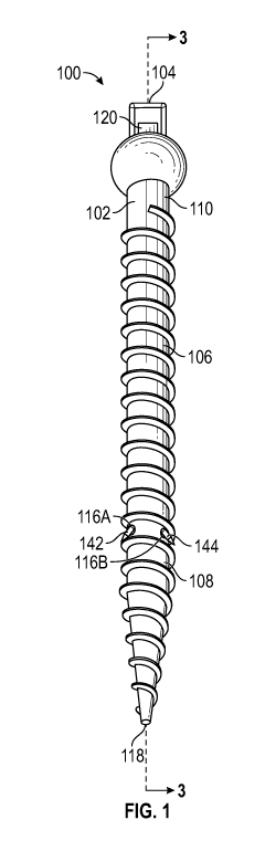
[0006] FIG. 1 is a perspective view of a first embodiment of a
pedicle
screw assembly having a pedicle screw and a bone anchor with integral anchor
ends, according to one aspect of the present disclosure;
[0007] FIG. 2 is an exploded view of the pedicle screw assembly of
FIG. 1 depicting the pedicle screw and the bone anchor, according to one
aspect of
the present disclosure;
[0008] FIG. 3 is a cross-sectional view of the pedicle screw taken
along
line 3-3 of FIG. 1, according to one aspect of the present disclosure;
[0009] FIG. 4 is a cross-sectional view of the pedicle screw of FIG.
3
taken along line 4-4 showing the bone anchor disposed partially within a
channel of
the pedicle screw, according to one aspect of the present disclosure;
[0010] FIG. 5 is a cross-sectional view of the pedicle screw of FIG.
3
showing the bone anchor disposed fully within the channel of the pedicle screw
and
deployed through an opening formed at the distal end of the pedicle screw,
according to one aspect of the present disclosure;
[0011] FIG. 6 is an anatomical perspective view of the pedicle screw
partially engaged inside a pedicle of a vertebra with the bone anchor in a pre-
deployment position, according to one aspect of the present disclosure;
[0012] FIG. 7 is an anatomical perspective view of the pedicle screw
partially engaged inside a pedicle vertebra with the anchor in a post-
deployment
position, according to one aspect of the present disclosure;
[0013] FIG. 8 is a perspective view of a second embodiment of a
pedicle screw assembly having a pedicle screw, a bone anchor, and anchor ends,
according to one aspect of the present disclosure;
[0014] FIG. 9 is an exploded view of the pedicle screw assembly of
FIG. 8 depicting the pedicle screw and the bone anchor, according to one
aspect of
the present disclosure;
[0015] FIG. 10 is a cross-sectional view of the pedicle screw of FIG.
9
taken along line 10-10, according to one aspect of the present disclosure;
[0016] FIG. 11 is a cross-sectional view of the pedicle screw of FIG.
8
taken along line 11-11 showing the bone anchor disposed partially within a
channel
of the pedicle screw, according to one aspect of the present disclosure;
2
CA 03089126 2020-07-20
WO 2019/161100
PCT/US2019/018070
[0017] FIG. 12 is a cross-sectional view of the pedicle screw of FIG.
8
showing the bone anchor disposed fully within the channel of the pedicle screw
and
deployed through an opening formed at the distal end of the pedicle screw,
according to one aspect of the present disclosure;
[0018] FIG. 13 is an anatomical perspective view of the pedicle screw
partially engaged inside a pedicle vertebra with the bone anchor in a pre-
deployment
position, according to one aspect of the present disclosure; and
[0019] FIG. 14 is an anatomical perspective view of the pedicle screw
partially engaged inside a pedicle vertebra with the anchor in a post-
deployment
position, according to one aspect of the present disclosure.
[0020] Corresponding reference characters indicate corresponding
elements among the view of the drawings. The headings used in the figures do
not
limit the scope of the claims.
DETAILED DESCRIPTION
[0021] Various embodiments of a pedicle screw assembly having one
or more bone anchors that are engaged along a pedicle screw for securing the
pedicle screw within a pedicle of a vertebra are disclosed. In some
embodiments,
the pedicle screw assembly includes a pedicle screw having external threads
configured to allow the pedicle screw to be partially engaged within a pedicle
vertebra. In addition, various embodiments of the pedicle screw may include
one or
more channels in communication with respective distal openings configured to
accommodate passage of bone anchor ends through the body of the pedicle screw
as described herein. In some embodiments, the bone anchor ends may extrude
outwardly from the pedicle screw when the bone anchor is in a post-deployment
configuration, thereby driving the anchor ends within bone, which further
engages
the pedicle screw to the pedicle vertebra.
[0022] In some embodiments, the bone anchor generally includes an
elongated anchor body defining a base and a pair of anchor members extending
from the base, with at least a portion of the anchor body being bendable
and/or
flexible. The anchor members of the bone anchor are configured for insertion
within
the channels of the pedicle screw and further configured to drive the anchor
ends
into the bone tissue as described herein.
3
CA 03089126 2020-07-20
WO 2019/161100
PCT/US2019/018070
[0023] In one method of engaging a pedicle screw to bone or other
target location, the pedicle screw is inserted into a pedicle of the vertebra
and the
bone anchor is inserted through the respective channels of the pedicle screw
such
that the anchor ends associated with the bone anchor extend outwardly from the
pedicle screw in a post-deployment position and engage bone tissue, thereby
further
engaging the pedicle screw to the pedicle vertebra. Referring to the drawings,
embodiments of a pedicle screw assembly are illustrated and generally
indicated as
100 and 200 in FIGS. 1-14.
[0024] Referring to FIGS. 1-7, a first embodiment of a pedicle screw
assembly, designated 100, is illustrated. As shown in FIGS. 1-3, the pedicle
screw
assembly 100 includes a pedicle screw 102 that is configured to be
mechanically
coupled to a bone anchor 104 in a manner that is suitable for anchoring the
pedicle
screw 102 to bodily tissue, such as the bone tissue of a vertebra. In some
embodiments, the pedicle screw 102 general includes an elongated screw body
106
defining a distal portion 108 (including a conical tip 118 defined at the free
end
thereof) and a proximal portion 110 that defines a pair of proximal openings
112
(shown in FIG. 3); designated a first proximal opening 112A and second
proximal
opening 112B. As indicated in FIG. 3, the first proximal opening 112A is in
communication with a first channel 114A formed at least partially along a
longitudinal
axis X1 of the pedicle screw 102, and the second proximal opening 112B is in
communication with a second channel 114B formed at least partially along the
longitudinal axis X1 of the pedicle screw 102. In some embodiments as shown,
at
least a portion of the first channel 114A is in parallel relation relative to
the second
channel 114B. Further, the screw body 106 may define a threaded portion 115
defining external threads that extends substantially along the length of the
screw
body 106 configured to accommodate the pedicle screw 102 to engage and be
retained within bone tissue.
[0025] Referring to FIG. 3, along the distal portion 108 of the
pedicle
screw 102, the first channel 114A is in communication with a first distal
opening
116A formed through a first lateral side 117A of the pedicle screw 102 along
the
distal portion 108 of the screw body 106, and the second channel 114B is in
communication with a second distal opening 116B formed through a second
lateral
side 117B of the pedicle screw 102 along the distal portion 108 of the screw
body
106. In some embodiments, the first lateral side 117A of the pedicle screw 102
is
4
CA 03089126 2020-07-20
WO 2019/161100
PCT/US2019/018070
generally defined opposite the second lateral side 117B of the pedicle screw
102
such that the first distal opening 116A is formed opposite the second distal
opening
116B, but the present disclosure is not limited in this regard. In addition,
as
indicated, the first channel 114A may include a first portion 118A extending
from the
proximal portion 110 of the screw body 106, and a second portion 119A adjacent
the
first distal opening 116A. Similarly, the second channel 114B may include a
first
portion 118B extending from the proximal portion 110 of the screw body 106,
and a
second portion 119B adjacent the second distal opening 116B. Each of the
second
portion 119A and the second portion 119B may be configured to be at least
partially
non-linear, and may define a curve as shown or a bend away from the
longitudinal
axis X1 of the pedicle screw body 106. In this manner, the first channel 114A
and
the second channel 114B may define a general partial C-shape or arcuate shape
configuration, a general partial Y-shape configuration (not shown), or T-shape
configuration (not shown). It is contemplated that the first channel 114A and
the
second channel 114B may take on any form so long as the first channel 114A is
in
communication with the first distal opening 116A and the second channel 114B
is in
communication with the second distal opening 116B so that portions of the bone
anchor 104 can traverse through the first channel 114A and extrude out the
first
distal opening 116A and portions of the bone anchor 104 can similarly traverse
through the second channel 114B and extrude out the second distal opening
116B,
as further described herein. In some embodiments, the first and second distal
openings 116A and 116B may be defined, respectively, directly adjacent the
first
portion 118A and the first portion 118B (not shown), above the first portion
118A and
the first portion 118B (not shown), below the first portion 118A and the first
portion
118B as shown in FIG. 3, such that the first distal opening 116A the second
distal
opening 116B are oriented closer to the conical tip 118 of the pedicle screw
102 than
the first portion 118A of the first channel 114A and the first portion 118B of
the
second channel 114B.
[0026] In addition, as shown in FIGS. 1-5, in some embodiments the
pedicle screw 102 may include a stopper 120 for obstructing the movement of at
least a portion of the bone anchor 104 relative to the screw body 106 in order
to
allow the bone anchor 104 to be removed from the screw body 106, as further
described herein. The stopper 120 may generally define a block or ridge shaped-
component formed along the proximal portion 110 of the screw body 106 between
CA 03089126 2020-07-20
WO 2019/161100
PCT/US2019/018070
the first proximal opening 112A and the second proximal opening 112B as shown,
and may comprise rubber, plastic, steel or any biocompatible material. In some
embodiments, the stopper 120 may further be removable from the screw body 106
where, e.g., it is desired to permanently engage the bone anchor 104 to the
screw
body 106 and bone tissue.
[0027] Referring back to FIG. 2, in some embodiments the bone anchor
104 includes an anchor body 130, defining a proximal portion 131A and a distal
portion 131B, with the anchor body 130 configured for at least partial
insertion within
the screw body 106 to engage the pedicle screw 102 to a pedicle or other
target
sites. In some embodiments, the anchor body 130 includes a base portion 132
along the proximal portion 131A of the anchor body 130, with the base portion
132
defining a first side 134 and a second side 136 opposite the first side 134,
as
indicated. The base portion 132 may generally define a cuboidal or
cylindrically
shaped configuration and may be manufactured in any number of ways sufficient
to
receive a force (F in FIG. 4) for driving the bone anchor 104 partially
through the
screw body 106, as further described herein. For example, the base portion 132
may include a handle or thumb print to receive a force applied by a human
hand, or
may define a rigid surface for receiving a force applied by a blunt instrument
such as
a hammer.
[0028] As shown, the anchor body 130 may further include a first
anchor member 138 extending orthogonally from the first side 134 of the base
portion 132 and configured for insertion within the first channel 114A of the
screw
body 106. Similarly, the anchor body 130 may include a second anchor member
140
extending orthogonally from the second side 136 of the base portion 132 and
configured for insertion within the second channel 114B. In some embodiments,
the
first anchor member 138 is in substantially parallel relation relative to the
second
anchor member 140, as in the case where at least a portion of the first
channel 114A
and the second channel 114B define a similar parallel configuration, but the
present
disclosure is not limited in this regard. In general, the first anchor member
138 and
the second anchor member 140 may define any shape suitable for insertion
within
the first channel 114A and the second channel 114B of the screw body 106,
respectively. For example, the first anchor member 138 and the second anchor
member 140 may be generally cylindrical in shape, may define planar plates or
tabs,
may define spikes, etc., and may be formed with any biocompatible material,
one or
6
CA 03089126 2020-07-20
WO 2019/161100
PCT/US2019/018070
more metals, plastics, or combinations thereof. In addition, while two anchor
members (the first anchor member 138 and the second anchor member 140) are
shown, the anchor body 130 may define a sole anchor member (not shown) or any
number of anchor members as desired (not shown). At least a portion of the
anchor
body 130, including e.g., the first anchor member 138 and the second anchor
member 140 may be at least partially bendable, biasable, and/or flexible to
accommodate passage of the first anchor member 138 and the second anchor
member 140 through the first channel 114A and the second channel 114B,
respectively.
[0029] In addition, the first anchor member 138 may define an anchor
end 142, and the second anchor member 140 may define an anchor end 144 along
the distal portion 131B of the anchor body 130. The anchor end 142 and the
anchor
end 144 may define any number of shapes or configurations, may be cylindrical
in
shape, may define planar plates or tabs, and/or may define spikes or blades,
etc.,
such that the anchor end 142 and the anchor end 144 can penetrate bone tissue.
In
some embodiments, the anchor end 142 and the anchor end 144 are integral with
the first anchor member 138 and the second anchor member 140, respectively. In
some embodiments, the anchor end 142 and the anchor end 144 define a general
tapered shape configuration.
[0030] Referring to FIGS. 4-7, one method of implanting the pedicle
screw assembly 100 is illustrated. As shown in FIG. 6, the pedicle screw 102
is first
engaged into bone tissue, such as a pedicle of the vertebra, so that the
proximal
portion 110 of the pedicle screw 102 extends from the bone tissue. Once the
pedicle
screw 102 is implanted into the bone tissue, the first anchor member 138 and
the
second anchor member 140 of the bone anchor 104 are inserted into the first
proximal opening 112A and the second proximal opening 112B, respectively, of
the
pedicle screw 102, in the manner indicated in FIG. 4 to form a pre-deployment
configuration 150 of the pedicle screw assembly 100.
[0031] Once the pre-deployment configuration 150 is formed, the first
anchor member 138 is at least partially disposed within the first channel
114A, and
the second anchor member 140 is at least partially disposed within the second
channel 114B as shown in FIG. 4. Thereafter, in some embodiments, a force F
may
be applied as shown to drive the first anchor member 138 further through the
first
channel 114A and simultaneously drive the second anchor member 140 further
7
CA 03089126 2020-07-20
WO 2019/161100
PCT/US2019/018070
through the second channel 114B in the direction Dl. In some embodiments, the
force F may be applied to the base portion 132 of the anchor body 130 by a
hammer
or other blunt instrument (not shown).
[0032] Referring to FIG. 5 and FIG. 7, the application of the force F
shifts the pedicle screw assembly 100 from the pre-deployment configuration
150 to
a post-deployment configuration 152. In this post-deployment configuration
152, the
anchor end 142 of the first anchor member 138 is extruded from the first
distal
opening 116A, and the anchor end 144 is extruded from the second distal
opening
116B as shown in FIG. 5, which drives the anchor end 142 and the anchor end
144
into bone tissue as shown in FIG. 7. In some embodiments, when the pedicle
screw
102 includes the stopper 120, the base portion 132 may contact the stopper 120
and
limit the depth to which the first anchor member 138 and the second anchor
member
140 can be passed within the first channel 114A and the second channel 114B
respectively in the direction Dl. In addition, in some embodiments, an axial
gap 154
may be formed around the stopper 120. In this manner, a pair of plyers, hook,
hammer, or other such instrument (not shown) may be engaged to the base
portion
132 by maneuvering around the axial gap 154 when the pedicle screw assembly
100
is in the post-deployment configuration 152, and the bone anchor 104 may be
pulled
away from the pedicle screw 102 in a direction opposite D1 (not shown),
thereby
removing the first anchor member 138 from within the first channel 114A and
removing the second anchor member 140 from within the second channel 114B.
Disengagement of the bone anchor 104 from the pedicle screw 102 in this manner
may be advantageous where implementation of the pedicle screw assembly 100 is
no longer desired or needs to be removed from the bone tissue for whatever
reason.
[0033] Referring to FIGS. 8-14, a second embodiment of the pedicle
screw assembly, designated 200, is illustrated. As shown in FIGS. 8-11, the
pedicle
screw assembly 200 includes a pedicle screw 202 that is configured to be
mechanically coupled to a bone anchor 204 in a manner that is suitable for
anchoring the pedicle screw 202 to bodily tissue, such as the bone tissue of a
vertebra. In some embodiments, the pedicle screw 202 generally includes an
elongated screw body 206 defining a distal portion 208 (including a conical
tip 218 at
the free end thereof) and a proximal portion 210 that defines a pair of
proximal
openings 212; including a first proximal opening 212A and second proximal
opening
212B. As indicated, the first proximal opening 212A is in communication with a
first
8
CA 03089126 2020-07-20
WO 2019/161100
PCT/US2019/018070
channel 214A formed at least partially along a longitudinal axis X2 of the
pedicle
screw 202, and the second proximal opening 212B is in communication with a
second channel 214B formed at least partially along the longitudinal axis X2
of the
pedicle screw 202. In some embodiments as shown, at least a portion of the
first
channel 214A is in parallel relation relative to the second channel 214B.
Further, the
screw body 206 may define a threaded portion 215 that extends substantially
along
the length of the screw body 206 configured to accommodate the pedicle screw
202
to engage and be retained within bone tissue.
[0034] Referring to FIG. 10, along the distal portion 208 of the
pedicle
screw 202, the first channel 214A is in communication with a first distal
opening
216A formed through a first lateral side 217A of the pedicle screw 202 along
the
distal portion 208 of the screw body 206, and the second channel 214B is in
communication with a second distal opening 216B formed through a second
lateral
side 217B of the pedicle screw 202 along the distal portion 208 of the screw
body
206. In some embodiments, the first lateral side 217A of the pedicle screw 202
is
generally defined opposite the second lateral side 217B of the pedicle screw
202
such that the first distal opening 216A is formed opposite the second distal
opening
216B, but the present disclosure is not limited in this regard. In addition,
as
indicated, the first channel 214A may include a first portion 218A extending
from the
proximal portion 210 of the screw body 206, and a second portion 219A adjacent
the
first distal opening 216A. Similarly, the second channel 114B may include a
first
portion 218B extending from the proximal portion 210 of the screw body 206,
and a
second portion 219B adjacent the second distal opening 216B. Each of the
second
portion 219A and the second portion 219B may be at least partially non-linear,
and
may define a curve as shown or a bend away from the longitudinal axis X2 of
the
pedicle screw body 206. In this manner, the first channel 214A and the second
channel 214B may define a general partial C-shape or arcuate shape
configuration,
a general partial Y-shape configuration (not shown), or T-shape configuration
(not
shown). It is contemplated that the first channel 214A and the second channel
214B
may take on any form so long as the first channel 214A is in communication
with the
first distal opening 216A and the second channel 214B is in communication with
the
second distal opening 216B so that portions of the bone anchor 204 can
traverse
through the first channel 214A and portions of the bone anchor 204 can
traverse
through the second channel 214B, as further described herein. In some
9
CA 03089126 2020-07-20
WO 2019/161100
PCT/US2019/018070
embodiments, the first distal opening 216A the second distal opening 216B may
be
defined, respectively, directly adjacent the first portion 218A and the first
portion
218B (not shown), may be defined above the first portion 218A and the first
portion
218B (not shown), or may be defined below the first portion 218A and the first
portion 218B as shown in FIG. 10, such that the first distal opening 216A the
second
distal opening 216B are oriented closer to the conical tip 218 of the pedicle
screw
202 than the first portion 218A of the first channel 214A and the first
portion 218B of
the second channel 214B.
[0035] In addition, the pedicle screw 202 may include a stopper 220
for
obstructing the movement of at least a portion of the bone anchor 204 relative
to the
screw body 206 in order to allow the bone anchor 204 to be removed from the
screw
body 206, as further described herein. The stopper 220 may generally define a
block or ridge shaped-component formed along the proximal portion 210 of the
screw body 206 between the first proximal opening 212A and the second proximal
opening 212B as shown, and may be formed from rubber, plastic, steel or any
biocompatible material. In some embodiments, the stopper 220 may further be
removable from the screw body 206 where, e.g., it is desired to permanently
engage
the bone anchor 204 to the screw body 206 and bone tissue.
[0036] Referring back to FIG. 9, in some embodiments the bone anchor
204 includes an anchor body 230, defining a proximal portion 231A and a distal
portion 231B, with the anchor body 230 configured for at least partial
insertion within
the screw body 206 to engage the pedicle screw 202 to a pedicle or other
target site.
In some embodiments, the anchor body 230 includes a base portion 232 along the
proximal portion 231A of the anchor body 230, with the base portion 232
defining a
first side 234 and a second side 236 opposite the first side 234 as indicated.
The
base portion 232 may generally define a cuboidal or cylindrically shaped
configuration and may be manufactured in any number of ways sufficient to
receive a
force (F in FIG. 11) for driving the bone anchor 204 partially through the
screw body
206, as further described herein. For example, the base portion 232 may
include a
handle or thumb print to receive a force applied by a human hand, or may
define a
rigid surface for receiving a force applied by a blunt instrument such as a
hammer.
[0037] As shown, the anchor body 230 may further include a first
anchor member 238 extending orthogonally from the first side 234 of the base
portion 232 and configured for insertion within the first channel 214A.
Similarly, the
CA 03089126 2020-07-20
WO 2019/161100
PCT/US2019/018070
anchor body 230 includes a second anchor member 240 extending orthogonally
from
the second side 236 of the base portion 232 and configured for insertion
within the
second channel 214B. In some embodiments, the first anchor member 238 is in
parallel relation relative to the second anchor member 240, as in the case
where at
least a portion of the first channel 214A and the second channel 214B define a
similar parallel configuration, but the present disclosure is not limited in
this regard.
In general, the first anchor member 238 and the second anchor member 240
define
any shape suitable for insertion within the first channel 214A and the second
channel
214B of the screw body 206, respectively. For example, the first anchor member
238 and the second anchor member 240 may be generally cylindrical in shape,
may
define planar plates or tabs, may define spikes, etc., and may be formed with
any
biocompatible material, one or more metals, plastics, or combinations thereof.
In
addition, while two anchor members (the first anchor member 238 and the second
anchor member 240) are shown, the anchor body 230 may define a sole anchor
member (not shown) for engaging a sole channel of the screw body 206 (not
shown)
or any number of anchor members as desired (not shown). At least a portion of
the
anchor body 230, including e.g., the first anchor member 238 and the second
anchor
member 240 may be at least partially bendable or flexible to accommodate
passage
of the first anchor member 238 and the second anchor member 240 at least
partially
through the first channel 214A and the second channel 214B, respectively.
[0038] Referring to FIGS. 8-12 and FIG. 14, the pedicle screw
assembly 200 may further include an anchor end 242 staged within the second
portion 219A of the first channel 214A, and a second anchor end 244 staged
within
the second portion 219B of the second channel 214B. In this embodiment of the
pedicle screw assembly 200, the anchor end 242 and the anchor end 244 are not
formed integrally with the anchor body 130, but are instead disposed within
the first
and second channels 214A and 214B of the screw body 206 as indicated. The
anchor end 242 may be staged just inside the screw body 206 within the second
portion 219A directly adjacent to the distal opening 216A, and the anchor end
244
may be staged just inside the screw body 206 within the second portion 219B
directly adjacent to the distal opening 216B as shown in FIG. 10. As further
described herein, when the anchor 204 is engaged to the pedicle screw body 206
(i.e., the first anchor member 238 is driven through the first channel 214A
and the
second anchor member 240 is concurrently driven through the second channel
11
CA 03089126 2020-07-20
WO 2019/161100
PCT/US2019/018070
214B), the first anchor member 238 drives the anchor end 242 at least
partially
through the first distal opening 216A, and concurrently the second anchor
member
240 drives the anchor end 244 at least partially through the second distal
opening
216B. The anchor end 242 and the anchor end 244 may define any number of
shapes or configurations, may be cylindrical in shape, may define planar
plates or
tabs, and/or may define spikes or blades, etc., such that the anchor end 242
and the
anchor end 244 can penetrate bone tissue. In some embodiments, the anchor end
242 and the anchor end 244 define a general tapered shape configuration.
[0039] As further indicated in FIG. 10, the pedicle screw assembly
200
may include a first tether member 245A and a second tether member 245B. The
first tether member 245A may be engaged to the anchor end 242 and the screw
body 206 within the channel 214A, and the second tether member 245B may be
engaged to the anchor end 244 and the screw body 206 within the channel 214B.
The first tether member 245A and the second tether member 245B may comprise a
rubber member or other such flexible component. In use, the first tether
member
245A restricts a predetermined degree of movement of the anchor end 242
relative
to the screw body 206 so that the anchor end 242 does not merely fall outside
the
distal opening 216A and the first channel 214A, and the second tether member
245B restricts a predetermined degree of movement of the anchor end 244
relative
to the screw body 206 so that the anchor end 244 does not merely fall outside
the
distal opening 216B and the second channel 214B.
[0040] Referring to FIGS. 11-14, one method of implanting the pedicle
screw assembly 200 is illustrated. As shown in FIG. 13, the pedicle screw 202
is first
engaged into bone tissue, such as a pedicle of the vertebra, such that the
proximal
portion 210 of the pedicle screw 202 extends from the bone tissue. Once the
pedicle
screw 202 is implanted into the bone tissue, the first anchor member 238 and
the
second anchor member 240 of the bone anchor 204 are inserted into the first
proximal opening 212A and the second proximal opening 212B, respectively, of
the
pedicle screw 202, in the manner indicated in FIG. 11 to form a pre-deployment
configuration 250 of the pedicle screw assembly 200. In this manner, the
terminal
ends of the first anchor member 238 and the second anchor member 240 contact
the
anchor end 242 and the anchor end 244, respectively, as shown.
[0041] Once the pre-deployment configuration 250 is formed, and the
first anchor member 238 is at least partially received within the first
channel 214A as
12
CA 03089126 2020-07-20
WO 2019/161100
PCT/US2019/018070
indicated, and the second anchor member 240 is at least partially received
within the
second channel 214B, a force F may be applied as shown to drive the first
anchor
member 238 against the first anchor end 242 and concurrently drive the second
anchor member 240 against the second anchor end 244 in the direction D1,
thereby
causing the anchor end 242 and the anchor end 244 to at least partially
extrude
through the distal opening 216A and the distal opening 216B, respectively. The
force F may be applied to the base portion 232 of the anchor body 230 by a
hammer
or other blunt instrument (not shown).
[0042] Referring to FIG. 12 and FIG. 14, the application of the force
F
shifts the pedicle screw assembly 200 from the pre-deployment configuration
250 to
a post-deployment configuration 252. In this post-deployment configuration
252, at
least a portion of the anchor end 242 is extruded outside the first distal
opening
216A, and at least a portion of the anchor end 244 is extruded outside the
second
distal opening 216B as shown in FIG. 12, which drives the anchor end 242 and
the
anchor end 244 into bone tissue as shown in FIG. 14. In some embodiments, when
the pedicle screw 202 includes the stopper 220, the base portion 232 may
contact
the stopper 220 and limit the depth to which the first anchor member 238 and
the
second anchor member 240 can traverse the first channel 214A and the second
channel 214B respectively in the direction Dl. In addition, in some
embodiments, an
axial gap 254 may be formed around the stopper 220. In this manner, a pair of
plyers, hook, hammer, or other such instrument (not shown) may be engaged to
the
base portion 232 by maneuvering around the axial gap 254 when the pedicle
screw
assembly 200 is in the post-deployment configuration 252, and the bone anchor
204
may be pulled away from the pedicle screw 202 in a direction opposite D1 (not
shown), thereby removing the first anchor member 238 from within the first
channel
214A and removing the second anchor member 240 from within the second channel
214B. Disengagement of the bone anchor 204 from the pedicle screw 202 in this
manner may be advantageous where, e.g., it is desired to re-use the anchor 204
for
other similar applications.
[0043] In some embodiments, the pedicle screw assemblies 100 and
200 are configured to be affixed to the larger vertebrae of the lumbar spine,
or the
smaller vertebrae of the thoracic or cervical spine.
[0044] In some embodiments, the pedicle screw assemblies 100 and
200 may be made from a metal, such as titanium, or a metal-based alloy, such
as
13
CA 03089126 2020-07-20
WO 2019/161100
PCT/US2019/018070
titanium-based alloy. Alternatively, the pedicle screw assemblies 100 and 200
may
comprise a reinforced polymer material. In some embodiments, the material used
to
manufacture the pedicle screw assemblies 100 and 200 can have a high
bioactivity
and high flexibility, and a result, can improve ingrowth and mechanical
fixation.
[0045] In some embodiments, the pedicle screws 102 and 202 may be
engaged to a tulip structure (not shown) which is configured to interface with
a
longitudinal bar or a plate. In some embodiments, the tulip structure can be
flexibly
coupled to the pedicle screws 102 and 202 by way of a ball-joint or other type
of
flexible joint such that the pedicle screw assemblies 100 and 200 can account
for
any bending of the individual's spine while still exerting an axial force on
the
longitudinal bar, thereby stabilizing the spine of the individual.
[0046] It should be understood from the foregoing that, while
particular
embodiments have been illustrated and described, various modifications can be
made thereto without departing from the spirit and scope of the invention as
will be
apparent to those skilled in the art. Such changes and modifications are
within the
scope and teachings of this invention as defined in the claims appended
hereto.
14